ESTRUTURA E BIOGÉNESE DA NADH:UBIQUINONA … · oxidorredutase (complexoi) da cadeia respiratÓria...
141
JORGE EDUARDO DA SILVA AZEVEDO ESTRUTURA E BIOGÉNESE DA NADH:UBIQUINONA OXfDORREDUTASE (COMPLEXO i) DA CADEIA RESPIRATÓRIA PORTO 1993
Transcript of ESTRUTURA E BIOGÉNESE DA NADH:UBIQUINONA … · oxidorredutase (complexoi) da cadeia respiratÓria...
JORGE EDUARDO DA SILVA AZEVEDO
ESTRUTURA E BIOGÉNESE DA NADH:UBIQUINONA OXfDORREDUTASE (COMPLEXO i) DA CADEIA
RESPIRATÓRIA
PORTO
1993
JORGE EDUARDO DA SILVA AZEVEDO
ESTRUTURA E BIOGÉNESE DA NADH:UBIQUINONA
OXIDORREDUTASE (COMPLEXO I) DA CADEIA
RESPIRATÓRIA
PORTO
1993
..
^
H h V
o
JORGE EDUARDO DA SILVA AZEVEDO
ESTRUTURA E BIOGÉNESE DA NADH:UBIQUINONA
OXIDORREDUTASE (COMPLEXO I) DA CADEIA
RESPIRATÓRIA
DISSERTAÇÃO DE CANDIDATURA AO GRAU DE DOUTOR EM CIÊNCIAS BIOMÉDICAS, ESPECIALIDADE DE BIOLOGIA MOLECULAR,
APRESENTADA AO INSTITUTO DE CIÊNCIAS BIOMÉDICAS DE ABEL SALAZAR DA UNIVERSIDADE DO PORTO
ORIENTADOR: Professor Doutor Sigurd Werner (Institut fur Physiologische Chemie - Universitat Mùnchen)
CO-ORIENTADOR: Professor Doutor Arnaldo Videira (Instituto de Ciências Biomédicas de Abel Salazar - Universidade do Porto)
PORTO 1993
À Isabel
I!
No cumprimento do Decreto-Lei 388/70, esclarece-se serem da nossa responsabilidade a execução das experiências apresentadas neste trabalho (excepto quando referido em contrário) assim como a sua interpretação e discussão.
II]
AGRADECIMENTOS
Ao Professor Doutor Sigurd Werner, pela sua contribuição para o alargamento dos meus conhecimentos e pela forma extraordinária como me recebeu e apoiou no seu laboratório em Munique.
Ao Professor Doutor Arnaldo Videira, pela orientação, acompanhamento e solidariedade ao longo deste processo.
À Sra. Heidi Kothe pela ajuda técnica na prossecução deste trabalho.
À Dra. Helga Heinrich e Dra. Jacqueline Abrolat-Scharff pela colaboração em algumas das experiências e, ainda, pela amizade e apoio durante a minha estadia em Munique.
Aos Drs. Tomeu Segui-Real e Klaus Dietemeier pelas discussões e apoio que me proporcionaram.
Aos Drs. Christoph Eckerskorn e Uwe Nehls pela sequenciação peptídica de várias subunidades do complexo I.
Ao Dr. Helmut Schneider pela cedência do banco de expressão de cDNAs.
A todos os membros do Laboratório de Genética Molecular - ICBAS pelo modo como me apoiaram neste último ano.
À Professora Doutora Leonor Teles Grilo por ter estabelecido o contacto com o Laboratório de Munique.
À Junta Nacional de Investigação Científica e Tecnológica, pela bolsa de estudo que me proporcionou e também pelo subsídio para a impressão desta tese.
IV
ABREVIATURAS
■ cDNA; ácido desoxirribonucleico complementar
FMN; mononucleótido de flavina
FMNH2; forma reduzida do FMN
NADH; dinucleótido de nicotinamida e adenina
NAD+; forma oxidada do NADH
e. p. r.; ressonância electrónica paramagnética
ATP; trifosfato de adenosina
mtDNA; DNA mitocondrial
SDS; dodecilsulfato de sódio
SDS-PAGE; electroforese em gel de poliacrilamida na presença de SDS
NUO-x; subunidade do complexo I de x kDa.
Nota: Alguns dos termos ingleses utilizados correntemente em Biologia
Molecular não foram traduzidos para português pela perda de clareza inerente à tradução. Tais palavras e expressões surgem no texto entre aspas.
V
INDICE
1 - RESUMO 1
2 - INTRODUÇÃO 5 2.1 - Distribuição filogenética do complexo I 6 2.2 - Composição do complexo I 8
2.2.1 - Componentes polipeptídicos 8 2.2.2 - Centros Fe-S e FMN 9
2.3 - Estrutura do complexo I 10 2.3.1 - Organização espacial das subunidades da enzima 10 2.3.2 - Localização dos sítios de ligação do NADH e FMN 14 2.3.3 - Localização dos centros Fe-S 15 2.3.4 - Localização do sítio de ligação da ubiquinona 16
2.4 - Relações estruturais/funcionais entre o complexo I mitocondrial e enzimas procarióticas 18
2.4.1 - A NAD+-hidrogenase de Alkaligenes eutrophus 18 2.4.2 - O complexo I de Paracoccus denitrificans 19 2.4.3 - A "acyl-carrier protein" 20
2.5 - O fluxo de electrões no complexo I 21 2.6 - Mecanismo de transdução energética no complexo I 23 2.7 - Biogénese e evolução do complexo I 25 2.8 - Aspectos médicos do complexo I 27
3 - OBJECTIVOS DO TRABALHO 31
4 - RESULTADOS OBTIDOS 33 4.1-Asubunidadede20.9kDa 34 4.2-Asubunidadede 12.3 kDa 37 4.3-Asubunidadede 17.8 kDa 38 4.4 - A subunidade de 14.8 kDa 41 4.5 - Isolamento e caracterização de um fragmento membranar do
complexo I 43
5-CONCLUSÕES FINAIS 45
VI
6 - BIBLIOGRAFIA 48
7 - TRABALHO EXPERIMENTAL 63
7.1 - "Primary structure and in vitro mitochondrial import of the 20.9 kDa subunit of complex I from Neurospora crassa" J. E. Azevedo, U. Nehls, C. Eckerskom, H. Heinrich, H. Rothe, H. Weiss & S. Werner (1992), Biochem. J. 288: 29-34.
7.2 - "The 12.3 kDa subunit of complex I (Respiratory-chain NADH dehydrogenase) from Neurospora crassa: cDNA cloning and chromosomal mapping cf the gene" A. Videira, J. E. Azevedo, S. Werner & P. Cabral (1993), Biochem. J. 2S1, 729-732.
7.3 - "Cloning, in vitro mitochondrial import and membrane assembly of the 17.8 kDa subunit of complex I from Neurospora crassa" J. E. Azevedo, J. Abrolat-Scharff, C. Eckerskom & S. Werner (1993), Biochem. J. 293: 501-506.
7.4 - "in organello assembly of respiratory chain complex I. Primary structure of the 14.8 kDa subunit of Neurospora crassa complex I" J. E. Azevedo, C. Eckerskom & S. Werner (1993). (submetido para publicação).
7.5 - "Characterization of a membrane fragment of complex I. Insights on the topology of the ubiquinone-binding site" J. E. Azevedo & A. Videira (1993). (submetido para publicação).
VII
VIII

1-RESUMO
Vários anticorpos policlonais dirigidos contra subunidades do complexo I de Neurospora crassa foram preparados e utilizados no rastreio de um banco de expressão de cDNAs. Deste modo, foram isolados clones codificantes das subunidades 20.9 kDa, 17.8 kDa e 14.8 kDa. As sequências destes cDNAs e, adicionalmente, de um cDNA codificante da subunidade de 12.3 kDa, foram determinadas. A análise das estruturas primárias destas subunidades deduzidas a partir dos cDNAs respectivos, revelou, em alguns casos, a existência de homologias e similaridades com proteínas de bovinos. As implicações estruturais e funcionais destas similaridades são discutidas.
Os mecanismos de montagem das subunidades de 20.9 kDa, 17.8 kDa e 14.8 kDa foram estudados utilizando um sistema in organelle É demonstrado que todas estas subunidades requerem a existência de um potencial de membrana para serem eficientemente importadas por mitocôndrias isoladas. Uma vez importadas in vitro, as subunidades de 20.9 kDa e 17.8 kDa são encontradas na membrana interna mitocondrial apresentando características típicas das subunidades endógenas. Os dados apresentados sugerem que nenhuma destas proteínas precursoras é translocada completamente para a matriz mitocondrial. Aparentemente, estas proteínas precursoras terminam o processo de translocação membranar ao nível da membrana interna mitocondrial.
Experiências de co-imunoprecipitação e de sedimentação em gradientes de sacarose, demonstraram que uma fracção significativa da subunidade de 14.8 kDa importada in vitro é montada no complexo I. Esta observação sugere que toda a maquinaria enzimática necessária para a biossíntese do complexo I se encontra operacional em mitocôndrias isoladas, permitindo, assim, a utilização deste sistema in organello no estudo do processo de montagem da enzima.
A técnica da extracção alcalina de biomembranas, desenvolvida por Fujiki et ai. (1982), foi utilizada com o intuito de caracterizar o tipo de interacção que várias subunidades do complexo I estabelecem com a membrana interna mitocondrial. Os resultados obtidos sugerem que as subunidades de 49 kDa e 30.4 kDa são proteínas extrínsecas de membrana; as subunidades de 20.9 kDa, 17.8 kDa e 12.3 kDa apresentam um comportamento típico de proteínas intrínsecas de membrana. Após extracção alcalina de mitocôndrias isoladas, foi
também possível isolar um subcomplexo que representa uma parte (ou a totalidade) do braço membranar do complexo I. Uma análise comparativa da composição polipeptídica deste fragmento e do subcomplexo la da enzima bovina (Finei et ai., 1992) sugere que a subunidade de 20.8 kDa [uma possível metalo-proteína (Videira et ai., 1990a)] e a subunidade de 9.3 kDa [um componente do sítio de ligação da ubiquinona (Heinrich & Werner, 1992; Heinrich et ai., 1992)], se encontram na interface dos braços membranar e periférico do complexo I.
2
SUMMARY
Several polyclonal antibodies were prepared against Neurospora crassa complex I subunits and used in the screening of a cDNA expression library. Full-length clones encoding the 20.9 kDa, 17.8 kDa e 14.8 kDa were isolated. The sequences of these cDNAs and, additionally, of a clone encoding the 12.3 kDa subunit of complex I were determined. Sequence analysis of the deduced primary structures of two of these subunits revealed interesting homologies and similarities with bovine proteins. The significance of these findings are discussed.
The biogenetic pathways of the 20.9 kDa, 17.8 kDa and 14.8 kDa subunits were studied. It is shown that all these subunits require a membrane potential to be imported into isolated mitochondria. Once imported, the 17.8 kDa and 20.9 kDa subunits are found in the mitochondrial inner membrane where they have acquired characteristics of the endogenous subunits. The data presented suggest that neither of these precursor proteins is completely translocated into the matrix, but instead, translocation of both membrane systems (/'. e., outer and inner membrane) is arrested at the level of the mitochondrial inner membrane.
A significant fraction of the in vitro imported 14.8 kDa subunit was found in completely assembled complex I demonstrating that even in isolated mitochondria all the machinery required for the assembly process is still operational. Tnis finding will allow the use of an in vitro system for the study of the biogenesis of complex I.
The technique of alkaline extraction of biological membranes (Fujiki et a/., 1982) was employed to study the kind of interaction of several complex I subunits with the mitochondrial inner membrane. The results obtained suggest the 49 kDa and 30.4 kDa subunits are extrinsic membrane proteins; the 20.9 kDa, 17.8 kDa and 12.3 kDa behave as intrinsic membrane proteins. After alkaline extraction of mitochondria a subcomplex of the enzyme representing a fraction (or the totality) of the membrane arm of the enzyme was isolated. A comparative analysis of the polypeptide composition of this fragment with subcomplex la of the bovine enzyme (Finel et ai., 1992) suggests that the 20.8 kDa subunit [a possible iron-sulphur protein (Videira ef a/., 1990a)] and the 9.3 kDa subunit [a polypeptide at or near the ubiquinone-binding site (Heinrich & Werner, 1992; Heinrich et ai., 1992)] are located in the junction of the two arms of complex I.
3
SOMMAIRE
Plusieurs anticorps policlonaux dirigés contre sousunités du complexe I de Neurospora crassa ont été préparés et utilisés pour chercher une banque d' expression de cDNAs. Nous avons isolé des clones qui codifient les sousunités de 20.9 kDa, 17.8 kDa et 14.8 kDa. Les séquences de ces cDNAs et, en plus, d' un autre cDNA pour la sousunité 12.3 kDa, ont été déterminés. L' analyse des structures primaires de ces sousunités a montré, dans certains cas, l'existence de homologies et similarités avec des protéines bovines.
Les méchanismes biogenétiques des sousunités 20.9 kDa, 17.8 kDa et 14.8 kDa ont été étudiés. Nous avons montré que toutes les sousunités ont besoin d' un potentiel de membrane pour être importées dans des mitochondries isolées. Après I' importation in vitro, les sousunités 20.9 kDa et 17.8 kDa apparaît dans la membrane interne de la mitochondrie avec des charactéristiques que ressemblent ceux des sousunités endogènes. Les résultats indiquent que aucune de certes sousunités est complètement transposée pour la matrice mitochondrielle.
Des expériences de co-immunoprécipitation et sédimentation ont montré que une partie significative de la sousunité 14.8 kDa importée in vitro est associée avec le complexe I. Ceci indique que toute la machinerie enzymatique nécessaire à la biosynthèse du complexe I est encore opéracionnelle dans des mitochondries isolées, ce qui permett I' utilization de ce système in organnelle pour P étude du processus de montage de P enzyme.
La technique d' extraction alcaline de biomembranes développée par Fujiki et al. (1982) a été utilisée pour charactériser P interaction entre les sousunités du complexe I et la membrane interne de la mitochondrie. Les résultats obtenus indiquent que les sousunités 49 kDa et 30.4 kDa sont extrinsèque à la membrane; les sousunités 20.9 kDa, 17.8 kDa et 12.3 kDa présentent un comportement de protéines intrinsèque.
Après P extraction alcaline de mitochondries isolées, nous avons isolé un sous-complèxe que réprésente une partie (ou la totalité) du domaine membranaire du complexe I. Une analyse comparative de la constitution en protéines de ce fragment et celle du sous-complèxe la de P enzyme bovine (Finel et al., 1992) indique que la sousunité 20.8 kDa [peut-être une métallo-protéine (Videira et al., 1990a)] et la sousunité 9.3 kDa [un composant du local de liaison de I' ubiquinone (Heinrich & Werner, 1992; Heinrich ef al., 1992)] existent dans la interface des domaines membranaire et périphérique du complexe I.
4
2- INTRODUÇÃO
As mitocôndrias são estruturas ubíquas no reino dos eucariotas. É nestes
organelos que ocorre a denominada fosforilação oxidativa, uma via metabólica
complexa onde a maior parte da energia resultante da oxidação dos alimentos
é convertida em ATP. A maquinaria enzimática catalizadora deste processo
contém cinco complexos proteicos multiméricos fulcrais:
(1) NADH.ubiquinona oxidorredutase (complexo I);
(2) Succinato:ubiquinona oxidorredutase (complexo II)
(3) Ubiquinol:citocromo c oxidorredutase (complexo III);
(4) Citocromo c oxidase (complexo IV); e
(5) ATP sintetase (complexo V).
De um modo sequencial e com a intervenção de outros componentes (e. g.,
ubiquinona e citocromo c) estes complexos catalizam a transferência de
electrões do NADH e succinato para o oxigénio. Ao nível dos complexos I, III e
IV, a energia libertada nas reacções redox é acoplada à extrusão de protões
através da membrana interna mitocondrial (como artigos de revisão ver Hatefi,
1985; Tzagoloff & Myers, 1986). A energia associada ao gradiente
electroquímico assim gerado é, então, utilizada pelo complexo V para a síntese
de ATP (Mitchell, 1966).
Dada a enorme complexidade deste sistema, muito pouco é ainda
conhecido a nível molecular sobre os mecanismos de catálise, biogénese,
estrutura e regulação dos seus diversos componentes. A compreensão de
alguns destes aspectos poderá ter importantes consequências, não só em
termos de bioenergética (e as suas aplicações biotecnológicas), mas também
no campo da medicina onde cada vez mais se detectam correlações entre
várias doenças e anomalias da cadeia respiratória.
5
O trabalho aqui apresentado incide sobre a estrutura e biogénese do
primeiro componente da cadeia de fosforilação oxidativa - a NADH:ubiquinona
oxidorredutase. Esta enzima é, sem dúvida, o mais complexo de todos os
elementos da cadeia respiratória, razão pela qual tem permanecido o menos
compreendido. Na exposição seguinte vários aspectos do conhecimento actual
sobre o complexo I são discutidos detalhadamente.
2.1- Distribuição filogenética do complexo I
A maior parte da informação actualmente disponível sobre a distribuição
filogenética do complexo I adveio da descoberta que algumas das subunidades
da enzima são codificadas e sintetizadas na mitocôndria (Chomyn et ai.,
1985,1986). Assim, a presença no DNA mitocondrial de um dado organismo de
"ORFs" ("Open Reading Frames") codificantes de proteínas homólogas às
subunidades ND1-ND6 e ND4L (ver 2.2.1) tem sido considerada como
evidência para a existência de complexo I nesse organismo. Explorando esta
característica, foi demonstrada a existência de complexo I em diversos
organismos eucarióticos desde mamíferos (Anderson et ai., 1981) a
protozoários (Pritchard et ai., 1990), passando por pássaros (Desjardins &
Morais, 1990), anfíbios (Roe et ai., 1985), peixes (Johansen et ai., 1990),
insectos (Clary & Wolstenholme, 1985), plantas (Oda et ai., 1992), algas
(Denovan-Wright & Lee, 1992) e fungos (Burger & Werner, 1986; Nelson &
Macino, 1987). A ubiquidade filogenética do complexo I tem, no entanto, uma
excepção infeliz - a levedura Saccharomyces cerevisiae - não permitindo,
assim, a utilização de um organismo de tão fácil manipulação genética no
estudo desta enzima.
Surpreendentemente, a existência de genes codificantes de proteínas
homólogas a subunidades do complexo I foi também constatada no genoma de
6
cloroplastos de várias plantas superiores (Fearnley et ai, 1989; Ohyama et ai.,
1986; Shinozaki et ai., 1986; Videira et al., 1990b). Este facto, indicativo da
existência de uma NADH.plastoquinona oxidorredutase em cloroplastos, veio
corroborar dados bioquímicos obtidos anteriormente que sugeriam a existência
nestes organelos de uma cadeia respiratória responsável pela clororespiração
(Bennoun, 1982; Godde & Trebst, 1980; ver Walker, 1992).
De acordo com a teoria endossimbiótica sobre a origem da mitocôndria,
não é de estranhar a presença de proteínas homólogas a subunidades do
complexo I em organismos procarióticos. Assim, foi recentemente sequenciado
em Paracoccus denitrifícans um operão codificante de uma NADH:ubiquinona
oxidorredutase (Xu et ai., 1991a,b, 1992a,b, 1993; Yagi et ai., 1992). Neste
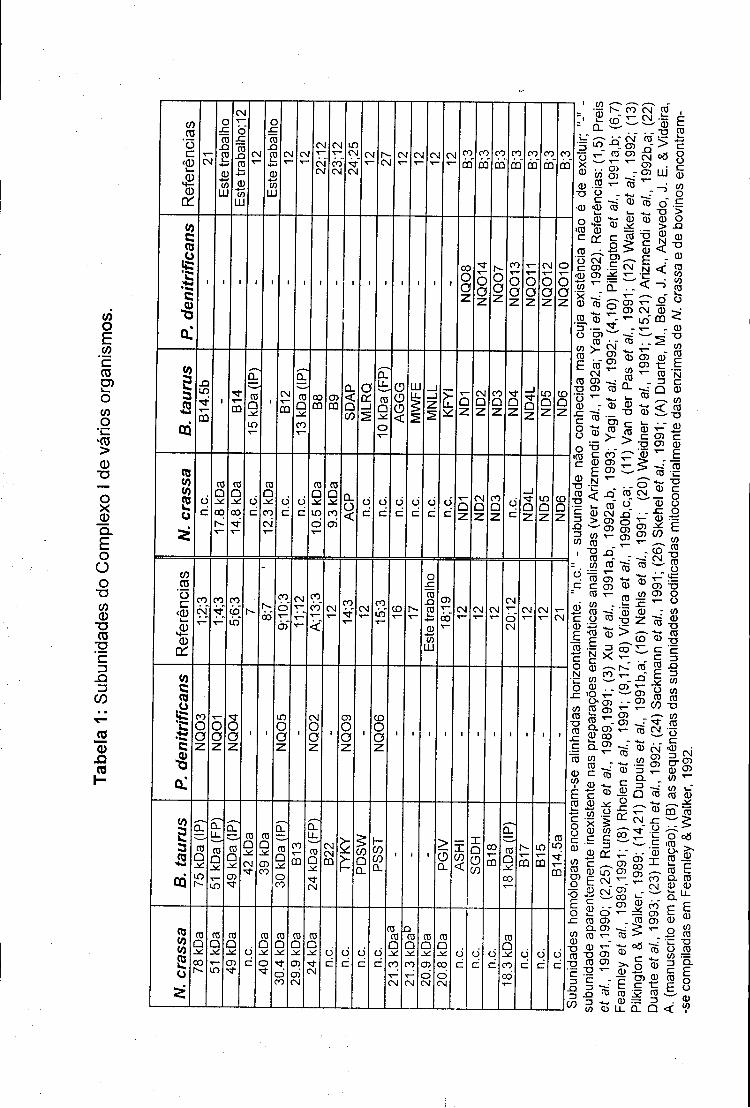
operão foram detectadas 20 "ORFs", 14 das quais codificam proteínas
homólogas a subunidades de complexo I mitocondrial (ver tabela 1). Embora a
enzima deste organismo seja visivelmente mais simples que o complexo I
mitocondrial, o seu estudo irá revelar características importantes da forma
mitocondrial. Por exemplo, dada a similaridade existente entre as duas
enzimas em termos de espectro de e.p.r. (ver 2.2.2 e 2.4.2) e a facilidade de
manipulação genética de um organismo procariótico, é de esperar a curto
prazo a identificação experimental directa das subunidades que possuem
centros de Fe-S.
No entanto, o aspecto mais interessante do estudo de procariotas é o
facto de proteínas homólogas a subunidades do complexo I nem sempre se
encontrarem associadas a NADH:ubiquinona oxidorredutases. A enzima
NAD+-hidrogenase de Alkaligenes eutrophus (Tran-Betcke et ai., 1990) ilustra
bem este ponto: duas das suas subunidades apresentam homologia com 3
subunidades do complexo I, sugerindo fortemente que estes componentes
representam uma unidade funcional e estrutural (ver 2.4.1).
7
2.2- Composição do complexo I
2.2.1- Componentes polipeptídicos
As primeiras análises electroforéticas em condições desnaturantes de
preparações relativamente puras da enzima bovina sugeriram a existência de
cerca de 25 subunidades polipeptídicas (ver Ragan, 1987). No entanto, à
medida que a resolução dos sistemas electroforéticos melhora, este número
tem vindo a aumentar. Actualmente, é unanimemente aceite que o complexo I
de mamíferos é composto por cerca 40 subunidades. De facto, Walker et ai.
(1992) encontraram evidência para a existência na enzima bovina de, pelo
menos, 41 subunidades polipeptídicas (ver tabela 1) e é natural que este
número ainda não seja definitivo. A estes componentes (e assumindo apenas
uma estequiometria unitária para cada subunidade) corresponde uma massa
molecular de aproximadamente 880 kDa, superior, portanto, à massa
molecular proteica de um ribossoma de Escherichia coli (Walker et ai., 1992).
No fungo Neurospora crassa, um dos organismos onde o complexo I se
encontra, também, bem caracterizado, são detectadas cerca de 35
subunidades, 24 das quais foram já caracterizadas ao nível da estrutura
primária (ver tabela 1 e Fig. 1 da secção 7.5). Cerca de 80 % das subunidades
da enzima fúngica têm homólogos na enzima de mamíferos indicando que as
duas enzimas são funcional e estruturalmente semelhantes possibilitando,
assim, a extrapolação da informação obtida experimentalmente entre os dois
sistemas.
Um aspecto interessante do complexo I reside no facto de nem todas as
suas subunidades polipeptídicas serem codificadas nuclearmente. Como
demonstrado por Chomin et ai. (1985, 1986), 7 das "URFs" ("Unidentified
Reading Frames") que haviam sido previamente detectadas no DNA
mitocondrial humano (Anderson et ai., 1981) codificam na realidade
8
CO o E CO
' c 03 O)
CO
g 'CO > Q)
■o
O X
Q. E o
O o
-o tf} 0
TJ 03
■g ' c Q
jo 0)
Q ca
co CO
" o c
«D
á CU
a:
CM
O
ro X I
S B w
LU
CM
Ò" XJ ro
X I ro
B tn LU
CN
o x j ro
X I
ro CM
CM CM CM
CM CM
CM CM CM CO
m CO
LU
<0 c ca o
g 5
00 O O
co O O
CM
O O
OQ
X I LO
5 m ro
Q
L O
CM
m
O cc CD
O <
LU > U .
CM Q Z
CO S3
2 o
ro Q
00
ro a ±£ CO
CM
CD
a L O
d
o o C M ça CM •!= - — 0 ■ - -a
CM ra s
i ro co ±j og
io g OT
LO X I
LU
CD <a>
B CD
CO _
0 75 0
_ro CD
E ro c o o c (D
CO o ç > o
XI CD
•a 0
CM Q
CO
a
C/3 ÇO O c
<a)
o ;
«o c CO o
5 ■8
tf) 5
N. CO
Ò" CO
CM CO
C O CM C O
-tf CM CO LO CD
O
ro XJ ro
co CM CM CM
00 O CM
CO LU
CO O a z
O o
LO O a
CM O o
co O a z
co O
a
■ o c
CO . - 2 , ^ CD " CM ? ; - E C CO . 5 CM N
«D CO ; * £ \ - e ^ W
» : °Cf lCN CD >• .ro CD T L O CQ < 5^— S T CD ° 2> ; ^^: o
CO ^ . r CO
' " " ' CD o:. ,D
ra a
CM co co
co ro E ro
CM CO CO ^
T3 CO co
CD CD
*~ ro
CO ro E N C 0 CO
C D ^ S .„ ro § co S « > 5 c en c
. 5 ^ g CO CD c co rr~ 0 ^ E ® ® «5 af.«
CD
E 2 . . o CO CM O" """" xT ■ -Ï.Sfis j
T3 c o o o
CM
03
co co to 2 o
ro Q
LO r--
0 . LL
ro Q
LO
D.
ro Q
CD
ro D
CM "tf
a. ro O _*: o C O
ro D
■tf CM
CM CM QQ
CO Q CL
r~ CO CO
a. > a
x Q O co
a ro Q
00
ro Q
co
ro Q .*: 0 5
O
ro Q co CN
CO Û
CO CO
CO
CD
CO ro
"O ro ço ro c ro co ro o •ro E N C 0 CO CD
<o . £co ro co ro ^-
. 8-co CD 00 * - CO
«TB CO * g CO 0
CO
CO
ro Sro
o CD
03 G> § _ co co o 2 2 , - o CD
•g >
CO CD -o co -o
CO
E Q. cõ co
CO c CD
co
E ro C o o c CD
CO ro co o
■o E o
-C co 0)
"O
ro
' c 3
. a
CO
co CD »~
CD co C
3 X I 3 CO CO ro
■o
CO ^ : M" co T - C0 CM Q
- * • . . c CD CM <CD CO &>
0 3 5 ?
c ro g
"si . •« to
ro 0 CO CO ^
C0 T 5
Q.
S P ' " * CM x:
5 Ï l E
c 0 w X 0 g 0 c OJ LO" C0 2 5 ; - . E CM_ - ? co 0 CM CO
0
CO i_ ro 0 ^^ ^ m ro
o ro co Q . C 0 ro T-_ 0 T - " "O co ro co c r E ro Z3 CO
. CO í 0 1 t ro co 0 > *" o
o a3 iffi .
c t t ro E CL CO 0 0
0
CO
•5 °° ro
II»! co S ro 0 = 3 a a Q
0 w ro n jo o. E o o 0 co
subunidades do complexo I. Estas subunidades, denominadas ND1-ND6 e
ND4L, têm a particularidade de serem extremamente hidrofóbicas e, como será
discutido posteriormente (ver 2.3.1), encontram-se muito provavelmente
embebidas na membrana interna mitocondrial.
2.2.2- Centros Fe-S e FMN
Para além de um elevado número de subunidades polipeptídicas, o
complexo I contém ainda como transportadores de electrões uma molécula de
FMN ligada não covalentemente e vários centros Fe-S (Hatefi et ai., 1962).
O número exacto de centros Fe-S existentes na enzima bovina é, ainda
hoje, assunto de debate (ver Beinert & Albracht, 1982). No entanto, vários
laboratórios referem consistentemente a existência de 3 centros Fe-S
tetranucleares, os denominados centros N2, N3 e N4, e um centro binuclear, o
centro N1b (Ingledew & Ohnishi, 1980; Beinert & Albracht, 1982). A existência
de um segundo centro binuclear de potencial redox do ponto médio muito
negativo (-400 mV a -500 mV), denominado centro N1a, tem também sido
defendida por alguns investigadores (Ohnishi et ai., 1985; Ohnishi & Salerno,
1982). Evidência para um quarto centro tetranuclear (o centro N5) tem sido
apresentada pelo grupo de Ohnishi (Ingledew & Ohnishi, 1980; Ohnishi, 1979;
Salerno et al., 1977) mas a sua existência é largamente contestada, existindo a
possibilidade de se tratar de uma contaminação da preparação analisada pelos
autores (ver Beinert & Albracht, 1982).
Aos centros N1a, N1b, N2-N4, e assumindo estequiometrias unitárias por
FMN*, correspondem 16 átomos de ferro. Este número é ainda bastante
O grupo de Albracht tem referido consistentemente uma estequiometria de 1 centro Nlb por cada duas moléculas de FMN (ver van Belzen et ai., 1990). Estes resultados, não unanimemente aceites (Ohnishi, 1979), são a base de um modelo estrutural em que o complexo I, na sua forma biologicamente activa, existirá sob uma forma dimérica em que um dos monómeros não possui o centro Nlb (ver van Belzen et ai., 1990).
inferior aos valores obtidos por análise química - 22 a 24 átomos de ferro por
FMN (Ragan et ai., 1982b) - sendo, por isso, provável a existência de outros
centros Fe-S (invisíveis pore.p.r.) na enzima bovina (ver também 2.3.3).
Análises por e.p.r. da enzima do fungo N. crassa revelaram a existência
dos centros N2, N3, N4 e de um centro binuclear - denominado N1 -
semelhante no seu potencial redox do ponto médio ao centro N1b da enzima
bovina (Wang et ai., 1991). Nenhuma evidência para a existência de centros
semelhantes aos centros N1a e N5 bovinos foi encontrada. No entanto, dadas
as semelhanças estruturais existentes entre as enzimas de N. crassa, bovinos
e P. denitrificans - organismo no qual também é detectado um centro N1a
(Meinhardt et ai., 1987) - a presença de um centro do tipo N1a na enzima
fúngica é altamente provável.
2.3- Estrutura do complexo I
2.3.1- Organização espacial das subunidades da enzima
Actualmente, dado que a maior parte das subunidades da enzima de
bovinos e de N. crassa foi já caracterizada em termos de estrutura primária, a
busca de informação estrutural sobre o complexo I é de uma importância fulcral
para a compreensão dos mecanismos de catálise, biogénese e função da
enzima.
Embora o complexo I de bovinos tenha sido isolado 23 anos antes de
existir um protocolo de purificação para a enzima de N. crassa (Hatefi et ai.,
1962; Ise et ai., 1985), a maior parte da informação disponível actualmente
sobre a estrutura geral do complexo I mitocondrial foi obtida com a enzima
10
fúngica. Isto deve-se principalmente ao facto de a enzima de A/, crassa, ao
contrário da enzima de bovinos, ser muito estável na presença de detergentes,
sendo por isso fácil de isolar num estado monodisperso, uma característica
essencial para estudos de microscopia electrónica e difracção de raios X.
Muitos dos estudos estruturais realizados em complexos proteicos
baseiam-se no uso de substâncias que desestabilizam de um modo
relativamente suave as interacções entre proteínas (e. g., agentes caotrópicos
e detergentes). Aplicando esta estratégia ao estudo do complexo I, Friedrich et
ai. (1989) mostraram que, quando a enzima de N. crassa é incubada na
presença do anião brometo, uma parte substancial das suas subunidades são
dissociadas perdendo qualquer estrutura definida. No entanto, uma fracção
contendo cerca de metade das subunidades da enzima resiste, de uma forma
aparentemente intacta, ao tratamento com sais caotrópicos podendo ser
isolada numa forma monodispersa. Este subcomplexo contém cerca de 15
subunidades codificadas nuclearmente (a maior parte das quais ainda com
estrutura primária desconhecida) e, adicionalmente, todas as subunidades
codificadas mitocondrialmente. O facto de subunidades altamente hidrofóbicas
serem encontradas neste subcomplexo levou Tuschen et ai. (1990) a especular
que este "fragmento hidrofóbico" corresponde à parte da enzima que se
encontra embebida na membrana interna mitocondrial, o que, de certo modo,
foi confirmado posteriormente (ver abaixo). Por outro lado, quando o fungo N.
crassa é crescido na presença de cloranfenicol (um inibidor da síntese proteica
mitocondrial), as suas mitocôndrias acumulam um subcomplexo contendo
cerca de 15 subunidades codificadas nuclearmente. Surpreendentemente,
nenhuma das subunidades deste subcomplexo - denominado "pequena forma
da enzima" - está presente no fragmento hidrofóbico e, em termos de
composição polipeptídica, os dois fragmentos representam a totalidade da
enzima fúngica (Friedrich et al., 1989).
! l
Estes resultados só foram plenamente compreendidos quando as
primeiras preparações de complexo I de N. crassa, no seu estado
monodisperso e cristalizado bidimensionalmente, foram analisadas por
microscopia electrónica e difracção de raios X (Hofhaus et ai., 1991). Estes
estudos revelaram que o complexo I tem uma estrutura em forma de L, em que
um dos braços se encontra embebido na membrana interna mitocondrial e o
outro, perpendicular ao primeiro, se orienta para o meio aquoso da matriz
mitocondrial. Por outro lado, quando estes cristais bidimensionais são
"lavados" com uma solução de brometo de sódio, praticamente todas as
subunidades presentes na pequena forma da enzima são removidas, restando
apenas o braço membranar cuja composição polipeptídica é semelhante à do
fragmento hidrófobico. Ficou, assim, claro que a pequena forma do complexo I
representa o braço mais hidrofílico (denominado braço periférico da enzima) e
que o fragmento hidrofóbico é, na realidade, semelhante ao braço membranar
do complexo I (Hofhaus et ai., 1991).
O complexo I de bovinos tem sido também alvo de estudos estruturais.
Muitos dos dados obtidos com a enzima deste organismo, corroboram e, acima
de tudo, refinam o modelo estrutural obtido com a enzima de N. crassa.
Como mostrado por Galante & Hatefi (1979), quando incubada na
presença de perclorato, a enzima bovina origina 3 fragmentos: (1) a
denominada FP ("Flavo-protein") constituída pelas subunidades 51 kDa (FP),
24 kDa (FP) e 10 kDa (FP), contendo todo o FMN da enzima e 6 átomos de
ferro (Ragan et ai., 1982a); (2) a IP ("Iron-protein"), uma fracção proteica
relativamente heterogénea em termos da estequiometria dos seus
componentes, englobando as subunidades de 75 kDa, 49 kDa, 30 kDa, 18 kDa
(IP), 15 kDa (IP), 13 kDa (IP) e B13 e ainda cerca de 10 átomos de ferro
(Ragan et ai., 1982b; Walker, 1992); e, finalmente, (3) a HP ("Hydrophobic
protein"), um aglomerado proteico insolúvel nas condições experimentais
12
usadas para o fraccionamento, contendo as restantes subunidades do
complexo I e, pelo menos, dois centros Fe-S (Ragan et ai., 1982b).
Mais recentemente, Finei et ai. (1992), explorando a labilidade estrutural
do complexo I bovino na presença de detergentes, isolaram e caracterizaram
dois fragmentos que, em termos de composição polipeptídica, são
complementares e representam a quase totalidade da enzima bovina. O maior
destes fragmentos, o denominado subcomplexo la, contém cerca de 25
subunidades entre as quais todas aquelas que são encontradas nos
fragmentos FP e IP descritos anteriormente. Adicionalmente, este
subcomplexo contém também outras subunidades que, à semelhança dos
componentes das frações FP e IP, possuem uma estrutura primária típica de
proteínas predominantemente hidrofílicas. No entanto, a presença neste
fragmento de cinco subunidades com potencial para atravessarem um sistema
membranar (uma das quais a subunidade ND-2, indiscutivelmente uma
proteína de membrana) sugere que uma pequena parte deste subcomplexo se
encontra embebida na membrana interna mitocondrial. Este fragmento contém,
também, todos os centros Fe-S que são detectados por e.p.r. na enzima
bovina.
Ao contrário do que acontece com o subcomplexo la, a maior parte das
cerca de 15 subunidades que constituem o denominado fragmento ip (Finei et
ai., 1992) possuem domínios suficientemente grandes de aminoácidos
hidrofóbicos para atravessarem a membrana interna mitocondrial, sugerindo,
assim, que grande parte deste fragmento se encontra embebido na membrana.
Extrapolando esta informação para o modelo estrutural obtido com a
enzima de N. crassa, foi sugerido que o subcomplexo la corresponde ao braço
periférico da enzima contendo, também, uma pequena porção do braço
membranar; o subcomplexo l(3 representa a maior parte do braço membranar
do complexo I (Finei et ai., 1992; Walker, 1992).
13
2.3.2- Localização dos sitios de ligação do NADH e FMN
Embora seja constituída por apenas três subunidades, a fracção FP do
complexo I bovino (ver 2.3.1) possui actividade de NADH desidrogenase. É de
salientar que os parâmetros cinéticos da ligação do NADH e NAD+ ao
fragmento FP são praticamente iguais aos obtidos com a enzima intacta,
sugerindo que neste subcomplexo o sítio de ligação do NADH permanece
perfeitamente funcional após o tratamento com perclorato (Dooijewaard &
Slater, 1976a,b). Com o intuito de identificar qual das três subunidades da FP é
responsável pela ligação do NADH, Chen & Guillory (1981) usaram um
análogo radioactivo do NAD+ em experiências de marcação por foto-afinidade.
A subunidade de 51 kDa da enzima bovina era marcada significativamente
sendo a incorporação do análogo inibida de uma forma competitiva por NADH.
Resultados semelhantes foram também obtidos com a NADH:ubiquinona
oxidorredutase de P. denitrificans onde um polipéptido de 50 kDa (homólogo à
subunidade de 51 kDa bovina) foi identificado como constituindo o sítio de
ligação do NADH (Yagi & Dinh, 1990).
A labilidade da ligação da FMN à(s) subunidade(s) do complexo I tem
dificultado a identificação experimental do seu sítio de ligação (ver Ragan,
1987). Como será discutido posteriormente (ver 2.4.2), das três subunidades
que constituem a fracção FP, o componente de 10 kDa não parece
desempenhar um papel directo na ligação da FMN. No entanto, apesar de
existir alguma (fraca) evidência baseada em análises estruturais para a
atribuição do sítio de ligação da FMN à subunidade de 51 kDa (ver Walker,
1992), não é ainda possível excluir a subunidade de 24 kDa desta função.
i !
2.3.3- Localização dos centros Fe-S
A maior parte dos estudos efectuados na tentativa de localizar os
diferentes centros Fe-S do complexo I tem-se confinado à caracterização por
e.p.r. dos vários fragmentos do complexo I acima descritos. Embora esta
estratégia possua algumas limitações, pois após resolução da enzima muitos
dos centros Fe-S perdem os seus sinais de e.p.r. característicos tornando a
sua identificação difícil, foi possível localizar os centros N-1b e N-3 na fracção
FP da enzima bovina (Ohnishi et ai., 1981, 1985; Ragan et ai., 1982a). Neste
caso, foi mesmo possível atribuir o centro N-3 à subunidade de 51 kDa, e o
centro N-1b às subunidades de 24 kDa e/ou 10 kDa (Ohnishi et ai., 1985; ver
também 2.4.2). A presença de centros Fe-S nas subunidades de 75 kDa, 49
kDa e no heterodímero constituído pelas subunidades de 30 kDa (IP) e 13 kDa
(IP) foi também sugerida (Ragan, 1987), embora nestes casos a alteração no
espectro de e.p.r. resultante da técnica utilizada para a resolução da enzima
não tivesse permitido a identificação desses centros metálicos.
A recente determinação da estrutura primária das subunidades do
complexo I bovino forneceu mais informação sobre o possível número e a
localização de centros Fe-S da enzima. Por exemplo, foi verificada a existência
do motivo CysXXCysXXCys(X)nCysPro (em que X é qualquer aminoácido) nas
subunidades de 75 kDa, 51 kDa e TYKY, esta última contendo mesmo duas
cópias deste domínio (Runswick et ai., 1989; Pilkington et al., 1991a; Dupuis et
ai., 1992,b). Esta organização de resíduos de cisteínas é característica de
ferrodoxinas do tipo [4Fe-4S] nas quais constituem os ligandos do centro
metálico (ver Cammack, 1992). Assim, a presença de quatro destes motivos
nas subunidades acima referidas, sugere fortemente a existência de quatro
centros tetranucleares Fe-S no complexo I, portanto, mais um do que aqueles
que são detectados por e. p. r. (Walker, 1992).
15
Ao contrário do que acontece com as metalo-proteínas do tipo [4Fe-4S], a
identificação das subunidades do complexo I que possuem centros de Fe-S
binucleares não pode ser efectuada com um grau de confiança tão elevado
recorrendo apenas à análise das suas sequências. Com efeito, a distribuição
dos aminoácidos que constituem os ligandos deste tipo de centros não
obedece, aparentemente, a parâmetros tão rígidos. Por exemplo, a distribuição
de cisteínas do polipéptido de 24 kDa (a subunidade que alberga o centro N-
1b; ver 2.4.2) não é encontrada em mais nenhuma metalo-proteína do tipo
[2Fe-2S] conhecida (Pilkington & Walker, 1989). Existe, no entanto, uma
fracção significativa de metalo-proteínas contendo este tipo de centros Fe-S
que apresentam o motivo CysXXCys(X)nCysXXCys (ver Cammack, 1992).
Esta distribuição de cisteínas é encontrada na subunidade de 75 kDa, o que
levou Walker (1992) a sugerir a existência de um centro binuclear neste
componente do complexo I.
2.3.4- Localização do sítio de ligação da Ubiquinona
Dada a localização membranar da ubiquinona, é de esperar que o seu
sítio de ligação ao complexo I resida também na membrana interna
mitocondrial. A aplicação de técnicas de marcação radioactiva por análogos da
ubiquinona tem sido impedida pela dificuldade em derivar quimicamente o
centro redox da molécula, mantendo a sua actividade biológica. Na tentativa de
circundar este problema, vários grupos tentaram identificar os sítios de acção
de vários inibidores do complexo I (e. gr., rotenóides, petidinas e piericidinas)
tendo como premissa que este tipo de compostos apresentaria uma acção
inibitória competitiva com a ubiquinona (ver Friedrich et a/., 1990). Nestes
estudos, vários investigadores demonstraram a marcação da subunidade ND-1
por análogos radioactivos e foto-activáveis da rotenona (Earley & Ragan, 1984;
16
Earley et al., 1987). Estes resultados, juntamente com o facto de o
componente ND-1 ser, de todas as subunidades codificadas
mitocondrialmente, aquele que filogeneticamente se encontra melhor
conservado, levou à hipótese de o sítio de ligação da ubiquinona se encontrar
nesta subunidade (ver Friedrich et ai., 1990). No entanto, estudos cinéticos
posteriores revelaram que a rotenona não actua de um modo competitivo com
a ubiquinona invalidando, pelo menos parcialmente, esta conclusão (Ahmed &
Krishnamoorthy, 1992; Singer & Ramsay, 1992).
Mais recentemente, Heinrich & Werner (1992) sintetizaram um análogo da
ubiquinona biologicamente activo, contendo um grupo arilazido (foto-activável)
na extremidade da cadeia alifática da molécula. Experiências de marcação por
foto-afinidade utilizando membranas mitocondriais de N. crassa levaram à
identificação de um polipéptido de 9.3 kDa homólogo à subunidade B9 da
enzima bovina (Heinrich et ai., 1992). Embora não seja possível afirmar que
esta subunidade é, de facto, o sítio de ligação da ubiquinona (a posição do
grupo foto-reactivo na molécula encontra-se a uma distância significativa do
seu centro redox; ver também 2.4.2), os resultados obtidos sugerem, no
mínimo, que esta proteína se encontra extremamente próxima do sítio de
redução da ubiquinona. No entanto, a localização exacta desta subunidade e,
consequentemente, a localização do sítio de ligação da ubiquinona, ainda não
é conhecida. Uma das experiências apresentadas neste trabalho permitiu
localizar (grosseiramente) a subunidade de 9.3 kDa (ver secção 7.5).
17
2.4- Relações estruturais/funcionais entre o complexo I mitocondrial e
enzimas procarióticas
2.4.1- A NAD+-hidrogenase de A. eutrophus
A NAD+-hidrogenase de Alkaligenes eutrophus, uma bactéria
quimiolitotrófica, cataliza a redução do NAD+ pelo H2. Funcional e
estruturalmente a enzima pode ser dividida em dois domínios (Tran-Betcke et
ai., 1990): o primeiro, constituído pelas subunidades p e ô, um átomo de níquel
e, possivelmente, dois centros Fe-S, retém a capacidade de redução de
variadíssimas substâncias pelo H2; o segundo, um heterodímero contendo as
subunidades a e y , uma molécula de FMN ligada não covalentemente e três
ou quatro centros Fe-S, possui actividade de NADH oxidorredutase. A recente
sequenciação do operão codificante desta enzima, o locus Hox S (Tran-Betcke
et ai., 1990), permitiu constatar uma surpreendente homologia entre as
subunidades a e y da enzima procariota e as subunidades de 75 kDa, 51 kDa e
24 kDa do complexo I mitocondrial (Pilkington et ai., 1991a). De facto, a
subunidade a representa uma fusão das subunidades de 51 kDa e 24 kDa; a
subunidade y apresenta homologia com os 200 aminoácidos N-terminais da
subunidade de 75 kDa. Esta observação não só permitiu estabelecer um elo
funcional entre as subunidades de 24 kDa e 51 kDa (ambas presentes na
fracção FP), mas também entre estas e a subunidade de 75 kDa (um
componente da fracção IP). É, assim, possível que os sítios de ligação do FMN
e NADH e os centros de Fe-S N-1b e N-3 (presentes nas subunidades de 24
kDa e 51 kDa) estejam espacialmente muito próximos dos dois centros Fe-S
putativamente presentes na subunidade de 75 kDa (ver 2.3.3).
18
2.4.2- O complexo I de P. denitrificans
O procariota Paracoccus denitrificans, quando crescido em condições
aeróbicas, produz uma NADH:ubiquinona oxidorredutase fosforilativa
semelhante ao complexo I mitccondrial em muitos aspectos (Yagi, 1986, 1990,
1991). De facto, a enzima deste organismo não só apresenta todos os centros
Fe-S que são detectados por e. p. r. na enzima bovina (Meinhardt et ai., 1987),
como também é inibida fortemente pelas mesmas substâncias que actuam
sobre a forma enzimática mitoccndrial. Assim, não deixa de ser surpreendente
que o recente isolamento desta enzima tenha revelado a existência de apenas
14 subunidades estruturais, cerca de um terço dos componentes do complexo I
bovino (Yagi et ai., 1992). Todas as subunidades da enzima de Paracoccus
têm homólogos na enzima de bovinos, corroborando assim os dados obtidos
por e. p. r. e as características enzimáticas do complexo I procariótico (Xu et
ai., 1991a,b, 1992a,b, 1993; ver tabela 1). A simplicidade da enzima deste
organismo permite, deste modo, inferir características importantes da forma
enzimática mitocondrial. Por exemplo, a ausência em Paracoccus de uma
subunidade homóloga ao componente de 10 kDa da fracção FP da enzima
bovina, sugere que esta subunidade não está envolvida directamente na
ligação do centro Fe-S N-1b nem na ligação do FMN. Outra observação
relevante é a ausência na enzima procariótica de um homólogo das
subunidades 9.3 kDa/B9 (ver 2.3.4), sugerindo que este componente não actua
como mediador directo na ligação e/ou redução da ubiquinona pelo complexo I.
No entanto, talvez a característica mais importante da enzima de
Paracoccus seja, como referido acima, a sua simplicidade. Assim, se as
catorze subunidades presentes nesta enzima são suficientes para garantir uma
actividade NADH:ubiquinona oxidorredutase associada a uma translocação de
protões, qual é então a função das cerca de 27 subunidades da enzima bovina
que não possuem homólogos no complexo de Paracoccus? É possível que
19
muitas destas subunidades supranumerárias tenham como função optimizar ou
regular a reacção catalizada pelo complexo I mitocondrial. O envolvimento de
algumas subunidades como agentes mediadores em fenómenos de "substrate-
chanelling" é também plausível (ver Srivastava & Bernhard, 1986; Porpaczy et
ai. 1987). Por outro lado, é possível que o complexo I mitocondrial
desempenhe funções bioquímicas adicionais, inexistentes na enzima
procariótica. Como discutido seguidamente, esta última hipótese é, cada vez
mais, encarada como altamente provável.
2.4.3- A "acyl-carrier protein"
Um dos factos mais salientes revelados pela determinação da estrutura
primária das subunidades do complexo I foi a descoberta de uma proteína
homóloga às "acyl-carrier proteins" procarióticas (ACP; Runswick et ai., 1991;
Sackmann et ai., 1991). Experiências de marcação radioactiva com
[14C]pantotenato em Neurospora crassa, assim como a determinação da
massa molecular da subunidade bovina, não só demonstraram a presença do
grupo fosfopanteteína covalentemente ligado a esta subunidade - o centro
activo das "acyl-carrier proteins" - como revelaram a existência de vários
ácidos gordos ligados por uma ligação tio-éster à fosfopanteteína (Brody &
Mikolajczyk, 1988; Runswick et ai., 1991; Sackmann et ai., 1991). Estes dados
indicam que a ACP presente no complexo I se encontra activa.
A função de uma "acyl-carrier protein" nas mitocôndrias tem sido alvo de
pesquisa. Foi verificado que o principal grupo acilo encontrado na ACP
mitocondrial de Neurospora advém do ácido hidroximirístico (Brody &
Mikolajczyk, 1988), o principal precursor da cardiolipina fúngica (Martin &
Johnson, 1983). Esta observação levou Brody et ai. (1990) a sugerirem que a
ACP mitocondrial tem como função satisfazer as necessidades lipídicas
20
específicas da mitocôndria (a cardiolipina tem uma localização exclusivamente
mitocondrial). A identificação do ácido gordo encontrado predominantemente
na ACP mitocondrial de bovinos - possivelmente o ácido hidroxioctadecanoato
(Runswick et ai., 1991) - e o facto de o grupo acilo mais abundante na
cardiolipina bovina ser do tipo C<|8:2 (Schlame ef ai., 1991) corroboram esta
hipótese. Existe, assim, um paralelismo entre os diferentes grupos acilo
encontrados nas ACPs destes organismos e a composição das suas
cardiolipinas.
Se a razão para a existência de uma ACP mitocondrial parece estar
compreendida, a sua associação a um componente da cadeia respiratória
permanece intrigante, levantando a hipótese de outras subunidades do
complexo I estarem também associadas com a função da ACP.
2.5- O fluxo de electrões no complexo I
A cinética de redução dos diferentes transportadores electrónicos do
complexo I por NADH (ou outros agentes redutores) é extremamente rápida,
verificando-se a redução de todos os centros Fe-S da enzima em alguns
milisegundos. Este facto, aliado à inexistência de inibidores da enzima que
interrompam o fluxo de electrões entre dois grupos redox, tem dificultado a
determinação experimental da sequência de redução dos diferentes centros
Fe-S (como artigo de revisão ver Beinert & Albracht, 1982). Assim, o modelo
actualmente aceite sobre o fluxo de electrões através do complexo I assenta,
basicamente, nos potenciais redox do ponto médio dos transportadores
electrónicos da enzima. Neste modelo, o FMN actua como o oxidante imediato
do NADH. Do FMNH2, os electrões são transferidos para o grupo isopotencial
dos centros N-1b, N-3 e N-4 (Krishnamoorty & Hinkle, 1988). É interessante
notar que a remoção do centro N-4 por tratamento do complexo I com N-
21
bromosuccinimida não altera a cinética de transferência de electrões do NADH
para a ubiquinona (Krishnamoorty & Hinkle, 1988). Este facto levou estes
autores a especular que o centro N-4 poderá constituir uma ramificação
(opcional) do fluxo electrónico, podendo ter como função o armazenamento
temporário de electrões.
Do grupo isopotencial dos centros N-1b, N-3 e N-4, os electrões são
então transferidos para o centro N-2. Embora o potencial redox do ponto médio
deste centro ainda não seja conhecido com exactidão (valores entre -20 mV e -
120 mV têm sido descritos), é ponto assente que este centro é o mais
electropositivo da enzima, razão pela qual é considerado o redutor imediato da
ubiquinona (Ingledew & Onhishi, 1980; Kotlyar et ai, 1990; ver Beinert &
Albracht, 1982).
Recentemente, foi detectada em partículas submitocondriais incubadas
na presença de NADH uma ubisemiquinona cujo comportamento de
relaxamento sugere uma interação com um centro Fe-S tetranuclear
(provavelmente o centro N-2) (Burbaev et ai, 1989; Suzuki & King, 1983). Por
outro lado, se o complexo I no seu estado oxidado não for pré-condicionado
com NADH, verifica-se a inexistência de actividade NADH:ubiquinona
oxidorredutase, assim como a incapacidade de redução do NAD+ por
transferência electrónica inversa (Kotlyar & Vinogradov, 1990). Estas
observações sugerem que a transferência de electrões do centro N-2 para a
"pool" de ubiquinona existente na membrana interna envolve uma ubiquinona
ligada à enzima a qual oscila entre as formas QH- (ou Q") e QH2 durante o
transporte electrónico (ver fig.1). A completa oxidação deste radical livre
explicaria, assim, a necessidade de se pré-incubar o complexo I com NADH
para activar a enzima (Burbaev et ai, 1989; Kotlyar & Vinogradov, 1990;
Kotlyar et ai, 1990).
22
2.6- Mecanismo de transdução energética no complexo I
A elaboração de modelos sobre os mecanismos de transdução energética
do complexo I tem sido um campo prolífero para bioenergeticistas. De tal modo
isto é verdade, que o número de modelos actualmente existentes sobre este
assunto praticamente iguala o número de autores que se debruçam sobre a
matéria. Uma das razões para estas divergências reside na própria
estequiometria da reacção catalizada pelo complexo I (/'. e., o número de
protões bombeados por molécula de NADH oxidada) que cada autor considera
como válida. De facto, os valores experimentais obtidos por vários grupos não
são muito coerentes e razões de 3 H+/2 e - (De Jong & Westerhoff, 1982), 4
H+/2 e- (Wikstrom, 1984; Beavis, 1987; Brown & Brand, 1988) e 5 H+/2 e"
(Lemasters, 1984; Lemasters et a!., 1984) têm sido descritas. Como sugerido
por Murphy & Brand (1987), é muito possível que a razão H+/2 e- não seja um
número inteiro, uma vez que a reacção catalizada pela enzima representa uma
forma deslocalizada de energia. De qualquer modo, a reacção
NADH + ubiquinona + 5 H+(matriz) = NAD+ + ubiquinol + 4 H+(E|M)
onde se assume que, por cada par de electrões, 4 protões são translocados da
matriz mitocondrial para o espaço intermembranar (EIM) é, em geral,
considerada a mais correcta (ver Weiss & Friedrich, 1991; Walker, 1992). Esta
razão H+/e" invalida, pelo menos parcialmente, o mecanismo de "loop"
proposto por Mitchel (1966), o qual prevê a translocação de dois protões por
cada dois electrões transferidos do NADH para a ubiquinona.
Muitos dos modelos existentes actualmente continuam a apoiar-se na
ideia base de Mitchel, procurando acoplar passos de transferência de protões
às diversas reacções de oxidação/redução que ocorrem na enzima. A julgar
pelos potenciais redox do ponto médio dos vários transportadores electrónicos
23
FMN NADH + H +
Q H 2 m i m ^ ± Q H 2
► 2H.+
B 2e-
Fe-S *- Fe-S (N-2)
Qmim ^ - ^ 2Q b / 2e "
HZ+ ^ Q b H " QbH-
Q H2 m i m ^ = ^ Q b
H2
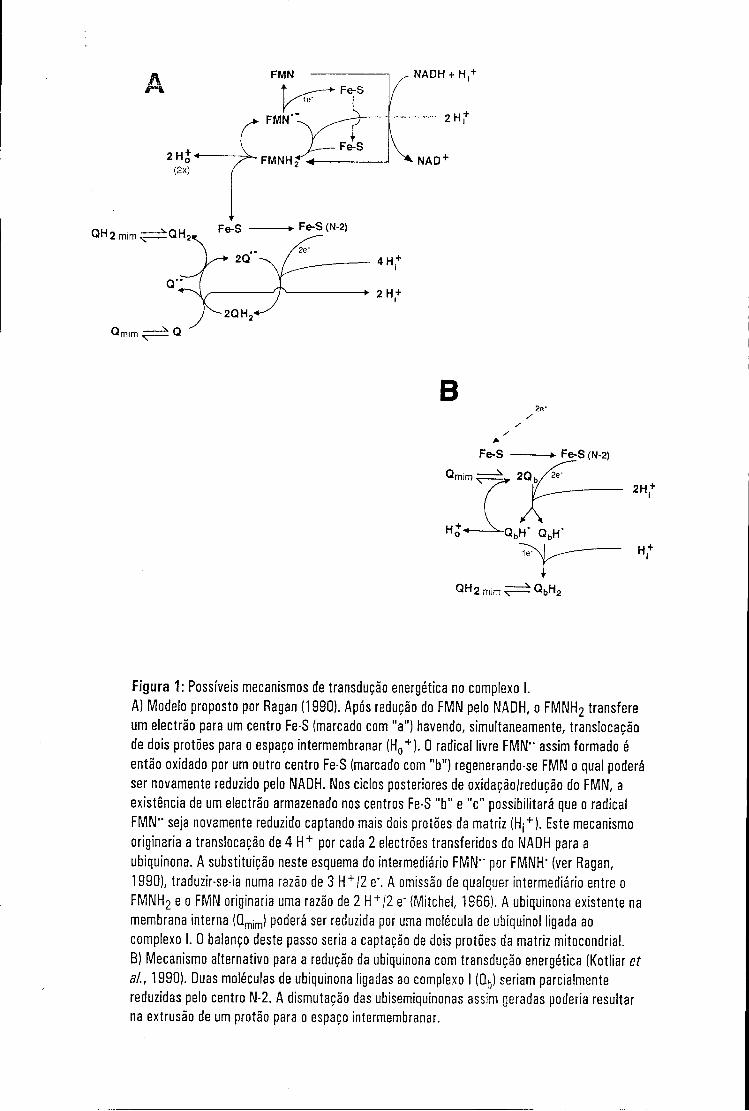
Figura 1: Possíveis mecanismos de transdução energética no complexo I. A) Modelo proposto por Ragan (1990). Após redução do FMN pelo NADH, o FMNH2 transfere um electrão para um centro Fe-S (marcado com "a") havendo, simultaneamente, translocação de dois protões para o espaço intermembranar (H0
+ ). 0 radical livre FMN" assim formado é então oxidado por um outro centro Fe-S (marcado com "b") regenerando-se FMN o qual poderá ser novamente reduzido pelo NADH. Nos ciclos posteriores de oxidação/redução do FMN, a existência de um electrão armazenado nos centros Fe-S "b" e "c" possibilitará que o radical FMN" seja novamente reduzido captando mais dois protões da matriz (Hj
+). Este mecanismo
originaria a translocação de 4 H + por cada 2 electrões transferidos do NADH para a ubiquinona. A substituição neste esquema do intermediário FMN" por FMNH" (ver Ragan, 1990), traduzir-se-ia numa razão de 3 H + /2 e". A omissão de qualquer intermediário entre o FMNH2 e o FMN originaria uma razão de 2 H + /2 e" (Mitchel, 1966). A ubiquinona existente na membrana interna (Qmim) poderá ser reduzida por uma molécula de ubiquinol ligada ao complexo I. 0 balanço deste passo seria a captação de dois protões da matriz mitocondrial.
B) Mecanismo alternativo para a redução da ubiquinona com transdução energética (Kotliar et ai., 1990). Duas moléculas de ubiquinona ligadas ao complexo I (Qb) seriam parcialmente reduzidas pelo centro N-2. A dismutação das ubisemiquinonas assim geradas poderia resultar na extrusão de um protão para o espaço intermembranar.
do complexo I, poderia haver transdução energética na oxidação do FMNH2
pelos centros isopotenciais N-1b, N-3 e N-4. Aqui, vários esquemas
envolvendo ciclos de oxidação/redução da FMN têm sido propostos na
tentativa de justificar razões de 3 ou 4 H+/2 e- (ver Ragan, 1990; fig.1).
A transferência electrónica dos centros isopotenciais N-1b, N-3 e N-4 para
o centro N-2, é também suficientemente exoenergética para suportar um
processo de translocação protónica (Weiss & Friedrich, 1991). No entanto,
dado o não envolvimento de protões nesta reacção redox e a ausência
completa de evidência experimental, este passo não é, geralmente, englobado
nos diferentes modelos de transdução energética existentes.
Finalmente, a transferência de electrões do centro N-2 para a ubiquinona
poderá também estar acoplada à translocação protónica, mas apenas se se
considerar um potencial redox do ponto médio para este centro de -120 mV
(ver 2.5). Num dos possíveis mecanismos (Kotlyar et ai., 1990), o centro N-2
doaria os dois electrões a duas moléculas de ubiquinona ligadas ao complexo I
(ver 2.5) as quais captariam dois protões da matriz mitocondrial. A dismutação
das moléculas de ubisemiquinona assim geradas poderia traduzir-se na
libertação de um protão no espaço intermembranar (ver fig. 1 B).
Como já referido, todos os mecanismos de transdução energética aqui
descritos procuram acoplar, de uma forma directa, passos de transferência de
protões a reacções redox. Estes modelos têm a vantagem da simplicidade e
poderão mesmo, com maior ou menor facilidade, ser experimentalmente
testados. No entanto, é possível que o acoplamento entre as duas actividades
do complexo I (/'. e., actividade de NADH:ubiquinona oxidorredutase e de
translocase protónica) ocorra de um modo indirecto. Por exemplo, a energia
libertada na reacção redox poderia ser utilizada para provocar alterações
conformacionais na enzima, alterações essas promotoras de translocação
protónica. Um mecanismo deste género tem sido proposto para o complexo IV
da cadeia respiratória (Citocromo c oxidase), uma enzima cuja conformação é
24
enormemente dependente do seu estado redox (como artigo de revisão ver
Capaldi, 1990). Embora especulativa, a existência de um mecanismo deste tipo
no complexo I não é de excluir.
2.7- Biogénese e evolução do complexo I
De acordo com a teoria endossimbiótica da origem da mitocôndria, este
organelo descende de um organismo procariota que, a dado momento da
evolução, estabeleceu uma relação de endossimbiose com uma célula
hospedeira. Com efeito, quer estrutural quer funcionalmente, estes organelos
retêm ainda hoje muitas das propriedades de um sistema procariótico (como
artigos de revisão ver Tzagoloff & Myers, 1986; Attardi & Schatz, 1988). É
óbvio que muitas das estruturas moleculares originalmente existentes no
antecessor mitocondrial foram adaptadas à medida das necessidades da célula
eucariótica, mas, pelo menos no caso do complexo I, essas alterações
saldaram-se basicamente pela introdução na enzima de novos componentes.
De facto, praticamente todas as subunidades do complexo I de Paracoccus
denitrificans [organismo considerado por alguns investigadores "a free-living
mitochondria" (Yagi et al., 1992)], possuem homólogos na forma enzimática
mitocondrial (ver tabela 1). Uma análise da estrutura primária dos
componentes codificados no núcleo e pertencentes a este grupo revela uma
característica interessante: todas estas subunidades possuem sequências sinal
cliváveis, uma das propriedades de proteínas cujos genes foram transferidos
da mitocôndria para o núcleo no decurso da evolução (ver Hartl & Neupert,
1990). Nesta perspectiva, é também relevante referir que nunca foram
encontradas em procariotas proteínas homólogas às subunidades do complexo
I mitocondrial sintetizadas no citoplasma e que não possuem pré-sequências.
Estas observações, enquadrando-se perfeitamente na teoria endossimbiótica
25
da origem mitocondrial, sugerem fortemente que o domínio do complexo I com
função respiratória é constituído por um "núcleo duro" de subunidades
conservadas filogeneticamente (grupo das subunidades com pré-sequência e
subunidades codificadas mitocondrialmente) ao qual a célula eucariótica
adicionou outros componentes (subunidades sem pré-sequência).
Mesmo considerando apenas a NADH:ubiquinona oxidorredutase
rotenona-sensível de P. denitrificans (o complexo I de mais simples
composição que é conhecido), é difícil imaginar como um complexo proteico
estrutural e funcionalmente tão complicado possa ter sido gerado. Uma das
hipóteses mais plausíveis é que o complexo I tenha sido originado por junção
de diferentes blocos ou módulos enzimáticos com actividades diferentes
(Walker, 1992). Por exemplo, a dado momento da evolução, uma enzima com
actividade de translocase protónica pode ter-se associado a um complexo
catalizador de uma reacção redox, tendo-se criado, subsequentemente, um
acoplamento entre as duas actividades. Este modelo para uma evolução
modular do complexo I encontra, de facto, suporte em algumas observações
efectuadas recentemente em enzimas procariotas. Por exemplo, 4 das
subunidades da formato hidrogenilase de E. coli apresentam homologia com
subunidades do complexo I (Boehm et ai., 1990). De um modo semelhante, e
como já foi referido (ver 2.4.1), as subunidades de 75 kDa, 51 kDa e 24 kDa do
complexo I são homólogas às subunidades a e y da NAD+-hidrogenase de
Alkaligenes eutrophus, sugerindo que estes componentes representam um
módulo enzimático responsável pela oxidação/redução do NADH/NAD+
(Pilkington et al., 1991a). É possível que mais enzimas com estas
características venham a ser detectadas futuramente o que, indubitavelmente,
aumentará o nosso conhecimento sobre a evolução, estrutura e função do
complexo I.
Alguns dos trabalhos realizados sobre os mecanismos de montagem do
complexo I de Neurospora têm também fornecido alguma evidência para uma
26
evolução modular do complexo I. Com efeito, um intermediário de montagem
da enzima, constituído por todas ' as subunidades codificadas
mitocondrialmente e cerca de 15 subunidades de origem nuclear, foi observado
em experiências de "pulse-labelling" por Tuschen et ai. (1990). Curiosamente,
este intermediário de montagem é totalmente idêntico ao fragmento hidrofóbico
da enzima (ver 2.3.1). Por outro lado, foi já referido que, quando crescidas na
presença de cloranfenicol, as células de Neurospora produzem apenas um
subcomplexo da enzima: a pequena forma do complexo I (Friedrich et ai.,
1989; ver 2.3.1). Um resultado semelhante foi observado numa estirpe de
Neurospora na qual o gene codificante da subunidade de 21.3 kDa do braço
membranar da enzima foi inactivado (Nehls et ai., 1992). Estas observações
levaram Weiss ef ai. (1991) não só a sugerir que os dois braços do complexo I
são montados de uma forma independente, mas que este resultado poderia
indicar uma origem filogenética também independente para as duas partes da
enzima. No decurso da evolução, os dois braços do complexo I ter-se-iam
então reunido en bloc para originar a enzima actual.
2.8- Aspectos médicos do complexo I
Num organismo estritamente aérobico como o homem, o complexo I
desempenha duas funções fulcrais: (1) contribuindo para a formação do
gradiente protónico através da membrana interna mitocondrial, a enzima é
responsável por quase um terço do ATP total produzido na fosforilação
oxidativa**; (2) o complexo I, como NADH desidrogenase, é um importante
ponto de entrada de equivalentes redutores na cadeia respiratória, sendo o
principal local de regeneração do NAD+. Não é, assim, surpreendente que uma
Cálculo baseado na oxidação completa de uma molécula de glucose (Smithed/., 1983)
27
alteração, mesmo que subtil, nas propriedades catalíticas da enzima possa ter
um efeito catastrófico para a célula.
Nos últimos anos, várias doenças humanas etiologicamente relacionadas
com deficiências do complexo I têm sido identificadas. A grande maioria destas
anomalias resulta de alterações, a vários níveis, do DNA mitocondrial. A
localização mitocondrial destas mutações confere características únicas a
estas doenças (como artigo de revisão ver Wallace, 1989), sendo a sua
transmissão genética efectuada de um modo não mendeliano, de mãe para
filhos. Por outro lado, na grande maioria dos transportadores destas mutações,
verifica-se a co-existência de moléculas de mtDNA normal e mutante. Durante
os ciclos de divisão celular, a população mitocondrial poderá oscilar para
genótipos puros havendo, assim, a possibilidade de se gerarem indivíduos
completamente sãos ou homoplásmicos para a mutação.
As consequências de uma cadeia respiratória deficiente são diferentes
para diferentes tecidos, segundo a sua dependência da energia gerada na
mitocôndria. Por esta razão, muitas das mutações no mtDNA dão origem a
encefalopatias, das quais a neuropatia óptica hereditária de Leber (NOHL)
constitui um dos exemplos melhor caracterizados a nível molecular (ver
Walker, 1992). Nesta doença, várias mutações nas subunidades ND1, ND2,
ND4 e ND5 têm sido identificadas, contribuindo para a compreensão da função
destes componentes. Por exemplo, a substituição da arginina 340 por histidina
na subunidade ND4, presente em 50 % dos indivíduos portadores de NOHL,
não provoca nenhuma alteração na actividade NADH:ubiquinona
oxidorredutase do complexo I (Majander et ai., 1991; ver Wallace et ai., 1988).
No entanto, a taxa de oxidação de vários substractos (e. gr., malato) por NAD+
em mitocôndrias isoladas destes pacientes é extremamente reduzida,
sugerindo que este domínio da subunidade ND4 poderá estar envolvido em
fenómenos de "substrate-chanelling" (Majander et ai., 1991). Uma outra
mutação frequentemente associada à NOHL afecta a subunidade ND1. Neste
28
caso, a substituição da alanina 52 por treonina reduz para 20 % a actividade
de NADH:ubiquinona oxidorredutase do complexo I, talvez por alteração do
sítio de ligação da ubiquinona (Howell era/., 1991; Huoponen et ai., 1991).
Para além de afectar o sistema nervoso central, muitas mutações
mitocondriais afectam também o músculo esquelético, o coração, o rim e o
fígado, dando origem a quadros clínicos de encefalopatias associadas a
miopatias, acidose láctica e "stroke-like episodes" (MELAS) ou de epilepsia
mioclónica associada a "ragged red fibre disease" (MERF) (ver Wallace, 1989).
Nestes casos, muitas das mutações identificadas por sequenciação do mtDNA
são de carácter mais geral, afectando de um modo pleiotrópico vários
componentes mitocondriais. Por exemplo, vários indivíduos afectados com
MELAS apresentam uma mutação pontual no tRNALeu(UUR) (Goto et ai., 1991;
Ciafaloni et ai., 1992). Também na síndrome de Kearns-Sayre as alterações
que se verificam no genótipo mitocondrial são drásticas. Esta doença é
caracterizada pela existência de grandes delecções no mtDNA (delecções de
5,9 kb e 7,0 kb têm sido detectadas nestes pacientes) provocando,
consequentemente, uma deficiência geral da cadeia respiratória (Zeviani et ai.,
1988; Holtefa/., 1988; Moraes et ai., 1989).
Deficiências em complexo I associadas a um estado patológico nem
sempre têm uma origem genética. Um exemplo recente é-nos dado pela
doença de Parkinson. Várias observações independentes efectuadas
recentemente apontam para a possibilidade de deficiências da cadeia
respiratória e, especificamente, do complexo I estarem envolvidas, como causa
primária, no Parkinsonismo. Por exemplo, os níveis de NADH:ubiquinona
oxidorredutase em amostras de substantia nigra (Schapira et al., 1989, 1990),
músculo esquelético (Nakagawa-Hattory et ai., 1992) e plaquetas (Yoshino et
ai., 1992) extraídas de indivíduos afectados com esta doença são bastante
reduzidos. Alguns estudos demonstraram a presença de quantidades
subestequiométricas, ou mesmo a ausência, de várias subunidades da enzima
29
(Bindoff et al., 1989; Parker et al., 1989; Schoffner et al., 1991). No entanto, o
argumento mais convincente para a existência de uma relação entre complexo
I e Parkinsonismo adveio da compreensão do mecanismo de acção sobre o
sistema nervoso central da droga MPTP (1-metil-4-fenil-1,2,3,6-
tetrahidropiridina), uma substância-modelo para o estudo desta doença (ver
Breakefield, 1992). Esta droga, uma vez no cérebro, é convertida em MPP+,
um catião lipofílico que é selectivamente captado pelo transportador de
dopamina existente nas sinapses dopaminérgicas. Uma vez no citosol destes
neurónios, o MPP+ é concentrado nas mitocôndrias por um processo de
difusão passiva. Apesar do MPP+ ser um inibidor fraco do complexo I, as altas
concentrações observadas na matriz mitocondrial (da ordem dos 10 mM) são
suficientes para bloquear significativamente toda a cadeia respiratória,
acabando por resultar na senescência da própria célula e, consequentemente,
numa histopatologia em tudo semelhante à observada no Parkinsonismo
(Singer et al., 1987; Singer & Ramsay, 1990).
30
3 - Objectivos do trabalho
O trabalho aqui apresentado teve como objecto de estudo o complexo I
de Neurospora crassa. As razões para a escolha de tal organismo são várias:
(1o) o complexo I deste fungo é extremamente semelhante ao de bovinos (ver
2.2.1) permitindo, assim, a extrapolação de informação entre os dois sistemas;
(2o) dadas as suas características morfológicas e bioquímicas (o fungo N.
crassa é um coenócito com alto teor em mitocôndrias) é possível obter grandes
quantidades de mitocôndrias intactas num curto espaço de tempo, um aspecto
importante em experiências de biogénese mitocondrial in vitro; e (3o),
relativamente a um eucariota superior, o fungo N. crassa é de fácil
manipulação genética, um factor a considerar em experiências (futuras) de
inactivação genética.
O trabalho executado poderá ser dividido em duas fases segundo o
aspecto do complexo I que foi abordado. Assim, a primeira fase deste estudo
consistiu na recolha de informação sobre a estrutura do complexo I de
Neurospora crassa. Tal objectivo foi abordado experimentalmente segundo
duas estratégias independentes, mas complementares, como a seguir se
explicita. Primeiramente, as estruturas primárias de várias subunidades da
enzima foram determinadas por sequenciação dos cDNAs respectivos. O
interesse deste trabalho era múltiplo: (1o) tentar inferir o papel de um dado
componente do complexo I pela eventual existência de homologias com outras
proteínas de função conhecida; (2o) observar a existência de domínios nestas
subunidades que poderiam, a priori, contribuir para o conhecimento das suas
estruturas e mecanismos de montagem; e (3o) aumentar a bateria de
subunidades do complexo I com estrutura primária conhecida, não só tendo em
vista a realização de experiências de inactivação genética, mas também para
possibilitar a localização de domínios proteicos relevantes para a função
dessas subunidades, recorrendo a análises filogenéticas.
31
A segunda estratégia utilizada para a caracterização estrutural do
complexo I consistiu na realização de várias experiências com o intuito de
determinar a interacção que uma dada subunidade estabelece com a
membrana interna mitocondrial assim como, se possível, a sua topologia
membranar. Este conjunto de experiências culminou com o isolamento e
caracterização de um subcomplexo que representa uma parte (ou mesmo a
totalidade) do braço membranar do complexo I.
A segunda fase do trabalho aqui apresentado incidiu sobre a biogénese
do complexo I. Neste âmbito, foram estudados os mecanismos de "sorting" de
duas subunidades pertencentes ao braço membranar da enzima, uma das
quais possuidora de uma pré-sequência. Finalmente, tentou-se determinar se
era possível simular in organello todo o processo de montagem de uma
subunidade. O objectivo deste trabalho era o desenvolvimento de um sistema
que permitisse, de um modo rápido e eficaz, o estudo da biogénese do
complexo I.
32
4 - Resultados obtidos
Tendo como premissa que o conhecimento da estrutura primária de uma
proteína poderá, eventualmente, elucidar diversos aspectos da sua função e
estrutura, a primeira fase deste trabalho foi dedicada à caracterização da
estrutura primária de várias subunidades do complexo I. Esta estratégia
pareceu-nos de particular importância uma vez que, à data de início deste
projecto, apenas três das subunidades codificadas nuclearmente da enzima
fúngica e 3 subunidades da enzima de bovinos haviam sido caracterizadas
(Fearnley et ai., 1989; Pilkington & Walker, 1989; Runswick et ai., 1989; Videira
era/., 1990a,b,c).
Para este efeito, começou-se por produzir uma bateria de anticorpos
mono-específicos dirigidos contra várias subunidades do complexo I de N.
crassa. O objectivo era duplo: possibilitar a clonagem de cDNAs usando
técnicas de imuno-rastreio de um banco de expressão de cDNAs construído
em A.gt 11 e desenvolver ferramentas para a caracterização bioquímica das
várias subunidades.
Por rastreio do banco de expressão, clones codificantes das subunidades
de 78 kDa (resultados não mostrados; ver Preis et ai., 1991), 20.9 kDa (secção
7.1), 17.8 kDa (secção 7.3), 14.8 kDa (secção 7.4) e 9.3 kDa (resultados não
mostrados; ver Heinrich et ai., 1992) foram isolados. A sequenciação dos
cDNAs codificantes das subunidades de 20.9 kDa, 17.8 kDa, 14.8 kDa e,
ainda, da subunidade de 12.3 kDa (um clone isolado e mapeado
geneticamente pelo grupo de A. Videira; secção 7.2) permitiu a racionalização
de algumas observações efectuadas em experiências de localização e de
biogénese. Estes três aspectos, /'. e., estrutura, localização e biogénese
(quando abordada), serão descritos e discutidos conjuntamente para cada uma
das subunidades caracterizadas.
33
4.1 - A subunidade de 20. 9 kDa
A estrutura primária da subunidade de 20.9 kDa (NUO-20.9) foi
comparada com as sequências de proteínas compiladas em várias bases de
dados. Nenhuma homologia com proteínas de função conhecida foi detectada,
pelo que o seu papel no complexo I permanece por definir. É, no entanto, de
referir a existência de uma similaridade de 62% entre os aminoácidos 10-72 da
NUO-20.9 e os aminoácidos 283-353 da proteína M (matriz) do virus da para
influenza (Galinski et ai., 1987). O significado desta similaridade é, porém,
desconhecido.
A análise da estrutura primária da NUO-20.9 revelou a existência de um
domínio com potencial para atravessar um sistema membranar (Rao & Argos,
1986; ver secção 7.1), sugerindo que esta subunidade é uma proteína
intrínseca de membrana. De facto, duas observações independentes
corroboram esta possibilidade: (1o) a subunidade de 20.9 kDa não é extraível
de membranas mitocondriais por tratamento alcalino, podendo, aliás, ser
isolada como constituinte de um subcomplexo que representa uma parte (ou
mesmo a totalidade) do braço membranar do complexo I (ver secção 7.5); (2o)
a subunidade de 20.9 kDa é um componente do fragmento hidrofóbico do
complexo I (ver 2.3.1; U. Nehls, comunicação pessoal).
A topologia membranar da NUO-20.9 foi também um dos parâmetros
experimentalmente abordado. Os resultados obtidos com a técnica de digestão
proteolítica de mitocôndrias na presença de digitononina (Hartl et ai., 1986; ver
secção 7.1 para detalhes) sugerem que a subunidade de 20.9 kDa se encontra
exposta ao meio aquoso do espaço intermembranar.
Na tentativa de elucidar os mecanismos de "sorting" desta subunidade, o
cDNA codificante da NUO-20.9 foi transcrito e traduzido in vitro na presença de
[35S]metionina. O precursor radioactivo assim obtido foi então utilizado em
experiências de importação mitocondrial in vitro. Os resultados destas
34
experiências demonstraram que esta subunidade não possui uma sequência-
-sinal clivável. O facto de o precursor sintetizado in vitro co-migrar em SDS-
PAGE com a subunidade isolada a partir de uma preparação de complexo I
(resultados não mostrados) apoia, também, esta conclusão. Assim, é óbvio que
a informação que dirige este polipéptido para a mitocôndria reside na proteína
matura. Tal função poderá ser desempenhada pelo domínio N-terminal da
NUO-20.9 (resíduos 1-17), dado esta região apresentar propriedades típicas de
sequências-sinal mitocondriais (ver Hartl et ai., 1989).
A subunidade de 20.9 kDa não requer a presença de um potencial de
membrana (AT) para interactuar com a membrana externa mitocondrial. No
entanto, a translocação através deste sistema membranar só ocorre na
presença de um potencial na membrana interna. Este facto sugere que a
importação mitocondrial da NUO-20.9 ocorre nos sítios de contacto entre as
duas membranas (ver Pfanner et ai., 1992).
Significativamente, cerca de 80 % da subunidade importada in vitro não é
extraível de membranas mitocondriais por tratamento alcalino. Adicionalmente,
à semelhança da subunidade endógena (/'. e., a subunidade presente no
complexo I), este material só é sensível à acção de proteases quando a
membrana externa mitocondrial é dissolvida pela acção da digitonina. Estas
duas observações sugerem fortemente que a subunidade importada in vitro,
não só foi inserida na membrana interna mitocondrial, como adquiriu uma
topologia membranar similar à da subunidade endógena. Aparentemente, esta
espécie encontra-se na sua verdadeira via biossintética.
As proteínas com destino à membrana interna mitocondrial podem seguir
dois mecanismos de "sorting" distintos (como artigo de revisão ver Pfanner &
Neupert, 1990). Certas proteínas (e. gr., a translocase de ATP/ADP; Pfanner &
Neupert, 1987) são inseridas na membrana interna mitocondrial imediatamente
após terem translocado a membrana externa (mecanismo não conservativo).
Alternativamente, algumas proteínas são, numa fase inicial, completamente
35
translocadas para a matriz mitocondrial e, só depois, inseridas na membrana
interna (e. g., a proteína Fe-S de Rieske; Hartl et ai., 1986; mecanismo
conservativo). Qual o tipo de mecanismo seguido pela subunidade de 20.9
kDa? Duas observações sugerem que este polipéptido é inserido na
membrana interna imediatamente após ter translocado a membrana externa:
(1o) A subunidade de 20.9 kDa não possui nenhuma sequência-sinal clivável,
uma característica unanimemente aceite como fundamental para dirigir uma
dada proteína para a matriz mitocondrial (ver Hartl et ai., 1989); (2o) toda a
NUO-20.9 importada in vitro, mesmo a fracção que ainda não foi inserida na
membrana interna (mas que permanece associada com os sistemas
membranares mitocondriais), é virtualmente encontrada exposta ao meio
aquoso do espaço intermembranar mitocondrial.
Deste modo, os dados apresentados sugerem a seguinte via biossintética
para a subunidade de 20.9 kDa: (i) síntese da proteína precursora nos
ribossomas citosólicos; (ii) interacção com a membrana externa mitocondrial
num processo AT-independente; (iii) translocação da membrana externa,
provavelmente nos sítios de contacto existentes entre as duas membranas; e
(iv) inserção na membrana interna, onde já apresenta características da
subunidade endógena. É possível que, uma vez na membrana interna, a
subunidade de 20.9 kDa se associe primeiro a outros componentes do braço
membranar do complexo I e, só depois, com as restantes subunidades da
enzima. De facto, a subunidade de 20.9 kDa foi detectada num intermediário
biossintético do complexo I semelhante, em composição, ao braço membranar
da enzima (Tuschen et ai., 1990; Uwe Nehls, comunicação pessoal; ver 2.7).
36
4.2 - A subunidade de 12.3 kDa
A subunidade de 12.3 kDa não é extraível de membranas mitocondriais
por tratamento alcalino, sugerindo que este componente do complexo pertence
à parte membranar da enzima. De facto, esta subunidade é um dos
componentes do fragmento membranar caracterizado neste trabalho (ver
secção 7.5). Assim, poderá parecer surpreendente que a análise da estrutura
primária deste polipéptido utilizando os métodos de Kyte & Doolittle (1982), de
Rao & Argos (1986) ou de Engelmann et ai. (1986) tenha revelado uma
proteína essencialmente hidrofílica, uma característica aparentemente
contraditória com a evidência experimental acima descrita. Convém referir, no
entanto, que todos estes algoritmos procuram detectar domínios proteicos com
potencial para atravessar um sistema membranar numa conformação de a-
hélice. Como é óbvio, tais métodos não são aplicáveis a proteínas que
adquirem outro tipo de conformação secundária quando embebidas numa
membrana biológica. A porina OMP F de E. co//constitui um bom exemplo para
o que acabámos de referir (ver Cowan et ai., 1992). Por outro lado, as
interações proteicas que se estabelecem entre as subunidades de qualquer
complexo membranar podem possibilitar a existência de resíduos de
aminoácidos não hidrofóbicos no interior da membrana. É possível que a
subunidade de 12.3 kDa não seja extraível de membranas mitocondriais por
tratamento alcalino devido a este tipo de interacções. Aliás, análises
imunológicas efectuadas com complexo I intacto demonstraram que, pelo
menos os determinantes antigénicos deste polipéptido, não são acessíveis a
imunoglobulinas, sugerindo que a subunidade de 12.3 kDa se encontra
escudada do meio aquoso por outras subunidades da enzima (A. Videira & S.
Werner, comunicação pessoal).
A comparação da estrutura primária da subunidade de 12.3 kDa com a de
proteínas compiladas em bases de dados revelou a existência de uma
37
similaridade com a denominada "hinge-protein" do complexo III
(ubiquinol.'citocromo c oxidorredutase) de mamíferos (Ohta et al., 1987;
Wakabayashi et al., 1982). É possível que estes dois polipéptidos pertençam a
uma família de proteínas com a mesma (ou similar) função (ver secção 7.2).
Tal hipótese demonstraria, pela primeira vez, uma relação estrutural entre dois
complexos da cadeia respiratória, sugerindo que ambas as enzimas foram
sujeitas a adaptações similares durante o processo evolutivo. Só a recolha de
mais informação (especificamente, a identificação de outras proteínas
similares) poderá confirmar esta hipótese.
4.3 - A subunidade de 17.8 kDa
A sequenciação de um cDNA codificante da subunidade de 17.8 kDa
revelou a presença de uma região codificante para um polipéptido de 186
aminoácidos. No entanto, os 26 aminoácidos N-terminais da estrutura primária
proteica deduzida a partir do cDNA não são encontrados na subunidade
matura, conforme determinado por sequenciação de Edman da proteína
isolada a partir de uma preparação de complexo I. Este facto sugere que este
domínio da proteína corresponde a uma sequência-sinal clivável. De facto, a
abundância em argininas e aminoácidos hidroxilados, assim como a
potencialidade desta região em formar uma hélice anfipática (resultados não
mostrados) - características típicas de pré-sequências mitocondriais (ver Hartl
et ai., 1989) - suportam esta hipótese (ver também abaixo).
Uma característica intrigante desta subunidade é a existência de vários
resíduos de histidina agrupados na extremidade N-terminal da proteína matura.
O significado deste agrupamento não é, porém, conhecido. Nenhuma
similaridade foi detectada entre a subunidade de 17.8 kDa e as várias
38
proteínas compiladas em bases de dados, pelo que este componente é mais
uma das subunidades da enzima com função desconhecida.
A análise da estrutura primária da subunidade de 17.8 kDa (NUO-17.8)
revela a existência de um domínio hidrofóbico com potencial para atravessar
um sistema membranar (resíduos 58-78). Considerando que esta subunidade
resiste à extracção alcalina de membranas mitocondriais e que é um
componente do fragmento membranar caracterizado neste trabalho (ver
secção 7.5), é provável que este domínio da proteína esteja, de facto,
embebido no seio lipídico da membrana interna mitocondrial.
Uma característica relevante da subunidade endógena (/'. e., a
subunidade montada no complexo I) reside no facto de só ser acessível a
proteases quando a membrana interna é destruída. Nestas condições, e na
presença de proteinase K, uma fracção considerável da proteína é degradada
originando um péptido de 15 kDa, o qual permanece associado à membrana
interna.
O cDNA codificante da subunidade de 17.8 kDa foi transcrito e traduzido
in vitro. O precursor da subunidade foi então utilizado em experiências de
importação mitocondrial. Os resultados obtidos mostram que a proteína
precursora da subunidade de 17.8 kDa é eficientemente importada por
mitocôndrias isoladas sendo, posteriormente, clivada pela peptidase
processadora da matriz mitocondrial (MPP; Hawlitschek et ai., 1988; Schneider
et al., 1990).
As experiências de localização efectuadas após importação in vitro da
pré-proteína mostraram que uma fracção da subunidade importada não só foi
inserida na membrana interna, como adquiriu uma topologia membranar
correcta. No entanto, este processo é relativamente ineficiente uma vez que
apenas 10% do material importado alcança estas características. É
interessante notar que a maioria da subunidade importada in vitro
(presumivelmente acumulada num passo biossintético anterior) é
39
completamente degradada por proteinase K pelo lado citosólico da membrana
interna. Aparentemente, esta população de moléculas encontra-se na
membrana interna de tal modo que a pré-sequência foi já clivada na matriz
pela MPP, mas a parte C-terminal da proteína ainda se encontra exposta ao
meio aquoso do espaço intermembranar. O facto de 90 % da subunidade
importada in vitro ser solúvel em meio alcalino sugere que este intermediário
está ligado à membrana interna mitocondrial através de interacções proteína-
proteína.
O braço membranar do complexo I de N. crassa contém, para além das
subunidades codificadas na mitocôndria, cerca de 12 subunidades codificadas
nuclearmente. Deste grupo, a estrutura primária das subunidades de 21.3
kDaa (Nehls et ai., 1991), 20.9 kDa (secção 7.1), 20.8 kDa (Videira et ai.,
1990a), 12.3 kDa (secção 7.2) e 9.3 kDa (Heinrich et ai., 1992) foram já
determinadas. Todas estas subunidades têm em comum o facto de não
possuírem sequências-sinal cliváveis e, peio menos no caso da subunidade de
20.9 kDa, a inserção da proteína na membrana interna mitocondrial parece
ocorrer pelo lado citosólico (ver secção 4.1). O facto de a subunidade de 17.8
kDa possuir uma sequência-sinal clivável poderia sugerir que este componente
da enzima segue uma via de importação diferente. Obviamente que esta
subunidade requer um passo adicional antes de ser incorporada no complexo I
(clivagem da pré-sequência pela MPP), mas os dados obtidos não favorecem
um mecanismo de "sorting" do tipo conservativo (ver 4.1). De facto, a
totalidade da subunidade importada in vitro (e já clivada), mas ainda não
integrada na membrana interna mitocondrial, é completamente degradada por
proteinase K pelo lado citosólico da membrana interna. Intermediários de
importação mitocondrial com estas características foram recentemente
detectados em estudos efectuados com proteínas da matriz mitocondrial
(Hwang et ai., 1991). No entanto, em todos os exemplos documentados, estes
intermediários foram obtidos em condições sub-óptimas de importação in vitro
40
(/. e., concentrações baixas de ATP na matriz mitocondrial). O sistema de
importação in vitro utilizado no estudo aqui apresentado, para além de conter
ATP, contém também um sistema de regeneração de ATP (ver Pelham &
Jackson, 1976). Assim, é improvável que esta seja a razão para a existência
deste intermediário, sugerindo que esta espécie resulta do próprio mecanismo
de importação da subunidade de 17.8 kDa. Deste modo, poder-se-á sugerir
que este componente do complexo I segue um mecanismo de importação não
conservativo segundo o qual o polipéptido, uma vez em contacto com a
maquinaria de importação da membrana interna, escapa, por movimento
lateral, para a fase lipídica da membrana.
É possível que, uma vez na membrana interna, a subunidade de 17.8 kDa
siga um mecanismo de montagem similar ao proposto para a subunidade de
20.9 kDa (ver secção 4.1), mas tal possibilidade, na ausência de dados
experimentais, permanece hipotética.
4.4 - A subunidade de 14.8 kDa
A análise da estrutura primária da subunidade de 14.8 kDa (NUO-14.8)
revelou que este componente do complexo I é uma proteína essencialmente
hidrofílica, sem domínios com potencial para atravessar um sistema
membranar numa conformação de a-hélice. Por outro lado, esta subunidade
está presente na pequena forma do complexo I, um subcomplexo de
composição polipeptídica semelhante à do braço periférico da enzima
(Friedrich et al., 1989; Hofhaus et ai., 1991; ver secção 2.3.1). Adicionalmente,
a subunidade de 14.8 kDa não é detectada no fragmento membranar
caracterizado neste trabalho (ver secção 7.5), sendo extraível de membranas
mitocondriais por tratamento alcalino (resultados não mostrados). Estas
41
observações sugerem que a NUO-14.8 é uma proteína de membrana
extrínseca.
Das quatro subunidades caracterizadas neste estudo, a NUO-14.8 é o
único componente da enzima fúngica conservado no complexo I de bovinos.
Dada a distância filogenética entre estes dois organismos é de supor que este
componente desempenha um papel importante na enzima. É, no entanto, de
referir que esta subunidade não está presente no complexo I de P. denitrificans
nem em enzimas procarióticas relacionadas com o complexo I (Boehm et ai.,
1990; Tran-Betcke et ai., 1990; Yagi et ai., 1992; ver secção 2.4), sugerindo
que a sua função é exclusiva à forma enzimática mitocondrial. Tal hipótese
implicaria, em termos evolutivos, que a subunidade de 14.8 kDa é já uma
invenção da célula detentora de mitocôndrias, o que, de certo modo, a
ausência de uma sequência sinal clivável na NUO-14.8 leva a supor (ver
secção 2.7).
Experiências de importação mitocondrial in vitro demonstraram que a
subunidade de 14.8 kDa requer a existência de um potencial de membrana
para ser importada por mitocôndrias isoladas. Se, previamente a uma
experiência de importação, as mitocôndrias forem sujeitas a um suave
tratamento proteolítico (Pfaller et ai., 1987), a taxa de importação da
subunidade de 14.8 kDa é significativamente diminuída, sugerindo que a
importação deste componente do complexo I é mediada por componentes
proteináceos existentes na membrana externa mitocondrial.
Uma vez importado, uma pequena fracção do polipéptido sintetizado in
vitro é encontrada associada com o complexo I. Várias experiências controlo
demonstraram que esta associação reflecte, de facto, uma montagem
autêntica da subunidade de 14.8 kDa no complexo I. Tal observação sugere
que toda a maquinaria enzimática necessária para a biogénese do complexo I
se encontra operacional em mitocôndrias isoladas, permitindo, assim, o uso
deste sistema experimental no estudo deste fenómeno.
42
4.5 - Isolamento e caracterização de um fragmento membranar do
complexo I
A técnica da extracção alcalina de membranas é uma metodologia que
permite caracterizar o tipo de interacção que uma dada proteína estabelece
com um sistema membranar (Fujiki et ai., 1982). Ao contrário de outras
metodologias bioquímicas empregues no estudo de proteínas de membrana (e.
g., extracção com agentes caotrópicos, experiências de separação de fases
utilizando detergentes do tipo Triton X-114, marcação com sondas
hidrofóbicas) a extracção alcalina origina resultados claros e facilmente
reprodutíveis. Esta foi a razão para o uso intensivo desta técnica neste
trabalho. Foi, assim, possível classificar as subunidades de 78 kDa (resultados
não mostrados; ver Heinrich et ai., 1992), 49 kDa (secção 7.3) e 30.4 kDa
(secção 7.1) como proteínas de membrana extrínsecas; as subunidades de
20.9 kDa, 17.8 kDa, 12.3 kDa (ver secções 7.1 a 7.3) e 9.3 kDa (resultados
não mostrados; ver Heinrich et ai., 1992) foram classificadas como proteínas
de membrana intrínsecas.
Recentemente, verificamos também que a subunidade de 20.8 kDa
(Videira et ai., 1990a) é resistente à extracção alcalina de membranas
mitocondriais (ver abaixo). Por outro lado, anticorpos dirigidos contra esta
subunidade têm a capacidade de imunoprecipitar complexo I a partir de
mitocôndrias previamente solubilizadas com triton X-100. Estas observações
levaram-nos a testar se, após extracção alcalina de membranas mitocondriais,
era ainda possível co-imunoprecipitar subunidades do complexo I utilizando um
anticorpo (denominado As-22a; Videira & Werner, 1989) que reconhece
fortemente o componente de 20.8 kDa. Tal experiência poderia permitir a
identificação de subunidades do complexo I que se encontram embebidas na
fase lipídica da membrana interna.
43
Com efeito, um fragmento constituído por cerca de 18 subunidades foi
obtido. A composição polipeptídica deste subcomplexo é semelhante à descrita
para o denominado fragmento hidrofóbico do complexo I (Tuschen et ai., 1990;
ver secção 2.3.1). Por exemplo, várias subunidades codificadas
mitocondrialmente e a subunidade de 20.9 kDa são encontradas em ambos os
fragmentos (ver secção 7.5). No entanto, os dois subcomplexos não são
idênticos uma vez que, pelo menos a subunidade de 20.8 kDa, não está
presente no fragmento hidrofóbico descrito por Tuschen et ai. (1990) (ver
também Weiss era/., 1991).
Uma análise comparativa entre a composição polipeptídica do fragmento
membranar descrito aqui e a do subcomplexo la da enzima bovina (Finei et ai.,
1992; Walker, 1992; ver secção 2.3.1), revelou a existência de componentes
em comum. Especificamente, as subunidades de 20.8 kDa e 9.3 kDa da
enzima fúngica possuem homólogos no subcomplexo la: as subunidades PGIV
e B9, respectivamente (Walker et ai., 1992). Este resultado sugere que estes
dois componentes do complexo I se encontram na interface dos braços
membranar e periférico do complexo I: a sua co-purificação com um ou outro
domínio da enzima depende do organismo e técnica utilizada.
Esta observação tem importantes implicações no caso da subunidade de
9.3 kDa. Este polipéptido foi identificado como fazendo parte do sítio de ligação
da ubiquinona (Heinrich & Werner, 1992; Heinrich et ai., 1992). É, assim,
plausível assumir que o sítio de ligação da ubiquinona se encontra na junção
dos dois braços do complexo I. Tal topologia para o sítio de ligação da
ubiquinona poderia sugerir que os electrões não atravessam longitudinalmente
o braço membranar do complexo I [um esquema que surge frequentemente em
vários modelos enzimáticos para o complexo I (ver por exemplo Weiss et ai.,
1991; Walker, 1992)] mas apenas o atravessam parcialmente (ver figura 2 na
secção 7.5).
44
5 - CONCLUSÕES FINAIS
O estudo aqui apresentado foi delineado de acordo com dois objectivos
principais: (i) contribuir para a compreensão da estrutura e, se possível, função
de alguns dos componentes do complexo I e (ii) aprofundar os conhecimentos
no domínio da biogénese desta enzima.
Especial relevo foi dado ao primeiro objectivo. A razão de tal opção é
óbvia: só o conhecimento da estrutura de uma enzima possibilitará
compreender plenamente a sua função, biogénese e evolução. Uma das
estratégias experimentais utilizadas para este fim consistiu na determinação da
estrutura primária de 4 subunidades do complexo I. As localizações destas (e
de outras) subunidades da enzima foi outro dos aspectos abordados,
conseguindo-se, deste modo, a racionalização de alguma da informação obtida
por sequenciação. Adicionalmente, foi isolado um subcomplexo que representa
uma parte (ou a totalidade) do braço membranar do complexo I, o que
possibilitou não só ter uma perspectiva mais completa sobre os componentes
da enzima que se encontram embebidos na membrana interna mitocondrial,
como também localizar (grosseiramente) o sítio de ligação da ubiquinona.
Nenhuma das subunidades caracterizadas neste estudo apresenta
homologia com proteínas de função conhecida, pelo que os seus papéis no
complexo I continuam por definir. Obviamente que outro tipo de estratégia terá
que ser utilizada para descortinar a função deste componentes do complexo I.
Neste sentido, os cDNAs isolados neste trabalho permitirão, por exemplo, a
realização de experiências de inactivação genética.
Apesar da determinação da estrutura primária de subunidades do
complexo I nem sempre permitir inferir as suas funções na enzima, a
informação adquirida por sequenciação, quando perspectivada
filogeneticamente, pode revelar aspectos importantes da evolução e mesmo da
estrutura/função do complexo I. Neste contexto, é de interesse referir que, das
45
4 subunidades caracterizadas neste trabalho, 3 não apresentam homologias
com componentes do complexo I de bovinos, elevando, assim, para cinco o
número de subunidades de N. crassa com estas características (ver tabela 1,
secção 2.2.1). Tal observação não implica que as duas enzimas sejam
significativamente diferentes. De facto, em termos de massa proteica, estas
cinco subunidades representam apenas cerca de 10% da enzima de N. crassa
[assume-se uma massa de 700 kDa para o complexo I deste organismo (ver
Weiss et ai., 1991)]. O que é relevante notar é o facto de 4 destas subunidades
pertencerem ao braço membranar do complexo I. Do mesmo modo, é
intrigante que para todos os componentes codificados nuclearmente e
pertencentes ao subcomplexo l(3 da enzima bovina ainda não tenham sido
encontrados homólogos na enzima fúngica. Aparentemente, uma parte do
braço membranar do complexo I é, filogeneticamente, bastante variável. É
possível que este domínio do complexo I tenha uma contribuição menor para a
função respiratória da enzima do que a desempenhada pelo braço periférico
(ver também secção 7.5).
No domínio da biogénese do complexo I, o segundo objectivo deste
trabalho, os mecanismos de "sorting" de duas subunidades pertencentes ao
braço membranar do complexo I foram estudados. Os dados obtidos sugerem
que as duas proteínas precursoras (uma das quais detentora de uma pré-
sequência) seguem um mecanismo de "sorting" do tipo não conservativo. No
entanto, a demonstração de que é possível utilizar um sistema in organelle*
para o estudo da biogénese do complexo I foi, sem dúvida, uma das
conclusões mais relevantes deste trabalho. Tal observação permitirá estudar
vários aspectos da montagem da enzima que dificilmente poderiam ser
abordados in vivo. Por exemplo, os efeitos na biogénese do complexo I de
variáveis como a concentração de ATP e de iões metálicos na matriz
mitocondrial e a existência de um potencial de membrana poderão, agora, ser
facilmente determinados. É possível que tais estudos conduzam à identificação
46
de intermediários biossintéticos do complexo I aumentando, simultaneamente,
o nosso conhecimento sobre a estrutura da enzima.
47
6 - Bibliografia
Ahmed, I. & Krishnamoorthy, G. (1992). The non-equivalence of binding-sites of coenzyme
quinone and rotenone in mitochondrial NADH-CoQ reductase. FEBS Lett. 300, 275-278.
Anderson, S., Bankier, A. T., Barrel, B. G., De Bruijn, M. H. L, Coulson, A. R., Drouin, J.,
Eperon, I. C, Nierlich, D. D., Roe, B. A., Sanger, F., Schreier, P. H., Smith, A. J. H., Staden, R.
& Young, I. G. (1981). Sequence and organization of the human mitochondrial genome. Nature
290, 457-465.
Arizmendi, J. M., Runswick, M. J., Skehel, J. M. & Walker, J. E. (1992b). NADH:ubiquinone
oxidoreductase from bovine heart mitochondria: a fourth nuclear coded subunit with a
homologue encoded in chloroplast genomes. FEBS Lett. 301, 237-242.
Arizmendi, J. M., Skehel, J. M., Runswick, M. J., Fearnley, I. M. & Walker, J. E. (1992a).
Complementary DNA sequences of two 14.5 kDa subunits of NADH:ubiquinone oxidoreductase
from bovine heart mitochondria. Completion of the primary structure of the complex? FEBS Lett.
313, 80-84.
Attardi, G. & Schatz, G. (1988). Assembly of the mitochondrial membrane system. Annu. Rev.
Cell Biol. 4, 289-333.
Beavis, A. D. (1987). Upper and lower limits of the charge translocation stoichiometry of
mitochondrial electron transport. J. Biol. Chem. 262, 6165-6173.
Beinert, H. & Albracht, S. P. J. (1982). New insights, ideas and unanswered questions
concerning iron-sulphur clusters in mitochondria. Biochim. Biophys. Acta 683, 245-277.
Bennoun, P. (1982). Evidence for a respiratory chain in the chloroplast. Proc. Nat. Acad. Sci.
USA 79, 4352-4356.
Bindoff, L A., Birch-Machin, M., Cartlidge, N. E. F., Parker, W. D. & Turnbull, D. M. (1989).
Mitochondrial function in Parkinsons disease. Lancet ii, 49.
Boehm, R., Sauter, M. & Bock, A. (1990). Nucletide sequence and expression of an operon in
Escherichia co//'coding for formate hydrogenylase components. Mol. Microbiol. 4,231-243
48
Breakefield, X. O. (1992). Molecular approaches to diseases of the nervous system. Em "An
introduction to molecular neurobiology" (ed. Z. W. Hall), pp 517. Sunderland, Mass., U.S.A.:
Sinauer Associates, Inc.
Brody, S. & Mikolajczyc, S. (1988). Neurospora mitochondria contain an acyl-carrier protein.
Eur. J. Biochem. 173, 353-359.
Brody, S., Mikolajczyc, S.& Chuman, L. (1990). Studies on de novo fatty acid synthesis in
mitochondria. Em "Plant lipid Biochemistry, structure and utilization (ed. P.J. Quinn ef a/.), pp.
117-119. London: Harwood, Portland Press Limited.
Brown, G. C. & Brand, M. D. (1988). Proton/electron stoichiometry of mitochondrial complex I
estimated from the equilibrium termodynamic force ratio. Biochem. J. 252, 437-479.
Burbaev, D. Sh., Moroz, I. A., Kotlyar, A. B., Sled, V. D. & Vinogradov, A. D. (1989).
Ubisemiquinone in the NADH-ubiquinone reductase region of the mitochondrial respiratory
chain. FEBS Lett. 254, 47-51.
Burger, G. & Werner, S. (1986). The mitochondrial URF 1 gene in Neurospora crassa has an
intron that contains a novel kind of URF. J. Mol. Biol. 186, 231-242.
Cammack, R. (1992). Iron-sulphur clusters in enzymes - Themes and variations. Adv. Inorg.
Chem. 38, 281-322.
Capaldi, R. A. (1990) Struture and function of cytochrome c oxidase. Annu. Rev. Biochem. 59,
569-596.
Chen, S. & Guillory, R. J. (1981). Studies on the interaction of arylazido-b-alanyl NAD+ with the
mitochondrial NADH dehydrogenase. J. Biol. Chem. 256, 8318-8323.
Chomyn, A., Cleeter, M. W., Ragan, C. I., Riley, M., Doolittle, R. F. & Attardi, G. (1986). URF 6,
last unidentified reading frame of human mtDNA, codes for an NADH dehydrogenase subunit.
Science 234, 614-618.
Chomyn, A., Mariottini, P., Cleeter, M. W. J., Ragan, C. I., Matsuno-Yagi, A., Hatefi, Y., Doolittle,
R. F. & Attardi, G. (1985). Six unidentified reading frames of human mitochondrial DNA encode
components of the respiratory-chain NADH dehydrogenase. Nature 314, 592-597.
49
Ciafaloni, E., Ricci, E., Shanske, S., Moraes, C. T., Sivestri, G., Hirano, M., Simonetti, S.,
Angelini, C, Doneti, M. A., Garcia, C, Martinuzzi, A., Mosewich, R., Servidei, S., Zammarchi, E.,
Bonilla, E. De Vivo, D. C, Rowland, L. P., Schon, E. A., & Dimauro, S. (1992). MELAS: clinical
features, biochemistry and molecular genetics. Ann. Neurol. 31, 391-398.
Clary, D. O. & Woistenholme, D. R. (1985). The mitochondrial DNA molecule of Drosophila
Yakuba: nucleotide sequence, gene organization and genetic code. J. Molec. Evol. 22, 252-271.
Cowan, S. W., Schirmer, T., Rummel, G., Steiert, M., Ghosh, R., Pauptit, R. A., Jansonius, J. N.
& Rosenbusch, J. P. (1992). Crystal structures explain functional properties of two E. coli porins.
Nature 358, 727-733.
De Jonge, P. C, & Westerhoff, V. H. (1982). The proton-per-electron stoichiometry of "site I" of
oxidative phosphorilation at a high protonmotive force is close to 1.5. Biochem. J. 204, 515-523.
Denovan-Wright, E. M. & Lee, R. W. (1992). Comparative analysis of the mitochondrial genomes
of Chlamydomonas eugametos and Chlamydomonas moewusii. Curr. Genet. 21, 197-202.
Desjardins, P. & Morais, R. (1990). Sequence and gene organization of the chicken
mitochondrial genome: a novel gene order in higher vertebrates. J. Mol. Biol. 212, 599-634.
Dooijewaard, G. & Slater, E. C. (1976a). Steady state kinetics of high molecular weight (type I)
NADH dehydrogenase. Biochim. Biophys. Acta 440, 1-15.
Dooijewaard, G. & Slater, E. C. (1976a). Steady state kinetics of low molecular weight (type II)
NADH dehydrogenase. Biochim. Biophys. Acta 440, 16-35.
Duarte, M., Belo, J.A. & Videira, A. (1993). Primary structure of the nuclear-encoded 10.5 kDa
subunit of complex I from Neurospora crassa. Biochim. Biophys. Acta 1172, 327-328
Dupuis, A., Skehel, J. M. & Walker, J. E. (1991a). NADH: ubiquinone reductase from bovine
mitochondria: complementary DNA sequence of a 19 kDa cysteine rich subunit. Biochem. J.
277, 11-15.
Dupuis, A., Skehel, J. M. & Walker, J. E. (1991b). Plant chloroplast genomes encode a
homologue of a nuclear coded iron-sulphur protein subunit of bovine mitochondrial complex I.
Biochemistry 30, 2954-2960.
50
Earley, F. G. P. & Ragan, C. I. (1984). Photoaffinity labelling of mitochondrial NADH
dehydrogenase with arylazidomorphogenin, an analogue of rotenone. Biochem. J. 224, 525-534.
Earley, F. G. P., Patel, S. D., Ragan, C. I. & Attardi, G. (1987). Photolabelling of a
mitochondrial^ encoded subunit of NADH dehydrogenase with [3H]dihydrorotenone. FEBS Lett.
219, 108-113.
Engelman, D.M., Steitz, T.A. & Goldman, A.(1986). Identifying nonpolar transbilayer helices in
amino acid sequences of membrane proteins. Ann. Rev. Biophys. Biophys. Chem. 15, 321-353.
Feamley, I. M., Finel, M., Skehel, J. M. & Walker, J. E. (1991). NADH: ubiquinone
oxidoreductase from bovine mitochondria: cDNA sequences of the import precursors of the
nuclear coded 39-kDa and 42-kDa subunits. Biochem. J. 278, 821-829.
Fearnley, I. M., Runswick, M. J. & Walker, J. E. (1989). A homologue of the nuclear coded 49
kDa subunit of bovine mitochondrial NADH:ubiquinone reductase is coded in chloroplast DNA.
EMBO J. 8, 665-672.
Fearnley, I. M. & Walker, J. E. (1992). Conservation of sequences of subunits of mitochondrial
complex I and their relationships with other proteins. Biochim. Biophys. Acta 1140, 105-134.
Finel, M., Skehel, J. M., Albracht, S. P. J., Fearnley, I. M. & Walker, J. E. (1992). Resolution of
NADH:ubiquinone oxidoreductase from bovine heart mitochondria into two subcomplexes one of
which contains the redox centres of the enzyme. Biochemistry 31, 11425-11434.
Friedrich, T., Strohdeicher, M., Hofhaus, G., Preis, D., Sahm, H. & Weiss, H. (1990). The same
domain motif for ubiquinone reduction in mitochondrial or chloroplast NADH dehydrogenase and
bacterial glucode dehydrogenase. FEBS Lett. 265, 37-40.
Friedrich, T., Hofhaus, G., Ise, W., Nehls, U., Schmitz, B. & Weiss, H. (1989). A small isoform of
NADH:ubiquinone oxidoreductase (complex I) without mitochondrially encoded subunits is made
in chloramphenicol-treated Neurospora crassa. Eur. J. Biochem. 180, 173-180
Fujiki, Y., Hubbard, A. L, Fowler, S. & Lazarow, P. B. (1982). Isolation of intracellular
membranes by means of sodium carbonate treatment: application to endoplasmic reticulum. J.
Cell Biol. 93, 97-102
51
Galante, Y. M. & Hatefi, Y. (1979). Purification and molecular properties of mitochondrial NADH
dehydrogenase. Arch. Biochem. Biophys. 192, 559-568.
Galinski, M.S., Mink, M.A., Lambert, D.M., Wechsler, S.L & Pons, M.W. (1987). Molecular
cloning and sequence analysis of the human parainfluenza 3 virus gene encoding the matrix
protein. VIROLOGY 157, 24-30.
Godde, D. & Trebst, A. (1980). NADH as electron donor for the photosynthetic membrane of
Chlamydomonas reinhardii. Arch. Microbiol. 127, 245-252.
Goto, Y., Nonaka, I. & Horai, S. (1991). A new mtDAN mutation associated with mitochondrial
miopathy, encephalopathy, lactic acidosis and stroke-like episodes (MELAS). Biochim. Biophys.
Acta 1097, 238-240.
Hartl, F.-U. & Neupert, W. (1990) Protein sorting to mitochondria: evolutionary conservations of
folding and assembly. Science 247, 930-938.
Hartl, F.-U., Pfanner, N., Nicholson, D.W. & Neupert, W. (1989). Mitochondrial protein import.
Biochim. Biophys. Acta 988, 1-45.
Hartl, F.-U., Schmidt, B., Wachter, E., Weiss, H. & Neupert, W. (1986). Transport into
mitochondria and intramitochondrial sorting of the Fe/S protein of ubiquinol-cytochrome c
reductase. Cell 47, 939-951.
Hatefi, Y. (1985). The mitochondrial electron transport and oxidative phosphorilation system.
Ann. Rev. Biochem. 54, 1051-1069.
Hatefi, Y., Haavik, A. G. & Griffiths, D. E. (1962). Studies on the electron transfer system. XL.
Preparation and properties of mitochondrial DPNH-coenzyme Q reductase. J. Biol. Chem. 237,
1676-1680.
Hawlitschek, G., Schneider, H., Schmidt, B., Tropschug, M., Hartl, F.-U. & Neupert, W. (1988).
Mitochondrial protein import: identification of processing peptidase and of PEP, a processing
enhancing protein. Cell 53, 795-806.
Heinrich, H. & Werner, S. (1992). Identification of the ubiquinone-binding site of
NADH:ubiquinone oxidoreductase (complex I) from Neurospora crassa. Biochemistry 31, 11413-
11419
52
Heinrich, H., Azevedo, J. E. & Werner, S. (1992). Characterization of the 9.5-kDa ubiquinone-
binding protein of NADH:ubiquinone oxidoreductase (complex I) from Neurospora crassa.
Biochemistry 31, 11420-11424
Hofhaus, G., Weiss, H. & Leonard, K. (1991). Electron microscopic analysis of the peripheral
and membrane parts of mitochondrial NADH dehydrogenase (complex I). J. Mol. Biol. 221,
1027-1043
Holt, I. J., Harding, A. E. & Morgan-Hughes, J. A. (1988). Deletions of muscle mitochondrial DNA
in patients with mitochondrial miopathies. Nature 331, 717-719.
Howell, N. Kubacka, I., Xu, M. & McCullough, D. A. (1991). Leber hereditary optic neuropathy:
involvment of the mitochondrial ND1 gene and evidence for an intragenic supressor mutation.
Am. J. Hum. Genet. 48, 935-942.
Huoponen, K., Vilkki, J., Aula, P., Nikoskelainen, E. K. & Savontaus, M. L. (1991). A new mtDNA
mutation associated with Leber hereditary optic neuropathy. Am. J. Hum. Genet. 48, 1147-1153.
Hwang, S. T., Wachter, C. & Schatz, G. (1991). Protein import into the yeast mitochondrial
matrix. A new translocation intermediate between the two mitochondrial membranes. J. Biol.
Chem. 266, 21083-21089.
Ingledew, W. J. & Ohnishi, T. (1980). An analysis of some thermodynamic properties of iron-
sulphur centres in site I of mitochondria. Biochem. J. 186, 11-117.
Ise, W., Haiker, H. & Weiss, H. (1985). Mitochondria translation of subunits of the rotenone-
-sensitive NADH:ubiquinone reductase in Neurospora crassa. EMBO J. 4, 2075-2080
Johansen, S., Guddal, P. H. & Johansen, T. (1990). Organization of the mitochondrial genome
of atlanticcod, Gadus morhua. Nucleic Acids Res. 18, 411-419.
Kotlyar, A. B. & Vinogradov, A. D. (1990). Slow active/inactive transition of the mitochondrial
NADH-ubiquinone reductase. Biochim. Biophys. Acta 1091, 151-158.
Kotlyar, A. B., Sled, V. D., Burbaev, D. Sh., Moroz, I. A. & Vinogradov, A. D. (1990). Coupling
site I and the rotenone-sensitive ubisemiquinone in tightly coupled submitochondrial particles.
FEBSLett. 264, 17-20.
53
Krishnamoorthy, G & Hinkle, P. C. (1988). Studies on the electron transfer pathway, topography
of iron-sulphur centres, and site of coupling in NADH-Q oxidoreductase. J. Biol. Chem. 263,
17566-17575.
Kyte, J. & Doolittle, R.F. (1982). A simple method for displaying the hydropathic character of a
protein. J. Mol. Biol. 157, 105-132
Lemasters, J. J. (1984). The ATP to oxygen stoichiometries of oxidative phosphorilation by rat
liver mitochondria. An analysis of ADP-induced oxygen jumps by linear nonequilibrium
thermodynamics. J. Biol. Chem. 259, 13123-13130.
Lemasters. J. J., Grunwald, R. & Emaus, R. K. (1984). Thermodynamic limits to the ATP/site
stoichiometries of oxidative phosphorilation by rat liver mitochondria. J. Biol. Chem. 259, 3058-
3063.
Majander, A., Huoponen, K., Savontaus, M. L, Nikoskelainen, E. & Wikstrom, M. (1991).
Electron transfer properties of NADH:ubiquinone reductase in the ND1/3460 and the ND4/11778
mutations of the Leber hereditary optic neuropathy (LHON). FEBS Lett. 292, 289-292.
Martin, C. E. & Johnston, A. M. (1983). Changes in fatty acid distribution and thermotropic
properties of phospholipids following phosphatidlycholine depletion in a choline requiring mutant
of Neurospora crassa. Biochim. Biophys. Acta 730, 10-16.
Meinhardt, S., Kula, T., Yagi, T., Lillich, T. & Ohnishi, T (1987). EPR characterization of the iron-
sulphur clusters in the NADH:ubiquinone oxidoreductase segment of the respiratory chain in
Paracoccus denitrificans. J. Biol. Chem. 262, 9147-9153.
Mikolajczyk, S. & Brody, S. (1990). De novo fatty acid synthesis mediated by acyl-carrier protein
in Neurospora crassa mitochondria. Eur. J. Biochem. 187, 431-437.
Mitchell, P. (1966). Chemiosmotic coupling in oxidative and photosynthetic phosphorylation. Biol.
Reviews 41, 455-502.
54
Moraes, C. T., Dimauro, S., Zeviani, M., Lombes, A., Shanske, S., Miranda, A. F., Nakase, H.,
Bonila, E., Werneck, L. C, Servidei, S., Nonaka, I., Koga, Y., Spiro, A. J , Brownell, K. W.,
Schmidt, B., Schotland, D. L, Zupang, M., De Vivo, D. C, Schon, E. A. & Rowland, L. P. (1989).
Mitochondrial DNA deletions in progressive external ophthalmoplegia and Kearns-Sayre
syndrome. New Engl. J. Med. 320, 1293-1299.
Murphy, M. P. & Brand, M. D. (1987). Variable stoichiometry of proton pumping by the
respiratory chain. Nature 329, 170-172.
Nakagawa-Hattori, Y., Yoshino, H., Kondo, T., Mizuno, Y. & Horais, S. (1992). Is Parkinson's
disease a mitochondrial disorder? J. Neurol. Sci. 107, 1167-1173.
Nehls, U., Friedrich, T., Schmiede, A., Ohnishi, T. & Weiss, H. (1992). Characterization of
assembly intermediates of NADH:ubiquinone oxidoreductase (complex I) accumulated in
Neurospora mitochondria by gene disruption. J. Mol. Biol. 277, 1032-1042.
Nehls, U., Hemmer, S., Rohlen, D. A., van der Pas, J. C, Preis, D., Sackmann, U. & Weiss, H.
(1991). cDNA and genomic DNA sequence of the 21.3 kDa subunit of NADH:ubiquinone
reductase (complex I) from Neurospora crassa. Biochim. Biophys. Acta 1088, 325-326
Nelson, M. A. & Macino, G. (1987). Structure and expression of the overlapping ND4L and ND5
gene of Neurospora crassa mitochondria. Mol. Gen. Genet. 206, 307-317.
Oda, K., Yamato, K., Ohta, E., Nakamura, Y., Takemura, M., Nozato, N., Akashi, K., Kanege, T.,
Ogura, Y., Kohchi, T. & Ohyama, K. (1992). Gene organization deduced from the complete
sequence of liverwort Marchantia polymorpha mitochondrial DNA. A primitive form of plant
mitochondrial DNA. J. Mol. Biol. 223, 1-7.
Ohnishi, T. & Salerno, J. C. (1982). em Iron-sulfur proteins, Vol. 4 (T. G. Spiro, ed.) pp. 285-327,
John Wiley & Sons, Inc., New York.
Ohnishi, T. (1979). Mitochondrial iron-sulphur flavodehydrogenases. Em Membrane Proteins in
Energy Transduction (Ed. R. A. Capaldi), pp. 1-87. New York: Dekker.
Ohnishi, T., Blum, H., Galante, Y. M. & Hatefi, Y. (1981). Iron-sulphur clusters studied in NADH-
ubiquinone oxidoreductase and in soluble NADH dehydrogenase. J. Biol. Chem. 256, 9216-
9220.
55
Ohnishi, T., Ragan, C. I. & Hatefi, Y. (1985). EPR studies of iron-sulfur clusters in isolated
subunits and subfractions of NADH-ubiquinone oxidoreductase. J. Biol. Chem. 260, 2782-2788.
Ohta, S., Kimihiko, G., Arai, H. & Kagawa, Y. (1987). An extremely acidic amino-terminal
presequence of the precursor for the human mitochondrial hinge protein. FEBS Lett. 226, 171-
175.
Ohyama, K., Fukuzawa, H., Kohchi, T., Shirai, H., Sano, T., Sano, S., Umesono, K., Shiki, Y.,
Takeuchi, M., Chang, Z., Aota, S., Inokuchi, H. & Ozeki, H. (1986). Chloroplast gene
organization deduced from the complete sequence of liverwort Marchantia polymorpha
chloroplast DNA. Nature 322, 571-574.
Parker, W. D., Boyson, S. J. & Parks, J. K. (1989). Abnormalities of the electron transport chain
in idiopathic Parkinson's disease. Ann. Neurol. 26, 719-723.
Pelham, N. R. B. & Jackson, R. J. (1976). An efficient mRNA-dependent translation system from
reticulocytes lysates. Eur. J. Biochem. 67, 247-256.
Pfaller, R. & Neupert, W. (1987). High-affinity binding sites involved in the import of porin into
mitochondria. EMBO J. 6, 2635-2642.
Pfanner, N. & Neupert, W. (1987). Distinct steps in the import of ADP/ATP carrier into
mitochondria. J. Biol. Chem. 262, 7528-7536.
Pfanner, N. & Neupert, W. (1990). The mitochondrial protein import apparatus. Ann. Rev.
Biochem. 59, 331-353.
Pfanner, N., Rassow, J., van der Klei, I. & Neupert, W. (1992). A dynamic model of the
mitochondrial import machinery. Cell 68, 999-1002.
Pilkington, S. J., Skehel, J. M. & Walker, J. E. (1991). Plant chloroplast genomes encode a
homologue of the nuclear coded 30 kDa subunit of bovine mitochondrial complex I. Biochemistry
30, 1901-1908.
Pilkington, S. J., Skehel, J. M., Gennis, R. B. & Walker, J. E. (1991). Relationship between
mitochondrial NADH-ubiquinone reductase and a NAD+ reducing dehydrogenase. Biochemistry
30,2166-2175.
56
Pilkington, S. J..& Walker, J. E. (1989). Mitochondrial NADH:ubiquinone reductase:
complementary DNA sequences of import precursors of the bovine and human 24-kDa subunit.
Biochemistry 28,3257-3264.
Porpaczy, Z. Sumegi, B. & Alkonyi, I. (1987). Interaction between NAD-dependent isocitrate
dehydrogenase, cc-ketoglutarate dehydrogenase complex and NADH:ubiquinone
oxidoreductase. J. Biol. Chem. 262, 9509-9514.
Preis, D., van der Pas, Nehls, U., Rõhlen, D., Sackmann, U., Jahnke, U. & Weiss, H. (1990).
The 49 kDa subunit of NADH:ubiquinone reductase (complex I) from Neurospora crassa
mitochondria: primary structure of the gene and the protein. Curr. Genet 18, 59-64
Preis, D., Weidner, U., Conzen, C, Azevedo, J. E., Nehls, U., Rohlen, D., Van der Pas, J.,
Sackmann, U., Schneider, R., Werner, S. & Weiss, H. (1991). Primary structure of two subunits
of NADH:ubiquinone reductase (complex I) from Neurospora crassa mitochondria: relationship
to a soluble NAD-reducing hydrogenase from Alkaligenes eutrophus. Biochim. Biophys. Acta
1090, 133-138
Pritchard, A. E., Seilhamer, J. J., Mahalingam, R., Sable, C. L, Venuti, S. E. & Cummings, D. J.
(1990). Nucleotide sequence of the mitochondrial genome of Paramecium. Nucl. Acids Res. 18,
173-180.
Ragan, C. I. (1987). Structure of NADH-ubiquinone reductase (complex I). Current Top.
Bioenerg. 15, 1-36
Ragan, C. I. (1990). Structure and function of an archetypal respiratory chain complex: NADH-
ubiquinone reductase. Biochem. Soc. Trans. 18, 515-516.
Ragan, C. I., Galante, Y. M. & Hatefi, Y. (1982). Purification of three iron-sulfur proteins from the
iron-protein fragment of mitochondrial NADH-ubiquinone oxidoreductase. Biochemistry 21,
2518-2525.
Ragan, C. I., Galante, Y. M., Hatefi, Y. & Ohnishi, T. (1982a). Resolution of mitochondrial NADH
dehydrogenase and the isolation of two iron-sulphur proteins. Biochemistry 21, 590-594.
Rao, J.K.M. & Argos, P. (1986). A conformational preference parameter to predict helices in
integral membrane proteins. Biochim. Biophys. Acta 869, 197-214
57
Roe, B. A., Ma, D.-P., Wilson, R. K. & Wong, J. F. H. (1985). The complete nucleotide sequence
of the Xenopus laevis mitochondrial genome. J. Biol. Chem. 260, 9759-9774.
Rohlen, D. A., Hoffmann, J. Van der Pas, J. C, Nehls, U., Preis, D., Sackman, U. & Weiss, H.
(1991). Relationship between a subunit of NADH dehydrogenase (complex I) and a protein
family including subunits of cytochrome reductase and processing protease from mitochondria.
FEBS Lett. 278, 75-78.
Runswick, M. J., Fearnley, I. M., Skehel, J. M. & Walker, J. E. (1991). Presence of an acyl
carrier protein in NADH:ubiquinone oxidoreductase from bovine heart mitochondria. FEBS Lett.
286, 121-124.
Runswick, M. J., Gennis, R. B., Fearnley, I. M. & Walker, J. E. (1989). Mitochondrial
NADH:ubiquinone reductase: complementary DNA sequence of the import precursor of the
bovine 75 kDa subunit. Biochemistry 28, 9452-9459.
Sackmann, U., Zensen, R., Rohlen, D., Jahnke, U. & Weiss, H. (1991). The acyl carrier protein
in Neurospora crassa mitochondria is a subunit of NADH:ubiquinone reductase (complex I). Eur.
J. Biochem. 200, 463-469
Salerno, J. C, Ohnishi, T , Lim, J., Wdger, R. & King, T. E. (1977). Spin coupling between
electron carriers in the dehydrogenase segments of the respiratory chain. Biochem. Biophys.
Res. Commun. 75, 618-624.
Schapira, A. H. V., Cooper, J. M., Dexter, D., Clark, J. B., Jenner, P. & Marsden, C. D. (1990).
Mitochondrial Complex I deficiency in Parkinson's disease. J. Neurochem. 54, 823-837.
Schapira, A. H. V., Cooper, J. M., Dexter, D., Jenner, P., Clark, J. B. & Marsden, C. D. (1989).
Mitochondrial Complex I deficiency in Parkinson's disease. Lancet i, 1269
Schlame, M, Beyer, K., Hayer-Hartl, M. & Klingenberg. M. (1991). Molecular species of
cardiolipin in relation to other mitochondrial phospholipids. Is there an acyl specificity of the
interaction between cardiolipin and the ADP/ATP carrier ? Eur. J. Biochem. 199, 459-466.
Schneider, H., Arretz, M., Wachter, E. & Neupert, W. (1990). Matrix processing peptidase of
mitochondria - structure-function relationships. J. Biol. Chem. 265, 9881-9887.
58
Shinozaki, K., Ohme, M., Tanaka, M., Wakasugi, T., Hayashida, N., Matsubayashi, T., Zaita, N.,
Chunwongse, J., Obokata, J., Yamaguchi-Shinozaki, K., Ohto, C, Torazawa, K., Meng, B. Y.,
Sugita, M., Deno, H., Kamogashira, T., Yamada, K., Kusuda, J., Takaiwa, F., Kato, A., Tohdoh,
N., Shimada, H. & Sugiura, M. (1986). The complete nucleotide sequence of the tobacco
chloroplast genome: its gene organization and expression. EMBO J. 5, 2043-2049.
Shoffner, J. M., Watts, R. L, Juncos, J. L, Torroni, A. & Wallace, D. C. (1991). Mitochondrial
oxidative phosphorilation defects in Parkinson's disease. Ann. Neurol. 30, 332-339.
Singer, T. P. & Ramsay, R. R. (1992). Mechanism of the neurotoxicity of MPTP. FEBS Lett. 274,
1-8.
Singer, T. P., Trevor, A. J. & Castagnoli, N. (1987). Biochemistry of the neurotoxic action of
MPTP: or how a faulty batch of "designer drug" led to Parkinsonism in drug abusers. TIBS 12,
266-270.
Skehel, J. M., Pilkington, S. J., Runswick, M. J., Fearnley, I. M. & Walker, J. E. (1991).
NADH:ubiquinone oxidoreductase from bovine heart mitochondria: complementary DNA
sequence of the import precursor of the 10 kDa subunit of the flavoprotein fragment. FEBS Lett.
282, 135-138.
Smith, E. L, Hill, R. L, Lehman, I. R., Lefkowitz, R. J., Handler, P. & White, A. (1983). Principles
of biochemistry. General aspects. Baskerville: McGraw-Hill.
Srivastava, D. K. & Bernhard, S. A. (1986). Metabolite transfer via enzyme-enzyme complexes.
Science 234, 1081-1086.
Suzuki, H. & King, T. E. (1983). Evidence of an ubisemiquinone radical(s) from the NADH-
ubiquinone reductase of the mitochondrial respiratory chain. J. Biol. Chem. 258, 352-358.
Tran-Betcke, A., Wamecke, U., Bocker, C, Zabarosch, C. & Friedrich, B. (1990). Cloning and
nucleotide sequences of the genes for the subunits of NAD-reducing hydrogenase of
Alkaligenes eutrophus. J. Bacteriol. 172, 2920-2929
Tuschen, G., Sackmann, U., Nehls, U., Haiker, H., Buse, G. & Weiss, H. (1990). Assembly of
NADH.ubiquinone reductase (complex I) in Neurospora mitochondria: independent pathways of
nuclear encoded and mitochondrially encoded subunits. J. Mol. Biol. 213, 845-857
59

Tzagoloff, A. & Myers, A. M. (1986). Genetics of mitochondrial biogenesis. Annu. Rev. Biochem.
55, 249-285.
Van Belzen, R., Van Gaalen, M. C. M., Cupyers, P. A. & Albracht, S. P. J. (1990). New evidence
for the dimeric Nature of NADH:Q oxidoreductase in bovine heart submitochondrial particles.
Biochim. Biophys. Acta 1017, 152-159.
Van der Pas, J. C, Rohlen, D. A., Weidner U. & Weiss, H. (1991). Primary structure of the
nuclear encoded 29.9 kDa subunit of NADH:ubiquinone reductase from Neurospora
mitochondria. Biochim. Biophys. Acta 1089, 389-390
Videira, A., Tropschug, M., Wachter, E., Schneider, H. & Werner, S. (1990a) Molecular cloning
of subunits of complex I from Neurospora crassa. Primary structure and in vitro expression of a
22-kDa polypeptide J. Biol. Chem. 265, 13060-13065
Videira, A., Tropschug, M. & Werner, S. (1990b). Primary structure and expression of a nuclear-
encoded subunit of complex I homologous to proteins specified by the chloroplast genome.
Biochem. Biophys. Res. Commun. 171, 1168-1174.
Videira, A., Tropschug, M. & Werner, S. (1990c) Primary structure, in vitro expression and
import into mitochondria of a 29/21 kDa subunit of complex I from Neurospora crassa. Biochem.
Biophys. Res. Commun. 166, 280-285
Videira, A. & Werner, S. (1989) Assembly kinetics and identification of precursor proteins of
complex I from Neurospora crassa Eur. J. Biochem. 181, 493-502
Wakabayashi, S., Takeda, H., Matsubara, H., Kim, C.H. & King, T.E. (1982). Identity of the
heme-not-containing protein in bovine heart cytochrome c-| preparation with the protein
mediating c^-c complex formation - a protein with high glutamic acid content. J. Biochem. 91,
2077-2085.
Walker, J. E. (1992). The NADH:ubiquinone oxidoreductase (complex I) of respiratory chain.
Quater. Rev. Biophys. 25, 253-324
Waiker, J. E., Arizmendi, J. M., Dupuis, A , Fearnley, I. M., Finel, M., Medd, S. M., Pilkington, S.
J., Runswick, M. J. & Skehel, J. M. (1992). Sequences of twenty subunits of NADH:ubiquinone
oxidoreductase from bovine heart mitochondria. Application of a novel strategy for sequencing
proteins using the polymerase chain reaction. J. Mol. Biol. 226, 1051-1072
60
Wallace, D. C. (1989). Mitochondrial DNA mutations and neuromuscular disease. Trends in
Genetics 5, 9-13.
Wallace, D. C, Singh, G., Lott, M. T., Hodge, J. A., Schurr, T. G., Lezza, A. M. S., Elsas, L J. &
Nikoskelainen (1988). Mitochondrial DNA mutation associated with Leber's hereditary optic
neuropathy. Science 242, 1427-1430.
Wang, D., Meinhardt, S. W., Sackmann, U., Weiss, H. & Ohnishi, T. (1991). The iron-sulfur
clusters in two related forms of mitochondrial NADH:ubiquinone reductase (complex I) from
Neurospora crassa mitochondria. Eur. J. Biochem. 197, 257-264
Weidner, U., Sckmann, U., Nehls, U. & Weiss, H. (1991). Primary structure of the nuclear
encoded 18.3 kDa subunit of NADH:ubiquinone reductase (complex I) from Neurospora
mitochondria. Biochim. Biophys. Acta 1089, 391-392.
Weiss, H. & Friedrich, T. (1991). Redox linked proton translocation by NADH-ubiquinone
reductase (complex I). J. Bioenerget. Biomembr. 23, 743-754.
Weiss, H., Friedrich, T., Hofhaus, G., & Preis, D. (1991). The respiratory chain NADH
dehydrogenase (complex I) of mitochondria. Eur. J. Biochem. 197, 563-576
Wkstroem, M. (1984). Two protons are pumped from the mitochondrial matrix per electron
transfered between NADH and ubiquinone. FEBS Lett. 169, 300-304.
Xu, X., Matsuno-Yagi, A. & Yagi, T. (1991a). Characterization of the 25-kilodalton subunit of the
energy-transducing NADH-ubiquinone oxidoreductase of Paracoccus denitrificans: sequence
similarity to the 24-kilodalton subunit of the flavoprotein fraction of mammalian complex I.
Biochemistry 30, 8678-8684.
Xu, X., Matsuno-Yagi, A. & Yagi, T (1991b). The NADH-binding subunit of the energy-
transducing NADH-ubiquinone oxidoreductase of Paracoccus denitrificans: gene cloning and
deduced primary structure. Biochemistry 30, 6422-6428.
Xu, X., Matsuno-Yagi, A. & Yagi, T. (1992a). Gene cluster of the energy-transducing NADH-
ubiquinone oxidoreductase of Paracoccus denitrificans: characterization of four structural gene
products. Biochemistry 31, 6925-6932.
61
Xu, X., Matsuno-Yagi, A. & Yagi, T. (1992b). Structural features of the 66-kDa subunit of the
energy-transducing NADH-ubiquinone oxidoreductase (NDH-1) of Paracoccus denitrificans.
Arch. Biochem. Biophys. 296, 40-48.
Xu, X., Matsuno-Yagi, A. & Yagi, T. (1993). DNA sequencing of the seven remaining structural
genes of the gene cluster encoding the energy-transducing NADH-quinone oxidoreductase of
Paracoccus denitrificans. Biochemistry 32, 968-981.
Yagi, T. & Dinh, T. M. (1990). Identification of the NADH-binding subunit of NADH-ubiquinone
oxidoreductase of Paracoccus denitrificans. Biochemistry 29, 5515-5520.
Yagi, T. (1986). Purification and characterization of NADH dehydrogenase complex from
Paracoccus denitrificans. Arch. Biochem. Biophys. 250, 302-311.
Yagi, T. (1990). Inhibition by capsaicin of NADH-ubiquinone oxidoreductase is correlated with
the presence of energy-coupling site I in various organisms. Arch. Biochem. Biophys. 281, 305-
311.
Yagi, T. (1991). Bacterial NADH-quinone oxidoreductases. J. Bioenerget. Biomembr. 23, 211-
225.
Yagi, T., Xu, X. & Matsuno-Yagi, A. (1992). The energy-transducing NADH-quinone
oxidoreductase (NDH-1) of Paracoccus denitrificans. Biochim. Biophys. Acta 1101, 181-183
Yoshino, H., Nakaga-Wahattori, Y., Kondo, T. & Mizuno, Y. (1992). Mitochondrial complex I and
complex II activities of lymphocytes and platelets in Parkinson's disease. J. Neural. Transm. 4,
27-34.
Zeviani, M., Moraes, C. T., Dimauro, S., Nakase, H., Bonilla, E., Schon, E. A. & Rowland, L. P.
(1988). Deletions of mitochondrial DNA in Kearns-Sayre syndrome. Neurology 38, 1339-1346.
62
7 - TRABALHO EXPERIMENTAL
63
TRABALHO 7.1
64
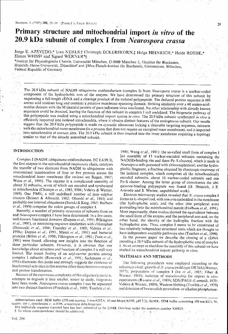
Biochem. J. (1992) 288, 29 34 (Printed in Great Britain)
Primary structure and mitochondrial import in vitro of the 20.9 kDa subunit of complex I from Neurospova crassa Jorge E. A Z E V E D O , * Uwe NEHLS, f Christoph E C K E R S K O R N , } Helga H E I N R I C H * Heide R O T H E * Harms WEISSf and Sigurd W E R N E R * § "Institut fur Physiologische Chemie, Universitát Munchen, D-8000 Miinchen 2, flnstitut fur Biochemie, Heinrich-Heine-Universitat, Diisseldorf and JMax-Planck-Institut fur Biochemie, Genzentrum, Mùnchen, Federal Republic of Germany
29
The 20.9 kDa subunit of NADH: ubiquinone oxidoreductase (complex I) from Neurospora crassa is a nuclear-coded component of the hydrophobic arm of the enzyme. We have determined the primary structure of this subunit by sequencing a full-length cDNA and a cleavage product of the isolated polypeptide. The deduced protein sequence is 189 amino acid residues long and contains a putative membrane-spanning domain. Striking similarity over a 60 amino-acid-residue domain with the M (matrix) protein of para-influenza virus was found. No other relationship with already known sequences could be detected, leaving the function of this subunit in complex I still undefined. The biogenetic pathway of this polypeptide was studied using a mitochondrial import system in vitro. The 20.9 kDa subunit synthesized in vitro is efficiently imported into isolated mitochondria, where it obtains distinct features of the endogenous subunit. Our results suggest that the 20.9 kDa polypeptide is made on cytosolic ribosomes lacking a cleavable targeting sequence, interacts with the mitochondrial outer membrane (in a process that does not require an energized inner membrane), and is'imported into mitochondria at contact sites. The 20.9 kDa subunit is then inserted into the inner membrane acquiring a topology similar to that of the already assembled subunit.
INTRODUCTION
Complex I (NADH:ubiquinone oxidoreductase, EC 1.6.99.3), the first enzyme in the mitochondrial respiratory chain, catalyses the transfer of two electrons from NADH to ubiquinone with concomitant translocation of four or five protons across the mitochondrial inner membrane (for reviews see Ragan, 1987; Weiss et al., 1991). The enzyme is a complicated assembly of about 32 subunits, seven of which are encoded and synthesized in mitochondria (Chomyn et ai, 1985, 1986; Videira & Werner, 1989). One FMN, a still undefined number of iron-sulphur clusters (Beinert & Albracht, 1982; Ohnishi et al., 1985) and probably one internal ubiquinone (Suzuki & King, 1983 ; Burbaev et ai, 1989) comprise the redox groups of complex I.
Recently, a number of primary structures of subunits from ox and Neurospora complex I have been determined. In a few cases, well-known functional domains (Dupuis et al., 1991 ; Pilkington el al., 1991), or interesting sequence similarities with chloroplast (Shinozaki et al., 1986; Fearnley et ai, 1989; Videira et ai, 1990a; Dupuis et ai, 1991; Masui et ai, 1991) and bacterial proteins (Bõhm et ai, 1990; Pilkington et al., 1991 ; Preis et ai, 1991) were found, allowing new insights into the function of these particular subunits. However, it is obvious that our knowledge about structure and function of complex I is still very limited. The recent finding of an acyl-carrier protein among complex I subunits (Runswick et ai, 1991; Sackmann et ai, 1991) illustrates this point and strongly suggests the existence of biochemical activities in this enzyme other than electron transport and proton translocation.
Because of the enormous complexity of this oligomeric protein, attempts to degrade it into smaller, easier to study, fragments have been made. Neurospora crassa complex 1 can be separated into two distinct fractions (Friedrich et ai, 1989; Tuschen et al..
1990; Wang et al., 1991): the so-called small form of complex I [an assembly of 13 nuclear-encoded subunits containing the NAD(H)-binding site and three Fe-S clusters], which is made in Neurospora cells poisoned with chloramphenicol ; and the hydrophobic fragment, a fraction obtained by chaotropic treatment of the isolated complex, which comprises all the mitochondrially encoded subunits, about 10 nuclear-coded subunits and one Fe-S cluster. Among the latter group of constituents the ubi-quinone-binding polypeptide was found (H. Heinrich, J. E. Azevedo and S. Werner, unpublished work).
Electron microscopy studies revealed that N. crassa complex I forms an L-shaped rod, with one arm embedded in the membrane (the hydrophobic arm), and the other (the peripheral arm) protruding into the mitochondrial matrix (Hofhaus et al., 1991). Most importantly, these studies showed the equivalence between the small form of the enzyme and the peripheral arm and, on the other hand, the identity of the hydrophic fraction with the hydrophobic arm. Thus, complex I seems to be constituted of two relatively independent structural units which are thought to have independent assembly pathways also (Tuschen et al., 1990).
In the present paper we describe the cloning of a cDNA encoding a 20.9 kDa subunit of the hydrophobic arm of complex I. As an attempt to elucidate the assembly of this subunit we have studied its mitochondrial import pathway in vitro.
MATERIALS AND METHODS
The following procedures were employed according to the references cited : growth of A', crassa (wild type OR74A) (Werner, 1977); preparation of complex I (Ise et al., 1985; Filser & Werner, 1988); isolation of mitochondria for import in vitro experiments (Rassow et al., 1989); SDS/PAGE (Laemmli, 1970; Videira & Werner, 1989); Western-blotting (Towbin et al., 1979) and detection of horseradish peroxidase- or alkaline phosphatase-
Abbreviations used: SEM buffer (250 mM-sucrose, 1 niM-EDTA, 10 mM-Mops/KOH, pH 7.2); SEMK, SEM buffer containing 100 mM-KCl- M matrix; cyt c, cytochrome c: a-IDH, a-isocitrate dehydrogenase.
The nucleotide sequence reported here has been submitted to the EMBL Database under the accession number X60829. § To whom correspondence should be addressed.
Vol. 288
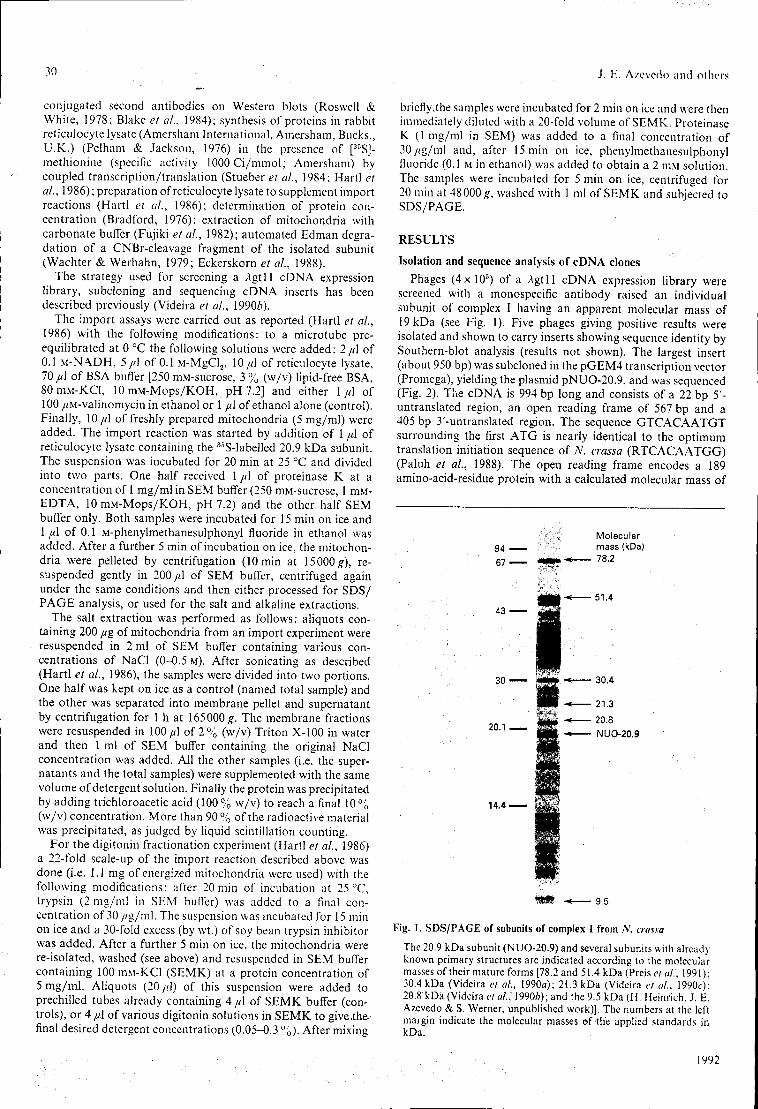
30 J. E. Azevedo and others
conjugated second antibodies on Western blots (Roswell & White, 1978; Blake et al., 1984); synthesis of proteins in rabbit reticulocyte lysate (Amersham International, Amersham, Bucks., U.K.) (Pelham & Jackson, 1976) in the presence of [;i5SJ
methionine (specific activity 1000 Ci/mmol; Amersham) by coupled transcription/translation (Stueber et al., 1984; Hartl et al., 1986); preparation of reticulocyte lysate to supplement import reactions (Hartl et ai, 1986); determination of protein con
centration (Bradford, 1976); extraction of mitochondria with carbonate buffer (Fujiki et a!., 1982); automated Edman degra
dation of a CNBrcIeavage fragment of the isolated subunit (Wachter & Werhahn, 1979; Eckerskorn et al., 1988).
The strategy used for screening a Àgtll cDNA expression library, subcloning and sequencing cDNA inserts has been described previously (Videira et al., 19906).
The import assays were carried out as reported (Hartl et al., 1986) with the following modifications: to a microtube pre
equilibrated at 0 °C the following solutions were added: 2//1 of 0.1 MNA.DH, 5//1 of 0.1 MMgCl2, 10//1 of reticulocyte lysate, 70//1 of BSA buffer [250 mMsucrose, 3 % (w/v) lipidfree BSA, 80mMKCl, lOniMMops/KOH, pH 7.2] and either 1//1 of 100 //Mvalinomycin in ethanol or 1//1 of ethanol alone (control). Finally, 10//1 of freshly prepared mitochondria (5 mg/ml) were added. The import reaction was started by addition of 1//1 of reticulocyte lysate containing the 35Slabelled 20.9 kDa subunit. The suspension was incubated for 20 min at 25 °C and divided into two parts. One half received 1 //1 of proteinase K at a concentration of 1 mg/ml in SEM buffer (250 mMsucrose, 1 niM
EDTA, 10 mMMops/KOH, pH 7.2) and the other half SEM buffer only. Both samples were incubated for 15 min on ice and 1//1 of 0.1 Mphenylmethanesulphonyl fluoride in ethanol was added. After a further 5 min of incubation on ice, the mitochon
dria were pelleted by centrifugation (10 min at 15000 g), re
suspended gently in 200//.1 of SEM buffer, centrifuged again under the same conditions and then either processed for SDS/ PAGE analysis, or used for the salt and alkaline extractions.
The salt extraction was performed as follows: aliquots con
taining 200 //g of mitochondria from an import experiment were resuspended in 2 ml of SEM buffer containing various con
centrations of NaCl (00.5 M). After sonicating as described (Hartl et ai, 1986), the samples were divided into two portions. One half was kept on ice as a control (named total sample) and the other was separated into membrane pellet and supernatant by centrifugation for 1 h at 165000g. The membrane fractions were resuspended in 100//1 of 2 % (w/v) Triton X100 in water and then 1 ml of SEM buffer containing the original NaCl concentration was added. All the other samples (i.e. the super
natants and the total samples) were supplemented with the same volume of detergent solution. Finally the protein was precipitated by adding trichloroacetic acid (100% w/v) to reach a final 10% (w/v) concentration. More than 90 % of the radioactive material was precipitated, as judged by liquid scintillation counting.
For the digitonin fractionation experiment (Hartl el ah, 1986) a 22fold scaleup of the import reaction described above was done (i.e. 1.1 mg of energized mitochondria were used) with the following modifications: after 20 min of incubation at 25 °C, trypsin (2 mg/ml in SEM buffer) was added to a final con
centration of 30//g/ml. The suspension was incubated for 15 min on ice and a 30fold excess (by wt.) of soy bean trypsin inhibitor was added. After a further 5 min on ice, the mitochondria were reisolated, washed (see above) and resuspended in SEM buffer containing 100 mMKCl (SEMK) at a protein concentration of 5 mg/ml. Aliquots (20 //1) of this suspension were added to prechilled tubes already containing 4 //1 of SEMK buffer (con
trols), or 4 //1 of various digitonin solutions in SEMK to give.the
final desired detergent concentrations (0.050.3 %). After mixing
briefly,the samples were incubated for 2 min on ice and were then immediately diluted with a 20fold volume of SEMK. Proteinase K (1 mg/ml in SEM) was added to a final concentration of 30//g/ml and, after 15 min on ice, phenylmethanesulphonyl fluoride (0.1 M in ethanol) was added to obtain a 2 m.\i solution. The samples were incubated for 5 min on ice, centrifuged for 20 min at 48000 #, washed with 1 ml of SEMK and subjected to SDS/PAGE.
RESULTS
Isolation and sequence analysis of cDNA clones Phages (4x 10⁵) of a Agtll cDNA expression library were
screened with a monospecific antibody raised an individual subunit of complex I having an apparent molecular mass of 19 kDa (see Fig. 1). Five phages giving positive results were isolated and shown to carry inserts showing sequence identity by Southernblot analysis (results not shown). The largest insert (about 950 bp) was subcloned in the pGEM4 transcription vector (Promega), yielding the plasmid pNUO20.9, and was sequenced (Fig. 2). The cDNA is 994 bp long and consists of a 22 bp 5'
untranslated region, an open reading frame of 567 bp and a 405 bp 3'untranslated region. The sequence GTCACAATGT surrounding the first ATG is nearly identical to the optimum translation initiation sequence of N. crassa (RTCACAATGG) (Paluh et ai, 1988). The open reading frame encodes a 189 aminoacidresidue protein with a calculated molecular mass of
Molecular 94 — mass (kDa) 67 — . - 78.2
<gttj-« 51.4 43 o B g
30 m ■« 30.4
wm -*— 21.3
l i l t -* NUO-20.9
14.4
'JSJB ■* 9.5
Fig. 1. SDS/PAGE of subunits of complex I from N. crassa The 20.9 kDa subunit (NUO20.9) and several subunits with already known primary structures are indicated according to the molecular masses of their mature forms [78.2 and 51.4 kDa (Preis et al., 1991): 30.4 kDa (Videira et al., 1990a); 21.3 kDa (Videira et al., 1990c); 20.8 kDa (Videira et al/, \990b); and the 9.5 kDa (H. Heinrich, J. E. Azevedo & S. Werner, unpublished work)]. The numbers at the left margin indicate the molecular masses of the applied standards in kDa.
1992
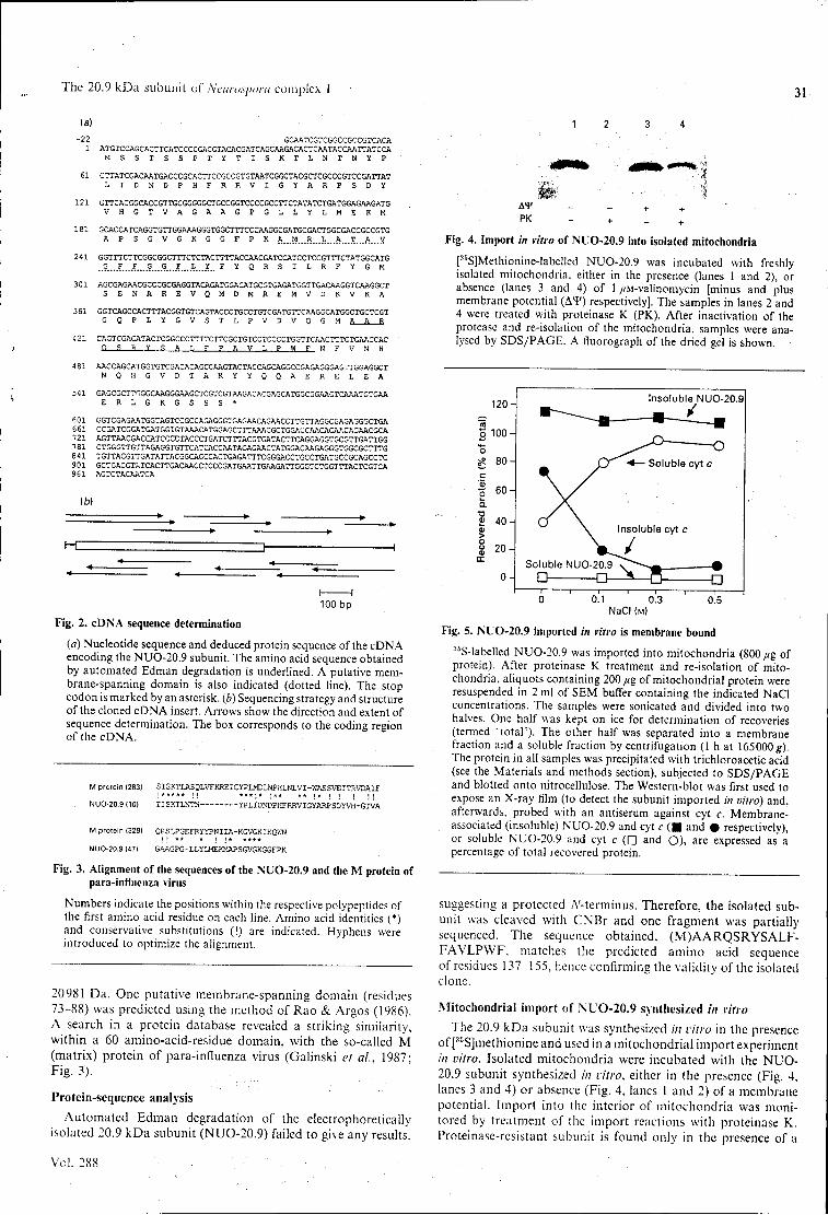
The 20.9 kDa subunit of Neurospora complex I 31
(a) -22 . GCAATCGTCGGCCGCCGTCACA 1 ATGTCCAGCACTTCATCCCCGACGTACACGATCAGCAAGACACTCAATACCAATTATCCA
M S S T S S P T Y T I S K T L N T N Y P 61 CTTATCGACAATGACCCGCACTTCCGCCGTGTAATCGGCTACGCTCGCCCGTCCGATTAT
L I D N D P H F R R V I G Y A R P S D Ï
121 GTTCACGGCACCGTTGCGGGGGCTGCCGGTCCCCGCCTTCTATATCTGATGGAGAAGATG V H G T V A G A A G P G L L Y L M E K M
181 GCACCATCAGGTGTTGGAAAGGGTGGCTTTCCCAAGGCGATGCGACTGGCGACCGCCGTG A P S G V G K G G F P K A. M ..R . L ..A...T...A. V.
241 GGTTTCTTCGGCGGCTTTCTCTACTTTTACCAACGATCCATCCTCCGTTTCTATGGCATG ..9...X...X....?...S...X...h...X.. F Y Q R S I L R F Y G M
301 AGCGAGAACGCCCGCGAGGTACAGATGGACATGCGTGAGATGGTTGACAAGGTCAAGGCT S E N A R E V Q M D M R E M V D K V K A
361 GGTCAGCCACTTTACGGTGTCAGTACCCTGCCTGTCGATGTTCAAGGCATGGCTGCTCGT G Q P L Y G V S T L P V D V Q G M A A R
421 CAGTCGAGATACTCGGCCCTTTTCTTCGCTGTCCTCCCCTGGTTCAACTTTGTGAACCAC 2 S R 1 S A L F F A V L P W F N F V N H
481 AACCAGCATGGTGTCGACACAGCCAAGTACTACCAGCAGGCCGAGAGGGAGTTGGAGGCT N Q H G V D T A K Y Y Q Q A E R E L E A
541 GAGCGCTT^GGCAAGGGAAGCTCGTCGTAAGACACGAGCATGGCGGAAGTCAAATGTGAA E R L G K G S S S *
601 GGTCGAGAATGGTAGTCCGCCAGAGGGTGAGAACAGAACCTTGTTAGGCGAGAGGGCTGA 661 CCGATCGGATGATGGIGTAAACATGGAGCTTTAAACGCTGGACCAACAGAACAGAACGCA 721 AGTTAACGACCATCCCCTACCCTGATCTTTACGTGATACTTCAGGAGGTGCGTTGATTGG 781 CTGGGTTGTTAGAGGTGTTCATCACCAATAGAGAACTATGGACAAGAGGGTGGCGCTTTG 841 TGTTACGTTGATATTACGGCAGCCACTGAGATTTCGGGACCTGCCTGATGCCGCAGCCTC 901 GCTGACGTATCACTTGACAACCTCCCGATGAATTGAAGATTGGGTCTGGTTTACTCGTCA 361 AGTCTACAATCA
(b)
h H 100 bp
Fig. 2. cDNA sequence determination (a) Nucleotide sequence and deduced protein sequence of the cDNA encoding the NUO20.9 subunit. The amino acid sequence obtained by automated Edman degradation is underlined. A putative membranespanning domain is also indicated (dotted line). The stop codon is marked by an asterisk, (b) Sequencing strategy and structure of the cloned cDNA insert. Arrows show the direction and extent of sequence determination. The box corresponds to the coding region of the cDNA.
M protein (283] SISKTLASQLVFKREICYPLMDLNPHLNLVIWASSVEITRVDAIF
NUO-20.9 (10) TISKTLNTN- -YPLIDNDPHFRRVIGYARPSDYVH-GTVA
M protein (328) OPSLPGEFRYYPNIIA-KGVGKIKQWN NUO-20.9 (47) GAAGPG-LLYLMEKKAPSGVGKGGFPK
Fig. 3. Alignment of the sequences of the NUO20.9 and the M protein of parainfluenza virus
Numbers indicate the positions within the respective polypeptides of the first amino acid residue on each line. Amino acid identities (*) and conservative substitutions (!) are indicated. Hyphens were introduced to optimize the alignment.
20981 Da. One putative membranespanning domain (residues 7388) was predicted using the method of Rao & Argos (1986). A search in a protein database revealed a striking similarity, within a 60 aminoacidresidue domain, with the socalled M (matrix) protein of parainfluenza virus (Galinski et ai, 1987 ' Fig. 3).
Proteinsequence analysis Automated Edman degradation of the electrophoretically
isolated 20.9 kDa subunit (NUO20.9) failed to give any results.
Vol. 288
A¥ + + PK - + _ +
Fig. 4. Import in vitro of NUO20.9 into isolated mitochondria [35S]MethionineIabelled NUO20.9 was incubated with freshly isolated mitochondria, either in the presence (lanes 1 and 2), or absence (lanes 3 and 4) of 1 //Mvalinomycin [minus and plus membrane potential (Af) respectively]. The samples in lanes 2 and 4 were treated with proteinase K (PK). After inactivation of the protease and reisolation of the mitochondria, samples were analysed by SDS/PAGE. A fluorograph of the dried gel is shown.
120
100
80
60
4 0 -
20
0
Insoluble NUO-20.9
Çj ■*— Soluble cyt c
\ Insoluble cyt c
Snluhlfi NI m-?n q 7 ^ - ^ - -% □ D -s B -D 0 0.1 0.3 0.5
NaCI (M)
Fig. 5. NUO20.9 imported in vitro is membrane bound 36Slabelled NUO20.9 was imported into mitochondria (800 fig of protein). After proteinase K treatment and reisolation of mitochondria, aliquots containing 200 pig of mitochondrial protein were resuspended in 2 ml of SEM buffer containing the indicated NaCI concentrations. The samples were sonicated and divided into two halves. One half was kept on ice for determination of recoveries (termed 'total'). The other half was separated into a membrane fraction and a soluble fraction by centrifugation (1 h at 165000;?). The protein in all samples was precipitated with trichloroacetic acid (see the Materials and methods section), subjected to SDS/PAGE and blotted onto nitrocellulose. The Westernblot was first used to expose an Xray film (to detect the subunit imported in vitro) and, afterwards, probed with an antiserum against cyt c. Membraneassociated (insoluble) NUO20.9 and cyt c ( B and • respectively), or soluble NUO20.9 and cyt c (□ and O), are expressed as a percentage of total recovered protein.
suggesting a protected A'terminus. Therefore, the isolated sub
unit was cleaved with CNBr and one fragment was partially sequenced. The sequence obtained. (M)AARQSRYSALF
FAVLPWF, matches the predicted amino acid sequence of residues 137155, hence confirming the validity of the isolated clone.
.Mitochondrial import of NUO20.9 synthesized in vitro The 20.9 kDa subunit was synthesized in vitro in the presence
of [35S]methionine and used in a mitochondrial import experiment in vitro. Isolated mitochondria were incubated with the NUO
20.9 subunit synthesized in vitro, either in the presence (Fig. 4, lanes 3 and 4) or absence (Fig. 4, lanes 1 and 2) of a membrane potential. Import into the interior of mitochondria was moni
tored by treatment of the import reactions with proteinase K. Proteinasercsistant subunit is found only in the presence of a
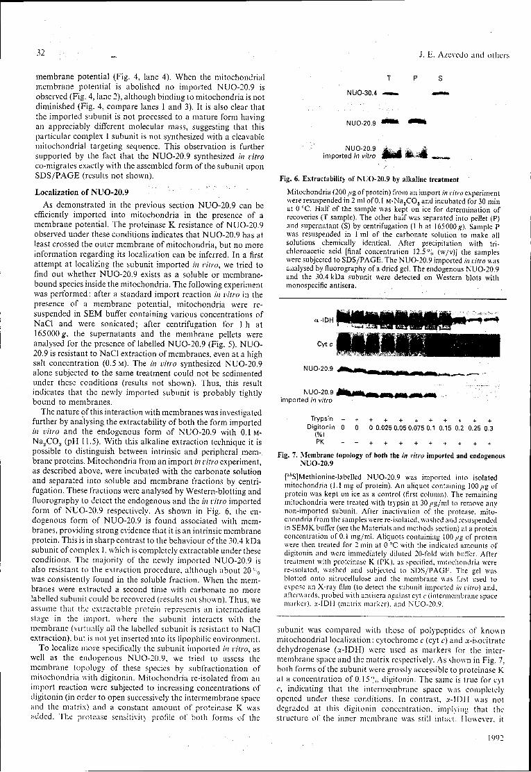
32 J. H. Azevedo and others
membrane potential (Fig. 4, lane 4). When the mitochondrial membrane potential is abolished no imported NUO20.9 is observed (Fig. 4, lane 2), although binding to mitochondria is not diminished (Fig. 4, compare lanes 1 and 3). It is also clear that the imported subunit is not processed to a mature form having an appreciably different molecular mass, suggesting that this particular complex I subunit is not synthesized with a cleavable mitochondrial targeting sequence. This observation is further supported by the fact that the NUO20.9 synthesized in vitro comigrates exactly with the assembled form of the subunit upon SDS/PAGE (results not shown).
Localization of NUO20.9 As demonstrated in the previous section NUO20.9 can be
efficiently imported into mitochondria in the presence of a membrane potential. The proteinase K resistance of NUO20.9 observed under these conditions indicates that NUO20.9 has at least crossed the outer membrane of mitochondria, but no more information regarding its localization can be inferred. In a first attempt at localizing the subunit imported in vitro, we tried to find out whether NUO20.9 exists as a soluble or membrane
bound species inside the mitochondria. The following experiment was performed : after a standard import reaction in vitro in the presence of a membrane potential, mitochondria were re
suspended in SEM buffer containing various concentrations of NaCl and were sonicated; after centrifugation for 1 h at 165000 ^, the supernatants and the membrane pellets were analysed for the presence of labelled NUO20.9 (Fig. 5). NUO
20.9 is resistant to NaCl extraction of membranes, even at a high salt concentration (0.5 M). The in vitro synthesized NUO20.9 alone subjected to the same treatment could not be sedimented under these conditions (results not shown). Thus, this result indicates that the newly imported subunit is probably tightly bound to membranes.
The nature of this interaction with membranes was investigated further by analysing the extractability of both the form imported in vitro and the endogenous form of NUO20.9 with 0 .1M
Na 2C0 3 (pH 11.5). With this alkaline extraction technique it is possible to distinguish between intrinsic and peripheral mem
brane proteins. Mitochondria from an import in vitro experiment, as described above, were incubated with the carbonate solution and separated into soluble and membrane fractions by centri
fugation. These fractions were analysed by Westernblotting and fluorography to detect the endogenous and the in vitro imported form of NUO20.9 respectively. As shown in Fig. 6, the en
dogenous form of NUO20.9 is found associated with mem
branes, providing strong evidence that it is an intrinsic membrane protein. This is in sharp contrast to the behaviour of the 30.4 kDa subunit of complex I, which is completely extractable under these conditions. The majority of the newly imported NUO20.9 is also resistant to the extraction procedure, although about 20 % was consistently found in the soluble fraction. When the mem
branes were extracted a second time with carbonate no more labelled subunit could be recovered (results not shown). Thus, we assume that the extractable protein represents an intermediate stage in the import, where the subunit interacts with the membrane (virtually all the labelled subunit is resistant to NaCl extraction), but is not yet inserted into its lipophilic environment.
To localize more specifically the subunit imported ;'/; vitro, as well as the endogenous NUO20.9, we tried to assess the membrane topology of these species by subfractionation of mitochondria with digitonin. Mitochondria reisolated from an import reaction were subjected to increasing concentrations of digitonin (in order to open successively the intermembrane space and the matrix) and a constant amount of proteinase K was added. The protease sensitivity profile of both forms of the
T P S
NUO-30.4 — — .
NUO-20.9 * ■ " » * ■ " *
NUO-20.9 j ^ ~ .M imported in vitro wÊÉm ÉÉ»8B ,,„.
Fig. 6. Extractability of NUO20.9 by alkaline treatment Mitochondria (200 /<g of protein) from an import in vitro experiment were resuspended in 2 ml of 0.1 MNa2C03 and incubated for 30 min at 0 °C. Half of the sample was kept on ice for determination of recoveries (T sample). The other half was separated into pellet (P) and supernatant (S) by centrifugation (1 h at 165000 #). Sample P was resuspended in 1 ml of the carbonate solution to make all solutions chemically identical. After precipitation with trichloroacetic acid [final concentration 12.5 % (w/v)] the samples were subjected to SDS/PAGE. The NUO20.9 imported in vitro was analysed by fluorography of a dried gel. The endogenous NUO20.9 and the 30.4 kDa subunit were detected on Western blots with monospecific antisera.
Trypsin - + + + + + + + + + + Digitonin 0 0 0 0.025 0.05 0.075 0.1 0.15 0.2 0.25 0.3
(%) PK - - + + + + + + + + +
Fig. 7. Membrane topology of both the in vitro imported and endogenous NUO20.9
[35S]Methioninelabelled NUO20.9 was imported into isolated mitochondria (1.1 mg of protein). An aliquot containing 100//g of protein was kept on ice as a control (first column). The remaining mitochondria were treated with trypsin at 30 /<g/ml to remove any nonimported subunit. After inactivation of the protease, mitochondria from the samples were reisolated, washed and resuspended in SEMK buffer (see the Materials and methods section) at a protein concentration of 0.1 mg/ml. Aliquots containing 100 //g of protein were then treated for 2 min at 0 °C with the indicated amounts of digitonin and were immediately diluted 20fold with buffer. After treatment with proteinase K (PK). as specified, mitochondria were reisolated, washed and subjected to SDS/PAGE. The gel was blotted onto nitrocellulose and the membrane was first used to expose an Xray film (to detect the subunit imported in vitro) and, afterwards, probed with antisera against cyt c (intermembrane space marker). aIDH (matrix marker).*and NUO20.9.
subunit was compared with those of polypeptides of known mitochondrial localization: cytochrome c (cyt c) and ^isocitrate dehydrogenase (aIDH) were used as markers for the inter
membrane space and the matrix respectively. As shown in Fig. 7, both forms of the subunit were grossly accessible to proteinase K at a concentration of 0.15 "„ digitonin. The same is true for cyt c, indicating that the intermembrane space was completely opened under these conditions. In contrast, aIDH was not degraded at this digitonin concentration, implying that the structure of the inner membrane was still intact. However, it
1992
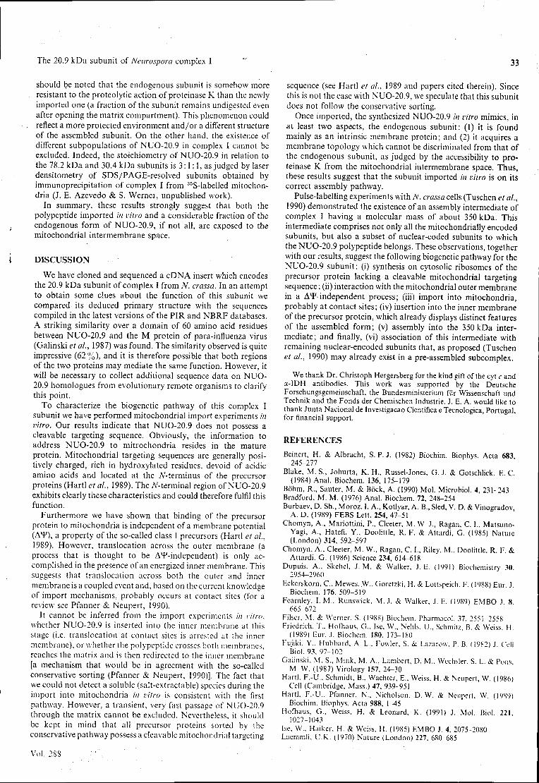
The 20.9 kDa subunit of Neurospora complex 1 33
should be noted that the endogenous subunit is somehow more resistant to the proteolytic action of proteinase K than the newly imported one (a fraction of the subunit remains undigested even after opening the matrix compartment). This phenomenon could reflect a more protected environment and/or a different structure of the assembled subunit. On the other hand, the existence of different subpopulations of NUO-20.9 in complex I cannot be excluded. Indeed, the stoichiometry of NUO-20.9 in relation to the 78.2 kDa and 30.4 kDa subunits is 3:1:1, as judged by laser densitometry of SDS/PAGE-resolved subunits obtained by immunoprecipitation of complex I from 35S-labelled mitochondria (J. E. Azevedo & S. Werner, unpublished work).
In summary, these results strongly suggest that both the polypeptide imported in vitro and a considerable fraction of the endogenous form of NUO-20.9, if not all, are exposed to the mitochondrial intermembrane space.
DISCUSSION
We have cloned and sequenced a cDNA insert which encodes the 20.9 kDa subunit of complex I from N. crassa. In an attempt to obtain some clues about the function of this subunit we compared its deduced primary structure with the sequences compiled in the latest versions of the PIR and NBRF databases. A striking similarity over a domain of 60 amino acid residues between NUO-20.9 and the M protein of para-influenza virus (Galinski et al, 1987) was found. The similarity observed is quite impressive (62 %) , and it is therefore possible that both regions of the two proteins may mediate the same function. However, it will be necessary to collect additional sequence data on NUO-20.9 homologues from evolutionary remote organisms to clarify this point.
To characterize the biogenetic pathway of this complex I subunit we have performed mitochondrial import experiments in vitro. Our results indicate that NUO-20.9 does not possess a cleavable targeting sequence. Obviously, the information to address NUO-20.9 to mitrochondria resides in the mature protein. Mitochondrial targeting sequences are generally positively charged, rich in hydroxylated residues, devoid of acidic amino acids and located at the /V-terminus of the precursor proteins (Hartl et al, 1989). The TV-terminal region of NUO-20.9 exhibits clearly these characteristics and could therefore fulfil this function.
Furthermore we have shown that binding of the precursor protein to mitochondria is independent of a membrane potential (A*), a property of the so-called class I precursors (Hartl et al., 1989). However, translocation across the outer membrane (a process that is thought to be A^-independent) is only accomplished in the presence of an energized inner membrane. This suggests that translocation across both the outer and inner membrane is a coupled event and, based on the current knowledge of import mechanisms, probably occurs at contact sites (for a review see Pfanner & Neupert, 1990).
It cannot be inferred from the import experiments in vitro, whether NUO-20.9 is inserted into the inner membrane at this stage (i.e. translocation at contact sites is arrested at the inner membrane), or whether the polypeptide crosses both membranes, reaches the matrix and is then redirected to the inner membrane [a mechanism that would be in agreement with the so-called conservative sorting (Pfanner & Neupert, 1990)]. The fact that we could not detect a soluble (salt-extractable) species during the import into mitochondria in vitro is consistent with the first pathway. However, a transient, very fast passage of NUO-20.9 through the matrix cannot be excluded. Nevertheless, it should be kept in mind that all precursor proteins sorted by the conservative pathway possess a cleavable mitochondrial targeting
sequence (see Hartl et al., 1989 and papers cited therein). Since this is not the case with NUO-20.9, we speculate that this subunit does not follow the conservative sorting.
Once imported, the synthesized NUO-20.9 in vitro mimics, in at least two aspects, the endogenous subunit: (1) it is found mainly as an intrinsic membrane protein; and (2) it acquires a membrane topology which cannot be discriminated from that of the endogenous subunit, as judged by the accessibility to proteinase K from the mitochondrial intermembrane space. Thus, these results suggest that the subunit imported in vitro is on its correct assembly pathway.
Pulse-labelling experiments with N. crassa cells (Tuschen et al., 1990) demonstrated the existence of an assembly intermediate of complex I having a molecular mass of about 350 kDa. This intermediate comprises not only all the mitochondrially encoded subunits, but also a subset of nuclear-coded subunits to which the NUO-20.9 polypeptide belongs. These observations, together with our results, suggest the following biogenetic pathway for the NUO-20.9 subunit: (i) synthesis on cytosolic ribosomes of the precursor protein lacking a cleavable mitochondrial targeting sequence; (ii) interaction with the mitochondrial outer membrane in a A*Findependent process; (iii) import into mitochondria, probably at contact sites; (iv) insertion into the inner membrane of the precursor protein, which already displays distinct features of the assembled form ; (v) assembly into the 350 kDa intermediate; and finally, (vi) association of this intermediate with remaining nuclear-encoded subunits that, as proposed (Tuschen et al., 1990) may already exist in a pre-assembled subcomplex.
We thank Dr. Christoph Hergersberg for the kind gift of the cyt c and a-IDH antibodies. This work was supported by the Deutsche Forschungsgemeinschaft, the Bundesministerium fur Wissenschaft und Technik and the Fonds der Chemischen Industrie. J. E. A. would like to thank Junta Nacional de Investigação Cientifica e Tecnológica, Portugal, for financial support.
REFERENCES
Beinert, H. & Albracht, S.P.J. (1982) Biochim. Biophys. Acta 683, 245-277
Blake, M. S., Johurta, K. H., Russel-Jones, G. J. & Gotschlick, E. C. (1984) Anal. Biochem. 136, 175-179
Bõhm, R.( Sauter, M. & Bõck, A. (1990) Mol. Microbiol. 4, 231-243 Bradford, M. M. (1976) Anal. Biochem. 72, 248-254 Burbaev, D. Sh., Moroz, I. A., Kotlyar, A. B., Sied, V. D. & Vinogradov.
A. D. (1989) FEBS Lett. 254, 47-51 Chomyn, A., Mariottini, P., Cleeter, M. W J., Ragan, C. 1., Matsuno-
Yagi, A., Hatefi, Y., Doolittle, R. F. & Attardi, G. (1985) Nature (London) 314, 592-597
Chomyn, A., Cleeter, M. W., Ragan, C. I., Riley, M., Doolittle, R. F. & Attardi, G. (1986) Science 234, 614-618
Dupuis, A„ Skehel, J. M. & Walker, J. E. (199!) Biochemistry 30 2954-2960
Eckerskorn, C, Mewes,.W., Goretzki. H. & Lottspeich. F. (1988) Eur J Biochem. 176, 509-519
Fearnley. I. M., Runswick, M. J. & Walker, J. E. (1989) EMBO J 8 665-672
Filser. M. & Werner, S. (1988) Biochem. Pharmacol. 37. 2551-2558 Friedrich, T., Hofhaus. G., Ise. W., Nelils, U., Schmitz, B. & Weiss. H.
(1989) Eur. J. Biochem. 180, 173-180 Fujiki, Y., Hubbard, A. L., Fowler, S. & Lazarow. P. B. (1982) J. Cell
Biol. 93, 97-102 Galinski, M. S., Mink, M. A., Lambert, D. M., Wechsler. S. L. & Pons
M. W. (1987) Virology 157, 24-30 Hartl, F.-U.. Schmidt, B.. Wachter, E., Weiss, H. & Neupert, W. (1986)
Cell (Cambridge. Mass.) 47, 939-951 Hartl, F.-U.. Pfanner, N., Nicholson, D. W. & Neupcrl. W. (1989)
Biochim. Biophys. Acta 988, 1-45 Hofliaus, G., Weiss, H. & Leonard, K. (1991) J. Mol. Biol. 221.
1027-1043 Ise. W.. Haiker, H. & Weiss. H. (1985) EMBO J. 4. 2075-2080 Laemmli. U.K. (1970) Nature (London) 227. 680 685
Vol. 288
34 J. E. Azevedo and others
Masui, R., Wakabayashi, S., Matsubara, H. & Halefi, Y. (1991) J. Biochem. 110, 575-582
Ohnishi, T., Ragan, C. I. & Hatefi, Y. (1985) J. Biol. Chem. 260, 2782-2788
Paluh, J. L.,Orbach, M. J., Legerton, T. L. & Yanofsky, C.{1988) Proc. Natl. Acad. Sci. U.S.A. 85, 3728-3732
Pelham, H. R. B. & Jackson, R. J. (1976) Eur. J. Biochem. 67, 247-256 Pfanner, N. & Neupert, W.(1990) Annu. Rev. Biochem. 59, 331-353 Pilkington, S. J., Skehel, J. M., Gennis, R. B. & Walker, J. E. (1991)
Biochemistry 30, 2166-2175 Preis, D., Weidner, U., Conzen, C , Azevedo, J. E., Nehls, U., Rohlen,
D., Van der Pas, J., Sackmann, U., Schneider, R., Werner, S. & Weiss, H. (1991) Biochim. Biophys. Acta 1090, 133-138
Ragan, C. I. (1987) Curr. Topics Bioenerg. 15, 1-36 Rao, J. K. M.&Argos, P. (1986) Biochem. Biophys. Res. Commun. 869,
197-214 Rassow, J., Guiard, B., Weinhues, U., Herzog, V., Hartl, F.-U. &
Neupert, W. (1989) J. Cell Biol. 109, 1421-1428 Roswell, D. F. & White, E. H. (1978) Methods Enzymol. 57, 409-^23 Runswick, M. J., Fearnley, I. M., Skehel, J. M. & Walker, J. E. (1991)
FEBS Lett. 286, 121-124 Sackmann, U., Zensen, R., Rõhlen, D., Jahnke, U. & Weiss, H. (1991)
Eur. J. Biochem. 200, 463^469
Received 29 April 1992; accepted 22 May 1992
Shinozaki. K., Ohme, M., Tanaka, M., Wakasugi, T., Hayashida, N., Matsubayashi, T., Zaita, N., Chunwongse, J., Obokata, J., Yamaguchi-Shinozaki, K. el al. (1986) EMBO J. 5, 2043-2049
Stueber, D., Ibrahimi, I., Cutler, D., Dobberstein, B. & Bujard, H. (1984) EMBO J. 3, 3143-3148
Suzuki, H. & King, T. E. (1983) J. Biol. Chem. 258, 352-358 Towbin, H., Staehelin, T. & Gordon, J. (1979) Proc. Natl. Acad. Sci.
U.S.A. 76, 4350-4354 Tuschen, G , Sackmann, U , Nehls, U., Haiker, H., Buse, G. & Weiss, H.
(1990) J. Mol. Biol. 213, 845-857 Videira, A. & Werner, S. (1989) Eur. J. Biochem. 181, 493-502 Videira, A., Tropschug, M. & Werner, S. (1990«) Biochem. Biophys. Res.
Commun. 171, 1168-1174 Videira, A., Tropschug, M., Wachter, E., Schneider, H. & Werner, S.
(19906) J. Biol. Chem. 265, 13060-13065 Videira, A., Tropschug, M. & Werner, S. (1990c) Biochem. Biophys. Res.
Commun. 166, 280-285 Wachter, E. & Werhahn, R. (1979) Anal. Biochem. 97, 56-64 Wang, D., Meinhardt, S. W., Sackmann, U., Weiss, H. & Ohnishi, T.
(1991) Eur. J. Biochem. 197, 257-264 Weiss, H., Friedrich, T., Hofhaus, G. & Preis, D. (1991) Eur. J. Biochem.
197, 563-576 Werner, S. (1977) Eur. J. Biochem. 79, 103-110
1992
TRABALHO 7.2
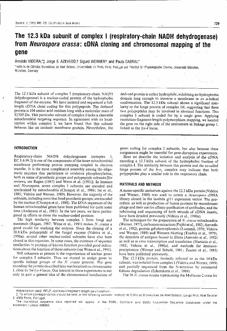
Biochem. J. (1993) 291, 729-732 (Printed in Great Britain) 729
The 12.3 kDa subunit of complex I (respiratory-chain NADH dehydrogenase) from Neurospora crassa: cDNA cloning and chromosomal mapping of the gene Arnaldo VIDEIRA,*} Jorge E. AZEVEDO, t Sigurd WERNERf and Paula CABRAL* Instituto de Ciências Biomédicas de Abel Salazar, Universidade do Porto, Porto, Portugal and flnstltut fur Physiologische Chemie, Universitàt Munchen, Munchen, Germany
The 12.3 kDa subunit of complex I (respiratory-chain NADH dehydrogenase) is a nuclear-coded protein of the hydrophobic fragment of the enzyme. We have isolated and sequenced a full-length cDNA clone coding for this polypeptide. The deduced protein is 104 amino acid residues long with a molecular mass of 12305 Da. This particular subunit of complex I lacks a cleavable mitochondrial targeting sequence. In agreement with its localization within complex I, we have found that this subunit behaves like an intrinsic membrane protein. Nevertheless, the
deduced protein is rather hydrophilic, exhibiting no hydrophobic domain long enough to traverse a membrane in an a-helical conformation. The 12.3 kDa subunit shows a significant similarity to the hinge protein of complex III, suggesting that these two polypeptides may be involved in identical functions. This complex I subunit is coded for by a single gene. Applying restriction-fragment-length-polymorphism mapping, we located the gene on the right side of the centromere in linkage group I, linked to the lys-4 locus.
INTRODUCTION
Respiratory-chain NADH dehydrogenase (complex I, EC 1.6.99.3) is one of the components of the inner mitochondrial membrane performing proton pumping coupled to electron transfer. It is the most complicated assembly among the oligo-meric enzymes that participate in oxidative phosphorylation, both in terms of prosthetic groups and polypeptide subunits [for reviews, see Ragan (1987) and Weiss et al. (1991)]. In humans and ' Neurospora, seven complex I subunits are encoded and synthesized by mitochondria (Chomyn et al., 1986; Ise et al., 1985; Videira and Werner, 1989). However, the majority of the subunits, including most that bind prosthetic groups, are encoded by the nucleus (Chomyn et al., 1988). The DNA sequences of the human mitochondrial genes have been published for quite some time (Anderson et al., 1981). In the last years, we have participated in efforts to clone the nuclear-coded proteins.
The high similarity between complex I from fungi and mammals (Ragan, 1987; Weiss et al., 1991) makes N. crassa a good model for studying the enzyme. Since the cloning of a 20.8 kDa polypeptide of the fungal enzyme (Videira et al., 1990a), several other nuclear-coded subunits have also been cloned in this organism. In some cases, the existence of sequence similarities to proteins of known function provided good indications about the function of those subunits (see Weiss et al., 1991).
Still unknown at present is the organization of nuclear genes for complex I subunits. Thus we started to assign genes to specific linkage groups of the N. crassa genome. The gene encoding the protein described here was localized on chromosome I, close to the lys-4 locus. Our interest in these experiments is not only to gain a general idea of the chromosomal localization of
genes coding for complex I subunits, but also because these assignments might be essential for gene-disruption experiments.
Here we describe the isolation and analysis of the cDNA encoding a 12.3 kDa subunit of the hydrophobic fraction of complex I. The similarity between this protein and the so-called hinge protein of the b-c1 complex may indicate that both polypeptides play a similar role in the respiratory chain.
MATERIALS AND METHODS
A mono-specific antiserum against the 12.3 kDa protein (Videira and Werner, 1989) was used to screen a Neurospora cDNA library cloned in the lambda gt l l expression vector. The procedure, as well as production of fusion proteins by recombinant phage and their use for affinity purification of antibodies, and the subcloning and sequencing of both strands of cDNA inserts, have been detailed previously (Videira et al., 1990a).
The techniques for the preparation of N. crassa mitochondria (Werner, 1977), carbonate extraction (Fujiki et al., 1982;Azevedo et al., 1992), protein gel electrophoresis (Laemmli, 1970; Videira and Werner, 1989) and Western blotting (Towbin et al., 1979), the detection of antigens bound to filters (Azevedo et al., 1992) as well as in vitro transcription and translation (Maniatis et al.. 1982; Videira et al., 1990a), and methods for immuno-precipitation (Werner and Sebald, 1981; Zauner et al., 1985) have been published previously.
The 12.3 kDa protein, formerly subunit, was isolated from complex and partially sequenced from the Edman degradation (Eckerskorn et ai.. 1988).
The 38 A', crassa strains representing the Multicent-2 cross kit
referred to as the 14 kDa (Videira and Werner, 1989) N-terminus by automated
Abbreviation used: RFLP, restriction-fragment-length polymorphism. X To whom correspondence should be sent, at the following address: Instituto de Ciências Biomédicas de Abel Salazar Largo Prof Abel Salazar
2, 4000 Porto, Portugal. The nucleotide sequence data reported will appear in the EMBL, GenBank and DDBJ Nucleotide Sequence Databases under the
accession number X68965.
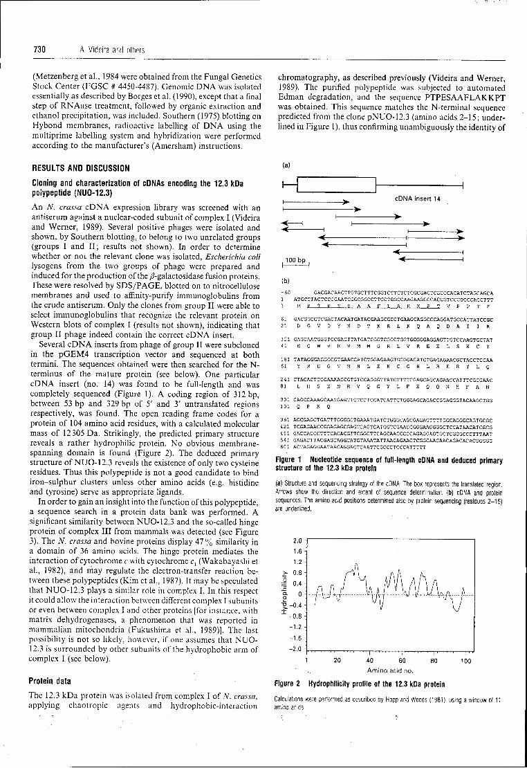
730 A. Videira and others
(Metzenberg et al., 1984 were obtained from the Fungal Genetics Stock Center (FGSC # 4450-4487). Genomic DNA was isolated essentially as described by Borges et al. (1990), except that a final step of RNAase treatment, followed by organic extraction and ethanol precipitation, was included. Southern (1975) blotting on Hybond membranes, radioactive labelling of DNA using the multiprime labelling system and hybridization were performed according to the manufacturer's (Amersham) instructions.
chromatography, as described previously (Videira and Werner, 1989). The purified polypeptide was subjected to automated Edman degradation, and the sequence PTPESAAFLAKKPT was obtained. This sequence matches the N-terminal sequence predicted from the clone pNUO-12.3 (amino acids 2-15; underlined in Figure 1), thus confirming unambiguously the identity of
RESULTS AND DISCUSSION
Cloning and characterization of cDNAs encoding the 12.3 kDa polypeptide (NU0-12.3)
An N. crassa cDNA expression library was screened with an antiserum against a nuclear-coded subunit of complex I (Videira and Werner, 1989). Several positive phages were isolated and shown, by Southern blotting, to belong to two unrelated groups (groups I and II; results not shown). In order to determine whether or not the relevant clone was isolated, Escherichia coli lysogens from the two groups of phage were prepared and induced for the production of the /?-galactosidase fusion proteins. These were resolved by SDS/PAGE, blotted on to nitrocellulose membranes and used to affinity-purify immunoglobulins from the crude antiserum. Only the clones from group II were able to select immunoglobulins that recognize the relevant protein on Western blots of complex I (results not shown), indicating that group II phage indeed contain the correct cDNA insert.
Several cDNA inserts from phage of group II were subcloned in the pGEM4 transcription vector and sequenced at both termini. The sequences obtained were then searched for the N-terminus of the mature protein (see below). One particular cDNA insert (no. 14) was found to be full-length and was completely sequenced (Figure 1). A coding region of 312 bp, between 53 bp and 329 bp of 5' and 3' untranslated regions respectively, was found. The open reading frame codes for a protein of 104 amino acid residues, with a calculated molecular mass of 12305 Da. Strikingly, the predicted primary structure reveals a rather hydrophilic protein. No obvious membrane-spanning domain is found (Figure 2). The deduced primary structure of NUO-12.3 reveals the existence of only two cysteine residues. Thus this polypeptide is not a good candidate to bind iron-sulphur clusters unless other amino acids (e.g. histidine and tyrosine) serve as appropriate ligands.
In order to gain an insight into the function of this polypeptide, a sequence search in a protein data bank was performed. A significant similarity between NUO-12.3 and the so-called hinge protein of complex III from mammals was detected (see Figure 3). The N. crassa and bovine proteins display 47% similarity in a domain of 36 amino acids. The hinge protein mediates the interaction of cytochrome c with cytochrome c, (Wakabayashi et al., 1982), and may regulate the electron-transfer reaction between these polypeptides (Kim et al., 1987). It may be speculated that NUO-12.3 plays a similar role in complex I. In this respect it could allow the interaction between different complex I subunits or even between complex I and other proteins [for instance, with matrix dehydrogenases, a phenomenon that was reported in mammalian mitochondria (Fukushima et al., 1989)]. The last possibility is not so likely, however, if one assumes that NUO-12.3 is surrounded by other subunits of the hydrophobic arm of complex I (see below).
Protein data
The 12.3 kDa protein was isolated from complex I of V. crassa, applying chaotropic agents and hydrophobic-interaction
(a)
cDNA insert 14
100 bp
(b) -60 GACGACAACTTGTGCTTTCGGTCTTCTCTCGCGAGTCGCCCACATCTAGCAGCA 1 ATGCCTACTCCCGAATCGGCGGCCTTCCTGGCCAAGAAGCCCACCGTCCCGCCCACCTTT 1 M £ 1 E E S A A F T. A K K P T V P P T F
61 GACGGCGTCGACTACAATGATACGAAGCGCCTGAAGCAGGCCCAGGATGCCATTATCCGC 21 D G V D Y N D T K R L K Q A Q D A I I R
121 GAGCAATGGGTCCGAGTTATGATGGGTCGCCTGGTGCGGGAGGAGTTGTCCAAGTGCTAT 41 E Q W V R V M M G R L V R E E L S K C Y
181 TATAGGGAGGGCGTGAACCATCTGGAGAAGTGCGGACATCTGAGAGAACGCTACCTCCAA 61 Y R E G V N H L E K C G H L R E R Y L Q
241 CTACACTCCGAAAACCGTGTCCAGGGTTATCTTTTCGAGCAGCAGAACCATTTCGCGAAC 81 L H S E N R V Q G Y L F E Q Q N H F A N
301 CAGCCAAAGCAATGAGTTCTCCTCCATCATTCTGGGAGCAGACCGGAGGGTACAAGCTGG 101 Q P K Q
361 AGCGAGCTGATTTGGGGCTGAAATGATCTGGGCAGCGAGAGTTTTGGCAGGGCAGTGCGC 421 TCGAGAACCGGAGAGCGAGTCAGTCATGGTCGAACCGGGAAGGGGCTCCATAACATCGCG 4 81 GACCACCCTTTCAGACGTTCGGCTTCAGCAACCCCCAGAGGAGTGCTCGGGCCCTTTAAT 541 GAGACTTACGAGCAGGCATGTAAATATTAACAGAACTCGGCAACAACAGACACACGGGGG 601 ACTAGAGGAATAACAGGACTCAATTCGGCCTCCCATTTTT
Figure 1 Nucleotide sequence of full- length cDNA and deduced primary structure of the 12.3 kDa protein
(a) Structure and sequencing strategy of the cDNA. The box represents the translated region. Arrows show the direction and extent of sequence determination, (b) cDNA and protein sequences. The amino acid positions determined also by protein sequencing (residues 2-15) are underlined.
60 Amino acid no.
100
Figure 2 Hydrophilicity profile of the 12.3 kDa protein
Calculations were performed as described by Hopp and Woods (1981), using a window of 11 amino acids.
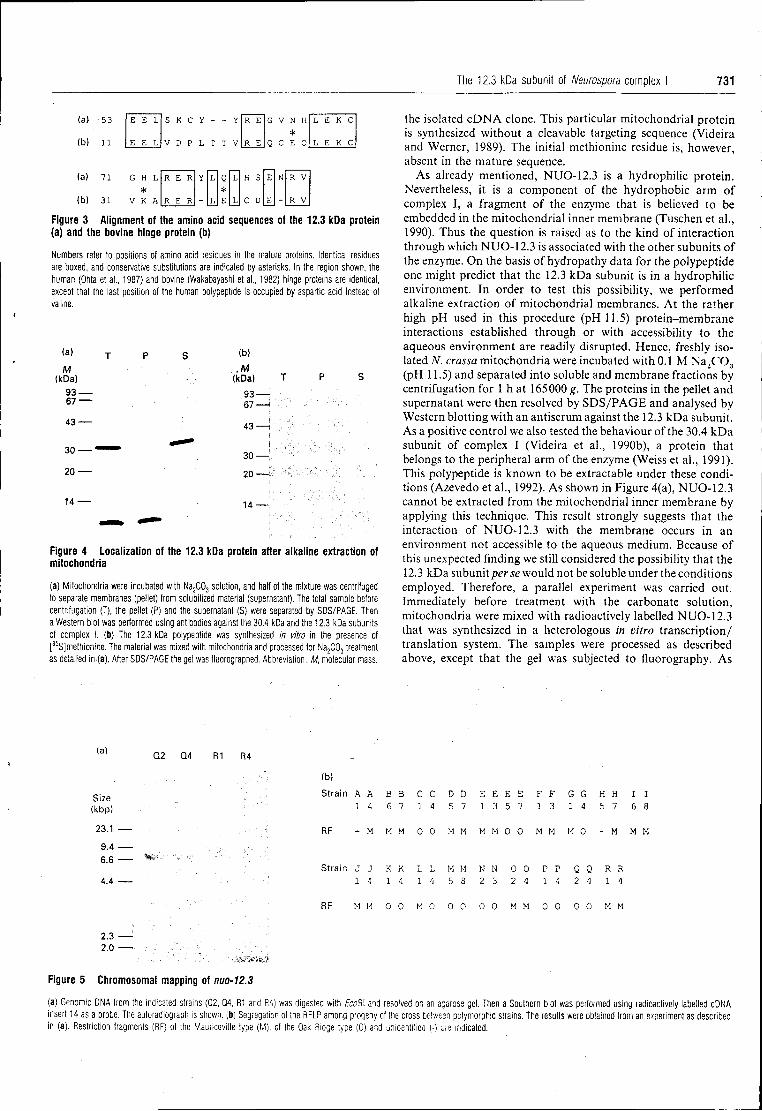
The 12.3 kDa subunit of Neuwspora complex I 731
(a> 53
(b) 11
(a)
(b)
71
31
E E L
E E L
S K C
V D P
G H L *
V K A
R E R
R E R
R E
R E
G V N H *
Q C E Q
L E K C
L E K C
H S
C D
R V
R V
Figure 3 Alignment of the amino acid sequences of the 12.3 kDa protein (a) and the bovine hinge protein (b)
Numbers refer to positions of amino acid residues in the mature proteins. Identical residues are boxed, and conservative substitutions are indicated by asterisks. In the region shown, the human (Ohta et al., 1987) and bovine (Wakabayashi et al., 1982) hinge proteins are identical, except that the last position of the human polypeptide is occupied by aspartic acid instead of valine.
(a)
M (kDa)
93 — 67 —
43 —
30 — '
20 —
1 4 —
(b)
M (kDa)
93-67-
4 3 — !
30—I 20—I
14—I
Figure 4 Localization of the 12.3 kDa protein after alkaline extraction of mitochondria
(a) Mitochondria were incubated with Na2C03 solution, and half of the mixture was centrifuged to separate membranes (pellet) from solubilized material (supernatant). The total sample before centrifugation (T), the pellet (P) and the supernatant (S) were separated by SDS/PAGE. Then a Western blot was performed using antibodies against the 30.4 kDa and the 12.3 kDa subunits of complex I. (b) The 12.3 kDa polypeptide was synthesized in vitro in the presence of [35
S]methionine. The material was mixed with mitochondria and processed for Na2C03 treatment as detailed in-(a). After SDS/PAGE the gel was fluorographed. Abbreviation : M, molecular mass.
the isolated cDNA clone. This particular mitochondrial protein is synthesized without a cleavable targeting sequence (Videira and Werner, 1989). The initial methionine residue is, however, absent in the mature sequence.
As already mentioned, NUO12.3 is a hydrophilic protein. Nevertheless, it is a component of the hydrophobic arm of complex I, a fragment of the enzyme that is believed to be embedded in the mitochondrial inner membrane (Tuschen et al., 1990). Thus the question is raised as to the kind of interaction through which NUO12.3 is associated with the other subunits of the enzyme. On the basis of hydropathy data for the polypeptide one might predict that the 12.3 kDa subunit is in a hydrophilic environment. In order to test this possibility, we performed alkaline extraction of mitochondrial membranes. At the rather high pH used in this procedure (pH 11.5) proteinmembrane interactions established through or with accessibility to the aqueous environment are readily disrupted. Hence, freshly isolated N. crassa mitochondria were incubated with 0.1 M Na2C03 (pH 11.5) and separated into soluble and membrane fractions by centrifugation for 1 h at 165000 g. The proteins in the pellet and supernatant were then resolved by SDS/PAGE and analysed by Western blotting with an antiserum against the 12.3 kDa subunit. As a positive control we also tested the behaviour of the 30.4 kDa subunit of complex I (Videira et al., 1990b), a protein that belongs to the peripheral arm of the enzyme (Weiss et al., 1991). This polypeptide is known to be extractable under these conditions (Azevedo et al., 1992). As shown in Figure 4(a), NUO12.3 cannot be extracted from the mitochondrial inner membrane by applying this technique. This result strongly suggests that the interaction of NUO12.3 with the membrane occurs in an environment not accessible to the aqueous medium. Because of this unexpected finding we still considered the possibility that the 12.3 kDa subunit per se would not be soluble under the conditions employed. Therefore, a parallel experiment was carried out. Immediately before treatment with the carbonate solution, mitochondria were mixed with radioactively labelled NUO12.3 that was synthesized in a heterologous in vitro transcription/ translation system. The samples were processed as described above, except that the gel was subjected to fluorography. As
(a) Q2 Q4 RI R4
Size (kbp)
(b)
Strain A A B B C C D D E E E E F F G G H H I I 1 4 6 7 1 4 5 7 1 3 5 7 1 3 1 4 5 7 6 8
23.1 9.4 6.6
4.4
RF M MM OO MM M M O O MM MO -M MM
Strain JJ KK LL MM N N OO PP QQ RR 14 14 14 58 23 24 14 24 14
FÎF MM O O M O O O O O MM O O O O MM
2.3 2.0
Figure 5 Chromosomal mapping of nuo-12.3
(a) Genomic DNA from the indicated strains (02, Q4, R1 and R4) was digested with feoRI and resolved on an agarose gel. Then a Southern blot was performed using radioactively labelled cDNA insert 14 as a probe. The autoradiograph is shown, (b) Segregation of the RFLP among progeny of the cross between polymorphic strains. The results were obtained from an experiment as described in (a). Restriction fragments (RF) of the Mauriceville type (M), of the Oak Ridge type (0) and unidentified (■) are indicated.
732 A. Videira and others
expected, NUO-12.3 alone does not precipitate under these conditions (Figure 4b), providing evidence that only the correctly folded/assembled subunit behaves as an intrinsic membrane protein. It should be noted that the in w'/ro-synthesized subunit co-migrates with the mature/endogenous subunit upon SDS/ PAGE performed under reductive or non-reductive conditions (A. Videira and S. Werner, unpublished work). Thus it is unlikely that the assembled subunit undergoes some covalent modification (e.g., lipoylation or cysteine bridging with other intrinsic membrane protein) that could explain the resistance to the alkaline extraction.
We have described the molecular cloning of the 12.3 kDa subunit of the hydrophobic arm of complex I from N. crassa. Notwithstanding its hydrophilic characteristics and the absence of an obvious membrane spanning domain, NUO-12.3 behaves like an intrinsic membrane protein. On the bases of double immunodiffusion (Werner and Sebald, 1981), it was already known that the protein was not recognized by the specific antiserum using intact complex I; only when isolated complex I is exposed to a detergent like SDS or a combination of deoxy-cholate and 2-mercaptoethanol does a precipitation reaction occur (A. Videira and S. Werner, unpublished work). Thus at least the antigenic determinants of the polypeptide seem not to be accessible to the immunoglobulins in the intact enzyme. This result, as well as the resistance of NUO-12.3 to alkaline extraction from the mitochondrial inner membrane, suggest that the polypeptide is embedded within other complex I constituents.
Chromosomal assignment of the nuo-12.3 gene
In order to localize the gene for NUO-12.3 in the genome of N. crassa, the restriction-fragment-length-polymorphism (RFLP) mapping approach was applied. Genomic DNA from the individual progeny of the Multicent-2 cross kit was isolated, treated with several restriction enzymes and separated by electrophoresis in agarose gels. Southern blots were then probed with 32P-labelled cDNA encoding NUO-12.3. A RFLP was found when using the enzyme Eco RI. The results obtained are summarized in Figure 5. Comparison with known markers (Metzenberg and Grotelueschen, 1989), revealed that the segregation pattern observed is very similar to those displayed by several genes of linkage group I. To test the linkage, the ^'-square statistical test was employed. The highest value, 28.2, (considering a limit value of 3.85 for one degree of freedom), was obtained between nuo-12.3 and lys-4, clearly indicating that the two genes are linked.
Significant values were also obtained for other genes located in the vicinity of the lys-4 locus.
J.E.A. and P.C. thank Junta Nacional de Investigação Científica e Tecnológica (JNICT; Portugal) for financial support. We are grateful to Mrs. Natália Mota for excellent technical assistance. This research was supported by JNICT (project SAU 48/90) and the Deutsche Forschungsgemeinschaft.
REFERENCES
Anderson, S., Bankier, A. T., Barrell, B. G., de Bruijn, M. H. L, Coulson, A. R., Drourn. J., Eperon, I. C, Nierlich. D. D„ Roe, B. A., Sanger, F„ Schreier, P. H., Smith, A, J. H., Staden, R, and Young, I. J. (1981) Nature (London) 290,457-465
Azevedo, J. E„ Nehls, U., Eckerskom, C, Heinrich. H., Rolhe, H„ Weiss, H. and Werner S (1992) Biochem. J. 288, 29-34
Borges, M. !., Azevedo, M. 0., Bonatelli, R., Felipe, M. S, S. and Astolti-Filho, S. (1990) Fungal Genet. Newsl. 37, 10
Chomyn, A., Cleeter, M. W. J., Ragan, C. I., Riley, M., Doolittle, R. F. and Attardi, G. (1966) Science 234, 614-618
Chomyn, A., Patel, S. D., Cleeter. M. W. J„ Ragan, C. I. and Attardi, G. (1988) J. Biol Chem. 263, 16395-16400
Eckerskom, C, Mewes, W., Goretzki, H. and Lottspeich, F. (1988) Eur. J. Biochem 176 509-519
Fujiki, Y., Hubbard, A. L, Fowler, S. and Lazarow, P. B. (1982) J. Cell Biol. 93, 97-102 Fukushima, T., Decker, R. V., Anderson, W. M. and Spivey, H. 0.. (1989) J. Biol Chem
264,16483-16488 Hopp, T. P. and Woods, K. R. (1981) Proc. Natl. Acad. Sci. U.S.A. 78, 3824-3838 Ise, W., Haiker, H. and Weiss, H. (1985) EMBO J. 4, 2075-2080 Kim, C. H., Balny, C. and King, T. E. (1987) J. Biol. Chem. 262, 8103-8108 Laemmli, U. K. (1970) Nature (London) 227, 680-685 Maniatis, T., Fritsch, E. F. and Sambrook, J. (1982) Molecular Cloning: A Laboratory
Manual, Cold Spring Harbor Laboratory Press, Cold Spring Harbor, NY Metzenberg, R. L. and Grotelueschen, J. (1989) Fungal Genet. Newsl. 36, 51-57 Metzenberg, R. L, Stevens, J. N„ Selker, E. U. and Morzycka-Wroblewska, E. (1984)
Neurospora Newsl. 31 , 35-39 Ohta, S., Kimihiko, G„ Arai, H. and Kagawa, Y. (1987) FEBS Lett. 226, 171-175 Ragan, C. I. (1987) Curr. Top. Bioenerg. 15, 1-36 Southern, E. M. (1975) J. Mol. Biol. 98, 503-517 Towbin, H., Staehlin, T. and Gordon, J. (1979) Proc. Natl Acad. Sci. U.S.A. 76, 4350-4354 Tuschen, G„ Sackmann, U.. Nehls, U„ Haiker, H„ Buse, G. and Weiss, H. (1990) J Mol
Biol. 213, 845-857 Videira, A. and Werner, S. (1989) Eur. J. Biochem. 181, 493-502 Videira, A., Tropschug, M., Wachter, E., Schneider, H. and Werner, S. (1990a) J. Biol.
Chem. 265, 13060-13065 Videira, A., Tropschug, M. and Werner, S. (1990b) Biochem. Biophys. Res. Commun 171
1168-1174 Wakabayashi, S„ Takeda, H., Matsubara, H., Kim, C. H. and King, T. E. (1982) J Biochem
(Tokyo) 91, 2077-2085 Weiss, H., Friedrich, T., Hofhaus, G. and Preis, D. (1991) Eur. J. Biochem. 197, 563-576 Werner, S. (1977) Eur. J. Biochem. 79,103-110 Werner, S. and Sebald, W. (1981) Methods Biochem. Anal. 27, 109-170 Zauner, R., Christner, J., Jung, G.. Borchart, U„ Machleidt, W„ Videira, A. and Werner, S.
(1985) Eur. J. Biochem. 150, 447-454
Received 22 October 1992; accepted 24 November 1992
TRABALHO 7.3
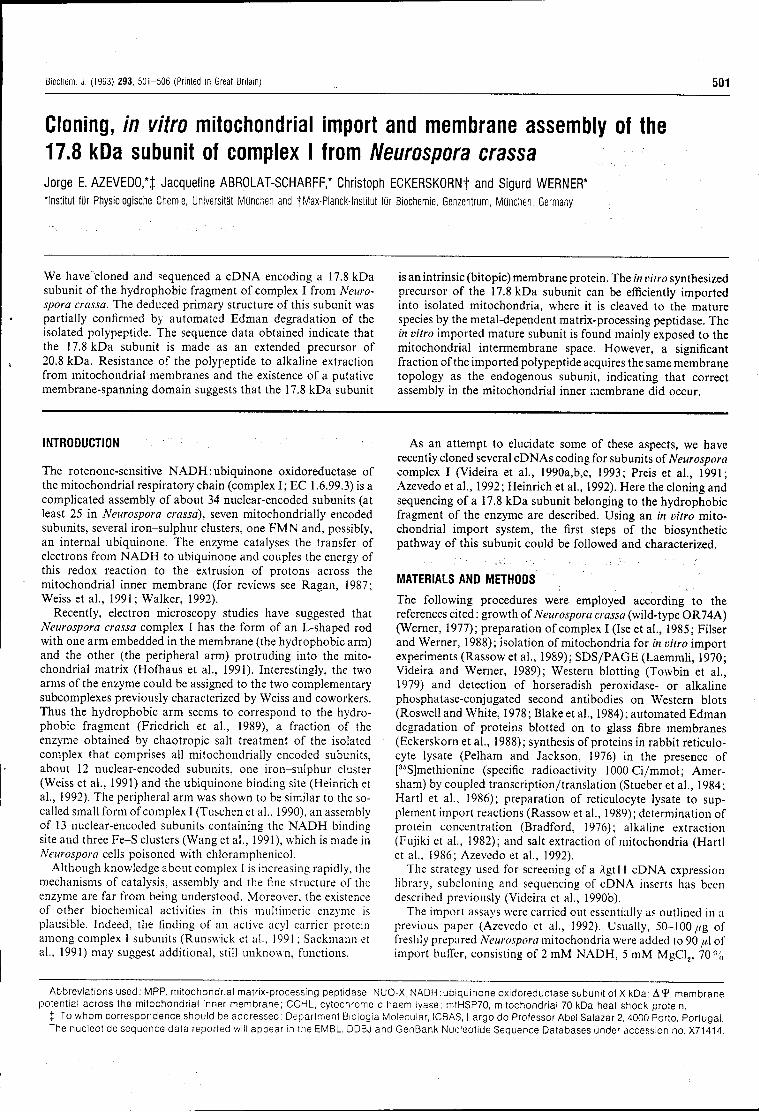
Biochem. J. (1993) 293, 501-506 (Printed in Great Britain) 51)1
Cloning, in vitro mitochondrial import and membrane assembly of the 17.8 kDa subunit of complex I from Neurospora crassa Jorge E. AZEVEDO,*J Jacqueline ABROLAT-SCHARFF,* Christoph ECKERSKORNt and Sigurd WERNER* Institut fur Physiologische Chemie, Universitãt Munchen and tMax-Planck-lnstitut fur Biochemie, Genzentrum, Munchen, Germany
We have cloned and sequenced a cDNA encoding a 17.8 kDa subunit of the hydrophobic fragment of complex I from Neurospora crassa. The deduced primary structure of this subunit was partially confirmed by automated Edman degradation of the isolated polypeptide. The sequence data obtained indicate that the 17.8 kDa subunit is made as an extended precursor of 20.8 kDa. Resistance of the polypeptide to alkaline extraction from mitochondrial membranes and the existence of a putative membrane-spanning domain suggests that the 17.8 kDa subunit
is an intrinsic (bitopic) membrane protein. The in vitro synthesized precursor of the 17.8 kDa subunit can be efficiently imported into isolated mitochondria, where it is cleaved to the mature species by the metal-dependent matrix-processing peptidase. The in vitro imported mature subunit is found mainly exposed to the mitochondrial intermembrane space. However, a significant fraction of the imported polypeptide acquires the same membrane topology as the endogenous subunit, indicating that correct assembly in the mitochondrial inner membrane did occur.
INTRODUCTION
The rotenone-sensitive NADH : ubiquinone oxidoreductase of the mitochondrial respiratory chain (complex I; EC 1.6.99.3) is a complicated assembly of about 34 nuclear-encoded subunits (at least 25 in Neurospora crassa), seven mitochondrially encoded subunits, several iron-sulphur clusters, one FMN and, possibly, an internal ubiquinone. The enzyme catalyses the transfer of electrons from NADH to ubiquinone and couples the energy of this redox reaction to the extrusion of protons across the mitochondrial inner membrane (for reviews see Ragan, 1987; Weiss et al., 1991; Walker, 1992).
Recently, electron microscopy studies have suggested that Neurospora crassa complex I has the form of an L-shaped rod with one arm embedded in the membrane (the hydrophobic arm) and the other (the peripheral arm) protruding into the mitochondrial matrix (Hofhaus et al., 1991). Interestingly, the two arms of the enzyme could be assigned to the two complementary subcomplexes previously characterized by Weiss and coworkers. Thus the hydrophobic arm seems to correspond to the hydrophobic fragment (Friedrich et al., 1989), a fraction of the enzyme obtained by chaotropic salt treatment of the isolated complex that comprises all mitochondrially encoded subunits, about 12 nuclear-encoded subunits, one iron-sulphur cluster (Weiss et al., 1991) and the ubiquinone binding site (Heinrich et al., 1992). The peripheral arm was shown to be similar to the so-called small form of complex I (Tuschen et al., 1990), an assembly of 13 nuclear-encoded subunits containing the NADH binding site and three Fe-S clusters (Wang et al., 1991), which is made in Neurospora cells poisoned with chloramphenicol.
Although knowledge about complex I is increasing rapidly, the mechanisms of catalysis, assembly and the fine structure of the enzyme are far from being understood. Moreover, the existence of other biochemical activities in this multimeric enzyme is plausible. Indeed, the finding of an active acyl carrier protein among complex I subunits (Runswick et al., 1991 ; Sackmann et al., 1991) may suggest additional, still unknown, functions.
As an attempt to elucidate some of these aspects, we have recently cloned several cDNAs coding for subunits of Neurospora complex I (Videira et al., 1990a,b,c, 1993; Preis et al., 1991; Azevedo et a l , 1992; Heinrich et al., 1992). Here the cloning and sequencing of a 17.8 kDa subunit belonging to the hydrophobic fragment of the enzyme are described. Using an in vitro mitochondrial import system, the first steps of the biosynthetic pathway of this subunit could be followed and characterized.
MATERIALS AND METHODS
The following procedures were employed according to the references cited : growth of Neurospora crassa (wild-type OR74A) (Werner, 1977); preparation of complex I (Ise et al., 1985; Filser and Werner, 1988); isolation of mitochondria for in vitro import experiments (Rassow et al., 1989); SDS/PAGE (Laemmli, 1970; Videira and Werner, 1989); Western blotting (Towbin et al., 1979) and detection of horseradish peroxidase- or alkaline phosphatase-conjugated second antibodies on Western blots (Roswell and White, 1978; Blake et al., 1984); automated Edman degradation of proteins blotted on to glass fibre membranes (Eckerskorn et al., 1988); synthesis of proteins in rabbit reticulocyte lysate (Pelham and Jackson, 1976) in the presence of [36S]methionine (specific radioactivity lOOOCi/mmol; Amer-sham) by coupled transcription/translation (Stueber et ah, 1984 ; Hartl et al., 1986); preparation of reticulocyte lysate to supplement import reactions (Rassow et al., 1989); determination of protein concentration (Bradford, 1976); alkaline extraction (Fujiki et al., 1982); and salt extraction of mitochondria (Hartl et al., 1986; Azevedo et al., 1992).
The strategy used for screening of a Agtll cDNA expression library, subcloning and sequencing of cDNA inserts has been described previously (Videira et al., 1990b).
The import assays were carried out essentially as outlined in a previous paper (Azevedo et al., 1992). Usually, 50-100//g of freshly prepared Neurospora mitochondria were added to 90 //1 of import buffer, consisting of 2 mM NADH, 5 mM MgCl2, 70%
Abbreviations used: MPP, mitochondrial matrix-processing peptidase; NUO-X, NADH:ubiquinone oxidoreductase subunit of X kDa; A I " , membrane potential across the mitochondrial inner membrane; CCHL, cytochrome c haem lyase; m1HSP70, mitochondrial 70 kDa heat shock protein.
% To whom correspondence should be addressed : Department Biologia Molecular, ICBAS, Largo do Professor Abel Salazar 2, 4000 Porto, Portugal. The nucleotide sequence data reported will appear in the EMBL, DDBJ and GenBank Nucleotide Sequence Databases under accession no. X71414,
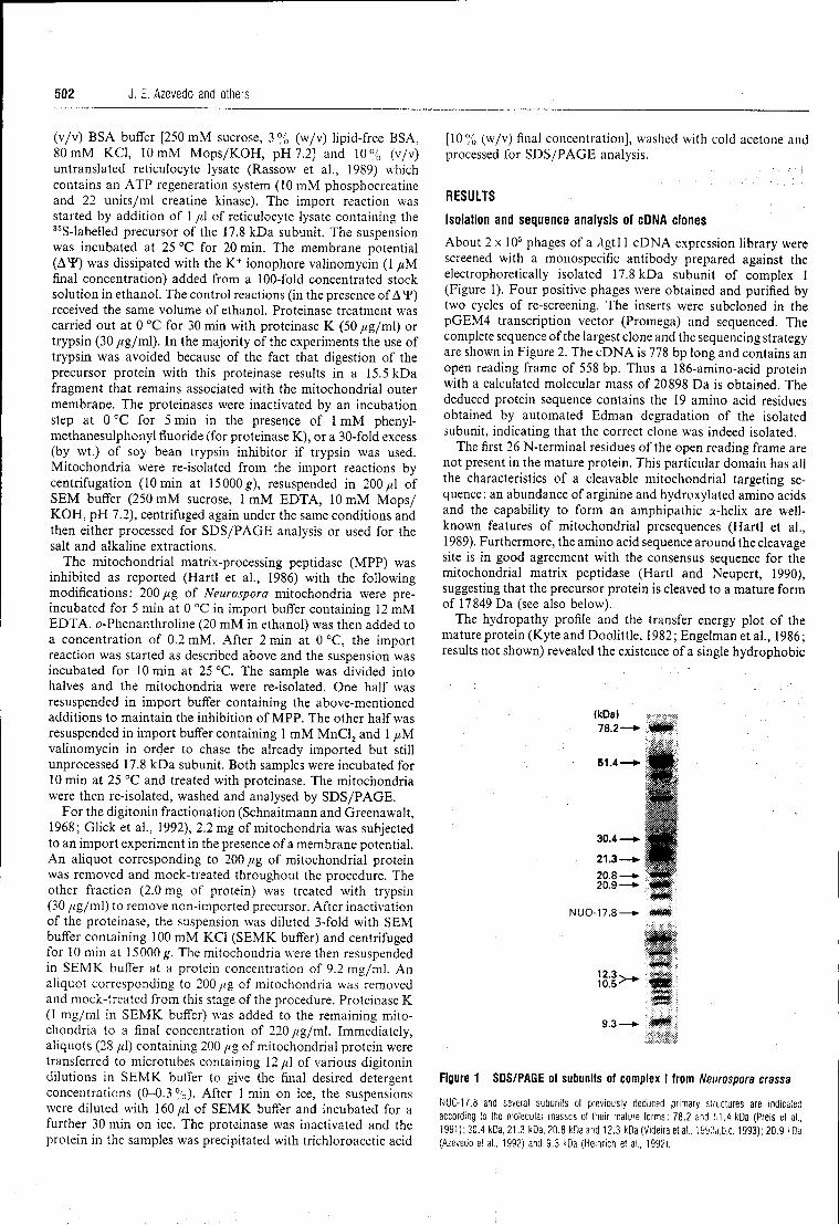
502 J. E. Azevedo and others
(v/v) BSA buffer [250 mM sucrose, 3 % (w/v) lipidfree BSA, 80 mM KCI, 10 mM Mops/KOH, pH 7.2] and 10% (v/v) untranslated reticulocyte lysate (Rassow et al., 1989) which contains an ATP regeneration system (10 mM phosphocreatine and 22 units/ml creatine kinase). The import reaction was started by addition of 1 //,1 of reticulocyte lysate containing the 36Slabelled precursor of the 17.8 kDa subunit. The suspension was incubated at 25 °C for 20 min. The membrane potential (AY) was dissipated with the K+ ionophore valinomycin (1 //M final concentration) added from a 100fold concentrated stock solution in ethanol. The control reactions (in the presence of AY) received the same volume of ethanol. Proteinase treatment was carried out at 0 °C for 30 min with proteinase K (50 /ig/ml) or trypsin (30 /ig/ml). In the majority of the experiments the use of trypsin was avoided because of the fact that digestion of the precursor protein with this proteinase results in a 15.5 kDa fragment that remains associated with the mitochondrial outer membrane. The proteinases were inactivated by an incubation step at 0 °C for 5 min in the presence of 1 mM phenyl
methanesulphonyl fluoride (for proteinase K), or a 30fold excess (by wt.) of soy bean trypsin inhibitor if trypsin was used. Mitochondria were reisolated from the import reactions by centrifugation (10 min at 15000^), resuspended in 200//1 of SEM buffer (250 mM sucrose, 1 mM EDTA, 10 mM Mops/ KOH, pH 7.2), centrifuged again under the same conditions and then either processed for SDS/PAGE analysis or used for the salt and alkaline extractions.
The mitochondrial matrixprocessing peptidase (MPP) was inhibited as reported (Hartl et al., 1986) with the following modifications : 200 /zg of Neurospora mitochondria were pre
incubated for 5 min at 0 °C in import buffer containing 12 mM EDTA. 0Phenanthroline (20 mM in ethanol) was then added to a concentration of 0.2 mM. After 2 min at 0 °C, the import reaction was started as described above and the suspension was incubated for 10 min at 25 °C. The sample was divided into halves and the mitochondria were reisolated. One half was resuspended in import buffer containing the abovementioned additions to maintain the inhibition of MPP. The other half was resuspended in import buffer containing 1 mM MnCl2 and 1 /nM valinomycin in order to chase the already imported but still unprocessed 17.8 kDa subunit. Both samples were incubated for 10 min at 25 °C and treated with proteinase. The mitochondria were then reisolated, washed and analysed by SDS/PAGE.
For the digitonin fractionation (Schnaitmann and Greenawalt, 1968; Glick et al., 1992), 2.2 mg of mitochondria was subjected to an import experiment in the presence of a membrane potential. An aliquot corresponding to 200 //g of mitochondrial protein was removed and mocktreated throughout the procedure. The other fraction (2.0 mg of protein) was treated with trypsin (30 //g/ml) to remove nonimported precursor. After inactivation of the proteinase, the suspension was diluted 3fold with SEM buffer containing 100 mM KCI (SEMK buffer) and centrifuged for 10 min at 15000^. The mitochondria were then resuspended in SEMK buffer at a protein concentration of 9.2 mg/ml. An aliquot corresponding to 200 //g of mitochondria was removed and mocktreated from this stage of the procedure. Proteinase K (1 mg/ml in SEMK buffer) was added to the remaining mito
chondria to a final concentration of 220 //g/ml. Immediately, aliquots (28 //1) containing 200 //g of mitochondrial protein were transferred to microtubes containing 12//1 of various digitonin dilutions in SEMK buffer to give the final desired detergent concentrations (00.3%). After 1 min on ice, the suspensions were diluted with 160/tl of SEMK buffer and incubated for a further 30 min on ice. The proteinase was inactivated and the protein in the samples was precipitated with trichloroacetic acid
[10% (w/v) final concentration], washed with cold acetone and processed for SDS/PAGE analysis.
RESULTS
Isolation and sequence analysis of cDNA clones
About 2 x 10⁵ phages of a Agtl 1 cDNA expression library were screened with a monospecific antibody prepared against the electrophoretically isolated 17.8 kDa subunit of complex I (Figure 1). Four positive phages were obtained and purified by two cycles of rescreening. The inserts were subcloned in the pGEM4 transcription vector (Promega) and sequenced. The complete sequence of the largest clone and the sequencing strategy are shown in Figure 2. The cDNA is 778 bp long and contains an open reading frame of 558 bp. Thus a 186aminoacid protein with a calculated molecular mass of 20898 Da is obtained. The deduced protein sequence contains the 19 amino acid residues obtained by automated Edman degradation of the isolated subunit, indicating that the correct clone was indeed isolated.
The first 26 Nterminal residues of the open reading frame are not present in the mature protein. This particular domain has all the characteristics of a cleavable mitochondrial targeting se
quence: an abundance of arginine and hydroxylated amino acids and the capability to form an amphipathic ahelix are well
known features of mitochondrial presequences (Hartl et al., 1989). Furthermore, the amino acid sequence around the cleavage site is in good agreement with the consensus sequence for the mitochondrial matrix peptidase (Hartl and Neupert, 1990), suggesting that the precursor protein is cleaved to a mature form of 17849 Da (see also below).
The hydropathy profile and the transfer energy plot of the mature protein (Kyte and Doolittle, 1982 ; Engelman et al., 1986 ; results not shown) revealed the existence of a single hydrophobic
(kDa) 78.2—* mm
51.4—*- H Í
30.4—». mm
21.3—«. P P
NUO-17.8—► * * •
10-5 IH j
9.3 —► $**
Figure 1 SDS/PAGE of subunits of complex I from Neurospora crassa
NUO-17.8 and several subunits ot previously deduced primary structures are indicated according to the molecular masses of their mature forms: 78.2 and 51.4 kDa (Preis et al., 1991); 30.4 kDa, 21.3 kDa, 20.8 kDa and 12.3 kDa (Videira et al., 1990a,b,c, 1993); 20.9 kDa (Azevedo e! al., 1992) and 9.3 kDa (Heinrich et al., 1992).
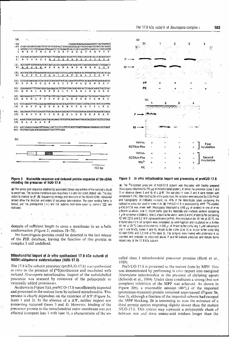
The 17.8 kDa subunit of Neurospora complex I 503
(a)
-8B CAGGAGGAGAGAAAGGGTCCACTATTCC - 6 O CGTCGACGCACGACTCGCGCTCCGCACTTGTCAATTACACGATATAAGTCGACGACAGCC
1 ATGTCCTCCTTTAGACTTGGAGTCTCTAGGGTCGCCCGCCAGGTTCGGGCCCCTTGTGTG 1 M S S F R L G V S R V A R Q V R A P C V
61 CGCAACACGAGGCGATATGCCTCCGACAGCCACGCTCCTGCCGACCACACCCACAGCGCT 21 R N T R R ï A S D S H A P A D H T H S A
121 GCCGGTCATGGGGAGCACCACCACGCCAATGCCGCAGATGCCAACGAGGAGCTTGGAACT 41 A G H G E H H H A N A A D A N E E L G T
181 GCCTTCTATGTGATATTCGGCGCCATCCCCGCATTCGGTGCTCTTTATTACTTCTCTCGG 61 A.__.E._X...V_..I__X S A I P ,, jy F G A L Y Y f__ S R
241 CCGGGCAAGGACGGAC AGCCGAACAGTATCACGAAGTGGTTGCAGAAGTGGGAGGAGC AC 8 1 P G K D G Q P N S I T K W L Q . K W E E K
301 CAAGAGGCTTTGGCGGACAAGAACGCTCTGGTGACAGCGGCACTCGAGCAGGCTGCTCAC 101 Q E A L A D K N A L V T A A L E Q A A H
361 GACAAGCATCTTTTCTATTACGTCGACCAACTGAGAAGCGGACACTACGAAATGAAATAT 121 D K H L F Y Y V D Q L R S G H Y E M K Y 421 CCCGAGGTTTTCCAGCACGGTAGCGCGCGCAATGTCCCGGCCGGTACCTATATCCCCTTG 1 4 1 P E V F Q . H G S A R N V P A G T Y I P L
481 GACAAGGTTGTCGAGGTTTACAGGAAGCAGCACCTTGACGAAGAGGAGCGCAAGGCGAAG 1 6 1 D K V V E V Y R K Q . H L D E E E R K A K
541 AAGCTGGCGGCGGCCAACTAGACAAGTGTCGTGCAGTAATTGCATACTTCCTCTTCTTAT 181 K L A A A N #
601 ATCTAAGTTAGACGTTAGACTGCTCCTCCTTCCCATCTTATTGAGACGGGAGCCGTCGGT 661 TCGTAGCAACATCAGAAAGTTACCTTCAAA
(a) St
P
M
M
A4> + +
PK - +
(b) 1 2 3
(b)
100 bp
Val ino.
EDTA/o-Phe
Val ino.
EDTA/o-Phe
Mn2 +
PK
] First i ncuba t ion
Second incuba t ion
Figure 2 Nucleot ide sequence and deduced prote in sequence of the cDNA encoding the precursor of NUO-17.8
(a) The amino acid sequence obtained by automated Edman degradation of the isolated subunit is underlined. The putative membrane-spanning domain is also indicated (dotted line). The stop codon is marked by# . (b) Sequencing strategy and structure of the cloned cDNA. Horizontal arrows show the direction and extent of sequence determination. The open reading frame is boxed, and the presequence ( + ) and the putative membrane-spanning domain (EJ) are indicated.
domain of sufficient length to cross a membrane in an a-helix conformation (Figure 2; residues 58-78).
No homologous proteins could be detected in the last release of the PIR database, leaving the function of this protein in complex I still undefined.
Figure 3 In vitro mi tochondr ia l impor t and processing of preNUO-17.8
(a) The 35S-labelled precursor of NUO-17.8 subunit was incubated with freshly prepared Neurospora mitochondria (30 /tg of mitochondrial protein), in either the presence (lanes 1 and 2) or absence (lanes 3 and 4) of a A 4 * . The samples in lanes 2 and 4 were treated with proteinase K (PK). After inactivation of the proteinase, the samples were analysed by SDS/PAGE and fluorography. St indicates standard, i.e. 40% of the reticulocyte lysate containing the radioactive precursor used in lanes 1-4. (b) PreNUO-17.8 is processed by MPP. 35S-labelled preNUO-17.8 was mixed with Neurospora mitochondria (100/zg of protein) in one of three different solutions: lane 1, import buffer (see the Materials and methods section) containing 1 /M valinomycin (Valino.); lane 2, import buffer alone; lanes 3 and 4, import buffer containing 12 mM EDTA and 0.2 mM o-phenanthroline (o-Phe). After incubation for 10 min at 25 °C, the mitochondria from all samples were re-isolated by centrifugation and incubated for a further 10 min at 25 °C (second incubation) in 500 / i l of import buffer containing 1 /M valinomycin and 1 mM MnCI2 (lanes 1 and 4), import buffer alone (lane 2) or import buffer containing 12 mM EDTA and 0.2 mM oPhe (lane 3). The samples were treated with proteinase K as specified and analysed as described above. P and M indicate precursor and mature forms respectively of the 17.8 kDa subunit.
Mitochondrial import of in vitro synthesized 17.8 kDa subunit of NADH: ubiquinone oxidoreductase (NUO-17.8)
The 17.8 kDa subunit precursor (preNUO-17.8) was synthesized in vitro in the presence of [35S]methionine and incubated with isolated Neurospora mitochondria. Import of the radiolabeled precursor was assessed by resistance of the polypeptide to externally added proteinases.
As shown in Figure 3(a), preN UO-17.8 was efficiently imported and processed to the mature form by isolated mitochondria. This process is clearly dependent on the existence of AY (Figure 3a, lanes 1 and 2). In the absence of a AY, neither import nor processing occurred (lanes 3 and 4). However, binding of the precursor protein to the mitochondrial outer membrane was not affected (compare lane 1 with lane 3), a characteristic of the so-
called class I mitochondrial precursor proteins (Hartl et al., 1989).
PreNUO-17.8 is processed to the mature form by MPP. This was demonstrated by performing in vitro import into energized Neurospora mitochondria in the presence of chelating agents (Schmidt et al., 1984). Under these conditions a strong (but not complete) inhibition of the MPP was achieved. As shown in Figure 3(b), a reasonable amount (40%) of the imported (proteinase-resistant) protein remained unprocessed (Figure 3b, lane 3), although a fraction of the imported subunit had escaped the MPP blocking. [It is interesting to note the existence of a third protein species migrating slightly slower than the mature NUO-17.8. This species may represent a polypeptide chain of between one and three amino-acid residues longer than the
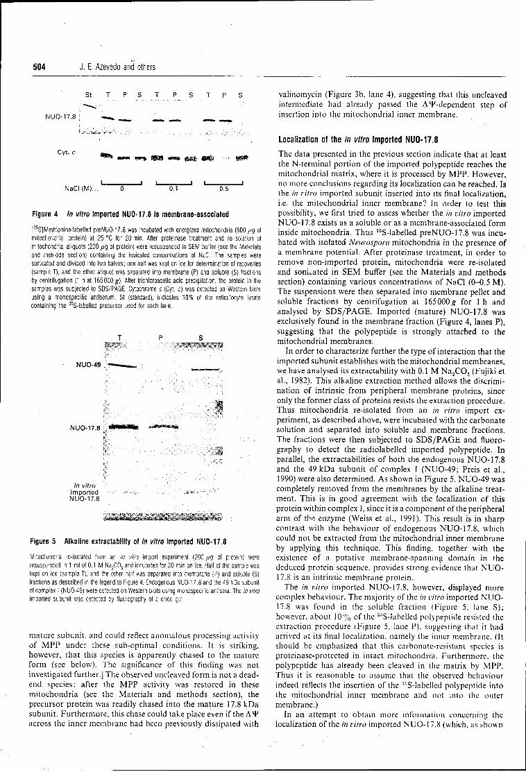
504 J. E. Azevedo and others
St T P S T P S T P S
NUO-17.8 |
**» mem ww» <as» <*»• e e j ««& .... g M
i i i i i N a C I ( M ) . . . 0 0.1 0.5
Figure 4 In vitro impor ted NUO-17.8 is membrane-assoc ia ted
[35
S]Methionine-labelled preNUO-17.8 was incubated with energized mitochondria (600 /IQ of mitochondrial protein) at 25 °C (or 20 min. After proteinase treatment and re-isolation of mitochondria, aliquots (200 /ug of protein) were resuspended in SEM buffer (see the Materials and metnods seclion) containing the indicated concentrations ot NaCI. The samples were sonicated and divided into two halves; one half was kept on ice for determination of recoveries (sample T), and the other aliquot was separated into membrane (P) and soluble (S) fractions by centrifugation (1 h at 165000 ^ ) . After trichloroacetic acid precipitation, the protein in the samples was subjected to SOS/PAGE. Cytochrome c (Cyl c) was detected on Western blots using a monospecific antiserum. St (standard), indicates 10% of the reticulocyte lysate containing the
35S-labelled precursor used for each lane.
T P S
: MSB ■ ■ H B H . I
NUO-49 —■ i .. ..
MUO17.8 .«llwWfc *M*»mm-
!&'.;"'.' > v ' ' 7 \ 7 7 7 ^ ^ 7 ^ 7 7 ^ 7 ^ ■*.'
In vitro , i m p o r t e d ' «^ÉéppraSav ..^..../ $¢^^-^-
NUO-17.8
" / 7 . ^ 7 7 7 / / . 7 7 7 - : / / - 7 : '. / . 7 .
Figure 5 Alkal ine extractabi l i ty of in vitro impor ted NUO-17.8
Mitochondria re-isolated from an In vitro import experiment (200 fig of protein) were resuspended in 1 ml of 0.1 M Na2C03 and incubated for 30 min on ice. Half of the sample was kept on ice (sample T), and the other half was separated into membrane (P) and soluble (S) fractions as described in the legend to Figure 4. Endogenous NUO-17.8 and the 49 kDa subunit of complex I (NU0-49) were delected on Western blots using monospecific antisera. The in vitro imported subunit was detected by fluorography of a dried gel.
mature subunit, and could reflect anomalous processing activity of MPP under these suboptimal conditions. It is striking, however, that this species is apparently chased to the mature form (see below). The significance of this finding was not investigated further.] The observed uncleaved form is not a dead
end species: after the MPP activity was restored in these mitochondria (see the Materials and methods section), the precursor protein was readily chased into the mature 17.8 kDa subunit. Furthermore, this chase could take place even if the A*P across the inner membrane had been previously dissipated with
valinomycin (Figure 3b, lane 4), suggesting that this uncleaved intermediate had already passed the AlFdependent step of insertion into the mitochondrial inner membrane.
Localization of the in vitro imported NUO-17.8
The data presented in the previous section indicate that at least the Nterminal portion of the imported polypeptide reaches the mitochondrial matrix, where it is processed by MPP. However, no more conclusions regarding its localization can be reached. Is the in vitro imported subunit inserted into its final localization, i.e. the mitochondrial inner membrane? In order to test this possibility, we first tried to assess whether the in vitro imported NUO17.8 exists as a soluble or as a membraneassociated form inside mitochondria. Thus '"Slabelled preNUO17.8 was incu
bated with isolated Neurospora mitochondria in the presence of a membrane potential. After proteinase treatment, in order to remove nonimported protein, mitochondria were reisolated and sonicated in SEM buffer (see the Materials and methods section) containing various concentrations of NaCI (00.5 M). The suspensions were then separated into membrane pellet and soluble fractions by centrifugation at 165000^ for 1 h and analysed by SDS/PAGE. Imported (mature) NUO17.8 was exclusively found in the membrane fraction (Figure 4, lanes P), suggesting that the polypeptide is strongly attached to the mitochondrial membranes.
In order to characterize further the type of interaction that the imported subunit establishes with the mitochondrial membranes, we have analysed its extractability with 0.1 M Na2C03 (Fujiki et al., 1982). This alkaline extraction method allows the discrimi
nation of intrinsic from peripheral membrane proteins, since only the former class of proteins resists the extraction procedure. Thus mitochondria reisolated from an in vitro import ex
periment, as described above, were incubated with the carbonate solution and separated into soluble and membrane fractions. The fractions were then subjected to SDS/PAGE and fluoro
graphy to detect the radiolabelled imported polypeptide. In parallel, the extractabilities of both the endogenous NUO17.8 and the 49 kDa subunit of complex I (NUO49; Preis et al., 1990) were also determined. As shown in Figure 5, NUO49 was completely removed from the membranes by the alkaline treat
ment. This is in good agreement with the localization of this protein within complex I, since it is a component of the peripheral arm of the enzyme (Weiss et al., 1991). This result is in sharp contrast with the behaviour of endogenous NUO17.8, which could not be extracted from the mitochondrial inner membrane by applying this technique. This finding, together with the existence of a putative membranespanning domain in the deduced protein sequence, provides strong evidence that NUO
17.8 is an intrinsic membrane protein. The in vitro imported NUO17.8, however, displayed more
complex behaviour. The majority of the in vitro imported NUO
17.8 was found in the soluble fraction (Figure 5, lane S); however, about 10% of the 35Slabelled polypeptide resisted the extraction procedure (Figure 5, lane P), suggesting that it had arrived at its final localization, namely the inner membrane. (It should be emphasized that this carbonateresistant species is proteinaseprotected in intact mitochondria. Furthermore, the polypeptide has already been cleaved in the matrix by MPP. Thus it is reasonable to assume that the observed behaviour indeed reflects the insertion of the 86Slabelled polypeptide into the mitochondrial inner membrane and not into the outer membrane.)
In an attempt to obtain more information concerning the localization of the in vitro imported NUO17.8 (which, as shown
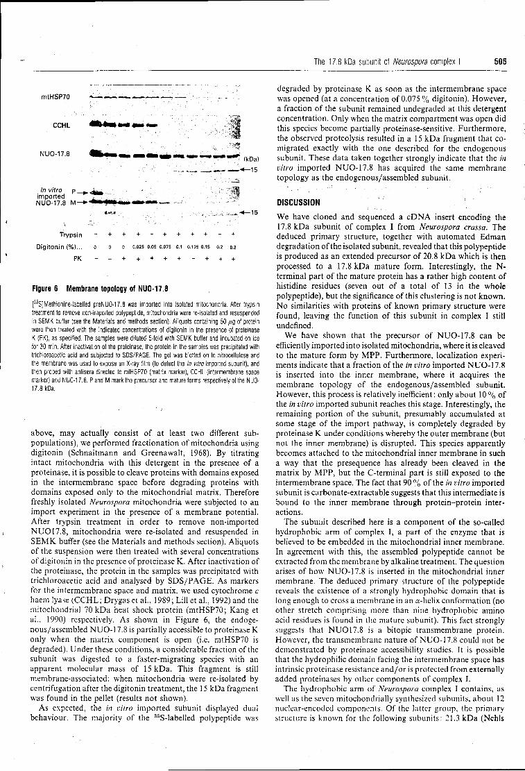
The 17.8 kDa subunit of Neurospora complex I 505
mtHSP70
CCHL
NUO-17.8
In vitro p. imported NUO-17.8 M-
Trypsin Digitonin (%)...
PK
(kDa) * -15
-15
0 0.025 0.05 0.075 0.1 0.125 0.15 0.2 0.3
+ + + + + + + + +
Figure 6 Membrane topology of NUO-17.8
[35S]Methionine-labelled preNUO-17.8 was imported into isolated mitochondria. After trypsin treatment to remove non-imported polypeptide, mitochondria were re-isolated and resuspended in SEMK buffer (see the Materials and methods section). Aliquots containing 50 /IQ of protein were then treated with the indicated concentrations of digitonin in the presence of proteinase K (PK), as specified. The samples were diluted 5-fold with SEMK buffer and incubated on ice for 30 min. After inactivation of the proteinase, the protein in the samples was precipitated with trichloroacetic acid and subjected to SDS/PAGE. The gel was blotted on to nitrocellulose and the membrane was used to expose an X-ray film (to detect the in vitro imported subunit), and then probed with antisera directed to mtHSP70 (matrix marker), CCHL (intermembrane space marker) and NUO-17.8. P and M mark the precursor and mature forms respectively of the NUO-17.8 kDa.
above, may actually consist of at least two different sub-populations), we performed fractionation of mitochondria using digitonin (Schnaitmann and Greenawalt, 1968). By titrating intact mitochondria with this detergent in the presence of a proteinase, it is possible to cleave proteins with domains exposed in the intermembrane space before degrading proteins with domains exposed only to the mitochondrial matrix. Therefore freshly isolated Neurospora mitochondria were subjected to an import experiment in the presence of a membrane potential. After trypsin treatment in order to remove non-imported NU017.8, mitochondria were re-isolated and resuspended in SEMK buffer (see the Materials and methods section). Aliquots of the suspension were then treated with several concentrations of digitonin in the presence of proteinase K. After inactivation of the proteinase, the protein in the samples was precipitated with trichloroacetic acid and analysed by SDS/PAGE. As markers for the intermembrane space and matrix, we used cytochrome c haem lyase (CCHL; Drygas et al., 1989; Lill et al., 1992) and the mitochondrial 70 kDa heat shock protein (mtHSP70; Kang et a l , 1990) respectively. As shown in Figure 6, the endogenous/assembled NUO-17.8 is partially accessible to proteinase K only when the matrix component is open (i.e. mtHSP70 is degraded). Under these conditions, a considerable fraction of the subunit was digested to a faster-migrating species with an apparent molecular mass of 15 kDa. This fragment is still membrane-associated: when mitochondria were re-isolated by centrifugation after the digitonin treatment, the 15 kDa fragment was found in the pellet (results not shown).
As expected, the in vitro imported subunit displayed dual behaviour. The majority of the 35S-labelled polypeptide was
degraded by proteinase K as soon as the intermembrane space was opened (at a concentration of 0.075 % digitonin). However, a fraction of the subunit remained undegraded at this detergent concentration. Only when the matrix compartment was open did this species become partially proteinase-sensitive. Furthermore, the observed proteolysis resulted in a 15 kDa fragment that co-migrated exactly with the one described for the endogenous subunit. These data taken together strongly indicate that the in vitro imported NUO-17.8 has acquired the same membrane topology as the endogenous/assembled subunit.
DISCUSSION
We have cloned and sequenced a cDNA insert encoding the 17.8 kDa subunit of complex I from Neurospora crassa. The deduced primary structure, together with automated Edman degradation of the isolated subunit, revealed that this polypeptide is produced as an extended precursor of 20.8 kDa which is then processed to a 17.8 kDa mature form. Interestingly, the N-terminal part of the mature protein has a rather high content of histidine residues (seven out of a total of 13 in the whole polypeptide), but the significance of this clustering is not known. No similarities with proteins of known primary structure were found, leaving the function of this subunit in complex I still undefined.
We have shown that the precursor of NUO-17.8 can be efficiently imported into isolated mitochondria, where it is cleaved to the mature form by MPP. Furthermore, localization experiments indicate that a fraction of the in vitro imported NUO-17.8 is inserted into the inner membrane, where it acquires the membrane topology of the endogenous/assembled subunit. However, this process is relatively inefficient: only about 10 % of the in vitro imported subunit reaches this stage. Interestingly, the remaining portion of the subunit, presumably accumulated at some stage of the import pathway, is completely degraded by proteinase K under conditions whereby the outer membrane (but not the inner membrane) is disrupted. This species apparently becomes attached to the mitochondrial inner membrane in such a way that the presequence has already been cleaved in the matrix by MPP, but the C-terminal part is still exposed to the intermembrane space. The fact that 90 % of the in vitro imported subunit is carbonate-extractable suggests that this intermediate is bound to the inner membrane through protein-protein interactions.
The subunit described here is a component of the so-called hydrophobic arm of complex I, a part of the enzyme that is believed to be embedded in the mitochondrial inner membrane. In agreement with this, the assembled polypeptide cannot be extracted from the membrane by alkaline treatment. The question arises of how NUO-17.8 is inserted in the mitochondrial inner membrane. The deduced primary structure of the polypeptide reveals the existence of a strongly hydrophobic domain that is long enough to cross a membrane in an a-helix conformation (no other stretch comprising more than nine hydrophobic amino acid residues is found in the mature subunit). This fact strongly suggests that NU017.8 is a bitopic transmembrane protein. However, the transmembrane nature of NUO-17.8 could not be demonstrated by proteinase accessibility studies. It is possible that the hydrophilic domain facing the intermembrane space has intrinsic proteinase resistance and/or is protected from externally added proteinases by other components of complex I.
The hydrophobic arm of Neurospora complex I contains, as well as the seven mitochondrially synthesized subunits, about 12 nuclear-encoded components. Of the latter group, the primary structure is known for the following subunits: 21.3 kDa (Nchls
506 ... J. E. Azevedo and others
et al., 1990), 20.9 kDa (Azevedo et al., 1992), 20.8 kDa (Videira et al., 1990b), 12.3 kDa (Videira et al., 1993) and 9.3 kDa (Heinrich et al., 1992). All these subunits have in common the absence of a cleavable mitochondrial targeting sequence and, at least in the case of the 20.9 kDa component, insertion into the inner membrane seems to take place from the cytosolic side (Azevedo et al., 1992). The fact that NUO-17.8 has a matrix targeting sequence could suggest that this polypeptide follows a different biosynthetic pathway. Obviously, this subunit requires an additional step before being assembled in complex I (cleavage of the presequence by MPP). However, it is unclear whether NUO-17.8 really does follow a different import pathway. In particular, is the polypeptide completely translocated into the matrix before undergoing insertion into the inner membrane, a mechanism that would be in agreement with the so-called conservative sorting (for a review see Hartl et al., 1989)? The data presented here provide no evidence for such an intra-mitochondrial sorting pathway. For instance, no salt-extractable species could be detected after in vitro import experiments, suggesting that the in vitro imported NUO-17.8 does not exisi in a soluble form in the matrix. In addition, it is quite striking that the majority of the imported (mature) subunit is found exposed to the intermembrane space. Import intermediates displaying these characteristics have been recently described for proteins en route to the matrix (Hwang et al., 1991). However, in all the documented examples, these intermediates were obtained by performing import under sub-optimal conditions (i.e. ATP depletion in the mitochondrial matrix). It is unlikely that this is the reason why the majority of the in vitro imported NUO-17.8 is found attached to the mitochondrial inner membrane ('standard' import conditions were used in these experiments; see the Materials and methods section). Rather, we think that the existence of this species reflects the specific features of the import pathway of NUO-17.8. Our results could favour a non-conservative sorting mechanism in which insertion of NUO-17.8 into the lipophilic environment of the inner membrane occurs through the inner membrane itself (probably by lateral movement of the polypeptide away from the inner membrane translocation machinery directly into the lipid bilayer). It is clear, however, that more data are needed in order to confirm the existence of such an intramitochondrial sorting pathway. In this context, it will be of major importance to determine whether or not the assembly of NUO-17.8 in the inner membrane requires the action of the matrix ATP-dependent chaperonins (Cheng et al., 1989; Kang et al., 1990). We are currently pursuing this aim.
We are indebted to Dr. B. Segui-Real and Dr. R. A. Stuart for the gift of antisera against the mitochondrial HSP 70, cytochrome c and CCHL. We thank Heidi Rothe for excellent technical assistance. This work was supported by the Deutsche Forschungsgemeinschaft. J.E.A. thanks Junta Nacional de Investigação Cientifica e Tecnológica, Portugal, for financial support.
R E F E R E N C E S
Azevedo, J. E., Nehls, U„ Eckerskorn, C, Heinrich, H., Rothe, H., Weiss, H. and Werner, S. (1992) Biochem. J. 288, 29-34
Blake, M. S„ Johnston, K. H„ Russel-Jones, G. J. and Gotschlich, E. C. (1984) Anal. Bioctiem. 136, 175-179
Bradford, M. M. (1976) Anal. Biochem. 72, 248-254 Cheng, M. Y., Hartl, F.-U., Martin, J., Pollock, R. A., Kalousek, F., Neupert, W„ Hallberg,
E. M, Hallberg, R. L. and Horwich, A. L. (1989) Nature (London) 337, 620-625 Drygas, M. E„ Lambowitz, A. M. and Nargang, F. E. (1989) J. Biol. Chem. 264,
17897-17906 Eckerskorn, C, Mewes, W„ Goretzki, H. and Lottspeich, F (1988) Eur. J. Biochem. 176,
509-519 Engelman, D. M., Steitz, T. A. and Goldman, A. (1986) Annu. Rev. Biophys. Biophys. Chem.
15, 321-353 Filser, M. and Werner, S. (1988) Biochem. Pharmacol. 37, 2551-2558 Friedrich, T., Hofhaus, G., Ise, W., Nehls, U„ Schmitz, B. and Weiss, H. (1989) Eur. J.
Biochem. 180, 173-180 Fujiki, Y., Hubbard, A. L, Fowler, S. and Lazarow, P. B. (1982) J. Cell Biol. 93, 97-102 Glick, B. S., Brandt, A„ Cunningham, K„ Muller, S., Hallberg, R. L. and Schatz, G. (1992)
Cell 69, 809-822 Hartl, F.-U. and Neupert, W. (1990) Science 247. 930-938 Hartl, F.-U., Schmidt, B., Wachter, E„ Weiss, H. and Neupert, W. (1986) Cell 47, 939-951 Hartl, F.-U., Pfanner, N., Nicholson, D. W. and Neupert, W. (1989) Biochim. Biophys. Acta
988, 1-45 Heinrich, H„ Azevedo, J. E. and Werner, S. (1992) Biochemistry 31 , 11420-11424 Hofhaus, G., Weiss, H. and Leonard, K. (1991) J. Mol. Biol. 221, 1027-1043 Hwang, S. T., Wachter, C. and Schatz, G. (1991) J. Biol. Chem. 266, 21083-21089 Ise, W„ Haiker, H. and Weiss, H. (1985) EMB0 J. 4, 2075-2080 Kang, P.-J., Ostermann, J., Shilling, J., Neupert, W., Craig, E. A. and Pfanner, N. (1990)
Nature (London) 348, 137-142 Kyte, J. and Doolittle, R. F. (1982) J. Mol. Biol. 157, 105-132 Laemmli, U. K. (1970) Nature (London) 227, 680-685 LUI, R„ Stuart, R. A., Drygas, M. E„ Nargang, F. E. and Neupert, W. (1992) EMBO J. 11,
449-456 Nehls, U„ Hemmer, S., Róhlen, D. A., van der Pas, J. C. Preis, D., Sackmann, U. and
Weiss, H. (1990) Biochim. Biophys. Acta 1088, 325-326 Pelham, H. R. B. and Jackson, R. J. (1976) Eur. J. Biochem. 67, 247-256 Preis, D„ Van der Pas, J., Nehls, U., Rõhlen, D., Sackmann, U., Jahnke, U. and Weiss, H.
(1990) Curr. Genet. 18, 59-64 Preis, D., Weidner, U., Conzen, C, Azevedo, J. E., Nehls, U., Rõhlen, D., Van der Pas, J.,
Sackman, U., Schneider, R., Werner, S. and Weiss, H. (1991) Biochim. Biophys. Acta 1090, 133-138
Ragan, C. I. (1987) Curr. Top. Bioenerg. 15, 1-36 Rassow, J„ Guiard, B., Wienhues, U., Herzog, V., Hartl, F.-U. and Neupert. W. (1989)
J. Cell Biol. 109, 1421-1428 Roswell, D. F. and White, E. H. (1978) Methods Enzymol. 57, 409^423 Runswick, M. J., Fearnley, I. M„ Skehel, J. M. and Walker, J. E. (1991) FEBS Lett. 286,
121-124 Sackmann, U„ Zensen, R„ Rohlen, D., Jahnke, U. and Weiss, H. (1991) Eur. J. Biochem.
200, 463-469 Schmidt, B., Wachter, E„ Sebald, W. and Neupert, W. (1984) Eur. J. Biochem. 144,
581-588 Schnaitmann, C. A. and Greenawalt, D. S. (1968) J. Cell Biol. 38, 158-175 Stueber, D., Ibrahimi, l„ Cutler, D., Dobberstein, B. and Bujard, H. (1984) EMBO J. 3,
3143-3148 Towbin, H., Staehelin, T. and Gordon, J. (1979) Proc. Natl. Acad. Sci. U.S.A. 76,
4350-4354 Tuschen, G., Sackmann, U.. Nehls, U., Haiker, H., Buse, G. and Weiss, H. (1990) J. Mol.
Biol. 213, 845-857 Videira, A. and Werner, S. (1989) Eur. J. Biochem. 181, 493-502 Videira, A., Tropschug, M. and Werner, S. (1990a) Biochem. Biophys. Res. Commun. 166,
280-285 Videira, A., Tropschug, M. and Werner, S. (1990b) J. Biol. Chem. 265, 13060-13065 Videira, A., Tropschug, M. and Werner, S. (1990c) Biochem. Biophys. Res. Commun. 171,
1168-1174 Videira, A., Azevedo, J. E., Werner, S. and Cabral, P. (1993) Biochem. J. 291, 729-732 Walker, J. E. (1992) Q. Rev. Biophys. 25, 253-324 Wang, D„ Meinhardt, S. W„ Sackmann, U„ Weiss, H. and Ohnishi, T. (1991) Eur. J.
Biochem. 197, 257-264 Weiss, H., Friedrich, T., Hofhaus, G. and Preis, D. (1991) Eur. J. Biochem. 197, 563-576 Werner, S. (1977) Eur. J. Biochem. 79, 103-110
Received 20 January 1993/3 March 1993; accepted 8 March 1993
TRABALHO 7.4
In organello assembly of respiratory chain complex I Primary structure of the 14.8 kDa subunit of Neurospora crassa
complex I
Jorge E. Azevedo§#, Christoph Eckerskom* and Sigurd Werner5
inst i tut fur Physiologische Chemie, Universitat Munchen and *Max-Planck-
Institut fur Biochemie, Genzentrum, Mùnchen (F.R.G.)
Short title: In organello assembly of complex I
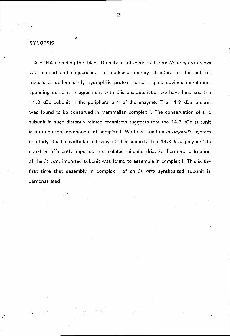
2
SYNOPSIS
A cDNA encoding the 14.8 kDa subunit of complex I from Neurospora crassa
was cloned and sequenced. The deduced primary structure of this subunit
reveals a predominantly hydrophilic protein containing no obvious membrane-
spanning domain. In agreement with this characteristic, we have localised the
14.8 kDa subunit in the peripheral arm of the enzyme. The 14.8 kDa subunit
was found to be conserved in mammalian complex I. The conservation of this
subunit in such distantly related organisms suggests that the 14.8 kDa subunit
is an important component of complex I. We have used an in organelle- system
to study the biosynthetic pathway of this subunit. The 14.8 kDa polypeptide
could be efficiently imported into isolated mitochondria. Furthermore, a fraction
of the in vitro imported subunit was found to assemble in complex I. This is the
first t ime that assembly in complex I of an in vitro synthesized subunit is
demonstrated.
3
INTRODUCTION
Complex I (NADH:ubiquinone oxidoreductase) is the most intricated enzyme of
the mitochondrial respiratory chain. It is constituted by some 30-40 subunits, 7
of which are encoded and synthesized in mitochondria. The enzyme is known to
catalyse the transfer of electrons from NADH to ubiquinone coupling the energy
of this redox reaction to the extrusion of protons across the mitochondrial inner
membrane (for reviews see Ragan, 1987; Walker, 1992; Weiss et al., 1991). In
addition, the recent finding of an acyl-carrier protein among complex I subunits
(Runswick et al., 1 9 9 1 ; Sackmann et al., 1991) suggests the existence of
other, still unknown, biochemical functions in this multimeric enzyme.
Structurally, complex I from Neurospora crassa has the form of an L-shaped
rod w i th one arm embedded in the mitochondrial inner membrane (the
hydrophobic arm) and the other ( the peripheral arm) protruding into the matrix
(Friedrich et al., 1989; Hofhaus et al., 1991 ; Tuschen et al., 1990). In terms of
polypeptide composition, the hydrophobic arm contains all the mitochondrially
encoded subunits and about 15 nuclear-coded subunits; the peripheral arm
comprises about 13 subunits and contains the majority of the iron-sulphur
clusters of the enzyme (Wang et al., 1991).
One major goal of our work is the understanding of the biogenesis of complex
I. Presently, all the information available concerning the assembly of the enzyme
was obtained from pulse-labelling experiments performed in intact Neurospora
cells. Using this method, an assembly intermediate of complex I corresponding
to the membrane arm of the enzyme could be detected (Tuschen et al., 1990).
Although elegant, this approach has an obvious drawback: the number of
different factors affecting the assembly process that can be experimentally
changed is quite limited. In an attempt to surround this problem we have been
4
trying to reproduce the biosynthetic pathway of some complex I subunits using
an in organello system. Recently, we have shown that the 20.9 kDa subunit of
Neurospora complex I could be imported into isolated mitochondria and correctly
sorted to the inner membrane (Azevedo et al., 1992). Similar results were
obtained wi th another subunit belonging to the membrane arm of the enzyme -
upon mitochondrial in vitro import a fraction of the 17.8 kDa subunit was found
correctly assembled in the inner membrane (Azevedo et al., 1993). In this report
we present further studies on the structure and biogenesis of complex I. We
have cloned and sequenced a cDNA encoding the 14.8 kDa subunit of
Neurospora crassa complex I. This subunit was synthesized in vitro and used in
mitochondrial in vitro import experiments. Our results show, for the first t ime,
full assembly of an in vitro synthesized complex I subunit using an in organello
system.
5
MATERIALS AND METHODS
The fol lowing procedures were employed according to the references cited:
growth of N. crassa (wild type OR74A) (Werner, 1977); preparation of complex
I (Wang et al., 1991); production of antisera in rabbits (Videira & Werner,
1989); isolation of mitochondria for in vitro import experiments (Rassow et al.,
1989); determination of protein concentration (Bradford, 1976); sucrose
gradient centrifugation of detergent-solubilized mitochondrial proteins and
determination of NADH:[Fe(CN)6]3" oxidoreductase activity (Tuschen et al.,
1990; Friedrich et al., 1989); sodium dodecylsulfate gel electrophoresis
(Laemmli, 1970; Videira & Werner, 1989): western-blotting (Towbin et al.,
1979); synthesis of proteins in rabbit reticulocyte lysate (Amersham
International, Amersham, Bucks., UK.) (Pelham & Jackson, 1976) in the
presence of [35S]methionine (specific activity 1000 Ci/mmol; Amersham) by
coupled transcription/translation (Stueber et al., 1984; Hartl et al., 1986);
automated Edman degradation (Eckerskorn et al., 1988).
The small form of complex I (Friedrich et al., 1989) was kindly provided by Dr.
Hans Weiss, University of Dusseldorf, Germany.
The strategy used for screening a Àgt11 cDNA expression library, subcloning
and sequencing cDNA inserts has been detailed previously (Videira et al.,
1990c; Heinrich et al., 1992).
The mitochondrial in vitro import experiments (usually 50 [ig of mitochondrial
protein in a 100 jal reaction) were performed in import buffer: 2 mM NADH, 5
mM MgCl2, 250 mM sucrose, 2 % (w/v) lipid-free bovine serum albumin, 80
mM KCI, 10 mM Mops/KOH pH 7.2 and 10 % (v/v) of "untranslated"
reticulocyte lysate (see Rassow et al., 1989) which contains an ATP-
regeneration system (10 mM creatine phosphate and 22 units/ml creatine
6
kinase). Protease treatment of import reactions and addition of inhibitors were
performed exactly as described recently (Azevedo et al., 1993).
The immunoprecipitation experiments were performed as fol lows: mitochondria
(generally 50 jag of protein) were ressuspended in 1 ml of Triton buffer [ 1 %
(w/v) Triton X-100, 0.3 M NaCI, 5 mM EDTA, 20 mM Tris.HCI pH 7.0 and 1
mM phenylmethylsulfonyl fluoride (PMSF) freshly added from a 200 mM stock
solution in ethanol] and incubated on ice for 10 minutes. The samples were
subjected to a clarifying spin (15 000 X g, 10 minutes) to remove insoluble
material. Five mg of protein A sepharose preincubated for one hour wi th 10 uJ
of the relevant antisera in 500 \JL\ of Triton buffer, were then added to each
sample. The suspension was gently rocked for 2-3 hours at 4 ° C. The protein A
sepharose beads were centrifuged (1 minute at 10 000 X g), washed four times
wi th 1 ml of Triton buffer and, finally, with 1 ml of 20 mM Tris/HCI pH 7.0. The
immunoprecipitated proteins were denatured in Laemmli sample buffer (Laemmli,
1972) and subjected to SDS-PAGE.
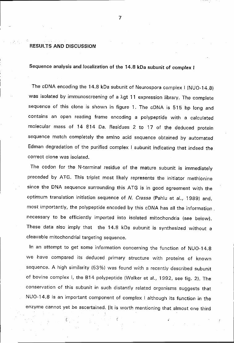
7
RESULTS AND DISCUSSION
Sequence analysis and localization of the 14.8 kDa subunit of complex I
The cDNA encoding the 14.8 kDa subunit of Neurospora complex I (NUO-14.8)
was isolated by immunoscreening of a Xgt 11 expression library. The complete
sequence of this clone is shown in figure 1. The cDNA is 515 bp long and
contains an open reading frame encoding a polypeptide wi th a calculated
molecular mass of 14 814 Da. Residues 2 to 17 of the deduced protein
sequence match completely the amino acid sequence obtained by automated
Edman degradation of the purified complex I subunit indicating that indeed the
correct clone was isolated.
The codon for the N-terminal residue of the mature subunit is immediately
preceded by ATG. This triplet most likely represents the initiator methionine
since the DNA sequence surrounding this ATG is in good agreement wi th the
optimum translation initiation sequence of N. Crassa (Pahlu et al., 1989) and,
most importantly, the polypeptide encoded by this cDNA has all the information
necessary to be efficiently imported into isolated mitochondria (see below).
These data also imply that the 14.8 kDa subunit is synthesized without a
cleavable mitochondrial targeting sequence.
In an attempt to get some information concerning the function of NUO-14.8
we have compared its deduced primary structure with proteins of known
sequence. A high similarity (53%) was found wi th a recently described subunit
of bovine complex I, the B14 polypeptide (Walker et al., 1992, see f ig. 2). The
conservation of this subunit in such distantly related organisms suggests that
NUO-14.8 is an important component of complex I although its function in the
enzyme cannot yet be ascertained. [It is worth mentioning that almost one third
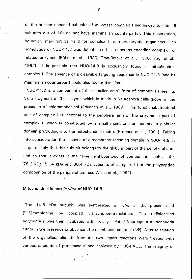
8
of the nuclear encoded subunits of A/, crassa complex I sequenced to date (5
subunits out of 18) do not have mammalian counterparts]. This observation,
however, may not be valid for complex I from prokaryotic organisms - no
homologue of NUO-14.8 was detected so far in opérons encoding complex I or
related enzymes (Bohrn et al., 1990; Tran-Betcke et al., 1990; Yagi et al.,
1992). It is possible that NUO-14.8 is exclusively found in mitochondrial
complex I. The absence of a cleavable targeting sequence in NUO-14.8 (and its
mammalian counterpart) could also favour this idea1.
NUO-14.8 is a component of the so-called small form of complex I ( see f ig.
3), a fragment of the enzyme which is made in Neurospora cells grown in the
presence of chloramphenicol (Friedrich et al., 1989). This functional/structural
unit of complex I is identical to the peripheral arm of the enzyme, a part of
complex I which is constituted by a small membrane anchor and a globular
domain protruding into the mitochondrial matrix (Hofhaus et al., 1991). Taking
into consideration the absence of a membrane spanning domain in NUO-14.8, it
is quite likely that this subunit belongs to the globular part of the peripheral arm,
and so that it exists in the close neighbourhood of components such as the
78.2 kDa, 51.4 kDa and 30.4 kDa subunits of complex I (for the polypeptide
composition of the peripheral arm see Weiss et al., 1991).
Mitochondrial import in vitro of NUO-14.8
The 14.8 kDa subunit was synthesized in vitro in the presence of
[35S]methionine by coupled transcription-translation. The radiolabeled
polypeptide was then incubated with freshly isolated Neurospora mitochondria
either in the presence or absence of a membrane potential (AT). After reisolation
of the organelles, aliquots from the two import reactions were treated with
various amounts of proteinase K and analysed by SDS-PAGE. The integrity of
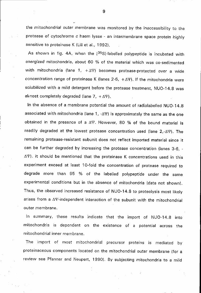
9
the mitochondrial outer membrane was monitored by the inaccessibility to the
protease of cytochrome c haem lyase - an intermembrane space protein highly
sensitive to proteinase K (Lill et al., 1992).
As shown in f ig. 4A, when the [35S]-labelled polypeptide is incubated wi th
energized mitochondria, about 60 % of the material which was co-sedimented
wi th mitochondria (lane 1, + AT) becomes protease-protected over a wide
concentration range of proteinase K (lanes 2-6, + AT). If the mitochondria were
solubilized wi th a mild detergent before the protease treatment, NUO-14.8 was
almost completely degraded (lane 7, + AT).
In the absence of a membrane potential the amount of radiolabeled NUO-14.8
associated wi th mitochondria (lane 1, -AT) is approximately the same as the one
obtained in the presence of a AT. However, 80 % of the bound material is
readily degraded at the lowest protease concentration used (lane 2,-AT). The
remaining protease-resistant subunit does not reflect imported material since it
can be further degraded by increasing the protease concentration (lanes 3-6, -
AT). It should be mentioned that the proteinase K concentrations used in this
experiment exceed at least 10-fold the concentration of protease required to
degrade more than 95 % of the labelled polypeptide under the same
experimental conditions but in the absence of mitochondria (data not shown).
Thus, the observed increased resistance of NUO-14.8 to proteolysis most likely
arises from a AT-independent interaction of the subunit with the mitochondrial
outer membrane.
In summary, these results indicate that the import of NUO-14.8 into
mitochondria is dependent on the existence of a potential across the
mitochondrial inner membrane.
The import of most mitochondrial precursor proteins is mediated by
proteinaceous components located on the mitochondrial outer membrane (for a
review see Pfanner and Neupert, 1990). By subjecting mitochondria to a mild
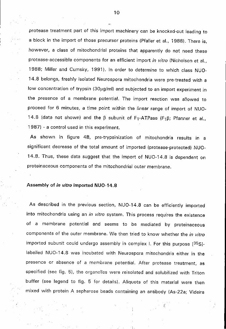
10
protease treatment part of this import machinery can be knocked-out leading to
a block in the import of those precursor proteins (Pfaller et al., 1988). There is,
however, a class of mitochondrial proteins that apparently do not need these
protease-accessible components for an efficient import in vitro (Nicholson et al.,
1988; Miller and Cumsky, 1991). In order to determine to which class NUO-
14.8 belongs, freshly isolated Neurospora mitochondria were pre-treated wi th a
low concentration of trypsin (30(ag/ml) and subjected to an import experiment in
the presence of a membrane potential. The import reaction was allowed to
proceed for 6 minutes, a time point within the linear range of import of NUO-
14.8 (data not shown) and the p subunit of Fi-ATPase (FiP; Pfanner et al.,
1 987) - a control used in this experiment.
As shown in figure 4B, pre-trypsinization of mitochondria results in a
significant decrease of the total amount of imported (protease-protected) NUO-
14.8. Thus, these data suggest that the import of NUO-14.8 is dependent on
proteinaceous components of the mitochondrial outer membrane.
Assembly of in vitro imported NUO-14.8
As described in the previous section, NUO-14.8 can be efficiently imported
into mitochondria using an in vitro system. This process requires the existence
of a membrane potential and seems to be mediated by proteinaceous
components of the outer membrane. We then tried to know whether the in vitro
imported subunit could undergo assembly in complex I. For this purpose [3 5S]-
labelled NUO-14.8 was incubated with Neurospora mitochondria either in the
presence or absence of a membrane potential. After protease treatment, as
specified (see f ig. 5), the organelles were reisolated and solubilized with Triton
buffer (see legend to f ig. 5 for details). Aliquots of this material were then
mixed w i th protein A sepharose beads containing an antibody (As-22a; Videira
11
et al., 1989) which is able to immunoprecipitate whole complex I but that does
not cross-react wi th the 14.8 kDa subunit. As shown in figure 5, in vitro
synthesized NUO-14.8 can be co-immunoprecipitated by As-22a only when
energized mitochondria are used (fig. 5, lanes 5 and 6). In the absence of a A*F
(lanes 7 and 8) no radiolabeled subunit is detected indicating that the antibody
used indeed does not recognise NUO-14.8 per se. [This conclusion is further
supported by the observation that no [35S]-labelled material is precipitated when
the mitochondrial proteins are incubated in the presence of 2 % SDS (to
dissociate complex I subunits) prior to the immunoprecipitation procedure (lanes
1-4)]. The fact that no signal is detected in the absence of a AT also indicates
that the observed co-immunoprecipitation arises from an authentic assembly of
NUO-14.8 during the import reaction and not from an association of the subunit
wi th complex I (or some of its components) after the solubilization step of
mitochondria.
The antibody used in this experiment recognises two subunits of complex I:
the 20.8 kDa (Videira et al., 1990c) and the 29.9 kDa subunits (Van der Pas et
al., 1991). Thus, it can not be inferred from the results presented above at what
stage(s) of assembly is the in vitro imported subunit ( full assembly of NUO-
14.8 into complex I or association of the radiolabeled polypeptide wi th just one
of the subunits recognised by the antibody would lead to the same experimental
result). In order to clarify this point, the following experiment was performed:
[35S]-labelled NUO-14.8 was incubated with Neurospora mitochondria in the
presence or absence (as a negative control) of a membrane potential. After
reisolation of the mitochondria, the samples were solubilized wi th triton X-100
and subjected to sucrose gradient centrifugation. The conditions of the
centrifugation were such that complex I sedimented through two thirds of the
gradient, well separated from proteins wi th a molecular mass smaller than 50
kDa which remained at the top of the gradient (Tuschen et al., 1990; data not
12
shown). Aliquots of the gradients were then analysed for complex I activity
(measured as NADH:[Fe(CN)6]3" oxidoreductase activity) and the distribution of
[35S]-labelled material was monitored. As shown in the graphic of figure 6,
complex I activity is almost completely recovered in fraction 10 of the gradients.
As expected, in the absence of a membrane potential all the [35S]-labelled
material is found at the top of the gradients (fractions 1-3), representing non-
assembled NUO-14.8. In the presence of a AT the majority of the in vitro
imported subunit is also detected in this region of the gradient. However, a
small amount of radioactive material is visible in the fraction enriched in
complex I activity (fraction 10, -f-AT) suggesting that assembly of NUO-14.8
into complex I has occurred
In order to prove unambiguously that the observed radiolabeled material co-
sedimenting wi th complex I represents NUO-14.8 fully assembled in the enzyme
(and not just some fortuitous AT-dependent aggregation of the radioactively
labelled subunit), all the fractions from the two gradients were subjected to
immunoprecipitation using the antibody mentioned above. The proteins in the
immunoprecipitates were then analysed by SDS-PAGE, fluorography and probed
wi th monospecific antibodies directed to the 14.8 and 20.8 kDa subunits of
complex I. As shown in figure 6, both the distribution of the enzymatic activity
and the profile of the radioactively labelled protein in the gradients correlate well
wi th the pattern of "bands" obtained in the western-blot and fluorograph. This
result indicates that the in vitro imported NUO-14.8 co-sedimenting with
complex I is indeed assembled in the enzyme.
Finally, it is worth mentioning that no intermediates of assembly containing the
radiolabeled subunit are detected in this experiment: the in vitro imported NUO-
14.8 could only be found in the low molecular region of the gradients and in the
fractions containing fully assembled complex I. There may be two reasons for
this behaviour: either the biosynthetic pathway of this subunit is too fast to
13
allow the detection of assembly intermediates under the experimental conditions
applied (20 minutes of import at 25°C) or, NUO-14.8 may be one of the last
components to be assembled into the enzyme. This and other questions can
now be easily addressed since an appropriate in organello system has been
developed.
In this article we describe and characterise in terms of localization and
biosynthetic pathway the 14.8 kDa subunit of N. crassa complex I. Our data
suggest that NUO-14.8, the fungal counterpart of the bovine B14 subunit
(Walker et al., 1992), is a component of the peripheral arm of the enzyme
(Hofhaus et al., 1991). This complex I subunit is synthesized without a
cleavable targeting sequence and requires a AT to be imported into
mitochondria. In addition, import of NUO-14.8 seems to be mediated by
proteinaceous components located in the outer membrane. However, the most
important aspect of the work presented here arises from the co-
immunoprecipitation and sedimentation experiments. For the first t ime it is
shown that the assembly process of a complex I subunit can be reproduced
using an in organello system, opening new perspectives for the study of
complex I biogenesis.
Acknowledgements
We are indebted to Dr. Rosemary Stuart for the gift of an antiserum against
Neurospora cytochrome c haem lyase. We thank Dr. Hans Weiss for the samples
of the small form of complex I. This research was supported by the Deutsche
Forschungsgemeinschaft (SFB 184). J . E. A. is supported by Junta Nacional de
Investigação Científica e Tecnológica, Portugal
14
Footnotes
Abbreviations used: AT, mitochondrial inner membrane potential; NUO-X,
NADH:ubiquinone subunit of X kDa; CCHL, cytochrome c haem lyase.
1 According to the endossymbiotic theory for the origin of mitochondria,
mitochondrial nuclear-encoded proteins originally encoded in the genome of the
ancestral organelle have acquired presequences in order to enable them to be
targeted back to mitochondria (for a review see Hartl et al., 1989). Although
there are some exceptions to this rule (e. g., cytochrome c haem lyase and
cytochrome c), it is noteworthy that all nuclearly encoded complex I subunits
having homologues in prokaryotes have a cleavable targeting sequence.
Homologues of nuclear-coded subunits lacking a presequence were never found in
prokaryotes.
15
References
Azevedo, J . E., Nehls, U., Eckerskorn, C , Heinrich, H., Rothe, H., Weiss, H. & Werner, S. (1992) Biochem. J. 288, 29-34
Azevedo, J . E., Abrolatt-Scharf, J . , Eckerskorn, C. and Werner, S. (1993) Biochem. J . 293 , 501-506
Bõhm, R., Sauter, M. & Bõck, A. (1990) Mol. Microbiol. 4 ,231-243 Bradford, M.M. (1976) Anal. Biochem. 72, 248-254 Eckerskorn, C , Mewes, W., Goretzki, H. & Lottspeich, F. (1988) Eur. J . Biochem.
176, 509-519 Friedrich, T., Hofhaus, G., Ise, W., Nehls, U., Schmitz, B. & Weiss, H. (1989) Eur.
J . Biochem. 180, 173-180 Hartl, F.-U., Pfanner, N., Nicholson, D.W. & Neupert, W. (1989) Biochim. Biophys.
Acta 988 , 1-45
Hartl, F.-U., Schmidt, B., Wachter, E., Weiss, H. & Neupert, W. (1986) Cell 47 ,
939-951 Heinrich, H., Azevedo, J . E. & Werner, S. (1992) Biochemistry 3 1 , 11420-11424 Hofhaus, G., Weiss, H. & Leonard, K. (1991) J . Mol. Biol. 2 2 1 , 1027-1043 Laemmli, U. K. (1970) Nature 227, 680-685 Lill, R., Stuart, R. A., Drygas, M. E., Nargang, F. E. & Neupert, W. ( Í992) EMBO J .
1 1 , 449-456 Miller, B. R. & Cumsky, M. G. (1991) J . Cell Biol. 112, 833-841 Nicholson, D. W., Hergersberg, C. & Neupert, W. (1988) J . Biol. Chem. 263,
19034-19042 Pelham, H. R. B. & Jackson, R. J. (1976) Eur. J . Biochem. 67 , 247-256 Pfaller, R., Steger, H. F., Rassow, J . , Pfanner, N. & Neupert, W. (1988) J . Cell
Biol. 107, 2483-2490 Pfanner, N., Muller, H. K., Harney, M. A. & Neupert, W. (1987) EMBO J. 6, 3449-
3454 Pfanner, N. & Neupert, W. (1990) Annu. Rev. Biochem. 59, 331-353 Preis, D., Weidner, U., Conzen, C , Azevedo, J . E., Nehls, U., Rõhlen, D., Van der
Pas, J . , Sackmann, U., Schneider, R., Werner, S. & Weiss, H. (1991) Biochim. Biophys. Acta 1090, 133-138
Ragan, C. I. (1987) Current Top. Bioenerg. 15, 1-36 Rassow, J . , Guiard, B., Wienhues, U., Herzog, V., Hartl, F.-U. & Neupert, W.
(1989) J . Cell Biol. 109, 1421-1428
16
Runswick, M. J . , Fearnley, I. M., Skehel, J . M. & Walker, J . E. (1991) FEBS Lett. 286 , 121-124
Sackmann, U., Zensen, R., Rõhlen, D., Jahnke, U. & Weiss, H. (1991) Eur. J . Biochem. 200, 463-469
Stueber, D., Ibrahimi, I., Cutler, D., Dobberstein, B. & Bujard, H. (1984) EMBO J. 3, 3143-3148
Towbin, H., Staehelin, T. & Gordon, J . (1979) Proc. Natl. Acad. Sci. USA 76, 4350-4354
Tran-Betcke, A., Wamecke, U., Bocker, C , Zabarosch, C. & Friedrich, B. (1990) J .
Bacterid. 172, 2920-2929 Tuschen, G., Sackmann, U., Nehls, U., Haiker, H., Buse, G. & Weiss, H. (1990) J .
Mol. Biol. 213, 845-857 Van der Pas, J . C , Rõhlen, D. A. & Weiss, H. (1991) Biochim. Biophys. Acta
1089, 389-390 Videira, A., Azevedo, J . E., Werner, S. & Cabral, P. (1993) Biochem. J. 2 9 1 , 729-
732 Videira, A., Tropschug, M., Wachter, E., Schneider, H. & Werner, S. (1990c) J .
Biol. Chem. 265, 13060-13065 Videira, A., Tropschug, M. & Werner, S. (19906) Biochem. Biophys. Res.
Commun. 166, 280-285 Videira, A., Tropschug, M. & Werner, S. (1990a) Biochem. Biophys. Res. Commun.
1 7 1 , 1168-1174 Videira, A. & Werner, S. (1989) Eur. J . Biochem. 1 8 1 , 493-502 Walker, J . E. (1992) Quater. Rev. Biophys. 25, 253-324 Walker, J . E., Arizmendi, J . M., Dupuis, A., Fearnley, I. M., Finel, M., Medd, S. M.,
Pilkington, S. J . , Runswick, M. J . & Skehel, J . M. (1992) J . Mol. Biol. 226, 1051-1072
Wang, D., Meinhardt, S. W., Sackmann, U., Weiss, H. & Ohnishi, T. (1991) Eur. J . Biochem. 197, 257-264
Weiss, H., Friedrich, T., Hofhaus, G., & Preis, D. (1991) Eur. J . Biochem. 197, 563-576
Werner, S. (1977) Eur. J. Biochem. 79, 103-110 Yagi, T., Xu, X. & Matsuno-Yagi, A. (1992) Biochim. Biophys. Acta 1 1 0 1 , 181-
183
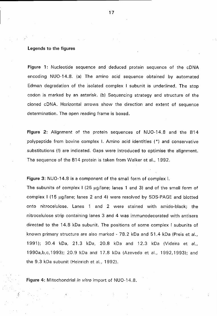
17
Legends to the figures
Figure 1 : Nucleotide sequence and deduced protein sequence of the cDNA
encoding NUO-14.8. (a) The amino acid sequence obtained by automated
Edman degradation of the isolated complex I subunit is underlined. The stop
codon is marked by an asterisk, (b) Sequencing strategy and structure of the
cloned cDNA. Horizontal arrows show the direction and extent of sequence
determination. The open reading frame is boxed.
Figure 2: Alignment of the protein sequences of NUO-14.8 and the B14
polypeptide from bovine complex I. Amino acid identities (*) and conservative
substitutions (!) are indicated. Gaps were introduced to optimise the alignment.
The sequence of the B14 protein is taken from Walker et al., 1992.
Figure 3: NUO-14.8 is a component of the small form of complex I.
The subunits of complex I (25 |ag/lane; lanes 1 and 3) and of the small form of
complex I (15 (ig/lane; lanes 2 and 4) were resolved by SDS-PAGE and blotted
onto nitrocelulose. Lanes 1 and 2 were stained wi th amido-black; the
nitrocelulose strip containing lanes 3 and 4 was immunodecorated wi th antisera
directed to the 14.8 kDa subunit. The positions of some complex I subunits of
known primary structure are also marked - 78.2 kDa and 51.4 kDa (Preis et al.,
1991); 30.4 kDa, 21.3 kDa, 20.8 kDa and 12.3 kDa (Videira et al.,
1990a,b,c,1993); 20.9 kDa and 17.8 kDa (Azevedo et al., 1992,1993) ; and
the 9.3 kDa subunit (Heinrich et al., 1992).
Figure 4 : Mitochondrial in vitro import of NUO-14.8.
18
A) [35S]Methionine-labelled NUO-14.8 was incubated wi th freshly isolated
mitochondria (350 \xg of protein) either in the presence (- AW) or absence ( +
A1?) of 1 \xM valinomycin. At the end of the incubation (20 minutes at 2 5 ° C)
the sample " + A l F " received the same amount of valinomycin. The organelles
from the two samples were re-isolated by centrifugation (10 minutes, 12 000 X
g) washed wi th SEM buffer (250 mM sucrose, 1 mM EDTA, 10 mM Mops/KOH
pH 7.2) and gently resuspended in the same buffer. Aliquots (50 |ag protein)
were then treated wi th the indicated amounts of proteinase K, but the
organelles in lane 7 were first dissolved with 2 % Triton X-100 prior to the
treatment. After 25 minutes at 0 ° C, proteinase K was inactivated by addition
of 1 mM phenylmethylsulfonyl fluoride and the protein in all the aliquots was
precipitated wi th trichloroacetic acid (10% final concentration), dissolved in
Laemmli sample buffer and subjected to SDS-PAGE. The protein in the gel was
then blotted onto nitrocellulose and the membrane was first used to expose an
X-ray film (to detect the in vitro imported subunit) and, afterwards probed wi th
an antiserum directed to cytochrome c haem lyase (CCHL).
B) Freshly isolated Neurospora mitochondria [50 (ig of protein in SEM buffer
(see above)] were incubated for 15 minutes at 0 ° C in the presence (lanes 3
and 4) or absence (lanes 1 and 2) of 30 |ig/ml trypsin. After addition of soy
bean trypsin inhibitor (1 mg/ml final concentration), the organelles were
reisolated, washed and resuspended in import buffer (see materials and
methods) containing the [35S]-labelled precursors of NUO-14.8 and the (3
subunit of F1 ATPase (F1p). Import was allowed to proceed for 6 minutes at
2 5 ° C. The import reactions were then stopped by adding valinomycin to 1 j.iM.
The samples applied to lanes 2 and 4 were treated wi th proteinase K (PK; 100
|ig/ml final concentration) as described above. Finally, the samples were
analysed by SDS-PAGE. A fluorograph of the dried gel is shown. P and M
indicate the precursor and mature form of the p subunit of F1 ATPase,
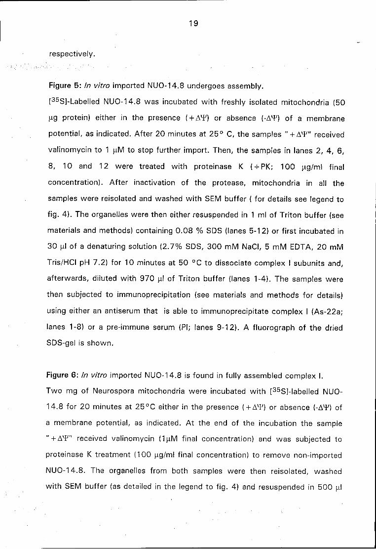
respectively.
19
Figure 5: In vitro imported NUO-14.8 undergoes assembly.
[35S]-Labelled NUO-14.8 was incubated wi th freshly isolated mitochondria (50
u.g protein) either in the presence ( + AX¥) or absence (-A^F) of a membrane
potential, as indicated. After 20 minutes at 25 ° C, the samples " + AT" received
valinomycin to 1 fiM to stop further import. Then, the samples in lanes 2, 4, 6,
8, 10 and 12 were treated with proteinase K ( -i- PK; 100 jag/ml final
concentration). After inactivation of the protease, mitochondria in all the
samples were reisolated and washed wi th SEM buffer ( for details see legend to
f ig. 4). The organelles were then either resuspended in 1 ml of Triton buffer (see
materials and methods) containing 0.08 % SDS (lanes 5-12) or first incubated in
30 ul of a denaturing solution (2.7% SDS, 300 mM NaCI, 5 mM EDTA, 20 mM
Tris/HCI pH 7.2) for 10 minutes at 50 °C to dissociate complex I subunits and,
afterwards, diluted with 970 uJ of Triton buffer (lanes 1-4). The samples were
then subjected to immunoprecipitation (see materials and methods for details)
using either an antiserum that is able to immunoprecipitate complex I (As-22a;
lanes 1-8) or a pre-immune serum (PI; lanes 9-12). A fluorograph of the dried
SDS-gel is shown.
Figure 6: In vitro imported NUO-14.8 is found in fully assembled complex I.
Two mg of Neurospora mitochondria were incubated wi th [35S]-labelled NUO-
14.8 for 20 minutes at 25°C either in the presence (-FAT) or absence (-A^F) of
a membrane potential, as indicated. At the end of the incubation the sample
" + A ^ / " received valinomycin (1uM final concentration) and was subjected to
proteinase K treatment (100 ug/ml final concentration) to remove non-imported
NUO-14.8. The organelles from both samples were then reisolated, washed
wi th SEM buffer (as detailed in the legend to f ig. 4) and resuspended in 500 ul
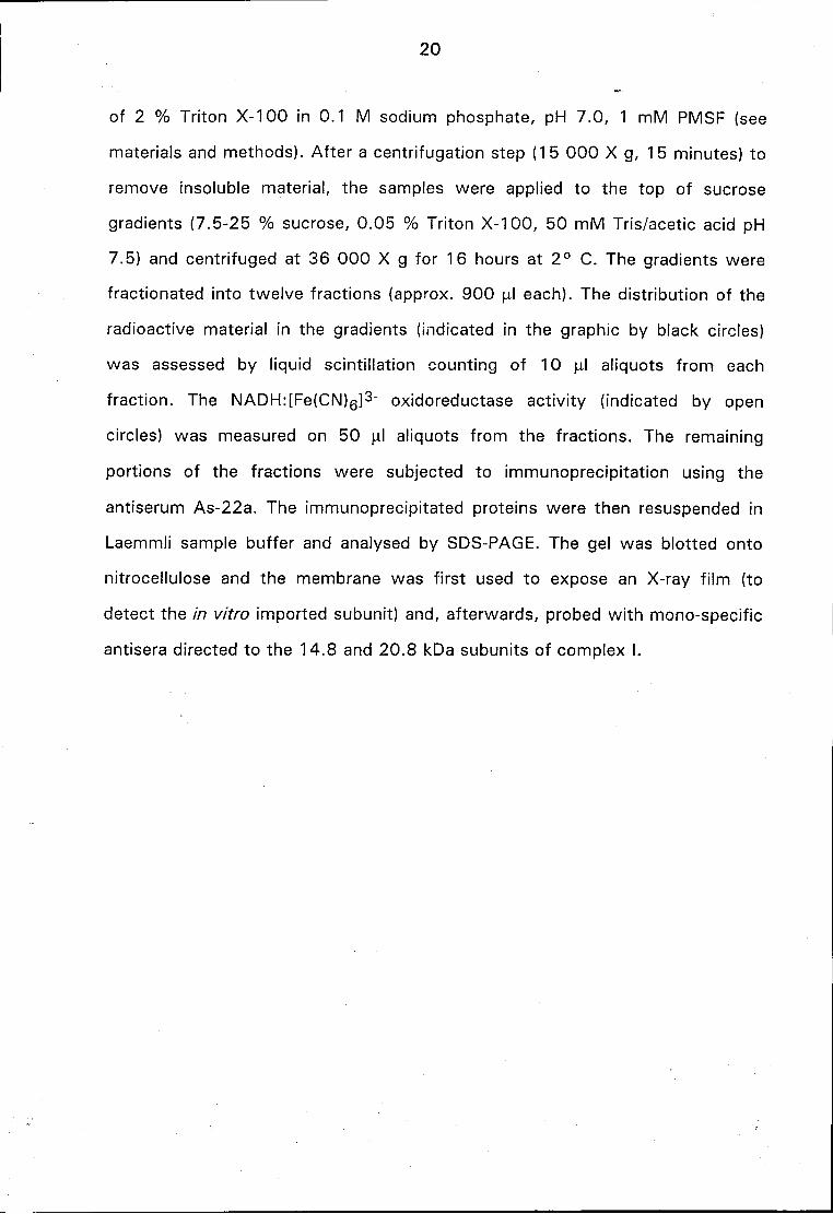
20
of 2 % Triton X-100 in 0.1 M sodium phosphate, pH 7.0, 1 mM PMSF (see
materials and methods). After a centrifugation step (15 000 X g, 1 5 minutes) to
remove insoluble material, the samples were applied to the top of sucrose
gradients (7.5-25 % sucrose, 0.05 % Triton X-100, 50 mM Tris/acetic acid pH
7.5) and centrifuged at 36 000 X g for 16 hours at 2 ° C. The gradients were
fractionated into twelve fractions (approx. 900 ^l each). The distribution of the
radioactive material in the gradients (indicated in the graphic by black circles)
was assessed by liquid scintillation counting of 10 \x\ aliquots from each
fraction. The NADH:[Fe(CN)6]3" oxidoreductase activity (indicated by open
circles) was measured on 50 \i\ aliquots from the fractions. The remaining
portions of the fractions were subjected to immunoprecipitation using the
antiserum As-22a. The immunoprecipitated proteins were then resuspended in
Laemmli sample buffer and analysed by SDS-PAGE. The gel was blotted onto
nitrocellulose and the membrane was first used to expose an X-ray film (to
detect the in vitro imported subunit) and, afterwards, probed wi th mono-specific
antisera directed to the 14.8 and 20.8 kDa subunits of complex I.
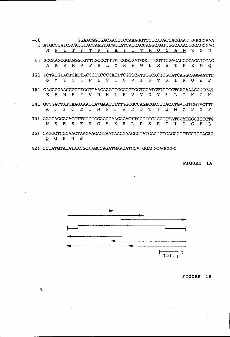
-48 GGAACGGCGACAACCTCCAAAGGTCCTCAAGTCACGAATTGGCCCAAA 1 ATGCCCATCACACCTACCAAGTACGCCATCACCACCAGGCAGTCGGCAAACTGGAGCGAC
M P I T P T K Y A I T T R O S A N W S D 61 GCCAAGCGGAGGGTGTTCGCCCTTTATCGGCGATGGCTTCGTTCGACACCCGAGATGCAG
A K R R V F A L Y R R W L R S T P E M Q 121 TCTATGTACTCACTACCCCTCCCCATTTCGGTCATTCGCACTCGCATCAGGCAGGAATTC
S M Y S L P L P I S V I R T R I R Q E F 181 GAGCGCAACCGCTTCGTTAACAAGTTGCCCGTGGTGGATGTTCTGCTCACAAAGGGCCAT
E R N R F V N K L P V V D V L L T K G H 241 GCCGACTATCAAGAAACCATGAACTTTTGGCGCCAGACGACTCACATGATGTCGTACTTC
A D Y Q E T M N F W R Q T T H M M S Y F 301 AACGAGGAGAGCTTCCGTGGAGCCAAGAGACTTCCCTCCAGCTTTATCGATGGCTTCCTG
N E E S F R G A K R L P S S F I D G F L 361 CAGGGTCGCAACTAAGAAGAGTAATAACGAAGGGTATCAATGTCAGCCTTTCCTCTAGAG
Q G R N # 421 CCTATGTATATAACGCAAGCCAGATGAACATCCATGGACGCAGCCGC
FIGURE 1A
\
^4 -*
100 b.p.
FIGURE IB
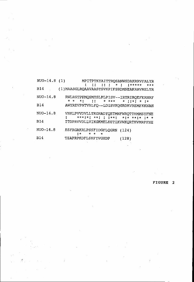
NUO-14.8 (1) MPITPTKYAITTRQSANWSDAKRRVFALYR ! M M i * i j * * * * * ***
B14 (1)MAASGLRQAAVAASTSVKPIFSRDMNEAKRRVRELYR NUO-14.8 RWLRSTPEMQSMYSLPLPISV--IRTRIRQEFERNRF
* * * i if * * * * * i i * i * j * B14 AWYREVPNTVHLFQ--LDISVKQGRDKVREMFKKNAH NUO-14.8 VNKLPWDVLLTKGHADYQETMNFWRQTTHMMS YFNE
j ***j*i **j j j**j * j * * * j * j * * B14 ITDPRWDLLVIKGKMELEETIKVWKQRTHVMRFFHE NUO-14.8 ESFRGAKRLPSSFIDGFLQGRN (124)
i * * * * B14 TEAPRPKDFLSKFYVGHDP (128)
FIGURE 2
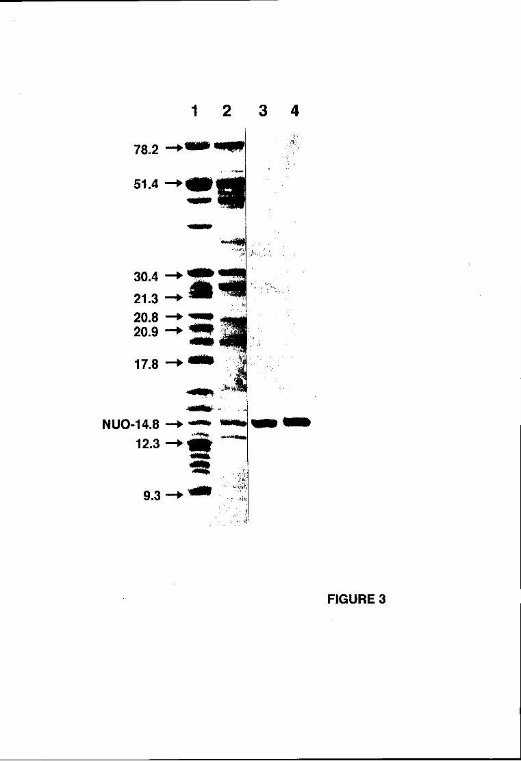
1 3 4
78.2
51.4
30.4 21.3 20.8 20.9
17.8
S
NUO-14.8 12.3
9.3
■ ■ I
a
FIGURE 3
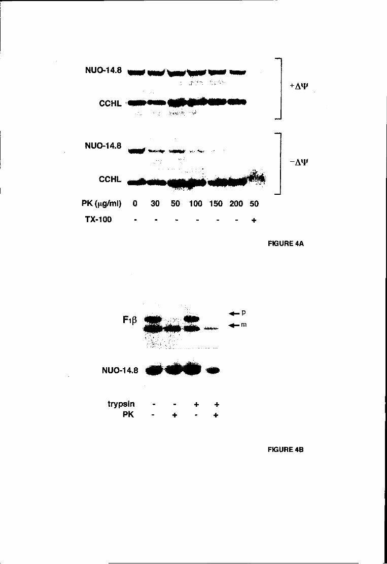
NUO-14.8
CCHL
+AW
NUO-14.8
CCHL
AW
PK(fxg/ml) 0 30 50 100 150 200 50
TX-100 +
FIGURE 4A
p.. ft MUÉE" • «M» rip m.1 ; m
NUO-14.8 ^ ^ ^ ^ ^ ^ ^ ^ ^
trypsin - - + + PK - + - +
FIGURE 4B
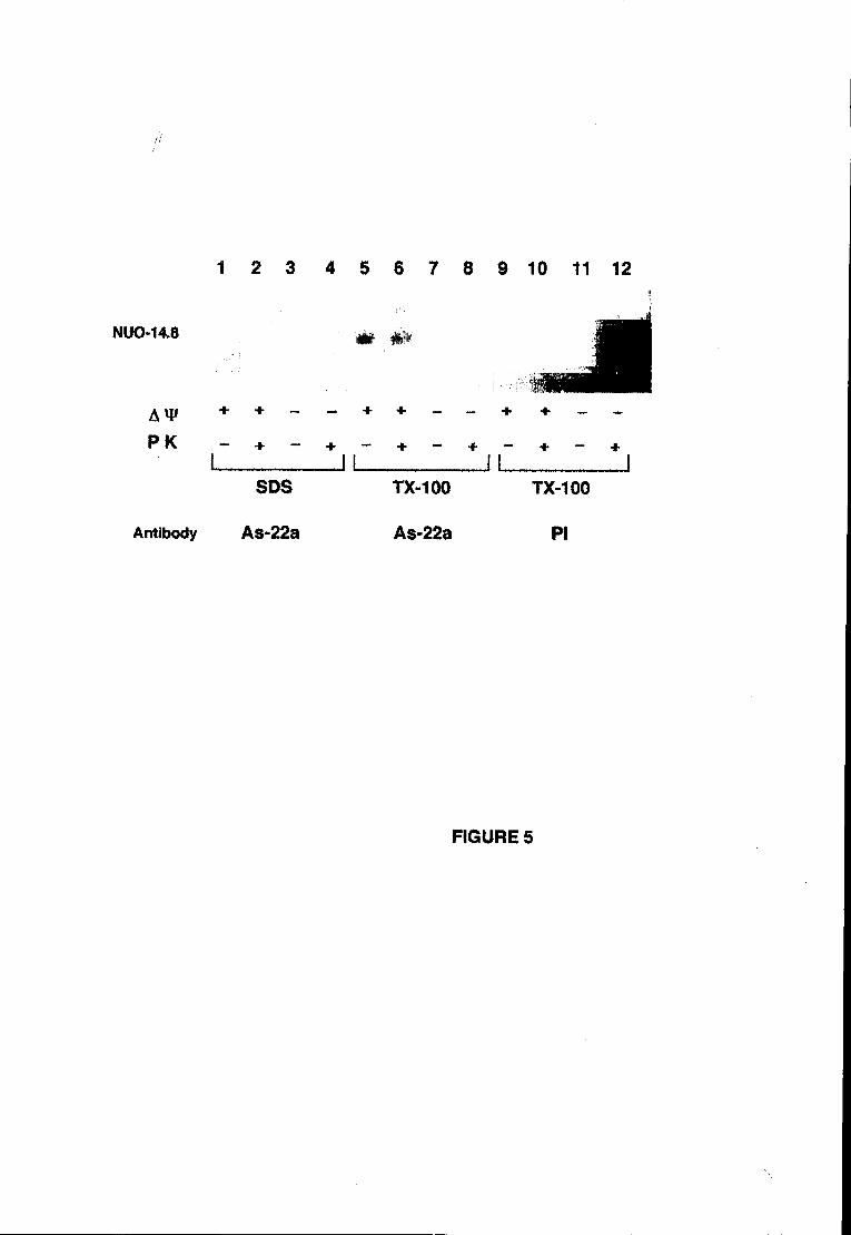
1 2 3 4 5 6 7 8 9 10 11 12
NUO-14.8
AW P K
# *
+ + + + -
+ -JL
+ - -
- +
SDS
Antibody As-22a
TX-100
As-22a
TX-100
PI
FIGURE 5
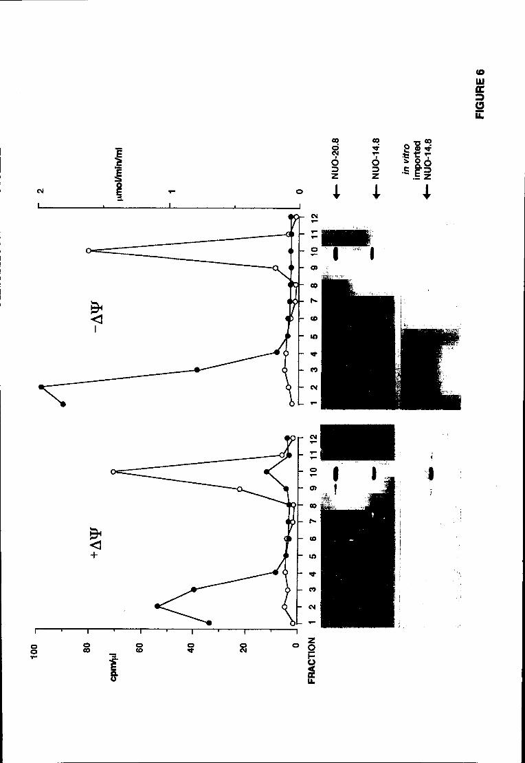
(O uu Œ
LL
TRABALHO 7.5
Characterisation of a membrane fragment of respiratory chain
complex I. Insights on the topology of the ubiquinone-binding site
Jorge E. Azevedo* & Arnaldo Videira
Instituto de Ciências Biomédicas de Abel Salazar - Universidade do Porto.
Lg. Prof. Abel Salazar 2, 4000 Porto, Portugal
Running head: The hydrophobic arm of A/, crassa complex I
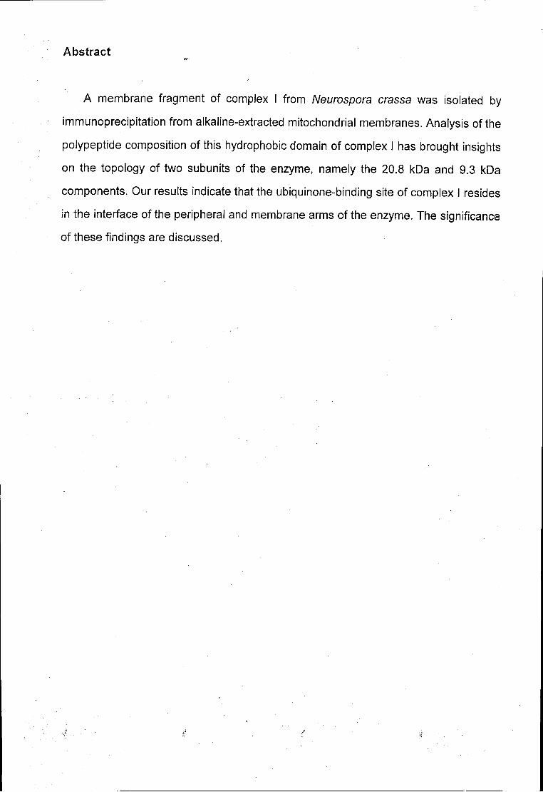
Abstract
A membrane fragment of complex I from Neurospora crassa was isolated by
immunoprecipitation from alkaline-extracted mitochondrial membranes. Analysis of the
polypeptide composition of this hydrophobic domain of complex I has brought insights
on the topology of two subunits of the enzyme, namely the 20.8 kDa and 9.3 kDa
components. Our results indicate that the ubiquinone-binding site of complex I resides
in the interface of the peripheral and membrane arms of the enzyme. The significance
of these findings are discussed.
Introduction
The rotenone-sensitive NADH.ubiquinone oxidoreductase (complex I) is a
complicated assembly of about 40 subunits, 7 of which are encoded and synthesized
in mitochondria. The enzyme catalyses the transfer of two electrons from NADH to
ubiquinone with concomitant translocation of 4 or 5 protons across the mitochondrial
inner membrane (for reviews see Walker, 1992; Weiss et al., 1991).
Recently, a low resolution image of complex I from N. crassa was obtained by
electron microscopy of two-dimensional crystals of the purified enzyme (Hofhaus et
al., 1991). It was concluded that complex I has an L-shaped form with one arm
embedded in the membrane and the other (the peripheral arm) protruding into the
mitochondrial matrix. Interestingly, when Neurospora cells are grown in the presence
of chloramphenicol only a fragment of the enzyme is made - the so-called small form
of complex I (Friedrich et al., 1989). This fragment is thought to correspond to the
peripheral arm of the enzyme. On the other hand, when isolated complex I is treated
with chaotropic agents (Tuschen et ai, 1990) practically all the subunits present in the
small form of the enzyme are stripped-off. However, a fraction of complex I retains
some structural integrity and can be isolated. The presence in this subcomplex (the
hydrophobic fragment) of many subunits with putative membrane spanning domains
has led to the suggestion that this fragment represents the membrane arm of complex
I (Tuschen etal., 1990).
Although no data concerning the over-all shape of mammalian complex I is
available, the high number of homologous subunits between the enzyme from N.
crassa and bovine (see table 1 in the results and discussion section) suggests that
mammalian complex I has a structure similar to the fungal enzyme. Attempts to
resolve complex I from bovine into smaller fragments have been quite successful.
Recently, Walker and co-workers could separate this enzyme into two major
subcomplexes: fragments la and ip (Finel etal., 1992). Subcomplex la contains most
of the enzyme polypeptides, including several homologues of subunits of the small
form of N. crassa complex I. However, at least one homologue of a subunit belonging
to the hydrophobic fragment of A/, crassa complex I is found in subcomplex la (the
ND-2 component), suggesting that this bovine fragment contains a part of the
membrane arm of the enzyme (Walker, 1992). Subcomplex lp contains several
mitochondrial encoded subunits and at least 11 nuclear encoded subunits. This
fragment was proposed to represent a membrane fragment of the bovine enzyme
(Finel era/., 1992; Walker, 1992).
In this article, we describe the isolation and characterisation of a membrane
fragment of complex I from N. crassa. Analysis of the polypeptide composition of this
subcomplex has brought important insights on the topology of two subunits - the 20.8
kDa and 9.3 kDa components. Our results indicate that the ubiquinone-binding site of
complex I is in the region where the two arms of the enzyme join together.
Materials and methods (this section may be printed in small type)
The following procedures were employed according to the references cited:
preparation of complex I (Wang et al., 1991); determination of protein concentration
(Lowry et al., 1951) and sodium dodecylsulfate polyacrylamide gel electrophoresis
(SDS-PAGE) (Laemmli, 1970; Videira & Werner, 1989). The alkaline extraction of
mitochondrial membranes (Fujiki et al., 1982) was performed at a protein
concentration of 1 mg/ml.
The immunoprecipitation experiments were performed in Triton buffer (1% Triton
X-100, 0.3 M NaCI, 5 mM EDTA, 20 mM Tris.HCI pH 7.0 and 1 mM
phenylmethanesulfonyl fluoride) using antibodies (Videira & Werner, 1989) cross-
linked to protein A sepharose (Harlow & Lane, 1998). About 5 mg of these protein A
sepharose beads were used per 1 mg of membranes. The suspension was gently
rocked for 2-3 hours at 4 °C. The protein A sepharose beads were centrifuged (1
minute at 10000 g), washed four times with 1 ml of Triton buffer and, finally, with 1 ml
of 20 mM Tris.HCI pH 7.0. The immunoprecipitated proteins were incubated in
Laemmli sample buffer (Laemmli, 1972) for 30 minutes at room temperature and
subjected to SDS-PAGE analysis. The gels were first stained with coomassie blue
and, afterwards, with silver (Tuschen era/., 1990).
Materials and methods (this section may be printed in small type)
The following procedures were employed according to the references cited:
preparation of complex I (Wang et al., 1991); determination of protein concentration
(Lowry et a/., 1951) and sodium dodecylsulfate polyacrylamide gel electrophoresis
(SDS-PAGE) (Laemmli, 1970; Videira & Werner, 1989). The alkaline extraction of
mitochondrial membranes (Fujiki et al., 1982) was performed at a protein
concentration of 1 mg/ml.
The immunoprecipitation experiments were performed in Triton buffer ( 1 % Triton
X-100, 0.3 M NaCI, 5 mM EDTA, 20 mM Tris.HCI pH 7.0 and 1 mM
phenylmethanesulfonyl fluoride) using antibodies (Videira & Werner, 1989) cross-
linked to protein A sepharose (Harlow & Lane, 1998). About 5 mg of these protein A
sepharose beads were used per 1 mg of membranes. The suspension was gently
rocked for 2-3 hours at 4 °C. The protein A sepharose beads were centrifuged (1
minute at 10000 g), washed four times with 1 ml of Triton buffer and, finally, with 1 ml
of 20 mM Tris.HCI pH 7.0. The immunoprecipitated proteins were incubated in
Laemmli sample buffer (Laemmli, 1972) for 30 minutes at room temperature and
subjected to SDS-PAGE analysis. The gels were first stained with coomassie blue
and, afterwards, with silver (Tuschen et al., 1990).
Results and discussion
The alkaline extraction of membranes (Fujiki et al., 1982) has been widely used in
the study of membrane proteins. Under the high pH used in this procedure, proteins
acquire a very negative net charge leading to strong inter-molecular electrostatic
repulsions. Thus, extrinsic membrane proteins are easily removed from membranes
applying this technique. In contrast, intrinsic membrane proteins (whose interaction
with the membrane occurs in an environment not accessible to the aqueous medium
and is thus pH insensitive) resist the extraction procedure. On the other hand, this
technique seems to be relatively mild in terms of protein tertiary structure - after
alkaline extraction of peroxisomal membranes several membrane enzymes are still
active (Fujiki et al., 1982).
Due to the high performance of this technique, both in reproducibility and
obtention of clear-cut results, we have been using it to study the mitochondrial
respiratory chain complex I. It was shown that the 78 kDa, 49 kDa and 30.4 kDa
subunits of complex I are extractable from membranes under alkaline pH; the 20.9
kDa, 17.8 kDa, 12.3 kDa and 9.3 kDa subunits are alkaline-resistant (Azevedo et al.,
1992, 1993; Heinrich et al., 1992; Videira ef al., 1993). Here, we identify the majority
of complex I subunits (if not all) that cannot be extracted from the mitochondrial inner
membrane under alkaline pH. Our experiments were based on the striking observation
that the 20.8 kDa subunit cannot be extracted from membranes by alkaline treatment.
Like the 12.3 kDa subunit (Videira et al., 1993), the 20.8 kDa subunit (Videira et al.,
1990a) does not contain any obvious membrane spanning domain; yet, both subunits
behave as intrinsic membrane proteins. On the other hand, immunopurified antibodies
directed to the 20.8 kDa subunit are able to immunoprecipitate complex I from Triton
X-100 solubilized mitochondrial membranes (data not shown). These two observations
led us to investigate whether, after an alkaline extraction, the 20.8 kDa component is
still associated with other subunits of the enzyme. In this case, such experiment would
define subunits of the enzyme which are embedded in the lipid phase of the inner
membrane. For this purpose, freshly isolated Neurospora mitochondria were
resuspended in 0.1 M Na2C03 (pH 11.5) and incubated on ice for 30 minutes. The
mitochondrial membranes were isolated by centrifugation and either dissolved in 2.5
% SDS (to dissociate complex I subunits) or in 1 % Triton X-100. The protein solutions
were then subjected to immunoprecipitation using an antiserum which is able to
precipitate complex I but that precipitates mainly 3 subunits from the SDS-dissociated
enzyme. As shown in fig. 1, when SDS-solubilized membranes are used in the
immunoprecipitation experiment only the 20.8 kDa and 12.3 kDa complex I subunits
are observed (the third subunit recognised by the antibody - the 29.9 kDa component -
was removed by the alkaline treatment). However, if the membranes are solubilized
with the mild detergent Triton X-100 and then subjected to immunoprecipitation, a
large number of subunits is co-immunoprecipitated with the 20.8 kDa and 12.3 kDa
subunits indicating that this fraction of the enzyme resists the extraction procedure.
These subunits constitute a part (or the totality) of the membrane arm of complex I.
The polypeptide composition of this fraction of complex I (see table 1 for details)
appears to be similar to the one described for the so-called hydrophobic fragment of
complex I (Tuschen et al., 1990). For example, several mitochondrial encoded
subunits and the 20.9 kDa subunit are present in both subcomplexes (Azevedo et al.,
1992; Weiss ef al., 1991). However, the two fragments may not be identical as the
20.8 kDa subunit was assigned to the peripheral arm of the enzyme (Weiss et al.,
1991). If this is indeed the case, then the 20.8 kDa subunit is part of the small
membrane anchor of the small form of complex I (Hofhaus etal., 1991).
A comparative analysis between the polypeptide composition of the complex I
fragment described here and subcomplex la from the bovine enzyme (Finel et al.,
1992; Walker, 1992), revealed some common components. In particular, the 20.8 kDa
(a possible iron-sulphur protein) and 9.3 kDa subunits present in the membrane
fragment of the fungal enzyme, have homologues in bovine subcomplex la (subunits
PGIV and B9, respectively). This result indicates that these two components are
localized in the interface of the membrane and peripheral arms of complex I; their co-
purification with one or another arm depends on the organism and resolving technique
used.
These topological considerations acquire a special importance in the case of the
9.3 kDa subunit. This polypeptide was identified using a photo-affinity ubiquinone
analogue as being part of the ubiquinone-binding site (Heinrich & Werner, 1992).
Thus, we assume that the ubiquinone-binding site of complex I is localized in the
junction of the two arms of the enzyme. This conclusion is supported by the fact that
bovine subcomplex la has all the e.p.r.-detectable Fe-S clusters of the enzyme
(including cluster N-2, assumed to be the immediate reductor of ubiquinone) (Finel et
al., 1992).
The results presented here are depicted in figure 2 in the context of all the
structural data presently available for complex I both from N. crassa and bovine [for
detailed information regarding the function of some complex I components see
(Walker, 1992)]. The most striking feature of this working model is that all the redox
groups and substrate binding-sites of the enzyme are localized in the peripheral arm
and in the part of the membrane arm in contact with it. This observation could suggest
that the electrons do not flow along the membrane arm of complex I but just cross it
partially. It is clear, however, that more data are needed to test this hypothesis.
Finally, it is worth mentioning that 11 out of the 12 Neurospora complex I subunits
with known primary structure that are not resistant to carbonate extraction have
counterparts in the bovine enzyme. In sharp contrast, 4 out the 6 nuclear-encoded
subunits with known primary structure that belong to the membrane fragment
described here have no bovine homologues. It is possible that these two domains of
complex I have been under different evolutionary constraints.
Acknowledgements
This work was supported by Junta Nacional de Investigação Científica e Tecnológica
(JNICT), Portugal. J.E.A. would like to thank JNICT for financial support.
Summary
Neurospora crassa mitochondrial membranes were subjected to alkaline
extraction in order to remove peripheral membrane proteins. When these membranes
were solubilized with a mild non-denaturing detergent and, afterwards, subjected to
immunoprecipitation using an antibody that recognises mainly three subunits of
complex I, a fragment comprising about 15 subunits of the enzyme was obtained. This
fragment contains a fraction (or the totality) of the membrane-embedded complex I
subunits. A comparative analysis of the polypeptide composition of this hydrophobic
fragment with subcomplex la from the bovine enzyme (Finei et al., 1992) revealed the
existence of two common components: the ubiquinone-binding 9.3-kDa polypeptide
(Heinrich et al., 1992) and the 20.8 kDa subunit (Videira et al., 1990a). This finding
provides evidence that these two components are localized in the interface of the
peripheral and membrane arms of the enzyme.
References
Azevedo, J. E., Abrolat-Scharff, J„ Eckerskorn, C. & Werner, S. (1993) Cloning, in
vitro mitochondrial import and membrane assembly of the 17.8 kDa subunit of
complex I from Neurospora crassa. Biochem. J. 293, 501-506.
Azevedo, J. E., Nehls, U., Eckerskorn, C, Heinrich, H., Rothe, H., Weiss, H. &
Werner, S. (1992) Primary structure and mitochondrial in vitro import of the 20.9
kDa subunit of complex I from Neurospora crassa. Biochem. J. 288, 29-34.
Duarte, M., Belo, J.A. & Videira, A. (1993) Primary structure of the nuclear-encoded
10.5 kDa subunit of complex I from Neurospora crassa. Biochim. Biophys. Acta
1172,327-328.
Finel, M., Skehel, J. M., Albracht, S. P. J., Fearnley, I. M. & Walker, J. E. (1992)
Resolution of NADH:ubiquinone oxidoreductase from bovine heart mitochondria
into two subcomplexes one of which contains the redox centres of the enzyme.
Biochemistry 31, 11425-11434.
Friedrich, T., Hofhaus, G., Ise, W., Nehls, U., Schmitz, B. & Weiss, H. (1989) A small
isoform of NADH:ubiquinone oxidoreductase (complex I) without mitochondrially
encoded subunits is made in chloramphenicol-treated Neurospora crassa. Eur. J.
Biochem. 180, 173-180.
Fujiki, Y., Hubbard, A. L, Fowler, S. & Lazarow, P. B. (1982) Isolation of intracellular
membranes by means of sodium carbonate treatment : application to the
endoplasmic reticulum. J. Cell Biol. 93, 97-102.
Harlow, E. & Lane, D. (1988) Antibodies: a laboratory manual, pp. 522-523. Cold
Spring Harbour laboratory, New York.
Heinrich, H. & Werner, S. (1992) Identification of the ubiquinone-binding site of
NADH.ubiquinone oxidoreductase (complex I) from Neurospora crassa.
Biochemistry 31, 11413-11419.
Heinrich, H., Azevedo, J. E. & Werner, S. (1992) Characterization of the 9.5-kDa
ubiquinone-binding protein of NADH:ubiquinone oxidoreductase (complex I) from
Neurospora crassa. Biochemistry 31, 11420-11424.
Hofhaus, G., Weiss, H. & Leonard, K. (1991) Electron microscopic analysis of the
peripheral and membrane parts of mitochondrial NADH dehydrogenase (complex
I). J. Mol. Biol. 221, 1027-1043.
Laemmli, U. K. (1970) Cleavage of structural proteins during the assembly of the head
of bacteriophage T4. Nature 227, 680-685
Lowry, H., Rosebrough, N. J., Farr, A. L. & Randall, R. J. (1951) Protein measurement
with the Folin phenol reagent. J. Biol. Chem. 193, 265-275.
Nehls, U., Hemmer, S., Rohlen, D. A., van der Pas, J. C , Preis, D., Sackmann, U. &
Weiss, H. (1991 ) cDNA and genomic DNA sequence of the 21.3 kDa subunit of
NADH:ubiquinone reductase (complex I) from Neurospora crassa. Biochim.
Biophys. Acta 1088, 325-326.
Preis, D., van der Pas, Nehls, U., Rõhlen, D., Sackmann, U., Jahnke, U. & Weiss, H.
(1990) The 49 kDa subunit of NADH:ubiquinone reductase (complex I) from
Neurospora crassa mitochondria: primary structure of the gene and the protein.
Curr. Genet. 18, 59-64.
Preis, D.r Weidner, U., Conzen, C, Azevedo, J. E., Nehls, U., Rohlen, D., Van der
Pas, J., Sackmann, 1)., Schneider, R., Werner, S. & Weiss, H. (1991) Primary
structure of two subunits of NADH:ubiquinone reductase (complex I) from
Neurospora crassa mitochondria: relationship to a soluble NAD-reducing
hydrogenase from Alkaligenes eutrophus. Biochim. Biophys. Acta 1090,133-138.
Rohlen, D. A., Hoffmann, J. Van der Pas, J. C, Nehls, U., Preis, D., Sackman, U. &
Weiss, H. (1991) Relationship between a subunit of NADH dehydrogenase
(complex I) and a protein family including subunits of cytochrome reductase and
processing protease from mitochondria. FEBS Lett. 278, 75-78.
Sackmann, U., Zensen, R., Rohlen, D., Jahnke, U. & Weiss, H. (1991) The acyl carrier
protein in Neurospora crassa mitochondria is a subunit of NADH:ubiquinone
reductase (complex I). Eur. J. Biochem. 200, 463-469.
Tuschen, G., Sackmann, U., Nehls, U., Haiker, H., Buse, G. & Weiss, H. (1990)
Assembly of NADH.ubiquinone reductase (complex I) in Neurospora mitochondria:
independent pathways of nuclear encoded and mitochondrially encoded subunits.
J. Mol. Biol. 213, 845-857.
Van der Pas, J. C, Rohlen, D. A., Weidner U. & Weiss, H. (1991) Primary structure of
the nuclear encoded 29.9 kDa subunit of NADH:ubiquinone reductase from
Neurospora mitochondria. Biochim. Biophys. Acta 1089, 389-390.
Videira, A. & Werner, S. (1989) Assembly kinetics and identification of precursor
proteins of complex I from Neurospora crassa. Eur. J. Biochem. 181, 493-502.
Videira, A., Azevedo, J. E., Werner, S. & Cabral, P. (1993) The 12.3 kDa subunit of
complex I (respiratory-chain NADH dehydrogenase) from Neurospora crassa:
cDNA cloning and chromosomal mapping of the gene. Biochem. J. 291, 729-732.
Videira, A., Tropschug, M. & Werner, S. (1990b) Primary structure and expression of a
nuclear-encoded subunit of complex I homologous to proteins specified by the
chloroplast genome. Biochem. Biophys. Res. Commun. 171, 1168-1174.
Videira, A., Tropschug, M. & Werner, S. (1990c) Primary structure, in vitro expression
and import into mitochondria of a 29/21 kDa subunit of complex I from Neurospora
crassa. Biochem. Biophys. Res. Commun. 166, 280-285
Videira, A., Tropschug, M., Wachter, E., Schneider, H. & Werner, S. (1990a)
Molecular cloning of subunits of complex I from Neurospora crassa. Primary
structure and in vitro expression of a 22-kDa polypeptide J. Biol. Chem. 265,
13060-13065.
Walker, J. E. (1992) The NADH:ubiquinone oxidoreductase (complex I) of respiratory
chain. Quater. Rev. Biophys. 25, 253-324.
Wang, D., Meinhardt, S. W., Sackmann, U., Weiss, H. & Ohnishi, T. (1991) The iron-
sulfur clusters in two related forms of mitochondrial NADH:ubiquinone reductase
(complex I) from Neurospora crassa mitochondria. Eur. J. Biochem. 197, 257-264.
Weidner, U., Sckmann, U., Nehls, U. & Weiss, H. (1991) Primary structure of the
nuclear encoded 18.3 kDa subunit of NADH:ubiquinone reductase (complex I)
from Neurospora mitochondria. Biochim. Biophys. Acta 1089, 391-392.
Weiss, H., Friedrich, T., Hofhaus, G., & Preis, D. (1991) The respiratory chain NADH
dehydrogenase (complex I) of mitochondria. Eur. J. Biochem. 197, 563-576.
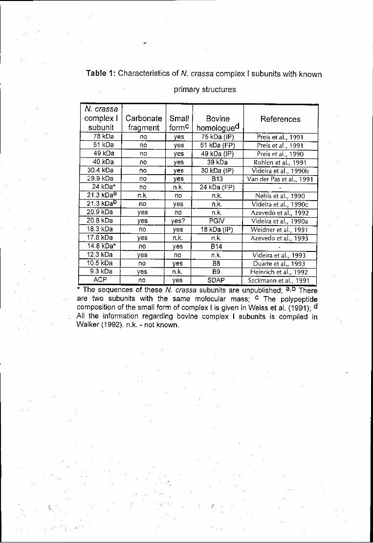
Table 1: Characteristics of N. crassa complex I subunits with known
primary structures
N. crassa complex I subunit
Carbonate fragment
Small formc
Bovine homologue0'
References
78 kDa no yes 75 kDa (IP) Preis et al., 1991 51 kDa no yes 51 kDa (FP) Preis et al., 1991 49kDa no yes 49 kDa (IP) Preis et al., 1990 40 kDa no yes 39 kDa Rohlenetal., 1991
30.4 kDa no yes 30 kDa (IP) Videira et al., 1990b 29.9 kDa no yes B13 Van der Pas et al., 1991
24 kDa* no n.k. 24 kDa (FP) -21.3 kDaa n.k. no n.k. Nehlsetal., 1990 21.3 kDaD no yes n.k. Videira et al., 1990c 20.9 kDa yes no n.k. Azevedo et al., 1992 20.8 kDa yes yes? PGIV Videira et al., 1990a 18.3 kDa no yes 18 kDa (IP) Weidneretal., 1991 17.8 kDa yes n.k. n.k. Azevedo et al., 1993 14.8 kDa* no yes B14 -12.3 kDa yes no n.k. Videira et al., 1993 10.5 kDa no yes B8 Duarte et al., 1993 9.3 kDa yes n.k. B9 Heinrich étal., 1992 ACP no yes SDAP Sackmann étal., 1991
* The sequences of these N. crassa subunits are unpublished; a-D There are two subunits with the same molecular mass; c The polypeptide composition of the small form of complex I is given in Weiss et al. (1991); d
All the information regarding bovine complex I subunits is compiled in Walker (1992). n.k. - not known.
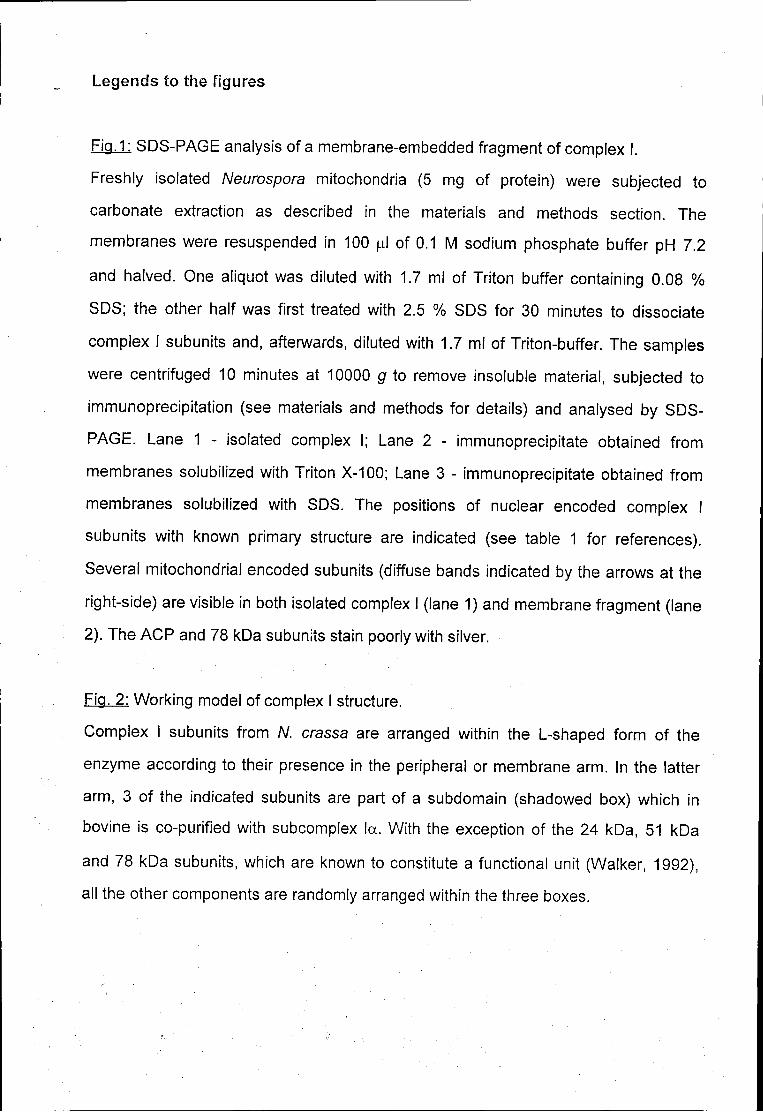
Legends to the figures
Fig.1: SDS-PAGE analysis of a membrane-embedded fragment of complex I.
Freshly isolated Neurospora mitochondria (5 mg of protein) were subjected to
carbonate extraction as described in the materials and methods section. The
membranes were resuspended in 100 ul of 0.1 M sodium phosphate buffer pH 7.2
and halved. One aliquot was diluted with 1.7 ml of Triton buffer containing 0.08 %
SDS; the other half was first treated with 2.5 % SDS for 30 minutes to dissociate
complex I subunits and, afterwards, diluted with 1.7 ml of Triton-buffer. The samples
were centrifuged 10 minutes at 10000 g to remove insoluble material, subjected to
immunoprecipitation (see materials and methods for details) and analysed by SDS-
PAGE. Lane 1 - isolated complex I; Lane 2 - immunoprecipitate obtained from
membranes solubilized with Triton X-100; Lane 3 - immunoprecipitate obtained from
membranes solubilized with SDS. The positions of nuclear encoded complex I
subunits with known primary structure are indicated (see table 1 for references).
Several mitochondrial encoded subunits (diffuse bands indicated by the arrows at the
right-side) are visible in both isolated complex I (lane 1) and membrane fragment (lane
2). The ACP and 78 kDa subunits stain poorly with silver.
Fig. 2: Working model of complex I structure.
Complex I subunits from N. crassa are arranged within the L-shaped form of the
enzyme according to their presence in the peripheral or membrane arm. In the latter
arm, 3 of the indicated subunits are part of a subdomain (shadowed box) which in
bovine is co-purified with subcomplex la. With the exception of the 24 kDa, 51 kDa
and 78 kDa subunits, which are known to constitute a functional unit (Walker, 1992),
all the other components are randomly arranged within the three boxes.
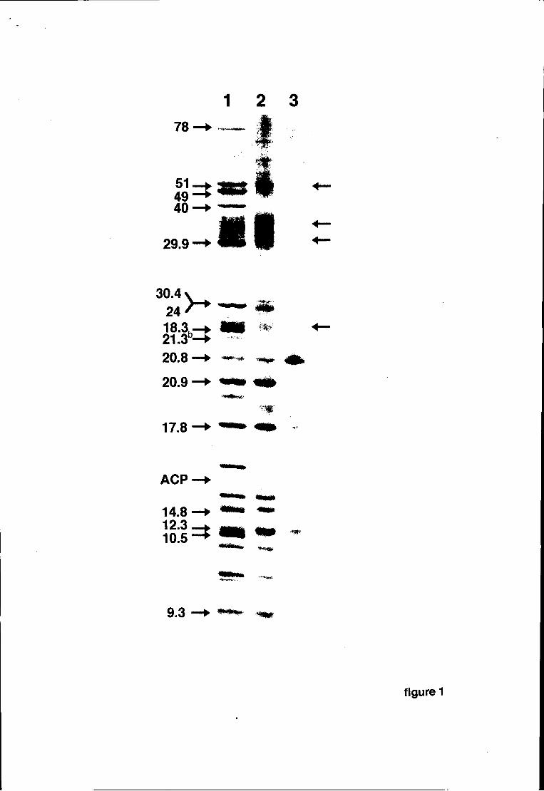
1 2 3 78
51 =± 49—► 40-
29.9 ■ HI „ m
30.4 24 18.3h-21.3
b-
20.8
20.9
17.8
> -
ACP
14.8 12.3 10.5
9.3
figure 1
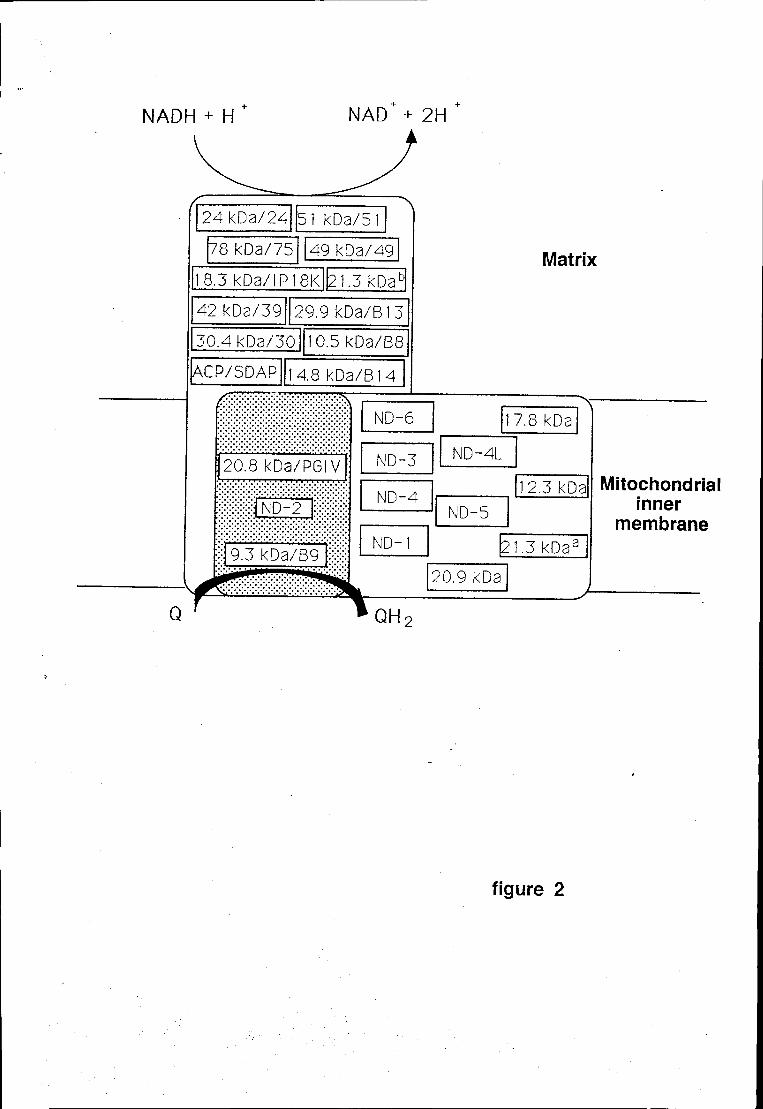
NADH + H NAD + 2H
24kDa/24 51 kDa/51
78 kDa/75 49 kDa/49
8.3kDa/IP18K 21.3 kDab
Matrix
1 7.8 kDa ■N
ND-4L
12.3 kDa ND-5
2 1.3kDaa
20.9 kDa
Mitochondrial inner
membrane
figure 2
IL; ,o\
p±~