GRAA CASAL MICROSPORIDIOSES E MIXOSPORIDIOSES DA ...
263
Microsporidioses e Mixosporidioses da ictiofauna portuguesa e brasileira: caracterização ultrastrutural e filogenética i GRAÇA MARIA FIGUEIREDO CASAL MICROSPORIDIOSES E MIXOSPORIDIOSES DA ICTIOFAUNA PORTUGUESA E BRASILEIRA: CARACTERIZAÇÃO ULTRASTRUTURAL E FILOGENÉTICA Dissertação de Candidatura ao grau de Doutor em Ciências Biomédicas submetida ao Instituto de Ciências Biomédicas de Abel Salazar da Universidade do Porto. Orientador - Doutor Jorge Guimarães da Costa Eiras Categoria – Professor Catedrático Afiliação - Faculdade de Ciências da Universidade do Porto. Co-orientadora - Doutora Maria Leonor Hermenegildo Teles Grilo Categoria - Professora Associada Afiliação - Instituto de Ciências Biomédicas de Abel Salazar da Universidade do Porto.
-
Upload
phungduong -
Category
Documents
-
view
310 -
download
57
Transcript of GRAA CASAL MICROSPORIDIOSES E MIXOSPORIDIOSES DA ...

Microsporidioses e Mixosporidioses da ictiofauna portuguesa e brasileira: caracterização ultrastrutural e filogenética i
GRAÇA MARIA FIGUEIREDO CASAL
MICROSPORIDIOSES E MIXOSPORIDIOSES DA ICTIOFAUNA
PORTUGUESA E BRASILEIRA: CARACTERIZAÇÃO
ULTRASTRUTURAL E FILOGENÉTICA
Dissertação de Candidatura ao grau de Doutor em Ciências Biomédicas submetida ao Instituto de Ciências Biomédicas de Abel Salazar da Universidade do Porto. Orientador - Doutor Jorge Guimarães da Costa Eiras Categoria – Professor Catedrático Afiliação - Faculdade de Ciências da Universidade do Porto. Co-orientadora - Doutora Maria Leonor Hermenegildo Teles Grilo Categoria - Professora Associada Afiliação - Instituto de Ciências Biomédicas de Abel Salazar da Universidade do Porto.

ii Microsporidioses e Mixosporidioses da ictiofauna portuguesa e brasileira: caracterização ultrastrutural e filogenética

Microsporidioses e Mixosporidioses da ictiofauna portuguesa e brasileira: caracterização ultrastrutural e filogenética iii
Ao Prof. Carlos Azevedo,
Pela amizade e por tudo que me ensinou

iv Microsporidioses e Mixosporidioses da ictiofauna portuguesa e brasileira: caracterização ultrastrutural e filogenética

Microsporidioses e Mixosporidioses da ictiofauna portuguesa e brasileira: caracterização ultrastrutural e filogenética v
AGRADECIMENTOS
Ao Professor Doutor Carlos Azevedo por ter aceite orientar esta Tese até Março de 2007,
apesar do obstante por dispositivos legais em oficialmente dar continuidade, para todos
os efeitos fê-lo até à entrega da dissertação para apreciação. Aproveito esta ocasião para
manifestar o meu profundo reconhecimento, por me ter dado a oportunidade de estagiar
e, posteriormente, ser monitora no Laboratório de Biologia Celular do Instituto de
Ciências Biomédicas de Abel Salazar, onde me iniciei na investigação científica,
culminando o percurso com a realização desta Tese. Agradeço igualmente os inúmeros
ensinamentos de carácter pedagógico e científico, bem como toda a paciência, conselhos
e amizade demonstrada durante estes anos.
Ao Professor Doutor Jorge Eiras, por ter aceite em fazer parte da Comissão de
acompanhamento e também a responsabilidade de assumir oficialmente a orientação dos
trabalhos em substituição do Professor Doutor Carlos Azevedo que entretanto se jubilou.
Desejaria aqui expressar o meu sincero agradecimento, bem como reiterar os laços
científicos partilhados em diversas reuniões da Sociedade Portuguesa de Parasitologia.
À Professora Doutora Leonor Teles-Grilo o meu agradecimento por ter aceite co-orientar
os trabalhos no âmbito da Biologia Molecular, área na qual dei os primeiros passos ao
iniciar esta tese. Agradeço igualmente ter-me disponibilizado todo os meios do
Laboratório de Genética Molecular, bem como todos os conselhos e apoio dispendido.
Às inúmeras pessoas do Laboratório de Biologia Celular um muito obrigado por me
acolherem ainda como aluna do ICBAS e por toda amizade que têm demonstrado.
Agradeço ao Professor Doutor Mário Sousa e ao Professor Doutor Alexandre Lobo da
Cunha por me terem possibilitado continuar a usufruir das instalações e dos
equipamentos do Laboratório de Biologia Celular, após a jubilação do Prof. Carlos
Azevedo. Agradeço, igualmente, ao Professor Doutor Alexandre Lobo da Cunha todos os
conselhos de índole científica e pessoais, bem como pela amizade e camaradagem
demonstrada durante todos estes anos. À Sra. D. Laura Corral pelo ensino das técnicas
de microscopia electrónica e preparação de materiais biológicos, ferramenta que serviu
de base para o arranque desta Tese. À Sra. Dª. Elsa Oliveira e à Sra. Dª. Ângela Alves
agradeço o apoio e conselhos técnicos diários, que sem sombra para dúvida, fazem toda
a diferença.
À Doutora Camino Gestal do Instituto de Investigaciones Marinas de Vigo, Espanha pelos
inúmeros conselhos diários, bem como pela agradável convivência durante a sua estadia
de dois anos no Laboratório de Biologia Celular como bolseira do Programa Post-Doc

vi Microsporidioses e Mixosporidioses da ictiofauna portuguesa e brasileira: caracterização ultrastrutural e filogenética
“Fellowships” Marie Curie, supervisionada pelo Prof. Doutor Carlos Azevedo. Da colega
Camino, além de uma grande amizade, ficou a saudade de alguém que partilha os
mesmos interesses científicos.
Agradeço à Eng.ª Carla Oliveira do Laboratório de Genética Molecular do ICBAS, pela
prontidão e amabilidade que sempre demonstrou em me auxiliar nas questões
laboratoriais e à Sra. Dª. Matilde Rocha pelo auxílio na esterilização do material.
Agradeço também aos alunos de Mestrado, de Estágio para conclusão de licenciatura,
Bolseiros e Estagiários a título voluntário que passaram por este laboratório,
nomeadamente aos licenciados Joana Tato Costa, Américo Marques, Sérgio Duarte que,
pontualmente, me transmitiram um pouco das suas experiências laboratoriais.
Agradeço também ao Técnico Emanuel Monteiro do ICBAS, pelo auxílio na preparação
das amostras a serem observadas no Microscópio Electrónico de Varrimento (SEM). Do
Centro de Materiais da Universidade do Porto, gostaria também de agradecer à Drª
Daniela Silva no auxílio da observação das amostras no SEM. Do Departamento de
Informática do ICBAS, agradeço, aos Licenciados Rui Claro, João Morais e Nuno Santos
a rápida prontidão na resolução dos problemas de informática que foram surgindo. Ao Sr.
João Carvalheiro e à Sra Dª. Joana Carvalheiro do Serviço de Iconografia do ICBAS, pela
reprodução das fotografias de microscopia electrónica, bem como pelos ensinamentos
técnicos sobre fotografia.
À CESPU – Cooperativa de Ensino Superior Politécnico e Universitário pela atribuição de
uma bolsa para custear as propinas inerentes à minha inscrição como aluna de
Doutoramento no ICBAS. Ao Professor Doutor Victor Seabra, na qualidade de
Coordenador do Gabinete de Formação, Investigação e Desenvolvimento, agradeço a
disponibilidade e o apoio prestado.
Agradeço ao Professor Doutor Jorge Proença, Director do Instituto Superior de Ciências
da Saúde - Norte (ISCS-N), e à Professora Doutora Roxana Moreira, Directora do
Departamento de Ciências, as facilidades concedidas na redução da carga horária do
serviço docente para o valor mínimo, bem como a compreensão e autorização em repartir
a marcação de férias em períodos distintos dos contemplados pela Instituição.
Ao Professor Doutor Hassan Bousbaa, regente das disciplinas do ISCS-N (CESPU) das
quais sou Assistente, por todos os ensinamentos teóricos e práticos que tem transmitido,
bem como por toda a amizade e confiança depositada durante os últimos anos. A todos
colegas de docência, Professora Doutora Carla Batista, Professora Doutora Catarina
Lemos, Professor Doutor Frederico Silva, Doutora Manuela Henrique, Mestre Paulo
Microsporidioses e Mixosporidioses da ictiofauna portuguesa e brasileira: caracterização ultrastrutural e filogenética vii
Barros, Mestre Vanessa Nascimento, Licenciada Georgina Rodrigues e Licenciada
Tatiana Resende, o meu obrigado pela inter-ajuda na leccionação das várias disciplinas.
Aproveito esta oportunidade para agradecer a todos os colegas das diferentes
instituições, com os quais partilhámos os mesmos anseios, a ajuda nas colheitas
efectuadas no Brasil. Ao Professor Doutor Edilson Matos, Director do Laboratório de
Pesquisa Carlos Azevedo da Universidade Federal Rural da Amazónia, Belém, Brasil, a
quem eu muito agradeço toda a preciosa ajuda, empenho, coordenação e dedicação nas
inúmeras colheitas efectuadas, por iniciativa própria e por nós solicitadas, bem como o
processamento inicial das mesmas. Agradeço igualmente aos seus colaboradores mais
directos, Mestre Patrícia Matos do Laboratório de Animais Aquáticos da Universidade
Federal do Pará, Belém e à Mestre Patrícia Garcia do Laboratório de Diagnóstico e
Patologia em Aquacultura da Universidade Federal de Santa Catarina. O meu
agradecimento também para o Professor Doutor Sérgio Carmona Clemente da Faculdade
de Medicina Veterinária da Universidade Federal Fluminense de Niterói pela colaboração
num dos trabalhos, bem como à Doutora Débora Marques do Embrapa (Pantanal,
Corumbá) e à Professora Ivete Mendonça da Faculdade de Medicina Veterinária da
Universidade Federal do Piauí de Teresina, pelo envio de algumas das amostras com
material parasitado.
À Professora Doutora Maria de Lurdes Pereira do Departamento de Biologia da
Universidade de Aveiro e à Licenciada Fernanda Castilho (Directora do IPIMAR-
Matosinhos) agradeço as facilidades concedidas na obtenção de vários especímenes
utilizados na nossa investigação.
Ao Centro Interdisciplinar de Investigação Marinha e Ambiental (CIIMAR) e à Fundação
Eng.º António de Almeida agradeço os apoios financeiros despendidos durante estes
anos, tendo em muito contribuído nos custos inerentes à investigação científica. Gostaria
também de agradecer ao Mestre Hugo Santos e ao Sr. Carlos Rosa (CIIMAR- Biotério de
Organismos Aquáticos) pelas ocasiões em que necessitei de água salgada para a
manutenção de alguns especímenes.
Por último, gostaria de agradecer a algumas pessoas que, apesar de não terem estado
envolvidas directamente, foram no entanto importantes pilares emocionais durante os
diferentes estados de humor pelos quais passei até concluir esta tese. À Carla Batista
amiga e colega de bancada no ICBAS e, simultaneamente, colega na CESPU, pela
amizade, camaradagem e pelo espírito de inter-ajuda relativamente ao serviço docente
atribuído pelo ISCS-N. À Dolores Resende por toda amizade, apoio e partilha de histórias
por um “hobby” comum, que por vezes me deram alento e coragem para continuar. Aos

viii Microsporidioses e Mixosporidioses da ictiofauna portuguesa e brasileira: caracterização ultrastrutural e filogenética
meus Pais que sempre me apoiaram e providenciaram meios para que nada me faltasse,
bem como por toda a paciência que tiveram para aturar as minhas más disposições.
Finalmente, a todos os meus amigos mergulhadores, ou não, que sempre me apoiaram
nos bons e maus momentos.
FUNDAÇÃO ENG. ANTÓNIO DE ALMEIDA

Microsporidioses e Mixosporidioses da ictiofauna portuguesa e brasileira: caracterização ultrastrutural e filogenética ix
DIRECTIVAS LEGAIS
No cumprimento do disposto no Decreto-Lei nº 216/92 de 13 de Outubro, declara-se que
a autora desta dissertação participou na concepção e na execução do trabalho
experimental que esteve na origem dos resultados apresentados, bem como na sua
interpretação e na redacção dos respectivos manuscritos.
Nesta tese incluem-se 10 artigos científicos publicados em revistas internacionais
provenientes de uma parte dos resultados obtidos no trabalho experimental,
referenciados como:
Casal, G., Matos, E. & Azevedo, C. (2002) Ultrastructural data on the spore of Myxobolus
maculatus n. sp. (phylum Myxozoa), parasite from the Amazonian fish Metynnis
maculatus (Teleostei). Diseases of Aquatic Organisms 51: 107-112.
Casal, G., Matos, E. & Azevedo, C. (2003) Light and electron microscopic study of the
myxosporean, Henneguya friderici n. sp. from the Amazonian teleostean fish, Leporinus
friderici. Parasitology 126: 313-319.
Casal, G., Matos, E. & Azevedo, C. (2006) A new myxozoan parasite from the Amazonian fish
Metynnis argenteus (Teleostei, Characidae): light and electron microscope observations.
Journal of Parasitology 92: 817-821.
Casal, G., Costa, G. & Azevedo, C. (2007) Ultrastructural description of Ceratomyxa tenuispora
(Myxozoa), a parasite of the marine fish Aphanopus carbo (Trichiuridae), from the
Atlantic coast of Madeira Island (Portugal). Folia Parasitologica 54: 165-171.
Azevedo, C., Casal, G., Matos, P. & Matos, E. (2008) A new species of Myxozoa, Henneguya
rondoni n. sp. (Myxozoa) from the peripheral nervous system of the Amazonian fish,
Gymnorhamphichthys rondoni (Teleostei). The Journal Eukaryotic of Microbiology 55:
229–234.
Casal, G., Matos, E., Matos, P. & Azevedo, C. (2008) Ultrastructural description of a new
myxosporean parasite Kudoa aequidens sp. n. (Myxozoa, Myxosporea), found in the
Sub-opercular musculature of Aequidens plagiozonatus (Teleostei) from the Amazon
River. Acta Protozoologica 47: 135–141.
Casal, G., Matos, E., Teles-Grilo, M.L. & Azevedo, C. (2008) A new microsporidian parasite,
Potaspora morhaphis n. gen., n. sp. (Microsporidia) infecting the teleostean fish
Potamorhaphis guianensis from Amazon River. Morphological, ultrastructural and
molecular characterization. Parasitology 135: 1053-1064.

x Microsporidioses e Mixosporidioses da ictiofauna portuguesa e brasileira: caracterização ultrastrutural e filogenética
Casal, G., Garcia, P., Matos, P., Monteiro, E., Matos, E. & Azevedo, C. (2009) Fine structure of
Chloromyxum menticirrhi n. sp. (Myxozoa) infecting urinary bladder of the marine teleost
Menticirrhus americanus (Sciaenidae) in Southern Brazil. European Journal of
Protistology 45: 139-146.
Azevedo, C., Casal, G., Garcia, P., Matos, P., Teles-Grilo, L. & Matos, E. (2009) Ultrastructural
and phylogenetic data of Chloromyxum riorajum sp. nov. (Myxozoa), a parasite of the
stingray Rioraja agassizii in Southern Brazil. Diseases of Aquatic Organisms 85: 41-51.
Casal, G., Matos, E., Teles-Grilo, M.L. & Azevedo, C. (2009) Morphological and genetical
description of Loma psittaca sp. n. isolated from the Amazonian fish Colomesus
psittacus. Parasitology Research (in press)
Microsporidioses e Mixosporidioses da ictiofauna portuguesa e brasileira: caracterização ultrastrutural e filogenética xi
ÍNDICE
PREÂMBULO 1
RESUMO 3
ABSTRACT 7
RÉSUMÉ 11
PARTE I Introdução Geral
Capítulo 1 17
1.1. Microparasitas da ictiofauna 17
1.2. Microsporidioses 17
1.2.1. Posição taxonómica 18
1.2.2. Esporo 19
Morfologia externa 19
Morfologia interna 20
Aparelho de extrusão 21
Extrusão do filamento polar 22
1.2.3. Ciclo de vida 23
Merogonia e merontes 23
Esporogonia e esporontes 24
Esporogonia e esporoblastos 26
1.2.4. Classificação taxonómica 26
1.2.5. Diagnose dos géneros que parasitam a ictiofauna 27
Listagem das espécies de microsporídios da ictiofauna 30
1.2.6. Patologia: interacção hospedeiro-parasita 36
Desenvolvimento sem formação de xenoma 37
Desenvolvimento com formação de xenoma 37
1.2.7. Estudos moleculares e filogenéticos 38
1.3. Mixosporidioses 43
1.3.1. Posição taxonómica 43
1.3.2. Classificação taxonómica 44
1.3.3. Ciclo de vida 46
1.3.4. Fases de desenvolvimento na ictiofauna 47
Mixosporos 47
Plasmódios 48
Diferenciação celular 49

xii Microsporidioses e Mixosporidioses da ictiofauna portuguesa e brasileira: caracterização ultrastrutural e filogenética
1.3.5. Diagnose de alguns géneros de mixosporídios 50
1.3.6. Patologia 53
1.3.7. Estudos moleculares e filogenéticos 53
1. 4. Microsporidioses e mixosporidioses da ictiofauna portuguesa e brasileira 57
1. 5. Referências 63
1. 6. Objectivos 87
PARTE II Microsporidioses
Capítulo 2 91
A new microsporidian parasite, Potaspora morhaphis n. gen., n. sp. (Microsporidia)
infecting the teleostean fish Potamorhaphis guianensis from Amazon River.
Morphological, ultrastructural and molecular characterization
Capítulo 3 105
Morphological and genetical description of Loma psittaca sp. n. isolated from the
Amazonian fish species Colomesus psittacus
Capítulo 4 119
Ultrastructural and molecular characterization of a new microsporidian parasite
from the Amazonian fish, Gymnorhamphichthys rondoni (Rhamphichthyidae)
Capítulo 5 139
Fine structure and phylogeny of a new species, Spraguea gastrophysus (Phylum
Microsporidia), a parasite of the anglerfish Lophius gastrophysus (Teleostei,
Lophiidae) from Brazil
PARTE III Mixosporidioses
Capítulo 6 159
Ultrastructural data on the spore of Myxobolus maculatus n. sp. (Phylum Myxozoa),
parasite from the Amazonian fish Metynnis maculatus (Teleostei)
Capítulo 7 167
Light and electron microscopic study of the myxosporean, Henneguya friderici n. sp.
from the Amazonian teleostean fish, Leporinus friderici
Capítulo 8 177
A new myxozoan parasite from the Amazonian fish Metynnis argenteus (Teleostei,
Characidae): light and electron microscope observations
Microsporidioses e Mixosporidioses da ictiofauna portuguesa e brasileira: caracterização ultrastrutural e filogenética xiii
Capítulo 9 185
Ultrastructural description of Ceratomyxa tenuispora (Myxozoa), a parasite of the
marine fish Aphanopus carbo (Trichiuridae), from the Atlantic coast of Madeira
Island (Portugal)
Capítulo 10 195
A new species of Myxozoa, Henneguya rondoni n. sp. (Myxozoa) from the
peripheral nervous system of the Amazonian fish, Gymnorhamphichthys rondoni
(Teleostei)
Capítulo 11 203
Ultrastructural description of a new myxosporean parasite Kudoa aequidens sp. n.
(Myxozoa, myxosporea), found in the sub-opercular musculature of Aequidens
plagiozonatus (Teleostei) from the Amazon River
Capítulo 12 213
Fine structure of Chloromyxum menticirrhi n. sp. (Myxozoa) infecting urinary bladder
of the marine teleost Menticirrhus americanus (Sciaenidae) in southern Brazil
Capítulo 13 223
Ultrastructural and phylogenetic data of Chloromyxum riorajum n. sp. (Myxozoa),
a parasite of the fish Rioraja agassizii in Southern Brazil
PARTE IV Considerações Gerais e Conclusões Finais
Capítulo 14 239
14.1. Considerações gerais 239
14.2. Conclusões finais 241
14.3. Perspectivas para futuras investigações 244
ANEXOS
Anexo 1 - Listagem das microsporidioses diagnosticadas em hospedeiros da
ictiofauna portuguesa e brasileira 245
Anexo 2 - Listagem das mixosporidioses diagnosticadas em hospedeiros da
ictiofauna portuguesa e brasileira 246
Anexo 3 – Árvore filogenética do gene SSU rRNA de microsporídios de peixes 247
Anexo 4 – Árvore filogenética da região SSU, ITS e LSU do rRNA de
microsporídios de peixes 248
Anexo 5 – Árvore filogenética do gene SSU rRNA de espécies de mixosporídios 249

xiv Microsporidioses e Mixosporidioses da ictiofauna portuguesa e brasileira: caracterização ultrastrutural e filogenética

PREÂMBULO
Durante os últimos anos da nossa investigação dedicámos particular atenção ao estudo
de microparasitas pertencentes aos filos Microsporidia, Myxozoa, Apicomplexa e
Haplosporidia e às suas relações com os hospedeiros, tendo por objectivo o estudo de
alguns grupos de animais aquáticos, nomeadamente peixes, crustáceos e moluscos.
Para além da pesquisa na fauna portuguesa continental e regiões autónomas, o grupo no
qual estou inserida, liderado pelo Professor Doutor Carlos Azevedo, tem também
colaborado em trabalhos com colegas espanhóis (Galiza), de Angola e, principalmente,
com diversos investigadores a norte e sul do território brasileiro.
As amostras de peixes parasitados correspondentes aos exemplares capturados na
fauna brasileira, que constam nesta tese, provêm do baixo Amazonas (Estado do Pará),
do Estado de Piauí (Teresina), do Estado do Rio de Janeiro (Niterói), do Estado do
Paraná (Curitiba), do Estado do Mato Grosso do Sul (Corumbá) e do Estado de Santa
Catarina (Florianópolis), em resultado de várias colaborações efectuadas pelo Professor
Doutor Carlos Azevedo ao longo dos últimos anos. Inicialmente, nesta tese não estava
previsto caracterizar parasitoses provenientes das regiões autónomas portuguesas.
Contudo, pareceu-nos pertinente incluir uma importante parasitose que ocorre,
frequentemente, no peixe-espada preto capturado na costa marítima da ilha da Madeira,
tendo sido caracterizada ultrastruturalmente.
Relativamente ao material proveniente do Brasil, os colaboradores de cada laboratório de
apoio das Universidades correspondentes a cada local de colheita, enviaram as amostras
fixadas para o Laboratório de Pesquisa Carlos Azevedo da Universidade Federal Rural
da Amazónia (Belém, Pará), dirigido pelo Professor Doutor Edilson Matos, onde
prosseguiu o processamento das amostras até à formação do bloco (Epon), para
posteriormente serem observadas no TEM, do Laboratório de Biologia Celular do ICBAS.
As amostras destinadas ao SEM foram somente fixadas em glutaraldeído, enquanto que
as destinadas aos estudos de biologia molecular foram preservadas em etanol a 80% e,
posteriormente, enviadas para o nosso laboratório onde foram processadas consoante os
estudos previstos.
_____________________________________________________________________________________________________ Microsporidioses Mixosporidioses da ictiofauna portuguesa e brasileira: caracterização ultrastrutural e filogenética 1

_____________________________________________________________________________________________________ 2 Microsporidioses Mixosporidioses da ictiofauna portuguesa e brasileira: caracterização ultrastrutural e filogenética
RESUMO
A identificação das possíveis parasitoses da ictiofauna tem sido considerada de grande
interesse em piscicultura. A nível mundial tem-se assistido, nas últimas décadas, à sua
expansão, prevendo-se que, cada vez mais, espécies de peixes, crustáceos e moluscos
possam vir a ser introduzidas em aquacultura. Sabe-se também que os peixes cultivados,
em comparação com os nativos, são particularmente susceptíveis de adquirir várias
infecções parasitárias, devido ao facto de se encontrarem em elevadas densidades
populacionais. Assim, a caracterização e a identificação dos organismos patogénicos são
fundamentais, tendo em vista o desenvolvimento de métodos de rápida detecção dos
agentes parasitários, bem como a pesquisa de drogas e de vacinas susceptíveis de
combater essas infecções. Dada a grande variedade de agentes patogénicos que
ocorrem na ictiofauna, na presente tese foram eleitos dois grupos importantes de
parasitas, os microsporídios (filo Microsporidia Balbiani, 1882) e os mixosporídios (filo
Myxozoa Grassé, 1970), com o objectivo de os caracterizar a nível morfológico,
ultrastrutural e filogenético.
Os microsporídios são microrganismos de reduzidas dimensões, unicelulares, com um
ciclo de vida obrigatoriamente intracelular. Este grupo de parasitas possui características
celulares e moleculares invulgares e tem como hospedeiros variados grupos de animais
invertebrados e vertebrados de diferentes habitats de diversas áreas geográficas.
Considerando os microsporídios como agentes patogénicos que além de provocarem
grande mortalidade em várias espécies, podem entrar na cadeia alimentar animal,
inclusive na humana, o seu estudo torna-se fundamental em várias vertentes.
Por seu lado, os mixosporídios são agentes patogénicos multicelulares que têm sido
descritos, principalmente, em peixes de vários habitats de diferentes áreas geográficas.
As parasitoses por mixosporídios são, geralmente, um grave problema, principalmente
quando se encontram associadas ao tecido muscular esquelético, uma vez que podem
induzir uma generalizada liquefacção do músculo infectado, acarretando perdas
avultadas no seu valor comercial, chegando mesmo a inviabilizar a sua comercialização.
Os estudos destes dois grupos de parasitas de animais aquáticos provenientes da fauna
portuguesa e brasileira são escassos, comparativamente com os de outras regiões
geográficas. Neste sentido, a pesquisa de material biológico parasitado por
microsporídios e por mixosporídios foi direccionada para algumas espécies de peixes
marinhos e de água doce, com valor comercial, da fauna portuguesa e brasileira. Da
costa atlântica portuguesa, a região norte foi a zona seleccionada para a amostragem de
peixes. Por outro lado, os exemplares provenientes da fauna brasileira abrangeram vários
_____________________________________________________________________________________________________ Microsporidioses Mixosporidioses da ictiofauna portuguesa e brasileira: caracterização ultrastrutural e filogenética 3

Estados (Pará, Piauí, Rio de Janeiro, Paraná, Mato Grosso do Sul e Santa Catarina) do
norte e sul do país.
As amostras de tecido parasitado foram processadas para microscopia de luz (LM),
microscopia electrónica de transmissão (TEM) e microscopia electrónica de varrimento
(SEM). Adicionalmente, parte do material parasitado foi processado com vista à obtenção
do DNA genómico, seguida da sua amplificação, clonagem, sequenciação de genes ou
porções de genes, tais como SSU rDNA e LSU rDNA, incluindo a região ITS.
Assim, este estudo incidiu essencialmente em duas vertentes, tendo como objectivo a
classificação taxonómica das espécies de parasitas diagnosticadas: a caracterização
morfológica e ultrastrutural dos diferentes estádios do ciclo de vida dos parasitas
(microsporídios e mixosporídios) e, paralelamente para algumas espécies, a
caracterização molecular de genes conservados com o objectivo de estabelecer relações
filogenéticas com espécies afins. Nos estudos filogenéticos, a análise foi efectuada
consoante os casos, pelos métodos máximo parcimónio, máxima verossimilhança e
inferência Bayesiana. Foram tidos em conta, igualmente, os aspectos relacionados com a
histopatologia associada às respectivas parasitoses.
No decurso desta tese, foram pesquisados e diagnosticados vários microsporídios e
mixosporídios em peixes provenientes de ambas as origens descritas. Relativamente aos
microsporídios caracterizados (Parte II), foi criado um novo género e descritas 4 novas
espécies com base na ultrastrutura da esporogénese e na filogenia do gene SSU rRNA.
Três dos parasitas provêm do Estado do Pará, sendo elas Potaspora morhaphis n. gen.,
n. sp., que desenvolve xenomas encontrados na parede da cavidade celómica
abdominal, localizada na região posterior, do peixe de água doce Potamorhaphis
guianensis (Belonidae) (Capítulo 2); Loma spittaca n. sp., espécie que também diferencia
xenomas, na mucosa intestinal de Colomesus psittacus (Tetraodontidae) (Capítulo 3); e
uma terceira espécie localizada no tecido muscular esquelético de Gymnorhamphichthys
rondoni (Rhamphichthyidae), sem a formação de xenomas. Esta espécie foi incluída,
provisoriamente, no grupo colectivo dos microsporídios, tendo sido classificada como
Microsporidium rondoni n. sp., dado que os resultados ultrastruturais e moleculares não
foram conclusivos (Capítulo 4). Por último, foi descrito um microsporídio identificado
como pertencendo ao género Spraguea, localizado nos nervos da medula espinal do
tamboril Lophius gastrophysus (Lophiidae), peixe de grande importância económica,
capturado perto da cidade de Niterói (Estado do Rio de Janeiro) (Capítulo 5).
Relativamente às mixosporidioses estudadas (Parte III), foram identificadas 7 novas
espécies com base em resultados obtidos através de microscopia óptica (DIC), TEM e,
_____________________________________________________________________________________________________ 4 Microsporidioses Mixosporidioses da ictiofauna portuguesa e brasileira: caracterização ultrastrutural e filogenética

em alguns casos, recorreu-se também a observações efectuadas no SEM. Cinco das
mixosporidioses ocorreram em peixes capturados no Estado do Pará: 2 espécies
pertencentes ao género Myxobolus, 2 ao género Henneguya e uma outra do género
Kudoa (ordem Multivalvulida). A espécie M. maculatus parasita o rim do peixe de água
doce Metynnis maculatus (Characidae), enquanto que a espécie M. metynnis ocorre nos
tecidos conjuntivos subcutâneos da região orbicular do peixe Metynnis argenteus
(Characidae), descritas nos Capítulos 6 e 8, respectivamente. A parasitose por H.
friderici foi observada em vários órgãos, tais como filamentos branquiais, intestino, rim e
fígado de Leporinus friderici (Anostomidae) (Capítulo 7). Já a espécie H. rondoni ocorre
no sistema nervoso periférico do peixe de água doce, conhecido por peixe-faca,
Gymnorhamphichthys rondoni (Rhamphichthyidae) (Capítulo 10). Foi ainda descrita
como nova espécie, Kudoa aequidens, encontrada na musculatura subopercular do peixe
de água doce Aequidens plagiozonatus (Cichlidae) (Capítulo 11). Nos peixes oriundos do
Estado de Santa Catarina foram descritas mais duas novas espécies de mixosporídios
pertencentes ao género Chloromyxum. A espécie. C. menticirrhi foi encontrada na
vesícula urinária do peixe teleósteo marinho Menticirrhus americanus (Sciaenidae)
(Capítulo 12), enquanto que a espécie C. riorajum foi diagnosticada na vesícula biliar do
peixe cartilagíneo marinho Rioraja agassizii (Rajidae) (Capítulo 13). Em peixes
capturados na costa portuguesa da ilha da Madeira, foi feita a caracterização dos
estádios de desenvolvimento do ciclo de vida inerentes à esporogénese da espécie
Ceratomyxa tenuispora (Capítulo 9). Este mixosporídio parasita a vesícula biliar do
peixe-espada, Aphanopus carbo (Trichiuridae), espécie de grande interesse comercial.
Apenas para o mixosporídio C. riorajum, foram realizadas análises moleculares e
filogenéticas com base na sequenciação do gene SSU rDNA.
Pela análise destes resultados, constata-se que a classificação de qualquer grupo de
organismos não deveria ser baseada numa única característica, mas tendo em conta
uma combinação de vários factores, tais como: habitat, especificidade do hospedeiro,
local de infecção, interacção com as células hospedeiras e as características
morfológicas ultrastruturais do ciclo de vida do parasita. Adicionalmente, a análise de
sequências moleculares e, consequentemente, as inferências filogenéticas estabelecidas
entre espécies afins são de grande relevância para uma classificação mais precisa.
Assim, o conjunto destes resultados é um contributo significativo para o conhecimento
deste grupo de parasitas, servindo de ponto de partida para estudos de investigação
abrangendo outras áreas, bem como uma aplicação mais directa, como por exemplo, no
desenvolvimento de tratamentos específicos contra estas espécies.
_____________________________________________________________________________________________________ Microsporidioses Mixosporidioses da ictiofauna portuguesa e brasileira: caracterização ultrastrutural e filogenética 5

_____________________________________________________________________________________________________ 6 Microsporidioses Mixosporidioses da ictiofauna portuguesa e brasileira: caracterização ultrastrutural e filogenética

ABSTRACT
The identification of the possible parasitosis in the ichthyofauna has been considered of a
great interest in fisheries. Globally, in the last decades, their enlargement has been
observed suspecting that more fish, crustaceans and clams species could be introduced
in aquaculture. It is also known that fishes from captivity, when compared to the natives,
are particularly susceptible to be infected by some parasites, due to high population
densities. Thus, the characterization and the identification of the pathogenic organisms
are fundamental, taking into account the development of fast detection methods of the
parasitic agent, as well as the research of drugs and susceptible vaccines against these
infections. Recognized the wide pathogens variety that occur in fish species, this thesis
was focused on two important groups of parasites, the microsporidian (phylum
Microsporidia Balbiani, 1882) and the myxosporidian (phylum Myxozoa Grassé, 1970),
which occurred frequently in the ichthyofauna, aiming their morphological, ultrastructural
and phylogenetic characterization.
Microsporidian are microorganisms of reduced dimensions, unicellular, with an obligatorily
intracellular life cycle. This group of parasites possesses unusual cellular and molecular
characteristics. They are hosted by several groups of invertebrate and vertebrate
organisms from different habitats and distinct geographic areas. Because microsporidian
can be considered as pathogenic agents that cause great mortality in some species and
they are able to be introduced in the animal food chain, including in humans, their study
becomes fundamental in several aspects.
On the other hand, myxosporidian are multicellular pathogenic agents, which have been
described, mainly, in fishes from several habitats and different geographic areas. In
general, the parasitosis by myxosporidian are a serious problem, mainly when associated
with muscular tissues, because they can induce a generalized liquefaction of the infected
muscle, causing high losses of its commercial value, leading to impracticable
commercialization.
Studies in these parasite groups of aquatic animals proceeding from the Portuguese and
Brazilian fauna are limited, when compared to other from different geographic regions.
Thus, this work focused on biological samples parasitized by microsporidian and
myxosporidian from some marine and freshwater fish species with commercial value from
Portuguese and Brazilian coasts. From the Portuguese Atlantic coast, the north region
was the elected zone for the fish sampling. On the other hand, the specimens from the
Brazilian fauna were caught in different States, from north and south of the country (Pará,
Piauí, Rio de Janeiro, Paraná, Mato Grosso do Sul, Santa Catarina).
_____________________________________________________________________________________________________ Microsporidioses Mixosporidioses da ictiofauna portuguesa e brasileira: caracterização ultrastrutural e filogenética 7

Samples of parasitized tissues were processed for light microscopy (LM), transmission
electron microscopy (TEM) and scanning electron microscopy (SEM). Additionally, part of
the samples were processed in order to obtain genomic DNA, as well as the amplification,
cloning and sequentiation of genes or conserved gene portions, such as SSU rDNA and
LSU rDNA, including ITS region.
Thus, the aim of this study consisted in the taxonomic classification of the parasite
species diagnosticated (microsporidian and myxosporidian), in particular taking into
account the morphological and ultrastructural characterization of different life cycle stages
and, simultaneously in some species, the molecular characterization of conserved genes
with the objective to establish phylogenetic relations with similar species. In the
phylogenetic studies, the analysis for maximum parsimony, maximum likelihood methods
and Bayesian inference were performed. In addition, histopathological aspects associated
with the parasitosis were considered.
Hence, in this thesis, some microsporidian and myxosporidian of fishes originated from
the referred habitat were studied and diagnosticated. In relation to the microsporidian
(Part II), a new genus and 4 new species were named and described, based on the
ultrastructure of the sporogenesis and on SSU rRNA gene phylogeny. Three parasites
were from the State of Pará, namely Potaspora morhaphis n. gen., n. sp., which develops
xenomas in the wall of the posterior region of the abdominal celomic cavity in the
freshwater fish Potamorhaphis guianensis (Belonidae) (Chapter 2); Loma spittaca n. sp.,
a species that also forms xenomas in the intestinal mucosa of Colomesus psittacus
(Tetraodontidae) (Chapter 3); and a third species located in the skeletal muscular tissue
of Gymnorhamphichthys rondoni (Rhamphichthyidae), without the xenoma formation. This
last parasite species was included in the collective group of microsporidian, and was
classified as Microsporidium species rondoni n. sp., because the ultrastructural and
molecular results were not conclusive (Chapter 4). Finally, a microsporidian was
described and identified as belonging to the genus Spraguea. It was found in the spinal
marrow nerves of the anglerfish Lophius gastrophysus (Lophiidae), a fish with great
economic importance, captured close to the city of Niterói (State of Rio de Janeiro)
(Chapter 5).
In relation to the studied myxosporidiosis (Part III), 7 new species were identified based
on results obtained by optical microscopy (DIC), TEM and, in some cases, on SEM
observations. Five of the myxosporidiosis occurred in fish caught in the State of Pará: 2
species belonging to the genus Myxobolus, 2 to the genus Henneguya and another one
from genus Kudoa (Order Multivalvulida). Myxobolus maculatus n. sp. parasites the
kidney of freshwater fish Metynnis maculatus (Characidae), while Myxobolus metynnis
_____________________________________________________________________________________________________ 8 Microsporidioses Mixosporidioses da ictiofauna portuguesa e brasileira: caracterização ultrastrutural e filogenética

species occurs in the subcutaneous conjunctive tissue from the orbicular region of the fish
Metynnis argenteus (Characidae), described in chapters 6 and 8 respectively. The
Henneguya friderici n. sp. parasitosis was observed in some organs, such as gill
filaments, intestine, kidney and liver of Leporinus friderici (Anostomidae) (Chapter 7). On
the other hand the species Henneguya rondoni occurs in the peripheral nervous system of
a freshwater fish, known as sand knifefish, Gymnorhamphichthys rondoni
(Rhamphichthyidae) (Chapter 10). Kudoa aequidens was also described as a new
species, which was found in the sub-opercular musculature of the freshwater fish
Aequidens plagiozonatus (Cichlidae) (Chapter 11). In fishes from the State of Santa
Catarina two new myxosporidian species belonging to genus Chloromyxum were
described. The C. menticirrhi was found in the urinary bladder of the marine teleostean
fish, Menticirrhus americanus (Sciaenidae) (Chapter 12), while the C. riorajum was
diagnosticated in the gall bladder of the cartilaginous marine fish Rioraja agassizii
(Rajidae) (Chapter 13). From the fishes captured on the Madeira island coast, the
characterization of the Ceratomyxa tenuispora life cycle stages inherent to the
sporogenesis stage was carried out (Chapter 9). This myxosporidian infects the gall
bladder of the black-scabbard fish, Aphanopus carbo (Trichiuridae), being a species of
great commercial interest. Only for the myxosporidian C. riorajum, molecular analyses and
phylogenetic relationships were carried out based on SSU rDNA gene sequentiation.
From the examination of these results, it seems that the classification of any group of
organisms should not be based on a single characteristic. It would consider a combination
of several factors, such as the habitat, host specificity, local of infection, interaction with
host cells and the morphological and ultrastructural details of the parasite life cycle. In
addition, molecular sequences analysis and, consequently, the phylogenetic inferences
established between related species are of great importance for an accurate classification.
Thus, all these results contribute significantly to the knowledge of this parasite group,
being a baseline for research in other areas, as well as for a practical application, e. g., in
the development of specific treatments against these species.
_____________________________________________________________________________________________________ Microsporidioses Mixosporidioses da ictiofauna portuguesa e brasileira: caracterização ultrastrutural e filogenética 9

_____________________________________________________________________________________________________ 10 Microsporidioses Mixosporidioses da ictiofauna portuguesa e brasileira: caracterização ultrastrutural e filogenética
RÉSUMÉ
L'identification des possibles parasitoses de l'ichthyofaune a été considérée de grand
intérêt en pisciculture. À niveau mondial il s'est assisté, les dernières décennies, à son
expansion, en se prévoyant que, de plus en plus, des espèces de poissons, crustacés et
mollusques puissent venir à être introduits dans l’aquaculture. On sait aussi que les
poissons cultivés, par rapport aux indigènes, sont particulièrement susceptibles de
contracter plusieurs infections parasitaires, dû au fait d’ils se trouvers dans de hautes
densités populationnels. Ainsi, la caractérisation et l'identification des organismes
pathogènes sont fondamentales, en vue du développement des méthodes de détection
rapide des agents parasitaires bien aussi dans la recherche des drogues et des vaccins
susceptibles de combattre les infections. En vue de la grande variété d'agents
pathogènes qui se produisent dans l'ichthyofaune, dans la présente thèse ont été élus
deux groupes importants de parasites, les microsporidies (phylum Microsporidia Balbiani,
1882) et les myxosporidies (phylum Myxozoa Grassé, 1970), avec l'objectif de les
caractériser à travers la morphologie, de l'ultrastructure et de la phylogénie.
Les microsporidies sont des microorganismes de dimensions réduites, unicellulaires, avec
un cycle de vie obligatoirement intracellulaire. Ce groupe de parasites possède des
caractéristiques cellulaires et moléculaires rares et ont, comme hôte, différents groupes
d’animaux invertébrés et vertébrés de différents habitats de divers régions
géographiques. Considérant que les microsporidies sont agents pathogènes qui causent
grande mortalité dans plusieurs espèces, en pouvant entrer dans la chaîne alimentaire
animale et humaine, il se rend fondamental son étude dans plusieurs aspects.
D’autre côté, les myxosporidies sont agents pathogènes multicellulaires qui ont été
décrits, principalement, dans des poissons d'eau douce et marins dans de différentes
régions géographiques. Les parasitoses par des myxosporidies sont un grave problème
quand ils se trouvent associés, principalement, au tissue musculaire squelettique, une fois
que peuvent induire une liquéfaction généralisée du muscle qui cause des pertes
importantes de leur valeur commerciale, en arrivant même à rendre impraticables leur
commercialisation.
Des études de ces deux groupes de parasites des animaux aquatiques provenant de la
faune portugaise et brésilienne sont insuffisantes, par rapport aux autres régions
géographiques. Dans ce sens, la recherche du matériel biologique parasité par des
microsporidies et par des myxosporidies a été dirigée pour quelques espèces de poissons
marins et d'eau douce, avec valeur commerciale, de la faune portugaise et brésilienne. De
la côte atlantique portugaise, la région nord a été la zone sélectionnée pour
_____________________________________________________________________________________________________ Microsporidioses Mixosporidioses da ictiofauna portuguesa e brasileira: caracterização ultrastrutural e filogenética 11

l'échantillonnage de poissons. D'autre part, les exemplaires provenant de la faune
brésilienne ont inclus plusieurs états (Pará, Piauí, Rio de Janeiro, Paraná, Mato Grosso
do Sul et Santa Catarina) du nord et du sud du pays.
Les échantillons de tissue parasités ont été préparés pour la microscopie de lumière (LM),
microscopie électronique de transmission (TEM) et microscopie électronique à balayage
(SEM). D'autre part, une portion du matériel parasité a été traitée pour obtenir du DNA
génomique, suivi par son amplification, clonage et séquençage des gènes ou de portions
des gènes, tels que SSU rDNA et LSU rDNA, y compris la région ITS.
Ainsi, l'étude il est arrivé, essentiellement, dans deux aspects, en ayant comme objectif la
classification taxonomique des espèces: la caractérisation morphologique et ultrastructure
des différents stades du cycle de vie des parasites (microsporidies et myxosporidies) et,
parallèlement pour quelques espèces, la caractérisation moléculaire des gènes conservés
avec l'objectif d'établir des relations phylogénétiques avec les espèces semblables. Dans
des études phylogénétiques, l'analyse a été effectuée selon les cas, par les méthodes
maximum parcimonie, maximum de vraisemblance et inférence Bayésienne. Ils ont été
tenus compte, également, des aspects rapportés avec l’histopathologie associé aux
respectives parasitoses.
Au cours de cette thèse, ils ont été cherchés et diagnostiqués plusieurs microsporidies et
myxosporidies dans des poissons provenant des deux faunes. En relation aux
microsporidies caractérisées (Partie II), il été créé un nouveau genre et décrites 4
nouvelles espèces basées sur l'ultrastructure de l'esporogenèse et sur la phylogénie du
gène SSU rRNA. Trois des parasites viennent de l'État du Pará, sont elles Potaspora
morhaphis n. gen. et n. sp., qui développe des xenomes trouvées dans la paroi de la
cavité cœlomique abdominale, localisée dans la région postérieure, du poisson d'eau
douce Potamorhaphis guianensis (Belonidae) (Chapitre 2) ; Loma spittaca n. sp. espèce
qui aussi forme des xenomes dans la muqueuse intestinale de Colomesus psittacus
(Tetraodontidae) (Chapitre 3) et la troisième espèce localisée dans le tissu musculaire
squelettique de Gymnorhamphichthys rondoni (Rhamphichthyidae), sans la formation de
xenomes. Cette espèce a été introduit, provisoirement, dans le groupe collectif des
microsporidies, en ayant été classifié comme Microsporidium rondoni n. sp., étant donné
que les résultats ultrastructurales et moléculaires n'ont pas été concluants (Chapitre 4).
Dernièrement, une microsporidie identifiée comme en appartenant au genre Spraguea, a
été décrit dans les nerfs de la moelle épinière de baudroie pêcheuse Lophius
gastrophysus (Lophiidae), poisson de grande importance économique, capturée près de
la ville de Niterói (l'État du Rio de Janeiro) (Chapitre 5).
_____________________________________________________________________________________________________ 12 Microsporidioses Mixosporidioses da ictiofauna portuguesa e brasileira: caracterização ultrastrutural e filogenética

En relation aux myxosporidioses étudiées (Partie III), ont été identifiées 7 nouvelles
espèces basées sur des résultats obtenus par microscopie optique (DIC), TEM et dans
quelques cas il s'est fait appel, aussi, à des observations effectuées dans ce SEM. Cinq
de myxosporidioses s'ont produits dans des poissons capturés dans l'État du Pará: 2
espèces appartenant au genre Myxobolus, 2 au genre Henneguya et une autre au genre
Kudoa (ordre Multivalvulida). L'espèce M. maculatus parasite le rein du poisson d'eau
douce Metynnis maculatus (Characidae), tandis que l'espèce M. metynnis se retrouve
dans les tissus conjonctifs sous-cutanées de la région orbiculaire du poisson Metynnis
argenteus (Characidae), ont été décrites dans les Chapitres 6 et 8, respectivement. La
parasitose par H. friderici a été observée dans plusieurs organes, tels que des filaments
branchiaux, intestin, rein et foie de Leporinus friderici (Anostomidae) (Chapitre 7). Déjà
l'espèce H. rondoni se produit dans le système nerveux périphérique du poisson d'eau
douce, connu par poisson électrique, Gymnorhamphichthys rondoni (Rhamphichthyidae)
(Chapitre 10). Le parasite décrit comme nouvelle espèce, Kudoa aequidens, a été trouvé
dans la musculature sub-operculaire du poisson d'eau douce Aequidens plagiozonatus
(Cichlidae) (Chapitre 11). Dans les poissons originaires de l'État de Santa Catarina ont
été décrits plus deux nouvelles espèces de myxosporidies appartenant au genre
Chloromyxum. L'espèce C. menticirrhi a été trouvée dans la vésicule urinaire du poisson
téléostéen marin, Menticirrhus americanus (Sciaenidae) (Chapitre 12), tandis que
l'espèce C. riorajum a été diagnostiquée dans la vésicule biliaire du poisson cartilagineux
marin Rioraja agassizii (Rajidae) (Chapitre 13). Dans des poissons capturés dans la côte
portugaise d’Île de Madère, a été faite la caractérisation des stades de développement du
cycle de vie inhérents à l'esporogenèse de l'espèce Ceratomyxa tenuispora (Chapitre 9).
Cette myxosporidie parasite la vésicule biliaire du sabre noir, Aphanopus carbo
(Trichiuridae), espèce de grand intérêt commercial. Seulement pour la myxosporidie C.
riorajum, ont été réalisées des analyses moléculaires et phylogénétiques basées sur la
séquenciation du gène SSU rDNA.
Par l'analyse de ces résultats, se constate que le classement de tout groupe d'organismes
ne doit pas être basé sur une seule caractéristique, mais vu une combinaison de plusieurs
facteurs, comme l’habitat, la spécificité de l'hôte, lieu d'infection, interaction avec les
cellules hôtesses, les caractéristiques morphologiques et les détails ultrastructurelles du
cycle de vie du parasite. Supplémentairement, l'analyse des séquences moléculaires et,
en conséquence, les inférences phylogénétiques établies entre des espèces semblables
sont de grande importance pour un classement plus précis. Ainsi, l'ensemble de ces
résultats sont une contribution significative pour la connaissance de ce groupe de
parasites, en servant de point de départ pour recherche dans d'autres contextes, ainsi
_____________________________________________________________________________________________________ Microsporidioses Mixosporidioses da ictiofauna portuguesa e brasileira: caracterização ultrastrutural e filogenética 13

qu'une application plus directe, comme par exemple, dans le développement de
traitements spécifiques contre ces espèces.
_____________________________________________________________________________________________________ 14 Microsporidioses Mixosporidioses da ictiofauna portuguesa e brasileira: caracterização ultrastrutural e filogenética
PARTE I
INTRODUÇÃO GERAL


Introdução geral
Capítulo 1
1.1. Microparasitoses da ictiofauna
A fauna aquática dos diferentes meios ambientes e das variadas áreas geográficas estão
sujeitos à acção nefasta de diferentes tipos de microparasitoses. Esta situação tem
grande relevância quando se trata de animais de interesse económico, como peixes,
moluscos e crustáceos, os quais concorrem para uma baixa de produção.
De entre os agentes patogénicos que ocorrem geralmente na fauna aquática, como vírus,
bactérias, rickettsias, apicomplexos, haplosporídios, ciliados, entre outros, destacamos
dois grupos de agentes que ocorrem, frequentemente, na fauna ictiológica, induzindo
microsporidioses e mixosporidioses.
1.2. Microsporidioses
As microsporidioses são doenças provocadas pela acção parasitária dos microsporídios
(filo Microsporidia Balbiani, 1882), organismos unicelulares eucariotas de reduzidas
dimensões, que têm um ciclo de vida obrigatoriamente intracelular. Estes parasitas
podem causar enormes malefícios e, em muitos casos, são a causa da morte dos seus
hospedeiros. Este grupo de parasitas patogénicos ocorre em alguns organismos
unicelulares (ciliados e gregarinas) e em quase todos os filos dos metazoários, tais como
mixosporídios, celenterados, platelmintas, nemátodes, rotíferos, anelados, moluscos,
briozoários, artrópodes e em todas as classes de vertebrados, incluindo os humanos.
Neste caso, as parasitoses estão, muitas vezes, associadas a infecções provocadas pelo
vírus da imunodeficiência humana (HIV) (Desportes et al. 1985). Alguns géneros destes
microrganismos são também referidos como sendo a causa primária de diarreias crónicas
em pacientes com a síndrome da imunodeficiência adquirida (SIDA) (Wasson & Peper
2000, Didier & Weiss 2006).
Actualmente são reconhecidas mais de 1300 espécies pertencendo a 144 géneros
(Larsson 1999), números com tendência a aumentar com a descoberta de novos géneros
e espécies, que têm como hospedeiro, em larga maioria, espécies de artrópodes e de
peixes (Sprague 1977, Canning & Lom 1986, Larsson 1986, Lom & Dyková 1992a,
Sprague et al. 1992, Lom 2002, Lom & Nilsen 2003).
O estudo dos microsporídios tem suscitado um grande interesse por parte dos
investigadores e das entidades sanitárias. Tem sido de fundamental importância o estudo
destes parasitas nas vertentes morfológica, fisiológica, citoquímica, imunológica,
_____________________________________________________________________________________________________ Microsporidioses Mixosporidioses da ictiofauna portuguesa e brasileira: caracterização ultrastrutural e filogenética 17

Introdução geral
molecular e filogenética. Também tem havido a preocupação de tentar encontrar métodos
mais eficazes de diagnóstico das microsporidioses, assim como de tentar a optimização
de recursos à profilaxia mais apropriada, de forma a combater as infecções oportunistas
causadas por este tipo de microrganismos.
1.2.1. Posição taxonómica
A primeira referência a microsporídios data do século XIX, com a identificação de um
parasita em França, encontrado em insectos produtores de seda (Bombyx mori) e
associado à doença conhecida por “Pebrina”, da qual resultaram graves prejuízos
económicos (Becnel & Andreadis 1999). Este parasita foi, inicialmente, classificado como
pertencendo ao grupo comum das leveduras e bactérias (Schizomycetes), ao qual se deu
o nome de Nosema bombycis Naegli, 1857 (Sprague & Becnel 1998).
Durante muitos anos, os microsporídios foram incluídos nos protozoários (Protozoa). Só
na última década do século XX, a taxonomia sofreu grandes transformações, tendo sido
reconhecidos como dos mais primitivos seres da árvore filogenética dos eucariotas,
divergindo antes de ocorrer a endossimbiose mitocondrial (Vossbrinck et al. 1987). Em
1993, Cavalier-Smith agrupou-os no reino designado por Archezoa, juntamente com os
Archamoebae, Metamonada e Parabasalia. Entre as características citológicas e
moleculares invulgares que possuem, salientam-se uma aparente ausência de
mitocôndrias, estruturas comparáveis aos cinetossomas, peroxissomas, lisossomas e
flagelos (Marquardt & Demeree 1985, Larsson 1986, 1999 Cavalier-Smith 1987, Perkins
1991). Por outro lado, os núcleos são constituídos por um invólucro nuclear, constituído
por duas membranas, mas com uma divisão nuclear considerada primitiva, embora
mostrem evidentes características dos eucariotas (Vossbrinck et al. 1987). Os
ribossomas e os RNAs ribossomais têm afinidades, simultaneamente, com os seres
procariotas e eucariotas (Vossbrinck & Woese 1986, Vossbrinck et al. 1987). Várias
teorias foram propostas com o intuito de explicar o seu primitivismo. Segundo Cavalier-
Smith (1993), os microsporídios tiveram origem a partir de formas pré-mitocondriais, ou
então, como outra hipótese, estes organismos perderam as suas mitocôndrias, em
consequência do tipo de vida parasitária.
Nos finais do século XX, a sequenciação do gene HSP70 (codifica proteínas de choque
de 70 kDa, do tipo chaperone, normalmente funcionais nas mitocôndrias dos eucariotas)
do microsporídio Vairimorpha necatrix sugere que, em períodos ancestrais, este grupo de
parasitas possuiu mitocôndrias, acabando por as perder (Germot et al. 1997, Hirt et al. 1997). Presentemente, com o conhecimento na íntegra do genoma do microsporídio
_____________________________________________________________________________________________________ 18 Microsporidioses Mixosporidioses da ictiofauna portuguesa e brasileira: caracterização ultrastrutural e filogenética
Introdução geral
Encephalitozoon cuniculi (Katinka et al. 2001), foram descobertas apenas 22 proteínas
envolvidas em processos mitocondriais, tais como a formação dos complexos Fe-S da
mitocôndria não intervindo nenhuma delas, no entanto, em funções mitocondriais
canónicas, tais como a respiração aeróbica (Goldberg et al. 2008). A elaboração de
árvores filogenéticas com base na sequenciação dos genes que codificam para as
proteínas tubulina α e β (Keeling & Doolittle 1996, Edlind et al. 1996), nas sequências
genéticas que codificam os factores de elongação da tradução EF–1α e EF-2 (Hashimoto
et al. 1997), proteínas de ligação à “TATA box” (Fast et al. 1999), valil-tRNA sintetase
(Weiss et al. 1999), a grande subunidade da RNA polimerase II (RPB1) (Hirt et al. 1999)
apontam para uma grande proximidade dos microsporídios com o reino Fungi (Gill & Fast
2006). Estas evidências genéticas e moleculares, bem como a presença de quitina e
trehalose nos microsporídios, componente igualmente presente nos fungos (Keeling &
McFadden 1998), vêm reforçar a 2ª hipótese proposta por Cavalier-Smith (1993). Como
resultado do acumular de inúmeras evidências filogenéticas, aparentemente, os
microsporídios encontram-se incluídos no reino Fungi (Cavalier-Smith 1998) persistindo a
dúvida se eles partilharam o mesmo ancestral com os fungos ou, se então, derivaram a
partir destes (Gill & Fast 2006). Recentes análises filogenéticas, efectuadas por Lee e
colaboradores (2008), demonstram que os microsporídios são fungos verdadeiros,
especificamente relacionados com os zigomicetes, que possuem componentes
reguladores genéticos que poderiam funcionar na determinação do sexo e na reprodução
sexual.
1.2.2. Esporo
Morfologia externa
Ultrastruturalmente caracterizam-se por possuir esporos unicelulares com parede rígida e
espessa sem qualquer tipo de perfuração. Os esporos encontrados na ictiofauna são, na
maior parte dos casos, de características morfológicas similares, de forma oval ou
elipsoidal (Larsson 1986, Lom & Dyková 1992a). As suas dimensões oscilam entre os
limites de 2 μm de comprimento na espécie Nucleospora salmonis (Hedrick et al. 1991)
até 20 μm na espécie Jirovecia piscicola, descrita no peixe Gadus merlangus (Lom &
Dyková 1992a) (Esquema 1).
Externamente, a superfície é geralmente lisa, no entanto em algumas espécies, podem
existir sulcos de diferente forma e organização, que lhe confere uma certa especificidade
(Lom & Weiser 1972). A parede é espessa, excepto no local de extrusão do filamento
polar, e constituída por duas camadas finas. A exterior, exosporo, é electrodensa,
_____________________________________________________________________________________________________ Microsporidioses Mixosporidioses da ictiofauna portuguesa e brasileira: caracterização ultrastrutural e filogenética 19

Introdução geral
proteica e de pequena espessura (15-100 nm), recobre uma camada mais interna,
endosporo, que é mais espessa (150-200 nm), electrolucente e de natureza quitinosa e
proteica (Erickson & Blanquet 1969, Vávra 1976). A parede do esporo é uma estrutura
que funciona como uma barreira protectora ambiental e, simultaneamente, as proteínas
da parede intervêm no processo de aderência aos glicosaminoglicanos sulfatados da
superfície das células hospedeiras (Hayman et al. 2005). A identificação das proteínas da
parede do esporo pode ser útil no diagnóstico e na elaboração de drogas apropriadas no
combate a organismos patogénicos. Até ao momento foram identificadas em
Encephalitozoon spp. 2 proteínas exosporais SWP1 e SWP2 (Bohne et al. 2000, Hayman
et al. 2001) e 3 proteínas endosporais EnP2 ou SWP3,
EnP1 (Peuvel-Fanget et al. 2006, Xu et al. 2006) e
EcCDA (Brosson et al. 2005). Em Nosema bombycis
estão descritas 3 proteínas, SW30, SW32 (Wu et al.
2008) e SW26 (Li et al. 2009).
Esquema 1 – Desenho esquemático do esporo de um
microsporídio, em corte longitudinal mostrando a parede
(Pa), disco de ancoragem (DA), polaroplasto (Pp), núcleo
(Nu), vacúolo (Va) e o filamento polar (FP).
Morfologia interna
Internamente, envolvida pela parede, encontra-se a célula germinal chamada
esporoplasma. Esta é delimitada por uma membrana simples e diferencia as estruturas
típicas deste grupo de parasitas, sendo elas um núcleo individualizado idêntico aos
eucariotas (Larsson 1986), podendo este ser 1 simples ou 2 associados em que as
superfícies adjacentes achatadas estabelecem contacto, formando um diplocário
(Sprague & Vernick 1974) e um aparelho de extrusão de origem golgiana que serve para
injectar o esporoplasma dentro da célula hospedeira (Canning & Lom 1986) (Esquema 1).
Nesta célula também estão presentes ribossomas, por vezes organizados numa
disposição linear ou em espiral semelhante aos polirribossomas. Os ribossomas tipo-
procariotas têm um coeficiente de sedimentação 70S e dissociam-se nas subunidades
50S e 30S (Ishihara & Hayashi 1968); cisternas de RE liso e rugoso; microtúbulos
também estão presentes, no entanto, foram somente observados associados à divisão
nuclear (Vávra 1976). Aparentemente, durante todo o ciclo de vida, não existem
mitocôndrias, substâncias de reserva, bem como estruturas comparáveis aos
cinetossomas, peroxissomas e lisossomas (Larsson 1986, Perkins 1991). Em 2002,
Williams e colaboradores, ao imunolocalizar a proteína mitocondrial HSP70 em
_____________________________________________________________________________________________________ 20 Microsporidioses Mixosporidioses da ictiofauna portuguesa e brasileira: caracterização ultrastrutural e filogenética

Introdução geral
Trachipleistophora hominis, provaram a existência de um pequeno organelo sem cristas
delimitado por dupla membrana, designado de mitossoma (Tovar et al. 1999). Com base
nas observações ultrastruturais efectuadas por Vávra (2005), este organelo existe em
várias espécies de microsporídios, sendo referido como mitocôndria rudimentar.
Aparelho de extrusão
O aparelho de extrusão é constituído por quatro estruturas, que determinam a polaridade
do esporo, conhecidas pelo nome de disco de ancoragem (DA), polaroplasto (Pp),
filamento polar (FP) e vacúolo posterior (VP) (Esquema 1).
O DA é uma estrutura laminar achatada, revestida por membrana, em forma de
cogumelo, localizado no pólo anterior do esporo (neste local o endosporo é menos
espesso) e que reage positivamente à reacção do PAS para os polissacarídios (Perkins
1991). No DA insere-se a primeira porção do FP, designada de manúbrio, numa zona
proximal e central, projectando-se rectilínea e obliquamente em relação ao eixo do
esporo, enrolando-se de seguida em várias voltas, ficando estas dispostas numa ou mais
fiadas. O número de voltas tem sido considerado como um dos critérios na identificação
de espécies pertencentes ao mesmo género (Perkins 1991). Existem espécies, como
Neonosemoides tilapiae, com um número diminuto de enrolamentos (Faye et al. 1996) e,
contrariamente, existem outras, como Icthyosporidium giganteum, com mais de 40
enrolamentos em volta do vacúolo (Casal & Azevedo 1995).
Em secção transversal, o FP apresenta-se constituído por 3 a 20 camadas concêntricas,
alternadamente electrodensas e electrolucentes (Franzen 2004). Os polissacarídios
fazem parte da composição do FP (Takizawa et al. 1975). Contudo, o principal
componente são proteínas (Weidner 1976), tendo sido identificadas 4, respectivamente
com 23, 27, 34 e 43 kDa (Keohane et al. 1996). As proteínas do FP (PTPs) foram
descritas em alguns microsporídios, inclusive em 2 espécies parasitas de peixes,
Spraguea americana e Glugea atherinae (Keohane & Weiss 1999). Presentemente,
conhecem-se 3 tipos de PTPs (PTP1, PTP2 e PTP3), havendo evidências que possam
exercer uma função de controlo na extrusão do filamento polar (Delbac et al. 2001,
Peuvel et al. 2002). Em muitas espécies, o FP é isofilar, isto é, do mesmo diâmetro em
toda a sua extensão, enquanto que, noutras espécies, o manúbrio tem maior diâmetro do
que a porção posterior designando-se de anisofilar. O manúbrio pode, em alguns casos,
ser a única porção constituinte do FP (Faye et al. 1991).
A membrana do DA está em continuidade com a membrana que reveste o FP (Petri &
SchiØdt 1966). Esta, em volta do manúbrio, diferencia-se no principal organelo do
_____________________________________________________________________________________________________ Microsporidioses Mixosporidioses da ictiofauna portuguesa e brasileira: caracterização ultrastrutural e filogenética 21

Introdução geral
aparelho de extrusão, o polaroplasto, que consiste num empilhamento de membranas,
resultante de projecções consecutivas da mesma (Larsson 1986). Este organelo, de
origem golgiana, apresenta uma organização lamelar, excepto na extremidade posterior
que, geralmente, é de menor periodicidade e mais desorganizada (Weidner 1972,
Azevedo & Matos 2002a, 2003a), podendo ocupar grande parte do volume do esporo
(Perkins 1991). Por último, um VP delimitado por uma membrana, geralmente de grandes
proporções nas espécies que parasitam peixes, contém no seu interior, frequentemente,
corpos densos designados de posterossomas (Matthews & Matthews 1980, Lom et al.
1999, 2001, McGourty et al. 2007, Casal et al. 2008b). Relativamente ao VP têm surgido
algumas opiniões contraditórias, nomeadamente uma suposta ligação com a extremidade
posterior do FP (Larsson 1986, Perkins 1991). A inexistência de observações
microscópicas da extremidade do FP faz com que a grande maioria dos autores pense na
descontinuidade destas estruturas (Vávra 1976, Vinckier et al. 1993). Recentemente,
foram detectadas dentro do VP, moléculas marcadoras dos peroxissomas, tais como
catalase, oxidase actil-Coa e ácido gordo nervónico, que muito provavelmente estão
envolvidas no processo de extrusão do FP (Weidner & Findley 2002, Findley et al. 2005).
Extrusão do filamento polar
Os microsporídios podem ser transmitidos a um novo hospedeiro por diferentes vias. A
entrada mais comum parece ser por via do tracto digestivo. Uma vez dentro do
hospedeiro, mais precisamente no intestino, sob acção de apropriados estímulos,
nomeadamente o aumento de pH (Weidner et al. 1984) e o aumento da pressão
osmótica, gera-se um aumento da pressão dentro do esporo, que desencadeia a
extrusão do filamento polar (Undeen & Frixione 1990). Por outro lado, a presença nos
esporos de grandes concentrações do dissacarídio trehalose (Wood et al. 1970), bem
como da enzima trehalase (Vandermeer & Gochnauer 1971), degradando-o em
moléculas mais pequenas de glucose ou outros açúcares (Undeen 1990), também
contribui para o aumento da pressão osmótica (Undeen & Frixione 1990, Undeen &
Vander Meer 1994).
O aumento da pressão osmótica, associado à dilatação do Pp e do VP, desencadeia a
extrusão do FP, com início na sua porção posterior (processo semelhante à inversão dos
dedos de uma luva). A acção combinada do Pp e do VP conduz o conteúdo do esporo
para dentro do filamento oco, que possui rigidez suficiente para permitir a penetração no
citoplasma ou no nucleoplasma da célula hospedeira e, consequentemente, a libertação
do esporoplasma (Weidner 1972, Lom & Dyková 1992a). A ruptura da membrana da
_____________________________________________________________________________________________________ 22 Microsporidioses Mixosporidioses da ictiofauna portuguesa e brasileira: caracterização ultrastrutural e filogenética

Introdução geral
célula hospedeira pelo FP ocorre sem haver perda do citoplasma (Perkins 1991). Através
da microscopia de fluorescência, Weidner e colaboradores (1984) verificaram que, após a
extrusão do filamento, a membrana celular do esporoplasma é proveniente da membrana
do polaroplasto. Por vezes, a pressão exercida pelo FP extrudido é tal, que permite que o
tubo polar atravesse grandes porções de citoplasma e as membranas dos núcleos das
células hospedeiras (Lom & Pekkarinen 1999, Matos et al. 2003).
1.2.3. Ciclo de vida
Os esporos maduros podem ser libertados dos seus hospedeiros (ou pelas fezes, ruptura
da pele e brânquias ou após a sua morte), resistindo às condições externas até
determinado ponto de secura do meio ambiente (Lom 2008). Após a ingestão dos
esporos, o esporoplasma é libertado do esporo, no intestino, infectando as células
epiteliais. O desenvolvimento pode dar-se no local de contacto do esporo, ou como
também sucede, em tecidos situados a longa distância do local de infecção. Neste caso,
presume-se que sejam células transportadoras, nomeadamente células
mesenquimatosas indiferenciadas, macrófagos e fluídos corporais que possibilitam, por
vezes, uma generalizada distribuição (Canning & Lom 1986, Lom & Dyková 1992a).
O ciclo de vida (Esquema 2) compreende sequências proliferativas: merogonia (também
conhecida de esquizogonia), que produz um grande número de células, as quais, numa
segunda fase, a esporogonia, originam os esporoblastos. Estas células, mediante
profundas alterações ultrastruturais, diferenciam-se em esporos altamente
especializados, com capacidade de transmissão (Canning & Lom 1986, Lom & Dyková
1992a). Nos microsporídios da ictiofauna não existe nenhuma referência de propagação
dependente de hospedeiros intermediários (Lom & Nilsen 2003).
Merogonia e merontes
Quando o esporoplasma penetra uma célula hospedeira, num curto espaço de tempo
perde a compartimentação citoplasmática característica (Perkins 1991). Posteriormente,
esta célula, possuindo em regra um núcleo isolado ou dois em diplocário, como sucede
nos géneros Ichthyosporidium (Casal et al. 1995) e Neonosemoides (Faye et al. 1996),
aumenta de tamanho e adquire uma forma irregular arredondada ou alongada. No
citoplasma observam-se poucos organelos, entre eles um retículo endoplasmático (RE) e
um complexo de Golgi (Youssef & Hammond 1971, Canning & Lom 1986). Estas células,
designadas de merontes, podem dividir-se por fissão binária ou múltipla. Em alguns
casos, pode formar-se um plasmódio merogonial, que, posteriormente, se divide por
_____________________________________________________________________________________________________ Microsporidioses Mixosporidioses da ictiofauna portuguesa e brasileira: caracterização ultrastrutural e filogenética 23

Introdução geral
plasmotomia (Lom & Dyková 1992a). Regra geral, os merontes encontram-se em
contacto directo com o citoplasma da célula hospedeira, excepto para o género
Nucleospora, que se desenvolve no nucleoplasma das células hospedeiras (Hedrick et al.
1991, Lom & Dyková 2002). Em alguns géneros podem diferenciar-se estruturas
invulgares, tais como: uma cutícula electrodensa intimamente associada a cisternas de
RE liso, no género Pleistophora (Canning & Nicholas 1980), que pode acabar por
desaparecer durante a esporogénese no género Glugea (Canning et al. 1982); os
merontes localizados dentro de um vacúolo, originado pelo hospedeiro, são observados
em Tetramicra brevifilum (Matthews & Matthews 1980); o envolvimento dos merontes por
uma cisterna de RE, como sucede nas espécies Microgemma (Ralphs & Matthews 1986).
Esquema 2 - Desenho esquemático do ciclo de vida simplificado do microsporídio Ichthyosporidium giganteum. a – meronte com núcleo em diplocário; b, c, d – esporontes (fases sequenciais da esporogonia tetrasporoblástica); e – quatro esporoblastos; f - esporo maduro; g - esporo sem conteúdo celular mostrando a extrusão do filamento polar; h - núcleo na extremidade do filamento polar extrudido.
Esporogonia e esporontes
A esporogonia caracteriza-se pela diferenciação de merontes em esporontes e pela
divisão destes últimos em células designadas de esporoblastos (Perkins 1991). Durante
este processo, na superfície externa do plasmalema dos esporontes, ou em ambas as
faces, ocorre gradualmente uma deposição de material electrodenso que, posteriormente,
tornar-se-á no exosporo da parede celular (Lom & Dyková 1992a). Perto de cada
invaginação da membrana citoplasmática dos esporontes pode ocorrer um organelo
a
b
c
de
f
g
h
_____________________________________________________________________________________________________ 24 Microsporidioses Mixosporidioses da ictiofauna portuguesa e brasileira: caracterização ultrastrutural e filogenética
Introdução geral
designado de corpo paramural. Este corpo consiste num aglomerado de túbulos limitados
por membrana, semelhantes aos mesossomas das bactérias (Vávra 1976).
As sequências de divisão são variáveis e características para cada género (Canning &
Lom 1986). Esporontes binucleados, originando dois esporoblastos, foram observados na
espécie Microsporidium chloroscombri (Toguebaye et al. 1989). Contudo, é mais
frequente a esporogénese da qual resulta a formação de vários esporoblastos. Neste
caso, ocorrem várias nucleocineses formando esporontes multinucleados, designados de
plasmódios esporogoniais, que se podem dividir directamente por fissão múltipla. A
esporogénese tetrasporoblástica é uma característica dos géneros Microfilum (Faye et al.
1991), Potaspora (Casal et al. 2008b) e Tetramicra (Matthews & Matthews 1980). Nos
géneros Loma, Nucleospora e Pleistophora, os esporoblastos formam-se por
plasmotomia sucessiva, a partir de plasmódios mais pequenos. No género Glugea, os
plasmódios esporogoniais, através de fissão múltipla, originam muitos estádios
intermédios, designados células-mãe dos esporoblastos, que se dividem, posteriormente,
por fissão binária originando dois esporoblastos (Canning & Lom 1986, Perkins 1991).
Somente em alguns géneros de microsporídios, todos os estádios de desenvolvimento
esporogoniais ocorrem em directo contacto com o citoplasma da célula hospedeira:
Amazonspora, Ichthyosporidium, Kabatana, Microgemma, Microfilum, Neonosemoides,
Nucleospora, Potaspora e Tetramicra. Contudo, nos géneros Glugea, Heterosporis,
Loma, Myosporidium e Pleistophora, diferencia-se um espaço entre os esporontes e o
citoplasma da célula hospedeira em resultado da formação de uma membrana à volta do
parasita. Consoante a sua origem, é designada de vesícula esporófora (VE), quando se
forma a partir do parasita, e de vacúolo parasitóforo (VPa) se for originada pelo
hospedeiro. Em Glugea spp., o VPa não é mais do que uma frágil membrana (Canning et
al. 1982). Pelo contrário, no género Pleistophora, o VPa desenvolve-se a partir de uma
camada amorfa da superfície do esporonte, numa parede persistente espessa (com mais
de 0,5 μm) constituída por 3 camadas distintas (Canning & Nicholas 1980). No espaço
episporal, espaço confinado pelo VPa ou VE, podem diferenciar-se estruturas tubulares
de função desconhecida.
A divisão mitótica nos microsporídios tem sido, frequentemente, observada em
esporontes em fase de divisão. O invólucro nuclear não se fragmenta durante a divisão e
o fuso mitótico forma-se internamente no núcleo, sem a presença de centríolos (Vávra
1976, Canning & Lom 1986). O aparelho mitótico consiste em duas placas centriolares,
associadas e localizadas em depressões do invólucro nuclear, para as quais convergem
os microtúbulos (Youssef & Hammond 1971, Sprague & Vernick 1974, Canning & Hazard
1982, Ralphs & Matthews 1986, Lom & Pekkarinen 1999). Estes têm 15 nm de diâmetro e
_____________________________________________________________________________________________________ Microsporidioses Mixosporidioses da ictiofauna portuguesa e brasileira: caracterização ultrastrutural e filogenética 25
Introdução geral
encontram-se ligados numa das extremidades aos cinetocoros, enquanto que a outra
extremidade se liga ao centro organizador microtubular (placa centriolar) (Larsson 1986). Por vezes, podem existir no citoplasma discos electrodensos, em associação com as
placas centriolares (Morrison & Sprague 1981b).
Esporogonia e esporoblastos
Estas células são geralmente de forma ovóide, na qual se diferenciam os organelos
típicos dos esporos. Constata-se, ao nível do esporoplasma, um aumento considerável
de RE liso e rugoso e de inúmeras vesículas golgianas, que irão ser responsáveis pela
formação do FP e do DA. Contrariamente ao que ocorre com mais frequência, no género
Nucleospora inicia-se a diferenciação do aparelho de extrusão ainda na fase de meronte
(Hedrick et al. 1991, Lom & Dyková 2002). Na parede celular, o endosporo forma-se
gradualmente sob a camada externa já sintetizada, exosporo (Larsson 1986). Após a
conclusão do processo de maturação, as vesículas do complexo de Golgi excedentes
confluem e formam o VPa (Canning & Lom 1986).
Em regra, verifica-se uma uniformidade, quer em tamanho quer em forma, nos esporos
maduros, no entanto, existem algumas excepções, como no caso dos géneros
Pleistophora (Canning & Nicholas 1980) e Heterosporis (Michel et al. 1989), em que se
observa uma heterogeneidade de tamanhos, com a formação de macrosporos e
microsporos, em resultado do desigual número de divisões celulares precedentes à
formação de esporoblastos. O dimorfismo, que envolve todo o ciclo de vida, é muito
vulgar em géneros de microsporídios que têm insectos como hospedeiros. Spraguea é o
único género parasita de vertebrados, em que, simultaneamente no mesmo hospedeiro e
no mesmo xenoma, ocorrem dois ciclos distintos. Um deles origina esporos uninucleados
sem a formação de VPa, enquanto que o outro ciclo permite a formação de esporos em
diplocário em contacto directo com a célula hospedeira (Loubès et al. 1979).
1.2.4. Classificação taxonómica
Em 1909, Stempell elaborou, pela primeira vez, uma classificação dos microsporídios em
que estes eram distinguidos dos mixosporídios, grupo com o qual, até então, eram
frequentemente confundidos. Posteriormente, esta classificação foi alterada por Léger e
Hesse (1922) e Kudo (1924), vigorando até meados da década 70 (consultar a
publicação, Sprague 1977). No final deste período, já existiam inúmeras publicações com
dados obtidos de microscopia electrónica, consequentemente tornava-se primordial haver
uma reestruturação da classificação até então utilizada. Os primeiros modelos modernos
_____________________________________________________________________________________________________ 26 Microsporidioses Mixosporidioses da ictiofauna portuguesa e brasileira: caracterização ultrastrutural e filogenética

Introdução geral
foram propostos por Weiser (1976) e Sprague (1977). Este último autor propôs a
designação de filo Microspora, bem como reclassificou as espécies-tipo em géneros,
segundo aspectos inerentes ao desenvolvimento do ciclo de vida do parasita. Nos anos
seguintes, Sprague (1982) propôs um segundo modelo, que serviria, posteriormente, de
suporte às classificações de Larsson (1986, 1988) e de Canning (1990), contudo não
existiam critérios em relação aos taxa superiores à família. Em 1992, numa revisão
elaborada por Sprague e colaboradores foram introduzidos na taxonomia, como critério
principal, os aspectos referentes à divisão nuclear, sem que isso implicasse profundas
alterações em relação à definição dos taxa família e género. Assim, o modelo proposto
tornou-se complexo e de difícil utilização, constatando-se um uso preferencial do modelo
proposto por Canning (1990).
Com o acumular de dados moleculares e filogenéticos obtidos durante a última década
do século XX, constatou-se que os microsporídios são aparentados com os fungos.
Cavalier-Smith (1998) transferiu o filo Microsporidia Balbiani, 1882 para o sub-Reino
Eomycota Cavalier-Smith, 1998 e subdividiu o filo em duas classes: Minisporea Cavalier-
Smith, 1993 e Microsporea Levine & Corliss, 1963. Esta última classe é composta por
duas sub-classes: Pleistophorea Cavalier-Smith, 1993 (microsporídios com divisão em
plasmotomia e diferenciação de um único tipo de esporo) e Disporea Cavalier-Smith,
1993 (fissão binária e diferenciação de 2 tipos de esporos). Por outro lado, as análises
moleculares filogenéticas revelaram-se inconsistentes, isto é, os agrupamentos dos taxa
diferiram substancialmente da classificação proposta por Cavalier-Smith (Baker et al.
1998, Nilsen 2000, Lom & Nilsen 2003) chegando mesmo a ser proposta a sua
subdivisão em 3 classes, Aquasporidia, Marinosporidia e Terresporidia reflectindo o
habitat de cada grupo (Vossbrinck & Debrunner-Vossbrinck 2005). Relativamente aos
microsporídios que ocorrem na ictiofauna, os cladogramas, tendo por base os genes
ribossomais, sugerem 5 agrupamentos, alguns deles correspondendo ao taxon família
(Lom & Nilsen 2003, McGourty et al. 2007, Casal et al. 2008b).
1.2.5. Diagnose dos géneros que parasitam a ictiofauna Os peixes teleósteos de água doce, estuarina e salgada são o segundo maior grupo
parasitado por microsporídios, existindo referências em praticamente todas as famílias. O
género Nosema Naegeli, 1857, foi o primeiro a ter sido identificado, contudo,
presentemente, não tem representatividade na ictiofauna. Actualmente estão
identificados 17 géneros (Tabela 1) e 91 espécies. Existe, aproximadamente, igual
número de espécies nomeadas provisoriamente num dos géneros ou no grupo colectivo
Microsporidium (Tabela 2, páginas 28 a 33).
_____________________________________________________________________________________________________ Microsporidioses Mixosporidioses da ictiofauna portuguesa e brasileira: caracterização ultrastrutural e filogenética 27

Introdução geral
Tabela 1 - Diagnose dos géneros de microsporídios da ictiofauna Género
Família
Local da
infecção
Núcleo Merogonia:
Merontes (Me)
Esporogonia: esporontes (Sp),
esporoblastos (Sb), esporos (Sp)
Interface Xenoma Habitat Tecido/órgão
Amazonspora Azevedo & Matos,
2003
(Fam. Glugeidae)
Citop Mono Merontes uninucleados
Tetrasporoblástica Contacto directo (+) estruturas ~ a microvilosidades
anastomosadas; na parede 22 camadas
de fibras de colagénio justapostas com
alternância de orientação
D Brânquias
Glugea Thélohan, 1891
(Fam. Glugeidae)
Citop Mono Merontes multinucleados
Cisterna de RER
2 fases: 1ª divisão por fissão múltipla;
2ª divisão por fissão binária
VPa – membrana
espessa
(+) de grandes dimensões;
externamente encapsulado por uma
parede refráctil formada por várias
camadas justapostas de material opaco
M / D Vários órgãos
Heterosporis Schubert, 1969
(Fam. Glugeidae)
Citop Mono Merontes encapsulados numa parede
sintetizada pelo parasita (esporoforocisto)
durante a merogonia e esporogonia
Macrosporos e microsporos VPa (-) D Maioritariamente, o
músculo esquelético
Ichthyosporidium Caullery & Mesnil,
1905
(Fam. Ichthyosporidiidae)
Citop Diplo Fissão binária dentro de cápsulas císticas
globulares compartimentadas, originadas
por coalescência e hipertrofia de
fibroblastos infectados
Tetrasporoblástica; esporos com um
filamento polar enrolado cerca de 40 voltas
Contacto directo (+) xenomas lobulados de grandes
dimensões (4 mm) induzindo extensas
alterações aos tecidos envolventes
M Tecido conjuntivo,
fígado, brânquia
Kabatana Lom, Dyková & Tonguthai,
2000
Citop Mono Multinucleada com divisão por
plasmotomia ou fissão binária
Plasmódio esporogonial que por
segmentação forma células-mãe
esporoblásticas; origina dois esporoblastos
Contacto directo (-) M / D Músculo esquelético
Loma Morrison & Sprague, 1981
(Fam. Glugeidae)
Citop Mono Plasmódios multinucleados revestidos
por uma cisterna de RER
Esporogonia polisporoblástica que se divide
por plasmotomia originando 8 esporos
VPa;
diferenciação de
estruturas no
espaço episporal
(+) com 1 a 1,5 mm de diâmetro;
externamente, parede espessa e
amorfa
M / D Filamentos
branquiais, aparelho
digestivo
Microfilum Faye, Toguebaye &
Bouix, 1991
(Fam. Microfilidae)
Citop Mono Divisão binária Tetrasporoblástica; exosporo muito espesso;
(DA) com grandes alterações; manúbrio que
termina num filamento polar muito curto, sem
enrolamento, e em forma de gancho
Contacto directo (+) diferenciação de microvilosidades
na superfície
M Filamentos
branquiais
Microgemma Ralphs & Matthews,
1986
(Fam. Tetramicridae)
Citop Mono Multinucleados. Divisão por plasmotomia Divisão gemulação exógena simples e
múltipla, ou então por fragmentação do
plasmódio
Contacto directo (+) com 0,5 mm de diâmetro;
microvilosidades na membrana
citoplasmática
M Fígado
Myosporidium Baquero, Rubio,
Moura, Pieniazek & Jordana, 2005
Citop Mono Não foram observados Polisporoblástica; origina 30 a 50 esporos;
filamento polar anisofilar
VE (+) filamentosos de coloração negra,
revestidos por várias camadas de
fibroblastos
M Músculo esquelético
_____________________________________________________________________________________________________ 28 Microsporidioses Mixosporidioses da ictiofauna portuguesa e brasileira: caracterização ultrastrutural e filogenética

Introdução geral
Género
Família
Local da
infecção
Núcleo Merogonia:
Merontes (Me)
Esporogonia: esporontes (Sp),
esporoblastos (Sb), esporos (Sp)
Interface Xenoma Habitat Tecido/órgão
Neonosemoides Faye, Toguebaye &
Bouix, 1996
(Fam. Neonosemoidiidae)
Citop Mono /
Diplo
2 fases: diplocariótica com divisão por
fissão binária; plasmódios moniliformes
monocariontes dividem-se por plasmotomia
Fissão múltipla com um número
indeterminado de esporos; filamento polar
anisofilar
Contacto directo (+) Região periférica extremamente
vacuolizada.
Estuarino Brânquia e intestino
Nucleospora Hedrick, Graff & Baxa,
1991
(Fam. Enterocytozoonidae)
Nu Mono Multinucleada; início da diferenciação do
aparelho de extrusão
Multinucleada Contacto directo (-) M / D Células
hematopoiéticas e
enterócitos
Ovipleistophora Pekkarinen, Lom &
Nilsen, 2002
(Fam. Pleistophoridae)
Citop Mono (Me) uni ou multinucleados revestidos por
uma cutícula que acompanha a divisão. 2º
revestimento espesso que não se divide,
constituído por vesículas e material
granular
Polisporoblástica; número variado de
esporos; Macrosporos e microsporos
VE (-) D Ovócitos e
testículo
Pleistophora Gurley, 1893
(Fam. Pleistophoridae)
Citop Mono Merontes multinucleados com parede
amorfa espessa; Divisão por plasmotomia
Polisporoblástica, 4 a 200 (Sb) por VPa;
Diferenciação de uma 2ª camada na
superfície dos (Sp); macro e microsporos
similares
VPa - parede
espessa
(-) M / D Maioritariamente, o
músculo esquelético
Potaspora Casal, Matos, Teles-Grilo
& Azevedo, 2008
(Fam. Tetramicridae)
Citop Mono Divisão por fissão binária Tetrasporoblástica; esporoblastos
diferenciam um corpo de forma irregular
electrodenso
Contacto directo (+) Diferenciação de estruturas
filamentosas e anastomosadas, ~ a
microvilosidades ao nível do
plasmalema
D Cavidade celómica
perto da região
anal
Pseudoloma Matthews, Brown,
Larison, Bishop-Stewart & Kent,
2001
Citop Mono Não foram observados Diferenciação de 8 a 16 esporos
uninucleados
VE (+) D Sistema nervosa
central
Spraguea Weissenberg, 1976
(Fam. Spraguidae)
Citop Mono /
Diplo
Merontes multinucleados Dimórfica (2 tipos de esporos): monocariontes
e polisporoblásticos por divisão radial;
diferenciação de diplocariontes
disporoblásticos somente na espécie tipo.
Contacto directo (+) de grandes dimensões sem parede
espessa; o volume da célula hospedeira
não é transformado numa estrutura
xenómica
M Células
ganglionares do
sistema nervoso
central
Tetramicra Matthews & Matthews,
1980
(Fam. Tetramicridae)
Citop Mono Merontes binucleados Tetrasporoblástica; esporoblastos
permanecem interligados pelas porções
posteriores, em forma semelhante a um trevo
Todo ciclo de vida
em vacúolos
originados pelo
hospedeiro
(+) numerosas projecções na superfície,
semelhantes a microvilosidades
M Tecido conjuntivo
da musculatura
esquelética
Diagnose dos géneros de microsporídios de peixes: citoplasma (Citop), núcleo (Nu) monocário (Mono), diplocário (Diplo), disco de ancoragem (DA), vacúolo parasitóforo (VPa), vesícula esporófora (VE), sem formação de xenoma (-), com formação de xenoma (+), marinho (M), água doce (D).
_____________________________________________________________________________________________________ Microsporidioses Mixosporidioses da ictiofauna portuguesa e brasileira: caracterização ultrastrutural e filogenética 29

Introdução geral
Tabela 2 - Listagem das espécies de microsporídios da ictiofauna
ESPÉCIES HOSPEDEIROS LOCAIS DE INFECÇÃO HABITAT REGIÃO / PAÍS REFERÊNCIAS BIBLIOGRÁFICAS
Amazonspora hassar Hassar orestis Brânquias Água doce Pará, Brasil Azevedo & Matos 2003a
Glugea anomala Gasterosteus aculeatus, Pungitius pungitius Tecidos conjuntivos de vários órgãos Água doce, eurihalino Europa, Ásia, América do Norte Canning et al. 1982
Glugea acuta Synganthus acus, Nerophis aequorus Tec. conj. do músculo da barbatana dorsal Marinho França - costa Atlântica Thélohan 1895 (*)
Glugea atherinae Atherina boyeri Tecidos conjuntivos de vários órgãos Eurihalino, salobro França – costa Mediterrânica Berrebi & Bouix 1978 (*)
Glugea berglax Macrourus berglax Vesícula biliar Marinho Terranova, Canadá Lom & Laird 1976 (*)
Glugea bychowskyi Alosa kessleri volgensis Intestino, testículo Água doce Mar Cáspio Gasimagomedov & Issi 1970 (*)
Glugea capverdensis Myctophum punctatum Intestino Marinho Cabo Verde Lom, Gaevskaya & Dyková 1980 (*)
Glugea cepedianae Dorosoma cepedianum Cavidade visceral Água doce USA Canning & Lom 1986
Glugea cordis Sardina pilchardus sardine Tec. conjuntivo e musculatura cardíaca Marinho França – costa Mediterrânica Thélohan 1895 (*)
Glugea depressa Coris julis Fígado Marinho França – costa Mediterrânica Thélohan 1895 (*)
Glugea destruens Callionymus lyra Músculos Marinho França - costa Atlântica Gaevskaya & Kovaleva 1975 (*)
Glugea fennica Lota lota Tecidos subcutâneos e barbatanas Água doce Finlândia, Polónia e Russia Lom & Laird 1976 (*)
Glugea heraldi Hippocampus erectus Tecidos subcutâneos Marinho Florida Blasiola 1979 (*)
Glugea hertwigi Osmerus eperlanus, outras espécies Intestino e outros órgãos Eurihalino Holoártico Fantham, Porter & Richardson 1941 (*)
Glugea intestinalis Mylopharyngodon piceus Intestino Água doce China Chen 1956 (*)
Glugea luciopercae Stizostedion lucioperca Intestino, ovário e brânquias Água doce, salobro Rússia e Bulgária Dogiel & Bykhowsky 1939 (*)
Glugea machari Dentex dentex Fígado Marinho Croácia Sprague 1977
Glugea nemipteri Nemipterus japonicus Músculo liso, gónadas Marinho Índia Weiser, Kalavati & Sandeep 1981 (*)
Glugea pimephales Pimephales promelas Mesentério Água doce USA Fantham, Porter & Richardson 1941 (*)
Glugea plecoglossi Plecoglossus altivelis Vários órgãos Água doce Japão Takahashi & Egusa 1977 (*)
Glugea punctifera Pollachius virens; Theragra chalcogramma Tec. conjuntivo do músculo ocular Marinho França - costa Atlântica; Japão Thélohan 1895, Akhmerov 1951 (*)
Glugea rodei Rhodeus sericeus amarus Intestino Água doce Azerbeijão Kazieva & Voronin 1981 (*)
Glugea shiplei Trisopterus luscus Músculo esquelético, estômago e intestino Marinho Inglaterra Drew 1910 (*)
Glugea schulmani Neogobius caspius, outras espécies Intestino Marinho Mar Cáspio Gasimagomedov & Issi 1970 (*)
Glugea stephani Pleuronectes flesus, outras espécies Tracto intestinal Marinho Holoártico Lom & Dyková 1992a
Glugea tisae Silurus glanis Intestino Água doce Hungria Lom & Laird 1976 (*)
Glugea truttae Salmo trutta fario Saco vitelino Água doce Suíça Berrebi 1979
Glugea vincentiae Vincentia conspersa Tec. subcutâneo do corpo e nas barbatanas Marinho Australia Vagelli et al. 2005
Glugea sp. Abramis ballerus Parede intestinal Água doce Rio Volga Bogdanova 1961 (*)
Glugea sp. Fundulus heteroclitus Mucosa estomacal, ducros biliares Marinho USA Bond 1938 (*)
Glugea sp. Pseudopleuronectes americanus Submucosa intestinal Marinho USA Canning & Lom 1986
(*) Consultar a publicação de Lom (2002)
_____________________________________________________________________________________________________ 30 Microsporidioses Mixosporidioses da ictiofauna portuguesa e brasileira: caracterização ultrastrutural e filogenética

Introdução geral
ESPÉCIES HOSPEDEIROS LOCAIS DE INFECÇÃO HABITAT REGIÃO / PAÍS REFERÊNCIAS BIBLIOGRÁFICAS
Glugea sp. Sphaeroides maculates Mucosa intestinal Marinho Costa Atlântica dos USA Canning & Lom 1986
Glugea sp. Gambusia affinis Fígado, ovários, tec. conjuntivo subcutâneo Água doce Califórnia, USA Crandall & Bowser 1981 (*)
Glugea sp. Sparus auratus Tec. conj. (vários órgãos) Marinho Aquacultura em França Mathieu et al. 1992 (*)
Glugea sp. Cyprinodon variegates Órgãos abdominais Estuarino Inglaterra Majeed, Douglas & Jolly 1985 (*)
Glugea sp. Gobius niger, G. paganellus, G. cobitis, G.
ophiocephalus, G. ratan, G. platyrostris,
Neogobius fluviatus, N. melanostomus, N.
cephalarges, Mesogobius batrachocephalus,
Proterorhinus marmoratus
Submucosa intestinal, raramente o fígado Estuarino,
Marinho
Mar Negro e Mar de Azov Naidenova 1974 (*)
Glugea sp. Phoxinus phoxinus - Água doce Alemanha Pfeiffer 1895 (*)
Glugea sp. Abudefduf saxatilis Intestino Marinho Florida, USA Reimchuessel et al. 1987 (*)
Glugea sp. Perca flutiatilis - Água doce Rio Danúbio, Roménia Roman 1955 (*)
Glugea sp. Lota lota Pele Água doce Lago Vrevo, Russia Voronin 1974
Heterosporis finki Pterophyllum scalare T. muscular e conjuntivo do esófago Água doce Alemanha, França (aquário) Schubert 1969
Heterosporis anguillarum Anguilla japonica Tecido muscular Eurialina Japão Lom et al. 2000b
Heterosporis cichlidarum Hemichromis bimaculatus Brânquias Água doce França Coste & Bouix 1998
Heterosporis schuberti Pseudocrenilabrus multicolor, Ancistrus cirrhosus Tecido muscular Água doce Alemnaha (Aquário) Lom et al. 1989a
Heterosporis sp. Betta splendens Tecido muscular Água doce Tailândia Lom et al. 1993
Heterosporis sp. Perca flavescens Tecido muscular Água doce Winconsin e Minnesota, USA Sutherland et al. 2000
Ichthyosporidium gigateum Crenilabrus melops, C. ocellatus,
Leiostomus xanthurus, Ctenolabrus rupestris
T. conjuntivo subcutâneo, tecido adiposo,
fígado
Marinho França (costa Atlântica) Holanda,
Portugal; Mar Negro, Ucrânia
Swarczewsky 1914 (*); Schwartz
1963; Casal & Azevedo 1995
Ichthyosporidium herwigi Crenilabrus tinca Brânquias Marinho Mar Negro, Ucránia Swarczewsky 1914 (*)
Kabatana arthuri Pangasius sutchi Tecido muscular esquelético Água doce Tailândia Lom et al. 1990, 1999, 2000a
Kabatana seriolae Seriola quinqueradiata, Pagrus major Tecido muscular Marinho Japão Egusa 1982, Lom et al. 1999
Kabatana takedai Oncorhynchus mykiss Tecido muscular cardíaco, esquelético Água doce Japão, Rússia Lom et al. 2001
Kabatana newberryi Eucyclogobius newberryi;
Gobiusculus flavescens
Tecido muscular esquelético Estuarino,
Marinho
Pacífico, USA;
Oceano Atlântico
McGourty et al. 2007;
Barber et al. 2009
Loma branchialis Melanogrammus aeglefinus Filamentos brânquiais Marinho Boreo-ártico Morrison & Sprague 1981a
Loma acerinae Gymnocaphalus cernuus Parede intestinal Água doce República Checa Lom & Pekkarinen 1999
Loma boopsi Boops boops Tracto intestinal e fígado Marinho Senegal Faye et al. 1995
Loma camerounensis Oerochromis niloticus Intestino e esófago Água doce Camarãos Fomena et al. 1992
(*) Consultar a publicação de Lom (2002)
_____________________________________________________________________________________________________ Microsporidioses Mixosporidioses da ictiofauna portuguesa e brasileira: caracterização ultrastrutural e filogenética 31
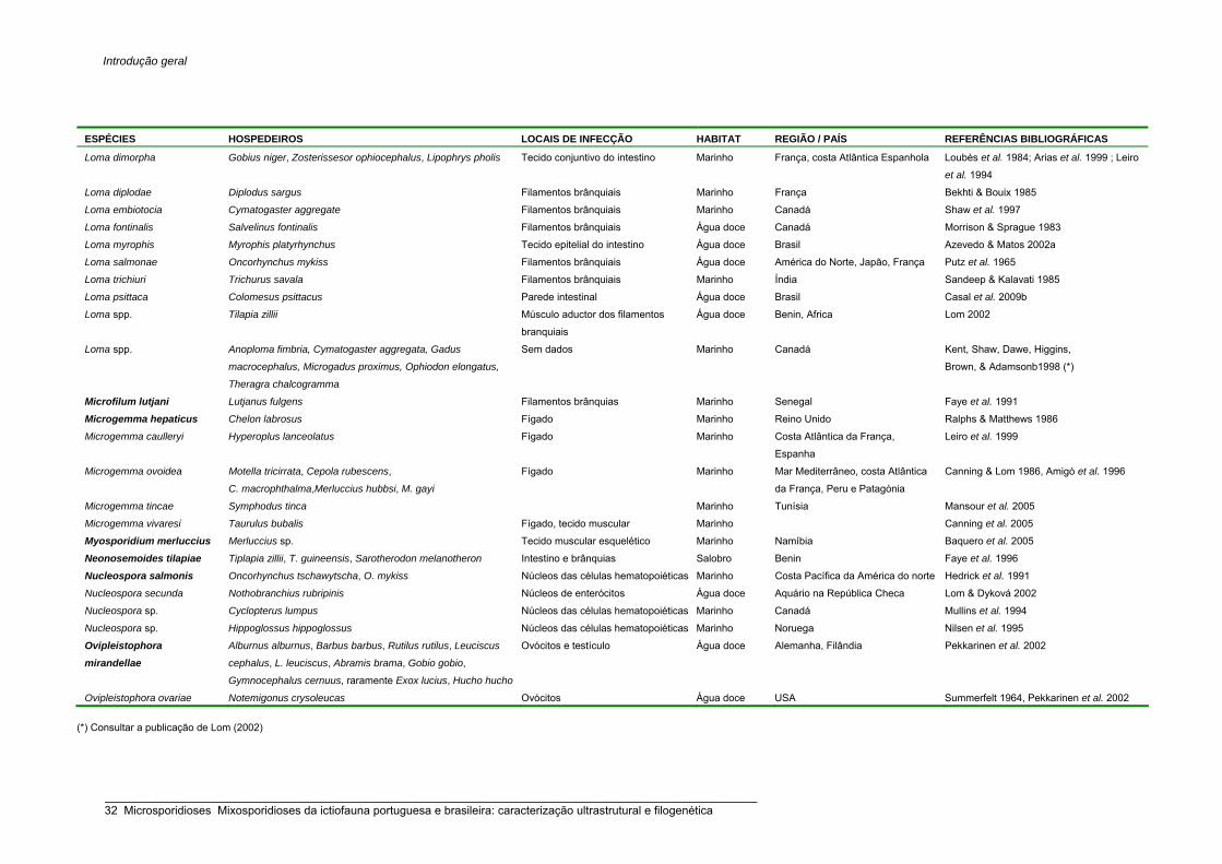
Introdução geral
ESPÉCIES HOSPEDEIROS LOCAIS DE INFECÇÃO HABITAT REGIÃO / PAÍS REFERÊNCIAS BIBLIOGRÁFICAS
Loma dimorpha Gobius niger, Zosterissesor ophiocephalus, Lipophrys pholis Tecido conjuntivo do intestino Marinho França, costa Atlântica Espanhola Loubès et al. 1984; Arias et al. 1999 ; Leiro
et al. 1994
Loma diplodae Diplodus sargus Filamentos brânquiais Marinho França Bekhti & Bouix 1985
Loma embiotocia Cymatogaster aggregate Filamentos brânquiais Marinho Canadá Shaw et al. 1997
Loma fontinalis Salvelinus fontinalis Filamentos brânquiais Água doce Canadá Morrison & Sprague 1983
Loma myrophis Myrophis platyrhynchus Tecido epitelial do intestino Água doce Brasil Azevedo & Matos 2002a
Loma salmonae Oncorhynchus mykiss Filamentos brânquiais Água doce América do Norte, Japão, França Putz et al. 1965
Loma trichiuri Trichurus savala Filamentos brânquiais Marinho Índia Sandeep & Kalavati 1985
Loma psittaca Colomesus psittacus Parede intestinal Água doce Brasil Casal et al. 2009b
Loma spp. Tilapia zillii Músculo aductor dos filamentos
branquiais
Água doce Benin, Africa Lom 2002
Loma spp. Anoploma fimbria, Cymatogaster aggregata, Gadus
macrocephalus, Microgadus proximus, Ophiodon elongatus,
Theragra chalcogramma
Sem dados Marinho Canadá Kent, Shaw, Dawe, Higgins,
Brown, & Adamsonb1998 (*)
Microfilum lutjani Lutjanus fulgens Filamentos brânquias Marinho Senegal Faye et al. 1991
Microgemma hepaticus Chelon labrosus Fígado Marinho Reino Unido Ralphs & Matthews 1986
Microgemma caulleryi Hyperoplus lanceolatus Fígado Marinho Costa Atlântica da França,
Espanha
Leiro et al. 1999
Microgemma ovoidea Motella tricirrata, Cepola rubescens,
C. macrophthalma,Merluccius hubbsi, M. gayi
Fígado Marinho Mar Mediterrâneo, costa Atlântica
da França, Peru e Patagónia
Canning & Lom 1986, Amigó et al. 1996
Microgemma tincae Symphodus tinca Marinho Tunísia Mansour et al. 2005
Microgemma vivaresi Taurulus bubalis Fígado, tecido muscular Marinho Canning et al. 2005
Myosporidium merluccius Merluccius sp. Tecido muscular esquelético Marinho Namíbia Baquero et al. 2005
Neonosemoides tilapiae Tiplapia zillii, T. guineensis, Sarotherodon melanotheron Intestino e brânquias Salobro Benin Faye et al. 1996
Nucleospora salmonis Oncorhynchus tschawytscha, O. mykiss Núcleos das células hematopoiéticas Marinho Costa Pacífica da América do norte Hedrick et al. 1991
Nucleospora secunda Nothobranchius rubripinis Núcleos de enterócitos Água doce Aquário na República Checa Lom & Dyková 2002
Nucleospora sp. Cyclopterus lumpus Núcleos das células hematopoiéticas Marinho Canadá Mullins et al. 1994
Nucleospora sp. Hippoglossus hippoglossus Núcleos das células hematopoiéticas Marinho Noruega Nilsen et al. 1995
Ovipleistophora mirandellae
Alburnus alburnus, Barbus barbus, Rutilus rutilus, Leuciscus
cephalus, L. leuciscus, Abramis brama, Gobio gobio,
Gymnocephalus cernuus, raramente Exox lucius, Hucho hucho
Ovócitos e testículo Água doce Alemanha, Filândia Pekkarinen et al. 2002
Ovipleistophora ovariae Notemigonus crysoleucas Ovócitos Água doce USA Summerfelt 1964, Pekkarinen et al. 2002
(*) Consultar a publicação de Lom (2002)
_____________________________________________________________________________________________________ 32 Microsporidioses Mixosporidioses da ictiofauna portuguesa e brasileira: caracterização ultrastrutural e filogenética

Introdução geral
ESPÉCIES HOSPEDEIROS LOCAIS DE INFECÇÃO HABITAT REGIÃO / PAÍS REFERÊNCIAS BIBLIOGRÁFICAS
Pleistophora typicalis Myoxocephalus scorpius, M. quadricornis
labradoricus e outras espécies
Tec. muscular esquelético Marinho Costa Atlântica da França, Escócia;
Mar Báltico e Branco
Canning & Nicholas 1980
Pleistophora acerinae Gymnocephalus cernuus, Perca schrenki Mesentério, Intestino Água doce França, Ucrânia, Rússia Agapova 1966 (*)
Pleistophora aegytiacus Tilapia zilli Tecido muscular do estômago Água doce Nilo, Egipto Negm-Eldin 1992 (*)
Pleistophora carangoidi Carangoides malabaricus Tec. muscular esquelético Marinho Oceano Índico Narasimhamurti & Sonabai 1977 (*)
Pleistophora dallii Dallia pectoralis Tec. conj. subcutâneo perto das barbatanas Água doce Russia Zhukov 1964 (*)
Pleistophora destruens Mugil auratus Tecido muscular Marinho Island Tatihou, Cherbourg, França Delphy 1916 (*)
Pleistophora duodecimae Coryphaenoides nasutus Tec. muscular esquelético Marinho Northern Ocenao Atlântico Gaevskaya & Dyková 1980 (*)
Pleistophora ehrenbaumi Anarhichas lúpus, A. Minor Tec. muscular esquelético Marinho Mar do Norte Reichenow 1929 (*)
Pleistophora finisterrensis Micromesistius poutassou Tecido muscular Marinho Galiza, Espanha Leiro et al. 1996
Pleistophora gadi Gadus morhua morhua Tec. muscular esquelético Marinho Mar de Barents Polyansky 1955 (*)
Pleistophora hippoglossoideos Drepanopsetta hippoglossoides, Hippoglossoides
platessoides, Solea solea
Parede da cavidade abdominal, músculo
das barbatanas
Marinho Nordeste do Mar do Norte Bosanquet 1910 (*)
Pleistophora hyphessobryconis Paracheirodon inessi, várias espécies Tec. muscular esquelético e outros órgãos _ _ Schäperclaus 1941 (*); Canning &
Lom 1986
Pleistophora ladogensis Lota lota, Osmerus eperlanus eperlanus Tec. muscular esquelético Água doce, eurihalino Lagos, S. Peterburgo, Rússia Voronin 1978 (*)
Pleistophora littoralis Blennius pholis Tec. muscular esquelético Marinho Reino Unido Canning et al. 1979
Pleistophora macrospora Noemacheilus barbatulus Tecido muscular da região abdominal Água doce França, Mar Negro Issi & Voronin 1984 (*)
Pleistophora macrozoarcidis Macrozoarces americanus Tec. muscular esquelético Marinho Atlântico Norte na região oeste Nigrelli 1946 (*)
Pleistophora oolyticus Saurida tumbil Ovários Marinho Mar Vermelho, Egipto Negm-Eldin 1992 (*)
Pleistophora priacanthicola Priacanthus tayenus, P. macrocanthus Ceg. pilóricos, pericárdio, intestino, gónadas Marinho Mar Sul da China He 1982 (*)
Pleistophora sauridae Saurida tumbil Tecido muscular liso Marinho Índia Narasimhamurti & Kalavati 1972 (*)
Pleistophora senegalensis Sparus auratus Parede intestinal Marinho Senegal Faye et al. 1990
Pleistophora siluri Silurus glanis Parede intestinal Água doce Mar Cáspio Gasimagimedov & Issi 1970 (*)
Pleistophora tahoensis Cottus beldingi Músculo esquelético abdominal Água doce Califórnia, USA Summerfelt & Ebert 1969 (*)
Pleistophora tuberifera Neogobius kessleri gorlap, N. caspius, N.
melanostomus affinis
Músculos subcutâneos Água doce Mar Cáspio Gasimagomedov & Issi 1970 (*)
Pleistophora vermiformis Cottus gobio Músculo esquelético Água doce Rio Danúbio, França e Áustria Léger 1905 (*)
Pleistophora spp. Theragra chalcogramma, Gobiodon okinawae,
Fundulus heteroclitus, Noemacheilus malapterus
longicauda, Salmo salar, Dorosoma petenense
_ _ _ Lom 2002
(*) Consultar a publicação de Lom (2002)
_____________________________________________________________________________________________________ Microsporidioses Mixosporidioses da ictiofauna portuguesa e brasileira: caracterização ultrastrutural e filogenética 33

Introdução geral
ESPÉCIES HOSPEDEIROS LOCAIS DE INFECÇÃO HABITAT REGIÃO / PAÍS REFERÊNCIAS BIBLIOGRÁFICAS
Potaspora morhaphis Potamorhaphis guianensis Cavidade celómica perto da região
anal
Água doce Pará, Brasil Casal et al. 2008b
Pseudoloma neutrophila Danio rerio Sistema nervosa central Água doce Universidade de Oregon, USA Matthews et al. 2001
Spraguea lophii Lophius piscatorius, L. budegassa,
Células ganglionares do sistema
nervoso central
Marinho Costa europeia Atlântica e
mediterrânica (Reino Unido,
Noruega e Islândia);
Weissenberg 1976, Loubès et al. 1979
Spraguea americana Lophius americanus, L. litulon Células ganglionares do sistema
nervoso central
Marinho Costa Atlântica dos USA,
Japão
Takvorian & Cali 1986,
Freeman et al. 2004
Spraguea sp.
Lophius gastrophysus Musculatura abdominal interna perto
do gânglio dorsal
Marinho Rio de Janeiro, Brasil Jakowska 1964
Tetramicra brevifilum Scophtalmus maximus,
Lophius budegassa
Tecido connectivo da musculatura
esquelética
Marinho Reino Unido, costa Atlântica
espanhola; costa mediterrânica
espanhola
Matthews & Mattews 1980, Estevez et
al. 1992
Maíllo et al. 1998
Microsporidium (grupo colectivo)
Microsporidium bengalis Nemipterus japonicus Brânquias Marinho Golfo de Bengal, Índia Weiser, Kalavati & Sandeep 1981 (*)
Microsporidium brevirostris Brachyhypopomus brevirostris Músculo da cavidade abdominal Estuarino Pará, Brasil Matos & Azevedo 2004
Microsporidium cerebralis Salmo salar Cerebro Marinho Aquacultura, Canadá Brocklebank, Speare & Kent 1995 (*)
Microsporidium chloroscombri Chloroscombrus chrysurus Fígado Marinho Senegal Toguebaye, Marchand & Faye 1989
(*)
Microsporidium cypselurus Cypselurus pinnatibarbatus Tecido muscular esquelético Marinho Japão Yokoyama et al. 2002
Microsporidium dicologoglossae Dicologoglossa cuneata Fígado Marinho Senegal Faye et al. 2004
Microsporidium girardini Girardinus caudimaculatus Pele, músculo, intestino Água doce São Paulo, Brasil Sprague 1977
Microsporidium peponoides Percottus plehni Tecido subcutâneo connectivo Água doce Russia Sprague 1977
Microsporidium pseudotumefaciens Xiphophorus maculatus, Molienesia sphenops,
Colisa lalia
Vários órgãos Água doce Aquário na Alemanha Canning & Lom 1986
Microsporidium prosopium Prosopium williamsoni Tecido muscular esquelético Água doce Canadá Kent et al. 1999
Microsporidium rhabdophilia Oncorhynchus tschawyscha, O. mykiss Núcleo de células rodlet da pele,
brânquias, intestino
Água doce Califórnia, USA Modin 1981
Microsporidium sauridae Saurida tumbil Musculatura visceral Marinho Índia Narasinhamurti & Kalavati 1972 (*)
Microsporidium sciaenae Sciaena australis Tec. conjuntivo à volta do ovário Água doce Australia Canning & Lom 1986
Microsporidium sulci Acipenser ruthenus, A. guldenstadti Ovócitos Água doce Rio Danúbio, Volga, Kura Sprague 1977
Microsporidium synapturae Synaptura cadenati Fígado Marinho Senegal Faye et al. 2004
Microsporidium valamugili Valamugil sp. Parede intestinal Estuarino Índia Canning & Lom 1986
Microsporidium vantraeleniae Vanstraelenia chirophthalmus Fígado Marinho Senegal Faye et al. 2004
_____________________________________________________________________________________________________ 34 Microsporidioses Mixosporidioses da ictiofauna portuguesa e brasileira: caracterização ultrastrutural e filogenética

Introdução geral
Microsporidium zhanjiangensis Priacanthus tayenus, P. macracanthus Parede intestinal e baço Marinho China Hua & Zhang 1988 (*)
Pagrus major e Sparus aurata Marinho Japão; Malta Bell et al. 2001
Branchiostegus semifaciatus, Caranx crysos,
C. senegallus, Selene dorsalis, Trachurus
trachurus, Erythrocles monodi, Eucinostomus
melanopterus, Galeoides decadactylus,
Umbrina canariensis, Dicologoglossa cuneata,
Synaptura cadenati, S. lusitanica, Dentex
canarensis, D. marocanus, Sparus
caeruleosticus, S. pagrus pagrus
Fígado Marinho Senegal Lom 2002 (Doutoral Thesis Faye,
1992)
Chilomycterus reticulatus Intestino Marinho Doutoral Thesis Faye, 1992 (*)
Trichiurus lepturus Ovário Marinho Doutoral Thesis Faye, 1992 (*)
Vimba vimba Intestino e rim Eurihalino Mar Cáspio Lom 2002
Ictalurus punctarus, Lycodopsis pacifica Músculo cardíaco e intestino Água doce USA Lom 2002
Trachurus declivis Cavidade pericardial e nervos Marinho Nova Zelândia Lom 2002
Mallotus villosus Epitélio peritoneal, ovários Marinho Newfoundland Lom 2002
Salmo trutta Ovário Eurihalino Noruega Lom 2002
Microsporidium spp.
(30 espécies)
Pleuronectes flesus Pele Marinho Polónia Lom 2002
(*) Consultar a publicação de Lom (2002)
_____________________________________________________________________________________________________ Microsporidioses Mixosporidioses da ictiofauna portuguesa e brasileira: caracterização ultrastrutural e filogenética 35

Introdução geral
Grupo colectivo Microsporidium Balbiani, 1884 As espécies que não foram detalhadamente identificadas, devido à escassez de
informação disponível relativamente às fases da merogonia e esporogonia são,
temporariamente, incluídas no género Microsporidium. Existem, pelo menos, 18 espécies
descritas em peixes, algumas de significante interesse comercial (Tabela 2).
Microsporídios de classificação taxonómica duvidosa
Algumas referências de microsporídios em peixes necessitam de ser devidamente
caracterizadas, nomeadamente através da análise molecular, dado terem sido
classificadas em géneros típicos de platelmintas, crustáceos e insectos, entre os quais
espécies do género Jirovecia (Sprague 1977), Nosemoides (Faye et al. 1994) e
Thelohania (Voronin 1974), respectivamente.
Casos de hiperparasitismo foram descritos em mixosporídios: Nosema notabilis foi
observada em plasmódios de Ortholinea polymorpha, a parasitar a bexiga urinária de um
peixe-sapo da costa Atlântica dos EUA (Lom & Dyková 1992a); N. ceratomyxae ocorre
em plasmódios de Ceratomyxa sp. na bexiga natatória de um peixe-coelho do Mar
Vermelho (Diamant & Paperna 1985, 1989); microsporídios por identificar foram
igualmente observados nos trofozóitos dos mixosporídios Leptotheca fugu e Myxidium
fugu, por sua vez localizados no epitélio intestinal de Takifugu rubripes, peixe marinho
das costas japonesas (Tun et al. 2000).
1.2.6. Patologia: interacção hospedeiro-parasita
Os microsporídios desenvolvem-se directamente no citoplasma, eventualmente no
nucleoplasma, das células hospedeiras, induzindo-lhes uma generalizada destruição
celular ou uma hipertrofia celular, culminando com a formação de xenomas. A
proliferação dos estádios merogoniais e esporogoniais ocasiona progressivas
degradações do citoplasma e dos organelos da célula hospedeira, acabando por a
destruir praticamente na sua totalidade, sendo o espaço preenchido por esporos
maduros. As mitocôndrias são, provavelmente, a única excepção, uma vez que
permanecem inalteradas e em grandes concentrações à volta dos parasitas. Dada a
ausência de mitocôndrias em qualquer estádio de desenvolvimento do parasita, sabe-se
que, energeticamente, este depende totalmente das mitocôndrias circundantes (Dyková &
Lom 1980, 2000, Lom & Dyková 2005, Lom & Nilsen 2003).
_____________________________________________________________________________________________________ 36 Microsporidioses Mixosporidioses da ictiofauna portuguesa e brasileira: caracterização ultrastrutural e filogenética

Introdução geral
Desenvolvimento sem formação de xenoma
Em alguns géneros (Heterosporis, Kabatana, Nucleospora, Ovipleistophora e
Pleistophora), os estádios de desenvolvimento desenrolam-se no citoplasma,
eventualmente no núcleo da célula hospedeira, sem que haja a diferenciação de uma
estrutura protectora para ambos, isto é parasita-hospedeiro, designada de xenoma. O
género Kabatana parasita preferencialmente o tecido muscular esquelético, no qual os
estádios proliferativos formam gradualmente focos de infecção dentro das fibras
musculares, induzindo uma generalizada desorganização do sarcoplasma (Dyková &
Lom 2000). Em Pleistophora typicalis, os estádios de desenvolvimento estão separados
das miofibrilas intactas, apenas por uma camada amorfa com cerca de 0,2 a 0,6 μm de
espessura. Também se pode observar à volta de Pleistophora hyphessobryconis uma
auréola que, ultrastruturalmente, traduz uma acção lítica local, resultante da
desorganização do sarcoplasma, pelo que essa zona se apresenta desprovida de
miofibrilas (Dyková & Lom 1980, 2000). A diferenciação de invólucros densos
(esporoforocistos) de origem parasítica dentro do sarcoplasma, contendo exclusivamente
estádios do parasita sem citoplasma e núcleo da célula hospedeira, verifica-se em
espécies de Heterosporis (Schubert 1969, Lom et al. 1989a, Michel et al. 1989).
Desenvolvimento com formação de xenoma
A célula hospedeira e o parasita encontram-se fisiologica e morfologicamente integrados,
formando uma entidade distinta do hospedeiro, com um desenvolvimento próprio e
capacidade de crescimento. Ambos parecem beneficiar da formação de xenomas: ao
parasita oferece um meio susceptível para poder proliferar e, simultaneamente, protege-o
contra os ataques do hospedeiro, uma vez que se encontra camuflado por estruturas
celulares da célula hospedeira: em contrapartida, esta última também fica beneficiada na
medida que restringe a propagação do parasita às células vizinhas. Externamente, pode
diferenciar-se uma parede refráctil e, perifericamente a esta, pode haver a deposição de
camadas de tecido conjuntivo, resultantes da resposta do hospedeiro. Internamente, a
célula hospedeira possui um núcleo hipertrófico, muitas vezes ramificado ou
fragmentado, por um processo amitótico, em numerosos pequenos núcleos. O citoplasma
torna-se hipertrófico à medida que o número de esporos aumenta, podendo a célula
hospedeira atingir dimensões superiores a 1 mm. O plasmalema pode apresentar
modificações que se traduzem por um aumento da área de absorção (Weissenberg 1968,
Lom & Dyková 1992a, Lom 2008).
_____________________________________________________________________________________________________ Microsporidioses Mixosporidioses da ictiofauna portuguesa e brasileira: caracterização ultrastrutural e filogenética 37

Introdução geral
Segundo Lom e Dyková (2005) os xenomas podem ser classificados em:
a) Xenomas delimitados por uma única membrana, em que o volume total da célula
hospedeira não é transformado num xenoma. No género Spraguea formam-se xenomas
de grandes dimensões, com semelhanças a “cachos de uvas”, que infectam
principalmente grandes gânglios extracraniais do cérebro, nervos espinais, bem como
qualquer outra célula ganglionar (Freeman et al. 2004).
b) Xenomas delimitados por uma única membrana, em que o volume total da célula
hospedeira é transformado num xenoma. Nos géneros Ichthyosporidium (Sprague &
Vernick 1974 Casal & Azevedo 1995), Microgemma (Ralphs & Matthews 1986, Amigó et
al. 1996, Leiro et al. 1999), Microfilum (Faye et al. 1991), Potaspora (Casal et al. 2008b) e
Tetramicra (Matthews & Matthews 1980), o plasmalema expande-se, formando estruturas
semelhantes a microvilosidades e, externamente, não se diferencia qualquer
revestimento estratificado ou hialinalizado.
c) Xenomas em que o plasmalema é recoberto por fibras de colagénio. No género
Amazonspora dispõem-se 22 camadas de colagénio justapostas e orientadas longitudinal
e transversalmente (Azevedo & Matos 2003a). Plasmalema recoberto por fina camada de
glicocálice, seguida de fibras de colagénio, foi descrito no género Neonosemoides (Faye
et al. 1996).
d) Xenomas de parede espessa. Formação típica nos géneros Glugea, Loma e
Pseudoloma. Parede estratificada formada por camadas laminares, podendo diferenciar
60 camadas em alguns casos, e aderente ao revestimento do plasmalema, observa-se
em espécies do género Glugea. Internamente, o núcleo ocupa uma posição central, é
hipertrófico e ramificado (Canning et al. 1982, Vagelli et al. 2005). No género Loma a
parede é estreita, formada por uma substância fibrosa ou granular. Externamente, é
notória a presença de várias camadas de fibroblastos (Morrison & Sprague 1981a, 1983,
Azevedo & Matos 2002a). Presentemente não existem dados referentes à caracterização
do xenoma no género Pseudoloma.
1.2.7. Estudos moleculares e filogenéticos
Análises moleculares revelaram que os microsporídios são organismos eucariotas com
um genoma muito reduzido com a amplitude de 2,3 Mb para a espécie Encephalitozoon
intestinalis e 19,5 Mb em Glugea atherinae, chegando a ser inferior ao da bactéria E. coli
(4,6 Mb) (Keeling & Slamovits 2004). A redução genómica terá implicado o desenvolvido
de estratégias de compactação da informação genética ou, então, os microsporidios, no
decurso da evolução, perderam informação genética correspondente a vias metabólicas
_____________________________________________________________________________________________________ 38 Microsporidioses Mixosporidioses da ictiofauna portuguesa e brasileira: caracterização ultrastrutural e filogenética

Introdução geral
dependo assim dos recursos dos seus hospedeiros (Keeling et al. 2005). Sabe-se
também que o genoma está organizado sob a forma de múltiplos cromossomas lineares
(8 a 18) com as extremidades organizadas em telómeros (Weiss & Vossbrinck 1999,
Keeling & Fast 2002). Em 2001, Katinka e colaboradores, com a sequenciação na íntegra
do genoma de E. cuniculi, observaram a existência de somente 1997 genes codificantes
para proteínas. Entre elas, as envolvidas nos processos de replicação de DNA, formação
dos ribossomas e via da glicólise, bem como proteínas mitocondriais. Por outro lado,
verificaram a inexistência de genes correspondentes à biossíntese de vários
componentes, tais como nucleótidos, ácidos gordos e alguns aminoácidos.
De todas as moléculas, o rDNA é a molécula “alvo” em muitos dos estudos filogenéticos
devido à presença de regiões hiper-conservadas na sequência, tornando susceptível o
seu uso para efeito de comparação entre organismos distantes (Weiss & Vossbrinck
1999). Além disso, a estrutura secundária do rRNA torna possível a realização de
alinhamentos baseados na sua estrutura e, assim, assegurar a comparação de
caracteres homólogos nas análises filogenéticas (De Rijk & Wachter 1997, Lom & Nilsen
2003). Há muito que se sabe que ribossomas dos microsporídios assemelham-se aos
dos procariotas (Ishihara & Hayashi 1968), no entanto, apresenta também algumas
particularidades dos eucariotas (Vossbrinck et al. 1987). A SSU possui uma molécula de
rRNA de 16S, enquanto que a LSU uma molécula de rRNA de 23S. A sequência do rRNA
de 5,8S individualizada, tal como se encontra nos eucariotas, não existe nos
microsporídios, mas existem algumas sequências homólogas da região 5,8S contidas no
início da subunidade 23S, em resultado da ausência, entre ambas, da região espaço
transcricional interno 2 (ITS2) (Vossbrinck & Woese 1986, Vossbrinck et al. 1987,
Cavalier-Smith 1993). Esta invulgar característica, igualmente reportada em bactérias,
nunca foi descrita noutros grupos de eucariotas.
Apesar da sequenciação do gene para o SSU rRNA ser largamente utilizada como
marcador molecular para muitas espécies, recentemente tem sido sugerida a
sequenciação preferencial do espaço ITS e do gene para o LSU rRNA para efeitos de
comparações filogenéticas entre espécies muito afins (Tsai et al. 2005), nomeadamente
entre as espécies que ocorrem na ictiofauna (Cheney et al. 2000). Presentemente, é
conhecida a região ITS e o LSU rDNA (parcialmente) para algumas espécies. Dos
microsporídios presentes na ictiofauna, somente para a espécie Heterosporis
anguillarum, o gene LSU rDNA foi sequenciado na sua totalidade (Tsai et al. 2002). A
estrutura da unidade do rDNA na íntegra é conhecida unicamente para a espécie
Encephalitozoon cuniculi, parasita que ocorre em várias espécies de mamíferos, incluindo
os humanos (Peyretaillade et al. 1998).
_____________________________________________________________________________________________________ Microsporidioses Mixosporidioses da ictiofauna portuguesa e brasileira: caracterização ultrastrutural e filogenética 39

Introdução geral
Infelizmente, os caracteres que são, geralmente, usados na classificação dos
microsporídios para diferenciar os níveis superiores, isto é, o número de núcleos/célula, a
presença de uma membrana a rodear o parasita (vesícula esporófora ou vacúolo
parasitóforo) e o tipo de divisão nuclear, são incoerentes ao nível dos taxa género e
espécie (Vossbrinck & Debrunner-Vossbrinck 2005). Sabe-se que a sequenciação
unicamente do gene para o SSU rRNA contribuiu para a posição basal dos
microsporídios nos cladogramas dos eucariotas, posicionamento erróneo até finais do
século XX, devido ao artefacto gerado pela atracção dos ramos longos (Van der Peer et
al. 2000, Fast et al. 2003). Contudo, a sequenciação do mesmo tem provado ser bastante
útil no estabelecimento das relações filogenéticas dentro do grupo dos microsporídios,
nomeadamente, para os taxa superiores ao género (Lom & Nilsen 2003). Idealmente,
quando se pretende inferir em termos filogenéticos, devem ser usadas várias diferentes
moléculas. Contudo, a análise dos resultados obtidos por sequenciação dos genes que
codificam as proteínas tem-se revelado ser mais difícil devido à presença de codões
degenerados para muitos aminoácidos, bem como devido da existência de várias formas
de genes codificantes de proteínas estarem presentes no genoma (parálogos) fazendo
com que seja mais difícil a identificação de homólogos (Lom & Nilsen 2003).
Pela análise filogenética efectuada por Vossbrinck e Debrunner-Vossbrinck (2005) num
total de 125 sequências do gene para o SSU rRNA, disponíveis em várias bases de
dados (p. e. GenBank), verificou-se que os microsporídios agrupam em 5 clados
principais correspondentes a 3 classes: classe Aquasporidia é um grupo parafilético
constituído por 3 clados, maioritariamente formado por microsporídios, que têm como
hospedeiro animais de água doce; classe Marinosporidia correspondente aos
microsporídios que ocorrem em animais aquáticos marinhos, salvo algumas excepções,
tais como parasitas que têm como hospedeiro peixes de água doce, bem como
microsporídios pertencentes ao género Dictyocoela, que ocorrem em anfípodes de água
doce. Adaptação do hospedeiro a um novo habitat, aparentemente, é a explicação mais
provável. O mesmo poderá ter acontecido a 2 microsporídios do género Vavraia e à
espécie Trachipleistophora hominis, respectivamente parasitas de insectos e de
humanos. Por sua vez, a classe Terresporidia engloba microsporídios que têm como
hospedeiro, maioritariamente, insectos, répteis, aves e mamíferos, inclusivamente,
humanos. O microsporídio Vittaforma cornae é uma das excepções, levando a crer tratar-
se de um falso microsporídio de humanos, uma vez que parasita pacientes
imunodeficientes. Segundo Lom e Nilsen (2003), as análises filogenéticas para os
microsporídios que ocorrem na ictiofauna, sugere a formação de 5 grupos. Excepto o
grupo 5, os restantes encontram-se englobados na classe Marinosporidia, recentemente
_____________________________________________________________________________________________________ 40 Microsporidioses Mixosporidioses da ictiofauna portuguesa e brasileira: caracterização ultrastrutural e filogenética

Introdução geral
designada por Vossbrinck e Debrunner-Vossbrinck (2005).
Classificação segundo Lom & Nilsen 2003
Grupo 1 (Grupo correspondente a nenhum taxon): Loma, Ichthyosporidium e Pseudoloma.
Grupo 2
(Família Glugeidae Thélohan, 1892): Glugea.
Grupo 3 (Fam. Pleistophoridae Doflein, 1901): Pleistotphora, Heterosporis e Ovipleistophora.
Grupo 4
(Família Spragueidae Weissenberg, 1976): Spraguea; (Família Tetramicridae Matthews
& Matthews, 1980) Microgemma, Tetramicra e Potaspora. Inclui também o género
Kabatana.
Grupo 5
(Família Enterocytozoonidae Cali & Owen, 1990): Nucleospora.
A análise filogenética do gene SSU rRNA do microsporídio Myosporidium não permitiu
agrupá-lo em nenhum dos grupos (Baquero et al. 2005). Para os géneros com uma única
espécie, Amazonspora (Azevedo & Matos 2003a), Microfilum (Faye et al. 1991) e
Neonosemoides (Faye et al. 1996), não existe informação molecular disponível.
Grupo 1
Existe informação molecular referente ao gene SSU rRNA para 4 das 12 espécies
pertencentes ao género Loma, tratando-se de um grupo parafilético em que a espécie
tipo, Loma branchialis, não está sequenciada. Para além de Loma spp., este grupo
engloba sequências referentes a parasitas dos géneros Pseudoloma (Matthews et al.
2001) e Ichthyosporidium (Sprague & Vernick 1974). Os caracteres morfológicos do ciclo
de vida do género Ichthyosporidium diferem em muito dos restantes do grupo, entre eles
inclui-se núcleo em diplocário e todo o desenvolvimento em directo contacto com
citoplasma da célula hospedeira. Possivelmente, a sequência (GenBank L39110)
referente ao Ichthyosporidium sp., corresponderá a um parasita erroneamente
classificado.
Grupo 2
Grupo monofilético com um alto “bootstrap”, que reúne todas as espécies do género
Glugea. Para além das espécies que ocorrem em peixes, o microsporídio marinho
_____________________________________________________________________________________________________ Microsporidioses Mixosporidioses da ictiofauna portuguesa e brasileira: caracterização ultrastrutural e filogenética 41

Introdução geral
Tuzetia weidneri (= Pleistophora), descrito num crustáceo, agrupa com a espécie tipo G.
anomala. Em todos os cladogramas elaborados, a espécie descrita como Pleistophora
finisterrensis, parasita do verdinho Micromesistius poutassou, não agrupa com as
restantes do género, mas sim com G. anomala, situação que, indiscutivelmente, carece
de uma reclassificação.
Grupo 3
Este grupo apresenta uma heterogeneidade, na medida em que engloba representantes
que parasitam diferentes hospedeiros. O bootstrap para as sequências de Heterosporis
spp. e Ovipleistophora spp. é elevado, constituindo um grupo formado, exclusivamente,
por microsporídios em que o hospedeiro é de água doce. Apesar do género Pleistophora
ocorrer, simultaneamente, em espécies marinhas e de água doce, não é de estranhar
bootstrap superiores a 98% em vários cladogramas, uma vez que todas as sequências
disponíveis dizem respeito a microsporídios marinhos. Este grupo engloba também
espécies dos géneros Dictyocoela e Vavraia, bem como a espécie Trachipleistophora
hominis.
Grupo 4
Grupo composto, exclusivamente, por microsporídios que parasitam peixes, que alberga
5 géneros estritamente relacionados, tendo em conta os carateres morfológicos. Entre
eles incluem-se a ausência da diferenciação de uma vesícula esporófora/ vacúolo
parasitóforo, bem como núcleo unicário (excepto uma das fases do microsporídio
dimórfico Spraguea spp.) durante todo o ciclo de vida. Os géneros Tetramicra e
Microgemma, aparentemente, constituem um grupo monofilético em todos os
cladogramas. O mesmo não sucede ao género Kabatana, que se caracteriza por
parasitar, exclusivamente, o tecido muscular esquelético de peixes de água doce e de
água salgada, motivo pelo qual, muito possivelmente, explica a parafilia do género.
Grupo 5
Nos cladogramas elaborados por Vossbrinck e Debrunner-Vossbrinck (2005), este grupo
encontra-se excluído da classe Marinosporidia, onde se encontram agrupados todos os
microsporídios que parasitam a ictiofauna, para se posicionar na classe Terresporidia,
formando um clado com o microsporídio Enterocytozoon bieneusi, parasita de mamíferos,
incluindo humanos. Não é de estranhar, que em todas as análises filogenéticas, o género
Nucleospora se posiciona agrupado com as sequências seleccionadas como “outgroup”
(Docker et al. 1997, Lom & Nilsen 2003), visto que possui a invulgar característica de
parasitar o nucleoplasma, em vez do citoplasma, como sucede com os outros
microsporídios (Lom & Dyková 2002).
_____________________________________________________________________________________________________ 42 Microsporidioses Mixosporidioses da ictiofauna portuguesa e brasileira: caracterização ultrastrutural e filogenética
Introdução geral
1.3. Mixosporidioses
As mixosporidioses são doenças causadas pela acção de parasitas do grupo dos
mixosporídios (= mixozoários, como também são designados). Estes parasitas são
metazoários, pertencentes taxonomicamente ao filo Myxozoa Grassé, 1970, e
representam um grupo de grande importância económica, devido aos seus efeitos
nefastos na aquacultura e na natureza. São parasitas, quase exclusivamente, de
vertebrados poiquilotérmicos, principalmente peixes de água doce e salgada, bem como
de alguns invertebrados. Presentemente, conhecem-se mais de 2200 espécies,
agrupadas em 16 famílias e 65 géneros, a parasitar vertebrados (Lom & Dyková 2006,
Køie et al. 2007b, Prunescu et al. 2007). Apesar de se saber que têm um ciclo de vida
indirecto, envolvendo um invertebrado como hospedeiro definitivo e um peixe como
hospedeiro intermediário, estes parasitas são conhecidos, principalmente, por
provocarem infecções em peixes, visto que somente 180 tipos (17 grupos) de
actinosporos foram descritos em invertebrados (Lom & Dyková 2006). Algumas
ocorrências em platelmintas, anfíbios, répteis, aves e mamíferos foram também
documentadas (Friedrich et al. 2000, Kent et al. 2001, Duncan et al. 2004, Eiras 2005,
Garner et al. 2005, Jirkù et al. 2006, Prunescu et al. 2007, Bartholomew et al. 2008). Em
humanos verificaram-se esporádicas ocorrências, que indicam tratar-se de presenças
acidentais. Em alguns trabalhos foram descritos esporos encontrados nas fezes de
pacientes que, muito provavelmente, serão provenientes da ingestão de peixes
infectados (McClelland et al. 1997, Boreham et al. 1998, Moncada et al. 2001). O estudo
dos mixosporídios tem sido desenvolvido nas mais variadas vertentes de investigação,
nomeadamente nos aspectos morfológicos do seu ciclo de vida, processo de
transmissão, taxonomia e identificação filogenética.
1.3.1. Posição taxonómica
Os esporos de mixosporídios foram, pela primeira vez, identificados por Jurine (1825),
tendo sido classificados, mais tarde, por Otto Bütschii (1882) e incluídos na subclasse
Myxosporida, pertencente à classe designada de Sporozoa (consultar a publicação, Lom
& Dyková 2006). Desde então, os mixosporídios foram considerados protistas, em parte
devido ao tamanho e à forma dos esporos, apesar de na época lhes ter sido reconhecido
o parentesco com os organismos do filo Cnidaria. Após alguma hesitação inicial, os
mixosporídios acabaram por ser reconhecidos como organismos metazoários.
Presentemente, constituem uma superclasse do infrafilo Metazoa dentro do filo
Opisthoconta (Cavalier-Smith 1998, Hausmann et al. 2003), em que a morfologia e
_____________________________________________________________________________________________________ Microsporidioses Mixosporidioses da ictiofauna portuguesa e brasileira: caracterização ultrastrutural e filogenética 43
Introdução geral
ultrastrutura está bem documentada através das inúmeras publicações (Azevedo et al.
1989, 2005, Current et al. 1979, Desser et al. 1983, Lom & Puytorac 1965a, 1965b, Lom
1969, Lom & Dyková 1992b, Sitjà-Bobadilla & Álvarez-Pellitero 1992, 1993a, 1993b,
1995, 2001), Por outro lado, vários estudos de biologia molecular sugerem semelhanças
filogenéticas com os metazoários (Siddall et al. 1995), nomeadamente com os que
apresentam simetria bilateral (Smothers et al. 1994, Anderson et al. 1998, Okamura et al.
2002, Siddall & Whiting 1999, Zrzavý & Hypša 2003).
1.3.2. Classificação Taxonómica
Phylum Myxozoa Grassé, 1970
Tipicamente, são organismos formados por células eucarióticas sem centríolos e sem
flagelos, no entanto, observam-se, frequentemente, muitos microtúbulos intimamente
envolvidos na divisão nuclear e na diferenciação celular, mitocôndrias com cristas de
forma tubular, discóide ou então achadas, bem como junções celulares de aderência
semelhantes a desmossomas e junções comunicantes. Parasitam alternadamente seres
invertebrados e vertebrados, caracterizando-se por diferenciarem esporos de forma e
estrutura variada. Os esporos são constituídos por uma ou mais valvas, podendo
eventualmente possuir projecções simples ou elaboradas. Internamente, localizam-se
uma ou várias células amibóides germinais infectivas (esporoplasmas) e uma ou várias
cápsulas polares (semelhantes aos nematocistos dos cnidários), cada uma apetrechada
com um filamento polar extrusível. Este filo divide-se em 2 classes: Malacosporea
(esporos de valvas sem rigidez que infectam os briozoários e os peixes) e Myxosporea
(esporos de valvas rígidas que ocorrem em anelados e peixes). Esta última classe é
composta por duas ordens: Bivalvulida (esporos com 2 valvas e geralmente com 2
cápsulas polares) e Multivalvulida (esporos formados por mais de 2 valvas e por mais de
2 cápsulas polares (Kent et al. 2001, Lom & Dyková 1992b, 2006).
Classe Malacosporea Canning, Curry, Feist, Longshaw & Okamura, 2000
Os malacosporos são parasitas de briozoários de água doce (filo Bryozoa, classe
Phylactolaemata). Os estádios vegetativos desenvolvem-se dentro da cavidade do corpo
dos briozoários em forma de sacos multicelulares fechados ou, então, em organismos em
forma de verme (Canning et al. 1996, 2000, 2002, Okamura et al. 2002, Morris & Adams
2008).
_____________________________________________________________________________________________________ 44 Microsporidioses Mixosporidioses da ictiofauna portuguesa e brasileira: caracterização ultrastrutural e filogenética

Introdução geral
Filo Myxozoa
Classe Myxosporea Ordem Bivalvulida
Subordem Sphaeromyxina Família: Sphaeromyxidae - Sphaeromyxa
Subordem Variisporina Família: Myxidiidae - Myxidium, Enteromyxum, Zschokkella, Coccomyxa, Soricimyxum Família: Ortholineidae - Ortholinea, Neomyxobolus, Cardimyxobolus, Triangula, Triangulamyxa, Kentmoseria Família: Sinuolineidae - Sinuolinea, Myxodavisia, Myxoproteus, Bipteria, Paramyxoproteus, Neobipteria, Schulmania, Noblea Família: Fabesporidae - Fabespora Família: Ceratomyxidae - Ceratomyxa, Leptotheca, Meglitschia, Ellipsomyxa Família: Sphaerosporidae - Sphaerospora, Polysporoplasma, Hoferellus, Wardia, Palliatus, Myxobilatus Família: Chloromyxidae - Chloromyxum, Caudomyxum, Agarella Família: Auerbachiidae - Auerbachia, Globospora Família: Alatosporidae - Alatospora, Pseudoalatospora, Renispora Família: Parvicapsulidae - Parvicapsula, Neoparvicapsula, Gadimyxa
Subordem Platysporina Família: Myxobolidae – Myxobolus, Spirosuturia, Unicauda, Dicauda, Phlogospora, Laterocaudata, Henneguya,
Hennegoides, Tetrauronema, Thelohanellus, Neothelohanellus, Neohenneguya, Trigonosporus Ordem Multivalvulida Família: Trilosporidae – Trilospora, Unicapsula
Família: Kudoidae - Kudoa Família: Spinavaculidae - Octospina
Trilosporoides (incertae sedis) Classe Malacosporea
Ordem Malacovalvulida Família: Saccosporidae – Buddenbrockia (sinónimo:Tetracapsula), Tetracapsuloides
Classificação taxonómica do filo Myxozoa: Adaptado da última revisão efectuada por Lom e Dyková (2006) com a reestruturação e introdução de novos géneros. Kudoa (Whipps et al. 2004), Gadimyxa n. gen. (Køie et al. 2007b), Soricimyxum n. gen. (Prunescu et al. 2007) e Myxodavisia (= Davisia) (Zhao et al. 2008b).
_____________________________________________________________________________________________________ Microsporidioses Mixosporidioses da ictiofauna portuguesa e brasileira: caracterização ultrastrutural e filogenética 45
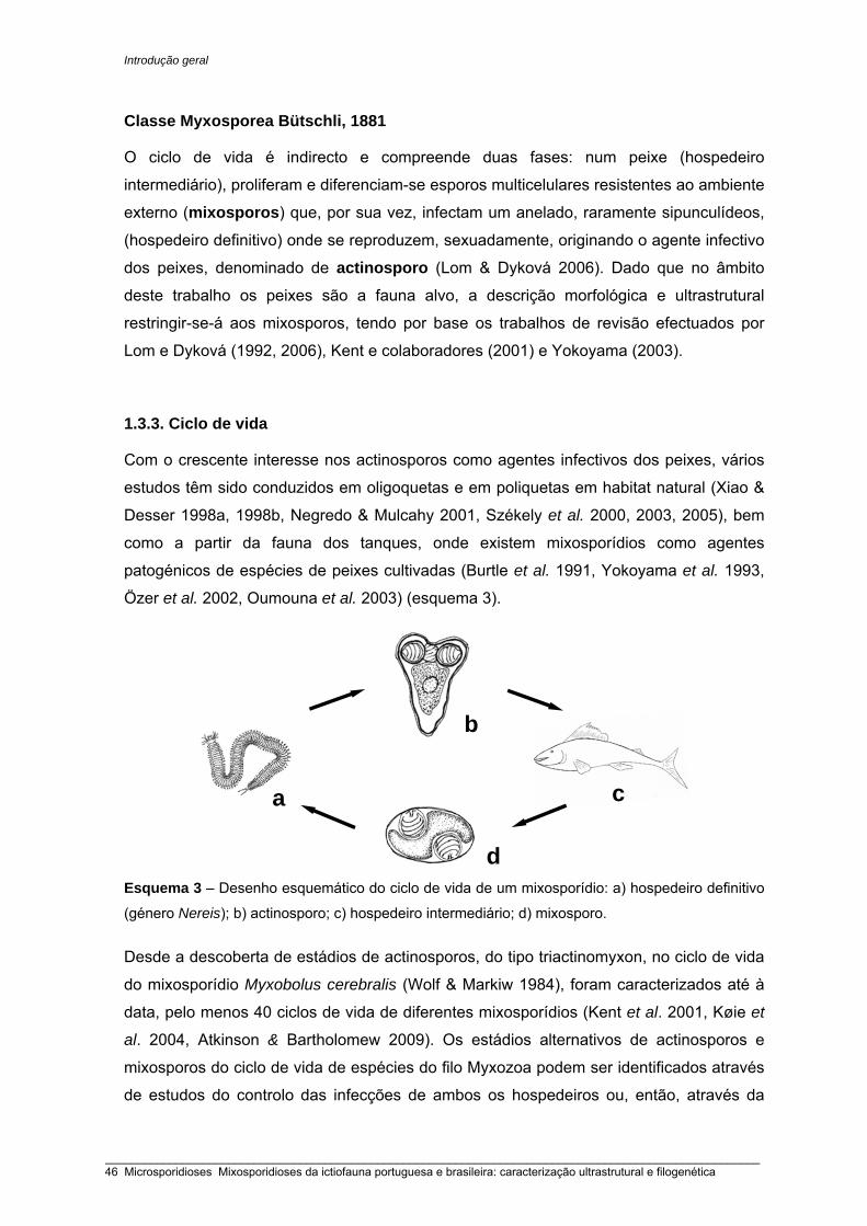
Introdução geral
Classe Myxosporea Bütschli, 1881
O ciclo de vida é indirecto e compreende duas fases: num peixe (hospedeiro
intermediário), proliferam e diferenciam-se esporos multicelulares resistentes ao ambiente
externo (mixosporos) que, por sua vez, infectam um anelado, raramente sipunculídeos,
(hospedeiro definitivo) onde se reproduzem, sexuadamente, originando o agente infectivo
dos peixes, denominado de actinosporo (Lom & Dyková 2006). Dado que no âmbito
deste trabalho os peixes são a fauna alvo, a descrição morfológica e ultrastrutural
restringir-se-á aos mixosporos, tendo por base os trabalhos de revisão efectuados por
Lom e Dyková (1992, 2006), Kent e colaboradores (2001) e Yokoyama (2003).
1.3.3. Ciclo de vida
Com o crescente interesse nos actinosporos como agentes infectivos dos peixes, vários
estudos têm sido conduzidos em oligoquetas e em poliquetas em habitat natural (Xiao &
Desser 1998a, 1998b, Negredo & Mulcahy 2001, Székely et al. 2000, 2003, 2005), bem
como a partir da fauna dos tanques, onde existem mixosporídios como agentes
patogénicos de espécies de peixes cultivadas (Burtle et al. 1991, Yokoyama et al. 1993,
Özer et al. 2002, Oumouna et al. 2003) (esquema 3).
Esquema 3 – Desenho esquemático do ciclo de vida de um mixosporídio: a) hospedeiro definitivo
(género Nereis); b) actinosporo; c) hospedeiro intermediário; d) mixosporo.
Desde a descoberta de estádios de actinosporos, do tipo triactinomyxon, no ciclo de vida
do mixosporídio Myxobolus cerebralis (Wolf & Markiw 1984), foram caracterizados até à
data, pelo menos 40 ciclos de vida de diferentes mixosporídios (Kent et al. 2001, Køie et
al. 2004, Atkinson & Bartholomew 2009). Os estádios alternativos de actinosporos e
mixosporos do ciclo de vida de espécies do filo Myxozoa podem ser identificados através
de estudos do controlo das infecções de ambos os hospedeiros ou, então, através da
d
a
b
c
_____________________________________________________________________________________________________ 46 Microsporidioses Mixosporidioses da ictiofauna portuguesa e brasileira: caracterização ultrastrutural e filogenética

Introdução geral
análise das sequências de DNA obtidas de ambos as fases. Presentemente, já se
conhecem as sequências para alguns actinosporídios (El-Mansy et al. 1998, Hallett et al.
1999, Negredo et al. 2003, Holzer et al. 2004). A utilização do gene de SSU rRNA tem
permitido, com sucesso, a confirmação de diferentes estádios do ciclo de vida dos
mixosporídios, pertencentes a diferentes géneros, tais como Myxobolus cerebralis
(Andree et al. 1997), Ceratomyxa shasta (Bartholomew et al. 1997), Tetracapsuloides
bryosalmonae (Anderson et al. 1999), Henneguya ictaluri (Lin et al. 1999), Thelohanellus
hovorkai (Anderson et al. 2000), Ellipsomyxa gobii (Køie et al. 2004), Chloromyxum
auratum (Atkinson et al. 2007), Gadimyxa atlantica (Køie et al. 2007b), Ceratomyxa
auerbachi (Køie et al. 2008) e Myxobilatus gasterostei (Atkinson & Bartholomew 2009).
1.3.4. Fases de desenvolvimento na ictiofauna
Mixosporos
Os mixosporos, muitas vezes, são diagnosticados através da detecção macroscópica ou
microscópica de pequenos plasmódios alojados nos tecidos. Em alguns casos,
encontram-se localizados livremente dentro da cavidade de órgãos. O corpo dos
mixosporos maduros pode apresentar variadas formas, tais como: ovóide, elipsóide
piriforme, fusiforme, encurvada, arredondada, quadrangular, triangular atingindo,
habitualmente, entre 10 a 20 µm de comprimento ou de espessura variável de acordo
com o género (esquema 4).
Esquema 4 - Desenhos esquemáticos de cinco mixosporos correspondentes a diferentes géneros: a) Henneguya; b) Myxobolus; c) Ceratomyxa; d) Kudoa; e) Chloromyxum.
a
b
cd e
_____________________________________________________________________________________________________ Microsporidioses Mixosporidioses da ictiofauna portuguesa e brasileira: caracterização ultrastrutural e filogenética 47

Introdução geral
Algumas espécies do género Ceratomyxa podem alcançar algumas centenas de µm de
comprimento total (Eiras et al. 2006, Casal et al. 2007). Os esporos podem possuir 2 a 12
valvas, 1 a 13 cápsulas polares e 1 esporoplasma infectivo, formado por uma célula
binucleada ou por 2 células uninucleadas como sucede no género Kudoa (Lom & Dyková
2006). Excepcionalmente, o género Polysporoplasma pode conter até 12 células
esporoplasmáticas uninucleadas (Sitjà-Bobadilla & Álvarez-Pellitero 1995).
Plasmódios
Nos hospedeiros intermediários, o ciclo inicia-se, quando os actinosporos ou os
malacosporos, uma vez livres na coluna de água, extrudem os seus filamentos polares,
se fixam nos tecidos epiteliais (intestino, brânquia, pele, rim) dos peixes e,
consequentemente, a abertura das valvas permitem a libertação da ou das células
esporoplasmáticas no tecido hospedeiro (Kallert et al. 2007). Simultaneamente, dá-se a
fusão dos núcleos haplóides, transformando-se num trofozóito (fase pré-esporogónica).
Os trofozóitos migram do local de infecção e continuam a desenvolver-se num
plasmódio ou num pseudoplasmódio. Dentro do plasmódio existem estádios pré-
esporogónicos e esporogónicos do parasita, que, eventualmente, acabam por se
diferenciar em esporos, adquirindo as características morfológicas típicas através das
quais são frequentemente classificados. Os plasmódios desenvolvem-se por dois
processos: intercelular ou intracelularmente nos tecidos (histozóicos), assemelhando-se
a cistos ou, então, na cavidade dos órgãos nomeadamente no tracto urinário e na
vesícula biliar, onde estabelecem ligações com o epitélio do órgão e/ou flutuam
livremente no fluído dentro da cavidade (parasitas coelozóicos) (El-Matbouli et al. 1992,
Lom & Dyková 2006). Os plasmódios histozóicos são delimitados por uma membrana
pinocítica, por vezes com invaginações através da qual os nutrientes alcançam o interior
do plasmódio (Lom & Puytorac 1965a, Casal et al. 1997, 2002, Rocha et al. 1992). Nos
plasmódios coelozóicos, a superfície de absorção é incrementada devido à diferenciação
de inúmeras extensões citoplasmáticas em torno da membrana (Sitjà-Bobadilla &
Álvarez-Pellitero 1993b, Canning et al. 1999, Casal et al. 2007). Geralmente, os
plasmódios polispóricos são macroscópicos, podendo mesmo alcançar cerca de 2 cm de
diâmetro (Sphaeromyxa maiyai). Em contrapartida, alguns géneros formam
pseudoplasmódios (plasmódios monospóricos ou dispóricos), que contêm um núcleo e as
células generativas necessárias para se formar um ou dois esporos (Lom & Dyková
1992b, 2006).
No interior dos plasmódios encontram-se núcleos vegetativos e células generativas, que
iniciam o processo da esporogonia através do envolvimento completo de uma célula
_____________________________________________________________________________________________________ 48 Microsporidioses Mixosporidioses da ictiofauna portuguesa e brasileira: caracterização ultrastrutural e filogenética

Introdução geral
generativa por uma outra célula generativa, que irá diferenciar-se no pericito. A célula
envolvida, esporogónica, divide-se várias vezes consoante o género de mixosporídio que
irá originar, formando agregados de células envolvidas pelo pericito designado de
pansporoblasto. As células diferenciam-se gradualmente em células valvogénicas,
capsulogénicas e no esporoplasma binucleado, culminando com a formação dos esporos
(Lom & Puytorac 1965a, Feist 2008). Os esporos de algumas espécies do género Kudoa
diferenciam-se, sem que haja a formação de pansporoblastos (Moran et al. 1999).
Diferenciação celular
As células valvogénicas envolvem as células capsulogénicas e a/as células
esporoplasmáticas. Estas, gradualmente, tornam-se electronodensas, acabando por ficar
ligadas entre si ao nível da linha de sutura, através de junções semelhantes a
desmossomas. Frequentemente, observam-se feixes de microtúbulos dispostos ao longo
das futuras valvas (Lom 1969, Desser et al. 1983, Azevedo et al. 1989, Casal et al. 2002).
A linha de sutura é linear, raramente sinuosa como sucede nos géneros pertencentes à
família Sinuolineidae: Sinuolinea, Davisia, Myxoproteus, Bipteria, Paramyxoproteus,
Neobipteria, Schulmania, Noblea (Lom & Dyková 1992b). A superfície externa das valvas
é geralmente lisa. Contudo, em algumas espécies dos géneros Chloromyxum (Azevedo
et al. 2009a, Lom & Dyková 1993), Myxidium (Azevedo et al. 1989, Canning et al. 1999),
Hoferellus, Myxobilatus, Neomyxobolus, Ortholinea, Sphaeromyxa, Zschokkella (Lom &
Dyková 1992b) e Triangulamyxa (Azevedo et al. 2005) caracteriza-se por ser enrugada.
Diferenciação de projecções caudais em continuidade com as valvas é típica, entre
outros, dos géneros Henneguya (Azevedo & Matos 1995, 1996a, 2002b, 2003b, Casal et
al. 1996, 2003) e Tetrauronema (Azevedo & Matos 1996b). Por outro lado,
prolongamentos de diferente natureza associados às valvas são característicos dos
géneros Unicauda e Dicauda (Lom & Dyková 1992b).
Nas células capsulogénicas diferencia-se um primórdio capsular arredondado em
continuidade com um tubo externo rodeado de microtúbulos. O tubo externo invagina-se
no primórdio, gradualmente, enrolando-se obliquamente várias vezes no seu interior,
acabando por se transformar no filamento polar (Lom & Puytorac 1965b, Lom 1969,
Current et al. 1979, Desser et al. 1983). A matriz capsular densifica-se, podendo,
eventualmente, diferenciarem-se também estruturas electronodensas concêntricas (Lom
et al. 1989b), estruturas semelhantes a microfilamentos (Casal et al. 2002) ou inclusões
cristalóides (Casal et al. 2007). Feixes de tubulina na matriz capsular foram descritos nos
géneros Sphaeromyxa (Lom 1969), Henneguya (Rocha et al. 1992) e Myxobolus (Casal
_____________________________________________________________________________________________________ Microsporidioses Mixosporidioses da ictiofauna portuguesa e brasileira: caracterização ultrastrutural e filogenética 49
Introdução geral
et al. 2002).
A célula germinal, denominada esporoplasma, é geralmente binucleada, rica em RE,
mitocôndrias e em vesículas designadas de esporoplasmossomas. Estas vesículas
podem possuir 1 ou 2 membranas e ter grande heterogeneidade quanto à forma e
electrodensidade (Lom et al. 1989b), permitindo diferenciar espécies pertencentes ao
mesmo género, tais como Henneguya (Rocha et al. 1992, Azevedo & Matos 2002b, Casal
et al. 2003, Vita et al. 2003, Azevedo et al. 2008) e Myxobolus (Azevedo et al. 2002,
Casal et al. 1996, 2006). Em alguns géneros diferencia-se, eventualmente, um vacúolo
iodinóforo. No género Kudoa, formam-se 2 células esporoplasmáticas uninucleadas, uma
designada de primária que envolve completamente uma outra, designada de secundária
(Moran et al. 1999, Casal et al. 2008a, Dyková et al. 2009).
1.3.5. Diagnose de alguns géneros de mixosporídios
No âmbito desta tese foram caracterizadas algumas parasitoses correspondentes a 5
géneros do filo Myxozoa: 4 géneros pertencentes à ordem Bivalvulida (Ceratomyxa,
Chloromyxum, Henneguya, Myxobolus) e 1 género pertencente à ordem Multivalvulida
(Kudoa) (ver esquema 4).
Ceratomyxa Thélohan, 1892
Bivalvulida; 2 cápsulas polares próximas uma da outra; simetria bilateral; cápsulas
polares (CPs) em plano perpendicular à linha de sutura; CPs próximas da região apical;
esporos sem projecções ou estruturas membranosas; esporos em forma de meia-lua,
extremamente alongados na direcção perpendicular à linha de sutura. Diferencia
trofozóitos mono a polispóricos, preferencialmente dispóricos. O esporoplasma
binucleado não preenche completamente a cavidade. São parasitas coelozóicos de
peixes marinhos, excepcionalmente parasitam peixes de água doce e, raramente, são
histozóicos. Existem, pelo menos, 5 referências em peixes de água doce e entre elas
inclui-se a espécie, Ceratomyxa shasta, parasita histozóico do intestino de salmonídeos,
sendo uma das poucas espécies em que se conhece o seu ciclo de vida. A fase dos
actinosporos do tipo tetractinomyxon desenvolve-se na poliqueta de água doce
Manyunkia speciosa (Bartholomew et al. 1997, Lom & Dyková 1992b, 2006). Segundo a
revisão elaborada por Eiras (2006), foram descritas pelo menos 147 espécies em
diferentes áreas geográficas. Recentemente, foram descritas mais 32 espécies: 1 num
tunídeo do Mar Mediterrânico (Mladineo & Bocina 2006), 3 na África do Sul (Reed et al.
2007), 1 no tamboril do Atlântico Norte (Afonso-Dias et al. 2007), 1 no tamboril do Japão
(Freeman et al. 2008), 4 no Mar Vermelho (Abdel-Ghaffar et al. 2008a). Na vesícula biliar
_____________________________________________________________________________________________________ 50 Microsporidioses Mixosporidioses da ictiofauna portuguesa e brasileira: caracterização ultrastrutural e filogenética

Introdução geral
da fauna australiana foram descritas 11 espécies pertencentes à família Pomacentridae
(Gunter et al. 2008), 4 em labrídeos (Heiniger et al. 2008) e 7 em serranídeos (Gunder &
Adlard 2009).
Chloromyum Mingazzini, 1890 Bivalvulida; 4 cápsulas polares posicionadas na região apical do esporo, por vezes de
tamanho desigual; 1 par de CP paralelo à linha de sutura e um segundo par
perpendicular à linha de sutura; esporos esféricos em que valvas podem ser lisas ou
apresentar sulcos; algumas espécies possuem projecções filamentosas caudais;
esporoplasma binucleado; em regra são parasitas coelozóicos de peixes de água doce e
salgada, raramente são histozóicos. No hospedeiro definitivo foram descritos
actinosporos do tipo neoactinomyxum, antonactinomyxon e aurantiactinomyxon em
espécies de água doce e salgada. Existem descritas pelo menos 118 espécies em peixes
(Lom & Dyková 2006, Abdel-Baki 2007, Azevedo et al. 2009a, Casal et al. 2009a) e 3
referências em anfíbios (Lom & Dyková 2006).
Henneguya Thélohan, 1892 Bivalvulida, 2 CP próximas uma da outra; simetria bilateral; CPs alongadas geralmente de
igual tamanho posicionadas paralelamente à linha de sutura; CPs próximas da região
apical; esporos de forma ovóide, fusiforme ou arredondada em perspectiva valvular;
achatados paralelamente à linha de sutura; valvas lisas que se prolongam na região
posterior em 2 projecções caudais independentes, muitas vezes revestidas por material
hialino (Azevedo & Matos 1995, 1996a) ou floculento (Azevedo & Matos 2003b, Casal et
al. 1997). Diferencia trofozóitos grandes, polispóricos, com formação pansporoblástica. O
esporoplasma binucleado, por vezes com uma inclusão polissacarídica esférica. São
parasitas histozóicos, maioritariamente de peixes de água doce, existindo escassas
referências em peixes marinhos. O ciclo de vida é conhecido somente para 3 espécies:
H. exilis e H. ictaluri diferenciam aurantiactinomyxon em oligoquetas (Kent et al. 2001) e
actinosporos tipo triactinomyxon formam-se na espécie H. nuesslini (Kallert et al. 2005).
Estão descritas pelo menos 204 espécies (Eiras 2002, Lom & Dyková 2006).
Recentemente foram descritas mais 14 espécies (Adriano et al. 2005a, 2005b, Brickle et
al. 2006, Martins & Onaka 2006, Molnár et al. 2006b, 2006c, Reed et al. 2007, Azevedo et
al. 2008, Eiras et al. 2008, Feijó et al. 2008, Work et al. 2008, Azevedo et al. 2009c, Eiras
et al. 2009, Kageyama et al. 2009, Székely et al. 2009a).
Myxobolus Bütschli, 1882 Bivalvulida, 2 CP próximas uma da outra; simetria bilateral; CPs posicionadas
paralelamente à linha de sutura e próximas da região apical; esporos de forma elipsoidal,
_____________________________________________________________________________________________________ Microsporidioses Mixosporidioses da ictiofauna portuguesa e brasileira: caracterização ultrastrutural e filogenética 51
Introdução geral
ovóide, arredondada em perspectiva valvular, achatados paralelamente à linha de sutura
que se apresenta direita; o bordo sutural pode, eventualmente, estender-se na porção
posterior; ausência de projecções ou estruturas membranosas, Diferencia trofozóitos
grandes, polispóricos, com formação de pansporoblastos. CPs, geralmente, piriformes e
de igual tamanho. O esporoplasma binucleado, por vezes com um vacúolo iodinóforo.
São parasitas histozóicos maioritariamente de peixes de água doce. Nas 14 espécies em
que o ciclo de vida foi descrito, na fase dos actinosporos diferenciam-se triactinomyxon,
hexactinomyxon ou raabeia de acordo com a espécie. Das cerca de 792 espécies, 30
ocorrem em peixes marinhos, sendo em maioria espécies coelozóicas (Eiras et al. 2005,
Lom & Dyková 2006), existindo ainda 7 referenciadas em anfíbios e répteis (Eiras 2005).
Nos últimos 3 anos foram descritas mais 30 espécies (Adriano et al. 2006, Casal et al.
2006, Martins & Onaka 2006, Molnár et al. 2006a, 2006b, Ali et al. 2007, Diamanka et al.
2007, Eiras et al. 2007, Molnár et al. 2007, Yokoyama et al. 2007, Abdel-Ghaffar et al.
2008b, Ferguson et al. 2008, Hogge et al. 2008, Molnár et al. 2008, Zhao et al. 2008a,
Adriano et al. 2009, Azevedo et al. 2009b, Hemananda et al. 2009, Molnár et al. 2009,
Székely et al. 2009a, 2009b).
Kudoa Meglitsch, 1947 Multivalvulida; esporos com 4 ou mais valvas de simetria radial em forma de estrela,
quadrada ou redondo quadrangular; por vezes são assimétricos, sendo uma das valvas
maior; a face posterior é achatada ou semiesférica; 1 cápsula polar piriforme por valva.
Dois esporoplasmas uninucleados em que uma célula é completamente envolvida pela
outra. Trofozóitos pequenos, originando 1 a 7 esporos, ou grandes e polispóricos.
Ausência de formação de pansporoblastos; parasitas histozóicos de peixes marinhos,
maioritariamente, ocorrem no tecido muscular; excepcionalmente podem ser coelozóicos,
bem como parasitar outros órgãos. Salvo raras excepções, conforme se verificou na
espécie K. permulticapsula, constituída por 13 valvas e por 13 CPs (Whipps et al. 2003b),
em regra, as espécies de Kudoa possuem 4 valvas e 4 CPs. Recentemente, evidências
filogenéticas implicaram uma reestruturação da família Kudoidae e, consequentemente, 6
espécies (espécies com mais de 4 valvas e 4 CPs) foram transferidas para o género
Kudoa em resultado da renomeação dos géneros Pentacapsula, Hexacapsula e
Septemcapsula (Whipps et al. 2004). Em nenhuma das 69 espécies descritas foram
identificados estádios da fase dos actinosporos (Moran et al. 1999, Swearer & Robertson
1999, Lom & Dyková 2006). Recentemente, foram descritas mais 12 espécies (Wang et
al. 2005, Adlard et al. 2005, Gunter et al. 2006, Holzer et al. 2006a, Burger et al. 2007,
Yurakhno et al. 2007, Casal et al. 2008a, Quraishy et al. 2008, Dyková et al. 2009).
_____________________________________________________________________________________________________ 52 Microsporidioses Mixosporidioses da ictiofauna portuguesa e brasileira: caracterização ultrastrutural e filogenética

Introdução geral
1.3.6. Patologia
A maioria das infecções por mixosporídios são relativamente benignas, existindo algumas
espécies patogénicas que podem causar mortalidade ou então infringir severos danos
aos seus hospedeiros. Dos mixosporídios conhecidos, o que provoca a doença “whirling”
é de longe o mais estudado, tendo como agente a espécie Myxobolus cerebralis que
parasita as cartilagens da cabeça e da coluna vertebral de salmonídeos, induzindo
deformações consideráveis aos seus hospedeiros (Kent et al. 2001). Para além deste
mixosporídio, foram descritos outros agentes igualmente nefastos para os seus
hospedeiros: O malacosporo, Tetracapsuloides brysalmonae também conhecido por
PKX, provoca a doença renal proliferativa (PKD) no salmão do Pacífico (Kent et al. 2001),
principalmente em especímenes em aquacultura. Este hospedeiro é, igualmente,
susceptível a infecções pelo agente Ceratomyxa shasta (Bartholomew et al. 1997) e por
espécies pertencentes ao género Parvicapsula (Feist 2008).
Como exemplos de outras patologias, podem referir-se enterites devido à presença de
Enteromyxum leei no intestino (Diamant 1992), castração parasítica por Sphaerospora
testicularis (Sitjà-Bobadilla & Alvarez-Pellitero 1990) no robalo do Mediterrâneo e
mixosporidioses por Henneguya exilis (Current & Janovy 1977) e H. ictaluri (Pote et al.
2000) em peixe-gato de aquacultura, originando a doença da brânquia proliferativa.
Sabe-se que várias espécies de interesse comercial, tais como o arenque do Atlântico, o
espadarte, a pescada do Pacífico e a cavala, estão parasitadas ao nível do tecido
muscular esquelético por espécies pertencentes ao género Kudoa (Moran et al. (1999). A
presença deste grupo de parasitas está associada, frequentemente, à liquefacção pós-
mortem das fibras musculares causando um aspecto leitoso dos músculos, e
consequentemente inviabilizando a sua comercialização (Feist 2008, Lom & Dyková
1992b). Existem também casos de infecções das gónadas dos peixes por Kudoa spp. A
espécie K. ovivora ocorre ao nível dos ovócitos implicando neste caso uma redução do
crescimento e da fecundidade dos animais (Swearer & Roberton 1999).
1.3.7. Estudos moleculares e filogenéticos
Nos estudos iniciais, as sequências de DNA eram usadas em análises filogenéticas com
o objectivo de investigar as relações do filo Myxozoa com os outros filos aparentemente
mais afins (Smothers 1994, Siddall et al. 1995). A utilização de sequências de DNA em
estudos comparativos das diferentes espécies de mixosporídios tem demonstrado ser
fundamental na classificação de novas espécies, nomeadamente em géneros detentores
de centenas de espécies descritas com morfologia muito semelhante (Andree et al. 1999,
_____________________________________________________________________________________________________ Microsporidioses Mixosporidioses da ictiofauna portuguesa e brasileira: caracterização ultrastrutural e filogenética 53
Introdução geral
Hervio et al. 1997, Kent et al. 2001, Fiala 2006). Para o efeito, recorre-se à informação
genética contida no SSU rDNA. Existem também dados referentes à sequenciação do
ITS, bem como do gene LSU rDNA, situação análoga ao que acontece com outros
organismos, devido ao facto de serem genes altamente conservados, susceptíveis de
serem alinhados com sequências afins (Hillis & Dixon 1991). Outro ponto favorável para a
escolha destes genes prende-se com o facto de cada célula conter múltiplas cópias dos
genes que transcrevem para os rRNAs.
Presentemente existem, pelo menos, 141 sequências referentes ao SSU rDNA do grupo
dos mixosporídios, pertencentes a 19 géneros (Fiala 2006). Ao nível dos taxa superior ao
género, as análises moleculares têm-se revelado claramente consistentes com a
taxonomia tradicional baseada na morfologia ultrastrutural. Contrariamente ao que
verifica entre os géneros e dentro de um mesmo género, a análise de sequências de SSU
rDNA de tetracapsuloides (Classe Malacosporea) revela bem as diferenças morfológicas,
visto que o grupo diverge bastante dos restantes mixosporídios, encontrando-se
posicionado na raiz de todos os cladogramas (Canning et al. 1996, 2000, 2002, Okamura
et al. 2002).
Há muito que as análises filogenéticas prevêem uma nítida separação dos mixosporídios
em 2 grupos, em função do habitat, isto é, os de água doce e salgada. Para a grande
maioria das espécies, este é o principal critério taxonómico, no entanto existem várias
excepções (Kent et al. 2001) nas quais estão incluídos dois mixosporídios parasitas de
peixes cartilagíneos marinhos, Chloromyxum leydigi (Fiala & Dyková 2004) e C. riorajum
(Azevedo et al. 2009a). A posição basal destas duas espécies de água doce é explicada
pelo tamanho da sequência do gene SSU rDNA. Para a maioria das espécies marinhas, o
comprimento do gene SSU rDNA revelou ser mais curto em relação às espécies de água
doce, devido ao facto de lhes faltar as sequências nucleotídicas correspondentes à região
V7 do gene. Curiosamente, o comprimento intermédio do gene SSU rDNA destas duas
espécies de Chloromyxum reflecte bem a posição filogenética ocupada dentro do grupo
dos mixosporídios de água doce (Fiala & Dyková 2004, Fiala 2006, Holzer et al. 2006b,
Azevedo et al. 2009a). Segundo Fiala (2006), além das 2 principais linhagens de
mixosporídios (água doce e salgada), existe um terceiro grupo formado pelas espécies
Sphaerospora truttae, S. elegans e Leptotheca ramae correspondendo a organismos com
sequências do gene SSU rDNA muito longas, na ordem dos 2500 nucleótidos.
À medida que novas sequências nucleotídicas vão sendo incorporadas aos cladogramas
já existentes, deparamo-nos com um crescente número de excepções, isto é, de
espécies marinhas agrupadas filogeneticamente com as de água doce e vice-versa,
situação que, frequentemente, se verifica nos géneros mais representativos da fauna dos
_____________________________________________________________________________________________________ 54 Microsporidioses Mixosporidioses da ictiofauna portuguesa e brasileira: caracterização ultrastrutural e filogenética

Introdução geral
mixosporídios (Kent et al. 2001, Fiala 2006). Bahri e colaboradores (2003) descreveram 6
Myxobolus spp. marinhas, de mugilídeos, filogeneticamente posicionados no grupo
correspondente ao habitat de água doce, sucedendo o mesmo a 2 Sphaerospora spp.
marinhas parasitas da bexiga biliar. Entre as várias explicações possíveis para este
fenómeno, inclui-se o facto de alguns hospedeiros poderem co-habitar, simultaneamente,
em ambientes marinhos e de água doce (Fiala 2006). O contrário também se verifica e,
como exemplo disso, Ceratomyxa shasta, parasita de salmonídeos de água doce, é uma
das poucas espécies para qual se conhece o ciclo de vida (Kent et al. 2001). Uma
possível explicação prende-se com o facto de ocorrerem em poliquetas marinhas na fase
correspondente à formação dos actinosporos, agrupando-se assim, com os mixosporídios
marinhos, no entanto, constituindo uma linhagem independente (Bartholomew et al. 1997,
Køie et al. 2004).
Através da análise do gene SSU rDNA pode-se, igualmente, constatar que o local de
infecção é um factor importante que influi na evolução dos mixosporídios. Andree e
colaboradores (1999) constataram haver afinidades entre as 10 espécies de Myxobolus
analisadas em função do local da infecção. A análise molecular de 5 espécies, de
géneros diferentes, todas de água doce e a parasitar a bexiga urinária, demonstrou
estarem filogeneticamente muito próximas (Holzer et al. 2004). Whipps e colaboradores
(2004), provaram que o local de infecção é um critério muito importante no
estabelecimento das relações dentro do grupo dos Multivalvulida, nomeadamente entre
as espécies de Kudoa. Sabe-se, também, que os mixosporídios, que ocorrem na vesícula
biliar de peixes marinhos e de água doce, estão filogeneticamente relacionados em
ambos os cladogramas, onde estão incluídos representantes, de forma muito similar,
pertencentes aos géneros Myxidium e Zschokkella (Fiala 2006). São igualmente notórias
as relações monofiléticas dos mixosporídios entéricos do género Enteromyxum
(Palenzuela et al. 2002, Yanagida et al. 2004), bem como entre as espécies que ocorrem
no sistema urinário pertencentes aos géneros Parvicapsula (Køie et al. 2007a) e
Gadimyxa (Køie et al. 2007b). Entre os géneros de mixosporídios que necessitam de
serem revistos, dadas as discrepâncias classificativas morfológicas e moleculares,
incluem-se alguns géneros coelozóicos: Myxidium, Zschokkella e Ceratomyxa (Fiala,
2006).
Em regra, as espécies coelozóicas parasitam a vesícula biliar e ocupam em ambos os
cladogramas uma posição basal relativamente às histozóicas, levando a pressupor que
estas últimas evoluíram a partir das primeiras (Kent et al. 2001, Fiala 2006). Entre os
géneros ditos quase exclusivamente histozóicos, incluem-se 3 dos mais representativos
dos Myxozoa: Myxobolus, Henneguya e Kudoa. As espécies de Myxobolus parasitam,
_____________________________________________________________________________________________________ Microsporidioses Mixosporidioses da ictiofauna portuguesa e brasileira: caracterização ultrastrutural e filogenética 55

Introdução geral
preferencialmente, as brânquias, mas também outros órgãos. Segundo Fiala (2006), das
cerca de 50 espécies sequenciadas (aproximadamente um 1/10 do total), somente
algumas formam um grupo monofilético, tendo como local preferencial de infecção as
brânquias. Recentemente, evidências filogenéticas do grupo Multivalvulida colocam em
causa a monofilia do género Kudoa, caracterizado por ter 4 valvas e 4 CPs e por ocorrer
predominantemente no tecido muscular de peixes marinhos. A análise do gene SSU
rDNA de parasitas com uma forma tão distinta, isto é, com 5 valvas e cápsulas
(Pentacapsula spp.), 6 (Hexacapsula spp.) e 7 (Septemcapsula spp.), revelou uma
grande proximidade evolutiva com o género Kudoa. Whipps e colaboradores (2004)
propuseram uma nova descrição do género Kudoa, de forma a albergar estas espécies,
enquanto que Lom e Dyková (2006), apesar de considerarem ser pertinente esta fusão,
optaram por conservar os referidos géneros como distintos.
À partida, a forma dos esporos, de acordo com o modelo cladístico, deveria predizer que
as espécies filogeneticamente relacionadas pertencem ao mesmo género. Tal
pressuposto não se verifica nos géneros Mixidium, Sphaerospora e Zschokkella, visto
que parasitam peixes de água doce e salgada, formando, por sua vez, vários subgrupos
com outros parasitas. Uma das questões taxonómicas para a qual ainda não existe uma
resposta (Kunz 2002), prende-se com a dificuldade em se definir quais as diferenças
qualitativas e / ou quantitativas entre duas sequências nucleotídicas, seja dos genes para
os rRNAs ou de outros, que confiram diversidade intragenómica significativa para que
possam ser reconhecidas como pertencentes a indivíduos distintos (Buckler et al. 1997).
Designar espécies com base unicamente nos agrupamentos filogenéticos e nas
distâncias genéticas é problemático. Em populações de Mixidium lieberkuehni, verificou-
se que distavam entre si na ordem dos 2,6% (Schlegel et al. 1996), o mesmo sucedendo
a Zschokkella nova proveniente de diferentes áreas geográficas (Fiala 2006). Em
contrapartida, existem espécies de morfologia muito distinta, Kudoa thyrsites e K.
minithyrsites, que distam entre si 1,5% (Whipps et al. 2003a), ou somente 0,1%, como
sucede com as espécies Myxobolus pellicides e M. pendula (Kent et al. 2001).
É bem evidente que vários géneros são parafiléticos ou polifiléticos, tendo em conta que
mixosporídios com a mesma morfologia se agrupam em linhagens bem distintas (Kent et
al. 2001, Fiala 2006). No entanto, é indiscutível que existe uma relação directa entre o
comprimento do gene SSU rDNA, o habitat, área geográfica e o local de desenvolvimento
dos mixosporos, colocando em causa, muitas vezes, os relacionamentos filogenéticos
baseados unicamente na morfologia do esporo.
_____________________________________________________________________________________________________ 56 Microsporidioses Mixosporidioses da ictiofauna portuguesa e brasileira: caracterização ultrastrutural e filogenética
Introdução geral
1. 4. Microsporidioses e mixosporidioses da ictiofauna portuguesa e brasileira
No âmbito desta tese foi elaborada uma listagem de todas as microsporidioses da
ictiofauna (Tabela 2) descritas até à data. Das 109 espécies descritas em peixes de água
doce e salgada, apenas 12 ocorrem em hospedeiros da fauna Ibérica e brasileira (Tabela 3). Entre as espécies descritas incluem-se os géneros Amazonspora, Ichthyosporidium,
Loma, Glugea, Microgemma, Pleistophora, Potaspora e Tetramicra. Algumas espécies
foram incluídas no género colectivo (Microsporidium). Dada a proximidade geográfica,
incluíram-se todas as ocorrências na Península Ibérica quer para as microsporidioses
como para as mixosporidioses.
Na Tabela 4 encontram-se listadas todas as mixosporidioses diagnosticadas na
ictiofauna portuguesa, bem como as provenientes da fauna espanhola tendo em conta a
proximidade geográfica. A maioria dos trabalhos referem-se a mixosporídios que
parasitam espécies nativas marinhas, capturadas em águas mediterrânicas, bem como
espécies introduzidas em piscicultura intensiva na região de Valência. Na Península
Ibérica existem 3 referências de actinosporídios: Aurantiactinomyxon em specimens de
oligoquetas Brachiura sowerbyi capturadas em águas espanholas (Székely et al. 2000) e
Synactinomyxon em oligoquetas Tubifex tubifex capturadas no rio Sousa em Portugal
(Székely et al. 2005). Infecções experimentais da oligoqueta Tubifex tubifex, com esporos
das espécies Myxobolus bramae e M. pseudodispar, foram efectuadas com êxito,
permitindo assim a caracterização dos estádios correspondentes dos triactinosporos
(Álvarez-Pellitero et al. 2002).
Relativamente à fauna brasileira, foram, até ao momento, descritos 12 géneros de
mixosporídios num total de 83 espécies, predominando em larga escala espécies de
habitat de água doce (Tabelas 5). Das cerca 744 Myxobolus spp. descritas (Eiras et al.
2005), apenas 26 espécies têm como hospedeiro espécies brasileiras. Sabe-se que este
número está muito aquém do que seria esperado, atendendo que os peixes brasileiros
representam cerca de 24% de todas as espécies existentes (Cellere et al. 2002).
Na fauna brasileira foram também identificados 3 mixosporídios em anfíbios: Leptotheca
chagasi, Myxidium immersum e Myxidium sp. (Gioia & Cordeiro 1996). Békési e
colaboradores (2002) efectuaram o primeiro estudo de actinosporos na fauna brasileira,
tendo sido identificado, positivamente, o actinosporo do tipo – Raabeia, em oligoquetas
pertencentes à família Ocnerodrilidae.
_____________________________________________________________________________________________________ Microsporidioses Mixosporidioses da ictiofauna portuguesa e brasileira: caracterização ultrastrutural e filogenética 57
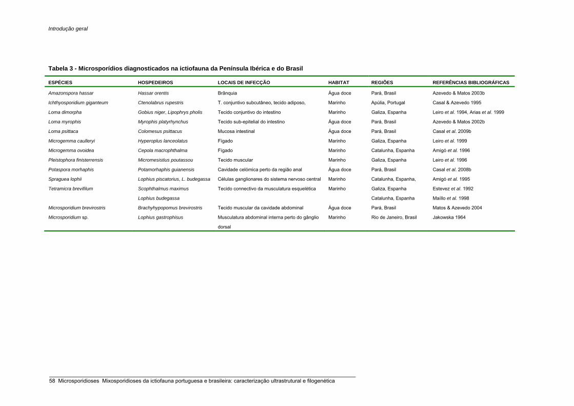
Introdução geral
Tabela 3 - Microsporídios diagnosticados na ictiofauna da Península Ibérica e do Brasil
ESPÉCIES HOSPEDEIROS LOCAIS DE INFECÇÃO HABITAT REGIÕES REFERÊNCIAS BIBLIOGRÁFICAS
Amazonspora hassar Hassar orentis Brânquia Água doce Pará, Brasil Azevedo & Matos 2003b
Ichthyosporidium giganteum Ctenolabrus rupestris T. conjuntivo subcutâneo, tecido adiposo, Marinho Apúlia, Portugal Casal & Azevedo 1995
Loma dimorpha Gobius niger, Lipophrys pholis Tecido conjuntivo do intestino Marinho Galiza, Espanha Leiro et al. 1994, Arias et al. 1999
Loma myrophis Myrophis platyrhynchus Tecido sub-epitelial do intestino Água doce Pará, Brasil Azevedo & Matos 2002b
Loma psittaca Colomesus psittacus Mucosa intestinal Água doce Pará, Brasil Casal et al. 2009b
Microgemma caulleryi Hyperoplus lanceolatus Fígado Marinho Galiza, Espanha Leiro et al. 1999
Microgemma ovoidea Cepola macrophthalma Fígado Marinho Catalunha, Espanha Amigó et al. 1996
Pleistophora finisterrensis Micromesistius poutassou Tecido muscular Marinho Galiza, Espanha Leiro et al. 1996
Potaspora morhaphis Potamorhaphis guianensis Cavidade celómica perto da região anal Água doce Pará, Brasil Casal et al. 2008b
Spraguea lophii Lophius piscatorius, L. budegassa Células ganglionares do sistema nervoso central Marinho Catalunha, Espanha, Amigó et al. 1995
Tetramicra brevifilum Scophthalmus maximus
Lophius budegassa
Tecido connectivo da musculatura esquelética Marinho Galiza, Espanha
Catalunha, Espanha
Estevez et al. 1992
Maíllo et al. 1998
Microsporidium brevirostris Brachyhypopomus brevirostris Tecido muscular da cavidade abdominal Água doce Pará, Brasil Matos & Azevedo 2004
Microsporidium sp. Lophius gastrophisus Musculatura abdominal interna perto do gânglio
dorsal
Marinho Rio de Janeiro, Brasil Jakowska 1964
_____________________________________________________________________________________________________ 58 Microsporidioses Mixosporidioses da ictiofauna portuguesa e brasileira: caracterização ultrastrutural e filogenética

Introdução geral
Tabela 4 - Mixosporídios diagnosticados na ictiofauna portuguesa e espanhola
ESPÉCIES HOSPEDEIROS LOCAIS DE INFECÇÃO HABITAT REGIÕES REFERÊNCIAS BIBLIOGRÁFICAS
Alataspora budegassai Lophius budegassa Vesícula biliar Marinho Costa Algarvia Afonso-Dias et al. 2007
Ceratomyxa appendiculata Lophius budegassa Vesícula biliar Marinho Mditerrâneo Maillo-Bellon & Gracia-Royo 2007
Ceratomyxa diplodae Dicentrarchus labrax Vesícula biliar Marinho Mediterrâneo Sitjà-Bobadilla & Álvarez-Pellitero 1993a
Ceratomyxa labracis Dicentrarchus labrax Vesícula biliar Marinho Mediterrâneo Sitjà-Bobadilla & Álvarez-Pellitero 1993a
Ceratomyxa sparusaurati Sparus aurata Vesícula biliar Marinho Mediterrâneo Sitjà-Bobadilla et al. 1995; Costa et al. 1998
Ceratomyxa tenuispora Aphanopus carbo Vesícula biliar Marinho Ilha da Madeira Casal et al. 2007
Ceratomyxa sp. Dentex dentex Vesícula biliar Marinho Mediterrâneo Company et al. 1999
Enteromyxum scophthalmi Scophthalmus maximus Intestino Marinho Mediterrâneo Palenzuela et al. 2002
Enteromyxum leei Coris julis, Symphodus tinca, S. ocellatus, S. mediterraneus, S. rostratus, S. roissali, S. cinereus, S. melops, Thalassoma pavo, Labrus viridis, L. merula, L. bergylta, Xyrichtys novacula, Spicara maena, Sparus aurata, Diplodus sargus, D. vulgaris, Mola mola, Mullus surmuletus, Halobatrachus didactylus, Chromis chromis, Lipophrys pavo, Gobius níger, Scorpaena porcus
Intestino Marinho Mediterrâneo Padrós et al. 2001
Kudoa trifolia Liza aurata, L. ramada Baço, v. biiiar, intest., brânquias Marinho Mediterrâneo Holzer et al. 2006a
Kudoa unicapsula Liza aurata, L. ramada Intestino, cecos pilóricos Marinho Mediterrâneo Yurakhno et al. 2007
Kudoa sp. Trachurus trachurus Músculo Marinho Norte, Portugal Cruz et al. 2003
Leptotheca sparidarum Dentex dentex, Sparus aurata Rim Marinho Mediterrâneo Sitjà-Bobadilla & Álvarez-Pellitero 2001
Myxidium giardi Anguilla anguilla Brânquias Estuarino Norte de Portugal;
Galiza, Espanha
Azevedo et al. 1989;
Aguilar et al. 2005
Myxidium rhodei Leuciscus cephalus cabeda, Chondrostoma polylepis Rim Água doce
Noroeste, Espanha
e no Norte, Portugal
Álvarez-Pellitero 1989
Saraiva et al. 2000
Myxobolus sp. Seriola dumerili Cérebro Marinho Baleares, Espanha Grau et al. 1999
Myxobolus pseudodispar Chondrostoma polylepis, Leuciscus cephalus Músculo, rim, ductos urinários,
fígado baço, brânquias
Portugal Cruz et al. 2000
Myxobolus portucalensis Anguilla anguilla Barbatanas Água doce Norte, Portugal Saraiva & Molnár 1990
Pentacapsula cutanea Serranus atricauda Subcutâneo Marinho Canárias Cuyas et al. 2004
Polysporoplasma sparis Sparus aurata Rim Marinho Mediterrâneo Sitjà-Bobadilla & Álvarez-Pellitero 1995
Polysporoplasma mugilis Liza aurata Rim Marinho Mediterrâneo Sitjà-Bobadilla & Álvarez-Pellitero 1995
Sphaeromyxa balbiani Cepola macrophthalma Vesícula biliar Marinho Catalunha, Espanha Gracia et al. 1997
Sphaerospora dicentrarchi Dicentrarchus labrax Marinho Mediterrâneo Sitjà-Bobadilla & Álvarez-Pellitero 1992
Sphaerospora testicularis Dicentrarchus labrax Testículo Marinho Mediterrâneo Sitjà-Bobadilla & Álvarez-Pellitero 1990
Unicapsula pflugfelderi Lithognathus mormyrus, Spicara smaris Tecido muscular Marinho Mediterrâneo Alama-Bermejo et al. 2009
Zschokkella mugilis Mugil capito, Mugil cephalus, Liza saliens, Vesícula biliar Marinho Mediterrâneo Sitjà-Bobadilla & Álvarez-Pellitero 1993b
PKX - Myxozoa
desconhecido
Salmonídeos - doença proliferativa do rim Rim Água doce Aragão, Espanha Peribanez et al. 1997
_____________________________________________________________________________________________________ Microsporidioses Mixosporidioses da ictiofauna portuguesa e brasileira: caracterização ultrastrutural e filogenética 59

Introdução geral
Tabela 5a - Mixosporídios diagnosticados na ictiofauna brasileira
ESPÉCIES HOSPEDEIROS LOCAIS DE INFECÇÃO ESTADOS HABITAT REFERÊNCIAS BIBLIOGRÁFICAS
Agarella gracilis Lepidosiren paradoxa Testículo e rim Pará Água doce Pinto 1928; Vita et al. 2004
Ceratomyxa sphaerulosa Sphyrna tudes Vesícula biliar Rio de Janeiro Marinho Pinto 1928
Ceratomyxa truncata ? Vesícula biliar Brasil - Pinto 1928
Ceratomyxa curvata Odontaspis americanus Vesícula biliar Rio de Janeiro Marinho Pinto 1928
Ceratomyxa hippocampi Hippocampus punctulatus Vesícula biliar Brasil Marinho Pinto 1928
Chloromyum leydigi Scoliodon terra-novae Vesícula biliar Rio de Janeiro Marinho Pinto 1928
Chloromyxum menticirrhi Menticirrhus americanus Vesícula urinária Florianópolis Marinho Casal et al. 2009a
Chloromyxum riorajum Rioraja agassizii Vesícula biliar Florianópolis Marinho Azevedo et al. 2009a
Chloromyxum sphyrnae Sphyrna tibura Vesícula biliar Rio de Janeiro Marinho Cunha & Fonseca, 1918
Coccomyxa claviforme Chilomycterus spinosus Vesícula biliar Brasil Pinto 1928
Kudoa aequidens Aequidens plagiozonatus Musculatura sub-opercular Pará Água doce Casal et al. 2008a
Henneguya spp. (Consultar a tabela 5b)
Myxidium striatum Menticirrhus americanus Vesícula biliar Rio de Janeiro Marinho Jayasri & Hoffman 1982
Myxidium fonsecai Carapus fasciatus Vesícula biliar Mato Grosso Água doce Jayasri & Hoffman 1982
Myxidium cruzi Chalcinus nematurus Vesícula biliar Mato Grosso Água doce Jayasri & Hoffman 1982
Myxidium gurgeli Acestrorhamphus sp. Vesícula biliar São Paulo Água doce Jayasri & Hoffman 1982
Myxidium cholecysticum Astyanax scabripinnis Vesícula biliar São Paulo Água doce Cordeiro & Gioia 1990
Myxobolus spp. (Consultar a tabela 5c)
Sphaeromyxa balbiani Scorpena plumieri Vesícula biliar Brasil Marinho Pinto 1928
Tetrauronema desaequalis Hoplias malabaricus Base da barbatana ventral Pará Água doce Azevedo & Matos 1996b
Triangulamyxa amazonica Sphoeroides testudineus Intestino Pará Água doce Azevedo et al. 2005
_____________________________________________________________________________________________________ 60 Microsporidioses Mixosporidioses da ictiofauna portuguesa e brasileira: caracterização ultrastrutural e filogenética

Introdução geral
Tabela 5b - Comparação morfológica dos esporos de diferentes espécies de Henneguya diagnosticadas em peixes da fauna brasileira.
Espécies CT CC LC CCa CCP LCP NFP VI R Hospedeiros Habitat Locais de infecção Estados Referências Bibliográficas
H. occulta 36–46 16 8 20 8 – – – Loricaia sp. D Brânquias Rio de Janeiro Nemeczek 1926 H. leporini 28–33 13–15 5 15–18 5-8 – – – Leporinus mormyrops D Ductos urinários Minas Gerais Nemeczek 1926 H. wenyoni 21 11-12 5,2 10,8 3,7 1,5 – – – Tetragonopterus sp. D Brânquias São Paulo Pinto 1928 H. travassosi 27,3 10,6 4,3 16,7 3,6 – - – Astyanax fasciatus D D São Paulo Guimarães & Bergamin 1933 H. santae 21,0 9,6 5,3 11,2 2,9 – Sim – Tetragnopterus santae D Brânquias São Paulo Guimarães & Bergamin 1934 H. visceralis 22–24 11–12 5–6,5 11–12 6,5–8 2 – – – D D Rim, fígado, coração Brasil Jakowska & Nigrelli 1953 H. electrica 35–39 11–13 6–8 24–27 5–7 2 – – – Electrophorus electricus D Órgãos eléctricos Brasil Jakowska & Nigrelli 1953 H. pisciforme 20,4 – 6,1 10,7 4,3 1,7 – Sim – Hyphessobrycon anisitsi D Brânquias São Paulo Cordeiro et al. 1984 H. theca 48,0 24,8 3,5 23,2 11,1 1,4 – Sim + Eigemannia virescens D Cérebro Brasil Kent & Hoffman 1984 H. intracornea 42,4 – 6,7 24,3 8,6 2,4 – Sim – Astyanax scabripinnis D Córnea São Paulo Gioia et al. 1986 H. hoimba 24,7 – 7,5 – 4,4 1,9 – Sim – Astyanax fasciatus D Brânquias São Paulo Cordeiro & Gioia 1987 H. artigasi 16,4 – 4,4 – 3,3 1,5 – - – Astyanax scabripinnis D Brânquias São Paulo Gioia & Cordeiro 1987 H. amazonica 59,3 13,9 5,7 45,4 3,3 1,5 6 – – Crenicichla lepidota D Brânquias Pará Rocha et al. 1992 H. adherens 32,3 12,4 5,8 20,5 3,1 1,2 3-4 – – Acestrorhynchus falcatus D Brânquias Pará Azevedo & Matos 1995 H. malabarica 28,3 12,6 4,8 17,1 3,7 1,8 6-7 – + Hoplias malabaricus D Brânquias Pará Azevedo & Matos 1996a H. piaractus 52,5 12,7 3,6 41,2 6,7 1,2 8-9 Sim + Piaractus mesopotamicus D Brânquias São Paulo Martins & Souza 1997 H. testicularis 27,5 14,0 6,5 13,5 9,0 2,0 12–13 – + Moenkhausia oligolepis D Testículo Pará Azevedo et al. 1997 H. striolata 42,2 15,8 5,3 25,9 6,8 1,2 13–14 Não + Serrasalmus striolatus D Brânquias Pará Casal et al. 1997 H, leporinicola - 5,5-8,7 3,6-4,9 12,9-32,2 2,0-3,6 1,2-2,0 – – – Leporinus macrocephalus D F. branquiais secund. São Paulo Martins et al. 1999 H. curimata 35,4 16,6 6,2 19,1 6,5 1,2 10–11 – – Curimata inormata D Rim Pará Azevedo & Matos 2002b H. astyanax 47,8 15,2 5,7 32,6 5,0 1,5 8–9 – – Astyanax keithi D Brânquias Pará Vita et al. 2003 H. chydadea 17,6-20 8,8-11,2 3,2-5,6 8-9,6 3,2-4,4 1,2-1,6 9-10 Não – Astyanax altiparanae D Brânquias São Paulo Barassa et al. 2003b H. curvata 41,7 16,4 4,7 25,3 7,8 1,4 10-11 - – Serrasalmus spilopleura D Brânquias São Paulo Barassa et al. 2003a H. friderici 28,7-39,3 9,6-11,8 4,8-6,6 19,1-28,7 4,2-5,9 1,5-2,6 7-8 Sim – Leporinus friderici D Vários órgãos Pará Casal et al. 2003 H. pilosa 52,3-56,0 20,0-23,1 5,5-6,3 30,5-34,9 7,1-7,6 1-1,3 11-12 – + Serrasalmus altuvei D Brânquias Pará Azevedo & Matos 2003b H. schizodon 27-30 12-14 3-4 15-17 5-6 1-1,5 8-10 – – Schizodon fasciatum D Rim Amazonas Eiras et al. 2004a H. paranaensis 56-63 14-17 6-7 41-46 8-9 e 6-7 2 10-12 – – Prochilodus lineatus D F. branquiais secund. Paraná Eiras et al. 2004b H. caudalongula 71 16,6 4,6 52,6 6,1 1,6 10-11 – – Prochilodus lineatus D Brânquias São Paulo Adriano et al. 2005a H. pellucida 33,3 11,4 4,1 24,1 4,0 1,6 6-7 – – Piaractus mesopotamicus D Cavidade visceral Adriano et al. 2005b H. rhamdia 48,2-51,8 12,0-14,2 4,7-5,7 35,3-38,5 4,3-5,1 0,9-1,3 10-11 – + Rhamdia quelen D Lamelas branquiais Pará Matos et al. 2005 H. garavelli 41,2-51,5 12,0-14,4 3,9-4,1 31,4-35,6 4,8-6,0 1,0-1,5 8-9 Sim – Cyphocharax nagelli D Brânquias São Paulo Martins & Onaka 2006 H. cyphocharax 29,6-44,4 7,7-13,4 2,9-6,3 20,8-31,5 4,2-6,3 / 3,4-5,2 1,5-2,3 / 1,3-2,2 7-9 – – Cyphocharax gilbert D Brânquias Rio de Janeiro Abdallah et al. 2007 H. guanduensis 27,3-38,1 11,4-16,7 4,9-7,6 15,6-22,5 3,3-5,6 / 3,3-5,3 1,6-2,3 / 1,5-2,8 3-6 – – Hoplosternum littorale D Brânquias Rio de Janeiro Abdallah et al. 2007 H. caudicula 14-16 11-12 5-6 3,4 3-4 1,5 3 Sim – Leporinus lacustris D Brânquias Paraná Eiras et al. 2008 H. arapaima 48,4-53,1 13,5-15,2 5,1-6,1 38,0-41,2 6,3-6,8 / 6,2-6,6 1,4-1,6 5 – – Arapaima gigas D Vesícula biliar Goiás Feijó et al. 2008 H. rondoni 16,9-18,1 6,8-7,3 3,0-3,9 10,3-11 2,2-2,7 0,8-0,9 6-7 Não + Gymnorhamphichthys rondoni D Sist. nervoso periférico Pará Azevedo et al. 2008 H. corruscans 25,0-29,0 13-15 5,0 12-15 6,8 2,0 5-6 Não – Pseudoplatystoma corruscans D Brânquias Paraná Eiras et al. 2009 H. hemiodopsis 18.8-20.6 10.3-11.3 2.9-3.7 8.1-9.3 3.2-3.8 0.8-1.2 5-6 Não – Hemiodopsis microlepes D Brânquias Piauí Azevedo et al. 2009c
Abreviaturas: CT: comprimento total; CC: comprimento do corpo; LC: largura do corpo; CCa: Comprimento das caudas; CCP: comprimento da cápsula polar; LCP: largura da cápsula polar; NFP: número de voltas do filamento polar; VI: vacúolo iodinóforo; R: + revestimento em torno das caudas; Habitat: água doce (D), marinho (M); -: sem dados. (medidas em µm)
_____________________________________________________________________________________________________ Microsporidioses Mixosporidioses da ictiofauna portuguesa e brasileira: caracterização ultrastrutural e filogenética 61
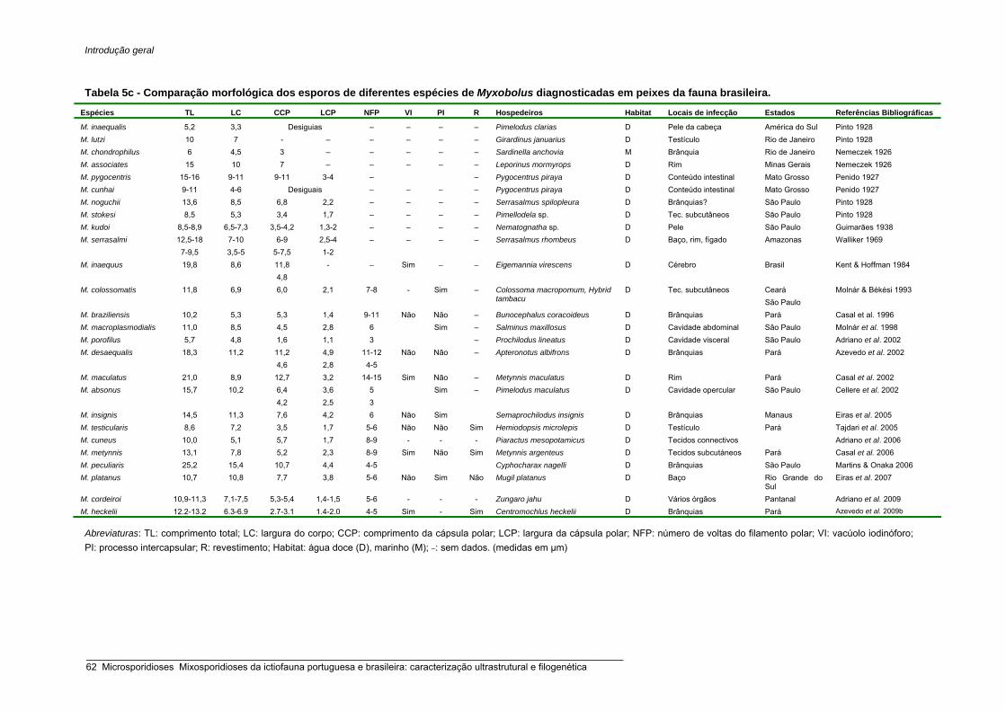
Introdução geral
Tabela 5c - Comparação morfológica dos esporos de diferentes espécies de Myxobolus diagnosticadas em peixes da fauna brasileira.
Espécies TL LC CCP LCP NFP VI PI R Hospedeiros Habitat Locais de infecção Estados Referências Bibliográficas
M. inaequalis 5,2 3,3 Desiguias – – – – Pimelodus clarias D Pele da cabeça América do Sul Pinto 1928 M. lutzi 10 7 - – – – – – Girardinus januarius D Testículo Rio de Janeiro Pinto 1928 M. chondrophilus 6 4,5 3 – – – – – Sardinella anchovia M Brânquia Rio de Janeiro Nemeczek 1926 M. associates 15 10 7 – – – – – Leporinus mormyrops D Rim Minas Gerais Nemeczek 1926 M. pygocentris 15-16 9-11 9-11 3-4 – – Pygocentrus piraya D Conteúdo intestinal Mato Grosso Penido 1927 M. cunhai 9-11 4-6 Desiguais – – – – Pygocentrus piraya D Conteúdo intestinal Mato Grosso Penido 1927 M. noguchii 13,6 8,5 6,8 2,2 – – – – Serrasalmus spilopleura D Brânquias? São Paulo Pinto 1928 M. stokesi 8,5 5,3 3,4 1,7 – – – – Pimellodela sp. D Tec. subcutâneos São Paulo Pinto 1928 M. kudoi 8,5-8,9 6,5-7,3 3,5-4,2 1,3-2 – – – – Nematognatha sp. D Pele São Paulo Guimarães 1938 M. serrasalmi 12,5-18
7-9,5 7-10 3,5-5
6-9 5-7,5
2,5-4 1-2
– – – – Serrasalmus rhombeus D Baço, rim, fígado Amazonas Walliker 1969
M. inaequus 19,8 8,6 11,8 4,8
- – Sim – – Eigemannia virescens D Cérebro Brasil Kent & Hoffman 1984
M. colossomatis 11,8 6,9 6,0 2,1 7-8 - Sim – Colossoma macropomum, Hybrid tambacu
D Tec. subcutâneos Ceará São Paulo
Molnár & Békési 1993
M. braziliensis 10,2 5,3 5,3 1,4 9-11 Não Não – Bunocephalus coracoideus D Brânquias Pará Casal et al. 1996 M. macroplasmodialis 11,0 8,5 4,5 2,8 6 Sim – Salminus maxillosus D Cavidade abdominal São Paulo Molnár et al. 1998 M. porofilus 5,7 4,8 1,6 1,1 3 – Prochilodus lineatus D Cavidade visceral São Paulo Adriano et al. 2002 M. desaequalis 18,3 11,2 11,2
4,6 4,9 2,8
11-12 4-5
Não Não – Apteronotus albifrons D Brânquias Pará Azevedo et al. 2002
M. maculatus 21,0 8,9 12,7 3,2 14-15 Sim Não – Metynnis maculatus D Rim Pará Casal et al. 2002 M. absonus 15,7 10,2 6,4
4,2 3,6 2,5
5 3
Sim – Pimelodus maculatus D Cavidade opercular São Paulo Cellere et al. 2002
M. insignis 14,5 11,3 7,6 4,2 6 Não Sim Semaprochilodus insignis D Brânquias Manaus Eiras et al. 2005 M. testicularis 8,6 7,2 3,5 1,7 5-6 Não Não Sim Hemiodopsis microlepis D Testículo Pará Tajdari et al. 2005 M. cuneus 10,0 5,1 5,7 1,7 8-9 - - - Piaractus mesopotamicus D Tecidos connectivos Adriano et al. 2006 M. metynnis 13,1 7,8 5,2 2,3 8-9 Sim Não Sim Metynnis argenteus D Tecidos subcutáneos Pará Casal et al. 2006 M. peculiaris 25,2 15,4 10,7 4,4 4-5 Cyphocharax nagelli D Brânquias São Paulo Martins & Onaka 2006 M. platanus 10,7 10,8 7,7 3,8 5-6 Não Sim Não Mugil platanus D Baço Rio Grande do
Sul Eiras et al. 2007
M. cordeiroi 10,9-11,3 7,1-7,5 5,3-5,4 1,4-1,5 5-6 - - - Zungaro jahu D Vários órgãos Pantanal Adriano et al. 2009 M. heckelii 12.2-13.2 6.3-6.9 2.7-3.1 1.4-2.0 4-5 Sim - Sim Centromochlus heckelii D Brânquias Pará Azevedo et al. 2009b
Abreviaturas: TL: comprimento total; LC: largura do corpo; CCP: comprimento da cápsula polar; LCP: largura da cápsula polar; NFP: número de voltas do filamento polar; VI: vacúolo iodinóforo; PI: processo intercapsular; R: revestimento; Habitat: água doce (D), marinho (M); –: sem dados. (medidas em µm)
_____________________________________________________________________________________________________ 62 Microsporidioses Mixosporidioses da ictiofauna portuguesa e brasileira: caracterização ultrastrutural e filogenética

Introdução geral
1. 5. Referências Bibliográficas
Abdallah, V.D., Azevedo, R.K., Luque, J.L. & Bomfim, T.C.B. (2007) Two new species of Henneguya
Thélohan, 1892 (Myxozoa, Myxobolidae), parasitic on the gills of Hoplosternum littorale (Callichthydae)
and Cyphocharax gilbert (Curimatidae) from Guandu River, State of Rio de Janeiro, Brazil. Parasitol.
Latinoamer. 62: 35-41.
Abdel-Baki, A.A.S. (2007) Chloromyxum alii sp. n. (Myxozoa: Myxosporea) infecting the gallbladder of African
butter catfish Schilbe mystus (Linnaeus, 1758) from the River Nile, Egypt: Light and scanning electron
microscopy. Acta Protozool. 46: 263-267.
Abdel-Ghaffar, F., Ali, M.A., Al Quraishy, S., Al Rasheid, K.A.S., Al Farraj, S., Abdel-Baki, A.S. & Bashtar, A.R.
(2008a) Four new species of Ceratomyxa Thelohan 1892 (Myxozoa: Myxosporea: Ceratomyxidae)
infecting the gallbladder of some Red Sea fishes. Parasitol. Res.103: 559-565.
Abdel-Ghaffar, F., El-Toukhy, A., Al-Quraishy, S., Al-Rasheid, KAS., Abdel-Baki, A.S., Hegazy, A. & Bashtar,
A.R. (2008b) Five new myxosporean species (Myxozoa: Myxosporea) infecting the Nile Tilapia
Oreochromis niloticus in Bahr Shebin, Nile Tributary, Nile Delta, Egypt. Parasitol. Res. 103: 1197-1205.
Adlard, R.D., Bryant, M.S., Whipps, C.M. & Kent, M.L. (2005) Multivalvulid myxozoans from eastern Australia:
Three new species of kudoa from scombrid and labrid fishes of the Great Barrier Reef, Queensland,
Australia. J. Parasitol. 91: 1138-1142.
Adriano, E.A., Arana, S., Ceccarelli, P.S. & Cordeiro, N.S. (2002) Light and scanning microscopy of
Myxobolus porofilus sp. n. (Myxosporea: Myxobolidae) infecting the visceral cavity of Prochilodus lineatus
(Pisces: Characiformes: Prochilodontidae) cultivated in Brazil. Folia Parasitol. 49: 259-262.
Adriano, E.A., Arana, S. & Cordeiro, N.S. (2005a) Histopathology and ultrastructure of Henneguya
caudalongula sp. n. infecting Prochilodus lineatus (Pisces: Prochilodontidae) cultivated in the state of São
Paulo, Brazil. Mem. Inst. Oswaldo Cruz 100: 177–181.
Adriano, E.A., Arana, S. & Cordeiro, N.S. (2005b) An ultrastructural and histopathological study of Henneguya
pellucida n. sp. (Myxosporea: Myxobolidae) infecting Piaractus mesopotamicus (Characidae) cultivated in
Brazil. Parasite 12: 221–227.
Adriano, E.A., Arana, S. & Cordeiro, N.S. (2006) Myxobolus cuneus n. sp (Myxosporea) infecting the
connective tissue of Piaractus mesopotamicus (Pisces: Characidae) in Brazil: Histopathology and
ultrastructure. Parasite 13: 137-142.
Adriano, E.A., Arana, S., Alves, A.L., Silva, M.R.M. & Ceccarelli, P.S. (2009) Myxobolus cordeiroi n. sp, a
parasite of Zungaro jahu (Siluriformes: Pimelodiade) from Brazilian Pantanal: morphology, phylogeny and
histopathology. Vet. Parasitol. 162: 221-229.
Afonso-Dias, I., Kalavati, C., Mackenzie, K. & Mackenzie, K.S. (2007) Three new species of Myxosporea
(Bivalvulida: Ceratomyxidae: Alatasporidae) from the gall bladders of anglerfishes Lophius spp.
(Teleostei: Lophiidae) in the Northeast Atlantic Ocean. Zootaxa 1466: 35-46.
Aguilar, A., Alvarez, M.F., Leiro, J.M. & Sanmartin, M.L. (2005) Parasite populations of the European eel
(Anguilla anguilla L.) in the Rivers Ulla and Tea (Galicia, northwest Spain). Aquaculture 249: 85-94.
Alama-Bermejo, G., Cuadrado, M., Raga, J.A. & Holzer, A.S. (2009) Morphological and molecular
redescription of the myxozoan Unicapsula pflugfelderi Schubert, Sprague & Reinboth, 1975 from two
_____________________________________________________________________________________________________ Microsporidioses Mixosporidioses da ictiofauna portuguesa e brasileira: caracterização ultrastrutural e filogenética 63
Introdução geral
teleost hosts in the Mediterranean. A review of the genus Unicapsula Davis, 1924. J. Fish Dis. 32: 335-
350.
Ali, M.A., Abdel-Baki, A.S., Sakran, T., Entzeroth, R. & Abdel-Ghaffar, F. (2007) Myxobolus lubati n. sp.
(Myxosporea: Myxobolidae), a new parasite of haffara seabream Rhabdosargus haffara (Forsskal, 1775),
Red Sea, Egypt: a light and transmission electron microscopy. Parasitol. Res. 100: 819-827.
Al-Quraishy, S., Koura, E., Abdel-Baki, A.S., Bashtar, A.R., El Deed, N., Al-Rasheid, K.A.S. & Abdel-Ghaffar,
F. (2008) Light and electron microscopic studies on Kudoa pagrusi sp. n. (Myxosporea: Multivalvulida)
infecting the heart of sea bream Pagrus pagrus (L.) from the Red Sea. Parasitol. Res. 102: 205-209.
Álvarez-Pellitero, P. (1989) Myxidium rhodei (Protozoa, Myxozoa, Myxosporea) in cyprinid fish from NW
Spain. Dis. Aquat. Org. 7: 13-16.
Álvarez-Pellitero, P., Molnár, K., Sitjà-Bobadilla, A. & Székely, C. (2002) Comparative ultrastructure of the
actinosporean stages of Myxoholus bramae and M. pseadodispar (Myxozoa). Parasitol. Res. 88:198-207.
Amigó, J. M. (1995) Estudi de microsporidiosis en peixos d’interès comercial. Ph. D. Thesis, Universitat de Barcelona, España.
Amigó, J.M., Salvadó, H., Gracia, M.P. & Vivarès, C.P. (1996) Ultrastructure and development of
Microsporidium ovoideum (Thélohan, 1895) Sprague, 1977, a microsporidian parasite of the red band fish
(Cepola macrophthalma L.) – redescription of the organism and reassignment to the genus Microgemma,
Ralphs & Matthees 1986. Eur. J. Protistol. 32: 532-538.
Anderson, C.L., Canning, E.U. & Okamura, B. (1998) A triploblast origin for Myxozoa? Nature 392: 346.
Anderson, C.L., Canning, E.U. & Okamura, B. (1999) Molecular data implicate bryozoans as hosts for PKX
(phylum Myxozoa) and identify a clade of bryozoan parasites within the Myxozoa. Parasitology 119: 555–
561.
Anderson, C.L., Canning, E.U., Schaefer, S.M., Yoyama, H. & Okamura, B. (2000) Molecular confirmation of
the life cycle of Thelohanellus hovorkai Achmerov, 1960 (Myxozoa: Myxosporea). Bull. Eur. Ass. Fish
Pathol. 20: 111–115.
Andree, K.B., Gresoviac, S.J. & Hedrick, R.P. (1997) Small subunit ribosomal RNA sequences unite alternate
actinosporean and myxosporean stages of Myxobolus cerebralis, the causative agent of whirling disease
in salmonid fish. J. Eukaryot. Microbiol. 44: 208–215.
Andree, K.B., Székely, C., Molnár, K., Gresoviac, S.J. & Hedrick, R.P. (1999) Relationships among members
of the genus Myxobolus (Myxozoa: Bivalvulidae) based on small subunit ribosomal DNA sequences. J.
Parasitol. 85: 68–74.
Arias, C., Estévez, J., Lores, B., Sanmartín, M.L. & Leiro, J. (1999) Discovery of Loma dimorpha Loubès,
Maurant, Gasc, De Buron & Barral 1984, in a new host, the shanny, Lipophrys pholis (L.) from the north-
west coast of Spain. J. Fish Dis. 22: 155–159.
Atkinson, S.D. & Bartholomew, J.L. (2009) Alternate spore stages of Myxobilatus gasterostei, a myxosporean
parasite of three-spined sticklebacks (Gasterosteus aculeatus) and oligochaetes (Nais communis).
Parasitol. Res. 104: 1173–1181.
Atkinson, S.D., Hallett, S.L. & Bartholomew, J.L. (2007) The life cycle of Chloromyxum auratum (Myxozoa)
from goldfish, Carassius auratus (L.), involves an antonactinomyxon actinospore. J. Fish Dis. 30: 149–
156.
_____________________________________________________________________________________________________ 64 Microsporidioses Mixosporidioses da ictiofauna portuguesa e brasileira: caracterização ultrastrutural e filogenética
Introdução geral
Azevedo, C., Casal, G., Garcia, P., Matos, P., Teles-Grilo, L. & Matos, E. (2009a) Ultrastructural and
phylogenetic data of Chloromyxum riorajum sp. nov. (Myxozoa), a parasite of the stingray Rioraja
agassizii in Southern Brazil. Dis. Aquat. Org. 85: 41-51.
Azevedo, C., Casal, G., Matos, P., Ferreira, I. & Matos, E. (2009b) Ultrastructural data on the spore of
Myxobolus heckelii n. sp. (Myxozoa), parasite from the Brazilian fish Centromochlus heckelii (Teleostei,
Auchenipteridae). J. Eukaryot. Microbiol. (in press)
Azevedo, C., Casal, G., Matos, P. & Matos, E. (2008) A new species of Myxozoa, Henneguya rondoni n. sp.
(Myxozoa), from the peripheral nervous system of the Amazonian fish, Gymnorhamphichthys rondoni
(Teleostei). J. Eukaryot. Microbiol. 55: 229-234.
Azevedo, C., Casal, G., Mendonça, I. & Matos, E. (2009c) Fine structure of Henneguya hemiodopsis n. sp.
(Myxozoa), a parasite of the gills of Brazilian teleostean fish, Hemiodopsis microlepes (Hemiodontidae). Mem. Inst. Oswaldo Cruz (in press)
Azevedo, C., Corral, L. & Matos, E. (1997) Light and ultrastructural data on Henneguya testicularis n. sp.
(Myxozoa, Myxobolidae), a parasite from the testis of the Amazonian fish Moenkhausia oligolepis. Syst.
Parasitol. 37: 111–114.
Azevedo, C., Corral, L. & Matos, E. (2002) Myxobolus desaequalis n. sp. (Myxozoa, Myxosporea), parasite of
the Amazonian freshwater fish, Apteronotus albifrons (Teleostei, Apteronotidae). J. Eukaryot. Microbiol.
49: 485-488.
Azevedo, C., Corral, L. & Matos, E. (2005) Ultrastructure of Triangulamyxa amazonica n. gen. and n. sp.
(Myxozoa, Myxosporea), a parasite of the Amazonian freshwater fish, Sphoeroides testudineus
(Teleostei, Tetrodontidae). Eur. J. Protistol. 41: 57-63.
Azevedo, C., Lom, J. & Corral, L (1989) Fine ultrastructural aspects of Myxidium giardi (Myxozoa,
Myxosporea) from Portuguese eels (Anguilla anguila). Dis. Aquat. Org. 6: 55-61.
Azevedo, C. & Matos, E. (1995) Henneguya adherens n. sp. (Myxozoa, Myxosporea), parasite of the
Amazonian fish, Acestrorhynchus falcatus. J. Eukaryot. Microbiol. 42: 515–518.
Azevedo, C. & Matos, E. (1996a) Henneguya malabarica sp. nov. (Myxozoa, Myxobolidae) in the Amazonian
fish Hoplias malabaricus. Parasitol. Res. 82: 222–224.
Azevedo, C. & Matos, E. (1996b) Light and electron microscopic study of a myxosporean, Tetrauronema
desaequalis n. sp. (Fam. Tetrauronematidae) from an Amazonian fish. J. Parasitol. 82: 288-291.
Azevedo, C. & Matos, E. (2002a) Fine structure of a new species, Loma myrophis (Phylum Microsporidia),
parasite of the Amazonian fish Myrophis platyrhynchus (Teleostei, Ophichthidae). Eur. J. Protistol. 37:
445-452.
Azevedo, C. & Matos, E. (2002b) Fine structure of the myxosporean, Henneguya curimata n. sp., parasite of
the Amazonian fish, Curimata inormata (Teleostei, Curimatidae). J. Eukaryot. Microbiol. 49: 197–200.
Azevedo, C. & Matos, E. (2003a) Amazonspora hassar n. gen. and n. sp. (phylum Microsporidia, fam
Glugeidae), a parasite of the Amazonian teleost Hassar orestis (fam. Doradidae). J. Parasitol. 89: 336-
341.
Azevedo, C. & Matos, E. (2003b) Fine structure of Henneguya pilosa sp. n. (Myxozoa: Myxosporea), parasite
of Serrasalmus altuvei (Characidae), in Brazil. Folia Parasitol. 50: 37–42.
_____________________________________________________________________________________________________ Microsporidioses Mixosporidioses da ictiofauna portuguesa e brasileira: caracterização ultrastrutural e filogenética 65
Introdução geral
Bahri, S., Andree, K.B. & Hedrick, R.P. (2003) Morphological and phylogenetic studies of marine Myxobolus.
J. Eukaryot. Microbiol. 50: 463–470.
Baker, M.D., Vossbrinck, C.R., Becnel, J.J. & Andreadis, T.G. (1998) Phylogeny of Amblyospora
(Microsporida: Amblyosporidae) and related genera based on small subunit ribosomal DNA data: A
possible example of host parasite cospeciation. J. Invertebr. Pathol. 71: 199–206.
Baquero, E., Rubio, M., Moura, I.N.S., Pieniazek, J. & Jordana, R. (2005) Myosporidium merluccius n. g., n.
sp. infecting muscle of commercial hake (Merluccius sp.) from fisheries near Namibia. J. Eukaryot.
Microbiol. 52: 476-483.
Barassa, B., Adriano, E. A., Arana, S. & Cordeiro, N.C. (2003a) Henneguya curvata sp. n. (Myxosporea:
Myxobolidae) parasitizing the gills of Serrasalmus spilopleura (Characidae: Serrasalminae), a South
American freshwater fish. Folia Parasitol. 50: 151–153.
Barassa, B., Cordeiro, N.S. & Arana, S. (2003b) A new species of Henneguya, a gill parasite of Astyanax
altiparanae (Pisces: Characidae) from Brazil, with comments on histopathology and seasonality. Mem.
Inst. Oswaldo Cruz 98: 761–765.
Barber, I., Davies, A.J., Ironside, J.E., Forsgren, E., & Amundsen, T. (2009) First record of a Kabatana sp.
microsporidium infecting fish in the Atlantic Ocean. Dis. Aquat. Org. 83: 145-152.
Bartholomew, J.L., Atkinson, S.D., Hallett, S.L., Lowenstine, L.J., Garner, M.M., Gardiner, C.H., Rideout, B.A.,
Keel, M.K. & Brown, J.D. (2008) Myxozoan parasitism in waterfowl. Int. J. Parasitol. 38: 1199-1207.
Bartholomew, J.L., Whipple, M.J., Stevens, D.G. & Fryer, J.L. (1997) The life cycle of Ceratomyxa shasta, a
myxosporean parasite of salmonids, requires a freshwater polychaete as an alternate host. J. Parasitol.
83: 859–868.
Becnel, J.J. & Andreadis, T.G. (1999) Microsporidia of Insects. In: The Microsporidia and Microsporidiosis.
(Wittner, M. & Weiss, L. M. Eds.), ASM Press, Washington D. C. pp. 447–501.
Bekhti, M. & Bouix, G. (1985) Loma salmonae (Putz, Hoffmann et Dunbar, 1965) et Loma diplodae n. sp.,
Microsporidies parasites de branchies de Poissons Téléosteéns: implantation et données
ultrastructurales. Protistologica 21: 47-59.
Békési, L., Székely, C. & Molnár, K. (2002) Atuais conhecimentos sobre Myxosporea (Myxozoa), parasitas de
peixes. Um estágio alternativo dos parasitas no Brasil. Braz. J. Vet. Res. Anim. Sci. 39: 271-276.
Bell, A.S., Aoki, T. & Yokoyama, H. (2001) Phylogenetic relationships among microsporidia based on rDNA
sequences data, with particular reference to fish-infecting Microsporidium Balbiani, 1884 species. J.
Eukaryot. Microbiol. 48: 258–265.
Berrebi, P. (1979) Étude ultrastructurale de Glugea atherinae n. sp., microsporidie parasite de l’athérine
Atherina boyeri Risso 1810 (poisson téléostéen) dans les Lagunes du Languedoc et de Provence. Z.
Parasitenkd. 60: 105-122.
Bohne, W., Ferguson, D.J., Kohler, K. & Gross, U. (2000) Developmental expression of a tandemly repeated,
glycine- and serine-rich spore wall protein in the microsporidian pathogen Encephalitozoon cuniculi.
Infect. Immun. 68: 2268–2275.
Boreham, R.E., Hendrick, S., O’Donoghue, P.J. & Stenzel, D.J. (1998) Incidental finding of Myxobolus spores
(Protozoa: Myxozoa) in stool samples from patients with gastrointestinal symptoms. J. Clin. Microbiol. 36:
3728–3730.
_____________________________________________________________________________________________________ 66 Microsporidioses Mixosporidioses da ictiofauna portuguesa e brasileira: caracterização ultrastrutural e filogenética

Introdução geral
Brickle, P., Kalavati, C. & MacKenzie, K. (2006) Henneguya shackletoni sp. nov. (Myxosporea, Bivalvulida,
Myxobolidae) from the Falklands mullet, Eleginops maclovinus (Cuvier) (Teleostei, Eleginopidae) in the
Falkland Islands. Acta Parasitol. 51: 36-39.
Brosson, D., Kuhn, L., Prensier, G., Vivarès, C.P. & Texier, C. (2005) The putative chitin deacetylase of
Encephalitozoon cuniculi: a surface protein implicated in microsporidian spore-wall formation. FEMS
Microbiol. Lett. 247: 81–90.
Buckler, E.S., Ippolito, A. & Holtsford, T.P. (1997) The evolution of ribosomal DNA: divergent paralogues and
phylogenetic implications. Genetics 145: 821–832.
Burger, M.A.A., Cribb, T.H. & Adlard, R.D. (2007) Patterns of relatedness in the Kudoidae with descriptions of
Kudoa chaetodoni n. sp. and K. lethrini n. sp. (Myxosporea: Multivalvulida). Parasitology 134: 669-681.
Burtle, G.J., Harrison, L.R. & Styer, E.L. (1991) Detection of a triactinomyxid myxozoan in an oligochaete from
ponds with proliferative gill disease in channel catfish. J. Aquat. Anim. Health 3: 281–287.
Canning, E.U. (1990) Phylum Microspora. In: The Handbook of Protoctists (Margulis, L., Chapman, D.J.,
Melkonian, M. & Corliss, J.O. Eds.), Jones & Bartlett, Boston, pp. 53-72.
Canning, E.U., Feist, S.W., Longshaw, M., Okamura, B., Anderson, C.L., Tse, M.T. & Curry, A. (2005)
Microgemma vivaresi n. sp. (Microsporidia, Tetramicridae), infecting liver and skeletal muscle of sea
scorpions, Taurulus bubalis (Euphrasen 1786) (Osteichthyes, Cottidae), an inshore, littoral fish. J.
Eukaryot. Microbiol. 52: 123–131.
Canning, E.U., Curry, A., Anderson, C.L. & Okamura, B. (1999) Ultrastructure of Myxidium trachinorum sp.
nov. from the gallbladder of the lesser weever fish Echiichthys vipera. Parasitol. Res. 85: 910-919.
Canning, E.U., Curry, A., Feist, S.W., Longshaw, M. & Okamura, B. (2000) A new class and order of
myxozoans to accommodate parasites of bryozoans with ultrastructural observations on Tetracapsula
bryosalmonae (PKX organism). J. Eukaryot. Microbiol. 47: 456-468.
Canning, E.U. & Hazard, E.I. (1982) Genus Pleistophora Gurley, 1893: An assemblage of at least three
genera. J. Protozool. 29: 38-49.
Canning, E.U., Hazard, E.I. & Nicholas, J.P. (1979) Light and electron microscopy of Pleistophora sp. from
skeletal muscle of Blennius pholis. Protistologica, 15: 317-332.
Canning, E.U. & Lom, J. (1986) The Microsporidia of Vertebrates, Academic Press, London.
Canning, E.U., Lom, J. & Nicholas, J.P. (1982) Genus Glugea Thélohan, 1891 (Phylum Microspora):
redescription of the type species Glugea anomala (Moniez, 1887) and recognition of its sporogonic
development within sporophorous vesicles (pansporoblastic membranes). Protistologica 18: 193-210.
Canning, E.U. & Nicholas, J.P. (1980) Genus Pleistophora (Phylum Microspora): redescription of the type
species, Pleistophora typicalis Gurley, 1893 and ultrastructural characterization of the genus. J. Fish Dis.
3: 317-338.
Canning, E.U., Okamura, B. & Curry, A. (1996) Tetracapsula bryozoides gen. n. et sp. n. in the body cavity of
Cristatella mucedo (Bryozoa, Phylactolaemata). Folia Parasitol. 43: 249-261.
Canning, E.U., Tops, S., Curry, A., Wood, T.S. & Okamura, B. (2002) Ecology, development and pathogenicity
of Buddenbrockia plumatellae Schröder, 1910 (Myxozoa, Malacosporea) (syn. Tetracapsula bryozoides)
and establishment of Tetracapsuloides n. gen. for Tetracapsula bryosalmonae. J. Eukaryot. Microbiol. 49:
_____________________________________________________________________________________________________ Microsporidioses Mixosporidioses da ictiofauna portuguesa e brasileira: caracterização ultrastrutural e filogenética 67

Introdução geral
280-295.
Casal, G. & Azevedo, C. (1995) New ultrastructural data on the microsporidian Ichthyosporidium giganteum
infecting the marine teleostean fish Ctenolabrus rupestris. J. Fish Dis. 18: 191-194.
Casal, G., Costa, G. & Azevedo, C. (2007) Ultrastructural description of Ceratomyxa tenuispora (Myxozoa), a
parasite of the marine fish Aphanopus carbo (Trichiuridae), from the Atlantic coast of Madeira Island
(Portugal). Folia Parasitol. 54: 165-171.
Casal, G., Garcia, P., Matos, P., Monteiro, E., Matos, E. & Azevedo, C. (2009a) Fine structure of
Chloromyxum menticirrhi n. sp. (Myxozoa) infecting urinary bladder of the marine teleost Menticirrhus
americanus (Sciaenidae) in Southern Brazil. Eur. J. Protistol. 45: 139-146.
Casal, G., Matos, E. & Azevedo, C. (1996) Ultrastructural data on the life stages of Myxobolus braziliensis n.
sp., parasite of an Amazonian fish. Eur. J. Protistol. 32: 123-127.
Casal, G., Matos, E. & Azevedo, C. (1997) Some ultrastructural aspects of Henneguya striolata sp. nov.
(Myxozoa, Myxosporea), a parasite of the Amazonian fish Serrasalmus striolatus. Parasitol. Res. 83: 93–
95.
Casal, G., Matos, E. & Azevedo, C. (2002) Ultrastructural data on the spore of Myxobolus maculatus n. sp.
(phylum Myxozoa), parasite from the Amazonian fish Metynnis maculatus (Teleostei). Dis. Aquat. Org. 51:
107-112.
Casal, G., Matos, E. & Azevedo, C. (2003) Light and electron microscopic study of the myxosporean,
Henneguya friderici n. sp. from the Amazonian teleostean fish, Leporinus friderici. Parasitology 126: 313–
319.
Casal, G., Matos, E. & Azevedo, C. (2006) A new myxozoan parasite from the Amazonian fish Metynnis
argenteus (Teleostei, Characidae): Light and electron microscope observations. J. Parasitol. 92: 817-821.
Casal, G., Matos, E., Matos, P. & Azevedo, C. (2008a) Ultrastructural description of a new myxosporean
parasite Kudoa aequidens sp. n. (Myxozoa, Myxosporea), found in the sub-opercular musculature of
Aequidens plagiozonatus (Teleostei) from the Amazon River. Acta Protozool. 47: 135-141.
Casal, G., Matos, E., Teles-Grilo, M.L. & Azevedo, C. (2008b) A new microsporidian parasite, Potaspora
morhaphis n. gen., n. sp. (Microsporidia) infecting the teleostean fish Potamorhaphis guianensis from the
River Amazon. Morphological, ultrastructural and molecular characterization. Parasitology 135: 1053-
1064.
Casal, G., Matos, E., Teles-Grilo, M.L. & Azevedo, C. (2009b) Morphological and genetical description of
Loma psittaca sp. n. isolated from the Amazonian fish Colomesus psittacus. Parasitol. Res. (in press)
Cavalier-Smith, T. (1987) Eukaryotes with no mitochondria. Nature (London) 326: 332-333.
Cavalier-Smith, T. (1993) Kingdom Protozoa and its 18 Phyla. Microbiol. Rev. 57: 953-994.
Cavalier-Smith, T. (1998) A revised six-kingdom system of life. Biol. Rev. 73: 203-266.
Cellere, E.F., Cordeiro, N.S. & Adriano, E.A. (2002) Myxobolus absonus sp. n. (Myxozoa: Myxosporea)
parasitizing Pimelodus maculatus (Siluriformes: Pimelodidae), a South American freshwater fish. Mem.
Inst. Oswaldo Cruz 97: 79-80.
Cheney, S.A., Lafranchi-Tristem, N.J. & Canning, E.U. (2000) Phylogenetic relationships of Pleistophora-like
microsporidia based on small subunit ribosomal DNA sequences and implications for the source of
_____________________________________________________________________________________________________ 68 Microsporidioses Mixosporidioses da ictiofauna portuguesa e brasileira: caracterização ultrastrutural e filogenética

Introdução geral
Trachipleistophora hominis infections. J. Eukaryot. Microbiol. 47: 280-287.
Company, R., Sitjà-Bobadilla, A., Pujalte, M.J., Garay, E., Álvarez-Pellitero, P. & Perez-Sanchez, J. (1999)
Bacterial and parasitic pathogens in cultured common dentex, Dentex dentex L. J. Fish Dis. 22: 299-309.
Cordeiro, N.S., Artigas, P.T., Gioia, I. & Lima, R.S. (1984) Henneguya pisciforme n. sp., mixosporídeo parasito
de brânquias do lambari Hyphessobrycon anisitsi (Pisces, Characidae). Mem. Inst. Butantan 47/48: 61–
69.
Cordeiro, N.S. & Gioia, I. (1987) Mixosporidios da ictiofauna brasileira. II. Henneguya hoimba n. sp.
(Myxosporea: Myxobolidae). X Cong. Soc. Bras. Parasitol., Salvador, Brasil, 176.
Cordeiro, N.S. & Gioia, I. (1990) On a new myxosporean parasite (Myxozoa), Myxidium cholecysticum sp. n.
from the freshwater fish Astyanax scabripinnis (Jenyns, 1842). Acta Protozool. 29: 157-161.
Costa, G., Lom, J., Andrade, C. & Barradas, R. (1998) First report of Ceratomyxa sparusaurati (Protozoa:
Myxosporea) and the occurrence of epitheliocystis in cultured sea bream, Sparus aurata L. from Madeira.
Bull. Eur. Ass. Fish Pathol. 18: 165-167.
Coste, F. & Bouix, G. (1998) Heterosporis cichlidarum n. sp. (Microspora), a parasite of the ornamental cichlid
fish Hemichromis bimaculatus Gill, 1862. Bull. Eur. Ass. Fish Pathol. 18: 157–161.
Cruz, C., Saraiva, A. & Ferreira, S. (2000) Preliminary observations on Myxobolus sp. from cyprinid fish in
Portugal. Bull. Eur. Assoc. Fish Pathol. 20: 65-69.
Cruz, C., Vaz, A. & Saraiva, A. (2003) Occurrence of Kudoa sp. (Myxozoa) in Trachurus trachurus L.
(Osteichthyes) in Portugal. Parasite, 10: 165-167.
Cunha, A.M. & Fonseca, O. (1918) Sobre os myxosporídeos dos peixes brasileiros. Bras. Med. 32: 691-695.
Current, W.L. & Janovy, J, (1977) Sporogenesis in Henneguya exilis infecting the channel catfish: an
ultrastructural study. Protistologica 13: 157–167.
Current, W.L., Janovy, J.Jr. & Knight, S.A. (1979) Myxosoma funduli Kudo (Myxosporida) in Fundulus kansae:
ultrastructure of the plasmodium wall and of sporogenesis. J. Protozool. 26: 574–583.
Cuyas, C., Castro, J.J., Santana-Ortega, A.T. & Carbonell, E. (2004) Insular stock identification of Serranus
atricauda (Pisces: Serranidae) through the presence of Ceratothoa steindachneri (Isopoda: Cymothoidae)
and Pentacapsula cutanea (Myxozoa: Pentacapsulidae) in the Canary Islands. Sci. Mar. 68: 159-163.
Delbac, F., Peuve, l.I, Méténier, G., Peyretaillade, E. & Vivarès C.P. (2001) Microsporidian invasion
apparatus: identification of a novel polar tube protein and evidence for clustering of ptp1 and ptp2 genes
in three Encephalitozoon species. Infect. Immun. 69: 1016-1024.
De Rijk, P. & De Wachter, R. (1997) RnaViz, a program for the visualization of RNA secondary structure.
Nucleic Acids Res. 25: 4679-4684.
Desportes, I., Le Charpentier, Y., Galian, A., Bernard, F., Cochand-Priollet, B., Lavergne, A., Ravisse, P. &
Modigliani, R. (1985) Occurrence of a new microsporidian Enterocytozoon bieneusi n. g., n. sp., in the
enterocytes of a human patient with AIDS. J. Protozool. 32: 250-254.
Desser, S.S., Molnár, K. & Weller, I. (1983) Ultrastructure of sporogenesis of Thelohanellus nikolskii
Akhmerov, 1955 (Myxozoa: Myxosporea) from the common carp, Cyprinus carpio. J. Parasitol. 69: 504–
518.
_____________________________________________________________________________________________________ Microsporidioses Mixosporidioses da ictiofauna portuguesa e brasileira: caracterização ultrastrutural e filogenética 69

Introdução geral
Diamanka, A., Faye, N., Fall, M. & Toguebaye, B.S. (2007) Myxosporidian parasites of the genus Myxobolus
Bütschii, 1882 found for the first time in cichlid fishes from Senegal River (West Africa). Acta Protozool.
46: 257-262.
Diamant, A. (1992) A new pathogenic histozoic Myxidium (Myxosporea) in cultured gilt-head sea bream
Sparus aurata L. Bull. Eur. Assoc. Fish Pathol. 12: 64-66.
Diamant, A. & Paperna, I. (1985) The development and ultrastructure of Nosema ceratomyxae sp. nov., a
microsporidian hyperparasite of the myxosporean Ceratomyxa sp. from red sea rabbitfish (Siganidae).
Protistologica, 21: 249-258.
Diamant, A. & Paperna, I. (1989) Cytopathology of Ceratomyxa sp. (Myxosporea) hyperparasitized with the
microsporidan Nosema ceratomyxae. Dis. Aquat. Org. 6: 75-79.
Didier, E.S. & Weiss, L.M. (2006) Microsporidiosis: current status. Curr. Opin. Infect. Dis. 19: 485–492.
Docker, M.F., Kent, M.L., Hervio, D.M.L., Khattra, J.S., Weiss, L.M., Cali, A. & Devlin, R.H. (1997) Ribosomal
DNA sequence of Nucleospora salmonis Hedrick, Groff and Baxa, 1991 (Microsporea:
Enterocytozoonidae): Implications for phylogeny and nomenclature. J. Eukaryot. Microbiol. 44: 55-60.
Duncan, A.E., Garner, M.M., Bartholomew, J.L., Reichard, T.A. & Nordhausen, R.W. (2004) Renal
myxosporidiasis in Asian horned frgos (Megophrys nasuta). J. Zoo Wild. Dis. 35: 381–386.
Dyková, I. & Lom, J. (1980) Tissue reactions to microsporidian infections in fish. J. Fish Dis. 3: 265-283.
Dyková, I & Lom, J. (2000) Histopatlogy of Kabatana arthuri (Microspora) infection in sutchi catfish, Pangasius
sutchi. Folia Parasitol. 47: 161–166.
Dyková, I., Buron, I., Fiala I. & Roumillat, W. A. (2009) Kudoa inornata sp. n. (Myxosporea: Multivalvulida)
from the skeletal muscles of Cynoscion nebulosus (Teleostei: Sciaenidae). Folia Parasitol. 56: 91-98.
Edlind, T.D., Li, J., Visvesvara, G.S., Vodkin, M.H., McLaughlin, G.L. & Katiyar, S.K. (1996) Phylogenetic
analysis of beta-tubulin sequences from amitochondrial protozoa. Mol. Phylogen. Evol. 5: 359–367.
Egusa, S. (1982) A microsporidian species from yellowtail juveniles, Seriola quinqueradiata, with ‘Beko’
disease. Fish Pathol. 16: 187-192.
Eiras, J.C. (2002) Synopsis of the species of the genus Henneguya Thélohan, 1892 (Myxozoa: Myxosporea:
Myxobolidae). Syst. Parasitol. 52: 43-54.
Eiras, J.C. (2005) An overview on the myxosporean parasites in amphibians and reptiles. Acta Parasitol. 50:
267–275.
Eiras, J.C. (2006) Synopsis of the species of Ceratomyxa Thélohan, 1892 (Myxozoa: Myxosporea:
Ceratomyxidae). Syst. Parasitol. 65: 49-71.
Eiras, J.C., Molnár, K. & Lu, Y.S. (2005) Synopsis of the species of Myxobolus Bütschli, 1882 (Myxozoa:
Myxosporea: Myxobolidae). Syst. Parasitol. 61: 1-46.
Eiras, J.C., Abreu, P.C., Robaldo, R. & Pereira, J. (2007) Myxobolus platanus n. sp (Myxosporea,
Myxobolidae), a parasite of Mugil platanus Gunther, 1880 (Osteichthyes, Mugilidae) from Lagoa dos
Patos, RS, Brazil. Arq. Bras. Med. Veter. Zootecn. 59: 895-898.
Eiras, J.C., Malta, J.C., Varela, A. & Pavanelli, G.C. (2004a) Henneguya schizodon n. sp. (Myxozoa,
Myxobolidae), a parasite of the Amazonian teleost fish Schizodon fasciatus (Characiformes,
_____________________________________________________________________________________________________ 70 Microsporidioses Mixosporidioses da ictiofauna portuguesa e brasileira: caracterização ultrastrutural e filogenética

Introdução geral
Anostomidae). Parasite 11: 169–173.
Eiras, J.C., Pavanelli, G.C. & Takemoto, R.M. (2004b) Henneguya paranaensis sp. n. (Myxozoa,
Myxobolidae), a parasite of the teleost fish Prochilodus lineatus (Characiformes, Prochilodontidae) from
the Paraná River, Brazil. Bull. Eur. Assoc. Fish Pathol. 34: 308–311.
Eiras, J.C, Takemoto, R.M. & Pavanelli, G.C. (2008) Henneguya caudicula n. sp. (Myxozoa, Myxobolidae) a
parasite of Leporinus lacustris (Osteichthyes, Anostomidae) from the high Paraná River, Brazil, with a
revision of Henneguya spp. infecting south American fish. Acta Protozool. 47: 149-154.
Eiras, J.C, Takemoto, R.M. & Pavanelli, G.C. (2009) Henneguya corruscans n. sp. (Myxozoa, Myxosporea,
Myxobolidae), a parasite of Pseudoplatystoma corruscans (Osteichthyes, Pimelodidae) from the Paraná
River, Brazil: A morphological and morphometric study. Vet. Parasitol. 159: 154-158.
El-Mansy, A., Székely, C. & Molnár, K. (1998) Studies on the occurrence of actinosporean stages of fi sh
myxosporeans in a fish farm of Hungary, with the description of triactinomyxon, raabeia,
aurantiactinomyxon and neoactinomyxum types. Acta Vet. Hung. 46: 259–284.
El-Matbouli, M., Fischer-Scherl, T. & Hoffmann, R.W. (1992) Present knowledge on the life cycle, taxonomy,
pathology, and therapy of some Myxosporea spp. important for freshwater fish. Ann. Rev. Fish Dis. 3:
367-402.
Erickson, B.W. & Blanquet, R.S. (1969) The occurrence of chitin in the spore wall of Glugea weissenbergi. J.
Invertebr. Pathol. 14: 358-364.
Estévez, J., Iglesias, R., Leiro, J., Ubeira, F.M. & Sanmartin, M.L. (1992) An unusual site of infection by a
microsporean in the turbot Scophthalmus maximus. Dis. Aquat. Org. 13: 139-142.
Fast, N.M., Law, J.S., Williams, B.A.P. & Keeling, P.J. (2003) Bacterial catalase in the microsporidian Nosema
locustae: implications for microsporidian metabolism and genome evolution. Eukaryot. Cell 2: 1069–1075.
Fast, N.M., Logsdon, Jr., J.M. & Doolittle, W.F (1999) Phylogenetic analysis of the TATA box binding protein
(TBP) gene from Nosema locustae: evidence for a microsporidia-fungi relationship and spliceosomal
intron loss. Mol. Biol. Evol. 16: 1415–1419.
Faye, A., Toguebaye, B.S. & Bouix, G. (1990) Ultrastructure and development of Pleistophora senegalensis
sp. nov. from the gilthead sea bream, Sparus aurata L. (Teleost, Sparidae) from the coast of Senegal. J.
Fish Dis. 3: 179–192.
Faye, N., Toguebaye, B.S. & Bouix, G. (1991) Microfilum lutjani n. g. n. sp. (Protozoa, Microsporida), a gill
parasite of the golden African snapper Lutjanus fulgens (Valenciennes, 1830) (Teleostei, Lutjanidae):
Developmental cycle and ultrastructure. J. Protozool. 38: 30-40.
Faye, N., Toguebaye, B.S. & Bouix, G. (1994) Nosemoides syacii n. sp., a microsporidian parasite of the west
African turbot, Syacium micrurum Ranzani, 1840. Syst. Parasitol. 28: 403-50.
Faye, N., Toguebaye, B.S. & Bouix, G. (1995) On the cytology and development of Loma boopsi n. sp.
(Microspora, Glugeidae), parasite of Boops boops (Pisces, Teleostei, Sparidae) from the coasts of
Senegal. Arch. Protistenkd. 146: 85-93.
Faye, N., Toguebaye, B.S. & Bouix, G. (1996) Ultrastructure and development of Neonosemoides tilapiae
(Sakiti & Bouix, 1987) n. g., n. comb. (Protozoa, Microspora) from African cichlid fish. Eur. J. Protistol. 32:
320-326.
_____________________________________________________________________________________________________ Microsporidioses Mixosporidioses da ictiofauna portuguesa e brasileira: caracterização ultrastrutural e filogenética 71

Introdução geral
Faye, N., Toguebaye, B.S. & Bouix, G. (2004) First report of microsporidian infections in solefishes from
Senegal coast (West Africa). Bull. Eur. Ass. Fish Pathol. 24: 115-118.
Feijó, M.M., Arana, S., Ceccarelli, P.S. & Adriano, E.A. (2008) Light and scanning electron microscopy of
Henneguya arapaima n. sp. (Myxozoa: Myxobolidae) and histology of infected sites in pirarucu (Arapaima
gigas: Pisces: Arapaimidae) from the Araguaia River, Brazil. Vet. Parasitol. 157: 59-64.
Feist, S.W. (2008) Myxozoan Disases. In: Fish Diseases (Eiras, J., Segner, H., Wahli, T.H. & Kapoor, B.G.,
Eds.), vol 2. Science Publishers Phymouth, pp. 613-682.
Ferguson, J.A., Atkinson, S.D., Whipps, C.M. & Kent, M.L. (2008) Molecular and morphological analysis of
Myxobolus spp. of salmonid fishes with the description of a new Myxobolus species. J. Parasitol. 94:
1322-1334.
Fiala, I. (2006) The phylogeny of Myxosporea (Myxozoa) based on small subunit ribosomal RNA gene
analysis. Int. J. Parasitol. 36:1521-1534.
Fiala, I. & Dyková, I. (2004) The phylogeny of marine and freshwater species of the genus Chloromyxum
Mingazzini, 1890 (Myxosporea: Bivalvulida) based on small subunit ribosomal RNA gene sequences.
Folia Parasitol. 51: 211-214.
Findley, A.M., Weidner, E.H., Carman, K.R., Xu, Z. & Godbar, J.S. (2005) Role of the posterior vacuole in
Spraguea lophii (Microsporidia) spore hatching. Folia Parasitol. 52: 111-117.
Fomena, A., Coste, F. & Bouix, G. (1992) Loma camerounensis sp. nov. (Protozoa: Microsporida) a parasite
of Oreochromis niloticus Linnaeus 1757 (Teleost: Cichlidae) in fish-rearing ponds in Melen, Yaoundé,
Cameroon. Parasitol. Res. 78: 201-208.
Franzen, C. (2004) Microsporidia: how can they invade other cells? Trends Parasitol. 20: 275-279.
Freeman, M.A., Yokoyama, H. & Ogawa, K. (2004) A microsporidian parasite of the genus Spraguea in the
nervous tissues of the Japanese anglerfish Lophius litulon. Folia Parasitol. 51: 167-176.
Freeman, M.A., Yokoyama, H. & Ogawa, K. (2008) Description and phylogeny of Ceratomyxa anko sp. n. and
Zschokkella lophii sp. n. from the Japanese anglerfish, Lophius litulon (Jordan). J. Fish Dis. 31: 921-930.
Friedrich, C., Ingolic, E., Freitag, B., Kastberger, G., Hohmann, V., Skofitsch, G., Neumeister, U. & Kepka, O.
(2000) A myxozoan-like parasite causing xenomas in the brain of the mole, Talpa europaea L., 1758
(Vertebrata, Mammalia). Parasitology 121: 483–492.
Garner, M.M., Bartholomew, J.L., Whipps, C.M., Nordhausen, R.W. & Raiti, P. (2005) Renal myxozoanosis in
Crowned River Turtles Hardella thurjii: description of the putative agent Myxidium hardella n. sp. by
histopathology, electron microscopy, and DNA sequencing. Vet. Pathol. 42: 589–595.
Gill, E.E. & Fast, N.M. (2006) Assessing the microsporidia-fungi relationship: Combined phylogenetic analysis
of eight genes. Gene 375: 103-109.
Gioia, I. & Cordeiro, N.S. (1987) Mixosporídeos da ictiofauna brasileira: Henneguya artigasi n. sp.
(Myxosporea: Myxobolidae). XIV Cong. Bras. Zool., Juiz de Fora, Brasil, 186.
Gioia, I. & Cordeiro, N.S. (1996) Brazilian myxosporidians’ checklist (Myxozoa). Acta Protozool. 35: 137–149.
Gioia, I., Cordeiro, N.S. & Artigas, P.T. (1986) Henneguya intracornea n. sp. (Myxozoa: Myxosporea) parasita
do olho do lambari, Astyanax scabripinnis (Jenyns, 1842) (Osteichthyes, Characidae). Mem. Inst.
Oswaldo Cruz 81: 401–407.
_____________________________________________________________________________________________________ 72 Microsporidioses Mixosporidioses da ictiofauna portuguesa e brasileira: caracterização ultrastrutural e filogenética

Introdução geral
Germot, A., Philippe, H. & Le Guyader, H. (1997) Evidence for loss of mitochondria in microsporidia from a
mitochondrial-type HSP70 in Nosema locustae. Mol. Biochem. Parasitol. 87: 159-168.
Goldberg, A.V., Molik, S., Tsaousis, A.D., Neumann, K., Kuhnke, G., Delbac, F., Vivarès, C.P., Hirt, R.P., Lill,
R. & Embley T.M. (2008) Localization and functionality of microsporidian iron–sulphur cluster assembly
proteins. Nature 452: 624–628.
Gracia, M.P., Maillo, P.A., Amigó, J.M. & Salvado, H. (1997) Ultrastructural study of Sphaeromyxa balbianii
Thélohan, 1892 (Myxozoa, Myxosporea: Bivalvulida), a parasite of Cepola macrophthalma Linnaeus,
1758. Acta Protozool. 36: 171-179.
Grau, A., Riera, F. & Carbonell, E. (1999) Some protozoan and metazoan parasites of the amberjack from the
Balearic sea (Western Mediterranean). Aquacult. Int. 7: 307-317.
Guimarăes, J.R.A. & Bergamin, F. (1933) Consideraçőes sobre as ictiopizootias produzidas pelos
Mixosporídeos do género Henneguya Thélohan, 1892. Rev. Indust. Anim., São Paulo 10: 1151–1156.
Guimarăes, J.R.A. & Bergamin, F. (1934) Henneguya santae sp. n. – um novo mixosporídeo parasito de
Tetragonopterus sp. Rev. Indust. Anim. 12: 110–113.
Guimarães, J.R.A. & Bergamin, F. (1938) Sobre um novo myxosporídeo parasito de peixe de água doce,
Myxobolus kudoi sp. n. Rev. Indust. Anim.1: 65-67.
Gunter, N.L. & Adlard, R.D. (2008) Bivalvulidan (Myxozoa: Myxosporea) parasites of damselfishes with
description of twelve novel species from Australia's Great Barrier Reef. Parasitology 135: 1165-1178.
Gunter, N.L., Cribb, T.H., Whipps, C.M. & Adlard, R.D. (2006) Characterization of Kudoa monodactyli n. sp.
(Myxosporea: Multivalvulida) from the muscle of Monodactylus argenteus (Teleostei: Monodactylidae)
from Moreton Bay, Queensland, Australia. J. Eukaryot. Microbiol. 53: 374-378.
Gunder, N.L. & Adlard, R. (2009) Seven new species of Ceratomyxa Thélohan, 1892 (Myxozoa) from the gall-
bladders of serranid fishes from the great barrier reef, Australia. Syst. Parasitol. 73: 1-11.
Hallett, S.L., Erseus, C. & Lester, R.J.G. (1999) Actinosporeans (Myxozoa) from marine oligochaetes of the
Great Barrier Reef. Syst. Parasitol. 44: 49–57.
Hausmann, K., Hülsmann, N. & Radek, R. (2003) Protistology. E. Schweizerbart’sche Verlagsbuchhandlung.
Berlin-Stuggart, 379 pp.
Hayman, J.R., Hayes, S.F., Amon, J. & Nash, T.E. (2001) Developmental expression of two spore wall
proteins during maturation of the microsporidian Encephalitozoon intestinalis. Infect. Immun. 69: 7057–
7066.
Hashimoto, T., Nakamura, Y., Kamaishi, T. & Hasegawa, M. (1997) Early evolution of eukaryotes inferred
from protein phylogenies of translation elongation factors 1 α and 2. Arch. Protistenkd. 148: 287-295.
Hayman, J.R., Southern, T.R. & Nash, T.E. (2005) Role of sulfated glycans in adherence of the microsporidian
Encephalitozoon intestinalis to host cells in vitro. Infect. Immun. 73: 841–848.
Hedrick, R.P., Groff, J.M. & Baxa, D.A. (1991) Experimental infections with Enterocytozoon salmonis
Chilmonczyk, Cox, Hedrick (Microsporea): an intranuclear microsporidium from chinook salmon
Oncorhynchus tshawytscha. Dis. Aquat. Org. 10: 103-108.
Heiniger, H., Gunter, N.L. & Adlard, R.D. (2008) Relationships between four novel ceratomyxid parasites from
the gall bladders of labrid fishes from Heron Island, Queensland, Australia. Parasitol. Int. 57: 158-165.
_____________________________________________________________________________________________________ Microsporidioses Mixosporidioses da ictiofauna portuguesa e brasileira: caracterização ultrastrutural e filogenética 73

Introdução geral
Hemananda, T., Mohilal, N., Bandyopadhyay, P.K. & Mitra, A.K. (2009) Two new Myxosporidia (Myxozoa:
Myxosporea) of the genus Myxobolus Bütschli, 1882 from cornea of Clarias batrachus (Linnaeus, 1758)
caught from a fish farm in India. North-West J. Zool. 5: 165-169.
Hervio, D.M.L., Kent, M.L., Khattra, J., Sakanari, J., Yokoyama, H. & Devlin, R.H. (1997) Taxonomy of Kudoa
species (Myxosporea), using a small-subunit ribosomal DNA sequence. Can. J. Zool. 75: 2112–2119.
Hillis, D.M. & Dixon, M.T. (1991) Ribosomal DNA: Molecular evolution and phylogenetic inference. Qrtl. Rev.
Biol. 66: 411-453.
Hirt, R.P., Healy, B., Vossbrinck, C.R., Canning, E.U. & Embley, T.M. (1997) A mitochondrial Hsp70
orthologue in Vairimorpha necatrix: molecular evidence that microsporidia once contained mitochondria.
Curr. Biol. 7: 995-998.
Hirt, R.P., Longsdon, J.M., Healy, B., Dorey, M.W., Doolittle, W.F. & Embley, T.M. (1999) Microsporidia are
related to Fungi: evidence from the largest subunit of RNA polymerase II and other proteins. Proc. Natl.
Acad. Sci. 96: 580-585.
Hogge, C.I., Campbell, M.R. & Johnson, K.A. (2008) A new species of myxozoan (Myxosporea) from the brain
and spinal cord of rainbow trout (Oncorhynchus mykiss) from Idaho. J. Parasitol. 94: 218-222.
Holzer, A.S., Blasco-Costa, I., Sarabeev, V.L., Ovcharenko, M.O. & Balbuena, J.A. (2006a) Kudoa trifolia sp n.
- molecular phylogeny suggests a new spore morphology and unusual tissue location for a well-known
genus. J. Fish Dis. 29: 743-755.
Holzer, A.S., Sommerville, C. & Wootten, R. (2004) Molecular relationships and phylogeny in a community of
myxosporeans and actinosporeans based on their 18S rDNA sequences. Int. J. Parasitol. 34: 1099–1111.
Holzer, A.S., Sommerville, C. & Wooden, R. (2006b) Molecular identity, phylogeny and life cycle of
Chloromyxum schurovi Shul’man and Ieshko, 2003. Parasitol. Res. 99: 90-96.
Ishihara, R. & Hayashi, Y. (1968) Some properties of ribosomes from the sporoplasm of Nosema bombycis. J.
Invertebr. Pathol. 11: 377-385.
Jakowska, S. (1964) Infecção microsporídea das células nervosas numa população de peixes marinhos,
Lophius americanus. Annual 2nd Congress Latin- American Zoology (S. Paulo, Brazil, 1962). 1: 265-273
(In Portuguese).
Jakowska, S. & Nigrelli, R.F. (1953) The pathology of myxosporidiosis in the electric eel, Electrophorus
electricus (Linnaeus), caused by Henneguya visceralis and Henneguya electrica spp. nov. Zoologica 38:
183–191.
Jayasri, M. & Hoffman, G.L. (1982) Review of Myxidium (Protozoa: Myxozoa: Myxosporea). Protozool. Abst.
6: 61-91.
Jirkù, M., Bolek, M.G., Whipps, C.M., Janovy Jr.J., Kent, M.L. & Modrý, D. (2006) A new species of Myxidium
(Myxosporea: Myxidiidae), from the western chorus frog, Pseudacris triseriata triseriata, and Blanchard’s
cricket frog, Acris crepitans blanchardi (Hylidae), from Eastern Nebraska: morphology, phylogeny, and
critical comment. J. Parasitol. 92: 611–619.
Kageyama, T., Yanagida, T., Ohara, K. & Yokoyama, H. (2009) Henneguya pseudorhinogobii n. sp (Myxozoa:
Myxosporea) parasitizing the gills of the freshwater goby Rhinogobius sp. OR from the Nagara River and
redescription of Henneguya rhinogobii. Fish. Sci. 75: 657-663.
_____________________________________________________________________________________________________ 74 Microsporidioses Mixosporidioses da ictiofauna portuguesa e brasileira: caracterização ultrastrutural e filogenética

Introdução geral
Kallert, D.M., Ponader, S., Eszterbauer, E., El-Matbouli, M. & Haas, W. (2007) Myxozoan transmission via
actinospores: new insights into mechanisms and adaptations for host invasion. Parasitology 134: 1741-
1750.
Kallert, D.M., Eszterbauer, E., Ereséus, C., El-Matbouli, M. & Haas, W. (2005) Life cycle studies of Myxobolus
parviformis sp. n. (Myxozoa: Myxobolidae) from bream. Dis. Aquat. Org. 66: 233-243.
Katinka, M.D., Duprat, S., Cornillot, E., Metenier, G., Thomarat, F., Prensier, G., Barbe, V., Peyretaillade, E.,
Brottier, P., Wincker, P., Delbac, F., El Alaoui, H., Peyret, P., Saurin, W., Gouy, M., Weissenbach, J. &
Vivarès, C.P. (2001) Genome sequence and gene compaction of the eukaryote parasite Encephalitozoon
cuniculi. Nature 414: 450–453.
Keeling, P.J. & Doolittle, W.F. (1996) Alpha-tubulin from early-diverging eukaryotic lineages and the evolution
of the tubulin family. Mol. Biol. Evol. 13: 1297-1305.
Keeling, P.J. & Fast, N.M. (2002) Microsporidia: biology and evolution of highly reduced intracellular parasites.
Ann. Rev. Microbiol., 56: 93–116.
Keeling, P.J. & Fast, N.M., Joyce, S.L., Williams, B.A.P. & Slamovits C.H. (2005) Comparative genomics of
microsporidia. Folia Parasitol. 52: 8-14.
Keeling, P.J. & McFadden, G.I. (1998) Origins of microsporidia. Trends Microbiol. 6: 19-23.
Keeling, P.J. & Slamovits, C.H. (2004) Simplicity and complexity of microsporidian genomes. Eukaryot. Cell 3:
1363-1369.
Kent, M.L., Andree, K.B., Bartholomew, J.L., El-Matbouli, M., Desser, S.S., Devlin, R.H., Feist, S.W., Hedrick,
R.P., Hoffmann, R.W., Khattra, J., Hallett, S.L., Lester, R.J.G., Longshaw, M., Palenzeula, O., Siddall,
M.E. & Xiao, C. (2001) Recent advances in our understanding of the Myxozoa. J. Eukaryot. Microbiol. 48: 395-413.
Kent, M.L., Docker, M., Khattra, J., Vossbrinck, C.R., Speare, DJ. & Devlin, R.H. (1999) A new Microsporidium
sp. (Microsporidia) from the musculature of the mountain white fish Prosopium williamsoni from British
Columbia: morphology and phylogeny. J. Parasitol. 85: 1114-1119.
Kent, M.L. & Hoffman, G.L. (1984) Two new species of Myxozoa, Myxobolus inaequus sp. n. and Henneguya
theca sp. n. from the brain of a South American knife fish, Eigemannia virescens (V.). J. Protozool. 31:
91–94.
Keohane, E.M., Takvorian, P.M., Cali, A., Tanowitz, H.B., Wittner, M. & Weiss, L.M. (1996) Identification of a
microsporidian polar tube protein reactive monoclonal antibody. J. Eukaryot. Microbiol. 43: 26-31.
Keohane, E.M. & Weiss, L.M. (1999) The structure, function, and composition of the microsporidian polar
tube. In: The Microsporidia and Microsporidiosis (Wittner, M. & Weiss, L., Eds.), Am. Soc. Microbiol.,
Washington, DC, pp. 196-224.
Køie, M., Karlsbakk, E. & Nylund, A. (2007a) Parvicapsula bicornis n. sp. and P. limandae n. sp. (Myxozoa,
Parvicapsulidae) in Pleuronectidae (Teleostei, Heterosomata) from Denmark. Dis. Aquat. Org. 63: 123-
129.
Køie, M., Karlsbakk, E. & Nylund, A. (2007b) A new genus Gadimyxa with three species (Myxozoa,
Parvicapsulidae) parasitic in marine fish (Gadidae) and the two-host life cycle of Gadimyxa atlantica n. sp.
J. Parasitol. 93: 1459-1467.
_____________________________________________________________________________________________________ Microsporidioses Mixosporidioses da ictiofauna portuguesa e brasileira: caracterização ultrastrutural e filogenética 75

Introdução geral
Køie, M., Karlsbakk, E. & Nylund, A. (2008) The marine herring myxozoan Ceratomyxa auerbachi (Myxozoa:
Ceratomyxidae) uses Chone infundibuliformis (Annelida: Polychaeta: Sabellidae) as invertebrate host.
Folia Parasitol. 55: 100–104.
Køie, M., Whipps, C.M. & Kent, M.L. (2004) Ellipsomyxa gobii (Myxozoa: Ceratomyxidae) in the common goby
Pomatoschistus microps (Teleostei: Gobiidae) uses Nereis spp. (Annelida: Polychaeta) as invertebrate
hosts. Folia Parasitol. 51: 14–18.
Kunz, W. (2002) When is a parasite species a species? Trends Parasitol. 18: 121–124.
Larsson, J.I.R. (1986) Ultrastructure, function, and classification of Microsporidia. In: Progress in Protistology
(Corliss J.O. & Patterson, D.J., Eds.), vol. 1. Biopress, Bristol, pp. 325-390.
Larsson, J.I.R. (1988) Identification of microsporidian genera (Protozoa, Microspora): a guide with comments
on the taxonomy. Arch. Protistenkd., 136: 1-37.
Larsson, J.I.R. (1999) Identification of Microsporidia. Acta Protozool. 38: 161-197.
Lee, S.C., Corradi, N., Byrnes, E.J., Torres-Martinez, S., Dietrich, F.S., Keeling, P.J. & Heitman, J. (2008)
Microsporidia evolved from ancestral sexual Fungi. Curr. Biol. 18: 1675–1679.
Leiro, J., Ortega, M., Iglesias, R., Estevez, J. & Sanmartin, M.L. (1996) Pleistophora finisterrensis n. sp., a
microsporidian parasite of blue whiting Micromesistius poutassou. Syst. Parasitol. 34: 163-170.
Leiro, J., Paramá, A., Ortega, M., Santamarina, M.T. & Sanmartin, M.L. (1999) Redescription of Glugea
caulleryi, a microsporidian parasite of the greater sand-eel, Hyperoplus lanceolatus (Le sauvage),
(Teleostei: Ammodytidae), as Microgemma caulleryi comb. nov. J. Fish Dis. 22: 101-110.
Leiro, J., Bos, J., Peris, D., Estevez, J., Santamarina, M.T. & Sanmartin, M.L. (1994) Infection of black goby
(Gobius niger L.) from the northwest coast of Spain by Loma dimorpha (Protozoa: Microspora). Res. Rev.
Parasitol. 54: 217–223.
Li, Y., Wu, Z., Pan, G., He, W., Zhang, R., Hu, J. & Zhou, Z. (2009) Identification of a novel spore wall protein
(SWP26) from microsporidia Nosema bombycis. Int. J. Parasitol. 39: 391–398.
Lin, D., Hanson, L.A. & Pote, L.M. (1999) Small subunit ribosomal RNA sequence of Henneguya exilis (Class
Myxosporea) identifies the actinosporean stage from an oligochaete host. J. Eukaryot. Microbiol. 46: 66–
68.
Lom, J. (1969) Notes on the ultrastructure and sporoblast development in fish parasitizing myxosporidian of
the genus Sphaeromyxa. Z. Zellforsch. 97: 416–437.
Lom, J. (2002) A catalogue of described genera and species of microsporidians parasitic in fish. Syst.
Parasitol. 53:81-99.
Lom, J. (2008) Microsporidia. In: Fish Diseases (Eiras, J. & Segner, H. Wahli, T.H. & Kapoor, B.G., Eds.), vol
1. Science Publishers Phymouth, pp. 351-395.
Lom, J. & Dyková, I. (1992a) Microsporidia (Phylum Microspora Sprague, 1977). In: Protozoan Parasites of
Fishes. Developments in Aquaculture and Fisheries Science (Lom, J. & Dyková, I., Eds), vol 26, Elsevier,
Amsterdam, pp. 125-157.
Lom, J. & Dyková, I. (1992b) Myxosporidia (Phylum Myxozoa) In: Protozoan Parasites of Fishes.
Developments in Aquaculture and Fisheries Science (Lom, J. & Dyková, I., Eds), vol 26, Elsevier,
Amsterdam, pp. 159-235.
_____________________________________________________________________________________________________ 76 Microsporidioses Mixosporidioses da ictiofauna portuguesa e brasileira: caracterização ultrastrutural e filogenética

Introdução geral
Lom, J. & Dyková, I. (1993) Scanning electron microscopic revision of common species of the genus
Chloromyxum (Myxozoa: Myxosporea) infecting European freshwater fishes. Folia Parasitol. 40: 161-174.
Lom, J. & Dyková, I. (2002) Ultrastructure of Nucleospora secunda n. sp. (Microsporidia), parasite of
enterocytes of Nothobranchius rubripinnis. Eur. J. Protistol. 38: 19–27.
Lom, J. & Dyková, I. (2005) Microsporidian xenomas in fish seen in wider perspective. Folia Parasitol. 52: 69-
81.
Lom, J. & Dyková, I. (2006) Myxozoan genera: definition and notes on taxonomy, life-cycle terminology and
pathogenic species. Folia Parasitol. 43: 1-36.
Lom, J., Dyková, I., Körting, W. & Klinger, H. (1989a) Heterosporis schuberti n. sp., a new microsporidian
parasite of aquarium fish. Eur. J. Protistol. 25: 129-135.
Lom, J., Feist, S.W., Dyková, I. & Kepr, T. (1989b) Brain myxoboliasis of bullhead, Cottus gobio L., due to
Myxobolus jiroveci sp. nov.: light and electron microscope observations. J. Fish Dis. 12: 15–27.
Lom, J., Dyková, I. & Shaharom, F. (1990) Microsporidium arthuri n. sp., parasite of Pangasius sutchi
(Pangasiidae, Siluroidea) in South-East Asia. Dis. Aquat. Org. 8: 65-67.
Lom, J., Dyková, I. & Tonguthai, K. (1999) Kabataia gen. n., a new genus proposed for Microsporidium spp.
infecting trunk muscles of fishes. Dis. Aquat. Org. 38: 39-46.
Lom, J., Dyková, I. & Tonguthai, K. (2000a) Kabatana gen. n., new name for the microsporidian genus
Kabataia Lom, Dyková et Tonguthai, 1999. Folia Parasitol. 47: 78.
Lom, J., Dyková, I., Tonguthai, K. & Chinabut, S. (1993) Muscle infection due to Heterosporis sp. in the
Siamese fighting fish, Betta splendens Regan. J. Fish Dis. 16: 513–516.
Lom, J., Dyková, I., Wang, C.H., Lo, C.F. & Kou, G.H. (2000b) Ultrastructural justification for the transfer of
Pleistophora anguillarum Hoshina, 1959 to the genus Heterosporis Schubert, 1969. Dis. Aquat. Org. 43:
225-231.
Lom, J. & Nilsen, F. (2003) Fish microsporidia: fine structural diversity and phylogeny. Int. J. Parasitol. 33:
107-127.
Lom, J., Nilsen, F. & Urawa, S. (2001) Redescription of Microsporidium takedai (Awakura, 1974) as Kabatana
takedai (Awakura, 1974) comb. n. Dis. Aquat. Org. 44: 223-230.
Lom, J. & Pekkarinen, M. (1999) Ultrastructural observations on Loma acerinae (Jírovec, 1930) comb. Nov.
(Phylum Microsporidia). Acta Protozool. 38: 61-74.
Lom, J. & Puytorac, P. (1965a) Observations sur l’ultrastructure des trophozoites des myxosporidies. C. R.
Acad. Sci. Paris 260: 2588-2590.
Lom, J. & Puytorac, P. (1965b) Studies on the myxosporidian ultrastructure and polar capsule development.
Protistologica 1: 53-65.
Lom, J. & Weiser, J. (1972) Surface pattern of some microsporidian spores as seen in the scanning electron
microscope. Folia Parasitol. 19: 359-363.
Loubès, C., Maurand, J., Gasc, C., De Buron, I. & Barral, J. (1984) Étude ultrastructurale de Loma dimorpha
n. sp., Microsporidie parasite de poissons Gobiidae lauguedociens. Protistologica 22: 579-589.
Loubès, C., Maurand, J. & Ormiéres, R. (1979) Étude ultrastructurale de Spraguea lophii (Doflein, 1898),
_____________________________________________________________________________________________________ Microsporidioses Mixosporidioses da ictiofauna portuguesa e brasileira: caracterização ultrastrutural e filogenética 77

Introdução geral
microsporidie parasite de la Baudroie: essai d’interpretation du dimorphisme sporal. Protistologica 15: 43-
54.
Maíllo, P.A., Amigó, J.M., Baena, R., Salvadó, H. & Gracia, M.P. (1998) Tetramicra brevifilum (Matthews &
Matthews, 1980) (Microsporita: Tetramicriidae) in a new fish host, Lophius budegassa (Spinola, 1807) in
Spain. Parasitol. Res. 84: 208-212.
Maillo-Bellon, P.A. & Gracia-Royo, M.P. (2007) Vegetative stages, sporogenesis and spore morphology of
Ceratomyxa appendiculata, Thélohan, 1892 (Myxozoa: Bibalvulida), from the gall bladder of Lophius
budegassa Spinola, 1807 (Teleostei: Lophiidae). Acta Protozool. 46: 247-256.
Mansour, L., Prensier, G., Jemaa, S.B., Hassine, O.K.B., Metenier, G., Vivarès, C.P. & Cornillot, E. (2005)
Description of a xenoma-inducing microsporidian, Microgemma tincae n. sp., a parasite of the teleost fish
Symphodus tinca from Tunisian coasts. Dis. Aquat. Org .65: 217-226.
Marquardt, W.C. & Demeree, R.S. (1985) Microspora. In: Parasitology, Macmillan, New York, pp. 197-204.
Martins, M.L. & Onaka, E.M. (2006) Henneguya garavelli n. sp. and Myxobolus peculiaris n. sp. (Myxozoa:
Myxobolidae) in the gills of Cyphocarax nagelli (Osteichthyes: Curimatidae) from Rio do Peixe Reservoir,
Săo José do Rio Pardo, Săo Paulo, Brazil. Vet. Parasitol. 137: 253–261.
Martins, M.L. & Souza, V.N. (1997) Henneguya piaractus n. sp. (Myxozoa: Myxobolidae), a gill parasite of
Piaractus mesopotamicus Holmberg, 1887 (Osteichthyes: Characidae), in Brazil. Rev. Brasil. Biol. 57:
239–245.
Martins, M.L., Souza, V.N., Moraes, J.R.E. & Moraes F.R. (1999) Gill infection of Leporinus macrocephalus
Garavello & Britski, 1988 (Osteichthyes: Anostomidae) by Henneguya leporinicola n. sp. (Myxozoa:
Myxobolidae). Description, histopathology and treatment. Rev. Brasil. Biol. 59: 527–534.
Matos, E. & Azevedo, C. (2004) Ultrastructural description of Microsporidium brevirostris sp. n., parasite of the
teleostean Brachyhypopomus brevirostris (Hypopomidae) from the Amazon River. Acta Protozool. 43:
261-267.
Matos, E., Corral, L. & Azevedo, C. (2003) Ultrastructural details of the xenoma of Loma myrophis (phylum
Microsporidia) and extrusion of the polar tube during autoinfection. Dis. Aquat. Org. 54: 203–207.
Matos, E., Tajdari, J. & Azevedo, C. (2005) Ultrastructural studies of Henneguya rhamdia n. sp. (Myxozoa) a
parasite from the Amazon teleost fish Rhamdia quelen (Pimelodidae). J. Eukaryot. Microbiol. 52: 532–
537.
Matthews, J.L., Brown, A.M.V., Larison, K., Bishop-Stewart, J.K. & Kent, M.L. (2001) Pseudoloma neurophilia,
n. g., n. sp., a new microsporidium from the central nervous system of the zebrafish. J. Eukaryot.
Microbiol. 48: 227–233.
Matthews, R.A. & Matthews, B.F. (1980) Cell and tissue reactions of turbot Scophthalmus maximus (L.) to
Tetramicra brevifilum gen. n., sp. n. (Microspora). J. Fish Dis. 3: 495-515.
McGourty, K.R., Kinziger, A.P., Hendrickson, G.L., Goldsmith, G.L., Casal, G. & Azevedo, C. (2007) A new
microsporidian infecting the musculature of the endangered tidewater goby (Gobiidae). J. Parasitol. 93:
655-660.
McClelland, R.S., Murphy, D.M. & Cone, D.K. (1997) Report of spores of Henneguya salminicola (Myxozoa) in
human stool specimens: possible source of confusion with human spermatozoa. J. Clin. Microbiol. 35:
2815–2818.
_____________________________________________________________________________________________________ 78 Microsporidioses Mixosporidioses da ictiofauna portuguesa e brasileira: caracterização ultrastrutural e filogenética

Introdução geral
Michel, C., Maurand, J., Loubes, C., Chilmonczyk, S. & de Kinkelin, P. (1989) Heterosporis finki, a
microsporidian parasite of the angel fish Pterophyllum scalare: pathology and ultrastructure. Dis. Aquat.
Org. 7: 103-109.
Mladineo, I. & Bocina, I. (2006) Ceratomyxa thunni sp. n. (Myxozoa: Ceratomyxidae) in Atlantic northern
bluefin tuna (Thunnus thynnus) caught in the Adriatic Sea, Island of Jabuka. Zootaxa 1224: 59-68.
Modin, J.C. (1981) Microsporidium rhabdophilia n. sp. from rodlet cells of salmonid fishes. J. Fish Dis. 4: 203-
211.
Molnár, K. & Békési, L. (1993) Description of a new Myxobolus species, M. colossomatis n. sp. from the
teleost Colossoma macropomum of the Amazon River basin. J. Appl. Ichthyol. 9: 57-63.
Molnár, K., Cech, G. & Székely, C. (2008) Myxobolus species infecting the cartilaginous rays of the gill
filaments in cyprinid fishes. Acta Parasitol. 53: 330-338.
Molnár, K., Eszterbauer, E., Marton, S., Cech, G. & Székely, C. (2009) Myxobolus erythrophthalmi sp. n. and
Myxobolus shaharomae sp. n. (Myxozoa: Myxobolidae) from the internal organs of rudd, Scardinius
erythrophthalmus (L.), and bleak, Alburnus alburnus (L.). J. Fish Dis. 32: 219-231.
Molnár, K., Marton, S., Eszterbauer, E. & Székely, C. (2006a) Comparative morphological and molecular
studies on Myxobolus spp. infecting chub from the River Danube, Hungary, and description of M.
muellericus sp. n. Dis. Aquat. Org. 73: 49-61.
Molnár, K., Marton, S., Eszterbauer, E. & Székely, C. (2007) Description of Myxobolus gayerae sp. n. and re-
description of M. leuciscini infecting European chub from the Hungarian stretch of the River Danube. Dis.
Aquat. Org. 78: 147-153.
Molnár, K., Ranzani-Paiva, M.J., Eiras, J.C. & Rodrigues, E.L. (1998) Myxobolus macroplasmodialis sp. n.
(Myxozoa: Myxosporea), a parasite of the abdominal cavity of the characid teleost, Salminus maxillosus,
in Brazil. Acta Protozool. 37: 241-245.
Molnár, K., Székély, C., Mohamed, K. & Shaharom-Harrison, F. (2006b) Myxozoan pathogens in cultured
Malaysian fishes. I. Myxozoan infections of the sutchi catfish Pangasius hyphothalmus in freshwater cage
cultures. Dis. Aquat. Org. 68: 209–218.
Molnár, K., Székély, C., Mohamed, K. & Shaharom-Harrison, F. (2006c) Myxozoan pathogens in cultured
Malaysian fishes. II. Myxozoan infections of redtail catfish Hemibagrus nemurus in freshwater cage
cultures. Dis. Aquat. Org. 68: 219-226.
Moncada, L.I., López, M.C., Murcia, M.I., Nicholls, S., León, F., Guío, O.L. & Corredor, A. (2001) Myxobolus
sp., another opportunistic parasite in immunosuppressed patients. J. Clin. Microbiol. 39: 1938–1940.
Moran, J.D.W., Whitaker, D.J. & Kent. M.L. (1999) A review of the myxosporean genus Kudoa Meglitsch,
1947, and its impact on the international aquaculture industry and commercial fisheries. Aquaculture 172:
163-196.
Moris, D.J. & Adams, A. (2008) Sporogony of Tetracapsuloides bryosalmonae in the brown trout Salmo trutta
and the role of the tertiary cell during the vertebrate phase of myxozoan life cycles. Parasitology 135:
1075–1092.
Morrison, C.M. & Sprague, V. (1981a) Electron microscopical study of a new genus and new species of
microsporida in the gills of Atlantic cod Gadus morhua L. J. Fish Dis. 4: 15-32.
_____________________________________________________________________________________________________ Microsporidioses Mixosporidioses da ictiofauna portuguesa e brasileira: caracterização ultrastrutural e filogenética 79

Introdução geral
Morrison, C.M. & Sprague, V. (1981b) Microsporidian parasites in the gills of salmonid fishes. J. Fish Dis. 4:
371-386.
Morrison, C.M. & Sprague, V. (1983) Loma salmonae (Putz, Hoffman & Dunbar, 1965) in the rainbow trout,
Salmo gairdneri Richardson, and L. fontinalis sp. nov. (Microsporida) in the brook trout, Salvelinus
fontinalis (Mitchell). J. Fish Dis. 6: 345-353.
Mullins, J.E., Powell, M., Speare, D.J. & Cawthorn, R. (1994) An intranuclear microsporidian in lumpfish
Cyclopterus lumpus. Dis. Aquat. Org. 20: 7-13.
Negredo, C. & Mulcahy, M.F. (2001) Actinosporean infections in oligochaetes in a river system in southwest
Ireland with descriptions of three new forms. Dis. Aquat. Org. 46: 67–77.
Negredo, C., Dillane, E. & Mulcahy, M.F. (2003) Small subunit ribosomal DNA characterisation of an
unidentified aurantiactinomyxon form and its oligochaete host Tubifex ignotus. Dis. Aquat. Org. 54: 229–
241.
Nemeczek, A. (1926) Beiträge zur Kenntnis der Myxosporidienfauna Brasiliens. Arch. Protistenk. 54: 137–149.
Nilsen, F. (2000) Small subunit ribosomal DNA phylogeny of Microsporidia with particular reference to genera
that infect fish. J. Parasitol. 86: 128-133.
Nilsen, F., Ness, A. & Nylund, A. (1995) Observations on an intracellular microsporidian in lymphoblasts from
farmed Atlantic halibut larvae (Hippoglossus hippoglossus L.). J. Eukaryot. Microbiol. 42: 131-135.
Okamura, B., Curry, A., Wood, T.S. & Canning, E.U. (2002) Ultrastructure of Buddenbrockia identifies it as a
myxozoan and verifies the bilaterian origin of the Myxozoa. Parasitology 124: 215-223.
Oumouna, M., Hallett, S.L., Hoffmann, R.W. & El-Matbouli, M. (2003) Seasonal occurrence of actinosporeans
(Myxozoa) and oligochaetes (Annelida) at a trout hatchery in Bavaria, Germany. Parasitol. Res. 89: 170–
184.
Özer, A., Wootten, R. & Shinn, A.P. (2002) Survey of actinosporean types (Myxozoa) belonging to seven
collective groups found in a freshwater salmon farm in Northern Scotland. Folia Parasitol. 49: 189–210.
Padros, F., Palenzuela, O., Hispano, C., Tosas, O., Zarza, C., Crespo, S. & Álvarez-Pellitero, P. (2001)
Myxidium leei (Myxozoa) infections in aquarium-reared Mediterranean fish species. Dis. Aquat. Org. 47:
57-62.
Palenzuela, O., Redondo, M.J. & Álvarez-Pellitero, P. (2002) Description of Enteromyxum scophthalmi gen.
nov., sp. nov. (Myxozoa), an intestinal parasite of turbot (Scophthalmus maximus L.) using morphological
and ribosomal RNA sequence data. Parasitology 124: 369–379.
Pekkarinen, M., Lom, J. & Nilsen, F. (2002) Ovipleistophora gen. n., a new genus for Pleistophora
mirandellae-like microsporidia. Dis. Aquat. Org. 48: 133-142.
Penido, J.C.N. (1927) Quelques nouvelles myxosporidies parasites des poissons d’eau douce du Brésil. C. R.
Séances Soc. Biol. 97: 850–852.
Peribanez, M.A., Luco, D.F., Garcia, L. & Castillo, J.A. (1997) The prevalence of proliferative kidney disease
from the kidney and muscle of rainbow and brown trout in Aragon (Spain). Prev. Vet. Med. 32: 287-297.
Perkins, F.O. (1991) ‘Sporozoa’: Apicomplexa, Microsporidia, Haplosporidia, Paramyxea, Myxosporidia and
Actinosporidia. In: Microscopic Anatomy of Invertebrates (Protozoa) (Harrison, F. W. & Corliss, J. O.,
Eds.), vol. 1, Wiley-Liss Inc., New York, NY, pp. 261-331.
_____________________________________________________________________________________________________ 80 Microsporidioses Mixosporidioses da ictiofauna portuguesa e brasileira: caracterização ultrastrutural e filogenética
Introdução geral
Petri, M. & SchiØdt, T. (1966) On the ultrastructural study of microsporidian development. Z. Zellforsch. 105:
33-54.
Peuvel, I., Peyret, P., Méténier, G., Vivarès, C. P. & Delbac, F. (2002) The microsporidian polar tube :
evidence for a third polar tube protein (PTP3) in Encephalitozoon cuniculi. Mol. Biochem. Parasitol. 122:
69–80.
Peuvel-Fanget, I., Polonais, V., Brosson, D., Texier, C., Kuhn, L., Peyret, P., Vivarès, C.P. & Delbac, F. (2006)
EnP1 and EnP2, two proteins associated with the Encephalitozoon cuniculi endospore, the chitin-rich
inner layer of the microsporidian spore wall. Int. J. Parasitol. 36: 309–318.
Peyretaillade, E., Biderre, C., Peyret, P., Duffieux, F., Metenier, G., Gouy, M., Michot, B. & Vivarès, C.P.
(1998) Microsporidian Encephalitozoon cuniculi, a unicellular eukaryote with an unusual chromosomal
dispersion of ribosomal genes and a LSU rRNA reduced to the universal core. Nucleic Acids Res. 26:
3513–3520.
Pinto, C. (1928) Myxosporideos e outros protozoários intestinaes de peixes observados na América do Sul.
Arch. Inst. Biol. 1: 102–136.
Pote, L.M., Hanson, L.A. & Shivaji. R. (2000) Small subunit ribosomal RNA sequences link the cause of
proliferative gill disease in channel catfish to Henneguya n. sp. (Myxozoa: Myxosporea). J. Aquat. Anim.
Health 12: 230-240.
Prunescu, C.C., Prunescu, P., Pucek, Z. & Lom, J. (2007) The first finding of myxosporean development from
plasmodia to spores in terrestrial mammals: Soricimyxum fegati gen. et sp. n. (Myxozoa) from Sorex
araneus (Soricomorpha). Folia Parasitol. 54: 159-164.
Putz, R.E., Hoffman, G.L. & Dunbar, C.E. (1965) Two new species of Pleistophora (Microsporidea) from North
America fish with a synopsis of Microsporidea of freshwater and euryhaline fishes. J. Protozool. 12: 228-
236.
Ralphs, J.R. & Matthews, R.A. (1986) Hepatic microsporidiosis of juvenile grey mullet, Chelon labrosus
(Risso), due to Microgemma hepaticus gen. nov. sp. nov. J. Fish Dis. 9: 225-242.
Reed, C.C., Basson, L., Van As, L.L. & Dyková, I. (2007) Four new myxozoans (Myxosporea: Bivalvulida)
from intertidal fishes along the south coast of Africa. Folia Parasitol. 54: 283-292.
Rocha, E., Matos, E. & Azevedo, C. (1992) Henneguya amazonica n. sp. (Myxozoa, Myxobolidae),
parasitizing the gills of Crenicichla lepidota Heckel, 1840 (Teleostei, Cichlidae) from Amazon River. Eur.
J. Protistol. 28: 273–277.
Sandeep, B.V. & Kalavati, C. (1985) A new microsporidian, Loma trichiuri n. sp., from the gill of a marine fish,
Trichiurus salva Cuv. (Trichiuridae). Indian J. Parasitol. 9: 257-259.
Saraiva, A., Cruz, C. & Ferreira, S. (2000) Studies of Myxdium rhodei Léger, 1905 (Myxozoa: Myxosporea) on
Chondrostoma polylepis from River Ave, North Portugal. Bull. Eur. Assoc. Fish Pathol. 20: 106-110.
Saraiva, A. & Molnár, K. (1990) Myxobolus portucalensis n. sp. in the fins of European eel Anguilla anguilla
(L.) in Portugal. Rev. Ibér. Parasitol. 50: 31-35.
Schubert, G. (1969) Ultrascytologische untersuchungen na der spore des mikrosporidienart, Heterosporis finki
gen. n., sp. n. Z. Parasitenkd. 32: 59-79.
Schlegel, M., Lom, J., Stechmann, A., Bernhard, D., Leipe, D., Dyková, I. & Sogin, M.L. (1996) Phylogenetic
_____________________________________________________________________________________________________ Microsporidioses Mixosporidioses da ictiofauna portuguesa e brasileira: caracterização ultrastrutural e filogenética 81

Introdução geral
analysis of complete small subunit ribosomal RNA coding region of Myxidium lieberkuehni: evidence that
Myxozoa are Metazoa and related to the Bilateria. Arch. Protist. 147: 1–9.
Schwartz, F.J. (1963) A new Ichthyosporidium parasite of the spot (Leiostomus xanthurus): a possible answer
to recent oyster mortalities. Progr. Fish Cult. 25: 181-186.
Shaw, R.W., Kent, M.L., Docker, M.F., Brown, A.M.V., Devlin, R.H. & Adamson, M.L. (1997) A new species of
Loma (Microsporea) in shiner perch (Cymatogaster aggregata). J. Parasitol. 83: 296-301.
Siddall, M.E., Martin, D.S., Bridge, D., Desser, S.S. & Cone, D.K. (1995) The demise of a phylum of protists:
phylogeny of the Myxozoa and other parasitic cnidaria. J. Parasitol. 81: 961-967.
Siddall, M.E. & Whiting, M.F. (1999) Long-branch abstractions. Cladistics 15: 9-24.
Sitjà-Bobadilla, A. & Álvarez-Pellitero, P. (1990) Sphaerospora testiculares sp. nov. (Myxosporea:
Sphaerosporidae) in wild and cultured sea bass, Dieentrarchus labrax (L.), from the Spanish
Mediterranean area. J. Fish Dis. 13: 193-203.
Sitjà-Bobadilla, A. & Álvarez-Pellitero, P. (1992) Light and electron microscopic description of Sphaerospora
dicentrarchi n. sp. (Myxosporea: Sphaerosporidae) from wild and cultured sea bass, Dicentrarchus labrax
L. J. Protozool. 39: 273-281.
Sitjà-Bobadilla, A. & Álvarez-Pellitero, P. (1993a) Light and electron microscopic description of Ceratomyxa
labracis n. sp. and a redescription of C. diplodae (Myxosporea, Bivalvulida) from wild and cultured
Mediterranean-sea bass Dicentrarchus labrax (L.) (Teleostei, Serranidae). Syst. Parasitol. 26: 215-223.
Sitjà-Bobadilla, A. & Álvarez-Pellitero, P. (1993b) Zschokkella mugilis n. sp. (Myxosporea, Bivalvulida) from
mullets (Teleostei, Mugilidae) of Mediterranean waters - light and electron-microscopic description. J.
Eukaryot. Microbiol. 40: 755-764.
Sitjà-Bobadilla, A. & Álvarez-Pellitero, P. (1995) Light and electron microscopic description of
Polysporoplasma n. g. (Myxosporea, Bivalvulida), Polysporoplasma sparis n. sp. from Sparus aurata (L)
and Polysporoplasma mugilis n. sp. from Liza aurata L. Eur. J. Protistol. 31: 77-89.
Sitja-Bobadilla, A. & Álvarez-Pellitero, P. (2001) Leptotheca sparidarum n. sp (Myxosporea: Bivalvulida), a
parasite from cultured common dentex (Dentex dentex L.) and Gilthead sea bream (Sparus aurata L.)
(Teleostei: Sparidae). J. Eukaryot. Microbiol. 48: 627-639.
Sitjà-Bobadilla, A., Palenzuela, O. & Álvarez-Pellitero, P. (1995) Ceratomyxa sparusaurati n. sp. (Myxosporea,
Bivalvulida), a new parasite from cultured gilthead seabream (Sparus aurata L.) (Teleostei, Sparidae).
Light and electron-microscopic description J. Eukaryot. Microbiol. 42: 529-539.
Smothers, J.F., von Dohlen, C.D., Smithe, L.H.Jr. & Spall, R.D. (1994) Molecular evidence that the myxozoan
protests are metazoans. Science 265: 1719-1721.
Sprague, V. (1977) Systematics of the microsporidia. In: Comparative Pathobiology, (Bulla L.A. & Cheng T.C.,
Eds.), vol 2, Plenum Press, New York, pp. 1-510.
Sprague, V. (1982) Microspora. In: Synopsis and classification of living organisms (Parker S.B. Ed.), Vol 1, Mc
Graw-Hill, London, pp. 589-594.
Sprague, V. & Becnel, J.J. (1998) Note on the name-author-date combination for the taxon Microsporidies
Balbiani, 1882, when ranked as a Phylum. J. Invertebr. Pathol. 71: 91-94.
Sprague, V., Becnel, J.J. & Hazard, E.I. (1992) Taxonomy of phylum Microspora. Crit. Rev. Microbiol. 18: 285-
_____________________________________________________________________________________________________ 82 Microsporidioses Mixosporidioses da ictiofauna portuguesa e brasileira: caracterização ultrastrutural e filogenética
Introdução geral
395.
Sprague, V. & Vernick, S.H. (1974) Fine structure of the cyst and some sporulation stages of Ichthyosporidium
(Microsporida). J. Protozool. 21: 667-677.
Summerfelt, R.C. (1964) A new microsporidian parasite from the golden shiner, Notemigonus crysoleucas.
Trans. Am. Fish. Soc. 93: 6-10.
Sutherland, D., Marcquenski, S., Marcino, J., Lom, J., Dyková, I., Hsu, H.M., Jahns, W., & Nilsen, F. (2000)
Heterosporis sp. (Microspora: Glugeidae): a new parasite from Perca flavescens in Wisconsin and
Minnesota. Proc. of the 62nd Midwest Fish & Wildlife Conference, Minneapolis, MN, December 3-6, 2000.
Swearer, S.E. & Roberston, D.R. (1999) Life history, pathology, and description of Kudoa ovivora n. sp.
(Myxozoa, Myxosporea): An ovarian parasite of Caribbean labroid fishes. J. Parasitol. 85: 337-353.
Székely, C., Eiras, J.C. & Eszterbauer, E. (2005) Description of a new synactinomyxon type from the River
Sousa, Portugal. Dis. Aquat. Org. 66: 9-14.
Székely, C., Shaharom-Harrison, F., Cech, G., Mohamed, K. & Molnár, K. (2009a) Myxozoan pathogens of
Malaysian fishes cultured in ponds and net-cages. Dis. Aquat. Org. 83: 49-57.
Székely, C., Shaharom-Harrison, F., Cech, G., Ostoros, G. & Molnár, K. (2009b) Myxozoan infections in fishes
of the Tasik Kenyir Water Reservoir, Terengganu, Malaysia. Dis. Aquat. Org. 83: 37-48.
Székely, C., Sitjà-Bobadilla, A. & Álvarez-Pellitero, P. (2000) First report on the occurrence of an
actinosporean stage (Myxozoa) in oligochaetes from Spanish freshwaters. Acta Vet. Hung. 48: 433-441.
Székely, C., Yokoyama, H., Urawa, S., Timm, T. & Ogawa, K. (2003) Description of two new actinosporean
types from a brook of Fuji Mountain, Honshu, and from Chitose River, Hokkaido, Japan. Dis. Aquat. Org.
53: 127–132.
Tajdari, J., Matos, E., Mendonça, I. & Azevedo, C. (2005) Ultrastructural morphology of Myxobolus testicularis
sp. n., parasite of the testis of Hemiodopsis microlepis (Teleostei: Hemiodontidae) from the NE of Brazil.
Acta Protozool. 44: 377–384.
Takizawa, H., Vivier, E. & Petriprez, A. (1975) Recherches cytochimiques sur la Microsporidie Nosema
bombycis au cours de son développement chez le ver à soie (Bombyx mori). J. Protozool. 22: 359-368.
Takvorian, P.M. & Cali, A. (1986) The ultrastructure of spores (Protozoa: Microsporida) from Lophius
americanus, the angler fish. J. Protozool. 33: 570-575.
Toguebaye, B.S., Marchand, B. & Faye, A. (1989) Preliminary observations on a microsporidian parasite of
Chloroscombrus chrysurus Linnaeus, 1776 (Teleost fish). Ann. Parasitol. Hum. Comp. 64: 157–160.
Tovar, J., Fischer, A. & Clark, C.G. (1999) The mitosome, a novel organelle related to mitochondria in the
amitochondrial parasite Entamoeba histolytica. Mol. Microbiol. 32 1013–1021.
Tsai, S.H., Huang, W.F. & Wang, C.H. (2005) Complete sequence and gene organization of the Nosema
spodopterae. J. Eukaryot. Microbiol. 52: 52-54.
Tsai, S.J., Kou, G.H., Lo, C.F. & Wang, C.H. (2002) Complete sequence and structure of ribosomal RNA gene
of Heterosporis anguillarum. Dis. Aquat. Org. 49: 199–206.
Tun, T., Yokoyama, H., Ogawa, K. & Wakabayashi, H. (2000) Myxosporeans and their hyperparasitic
microsporeans in the intestine of emaciated tiger puffer. Fish Pathol. 35: 145–156.
_____________________________________________________________________________________________________ Microsporidioses Mixosporidioses da ictiofauna portuguesa e brasileira: caracterização ultrastrutural e filogenética 83

Introdução geral
Undeen, A.H. (1990) A proposed mechanism for the germination of microsporidian (Protozoa: Microspora)
spores. J. Theor. Biol. 142: 223-235.
Undeen, A.H. & Frixione, E. (1990) The role of osmotic pressure in the germination of Nosema algerae
spores. J. Protozool. 37: 561-567.
Undeen, A.H. & Vander Meer, R.K. (1994) Conversion of intrasporal trehalose into reducing sugars during
germination of Nosema algerae (Protista: Microspora) spores: a quantitative study. J. Eukaryot. Microbiol.
41: 129-132.
Vagelli, A., Parama, A., Sanmartin, M.L. & Leiro, J. (2005) Glugea vincentiae n. sp. (Microsporidia: Glugeidae)
infecting the Australian marine fish Vincentia conspersa (Teleostei: Apogonidae) J. Parasitol. 91:152-157.
Van de Peer, Y., Ali, A.B. & Meyer, A. (2000) Microsporidia: accumulating molecular evidence that a group of
amitochondriate and suspectedly primitive eukaryotes are just curious fungi. Gene 246: 1–8.
Vandermeer, J.W. & Gochnaeur, T.A. (1971) Trehalase activity associated with spores of Nosema apis. J.
Invertebr. Pathol. 17: 38-41.
Vávra, J. (1976) Structure of the Microsporidia. In: Comparative Pathobiology, Biology of the Microsporidia (A.
Bulla, A., Jr. & T.C. Cheng, T.C., Eds.), vol. 1, Plenum Press, New York, pp. 1-86.
Vávra, J. (2005) ‘‘Polar vesicles’’ of microsporidia are mitochondrial remnants (‘‘mitosomes’’)? Folia Parasitol.
52: 193–195.
Vinckier, D., Porchet, E., Vivier, E., Vávra, J. & Torpier, G. (1993) A freeze-fracture study of microsporidia
(Protozoa: Microspora). II. The extrusion apparatus: polar filament, polaroplast, posterior vacuole. Eur. J.
Protistol. 29: 370-380.
Vita, P., Corral, L., Matos, E. & Azevedo, C. (2003) Ultrastructural aspects of the myxosporean Henneguya
astyanax n. sp. (Myxozoa: Myxobolidae), a parasite of the Amazonian teleost Astyanax keithi
(Characidae). Dis. Aquat. Org. 53: 55–60.
Vita, P., Corral, L., Matos, E. & Azevedo, C. (2004) Ultrastructural description of Agarella gracilis Dunkerly,
1915 (Myxozoa, Chloromyxidae) parasite of the dipnoan Lepidosiren paradoxa from the River Amazon.
Eur. J. Protistol. 40: 213-218.
Voronin, V.N. (1974) Some microsporidians (Microsporidia, Nosematidae) from sticklebacks Pungitius
pungitius and Gasterosteus aculeatus of the Finnish Bay. Acta Protozool. 13: 211-220.
Vossbrinck, C.R. & Debrunner-Vossbrinck, B.A. (2005) Molecular phylogeny of the Microsporidia: ecological,
ultrastructural and taxonomic considerations. Folia Parasitol. 52: 131-142.
Vossbrinck, C.R., Maddox, J.V., Friedman, S., Debrunner-Vossbrinck, B.A. & Woese, C.R. (1987) Ribosomal
RNA sequence suggests microsporidia are extremely ancient eukaryotes. Nature, 6111: 411-414.
Vossbrinck, C.R. & Woese, C.R. (1986) Eucaryotic ribosomes that lack a 5.8S RNA. Nature, 320: 287-288.
Walliker, D. (1969) Myxosporidea of some Brazilian freshwater fishes. J. Parasitol. 55: 942–948.
Wang, P.C., Huang, J.P., Tsai, M.A., Cheng, S.Y., Tsai, S.S., Chen, S.D., Chen, S.P., Chiu, S.H., Liaw, L.L.,
Chang, L.T. & Chen, S.C. (2005) Systemic infection of Kudoa lutjanus n. sp. (Myxozoa: Myxosporea) in
red snapper Lutjanus erythropterus from Taiwan. Dis. Aquat. Org. 67: 115-124.
Wasson, K. & Peper, R.L. (2000) Mammalian microsporidiosis. Vet. Pathol. 37: 113-128.
_____________________________________________________________________________________________________ 84 Microsporidioses Mixosporidioses da ictiofauna portuguesa e brasileira: caracterização ultrastrutural e filogenética

Introdução geral
Weidner, E. (1972) Ultrastructural study of microsporidian invasion into cells. Z. Parasitenkd. 40: 227-242.
Weidner, E. (1976) The microsporidian spore invasion tube. The ultrastructure, isolation and characterization
of the protein comprising the tube. J. Cell Biol. 71: 23-34.
Weidner, E., Byrd, W., Scarborough, A., Pleshinger, J. & Sibley, D. (1984) Microsporidian spore discarge and
the transfer of polaroplast organelle membrane into plasma membrane. J. Protozool. 31: 195-198.
Weidner, E. & Findley, A. (2002) Peroxisomal catalase in extrusion apparatus posterior vacuole of
microsporidian spores. Biol. Bull. 203: 212.
Weiser, J. (1976) Contribution to the classification of microsporidia. Vest. Cesk. Spol. Zool. 41: 308-320.
Weiss, L.M., Edlind, T.D., Vossbrinck, C.R. & Hashimoto, T. (1999) Microsporidian molecular phylogeny: the
fungal connection. J. Eukaryot. Microbiol. 46: 17S-18S.
Weiss, L. & Vossbrinck, C. (1999) Molecular biology, molecular phylogeny, and molecular diagnostic
approaches to the microsporidia. In: The Microsporidia and Microsporidiosis (Wittner, M. & Weiss, L.,
Eds.), Am. Soc. Microbiol., Washington, DC, pp. 129-171.
Weissenberg, R. (1976) Microsporidium interactions with the host cells. In: Comparative Pathobiology, Biology
of the Microsporidia (A. Bulla, A., Jr. & T. C. Cheng, T. C., Eds.), vol. 1, Plenum Press, New York, pp.
203-238.
Weissenberg, R., (1968) Intracellular development of the microsporidian Glugea anomala Moniez in
hypertrophying migratory cells of the fish Gasterosteus aculeatus L., an example of the formation of
“xenoma tumors”. J. Protozool. 15: 44-57.
Whipps, C.M., Adlard, R.D., Bryant, M.S., Lester, R.J.G., Findlay, V. & Kent, M.L. (2003a) First report of three
Kudoa species from Eastern Australia: Kudoa thyrsites from Mahi mahi (Coryphaena hippurus), Kudoa
amamiensis and Kudoa minithyrsites n. sp. from sweeper (Pempheris ypsilychnus). J. Eukaryot.
Microbiol. 50: 215–219.
Whipps, C.M., Adlard, R.D., Bryant, M.S. & Kent, M.L. (2003b) Two unusual myxozoans, Kudoa quadricornis
n. sp. (Multivalvulida) from the muscle of goldspotted trevally (Carangoides fulvoguttatus) and Kudoa
permulticapsula n. sp. (Multivalvulida) from the muscle of Spanish mackerel (Scomberomorus
commerson) from the Great Barrier Reef, Australia. J. Parasitol. 89: 168–173.
Whipps, C.M., Grossel, G., Adlard, R.D., Yokoyama, H., Bryant, M.S., Munday, B.L. & Kent, M.L. (2004)
Phylogeny of the Multivalvulidae (Myxozoa: Myxosporea) based on comparative ribosomal DNA
sequence analysis. J. Parasitol. 90: 618–622.
Williams, B.A., Hirt, R.P., Lucocq, J.M. & Embley, T.M. (2002) A mitochondrial remnant in the microsporidian
Trachipleistophora hominis. Nature 418: 865–869.
Wolf, K. & Markiw, M.E. (1984) Biology contravenes taxonomy in the Myxozoa: new discoveries show
alternation of invertebrate and vertebrate hosts. Science 255: 1449-1452.
Wood, P.J., Siddiqui, I.R., Vandermeer, J.W. & Gochnauer, T.A. (1970) Carbohydrates of Nosema apis
spores. Carbohydr. Res. 15: 154-158.
Work, T.M., Takata, G., Whipps, C.M. & Kent, M.L. (2008) A new species of Henneguya (Myxozoa) in the big-
eyed scad (Selar crumenophthalmus) from Hawaii. J. Parasitol. 94: 524-529.
Wu, Z., Li, Y., Pan, G., Tan, X., Hu, J., Zhou, Z. & Xiang, Z. (2008) Proteomic analysis of spore wall proteins
_____________________________________________________________________________________________________ Microsporidioses Mixosporidioses da ictiofauna portuguesa e brasileira: caracterização ultrastrutural e filogenética 85

Introdução geral
and identification of two spore wall proteins from Nosema bombycis (Microsporidia). Proteomics 8: 2447–
2461.
Xiao, C.X. & Desser, S.S. (1998a) Actinosporean stages of myxozoan parasites of oligochaetes from Lake
Sasajewun, Algonquin Park, Ontario: new forms of triactinomyxon and raabeia. J. Parasitol. 84: 998–
1009.
Xiao, C.X. & Desser, S.S. (1998b) Actinosporean stages of myxozoan parasites of oligochaetes from Lake
Sasajewun, Algonquin Park, Ontario: new forms of Echinactinomyxon, Neoactinomyxum,
Aurantiactinomyxon, Guyenotia, Synactinomyxon and Antonactinomyxon. J. Parasitol. 84: 1010–1019.
Xu, Y., Takvorian, P., Cali, A., Wang, F., Zhang, H., Orr, G. & Weiss, L.M. (2006) Identification of a new spore
wall protein from Encephalitozoon cuniculi. Infect. Immun. 74: 239–247.
Yanagida, T., Nomura, Y., Kimura, T., Fukuda, Y., Yokoyama, H. & Ogawa, K. (2004) Molecular and
morphological redescriptions of enteric myxozoans, Enteromyxum leei (formerly Myxidium sp. TFI) and
Enteromyxum fugu comb. n. (syn. Myxidium fugu) from cultured tiger puffer. Fish Pathol. 39: 137–143.
Yokoyama, H. (2003) A review: gaps in our knowledge on myxozoan parasites of fishes. Fish Pathol. 38: 125-
136.
Yokoyama, H., Kageyama, T., Ohara, K. & Yanagida, T. (2007) Myxobolus nagaraensis n. sp. (Myxozoa:
Myxosporea) causes abdominal distension of freshwater goby Rhinogobius sp. OR type from the Nagara
River. Fish. Sci. 73: 633-639.
Yokoyama, H., Lee, S.J. & Bell, A. (2002) Occurrence of a new microsporidian in the skeletal muscle of the
flying fish Cypselurus pinnibarbatus japonicus (Exocoetidae) from Yakushima, Japan. Folia Parasitol. 49:
9–15.
Yokoyama, H., Ogawa, K. & Wakabayashi, H. (1993) Involvement of Branchiura sowerbyi (Oligochaeta,
Annelida) in the transmission of Hoferellus carassii (Myxosporea, Myxozoa), the causative agent of
kidney enlargement disease (KED) of goldfish Carassius auratus. Fish Pathol. 28: 135–139.
Youssef, N.N. & Hammond, D.M. (1971) The fine structure of the developmental stages of the microsporidian
Nosema apis Zander. Tis. Cell 3: 283-294.
Yurakhno, V.M., Ovcharenko, M.O., Holzer, A.S., Sarabeev, V.L. & Balbuena, J.A. (2007) Kudoa unicapsula
n. sp. (Myxosporea: Kudoidae) a parasite of the Mediterranean mullets Liza ramada and L. aurata
(Teleostei: Mugilidae). Parasitol. Res. 101: 1671-1680.
Zhao, Y.J., Sun, C.Y., Kent, M.L., Deng, J.L. & Whipps, C.M. (2008a) Description of a new species of
Myxobolus (Myxozoa: Myxobolidae) based on morphological and molecular data. J. Parasitol. 94: 737-
742.
Zhao, Y.J., Zhou, Y., Kent, M.L. & Whipps, C.M. (2008b) Replacement of the preoccupied name Davisia Laird,
1953 and description of a new myxozoan species (Myxosporea: Sinuolineidae) from Sebastiscus
marmoratus (Cuvier, 1829) in the East China Sea. J. Parasitol. 94: 269-279.
Zrzavý, J. & Hypša, V. (2003) Myxozoa, Polypodium, and the origin of the Bilateria: The phylogenetic position
of “Endocnidozoa” in light of the rediscovery of Buddenbrockia. Cladistics 19: 164-169.
_____________________________________________________________________________________________________ 86 Microsporidioses Mixosporidioses da ictiofauna portuguesa e brasileira: caracterização ultrastrutural e filogenética
Introdução geral
1. 6. Objectivos
Os peixes são hospedeiros susceptíveis de serem infectados, entre outros, por vários
organismos microscópicos, tais como vírus, bactérias, fungos, amibas, apicomplexos,
flagelos, ciliados, microsporídios e mixosporídios. Estes dois últimos grupos foram
descritos, pela primeira vez, nos finais do século XIX, a parasitar peixes, induzindo em
alguns casos grandes mortalidades. A presença dos microsporídios e/ou mixosporídios é
facilmente detectada, devido ao facto de algumas espécies desenvolverem, em vários
órgãos, estruturas macroscópicas semelhantes a cistos/xenomas.
A identificação e caracterização do agente infectivo em peixes é muito importante em
termos sanitários. Esta situação reveste-se de carácter primordial, principalmente em
aquacultura intensiva, quer para efeitos de consumo alimentar ou de ornamentação.
Estudos experimentais têm indicado que a identificação dos microsporídios e
mixosporídios ao nível da espécie, bem como a sua detecção em estádios iniciais, são
aspectos importantes, na medida em que interfere na escolha das drogas a utilizar para
fins terapêuticos.
Após uma pesquisa bibliográfica aos grupos dos microsporídios e mixosporídios,
constatou-se que são escassos e, em alguns casos, superficiais, os trabalhos efectuados
na ictiofauna proveniente do território português e brasileiro, comparativamente a outras
regiões geográficas. Assim, nesta tese foram delineados alguns objectivos, com a
finalidade de contribuir para o estado da arte destes grupos de parasitas:
1. Diagnosticar parasitoses por microsporídios e mixosporídios em peixes teleósteos e
cartilagíneos de diferentes habitats (água doce, salobra e salgada) provenientes da
fauna portuguesa e brasileira. A escolha das espécies a estudar teve, por base, a
ausência de registo de parasitoses, muito possivelmente devido à inexistência de
estudos. Na medida do possível, procurou-se estudar parasitoses em espécies com
possível interesse comercial em aquacultura.
2. Caracterizar os microsporídios e mixosporídios com base em estudos morfológicos e
ultrastruturais, nomeadamente das diferentes fases do ciclo de vida, com a finalidade
e de os classificar em termos taxonómicos.
3. Caracterizar, através da biologia molecular, os genes ribossomais, nomeadamente o
SSU rDNA, dos microsporídios e mixosporídios previamente diagnosticados em
estudos anteriores, visando o estabelecimento de relações filogenéticas com as
espécies afins.
4. Avaliar epidemiologicamente a infecção das microsporidioses e mixosporidioses
_____________________________________________________________________________________________________ Microsporidioses Mixosporidioses da ictiofauna portuguesa e brasileira: caracterização ultrastrutural e filogenética 87
Introdução geral
diagnosticadas e analisar, igualmente, o grau de patogenicidade (interacções
parasita-hospedeiro) através de estudos microscópicos.
5. Identificação de eventuais novos taxa (géneros e espécies) de microsporídios e
mixosporídios, após análise dos dados obtidos através dos estudos morfológicos e
filogenéticos.
_____________________________________________________________________________________________________ 88 Microsporidioses Mixosporidioses da ictiofauna portuguesa e brasileira: caracterização ultrastrutural e filogenética

PARTE II
MICROSPORIDIOSES

Capítulo 2
A NEW MICROSPORIDIAN PARASITE, POTASPORA MORHAPHIS N. GEN., N. SP.
(MICROSPORIDIA) INFECTING THE TELEOSTEAN FISH
POTAMORHAPHIS GUIANENSIS FROM AMAZON RIVER. MORPHOLOGICAL,
ULTRASTRUCTURAL AND MOLECULAR CHARACTERIZATION
Parasitology (2008) 135: 1053-1064
Graça Casal, Edilson Matos, M. Leonor Teles-Grilo & Carlos Azevedo
_____________________________________________________________________________________________________ Microsporidioses Mixosporidioses da ictiofauna portuguesa e brasileira: caracterização ultrastrutural e filogenética 91

_____________________________________________________________________________________________________ 92 Microsporidioses Mixosporidioses da ictiofauna portuguesa e brasileira: caracterização ultrastrutural e filogenética

A new microsporidian parasite, Potaspora morhaphis n. gen.,n. sp. (Microsporidia) infecting the Teleostean fish,
Potamorhaphis guianensis from the River Amazon.
Morphological, ultrastructural and molecular
characterization
G. CASAL1,2,3, E. MATOS 4, M. L. TELES-GRILO5 and C. AZEVEDO1,3*
1Department of Cell Biology, Institute of Biomedical Sciences, University of Porto (ICBAS/UP), Lg. A. Salazar no. 2,P-4099-003 Porto, Portugal2Department of Sciences, High Institute of Health Sciences, P-4585-116 Gandra, Portugal3Laboratory of Pathology, Centre for Marine Environmental Research (CIIMAR/UP), 4050-123 Porto, Portugal4Carlos Azevedo Research Laboratory, Federal Rural University of Amazonia, 66.077-530 Belem (Para), Brazil5Genetics Molecular Laboratory, Institute of Biomedical Sciences, University of Porto (ICBAS/UP), Lg. A. Salazar no. 2,P-4099-003 Porto, Portugal
(Received 3 March 2008; revised 9 May 2008; accepted 10 May 2008)
SUMMARY
A fish-infecting Microsporidia Potaspora morhaphis n. gen., n. sp. found adherent to the wall of the coelomic cavity of the
freshwater fish, Potamorhaphis guianensis, from lower Amazon River is described, based on light microscope and ultra-
structural characteristics. This microsporidian forms whitish xenomas distinguished by the numerous filiform and anas-
tomosed microvilli. The xenoma was completely filled by several developmental stages. In all of these stages, the nuclei are
monokaryotic and develop in direct contact with host cell cytoplasm. Themerogonial plasmodium divides by binary fission
and the disporoblastic pyriform spores of sporont origin measure 2.8¡0.3r1.5¡0.2 mm. In mature spores the polar
filament was arranged into 9–10 coils in 2 layers. The polaroplast had 2 distinct regions around the manubrium and an
electron-dense globule was observed. The small subunit, intergenic space and partial large subunit rRNA gene were
sequenced andmaximum parsimony analysis placed themicrosporidian described here in the clade that includes the genera
Kabatana,Microgemma, Spraguea andTetramicra. The ultrastructural morphology of the xenoma, and the developmental
stages including the spores of this microsporidian parasite, as well as the phylogenetic analysis, suggest the erection of a new
genus and species.
Key words: Amazonian fish, parasite, Microsporidia, ultrastructure, developmental stages, phylogeny, Potaspora
morhaphis n. gen, n. sp.
INTRODUCTION
The phylum Microsporidia Balbiani, 1882 is rep-
resented by at least 144 available genera. It is
characterized by unicellular eukaryotic microorgan-
isms living as obligate intracellular parasites, com-
monly infecting fishes, insects, crustaceans, and
other invertebrate and vertebrate groups from dif-
ferent geographical areas (Lom and Dykova, 1992;
Sprague et al. 1992; Larsson, 1999; Lom, 2002). In
a recent paper, Lom and Nilsen (2003) described
the following 15 microsporidian genera as infecting
fish: Glugea Thelohan, 1891; Pleistophora Gurley,
1893; Ichthyosporidium Caullery and Mesnil, 1905;
Heterosporis Schubert, 1969; Nosemoides Vinckier,
1975; Spraguea Weissenberg, 1976; Loma Morrison
and Sprague, 1981; Tetramicra Matthews and
Matthews, 1980; Microgemma Ralphs and
Matthews, 1986; Microfilum Faye, Toguebaye and
Bouix, 1991; Nucleospora Hedrick, Graff and Baxa,
1991; Neonosemoides Faye, Toguebaye and Bouix,
1996;KabatanaLom,Dykova andTonguthai, 1999;
Pseudoloma Matthews, Brown, Larison, Bishop-
Stewart, Rogers and Kent, 2001; Ovipleistophora
Pekkarinen, Lom and Nilsen, 2002. Recently 2 new
genera were identified as infecting fish:Amazonspora
in the gills of an Amazonian fish (Azevedo and
Matos, 2003) and Myosporidium in muscle of com-
mercial hake (Merluccius sp.) from fisheries near
Namibia (Baquero et al. 2005).
There is very little knowledge about micro-
sporidiosis in the ichthyological fauna of South
* Corresponding author: Department of Cell Biology,Institute of Biomedical Sciences, University of Porto,Lg. A. Salazar no. 2, P-4099-003 Porto, Portugal. Tel:+351 22 206 22 00. Fax: +351 22 206 22 32/33. E-mail :[email protected], [email protected]
1053
Parasitology (2008), 135, 1053–1064. f 2008 Cambridge University Press
doi:10.1017/S0031182008004654 Printed in the United Kingdom_____________________________________________________________________________________________________ Microsporidioses Mixosporidioses da ictiofauna portuguesa e brasileira: caracterização ultrastrutural e filogenética 93

America. Two Microsporidia were found in
Amazonian fishes Loma myrophis (Azevedo and
Matos, 2002; Matos et al. 2003) and Microsporidium
brevirostris (Matos and Azevedo, 2004) in Myrophis
platyrhynchus and Brachyhypopomus brevirostris host
species respectively.
In this paper, we described a new genus and new
species of a microsporidian through morphological
and ultrastructural observations, with special refer-
ence to the ultrastructural aspects of the xenoma wall
and the spore differentiation. Phylogenetic relation-
ships comparing the Potaspora morhaphis SSU
rRNA gene with that of other fish infecting micro-
sporidian species was also done. The morphological
characteristics and taxonomic position are discussed.
MATERIALS AND METHODS
Fish, location of infection and prevalence
Thirty specimens of freshwater teleost fish
Potamorhaphis guianensis Schomburgk, 1843
(Teleostei, Belonidae) (Brazilian common name
‘Peixe-Agulha’), were collected from the estuarine
region of the Amazon River (01x11k S/47x18k W)
near the city of Belem (Para State), Brazil. The
specimens were anaesthetized by MS 222 (Merck)
and later measured (19–25 cm in length). Infection
was determined by the presence of several xenomas
located in the coelomatic cavity near the anal region.
The prevalence of infection was 40% (12 fishes in 30
examined), in both sexes.
Light (LM) and transmission electron microscopy
(TEM)
For LM smears of xenoma and free spores were
observed directly without any fixation or stain by a
light microscope equipped with Nomarski inter-
ference-contrast (DIC) optics.
For ultrastructural studies, the xenomas were ex-
cised and fixed in 3% glutaraldehyde in 0.2 M sodium
cacodylate buffer (pH 7.2) at 4 xC for 24 h. After
washing overnight in the same buffer at 4 xC and
post-fixation in 2% osmium tetroxide in the same
buffer and temperature for 3 h, the fragments were
dehydrated through a graded ethanol ascending
series, followed by propylene oxide (3 changes of 2 h
each) and embedded in Epon (12 h in each change).
Semi-thin sections were stained with methylene
blue-Azur II and observed by DIC optics. Ultrathin
sections were contrasted with aqueous uranyl acetate
and lead citrate and observed with a JEOL 100CXII
TEM, operated at 60 kV.
DNA isolation and PCR amplification
Several cysts were dissected from fishes, following
homogenization to isolate the spores, and were then
stored in 80% ethanol at 4 xC. The genomic DNA
of about 5r106 spores was extracted using a
GenEluteTM Mammalian Genomic DNA Miniprep
Kit (Sigma) following the manufacturer’s instruc-
tions for animal tissue, except for the incubation
time. The DNA was stored in 50 ml of TE buffer
at – 20 xC until used. The DNA concentration
was estimated with the QubitTM Fluorometer
(Invitrogen). The majority of the region coding for
the small subunit (SSU) rRNA gene was amplified
by PCR using the primers V1f (5kCACCAGG-
TTGATTCTGCC3k) and 1492r (5kGGTTACC-
TTGTTACGACTT3k) (Vossbrinck et al. 1993;
Nilsen, 2000). To amplify the 3k-end of the SSU,
internal transcribed spacer (ITS) and 5k-end of
the large subunit (LSU) rRNA gene, HG4F
(5kGCGGCTTAATTTGACTCAAC) and HG4R
(5kTCTCCTTGGTCCGTGTTTCAA) primers
were used (Gatehouse andMalone, 1998). To obtain
the 5k-end of the SSU gene region a primer was
designed (454r – 5kAATTAAGCCGCACACTCCAC).
PCR was carried out in 50 ml reactions using 10 pmol
of each primer, 10 nmol of each dNTP, 2 mM of
MgCl2, 5 ml of 10XTaq polymerase buffer, 1.25 units
Taq DNA polymerase (Invitrogen products), and
3 ml of the genomic DNA. The reactions were run on
Hybaid PxE Thermocycler (Thermo Electron Cor-
poration, Milford, MA). The amplification program
consisted of 94 xC denaturation for 5 min, followed
by 35 cycles of 94 xC for 1 min, 50 xC for 1 min and
72 xC for 2 min. A final elongation step was per-
formed at 72 xC for 10 min. Five ml aliquots of PCRproducts were visualized with ethidium bromide
staining after running on a 1% agarose gel.
DNA sequencing
PCRproducts for the SSU gene and ITS region have
approximate sizes of 1400 bp and 1100 bp respect-
ively. They were cleaned using the MinElute PCR
purification kit (QIAGEN) and then 3 purified
PCR products were sequenced in both directions.
Sequencing was done using BigDye Terminator v1.1
of Applied Biosytems Kit and the sequence reactions
were run on an ABI3700 DNA analyser Perkin-
Elmer, Applied Biosystems, Stabvida, Co., Oeiras,
Portugal).
Distance and phylogenetic analysis
To evaluate the relationship of Potaspora morhaphis
to other Microsporidia, we have used the 42 rDNA
sequences, listed with their hosts in Table 1, ob-
tained from GenBank data. The corresponding se-
quences and GenBank/NCBI Accession number of
Endoreticulatus schubergi (L39109), Enterocytozoon
bieneusi (L07123), Vairimorpha necatrix (Y00266)
and Vittaforma corneae (L39112) were used as the
G. Casal and others 1054
_____________________________________________________________________________________________________ 94 Microsporidioses Mixosporidioses da ictiofauna portuguesa e brasileira: caracterização ultrastrutural e filogenética

outgroup. Sequences were aligned as described by
Azevedo et al. (2006). Alignment using Clustal W
(Thompson et al. 1994), in MEGA 4 software
(Tamura et al. 2007), with an opening gap penalty of
10 and a gap extension penalty of 4 was done for both
pairwise and multiple alignments. Subsequent
phylogenetic and molecular evolutionary analyses
were conducted using MEGA 4, with the 42 rDNA
sequences for microsporidian species and the out-
group species selected. Distance estimation was
carried out using the Kimura-2 parameters model
distance matrix for transitions and transversions.
For the phylogentic tree reconstructions, maximum
parsimony analysis was conducted using the close
neighbour interchange (CNI) heuristic option with a
search factor of 2 and random initial trees addition
of 2000 replicates. Bootstrap values were calculated
over 100 replicates.
RESULTS
Macroscopical and light microscopical observations
Some spherical to elipsoidal whitish cysts (xenomas)
were macroscopically observed adherent to the in-
ternal wall of the coelomatic cavity of the teleost fish
near the anal region. These xenomas with a variable
number (up to 7) could reach dimensions of up to
y0.8 mm (Fig. 1A). In semi-thin section, the thick
xenoma wall showed a lucent area surrounded by a
Table 1. Hosts and GenBank Accession numbers for the SSU rRNA sequences of 42 microsporidian that
parasite fishes species used in the phylogenetic analyses
Microsporidian Host Accession number
Glugea anomala Gasterosteus aculeatus AF044391Glugea atherinae Atherina prebyster U15987Glugea plecoglossi Plecoglossus altivelis AJ295326Glugea stephani Platichthys flesus AF056015Glugea sp. GS1 Gasterosteus aculeatus AJ295325Glugea sp. Epinephelus awoara AY090038Heterosporis anguillarum Anguilla japonica AF387331Heterosporis sp. PF Perca flavescens AF356225Ichthyosporidium sp. Leiostomus xanthurus L39110Kabatana takedai Oncorhyncus masu AF356222Kabatana newberryi Eucyclogobius newberryi EF202572Kabatana seriolae Seriola quinqueradiata AJ295322Loma acerinae Gymnocephalus cernuus AJ252951Loma embiotocia Cymatogaster aggregate AF320310Loma salmonae Oncorhynchus tshawytscha U78736Loma sp. Encelyopus cimbrius AF104081Microgemma caulleryi Hyperoplus lanceolatus AY033054Microgemma tincae Symphodus tinca AY651319Microgemma vivaresi Taurulus bubalis AJ252952Microsporidium cypselurus Cypselurus pinnatibarbatus japonicus AJ300706Microsporidium prosopium Prosopium williamsoni AF151529Microsporidium sp. GHB1 Sparus aurata AJ295324Microsporidium sp. RSB1 Pagrus major AJ295323Microsporidium sp. STF Salmo trutta fario AY140647Microsporidium MYX1 Takifugu ruripes AJ295329Myosporidium merluccius Merluccius sp. AY530532Nucleospora salmonis Oncorhynchus tshawytscha U78176Ovipleistophora mirandellae Gymnocephalus cernuus AF356223Ovipleistophora ovariae Notemigonus crysoleucas AJ252955Pleistophora ehrenbaumi Anarhichas lupus AF044392Pleistophora finisterrensis Micromesistius poutassou AF044393Pleistophora hippoglossoideos Hippoglossoides platessoides AJ252953Pleistophora typicalis Myoxocephalus scorpius AF044387Pleistophora sp. 1 Glyptocephalus cynoglossus AF044394Pleistophora sp. 2 Zeugopterus punctatus AF044389Pleistophora sp. 3 Taurulus bubalis AF044390Pseudoloma neurophilia Danio rerio AF322654Spraguea americana Lophius americanus AF056014Spraguea lophii (1) Lophius piscatorius AF104086Spraguea lophii (2) Lophius piscatorius AF033197Spraguea sp. Lophius litulon AY465876Tetramicra brevifilum Scophthalmus maximus AF364303
Ultrastructure of Potaspora morhaphis n. gen., n. sp. 1055
_____________________________________________________________________________________________________ Microsporidioses Mixosporidioses da ictiofauna portuguesa e brasileira: caracterização ultrastrutural e filogenética 95

layer of cells and inside was filled with numerous
spores and other developmental stages (Fig. 1B).
After rupture of the xenoma wall, the free spores
were easily identified as belonging to the phylum
Microsporidia (Fig. 4).
Ultrastructural observations
Xenoma. The xenoma wall was formed by numer-
ous filiform and anastomosed microvilli-like struc-
tures, with a regular diameter, projected from the
Fig. 1. (A–E) Light and transmission electron micrographs of the microsporidian Potaspora morhaphis n. gen., n. sp.
(A) Some xenomas (arrowheads) on the abdominal cavity. (B) Semi-thin section of the xenoma periphery, showing the
xenoma wall (Wa) and the matrix of the xenoma containing developmental stages including spores (*). The boxed area
is enlarged in the figure C. (C) Ultrathin section of the xenoma wall (Wa) showing numerous filiform and anastomosed
microvilli-like structures (Mv) projected toward the periphery and, externally, an erythrocyte nucleus (E) in contact
with the wall. (D) Ultrathin section of the internal periphery of the xenoma, showing several host cell nuclei (Nu) and a
dividing meront (Me), showing some nuclei (*), in direct contact with the host-cell cytoplasm. (E) Ultrathin section of a
sporogonial plasmodium in division (Sr) showing the wall formation by a gradual deposition of the dense material on
the membrane (arrows).
G. Casal and others 1056
_____________________________________________________________________________________________________ 96 Microsporidioses Mixosporidioses da ictiofauna portuguesa e brasileira: caracterização ultrastrutural e filogenética
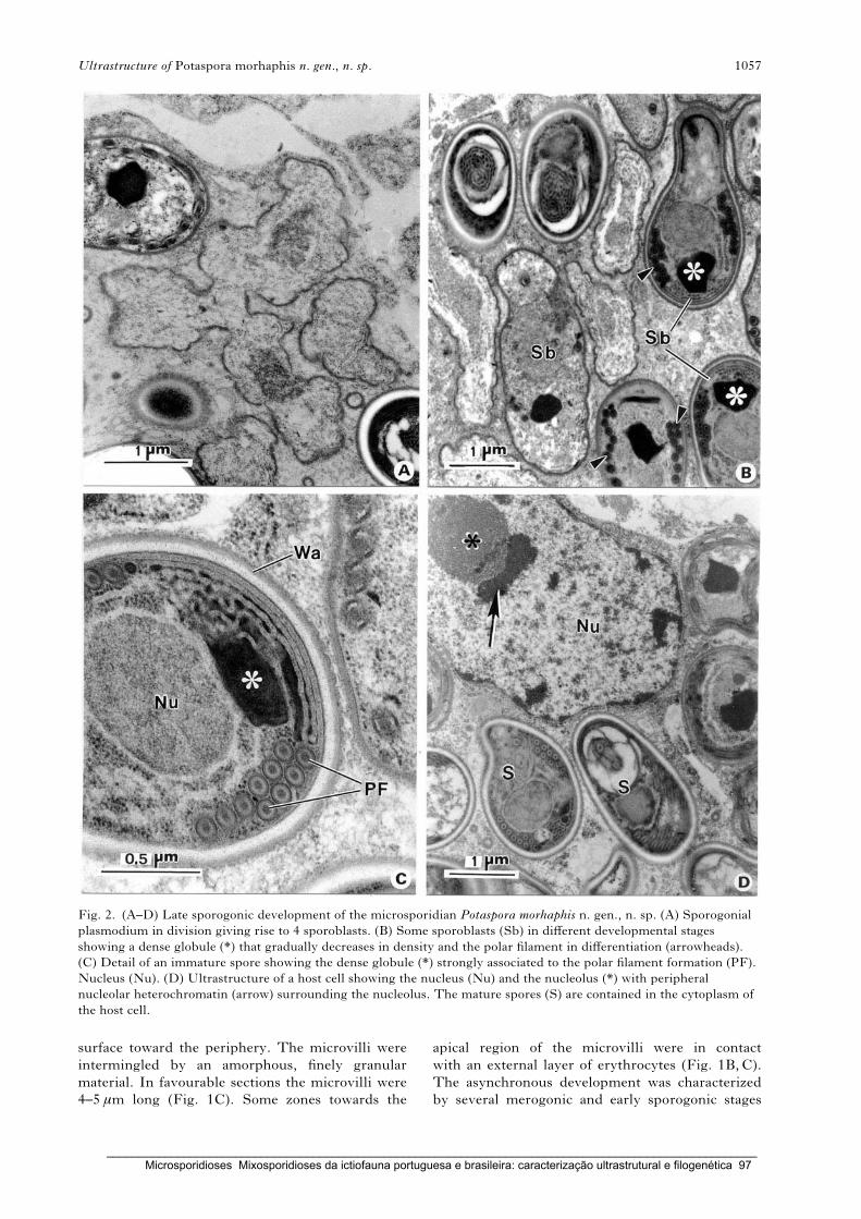
surface toward the periphery. The microvilli were
intermingled by an amorphous, finely granular
material. In favourable sections the microvilli were
4–5 mm long (Fig. 1C). Some zones towards the
apical region of the microvilli were in contact
with an external layer of erythrocytes (Fig. 1B,C).
The asynchronous development was characterized
by several merogonic and early sporogonic stages
Fig. 2. (A–D) Late sporogonic development of the microsporidian Potaspora morhaphis n. gen., n. sp. (A) Sporogonial
plasmodium in division giving rise to 4 sporoblasts. (B) Some sporoblasts (Sb) in different developmental stages
showing a dense globule (*) that gradually decreases in density and the polar filament in differentiation (arrowheads).
(C) Detail of an immature spore showing the dense globule (*) strongly associated to the polar filament formation (PF).
Nucleus (Nu). (D) Ultrastructure of a host cell showing the nucleus (Nu) and the nucleolus (*) with peripheral
nucleolar heterochromatin (arrow) surrounding the nucleolus. The mature spores (S) are contained in the cytoplasm of
the host cell.
Ultrastructure of Potaspora morhaphis n. gen., n. sp. 1057
_____________________________________________________________________________________________________ Microsporidioses Mixosporidioses da ictiofauna portuguesa e brasileira: caracterização ultrastrutural e filogenética 97
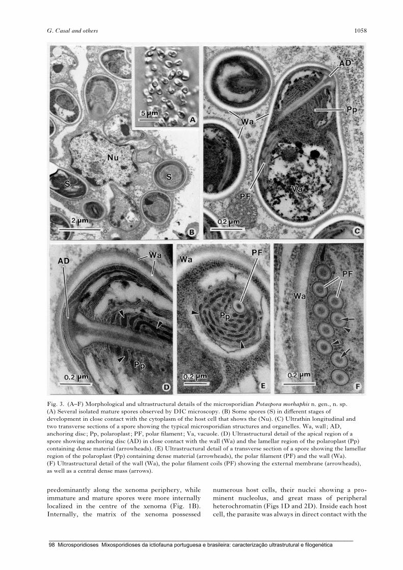
predominantly along the xenoma periphery, while
immature and mature spores were more internally
localized in the centre of the xenoma (Fig. 1B).
Internally, the matrix of the xenoma possessed
numerous host cells, their nuclei showing a pro-
minent nucleolus, and great mass of peripheral
heterochromatin (Figs 1D and 2D). Inside each host
cell, the parasite was always in direct contact with the
Fig. 3. (A–F) Morphological and ultrastructural details of the microsporidian Potaspora morhaphis n. gen., n. sp.
(A) Several isolated mature spores observed by DIC microscopy. (B) Some spores (S) in different stages of
development in close contact with the cytoplasm of the host cell that shows the (Nu). (C) Ultrathin longitudinal and
two transverse sections of a spore showing the typical microsporidian structures and organelles. Wa, wall ; AD,
anchoring disc; Pp, polaroplast ; PF, polar filament; Va, vacuole. (D) Ultrastructural detail of the apical region of a
spore showing anchoring disc (AD) in close contact with the wall (Wa) and the lamellar region of the polaroplast (Pp)
containing dense material (arrowheads). (E) Ultrastructural detail of a transverse section of a spore showing the lamellar
region of the polaroplast (Pp) containing dense material (arrowheads), the polar filament (PF) and the wall (Wa).
(F) Ultrastructural detail of the wall (Wa), the polar filament coils (PF) showing the external membrane (arrowheads),
as well as a central dense mass (arrows).
G. Casal and others 1058
_____________________________________________________________________________________________________ 98 Microsporidioses Mixosporidioses da ictiofauna portuguesa e brasileira: caracterização ultrastrutural e filogenética
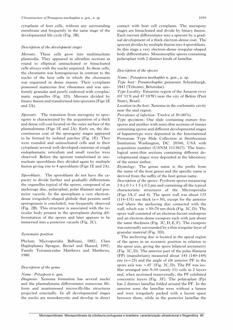
cytoplasm of host cells, without any surrounding
membrane and frequently in the same stage of the
developmental life cycle (Fig. 3B).
Description of the development stages
Meronts. These cells grow into multinucleate
plasmodia. They appeared in ultrathin sections as
round to elliptical uninucleated or binucleated
cells always with the nuclei unpaired. In these cells,
the chromatin was homogeneous in contrast to the
nuclei of the host cells in which the chromatin
was organized in dense masses. Their cytoplasm
possessed numerous free ribosomes and was uni-
formly granular and poorly endowed with cytoplas-
matic organelles (Fig. 1D). Meronts divided by
binary fission and transformed into sporonts (Figs 1E
and 2A).
Sporonts. The transition from merogony to spor-
ogony is characterized by the acquisition of a thick
and dense cell coat located on the outer surface of the
plasmalemma (Figs 1E and 2A). Early on, the dis-
continuous coat of the sporogony stages appeared
to be formed by isolated patches (Fig. 1E). They
were rounded and uninucleated cells and in their
cytoplasm several well-developed cisternae of rough
endoplasmatic reticulum and small vesicles were
observed. Before the sporont transformed in uni-
nucleate sporoblasts they divided again by multiple
fission giving rise to 4 sporoblasts (Figs 1E and 2A).
Sporoblasts. The sporoblasts do not have the ca-
pacity to divide further and gradually differentiate
the organelles typical of the spores, composed of an
anchorage disc, polaroplast, polar filament and pos-
terior vacuole. In the sporoplasm a very electron-
dense irregularly-shaped globule that persists until
sporogenesis is concluded, was frequently observed
(Fig. 2B). This structure is associated with the re-
ticular body present in the sporoplasm during dif-
ferentiation of the spores and later appears to be
immersed into a posterior vacuole (Fig. 2C).
Systematic position
Phylum Microsporidia Balbiani, 1882; Class
Haplophasea Sprague, Becnel and Hazard, 1992;
Family Tetramicridae Matthews and Matthews,
1980.
Description of the genus
Name: Potaspora n. gen.
Diagnosis : Xenoma formation has several nuclei
and the plasmalemma differentiates numerous fili-
form and anastomosed microvilli-like structures
projected externally. In all developmental stages
the nuclei are monokaryotic and develop in direct
contact with host cell cytoplasm. The merogony
stages are binucleated and divide by binary fission.
Each meront differentiates into a sporont by a grad-
ual development of a thick electron-dense coat. The
sporont divides bymultiple fission into 4 sporoblasts.
In this stage a very electron-dense irregular-shaped
body differentiates. Monomorphic spores containing
polaroplast with 2 distinct kinds of lamellae.
Description of the species
Name: Potaspora morhaphis n. gen., n. sp.
Type host : Potamorhaphis guianensis Schomburgk,
1843 (Teleostei, Belonidae).
Type Locality : Estuarine region of the Amazon river
(01x11kS and 47x18kW) near the city of Belem (Para
State), Brazil.
Location in the host : Xenoma in the coelomatic cavity
near the anal region.
Prevalence of infection : Twelve of 30 (40%).
Type specimens : One slide containing mature free
spores and another with semi-thin sections of tissues
containing spores and different developmental stages
of hapantotype were deposited in the International
Protozoan Type Slide Collection at Smithsonian
Institution Washington, DC. 20560, USA with
acquisition number (USNM 1113817). The histo-
logical semi-thin sections containing different de-
velopmental stages were deposited at the laboratory
of the senior author.
Etymology : The genus name is the prefix from
the name of the host genus and the specific name is
derived from the suffix of the host genus name.
Description of the spores : Pyriform spores measuring
2.8¡0.3r1.5¡0.2 mm and containing all the typical
characteristic structures of the Microsporidia
(Figs 3A,C and 4). The spore wall was about 125
(114–131) nm thick (n=30), except for the anterior
end where the anchoring disc contacted with the
wall, which was y50–70 nm thick (Fig. 3C,D). The
spore wall consisted of an electron-lucent endospore
and an electron-dense exospore each with just about
the same thickness (Fig. 3C,D,E,F). The exospore
was externally surrounded by a thin irregular layer of
granular material (Fig. 3D).
The anchoring disc is located in the apical region
of the spore in an eccentric position in relation to
the spore axis, giving the spore bilateral asymmetry
(Fig. 3C,D). The anterior part of the polar filament
(FP) (manubrium) measured about 145 (140–149)
nm (n=25) and the angle of tilt anterior PF to the
spore axis was y45x (Fig. 3C,D). The PF was iso-
filar arranged into 9–10 (rarely 11) coils in 2 layers
and, when sectioned transversally, the PF exhibited
concentric layers (Fig. 3F). The polaroplast (Pp)
has 2 distinct lamellae folded around the PF. In the
anterior zone the lamellae were without a lumen
and were irregularly packed with a lucent space
between them, while in the posterior lamellae the
Ultrastructure of Potaspora morhaphis n. gen., n. sp. 1059
_____________________________________________________________________________________________________ Microsporidioses Mixosporidioses da ictiofauna portuguesa e brasileira: caracterização ultrastrutural e filogenética 99
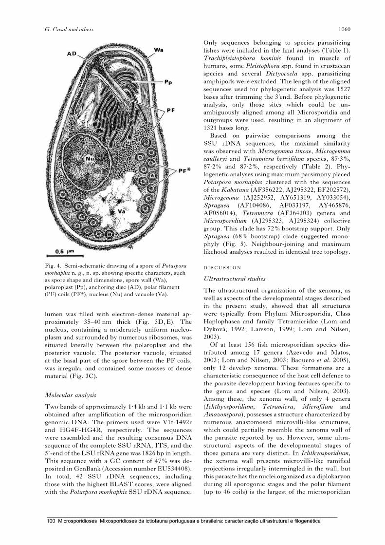
lumen was filled with electron-dense material ap-
proximately 35–40 nm thick (Fig. 3D,E). The
nucleus, containing a moderately uniform nucleo-
plasm and surrounded by numerous ribosomes, was
situated laterally between the polaroplast and the
posterior vacuole. The posterior vacuole, situated
at the basal part of the spore between the PF coils,
was irregular and contained some masses of dense
material (Fig. 3C).
Molecular analysis
Two bands of approximately 1.4 kb and 1.1 kb were
obtained after amplification of the microsporidian
genomic DNA. The primers used were V1f-1492r
and HG4F-HG4R, respectively. The sequences
were assembled and the resulting consensus DNA
sequence of the complete SSU rRNA, ITS, and the
5’-end of the LSU rRNA gene was 1826 bp in length.
This sequence with a GC content of 47% was de-
posited in GenBank (Accession number EU534408).
In total, 42 SSU rDNA sequences, including
those with the highest BLAST scores, were aligned
with the Potaspora morhaphis SSU rDNA sequence.
Only sequences belonging to species parasitizing
fishes were included in the final analyses (Table 1).
Trachipleistophora hominis found in muscle of
humans, some Pleistophora spp. found in crustacean
species and several Dictyocoela spp. parasitizing
amphipods were excluded. The length of the aligned
sequences used for phylogenetic analysis was 1527
bases after trimming the 3kend. Before phylogenetic
analysis, only those sites which could be un-
ambiguously aligned among all Microsporidia and
outgroups were used, resulting in an alignment of
1321 bases long.
Based on pairwise comparisons among the
SSU rDNA sequences, the maximal similarity
was observed with Microgemma tincae, Microgemma
caulleryi and Tetramicra brevifilum species, 87.3%,
87.2% and 87.2%, respectively (Table 2). Phy-
logenetic analyses using maximum parsimony placed
Potaspora morhaphis clustered with the sequences
of the Kabatana (AF356222, AJ295322, EF202572),
Microgemma (AJ252952, AY651319, AY033054),
Spraguea (AF104086, AF033197, AY465876,
AF056014), Tetramicra (AF364303) genera and
Microsporidium (AJ295323, AJ295324) collective
group. This clade has 72% bootstrap support. Only
Spraguea (68% bootstrap) clade suggested mono-
phyly (Fig. 5). Neighbour-joining and maximum
likehood analyses resulted in identical tree topology.
DISCUSSION
Ultrastructural studies
The ultrastructural organization of the xenoma, as
well as aspects of the developmental stages described
in the present study, showed that all structures
were typically from Phylum Microsporidia, Class
Haplophasea and family Tetramicridae (Lom and
Dykova, 1992; Larsson, 1999; Lom and Nilsen,
2003).
Of at least 156 fish microsporidian species dis-
tributed among 17 genera (Azevedo and Matos,
2003; Lom and Nilsen, 2003; Baquero et al. 2005),
only 12 develop xenoma. These formations are a
characteristic consequence of the host cell defence to
the parasite development having features specific to
the genus and species (Lom and Nilsen, 2003).
Among these, the xenoma wall, of only 4 genera
(Ichthyosporidium, Tetramicra, Microfilum and
Amazonspora), possesses a structure characterized by
numerous anastomosed microvilli-like structures,
which could partially resemble the xenoma wall of
the parasite reported by us. However, some ultra-
structural aspects of the developmental stages of
those genera are very distinct. In Ichthyosporidium,
the xenoma wall presents microvilli-like ramified
projections irregularly intermingled in the wall, but
this parasite has the nuclei organized as a diplokaryon
during all sporogonic stages and the polar filament
(up to 46 coils) is the largest of the microsporidian
Fig. 4. Semi-schematic drawing of a spore of Potaspora
morhaphis n. g., n. sp. showing specific characters, such
as spore shape and dimensions, spore wall (Wa),
polaroplast (Pp), anchoring disc (AD), polar filament
(PF) coils (PF*), nucleus (Nu) and vacuole (Va).
G. Casal and others 1060
_____________________________________________________________________________________________________ 100 Microsporidioses Mixosporidioses da ictiofauna portuguesa e brasileira: caracterização ultrastrutural e filogenética

group (Sprague and Vernick, 1974; Casal and
Azevedo, 1995). In Microfilum, the xenoma wall was
described as a very dense region covered by numer-
ous apparently disorganized and ramified microvilli.
This parasite gives rise to a spore characterized by a
manubrium inserted on a laterally offset anchoring
disc and extruding into a short non-coiled polar
filament, which was very different from those of
the present study (Faye et al. 1991). A xenoma wall
including a microvillous surface layer formed by
anastomosed elongated cytoplasmic processes have
been described in the genus Tetramicra (Matthews
and Matthews, 1980). Meronts located within a
vacuole in the host cytoplasm and spores with con-
spicuous posterosomes surrounded by a membrane
and located inside the posterior vacuole.
Recently in an Amazonian fish, a new genus
and species (Amazonspora hassar) having a xenoma,
strongly encapsulated, consisting of numerous
anastomosed microvilli-like projections penetrating
the 1–3 first layers of collagen fibres was described.
Up to approximately 22 juxtaposed crossed layers of
collagen fibres were observed (Azevedo and Matos,
2003).
The presence of dense globules of unknown
nature in the sporoplasm was also seen in other fish-
infecting Microsporidia. Several electron-dense
inclusion bodies, sometimes very large, measuring
up to 1.38 mm in diameter, were described in sporo-
blasts and spores of Tetramicra brevifilum species
(Matthews and Matthews, 1980). In Kabatana
arthuri (Lom et al. 1999) and K. takedai (Lom et al.
2001) a very similar globule was reported, while in
Loma acerinae (Lom and Pekkarinen, 1999) 1–3
homogeneous dense globules occupying all the
space of the posterior vacuole were observed. In our
observations a large inclusion consisting of reticular
material like that reported in Ichthyosporidium
giganteum, was also found (Sprague and Vernick,
1974; Casal and Azevedo, 1995).
The polaroplast of Potaspora morhaphis has a bi-
partite structure comprising the anterior region
having folds with a lamellar organization and the
posterior region with larger lamellae (cisternae) with
dense contents. A similar organization was reported
by Lom et al. (1999) in the species Kabatana arthuri
which infects the trunk muscles of fishes from the
South-East Asia freshwater fish, Pangasius sutchi, as
well as in Kabatana takedai (Lom et al. 2001). The
polaroplast of the microsporidian, Spraguea amer-
icana, found in the nervous tissues of the Japanese
anglerfish Lophius litulon (Freeman et al. 2004) has a
similar organization.
Phylogenetic relationships
The availability, in the public databases, of se-
quences from different species belonging to the
phylum Microsporidia makes the SSU rRNA geneTab
le2.ComparisonofsomeSSU
rDNA
sequen
ces:percentageofiden
tity
(topdiagonal)an
dpairw
isedistance
(bottom
diagonal)obtained
byKim
ura-2
parameteran
alysis
12
34
56
78
910
11
12
13
14
(1)Potasporamorhaphis
—86. 8
86. 8
86. 8
86. 8
86. 4
85. 9
87. 3
87. 2
87. 2
85. 2
85. 2
85. 2
86. 0
(2)Spraguea
lophii(1)
0. 132
—100
98. 2
100
99. 6
95. 3
98. 2
98. 9
98. 9
91. 0
91. 0
91. 0
96. 4
(3)Spraguea
sp.Lophiuslitulon
0. 132
0. 000
—98. 2
100
99. 6
95. 3
98. 2
98. 9
98. 9
91. 0
91. 0
91. 0
96. 4
(4)M
icrogemmavivaresi
0. 132
0. 018
0. 018
—98. 2
97. 9
94. 1
98. 6
98. 2
98. 2
90. 6
90. 6
90. 6
95. 3
(5)Spraguea
lophii(2)
0. 132
0. 000
0. 000
0. 018
—99. 6
95. 3
98. 2
98. 9
98. 9
91. 0
91. 0
91. 0
96. 4
(6)Spraguea
americana
0. 136
0. 004
0. 004
0. 021
0. 004
—94. 9
97. 9
97. 9
98. 6
98. 6
91. 6
91. 6
96. 0
(7)Kabatanatakedai
0. 141
0. 047
0. 047
0. 059
0. 047
0. 051
—94. 1
94. 9
94. 9
90. 2
90. 2
90. 2
96. 4
(8)M
icrogemmatincae
0. 127
0. 018
0. 018
0. 014
0. 018
0. 021
0. 059
—98. 2
98. 2
90. 6
90. 6
90. 6
95. 3
(9)Tetramicra
brevifilum
0. 128
0. 011
0. 011
0. 018
0. 011
0. 014
0. 051
0. 018
—100
91. 6
91. 6
91. 6
96. 0
(10)M
icrogemmacaulleryi
0. 128
0. 011
0. 011
0. 018
0. 011
0. 014
0. 051
0. 018
0. 000
—91. 6
91. 6
91. 6
96. 0
(11)M
icrosporidium
sp.RSB1
0. 148
0. 090
0. 090
0. 094
0. 090
0. 094
0. 098
0. 094
0. 094
0. 094
—100
100
89. 8
(12)Kabatanaseriolae
0. 148
0. 090
0. 090
0. 094
0. 090
0. 094
0. 098
0. 094
0. 094
0. 094
0. 000
—100
89. 8
(13)M
icrosporidium
GHB
0. 148
0. 090
0. 090
0. 094
0. 090
0. 094
0. 098
0. 094
0. 094
0. 094
0. 000
0. 000
—89. 8
(14)Kabatananew
berryi
0. 140
0. 036
0. 036
0. 047
0. 036
0. 040
0. 036
0. 047
0. 040
0. 040
0. 102
0. 102
0. 102
—
Ultrastructure of Potaspora morhaphis n. gen., n. sp. 1061
_____________________________________________________________________________________________________ Microsporidioses Mixosporidioses da ictiofauna portuguesa e brasileira: caracterização ultrastrutural e filogenética 101

Fig. 5. Parsimony tree of SSU rDNA sequences to compare Potaspora morhaphis with selected sequences from other
fish-infecting Microsporidia. The analysis was conducted using 1321 aligned nucleotide positions of the highest
G. Casal and others 1062
_____________________________________________________________________________________________________ 102 Microsporidioses Mixosporidioses da ictiofauna portuguesa e brasileira: caracterização ultrastrutural e filogenética

the most suitable not only for development of diag-
nostic tools and species identification, but also for
molecular characterization of new parasites through
phylogenetic studies (Weiss and Vossbrinck, 1999;
Lom and Nilsen, 2003).
In these studies we can see that there is 72%
bootstrap support for a clade composed of micro-
sporidian belonging to the Kabatana (Lom et al.
1999, 2001; McGourty et al. 2007), Microgemma
(Cheney et al. 2000; Leiro et al. 2000; Mansour et al.
2005), Spraguea (Freeman et al. 2004), Tetramicra
(Leiro et al. 2000) genera, 2 unclassified species of
Microsporidium group (Bell et al. 2001) and the
microsporidian reported in this study. This result
is in concordance with cladograms previously ob-
tained by Lom and Nilsen (2003) and designated
as group IV. Our SSU rDNA sequence analysis
also shows that Potaspora n. gen. does not have any
sister taxa and the lineage is distantly related to
the other species examined. Comparing SSU rRNA
gene sequences between P. morhaphis with species
Microgemma caulleryi and Tetramicra brevifilum
(clade with bootstrap 81%) the genetic distances are
12.8% for both species. The smallest genetic distance
was observed with the species Microgemma tincae
(12.7%) but the SSU rRNA was not completely se-
quenced. The bootstrap support for this species
and another Microgemma vivaresi is 81%. On the
other hand, K. takedai and K. newberryi group in a
clade with 83% bootstrap and allSpraguea species are
clustered in the clade with 68% bootstrap.
Conclusion
When comparing the xenoma wall of the parasite
described here with those fish Microsporidia which
form xenoma some structural differences were
found, such as the organization of the microvilli-like
structures. In addition, the ultrastructural organiz-
ation of the polaroplast and the presence of a dense
globule were the most evident differences found
compared with other mature spores of previously
described species. Concerning this last aspect, the
only exception is the spore ofKabatana genus which
presents some similarities. However, they were
found to parasitize only the muscle fishes and they do
not develop inside of xenomas (Lom et al. 1999,
2001; McGourty et al. 2007). As concerns molecular
biology, the most parsimonious cladogram has
shown that Potaspora morhaphis is placed in the same
group as the Kabatana, Microgemma, Spraguea
and Tetramicra genera, does not have any sister taxa
and has the lowest percentage identity within the
group.
So, our results suggest that this parasite does not fit
into any of the known fish microsporidian genera,
and for these reasons we propose a new genus
Potaspora and a new species, Potaspora morhaphis.
This work was partially supported by the Engx. A. AlmeidaFoundation (Porto, Portugal), Ph.D. grant from ‘CESPU’(G. Casal), ‘CNPq’ and ‘CAPES’ – Brazil. We would liketo thank the iconographic work of Mr Joao Carvalheiro.We would like to thank the anonymous reviewers for theirhelpful suggestions and comments.
REFERENCES
Azevedo, C. and Matos, E. (2002). Fine structure of a
new species, Loma myrophis (Phylum Microsporidia),
parasite of the Amazonian fish Myrophis platyrhynchus
(Teleostei, Ophichthidae). European Journal of
Protistology 37, 445–452.
Azevedo, C. and Matos, E. (2003). Amazonspora hassar
n. gen. and n. sp. (phylum Microsporidia, fam
Glugeidae), a parasite of the Amazonian teleost Hassar
orestis (fam. Doradidae). Journal of Parasitology 89,
336–341.
Azevedo, C., Balseiro, P., Casal, G., Gestal, C.,
Aranguren, R., Stokes, N. A., Carnegie, R. B.,
Novoa, N., Burreson, E. M. and Figueras, A. (2006).
Ultrastructural and molecular characterization of
Haplosporidium montforti n. sp., parasite of the European
abalone Haliotis tuberculata. Journal of Invertebrate
Pathology 92, 23–32.
Baquero, E., Rubio, M., Moura, I. N. S., Pieniazek, J.
and Jordana, R. (2005). Myosporidium merluccius n. g.,
n. sp. infecting muscle of commercial hake (Merluccius
sp.) from fisheries near Namibia. The Journal of
Eukaryotic Microbiology 52, 476–483.
Bell, A. S., Aoki, T. and Yokoyama, H. (2001).
Phylogenetic relationships among Microsporidia based
on rDNA sequence data, with particular reference to
fish-infectingMicrosporidium Balbiani 1884 species.The
Journal of Eukaryotic Microbiology 48, 258–265.
Casal, G. and Azevedo, C. (1995). New ultrastructural
data on the microsporidian Ichthyosporidium giganteum
infecting the marine teleostean fishCtenolabrus rupestris.
Journal of Fish Diseases 18, 191–194.
Cheney, S. A., Lafranchi-Tristem, N. J. and Canning,
E. U. (2000). Phylogenetic relationships of Pleistophora-
like Microsporidia based on small subunit ribosomal
DNA sequences and implications for the source of
Trachipleistophora hominis infections. The Journal of
Eukaryotic Microbiology 47, 280–287.
Faye, N., Toguebaye, B. S. and Bouix, G. (1991).
Microfilum lutjani n. g. n. sp. (Protozoa, Microsporida),
a gill parasite of the golden African snapper
Lutjanus fulgens (Valenciennes, 1830) (Teleost
BLAST score microsporidian sequences and 4 more microsporidian sequences as outgroup. The bar indicates the
equivalence between the distance and the number of changes. The numbers on the branches indicate bootstrap support
from 100 replicates. Potaspora morhaphis is placed within group IV (Lom and Nilsen, 2003) (highlighted box), which
includes the sequences of the genera Kabatana, Microgemma, Spraguea, Tetramicra, and Microsporidium.
Ultrastructure of Potaspora morhaphis n. gen., n. sp. 1063
_____________________________________________________________________________________________________ Microsporidioses Mixosporidioses da ictiofauna portuguesa e brasileira: caracterização ultrastrutural e filogenética 103

Lutjanidae): Developmental cycle and ultrastructure.
Journal of Protozoology 38, 30–40.
Freeman,M. A., Yokoyama,H. andOgawa, K. (2004).
A microsporidian parasite of the genus Spraguea in
the nervous tissues of the Japanese anglerfish Lophius
litulon. Folia Parasitologica 51, 167–176.
Gatehouse, H. S. and Malone, L. A. (1998). The
ribosomal RNA gene region of Nosema apis
(Microspora): DNA sequence for small and large
subunit rRNAgenes and evidence of a large tandem
repeat unit size. Journal of Invertebate Pathology 71,
97–105.
Larsson, J. I. R. (1999). Identification of Microsporidia.
Acta Protozoologica 38, 161–197.
Leiro, J., Siso, M. I. G., Parama, A., Ubeira, F. M. and
Sanmartin, M. L. (2000). RFLP analysis of PCR-
amplified small subunit ribosomal DNA of three fish
microsporidian species. Parasitology 120, 113–119.
Lom, J. (2002). A catalogue of described genera and
species of microsporidians parasitic in fish. Systematic
Parasitology 53, 81–99.
Lom, J. and Dykova, I. (1992). Microsporidia (Phylum
Microspora Sprague, 1977). In Protozoan Parasites of
Fishes. Developments in Aquaculture and Fisheries
Sciences (ed. Lom, J. and Dykova, I.), Vol. 26,
pp. 125–157. Elsevier, Amsterdam.
Lom, J., Dykova, I. and Tonguthai, K. (1999).Kabataia
gen. n., new genus proposed for Microsporidium spp.
infecting trunk muscles of fishes. Diseases of Aquatic
Organisms 38, 39–46.
Lom, J. and Nilsen, F. (2003). Fish Microsporidia : fine
structural diversity and phylogeny. International Journal
for Parasitology 33, 107–127.
Lom, J., Nilsen, F. and Urawa, S. (2001). Redescription
of Microsporidium takedai (Awakura, 1974) as Kabatana
takedai (Awakura, 1974) comb. n. Diseases of Aquatic
Organisms 44, 223–230.
Lom, J. and Pekkarinen, M. (1999). Ultrastructural
observations on Loma acerinae (Jırovec, 1930) comb.
nov. (Phylum Microsporidia). Acta Protozoologica 38,
61–74.
Mansour, L., Prensier, G., Jemaa, S. B., Hassine,
O. K. B., Metenier, G., Vivares, C. P. and Cornillot,
E. (2005). Description of a xenoma-inducing
microsporidian, Microgemma tincae n. sp., a parasite of
the teleost fish Symphodus tinca from Tunisian coasts.
Diseases of Aquatic Organisms 65, 217–226.
Matos, E. and Azevedo, C. (2004). Ultrastructural
description ofMicrosporidium brevirostris sp. n., parasite
of the teleostean Brachyhypopomus brevirostris
(Hypopomidae) from the Amazon River. Acta
Protozoologica 43, 261–267.
Matos, E., Corral, L. and Azevedo, C. (2003).
Ultrastructural details of the xenoma of Loma myrophis
(phylum Microsporidia) and extrusion of the polar tube
during autoinfection. Diseases of Aquatic Organisms 54,
203–207.
Matthews, R. A. and Matthews, B. F. (1980). Cell and
tissue reactions of turbot Scophthalmus maximus (L.)
to Tetramicra brevifilum gen. n., sp. n. (Microspora).
Journal of Fish Diseases 3, 495–515.
Nilsen, F. (2000). Small subunit ribosomal DNA
phylogeny of Microsporidia with particular reference
to genera that infect fish. Journal of Parasitology 86,
128–133.
McGourty, K. R., Kinzger, A. P., Hendrickson, G. L.,
Goldsmith, G. L., Casal, G. and Azevedo, C. (2007).
A new microsporidian infecting the musculature of the
endangered tidewater goby (Gobiidae). Journal of
Parasitology 93, 655–660.
Sprague, V., Becnel, J. J. and Hazard, E. I. (1992).
Taxonomy of phylum Microspora. Critical Reviews in
Microbiology 18, 285–395.
Sprague, V. and Vernick, S. (1974). Fine structure of the
cyst and some sporulation stages of Ichthyosporidium
(Microsporidia). Journal of Protozoology 21, 667–677.
Tamura, K., Dudley, J., Nei, M. and Kumar S. (2007).
MEGA4: Molecular evolutionary genetics analysis
(MEGA) software version 4.0. Molecular Biology and
Evolution 24, 1596–1599.
Thompson, J. D., Higgins, D. G. and Gilson, T. J.
(1994). Clustal W: improving the sensitivity of
progressive multiple sequence alignment through
sequence weighting, position-specific gap penalties
and weight matrix choice. Nucleic Acids Research 22,
4673–4680.
Vossbrinck, C. R., Baker, M. D., Didier, E. S.,
Debrunner-Vossbrinck, B. A. and Shadduck, J. A.
(1993). Ribosomal DNA sequences of Encephalitozoon
hellem and Encephalitozoon cuniculi : species
identification and phylogenetic construction. The
Journal Eukaryotic of Microbiology 40, 354–362.
Weiss, L. and Vossbrinck, C. (1999). Molecular biology,
molecular phylogeny, and molecular diagnostic
approaches to the Microsporidia. In The Microsporidia
and Microsporidiosis (ed. Wittner, M. and Weiss, L.),
pp. 129–171. American Society of Microbiology,
Washington, DC.
G. Casal and others 1064
_____________________________________________________________________________________________________ 104 Microsporidioses Mixosporidioses da ictiofauna portuguesa e brasileira: caracterização ultrastrutural e filogenética

Capítulo 3
MORPHOLOGICAL AND GENETICAL DESCRIPTION OF
LOMA PSITTACA SP. N. ISOLATED FROM THE AMAZONIAN FISH
SPECIES COLOMESUS PSITTACUS
Parasitology Research (2009) in press
Graça Casal, Edilson Matos, M. Leonor Teles-Grilo & Carlos Azevedo
_____________________________________________________________________________________________________ Microsporidioses Mixosporidioses da ictiofauna portuguesa e brasileira: caracterização ultrastrutural e filogenética 105

_____________________________________________________________________________________________________ 106 Microsporidioses Mixosporidioses da ictiofauna portuguesa e brasileira: caracterização ultrastrutural e filogenética

ORIGINAL PAPER
Morphological and genetical description of Loma psittaca sp. n.isolated from the Amazonian fish speciesColomesus psittacus
Graça Casal & Edilson Matos & M. Leonor Teles-Grilo &
Carlos Azevedo
Received: 2 April 2009 /Accepted: 19 June 2009# Springer-Verlag 2009
Abstract A previously unrecognised fish-infecting micro-sporidia (Loma psittaca n. sp.), found adherent to theintestinal mucosa of the freshwater puffer fish Colomesuspsittacus (Teleostei, Tetraodontidae) from lower AmazonRiver, was described based on light and transmissionelectron microscope and phylogenetic analysis. The whitishxenoma was completely filled by numerous spores, includ-ing several developmental stages of the parasite. In all ofthese stages, the nuclei were monokaryotic. The merogonialplasmodium divided by binary fission and the sporont gaverise to disporoblastic ovoid spores measuring 4.2 ± 0.4 ×2.8 ± 0.4 μm. In mature spores, the polar filament was
arranged in 10–11 (rarely 12) coils in one row in turn ofposterior vacuole. The polaroplast had two distinct regionsaround the manubrium. The polyribosomes were organisedin coiled tapes. The small subunit rRNA gene wassequenced and maximum parsimony analysis placed themicrosporidian described here in the clade that includes thegenera Ichthyosporidium, Loma and Pseudoloma. Based ondifferences from previously described microsporidians,such as ultrastructural characteristics of the xenoma,developmental stages including the spore and phylogeneticanalysis supported the recognition of a new species, hereinnamed L. psittaca n. sp.
Introduction
The members of the phylum Microsporidia Balbiani, 1882are widespread, minute, obligatory intracellular parasitesfound in most invertebrate phyla and in vertebrates, withthe majority of species in insects and fish (Lom andDyková 1992; Sprague et al. 1992; Larsson 1999; Lom2002). Presently, there are at least 144 available genera(Larsson 1999), 18 of them occurring in teleost fishes fromthe different geographic areas and habitat (Azevedo andMatos 2003; Lom and Nilsen 2003; Baquero et al. 2005;Casal et al. 2008), and some of them are recognised asserious pathogens for their hosts. Fishes are hosts to 156recorded species of microsporidia, 11 species belonging tothe genus Loma Morrison and Sprague, 1981 and the othereight parasitoses were classified as Loma spp. (Lom 2002).One of them, Loma myrophis, was found in the subepithe-lial tissues of the fish gut Myrophis platyrhynchus fromAmazonian fauna (Azevedo and Matos 2002; Matos et al.2003). About those from South America, particularly fromthe Amazon River where lives a diverse assemblage of
G. Casal : C. Azevedo (*)Department of Cell Biology, Institute of Biomedical Sciences,University of Porto (ICBAS/UP),Lg. A. Salazar no. 2,4099-003 Porto, Portugale-mail: [email protected]
G. Casal : C. AzevedoLaboratory of Pathology,Centre for Marine Environmental Research (CIIMAR/UP),4050-123 Porto, Portugal
G. CasalDepartamento de Ciências,Instituto Superior de Ciências da Saúde–Norte,Gandra, Portugal
E. MatosCarlos Azevedo Research Laboratory,Federal Rural University of Amazonia,Belém (Pará), Brazil
M. L. Teles-GriloLaboratory of Molecular Genetics,Institute of Biomedical Sciences,University of Porto (ICBAS/UP),Porto, Portugal
Parasitol ResDOI 10.1007/s00436-009-1547-1
_____________________________________________________________________________________________________ Microsporidioses Mixosporidioses da ictiofauna portuguesa e brasileira: caracterização ultrastrutural e filogenética 107
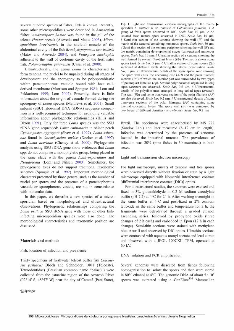
several hundred species of fishes, little is known. Recently,some other microsporidiosis were described in Amazonianfishes: Amazonspora hassar was found in the gill of theteleost Hassar orestis (Azevedo and Matos 2003), Micro-sporidium brevirostris in the skeletal muscle of theabdominal cavity of the fish Brachyhypopomus brevirostris(Matos and Azevedo 2004), and Potaspora morhaphisadherent to the wall of coelomic cavity of the freshwaterfish, Potamorhaphis guianensis (Casal et al. 2008).
Ultrastructurally, the genus Loma is characterised toform xenoma, the nuclei to be unpaired during all stages ofdevelopment and the sporogony to be polysporoblasticwithin parasitophorous vacuole bound with host cell-derived membrane (Morrison and Sprague 1981; Lom andPekkarinen 1999; Lom 2002). Presently, there is littleinformation about the origin of vacuole formed during thesporogony of Loma species (Matthews et al. 2001). Smallsubunit (SSU) ribosomal DNA (rDNA) sequence compar-ison is a well-recognised technique for providing valuableinformation about phylogenetic relationships (Hillis andDixon 1991). Only for three Loma species was the SSUrDNA gene sequenced: Loma embiotocia in shiner perchCymatogaster aggregata (Shaw et al. 1997), Loma salmo-nae found in Oncorhynchus mykiss (Docker et al. 1997)and Loma acerinae (Cheney et al. 2000). Phylogeneticanalysis using SSU rDNA gene show evidences that Lomaspp. do not comprise a monophyletic group, being placed inthe same clade with the genera Ichthyosporidium andPseudoloma (Lom and Nilsen 2003). Sometimes, thephylogenetic trees do not support traditional taxonomicschemes (Sprague et al. 1992). Important morphologicalcharacters presented by those genera, such as the number ofnuclei per spores and the presence of a parasitophorousvacuole or sporophorous vesicle, are not in concordancewith molecular data.
In this paper, we describe a new species of a micro-sporidian based on morphological and ultrastructuralobservations. Phylogenetic relationships comparing theLoma psittaca SSU rRNA gene with those of other fish-infecting microsporidian species were also done. Themorphological characteristics and taxonomic position arediscussed.
Materials and methods
Fish, location of infection and prevalence
Thirty specimens of freshwater teleost puffer fish Colome-sus psittacus Bloch and Schneider, 1801 (Teleostei,Tetraodontidae) (Brazilian common name “baiacú”) werecollected from the estuarine region of the Amazon River(02°14′ S, 48°57′ W) near the city of Cametá (Pará State),
Brazil. The specimens were anaesthetised by MS 222(Sandoz Lab.) and later measured (8–12 cm in length).Infection was determined by the presence of xenomaslocated in the intestinal mucosa. The prevalence ofinfection was 30% (nine fishes in 30 examined) in bothsexes.
Light and transmission electron microscopy
For light microscopy, smears of xenoma and free sporeswere observed directly without fixation or stain by a lightmicroscope equipped with Nomarski interference contrast[differential interference contrast (DIC)] optics.
For ultrastructural studies, the xenomas were excised andfixed in 3% glutaraldehyde in 0.2 M sodium cacodylatebuffer (pH 7.2) at 4°C for 24 h. After washing overnight inthe same buffer at 4°C and post-fixed in 2% osmiumtetroxide in the same buffer and temperature for 3 h, thefragments were dehydrated through a graded ethanolascending series, followed by propylene oxide (threechanges of 2 h each) and embedded in Epon (12 h in eachchange). Semi-thin sections were stained with methyleneblue-Azur II and observed by DIC optics. Ultrathin sectionswere contrasted with aqueous uranyl acetate and lead citrateand observed with a JEOL 100CXII TEM, operated at60 kV.
DNA isolation and PCR amplification
Several xenomas were dissected from fishes followinghomogenisation to isolate the spores and then were storedin 80% ethanol at 4°C. The genomic DNA of about 5×10⁶
spores was extracted using a GenEluteTM Mammalian
Fig. 1 Light and transmission electron micrographs of the micro-sporidian L. psittaca n. sp. parasite of Colomessus psittacus. 1 Agroup of fresh spores observed in DIC. Scale bar, 10 μm. 2 Anisolated fresh mature spore observed in DIC. Scale bar, 10 μm.3 Semi-thin section of the xenoma showing the wall (W) and thematrix of the xenoma containing numerous spores. Scale bar, 50 μm.4 Semi-thin section of the xenoma periphery showing the wall (W) andthe matrix containing developmental stages (asterisk) and numerousspores. Scale bar, 10 μm. 5 Ultrathin section of a xenoma showing thewall formed by several fibroblast layers (Fb). The matrix shows somespores (Sp). Scale bar, 5 μm. 6 Ultrathin section of some spores (Sp)sectioned at different levels showing the internal organisation. Scalebar, 1 μm. 7 Ultrastructural details of the spore apical zone showingthe spore wall (Wa), the anchoring disc (AD) and the polar filamentsections (PF) of which the anterior part was surrounded by two typesof polaroplast lamellae (Pp). Several polyribosomes organised in longtapes (arrows) are observed. Scale bar, 0.5 μm. 8 Ultrastructuraldetails of the polyribosomes arranged in long coiled tapes (arrows).The wall (Wa) and some transverse section of the polar filament (PF)are also observed. Scale bar, 0.2 μm. 9 Ultrastructural details of sometransverse sections of the polar filaments (PF) containing someinternal concentric layers. The spore wall (Wa) was composed bytwo layers of different densities (arrowheads). Scale bar, 0.2 μm
b
Parasitol Res
_____________________________________________________________________________________________________ 108 Microsporidioses Mixosporidioses da ictiofauna portuguesa e brasileira: caracterização ultrastrutural e filogenética
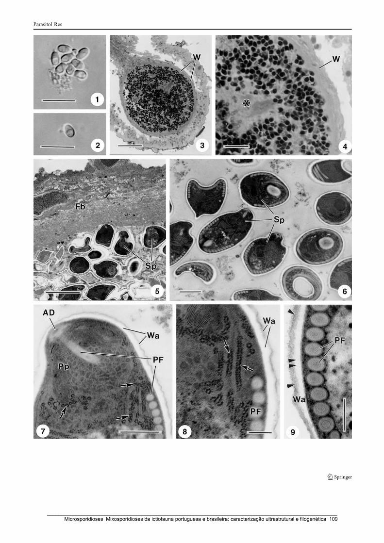
Parasitol Res
_____________________________________________________________________________________________________ Microsporidioses Mixosporidioses da ictiofauna portuguesa e brasileira: caracterização ultrastrutural e filogenética 109

Genomic DNA Miniprep kit (Sigma) according to themanufacturer’s instructions for animal tissue protocol,except for the incubation time. The DNA was stored in50 μl of TE buffer at −20°C until use. The DNAconcentration was estimated with the QubitTM Fluorometer(Invitrogen). The majority of the region coding for the SSUrRNA gene was amplified by polymerase chain reaction(PCR) using the primers V1f (5′ CACCAGGTTGATTCTGCC 3′) and 1492r (5′ GGTTACCTTGTTACGACTT 3′) (Vossbrinck et al. 1993; Nilsen 2000). PCR wascarried out in 50 μl reactions using 10 pmol of each primer,10 nmol of each dNTP, 2 mM of MgCl2, 5 μl 10X Taqpolymerase buffer, 1.25 U Taq DNA polymerase (Invitro-gen products) and 3 μl of the genomic DNA. The reactionswere run on Hybaid PxE Thermocycler (Thermo ElectronCorporation, Milford, MA, USA). The amplificationprogramme consisted of 94°C denaturation for 5 min,followed by 35 cycles of 94°C for 1 min, 50°C for 1 minand 72°C for 2 min. A final elongation step was performedat 72°C for 10 min. Aliquots (5 μl) of PCR products werevisualised with ethidium bromide staining after running ona 1% agarose gel.
DNA sequencing
PCR product for the SSU rRNA gene has an approximatesize of 1,400 bp. It was cleaned using the MinElute PCRpurification kit (Qiagen) and then three purified PCRproducts were sequenced in both directions. Sequencingwas done using BigDye terminator v1.1 of AppliedBiosytems kit, and the sequence reactions were run on anABI3700 DNA analyser Perkin-Elmer, Applied Biosys-tems, Stabvida, Co., Oeiras, Portugal).
Distance and phylogenetic analysis
To evaluate the relationship of L. psittaca to other micro-sporidians, a homology search was performed usingBLAST programme (Altschul et al. 1990). We used 44rDNA sequences belonging to the microsporidians para-sitising fish species. The sequence and NCBI accessionnumber data obtained from GenBank are the following:Aspalatospora milevae (EF990668); Glugea anomala(AF044391); Glugea atherinae (U15987); Glugea pleco-glossi (AJ295326); Glugea stephani (AF056015); Glugea
Table 1 Comparative measurements (in μm) from Loma spp.
Loma sp. Host and local infection Habitatcountries
Spore shape Spore Polar filament References
Length Width Coils Row
L. branchialis(=L. morhua)
Melanogrammusaeglefinus Gill filaments
MarineBoreo-Artic
Ellispoidal /ovoid
4.2 2.0 16–17 isofilar (Morrison andSprague 1981)6 4 16–19
L. salmonae Oncorhynchus mykiss Freshwater Pyriform/ellipsoidal
3.7 2.2 12–14 (Putz et al. 1965)Gill filaments Several countries 4.4 2.3 14–17
L. fontinalis Salvelinus fontinalis Freshwater – 12–14 (Morrison andSprague 1983)Gill lamellae Canada
L. dimorpha Gobius niger (andothers species)
Marine Ovoid/ellipsoidal
4.5 1.8–2.0 13–15 Isofilar (Loubès et al. 1984)
Connective tissue ofdigestive tract
France and Spain
L. diplodae Diplodus sargus Marine Ovoid 4.17 2.22 17–18 Bekhti and Bouix 1985)
Vessels of the gill filaments France
L. trichiuri Trichurus savala Marine Pyriform 3.0 2.0 – (Sandeep andKalavati 1985)Gill filaments India
L. camerounensis Oerochromis niloticus Freshwater Ovoid 3.96 2.16 11–12 (Fomena et al. 1992)Oesophagus to intestine Cameroon
L. boopsi Boops boops Marine Ovoid 3.7 2.4 12–14 Isofilar (Faye et al. 1995)Liver and digestive tract Senegal 16–18
L. embiotocia Cymatogaster aggregate Marine Ovoid 4.8 2.6 14–18 (Shaw et al. 1997)Gills Canada
L. acerinae Gymnocaphalus cernuus Freshwater Ellipsoidal 4.64 2.19 11–23 Isofilar (Lom andPekkarinen 1999)Intestine wall Czech Republic
L. myrophis Myrophis platyrhynchus Freshwater Ellipsoidal 4.06 1.61 13–14 Isofilar (Azevedo andMatos 2002)Subepithelial gut tissue Brazil
L. psittaca n. sp. Colomesus psittacus Freshwater Ovoid 4.2 2.8 11–12 Isofilar This studyIntestinal wall Brazil
Parasitol Res
_____________________________________________________________________________________________________ 110 Microsporidioses Mixosporidioses da ictiofauna portuguesa e brasileira: caracterização ultrastrutural e filogenética

sp. GS1 (AJ295325); Glugea sp. (AY090038); Heterospo-ris anguillarum (AF387331); Heterosporis sp. PF(AF356225); Ichthyosporidium sp. (L39110); Kabatanatakedai (AF356222); Kabatana newberryi (EF202572);Kabatana seriolae (AJ295322); Loma acerinae(AJ252951); Loma embiotocia (AF320310); L. salmonae(U78736); Loma sp. (AF104081); Microgemma caulleryi(AY033054); Microgemma tincae (AY651319); Micro-gemma vivaresi (AJ252952); Microsporidium cypselurus(AJ300706); Microsporidium prosopium (AF151529);Microsporidium GHB1 (AJ295324); Microsporidium sp.RSB1 (AJ295323); Microsporidium sp. STF (AY140647);Microsporidium MYX1 (AJ295329); Myosporidium mer-luccius (AY530532); Nucleospora salmonis (U78176);Ovipleistophora mirandellae (AF356223); Ovipleistophoraovariae (AJ252955); Pleis tophora ehrenbaumi(AF044392); Pleistophora finisterrensis (AF044393);Pleistophora hippoglossoideos (AJ252953); Pleistophoratypicalis (AF044387); Pleistophora sp. 1 (AF044394);Pleistophora sp. 2 (AF044389); Pleistophora sp. 3(AF044390); Potaspora morhaphis (EU534408); Pseudo-loma neurophilia (AF322654); Spraguea americana(AF056014); Spraguea lophii (1) (AF104086); S. lophii(2) (AF033197); Spraguea sp. (AY465876); Tetramicrabrevifilum (AF364303). Endoreticulatus schubergi(L39109); Enterocytozoon bieneusi (L07123); Vairimorphanecatrix (Y00266) and Vittaforma corneae (L39112) wereused as outgroup. Sequences were aligned as described byCasal et al. (2008). Alignment was done through Clustal W(Thompson et al. 1994) in MEGA 4 software (Tamura et al.2007), with an opening gap penalty of 10 and a gapextension penalty of 4 for both pairwise and multiplealignments. Subsequent phylogenetic and molecular evolu-tionary analyses were conducted using MEGA 4, with the44 rDNA sequences for microsporidian species and theoutgroup species selected. Distance estimation was carriedout using the Kimura-2 parameter model distance matrix fortransitions and transversions. For the phylogenetic treereconstructions, maximum parsimony analysis was con-ducted using the close neighbour interchange heuristicoption with a search factor 2 and random initial treesaddition of 2,000 replicates. Bootstrap values were calcu-lated over 100 replicates.
Results
Some spherical whitish xenomas were macroscopicallyobserved adherent to the intestinal mucosa of the fish.After rupture of the xenoma wall, free spores (1 and 2 inFig. 1) were easily microscopically observed and identifiedas belonging to the phylum Microsporidia. These xenomaswith up to ~310 μm diameter, filled with numerous spores
and different developmental stages, contained a thickxenoma wall formed by several juxtaposed fibroblast layers(3 and 4 in Fig. 1).
Description of L. psittaca n. sp.
Systematic position (Figs. 1 and 2)Phylum Microsporidia Balbiani, 1882Class Haplophasea Sprague, Becnel and Hazard, 1992Order Glugeida Issi, 1986Family Glugeidae Thélohan, 1892Genus Loma Morrison and Sprague, 1981Species: L. psittaca n. sp.
Description of the species
Type host: C. psittacus Bloch and Schneider, 1801 (Tele-ostei, Tetraodontidae) (Brazilian common name “baiacú”).
Type locality: Estuarine region of the Amazon River (02°14′ S, 48°57′ W) near the city of Cametá (Pará State),Brazil.
Pathogenecity: The whitish cysts (xenoma) wall wasformed by several juxtaposed collagen layers intermingledwith some fibroblasts, but no other tissue reactions wereobserved and no clinical signs were detected.
Location in the host: Xenoma in the intestinal mucosa.
Prevalence of infection: Nine of 30 (30%).
Type specimens: One glass slide containing mature freespores and others with semi-thin sections of tissuescontaining spores and different developmental stages ofhapantotype were deposited in the International ProtozoanType Slide Collection at Smithsonian Institution Washing-ton, DC, 20560, USA, with acquisition number USNM1123998. The histological semi-thin sections containingdifferent developmental stages were deposited at thelaboratory of the senior author.
Etymology: The specific name is derived from the genericname of the host species.
Description of the spores
Ovoid spores measuring 4.2 ± 0.4 × 2.8 ± 0.4 μm (n = 30)contained all typical characteristic structures of the Micro-sporidia (1, 2, 6 and 7 in Fig. 1). The spore wall was about87 nm thick, except for the anterior end where the centralzone of the anchoring disc contacted with the wall, whichwas about 20–35 nm thick consisting of an electron-lucent
Parasitol Res
_____________________________________________________________________________________________________ Microsporidioses Mixosporidioses da ictiofauna portuguesa e brasileira: caracterização ultrastrutural e filogenética 111
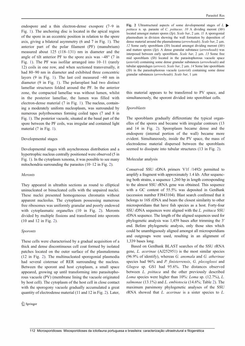
endospore and a thin electron-dense exospore (7–9 inFig. 1). The anchoring disc is located in the apical regionof the spore in an eccentric position in relation to the sporeaxis, giving a bilateral asymmetry (6 and 7 in Fig. 1). Theanterior part of the polar filament (PF) (manubrium)measured about 125 (118–131) nm in diameter and theangle of tilt anterior PF to the spore axis was ~48° (7 inFig. 1). The PF was isofilar arranged into 10–11 (rarely12) coils in one row, and when sectioned transversally, ithad 80–90 nm in diameter and exhibited three concentriclayers (9 in Fig. 1). The last coil measured ~60 nm indiameter (9 in Fig. 1). The polaroplast had two distinctlamellar structures folded around the PF. In the anteriorzone, the compacted lamellae was without lumen, whilstin the posterior lamellae, the lumen was filled withelectron-dense material (7 in Fig. 1). The nucleus, contain-ing a moderately uniform nucleoplasm, was surrounded bynumerous polyribosomes forming coiled tapes (7 and 8 inFig. 1). The posterior vacuole, situated at the basal part of thespore between the PF coils, was irregular and contained lightmaterial (7 in Fig. 1).
Developmental stages
Developmental stages with asynchronous distribution and ahypertrophic nucleus centrally positioned were observed (5 inFig. 1). In the cytoplasm xenoma, it was possible to see manymitochondria surrounding the parasites (10–12 in Fig. 2).
Meronts
They appeared in ultrathin sections as round to ellipticaluninucleated or binucleated cells with the unpaired nuclei.These nuclei presented homogeneous chromatin withoutapparent nucleolus. The cytoplasm possessing numerousfree ribosomes was uniformly granular and poorly endowedwith cytoplasmatic organelles (10 in Fig. 2). Merontsdivided by multiple fissions and transformed into sporonts(10 and 12 in Fig. 2).
Sporonts
These cells were characterised by a gradual acquisition of athick and dense discontinuous cell coat formed by isolatedpatches located on the outer surface of the plasmalemma(12 in Fig. 2). The multinucleated sporogonial plasmodiahad several cisternae of RER surrounding the nucleus.Between the sporont and host cytoplasm, a small spaceappeared, growing up until transforming into parasitopho-rous vacuole (PV) (membrane lining the vacuole originatedby host cell). The cytoplasm of the host cell in close contactwith the sporogony vacuole gradually accumulated a greatquantity of electrodense material (11 and 12 in Fig. 2). Later,
this material appears to be transferred to PV space, andsimultaneously, the sporont divided into sporoblast cells.
Sporoblasts
The sporoblasts gradually differentiate the typical organ-elles of the spores and became with irregular contours (13and 14 in Fig. 2). Sporoplasm became dense and theendospore (internal portion of the wall) became moreevident. Simultaneously, inside the PV space, the mass ofelectrodense material dispersed between the sporoblastsseemed to dissipate into tubular structures (13 in Fig. 2).
Molecular analysis
Conserved SSU rDNA primers V1f /1492r permitted toamplify a fragment with approximately 1.4 kb. After sequenc-ing both strains, a sequence 1,260 bp in length correspondingto the almost SSU rRNA gene was obtained. This sequencewith a GC content of 55.5% was deposited in GenBank(accession number FJ843104). Blast search confirmed that itbelongs to 16S rDNA and bears the closest similarity to othermicrosporidians that have fish species as a host. Forty-fourSSU rDNA sequences were aligned with the L. psittaca SSUrDNA sequence. The length of the aligned sequences used forphylogenetic analysis was 1,459 bases after trimming the 3′end. Before phylogenetic analysis, only those sites whichcould be unambiguously aligned amongst all microsporidiansand outgroups were used, resulting in an alignment of1,339 bases long.
Based on GenBank BLAST searches of the SSU rRNAgene, L. acerinae (AJ252951) is the most similar species(96.9% of identify), whereas G. anomala and G. atherinaespecies had 96% and P. finisterrensis, G. plecoglossi andGlugea sp. GS1 had 95.6%. The distances observedbetween L. psittaca and the other previously describedLoma species were higher than 10%: Loma sp. (12.7%), L.salmonae (13.1%) and L. embiotocia (14.6%; Table 2). Themaximum parsimony phylogenetic analyses of the SSUrRNA showed that L. acerinae is a sister species to L.
Fig. 2 Ultrastructural aspects of some developmental stages of L.psittaca n. sp. parasite of C. psittacus. 10 A dividing meront (Mr)located amongst mature spores (Sp). Scale bar, 2 μm. 11 A sporogonialplasmodium in division showing the wall formation by deposition ofdense material around the plasmalemma (arrowheads). Scale bar, 2 μm.12 Some early sporoblasts (Sb) located amongst dividing meront (Mr)and mature spores (Sp). A dense granular substance (arrowheads) wasinterposed between early sporoblasts. Scale bar, 2 μm. 13 Some freemid sporoblasts (Sb) located in the parasitophorous vacuole space(asterisk) containing some dense granular substances (arrowheads) andtubular appendages (arrows). Scale bar, 2 μm. 14 Some late sporoblasts(Sb) in the parasitophorous vacuole (asterisk) containing some densegranular substances (arrowheads). Scale bar, 1 μm
b
Parasitol Res
_____________________________________________________________________________________________________ 112 Microsporidioses Mixosporidioses da ictiofauna portuguesa e brasileira: caracterização ultrastrutural e filogenética
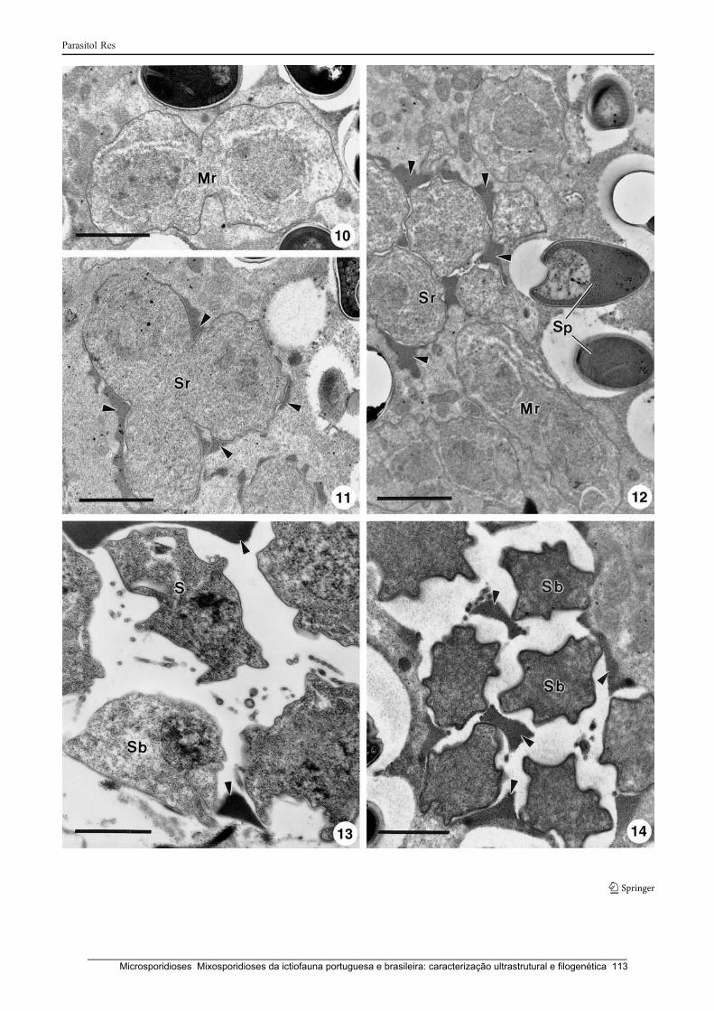
Parasitol Res
_____________________________________________________________________________________________________ Microsporidioses Mixosporidioses da ictiofauna portuguesa e brasileira: caracterização ultrastrutural e filogenética 113

psittaca, supported by 78% bootstrap. Both are clustered ina group together with Ichthyosporidium, Loma, Pseudo-loma genera and the Microsporidium sp. MX1. However,this clade is poorly supported with a bootstrap lower than50%. The most parsimonious trees suggested paraphyly forLoma species (Fig. 3).
Discussion
The light and ultrastructural observation of the xenoma,developmental stages as well as spore morphology de-scribed in the present study, showed all structures typical ofthe parasites belonging to the phylum Microsporidia (Lomand Dyková 1992; Larsson 1999; Lom and Nilsen 2003).
The fishes represented at least 156 species is one of thelargest group parasitised by microsporidians. They werefound in different geographic area, habitat and local ofinfection (Lom 2002). The parasite described in the presentwork is the second occurrence in teleost fish belonging tothe family Tetraodontidae. Ogawa and Yokoyama (1998)found in the intestine of the tiger puffer fish, Takifugurubripes, from a mariculture in Japan, another micro-sporidian, but it was not classified. Comparing themorphology and ultrastructural aspects of the developmen-tal stages of the parasite here described with microsporidianfish previously characterised, it seemed similar to Lomaspp. (Lom and Dyková 1992; Lom and Nilsen 2003).
Presently, there are 11 Loma species and they werereported in the gills and digestive tract of the fresh andmarine fishes (Lom 2002; Table 1). The species type Lomabranchialis was found in the gills of Atlantic cod (Morrisonand Sprague 1981), likely as L. salmonae in severalsalmonids species and from different regions (Putz et al.1965), Loma fontinalis (Morrison and Sprague 1983), Lomadimorpha found in different hosts (Loubès 1984), Lomatrichiuri (Sandeep and Kalavati 1985) and L. embiotocia(Shaw et al. 1997). Parasitising the intestine, oesophagus orliver, five species were reported: one in Europe, Lomadiplodae found in Diplodus sargus (Bekhti and Bouix 1985);Loma boopsi and Loma camerounensis identified in Africanfishes, Boops boops from Senegal (Faye et al. 1995) and inthe tilapia species Oreochromis niloticus from the Cameroon(Fomena et al. 1992), respectively; L. acerinae (Lom andPekkarinen 1999) in the freshwater Gymnocaphaluscernuus from Czech Republic. Finally, L. myrophis foundin the Amazonian fish M. platyrhynchus was described byAzevedo and Matos (2002). Concerning the habitat, shapeand size of the mature spores and the number of polarfilament coils, L. psittaca did not seem similar with otherpreviously described species. Comparing with the speciesfrom the same geographic area, L. myrophis found also inintestinal tissue of an Amazonian freshwater fish pre-T
able
2Com
parisonof
someSSUrDNAsequences:percentage
ofidentity(top
diagonal)andpairwisedistance
(bottom
diagonal)obtained
byKimura-2parameter
analysis
Species
12
34
56
78
910
1112
1314
15
Lom
apsittacan.sp.
–96.9
96.0
96.0
95.6
95.6
95.6
95.2
94.4
89.6
89.6
89.2
87.3
86.9
85.4
Lom
aacerinae
0.031
–97.3
97.3
96.5
96.5
96.5
96.5
96.5
90.6
89.7
91.0
88.8
88.3
86.9
Glugeaatherina
e0.040
0.027
–100
98.8
98.8
98.8
99.2
97.3
92.7
90.1
91.5
89.6
89.2
87.8
Glugeaan
omala
0.040
0.027
0.000
–98.8
98.8
98.8
99.2
97.3
92.7
90.1
91.5
89.6
89.2
87.8
Pleistoph
orafin
isterrensis
0.044
0.035
0.012
0.012
–100
100
98.1
96.5
93.6
89.6
91.0
89.6
90.5
89.2
Glugeaplecog
lossi
0.044
0.035
0.012
0.012
0.000
–100
98.1
96.5
93.6
89.6
91.0
89.6
90.5
89.2
Glugeasp.GS1
0.044
0.035
0.012
0.012
0.000
0.000
–98.1
96.5
93.6
89.6
91.0
89.6
90.5
89.2
Glugeastepha
ni0.048
0.035
0.008
0.008
0.019
0.019
0.019
–96.5
91.9
89.6
91.0
88.8
88.3
86.9
Glugeasp.
0.056
0.035
0.027
0.027
0.035
0.035
0.035
0.035
–90.6
90.1
90.0
87.8
87.4
86.0
Microsporidiumsp.MX1
0.104
0.094
0.073
0.073
0.064
0.064
0.064
0.081
0.094
–91.9
92.8
92.7
93.1
91.8
Pseud
olom
aneurop
hilia
0.104
0.103
0.099
0.099
0.104
0.104
0.104
0.104
0.099
0.081
–93.6
87.8
88.3
86.9
Ichthyospo
ridium
sp.
0.108
0.090
0.085
0.085
0.090
0.090
0.090
0.090
0.090
0.072
0.064
–90.6
90.2
88.8
Lom
asp.
0.127
0.112
0.104
0.104
0.104
0.104
0.104
0.112
0.122
0.073
0.122
0.094
–98.4
97.3
Lom
asalmon
ae0.131
0.117
0.108
0.108
0.095
0.095
0.095
0.117
0.126
0.069
0.117
0.098
0.016
–98.8
Lom
aem
biotocia
0.146
0.131
0.122
0.122
0.108
0.108
0.108
0.131
0.140
0.082
0.131
0.112
0.027
0.012
–
Parasitol Res
_____________________________________________________________________________________________________ 114 Microsporidioses Mixosporidioses da ictiofauna portuguesa e brasileira: caracterização ultrastrutural e filogenética

Fig. 3 Parsimony tree of SSU rDNA sequences to compare L.psittaca with selected sequences from other fish-infecting Micro-sporidia. The analysis was conducted using 1,339 aligned nucleotidepositions of the highest BLAST score microsporidian sequences andfour more microsporidian sequences as outgroup. The bar indicates
the equivalence between the distance and the number of changes. Thenumbers on the branches indicate bootstrap support from 100replicates. L. psittaca is placed within group I (highlighted box),which includes the sequences of the genera Ichthyosporidium, Loma,Pseudoloma and one Microsporidium sp.
Parasitol Res
_____________________________________________________________________________________________________ Microsporidioses Mixosporidioses da ictiofauna portuguesa e brasileira: caracterização ultrastrutural e filogenética 115

sented some differences, mainly in the shape and dimen-sions of spores, being 67% and 39.7% (relationship width/length) in L. psittaca and L. myrophis, respectively.
Ultrastructural studies
Comparing L. psittaca n. sp. with the other Loma species,we saw some ultrastructural similarities, namely thedevelopmental stage aspects. Small xenomas with acentrally located hypertrophic host cell nucleus wereobserved also in L. branchialis (Morrison and Sprague1981), L. acerinae (Lom and Pekkarinen 1999) and L.myrophis (Azevedo and Matos 2002). In these Lomaspecies, like in some Glugea species, it was possible tosee in the episporontal space electrodense masses thatdifferentiate several tubular appendages. Curiously, in thegenus Loma, the origin of the episporontal space is notconsensus. It has been described for some species bycoalescence of host cell vesicles (PV) (Morrison andSprague 1981, 1983; Lom and Pekkarinen 1999; Azevedoand Matos 2002), whilst in others, apparently episporontalspace has been originated from blisters at the surface of theparasite cell (Bekhti and Bouix 1985; Fomena et al. 1992;Faye et al. 1995).
Phylogenetic analysis
Phylogenetic analysis using the SSU rRNA sequences offish microsporidian suggested that the parasite found in thepuffer fish of the Amazonian fauna, L. psittaca n. sp., is asister species of L. acerinae. The most parsimonious treewas supported by 78% bootstrap. All previous phylogenetictrees obtained by parsimony and likelihood maximumpresented a similar topology (Docker et al. 1997; Lomand Nilsen 2003; Casal et al. 2008), clustering the almostLoma species together with Ichthyosporidium sp., P.neurophilia and Microsporidium sp. MX1 in the group Idefined by Lom and Nilsen (2003). The same trees alsoshow that the Loma species are a paraphyletic groupplacing L. acerinae and L. psittaca in a basal position ofthe group I or alternatively must be considered an outgroupmicrosporidian of group I, as suggested by Lom and Nilsen(2003). In this study, the genetic distances (Kimura 2-parameter methods) also show that there are some similarityin SSU rRNA sequences with the species belonging togroup II, namely with G. atherinae, G. anomala, G.plecoglossi, G. stephani, Glugea sp. GS1 and P. finister-rensis (last one probably needing to change taxonomicgroup). The diagnosis of Glugea and Loma genera presentsmany similarities that have been confirmed by phylogeneticanalysis. Definitely, the morphological and ultrastructuralaspects of L. psittaca do not accommodate within the generaGlugea (Canning et al. 1982), characterised by large xenomas
with a retractile wall and by the presence of a RER cisternsurrounding the meronts during developmental stages.
Based on all these morphological and ultrastructuralorganisation and host specificity described in the presentwork and comparing them with those of fish microsporidia,which form xenoma, we have found some ultrastructuraldifferences. On the other hand, the genetic data allowed thediagnosis of other fish-infecting microsporidian, supportingthe description of a new species. Lom and Nilsen (2003)have reported that a new genus to accommodate L. acerinaeand in this case L. psittaca also would be created. At themoment, we did not find significant ultrastructural differ-ences that justify the creation of a new genus.
Acknowledgements This work partially supported by the Eng. A.Almeida Foundation (Porto, Portugal), PhD grant from “CESPU” (G.Casal), “CNPq” and “CAPES”–Brazil. We would like to thank theiconographic work of Joana Carvalheiro and João Carvalheiro. Weassure that this work complies with the current laws of our countrieswhere this was performed.
References
Altschul SF, Gish W, Miller W, Myers EW, Lipman DJ (1990) Basiclocal aligment search tool. J Parasitol 215:403–410
Azevedo C, Matos E (2002) Fine structure of a new species, Lomamyrophis (phylum Microsporidia), parasite of the Amazonian fishMyrophis platyrhynchus (Teleostei, Ophichthidae). Eur J Protistol37:445–452
Azevedo C, Matos E (2003) Amazonspora hassar n. gen. and n. sp.(phylum Microsporidia, fam Glugeidae), a parasite of theAmazonian teleost Hassar orestis (fam. Doradidae). J Parasitol89:336–341
Baquero E, Rubio M, Moura INS, Pieniazek J, Jordana R (2005)Myosporidium merluccius n. g., n. sp. infecting muscle ofcommercial hake (Merluccius sp.) from fisheries near Namibia.J Eukaryot Microbiol 52:476–483
Bekhti M, Bouix G (1985) Loma salmonae (Putz, Hoffmann etDunbar, 1965) et Loma diplodae n. sp., microsporidies parasitesde branchies de poissons téléostéens: implantation et donnéesultrastructurales. Protistologica 21:47–59
Canning EU, Lom J, Nicholas JP (1982) Genus Glugea Thélohan,1891 (Phylum Microspora): redescription of the type speciesGlugea anomala (Moniez, 1887) and recognition of its sporo-gonic development within sporophorous vesicles (pansporoblas-tic membranes). Protistologica 18:193–210
Casal G, Matos E, Teles-Grilo ML, Azevedo C (2008) A newmicrosporidian parasite, Potaspora morhaphis n. gen., n. sp.(Microsporidia) infecting the Teleostean fish, Potamorhaphisguianensis from the River Amazon. Morphological, ultrastruc-tural and molecular characterization. Parasitology 135:1053–1064
Cheney SA, Lafranchi-Tristem NJ, Canning EU (2000) Phylogeneticrelationships of Pleistophora-like Microsporidia based on smallsubunit ribosomal DNA sequences and implications for thesource of Trachipleistophora hominis infections. J EukaryotMicrobiol 47:280–287
Docker MF, Devlin RH, Richard J, Kent ML (1997) Sensitive andspecific polymerase chain reaction assay for detection of Lomasalmonae (Microsporea). Dis Aquat Org 29:41–48
Parasitol Res
_____________________________________________________________________________________________________ 116 Microsporidioses Mixosporidioses da ictiofauna portuguesa e brasileira: caracterização ultrastrutural e filogenética
Faye N, Toguebaye BS, Bouix G (1995) On the cytology anddevelopment of Loma boopsi n. sp. (Microspora, Glugeidae),parasite of Boops boops (Pisces, Teleostei, Sparidae) from thecoasts of Senegal. Arch Protistenkd 146:85–93
Fomena A, Coste F, Bouix G (1992) Loma camerounensis sp. nov.(Protozoa: Microsporida) a parasite of Oreochromis niloticusLinnaeus, 1757 (Teleost: Cichlidae) in fish-rearing ponds inMelen, Yaoundé, Cameroon. Parasitol Res 78:201–208
Hillis DM, Dixon MT (1991) Ribosomal DNA: molecular evolutionand phylogenetic inference. Q Rev Biol 66:411–453
Larsson JIR (1999) Identification of Microsporidia. Acta Protozool38:161–197
Lom J (2002) A catalogue of described genera and species ofmicrosporidians parasitic in fish. Syst Parasitol 53:81–99
Lom J, Dyková I (1992) Protozoan parasites of fishes. Elsevier,Amsterdam, p 315
Lom J, Nilsen F (2003) Fish microsporidia: fine structural diversityand phylogeny. Int J Parasitol 33:107–127
Lom J, Pekkarinen M (1999) Ultrastructural observations on Lomaacerinae (Jírovec, 1930) comb. nov. (phylum Microsporidia).Acta Protozool 38:61–74
Loubès C, Maurand J, Gasc C, Buron I, Barral J (1984) Étudeultrastructurale de Loma dimorpha n. sp., microsporidie parasitede poissons Gobiidae languedociens. Protistologica 20:579–589
Matos E, Azevedo C (2004) Ultrastructural description of Micro-sporidium brevirostris sp. n., parasite of the teleostean Brachy-hypopomus brevirostris (Hypopomidae) from the Amazon River.Acta Protozool 43:261–267
Matos E, Corral L, Azevedo C (2003) Ultrastructural details of thexenoma of Loma myrophis (phylum Microsporidia) and extrusionof the polar tube during autoinfection. Dis Aquat Org 54:203–207
Matthews JL, Brown AMV, Larison K, Bishop-Stewart JK, Rogers P,Kent ML (2001) Pseudoloma neurophilia n. g., n. sp., a newmicrosporidium from the central nervous system of the zebrafish(Danio rerio). J Eukaryot Microbiol 48:227–233
Morrison CM, Sprague V (1981) Electron microscopical study of anew genus and new species of microsporida in the gills ofAtlantic cod Gadus morhua L. J Fish Dis 4:15–32
Morrison CM, Sprague V (1983) Loma salmonae (Putz, Hoffman andDunbar, 1965) in the rainbow trout, Salmo gairdneri Richarson,and L. fontinalis sp. nov. (Microsporida) in the brook trout,Salvelinus fontinalis (Mitchill). J Fish Dis 6:345–353
Nilsen F (2000) Small subunit ribosomal DNA phylogeny of micro-sporidia with particular reference to genera that infect fish. JParasitol 86:128–133
Ogawa K, Yokoyama H (1998) Parasitic diseases of cultured marinefish in Japan. Fish Pathol 33:303–309
Putz RE, Hoffman GL, Dunbar CE (1965) Two new species ofPleistophora (Microsporidea) from North America fish with asynopsis of Microsporidea of freshwater and euryhaline fishes. JProtozool 12:228–236
Sandeep BV, Kalavati C (1985) A new microsporidian, Loma trichiurin. sp., from the gill of a marine fish, Trichiurus salva Cuv.(Trichiuridae). Indian J Parasitol 9:257–259
Shaw RW, Kent ML, Docker MF, Brown AMV, Devlin RH, AdamsonML (1997) A new species of Loma (Microsporea) in shiner perch(Cymatogaster aggregata). J Parasitol 83:296–301
Sprague V, Becnel JJ, Hazard EI (1992) Taxonomy of phylumMicrospora. Crit Rev Microbiol 18:285–395
Tamura K, Dudley J, Nei M, Kumar S (2007) MEGA4: molecularevolutionary genetics analysis (MEGA) software version 4.0.Mol Biol Evol 24:1596–1599
Thompson JD, Higgins DG, Gilson TJ (1994) Clustal W: improvingthe sensitivity of progressive multiple sequence alignmentthrough sequence weighting, position-specific gap penalties andweight matrix choice. Nucleic Acids Res 22:4673–4680
Vossbrinck CR, Baker MD, Didier ES, Debrunner-Vossbrinck BA,Shadduck JA (1993) Ribosomal DNA sequences of Encephali-tozoon hellem and Encephalitozoon cuniculi: species identifica-tion and phylogenetic construction. J Eukaryot Microbiol40:354–362
Parasitol Res
_____________________________________________________________________________________________________ Microsporidioses Mixosporidioses da ictiofauna portuguesa e brasileira: caracterização ultrastrutural e filogenética 117

_____________________________________________________________________________________________________ 118 Microsporidioses Mixosporidioses da ictiofauna portuguesa e brasileira: caracterização ultrastrutural e filogenética

Capítulo 4
ULTRASTRUCTURAL AND MOLECULAR CHARACTERIZATION OF A NEW
MICROSPORIDIUM PARASITE FROM THE AMAZONIAN FISH,
GYMNORHAMPHICHTHYS RONDONI (RHAMPHICHTHYIDAE)
Journal of Parasitology (2009) em revisão
Graça Casal, Edilson Matos, M. Leonor Teles-Grilo & Carlos Azevedo
_____________________________________________________________________________________________________ Microsporidioses Mixosporidioses da ictiofauna portuguesa e brasileira: caracterização ultrastrutural e filogenética 119

_____________________________________________________________________________________________________ 120 Microsporidioses Mixosporidioses da ictiofauna portuguesa e brasileira: caracterização ultrastrutural e filogenética

ABSTRACT
A new species of a microsporidium identified as Microsporodium rondoni n. sp. found in
the freshwater teleost Gymnorhamphichthys rondoni collected on lower Amazon River
were described based on light, ultrastructural and phylogenetic studies. This parasite
develops in the skeletal muscle of the abdominal cavity forming whitish like-cysts
containing numerous spores. Mature spores, lightly pyriform to ellipsoidal with rounded
ends, measuring 4.25 ± 0.38 x 2.37 ± 0.42 μm (n= 30) were observed. The spore wall
which measured about 102 nm was composed of two layers with approximately the same
thickness. The isofilar polar filament was coiled with 9-10 (rarely 8) turns. The posterior
vacuole appeared as a pale area, occupying about 1/3 of the spore length, contained a
spherical posterosome composed of granular material, denser at the periphery. The
myofibrils located near the spores appeared to be in advanced degradation. Molecular
analysis of the rRNA genes, including the ITS region, and phylogenetic analyses using
maximum parsimony, maximum likelihood and Baysesian Inference were performed. The
ultrastructural characteristics of the spores and phylogenetic data strongly suggested that
it is a new species, related to Kabatana, Microgemma, Potaspora, Spraguea and
Tetramicra. We provisionally placed this new species from Amazonian fauna in the
collective group Microsporidium.
_____________________________________________________________________________________________________ Microsporidioses Mixosporidioses da ictiofauna portuguesa e brasileira: caracterização ultrastrutural e filogenética 121

INTRODUCTION
Microsporidia (phylum Microsporidia) are intracellular parasites that occur in almost all
taxonomic groups (Canning and Lom 1986; Sprague et al., 1992; Larsson 1999; Lom
2002), and are best known to cause diseases in commercially important fish hosts (Lom
and Dyková, 1992; Lom, 2002; Lom and Nilsen, 2003). Microsporidian species,
simultaneously parasitizing freshwater and marine fishes from different geographic areas,
are included into 17 genera assigned among about 150 genera of Microsporidia (Lom,
2002; Lom and Nilsen, 2003; Azevedo and Matos, 2003; Baquero et al., 2005; Casal et
al., 2008).
Presently Microsporidia contain about 156 species and two of them were identified as new
genera and new species in the freshwater fishes from the Amazon fauna: Amazonspora
hassar which occurs in the gills of Hassar orestis (Azevedo and Matos, 2003) and
Potaspora morhaphis in the coelomic cavity of Potamorhaphis guianensis (Casal et al.,
2008). Other two microsporidia from the same region were described: Loma myrophis
parasitizing the sub-epithelial gut tissues of Myrophis platyrhynchus (Azevedo and Matos,
2002) and Microsporidium brevirostris in the skeletal muscle adjacent to the abdominal
cavity of the teleost fish Brachyhypopomus brevirostris (family Hipopomidae) (Matos and
Azevedo, 2004). The last species and the microsporidium described in the present report
from Gymnorhamphichthys rondoni (fam. Rhamphichthyidae) were the first reference of
microsporidiosis in teleost knifefishes (Order Gymnotiformes). Phylogenetic studies based
on the molecular analysis of the rRNA genes have been a powerful tool in the
identification of new genus and species, as well as in grouping in family taxa (Weiss and
Vossbrinck, 1999; Vossbrinck and Debrunner-Vossbrinck, 2005). Presently, there are
several SSU rRNA sequences available in the Genbank, corresponding to around 44 fish-
microsporidian species. According Lom and Nilsen (2003), fish microsporidia are
clustered in five groups and only some of the genera are monophyletic.
Herein, we describe some light microscopic, morphological and ultrastructural features of
a new microsporidian species found in a fish from the Amazon River. The molecular
characterization and phylogenetic relationships for the SSU rRNA gene were also
performed, as well as an analysis of the pathological effects of the spores in the muscle.
_____________________________________________________________________________________________________ 122 Microsporidioses Mixosporidioses da ictiofauna portuguesa e brasileira: caracterização ultrastrutural e filogenética

MATERIALS AND METHODS
Fish, location of infection and prevalence
Several irregular whitish aggregated of spores (cyst-like structures), located in the skeletal
muscles of the internal wall of the ventral abdominal cavity, were removed from the
freshwater fish Gymnorhamphichthys rondoni (fam. Rhamphichthyidae) (Brazilian
common name: Itui transparente). The fish were collected in the lower Amazonian region
(01º 46´ S / 47º 26´ W), near Irituia city, Pará State, Brazil. The fish (12-25 cm long) were
taken alive to the laboratory, where they were anaesthetized with MS 222 (Sandoz
Laboratories), and necropsied. For measurements fresh isolated spores were observed in
the Nomarski differential interference – contrast (DIC) optics. The prevalence of infection
was 36% (18 fishes in 50 examined).
Electron microscopy
For transmission electron microscopy (TEM), small fragments of the infected tissues were
fixed in 3% glutaraldehyde with 0.2 M sodium cacodylate buffer (pH 7.2) for 12 h at 4 ºC,
washed overnight in the same buffer at 4 ºC and post-fixed in 2% OsO4 buffered in the
same solution for 3 h at same temperature. After dehydration in an ascending ethanol
series and propylene oxide, the fragments were embedded in Epon. The semithin
sections were stained with blue methylene-Azure II for light microscopy. The ultrathin
sections were contrasted with both aqueous uranyl acetate and lead citrate and observed
with JEOL 100CXII TEM operated at 60 kV.
DNA isolation and PCR amplification
Several cysts dissected from fishes, were homogenized to isolate the spores and
subsequently stored in 80% ethanol at 4 °C. The genomic DNA of about 5 x 10⁶ spores
was extracted using a GenEluteTM Mammalian Genomic DNA Miniprep Kit (Sigma)
following the manufacturer instructions for animal tissues, except for the incubation time
(12 h). The DNA was stored in 50 µl of TE buffer at – 20 ºC until further use. Further The
DNA concentration was estimated with the QubitTM Fluorometer (Invitrogen). The majority
of the region coding the small subunit (SSU) rRNA gene was amplified by PCR using the
primers V1f (5’CACCAGGTTGATTCTGCC3’) and 1492r (5’GGTTACCTTGTTACGAC
TT3’) (Vossbrinck et al., 1993; Nilsen, 2000). To amplify the 3’-end of the SSU, internal
transcribed spacer (ITS) and 5’-end of the large subunit (LSU) rRNA gene, HG4F
(5’GCGGCTTAATTTGACTCAAC) and HG4R (5’TCTCCTTGGTCCGTGTTTCAA) primers
_____________________________________________________________________________________________________ Microsporidioses Mixosporidioses da ictiofauna portuguesa e brasileira: caracterização ultrastrutural e filogenética 123

were used (Gatehouse and Malone, 1998). PCR was carried out in 50 µl reactions using
10 pmol of each primer, 10 nmol of each dNTP, 2 mM of MgCl2, 5 µl 10X Taq polymerase
buffer, 1.25 units Taq DNA polymerase (Invitrogen products), and 3 µl of the genomic
DNA. The reactions were run on Hybaid PxE Thermocycler (Thermo Electron Corporation,
Milford, MA). The amplification program consisted of 94 °C denaturation for 5 min,
followed by 35 cycles of 94 °C for 1 min, 50 °C for 1 min and 72 °C for 2 min. A final
elongation step was performed at 72 °C for 10 min. 5 µl aliquots of the PCR products were
electrophoresed through a 1% agarose 1x Tris-acetate-EDTA buffer (TAE) gel stained
with ethidium bromide.
DNA cloning and sequencing
The PCR product for the SSU gene with an approximate size of 1400 bp was excised
from the agarose gel and purified with NucleoSpin Extract II (Macherey-Nagel). The DNA
was cloned into a pGEM-T Easy Vector System II (Promega) following the manufacturer
instructions. JM109 Competent cells, high efficiency (Promega) were transformed and 2
positive clones selected. The plasmid DNA isolation were carried out with a NucleoSpin
Plasmid (Macherey-Nagel) according to the manufacturer manual. Cloning was confirmed
by digestion with the restriction enzyme EcoRI (Promega) and through sequencing with
the universal sequencing primers T7 forward / SP6. For the ITS region, a PCR product of
about 1100 bp was sequenced directly, after cleaning. The sequencing reactions were
done using BigDye Terminator v1.1 kit (Applied Biosytems) and were run on an ABI3700
DNA analyzer (Perkin-Elmer, Applied Biosystems, Stabvida, Co., Oeiras, Portugal).
Distance and phylogenetic analysis
Previously, the various forward and reverse sequence segments were aligned manually
with ClustalW (Thompson et al., 1994) in MEGA 4 software and ambiguous bases were
clarified using corresponding ABI chromatograms. To evaluate the relationship of
Microsporidium rondoni to other microsporidia, a homology search was performed using
BLAST (NCBI). We used 45 rDNA sequences belonging to the microsporidia having fish
as hosts. The sequence and NCBI accession number data obtained from GenBank are as
follows: Aspalatospora milevae (EF990668); Glugea anomala (AF044391); Glugea
atherinae (U15987); Glugea plecoglossi (AJ295326); Glugea stephani (AF056015);
Glugea sp. GS1 (AJ295325); Glugea sp. (AY090038); Heterosporis anguillarum
(AF387331); Heterosporis sp. PF (AF356225); Ichthyosporidium sp. (L39110); Kabatana
takedai (AF356222); Kabatana newberryi (EF202572); Kabatana seriolae (AJ295322);
_____________________________________________________________________________________________________ 124 Microsporidioses Mixosporidioses da ictiofauna portuguesa e brasileira: caracterização ultrastrutural e filogenética

Kabatana sp. (EU682928); Loma acerinae (AJ252951); Loma embiotocia (AF320310);
Loma salmonae (U78736); Loma sp. (AF104081); Microgemma caulleryi (AY033054);
Microgemma tincae (AY651319); Microgemma vivaresi (AJ252952); Microsporidium
cypselurus (AJ300706); Microsporidium prosopium (AF151529); Microsporidium GHB1
(AJ295324); Microsporidium sp. RSB1 (AJ295323); Microsporidium sp. STF (AY140647);
Microsporidium MYX1 (AJ295329); Myosporidium merluccius (AY530532); Nucleospora
salmonis (U78176); Ovipleistophora mirandellae (AF356223); Ovipleistophora ovariae
(AJ252955); Pleistophora ehrenbaumi (AF044392); Pleistophora finisterrensis
(AF044393); Pleistophora hippoglossoideos (AJ252953); Pleistophora typicalis
(AF044387); Pleistophora sp. 1 (AF044394); Pleistophora sp. 2 (AF044389); Pleistophora
sp. 3 (AF044390); Potaspora morhaphis (EU534408); Pseudoloma neurophilia
(AF322654); Spraguea americana (AF056014); Spraguea lophii (1) (AF104086);
Spraguea lophii (2) (AF033197); Spraguea sp. (AY465876); Tetramicra brevifilum
(AF364303). Endoreticulatus schubergi (L39109), Enterocytozoon bieneusi (L07123),
Vairimorpha necatrix (Y00266) and Vittaforma corneae (L39112) were used as outgroup.
The alignment was performed with ClustalW in MEGA 4 software (Tamura et al., 2007),
with an opening gap penalty of 10 and a gap extension penalty of 4 for both pairwise and
multiple alignments. Subsequent phylogenetic and molecular evolutionary analyses were
conducted using MEGA 4, with the 45 rDNA sequences for microsporidian species and
the outgroup species selected. Distance estimation was carried out using the Kimura-2
parameters model distance matrix for transitions and transversions. For the phylogentic
tree reconstructions, the maximum parsimony analysis was performed using the close
neighbour interchange heuristic option with a search factor of 2 and random initial trees
addition of 2000 replicates. Clade support was assessed with bootstrapping of 100
replicates.
Maximum likelihood (ML) and Bayesian Inferences (BI) analysis were performed on the
Phylogeny.fr platform (Dereeper et al., 2008) and sequences were aligned with ClustalW.
The ambiguous regions (i. e. containing gaps and/or poorly aligned) were subsequently
removed with Gblocks using the default parameters. The ML method was implemented in
the PhyML program (v3.0 aLRT) (Guindon et al., 2005). The GTR substitution model was
selected assuming an estimated proportion of invariant sites (of 0.282) and 4 gamma-
distributed rate categories to account for rate heterogeneity across sites. The gamma
shape parameter was estimated directly from the data (gamma = 1.386). Reliability for the
internal branch was assessed using the bootstrapping method (100 bootstrap replicates).
_____________________________________________________________________________________________________ Microsporidioses Mixosporidioses da ictiofauna portuguesa e brasileira: caracterização ultrastrutural e filogenética 125

Figure 1 Light and transmission electron micrographs of Microsporidium rondoni n. sp. infecting the muscle fibres of the teleost fish Gymnorhamphichtys rondoni. (a, b) Fresh spores released from the muscle observed in DIC, showing the pyriforme to ellipsoidal shape and their prominent posterior vacuole. (c) Semithin section of whitish patches containing numerous spores, located among muscle fibres (arrows). (d) Longitudinal section of a spore, showing the wall (W), anchoring disc (AD), different sections of the polar filament (F), polaroplast (P) and the nucleus (N). The posterior vacuole (V) contains a posterosome (Ps). (e) Detail of the anterior region of a spore showing the wall (W) composed of two evident layers (exospore and endospore), anchoring disc (AD) and polaroplast (P). (f) A packed of double layer coils of the polar filament (F) with 10 turns between the wall (W) and the vacuole (V). (g) Detail of a posterosome (Ps) composed by a granular matrix and surrounded by denser material. (h) Transverse
_____________________________________________________________________________________________________ 126 Microsporidioses Mixosporidioses da ictiofauna portuguesa e brasileira: caracterização ultrastrutural e filogenética
section of the spore wall (W) showing the external region of the exospore containing incisions distributed regularly on the spore surface (arrowheads).
BI was performed with the MrBayes program (v3.1.2) (Ronquist and Huelsenbeck, 2003)
with the following parameters: the standard (4 by 4) model of nucleotide substitution was
used, the number of substitution types = 6 and rates variation across sites was fixed to
"invgamma". Probability distributions were generated using Markov Chain Monte Carlo
methods. Four chains were run for 105 generations, sampling every 100 generations, with
the first 100 sampled trees discarded as "burn-in". Finally, a 50% majority rule consensus
tree was constructed. Both the trees were built with the TreeDyn program.
DESCRIPTION
Microsporidium rondoni n. sp.
(Figs. 1-3)
General diagnosis: Isolated and grouped whitish like-cysts in the skeletal muscle of
the abdominal cavity (Figs 1a, b). This parasite does not develop xenomas and spores in
direct contact with the myofibrils (Fig. 1c).
Description of the spores: Monomorphic, uninucleated mature spores, lightly pyriform
to ellipsoidal with rounded ends; 4.25 ± 0.38 μm long and 2.37 ± 0.42 μm wide (n = 30)
(Figs 1a, b). Nucleus in a central position between the apical polaroplast and the posterior
vacuole (Figs 1d, 3). Polaroplast lamellate, bipartite with the elements of distal position
somewhat expanded (Fig. 1e). Isofilar polar filament, formed by 3 concentric layers of
membranes (Fig. 1f), with 115 (110-121) nm (n = 50) in diameter, an angle of tilt of about
45º (42-47) (n = 10) (Fig. 1e) and posteriorly arranged in a packed double layered coils
with 9-10 (rarely 8) turns (Figs 1d, f). Posterior vacuole with 1/3 of the spore length,
contained generally 1-2 conspicuous inclusions - the posterosome, consists of a central
granular mass surrounded by amorphous and irregular material, denser at the periphery
(Figs 1d, g). Spore wall about 102 (95-110) nm thick (n = 50) composed of two layers: an
electron dense exospore of ~27 nm width and an electron lucent endospore, both with
approximately the same thickness (Figs 1d-h). Light incisions distributed regularly on the
exospore (Fig. 1h). Spores inside of the sphorophorous vesicles were never observed.
Histopathology: Whitish elongated cysts-like structures containing numerous spores
were observed in contact with the myofibrils of the internal wall of the abdominal cavity.
The infected muscle showed degradation characterized by the disorganization of the
_____________________________________________________________________________________________________ Microsporidioses Mixosporidioses da ictiofauna portuguesa e brasileira: caracterização ultrastrutural e filogenética 127

myofibrils (Figs 2a-c). The spores located within the cytoplasm of the host cells were in
close contact with the nuclei (Figs 1d, 2a-c) and its cytoplasm appeared partially
destroyed (Figs 1d, 2a, b). Phagocytic cells appearing to ingest mature spores were
frequently observed near the muscle fibres (Figs 2c, d).
Molecular characterization and phylogeny: Two bands of approximately 1.4 kb and 1.1
kb were obtained after amplification of the microsporidian genomic DNA with the primers
V1f-1492r and HG4F-HG4R, respectively. All the sequences obtained were aligned, and
the sequence consensus corresponding to the complete SSU rRNA gene, ITS and partial
LSU rRNA gene was 1914 bp in length, with a GC content of 43.7%. The sequence was
deposited in the Genbank database under the accession number FJ843105. BAST
analysis was performed and the highest alignment excluded all the microsporidian SSU
rRNA sequences that do not parasite fish species. Then the 3-end of SSU rRNA gene
was trimmed, it resulted in an alignment with 1536 bp. The SSU rRNA gene of
Microsporidium rondoni shows some nucleotidic insertions that allows the classification of
this species of others microsporidians: A 13 bp insert from position 779 and a13 bp at the
position 1057 that are common to Kabatana takedai. Before the phylogenetic analysis,
only those sites which could be unambiguously aligned among all microsporidia and
outgroups were used, resulting in an alignment of 1402 bp.
BLAST analysis of the Microsporidium rondoni sequence showed that Kabatana takedai
(AF356222) and Kabatana sp. (EU682928) had the highest score, followed by three
Spraguea spp. sequences. Based on pairwise comparisons among the SSU rDNA
sequences, the maximal similarity (Kimura 2-parameter) of Microsporidium rondoni with
the species of the same clade is for the Spraguea (96.4 – 96.8%), Microgemma and
Tetramicra (95.6 – 96.0 %) genera. A longest range of percentage of identity for Kabatana
species (88.2 – 95.2%) was also observed (Table 1). Maximum parsimony phylogenetic
analyses of the SSU rRNA gene strongly supported a clade (bootstrap 91%) where cluster
containing Kabatana, Microgemma, Potaspora, Spraguea, Tetramicra genera and some
species of the collective group Microsporidium (Fig. 4). Within this clade, the new
microsporidium forms a sister taxa with Spraguea and Microgemma species. After BLAST
search we also found a partial SSU rDNA for Aspalatospora milevae (EF990668) that
showed a 93.9% identify to M. rondoni. With the aim to clarify the phylogenetic position of
this new species, the Bayesian inference and maximum likelihood phylogenetic analyses
were also performed, confirming similar topology trees (Fig. 5).
_____________________________________________________________________________________________________ 128 Microsporidioses Mixosporidioses da ictiofauna portuguesa e brasileira: caracterização ultrastrutural e filogenética

Taxonomic summary
Type host: Gymnorhamphichthys rondoni (Miranda-Ribeiro, 1920) (Teleostei:
Rhamphichthyidae) with 12-25 cm of the length in average.
Type locality: lower Amazon River (01º 46´ S / 47º 26´ W) near Irituia city, Pará State,
Brazil.
Site of infection: skeletal muscle of the internal abdominal cavity.
Prevalence of infection: eighteen of 50 (36%) with no statistical difference between
sexes.
Type material: One glass slide with semithin sections containing mature spores of the
hapantotype were deposited in the International Protozoan Type Slide Collection at the
Smithsonian Institution, Washington D.C. 20560 (USNM no. 1123996).
Etymology: the specific name “rondoni” derives from the species epithet of the host
species G. rondoni.
Remarks
Of the 17 microsporidian genera found in teleost fishes, only Heterosporis, Kabatana,
Pleistophora and the collective group Microsporidium have affinity to the myocytes of the
skeletal muscle and some induce serious pathological changes (Dyková and Lom, 2000).
The genera Heterosporis, Kabatana, Ovipleistophora and Pleistophora are characterized
by the incapacity to develop structures known as xenomas which confer good conditions
for parasite development and simultaneously minimize the proliferation of the parasite to
other organs / tissues of the host (Lom, 2002; Lom and Nilsen, 2003).
Based on the spore’s morphological data (shape, dimensions), ultrastructural aspects of
the internal organization, with special evidence for the anchoring disc, polaroplast, polar
filament coils surrounding the posterior vacuole, the organization of the posterosome, as
well as lack of sporophorous vesicles differentiation, the site of infection, and absence of
xenoma formation, the microsporidium described here seems to be similar, at least in part,
to the genus Kabatana (Lom et al., 1999, 2000, 2001; McGourty et al., 2007).
_____________________________________________________________________________________________________ Microsporidioses Mixosporidioses da ictiofauna portuguesa e brasileira: caracterização ultrastrutural e filogenética 129
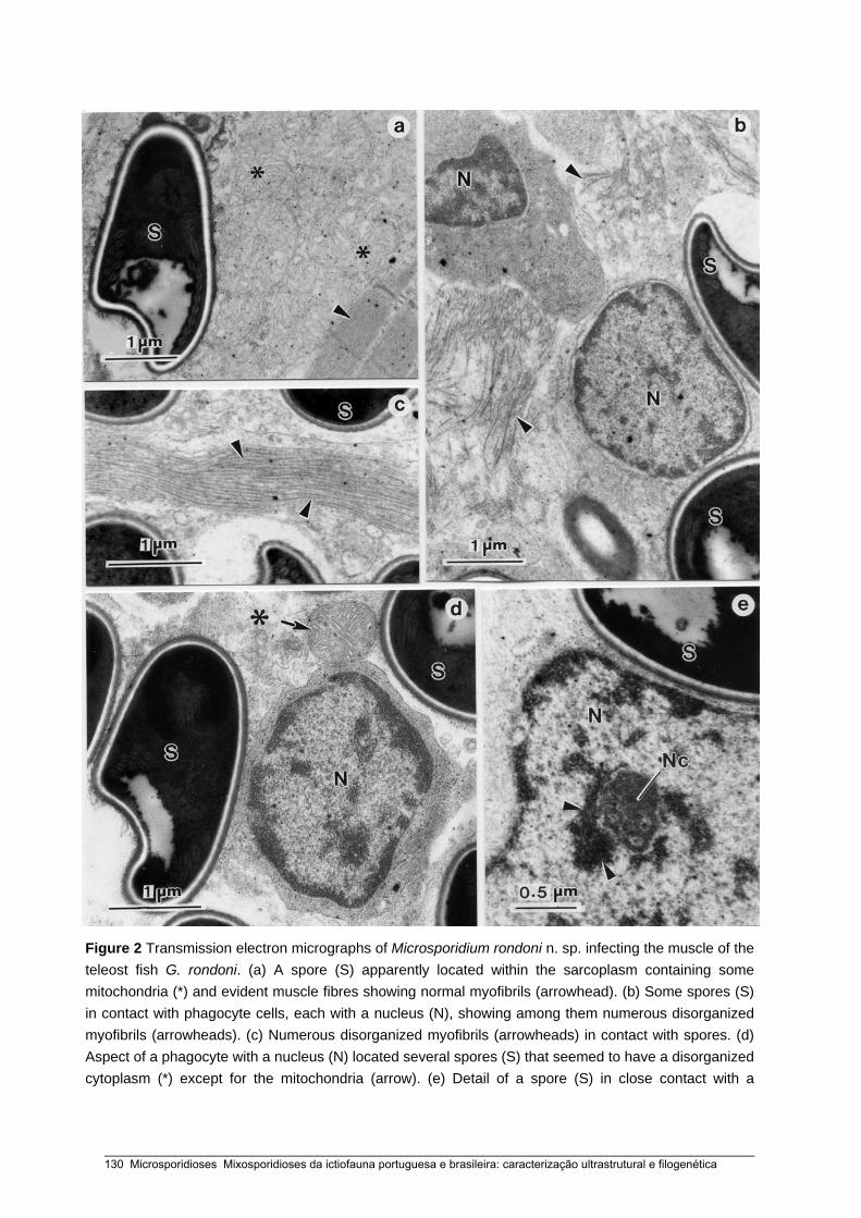
Figure 2 Transmission electron micrographs of Microsporidium rondoni n. sp. infecting the muscle of the teleost fish G. rondoni. (a) A spore (S) apparently located within the sarcoplasm containing some mitochondria (*) and evident muscle fibres showing normal myofibrils (arrowhead). (b) Some spores (S) in contact with phagocyte cells, each with a nucleus (N), showing among them numerous disorganized myofibrils (arrowheads). (c) Numerous disorganized myofibrils (arrowheads) in contact with spores. (d) Aspect of a phagocyte with a nucleus (N) located several spores (S) that seemed to have a disorganized cytoplasm (*) except for the mitochondria (arrow). (e) Detail of a spore (S) in close contact with a
_____________________________________________________________________________________________________ 130 Microsporidioses Mixosporidioses da ictiofauna portuguesa e brasileira: caracterização ultrastrutural e filogenética
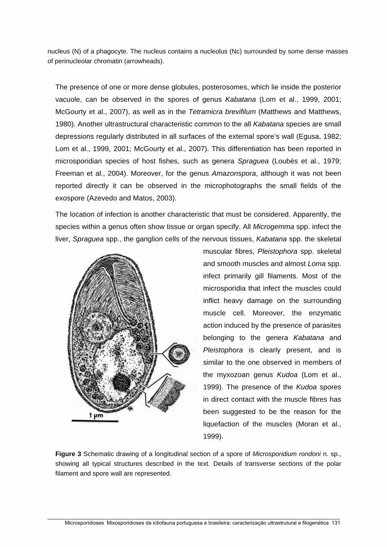
nucleus (N) of a phagocyte. The nucleus contains a nucleolus (Nc) surrounded by some dense masses of perinucleolar chromatin (arrowheads).
The presence of one or more dense globules, posterosomes, which lie inside the posterior
vacuole, can be observed in the spores of genus Kabatana (Lom et al., 1999, 2001;
McGourty et al., 2007), as well as in the Tetramicra brevifilum (Matthews and Matthews,
1980). Another ultrastructural characteristic common to the all Kabatana species are small
depressions regularly distributed in all surfaces of the external spore’s wall (Egusa, 1982;
Lom et al., 1999, 2001; McGourty et al., 2007). This differentiation has been reported in
microsporidian species of host fishes, such as genera Spraguea (Loubès et al., 1979;
Freeman et al., 2004). Moreover, for the genus Amazonspora, although it was not been
reported directly it can be observed in the microphotographs the small fields of the
exospore (Azevedo and Matos, 2003).
The location of infection is another characteristic that must be considered. Apparently, the
species within a genus often show tissue or organ specify. All Microgemma spp. infect the
liver, Spraguea spp., the ganglion cells of the nervous tissues, Kabatana spp. the skeletal
muscular fibres, Pleistophora spp. skeletal
and smooth muscles and almost Loma spp.
infect primarily gill filaments. Most of the
microsporidia that infect the muscles could
inflict heavy damage on the surrounding
muscle cell. Moreover, the enzymatic
action induced by the presence of parasites
belonging to the genera Kabatana and
Pleistophora is clearly present, and is
similar to the one observed in members of
the myxozoan genus Kudoa (Lom et al.,
1999). The presence of the Kudoa spores
in direct contact with the muscle fibres has
been suggested to be the reason for the
liquefaction of the muscles (Moran et al.,
1999).
Figure 3 Schematic drawing of a longitudinal section of a spore of Microsporidium rondoni n. sp., showing all typical structures described in the text. Details of transverse sections of the polar filament and spore wall are represented.
_____________________________________________________________________________________________________ Microsporidioses Mixosporidioses da ictiofauna portuguesa e brasileira: caracterização ultrastrutural e filogenética 131

Table 1 Comparison of some SSU rDNA sequences: percentage of identity (top diagonal) and pairwise distance (bottom diagonal) obtained by Kimura-2
parameter analysis
1 2 3 4 5 6 7 8 9 10 11 12 13 14 15 16 17
(1) Microsporidium rondoni - 96.8 96.8 96.8 96.4 96.0 95.6 95.6 95.6 95.2 94.7 93.9 93.9 88.2 88.2 88.2 83.9
(2) Spraguea sp. 0.032 - 100 100 99.6 99.2 98.8 98.8 98.8 96.8 96.4 95.6 95.6 89.5 89.5 89.5 85.3
(3) Spraguea lophii (1) 0.032 0.000 - 100 99.6 99.2 98.8 98.8 98.8 96.8 96.4 95.6 95.6 89.5 89.5 89.5 85.3
(4) Spraguea lophii (2) 0.032 0.000 0.000 - 99.6 99.2 98.8 98.8 98.8 96.8 96.4 95.6 95.6 89.5 89.5 89.5 85.3
(5) Spraguea americana 0.036 0.004 0.004 0.004 - 98.8 98.4 98.4 98.4 96.4 96.0 95.2 95.2 89.1 89.1 89.1 84.9
(6) Microgemma tincae 0.040 0.008 0.008 0.008 0.012 - 99.6 99.2 99.2 96.8 96.4 95.6 95.6 89.5 89.5 89.5 85.8
(7) Microgemma vivaresi 0.044 0.012 0.012 0.012 0.016 0.004 - 98.8 98.8 96.4 96.0 95.2 95.2 89.1 89.1 89.1 85.3
(8) Microgemma caulleryi 0.044 0.012 0.012 0.012 0.016 0.008 0.012 - 100 96.4 96.0 95.2 95.2 89.1 89.1 89.1 85.8
(9) Tetramicra brevifilum 0.044 0.012 0.012 0.012 0.016 0.008 0.012 0.000 - 96.4 96.0 95.2 95.2 89.1 89.1 89.1 85.2
(10) Kabatana sp. 0.048 0.032 0.032 0.032 0.036 0.032 0.036 0.036 0.036 - 99.6 96.8 97.2 89.5 89.5 89.5 85.3
(11) Kabatana newberryi 0.053 0.036 0.036 0.036 0.040 0.036 0.040 0.040 0.040 0.004 - 96.4 96.8 88.0 88.0 88.0 84.8
(12) Aspalatospora milevae 0.061 0.044 0.044 0.044 0.048 0.044 0.048 0.048 0.048 0.032 0.036 - 96.8 90.4 90.4 90.4 87.7
(13) Kabatana takedai 0.061 0.044 0.044 0.044 0.048 0.044 0.048 0.048 0.048 0.028 0.032 0.032 - 90.4 90.4 90.4 86.1
(14) Kabatana seriolae 0.118 0.105 0.105 0.105 0.109 0.105 0.109 0.109 0.109 0.105 0.110 0.096 0.096 - 100 100 85.3
(15) Microsporidium sp. GHB1 0.118 0.105 0.105 0.105 0.109 0.105 0.109 0.109 0.109 0.105 0.110 0.096 0.096 0.000 - 100 85.3
(16) Microsporidium sp. RSB1 0.118 0.105 0.105 0.105 0.109 0.105 0.109 0.109 0.109 0.105 0.110 0.096 0.096 0.000 0.000 - 85.3
(17) Potaspora morhaphis 0.161 0.147 0.147 0.147 0.151 0.142 0.147 0.142 0.142 0.147 0.152 0.123 0.139 0.147 0.147 0.147 -
_____________________________________________________________________________________________________ 132 Microsporidioses Mixosporidioses da ictiofauna portuguesa e brasileira: caracterização ultrastrutural e filogenética

Figure 4 The maximum parsimony tree of SSU rDNA sequences of Microsporidium rondoni n. sp. and other selected microsporidia. The numbers on the branches are bootstrap confidence levels on 100 replicates. GenBank accession numbers are in parentheses after the species names and the scale is given under the tree. Microsporidium rondoni places within the group 4 (Lom and Nilsen, 2003) (highlighted box), include the sequences of the genera Kabatana Microgemma, Potaspora, Spraguea, Tetramicra, and Microsporidium.
_____________________________________________________________________________________________________ Microsporidioses Mixosporidioses da ictiofauna portuguesa e brasileira: caracterização ultrastrutural e filogenética 133
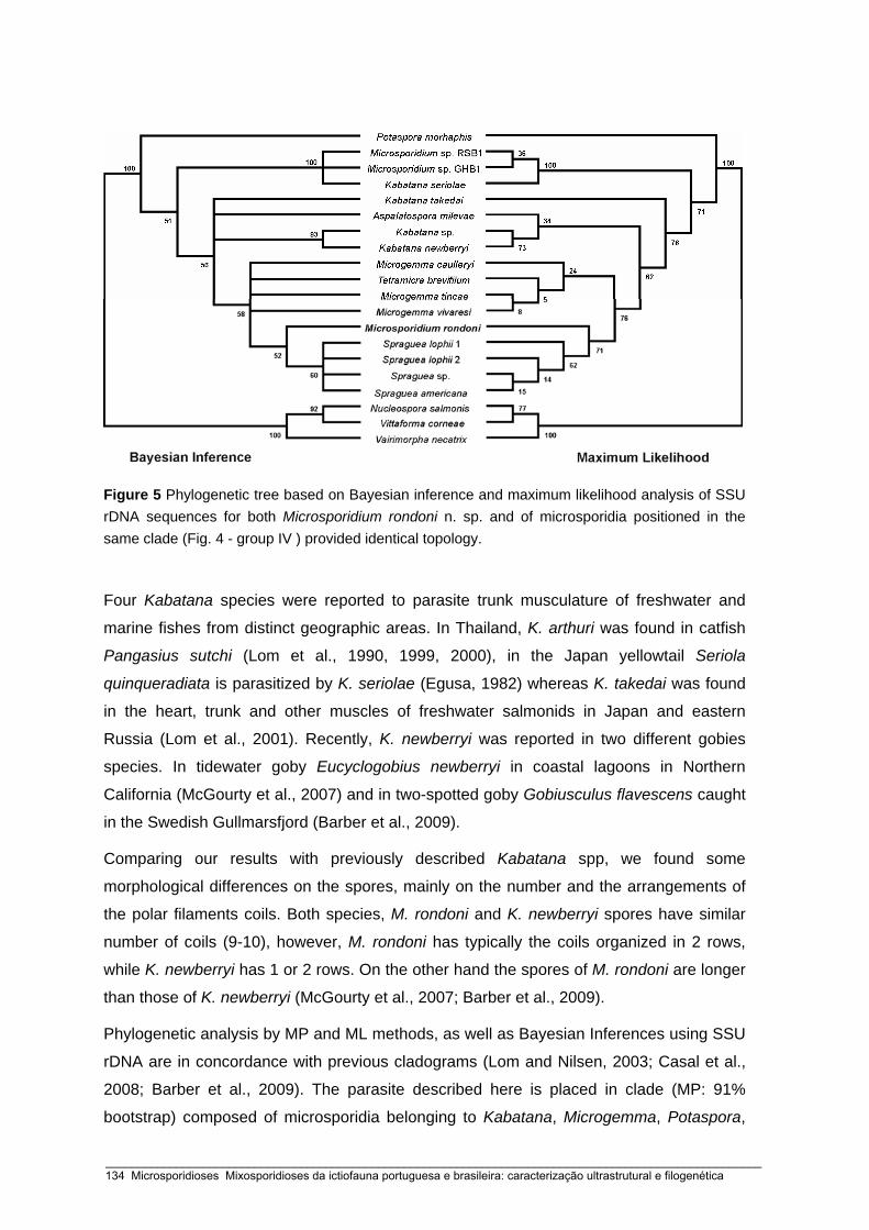
Figure 5 Phylogenetic tree based on Bayesian inference and maximum likelihood analysis of SSU rDNA sequences for both Microsporidium rondoni n. sp. and of microsporidia positioned in the same clade (Fig. 4 - group IV ) provided identical topology.
Four Kabatana species were reported to parasite trunk musculature of freshwater and
marine fishes from distinct geographic areas. In Thailand, K. arthuri was found in catfish
Pangasius sutchi (Lom et al., 1990, 1999, 2000), in the Japan yellowtail Seriola
quinqueradiata is parasitized by K. seriolae (Egusa, 1982) whereas K. takedai was found
in the heart, trunk and other muscles of freshwater salmonids in Japan and eastern
Russia (Lom et al., 2001). Recently, K. newberryi was reported in two different gobies
species. In tidewater goby Eucyclogobius newberryi in coastal lagoons in Northern
California (McGourty et al., 2007) and in two-spotted goby Gobiusculus flavescens caught
in the Swedish Gullmarsfjord (Barber et al., 2009).
Comparing our results with previously described Kabatana spp, we found some
morphological differences on the spores, mainly on the number and the arrangements of
the polar filaments coils. Both species, M. rondoni and K. newberryi spores have similar
number of coils (9-10), however, M. rondoni has typically the coils organized in 2 rows,
while K. newberryi has 1 or 2 rows. On the other hand the spores of M. rondoni are longer
than those of K. newberryi (McGourty et al., 2007; Barber et al., 2009).
Phylogenetic analysis by MP and ML methods, as well as Bayesian Inferences using SSU
rDNA are in concordance with previous cladograms (Lom and Nilsen, 2003; Casal et al.,
2008; Barber et al., 2009). The parasite described here is placed in clade (MP: 91%
bootstrap) composed of microsporidia belonging to Kabatana, Microgemma, Potaspora,
_____________________________________________________________________________________________________ 134 Microsporidioses Mixosporidioses da ictiofauna portuguesa e brasileira: caracterização ultrastrutural e filogenética

Spraguea, Tetramicra genera, 2 unclassified species of the Microsporidium group and
Aspalatospora milevae [Mladineo and Lovy - Aspalatospora milevae n. g., n. sp.: xenoma-
forming microsporidian from the intestine of the Atlantic bluefin tuna (Thunnus thynnus) -
personal communication]. Like Matthews et al. (2001) we tried to identify signature
sequences. We found two regions in the SSU rDNA sequence of M. rondoni similar to that
of Kabatana takedai. This kind of analysis has been encouraged the characterization of
the new species (Lom and Nilsen, 2003).
All methods provide evidences that Kabatana species are a paraphyletic group. The
exception is K. newberryi (parasite of a goby species from Pacific coast, USA) and
Kabatana sp. (parasite of a goby species from Atlantic coast, Sweden) considered to be of
the same species (Barber et al., 2009). MP analysis, clusters Aspalatospora milevae in a
sister taxa with K. takedai. Nevertheless, the bootstrap (21%) for this clade is poorly
supported. The species K. seriolae is the most genetically distinct (11.8%) and forms a
stable clade (bootstrap 100%), together with two Microsporidium spp. (Bell et al., 2001).
MP methods, M. rondoni occupies a basal position (bootstrap 50%) clustered with all
Microgemma spp., Spraguea spp. and Tetramicra brevifilum. Using phylogenetic analyses
by ML method (bootstrap 71%) and BI (bootstrap 52%), M. rondoni is included with
Spraguea spp. in the same clade.
In conclusion, morphological, ultrastructural and molecular analyses in the present study
demonstrated that this microsporidium is a new species belonging to the group 4. This
parasite probably belongs to the genus Kabatana. As there is not adequate information on
their developmental stages to assign this species to a specific genus, we provisionally
placed it in the collective group Microsporidium Balbiani, 1884.
ACKNOWLEDGMENTS
Work partially supported by the Engº. A. Almeida Foundation (Porto, Portugal), PhD grant
from “CESPU” (G. Casal), “CNPq” and “CAPES” - Brazil. We would like to thank the
iconographic work of Joana Carvalheiro and João Carvalheiro. This work comply with the
current laws of the countries in where they were performed.
_____________________________________________________________________________________________________ Microsporidioses Mixosporidioses da ictiofauna portuguesa e brasileira: caracterização ultrastrutural e filogenética 135
LITERATURE CITED
AZEVEDO, C., AND E. MATOS. 2002. Fine structure of a new species, Loma myrophis (Phylum
Microsporidia), parasite of the Amazonian fish Myrophis platyrhynchus (Teleostei, Ophichthidae).
European Journal of Protistology 37: 445-452
______, AND ______. 2003. Amazonspora hassar n. gen. and n. sp. (phylum Microsporidia, fam. Glugeidae),
a parasite of the Amazonian teleost Hassar orestis (fam. Doradidae). Journal of Parasitology 89: 336-
341.
BAQUERO, E., M. RUBIO, I. N. S. MOURA, J. PIENIAZEK, AND R. JORDANA. 2005. Myosporidium
merluccius n. g., n. sp. infecting muscle of commercial hake (Merluccius sp.) from fisheries near
Namibia. Journal of Eukaryotic Microbiology 52: 476-483.
BARBER, I., A. J. DAVIES, J. E. IRONSIDE, E. FORSGREN, AND T. AMUNDSEN. 2009. First record of a
Kabatana sp. microsporidium infecting fish in the Atlantic Ocean. Diseases of Aquatic Organisms 83:
145-152.
BELL, A. S., T. AOKI, AND H. YOKOYAMA. 2001. Phylogenetic relationships among Microsporidia based on
rDNA sequence data, with particular reference to fish-infecting Microsporidium Balbiani, 1884 species.
Journal of Eukaryotic Microbiology 48: 258-265.
CANNING, E. U., AND J. LOM. 1986. The Microsporidia of Vertebrates, Academic Press, London.
CASAL, G., E. MATOS, M. L. TELES-GRILO, AND C. AZEVEDO. 2008. A new microsporidian parasite,
Potaspora morhaphis n. gen., n. sp. (Microsporidia) infecting the teleostean fish, Potamorhaphis
guianensis from the River Amazon. Morphological, ultrastructural and molecular characterization.
Parasitology 135: 1053-1064.
DEREEPER A., V. GUIGNON, G. BLANC, S. AUDIC, S. BUFFET, F. CHEVENET, J. F. DUFAYARD, S.
GUINDON, V. LEFORT, M. LESCOT, J. M. CLAVERIE, AND O. GASCUEL. 2008. Phylogeny.fr: robust
phylogenetic analysis for the non-specialist. Nucleic Acids Research 36: 465-469.
DYKOVÁ, I., AND J. LOM. 2000. Histopatlogy of Kabatana arthuri (Microspora) infection in sutchi catfish,
Pangasius sutchi. Folia Parasitologica 47: 161-166.
EGUSA, S. 1982. A microsporidian species from yellowtail juveniles, Seriola quinqueradiata, with ‘Beko’
disease. Fish Pathology 16: 187-192.
FREEMAN, M. A., H. YOKOYAMA, AND K. OGAWA. 2004. A microsporidian parasite of the genus Spraguea
in the nervous tissues of the Japanese anglerfish Lophius litulon. Folia Parasitologica 51: 167-176.
GATEHOUSE, H. S., AND L. A. MALONE. 1998. The ribosomal RNA gene region of Nosema apis
(Microspora): DNA sequence for small and large subunit rRNA genes and evidence of a large tandem
repeat unit size. Journal of Invertebrate Pathology 71: 97-105.
GUINDON, S., F. LETHIEC, P. DUROX, AND O. GASCUEL. 2005. PHYML online – a web server for fast
maximum likelihood-based phylogenetic inference. Nucleic Acids Research 33: 557-559.
LARSSON, J. I. R. 1999. Identification of Microsporidia. Acta Protozoologica 38: 161-197.
LOM, J. 2002. A catalogue of described genera and species of microsporidians parasitic in fish. Systematic
Parasitology 53: 81-99.
_____________________________________________________________________________________________________ 136 Microsporidioses Mixosporidioses da ictiofauna portuguesa e brasileira: caracterização ultrastrutural e filogenética
______, AND I. DYKOVÁ. 1992. Microsporidia (Phylum Microspora Sprague, 1977). In Protozoan Parasites of
Fishes, J. Lom, and I. Dyková (eds.). Developments in Aquaculture and Fisheries Science, Elsevier,
Amsterdam. vol. 26, p. 125-157.
______, ______, AND F. SHAHAROM. 1990. Microsporidium arthuri n. sp., parasite of Pangasius sutchi
(Pangasiidae, Siluroidea) in South-East Asia. Diseases of Aquatic Organisms 8: 65-67.
______, ______, AND K. TONGUTHAI. 1999. Kabataia gen. n., new genus proposed for Microsporidium spp.
infecting trunk muscles of fishes. Diseases of Aquatic Organisms 38: 39-46.
______, ______, AND ______. 2000. Kabatana gen. n., new name for the microsporidian genus Kabataia
Lom, Dyková and Tonguthai, 1999. Folia Parasitologica 47: 78.
______, AND F. NILSEN. 2003. Fish microsporidia: fine structural diversity and phylogeny. International
Journal for Parasitology 33: 107-127.
______, ______, AND S. URAWA. 2001. Redescription of Microsporidium takedai (Awakura, 1974) as
Kabatana takedai (Awakura, 1974) comb. n. Diseases of Aquatic Organisms 44: 223-230.
LOUBÉS, C., J. MAURAND, AND R. ORMIÈRES. 1979. Étude ultrastructurale de Spraguea lophii (Doflein,
1898), microsporidie parasite de la Baudroie: essai d’interpretation du dimorphisme sporal.
Protistologica 15: 43-54.
MATOS, E., AND C. AZEVEDO. 2004. Ultrastructural description of Microsporidium brevirostris sp. n.,
parasite of the teleostean Brachyhypopomus brevirostris (Hypopomidae) from the Amazon River. Acta
Protozoologica 43: 261-267.
MATTHEWS, J. L., A. M. V. BROWN, K. LARISON, J. K. BISHOP–STEWART, P. ROGERS, AND M. L.
KENT. 2001. Pseudoloma neurophilia n. g., n. sp., a new microsporidium from the central nervous
system of the zebrafish (Danio rerio). Journal of Eukaryotic Microbiology 48: 227-233.
MATTHEWS, R. A., AND B. F. MATTHEWS. 1980. Cell and tissue reactions of turbot Scophthalmus maximus
(L.) to Tetramicra brevifilum gen. n., sp. n. (Microspora). Journal of Fish Diseases 3: 495-515.
MCGOURTY, K. R., A. P. KINZGER, G. L. HENDRICKSON, G. H. GOLDSMITH, G. CASAL, AND C.
AZEVEDO. 2007. A new microsporidian infecting the musculature of the endangered tidewater goby
(Gobiidae). Journal of Parasitology 93: 655-660.
MORAN, J. D. W., D. J. WHITAKER, AND M. L. KENT. 1999. A review of the myxosporean genus Kudoa
Meglitsch, 1947, and its impact on the international aquaculture industry and commercial fisheries.
Aquaculture 172: 163-196.
NILSEN, F. 2000. Small subunit ribosomal DNA phylogeny of Microsporidia with particular reference to genera
that infect fish. Journal of Parasitology 86: 128-133.
RONQUIST, F. AND J. P. HUELSENBECK. 2003. MrBayes 3: Bayesian phylogenetic inference under mixed
models. Bioinformatics 19: 1572-1574.
SPRAGUE, V., J. J. BECNEL, AND E. I. HAZARD. 1992. Taxonomy of phylum Microspora. Critical Review of
Microbiology 18: 285-395.
TAMURA, K., J. DUDLEY, M. NEI, AND S. KUMAR. 2007. MEGA4: Molecular evolutionary genetics analysis
(MEGA) software version 4.0. Molecular Biology Evolution 24: 1596-1599.
_____________________________________________________________________________________________________ Microsporidioses Mixosporidioses da ictiofauna portuguesa e brasileira: caracterização ultrastrutural e filogenética 137
THOMPSON, J. D., D. G. HIGGINS, AND T. J. GILSON. 1994. Clustal W: improving the sensitivity of
progressive multiple sequence alignment through sequence weighting, position-specific gap penalties
and weight matrix choice. Nucleic Acids Research 22: 4673-4680.
VOSSBRINCK, C. R., M. D. BAKER, E. S. DIDIER, B. A. DEBRUNNER-VOSSBRINCK, AND J. A.
SHADDUCK. 1993. Ribosomal DNA sequences of Encephalitozoon hellem and Encephalitozoon
cuniculi: species identification and phylogenetic construction. Journal of Eukaryotic Microbiology 40:
354-362.
VOSSBRINCK, C. R., AND B. A. DEBRUNNER-VOSSBRINCK. 2005. Molecular phylogeny of the
Microsporidia: ecological, ultrastructural and taxonomic considerations. Folia Parasitologica 52: 131-
142.
WEISS, L., AND C. VOSSBRINCK. 1999. Molecular biology, molecular phylogeny, and molecular diagnostic
approaches to the Microsporidia. In The Microsporidia and Microsporidiosis, M. Wittner, and L. Weiss
(eds.). American Society of Microbiology, Washington, DC, p. 129-171.
_____________________________________________________________________________________________________ 138 Microsporidioses Mixosporidioses da ictiofauna portuguesa e brasileira: caracterização ultrastrutural e filogenética

Capítulo 5
FINE STRUCTURE AND PHYLOGENY OF A NEW SPECIES,
SPRAGUEA GASTROPHYSUS (PHYLUM, MICROSPORIIDIA), A PARASITE OF THE
ANGLERFISH LOPHIUS GASTROPHYSUS (TELEOSTEI, LOPHIIDAE) FROM BRAZIL
European Journal of Protistology (2009) submetido
Graça Casal, Sérgio S. Clemente, Patríca Matos, Marcelo Knoff,
Edilson Matos & Carlos Azevedo
_____________________________________________________________________________________________________ Microsporidioses Mixosporidioses da ictiofauna portuguesa e brasileira: caracterização ultrastrutural e filogenética 139

_____________________________________________________________________________________________________ 140 Microsporidioses Mixosporidioses da ictiofauna portuguesa e brasileira: caracterização ultrastrutural e filogenética

ABSTRACT
The ultrastructure of the fish-infecting Microsporidium Spraguea gastrophysus n. sp.,
found in the dorsal ganglia and kidney of the anglerfish, Lophius gastrophysus (fam.
Lophiidae), collected on the Brazilian Atlantic coast is described. The parasite develops
several groups of whitish xenomas up to 3.1 x 1.8 mm. Inside, there is a hypertrophic host
cell surrounded by a hypertrophic cytoplasm containing some intermingled life cycle
stages, which consist mainly of mature spores, and several developmental stages with
unpaired nuclei. Monomorphic spores are ellipsoidal, lightly curved and measure about
3.35 ± 0.45 x 1.71 ± 0.36 μm (n = 50). Polar filament is isofilar with expended base
attached to the anchoring disc, constricting abruptly, and then tapering to form 5 - 6 coils
in a single row. Polaroplast with two distinct kinds of lamellae is located in the apical
portion of the spore occupying one-third of the total volume of the spore. It is composed
by an anterior portion that consists of a tightly patched lamellar and regularly spaced,
whereas the posterior one is larger, spaced and irregularly organized. Nucleus occupies a
central zone of the spores where several polyribosomes are present. The posterior
vacuole occupying one-quarter of the volume of the spore contained a voluminous
spherical and granular posterosome measuring up to ~0.65 μm in diameter. Ultrastructural
morphology of the spores and the molecular characterization of the SSU rRNA gene
suggest the generic assignment to the genus Spraguea and the name the parasite as a
new microsporidian species, Spraguea gastrophysus n. sp.
Keywords: Spraguea gastrophysus n. sp.; Parasite; Microsporidia; Ultrastructure;
Phylogeny; Lophius gastrophysus
_____________________________________________________________________________________________________ Microsporidioses Mixosporidioses da ictiofauna portuguesa e brasileira: caracterização ultrastrutural e filogenética 141
INTRODUCTION
The anglerfish of the genus Lophius occurring in different geographic areas are
represented by five species (L. piscatorius, L. budegassa, L. americanus, L. litulon and L.
gastrophysus). These species contain fish-infecting microsporidians which are mainly
located in the nervous tissues. This parasite was first reported from the spinal ganglia of
L. piscatorius Linnaeus, 1758 previously classified as genus Glugea and later identify as
belong to G. lophii (Doflein 1898). More detailed morphological studies developed by
Mrázek (1899) reinforced that this parasite belongs to this genus. However, this parasite
was subsequently transferred to the genus Nosema, as N. lophii (Pace 1908), name
posteriorly confirmed by Weissenberg (1909, 1911a, b, c). Later, Vávra and Sprague in a
footnote published in the Weissenberg’ paper (1976) refer for the first time the name
“Spraguea n. gen.” and simultaneously transfer Glugea lophii to Spraguea lophii (Doflein,
1898) Weissenberg, 1976 as type species.
The first ultrastuctural data of S. lophii, parasite of the European anglerfish L. budegassa
and L. piscatorius both having dimorphic spores, were carried by Loubès et al. (1979). On
the other hand, in the spinal and cranial ganglia of American anglerfish, L. americanus,
caught from the northeast Atlantic coast of the USA were also described the presence of
microsporidian spores (Takvorian and Cali 1986). Considering some ultrastructural
differences in L. americanus, mainly having monomorphic spore type, relatively to
previously described dimorphic spore occurring in L. budegassa and L. piscatorius from
Europe, the microsporidian found in L. americanus was included in the genus Glugea, as
G. americanus (Takvorian and Cali 1986). However, some recent molecular results based
on the SSU rRNA genes sequences suggested that this species would be transferred to
the genus Spraguea, as S. americana (Lom and Nilsen 2003; Nilsen 2000; Pomport-
Castillon et al. 2000). More recently, on the basis of ultrastructural and molecular studies,
xenomas containing monomorphic microsporidian parasite identified as Spraguea
americana was found in the nervous tissues of the Japanese anglerfish Lophius litulon
(Freeman et al. 2004).
The only reference to the presence of a similar microsporidian from the South America
anglerfish Lophius gastrophysus that was collected in the Brazilian and Venezuelan
coasts was reported by Jakowska and Nigrelli (1958, 1959) and Jakowska (1964, 1966),
however, with no microscopical images or drawings. Relatively to the Brazilian fauna two
new genera Amazonspora (Azevedo and Matos 2003) and Potaspora (Casal et al. 2008)
were identified. There are also information for another three parasitosis, Loma myrophis
_____________________________________________________________________________________________________ 142 Microsporidioses Mixosporidioses da ictiofauna portuguesa e brasileira: caracterização ultrastrutural e filogenética

(Azevedo and Matos 2002) and Microsporidium brevirostris (Matos and Azevedo 2004),
all of them found in Amazonian fishes.
In the present work, we describe a new microsporidian species on the basis of the
ultrastructural morphology of the spores, with special emphasis to host and tissues
specificity and molecular characterization of the SSU rRNA gene.
MATERIALS AND METHODS
Light and electron microscopy
Thirty six adult specimens of the marine anglerfish, Lophius gastrophysus Miranda-
Ribeiro, 1915 (Teleostei, Lophiidae) (Brazilian common name “peixe-sapo pescador”) (27
- 68 cm long; 0.550 – 5.600 gr weight) were collected from the Atlantic coast of “Cabo
Frio” (22º 50’S /42º 03’W), State of Rio de Janeiro, Brazil. The fishes were lightly
anesthetised with MS 222 (Sandoz Laboratories), transported to the laboratory (UFF -
Niterói), dissected and the infected tissues, containing several whitish cysts (cyst-like
plasmodia) were removed from the peripheral muscles of the internal abdominal cavity in
contact with the dorsal nerves and kidney, and examined by a light microscope equipped
with Nomarski interference-contrast (DIC) optics.
For ultrastructural studies, small fragments of the parasitized tissues containing xenoma
were excised and fixed in 3% glutaraldehyde in 0.2 M sodium cacodylate buffer (pH 7.2)
at 4 °C for 12 h. After rinsed overnight in the same buffer at 4 ºC and post-fixed in 2.0 %
osmium tetroxide in the same buffer for 3 h at 4 °C, the fragments were dehydrated
through an ascending ethanol series, followed by propylene oxide and embedded in Epon.
Semithin sections were stained with methylene blue-Azur II and observed by DIC optics.
Ultrathin sections were double stained with aqueous uranyl acetate and lead citrate and
observed under a transmission electron microscope (TEM) JEOL 100CXII operated at 60
kV.
DNA isolation, PCR amplification and DNA sequencing
Several cysts were dissected from fishes, were homogenized to isolate the spores that
were consequently stored in 80% ethanol at 4 °C. The genomic DNA of about 6 x 10⁶
spores was extracted using a GenEluteTM Mammalian Genomic DNA Miniprep Kit
(Sigma) following the manufacturer instructions for animal tissue, except for the incubation
_____________________________________________________________________________________________________ Microsporidioses Mixosporidioses da ictiofauna portuguesa e brasileira: caracterização ultrastrutural e filogenética 143
time. The DNA was stored in 50 μl of TE buffer at - 20ºC until further used. The majority of
the region coding for the small subunit (SSU) rRNA gene was amplified by PCR using the
primers V1f (5’CACCAGGTTGATTCTGCC3’) and 1492r (5’GGTTACCTTGTTACGAC
TT3’) (Nilsen, 2000; Vossbrinck et al. 1993). To amplify the 3’-end of the SSU, internal
transcribed spacer (ITS) and 5’-end of the large subunit (LSU) rRNA gene, HG4F
(5’GCGGCTTAATTTGACTCAAC) and HG4R (5’TCTCCTTGGTCCGTGTTTCAA) primers
were used (Gatehouse and Malone 1998). PCR was carried out in 50 μl reactions using
10 pmol of each primer, 10 nmol of each dNTP, 2 mM of MgCl2, 5 μl 10 X Taq polymerase
buffer, 1.25 units Taq DNA polymerase (Invitrogen products), and 3 μl of the genomic
DNA. The reactions were run on Hybaid PxE Thermocycler (Thermo Electron Corporation,
Milford, MA). The amplification program consisted of 94 °C denaturation for 5 min,
followed by 35 cycles of 94 °C for 1 min, 50 °C for 1 min and 72 °C for 2 min. A final
elongation step was performed at 72 °C for 10 min. 5 μl aliquots PCR products were
visualized with ethidium bromide staining after running on a 1% agarose gel. PCR
products for the SSU gene and ITS region have approximate sizes of 1400 bp and 1100
bp, respectively. These were cleaned using the NucleoSpin Extract II (Macherey-Nagel)
and then three purified PCR products were sequenced in both directions. The sequencing
reactions were done using BigDye Terminator v1.1 kit (Applied Biosytems) and were run
on an ABI3700 DNA analyzer (Perkin-Elmer, Applied Biosystems, Stabvida, Co., Oeiras,
Portugal).
Distance and phylogenetic analysis
Previously, the various forward and reverse sequence segments were aligned manually
with ClustalW (Thompson et al. 1994) in MEGA 4 software and ambiguous bases were
clarified using corresponding ABI chromatograms. To evaluate the relationship of
Spraguea gastrophysus n. sp. to other Microsporidia, we have used the 35 rDNA
sequences that have a fish as host. The sequence and NCBI accession number data
obtained from GenBank (Table 1). The corresponding sequences and GenBank/NCBI
accession number of Vairimorpha necatrix (Y00266) and Vittaforma corneae (L39112)
were used as the outgroup.
Sequences were aligned as described by Casal et al. (2008). The alignment was
performed through the use of Clustal W (Thompson et al. 1994) in MEGA 4 software
(Tamura et al. 2007), with an opening gap penalty of 10 and a gap extension penalty of 4
for both pairwise and multiple alignments. Subsequent phylogenetic and molecular
evolutionary analyses were conducted using MEGA 4, with the sequences for
_____________________________________________________________________________________________________ 144 Microsporidioses Mixosporidioses da ictiofauna portuguesa e brasileira: caracterização ultrastrutural e filogenética

microsporidian species and the outgroup species selected. Distance estimation was
carried out using the Kimura-2 parameters model distance matrix for transitions and
transversions. For the phylogenetic tree reconstructions, the maximum parsimony
analysis was performed using the close neighbour interchange (CNI) heuristic option with
a search factor of 2 and random initial trees addition of 2000 replicates. Bootstrap values
were calculated over 100 replicates.
Aspalatospora milevae (EF990668) Microsporidium sp. RSB1 (AJ295323)
Glugea anomala (AF044391) Myosporidium merluccius (AY530532)
Glugea atherinae (U15987) Nucleospora salmonis (U78176)
Glugea plecoglossi (AJ295326) Ovipleistophora mirandellae (AF356223)
Glugea stephani (AF056015) Ovipleistophora ovariae (AJ252955)
Heterosporis anguillarum (AF387331) Pleistophora ehrenbaumi (AF044392)
Heterosporis sp. PF (AF356225) Pleistophora finisterrensis (AF044393)
Ichthyosporidium sp. (L39110) Pleistophora hippoglossoideos (AJ252953)
Kabatana takedai (AF356222) Pleistophora typicalis (AF044387)
Kabatana newberryi (1) (EF202572) Potaspora morhaphis (EU534408)
Kabatana newberryi (2) (EU682928) Pseudoloma neurophilia (AF322654)
Kabatana seriolae (AJ295322) Spraguea americana (1) (AF056014)
Loma embiotocia (AF320310) Spraguea americana (2) (AY465876)
Loma salmonae (U78736) Spraguea lophii (1) (AF104086)
Microgemma caulleryi (AY033054) Spraguea lophii (2) (AF033197)
Microgemma tincae (AY651319) Spraguea lophii (3) (AF056013)
Microgemma vivaresi (AJ252952) Tetramicra brevifilum (AF364303)
Microsporidium GHB1 (AJ295324)
Table 1 GenBank accession numbers for 35 SSU rDNA sequences from some microsporidian fishes.
RESULTS
Large whitish cysts (up to 3.1 x 1.8 mm long) and several small cysts (xenomas) were
observed macroscopically in the abdominal cavity in closed contact with the internal
abdominal muscle near the dorsal ganglia of the anglerfish, Lophius gastrophysus (Fig. 1).
Similar groups of smaller cysts were observed in kidney. After dissection and rupture of
both types of xenomas, it was observed that they had numerous ellipsoidal spores (some
thousands) identified as belonging to the phylum Microsporidia (Fig. 2, inset). The
xenomas seen in semithin sections had an irregular form and contained several groups of
juxtaposed cysts (Fig. 3). At high magnification, it was observed that the xenomas were
formed by a wall encircling a hypertrophic cell with a central hypertrophic nucleus
surrounded by numerous spores in contact with the cytoplasm of the hypertrophic host
_____________________________________________________________________________________________________ Microsporidioses Mixosporidioses da ictiofauna portuguesa e brasileira: caracterização ultrastrutural e filogenética 145

cell. Different life cycle stages of the microsporidian were observed intermingled among
the spores in the matrix of the xenoma (Figs 3, 4).
Diagnosis
Phylum Microsporidia Balbiani, 1882
Class Haplosphasea Sprague, Becnel and Hazard, 1992
Family Spraguidae Vávra and Sprague, 1976
Genus Spraguea Vávra and Sprague, 1976
Species Spraguea gastrophysus n. sp.
Description of the species
Name: Spraguea gastrophysus n. sp.
Type host: Lophius gastrophysus Miranda-Ribeiro, 1915 (Teleostei, Lophiidae).
Type locality: Atlantic coast of Cabo Frio (22º 50’S /42º 03’W), State of Rio de Janeiro,
Brazil.
Location in the host: Xenoma in the dorsal muscle of the internal abdominal cavity and
kidney.
Prevalence of infection: Twenty of 36 examined (55.5%) with similar rates in both sexes.
Type specimens: One glass slide with a semithin section of a xenoma containing different
developmental stages, mainly mature spores of hapantotype were deposited in the
International Protozoan Type Slide Collection at Smithsonian Institution Washington, DC.
20560, USA, with the acquisition number (USNM ).
Etymology: The specific epithet “gastrophysus”, is derived from the specific epithet of the
host species.
Description of the spore:
Ellipsoidal spores measuring 3.35 ± 0.45 μm x 1.71 ± 0.36 μm (n = 50) (Fig. 2), and
containing all the typical characteristics of the Microsporidia (Figs 5, 8) were observed in
the two types of xenomas. The spore wall was 75.3 ± 2.9 (n = 20) nm in thickness and
consisted of a thin electron-dense exospore with 18.2 ± 2.2 (n = 20) nm and a thick
electron-lucent endospore with 60.6 ± 3.8 (n = 20) nm of thickness (Fig. 6). The spore wall
was thinner than the wall (~ 45 nm thick) over the sub-apical positioned anchoring disc.
_____________________________________________________________________________________________________ 146 Microsporidioses Mixosporidioses da ictiofauna portuguesa e brasileira: caracterização ultrastrutural e filogenética
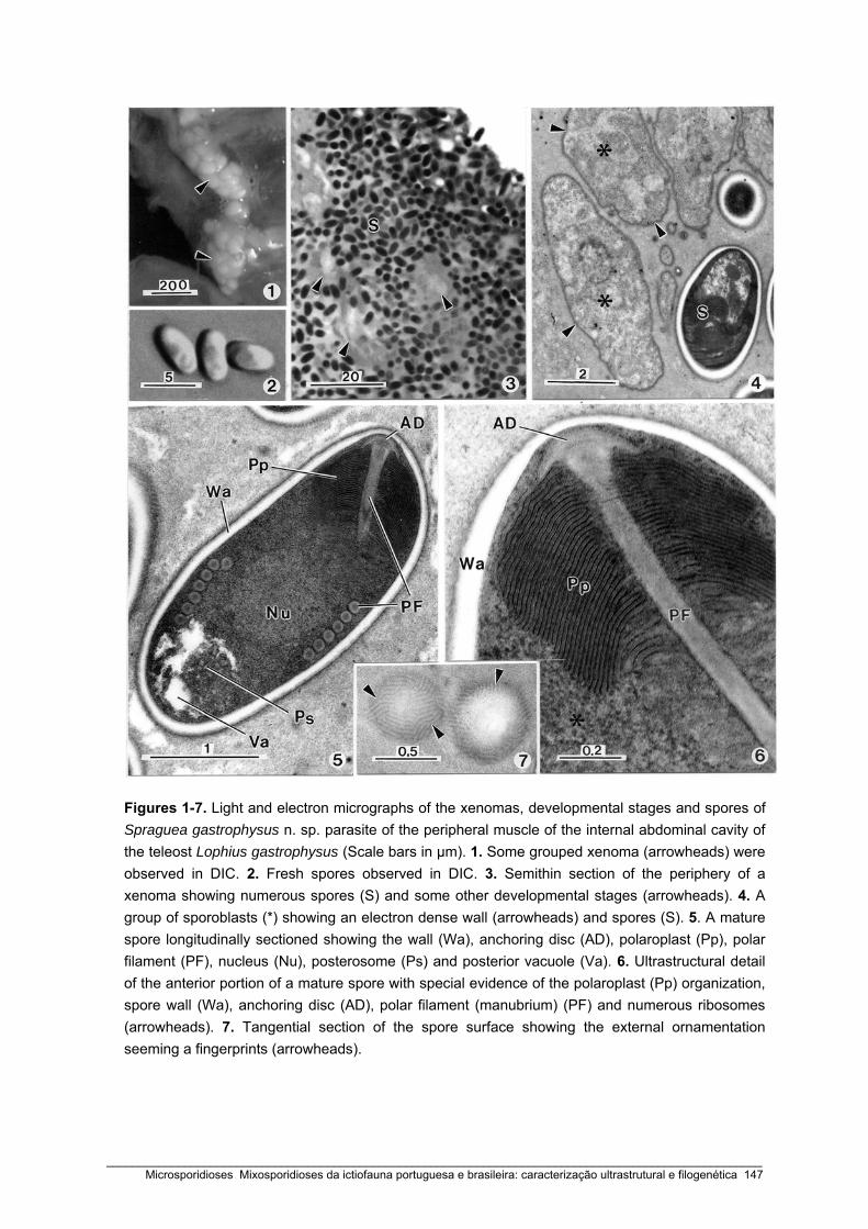
Figures 1-7. Light and electron micrographs of the xenomas, developmental stages and spores of Spraguea gastrophysus n. sp. parasite of the peripheral muscle of the internal abdominal cavity of the teleost Lophius gastrophysus (Scale bars in μm). 1. Some grouped xenoma (arrowheads) were observed in DIC. 2. Fresh spores observed in DIC. 3. Semithin section of the periphery of a xenoma showing numerous spores (S) and some other developmental stages (arrowheads). 4. A group of sporoblasts (*) showing an electron dense wall (arrowheads) and spores (S). 5. A mature spore longitudinally sectioned showing the wall (Wa), anchoring disc (AD), polaroplast (Pp), polar filament (PF), nucleus (Nu), posterosome (Ps) and posterior vacuole (Va). 6. Ultrastructural detail of the anterior portion of a mature spore with special evidence of the polaroplast (Pp) organization, spore wall (Wa), anchoring disc (AD), polar filament (manubrium) (PF) and numerous ribosomes (arrowheads). 7. Tangential section of the spore surface showing the external ornamentation seeming a fingerprints (arrowheads).
_____________________________________________________________________________________________________ Microsporidioses Mixosporidioses da ictiofauna portuguesa e brasileira: caracterização ultrastrutural e filogenética 147

The external surface of the exospore is ornamented with numerous fingerprints uniformly
distributed at the periphery of the spore wall (Fig. 7). The anchoring disc was in close
contact with the internal apical portion of the spore wall (Figs 5, 6). The anchoring disc is
located in the apical region of the spore in an eccentric position in relation to the spore
axis in continuity with the anterior part of the polar filament (PF) (manubrium) (Figs 5, 6).
The anterior part of the PF measured 120.2 ± 5.1 nm (n = 20) and was passing through
the polaroplast with an angle of tilt of ~ 30º. The PF was isofilar, measuring 100–110 nm
in diameter, arranged into 5 – 6 coils in one row (Fig. 5). The polaroplast consisted of a
complex membranous system with two distinct kinds
of lamellae. The anterior group of lamellae was
closely packed and parallel lamellae (~ 12 nm
between the folds) and the posterior was larger
spaced lamellae irregularly organized (Fig. 6). The
spores were monomorphic uninucleate and the
nucleus occupied a position between the apical
polaroplast and the basal vacuole (Fig. 5). The
posterior vacuole occupied about one-quarter of the
total volume of the spore (Fig. 5) and contained a
spherical electron dense posterosome, measuring
about 0.65 nm.
Figure 8. Schematic drawing of a spore of Spraguea gastrophysus n. sp., showing all typical specific structures of the microsporidian spore.
Small subunit rDNA and phylogenetic analysis
1824 bp sequence (GC content 46.8%) representing the partial SSU, complete ITS and
partial LSU rDNA of the parasite was successfully amplified and deposited in GenBank
with the accession number (GQ868443). A Blast search of the GenBank database with
the sequence obtained from Spraguea gastrophysus detected close matches to other
microsporidian rRNA sequences, namely with all Spraguea spp. sequences. Previously all
microsporidian sequences that have a fish as host were aligned and the most
parsimonious tree showed that Spraguea gastrophysus is grouped with all Spraguea spp.
A second alignment with 35 selected sequences, including all from the group 4 designed
by Nilsen and Lom (2003) was done. The 5-end and 3-end SSU rDNA were trimmed,
resulting in the alignment with 1448 bp. Before the phylogenetic analysis was performed
_____________________________________________________________________________________________________ 148 Microsporidioses Mixosporidioses da ictiofauna portuguesa e brasileira: caracterização ultrastrutural e filogenética

Length of SSU used in the analysis
Gaps Insertions Transitions Transversions
Spraguea lophii (1) AF104086 1287 7 5 7 6
Spraguea lophii (2) AF033197 1324 0 13 4 11
Spraguea lophii (3) AF056013 1175 0 9 6 6
Spraguea americana (1) AF056014 1174 2 10 4 6
Spraguea americana (2) AY465876 1206 0 4 1 1
Table 2 Comparative analysis of all Spraguea spp. sequences with the obtained in this study from of Lophius gastrophysus. Included are the SSU rDNA length, the number the gaps, insertions, transitions and transversions.
Host species
Parasite
Country (region)
Host tissues
Spore dimensions
(in μm)
Wall thick (in nm)
Exospore / Endospore
Polar filament
coils
Spore surface
ornamentation
Spore
dimorphism
References
Lophius piscatorius
Spraguea lophii
France
(Atlantic coast)
3.5× 1.5 - 5 - 6 + + Loubès et al., 1979
L. budegassa
S. lophii
France (Mediterranean)
4× 1.25 - 3 – 4 - + Loubès et al., 1979
L. americanus
S. americana
USA
(Atlantic coast)
2.8 × 1.5 -
12.5 / 70
6 - 9 + - Takvorian and Cali, 1986
L. litulon
S. americana
Japan
3.4 × 1.8 -
~30 / ~65
5 – 8 + - Freemann et al., 2004
L. gastrophysus
S. gastrophysus n. sp.
Brasil
(Atlantic coast)
3.35 × 1.71 ~ 75
~18 / ~60
5 - 6 + - Present study
Table 3 Comparative measurements (in μm) from Spraguea spp.
_____________________________________________________________________________________________________ Microsporidioses Mixosporidioses da ictiofauna portuguesa e brasileira: caracterização ultrastrutural e filogenética 149
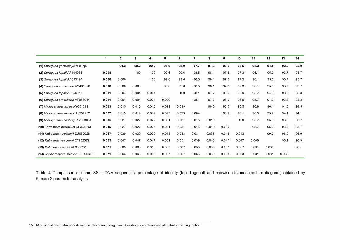
1 2 3 4 5 6 7 8 9 10 11 12 13 14
(1) Spraguea gastrophysus n. sp. 99.2 99.2 99.2 98.9 98.9 97.7 97.3 96.5 96.5 95.3 94.5 92.9 92.9
(2) Spraguea lophii AF104086 0.008 100 100 99.6 99.6 98.5 98.1 97.3 97.3 96.1 95.3 93.7 93.7
(3) Spraguea lophii AF033197 0.008 0.000 100 99.6 99.6 98.5 98.1 97.3 97.3 96.1 95.3 93.7 93.7
(4) Spraguea americana AY465876 0.008 0.000 0.000 99.6 99.6 98.5 98.1 97.3 97.3 96.1 95.3 93.7 93.7
(5) Spraguea lophii AF056013 0.011 0.004 0.004 0.004 100 98.1 97.7 96.9 96.9 95.7 94.9 93.3 93.3
(6) Spraguea americana AF056014 0.011 0.004 0.004 0.004 0.000 98.1 97.7 96.9 96.9 95.7 94.9 93.3 93.3
(7) Microgemma tincae AY651319 0.023 0.015 0.015 0.015 0.019 0.019 99.6 98.5 98.5 96.9 96.1 94.5 94.5
(8) Microgemma vivaresi AJ252952 0.027 0.019 0.019 0.019 0.023 0.023 0.004 98.1 98.1 96.5 95.7 94.1 94.1
(9) Microgemma caulleryi AY033054 0.035 0.027 0.027 0.027 0.031 0.031 0.015 0.019 100 95.7 95.3 93.3 93.7
(10) Tetramicra brevifilum AF364303 0.035 0.027 0.027 0.027 0.031 0.031 0.015 0.019 0.000 95.7 95.3 93.3 93.7
(11) Kabatana newberryi EU682928 0.047 0.039 0.039 0.039 0.043 0.043 0.031 0.035 0.043 0.043 99.2 96.9 96.9
(12) Kabatana newberryi EF202572 0.055 0.047 0.047 0.047 0.051 0.051 0.039 0.043 0.047 0.047 0.008 96.1 96.9
(13) Kabatana takedai AF356222 0.071 0.063 0.063 0.063 0.067 0.067 0.055 0.059 0.067 0.067 0.031 0.039 96.1
(14) Aspalatospora milevae EF990668 0.071 0.063 0.063 0.063 0.067 0.067 0.055 0.059 0.063 0.063 0.031 0.031 0.039
Table 4 Comparison of some SSU rDNA sequences: percentage of identity (top diagonal) and pairwise distance (bottom diagonal) obtained by Kimura-2 parameter analysis.
_____________________________________________________________________________________________________ 150 Microsporidioses Mixosporidioses da ictiofauna portuguesa e brasileira: caracterização ultrastrutural e filogenética

Figure 9. The maximum parsimony tree of SSU rDNA sequences of Spraguea gastrophysus n. sp. and other selected microsporidian species. The numbers on the branches are bootstrap confidence levels on 100 replicates. GenBank accession numbers are in parentheses after the species names and the scale is given under the tree. Spraguea gastrophysus clusters with all other Spraguea spp. (highlighted box).
_____________________________________________________________________________________________________ Microsporidioses Mixosporidioses da ictiofauna portuguesa e brasileira: caracterização ultrastrutural e filogenética 151

only those sites which could be unambiguously aligned among all microsporidia and
outgroup were used, resulting in an alignment of 1371 bases long.
By the Kimura-2-parameter model the pairwise distance shown that the sequences with
more affinities were all belonging to the Spraguea genus with a percentage of identity of
99.2% (S. lophii AF104086, S. lophii AF033197, S. americana AY465876) and 98.9% (S.
lophii AF056013, S. americana AF056014). The final phylogenetic tree was built to the
maximum parsimony and it shown that all Spraguea spp. are a monophyletic group with
91% of boostrap. The same % of bootstrap was found for the clade composes by all
Spraguea spp, Microgemma spp. and genus Tetramicra with a only species. Upon
analysis of the sequences, a small number of gaps, insertions, transitions and
transversions were found (Table 2).
Discussion
The parasite described in this paper presents all typical morphology and characters of the
phylum Microsporidia (Cali and Takvorian 1999; Larsson 1999; Lom and Dyková 1992).
Among 17 genera infecting fish, 12 of these produce xenomas: Amazonspora Azevedo
and Matos 2003; Glugea Thélohan, 1891; Ichthyosporidium Caullery and Mesnil, 1905;
Loma Morrison and Sprague, 1981; Microfilum Faye, Toguebaye and Bouix, 1991;
Microgemma Ralphs and Matthews, 1986; Myosporidium Baquero, Rubio, Moura,
Pieniazek and Jordana, 2005; Neonosemoides Faye, Toguebaye and Bouix, 1996;
Pseudoloma Matthews, Brown, Larison, Bishop-Stewart, Rogers and Kent, 2001;
Potaspora Casal, Matos, Teles-Grilo and Azevedo, 2008; Spraguea Vávra and Sprague,
1976 and Tetramicra Matthews and Matthews, 1980.
Among these microsporidians, the Spraguea genus is a typical case of close relationship
of parasite, host specificity and the local of infection. All infections by Spraguea spp. are
confined to the hosts belonging to the Lophius genus from different geographic areas, like
Europe, America and Japan. Presently, it is known that the five species (L. piscatorius, L.
budegassa, L. americanus, L. litulon and L. gastrophysus) are parasitized with Spraguea
spp. and all in the nervous tissues. They have been localized in the spinal nerves of the
vertebral column, trigeminal nerves, vagal nerves or on the medulla oblongata region of
the hind brain (Jakowska 1964; Takvorian and Cali 1986; Weissenberg 1911c, 1976). One
exception to the parasite-host specificity was observed in the anglerfish Lophius
budegassa. In Spain, the microsporidian Tetramicra brevifilum was also found in
musculature and hepatocytes of this lophii fish (Maíllo et al. 1998). This species, which is
phylogenetically close to the Spraguea spp., frequently parasite the connective tissues of
_____________________________________________________________________________________________________ 152 Microsporidioses Mixosporidioses da ictiofauna portuguesa e brasileira: caracterização ultrastrutural e filogenética

the musculature of Scophtalmus maximus (Matthews and Matthews 1980). Some details
like the absence of a conspicuous inclusion body inside the posterior vacuole and the
presence of ornamentation outside of the exospore exclude the possibility of the parasite
described here belonging to the genus Tetramicra.
Irrefutably the parasite described in this study is morphological and phylogenetically close
to two Spraguea species previously reported in the different anglerfish species. Spraguea
lophii was described in Lophius piscatorius and L. budegassa in the European Atlantic
coasts and Mediterranean coasts (Loubès et al. 1979) and Spraguea americana, first as
Glugea americanus, in the USA Atlantic coast (L. americanus) (Takvorian and Cali 1986)
and later in L. litulon from Japanese coast (Freeman et al. 2004). The molecular data has
shown that the Glugea americanus sequences are close to all others Spraguea lophii
sequences and consequently it was transferred to the genus Spraguea and renamed as
S. americana (Pomport-Castillon et al. 2000; Nilsen 2000; Lom and Nilsen 2003).
The morphology of the spores show several morphological similarities when compared
with the uninuclear spores sequence of the Spraguea genus, except for the thickness of
the two layers of the wall (exospore and endospore). The spore wall found in Lophius
gastrophysus is thicker than that of other species (Table 3).
For some genera, such as Amazonspora (Azevedo and Matos 2003), Kabatana (Lom et
al. 1999; 2001; McGourty et al. 2007) and Spraguea (Loubès et al. 1979; Freeman et al.,
2004) the fingerprint-like structures of the external surface of the exospore wall are a
morphological characteristic common at all species. Usually, the external ornamentation is
regularly distributed and the elevations on the surface of the mature spores, when they
are observed in tangential section, present a hexagonal fingerprint-like shape (genus
Amazonspora) or a tubular shape (genera Kabatana and Spraguea).
The most parsimonious phylogenetic tree (Fig. 9) clustered all Spraguea infections
sequences in same clade (bootstrap 91%) and this cladogram had a similar topology to
the previous described trees (Casal et al. 2008; Freeman et al. 2004; Lom and Nilsen
2003). Comparison of the SSU rDNA sequence of Spraguea gastrophysus with all the
others known sequences from Spraguea infections from Europe, Japan and America
showed that the genetic distances range from 0.8 to 1.1% (Table 4). Whereas, the genetic
distance between all previously reported Spraguea infections is equal or lower than 0.4%.
These data are in accordance with that obtained by Freeman et al. (2004) and it suggests
that the microsporidia found in anglerfish Lophius gastrophysus from South America is the
most phylogenetically distanced of the three Spraguea species.
_____________________________________________________________________________________________________ Microsporidioses Mixosporidioses da ictiofauna portuguesa e brasileira: caracterização ultrastrutural e filogenética 153

According the original description by Loubés et al. (1979) the genus Spraguea is a
dimorphic microsporidian that produces two types of spores in two distinct developmental
sequences. In one sequence, is characterized by all stages having unpaired nuclei and
polysporoblastic sporogony that produce uninucleate spores whereas the other present
diplokarya and disporoblastic sporogonic stages that give rise to slender curved
diplokaryotic spores. Curiously, the spore dimorphism of the genus and species type is in
contradiction with all others ultrastructural descriptions. Apparently, this is an exclusive
characteristic of the Spraguea infections from Lophius piscatorius and L. budegassa from
European species because the infections from American and Japanese species only
produces uninucleated spores. Definitely, as recommended by Lom (2002), Lom and
Nilsen (2003) and Freeman et al. (2004) the genus diagnosis need to be redescribed
since the exceptions must not be a general characteristic of the genus.
Considering the morphological and molecular data, as well as the host specificity, we
believe that this microorganism represents a new species that should be included in the
genus Spraguea with the name Spraguea gastrophysus n. sp.
ACKNOWLEDGEMENTS
This work was partially supported by Engº. A. Almeida Foundation, Porto, Portugal, PhD grant from
“CESPU” (G. Casal), “CNPq” and “CAPES”, Brazil. We would like the technical assistance of J.
Carvalheiro (ICBAS/UP) and Nilza Felizardo (UFF). This work is original and complies with the
current laws of the countries in which it has been performed.
REFERENCES
Azevedo, C., Matos, E., 2002. Fine structure of a new species, Loma myrophis (Phylum Microsporidia),
parasite of the Amazonian fish Myrophis phatyrhynchus (Teleostei, Ophichithidae). Europ. J. Protistol.
37, 445-452.
Azevedo, C., Matos, E., 2003. Amazonspora hassar n. gen. and n. sp. (Phylum Microsporidia, fam.
Glugeidae), a parasite of the Amazonian teleost Hassar orestis (fam. Doradidae). J. Parasitol. 89, 336-
341.
Cali, A., Takvorian, P.U., 1999. Developmental morphology and life cycles of the Microsporidia. In The
Microsporidia and Microsporidiosis (ed. Wittner, M. and. Weiss, L.), pp. 85-128, American Society of
Microbiology, Washington, DC.
Casal G., Matos E., Teles-Grilo M.L., Azevedo C., 2008. A new microsporidian parasite, Potaspora morhaphis
n. gen., n. sp. (Microsporidia) infecting the teleostean fish, Potamorhaphis guianensis from the River
Amazon. Morphological, ultrastructural and molecular characterization. Parasitology 135, 1053-1064.
_____________________________________________________________________________________________________ 154 Microsporidioses Mixosporidioses da ictiofauna portuguesa e brasileira: caracterização ultrastrutural e filogenética

Döflein, F., 1898. Studien zur Naturgeschichte der Protozoen. III. Ueber Myxosporidien. Zool. Jahrb. Anat. 11,
281-350.
Freeman, M.A., Yokoyama, H., Ogawa, K., 2004. A microsporidian parasite of the genus Spraguea in the
nervous tissues of the Japanese anglerfish Lophius litulon. Folia Parasitol. 51, 167-176.
Gatehouse, H.S., Malone, L.A., 1998. The ribosomal RNA gene region of Nosema apis (Microspora): DNA
sequence for small and large subunit rRNAgenes and evidence of a large tandem repeat unit size. J.
Invert. Pathol. 71, 97-105.
Jakowska, S., 1964. Infecção microsporídea das células nervosas numa população de peixes marinhos,
Lophius americanus. Annual 2nd Congress Latin- American Zoology (S. Paulo, Brazil, 1962). 1: 265-273
(In Portuguese).
Jakowska, S., 1966. Infection with neurotropic microsporidians in South American Lophius. Trans. Am.
Microsc. Soc. 85: 161-162.
Jakowska, S., Nigrelli, R.F., 1958. Preliminary biochemical studies on neurotropic microsporidil Glugea-cyst in
the American anglerfish. J. Protozool. 5 (Suppl.): 16.
Jakowska, S., Nigrelli, R.F., 1959. Nosematiasis in the American anglerfish. J. Protozool., 6 (Suppl.), 7.
Larsson, J.I.R. 1999. Identification of Microsporidia. Acta Protozool. 38, 161-197.
Lom, J. 2002. A catalogue of described genera and species of microsporidians parasitic in fish. Syst.
Parasitol. 53, 81-99.
Lom, J., Dyková, I., 1992. Microsporidia (Phylum Microspora Sprague, 1977). In Protozoan Parasites of
Fishes. Developments in Aquaculture and Fisheries Sciences (ed. Lom, J. and Dyková, I.), vol 26, pp.
125-157. Elsevier, Amsterdam.
Lom, J., Nilsen, F., 2003. Fish microsporidia: fine structural diversity and phylogeny. Int. J. Parasitol. 33, 107-
127.
Lom, J., Dyková, I., Tonguthai K., 1999. Kabataia gen. n., new genus proposed for Microsporidium spp.
infecting trunk muscles of fishes. Dis. Aquat. Org. 38, 39-46.
Lom, J., Nilsen, F., Urawa, S., 2001. Redescription of Microsporidium takedai (Awakura, 1974) as Kabatana
takedai (Awakura, 1974) comb. n. Dis. Aquat. Org. 44, 223-230.
Loubès, C., Maurand, J., Ormières, R., 1979. Étude ultrastructurale de Spraguea lophii (Doflein, 1898,
microsporidie parasite de la Baudroie: essai d’ interprétation du dimorphisme sporal. Protistologica 15,
43-54.
Maíllo, P.A., Amigó, J.M., Baena, R., Salvadó, H., Gracia, M.P., 1998. Tetramicra brevifilum (Matthews &
Matthews, 1980) (Microsporita: Tetramicriidae) in a new fish host, Lophius budegassa (Spinola, 1807) in
Spain. Parasitol. Res. 84, 208-212.
Matos, E., Azevedo, C., 2004. Ultrastructural description of Microsporidium brevirostris sp. n., parasite of the
teleostean Brachyhypopomus brevirostris (Hypopomidae) from the Amazon River. Acta Protozool. 43,
261-267.
Matthews, R.A., Matthews, B.F., 1980. Cell and tissue reactions of turbot Scophthalmus maximus (L.) to
Tetramicra brevifilum gen. n., sp. n. (Microspora). J. Fish Dis. 3, 495-515.
_____________________________________________________________________________________________________ Microsporidioses Mixosporidioses da ictiofauna portuguesa e brasileira: caracterização ultrastrutural e filogenética 155
McGourty, K.R., Kinzger, A.P., Hendrickson, G.L., Goldsmith, G.L., Casal, G., Azevedo, C., 2007. A new
microsporidian infecting the musculature of the endangered tidewater goby (Gobiidae). J. Parasitol. 93,
655-660.
Mrázek, A., 1899. Sporozoenstudien II. Glugea lophii Döflein. Stizungsber. Böhm. Ges. Wiss. Mathnaturwiss.
Cl., Prag. 34, 1-8.
Nilsen, F., 2000. Small subunit ribosomal DNA phylogeny of microsporidia with particular reference to genera
that infect fish. J. Parasitol. 86, 128-133.
Pace, D., 1908. Parasiten und Pseudoparasiten der Nervenzelle. Vorläufige Mitteilungen über vergleichende
Parasitologie des Nervensystems. Z. Hyg. Infekstions Kr. 60, 62-74.
Pomport-Castillon, C., De Jonckheere, J.F., Romestand, B., 2000. Ribosomal DNA sequences of Glugea
anomala, G. stephani, G. americanus and Spraguea lophii (Microsporidia): phylogenetic reconstruction.
Dis. Aquat. Org. 40, 125-129.
Takvorian, P.M., Cali, A., 1986. The ultrastructure of spores (Protozoa: Microsporidia) from Lophius
americanus, the angler fish. J. Protozool. 33, 570-575.
Tamura, K., Dudley, J., Nei, M., Kumar, S., 2007. MEGA4: Molecular evolutionary genetics analysis (MEGA)
software version 4.0. Mol. Biol. Evol. 24, 1596-1599.
Thompson, J.D., Higgins, D.G., Gilson, T.J., 1994. Clustal W: improving the sensitivity of progressive multiple
sequence alignment through sequence weighting, position-specific gap penalties and weight matrix
choice. Nucl. Acids Res. 22, 4673-4680.
Vossbrinck, C.R., Baker, M.D., Didier, E.S., Debrunner-Vossbrinck, B.A., Shadduck, J.A., 1993. Ribosomal
DNA sequences of Encephalitozoon hellem and Encephalitozoon cuniculi: species identification and
phylogenetic construction. J. Eukaryot. Microbiol. 40, 354-362.
Weissenberg, R., 1909. Beiträge zur Kenntnis von Glugea lophii Doflein. I. Üeber den Sitz und die Verbreitung
der Mikrosporidien-Zysten am Nervensystem von Lophius piscatorius und budegassa. Sitzungsber.
Ges. Naturforsch. Freunde Berlin 9, 557-565.
Weissenberg, R., 1911a. Über einige Mikrosporidien aus Fischen (Nosema lophii Doflein, Glugea anomala
Moniez, Glugea hertwigii nov. spec.). Sitzungsber. Ges. Naturforsch. Freunde Berlin. 8, 344-357.
Weissenberg, R., 1911b. Beiträge zur Kenntnis von Glugea lophii Doflein. II. Über den Bau der Zysten und die
Beziehungen zwischen Parasit und Wirtsgewebe. Sitzungsber. Ges. Naturforsch. Freunde Berlin. 3,
149-157.
Weissenberg, R., 1911c. Über Mikrosporidien aus dem Nervensystem von Fischen (Glugea lophii Doflein) und
die Hypertrophie der befallenen Ganglienzellen. Arch. Mikrosk. Anat. 78, 383-421.
Weissenberg, R., 1976. Microsporidian interactions with the host cell. In Comparative Pathobiology, (ed. Bulla
L. A. and Cheng, T. C.), Volume 1, pp. 203-237, New York and London, Plenum Press.
_____________________________________________________________________________________________________ 156 Microsporidioses Mixosporidioses da ictiofauna portuguesa e brasileira: caracterização ultrastrutural e filogenética
PARTE III
MIXOSPORIDIOSES


Capítulo 6
ULTRASTRUCTURAL DATA ON THE SPORE OF MYXOBOLUS MACULATUS N. SP.
(PHYLUM MYXOZOA), PARASITE FROM THE AMAZONIAN FISH
METYNNIS MACULATUS (TELEOSTEI)
Diseases of Aquatic Organisms (2002) 51: 107–112
Graça Casal, Edilson Matos & Carlos Azevedo
_____________________________________________________________________________________________________ Microsporidioses Mixosporidioses da ictiofauna portuguesa e brasileira: caracterização ultrastrutural e filogenética 159

_____________________________________________________________________________________________________ 160 Microsporidioses Mixosporidioses da ictiofauna portuguesa e brasileira: caracterização ultrastrutural e filogenética

DISEASES OF AQUATIC ORGANISMSDis Aquat Org
Vol. 51: 107–112, 2002 Published August 29
INTRODUCTION
The genus Myxobolus Bütschli, 1882 (family Myxo-bolidae), is the largest myxosporean group, and its mem-bers are important pathogens of freshwater and marinefishes in several geographical areas. The morphologyand ultrastructure of myxosporean species have beenwidely studied (Landsberg & Lom 1991, Lom & Dyková1992). However, in Brazilian host species, few have beendescribed and, with the exception of 1 ultrastructuralstudy (Casal et al. 1996), only light microscopy descrip-
tions are available (Walliker 1969, Kent & Hoffman 1984,Molnár & Békési 1993, Gioia & Cordeiro 1996, Molnár etal. 1998). In this paper we present light and electronmicroscopical data of a new myxosporidian species,M. maculatus n. sp., found in the teleost fish, Metynnismaculatus collected from the Amazon River. Somepeculiar ultrastructural aspects of the structure of theplasmodium and developmental stages of the capsulo-genesis are described and discussed.
MATERIALS AND METHODS
A parasite found in the kidney of the freshwaterteleost Metynnis maculatus Kner, 1860 (family Chara-cidae), known by the Brazilian common name ‘pacú’,was investigated. The specimens were collected peri-
© Inter-Research 2002 · www.int-res.com
*Corresponding author.Present address: Department of Cell Biology, Institute of Bio-medical Sciences, University of Oporto, Lg. Professor AbelSalazar no. 2, 4099-003 Porto, Portugal. E-mail: [email protected]
Ultrastructural data on the spore of Myxobolusmaculatus n. sp. (phylum Myxozoa), parasite from
the Amazonian fish Metynnis maculatus (Teleostei)
G. Casal1, 2, E. Matos3, C. Azevedo2, 4,*
1Department of Biological Sciences, High Institute of Health Sciences, 4580 Paredes, Portugal2CIIMAR—Centre for Marine and Environmental Research, University of Oporto, 4150-180 Porto, Portugal
3Laboratory of Animal Biology, Faculty of Agricultural Sciences, Belém, Brazil4Department of Cell Biology, Institute of Biomedical Sciences, University of Oporto, 4099-003 Porto, Portugal
ABSTRACT: Light and electron microscopy studies of a myxosporean, parasitic in the intertubularinterstitial tissue of the kidney of the freshwater teleost fish Metynnis maculatus Kner, 1860 (Characi-dae) from the lower Amazon River (Brazil), are described. We observed polysporic histozoic plas-modia delimited by a double membrane and with several pinocytic channels and containing severallife cycle stages, including mature spores. The spore body was of pyriform shape and was 21.0 μmlong, 8.9 μm wide and 7.5 μm thick. Elongated-pyriform polar capsules were of equal size (12.7 �3.2 μm) and contained a polar filament with 14 or 15 coils. The spore features fit those of the genusMyxobolus. Densification of the capsular primordium matrix, which increased in density from theinner core outwards, differentiating at the periphery into small microfilaments measuring 45 nmeach, and tubuli arranged in aggregates and dispersed within the capsular matrix of the maturespores, are described. Based on the morphological differences and specificity of the host, we proposethe creation of a new species named Myxobolus maculatus n. sp.
KEY WORDS: Ultrastructure · Parasite · Myxosporidian · Myxobolus maculatus · Amazonian fish
Resale or republication not permitted without written consent of the publisher
_____________________________________________________________________________________________________ Microsporidioses Mixosporidioses da ictiofauna portuguesa e brasileira: caracterização ultrastrutural e filogenética 161

Dis Aquat Org 51: 107–112, 2002
odically during the year 2000 from the estuarine regionof the Amazon River, near Belém, Brazil. Plasmodiawith mature spores were examined in fresh mountswith a light microscope equipped with Nomarski dif-ferential interference-contrast optics. For TEM, smallparasitized fragments were fixed in 3% glutaralde-hyde in 0.2 M sodium cacodylate buffer (pH = 7.4) at4°C for 6 h, then washed with the same bufferovernight and post-fixed in 2% OsO4 buffered with0.2 M sodium cacodylate for 2 h at the same tempera-ture. The fragments were dehydrated in an ascendingethanol and propylene oxide series and then embed-ded in Epon. Semithin sections were stained withmethylene blue and photographed under the lightmicroscope (DIC). Ultrathin sections, cut with a dia-mond knife, were stained with both aqueous uranylacetate and lead citrate and observed in a JEOL100CXII TEM operated at 60 Kv.
RESULTS
Several foci of infection, plasmodia of approximately150 μm diameter localized in the intertubular intersti-tial tissue of the kidney, were observed. The infectedkidney presented cellular and nuclear hypertrophyaccompanied by morphological changes, such asorganelle disorganization and cytoplasm vacuolization(Fig. 1). Sporogenic stages released into the renalinterstitium due to basement membrane rupture werefrequently observed (Fig. 7). Asynchronous histozoicplasmodia containing several life-cycle developmentalstages of the parasite (generative cells, sporogenicstages and mature spores) were observed (Figs. 1 & 3).The plasmodia were delimited by 2 membranes, theinnermost being continuous with a distinct zone ofpinocytic channels (Fig. 3: inset). The external plas-modial membrane was slightly separated from theendothelial cells and possessed a sinuous outline withsome papillary buds (Fig. 3).
Sporogenesis
Pansporoblast formation followed the well-knownpattern of a sporogenic cell developing into 2 sporeswithin a pericyte. Morphogenesis of capsulogenic cellscorresponded to that of most myxosporeans, yet somespecific features were found. Capsulogenesis beganwith a club-shaped formation that posteriorly changed toa globular structure, the capsular primordium, whichextended into an external tube (Figs. 4 & 6). Early indevelopment, the distal end of the external tube waslocated below the future discharge channel leadingthrough the shell valve, and was sealed by an electron-
dense structure (Fig. 6). Inside the capsular primordium,before the inversion of the external tube, the matrix wascomposed of a fine dense granular structure. This wasorganized in concentric layers that gradually densifiedfrom the inner core outwards, being differentiatedinto fine microfilaments at the periphery. These micro-filaments measured about 45 ± 5 nm in diameter andwere arranged in a continuous row (Figs. 4 & 5). Simul-taneously, the polar filament differentiated at theexternal tubule and then invaginated and coiled insidethe matrix (Fig. 7). In mature spores, the matrix be-came denser and numerous electrolucent aggregatesof tubuli, arranged in bundles around the polar fila-ment, were observed (Fig. 8).
Spore characteristics
Fresh mature spores were of pyriform shape, taper-ing anteriorly to a slightly knob-like end, and mea-sured ~21.0 � ~8.9 μm in anterior view (Figs. 2 & 9).The spore wall was thin and smooth, comprising 2equal valves joined by a sutural ridge. No mucus enve-lope was observed at the surface of the spore (Fig. 2).Internally, 2 capsulogenic cells, located side by side,contained prominent polar capsules (PCs) of elongatedpyriform shape and equal size, measuring ~12.7 �~3.2 μm (Figs. 2 & 9). The PCs occupied approximatelytwo-thirds of the total spore length. Inside the PCs, apolar filament displayed 14 or 15 coils perpendicular orslightly oblique to the longitudinal axis (Figs. 2 & 7).No intercapsular appendix was present (Fig. 2). At theposterior pole of the spore, a binucleated sporoplasmcontained numerous electron-dense vesicles, sporo-plasmosomes, glycogen granules and an extensive sys-tem of rough endoplasmic reticulum cisternae (Fig. 7).Fresh mature spores and ultrathin sections demon-strated the existence of a large iodinophilous vacuolein the sporogenic cell measuring approximately 4.5 μmin diameter (Fig. 2).
Diagnosis
Host: teleost fish, Metynnis maculatus Kner, 1860(family Characidae).Locality: estuarine region of the Amazon river(01°11’30’ S, 47° 18’ 54’’ W) near Belém, Brazil.Site of infection: spores were located in the kidney.Prevalence and intensity: 12 out of 30 (40%).Fresh spore measurements (n = 40): length = 21.0 (19.7to 23.0) μm, width = 8.9 (7.9 to 9.5) μm, thickness = 7.5(7.2 to 7.9) μm; polar capsules: length = 12.7 (11.8 to13.8) μm, width = 3.2 (3.0 to 3.6) μm; number of polarfilament turns = 14 to 15.
108
_____________________________________________________________________________________________________ 162 Microsporidioses Mixosporidioses da ictiofauna portuguesa e brasileira: caracterização ultrastrutural e filogenética

Casal et al.: Myxobolus maculatus n. sp., Amazonian fish parasite 109
Figs. 1–3. Myxobolus maculatus n. sp. Life cycle stages of the parasite in the kidney of Metynnis maculatus. Fig. 1. Semithinsection showing plasmodium (�) in the intertubular intertitial tissue of the kidney (KT). Fig. 2. Fresh mature spores observed withdifferential interference-contrast (Nomarski). Fig. 3. Plasmodium delimited by double membrane (arrows) showing muchcytoplasmatic degradation (C) and different life cycle stages, such as generative cells (G) and sporogenic stages (�); Outside theplasmodium an endothelial cell nucleus (N) is visible. Inset (�8800) shows detail of the plasmodium wall with membranes, the
innermost (I) of which is in direct contact with the pinocytic channels (arrows). (Scale bars: 1 = �500; 2 = �1525; 3 = �32 000)
1
3
2
_____________________________________________________________________________________________________ Microsporidioses Mixosporidioses da ictiofauna portuguesa e brasileira: caracterização ultrastrutural e filogenética 163

Dis Aquat Org 51: 107–112, 2002110
Figs. 4–8. Myxobolus maculatus n. sp. Life cycle stages of the parasite in the kidney of Metynnis maculatus. Fig. 4. Transversesection of a capsular primordium showing the matrix with different degrees of densification (a,b,c), and the periphery differ-entiated into fine microfilaments (d). Fig. 5. Detail of a capsular primordium in tangential section showing some microfilaments(arrows) near the capsular primordium wall (W). Fig. 6. Anterior pole of 2 capsulogenic cells, each with an external tubule (ET)surrounded by microtubules (arrow) and sealed by an electron-dense structure (arrowhead). Fig. 7. Immature spore localized inthe interstitial tissue showing the polar capsules (PC) with polar filaments (PF) coiled inside, sporoplasm (S) and epithelial cells(E) of the uninfected kidney tubule. Fig. 8. Detail of a densified mature polar capsule, showing numerous tubuli organized intoaggregates (arrows) and associated with the polar filament (PF). (Scale bars: 4 = �40 000; 5 = �50 400; 6 = �20 800; 7 = �4480;
8 = �40 000)
4
5
6
7 8
_____________________________________________________________________________________________________ 164 Microsporidioses Mixosporidioses da ictiofauna portuguesa e brasileira: caracterização ultrastrutural e filogenética

Casal et al.: Myxobolus maculatus n. sp., Amazonian fish parasite
Specimens deposited: slides with holotype weredeposited in the International Protozoan Type SlidesCollection at the Smithsonian Institution, Washington,DC 20560, USA (USNM #1002151) and in the collec-tion of the senior author.Etymology: the specific name is derived from the nameof the host species (‘maculatus’).
DISCUSSION
The mature spores obtained from Metynnis macula-tus revealed morphological similarities to those of thegenus Myxobolus Bütschli, 1882. Comparison of theplasmodium wall and sporogenesis of the cycle lifestages of this species to those of other Myxobolus spp.also revealed morphologic and ultrastructural similari-ties (Lom & Puytorac 1965, Desser & Paterson 1978,Current et al. 1979, Lom & Dyková 1992).
The plasmodial wall presented an organization typi-cal for histozoic Myxobolus species, with a doublemembrane and pinocytic channels region extending
into the ectoplasmic zone of the plasmodium (Desser &Paterson 1978, Current et al. 1979). All Myxobolusspecies give rise to histozoic plasmodia, except for onespecies, M. conei, that is found in the lumen of bileducts in the liver of Pseudocaranx dentex (Lom &Dyková 1994). Usually, myxosporidian species thatparasitize renal tubules are coelozoic and do notbelong to the genus Myxobolus (Lom 1969, Desser etal. 1983, Lom & Dyková 1985). In the present work wedescribe a new histozoic species found in the inter-tubular interstitial tissue of the kidney. Unfortunately,studies of other Myxobolus species reported to para-sitize the same organ make but few references to theultrastructural morphology of the plasmodia (Lom &Dyková 1992).
The ultrastructural process of capsulogenesis differ-entiation has been well documented in several myxo-sporidian genera (Lom & Puytorac 1965, Lom 1969,Current et al. 1979, Desser et al. 1983). The structureof the capsular matrix, i.e. immature spores formed byconcentric layers and differentiated at the peripheryinto only 1 row of microfilaments, presents some differ-ences with the one species (Thelohanellus nikolskii )described by Desser et al. (1983), who described amicrofilamentous girdle surrounding the capsularmatrix and speculated that it was probably connectedwith the contractions required for external tubule in-version (Desser et al. 1983). In mature spores, bundlesof tubuli in the capsular matrix have already beenreferred to in some genera, such as Sphaeromyxa (Lom1969) and Henneguya (Rocha et al. 1992), but never inMyxobolus species. This suggests that these tubulesprobably have an important function in the extrusionof the polar filament.
The pathological changes in the renal tissue such asdegeneration and vacuolisation of the renal epithelialcells associated with the presence of the parasitesSphaerospora spp. (Lom & Dyková 1985), are similar tothose observed in M. maculatus in the present study.
There are at least 444 species belonging to thisgenus (Landsberg & Lom 1991), and most of the earlyspecies descriptions are vague, presenting only linedrawings of the spores. At present there are 16Myxobolus species described in Amazonian fishes(Walliker 1969, Kent & Hoffman 1984, Molnár & Békési1993, Casal et al. 1996, Gioia & Cordeiro 1996, Molnáret al. 1998). The Brazilian species—M. cunhai (Penido1927), microspores of M. serrasalmi (Walliker 1969)and M. braziliensis (Casal et al. 1996)—all have asimilar body shape, but are smaller than the speciesdescribed here. Only M. inaequus (Kent & Hoffman1984) is of similar size, however its oval, unequal polarcapsules and its infestation of other host species allexclude the possibility of it being the same species asthat described herein.
111
Fig. 9. Myxobolus maculatus n. sp. Schematic drawings ofmorphology of a spore in anterior (left) and lateral (right) view
as described in ‘Results’ and illustrated in Figs. 2 & 7
_____________________________________________________________________________________________________ Microsporidioses Mixosporidioses da ictiofauna portuguesa e brasileira: caracterização ultrastrutural e filogenética 165

Dis Aquat Org 51: 107–112, 2002
Among Myxobolus spp. from other geographic local-ities, some present a similar body shape, such as M. koiand M. funduli (Kudo 1919), M. procerus (Kudo 1934),M. neurophilus and M. scleroperca (Guilford 1963),M. punctatus (Ray-Chaudhuri & Chakravarty 1970), M.pharyngeus (Parker et al. 1971), and M. maruliensis(Sarkar et al. 1985), but all are smaller in size. Compar-ison of the spores of 3 species—M. magnasherus(Cone & Anderson 1977), M. ovoidalis (Fantham 1930)and M. squamaphilus (Molnár 1997)—revealed similarsizes to that in our study, but all were oval in shapecompared to the pyriform shape in our study.
Comparison of our results with those for other Myxo-bolus species revealed some significant differences,mainly in size and body shape of the spores as well ashost-specificity and ultrastructural details, suggestingthat this parasite (M. maculatus) is a new species.
Acknowledgements. This work was partially supported by agrant from the Engenheiro António Almeida Foundation,Porto, Portugal. We would like to thank the iconographicwork of Mr. João Carvalheiro. The electron microscopy assis-tance provided by Mrs. Laura Corral is gratefully acknowl-edged.
LITERATURE CITED
Casal G, Matos E, Azevedo C (1996) Ultrastructural data onthe life cycle stages of Myxobolus braziliensis n. sp., para-site of an Amazonian fish. Eur J Protistol 32:123–127
Cone DK, Anderson RC (1977) Myxosporidian parasites ofpumpkinseed (Lepomis gibbosus L.) from Ontario. J Para-sitol 63:657–666
Current WL, Janovy J Jr, Knight SA (1979) Myxosoma funduliKudo (Myxosporida) in Fundulus kansae: ultrastructureof the plasmodium wall and of sporogenesis. J Protozool26:574–583
Desser SS, Paterson WB (1978) Ultrastructural and cytochem-ical observations on sporogenesis of Myxobolus sp. (Myx-osporida: Myxobolidae) from the common shiner Notropiscornutus. J Protozool 25:314–326
Desser SS, Molnar K, Weller I (1983) Ultrastructure of sporo-genesis of Thelohanellus nikolskii Akhmerov, 1955 (Myx-ozoa: Myxosporea) from the common carp, Cyprinus car-pio. J Parasitol 69:504–518
Fantham HB (1930) Some parasitic protozoa found in SouthAfrica. S Afr J Sci 27:376–390
Gioia I, Cordeiro NS (1996) Brazilian myxosporidians’ check-list (Myxozoa). Acta Protozool 35:137–149
Guilford HG (1963) New species of Myxosporidia found inpercid fishes from Green Bay (Lake Michigan). J Parasitol49:474–478
Kent ML, Hoffman GL (1984) Two new species of Myxozoa,Myxobolus inaequus sp. n. and Henneguya theca sp. n.
from the brain of a South American knife fish, Eigemanniavirescens (V.). J Protozool 31:91–94
Kudo RR (1919) Studies on Myxosporidia. III. Biol Monogr5:241–503
Kudo RR (1934) Studies on some protozoan parasites of fishesof Illinois. III. Biol Monogr 13:1–41
Landsberg JH, Lom J (1991) Taxonomy of the genera of theMyxobolus/Myxosoma group (Myxobolidae: Myxosporea):current listing of species and revision of synonyms. SystParasitol 18:165–186
Lom J (1969) Notes on the ultrastructure and sporoblast de-velopment in fish parasitizing myxosporidian of the genusSphaeromyxa. Z Zellforsch 97:416–437
Lom J, Dyková I (1985) Hoferellus cyprini Doflein, 1898 fromcarp kidney: a well established myxosporean species or asequence in the developmental cycle of Sphaerosporarenicola Dyková and Lom, 1982? Protistologica 21:195–206
Lom J, Dyková I (1992) Myxosporidia (phylum Myxozoa). In:Lom J, Dyková I (eds) Protozoan parasites of fishes. Devel-opments in aquaculture and fisheries science, Vol 26.Elsevier, Amsterdam, p 159–235
Lom J, Dyková I (1994) Studies on protozoan parasites of Aus-tralian fishes. III. Species of the genus Myxobolus Bütschli,1882. Eur J Protistol 30:431–439
Lom J, Puytorac P (1965) Studies on the myxosporidian ultra-structure and polar capsule development. Protistologica1:53–65
Molnár K (1997) Myxobolus squamaphilus sp. n. (Myxozoa:Myxosporea), a common parasite of the scales of bream(Abramis brama L.). Acta Protozoologica 36:221–226
Molnár K, Békési L (1993) Description of a new Myxobolusspecies, M. colossomatis n. sp. from the teleost Colossomamacropomum of the Amazon River basin. J Appl Ichthyol9:57–63
Molnár K, Ranzani-Paiva MJ, Eiras JC, Rodrigues EL (1998)Myxobolus macroplasmodialis sp. n. (Myxozoa: Myxo-sporea), a parasite of the abdominal cavity of the characidteleost, Salminus maxillosus. Acta Protozoologica 37:241–245
Parker JD, Spall RD, Warner MC (1971) Two new Myxo-sporida, Henneguya gambusi sp. n. and Myxosoma pha-ryngeus sp. n., in the mosquitofish, Gambusia affinis(Baird and Girard). J Parasitol 57:1297–1301
Penido JCN (1927) Quelques nouvelles myxosporidies para-sites des poissons d’eau douce du Brésil. CR Séances SocBiol 97:850–852
Ray-Chaudhuri S, Chakravarty MM (1970) Studies on Myxo-sporidia (Protozoa, Sporozoa) from the food fishes of Ben-gal. I. Three new species from Ophicephalus punctatusBloch. Acta Protozool 8:167–175
Rocha E, Matos E, Azevedo C (1992) Henneguya amazonican. sp. (Myxozoa, Myxobolidae), parasitizing the gills ofCrenicichla lepidota Heckel, 1840 (Teleostei, Cichlidae)from Amazon river. Eur J Protistol 28:273–278
Sarkar NK, Mazumder SK, Pramanik A (1985) Observationson 4 new species of Myxosporidia (Myxozoa) from chan-nid (ophicephalid) fishes of west Bengal, India. Arch Pro-tistenkd 130:289–296
Walliker D (1969) Myxosporidea of some Brazilian freshwaterfishes. J Parasitol 55:942–948
112
Editorial responsibility: Wolfgang Körting,Hannover, Germany
Submitted: July 15, 2001; Accepted: February 18, 2002Proofs received from author(s): August 9, 2002
_____________________________________________________________________________________________________ 166 Microsporidioses Mixosporidioses da ictiofauna portuguesa e brasileira: caracterização ultrastrutural e filogenética

Capítulo 7
LIGHT AND ELECTRON MICROSCOPIC STUDY OF THE MYXOSPOREAN,
HENNEGUYA FRIDERICI N. SP. FROM THE AMAZONIAN TELEOSTEAN FISH,
LEPORINUS FRIDERICI
Parasitology (2003) 126: 313-319
Graça Casal, Edilson Matos & Carlos Azevedo
_____________________________________________________________________________________________________ Microsporidioses Mixosporidioses da ictiofauna portuguesa e brasileira: caracterização ultrastrutural e filogenética 167

_____________________________________________________________________________________________________ 168 Microsporidioses Mixosporidioses da ictiofauna portuguesa e brasileira: caracterização ultrastrutural e filogenética
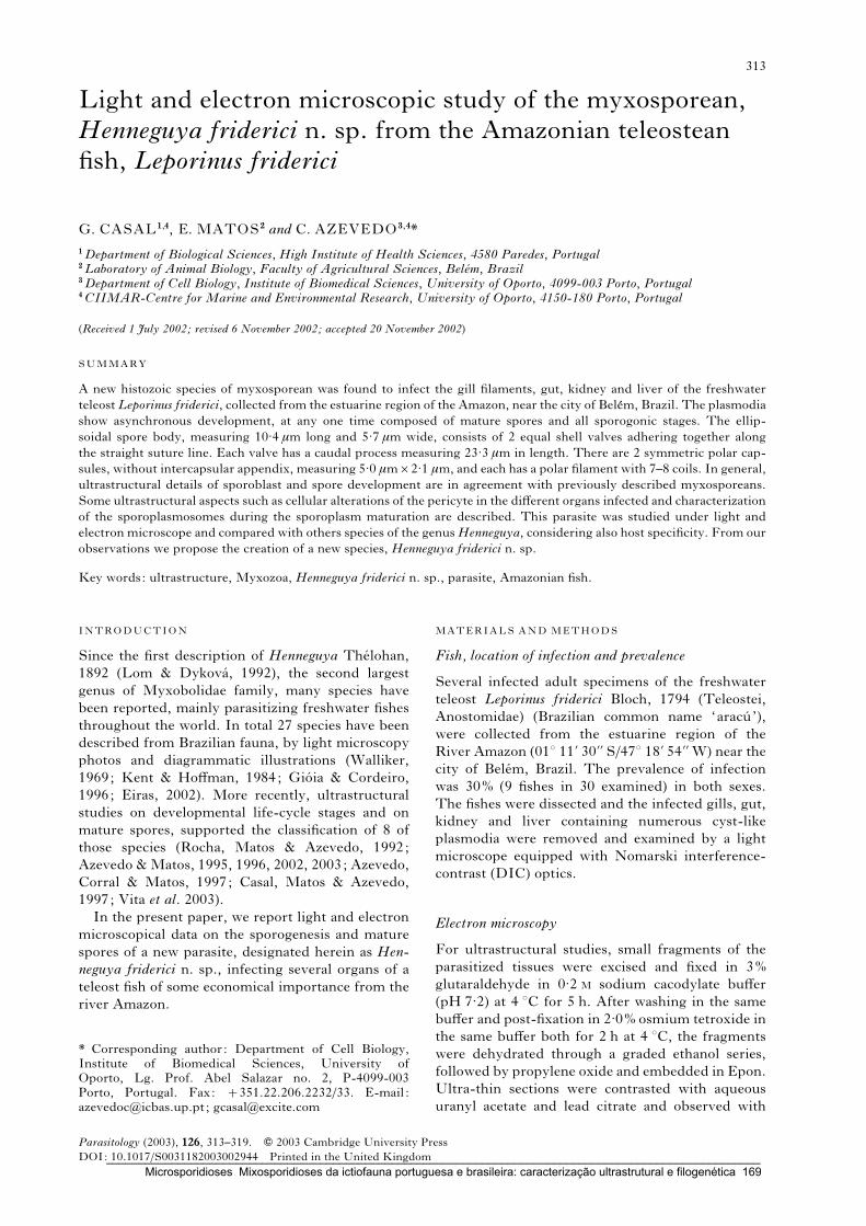
Light and electron microscopic study of the myxosporean,
Henneguya friderici n. sp. from the Amazonian teleostean
fish, Leporinus friderici
G. CASAL1,4, E. MATOS2 and C. AZEVEDO3,4*
1Department of Biological Sciences, High Institute of Health Sciences, 4580 Paredes, Portugal2Laboratory of Animal Biology, Faculty of Agricultural Sciences, Belem, Brazil3Department of Cell Biology, Institute of Biomedical Sciences, University of Oporto, 4099-003 Porto, Portugal4CIIMAR-Centre for Marine and Environmental Research, University of Oporto, 4150-180 Porto, Portugal
(Received 1 July 2002; revised 6 November 2002; accepted 20 November 2002)
SUMMARY
A new histozoic species of myxosporean was found to infect the gill filaments, gut, kidney and liver of the freshwater
teleost Leporinus friderici, collected from the estuarine region of the Amazon, near the city of Belem, Brazil. The plasmodia
show asynchronous development, at any one time composed of mature spores and all sporogonic stages. The ellip-
soidal spore body, measuring 10.4 mm long and 5.7 mm wide, consists of 2 equal shell valves adhering together along
the straight suture line. Each valve has a caudal process measuring 23.3 mm in length. There are 2 symmetric polar cap-
sules, without intercapsular appendix, measuring 5.0 mmr2.1 mm, and each has a polar filament with 7–8 coils. In general,
ultrastructural details of sporoblast and spore development are in agreement with previously described myxosporeans.
Some ultrastructural aspects such as cellular alterations of the pericyte in the different organs infected and characterization
of the sporoplasmosomes during the sporoplasm maturation are described. This parasite was studied under light and
electron microscope and compared with others species of the genusHenneguya, considering also host specificity. From our
observations we propose the creation of a new species, Henneguya friderici n. sp.
Key words: ultrastructure, Myxozoa, Henneguya friderici n. sp., parasite, Amazonian fish.
INTRODUCTION
Since the first description of Henneguya Thelohan,
1892 (Lom & Dykova, 1992), the second largest
genus of Myxobolidae family, many species have
been reported, mainly parasitizing freshwater fishes
throughout the world. In total 27 species have been
described from Brazilian fauna, by light microscopy
photos and diagrammatic illustrations (Walliker,
1969; Kent & Hoffman, 1984; Gioia & Cordeiro,
1996; Eiras, 2002). More recently, ultrastructural
studies on developmental life-cycle stages and on
mature spores, supported the classification of 8 of
those species (Rocha, Matos & Azevedo, 1992;
Azevedo &Matos, 1995, 1996, 2002, 2003; Azevedo,
Corral & Matos, 1997; Casal, Matos & Azevedo,
1997; Vita et al. 2003).
In the present paper, we report light and electron
microscopical data on the sporogenesis and mature
spores of a new parasite, designated herein as Hen-
neguya friderici n. sp., infecting several organs of a
teleost fish of some economical importance from the
river Amazon.
MATERIALS AND METHODS
Fish, location of infection and prevalence
Several infected adult specimens of the freshwater
teleost Leporinus friderici Bloch, 1794 (Teleostei,
Anostomidae) (Brazilian common name ‘aracu’),
were collected from the estuarine region of the
River Amazon (01x 11k 30kk S/47x 18k 54kk W) near the
city of Belem, Brazil. The prevalence of infection
was 30% (9 fishes in 30 examined) in both sexes.
The fishes were dissected and the infected gills, gut,
kidney and liver containing numerous cyst-like
plasmodia were removed and examined by a light
microscope equipped with Nomarski interference-
contrast (DIC) optics.
Electron microscopy
For ultrastructural studies, small fragments of the
parasitized tissues were excised and fixed in 3%
glutaraldehyde in 0.2 M sodium cacodylate buffer
(pH 7.2) at 4 xC for 5 h. After washing in the same
buffer and post-fixation in 2.0% osmium tetroxide in
the same buffer both for 2 h at 4 xC, the fragments
were dehydrated through a graded ethanol series,
followed by propylene oxide and embedded in Epon.
Ultra-thin sections were contrasted with aqueous
uranyl acetate and lead citrate and observed with
* Corresponding author: Department of Cell Biology,Institute of Biomedical Sciences, University ofOporto, Lg. Prof. Abel Salazar no. 2, P-4099-003Porto, Portugal. Fax: +351.22.206.2232/33. E-mail :[email protected] ; [email protected]
313
Parasitology (2003), 126, 313–319. f 2003 Cambridge University Press
DOI: 10.1017/S0031182003002944 Printed in the United Kingdom_____________________________________________________________________________________________________ Microsporidioses Mixosporidioses da ictiofauna portuguesa e brasileira: caracterização ultrastrutural e filogenética 169
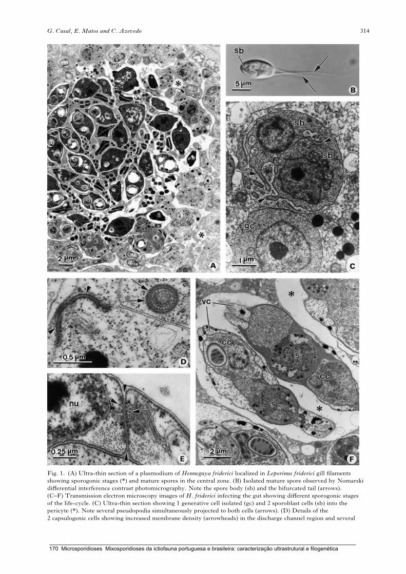
Fig. 1. (A) Ultra-thin section of a plasmodium of Henneguya friderici localized in Leporinus friderici gill filaments
showing sporogonic stages (*) and mature spores in the central zone. (B) Isolated mature spore observed by Nomarski
differential interference contrast photomicrography. Note the spore body (sb) and the bifurcated tail (arrows).
(C–F) Transmission electron microscopy images of H. friderici infecting the gut showing different sporogonic stages
of the life-cycle. (C) Ultra-thin section showing 1 generative cell isolated (gc) and 2 sporoblast cells (sb) into the
pericyte (*). Note several pseudopodia simultaneously projected to both cells (arrows). (D) Details of the
2 capsulogenic cells showing increased membrane density (arrowheads) in the discharge channel region and several
G. Casal, E. Matos and C. Azevedo 314
_____________________________________________________________________________________________________ 170 Microsporidioses Mixosporidioses da ictiofauna portuguesa e brasileira: caracterização ultrastrutural e filogenética

a transmission electron microscope (TEM) JEOL
100CXII operated at 60 kV.
For scanning electron microscopy (SEM), isolated
spores removed from mature plasmodia, were fixed
as described above. Then, the material was dehy-
drated in ethanol, gold coated and examined in a
JEOL 35 SEM operated at 15 kV.
RESULTS
Systematic position
Phylum Myxozoa, Class Myxosporea, Order Bi-
valvulida, and Family Myxobolidae, according to
the classification proposed by Lom & Noble (1984).
Description of the species
Henneguya friderici n. sp.
Type host : Leporinus friderici Bloch, 1794 (Teleostei,
Anostomidae).
Host size : 15 cm of the length in average.
Type locality : estuarine region of the river Amazon
(01x 11k 30kk S/47x 18k 54kk W), near Belem (Para),
Brazil.
Location in the host : histozoic infecting several or-
gans, such as gills, gut, kidney and liver.
Prevalence and intensity : 9 out of 30 adult fishes were
parasitized and in equal % in both sexes.
Type specimens : 2 slides containing matures spores
of the holotypes were deposited in the International
Protozoan Type Slide Collection at Smithsonian
Institution Washington, DC. 20560, USA with
acquisition number (USNMnx 1007181). The histo-
logical semi-thin sections showing varied develop-
mental stages were deposited at the laboratory of the
senior author.
Etymology : the specific name is derived from the
name of the host species (‘friderici ’).
Description of spores : for description of the ma-
ture spores scanning electron microscopy (SEM)
(Fig. 2A), light microscopy (DIC) (Fig. 1B) and a
schematic drawing (Fig. 3) were used. Variability in
shape and size of the spores on the different organs
was not observed and the measurements were
done with plasmodia obtained from the gut tissue.
The spores were ellipsoidal with a total length 33.8
(28.7–39.3) mm (n=25), body length 10.4 (9.6–
11.8) mm (n=25), body width (frontal view) 5.7
(4.8–6.6) mm (n=25) and body thickness (side view)
4.9 (4.6–5.2) mm (n=25). The valves, symmetric and
thin, are each prolonged by a caudal process 23.3
(19.1–28.7) mm (n=25) long. The spores presented
neither a mucous envelope nor particular details on
the surface when observed by SEM (Fig. 2A).
Elongated polar capsules localized in the anterior
pole of the spore were of equal size, measuring 4.98
(4.25–5.90) mm in length (n=25) by 2.14 (1.59–
2.62) mm (n=25) in width and there is not an inter-
capsular appendix. Inside the polar capsules, the
polar filament is coiled 7–8 turns obliquely to the
longitudinal axis (Fig. 2B).
Ultrastructural observations
Several organs of the freshwater teleost, L. friderici,
were found to be parasitized by a myxosporidian
of the genus Henneguya Thelohan, 1892. Whitish
and round-shaped polysporic plasmodia, measuring
about 0.5–1.0 mm, indicated an asynchronous devel-
opment. These were composed of vegetative nuclei
and different sporogenesis stages, such as generative
cells and early sporogonic stages predominantly
along the plasmodium periphery, while immature
and mature spores were more internally localized in
the centre (Fig. 1A). Pansporoblast formation was
disporoblastic (gives rise 2 spores), typical of the
myxobolids and comprised envelopment by a peri-
cyte capsulogenesis, valvogenesis and sporoplasm
maturation (Fig. 1F).
At the beginning of the sporogenesis phase, 2
morphologically identical rounded generative cells
were frequently observed in narrow association.
Initially both cells projected numerous pseudopodia
inside the cytoplasm of another cell and later this
association finished with the envelopment of one of
them, the sporogonic cell by the pericyte (Fig. 1C).
The sporogonic cell divided several times giving rise
to a disporic pansporoblast stage (Fig. 1F) composed
by valvogenic, capsulogenic and sporoplasm cells
that later gave rise to 2 mature spores (Figs 1B and
2A, B).
Frequently, several longitudinally oriented micro-
tubules were found in the cytoplasm of capsulogenic
and valvogenic cells, surrounding the external tube
(Fig. 1D) and giving form to the valve and caudal
process (Fig. 1E) respectively.
The binucleate sporoplasm cell, which contained
an iodinophilous vacuole, mitochondria, an extensive
system of cisternae of endoplasmic reticulum and nu-
merous electron-dense vesicles, sporoplasmosomes,
was located in the spore posterior pole (Figs 1F and
2E, F). A single membrane limits the sporoplasmo-
somes and during the early sporogonic stages they
appeared as a spherical body.After sporoplasmmatu-
ration, they changed their form and appeared like
a teardrop, approximately 190–210 nm in diameter
microtubules bundle-oriented to the external tubule in transverse section (arrows). (E) Detail of a sutural line
(arrowheads) of adjoining valvogenic cells with several associated microtubules (arrows). Nucleus of a valvogenic cell (nu).
(F) Two immature spores lie in a vacuole in the pericyte (*) showing cellular differentiation, into valvogenic cells (vc),
capsulogenic cells (cc) and sporoplasm binucleated (s).
Ultrastructure of Henneguya friderici n. sp. 315
_____________________________________________________________________________________________________ Microsporidioses Mixosporidioses da ictiofauna portuguesa e brasileira: caracterização ultrastrutural e filogenética 171
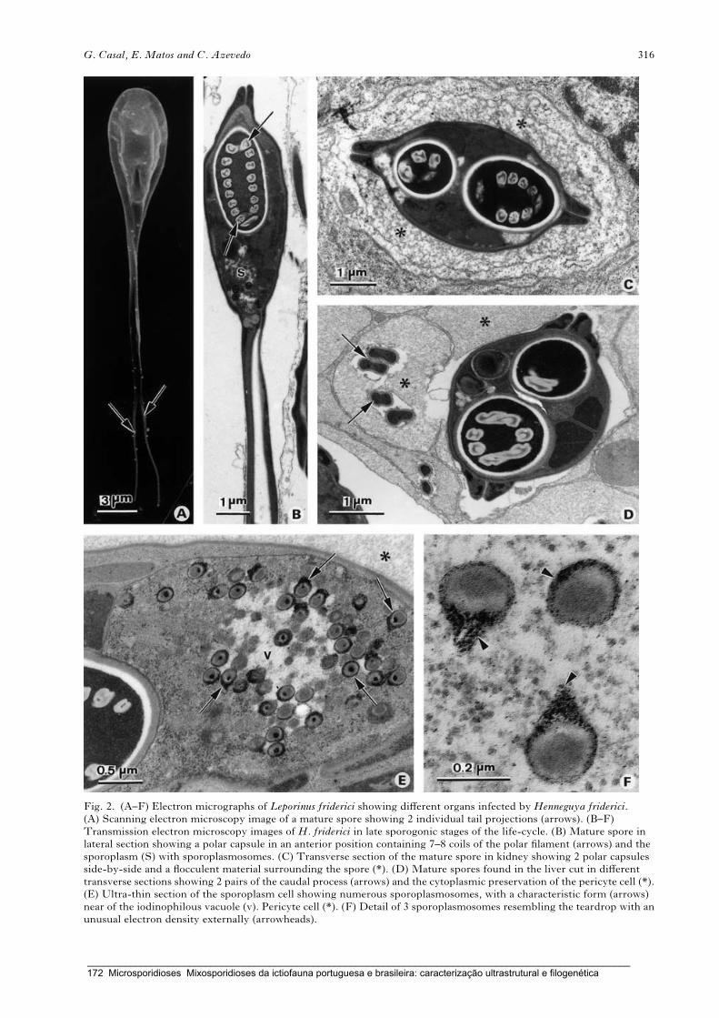
Fig. 2. (A–F) Electron micrographs of Leporinus friderici showing different organs infected by Henneguya friderici.(A) Scanning electron microscopy image of a mature spore showing 2 individual tail projections (arrows). (B–F)
Transmission electron microscopy images of H. friderici in late sporogonic stages of the life-cycle. (B) Mature spore in
lateral section showing a polar capsule in an anterior position containing 7–8 coils of the polar filament (arrows) and the
sporoplasm (S) with sporoplasmosomes. (C) Transverse section of the mature spore in kidney showing 2 polar capsules
side-by-side and a flocculent material surrounding the spore (*). (D) Mature spores found in the liver cut in different
transverse sections showing 2 pairs of the caudal process (arrows) and the cytoplasmic preservation of the pericyte cell (*).
(E) Ultra-thin section of the sporoplasm cell showing numerous sporoplasmosomes, with a characteristic form (arrows)
near of the iodinophilous vacuole (v). Pericyte cell (*). (F) Detail of 3 sporoplasmosomes resembling the teardrop with an
unusual electron density externally (arrowheads).
G. Casal, E. Matos and C. Azevedo 316
_____________________________________________________________________________________________________ 172 Microsporidioses Mixosporidioses da ictiofauna portuguesa e brasileira: caracterização ultrastrutural e filogenética
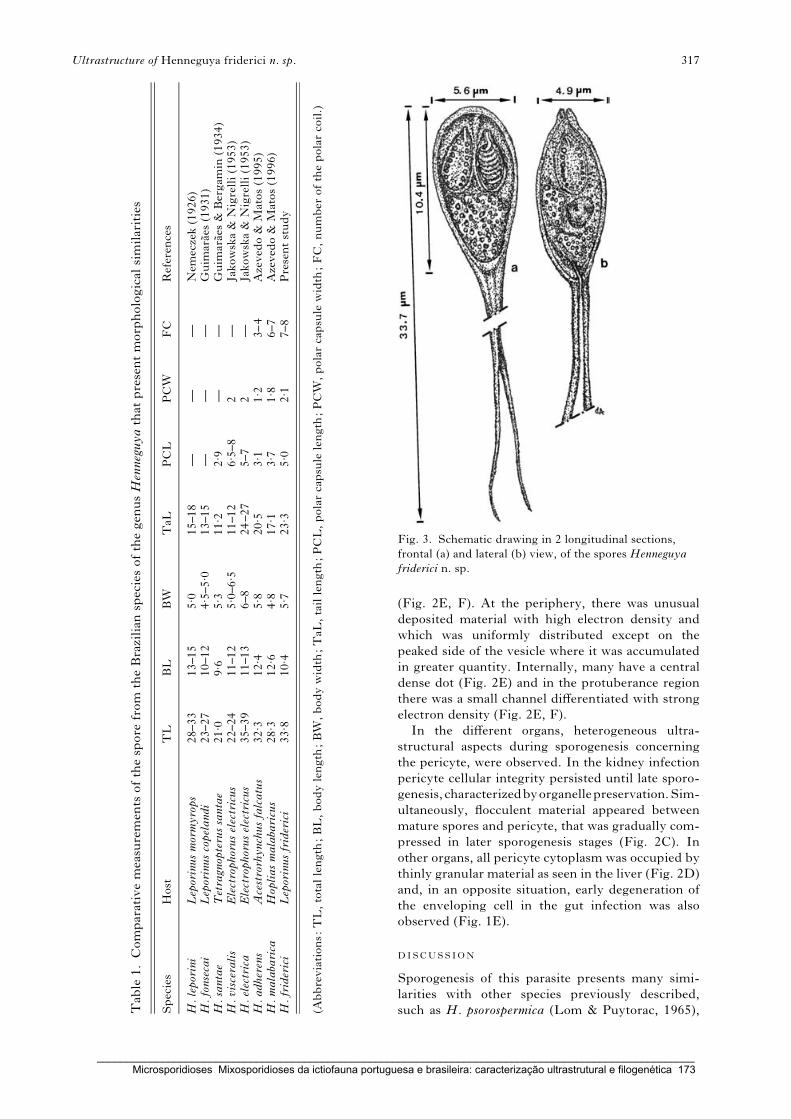
(Fig. 2E, F). At the periphery, there was unusual
deposited material with high electron density and
which was uniformly distributed except on the
peaked side of the vesicle where it was accumulated
in greater quantity. Internally, many have a central
dense dot (Fig. 2E) and in the protuberance region
there was a small channel differentiated with strong
electron density (Fig. 2E, F).
In the different organs, heterogeneous ultra-
structural aspects during sporogenesis concerning
the pericyte, were observed. In the kidney infection
pericyte cellular integrity persisted until late sporo-
genesis,characterizedbyorganellepreservation.Sim-
ultaneously, flocculent material appeared between
mature spores and pericyte, that was gradually com-
pressed in later sporogenesis stages (Fig. 2C). In
other organs, all pericyte cytoplasm was occupied by
thinly granular material as seen in the liver (Fig. 2D)
and, in an opposite situation, early degeneration of
the enveloping cell in the gut infection was also
observed (Fig. 1E).
DISCUSSION
Sporogenesis of this parasite presents many simi-
larities with other species previously described,
such as H. psorospermica (Lom & Puytorac, 1965),
Fig. 3. Schematic drawing in 2 longitudinal sections,
frontal (a) and lateral (b) view, of the spores Henneguya
friderici n. sp.
Tab
le1.Comparativemeasuremen
tsofthespore
from
theBrazilian
speciesofthegen
usHenneguyathat
presentmorphological
similarities
Species
Host
TL
BL
BW
TaL
PCL
PCW
FC
Referen
ces
H.leporini
Leporinusmormyrops
28–33
13–15
5. 0
15–18
——
—Nem
eczek(1926)
H.fonsecai
Leporinuscopelandi
23–27
10–12
4. 5–5. 0
13–15
——
—Guim
araes
(1931)
H.santae
Tetragnopterussantae
21. 0
9. 6
5. 3
11. 2
2. 9
——
Guim
araes
&Bergam
in(1934)
H.visceralis
Electrophoruselectricus
22–24
11–12
5. 0–6. 5
11–12
6. 5–8
2—
Jakowska&
Nigrelli(1953)
H.electrica
Electrophoruselectricus
35–39
11–13
6–8
24–27
5–7
2—
Jakowska&
Nigrelli(1953)
H.adherens
Acestrorhynchusfalcatus
32. 3
12. 4
5. 8
20. 5
3. 1
1. 2
3–4
Azeved
o&
Matos(1995)
H.malabarica
Hopliasmalabaricus
28. 3
12. 6
4. 8
17. 1
3. 7
1. 8
6–7
Azeved
o&
Matos(1996)
H.friderici
Leporinusfriderici
33. 8
10. 4
5. 7
23. 3
5. 0
2. 1
7–8
Presentstudy
(Abbreviations:TL,totallength
;BL,bodylength
;BW
,bodywidth
;TaL
,taillength
;PCL,polarcapsule
length
;PCW
,polarcapsule
width
;FC,number
ofthepolarcoil.)
Ultrastructure of Henneguya friderici n. sp. 317
_____________________________________________________________________________________________________ Microsporidioses Mixosporidioses da ictiofauna portuguesa e brasileira: caracterização ultrastrutural e filogenética 173

H. exilis (Current & Janovy, 1977), H. adiposa
(Current, 1979) and H. amazonica (Rocha et al.
1992). Although the differentiation and maturation
process of the sporoplasm cell has been well studied,
little is known about the nature and function of the
electron-dense sporoplasmosomes, typical of these
parasites. They have been in the sporoplasm of nu-
merous myxosporidia and their morphological
characteristics, such as form, size and inner organ-
ization have no relationship with genus or family
(Lom et al. 1989; Lom & Dykova, 1992). In the
studies of freshwater fish parasites from the River
Amazon belonging to the genus Henneguya (Rocha
et al. 1992; Azevedo & Matos, 2002; Vita et al.
2003) and Myxobolus (Casal, Matos & Azevedo,
1996) the sporoplasmosomes have been ultra-
structurally characterized. The form and size hetero-
geneity of these vesicles show that it is possible to
use this cellular structure as an ultrastructural par-
ameter to differentiate parasites of the same genus,
mainly when they are diagnosed in hosts from the
same hydrological basin. In this case, an atypical
electron-dense material surrounding the vesicle and
forming a protuberance has never been described
before.
The ultrastructural aspects of the enveloping cell
during sporogenesis have been occasionally de-
scribed as a gradual and generalized degeneration.
Sometimes microfibril-forming regions (Current &
Janovy, 1977) or long microfilaments resembling
myosin filaments (Casal et al. 1997) in the cyto-
plasmatic space have been described. In this study
we found some heterogeneity in the morphological
aspects of the pericyte in the different organs infected,
from a relative organelle preservation in liver tissue
to an extensive degradation, as verified in the gut
infection. This fact seemed indicative of the exist-
ence of some adaptation of the pericyte to the host
infected tissue.
The morphological and ultrastructural aspects of
the mature spores, as well as the host specificity and
localization of the infection with this parasite, were
compared with other myxosporidian species of the
genus Henneguya, from different geographical areas
mainly, with those that have Brazilian freshwater
fishes as host (Table 1).
Among the more than 100 non-Brazilian species
described in the literature (see the revision papers
Kostoıngue et al. 2001 and Eiras, 2002), 7 species,
H. lagodoni (Hall & Iversen, 1967), H. shaharini
(Shariff, 1982), H. latesi (Haldar, Das & Sharma,
1983),H. mystusia (Sarkar, 1985), H. laterocapsulata
and H. suprabranchiae (Landsberg, 1987) and
H. mbourensis (Kpatcha et al. 1997) present ap-
proximately the same body size. Although all have
a different body shape not many of those species were
ultrastructurally characterized.
Brazilian species have mostly been described by
light microscopy or simply by schematic drawings
(Walliker, 1969; Kent & Hoffman, 1984; Gioia &
Cordeiro, 1996) and some of them show morpho-
logical similarities. The species H. leporini (see
Nemeczek, 1926) and H. fonsecai (see Guimaraes,
1931) parasitize fishes of the same genus Leporinus,
but although they were caught in rivers with a
distinct geographical localization, they are similar
in total size and body shape, respectively. Of 3
other species with similar body shape, H. santae
(Guimaraes & Bergamin, 1934) and H. visceralis
(Jakowska & Nigrelli, 1953) present a smaller size,
while H. electrica (Jakowska & Nigrelli, 1953) is
longer.
During the last 10 years, this group of parasites
has been studied at the ultrastructural level, re-
sulting in the description of 8 new Brazilian Henne-
guya species, all in different hosts (Rocha et al. 1992;
Azevedo & Matos, 1995, 1996, 2002, 2003; Azevedo
et al. 1997; Casal et al. 1997; Vita et al. 2002). Of
these, only the spores of H. adherens (Azevedo &
Matos, 1995) and H. malabarica (Azevedo & Matos,
1996) resemble in size the parasite studied by us
in L. friderici. However, H. friderici lacks a sheath
around the 2 tails and differs in the arrangement of
the polar filament coil.
After a comparative study based on the morpho-
logical and ultrastructural differences with Henne-
guya species previously described, we conclude that
Henneguya friderici is a new species.
This work was partially supported by a grant from theEngx Antonio Almeida Foundation, Porto, Portugal. Wewould like to thank the iconographic work of Mr JoaoCarvalheiro. The technical assistance provided by MrsLaura Corral is gratefully acknowledged.
REFERENCES
AZEVEDO, C. & MATOS, E. (1995). Henneguya adherens n. sp.
(Myxozoa, Myxosporea), parasite of the Amazonian
fish, Acestrorhynchus falcatus. Journal of Eukaryotic
Microbiology 42, 515–518.
AZEVEDO, C. & MATOS, E. (1996). Henneguya malabarica
sp. nov. (Myxozoa, Myxobolidae) in the Amazonian
fish Hoplias malabaricus. Parasitology Research 82,
222–224.
AZEVEDO, C. & MATOS, E. (2002). Fine structure of the
Myxosporean,Henneguya curimata n. sp., parasite of the
Amazonian fish, Curimata inormata (Teleostei,
Curimatidae). Journal of Eukaryotic Microbiology 49,
197–200.
AZEVEDO, C. & MATOS, E. (2003). Fine structure of
Henneguya pilosa sp. n. (Myxozoa: Myxosporea),
parasite of Serrasalmus altuvei (Characidae) in Brazil.
Folia Parasitologica 50, 35–40.
AZEVEDO, C., CORRAL, L. & MATOS, E. (1997). Light and
ultrastructural data on Henneguya testicularis n. sp.
(Myxozoa, Myxobolidae), a parasite from the testis of
the Amazonian fish Moenkhausia oligolepis. Systematic
Parasitology 37, 111–114.
CASAL, G., MATOS, E. & AZEVEDO, C. (1996). Ultrastructural
data on the life cycle stages of Myxobolus braziliensis
G. Casal, E. Matos and C. Azevedo 318
_____________________________________________________________________________________________________ 174 Microsporidioses Mixosporidioses da ictiofauna portuguesa e brasileira: caracterização ultrastrutural e filogenética

n. sp. parasite of an Amazonian fish. European Journal
of Protistology 32, 123–127.
CASAL, G., MATOS, E. & AZEVEDO, C. (1997). Some
ultrastructural aspects of Henneguya striolata sp. nov.
(Myxozoa, Myxosporea), a parasite of the Amazonian
fish Serrasalmus striolatus. Parasitology Research 83,
93–95.
CURRENT, W. L. (1979). Henneguya adiposa Minchew
(Myxosporida) in the channel catfish: ultrastructure of
the plasmodium wall and sporogenesis. Journal of
Protozoology 26, 209–217.
CURRENT, W. L. & JANOVY, J. J. (1977). Sporogenesis in
Henneguya exilis infecting the channel catfish: an
ultrastructural study. Protistologica 13, 157–167.
EIRAS, J. C. (2002). Synopsis of the species of the genus
Henneguya Thelohan, 1892 (Myxozoa: Myxosporea:
Myxobolidae). Systematic Parasitology 52, 43–54.
GIOIA, I. & CORDEIRO, N. S. (1996). Brazilian
Myxosporidians’ check-list (Myxozoa). Acta
Protozoologica 35, 137–149.
GUIMARAES, J. R. A. (1931). Mixosporıdeos da ictiofauna
brasileira. Ph.D. thesis, Faculdade de Medicina de Sao
Paulo, Brazil.
GUIMARAES, J. R. A. & BERGAMIN, F. (1934). Henneguya
santae sp. n. Um novo mixosporideo parasito de
Tetragnopterus sp. Revista de Industria Animal 2,
110–113.
HALDAR, D. P., DAS, M. K. & SHARMA, B. K. (1983). Studies on
protozoan parasites from fishes. Four new species of the
generaHenneguyaThelohan, 1892, ThelohanellusKudo,
1933, and Myxobolus Butschli, 1892. Archive fur
Protistenkunde 127, 283–296.
HALL, D. L. & IVERSEN, E. S. (1967). Henneguya lagodoni, a
new species of myxosporidian parasitizing the pinfish,
Lagodon rhomboides. Bulletin of Marine Science 17,
274–279.
JAKOWSKA, S. & NIGRELLI, R. F. (1953). The pathology of
myxosporidiosis in the electric eel, Electrophorus
electricus (Linnaeus), caused by Henneguya visceralis
and E. electrica spp. nov. Zoologica 38, 183–191.
KENT, M. L. & HOFFMAN, G. L. (1984). Two new species of
Myxozoa, Myxobolus inaequus sp. n. and Henneguya
theca sp. n. from the brain of a South American knife
fish, Eigemannia virescens (V.). Journal of Protozoology
31, 91–94.
KOSTOINGUE, B., DIEBAKATE, C., FAYE, N. & TOGUEBAYE, B. S.
(2001). Presence of Myxosporidea (Myxozoa:
Myxosporea) of the genus Henneguya Thelohan, 1892
in freshwater fishes from Chad (Central Africa). Acta
Protozoologica 40, 117–123.
KPATCHA, T. K., FAYE, N., DIEBAKATE, C., FALL, N. &
TOGUEBAYE, B. S. (1997). Nouvelles especes d’Henneguya
Thelohan, 1895 (Myxozoa, Myxosporea) parasites des
poissons marins du Senegal : Etude en microscopie
photonique et electronique. Annales des Sciences
Naturelles Zoologie 18, 81–91.
LANDSBERG, J. H. (1987). Myxosporean parasites of the
catfish, Clarias lazera (Valenciennes). Systematic
Parasitology 9, 73–81.
LOM, J. & DYKOVA, I. (1992). Myxosporidia (phylum
Myxozoa). In Protozoan Parasites of Fishes.
Developments in Aquaculture and Fisheries Science,
Vol. 26 (ed. Lom, J. & Dykova, I.), pp. 159–235.
Elsevier, Amsterdam.
LOM, J. & NOBLE, E. R. (1984). Revised classification of the
class Myxosporea Butschli, 1881. Folia Parasitologica
31, 193–205.
LOM, J. & PUYTORAC, P. (1965). Studies on the
myxosporidian ultrastructure and polar capsule
development. Protistologica 1, 53–65.
LOM, J., FEIST, S. W., DYKOVA, I. & KEPR, T. (1989). Brain
myxoboliasis of bullhead, Cottus gobio L., due to
Myxobolus jiroveci sp. nov.: light and electron
microscope observations. Journal of Fish Diseases 12,
15–27.
NEMECZEK, A. (1926). Beitrage zur Kenntnis der
Myxosporidienfauna Brasiliens. Archive fur
Protistenkunde 54, 137–150.
ROCHA, E., MATOS, E. & AZEVEDO, C. (1992). Henneguya
amazonica n. sp. (Myxozoa, Myxobolidae), parasitizing
the gills of Crenicichla lepidota Heckel, 1840 (Teleostei,
Cichlidae) from Amazon river. European Journal of
Protistology 28, 273–278.
SARKAR, N. K. (1985). Myxosporidan Henneguya mystusia
sp. n. (Myxozoa, Myxosporea), from the gills of a fresh
water teleost fish Mystus sp. Acta Protozoologica 24,
55–58.
SHARIFF, M. (1982). Henneguya shaharini sp. nov.
(Protozoa: Myxozoa), a parasite of marble goby,
Oxyeleotris marmoratus (Bleeker). Journal of Fish
Diseases 5, 37–45.
VITA, P., CORRAL, L., MATOS, E. & AZEVEDO, C. (2003).
Ultrastructural aspects of the myxosporean Henneguya
astyanax n. sp. (Myxozoa: Myxobolidae), a parasite
of the Amazonian teleost Astyanax keithi
(Characidae). Diseases of Aquatic Organisms
53, 55–60.
WALLIKER, D. (1969). Myxosporidea of some
Brazilian freshwater fishes. Journal of Parasitology
55, 942–948.
Ultrastructure of Henneguya friderici n. sp. 319
_____________________________________________________________________________________________________ Microsporidioses Mixosporidioses da ictiofauna portuguesa e brasileira: caracterização ultrastrutural e filogenética 175

_____________________________________________________________________________________________________ 176 Microsporidioses Mixosporidioses da ictiofauna portuguesa e brasileira: caracterização ultrastrutural e filogenética
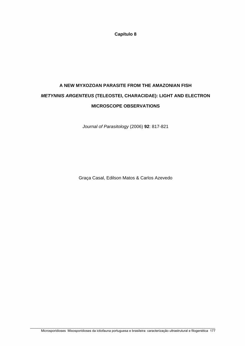
Capítulo 8
A NEW MYXOZOAN PARASITE FROM THE AMAZONIAN FISH
METYNNIS ARGENTEUS (TELEOSTEI, CHARACIDAE): LIGHT AND ELECTRON
MICROSCOPE OBSERVATIONS
Journal of Parasitology (2006) 92: 817-821
Graça Casal, Edilson Matos & Carlos Azevedo
_____________________________________________________________________________________________________ Microsporidioses Mixosporidioses da ictiofauna portuguesa e brasileira: caracterização ultrastrutural e filogenética 177

_____________________________________________________________________________________________________ 178 Microsporidioses Mixosporidioses da ictiofauna portuguesa e brasileira: caracterização ultrastrutural e filogenética

817
J. Parasitol., 92(4), 2006, pp. 817–821� American Society of Parasitologists 2006
A NEW MYXOZOAN PARASITE FROM THE AMAZONIAN FISH METYNNIS ARGENTEUS(TELEOSTEI, CHARACIDAE): LIGHT AND ELECTRON MICROSCOPE OBSERVATIONS
Graca Casal, Edilson Matos*, and Carlos Azevedo†Department of Sciences, High Institute of Health Sciences, 4585-116 Gandra, Portugal. e-mail: [email protected]
ABSTRACT: Myxobolus metynnis n. sp. (Phylum Myxozoa) is described in the connective subcutaneous tissues of the orbicularregion of the fish, Metynnis argenteus (Characidae), collected in the lower Amazon River, near the city of Peixe Boi, Para State,Brazil. Polysporic, histozoic plasmodia were delimited by a double membrane with numerous microvilli on the peripheral cyto-plasm. Several life-cycle stages, including mature spores, were observed. An envelope formed by numerous fine and anastomosedmicrofibrils was observed at the spore surface. The spore body presented an ellipsoidal shape and was about 13.1 �m long, 7.8�m wide, and 3.9 �m thick. Elongated-pyriform polar capsules were of equal size, measuring 5.2 �m in length, 3.2 �m in width,and possessing a polar filament with 8–9 turns around the longitudinal axis. The binucleated sporoplasm contained a vacuoleand numerous sporoplasmosomes. These were circular in cross-section, showing an adherent eccentric, dense structure, with ahalf-crescent section. Based on the morphological differences and host specificity, we propose that the parasite is a new speciesnamed Myxobolus metynnis n. sp.
The Myxosporea of the Myxozoa is an assemblage of morethan 1,500 species. They have been reported from different geo-graphic areas, by morphological and ultrastructural studies,mainly as fish parasites (Lom and Dykova, 1992). Among them,species of Myxobolus Butschli, 1882 (Myxobolidae), is the larg-est group and includes a number of important pathogens offreshwater and marine fishes (Lom and Dykova, 1992; Eiras etal., 2005). Some of the species of Myxobolus are consideredhighly pathogenic to their hosts (Longshaw et al., 2003). InBrazilian fishes, few myxozoans have been described and, withthe exception of 4 ultrastructural studies (Casal et al., 1996,2002; Azevedo et al., 2002; Tajdari et al., 2005), only lightmicroscopy descriptions and diagrammatic drawings are avail-able (Penido, 1927; Pinto, 1928; Walliker, 1969; Kent and Hoff-man, 1984; Molnar and Bekesi, 1993; Gioia and Cordeiro,1996; Molnar et al., 1998; Adriano et al., 2002; Cellere et al.,2002).In the present study, we describe light and electron ultrastruc-
tural features of a new myxosporidian found in the connectivesubcutaneous tissues of the orbicular region of a teleost fishcollected from the Amazon river.
MATERIALS AND METHODSThe freshwater teleost, Metynnis argenteus Aht, 1923 (Characidae)
(‘‘Piaba Chata’’), was collected in the estuarine region of the AmazonRiver (01�11�;30�S, 47�18�54�W) near the city of Peixe Boi (Para State),Brazil. Immediately after collection, 50 fish were transported alive tothe laboratory, where they were anesthetized, killed, and necropsied.Plasmodia with mature spores were examined in fresh mounts under
a light microscope (LM) equipped with Nomarski interference-contrastdifferential (DIC) optics. For transmission electron microscopy (TEM),small fragments of host tissue were fixed in 3% glutaraldehyde in 0.2M sodium cacodylate buffer (pH 7.4) at 4 C for 12 hr, then washedovernight with the same buffer at 4 C and postfixed in 2% osmiumtetroxide (OsO4) buffered with sodium cacodylate for 2 hr at the sametemperature. The fragments were dehydrated in an ascending ethanoland propylene oxide series (3 � 2 hr in each change) and embedded inEpon (10–12 hr in each change). Semithin sections were stained with
Received 28 September 2005; revised 26 January 2006; accepted 27January 2006.* Carlos Azevedo Research Laboratory, Federal Rural University of theAmazonia, 66.000 Belem (Para), Brazil.† To whom correspondence should be addressed. Department of CellBiology, Institute of Biomedical Sciences, University of Porto (IC-BAS/UP) and Laboratory of Protoparasitology, University of Porto(CIIMAR/UP), Lg. A. Salazar no. 2, 4099-003 Porto, Portugal.
methylene blue. The ultrathin sections, cut with a diamond knife, werecontrasted with both aqueous uranyl acetate and lead citrate and ob-served in a JEOL 100CXII TEM operated at 60 kV.
DESCRIPTIONMyxobolus metynnis n. sp.
(Figs. 1–10)
Plasmodia and vegetative stages: Cyst white, ellipsoidal to spherical,up to 350 �m in diameter (Fig. 1). Plasmodial membrane is coveredwith fine projections or microvilli (Figs. 3, 4) and surrounded by acontinuous layer of several fibroblasts intermingled with concentric col-lagen fiber layers (Fig. 5). Plasmodia polysporic, with asynchronousdevelopment, early sporogonic stages present in the external layer ofthe plasmodium; developing spores and mature spores are located moreinternally (Fig. 3).Mature spores: Spores typical of Myxobolus spp. Mature spores are
ellipsoidal shaped in frontal view, 13.1 (12.9–13.5) �m (n � 50) inlength, 7.8 (7.5–8.3) �m (n � 30) in width, and 3.9 (3.4–4.5) �m (n� 14) in thickness (Figs. 2, 10). Elongate-pyriform polar capsules areof equal size, 5.2 (5.0–5.5) �m (n � 25) in length and 3.2 (3.0–3.6)�m (n � 12) in width. Eight to 9 filament coils are slightly oblique tothe longitudinal axis (Figs. 6, 7). Intercapsule appendix is absent.
Taxonomic summaryType host: Teleost fish, Metynnis argenteus Aht, 1923 (Characidae).Type locality: Estuarine region of the Amazon River (01�11�30�S,
47�18�54�W) near the city of Peixe Boi (Para State), Brazil.Site of infection: Spores are located in the connective subcutaneous
tissues of the orbicular region.Prevalence of infection: Thirteen of 50 (26%).Etymology: The specific name is derived from the name of the host
species (‘‘metynnis’’).Type specimens: One slide containing matures spores of the syntypes
were deposited in the International Protozoan Type Slide Collection atthe Smithsonian Institution, Washington, D.C. 20560, USA, with ac-quisition number (USNM 1086177). The histological, semithin sections,showing varied developmental stages, were deposited at the laboratoryof the senior author (C.A.).
Ultrastructural studiesThe spore wall was thin and smooth, comprising 2 unequal valves
kept close by a sutural ridge. The spore wall was surrounded by a veryfine network of irregular and complex anastomosed microfibrils pro-jecting from the surface of the spore wall toward the surrounding spaces(Fig. 8). Internally, 2 capsulogenic cells, located side by side, containeda prominent polar capsule (PC). The PCs presented a very elongated,pyriform shape and were of equal size. The PC occupied approximately2/5 of the total spore length. Inside of them, a coiled polar filamentwith 8–9 turns can be observed. These coils showed a slightly obliqueposition around the longitudinal axis (Figs. 6, 7).The sporoplasm cell, located in the posterior pole of the spore, con-
_____________________________________________________________________________________________________ Microsporidioses Mixosporidioses da ictiofauna portuguesa e brasileira: caracterização ultrastrutural e filogenética 179

818 THE JOURNAL OF PARASITOLOGY, VOL. 92, NO. 4, AUGUST 2006
FIGURES 1–6. Light and transmission electron microscopy (TEM) aspects of Myxobolus metynnis n. sp., a parasite located in the connectivesubcutaneous tissues of the orbicular region of the freshwater fish Metynnis argenteus. (1) Semithin section of a cyst containing numerous spores.(2) Some isolated spores observed with interference-contrast differential (DIC) optics. (3) Ultrathin section of the periphery of a plasmodiumshowing different life-cycle stages (*) including spores (S). The boxed area (arrow) is enlarged in Figure 4. (4) Ultrastructural details of theperiphery of the plasmodium, showing the organization of the microvilli (arrowheads). (5) Ultrastructural detail of the external periphery of aplasmodium showing several fibroblasts (F), some of them surrounded by numerous collagen fibres (C). (6) Ultrathin section of some sporessectioned at different levels showing the transverse sections of the sporoplasm (S), polar capsules (PC), and polar filament sections (arrowheads).
tained 2 nuclei, surrounded by an irregularly dense matrix, plus an io-dinophilous vacuole, mitochondria, an extensive endoplasmic reticulum,numerous electron-dense vesicles, and sporoplasmosomes (Sps) (Figs.6, 8). At high magnification, the Sps appeared as spherical, double-membrane–bound bodies of about 180–200 nm in diameter, containinga massive dense core separated by narrow, lucent, circular space. EachSps contained an eccentric, dense structure with a half-crescent sectionin close contact with the membrane of the sporoplasmosome (Fig. 9).
DISCUSSION
There are at least 744 species belonging to Myxobolus (Eiraset al., 2005), and most of the early descriptions are vague be-cause they only present line drawings of the spores. Nineteenspecies of Myxobolus have been described in Brazilian fishesbefore the new one reported here (Kudo, 1920; Nemeczek,
_____________________________________________________________________________________________________ 180 Microsporidioses Mixosporidioses da ictiofauna portuguesa e brasileira: caracterização ultrastrutural e filogenética
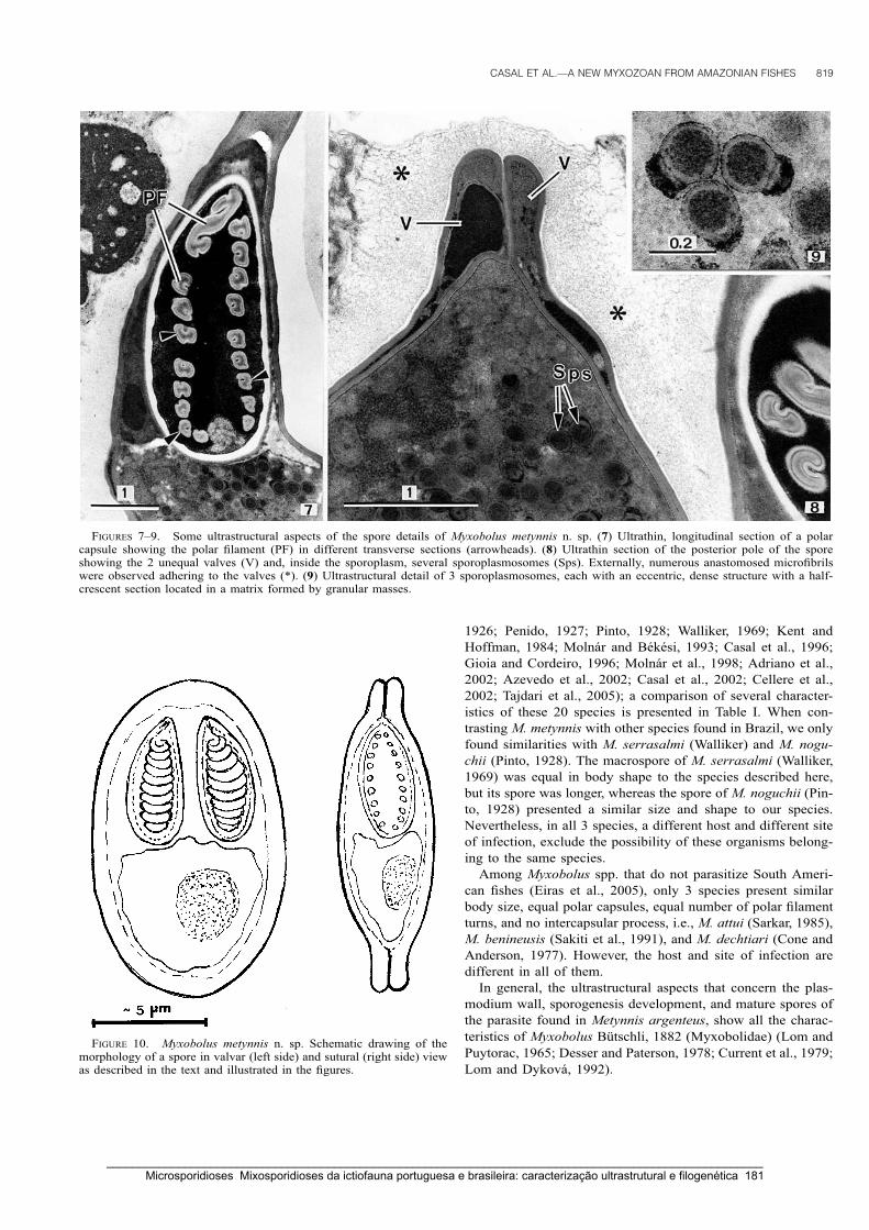
CASAL ET AL.—A NEW MYXOZOAN FROM AMAZONIAN FISHES 819
FIGURES 7–9. Some ultrastructural aspects of the spore details of Myxobolus metynnis n. sp. (7) Ultrathin, longitudinal section of a polarcapsule showing the polar filament (PF) in different transverse sections (arrowheads). (8) Ultrathin section of the posterior pole of the sporeshowing the 2 unequal valves (V) and, inside the sporoplasm, several sporoplasmosomes (Sps). Externally, numerous anastomosed microfibrilswere observed adhering to the valves (*). (9) Ultrastructural detail of 3 sporoplasmosomes, each with an eccentric, dense structure with a half-crescent section located in a matrix formed by granular masses.
FIGURE 10. Myxobolus metynnis n. sp. Schematic drawing of themorphology of a spore in valvar (left side) and sutural (right side) viewas described in the text and illustrated in the figures.
1926; Penido, 1927; Pinto, 1928; Walliker, 1969; Kent andHoffman, 1984; Molnar and Bekesi, 1993; Casal et al., 1996;Gioia and Cordeiro, 1996; Molnar et al., 1998; Adriano et al.,2002; Azevedo et al., 2002; Casal et al., 2002; Cellere et al.,2002; Tajdari et al., 2005); a comparison of several character-istics of these 20 species is presented in Table I. When con-trasting M. metynnis with other species found in Brazil, we onlyfound similarities with M. serrasalmi (Walliker) and M. nogu-chii (Pinto, 1928). The macrospore of M. serrasalmi (Walliker,1969) was equal in body shape to the species described here,but its spore was longer, whereas the spore of M. noguchii (Pin-to, 1928) presented a similar size and shape to our species.Nevertheless, in all 3 species, a different host and different siteof infection, exclude the possibility of these organisms belong-ing to the same species.Among Myxobolus spp. that do not parasitize South Ameri-
can fishes (Eiras et al., 2005), only 3 species present similarbody size, equal polar capsules, equal number of polar filamentturns, and no intercapsular process, i.e., M. attui (Sarkar, 1985),M. benineusis (Sakiti et al., 1991), and M. dechtiari (Cone andAnderson, 1977). However, the host and site of infection aredifferent in all of them.In general, the ultrastructural aspects that concern the plas-
modium wall, sporogenesis development, and mature spores ofthe parasite found in Metynnis argenteus, show all the charac-teristics of Myxobolus Butschli, 1882 (Myxobolidae) (Lom andPuytorac, 1965; Desser and Paterson, 1978; Current et al., 1979;Lom and Dykova, 1992).
_____________________________________________________________________________________________________ Microsporidioses Mixosporidioses da ictiofauna portuguesa e brasileira: caracterização ultrastrutural e filogenética 181

820 THE JOURNAL OF PARASITOLOGY, VOL. 92, NO. 4, AUGUST 2006
TABLEI.MorphologicalcomparisonofthesporesofdifferentMyxobolusspeciesreportedinBrazilianfishes.*
Myxobolusspecies
TL
BW
PCL
PCW
PFC
Host
Siteofinfection
References
M.inaequalis
5.2
3.3
Unequal
––
Piramutanablochii
Skinofhead
Kudo1920
M.lutzi
107
––
–Girardinusjanuarius
Testis
Kudo1920
M.chondrophilus
64.5
3–
–Sardinellaanchovia
Gills
Nemeczek1926
M.associatus
1510
7–
–Leporinusmormyrops
Kidney
Nemeczek1926
M.pygocentris
15–16
9–11
9–11
3–4
–Pygocentrispiraya
Intestinalcontents
Penido1927
M.cunhai
9–11
4–6
Unequal
––
Pygocentrispiraya
Intestinalcontents
Penido1927
M.noguchii
13.6
8.5
6.8
2.2
–Serrasalmospilopleura
Gills?
Pinto1928
M.stokesi
8.5
5.3
3.4
1.7
–Pimelodellasp.
Subcutaneoustissues
Pinto1928
M.kudoi
8.5–8.9
6.5–7.3
3.5–4.2
1.3–2
–Nematognathasp.
Skin
Walliker1969
M.serrasalmi
12.5–18
7–9.5
7–10
3.5–5
6–9
5–7.5
2.5–4
1–2
–Serrasalmusrhombeus
Spleen,kidney,liver
Walliker1969
M.inaequus
19.8
8.6
11.8 4.8
––
Eigemanniavirescens
Brain
KentandHoffman1984
M.colossomatis
11.8
6.9
6.0
2.1
7–8
Colossomamacropomum
Connectivetissues
MolnarandBekesi1993
M.braziliensis
10.2
5.3
5.3
1.4
9–11
Bunocephaluscoracoideus
Gills
Casaletal.1996
M.macroplasmodialis
11.0
8.5
4.5
2.8
6Salminusmaxillosus
Abdominalcavity
Molnaretal.1998
M.porofilus
5.7
4.8
1.6
1.1
3Prochiloduslineatus
Visceralcavity
Adrianoetal.2002
M.desaequalis
18.3
11.2
11.2 4.6
4.92.8
11–12
4–5
Apteronotusalbifrons
Gills
Azevedoetal.2002
M.maculatus
21.0
8.9
12.7
3.2
14–15
Metynnismaculatus
Kidney
Casaletal.2002
M.absonus
15.7
10.2
6.44.2
3.62.5
5 3Pimelodusmaculatus
Opercularcavity
Cellereetal.2002
M.testicularis
8.6
7.2
3.5
1.7
5–6
Hemiodopsismicrolepis
Testis
Tajdarietal.2005
M.metynnis
13.1
7.8
5.2
2.3
8–9
Metynnisargenteus
Connectivesubcutaneous
tissues
Presentstudy
*Abbreviations:TL:totallength;BW:bodywidth;PCL:polar-capsulelength;PCW:polar-capsulewidth;PFC:polar-filamentcoils;–:nodata.
_____________________________________________________________________________________________________ 182 Microsporidioses Mixosporidioses da ictiofauna portuguesa e brasileira: caracterização ultrastrutural e filogenética
CASAL ET AL.—A NEW MYXOZOAN FROM AMAZONIAN FISHES 821
The most obvious ultrastructural difference between the pre-viously described species and M. metynnis was in the organi-zation of the sporoplasmosomes. Unfortunately, the original de-scription of the earliest Myxobolus sp. did not present ultra-structural details, making it impossible to establish a compari-son with the results found for this parasite.Sporoplasmosomes (Sps) exhibiting great diversity in size
and structure located in the binucleated sporoplasm is a com-mon feature in myxosporidians. The Sps appeared in differentMyxobolus spp. as vesicular, single or double membrane-boundbodies (Lom et al., 1989; Casal et al., 1996). In our observa-tions, the Sps showed an electron-dense deposit, with a half-crescent shape, outside the single-membrane–bound bodies,which presented some similarities with the Sps previously de-scribed in M. desaequalis. However, the latter species is notcomparable with the present data, in spite of containing 2 strik-ingly unequal PCs (Azevedo et al., 2002).No Myxobolus species has previously been reported from the
host, Metynnis argenteus, which has a wide distribution in allAmazonian regions. In addition, the connective subcutaneoustissues of the orbicular region were not previously reported asa site of infection for Myxobolus species in Brazilian fishes.The pathology associated with this parasite appears, especially,during the later stages, when some signs of lyses in the hostcells were observed, corresponding to the higher mortality pe-riod. The parasite described here is the second report of a My-xobolus spp. in a species of the Characidae from the Amazonregion. Thus, M. maculatus was reported in the kidney of Me-tynnis maculatus and showed major morphometric and ultra-structural differences compared with our observations (Casal etal., 2002).
ACKNOWLEDGMENTS
This work was partially supported by the Antonio Almeida Founda-tion (Porto, Portugal). We would like to thank the iconographic workof Joao Carvalheiro and Jessica Tajdari for helping in the English re-vision.
LITERATURE CITED
ADRIANO, E. A., S. ARANA, P. S. CECCARELLI, AND N. S. CORDEIRO. 2002.Light and scanning electron microscopy of Myxobolus porofilus sp.n. (Myxosporea: Myxobolidae) infecting the visceral cavity of Pro-chilodus lineatus (Pisces: Characiformes: Prochilodontidae) culti-vated in Brazil. Folia Parasitologica 49: 259–262.
AZEVEDO, C., L. CORRAL, AND E. MATOS. 2002. Myxobolus desaequalisn. sp. (Myxozoa, Myxosporea), parasite of the Amazonian fresh-water fish, Apteronotus albifrons (Teleostei, Apteronotidae). Jour-nal of Eukaryotic Microbiology 49: 485–488.
CASAL, G., E. MATOS, AND C. AZEVEDO. 1996. Ultrastructural data onthe life cycle stages of Myxobolus braziliensis n. sp., parasite of anAmazonian fish. European Journal of Protistology 32: 123–127.
———, ———, AND ———. 2002. Ultrastructural data on the sporeof Myxobolus maculatus n. sp. (phylum Myxozoa), parasite fromthe Amazonian fish Metynnis maculatus (Teleostei). Diseases ofAquatic Organisms 51: 137–149.
CELLERE, E. F., N. S. CORDEIRO, AND E. ADRIANO. 2002. Myxobolusabsonus sp. n. (Myxozoa: Myxosporea) parasitizing Pimelodus ma-
culatus (Siluriformes: Pimelodidae), a South American freshwaterfish. Memorias do Instituto Oswaldo Cruz 97: 79–80.
CONE, D. K., AND R. C. ANDERSON. 1977. Myxosporidian parasites ofpumpkinseed (Lepomis gibbosus L.) from Ontario. Journal of Par-asitology 63: 657–666.
CURRENT, W. L., J. JANOVY, JR., AND S. A. KNIGHT. 1979. Myxosomafunduli Kudo (Myxosporida) in Fundulus kansae: Ultrastructure ofthe plasmodium wall and of sporogenesis. Journal of Protozoology26: 574–583.
DESSER, S. S., AND W. S. PATERSON. 1978. Ultrastructural and cyto-chemical observations on sporogenesis of Myxobolus sp. (Myxos-porida: Myxobolidae) from the common shiner Notropis cornutus.Journal of Protozoology 25: 314–326.
EIRAS, J. C., K. MOLNAR, AND Y. S. LU. 2005. Synopsis of the speciesof Myxobolus Butschli, 1882 (Myxozoa: Myxosporea: Myxoboli-dae). Systematic Parasitology 61: 1–46.
GIOIA, I., AND N. S. CORDEIRO. 1996. Brazilian myxosporidians, check-list (Myxozoa). Acta Protozoologica 35: 137–149.
KENT, M. L., AND G. L. HOFFMAN. 1984. Two new species of Myxozoa,Myxobolus inaequus sp. n. and Henneguya theca sp. n. from thebrain of a South American knife fish, Eigemannia virescens (V.).Journal of Protozoology 31: 91–94.
KUDO, R. R. 1920. Studies on Myxosporidia. III. Biological Mono-graphs 5: 1–265.
LOM, J., AND P. DE PUYTORAC. 1965. Studies on the myxosporidian ul-trastructure and polar capsule development. Protistologica 1: 53–65.
———, AND I. DYKOVA. 1992. Myxosporidia (Phylum Myxozoa). InProtozoan parasites of fishes: Developments in aquaculture andfisheries science, Vol. 26, J. Lom and I. Dykova (eds.). Elsevier,Amsterdam, The Netherlands, p. 159–235.
———, S. W. FEIST, I. DYKOVA, AND T. KEPR. 1989. Brain myxoboliasisof bullhead, Cottus gobio L., due to Myxobolus jiroveci sp. nov.:Light and electron microscope observations. Journal of Fish Dis-eases 12: 15–27.
LONGSHAW, M., P. FREAR, AND S. W. FEIST. 2003. Myxobolus buckei sp.n. (Myxozoa), a new pathogenic parasite from the spinal columnof three cyprinid fishes from the United Kingdom. Folia Parasito-logica 50: 251–162.
MOLNAR, K., AND L. BEKESI. 1993. Description of a new Myxobolusspecies, M. colossomatis n. sp. from the teleost Colossoma macro-pomum of the Amazon River basin. Journal of Applied Ichthyology9: 57–63.
———, M. J. RANZANI-PAIVA, J. C. EIRAS, AND E. L. RODRIGUES. 1998.Myxobolus macroplasmodialis sp. n. (Myxozoa: Myxosporea), aparasite of the abdominal cavity of the characid teleost, Salminusmaxillosus. Acta Protozoologica 37: 241–245.
NEMECZEK, A. 1926. Beitrage zur Kenntnis der MyxosporidienfaunaBrasiliens. Archive fur Protistenkunde 54: 137–150.
PENIDO, J. C. N. 1927. Quelques nouvelles Myxosporidies parasites despoissons d’eau douce du Bresil. Conte Rendue de la Societe deBiologie (Paris) 97: 850–852.
PINTO, C. 1928. Myxobolus noguchii, M. stokesi, Henneguya iheringi,especies novas de Myxosporidios de peixes de agua doce do Brasil.Boletim de Biologia 12: 41–43.
SAKITI, N., E. BLANC, A. MARQUES, AND G. BOUIX. 1991. Myxosporidies(Myxozoa, Myxosporea) du genre Myxobolus Butschli, 1882 par-asites de poissons cichlidae du lac Nokoue au Benim (Afrique del’Ouest). Journal of African Zoology 105: 173–186.
SARKAR, N. K. 1985. Some coelozoic myxosporidians (Myxozoa: My-xosporea) of anabantid fishes of West Bengal, India. Acta Proto-zoologica 24: 17–180.
TAJDARI, J., E. MATOS, I. MENDONCA, AND C. AZEVEDO. 2005. Ultra-structural morphology of Myxobolus testicularis sp. n., parasite ofthe testis of Hemiodopsis microlepis (Teleostei: Hemiodontidae)from the NE of Brazil. Acta Protozoologica 44: 377–384.
WALLIKER, D. 1969. Myxosporidea of some Brazilian freshwater fishes.Journal of Parasitology 55: 942–948.
_____________________________________________________________________________________________________ Microsporidioses Mixosporidioses da ictiofauna portuguesa e brasileira: caracterização ultrastrutural e filogenética 183

_____________________________________________________________________________________________________ 184 Microsporidioses Mixosporidioses da ictiofauna portuguesa e brasileira: caracterização ultrastrutural e filogenética
Capítulo 9
ULTRASTRUCTURAL DESCRIPTION OF CERATOMYXA TENUISPORA
(MYXOZOA), A PARASITE OF THE MARINE FISH APHANOPUS CARBO
(TRICHIURIDAE), FROM THE ATLANTIC COAST OF
MADEIRA ISLAND (PORTUGAL)
Folia Parasitologica (2007) 54: 165-171
Graça Casal, Graça Costa & Carlos Azevedo
_____________________________________________________________________________________________________ Microsporidioses Mixosporidioses da ictiofauna portuguesa e brasileira: caracterização ultrastrutural e filogenética 185

_____________________________________________________________________________________________________ 186 Microsporidioses Mixosporidioses da ictiofauna portuguesa e brasileira: caracterização ultrastrutural e filogenética

Ultrastructural description of Ceratomyxa tenuispora (Myxozoa), a parasite of the marine fish Aphanopus carbo (Trichiuridae), from the Atlantic coast of Madeira Island (Portugal)
Graça Casal1,2,3, Graça Costa4 and Carlos Azevedo1,2
1Department of Cell Biology, Institute of Biomedical Sciences (ICBAS/UP), University of Porto, Lg. A. Salazar no. 2, P-4099-003 Porto, Portugal;
2Laboratory of Pathology, CIIMAR/UP, University of Porto, Rua dos Bragas no. 117, P-4050-123 Porto, Portugal; 3Department of Sciences, High Institute of Health Sciences, P-4585-116 Gandra, Portugal; 4Centre for Macaronesian Studies (CEM), University of Madeira, P-9000-390 Funchal, Portugal
Key words: Myxozoa, Ceratomyxa tenuispora, parasite, marine fish, Aphanopus carbo, ultrastructure
Abstract. The first ultrastructural description of Ceratomyxa tenuispora Kabata, 1960 (Myxozoa, Bivalvulida) from Madeira Island (Portugal), a parasite found in the gall bladder of the commercially important black-scabbard fish, Aphanopus carbo Lowe is presented. This parasite possesses spherical to ellipsoidal disporous trophozoites. Spores have a central crescent-shaped body averaging 11.0 μm in length, 28.5 μm in thickness and 12.1 μm in width. The valves have two long opposite lateral processes (ribbon-like structures or tails), each averaging 173 μm in length. The total thickness of the spore averages 375 μm. The spore has two sub-spherical polar capsules (�5.2 × 4.1 μm), each with a polar filament with 7 to 8 coils. Some ultrastructural aspects of the sporogonic stages are described. The trophozoites develop without contact with epithelial cells. The cytoplasmic membrane has numerous evenly distributed external slender projections about 0.3 to 0.7 μm long. The sporogenesis produces two spores without pansporoblast formation. In the matrix of the capsular primordium, microtubules with an unusual organisation were observed. A binucleate sporoplasm that contains several sporoplasmosomes and dense bodies fills the spore cavity and extends to the tails without penetrating them.
The genus Ceratomyxa Thélohan, 1892 is one of the largest genera of Myxosporea (phylum Myxozoa), which includes about 172 species, mostly parasites of marine fish (Lom and Dyková 2006). Most of them are coelozoic, rarely histozoic. They have a world-wide distribution and cause severe infections, mainly of the digestive tract organs (for revision see Lom and Dyková 1992, Eiras 2006). Only some species were ultrastruc-turally studied, such as C. shasta (Yamamoto and Sand-ers 1979), C. globulifera (Desportes and Théodoridès 1982), Ceratomyxa sp. hyperparasitized with the micro-sporidian Nosema ceratomyxae (Diamant and Paperna 1989), C. labracis and C. diplodae (Alvarez-Pellitero and Sitjà-Bobadilla 1993, Sitjà-Bobadilla and Alvarez-Pellitero 1993a), C. sparusaurati (Sitjà-Bobadilla et al. 1995, Palenzuela et al. 1997), C. drepanopsettae (Mor-rison et al. 1996), and C. protopsettae (Cho et al. 2004).
Ceratomyxa tenuispora Kabata, 1960 was described from the black-scabbard fish, Aphanopus carbo Lowe based only on a schematic drawing (Kabata 1960). Later, this parasite was reported from the same fish host, a commercially important fish species, collected from deep-waters around Madeira Island (Costa et al. 1996). The present paper details the ultrastructure of the spores and some sporogonic stages of the life cycle of Ceratomyxa tenuispora are described.
MATERIALS AND METHODS
One hundred and one specimens of black-scabbard fish, Aphanopus carbo Lowe (Teleostei, Trichiuridae) (specimens from 100 to 130 cm long), were collected at depths of 600–1,200 meters and at 5 to 10 miles off the North Atlantic coast of Madeira Island (33°07’–32°02’N, 16°16’–17°16’W). Im-mediately after capture, fish were placed on ice and brought to the laboratory for parasitological examination. After necropsy, the gall bladders were removed and examined for infections with myxosporeans by light microscopy (LM). Wet smears revealed myxosporean spores and other life-cycle stages in the bile. Fresh isolated mature spores were observed using differ-ential interference contrast (DIC) (Nomarski) optics. For transmission electron microscopy (TEM), small fragments of parasitized gall bladders as well as bile fluid were fixed in 3% glutaraldehyde in 0.2 M sodium cacodylate buffer (pH 7.2) for 10h at 4°C, concentrated in agar and then washed overnight in the same buffer at 4°C. The samples were postfixed with 2% osmium tetroxide in the same buffer for 2h at 4°C, dehydrated through an ascending ethanol and propylene oxide series, and embedded in Epon. Semithin sections for LM observations were stained with methylene blue-Azur II. Ultrathin sections were double-stained with uranyl acetate and lead citrate before observation in a JEOL 100 CXII TEM, operated at 60 kV.
FOLIA PARASITOLOGICA 54: 165–171, 2007
Address for correspondence: G. Casal, Department of Cell Biology, Institute of Biomedical Sciences, University of Porto, Lg. A. Salazar no. 2, P-4099-003 Porto, Portugal. Phone: ++351 222 062 200; Fax: ++351 222 062 232/33; E-mail: [email protected]; [email protected]
_____________________________________________________________________________________________________ Microsporidioses Mixosporidioses da ictiofauna portuguesa e brasileira: caracterização ultrastrutural e filogenética 187
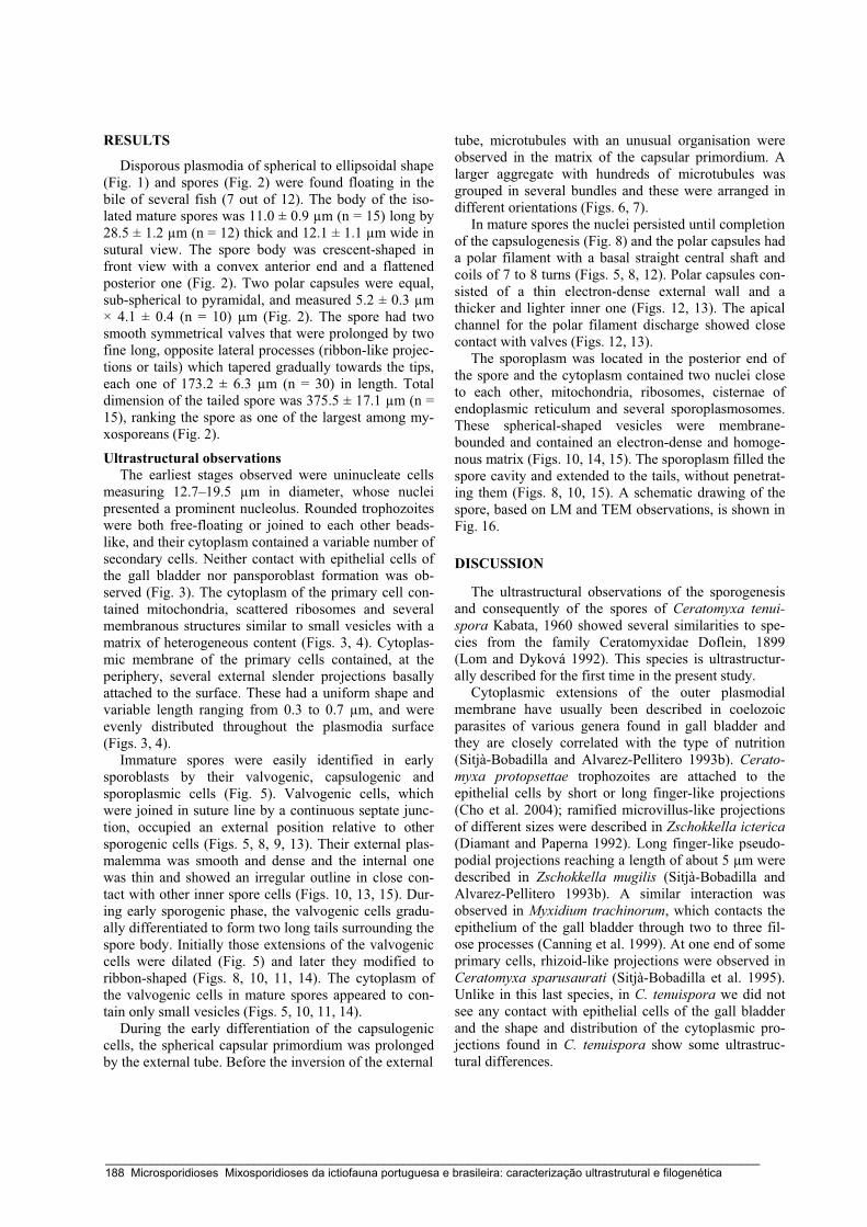
RESULTS
Disporous plasmodia of spherical to ellipsoidal shape (Fig. 1) and spores (Fig. 2) were found floating in the bile of several fish (7 out of 12). The body of the iso-lated mature spores was 11.0 ± 0.9 μm (n = 15) long by 28.5 ± 1.2 μm (n = 12) thick and 12.1 ± 1.1 μm wide in sutural view. The spore body was crescent-shaped in front view with a convex anterior end and a flattened posterior one (Fig. 2). Two polar capsules were equal, sub-spherical to pyramidal, and measured 5.2 ± 0.3 μm × 4.1 ± 0.4 (n = 10) μm (Fig. 2). The spore had two smooth symmetrical valves that were prolonged by two fine long, opposite lateral processes (ribbon-like projec-tions or tails) which tapered gradually towards the tips, each one of 173.2 ± 6.3 μm (n = 30) in length. Total dimension of the tailed spore was 375.5 ± 17.1 μm (n = 15), ranking the spore as one of the largest among my-xosporeans (Fig. 2).
Ultrastructural observations The earliest stages observed were uninucleate cells
measuring 12.7–19.5 μm in diameter, whose nuclei presented a prominent nucleolus. Rounded trophozoites were both free-floating or joined to each other beads-like, and their cytoplasm contained a variable number of secondary cells. Neither contact with epithelial cells of the gall bladder nor pansporoblast formation was ob-served (Fig. 3). The cytoplasm of the primary cell con-tained mitochondria, scattered ribosomes and several membranous structures similar to small vesicles with a matrix of heterogeneous content (Figs. 3, 4). Cytoplas-mic membrane of the primary cells contained, at the periphery, several external slender projections basally attached to the surface. These had a uniform shape and variable length ranging from 0.3 to 0.7 μm, and were evenly distributed throughout the plasmodia surface (Figs. 3, 4).
Immature spores were easily identified in early sporoblasts by their valvogenic, capsulogenic and sporoplasmic cells (Fig. 5). Valvogenic cells, which were joined in suture line by a continuous septate junc-tion, occupied an external position relative to other sporogenic cells (Figs. 5, 8, 9, 13). Their external plas-malemma was smooth and dense and the internal one was thin and showed an irregular outline in close con-tact with other inner spore cells (Figs. 10, 13, 15). Dur-ing early sporogenic phase, the valvogenic cells gradu-ally differentiated to form two long tails surrounding the spore body. Initially those extensions of the valvogenic cells were dilated (Fig. 5) and later they modified to ribbon-shaped (Figs. 8, 10, 11, 14). The cytoplasm of the valvogenic cells in mature spores appeared to con-tain only small vesicles (Figs. 5, 10, 11, 14).
During the early differentiation of the capsulogenic cells, the spherical capsular primordium was prolonged by the external tube. Before the inversion of the external
tube, microtubules with an unusual organisation were observed in the matrix of the capsular primordium. A larger aggregate with hundreds of microtubules was grouped in several bundles and these were arranged in different orientations (Figs. 6, 7).
In mature spores the nuclei persisted until completion of the capsulogenesis (Fig. 8) and the polar capsules had a polar filament with a basal straight central shaft and coils of 7 to 8 turns (Figs. 5, 8, 12). Polar capsules con-sisted of a thin electron-dense external wall and a thicker and lighter inner one (Figs. 12, 13). The apical channel for the polar filament discharge showed close contact with valves (Figs. 12, 13).
The sporoplasm was located in the posterior end of the spore and the cytoplasm contained two nuclei close to each other, mitochondria, ribosomes, cisternae of endoplasmic reticulum and several sporoplasmosomes. These spherical-shaped vesicles were membrane-bounded and contained an electron-dense and homoge-nous matrix (Figs. 10, 14, 15). The sporoplasm filled the spore cavity and extended to the tails, without penetrat-ing them (Figs. 8, 10, 15). A schematic drawing of the spore, based on LM and TEM observations, is shown in Fig. 16.
DISCUSSION
The ultrastructural observations of the sporogenesis and consequently of the spores of Ceratomyxa tenui-spora Kabata, 1960 showed several similarities to spe-cies from the family Ceratomyxidae Doflein, 1899 (Lom and Dyková 1992). This species is ultrastructur-ally described for the first time in the present study.
Cytoplasmic extensions of the outer plasmodial membrane have usually been described in coelozoic parasites of various genera found in gall bladder and they are closely correlated with the type of nutrition (Sitjà-Bobadilla and Alvarez-Pellitero 1993b). Cerato-myxa protopsettae trophozoites are attached to the epithelial cells by short or long finger-like projections (Cho et al. 2004); ramified microvillus-like projections of different sizes were described in Zschokkella icterica (Diamant and Paperna 1992). Long finger-like pseudo-podial projections reaching a length of about 5 μm were described in Zschokkella mugilis (Sitjà-Bobadilla and Alvarez-Pellitero 1993b). A similar interaction was observed in Myxidium trachinorum, which contacts the epithelium of the gall bladder through two to three fil-ose processes (Canning et al. 1999). At one end of some primary cells, rhizoid-like projections were observed in Ceratomyxa sparusaurati (Sitjà-Bobadilla et al. 1995). Unlike in this last species, in C. tenuispora we did not see any contact with epithelial cells of the gall bladder and the shape and distribution of the cytoplasmic pro-jections found in C. tenuispora show some ultrastruc-tural differences.
_____________________________________________________________________________________________________ 188 Microsporidioses Mixosporidioses da ictiofauna portuguesa e brasileira: caracterização ultrastrutural e filogenética

Casal et al.: Ultrastructure of Ceratomyxa tenuispora
Figs. 1–5. Ceratomyxa tenuispora, DIC (Figs. 1, 2) and transmission electron micrographs (Figs. 3–5). Fig. 1. Fresh disporous plasmodium. Fig. 2. Free mature spore showing two polar capsules (PCp) and two long tapering lateral opposite tails (T). Fig. 3. A secondary cell (SC) with two nuclei (N) within a primary cell (PC) containing several slender projections (arrowheads). Fig. 4. Detail of the periphery of the primary cell (PC) showing several slender projections (arrowheads). Fig. 5. Spore showing a polar capsule (PCp) with different sections of the polar filament (arrowheads), a sporoplasm (S) and valvogenic cells (V) with the suture line (arrows). At the periphery, several sections of tails (T) can be observed.
_____________________________________________________________________________________________________ Microsporidioses Mixosporidioses da ictiofauna portuguesa e brasileira: caracterização ultrastrutural e filogenética 189
Figs. 6–9. Transmission electron micrographs of Ceratomyxa tenuispora. Fig. 6. A spore showing the valves (V), several sec-tions of their two tails (T), the capsular primordium (C) containing a well-organized cluster of microtubules (boxed area) and the sporoplasm (S). Fig. 7. Detail of the cluster of microtubules (boxed area in Fig. 6). At the periphery, valvogenic cell (V). Fig. 8. Longitudinal section of a spore showing one nucleus (N) of a capsulogenic cell, two polar capsules (PCp), and the binucleate (N*) sporoplasm (S). Valvogenic cells (V) are reduced to a thin layer in close contact with internal cells. Sutures (double arrows) between the valves can be seen. Some tail sections (T) can be seen at the periphery of the spore. Fig. 9. Detail of the continuous septate junction of the suture line.
Ultrastructural description concerning the polar cap-
sule differentiation referred to the presence of unusual structures inside the capsular primordium, such as glob-ule of electron-dense material (Lom 1969), concentric structure (Lom et al. 1989) or the differentiation of the microfilament-like structures (Casal et al. 2002). Bun-dles of tubuli in the capsular matrix were reported in the polar capsules of mature spores in some genera, such as Sphaeromyxa (Lom 1969), Henneguya (Rocha et al.
1992) and Myxobolus (Casal et al. 2002). In the cyto-plasm of the capsulogenic cells, microtubules are regu-larly seen around the external tube interpreted to pro-vide the mechanic force needed for its inversion into the capsular primordium (Current 1979). We reported the presence of several microtubular bundles inside the capsular matrix before the inversion of the external tube which apparently also contributes to that.
_____________________________________________________________________________________________________ 190 Microsporidioses Mixosporidioses da ictiofauna portuguesa e brasileira: caracterização ultrastrutural e filogenética

Casal et al.: Ultrastructure of Ceratomyxa tenuispora
Figs. 10–15. Transmission electron micrographs of Ceratomyxa tenuispora. Fig. 10. Detail of valve (V), its tail (T) and the sporoplasm (S) with a nucleus (N) and some sporoplasmosomes (Ss). Fig. 11. Sections of spore tail (T) showing an electron-lucid matrix. Fig. 12. Longitudinal section of a polar capsule with electron-dense matrix (*), showing the polar filament sec-tioned at different levels (arrowheads). Fig. 13. Detail of the apical region of a polar capsule showing the wall (W), the channel for filament discharge (D), and some polar filament sections (arrowheads) within the capsular matrix (*). Externally, the valve (V) and the suture (double arrowhead). Fig. 14. Sporoplasm showing the two nuclei (N), several sporoplasmosomes (Ss) and several sections of the tails (T). Fig. 15. Detail of the sporoplasm showing several sporoplasmosomes (Ss), mitochondria (M) and some cisternae of endoplasmic reticulum (arrowheads). Externally, a surrounding valve (V) can be seen.
_____________________________________________________________________________________________________ Microsporidioses Mixosporidioses da ictiofauna portuguesa e brasileira: caracterização ultrastrutural e filogenética 191

Fig. 16. Schematic drawing of Ceratomyxa tenuispora showing the spore morphology, with special emphasis on the two long tapering opposite lateral tails. Table 1. Ceratomyxa tenuispora, comparison of spore characteristics between original description of Kabata (1960) and the present specimens (measurements in μm).
Original description Present material Host and organ Aphanopus carbo, gall bladder Geographical location
Scotland (United Kingdom)
North Atlantic coast of Madeira Island (Portugal)
Spore body crescent-shaped with convex anterior end and flattened posterior end – length 8.7 (8.4–9.8) 11.0 ± 0.9 – width – 12.1 ± 1.1 – thickness – 28.5 ± 1.2 Total thickness 387 (308–504) 375.5 ± 17.1 Polar capsules two equal, sub-spherical to pyramidal – length 6.4 (5.6–7.0) 5.2 ± 0.3 – width – 4.1 ± 0.4 Polar filament – coils of 7 to 8 turns Sporoplasm slightly granular with vacuoles binucleate
More than 30 species of Ceratomyxa with long lat-
eral processes have been described in different hosts and geographic areas (Sitjà-Bobadilla and Alvarez-Pellitero 1993a). Ceratomyxa tenuispora is the second longest of this Ceratomyxa spp. group. These processes have not been ultrastructurally described except for C. labracis (Sitjà-Bobadilla and Alvarez-Pellitero 1993a), showing similar ultrastructural aspects to those described in this study.
Since the description of C. tenuispora based on a drawing obtained from LM observations by Kabata (1960) and a later report by Costa et al. (1996), no other reports of this species have been published. The mor-
phological and morphometrical characteristics of the spore seem to be the same, except for total thickness, the present specimens being somewhat smaller than C. tenuispora studied previously (Kabata 1960, Costa et al. 1996). Also, the parasite was collected from the same host species (Aphanopus carbo) and the same organ (gall bladder) (Table 1). Acknowledgements. This work was supported by the António de Almeida Foundation-Porto-Portugal and a CESPU (Coop-erativa de Ensino Superior, Politécnico e Universitario) PhD grant. We would like to thank Mr. João Carvalheiro for tech-nical assistance and to Dr. Victor Ferreira for helping in the English revision. The helpful suggestions and comments of the reviewers are greatly appreciated.
_____________________________________________________________________________________________________ 192 Microsporidioses Mixosporidioses da ictiofauna portuguesa e brasileira: caracterização ultrastrutural e filogenética
Casal et al.: Ultrastructure of Ceratomyxa tenuispora
REFERENCES
ALVAREZ-PELLITERO P., SITJÀ-BOBADILLA A. 1993: Pathology of Myxosporea in marine fish culture. Dis. Aquat. Org. 17: 229–238.
CANNING E.U., CURRY A., ANDERSON C.L., OKAMURA B. 1999: Ultrastructure of Myxidium trachinorum sp. nov. from the gallbladder of the lesser weever fish Echiichthys vipera. Para-sitol. Res. 85: 910–919.
CASAL G., MATOS E., AZEVEDO C. 2002: Ultrastructural data of the spore of Myxobolus maculatus n. sp. (Phylum Myxozoa), parasite from the Amazonian fish Metynnis maculatus Kner, 1860 (Teleostei). Dis. Aquat. Org. 51: 107–112.
CHO J.B., KWON S.R., KIM S.K., NAM Y.K., KIM K.H. 2004: Ultrastructure and development of Ceratomyxa protopsettae Fujita, 1923 (Myxosporea) in the gallbladder of cultured olive flounder, Paralichthys olivaceus. Acta Protozool. 43: 241–250.
COSTA G., EIRAS J.C., CHUBB J., MACKENZIE K., BERLAND B. 1996: Parasites of the black scabbard fish, Aphanopus carbo Lowe, 1839 from Madeira. Bull. Eur. Assoc. Fish Pathol. 16: 13–16.
CURRENT W.L. 1979: Henneguya adiposa Minchew (Myxo-sporida) in the channel catfish: ultrastructure of the plasmo-dium wall and sporogenesis. J. Protozool. 26: 209–217.
DESPORTES I., THEODORIDES J. 1982: Données ultrastructurales sur la sporogenèse de deux myxosporidies rapportées aux genres Leptotheca et Ceratomyxa parasites de Merluccius merluccius (L.) (Téléostéen Merluciidae). Protistologica 18: 533–557.
DIAMANT A., PAPERNA I. 1989: Cytopathology of Ceratomyxa sp. (Myxosporea) hyperparasitized with the microsporidan Nosema ceratomyxae. Dis. Aquat. Org. 6: 75–79.
DIAMANT A., PAPERNA I. 1992: Zschokkella icterica sp. nov. (Myxozoa, Myxosporea), a pathogen of wild rabbitfish Si-ganus luridus (Ruppell, 1829) from the Red Sea. Eur. J. Pro-tistol. 28: 71–78.
EIRAS J.C. 2006: Synopsis of the species of the genus Ceratomyxa Thélohan, 1892 (Myxozoa: Myxosporea: Ceratomyxidae). Syst. Parasitol. 65: 49–71.
KABATA Z. 1960: On two myxosporidian parasites of marine fishes, including one new species (Ceratomyxa tenuispora). Ann. Mag. Nat. Hist. 13: 305–306.
LOM J. 1969: Notes on the ultrastructure and sporoblast develop-ment in fish parasitizing myxosporidian of the genus Sphaero-myxa. Z. Zellforsch. 97: 416–437.
LOM J., DYKOVÁ I. 1992: Protozoan Parasites of Fishes. Devel-opments in Aquaculture and Fisheries Science. Vol. 26. El-sevier, Amsterdam, 315 pp.
LOM J., DYKOVÁ I. 2006: Myxozoan genera: definition and notes on taxonomy, life-cycle terminology and pathogenic species. Folia Parasitol. 53: 1–36.
LOM J., FEIST S.W., DYKOVÁ I., KEPR T. 1989: Brain myxo-boliasis of bullhead, Cottus gobio L., due to Myxobolus ji-roveci sp. nov.: light and electron microscope observations. J. Fish Dis. 12: 15–27.
MORRISON C.M., MARTELL D.J., LEGGIARDO C., O’NEIL D. 1996: Ceratomyxa drepanopsettae in the gallbladder of Atlan-tic halibut, Hippoglossus hippoglossus, from the Northwest Atlantic Ocean. Folia Parasitol. 43: 20–36.
PALENZUELA O., SITJÀ-BOBADILLA A., ALVAREZ-PELLITERO P. 1997: Ceratomyxa sparusaurati (Protozoa: Myxosporea) infections in cultured gilthead sea bream Sparus aurata (Pisces: Teleostei) from Spain: aspects of the host-parasite relationship. Parasitol. Res. 83: 539–548.
ROCHA E., MATOS E., AZEVEDO C. 1992: Henneguya amazonica n. sp. (Myxozoa, Myxobolidae), parasitizing the gills of Crenicichla lepidota Heckel, 1840 (Teleostei, Cichlidae) from Amazon river. Eur. J. Protistol. 28: 273–278.
SITJÀ-BOBADILLA A., ALVAREZ-PELLITERO P. 1993a: Light and electron microscopical description of Ceratomyxa labracis n. sp. and a redescription of C. diplodae (Myxosporea: Bivalvu-lida) from wild and cultured Mediterranean sea bass Dicen-trarchus labrax (L.) (Teleostei: Serranidae). Syst. Parasitol. 26: 215–223.
SITJÀ-BOBADILLA A., ALVAREZ-PELLITERO P. 1993b: Zschok-kella mugilis n. sp (Myxosporea: Bivalvulida) from mullets (Teleostei: Mugilidae) of Mediterranean waters: light and electron microscopic description. J. Eukaryot. Microbiol. 40: 755–764.
SITJÀ-BOBADILLA A., PALENZUELA O., ALVAREZ-PELLITERO P. 1995: Ceratomyxa sparusaurati n. sp. (Myxosporea: Bival-vulida), a new parasite from cultured gilthead seabream (Sparus aurata L.) (Teleostei: Sparidae): light and electron microscopic description. J. Eukaryot. Microbiol. 42: 529–539.
YAMAMOTO T., SANDERS J.E. 1979: Light and electron micro-scopic observations of sporogenesis in the Ceratomyxa shasta (Noble, 1950). J. Fish Dis. 2: 411–428.
Received 22 May 2006 Accepted 6 June 2007
_____________________________________________________________________________________________________ Microsporidioses Mixosporidioses da ictiofauna portuguesa e brasileira: caracterização ultrastrutural e filogenética 193

_____________________________________________________________________________________________________ 194 Microsporidioses Mixosporidioses da ictiofauna portuguesa e brasileira: caracterização ultrastrutural e filogenética

Capítulo 10
A NEW SPECIES OF MYXOZOA, HENNEGUYA RONDONI N. SP. (MYXOZOA),
FROM THE PERIPHERAL NERVOUS SYSTEM OF THE AMAZONIAN FISH,
GYMNORHAMPHICHTHYS RONDONI (TELEOSTEI)
The Journal Eukaryotic Microbiology (2008) 55: 229-234
Carlos Azevedo, Graça Casal, Patrícia Matos & Edilson Matos
_____________________________________________________________________________________________________ Microsporidioses Mixosporidioses da ictiofauna portuguesa e brasileira: caracterização ultrastrutural e filogenética 195

_____________________________________________________________________________________________________ 196 Microsporidioses Mixosporidioses da ictiofauna portuguesa e brasileira: caracterização ultrastrutural e filogenética

A New Species of Myxozoa, Henneguya rondoni n. sp. (Myxozoa), from thePeripheral Nervous System of the Amazonian Fish,
Gymnorhamphichthys rondoni (Teleostei)
CARLOS AZEVEDO,a,b GRACA CASAL,a,b,c PATRICIA MATOSd and EDILSON MATOSe
aDepartment of Cell Biology, Institute of Biomedical Sciences, University of Porto (ICBAS/UP),
Lg. A. Salazar no. 2, P-4099-003 Porto, Portugal, andbLaboratory of Pathology, Centre for Marine and Environmental Research (CIIMAR/UP), P-4050-123 Porto, Portugal, and
cDepartment of Sciences, High Institute of Health Sciences, P-4585-116 Gandra, Portugal, anddLaboratory of Aquatic Animals, Federal University of Para, 66000 Belem, Brazil, and
eCarlos Azevedo Research Laboratory, Federal Rural University of Amazonia, 66000 Belem, Brazil
ABSTRACT. Henneguya rondoni n. sp. found in the peripheral lateral nerves located below the two lateral lines of the fish Gym-norhamphichthys rondoni (Teleostei, Rhamphichthyidae) from the Amazon river is described using light and electron microscopy.Spherical to ellipsoid cysts measuring up to 110 mm in length contained only immature and mature spores located in close contact with themyelin sheaths of the nervous fibres. Ellipsoidal spores measured 17.7 (16.9–18.1)-mm long, 3.6 (3.0–3.9)-mm wide, and 2.5 (2.2–2.8)-mm(n5 25) thick. The spore body measuring 7.0 (6.8–7.3)-mm long was formed by two equal symmetric valves, each with an equal taperingtail 10.7 (10.3–11.0) mm in length. The tails were composed of an internal dense material surrounded by an external homogeneous sheathof hyaline substance. The valves surrounded two equal pyriform polar capsules measuring 2.5 (2.2–2.8)-mm long and 0.85 (0.79–0.88)-mm(n5 25) wide and a binucleated sporoplasm cell containing globular sporoplasmosomes 0.38 (0.33–0.42) mm (n5 25) in diam. with aninternal eccentric dense structure with half-crescent section. Each polar capsule contains an anisofilar polar filament with 6–7 turnsobliquely to the long axis. The matrix of the polar capsule was dense and the wall filled with a hyaline substance. The spores differed fromthose of previously described species. Based on the ultrastructural morphology of the spore and specificity to the host species, we proposea new species name H. rondoni n. sp.
Key Words. Amazon river, myxosporean, parasite, spore, ultrastructure.
THE class Myxosporea Butschli, 1881 comprises more than2,180 available species (Lom and Dykova 2006) of which
actually about 2,160 were found in fish. Certainly, a large numberof species still remain to be discovered (Lom and Dykova 2006),in particular in Brazil, where an extremely high number of fishspecies live (about 8,000 species) (Cellere, Cordeiro, and Adriano2002). Among the myxosporeans, the genus Henneguya Thelo-han, 1892 with 204 species described is one of the largest of thefamily Myxobolidae (Lom and Dykova 2006). Some of these spe-cies have been reported as important pathogens in freshwater fish(Kent et al. 2001; Lom and Dykova 2006).
Very few South American myxosporean species have been de-scribed in detail. Most descriptions are in particular from Brazil,where species are only illustrated by light microscopy (LM) anddiagrammatic drawings (Cordeiro et al. 1984; Cunha and Fonseca1918; Eiras 2002; Eiras, Pavanelli, and Takemoto 2004; Eiraset al. 2004; Gioia and Cordeiro 1996; Gioia, Cordeiro, and Artigas1986; Kent and Hoffman 1984; Martins and de Souza 1997; Mar-tins et al. 1999; Nemeczek 1926; Walliker 1969). Recently, somespecies of the genus Henneguya were described on the basis of theultrastructural data (Azevedo, Corral, and Matos 1997; Azevedoand Matos 1995, 1996; Barassa, Cordeiro, and Arana 2003; Casal,Matos, and Azevedo 1997; Matos, Tajdari, and Azevedo 2005;Vita et al. 2003). None has been reported from peripheral nervousfibres of a fish. During a parasitological survey of Amazonian fish,a myxozoan parasite was discovered in the peripheral nervoussystem of a fish, and it is this isolate that we describe as a newspecies in this report.
MATERIALS AND METHODS
The teleost Gymnorhamphichthys rondoni (Teleostei,Rhamphichthyidae) (Brazilian common name ‘‘Ituı transpar-ente’’) was collected from the Amazon river near the beach ofIrituia, State of Para, Brazil (011460S/471260W). After collection,30 fish (14–19-cm long) were transported live to the laboratory,where their behaviour was studied. For microscopic study theywere anaesthetized with MS 222 (Sandoz Laboratories), killed,and necropsied.
Smears of small portions of the fresh peripheral nervous fibresof the lateral lines of the body containing cysts were prepared forobservation by LM using Nomarski differential interference con-trast (DIC) optics. For transmission electron microscopy (TEM),small fragments of the parasitized tissues were fixed in 5% (v/v)glutaraldehyde buffered in 0.2M sodium cacodylate (pH 7.2) at4 1C for 24 h, washed overnight in the same buffer at 4 1C, andpost-fixed in 2% (w/v) osmiun tetroxide with the same buffer andat the same temperature for 3 h. After dehydration in an ascendingethanol series followed by propylene oxide (three changes of 2 h),the parasitized fragments were embedded in Epon. Semithin sec-tions, were stained with methylene blue-Azure II for LM andultrathin sections were double contrasted with uranylacetate and lead citrate and observed in a JEOL 100CXII TEM(Japan) operated at 60 kV.
RESULTS
Some cysts, containing only immature and mature spores, wereobserved exclusively among the peripheral nervous fibres locatedin both longitudinal medial lines on the lateral body of specimenswith disturbances in their behaviour. No other tissue or organcontained visible parasites. Only infected fish exhibited lethargyand sudden, short disturbances in their movements and no para-sites were found in specimens with apparently normal behaviour.Ten of 30 (33.3%) of the fish contained spherical to ellipsoid cystsup to 110-mm long (Fig. 1, 2).
Corresponding Author: C. Azevedo, Department of Cell Biology,Institute of Biomedical Sciences, University of Porto (ICBAS/UP), Lg.A. Salazar no. 2, P-4099-003 Porto, Portugal—Telephone number:1351 22 206 22 00; FAX number:1351 22 206 22 32/33; e-mail: [email protected]
229
J. Eukaryot. Microbiol., 55(3), 2008 pp. 229–234r 2008 The Author(s)Journal compilation r 2008 by the International Society of ProtistologistsDOI: 10.1111/j.1550-7408.2008.00317.x
_____________________________________________________________________________________________________ Microsporidioses Mixosporidioses da ictiofauna portuguesa e brasileira: caracterização ultrastrutural e filogenética 197

Fig. 1–4. Light and transmission electron micrographs of the myxosporean Henneguya rondoni n. sp. from peripheral nervous fibres of the laterallines of the fish body of Gymnorhamphichthys rondoni. (Scale bars in mm). 1. Semithin section showing some cysts (C) filled by numerous spores incontact with numerous bundles of peripheral nervous fibres (�). 2. Semithin section of the cysts (C) containing spores (S). Inset. Free spores observed inDIC. 3.Ultrathin section of a cyst in close contact with the nervous fibres (�). The cyst wall shows some fibroblasts (F) surrounding the internal spores (S).4. Ultrastructural detail of the periphery of a cyst containing some fibroblasts (F) in contact with the myelin sheaths of nervous fibres (�). Two sporesshow the transverse section at the polar capsule (PC) level.
230 J. EUKARYOT. MICROBIOL., 55, NO. 3, MAY–JUNE 2008
_____________________________________________________________________________________________________ 198 Microsporidioses Mixosporidioses da ictiofauna portuguesa e brasileira: caracterização ultrastrutural e filogenética

Fig. 5–8. Transmission electron micrographs of the spores of the myxosporean Henneguya rondoni n. sp. from peripheral nervous fibres of the laterallines of the fish body of Gymnorhamphichthys rondoni. (Scale bars in mm). 5. Ultrastructural details of the contact zone of the nervous fibres (�) with theperiphery of the cyst formed by the fibroblast (F) layer. Internally the cyst contains numerous spores (S). 6. Ultrastructural aspects of a group of densebodies located near the cyst wall (CW) where it is possible to observe numerous collagen fibres (Cg) and fibroblasts (F). 7. Ultrastructural aspect of thecontact zone among the nervous fibres (�) and the cyst, showing numerous collagen fibres (Cg), fibroblasts (F), and spores sectioned at the differentlevels. Several aspects of the tail (T) and polar capsule are observed. 8. Details of some ultrastrutural aspects of the spores showing the spore wall (W),polar capsules (PC), sporoplasm (Sp), sporoplasmosomes (Ss) (details in inset – arrowheads), and transverse sections of the tails (T) sectioned at thedifferent levels. The tails show the internal dense zone surrounded by a sheath of an adherent hyaline substance (arrowheads).
231AZEVEDO ET AL.—ULTRASTRUCTURE OF HENNEGUYA RONDONI N. SP.
_____________________________________________________________________________________________________ Microsporidioses Mixosporidioses da ictiofauna portuguesa e brasileira: caracterização ultrastrutural e filogenética 199
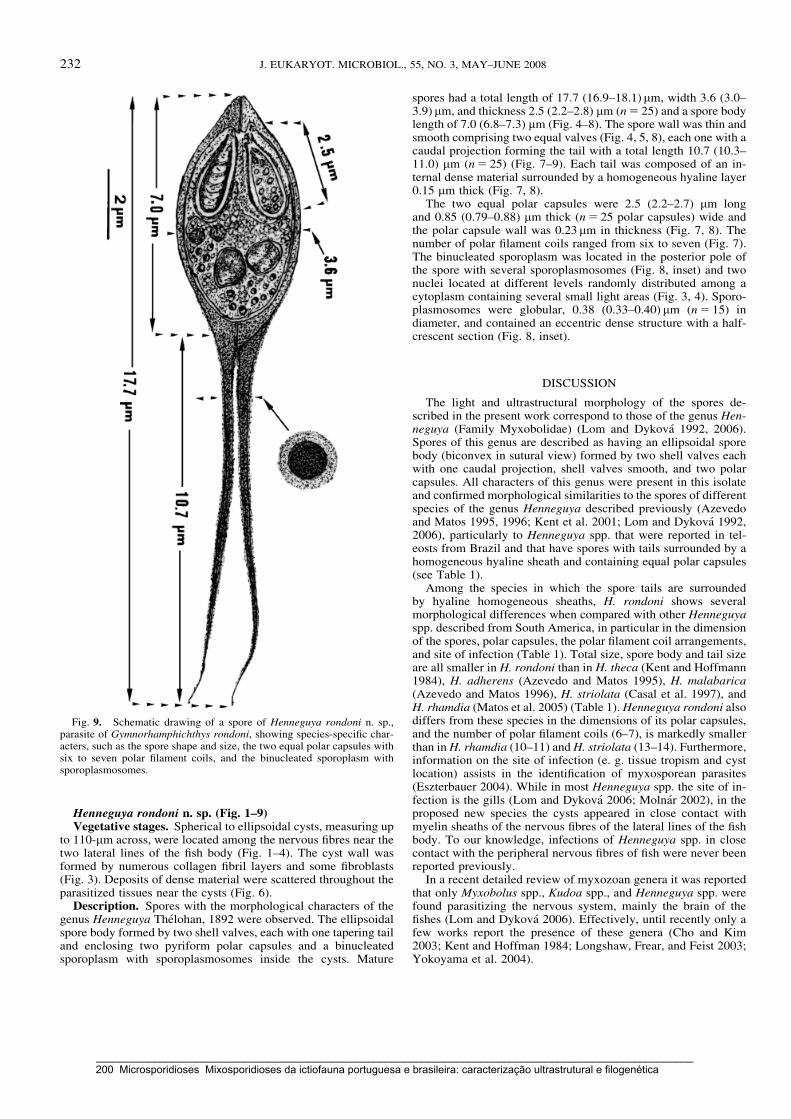
Henneguya rondoni n. sp. (Fig. 1–9)Vegetative stages. Spherical to ellipsoidal cysts, measuring up
to 110-mm across, were located among the nervous fibres near thetwo lateral lines of the fish body (Fig. 1–4). The cyst wall wasformed by numerous collagen fibril layers and some fibroblasts(Fig. 3). Deposits of dense material were scattered throughout theparasitized tissues near the cysts (Fig. 6).
Description. Spores with the morphological characters of thegenus Henneguya Thelohan, 1892 were observed. The ellipsoidalspore body formed by two shell valves, each with one tapering tailand enclosing two pyriform polar capsules and a binucleatedsporoplasm with sporoplasmosomes inside the cysts. Mature
spores had a total length of 17.7 (16.9–18.1)mm, width 3.6 (3.0–3.9) mm, and thickness 2.5 (2.2–2.8) mm (n5 25) and a spore bodylength of 7.0 (6.8–7.3) mm (Fig. 4–8). The spore wall was thin andsmooth comprising two equal valves (Fig. 4, 5, 8), each one with acaudal projection forming the tail with a total length 10.7 (10.3–11.0) mm (n5 25) (Fig. 7–9). Each tail was composed of an in-ternal dense material surrounded by a homogeneous hyaline layer0.15 mm thick (Fig. 7, 8).
The two equal polar capsules were 2.5 (2.2–2.7) mm longand 0.85 (0.79–0.88) mm thick (n5 25 polar capsules) wide andthe polar capsule wall was 0.23 mm in thickness (Fig. 7, 8). Thenumber of polar filament coils ranged from six to seven (Fig. 7).The binucleated sporoplasm was located in the posterior pole ofthe spore with several sporoplasmosomes (Fig. 8, inset) and twonuclei located at different levels randomly distributed among acytoplasm containing several small light areas (Fig. 3, 4). Sporo-plasmosomes were globular, 0.38 (0.33–0.40) mm (n5 15) indiameter, and contained an eccentric dense structure with a half-crescent section (Fig. 8, inset).
DISCUSSION
The light and ultrastructural morphology of the spores de-scribed in the present work correspond to those of the genus Hen-neguya (Family Myxobolidae) (Lom and Dykova 1992, 2006).Spores of this genus are described as having an ellipsoidal sporebody (biconvex in sutural view) formed by two shell valves eachwith one caudal projection, shell valves smooth, and two polarcapsules. All characters of this genus were present in this isolateand confirmed morphological similarities to the spores of differentspecies of the genus Henneguya described previously (Azevedoand Matos 1995, 1996; Kent et al. 2001; Lom and Dykova 1992,2006), particularly to Henneguya spp. that were reported in tel-eosts from Brazil and that have spores with tails surrounded by ahomogeneous hyaline sheath and containing equal polar capsules(see Table 1).
Among the species in which the spore tails are surroundedby hyaline homogeneous sheaths, H. rondoni shows severalmorphological differences when compared with other Henneguyaspp. described from South America, in particular in the dimensionof the spores, polar capsules, the polar filament coil arrangements,and site of infection (Table 1). Total size, spore body and tail sizeare all smaller in H. rondoni than in H. theca (Kent and Hoffmann1984), H. adherens (Azevedo and Matos 1995), H. malabarica(Azevedo and Matos 1996), H. striolata (Casal et al. 1997), andH. rhamdia (Matos et al. 2005) (Table 1).Henneguya rondoni alsodiffers from these species in the dimensions of its polar capsules,and the number of polar filament coils (6–7), is markedly smallerthan inH. rhamdia (10–11) andH. striolata (13–14). Furthermore,information on the site of infection (e. g. tissue tropism and cystlocation) assists in the identification of myxosporean parasites(Eszterbauer 2004). While in most Henneguya spp. the site of in-fection is the gills (Lom and Dykova 2006; Molnar 2002), in theproposed new species the cysts appeared in close contact withmyelin sheaths of the nervous fibres of the lateral lines of the fishbody. To our knowledge, infections of Henneguya spp. in closecontact with the peripheral nervous fibres of fish were never beenreported previously.
In a recent detailed review of myxozoan genera it was reportedthat only Myxobolus spp., Kudoa spp., and Henneguya spp. werefound parasitizing the nervous system, mainly the brain of thefishes (Lom and Dykova 2006). Effectively, until recently only afew works report the presence of these genera (Cho and Kim2003; Kent and Hoffman 1984; Longshaw, Frear, and Feist 2003;Yokoyama et al. 2004).
Fig. 9. Schematic drawing of a spore of Henneguya rondoni n. sp.,parasite of Gymnorhamphichthys rondoni, showing species-specific char-acters, such as the spore shape and size, the two equal polar capsules withsix to seven polar filament coils, and the binucleated sporoplasm withsporoplasmosomes.
232 J. EUKARYOT. MICROBIOL., 55, NO. 3, MAY–JUNE 2008
_____________________________________________________________________________________________________ 200 Microsporidioses Mixosporidioses da ictiofauna portuguesa e brasileira: caracterização ultrastrutural e filogenética
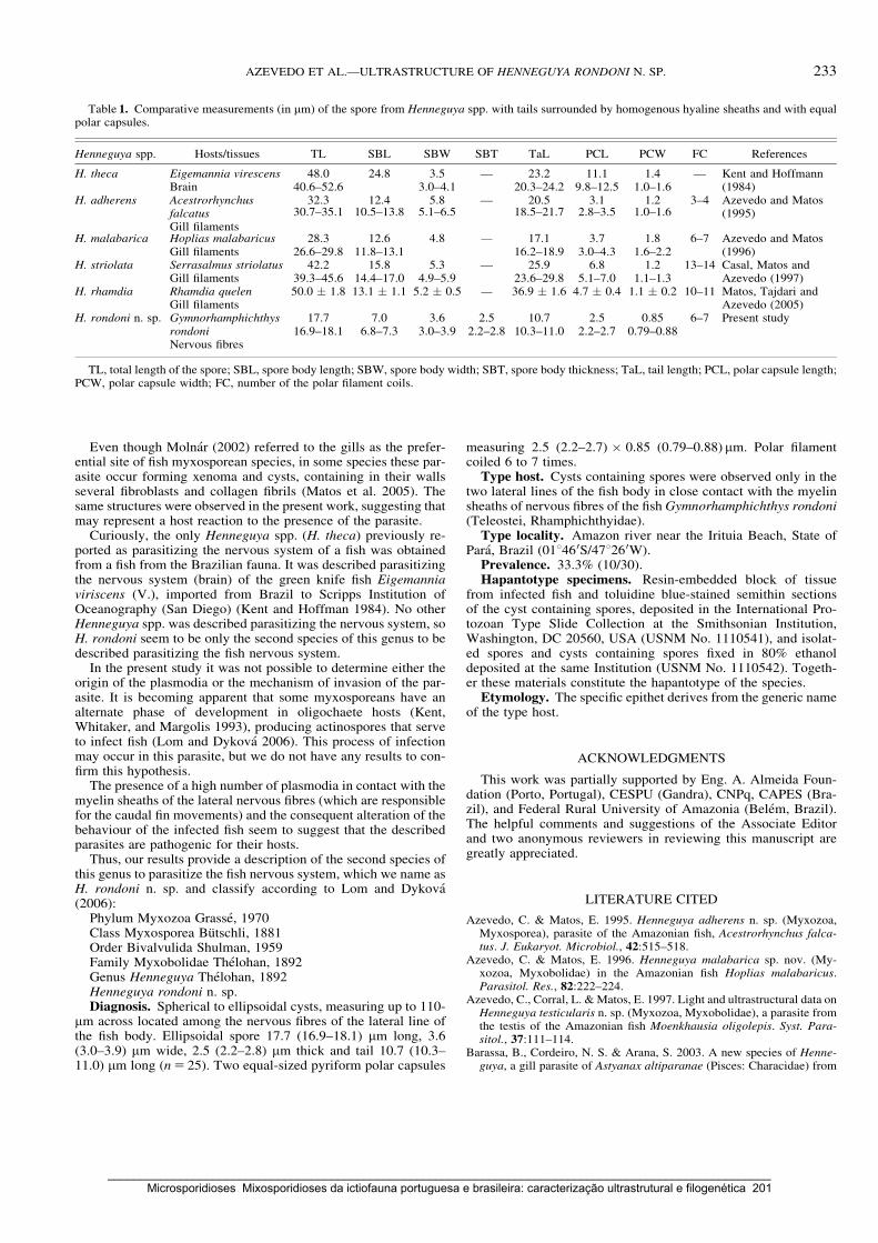
Even though Molnar (2002) referred to the gills as the prefer-ential site of fish myxosporean species, in some species these par-asite occur forming xenoma and cysts, containing in their wallsseveral fibroblasts and collagen fibrils (Matos et al. 2005). Thesame structures were observed in the present work, suggesting thatmay represent a host reaction to the presence of the parasite.
Curiously, the only Henneguya spp. (H. theca) previously re-ported as parasitizing the nervous system of a fish was obtainedfrom a fish from the Brazilian fauna. It was described parasitizingthe nervous system (brain) of the green knife fish Eigemanniaviriscens (V.), imported from Brazil to Scripps Institution ofOceanography (San Diego) (Kent and Hoffman 1984). No otherHenneguya spp. was described parasitizing the nervous system, soH. rondoni seem to be only the second species of this genus to bedescribed parasitizing the fish nervous system.
In the present study it was not possible to determine either theorigin of the plasmodia or the mechanism of invasion of the par-asite. It is becoming apparent that some myxosporeans have analternate phase of development in oligochaete hosts (Kent,Whitaker, and Margolis 1993), producing actinospores that serveto infect fish (Lom and Dykova 2006). This process of infectionmay occur in this parasite, but we do not have any results to con-firm this hypothesis.
The presence of a high number of plasmodia in contact with themyelin sheaths of the lateral nervous fibres (which are responsiblefor the caudal fin movements) and the consequent alteration of thebehaviour of the infected fish seem to suggest that the describedparasites are pathogenic for their hosts.
Thus, our results provide a description of the second species ofthis genus to parasitize the fish nervous system, which we name asH. rondoni n. sp. and classify according to Lom and Dykova(2006):
Phylum Myxozoa Grasse, 1970Class Myxosporea Butschli, 1881Order Bivalvulida Shulman, 1959Family Myxobolidae Thelohan, 1892Genus Henneguya Thelohan, 1892Henneguya rondoni n. sp.Diagnosis. Spherical to ellipsoidal cysts, measuring up to 110-
mm across located among the nervous fibres of the lateral line ofthe fish body. Ellipsoidal spore 17.7 (16.9–18.1) mm long, 3.6(3.0–3.9) mm wide, 2.5 (2.2–2.8) mm thick and tail 10.7 (10.3–11.0) mm long (n5 25). Two equal-sized pyriform polar capsules
measuring 2.5 (2.2–2.7) � 0.85 (0.79–0.88)mm. Polar filamentcoiled 6 to 7 times.
Type host. Cysts containing spores were observed only in thetwo lateral lines of the fish body in close contact with the myelinsheaths of nervous fibres of the fishGymnorhamphichthys rondoni(Teleostei, Rhamphichthyidae).
Type locality. Amazon river near the Irituia Beach, State ofPara, Brazil (011460S/471260W).
Prevalence. 33.3% (10/30).Hapantotype specimens. Resin-embedded block of tissue
from infected fish and toluidine blue-stained semithin sectionsof the cyst containing spores, deposited in the International Pro-tozoan Type Slide Collection at the Smithsonian Institution,Washington, DC 20560, USA (USNM No. 1110541), and isolat-ed spores and cysts containing spores fixed in 80% ethanoldeposited at the same Institution (USNM No. 1110542). Togeth-er these materials constitute the hapantotype of the species.
Etymology. The specific epithet derives from the generic nameof the type host.
ACKNOWLEDGMENTS
This work was partially supported by Eng. A. Almeida Foun-dation (Porto, Portugal), CESPU (Gandra), CNPq, CAPES (Bra-zil), and Federal Rural University of Amazonia (Belem, Brazil).The helpful comments and suggestions of the Associate Editorand two anonymous reviewers in reviewing this manuscript aregreatly appreciated.
LITERATURE CITED
Azevedo, C. & Matos, E. 1995. Henneguya adherens n. sp. (Myxozoa,Myxosporea), parasite of the Amazonian fish, Acestrorhynchus falca-tus. J. Eukaryot. Microbiol., 42:515–518.
Azevedo, C. & Matos, E. 1996. Henneguya malabarica sp. nov. (My-xozoa, Myxobolidae) in the Amazonian fish Hoplias malabaricus.Parasitol. Res., 82:222–224.
Azevedo, C., Corral, L. &Matos, E. 1997. Light and ultrastructural data onHenneguya testicularis n. sp. (Myxozoa, Myxobolidae), a parasite fromthe testis of the Amazonian fish Moenkhausia oligolepis. Syst. Para-sitol., 37:111–114.
Barassa, B., Cordeiro, N. S. & Arana, S. 2003. A new species of Henne-guya, a gill parasite of Astyanax altiparanae (Pisces: Characidae) from
Table 1. Comparative measurements (in mm) of the spore from Henneguya spp. with tails surrounded by homogenous hyaline sheaths and with equalpolar capsules.
Henneguya spp. Hosts/tissues TL SBL SBW SBT TaL PCL PCW FC References
H. theca Eigemannia virescens 48.0 24.8 3.5 — 23.2 11.1 1.4 — Kent and Hoffmann(1984)Brain 40.6–52.6 3.0–4.1 20.3–24.2 9.8–12.5 1.0–1.6
H. adherens AcestrorhynchusfalcatusGill filaments
32.3 12.4 5.8 — 20.5 3.1 1.2 3–4 Azevedo and Matos(1995)30.7–35.1 10.5–13.8 5.1–6.5 18.5–21.7 2.8–3.5 1.0–1.6
H. malabarica Hoplias malabaricus 28.3 12.6 4.8 — 17.1 3.7 1.8 6–7 Azevedo and Matos(1996)Gill filaments 26.6–29.8 11.8–13.1 16.2–18.9 3.0–4.3 1.6–2.2
H. striolata Serrasalmus striolatus 42.2 15.8 5.3 — 25.9 6.8 1.2 13–14 Casal, Matos andAzevedo (1997)Gill filaments 39.3–45.6 14.4–17.0 4.9–5.9 23.6–29.8 5.1–7.0 1.1–1.3
H. rhamdia Rhamdia quelen 50.0 � 1.8 13.1 � 1.1 5.2 � 0.5 — 36.9 � 1.6 4.7 � 0.4 1.1 � 0.2 10–11 Matos, Tajdari andAzevedo (2005)Gill filaments
H. rondoni n. sp. GymnorhamphichthysrondoniNervous fibres
17.7 7.0 3.6 2.5 10.7 2.5 0.85 6–7 Present study16.9–18.1 6.8–7.3 3.0–3.9 2.2–2.8 10.3–11.0 2.2–2.7 0.79–0.88
TL, total length of the spore; SBL, spore body length; SBW, spore body width; SBT, spore body thickness; TaL, tail length; PCL, polar capsule length;PCW, polar capsule width; FC, number of the polar filament coils.
233AZEVEDO ET AL.—ULTRASTRUCTURE OF HENNEGUYA RONDONI N. SP.
_____________________________________________________________________________________________________ Microsporidioses Mixosporidioses da ictiofauna portuguesa e brasileira: caracterização ultrastrutural e filogenética 201

Brazil, with comments on histopathology and seasonality. Mem. Inst.Oswaldo Cruz, Rio de Janeiro, 98:761–765.
Casal, G., Matos, E. & Azevedo, C. 1997. Some ultrastructural aspectsof Henneguya striolata sp. nov. (Myxozoa, Myxosporea), a parasiteof the Amazonian fish Serrasalmus striolatus. Parasitol. Res., 83:93–95.
Cellere, E. F., Cordeiro, N. S. & Adriano, E. A. 2002.Myxobolus absonussp. n. (Myxozoa: Myxosporea) parasitizing Pimelodus maculatus(Siluriformes: Pimelodidae), a South American freshwater fish. Mem.Inst. Oswaldo Cruz, Rio de Janeiro, 97:79–80.
Cho, J. B. & Kim, K. H. 2003. Light- and electron-microscope descriptionof Kudoa paralichthys n. sp. (Myxozoa, Myxosporea) from the brain ofcultured olive flounder Paralichthys olivaceus in Korea. Dis. Aquat.Org., 55:59–63.
Cordeiro, N. S., Artigas, P. T., Gioia, I. & Lima, R. S. 1984. Henneguyapisciforme n. sp., mixosporıdeo parasito de branquias do LambariHyphessobrycon anisitsi (Pisces, Characidae). Mem. Inst. Butantan,48:61–69.
Cunha, A. M. & Fonseca, O. 1918. Sobre os myxosporideos dos peixesbrasileiros. Bras. Med., 32:414.
Eiras, J. C. 2002. Synopsis of the species of the genus Henneguya Thelo-han, 1892 (Myxozoa: Myxosporea: Myxobolidae). Syst. Parasitol.,52:43–54.
Eiras, J. C., Pavanelli, G. C. & Takemoto, R. M. 2004. Henneguya para-naensis sp. n. (Myxozoa, Myxobolidae), a parasite of the teleost fishProchilodus lineatus (Characiforme, Prochilodontidae) from the Paranariver, Brazil. Bull. Eur. Ass. Fish Pathol., 24:308–311.
Eiras, J. C., Malta, J. C., Varela, A. & Pavanelli, G. C. 2004. Henneguyaschizodon n. sp. (Myxozoa, Myxobolidae), a parasite of the Amazonianteleost fish Schizodon fasciatus (Characiformes, Anostomidae). Para-site, 11:169–173.
Eszterbauer, E. 2004. Genetic relationship among gill-infecting Myxobo-lus species (Myxosporea) of cyprinids: molecular evidence of impor-tance of tissue-specificity. Dis. Aquat. Org., 58:35–40.
Gioia, I. & Cordeiro, N. S. 1996. Brazilian myxosporidians’ check-list(Myxozoa). Acta Protozool., 35:137–149.
Gioia, I., Cordeiro, N. S. & Artigas, P. T. 1986. Henneguya intracornean. sp. (Myxozoa: Myxosporea) parasita do olho do lambari, Astyanaxscabripinnis (Jenyns, 1842) (Osteichthyes, Characidae). Mem. Inst.Oswaldo Cruz, Rio de Janeiro, 81:401–407.
Kent, M. L. & Hoffman, G. L. 1984. Two new species of Myxozoa, My-xobolus inaequus sp. n. and Henneguya theca sp. n. from the brain of aSouth American knife fish, Eigemannia virescens (V.). J. Protozool.,31:91–94.
Kent, M. L., Whitaker, D. J. & Margolis, L. 1993. Transmission of My-xobolus arcticus Pugachev and Khokhlov, 1979, via a triactinomyxonfrom the aquatic oligochaete Stylodrilus heringianus (Lumbriculidae).Can. J. Zool., 71:1207–1211.
Kent, M. L., Andree, K. B., Bartholomew, J. L., El-Matbouli, M., Desser,S. S., Devlin, R. H., Feist, S. W., Hedrick, R. P., Hoffmann, R. W.,Khattra, J., Hallet, S. L., Lester, R. J. G., Longshaw, M., Palenzuela, O.,Siddall, M. E. & Xiao, C. 2001. Recent advances in our knowledge ofthe Myxozoa. J. Eukaryot. Microbiol., 48:395–413.
Lom, J. & Dykova, I. 1992. Myxosporidia (Phylum Myxozoa). ProtozoanParasites of Fishes. Developments in Aquaculture and Fisheries Sci-ence. Vol. 26. Elsevier, Amsterdam. p. 159–235.
Lom, J. & Dykova, I. 2006. Myxozoan genera: definition and notes ontaxonomy, life-cycle terminology and pathogenic species. Folia Para-sitol., 53:1–36.
Longshaw, M., Frear, P. A. & Feist, S. W. 2003. Myxobolus buckei sp. n.(Myxozoa), a new pathogenic parasite from the spinal column of threecyprinid fishes from the United Kingdom. Folia. Parasitol., 50:251–262.
Martins, M. L. & de Souza, V. N. 1997. Henneguya piaractus n. sp. (My-xozoa: Myxobolidae), a gill parasite of Piaractus mesopotamicusHolmberg, 1887 (Osteichthyes: Characidae), in Brazil. Rev. Brasil.Biol., 57:239–245.
Martins, M. L., de Souza, V. N., de Moraes, J. R. E. & de Moraes, F. R.1999. Gill infection of Leporinus macrocephalus Garavello & Britski,1988 (Osteichthyes: Anostomidae) by Henneguya leporinicola n. sp.(Myxozoa: Myxobolidae). Description, histopathology and treatment.Rev. Brasil. Biol., 59:527–534.
Matos, E., Tajdari, J. & Azevedo, C. 2005. Ultrastructural studies of Hen-neguya rhamdia n. sp. (Myxozoa) a parasite from the Amazon teleostfish, Rhamdia quelen (Pimelodidae). J. Eukaryot. Microbiol., 52:532–537.
Molnar, K. 2002. Site preference of fish myxosporeans in the gill. Dis.Aquat. Org., 48:197–207.
Nemeczek, A. 1926. Beitrage zur Kenntnis der MyxosporidienfaunaBrasiliens. Arch. Protistenkd., 54:137–150.
Vita, P., Corral, L., Matos, E. & Azevedo, C. 2003. Ultrastructural aspectsof the myxosporean Henneguya astyanax n. sp. (Myxozoa: Myxobol-idae) parasite of the Amazonian teleost Astyanax keithi (Characidae).Dis. Aquat. Org., 53:55–60.
Walliker, D. 1969. Myxosporidea of some Brazilian freshwater fishes.J. Parasitol., 55:942–948.
Yokoyama, H., Freeman, M. A., Yoshinaga, T. & Ogawa, K. 2004. My-xobolus buri, the myxosporean parasite causing scoliosis of yellowtail,is synonymous with Myxobolus acanthogobii infecting the brain of theyellowfin goby. Fish. Sci., 70:1036–1042.
Received: 10/17/07, 12/18/07, 01/30/08; accepted: 02/05/08
234 J. EUKARYOT. MICROBIOL., 55, NO. 3, MAY–JUNE 2008
_____________________________________________________________________________________________________ 202 Microsporidioses Mixosporidioses da ictiofauna portuguesa e brasileira: caracterização ultrastrutural e filogenética

Capítulo 11
ULTRASTRUCTURAL DESCRIPTION OF A NEW MYXOSPOREAN PARASITE
KUDOA AEQUIDENS SP. N. (MYXOZOA, MYXOSPOREA), FOUND IN THE
SUB-OPERCULAR MUSCULATURE OF AEQUIDENS PLAGIOZONATUS
(TELEOSTEI) FROM THE AMAZON RIVER
Acta Protozoologica (2008) 47: 135-141
Graça Casal, Edilson Matos, Patricia Matos & Carlos Azevedo
_____________________________________________________________________________________________________ Microsporidioses Mixosporidioses da ictiofauna portuguesa e brasileira: caracterização ultrastrutural e filogenética 203

_____________________________________________________________________________________________________ 204 Microsporidioses Mixosporidioses da ictiofauna portuguesa e brasileira: caracterização ultrastrutural e filogenética
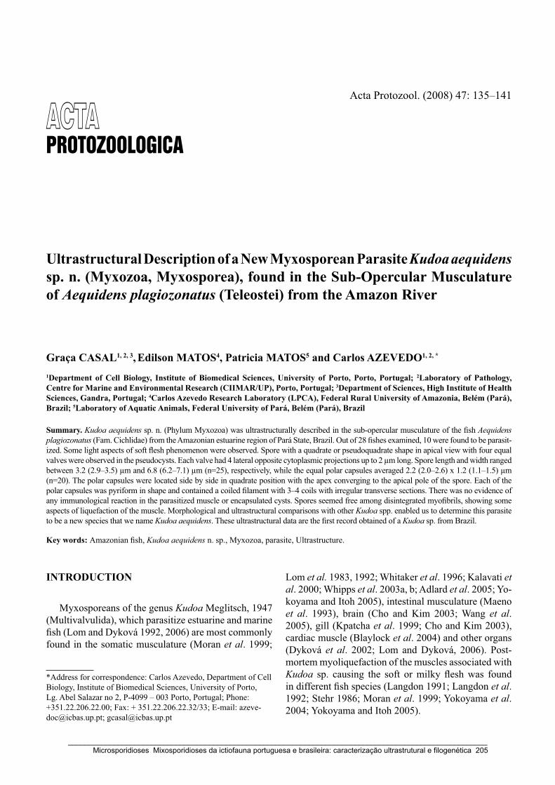
INTRODUCTION
Myxosporeans of the genus Kudoa Meglitsch, 1947 (Multivalvulida), which parasitize estuarine and marine fish (Lom and Dyková 1992, 2006) are most commonly found in the somatic musculature (Moran et al. 1999;
Lom et al. 1983, 1992; Whitaker et al. 1996; Kalavati et al. 2000; Whipps et al. 2003a, b; Adlard et al. 2005; Yo-koyama and Itoh 2005), intestinal musculature (Maeno et al. 1993), brain (Cho and Kim 2003; Wang et al. 2005), gill (Kpatcha et al. 1999; Cho and Kim 2003), cardiac muscle (Blaylock et al. 2004) and other organs (Dyková et al. 2002; Lom and Dyková, 2006). Post-mortem myoliquefaction of the muscles associated with Kudoa sp. causing the soft or milky flesh was found in different fish species (Langdon 1991; Langdon et al. 1992; Stehr 1986; Moran et al. 1999; Yokoyama et al. 2004; Yokoyama and Itoh 2005).
Ultrastructural Description of a New Myxosporean Parasite Kudoa aequidens sp. n. (Myxozoa, Myxosporea), found in the Sub-Opercular Musculature of Aequidens plagiozonatus (Teleostei) from the Amazon River
Graça CASAL1, 2, 3, Edilson MATOS4, Patricia MATOS5 and Carlos AZEVEDO1, 2, *
1Department of Cell Biology, Institute of Biomedical Sciences, University of Porto, Porto, Portugal; 2Laboratory of Pathology, Centre for Marine and Environmental Research (CIIMAR/UP), Porto, Portugal; 3Department of Sciences, High Institute of Health Sciences, Gandra, Portugal; 4Carlos Azevedo Research Laboratory (LPCA), Federal Rural University of Amazonia, Belém (Pará), Brazil; 5Laboratory of Aquatic Animals, Federal University of Pará, Belém (Pará), Brazil
Summary. Kudoa aequidens sp. n. (Phylum Myxozoa) was ultrastructurally described in the sub-opercular musculature of the fish Aequidens plagiozonatus (Fam. Cichlidae) from the Amazonian estuarine region of Pará State, Brazil. Out of 28 fishes examined, 10 were found to be parasit-ized. Some light aspects of soft flesh phenomenon were observed. Spore with a quadrate or pseudoquadrate shape in apical view with four equal valves were observed in the pseudocysts. Each valve had 4 lateral opposite cytoplasmic projections up to 2 μm long. Spore length and width ranged between 3.2 (2.9–3.5) μm and 6.8 (6.2–7.1) μm (n=25), respectively, while the equal polar capsules averaged 2.2 (2.0–2.6) x 1.2 (1.1–1.5) μm (n=20). The polar capsules were located side by side in quadrate position with the apex converging to the apical pole of the spore. Each of the polar capsules was pyriform in shape and contained a coiled filament with 3–4 coils with irregular transverse sections. There was no evidence of any immunological reaction in the parasitized muscle or encapsulated cysts. Spores seemed free among disintegrated myofibrils, showing some aspects of liquefaction of the muscle. Morphological and ultrastructural comparisons with other Kudoa spp. enabled us to determine this parasite to be a new species that we name Kudoa aequidens. These ultrastructural data are the first record obtained of a Kudoa sp. from Brazil.
Key words: Amazonian fish, Kudoa aequidens n. sp., Myxozoa, parasite, Ultrastructure.
*Address for correspondence: Carlos Azevedo, Department of Cell Biology, Institute of Biomedical Sciences, University of Porto, Lg. Abel Salazar no 2, P-4099 – 003 Porto, Portugal; Phone: +351.22.206.22.00; Fax: + 351.22.206.22.32/33; E-mail: [email protected]; [email protected]
Acta Protozool. (2008) 47: 135–141
_____________________________________________________________________________________________________ Microsporidioses Mixosporidioses da ictiofauna portuguesa e brasileira: caracterização ultrastrutural e filogenética 205

G. Casal et al.136
The genus Kudoa contained about 45 identified species (Moran et al. 1999; Swearer and Robertson 1999) and successively this number was increased by the erection of some other new species (Pampoulie et al. 1999; Swearer and Robertson 1999; Dyková et al. 2002; Yokoyama et al. 2004; Whipps et al. 2003a, b; Blaylock et al. 2004; Wang et al. 2005; Yokoyama and Itoh 2005). In a recent revision paper 63 species is the appointed number (Lom and Dyková 2006). These spe-cies are distributed in almost all geographic areas (Lom and Dyková 1992, 2006; Moran et al. 1999; Swearer and Robertson 1999). Although there is considerable information on the species of the genus Kudoa (Lom and Dyková 1992, 2006; Lom et al. 1992; Sarkar and Chaudhury 1996; Moran et al. 1999; Swearer and Robertson 1999), nothing is known about those from aquatic fauna of Brazil, and particularly those from the Amazon River (Gioia and Cordeiro 1996; Moran et al. 1999; Békési et al. 2002; Lom and Dyková 2006), where a diverse assemblage of several hundred species of fish lives. The present ultrastructural study is the first report of a Kudoa sp. from the Brazilian aquatic fauna. Light and transmission electron microscopic observa-tions suggested that this species of Kudoa differs from previously described species.
MATERIALS AND METHODS
Twenty eight specimens of the freshwater fish Aequidens pla-giozonatus Kullander, 1984 (Teleostei, Cichlidae) (Brazilian com-mon name “Cará pixuna”) were collected from the Amazonian es-tuarine region of the Peixe Boi River (01°11′S/47°18′W) near the city of Peixe Boi, State of the Pará, Brazil. The fishes ranged from 15 to 22 cm in total length, were lightly anaesthesed with MS 222 (Sandoz Laboratories) diluted in freshwater and samples of infected muscle from sub-opercular region, were taken for light and electron microscopic studies.
For light microscopy (LM) studies, free mature spores were fixed in 3% buffered glutaraldehyde and observed by a light microscope
equipped with Nomarsky interference-contrast (DIC) optics. For transmission electron microscopic (TEM) studies small fragments of infected muscle containing pseudocysts were fixed in 3% glutaralde-hyde in 0.2 M sodium cacodylate buffer (pH 7.2) at 4°C for 10 h. Af-ter washing overnight with the same buffer at 4°C and post-fixation in 2% OsO4 buffered with the same buffer for 2 h at same tempera-ture, the fragments were dehydrated through an ascending ethanol series, followed by propylene oxide and embedded in Epon. Semi-thin sections were stained with methylene blue-Azur II and observed by light microscopy and the ultrathin sections, cut with a diamond knife, contrasted with both aqueous uranyl acetate and lead citrate and were observed in a JEOL 100CXII TEM operated at 60 kV.
RESULTS
The parasitized tissue had some small spherical to ellipsoidal pseudocysts up to 125 μm containing ma-ture spores (Fig. 1). No other life cycle stages were observed. Some fibroblasts were observed surrounding the pseudocysts. Plasmodia were observed only within the parasitized muscle tissue from sub-opercular region of the fish. The parasite was easily identified as Kudoa sp. when observed in semithin sections (Fig. 1) and in free spores observed by DIC optics (Fig. 1, inset).
Histological observations revealed that the parasites developed intracellularly among the muscle fibres (Fig. 2). No inflammatory response was observed to be di-rectly related to fibres. Spores seemed liberated among disintegrated myofibrils, suggesting that liquefaction of the muscles was associated with the presence of the spores. The parasitized specimens seemed to have slower opercular movements, as they showed an evident disintegration of the myofibrils and a larger quantity of spores in the sub-opercular musculature (Fig. 2).
Kudoa aequidens sp. n. (Figs 1–11)
Type host: Aequidens plagiozonatus Kullander, 1984 (Teleostei, Perciformes, Cichlidae)
Host size: 15 to 22 cm of the total length in average.
Figs 1–6. Kudoa aequidens sp. n. light and electron micrographs. 1 – semithin section of a pseudocyst containing several spores sectioned at different levels, some of which showing the four polar capsules (arrowheads). Inset: Wet mount preparation showing a spore observed by DIC optics; 2 – ultrathin section observed at low magnification showing several spores (S) sectioned at different levels located near the muscle tissue (*); 3 – transverse section of the spore showing the four equal polar capsules (PC) located at the same level, and a transverse section of the api-cal region of the spore showing the polar filament sections (arrowheads); 4 – longitudinal (lightly oblique) section through a spore showing the polar capsules (PC) and one of the two nuclei (N) of the sporoplasmic cell. Laterally two cytoplasmic projections (arrows) of the valves (V) are present; 5 – transversal ultrathin section of a spore sectioned at the tips of the polar capsules (arrowheads); 6 – detail of a cytoplasmic projection (arrow) of the valve (V) and the polar capsule section (PC) showing the polar capsules wall (W) and polar filament sections (arrowheads).
�
_____________________________________________________________________________________________________ 206 Microsporidioses Mixosporidioses da ictiofauna portuguesa e brasileira: caracterização ultrastrutural e filogenética
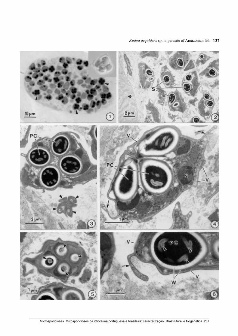
Kudoa aequidens sp. n. parasite of Amazonian fish 137
_____________________________________________________________________________________________________ Microsporidioses Mixosporidioses da ictiofauna portuguesa e brasileira: caracterização ultrastrutural e filogenética 207

G. Casal et al.138
Type locality: Amazonian estuarine region of the Peixe Boi river (01°11′S/47°18′W), near the city of Peixe Boi (State of the Pará), Brazil.
Site of infection: Spherical to ellipsoidal pseudo-cysts (up to 125 μm long) with numerous spores were found intermingled with the sub-opercular skeletal musculature. Some fibroblasts were observed surround-ing the pseudocysts.
Prevalence and intensity: 10 out of 28 fishes (35.7%) were parasitized with no observed difference in prevalence between sexes.
Type specimens: One slide of semithin sections containing mature spores of the syntypes was deposited in the International Protozoan Type Slide Collection at Smithsonian Institute Washington, DC. 20560 (USNM no 1112643
Figs 7–10. Kudoa aequidens sp. n. electron micrographs. 7 – two different aspects of the cytoplasmic projections (arrows) showing the internal organization; 8 – detail of the sporoplasmic cell (*) showing one of the two nuclei (N) and the surrounding glycogen particles (Gl). A cytoplasmic projection of the valve (arrow) is shown; 9 – the apical zone of the shell valves cut obliquely showing the suture-like connections between adjacent edges of the valves (V) (arrows) and the apical regions of the polar capsules (PC) with the apical plug-like structures (*) in continuity of the polar filaments; 10 – the four apical zones of the shell valves cut obliquely showing the suture-like connec-tions (arrows) between the adjacent edges of the valves and a longitudinal section of a polar capsule (PC) with their polar filament sections (arrowheads).
_____________________________________________________________________________________________________ 208 Microsporidioses Mixosporidioses da ictiofauna portuguesa e brasileira: caracterização ultrastrutural e filogenética
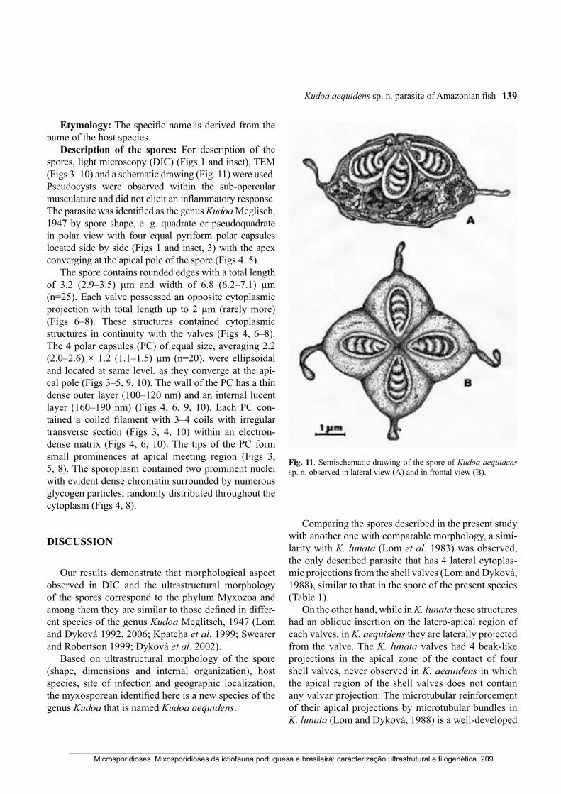
Kudoa aequidens sp. n. parasite of Amazonian fish 139
Etymology: The specific name is derived from the name of the host species.
Description of the spores: For description of the spores, light microscopy (DIC) (Figs 1 and inset), TEM (Figs 3–10) and a schematic drawing (Fig. 11) were used. Pseudocysts were observed within the sub-opercular musculature and did not elicit an inflammatory response. The parasite was identified as the genus Kudoa Meglisch, 1947 by spore shape, e. g. quadrate or pseudoquadrate in polar view with four equal pyriform polar capsules located side by side (Figs 1 and inset, 3) with the apex converging at the apical pole of the spore (Figs 4, 5).
The spore contains rounded edges with a total length of 3.2 (2.9–3.5) μm and width of 6.8 (6.2–7.1) μm (n=25). Each valve possessed an opposite cytoplasmic projection with total length up to 2 μm (rarely more) (Figs 6–8). These structures contained cytoplasmic structures in continuity with the valves (Figs 4, 6–8). The 4 polar capsules (PC) of equal size, averaging 2.2 (2.0–2.6) × 1.2 (1.1–1.5) μm (n=20), were ellipsoidal and located at same level, as they converge at the api-cal pole (Figs 3–5, 9, 10). The wall of the PC has a thin dense outer layer (100–120 nm) and an internal lucent layer (160–190 nm) (Figs 4, 6, 9, 10). Each PC con-tained a coiled filament with 3–4 coils with irregular transverse section (Figs 3, 4, 10) within an electron-dense matrix (Figs 4, 6, 10). The tips of the PC form small prominences at apical meeting region (Figs 3, 5, 8). The sporoplasm contained two prominent nuclei with evident dense chromatin surrounded by numerous glycogen particles, randomly distributed throughout the cytoplasm (Figs 4, 8).
DISCUSSION
Our results demonstrate that morphological aspect observed in DIC and the ultrastructural morphology of the spores correspond to the phylum Myxozoa and among them they are similar to those defined in differ-ent species of the genus Kudoa Meglitsch, 1947 (Lom and Dyková 1992, 2006; Kpatcha et al. 1999; Swearer and Robertson 1999; Dyková et al. 2002).
Based on ultrastructural morphology of the spore (shape, dimensions and internal organization), host species, site of infection and geographic localization, the myxosporean identified here is a new species of the genus Kudoa that is named Kudoa aequidens.
Fig. 11. Semischematic drawing of the spore of Kudoa aequidens sp. n. observed in lateral view (A) and in frontal view (B).
Comparing the spores described in the present study with another one with comparable morphology, a simi-larity with K. lunata (Lom et al. 1983) was observed, the only described parasite that has 4 lateral cytoplas-mic projections from the shell valves (Lom and Dyková, 1988), similar to that in the spore of the present species (Table 1).
On the other hand, while in K. lunata these structures had an oblique insertion on the latero-apical region of each valves, in K. aequidens they are laterally projected from the valve. The K. lunata valves had 4 beak-like projections in the apical zone of the contact of four shell valves, never observed in K. aequidens in which the apical region of the shell valves does not contain any valvar projection. The microtubular reinforcement of their apical projections by microtubular bundles in K. lunata (Lom and Dyková, 1988) is a well-developed
_____________________________________________________________________________________________________ Microsporidioses Mixosporidioses da ictiofauna portuguesa e brasileira: caracterização ultrastrutural e filogenética 209
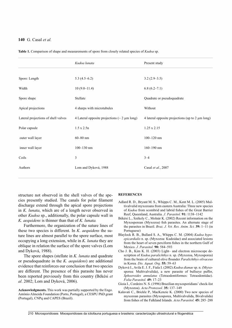
G. Casal et al.140
structure not observed in the shell valves of the spe-cies presently studied. The canals for polar filament discharge extend through the apical spore projections in K. lunata, which are of a length never observed in other Kudoa sp., additionally, the polar capsule wall in K. aequidens is thinner than that of K. lunata.
Furthermore, the organization of the suture lines of these two species is different. In K. aequidens the su-ture lines are almost parallel to the spore surface, most occupying a long extension, while in K. lunata they are oblique in relation the surface of the spore valves (Lom and Dyková, 1988).
The spore shapes (stellate in K. lunata and quadrate or pseudoquadrate in the K. aequidens) are additional evidence that reinforces our conclusion that two species are different. The presence of this parasite has never been reported previously from this country (Békési et al. 2002; Lom and Dyková, 2006).
Acknowledgments. This work was partially supported by the Engo. António Almeida Foundation (Porto, Portugal), a CESPU PhD grant (Portugal), CNPq and CAPES (Brazil).
REFERENCES
Adlard R. D., Bryant M. S., Whipps C. M., Kent M. L. (2005) Mul-tivalvulid myxozoans from eastern Australia: Three new species of Kudoa from scombrid and labrid fishes of the Great Barrier Reef, Queenland, Australia. J. Parasitol. 91: 1138–1142
Békési L., Székely C., Molnár K. (2002) Recent information on the Myxosporean (Myxozoa) fish parasites. An alternate stage of the parasites in Brazil. Braz. J. Vet. Res. Anim. Sci. 39: 1–11 (in Portuguese)
Blaylock R. B., Bullard S. A., Whipps C. M. (2004) Kudoa hypo-epicardialis n. sp. (Myxozoa: Kudoidae) and associated lesions from the heart of seven perciform fishes in the northern Gulf of Mexico. J. Parasitol. 90: 584–593
Cho J. B., Kim K. H. (2003) Light– and electron microscope de-scription of Kudoa paralichthys n. sp. (Myxozoa, Myxosporea) from the brain of cultured olive flounder Paralichthys olivaceus in Korea. Dis. Aquat. Org. 55: 59–63
Dyková I., Avila E. J. F., Fiala I. (2002) Kudoa dianae sp. n. (Myxo-sporea: Multivalvulida), a new parasite of bullseye puffer, Sphoeroides annulatus (Tetraodontiformes: Tetraodontidae). Folia Parasitol. 49: 17–23
Gioia I., Cordeiro N. S. (1996) Brazilian myxosporidians’ check-list (Myxozoa). Acta Protozool. 35: 137–149
Kalavati C., Brickle P., MacKenzie K. (2000) Two new species of myxozoan parasites (Myxosporea, Multivalvulida, Bivalvulida) from fishes of the Falkland Islands. Acta Parasitol. 45: 285–288
Table 1. Comparison of shape and measurements of spore from closely related species of Kudoa sp.
Kudoa lunata Present study
Spore: Length 5.3 (4.5–6.2) 3.2 (2.9–3.5)
Width 10 (9.0–11.4) 6.8 (6.2–7.1)
Spore shape Stellate Quadrate or pseudoquadrate
Apical projections 4 sharps with microtubules Without
Lateral projections of shell valves 4 Lateral opposite projections (~ 2 μm long) 4 lateral opposite projections (up to 2 μm long)
Polar capsule 1.5 x 2.5n 1.25 x 2.15
outer wall layer 60–80 nm 100–120 nm
inner wall layer 100–130 nm 160–190 nm
Coils 3 3–4
Authors Lom and Dyková, 1988 Casal et al., 2007
_____________________________________________________________________________________________________ 210 Microsporidioses Mixosporidioses da ictiofauna portuguesa e brasileira: caracterização ultrastrutural e filogenética
Kudoa aequidens sp. n. parasite of Amazonian fish 141
Kpatcha T. K., Diebakate C., Faye N., Toguebaye B. S. (1999) Light and electron microscopic observations on Kudoa boopsi sp. n. (Myxosporea: Kudoidae), a gill parasite of Boops boops (Pi-sces: Teleostei: Sparidae) from coasts of Senegal (West Africa). Acta Protozool. 38: 317–321
Langdon J. S. (1991) Myoliquefaction post-mortem (‘milky flesh’) due to Kudoa thyrsites (Gilchrist) (Myxosporea: Multivalvulida) in mahi mahi, Coryphaena hippurus L. J. Fish Dis. 14: 45–54
Langdon J. S., Thorne T., Fletcher W. J. (1992) Reservoir hosts and new clupeoid host records for the myoliquefactive myxosporean parasite Kudoa thyrsites (Gilchrist). J. Fish Dis. 15: 459–471
Lom J., Dyková I. (1988) Sporogenesis and spore structrure in Kudoa lunata (Myxosporea, Multivalvulida). Parasitol. Res. 74: 521–530
Lom J., Dyková I. (1992) Myxosporidia (Phylum Myxozoa). In: Protozoan Parasites of Fishes. Developments in Aquaculture and Fisheries Science, Elsevier, Amsterdam, Netherlands, 26: 159–235
Lom J., Dyková I. (2006) Myxozoan genera: definition and notes on taxonomy, life-cycle terminology and pathogenic species. Folia Parasitol. 53: 1–36
Lom J., Dyková I., Lhotáková S. (1983) Kudoa lunata n. sp. (Myxo-zoa, Myxosporea) and notes on the nature of muscular “cysts” of the genus Kudoa. Arch. Protistenk. 127: 387–397
Lom J., Rohde K., Dyková I. (1992) Studies on protozoan para-sites of Australia fishes I. New species of the genera Coccomyxa Léger et Hesse, 1907, Ortholinea Shulman, 1962 and Kudoa Meglitsch, 1947 (Myxozoa, Myxosporea). Folia Parasitol. 39: 289–306
Maeno Y., Nagasawa K., Sorimachi M. (1993) Kudoa intestinalis n. sp. (Myxosporea: Multivalvulida) from the intestinal muscula-ture of the striped mullet, Mulgil cephalus, from Japan. J. Para-sitol. 79: 190–192
Moran J. D. W., Whitaker D. J., Kent M. L. (1999) A review of the myxosporean genus Kudoa Meglitsch, 1947, and its impact on the international aquaculture industry and commercial fisheries. Aquaculture 172: 163–196
Pampoulie C., Marques A., Rosecchi E., Crivelli A. J., Bouchereau J. L. (1999) A new myxoporean parasite, Kudoa camarguensis n. sp., recorded on two goby species (Teleostei: Pisces) in the Rhône delta (Mediterranean Sea, France). J. Eukaryot. Micro-biol. 46: 304–310
Sarkar N. K., Chaudhury S. R. (1996) Kudoa cascasia sp. n. (Myxo-sporea: Kudoidae) parasitic in the mesentery of Sicamugil cas-
casia (Ham.) from Hooghly estuary of West Bengal, India. Acta Protozool. 35: 335–338
Stehr C. (1986) Sporogenesis of the myxosporean Kudoa pani-formis Kabata & Whitaker, 1981 infecting the muscle of the Pacific whiting, Merluccius productus (Ayres). J. Fish Dis. 9: 493–504
Swearer S. E., Robertson D. R. (1999) Life history, pathology, and description of Kudoa ovivora n. sp. (Myxozoa, Myxosporea): an ovarian parasite of Caribbean labroid fishes. J. Parasitol. 85: 337–353
Wang P.-C., Huang J.-P., Tsai M.-A., Cheng S.-Y., Tsai S.-S., Chen S.-D., Chen S.-P., Chui S.-H., Liaw L.-L., Chang L.-T., Chen S.-C. (2005) Systemic infection of Kudoa lutjanus n. sp. (Myxozoa: Myxosporea) in red snapper Lutjanus erythropterus from Taiwan. Dis. Aquat. Org. 67: 115–124
Whipps C. M., Adlard R. D., Bryant M. S., Kent M. L. (2003a) Two unusual myxozoans, Kudoa quadricornis n. sp. (Multival-vulida) from the muscle of goldspotted trevally (Carangoides fulvoguttatus) and Kudoa permulticapsula n. sp. (Multivalvu-lida) from the muscle of Spanish mackerel (Scomberomorus commerson) from the Great Barrier Reef, Australia. J. Parasi-tol. 89: 168–173
Whipps C. M., Adlard R. D., Bryant M. S., Lester R. J. G., Findlay V., Kent M. L. (2003b) First report of three Kudoa species from Eastern Australia: Kudoa thyrsites from Mahi mahi (Coryphae-na hippurus), Kudoa amamiensis and Kudoa minithyrsites n. sp. from sweeper (Pempheris ypsilychnus). J. Eukaryot. Microbiol. 50: 215–219
Whitaker D. J., Kent M. L., Sakanari J. A. (1996) Kudoa miniau-riculata n. sp. (Myxozoa, Myxosporea) from the musculature of bocaccio (Sebastes paucispinis) from California. J. Parasitol. 82: 312–315
Yokoyama H., Itoh N. (2005) Two multivalvulid myxozoans caus-ing postmortem myoliquefaction: Kudoa megacapsula n. sp. from red barracuda (Sphyraena pinguis) and Kudoa thyr-sites from splendid alfonso (Beryx splendens). J. Parasitol. 91: 1132–1137
Yokoyama H., Whipps C. M., Kent M. L., Mizuno K., Kawakami H. (2004) Kudoa thyrsites from Japanese flounder and Kudoa lateolabracis n. sp. from Chinese sea bass: Causative myxozo-ans of post-mortem myoliquefaction. Fish Pathol. 39: 79–85
Received on 24th September, 2007; revised version on 19th February, 2008; accepted on 22nd February, 2008
_____________________________________________________________________________________________________ Microsporidioses Mixosporidioses da ictiofauna portuguesa e brasileira: caracterização ultrastrutural e filogenética 211

_____________________________________________________________________________________________________ 212 Microsporidioses Mixosporidioses da ictiofauna portuguesa e brasileira: caracterização ultrastrutural e filogenética
Capítulo 12
FINE STRUCTURE OF CHLOROMYXUM MENTICIRRHI N. SP. (MYXOZOA)
INFECTING URINARY BLADDER OF THE MARINE TELEOST
MENTICIRRHUS AMERICANUS (SCIAENIDAE) IN SOUTHERN BRAZIL
European Journal of Protistology (2009) 45: 139-146
Graça Casal, Patrícia Garcia, Patrícia Matos, Emanuel Monteiro,
Edilson Matos & Carlos Azevedo
_____________________________________________________________________________________________________ Microsporidioses Mixosporidioses da ictiofauna portuguesa e brasileira: caracterização ultrastrutural e filogenética 213

_____________________________________________________________________________________________________ 214 Microsporidioses Mixosporidioses da ictiofauna portuguesa e brasileira: caracterização ultrastrutural e filogenética
European Journal of
PROTISTOLOGYEuropean Journal of Protistology 45 (2009) 139–146
Fine structure of Chloromyxum menticirrhi n. sp. (Myxozoa) infecting
the urinary bladder of the marine teleost Menticirrhus americanus(Sciaenidae) in Southern Brazil
Graca Casala,b,c, Patrıcia Garciad, Patrıcia Matose, Emanuel Monteirof,Edilson Matosg, Carlos Azevedoa,b,�
aDepartment of Cell Biology, Institute of Biomedical Sciences (ICBAS/UP), University of Porto, Largo A. Salazar, No. 2,
P-4099-003 Porto, PortugalbLaboratory of Pathology, Centre for Marine and Environmental Research (CIIMAR/UP), University of Porto, P-4050-123,
Porto, PortugalcLaboratory of Sciences, High Institute of Health Sciences (CESPU), P-4585-116 Gandra, PortugaldLaboratory of Diagnostic and Pathology in Aquaculture, Federal University of Santa Catarina,
88040-970 Florianopolis, SC, BrazileLaboratory of Aquatic Animals, Federal University of Para, 66075-110 Belem, PA, BrazilfDepartment of Anatomy, Institute of Biomedical Sciences (ICBAS/UP), University of Porto, P-4099-003 Porto, PortugalgCarlos Azevedo Research Laboratory, Federal Rural University of Amazonia, 66077-530 Belem, PA, Brazil
Received 29 January 2008; received in revised form 27 August 2008; accepted 28 August 2008
Abstract
A myxosporidian was found in the urinary bladder of the teleost Menticirrhus americanus Linnaeus, 1758(Sciaenidae) collected from the South Atlantic coast of Brazil. Polysporic amoeboid plasmodia containing sporoblasts,developing pansporoblasts and spores were free in the bladder lumen. The prevalence of infection was 17.64% (15/85).Unfixed spores were spherical to subspherical, on average 10.5 mm long, 9.8 mm wide and 10.1 mm thick (n ¼ 25), andfixed spores measured 10.1� 9.5� 9.7 mm. The two spore valves were of equal size and each possessed prominentsutural lines and about 41 (37–45) surface ridges aligned parallel with the suture line. These ridges gave transversesections a cog-wheel-like outline. The spores contained four pyriform polar capsules of equal size (3.20� 2.0 mm)(n ¼ 25) (fixed), each with a polar filament having 3–4 (rarely 5) coils. The binucleate sporoplasm was irregular inshape, with granular matrix and randomly distributed dense bodies. The shape and dimensions of the spore, as well asthe number, position and arrangement of the surface ridges, polar capsules and polar filament indicate that this is anew species, herein designated Chloromyxum menticirrhi. The gill, liver, gall bladder and intestine of the host showedno abnormalities.r 2008 Elsevier GmbH. All rights reserved.
Keywords: Teleost; Chloromyxum menticirrhi n. sp.; Myxosporea; Ultrastructure; Sporogenesis
ARTICLE IN PRESS
www.elsevier.de/ejop
0932-4739/$ - see front matter r 2008 Elsevier GmbH. All rights reserved.
doi:10.1016/j.ejop.2008.09.002
�Corresponding author at: Department of Cell Biology, Institute of Biomedical Sciences (ICBAS/UP), University of Porto, Largo A. Salazar,
No. 2, P-4099-003 Porto, Portugal. Tel.: +351 22 206 2200; fax: +351 22 206 2232/33.
E-mail address: [email protected] (C. Azevedo).
_____________________________________________________________________________________________________ Microsporidioses Mixosporidioses da ictiofauna portuguesa e brasileira: caracterização ultrastrutural e filogenética 215

Introduction
The Myxosporea of the phylum Myxozoa is anassemblage of more than 2180 species distributed amongsome 60 genera (Lom and Dykova 2006). They havebeen reported from different geographic areas, mainly asparasites and pathogens of fish, and are of importance infisheries and aquaculture (Lom and Dykova 2006).Among them, members of the genus Chloromyxum
Mingazzini, 1890, the fourth largest genus of Myxozoa,with 115 nominal species, are commonly coelozoic in theurinary tract and gall bladder of freshwater and marinefishes (Lom and Dykova 2006), as well as some non-fishhosts such as batrachian amphibians (Duncan et al.2004; Joseph 1905; Lom and Dykova 2006; Mutsch-mann 1999; Upton et al. 1995). The taxonomy of thegroup is difficult when relying solely on light microscopyof the spore because of the limited number of distinctcharacters for separation of species. Recent revisionof the genus makes use of the pattern of ridges on thespore surface revealed by electron microscopy, particu-larly scanning electron microscope (SEM) (Lom andDykova 1993).
Considering the high number of Brazilian fish species(about 8000) (Celere et al. 2002), the number ofdescribed parasite species is low (Gioia and Cordeiro1996). Only two myxosporean species of the genusChloromyxum, (C. leydigi Mingazzini, 1890 andC. sphyrnae Cunha and Fonseca, 1918) have beenobserved by light microscopy from the Brazilian faunaand illustrated by diagrammatic drawings (Cunha andFonseca 1918; Guimaraes 1931, Pinto 1928). One otherdescribed member of this genus from South Americawas found in the flatfish Paralichthys adspersus from thePacific coast of the Chile (Oliva et al. 1996).
Previously, only metazoan parasites have beenreported from the host Menticirrhus americanus
(Sciaenidae) (Luque and Oliva 1999). Myxosporeansbelonging to the genera Henneguya and Parvicapsula
(Landsberg 1993), Ceratomyxa (Sarkar and Pramanik1994), Myxoproteus and Zschokkella (Sarkar 1996),Sinuolinea (Sarkar 1997), Myxidium (Diamant 1998)and Kudoa (Blaylock et al. 2004; Oliva et al. 1992) havebeen described as parasites of members of the familySciaenidae.
We here describe Chloromyxum menticirrhi n. sp.from the urinary bladder of the marine teleostMenticirrhus americanus from coastal waters of South-ern Brazil from studies using light (LM), SEM andtransmission electron microscopes (TEM).
Materials and methods
The marine teleost Menticirrhus americanus Linnaeus,1758 (Sciaenidae) (Southern kingfish) (Brazilian
common name ‘‘Papa-Terra’’) was collected in the surfzone of the ‘‘Barra da Lagoa’’ Beach (271 340S, 481250W) near Florianopolis (Santa Catarina State), on thesouthern Atlantic coast of Brazil. Specimens werecollected once a month between October 2006 andSeptember 2007. Altogether, 85 fishes (10–22 cm long)were taken alive to the laboratory, where they wereanaesthetized with benzocain, killed and necropsied.Each fish was measured (total length) and its sexdetermined. No specimens showed macroscopical signsof disease.
Smears of fresh gill, liver, gall bladder, urinarybladder and intestine were microscopically examined.Smears of fresh urinary bladders, the only parasitizedorgan, containing plasmodia and free spores wereprepared for observation by LM using differentialinterference contrast (DIC) microscopy and unfixedand fixed plasmodia and free spores were measured withan ocular micrometer.
For SEM, the plasmodia were teased apart to releasespores, which were fixed at 4 1C for 24 h in 5%glutaraldehyde buffered with 0.2M sodium cacodylate(pH 7.4), washed in three changes of the same buffer,dehydrated in an ascending ethanol series, critical pointdried, coated with gold and examined in a JSM-630SEM operated at 15 kV. For TEM, small fragments ofthe parasitized tissues were fixed as for SEM, washedovernight in buffer at 4 1C, post-fixed in 2% osmiumtetroxide with the same buffer and temperature for 3 h,dehydrated in an ascending ethanol series followed bypropylene oxide, and embedded in Epon. Semithinsections were stained with methylene blue-Azure II forLM and ultrathin sections were double stained withuranyl acetate and lead citrate and observed in a JEOL100CXII TEM operated at 60 kV.
Results
During a parasitological survey it was observed thatsome specimens presented hypertrophy of the urinarybladder, which contained several masses of plasmodia.The prevalence of infection was 17.64% (15/85) in a hostpopulation where the numbers of females and maleswere about equal. Histological sections revealed that theplasmodia were on or near the surface of the innerbladder epithelium, sometimes forming several layers(Fig. 1). The largest polysporic plasmodia reachednearly 52 mm long and contained all sporogonic stagesas well as developed spores (up to 8) (Figs 2, 3, 6–8).Some free spores were observed among the plasmodia(Figs 4, 5). Plasmodia had irregular contours and thesurfaces were irregularly covered by slender cytoplasmicextensions (up to 1.3 mm long) (Figs 6–8). Numerousmitochondria (M) with dense contents were located inplasmodial cytoplasm among the developmental stages,
ARTICLE IN PRESSG. Casal et al. / European Journal of Protistology 45 (2009) 139–146140
_____________________________________________________________________________________________________ 216 Microsporidioses Mixosporidioses da ictiofauna portuguesa e brasileira: caracterização ultrastrutural e filogenética

ARTICLE IN PRESS
Figs 1–5. Light and scanning electron micrographs of developmental stages of Chloromyxum menticirrhi sp. n. infecting the urinary
bladder of the teleost Menticirrhus americanus (scale bars in mm). 1. Semithin section showing several plasmodia (arrowheads)
located free in the lumen and attaching to the urinary bladder wall (*). 2. Semithin section showing several plasmodia (arrowheads),
most of them containing spores in different phases of development (arrows). 3. Two unfixed spores (arrows) in the same plasmodium
(P) observed in DIC. 4. Two free-floating fixed spores (arrows) observed in DIC, showing polar capsules (arrowheads). 5. SEM of a
spore showing the valve ridge arrangement (arrows).
G. Casal et al. / European Journal of Protistology 45 (2009) 139–146 141
_____________________________________________________________________________________________________ Microsporidioses Mixosporidioses da ictiofauna portuguesa e brasileira: caracterização ultrastrutural e filogenética 217

more frequently in the peripheral areas (Fig. 6).Various stages of developing pansporoblasts (Pb) wererandomly distributed throughout the plasmodial cyto-plasm (Figs 7, 8).
Diagnosis
Phylum Myxozoa Grasse, 1970Class Myxosporea Butschli, 1881Order Bivalvulida Schulman, 1959Family Chloromyxidae Thelohan, 1892Genus Chloromyxum Mingazzini, 1890
Chloromyxum menticirrhi n. sp. (Figs 1–14)
Specific characters: spores spherical to subspherical,10.570.4 mm long in lateral view; 9.870.6 mm wide and10.170.6 mm thick (n ¼ 25) in apical view when unfixed
(Figs 3, 4); and 10.170.5� 9.570.5� 9.770.7 mm whenfixed (Fig. 5) (Table 1). Valves adhering together alongthe prominent longitudinal sutural lines (Figs 9–13).Two equal-sized valves without caudal projections butwith longitudinal surface ridges which appear like cog-wheel teeth around transverse sections (Figs 9–12). Eachvalve with �41 (37–45) ridges aligned parallel with thesuture line (74–90 ridges per spore) and of equalthickness 0.25 (0.21–0.30) mm (n ¼ 25) (Figs 9–13). Someridges coalesce towards the poles. Suture ridges form theouter edges of the valves (Figs 9–13). Four equal-sizedpyriform polar capsules (PC) (Figs 9–11); 3.270.4 mmlong and 2.070.3 mm wide (n ¼ 25); polar filament with3–4 (rarely 5) coils (Figs 9, 11, 14; Table 1). Sporoplasmirregular in shape with two nuclei in a granular matrixof variable density (Fig. 10). A longitudinal section ofthe spore structure is shown in the schematic drawing(Fig. 14).
ARTICLE IN PRESS
Figs 8–13. Transmission electron micrographs of developmental stages of Chloromyxum menticihrri sp. n. (scale bars in mm).
8. Plasmodium (P) showing several developmental stages: vegetative nuclei (arrows), pansporoblasts (Pb) and immature spores (*),
one of which shows the surface ridges having a cog-wheel-like appearance (**). The plasmodial periphery shows several cytoplasmic
extensions (arrowheads). Inset: Detail of a similar cytoplasmic extensions (arrowheads) boxed in this figure. 9. Transverse oblique
section of the apical region of a spore showing four transverse sections of the polar capsules (PC) located side-by-side. The valves
show the suture lines (arrows) and the ridge sections (arrowheads). In this picture it is possible to count more than 30 ridges on the
right valve. The surrounding space in contact with the valves is occupied by anastomosing filaments (*). 10. Longitudinal section of
an immature spore showing the valvogenic cells (VC), the sporoplasm (S) and longitudinal sections of two polar capsules (PC) with
the wall composed of two layers. 11. Detail of a portion of the spore valves showing the suture line (arrow) and the position of the
ridges (arrowheads) giving a cog-wheel outline, and transverse sections of two polar capsules (PC). 12. Transverse section of a
portion of the valves showing the suture line (arrow) and adjacent valve ridges (arrowheads). The space between the spore and
plasmodium is occupied by numerous anastomosing filaments (*). 13. Tangential section of the spore valve periphery showing the
position of the suture line (arrows) and neighbouring valve ridges (arrowheads). The convergence of two ridges is clearly visible
(double arrow). The surrounding space in contact with the ridges is occupied by numerous anastomosing filaments (*).
Figs 6–7. Transmission electron micrographs of developmental stages of Chloromyxum menticirrhi sp. n. (scale bars in mm).
6. Plasmodium (P) showing nuclei of several developmental stages (arrows) located among numerous mitochondria (M). The
plasmodium shows several peripheral cytoplasmic extensions (arrowheads). 7. Plasmodium (P) showing some vegetative nuclei
(arrows) and developing pansporoblasts (Pb).
G. Casal et al. / European Journal of Protistology 45 (2009) 139–146142
_____________________________________________________________________________________________________ 218 Microsporidioses Mixosporidioses da ictiofauna portuguesa e brasileira: caracterização ultrastrutural e filogenética

ARTICLE IN PRESSG. Casal et al. / European Journal of Protistology 45 (2009) 139–146 143
_____________________________________________________________________________________________________ Microsporidioses Mixosporidioses da ictiofauna portuguesa e brasileira: caracterização ultrastrutural e filogenética 219
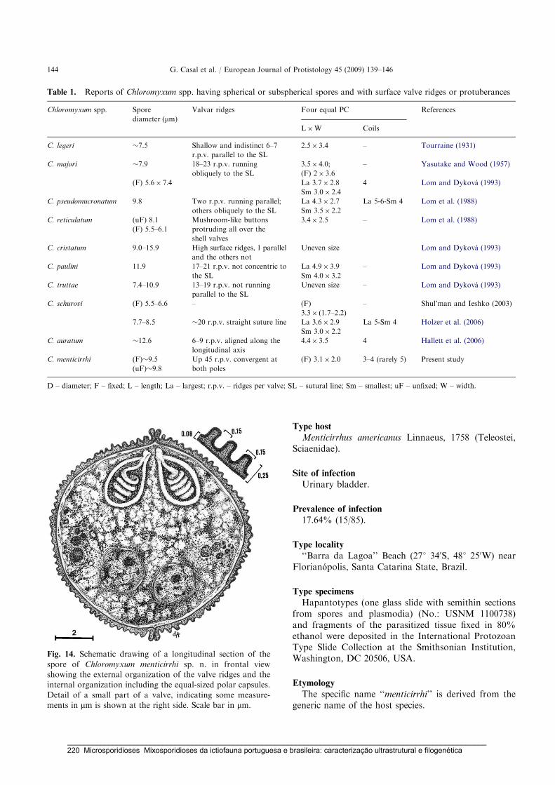
Type host
Menticirrhus americanus Linnaeus, 1758 (Teleostei,Sciaenidae).
Site of infection
Urinary bladder.
Prevalence of infection
17.64% (15/85).
Type locality
‘‘Barra da Lagoa’’ Beach (271 340S, 481 250W) nearFlorianopolis, Santa Catarina State, Brazil.
Type specimens
Hapantotypes (one glass slide with semithin sectionsfrom spores and plasmodia) (No.: USNM 1100738)and fragments of the parasitized tissue fixed in 80%ethanol were deposited in the International ProtozoanType Slide Collection at the Smithsonian Institution,Washington, DC 20506, USA.
Etymology
The specific name ‘‘menticirrhi’’ is derived from thegeneric name of the host species.
ARTICLE IN PRESS
Table 1. Reports of Chloromyxum spp. having spherical or subspherical spores and with surface valve ridges or protuberances
Chloromyxum spp. Spore
diameter (mm)
Valvar ridges Four equal PC References
L�W Coils
C. legeri �7.5 Shallow and indistinct 6–7
r.p.v. parallel to the SL
2.5� 3.4 – Tourraine (1931)
C. majori �7.9 18–23 r.p.v. running
obliquely to the SL
3.5� 4.0;
(F) 2� 3.6
– Yasutake and Wood (1957)
(F) 5.6� 7.4 La 3.7� 2.8
Sm 3.0� 2.4
4 Lom and Dykova (1993)
C. pseudomucronatum 9.8 Two r.p.v. running parallel;
others obliquely to the SL
La 4.3� 2.7
Sm 3.5� 2.2
La 5-6-Sm 4 Lom et al. (1988)
C. reticulatum (uF) 8.1 Mushroom-like buttons
protruding all over the
shell valves
3.4� 2.5 – Lom et al. (1988)
(F) 5.5–6.1
C. cristatum 9.0–15.9 High surface ridges, 1 parallel
and the others not
Uneven size Lom and Dykova (1993)
C. paulini 11.9 17–21 r.p.v. not concentric to
the SL
La 4.9� 3.9
Sm 4.0� 3.2
– Lom and Dykova (1993)
C. truttae 7.4–10.9 13–19 r.p.v. not running
parallel to the SL
Uneven size – Lom and Dykova (1993)
C. schurovi (F) 5.5–6.6 – (F)
3.3� (1.7–2.2)
– Shul’man and Ieshko (2003)
7.7–8.5 �20 r.p.v. straight suture line La 3.6� 2.9
Sm 3.0� 2.2
La 5-Sm 4 Holzer et al. (2006)
C. auratum �12.6 6–9 r.p.v. aligned along the
longitudinal axis
4.4� 3.5 4 Hallett et al. (2006)
C. menticirrhi (F)�9.5 Up 45 r.p.v. convergent at
both poles
(F) 3.1� 2.0 3–4 (rarely 5) Present study
(uF)�9.8
D – diameter; F – fixed; L – length; La – largest; r.p.v. – ridges per valve; SL – sutural line; Sm – smallest; uF – unfixed; W – width.
Fig. 14. Schematic drawing of a longitudinal section of the
spore of Chloromyxum menticirrhi sp. n. in frontal view
showing the external organization of the valve ridges and the
internal organization including the equal-sized polar capsules.
Detail of a small part of a valve, indicating some measure-
ments in mm is shown at the right side. Scale bar in mm.
G. Casal et al. / European Journal of Protistology 45 (2009) 139–146144
_____________________________________________________________________________________________________ 220 Microsporidioses Mixosporidioses da ictiofauna portuguesa e brasileira: caracterização ultrastrutural e filogenética
Discussion
The morphology of the spores described in the presentstudy places this parasite in the genus Chloromyxum
Mingazzini, 1890 according to the key for determinationof myxosporean genera published by Lom andDykova (2006). The morphology and ultrastructuralorganization of our species is similar to that of thepreviously described species of this genus (Ali 1998;Baska 1990, 1993; Hallett et al. 2006; Lom andDykova 1992, 2006; Lom et al. 1988; Molnar 1992;Mutschmann 1999; Shul’man and Ieshko 2003; Uptonet al. 1995), but the genus is a large one with about 115recognized species (Lom and Dykova 2006). The genusChloromyxum, which includes coelozoic myxosporeansthat develop in polysporic plasmodia, has previouslybeen reported in the South American aquatic fauna,with two species from Brazil and one from Chile, but nospecies of Chloromyxum has been described from fish ofthe family Sciaenidae (see Introduction). This studyprovides the first ultrastructural description of thisgenus from the Brazilian fauna as well as the first froma sciaenid fish.
Such morphological features as the surface structureof the spore wall, and particularly the ridges, whichoccur only in some species, are important characteristicsused to distinguish species of Chloromyxum (Lomand Dykova 1993). We therefore compared the mor-phology, size and ultrastructural organization of thespore valves, especially the number, organization anddistribution of the valvular ridges observed in ourspecimens, with similar features of other Chloromyxum
spp. with spherical or subspherical spores (or similarform) possessing surface ridges or protuberances,namely, C. legeri (Tourraine 1931), C. majori (Lomand Dykova 1993; Yasutake and Wood 1957),C. pseudomucronatum (Lom et al. 1988), C. reticulatum(Lom et al. 1988), C. cristatum (Lom and Dykova 1993),C. paulini (Lom and Dykova 1993), C. truttae (Lomand Dykova 1993) C. schurovi (Holzer et al. 2006;Shul’man and Ieshko 2003) and C. auratum (Hallettet al. 2006) (Table 1), and found that only four speciesshow some similarities with our results. C. majori
and C. truttae have approximately 20 ridges per valvebut the PC are of different size. Although the speciesC. auratum and C. legeri possess equal PC, the sporesof C. legeri have 6–7 shallow and indistinct ridgesper valve parallel with the suture line and C. auratum
have 6–9 ridges per valve aligned along the longi-tudinal axis. Spores observed in the present studyhave up to 90 ridges per spore, with an externalorganization that is clearly very different from the ridgeornamentations reliably described and figured for anyChloromyxum spp. previously described. These ultra-structural differences among the spores of differentChloromyxum spp., coupled with the host identity,
support our conclusion that this parasite is anew myxosporean species, which we have namedChloromyxum menticirrhi.
The developmental stages observed in the plasmodiaof C. menticirrhi are similar to those previously des-cribed in other myxozoans (Lom and Dykova 2006).Recently SSU rDNA sequence data have been used toprove that a Chloromyxum sp. has a two-host life cyclewhich involves an actinospore discharged from anoligochaete into water (Atkinson et al. 2007). Sadly,although attempts have been made to obtain furtherspecimens of C. menticirrhi, in order to determine its SSUrDNA sequence, these have not so far been successful.
Acknowledgments
This work was partially supported by the Eng. A.Almeida Foundation (Porto, Portugal), a Ph.D. grantfrom ‘‘CESPU’’ (G. Casal), ‘‘CNPq’’ and ‘‘CAPES’’ –Brazil. We would like to thank Prof. M. Martins(UFSC-Florianopolis) for use of the facilities in hisLaboratory.
References
Ali, M.A., 1998. Light and scanning electron microscopy of
Chloromyxum vanasi sp. n. (Myxozoa: Myxosporea)
infecting gall bladder of the Nile catfish Bagrus bayad
(Forskal, 1775) (Teleosti: Bagridae). Acta Protozool. 37,
57–61.
Atkinson, S.D., Hallett, S.L., Bartholomew, J.L., 2007. The
life cycle of Chloromyxum auratum (Myxozoa) from gold-
fish, Carassius auratus (L), involves an antonactinomyxon
actinospore. J. Fish Dis. 30, 149–156.
Baska, F., 1990. Chloromyxum inexpectatum n. sp. and
Sphaerospora colomani n. sp. (Myxozoa, Myxosporea),
parasites of the urinary system of the starlet, Acipenser
ruthenus. Syst. Parasitol. 16, 185–193.
Baska, F., 1993. Light and electron microscopic studies on the
development of Sphaerospora colomani Baska, 1990 and
Chloromyxum inespectatum Baska, 1990. Acta Vet. Hung.
41, 59–72.
Blaylock, R.B., Bullard, S.A., Whipps, C.M., 2004. Kudoa
hypoepicardialis n. sp. (Myxozoa: Kudoidae) and associated
lesions from the heart of seven perciform fishes in the
northern Gulf of Mexico. J. Parasitol. 90, 584–593.
Celere, E.F., Cordeiro, N., Adriano, E.S., 2002. Myxobolus
absonus sp. n. (Myxozoa: Myxosporea) parasitizing Pime-
lodus maculatus (Siluriformes: Pimelodidae), a South
American freshwater fish. Mem. Inst. Oswaldo Cruz 97,
79–80.
Cunha, A.M., Fonseca, O., 1918. About the myxosporidians
from Brazilian fishes. Braz.-Med. 32, 393 (in Portuguese).
Diamant, A., 1998. Red drum Sciaenops ocellatus (Sciaenidae),
a recent introduction to Mediterranean mariculture, is
susceptible to Myxidium leei. Aquaculture 162, 33–39.
ARTICLE IN PRESSG. Casal et al. / European Journal of Protistology 45 (2009) 139–146 145
_____________________________________________________________________________________________________ Microsporidioses Mixosporidioses da ictiofauna portuguesa e brasileira: caracterização ultrastrutural e filogenética 221

Duncan, A.E., Garner, M.M., Bartholomew, J.L., Reichard,
T.A., Nordhausen, R.W., 2004. Renal myxosporidiasis in
Asian horned frog (Megophrys nasuta). J. Zoo Wildl. Med.
35, 381–386.
Gioia, I., Cordeiro, N.S., 1996. Brazilian Myxosporidians’
check-list (Myxozoa). Acta Parasitol. 35, 137–149.
Guimaraes, J.R.A., 1931. Myxozoans of the Brazilian ictio-
fauna. Thesis. Faculty of Medicine, S. Paulo, Brazil, 1–50
(in Portuguese).
Hallett, S.L., Atkinson, S.D., Holt, R.A., Banner, C.R.,
Bartholomew, J.L., 2006. A new myxozoan from feral
goldfish (Carassius auratus). J. Parasitol. 92, 357–363.
Holzer, A.S., Sommerville, C., Wootten, R., 2006. Molecular
identity, phylogeny and life cycle of Chloromyxum schurovi
Shul’man & Ieshko 2003. Parasitol. Res. 99, 90–96.
Joseph, H., 1905. Chloromyxum protei n. sp. Zool. Anz. 29,
445–451.
Landsberg, J.H., 1993. Kidney myxosporean parasites in red
drum Sciaenops ocellatus (Sciaenidae) from Florida, USA,
with a description of Parvicapsula renalis n. sp. Dis. Aquat.
Org. 17, 9–16.
Lom, J., Dykova, I., 1992. Myxosporidia (phylum Myxozoa).
In: Lom, J., Dykova, I. (Eds.), Protozoan Parasites of
Fishes. Elsevier, Amsterdam, pp. 159–235.
Lom, J., Dykova, I., 1993. Scanning electron microscopic
revision of common species of the genus Chloromyxum
(Myxozoa: Myxosporea) infecting European freshwater
fishes. Folia Parasitol. 40, 161–174.
Lom, J., Dykova, I., 2006. Myxozoan genera: definition and
notes on taxonomy, life-cycle terminology and pathogenic
species. Folia Parasitol. 43, 1–36.
Lom, J., Dykova, I., Kepr, T., 1988. Species of the genus
Chloromyxum Mingazzini (Myxozoa: Myxosporea) infect-
ing burbot (Lota lota L.). Syst. Parasitol. 11, 231–237.
Luque, J.L., Oliva, M.E., 1999. Metazoan parasite infracom-
munities of Menticirrhus (Teleostei: Sciaenidae): an amphi-
oceanic approximation. J. Parasitol. 85, 379–381.
Molnar, K., 1992. Ceratomyxa hungarica n. sp. and Chlor-
omyxum proterorhini n. sp. (Myxozoa, Myxosporea) from
the freshwater gobi Proterorhinus marmoratus (Pallas).
Syst. Parasitol. 22, 25–31.
Mutschmann, F., 1999. A new myxozoan, Chloromyxum
careni sp. n. (Myxosporea: Chloromyxidae) from kidney
of Megophrys nasuta Schlegel, 1858 (Anura: Pelobatidae)
from Indonesia. Acta Protozool. 38, 83–86.
Oliva, M., Luque, J., Teran, L., Llican, L., 1992. Kudoa
sciaenae (Myxozoa, Multivalvulidae) cysts distribution in
the somatic muscles of Stellifer minor (Tschudi, 1884)
(Pisces, Sciaenidae). Mem. Inst. Oswaldo Cruz 87, 33–35.
Oliva, M.E., Castro, R.E., Burgos, R., 1996. Parasites of
the Flatfish Paralichthys adspersus (Steindachner, 1867)
(Pleuronectiformes) from Northern Chile. Mem. Inst.
Oswaldo Cruz 91, 301–306.
Pinto, C., 1928. Myxosporidians and other intestinal proto-
zoans observed in South America. Arch. Inst. Biol. 1,
101–126 (in Portuguese).
Sarkar, N.K., 1996. Zschokkella pseudasciaena sp. n. and
Myoproteus cujaeus sp. n. (Myxozoa: Myxosporea) from
sciaenid fish of Hooghly Estuary, West Bengal, India. Acta
Protozool. 35, 331–334.
Sarkar, N.K., 1997. Sinuolinea indica sp.n. (Myxosporea:
Sinuolineidae) parasitic in the urinary bladder of a sciaenid
fish from the Hooghly Estuary, West Bengal, India. Acta
Protozool. 36, 305–309.
Sarkar, N.K., Pramanik, A.K., 1994. Ceratomyxa daysciaenae
sp. n. (Myxozoa, Ceratomyxidae) a myxosporean parasite
in the gall bladder of a teleost from the Hooghly Estuary,
West Bengal, India. Acta Protozool. 33, 121–124.
Shul’man, B.S., Ieshko, E.P., 2003. Chloromyxum schurovi sp.
n. – a new myxosporidian species (Myxosporea: Sphaer-
osporidae) from salmonid fishes (Salmonidae). Parazitolo-
giya 37, 246–247.
Tourraine, F., 1931. Sur une nouvelle myxosporidie du genre
Chloromyxum observee chez le carpe. C. R. Acad. Sci. 192,
1125–1127.
Upton, S.J., McAllister, C.T., Trauth, S.E., 1995. A new
species of Chloromyxum (Myxozoa, Chloromyxidae) from
the gall bladder of Eurycea spp. (Caudata, Plethodontidae)
in North America. J. Wildl. Dis. 31, 394–396.
Yasutake, W.T., Wood, E.M., 1957. Some myxosporidia
found in Pacific Northwest salmonids. J. Parasitol. 43,
633–642.
ARTICLE IN PRESSG. Casal et al. / European Journal of Protistology 45 (2009) 139–146146
_____________________________________________________________________________________________________ 222 Microsporidioses Mixosporidioses da ictiofauna portuguesa e brasileira: caracterização ultrastrutural e filogenética

Capítulo 13
ULTRASTRUCTURAL AND PHYLOGENTIC DATA OF
CHLOROMYXUM RIORAJUM SP. NOV. (MYXOZOA), A PARASITE OF THE
STINGRAY RIORAJA AGASSIZII IN SOUTHERN BRAZIL
Diseases of Aquatic Organisms (2009) 85: 41-51
Carlos Azevedo, Graça Casal, Patrícia Garcia, Patrícia Matos, Leonor
Teles Grilo & Edilson Matos
_____________________________________________________________________________________________________ Microsporidioses Mixosporidioses da ictiofauna portuguesa e brasileira: caracterização ultrastrutural e filogenética 223

_____________________________________________________________________________________________________ 224 Microsporidioses Mixosporidioses da ictiofauna portuguesa e brasileira: caracterização ultrastrutural e filogenética

DISEASES OF AQUATIC ORGANISMSDis Aquat Org
Vol. 85: 41–51, 2009doi: 10.3354/dao02067
Published May 27
INTRODUCTION
The Myxosporea of the phylum Myxozoa is anassemblage of more than 2180 species distributedamong some 60 genera (Lom & Dyková 2006) with
more species being added regularly. They have beenreported from different geographic areas, mainly asparasitic and pathogenic of fish, where they are ofimportance in fisheries and aquaculture (Lom &Dyková 2006). Among them, the genus Chloromyxum
© Inter-Research 2009 · www.int-res.com*Email: [email protected]
Ultrastructural and phylogenetic data ofChloromyxum riorajum sp. nov. (Myxozoa), a parasite
of the stingray Rioraja agassizii in Southern Brazil
Carlos Azevedo1, 2,*, Graça Casal1, 2, 3, Patrícia Garcia4, Patrícia Matos5, Leonor Teles-Grilo6, Edilson Matos7
1Department of Cell Biology, Institute of Biomedical Sciences (ICBAS/UP), University of Porto, 4099-003 Porto, Portugal2Laboratory of Pathology, Centre for Marine and Environmental Research (CIIMAR/UP), University of Porto, 4050-123 Porto,
Portugal3Departmento de Ciências, Instituto Superior de Ciências da Saúde-Norte, 4585-116 Gandra, Portugal
4Laboratory of Diagnostic and Pathology in Aquaculture, Federal University of Santa Catarina, 88040-970 Florianópolis,SC, Brazil
5Laboratory of Histology of Aquatic Animals, Federal University of Pará, 66075-110 Belém, PA, Brazil6Laboratory of Molecular Genetics, Institute of Biomedical Sciences (ICBAS/UP), University of Porto, 4099-003 Porto,
Portugal7Carlos Azevedo Research Laboratory, Federal Rural University of Amazonia, 66077-530 Belém, PA, Brazil
ABSTRACT: We describe a new myxozoan parasite found infecting the gall bladder of the cartilagi-nous fish Rioraja agassizii (Rajidae) from the South Atlantic coast of Brazil. Light microscopy, scan-ning and transmission electron microscopy and phylogenetic data were used. Numerous irregularpolysporic plasmodia externally covered by numerous microvilli containing different stages of sporo-gony, including free spores, were observed in bile. Ellipsoidal spores, on average 11.41 μm long,8.48 μm wide and 7.32 μm thick, were formed by 2 equal-sized valves, each possessing 3 to 4 (rarely5) elevated ridges which projected from the basal portion of the spore, and joined along a sinuous S-shaped sutural line. The basal portion of the valves bore a bundle of 33 to 37 extended tapering cau-dal filaments attached to the basal portion of the last ridge and basal portion of the sutural edge of the2 valves. The caudal filaments, formed of material similar to the valves, were attached to the shellwall by a conical basis. The spores contained 4 equal-sized pyriform polar capsules (4.5 × 2.4 μm),located at the same level, each with a polar filament with 6 (rarely 7) coils. Binucleate sporoplasm wasirregular in shape, with a granular matrix and dense bodies randomly distributed in a light area.Based on the shape and dimensions of the spore, on the number, position and arrangements of thesurface ridges, caudal bundle of filaments, polar capsules and polar filament arrangements, as wellas phylogenetic analyses using the small subunit ribosomal DNA (SSU rDNA) sequences, we proposethe name Chloromyxum riorajum for this new myxozoan.
KEY WORDS: Cartilaginous fish · Chloromyxum riorajum sp. nov. · Parasite · Phylogeny · Ultrastructure
Resale or republication not permitted without written consent of the publisher
_____________________________________________________________________________________________________ Microsporidioses Mixosporidioses da ictiofauna portuguesa e brasileira: caracterização ultrastrutural e filogenética 225

Dis Aquat Org 85: 41–51, 2009
Mingazzini, 1890, the fourth largest genus of Myxozoawith 115 nominal species, is commonly coelozoic in theurinary tract and gall bladder of freshwater and marinefishes (Lom & Dyková 2006) and in some non-fish hostsuch as batrachian amphibians (Duncan et al. 2004,Lom & Dyková 2006). Determining the taxonomy ofthe group is difficult when relying solely on lightmicroscopy of the spore because there is only a limitednumber of distinct characters which can be used toseparate the species. Revision of the genus has shownthat ultrastructural microscopy, particularly scanningelectron microscopy (SEM), provides taxonomic datathat considers the pattern of the ridges on the sporesurface (Lom & Dyková 1993).
Considering the high number of Brazilian fish spe-cies (about 8000 species) (Cellere et al. 2002), the num-ber of described parasite species is low. Little has beenpublished on the myxosporeans, and that which hasbeen published mainly concerns the genus Chloro-myxum. From the Brazilian fauna, only 2 species (C.leydigi Mingazzini, 1890 and C. sphyrnae Cunha andFonseca, 1918) have been observed by lightmicroscopy and represented by diagrammatic draw-ings (Cunha & Fonseca 1918, Pinto 1928, Gioia &Cordeiro 1996). In South American fauna, there isanother description of this genus found in the flatfishParalichthys adspersus from the Pacific coast of theChile (Oliva et al. 1996). Recently, on the bases of themorphological and ultrastructural data, a new speciesC. menticirrhi was described parasitizing a Brazilianmarine teleost fish (Casal et al. 2009).
With respect to molecular data, there is informationon the 18S rDNA gene for only 7 Chloromyxum species(Fiala & Dyková 2004, Holzer et al. 2004, 2006, Hallettet al. 2006). An analysis of the small subunit ribosomalDNA (SSU rDNA) in phylogenetic studies shows thatthe majority of myxosporea can be divided into 2 mainclades: marine and freshwater clades. However, thereare exceptions to this division, notably regarding somespecies infecting anadromous hosts and species of thegenus Chloromyxum (Fiala 2006). The present studydescribes C. riorajum sp. nov. from the gall bladder ofthe marine stingray Rioraja agassizii from coastalwaters off Southern Brazil, making use of lightmicroscopy (LM), SEM, transmission electron micro-scopy (TEM) and phylogenetic data pertaining to the18S rDNA sequence.
MATERIALS AND METHODS
The marine stingray Rioraja agassizii (Müller &Henle, 1841) (Chondrichthyes, Rajidae) (Braziliancommon name ‘Raia-santa’) was collected during Feb-ruary and March 2008 in the surf zone of Joaquina
Beach (27° 37’ S, 48° 26’ W) located on the SouthAtlantic near the city of Florianópolis, Santa CatarinaState, Brazil. After collection, 4 stingrays (30 to 52 cmtotal length) were transported alive to the laboratory,where they were anaesthetized with MS-222 (SandozLaboratories), and necropsied. Smears of fresh gallbladder and bile, urinary bladder, gill, liver and intes-tine were examined microscopically. Smears of freshgall bladder contents, the only organ observed to beparasitized, containing free spores and plasmodiawere prepared for observation by LM using Nomarskidifferential interference contrast (DIC) optics, and freespores were measured with an ocular micrometeradapted to the photomicroscope.
Electron microscopy. For SEM, gall bladders wereteased apart to release spores. These were fixed in 5%glutaraldehyde buffered in 0.2 M sodium cacodylate(pH 7.4) at 4°C for 20 h, washed in 3 changes of thesame buffer, dehydrated in an ascending ethanolseries, critical point dried, coated with gold and exam-ined in a JSM-630 SEM operated at 15 kV. After dehy-dration some spores were observed in a DIC micro-scope. For TEM, small fragments of the parasitized gallbladder and fluid bile containing plasmodia and freespores were fixed as for the SEM procedure, washedovernight in buffer at 4°C, and post-fixed in 2%osmium tetroxide with the same buffer and at the sametemperature for 3 h, dehydrated in an ascendingethanol series followed by propylene oxide, andembedded in Epon. Semithin sections were stainedwith methylene blue-Azure II for LM, and ultrathinsections were double-stained with uranyl acetate andlead citrate and observed and photographed using aJEOL 100CXII TEM operated at 60 kV.
DNA isolation and PCR amplification. Several plas-modia and spores were preserved in 80% ethanol at4°C before genomic DNA extraction which was per-formed using a GenEluteTM Mammalian GenomicDNA Miniprep Kit (Sigma) following the manufac-turer’s instructions for animal tissue, except for incuba-tion time. The DNA was stored in 50 μl of TE buffer at–20°C until used. Initial amplification of the SSU rDNAgene was achieved using the universal eukaryoticprimers 18e (Hillis & Dixon 1991) and 18r (Whipps etal. 2003). PCR was carried out in 50 μl reactions using10 pmol of each primer, 10 nmol of each dNTP, 2.5 mMMgCl2, 5 μl 10× Taq polymerase buffer, 1.5 units TaqDNA polymerase (Invitrogen), and 5 μl of the genomicDNA. The reactions were run on a Hybaid PxE Ther-mocycler (Thermo Electron Corporation). The amplifi-cation program consisted of 95°C denaturation for3 min, followed by 35 cycles of 94°C for 45 s, 53°C for45 s and 72°C for 90 s. A final elongation step was per-formed at 72°C for 7 min. Nested PCR was done usingas template 2 μl of initial PCR: 5’-end with the primers
42
_____________________________________________________________________________________________________ 226 Microsporidioses Mixosporidioses da ictiofauna portuguesa e brasileira: caracterização ultrastrutural e filogenética

Azevedo et al.: Description of Chloromyxum riorajum sp. nov.
18e/MyxospecR, the central region of the gene withprimers MyxospecF/ChloromyxR1 and finally the 3’-end with the primers ChloromyxF1/18r (Table 1). Theamplification program consisted of 95°C denaturationfor 5 min, followed by 30 cycles of 95°C for 1 min, 52°Cfor 1 min and 72°C for 2 min. A final elongation stepwas performed at 72°C for 10 min. Then 5 μl aliquots ofPCR products were electrophoresed through a 1%agarose 1× Tris-acetate-EDTA buffer gel stained withethidium bromide.
DNA cloning and sequencing. PCR products for theSSU rDNA gene with an approximate size of 300 bp(18e/MyxospecR), 900 bp (MyxospecF/ChloromyxR1)and 600 (ChloromyxF1/18r) were obtained from theexcised band. Before cloning, the bands were puri-fied with NucleoSpin Extract II (Macherey-Nagel).DNA was cloned into a pGEM-T Easy Vector SystemII (Promega) following the manufacturer’s instruc-tions. JM109 Competent Cells with high efficiency(Promega) were transformed and then 2 positiveclones were selected using the blue–white colourscreening method. The minipreps were carried outwith a NucleoSpin Plasmid (Macherey-Nagel) ac-cording the manufacturer’s instructions. The clonedinserts were confirmed by digestion with restrictionenzyme EcoRI (Promega), and then they weresequenced in both directions with the universalsequencing primers T7 forward/SP6. Sequencing wasdone using BigDye Terminator v1.1 from the AppliedBiosytems Kit, and the sequence reactions were runon an ABI3700 DNA analyzer (Perkin-Elmer AppliedBiosystems).
Distance and phylogenetic analysis. To evaluate therelationship of Chloromyxum riorajum to other myxo-sporean species, we used 29 18SSU rDNA sequences,obtained from GenBank data: Ceratomyxa labracis(AF411472), Ceratomyxa sparusaurati (AF411471),Ceratomyxa shasta (AF001579), Chloromyxum aura-tum (AY971521), Chloromyxum legeri (AY604197),Chloromyxum leydigi (AY604199), Chloromyxum ley-digi (DQ377710), Chloromyxum cyprini (AY604198),Chloromyxum trijugum (AY954689), Chloromyxumtruttae (AJ581916), Chloromyxum sp. (AJ581917),
Enteromyxum leei (AF411334), Enteromyxum scoph-thalmi (AF411335), Henneguya ictaluri (AF195510),Henneguya salminicola (AF031411), Hoferellus gilsoni(AJ582062), Kudoa amamiensis (AF034638), Kudoacrumena (AF378347), Kudoa dianae (AF414692), Myx-idium lieberkuehni (X76639), Myxidium truttae(AF201374), Myxidium sp. (U13829), Myxobolus bibul-latus (AF378336), Myxobolus cerebralis (U96492),Myxobolus osburni (AF378338), Parvicapsula minibi-cornis (AF201375), Raabeia sp. (AF378352), Sphaero-spora oncorhynchi (AF201373), and Zschokkella mugi-lis (AF411336). The corresponding sequences andGenBank/NCBI accession number of Tetracapsuloidesbryosalmonae (U70623) and Buddenbrockia plumatel-lae (AY074915) were used as the outgroup.
Sequences were aligned as described by Azevedo etal. (2006). Alignment was made using Clustal W(Thompson et al. 1994) with MEGA 4 software (Ta-mura et al. 2007), with an opening gap penalty of 10and a gap extension penalty of 4 for both pairwise andmultiple alignments. Subsequent phylogenetic andmolecular evolutionary analyses were conducted usingMEGA 4, with the 29 rDNA sequences for myxosporid-ian species and the outgroup species selected. Dis-tance estimation was carried out using the Kimura 2-parameter model distance matrix for transitions andtransversions. For the phylogentic tree reconstructions,maximum parsimony analysis was conducted using theclose neighbour interchange heuristic option with asearch factor of 2 and random initial tree additions of2000 replicates. Bootstrap values were calculated over100 replicates.
RESULTS
Morphology of the parasite
During a parasitological survey conduced to detectparasites it was observed that some specimens of thestingray Rioraja agassizii presented hypertrophy of thegall bladder, the bile of which contained severalmasses of plasmodia and numerous free spores.
43
Primers Sequence (5’–3’) Position Used with Source
18e CTG GTT GAT CCT GCC AGT 1 MyxospecR, 18r Hillis & Dixon (1991)MyxospecF TTC TGC CCT ATC AAC TTG TTG 312 ChloromyxR1 Fiala (2006)ChloromyxF1 CTT AAA GGA ATT GAC GGA AGG 1209 18r Present studyMyxospecR CAA CAA GTT GAT AGG GCA GAA 332 18e Present studyChloromyxR1 CCT TCC GTC AAT TCC TTT AAG 1229 MyxospecF Present study18r CTA CGG AAA CCT TGT TAC G 1832 18e, ChloromyxF1 Whipps et al. (2003)
Table 1. Primer sequences and location used to amplify small subunit ribosomal DNA of Chloromyxum riorajum sp. nov.
_____________________________________________________________________________________________________ Microsporidioses Mixosporidioses da ictiofauna portuguesa e brasileira: caracterização ultrastrutural e filogenética 227

Dis Aquat Org 85: 41–51, 2009
Light microscopy
Numerous plasmodia and free spores were observedimmersed the bile (Figs. 1 to 3). Based on the morpho-logical aspects of the spores and particularities of thespore valves and attached caudal bundle of the taper-ing filamentous projections forming tails, the parasitewas identified as belonging to the genus Chloromy-xum as classified according to Lom & Dyková (2006).Our results provide a description of a new species:Phylum Myxozoa Grassé, 1970Class Myxosporea Bütschli, 1881Order Bivalvulida Schulman, 1959Family Chloromyxidae Thélohan, 1892Genus Chloromyxum Mingazzini, 1890
Chloromyxum riorajum sp. nov.
Life history stages observed: All developmentalstages in the polysporic plasmodia (up to 150 μm) andfree spores immersed in the bile (Figs. 1 to 3).
Description: Cell membrane of the plasmodia and itspseudopodia is covered by numerous microvilli (Figs. 4& 5). Spores are ellipsoidal to pyriform, 11.41 ± 0.31 μmlong in lateral view; 8.48 ± 0.45 μm wide and 5.92 ±0.54 μm thick (n = 25) in apical view (Figs. 1 & 2). Twoequal-sized valves with 3 to 4 (rarely 5) surface ridges inthe posterior half of the spore. Valves adhering togetheralong a sinuous S-like structure of the sutural line(Figs. 6 & 7). Ridges are located on the last half of thespore and run parallel to the basal portion of the suturalridges (Figs. 9 to 13). The ridges coalesce towards theapical pole of the spore (Figs. 9 to 11). Each valve consistsof a continuous layer of external and internal dense ma-terial surrounding a middle lighter area (Figs. 6 to 8). Abundle of 33 to 37 tapering caudal filamentous projec-tions or tails (12.10 ± 0.87 μm long) is attached to thebasal part of the last ridge and sutural ridge of the 2valves (Figs. 1, 2, 10, 12 & 13). There were no visiblejunctions between the tails and the wall. The tails wereformed of the same material as the valves, and had a cir-cular cross-section measuring 0.2 to 0.3 μm in diameternear the valvar insertion (Fig. 18), reducing gradually indiameter towards the end of the tail (Figs. 1, 17 & 19).Four anteriorly pointed equal-sized pyriform polar cap-sules (3.2 ± 0.4 μm long, 2.0 ± 0.3 μm wide; n = 25) werelocated all on the same level within the spore (Figs. 1 & 2);each contained 6 (rarely 7) obliquely coiled polar fila-ment (Figs. 15 & 20). Sporoplasm was irregular in shapewith 2 nuclei randomly distributed within a granular ma-trix with numerous light areas where the sporoplasmo-somes were hardly visible. The spore morphology is pre-sented in schematic drawings (Fig. 20) showing thearrangement of the valvar ridges and caudal bundle of
filaments and the ultrastructural details of the spore lon-gitudinal sections.
Type host: Rioraja agassizii (Müller & Henle, 1841)(Chondrichthyes, Rajidae).
Type locality: Joaquina Beach (27° 37’ S, 48° 26’ W)on the South Atlantic coast situated near the city of Flo-rianópolis, Santa Catarina State, Brazil.
Site of infection: Gall bladder in bile.Prevalence: 75% (3/4).Type specimens: One slide with semi-thin sections of
tissues containing spores and developmental stages ofhapantotype was deposited in the International Proto-zoan Type Slide Collection at the Smithsonian Institu-tion Washington, DC, USA, with the acquisition num-ber USNM 1122327. Another slide with semi-thinsections was deposited at the Laboratory of Pathology,Centre for Marine and Environmental Research, Uni-versity of Porto, Porto, Portugal.
Molecular analysis
The amplified sequences were assembled, and theresulting consensus DNA sequence of the partialSSU rRNA gene, which was 1807 bp in length, was de-posited in GenBank (Accession number FJ624481). Intotal, 29 SSU rDNA sequences, including those withthe highest BLAST scores, were aligned with theChloromyxum riorajum sp. nov. SSU rDNA sequence.The resulting alignment consisted of 1629 positionsafter trimming the 3’-end (642 ambiguously alignedpositions were excluded).
Based on pairwise comparisons among the SSUrDNA sequences, the maximal similarity was observedwith all Chloromyxum species: C. leydigi (97.7 and97.9%), C. auratum (85.4%), C. cyprini (85.2%), C.truttae (84.6%), C. trijugum (83.8%), C. schurovi(82.3%) and C. legeri (80.9%) (Table 2).
Maximum parsimony analysis of SSU rDNA gene se-quence places Chloromyxum riorajum sp. nov. within aclade comprising almost all Chloromyxum species, with2 exceptions: C. legeri (AY604197) and C. schurovi(AJ581917). The most closely related species is C. leydigi(AY604199, DQ377710) with 100% bootstrap support.
DISCUSSION
The morphology of the spores described in the pre-sent study, i.e. ellipsoidal spores with 2 shell valves, 4equal-sized polar capsules and a bundle of caudal fila-mentous projections, shows the characters of a parasitebelonging to the genus Chloromyxum (Lom & Noble1984, Lom & Dyková 2006). A comparison of our resultswith the morphology and ultrastructural organization
44
_____________________________________________________________________________________________________ 228 Microsporidioses Mixosporidioses da ictiofauna portuguesa e brasileira: caracterização ultrastrutural e filogenética

Azevedo et al.: Description of Chloromyxum riorajum sp. nov. 45
Figs. 1 to 8. Light and transmission electron micrographs of the myxosporean Chloromyxum riorajum sp. nov. from gall bladder ofRioraja agassizii. Fig. 1. Several unfixed mature spores observed using differential interference contrast (DIC). Fig. 2. A free un-fixed mature spore observed under DIC showing a bundle of filamentous tails (arrowheads) attached to the base of one of thespores. Fig. 3. Semithin section showing 2 plasmodia, one of which (*) contains several of the developmental stages (arrowheads);the other one contains spores (arrows). The periphery of the plasmodia shows numerous microvilli (double arrowheads). Fig. 4.Periphery of a plasmodium (*) showing pseudopodia and several microvilli (Mv). Fig. 5. Details of the microvilli (Mv). Fig. 6.Oblique section of the apical end of a spore showing the apical end of the polar capsules (*) and the sutural line (arrowheads).Fig. 7. Detail of the longitudinal section of the apical region of the S-like sutural line (arrowheads). Fig. 8. Detail of the dense
internal layer of the valve (arrowheads)
_____________________________________________________________________________________________________ Microsporidioses Mixosporidioses da ictiofauna portuguesa e brasileira: caracterização ultrastrutural e filogenética 229
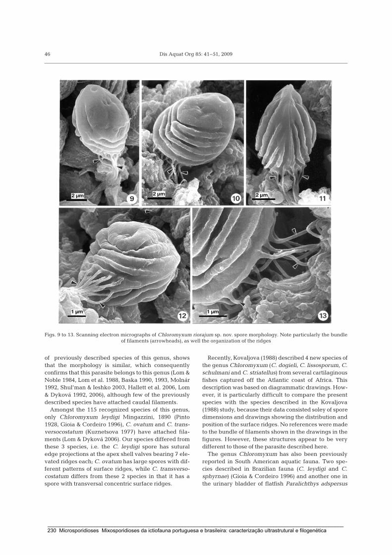
Dis Aquat Org 85: 41–51, 2009
of previously described species of this genus, showsthat the morphology is similar, which consequentlyconfirms that this parasite belongs to this genus (Lom &Noble 1984, Lom et al. 1988, Baska 1990, 1993, Molnár1992, Shul’man & Ieshko 2003, Hallett et al. 2006, Lom& Dyková 1992, 2006), although few of the previouslydescribed species have attached caudal filaments.
Amongst the 115 recognized species of this genus,only Chloromyxum leydigi Mingazzini, 1890 (Pinto1928, Gioia & Cordeiro 1996), C. ovatum and C. trans-versocostatum (Kuznetsova 1977) have attached fila-ments (Lom & Dyková 2006). Our species differed fromthese 3 species, i.e. the C. leydigi spore has suturaledge projections at the apex shell valves bearing 7 ele-vated ridges each; C. ovatum has large spores with dif-ferent patterns of surface ridges, while C. transverso-costatum differs from these 2 species in that it has aspore with transversal concentric surface ridges.
Recently, Kovaljova (1988) described 4 new species ofthe genus Chloromyxum (C. dogieli, C. lissosporum, C.schulmani and C. striatellus) from several cartilaginousfishes captured off the Atlantic coast of Africa. Thisdescription was based on diagrammatic drawings. How-ever, it is particularly difficult to compare the presentspecies with the species described in the Kovaljova(1988) study, because their data consisted soley of sporedimensions and drawings showing the distribution andposition of the surface ridges. No references were madeto the bundle of filaments shown in the drawings in thefigures. However, these structures appear to be verydifferent to those of the parasite described here.
The genus Chloromyxum has also been previouslyreported in South American aquatic fauna. Two spe-cies described in Brazilian fauna (C. leydigi and C.sphyrnae) (Gioia & Cordeiro 1996) and another one inthe urinary bladder of flatfish Paralichthys adspersus
46
Figs. 9 to 13. Scanning electron micrographs of Chloromyxum riorajum sp. nov. spore morphology. Note particularly the bundleof filaments (arrowheads), as well the organization of the ridges
_____________________________________________________________________________________________________ 230 Microsporidioses Mixosporidioses da ictiofauna portuguesa e brasileira: caracterização ultrastrutural e filogenética

Azevedo et al.: Description of Chloromyxum riorajum sp. nov.
from the Pacific coast of the Chile (Oliva et al. 1996)were described by use of LM only and representedby diagrammatic drawings. Recently, C. menticirrhispores, which do not have external filaments attachedto the wall, were described on the basis of SEM andTEM studies (Casal et al. 2009).
Morphological aspects, such as the surface structureof the spore, the different patterns of spore ridges andthe caudal bundle of 33 to 37 filaments (number never
referred to in other species) which is attached to themore basal ridge and suture line of the Chloromyxumspore wall, which differentiate only in some species,are important characteristics which can be used todistinguish Chloromyxum species (Lom & Dyková1993, 2006).
The external organization of the present species isclearly very different from that of all other Chloro-myxum spp. previously discussed, including those for
47
Figs. 14 to 19. Transmission electron micrographs of the myxosporean Chloromyxum riorajum sp. nov. from the gall bladder of Ri-oraja agassizii. Fig. 14. Transverse section at the apical region of the 4 polar capsules. Fig. 15. Detail of a transverse section of apolar capsule showing different sections of a polar filament (arrowheads). Fig. 16. Longitudinal section of the apical zone of a po-lar capsule. Fig. 17. Transverse section of a bundle of 37 filaments (arrowheads). Fig. 18. Transverse sections showing details ofseveral filaments (F). Fig. 19. Longitudinal and oblique sections of several filaments (F), one of which is attached to the valve (V)
_____________________________________________________________________________________________________ Microsporidioses Mixosporidioses da ictiofauna portuguesa e brasileira: caracterização ultrastrutural e filogenética 231

Dis Aquat Org 85: 41–51, 2009
which a bundle of filamentous projections and ridgeornamentations on the spore valves have beenreported. A comparison of the spore morphology of allChloromyxum spp. which have tailed spores showedthat there are similarities between C. riorajum and C.leydigi. However, C. riorajum differs from C. leydigi inthat the former have larger spores and polar capsules,
as well as different patterns of surface ridges. More-over, the bundle of filamentous tails of C. riorajum dif-fers from that of C. leydigi, because it has a large num-ber of filaments and is longer.
All morphological and ultrastructural aspects areuseful for the description of a new species despite thefact that of some them such as caudal appendages are
48
Fig. 20. Spore of Chloromyxum riorajum sp. nov. from the gall bladder of Rioraja agassizii. (A) Morphological aspect of the sporeas observed under differential interference contrast (DIC), showing the basal bundle of filamentous tails attached to the valves.(B) Longitudinal section (frontal view) with special emphasis on the polar capsules and basal bundle of the filamentous tails
C. C. C. C. C. C. C. C. C. M. Myxidium S.riorajum leydigi 1 leydigi 2 auratum cyprini truttae trijugum schurovi legeri truttae sp. oncorhynchi
C. riorajum (FJ624481) 97.9 97.7 85.4 85.2 84.6 83.8 82.3 80.9 82.8 81.9 81.7C. leydigi 1 (DQ377710) 0.021 99.4 86.0 85.7 85.2 84.3 82.6 81.2 83.3 82.5 81.9C. leydigi 2 (AY604199) 0.023 0.004 85.9 85.6 85.2 84.3 82.6 81.1 83.3 82.5 81.9C. auratum (AY971521) 0.146 0.140 0.141 99.8 97.7 91.8 91.8 88.4 91.3 91.3 92.3C. cyprini (AY604198) 0.148 0.143 0.143 0.002 97.4 91.6 91.6 88.1 90.9 90.9 92.0C. truttae (AJ581916) 0.154 0.148 0.148 0.023 0.026 92.1 91.6 88.6 90.9 90.9 92.0C. trijugum (AY954689) 0.162 0.157 0.157 0.082 0.084 0.079 90.5 86.2 95.5 95.9 89.9C. schurovi (AJ581917) 0.177 0.174 0.174 0.082 0.084 0.084 0.105 87.9 88.5 88.5 92.3C. legeri (AY604197) 0.191 0.188 0.189 0.116 0.119 0.114 0.138 0.121 85.8 84.8 89.9M. truttae (AF201374) 0.172 0.167 0.167 0.087 0.089 0.089 0.045 0.115 0.142 97.7 87.6Myxidium sp. (U13829) 0.181 0.175 0.175 0.087 0.089 0.089 0.041 0.115 0.152 0.023 88.1S. oncorhynchi (AF201373) 0.183 0.181 0.181 0.077 0.080 0.080 0.101 0.077 0.101 0.124 0.119
Table 2. Comparison of some small subunit ribosomal (SSU) rDNA sequences: percentage of identity (above diagonal) and pairwise distance(below diagonal) obtained by Kimura 2-parameter analysis. C.: Chloromyxum; M.: Myxidium; S.: Sphaerospora
_____________________________________________________________________________________________________ 232 Microsporidioses Mixosporidioses da ictiofauna portuguesa e brasileira: caracterização ultrastrutural e filogenética

Azevedo et al.: Description of Chloromyxum riorajum sp. nov.
not a phylogenetic character. Unfortunately, with theexception of Chloromyxum leydigi, there is no SSUrDNA information for the other Chloromyxum specieswith caudal appendages. Apparently, the habitat, thehost and the location of infection are the characteristicsthat have been used for species classification.
A BLAST search using SSU rDNA sequence datafound only 7 Chloromyxum spp. available in GenBank:All species, except C. leydigi (AY604199) isolated fromthe gall bladder of Torpedo marmorata caught in theMediterranean Sea (Fiala & Dyková 2004) and C. leydigi(DQ377710) from the gall bladder of Centroscymnuscoelolepis caught in the North Atlantic (Fiala 2006),infect freshwater fishes. C. cyprini (AY604198) and C.
legeri (AY604197) were found in the gall bladder ofHypophthalmichthys molitrix and Cyprinus carpio,respectively, from the Czech Republic (Fiala &Dyková 2004). C. auratum (AY971521) and C. trijugum(AY954689) were found in the gall bladder of Carassiusauratus and Pomoxis nigromaculatus, respectively,from Oregon, USA (Hallett et al. 2006). C. truttae(AJ581916) was found in the gall bladder epithelium andC. schurovi (AJ581917) in kidney tubules of Salmo salarin Scotland (Holzer et al. 2004). We obtained an almostcomplete SSU rRNA gene sequence with 1807 bp.
Our analysis of the phylogenetic relationship formaximum parsimony is in concordance with previouscladograms (Fiala & Dyková 2004, Fiala 2006, Holzer et
49
Myxobolus cerebralis (U96492)
Myxobolus osburni (AF378338)
Henneguya ictaluri (AF195510)
Myxobolus bibullatus (AF378336)
Henneguya salminicola (AF031411)
Chloromyxum schurovi (AJ581917)
Hoferellus gilsoni (AJ582062)
Sphaerospora oncorhynchi (AF201373)
Myxidium lieberkuehni (X76639)
Chloromyxum legeri (AY604197)
Myxidium sp. (U13829)
Myxidium truttae (AF201374)
Chloromyxum trijugum (AY954689)
Raabeia sp. (AF378352)
Chloromyxum cyprini (AY604198)
Chloromyxum auratum (AY971521)
Chloromyxum truttae (AJ581916)
Chloromyxum leydigi (DQ377710)
Chloromyxum leydigi (AY604199)
Chloromyxum riorajum (FJ624481)
Zschokkella mugilis (AF411336)
Ceratomyxa shasta (AF001579)
Parvicapsula minibicornis (AF201375)
Enteromyxum leei (AF411334)
Enteromyxum scophthalmi (AF411335)
Kudoa dianae (AF414692)
Kudoa crumena (AF378347)
Kudoa amamiensis (AF034638)
Ceratomyxa labracis (AF411472)
Ceratomyxa sparusaurati (AF411471)
Tetracapsuloides bryosalmonae (U70623)
Buddenbrockia plumatellae (AY074915)
100
89100
100
80
100
100
100
98
97
97
94
72
32
52
86
72
71
67
56
38
30
48
37
25
22
63
62
100
20Fig. 21. Maximum parsimony tree of small subunit ribosomal (SSU) rDNA sequences of Chloromyxum riorajum sp. nov. and otherselected myxosporean species. Numbers on the branches are bootstrap confidence levels on 100 replicates. GenBank accessionnumbers in parentheses after the species names; scale is given under the tree. C. riorajum and C. leydigi are placed in the basal
clade with several freshwater myxosporean species (shaded box)
_____________________________________________________________________________________________________ Microsporidioses Mixosporidioses da ictiofauna portuguesa e brasileira: caracterização ultrastrutural e filogenética 233

Dis Aquat Org 85: 41–51, 2009
al. 2006). The bootstrap for the 2 Chloromyxum speciesboth found in the gall gladder of cartilaginous fishes,C. leydigi and Chloromyxum sp. described here, is100% supported, and the pairwise sequence analysespresented 97.7 to 97.9% similarity (Table 2).
Most genera present in the myxosporean SSU rDNAtree are poly/paraphyletic (Kent et al. 2001). Presently,8 sequences of Chloromyxum species are known, andthe data suggest that they are paraphyletic groups.Monophyly was only observed in some freshwaterChloromyxum species, C. auratum, C. cyprini, and C.truttae, and were supported by a bootstrap value of94%. Previously phylogenetic analysis clearly shows adivision into 2 clades, namely freshwater and marinespecies (Kent et al. 2001). One possible justification forthis is the presence of numerous insertions in theV7 region of the SSU rDNA of all freshwater myxo-sporeans which have longer sequences than marinespecies. The exception are C. leydigi (Fiala & Dyková2004, Holzer et al. 2006) and the species we describe,C. riorajum sp. nov. These 2 species have marine carti-laginous fishes as hosts and do not have insertions nearthe 3’-end of the SSU rDNA gene, but they are clus-tered with freshwater myxosporeans. For this reason,they are basal species in the freshwater myxosporeanclade (Fiala & Dyková 2004).
Additional parsimony analyses of SSU rDNA genesequences support a close relationship between thedifferent Chloromyxum species, as well as a very goodbootstrap (100%) for the clade to which C. leygidi andC. riorajum belong (Fig. 21). In conclusion, molecularphylogenetic analysis reinforced by morphological andultrastructural data and specificity of the host suggestthat the parasite from Rioraja agassizii is a new spe-cies, named Chloromyxum riorajum sp. nov.
Acknowledgements. This work was partially supported bythe Engº. A. Almeida Foundation (Porto, Portugal), PhD grantfrom ‘CESPU’ (to G.C.), ‘CNPq’ and ‘CAPES’-Brazil. Wethank Prof. M. Martins (UFSC-Florianópolis) for use of thefacilities in his laboratory and the technical assistance of G.Ribeiro MSc (NEMAR — Florianópolis-Brazil) and J. Carval-heiro (ICBAS/UP). This work complies with the current lawsof the countries in which it was performed. The helpful com-ments and suggestions of the anonymous reviewers inreviewing this manuscript are greatly appreciated.
LITERATURE CITED
Azevedo C, Balseiro P, Casal G, Gestal C and others (2006)Ultrastructural and molecular characterization of Haplo-sporidium montforti sp. nov., parasite of the Europeanabalone Haliotis tuberculata. J Invertebr Pathol 92:23–32
Baska F (1990) Chloromyxum inexpectatum sp. nov. andSphaerospora colomani sp. nov. (Myxozoa, Myxosporea),parasites of the urinary system of the starlet, Acipenserruthenus. Syst Parasitol 16:185–193
Baska F (1993) Light and electron microscopic studies on thedevelopment of Sphaerospora colomani Baska, 1990 andChloromyxum inexpectatum Baska, 1990. Acta Vet Hung41:59–72
Casal G, Garcia P, Matos P, Monteiro E, Matos P, Azevedo C(2009) Fine structure of Chloromyxum menticirrhi sp. nov.(Myxozoa) infecting the urinary bladder of the marineteleost Menticirrhus americanus (Sciaenidae) in southernBrazil. Eur J Protistol 45:139–146
Cellere EF, Cordeiro N, Adriano ES (2002) Myxobolusabsonus sp. n. (Myxozoa: Myxosporea) parasitizing Pime-lodus maculatus (Siluriformes: Pimelodidae), a SouthAmerican freshwater fish. Mem Inst Oswaldo Cruz 97:79–80
Cunha AM, Fonseca O (1918) Sobre os mixosporídios dospeixes brasileiros. Bras Med 32:393
Duncan AE, Garner MM, Bartholomew JL, Reichard TA,Nordhausen RW (2004) Renal myxosporidiasis in Asianhorned frog (Megophrys nasuta). J Zoo Wildl Med35:381–386
Fiala I (2006) The phylogeny of Myxosporea (Myxozoa) basedon small subunit ribosomal RNA gene analysis. Int JParasitol 36:1521–1534
Fiala I, Dyková I (2004) The phylogeny of marine and fresh-water species of the genus Chloromyxum Mingazzini,1890 (Myxosporea: Bivalvulida) based on small subunitribosomal RNA gene sequences. Folia Parasitol 51:211–214
Gioia I, Cordeiro NS (1996) Brazilian Myxosporidians check-list (Myxozoa). Acta Parasitol 35:137–149
Hallett SL, Atkinson SD, Holt RA, Banner CR, BartholomewJL (2006) A new myxozoan from feral goldfish (Carassiusauratus). J Parasitol 92:357–363
Hillis DM, Dixon MT (1991) Ribosomal DNA: molecular evo-lution and phylogenetic inference. Q Rev Biol 66:411–453
Holzer AS, Sommerville C, Wooden R (2004) Molecular rela-tionships and phylogeny in a community of myxosporeansand actinosporeans based on their 18S rDNA sequences.Int J Parasitol 34:1099–1111
Holzer AS, Sommerville C, Wooden R (2006) Molecular iden-tity, phylogeny and life cycle of Chloromyxum schuroviShul’man and Ieshko, 2003. Parasitol Res 99:90–96
Kent ML, Andree KB, Bartholomew JL, El-Matbouli M andothers (2001) Recent advances in our knowledge of theMyxozoa. J Eukaryot Microbiol 48:395–413
Kovaljova AA (1988) Myxoporidia of the genus Chloromyxum(Cnidospora, Myxosporea) of cartilaginous fishes from theAtlantic coast of Africa. Parazitologiya 22:384–388 (inRussian with English summary)
Kuznetsova IG (1977) Myxosporidians of Chondrostei fromthe Patagonian shelf. Parazitologiya 11:74–77
Lom J, Dyková I (1992) Myxosporidia (Phylum Myxozoa). In:Lom J, Dyková I (eds) Protozoan parasites of fishes.Elsevier, Amsterdam
Lom J, Dyková I (1993) Scanning electron microscopicrevision of common species of the genus Chloromyxum(Myxozoa: Myxosporea) infecting European freshwaterfishes. Folia Parasitol 40:161–174
Lom J, Dyková I (2006) Myxozoan genera: definition andnotes on taxonomy, life-cycle terminology and pathogenicspecies. Folia Parasitol 43:1–36
Lom J, Noble ER (1984) Revised classification of the classMyxosporea Bütschli, 1881. Folia Parasitol 31:193–205
Lom J, Dyková I, Kepr T (1988) Species of the genusChloromyxum Mingazzini (Myxozoa: Myxosporea) infect-ing burbot (Lota lota L.). Syst Parasitol 11:231–237
50
�
��
�
�
�
��
��
��
�
��
��
��
��
��
_____________________________________________________________________________________________________ 234 Microsporidioses Mixosporidioses da ictiofauna portuguesa e brasileira: caracterização ultrastrutural e filogenética

Azevedo et al.: Description of Chloromyxum riorajum sp. nov.
Molnár K (1992) Ceratomyxa hungarica sp. nov. and Chloro-myxum proterorhini sp. nov. (Myxozoa, Myxosporea) fromthe freshwater gobi Proterorhinus marmoratus (Pallas).Syst Parasitol 22:25–31
Oliva ME, Castro RE, Burgos R (1996) Parasites of the flatfishParalichthys adspersus (Steindachner, 1867) (Pleuronecti-formes) from Northern Chile. Mem Inst Oswaldo Cruz 91:301–306
Pinto C (1928) Mixosporídeos e outros protozoários intestinaisobservados na América do Sul. Arch Inst Biol 1: 101–126
Shul’man BS, Ieshko EP (2003) Chloromyxum schurovi sp. n. —a new myxosporidian species (Myxosporea: Sphaero-sporidae) from salmonid fishes (Salmonidae). Parazitolo-giya 37:246–247
Tamura K, Dudley J, Nei M, Kumar S (2007) MEGA4: Molec-ular evolutionary genetics analysis (MEGA) software ver-sion 4.0. Mol Biol Evol 24:1596–1599
Thompson JD, Higgins DG, Gilson TJ (1994) Clustal W:improving the sensitivity of progressive multiple sequencealignment through sequence weighting, position-specificgap penalties and weight matrix choice. Nucleic Acids Res22:4673–4680
Whipps CM, Adlard RD, Bryant MS, Lester RJG, Findlay V,Kent ML (2003) First report of three Kudoa species fromEastern Australia: Kudoa thyrsites from Mahi mahi (Cory-phaena hippurus), Kudoa amamiensis and Kudoa mini-thyrsites sp. nov. from sweeper (Pempheris ypsilychnus).J Eukaryot Microbiol 50:215–219
51
Editorial responsibility: Sven Klimpel,Düsseldorf, Germany
Submitted: January 15, 2009; Accepted: April 7, 2009Proofs received from author(s): May 20, 2009
�
�
�
��
��
�
_____________________________________________________________________________________________________ Microsporidioses Mixosporidioses da ictiofauna portuguesa e brasileira: caracterização ultrastrutural e filogenética 235

_____________________________________________________________________________________________________ 236 Microsporidioses Mixosporidioses da ictiofauna portuguesa e brasileira: caracterização ultrastrutural e filogenética

PARTE IV
CONSIDERAÇÕES GERAIS
E
CONCLUSÕES FINAIS


Considerações gerais e Conclusões finais
Capítulo 14
14.1. Considerações Gerais
A classificação dos microparasitas tem sido, muitas vezes, baseada unicamente na
descrição da morfologia do esporo, principalmente no grupo dos mixosporídios. Vários
estudos indicam não ser este o tipo de abordagem mais correcta, dado que dentro da
mesma espécie podem existir variações morfológicas para as diferentes idades dos
parasitas. Também a variabilidade do hospedeiro deve ser tida em conta. Por outro lado,
é igualmente notório, que espécies biologicamente diferentes podem ser muito
semelhantes em morfologia. Na grande maioria das espécies de mixosporídios, para
além do esporo, foram caracterizados poucos aspectos inerentes ao ciclo de vida. Já os
microsporídios, dificilmente, são classificados apenas com base na morfologia e
ultrastrutura do esporo.
A especificidade do hospedeiro tem sido referida como um factor importante, por vezes
subestimado em taxonomia, havendo mesmo quem refira que a determinação do
hospedeiro definitivo (invertebrado) dos mixosporídios é importante para a correcta
classificação do taxon. Pensa-se que a análise de modelos de evolução, baseada no
hospedeiro definitivo, pode reflectir modelos de evolução mais correctos,
comparativamente aos utilizados em hospedeiros intermediários (peixes). Infelizmente,
para que se possa comprovar esta teoria, necessitam de ser determinados muitos dos
ciclos de vida dos Myxozoa.
Na última década, paralelamente às observações microscópicas, têm sido feitos esforços
para caracterizar o grupo dos microsporídios e mixosporídios através de dados
fornecidos pela sequenciação de genes conservados, tais como o SSU e LSU rDNA. Está
provado que as análises moleculares em muito têm contribuído para o conhecimento
actual destes grupos de parasitas. Muitas das análises filogenéticas, baseadas nos
caracteres moleculares diferem substancialmente das classificações morfológicas ao
nível dos géneros, tendo estes sido sofrivelmente descritos, em muitos casos, através de
desenhos esquemáticos.
Em 2003, Tauz e colaboradores chegaram mesmo a propor o abandono das
classificações morfológicas e esquemáticas, em favor de uma taxonomia baseada
exclusivamente em sequências de DNA. Efectivamente, a aposta no crescente aumento
de informação genética muito tem contribuído para a identificação do taxon ao nível do
género e espécie e, consequentemente, para propor pistas, de modo a que possamos
_____________________________________________________________________________________________________ Microsporidioses Mixosporidioses da ictiofauna portuguesa e brasileira: caracterização ultrastrutural e filogenética 239
Considerações gerais e Conclusões finais
compreender a biologia destes parasitas e quais os seus caracteres, que estão na base
da divergência filogenética.
Na nossa opinião, a classificação de qualquer grupo de organismos não deve ser
baseada numa única característica, mas antes tendo em consideração a combinação de
vários factores, tais como o habitat, especificidade do hospedeiro, local de infecção,
interacção com as células hospedeiras, características morfológicas e detalhes
ultrastruturais do ciclo de vida do parasita, bem como a análise de sequências
moleculares e, consequentemente, as inferências filogenéticas que se possam obter.
Durante a execução deste trabalho, foram diagnosticados, em ambas as faunas e
habitats, vários parasitas em diversos tecidos e órgãos, totalizando 13 microsporídios
(anexo 1) e 21 mixosporídios (anexo 2). Até à presente data, a análise dos resultados e
discussão dos mesmos permitiu redigir 12 artigos científicos (10 publicados, 1 em
revisão, 1 submetido) em revistas indexadas de divulgação internacional, que são
apresentados, nesta tese, sob a forma de capítulos.
Assim, na Parte II descrevemos, com base na ultrastrutura dos diferentes estádios do
ciclo de vida do parasita, 1 novo género, Potaspora, e 4 novas espécies de
microsporídios, todas ocorrendo na fauna brasileira: Potaspora morhaphis, Loma psittaca,
Microsporidium rondoni e Spraguea gastrophysus. Geralmente, neste grupo de parasitas,
dados baseados unicamente em aspectos morfológicos ou ultrastruturais da
esporogénese tardia não facilitam a sua classificação. Nesse sentido, procurámos fazer,
paralelamente, uma análise filogenética com os genes para os SSU e LSU rRNA.
Em relação às mixosporidioses diagnosticadas (Parte III), 7 novas espécies,
Chloromyxum menticirrhi, Chloromyxum riorajum, Myxobolus maculatus, Myxobolus
metynnis, Henneguya friderici, Henneguya rondoni e Kudoa aequidens, todas ocorrendo
na fauna aquática brasileira, foram descritas morfológica e ultrastruturalmente. Fez-se
igualmente uma caracterização ultrastrutural do mixosporídio Ceratomyxa tenuispora,
parasita do peixe-espada capturado na costa da Ilha da Madeira. Apenas para a espécie
C. riorajum foram realizadas análises moleculares e filogenéticas.
Alguns dos resultados obtidos no decurso da realização desta tese (anexos 1 & 2), não
foram apresentados e discutidos em nenhum dos capítulos, por motivos variados. Entre
eles, incluem-se a existência de dados parciais relativos aos aspectos ultrastruturais do
ciclo de vida e/ou a ausência de informação molecular, inviabilizando a classificação ao
nível da espécie, no caso de algumas microsporidioses. Não obstante, entendeu-se que
seria pertinente incluir neste capítulo, dedicado às considerações gerais e conclusões,
_____________________________________________________________________________________________________ 240 Microsporidioses Mixosporidioses da ictiofauna portuguesa e brasileira: caracterização ultrastrutural e filogenética

Considerações gerais e Conclusões finais
todos os dados moleculares e filogenéticos obtidos (anexos 3, 4 & 5), de modo a permitir
que a discussão da mesma seja feita de uma forma mais ampla e conclusiva.
14.2. Conclusões finais
- De acordo com referências prévias na literatura, os peixes são organismos susceptíveis
de serem parasitados por vários organismos. Nesta tese, em algumas das espécies
analisadas - Gymnorhamphichthis rondomi (Ituí transparente), Colomesus psittacus
(Baiacú), Trachinotus coralinus (Pampo), Aequidens plagiozonatus (Cará pixuna),
Trisopterus luscus (Faneca) e Gaidropsarus vulgaris (Lulão) - foram observados,
simultaneamente, microsporídios e mixosporídios, muitas vezes no mesmo tecido/órgão.
- Nesta tese foram, pela primeira vez, obtidos dados referentes à sequenciação de genes
conservados para os SSU e LSU rRNAs, nomeadamente de vários microsporídios
provenientes da ictiofauna capturada em águas continentais portuguesas e brasileiras.
No futuro, a sequenciação do rDNA para mais microsporídios, bem como os genes de
proteínas, irá permitir investigar se a área geográfica é ou não um factor determinante
nas relações filogenéticas entre os organismos pertencentes ao mesmo taxon (p. e.
género, família), à semelhança do que acontece com os mixosporídios (Fiala 2006). Por
outro lado, a sequenciação de genes conservados poderá ajudar a determinar se existem
hospedeiros definitivos em peixes, tal como sucede com os mixosporídios.
- Para muitas das descrições de novos taxa (espécies, géneros) ou reclassificações de
espécies já existentes, aparentemente parece bastar uma análise dos dados obtidos
através da microscopia de luz e da sequenciação de genes ribossomais. Pelo que se
pode constatar, durante a realização desta tese, este tipo de abordagem nem sempre foi
possível, nomeadamente para o grupo dos microsporídios. Regra geral, este grupo de
parasitas carece de uma conveniente caracterização ultrastrutural dos aspectos inerentes
às fases merogónicas, esporogónicas, organização estrutural do xenoma, para que
possam ser devidamente classificados ao nível da família, género e espécie.
- Em todos os cladogramas elaborados para os microsporídios de peixes verificou-se um
baixo “bootstrap” da espécie L. acerinae, juntamente com a espécie recém criada L.
psittaca (FJ843104), com as restantes espécies do grupo 1. A percentagem de identidade
destas 2 espécies é maior quando comparada com a das espécies do género Glugea.
Infelizmente, as observações ultrastruturais efectuadas em L. psittaca não evidenciaram
caracteres morfológicos significativos que permitissem diferenciar estas duas espécies
dos géneros Loma e Glugea e, assim, caracterizar um novo género, de acordo com a
sugestão proposta por Lom e Nilsen (2003).
_____________________________________________________________________________________________________ Microsporidioses Mixosporidioses da ictiofauna portuguesa e brasileira: caracterização ultrastrutural e filogenética 241

Considerações gerais e Conclusões finais
- Sete das sequências referentes ao gene para o SSU rRNA, obtidas no decurso da
execução dos trabalhos desta tese, dizem respeito a microsporídios posicionados no
grupo 4 das árvores filogenéticas: Potaspora morhaphis (EU534408), Potaspora 1 sp.
(hospedeiro: Cará pixuna), Spraguea gastrophysus (GQ868443), Microsporidium rondoni
(FJ843105), Microgemma 1 sp. (hospedeiro: Pampo), Tetramicra 1 sp. (Faneca) e
Tetramicra 2 sp. (Lulão). Os dados morfológicos e ultrastruturais para este grupo de
parasitas sequenciados demonstram haver várias características genéricas comuns,
nomeadamente nenhum possuir o núcleo em diplocário e não diferenciar vacúolos
parasitóforos. As espécies deste grupo demonstram também algumas afinidades
filogenéticas em função do habitat. Sendo um grupo maioritariamente marinho, não é de
estranhar, nos cladogramas, o posicionamento basal do grupo formado por duas
espécies de água doce pertencentes ao género Potaspora. E por último, o microsporídio
Microsporidium rondoni, classificado provisoriamente no grupo colectivo, apresenta
muitas semelhanças ultrastruturais com as espécies do género Kabatana, apesar das
análises filogenéticas não definirem, com clareza, com qual espécie ou espécies tem
afinidade. No entanto, vários indícios apontam para que seja uma nova espécie
Kabatana, dado que o grupo é parafilético, composto por espécies marinhas e de água
doce, com afinidade para se diferenciarem no tecido muscular esquelético.
- Em relação à sequenciação dos rDNAs de duas espécies que diferenciam
simultaneamente macrosporos e microsporos, estas foram classificadas como
pertencentes aos géneros Pleistophora (Microsporidium brevirostris) e Pleistophora
(hospedeiro: Ituí tuanga). As análises filogenéticas, uma vez mais, comprovam as
evidências ultrastruturais, visto que o “bootstrap” do grupo 3, grupo composto por
espécies dos géneros Pleistophora, Heterosporis e Ovipleistophora, é elevado.
- Relativamente às mixosporidioses descritas nesta tese (Parte III), somente a espécie
Chloromyxum riorajum foi, simultaneamente, caracterizada por análises ultrastruturais e
filogenéticas. A sequenciação do gene para o SSU rRNA permitiu corroborar as análises
filogenéticas previamente efectuadas por Kent e colaboradores (2001), Fiala e Dyková
(2004) e Fiala (2006) onde o seu posicionamento basal dentro do clado dos peixes de
água doce é facilmente explicado, por ter como hospedeiro um peixe cartilagíneo
marinho. A sequência para o gene SSU rDNA da espécie Kudoa sp., que ocorre em
Trisopterus luscus (faneca), foi igualmente obtida. Esta espécie forma um clado com a
espécie Kudoa neurophila.
- Ao caracterizar as mixosporidioses identificadas, descrevemos vários pormenores
ultrastruturais: a estrutura do revestimento em torno das valvas e projecções; a estrutura
dos esporoplasmossomas; a diferenciação da cápsula polar, bem como as interacções do
_____________________________________________________________________________________________________ 242 Microsporidioses Mixosporidioses da ictiofauna portuguesa e brasileira: caracterização ultrastrutural e filogenética

Considerações gerais e Conclusões finais
parasita-hospedeiro. Assim, verificou-se, na superfície externa das valvas e das
projecções caudais dos esporos de alguns mixosporídios, a presença de material
aderente de diferente natureza. Em H. rondoni um revestimento homogéneo hialino cobre
ambas as superfícies, enquanto que na espécie M. metynnis foi descrita a presença de
microfibrilas anastomosadas aderentes à superfície. Possivelmente, a presença de
revestimentos confere alguma protecção aos efeitos fagocíticos, que as células
hospedeiras possam infringir.
- Em algumas espécies, os esporoplasmossomas, foram observados e caracterizados: H.
friderici possui vesículas em forma de gota sendo externamente revestidas por material
electrodenso, em M. metynnis foi descrita uma estrutura densa e excêntrica em forma de
meia-lua aderente à vesícula, enquanto as da espécie Ceratomyxa tenuispora são
arredondadas, com uma matriz de conteúdo moderadamente electrodenso e homogéneo.
Presentemente, nos mixosporídios, desconhece-se a função destas vesículas
electrodensas.
- Na diferenciação do primórdio capsular da espécie M. maculatus observaram-se
diferentes graus de condensação da matriz, disposta em várias camadas. No final da
esporogénese, foram descritos, igualmente, feixes de tubulina, agregados ou dispersos
na matriz capsular. Curiosamente, na espécie C. tenuispora, foram descritas centenas de
microtúbulos agregados em vários feixes com diferentes organizações. A presença de
tubulina e de microtúbulos nas células capsulogénicas sugere que estão envolvidas no
mecanismo que força a inversão do tubo externo para dentro do primórdio capsular e,
possivelmente, que assumem também um papel importante durante a extrusão do
filamento polar, permitindo a fixação do esporo às células hospedeiras.
- Nesta tese, ao descrever 5 parasitas histozóicos (M. maculatus, M. metynnis, H.
friderici, H. rondoni e Kudoa aequidens) e 3 coelozóicos (C. menticirrhi, C. riorajum, C.
tenuispora), foram caracterizados alguns aspectos ultrastruturais relativos à interface
parasita-hospedeiro, que correlacionam o tipo de nutrição do parasita. Expansões
citoplasmáticas no lado externo da membrana plasmodial foram descritas nas 3 espécies
coelozóicas, enquanto que na espécie histozóica, M. maculatus, se observaram
plasmódios polispóricos delimitados por dupla membrana diferenciando a membrana
interna e vários canais pinocíticos. Em M. metynnis verificou-se a formação de pequenas
microvilosidades à superfície da membrana plasmodial.
_____________________________________________________________________________________________________ Microsporidioses Mixosporidioses da ictiofauna portuguesa e brasileira: caracterização ultrastrutural e filogenética 243

Considerações gerais e Conclusões finais
14.3. Perspectivas para futuras investigações
O estudo e descrição de novas espécies de microsporídios e mixosporídios, através da
caracterização nas vertentes morfológica, ultrastrutural, molecular e filogenética em
ictiofaunas, para as quais são escassas as referências na literatura, permitirão
incrementar o conhecimento actual destes grupos de parasitas. Futuramente, estes
resultados irão contribuir para um conhecimento mais amplo das relações filogenéticas,
bem como na definição de caracteres mais específicos para cada taxon, facilitando a
classificação taxonómica dos grupos.
Após a conclusão desta tese, gostaríamos de dar continuidade a este estudo,
descrevendo e publicando algumas das restantes parasitoses, cujos estudos já foram
iniciados, dando prioridade às espécies diagnosticadas na ictiofauna portuguesa.
Referências
Fiala, I. (2006) The phylogeny of Myxosporea (Myxozoa) based on small subunit ribosomal RNA gene
analysis. Intern. J. Parasitol. 36:1521-1534.
Fiala, I. & Dyková, I. (2004) The phylogeny of marine and freshwater species of the genus Chloromyxum
Mingazzini, 1890 (Myxosporea: Bivalvulida) based on small subunit ribosomal RNA gene sequences.
Folia Parasitol. 51: 211-214.
Kent, M.L., Andree, K.B., Bartholomew, J.L., El-Matbouli, M., Desser, S.S., Devlin, R.H., Feist, S.W., Hedrick,
R.P., Hoffmann, R.W., Khattra, J., Hallett, S.L., Lester, R.J.G., Longshaw, M., Palenzeula, O., Siddall,
M.E. & Xiao, C. (2001) Recent advances in our understanding of the Myxozoa. J. Euk Microbiol. 48: 395-
413.
Lom, J. & Nilsen, F. (2003) Fish microsporidia: fine structural diversity and phylogeny. Int. J. Parasitol. 33:
107-127.
Tautz, D., Arctander, P., Minelli, A., Thomas, R.H. & Vogler, A.P. (2003) A plea for DNA taxonomy. Trends
Ecol. Evol. 18: 70-74.
_____________________________________________________________________________________________________ 244 Microsporidioses Mixosporidioses da ictiofauna portuguesa e brasileira: caracterização ultrastrutural e filogenética

Anexo 1 - Listagem das microsporidioses diagnosticadas em hospedeiros da ictiofauna portuguesa e brasileira
Hospedeiro (nome vulgar)
Género / Espécie
Local de infecção Proveniência (Estado e Pais)
Habitat Morfologia e ultrastrutura
Biologia Molecular (genes do rDNA sequenciados)
Capítulo Referência bibliográfica
Gaidropsarus vulgaris (Lulão)
Microsporidium sp. Músculo esquelético Região Norte, Portugal
M Estádios tardios SSU + ITS + LSU * Diplodus annularis (Sargo)
Loma sp. Filamentos brânquias Região Norte, Portugal
M Estádios tardios __ ** Boops boops (Boga)
Microsporidium sp. Fígado Região Norte, Portugal
M Estádios tardios __ ** Trisopterus luscus (Faneca)
Microsporidium sp. Músculo esquelético Região Norte, Portugal
M Estádios tardios SSU + ITS + LSU * Potamorhaphis guianensis (Agulha)
Potaspora morhaphis n. gen. n. sp. Cavidade celómica Pará, Brasil D Todos os estádios SSU + ITS + LSU Cap. 2 - Casal et al. (2008) Parasitology 135: 1053-1064.
Colomesus psittacus (Baiacú)
Loma psittaca n. sp.
Mucosa intestinal Pará, Brasil D Estádios tardios SSU Cap. 3 - Casal et al. (2009) Res. Parasitol. (in press)
Gymnorhamphichthis rondomi (Ituí transparente)
Microsporidium rondoni n. sp.
Músculo esquelético Pará, Brasil D Todos os estádios SSU + ITS + LSU Capítulo 4
Lophius gastrophysus (Tamboril)
Spraguea gastrophysus n. sp. Tecido muscular da cavidade abdominal
Rio de Janeiro, Brasil
M Estádios tardios SSU + ITS + LSU Capítulo 5
Trachinotus coralinus (Pampo)
Microgemma sp. Fígado Santa Catarina, Brasil
M Todos os estádios SSU + ITS + LSU * Brachyhypopomus brevirostris (Ituí rajado)
Pleistophora brevirostris Tecido muscular da cavidade abdominal
Pará, Brasil D Estádios tardios SSU * Reclassificação da espécie Microsporidium brevirostris
Brachyhypopomus sp. (Ituí tuanga)
Pleistophora sp. Tecido muscular da cavidade abdominal
Pará, Brasil D Estádios tardios SSU + ITS + LSU * Aequidens plagiozonatus (Cará pixuna)
Potaspora sp. Musculatura da cavidade orofaríngea
Pará, Brasil D Estádios tardios SSU + ITS + LSU * Parauchenipterus galeatus (Anujá)
Amazonspora sp. Papila genital Pará, Brasil D Todos os estádios __ **
*Estudo em fase de conclusão; ** Estudo parcial
_____________________________________________________________________________________________________ Microsporidioses Mixosporidioses da ictiofauna portuguesa e brasileira: caracterização ultrastrutural e filogenética 245
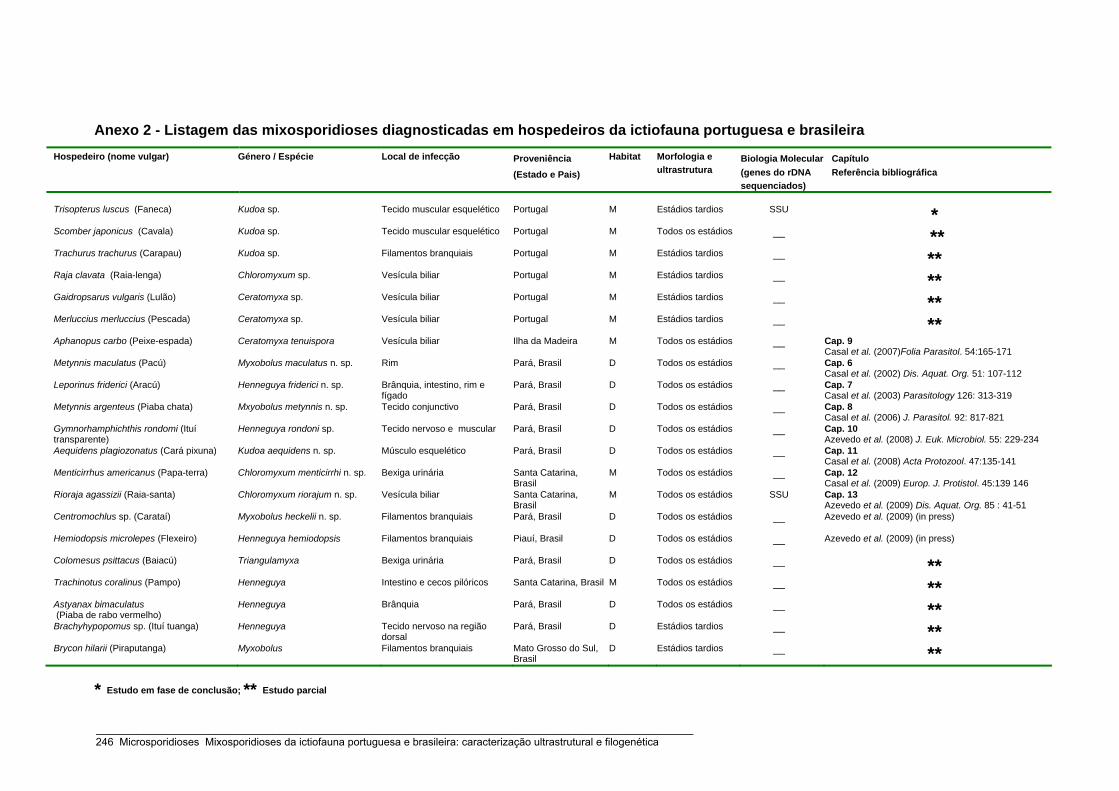
Anexo 2 - Listagem das mixosporidioses diagnosticadas em hospedeiros da ictiofauna portuguesa e brasileira Hospedeiro (nome vulgar) Género / Espécie
Local de infecção Proveniência
(Estado e Pais)
Habitat Morfologia e ultrastrutura
Biologia Molecular(genes do rDNA sequenciados)
Capítulo Referência bibliográfica
Trisopterus luscus (Faneca) Kudoa sp. Tecido muscular esquelético Portugal M Estádios tardios SSU * Scomber japonicus (Cavala) Kudoa sp. Tecido muscular esquelético Portugal M Todos os estádios __ ** Trachurus trachurus (Carapau) Kudoa sp. Filamentos branquiais Portugal M Estádios tardios __ ** Raja clavata (Raia-lenga) Chloromyxum sp. Vesícula biliar Portugal M Estádios tardios __ ** Gaidropsarus vulgaris (Lulão) Ceratomyxa sp. Vesícula biliar Portugal M Estádios tardios __ ** Merluccius merluccius (Pescada) Ceratomyxa sp. Vesícula biliar Portugal M Estádios tardios __ ** Aphanopus carbo (Peixe-espada) Ceratomyxa tenuispora Vesícula biliar Ilha da Madeira M Todos os estádios __ Cap. 9
Casal et al. (2007)Folia Parasitol. 54:165-171 Metynnis maculatus (Pacú) Myxobolus maculatus n. sp. Rim Pará, Brasil D Todos os estádios __ Cap. 6
Casal et al. (2002) Dis. Aquat. Org. 51: 107-112 Leporinus friderici (Aracú) Henneguya friderici n. sp. Brânquia, intestino, rim e
fígado Pará, Brasil D Todos os estádios __ Cap. 7
Casal et al. (2003) Parasitology 126: 313-319 Metynnis argenteus (Piaba chata) Mxyobolus metynnis n. sp. Tecido conjunctivo Pará, Brasil D Todos os estádios __ Cap. 8
Casal et al. (2006) J. Parasitol. 92: 817-821 Gymnorhamphichthis rondomi (Ituí transparente)
Henneguya rondoni sp. Tecido nervoso e muscular Pará, Brasil D Todos os estádios __ Cap. 10 Azevedo et al. (2008) J. Euk. Microbiol. 55: 229-234
Aequidens plagiozonatus (Cará pixuna) Kudoa aequidens n. sp. Músculo esquelético Pará, Brasil D Todos os estádios __ Cap. 11 Casal et al. (2008) Acta Protozool. 47:135-141
Menticirrhus americanus (Papa-terra) Chloromyxum menticirrhi n. sp. Bexiga urinária Santa Catarina, Brasil
M Todos os estádios __ Cap. 12 Casal et al. (2009) Europ. J. Protistol. 45:139 146
Rioraja agassizii (Raia-santa) Chloromyxum riorajum n. sp. Vesícula biliar Santa Catarina, Brasil
M Todos os estádios SSU Cap. 13 Azevedo et al. (2009) Dis. Aquat. Org. 85 : 41-51
Centromochlus sp. (Carataí) Myxobolus heckelii n. sp. Filamentos branquiais Pará, Brasil D Todos os estádios __ Azevedo et al. (2009) (in press)
Hemiodopsis microlepes (Flexeiro) Henneguya hemiodopsis Filamentos branquiais Piauí, Brasil D Todos os estádios __ Azevedo et al. (2009) (in press)
Colomesus psittacus (Baiacú) Triangulamyxa Bexiga urinária Pará, Brasil D Todos os estádios __ ** Trachinotus coralinus (Pampo) Henneguya Intestino e cecos pilóricos Santa Catarina, Brasil M Todos os estádios __ ** Astyanax bimaculatus (Piaba de rabo vermelho)
Henneguya Brânquia Pará, Brasil D Todos os estádios __ ** Brachyhypopomus sp. (Ituí tuanga) Henneguya Tecido nervoso na região
dorsal Pará, Brasil D Estádios tardios __ **
Brycon hilarii (Piraputanga) Myxobolus Filamentos branquiais Mato Grosso do Sul, Brasil
D Estádios tardios __ **
* Estudo em fase de conclusão; ** Estudo parcial
_____________________________________________________________________________________________________ 246 Microsporidioses Mixosporidioses da ictiofauna portuguesa e brasileira: caracterização ultrastrutural e filogenética

Anexo 3 - Árvore filogenética de consenso para o máximo parcimónio do gene SSU rRNA de microsporídios de peixes. A análise filogenética permitiu identificar os 5 grupos de definidos por Lom e Nilsen (2003). As sequências das espécies escritas a cor verde foram obtidas no decurso desta tese.
1
2
3
4
5
_____________________________________________________________________________________________________ Microsporidioses Mixosporidioses da ictiofauna portuguesa e brasileira: caracterização ultrastrutural e filogenética 247
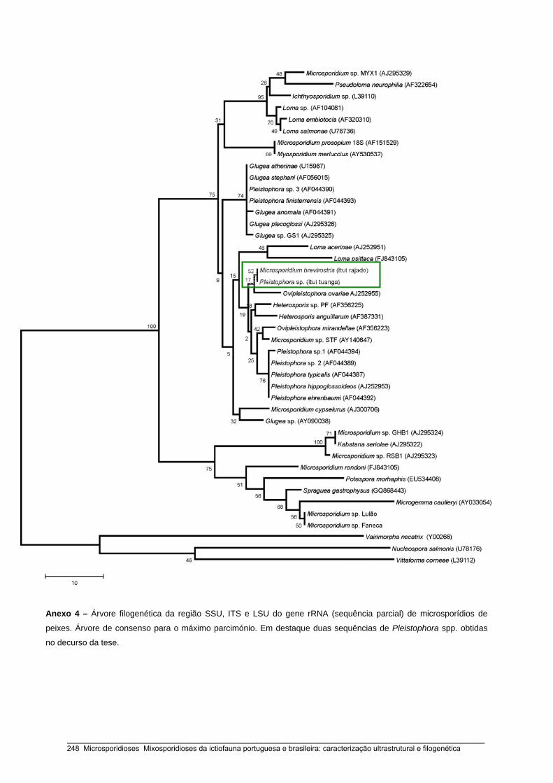
Anexo 4 – Árvore filogenética da região SSU, ITS e LSU do gene rRNA (sequência parcial) de microsporídios de
peixes. Árvore de consenso para o máximo parcimónio. Em destaque duas sequências de Pleistophora spp. obtidas
no decurso da tese.
_____________________________________________________________________________________________________ 248 Microsporidioses Mixosporidioses da ictiofauna portuguesa e brasileira: caracterização ultrastrutural e filogenética
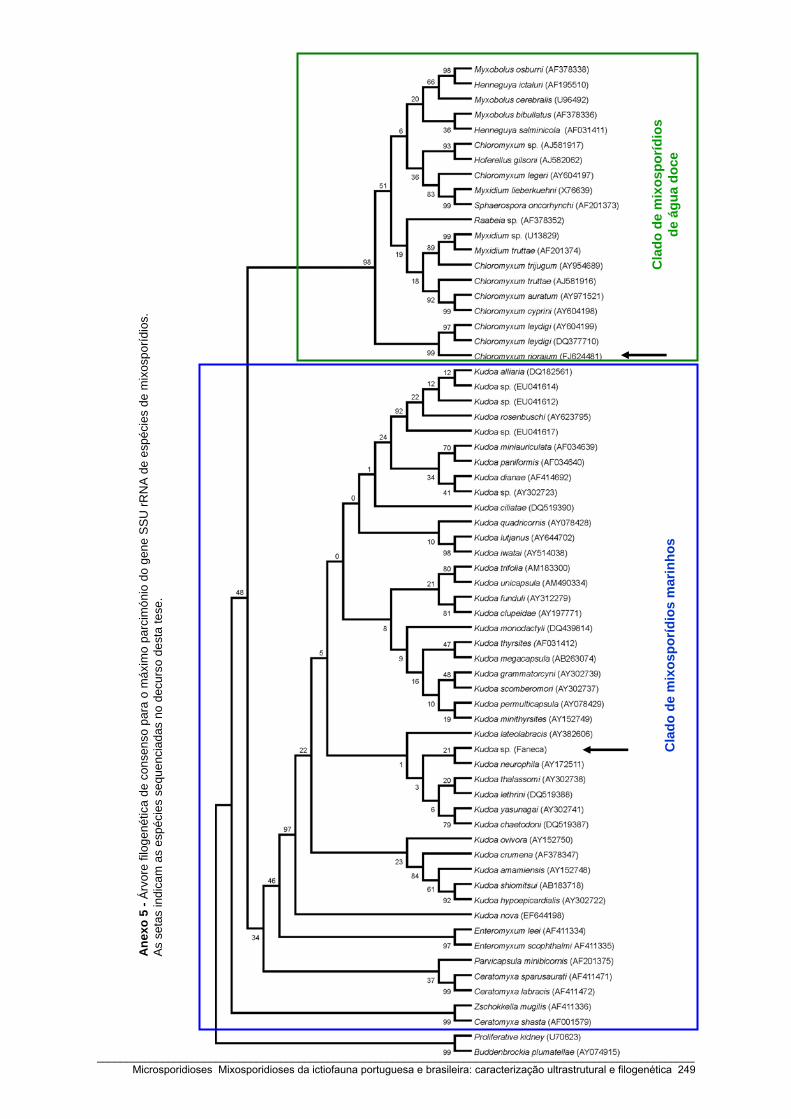
Ane
xo 5
-Ár
vore
filo
gené
tica
de c
onse
nso
para
o m
áxim
o pa
rcim
ónio
do g
ene
SS
U rR
NA
de
espé
cies
de
mix
ospo
rídio
s.
As
seta
s in
dica
m a
s es
péci
es s
eque
ncia
das
no d
ecur
so d
esta
tese
.
Cla
dode
mix
ospo
rídio
s m
arin
hos
Cla
dode
mix
ospo
rídio
s de
águ
a do
ce
_____________________________________________________________________________________________________ Microsporidioses Mixosporidioses da ictiofauna portuguesa e brasileira: caracterização ultrastrutural e filogenética 249