Importancia De La Preservación Del Anillo Pericervical En
86
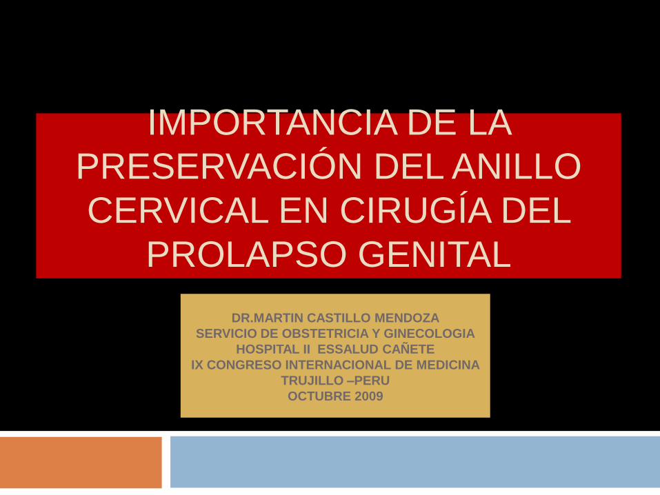
IMPORTANCIA DE LA PRESERVACIÓN DEL ANILLO CERVICAL EN CIRUGÍA DEL PROLAPSO GENITAL DR.MARTIN CASTILLO MENDOZA SERVICIO DE OBSTETRICIA Y GINECOLOGIA HOSPITAL II ESSALUD CAÑETE IX CONGRESO INTERNACIONAL DE MEDICINA TRUJILLO –PERU OCTUBRE 2009
-
Upload
martin-castillo-mendoza -
Category
Documents
-
view
2.604 -
download
0
description
LAS REPARACIONES QUIRURGICAS DE LOS SEGMENTOS ANTERIOR Y POSTERIOR DEBEN REALIZARSE SIEMPRE RESTAURANDO SU UNION AL SEGMENTO APICAL. LA VAGINA TIENE UNA BIOMECANICA EN TRES DIMENSIONES QUE DEBE RECORDARSE. SE MUESTRA UN MODELO 3D QUE GRAFICA LOS MECANISMOS DETRAS DE LOS CISTOCELES DONDE EL SEGMENTO APICAL TIENE UNA IMPORTANCIA CRUCIAL.
Transcript of Importancia De La Preservación Del Anillo Pericervical En
IMPORTANCIA DE LA
PRESERVACIOacuteN DEL ANILLO
CERVICAL EN CIRUGIacuteA DEL
PROLAPSO GENITAL
DRMARTIN CASTILLO MENDOZA
SERVICIO DE OBSTETRICIA Y GINECOLOGIA
HOSPITAL II ESSALUD CANtildeETE
IX CONGRESO INTERNACIONAL DE MEDICINA
TRUJILLO ndashPERU
OCTUBRE 2009
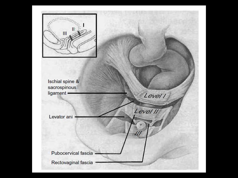
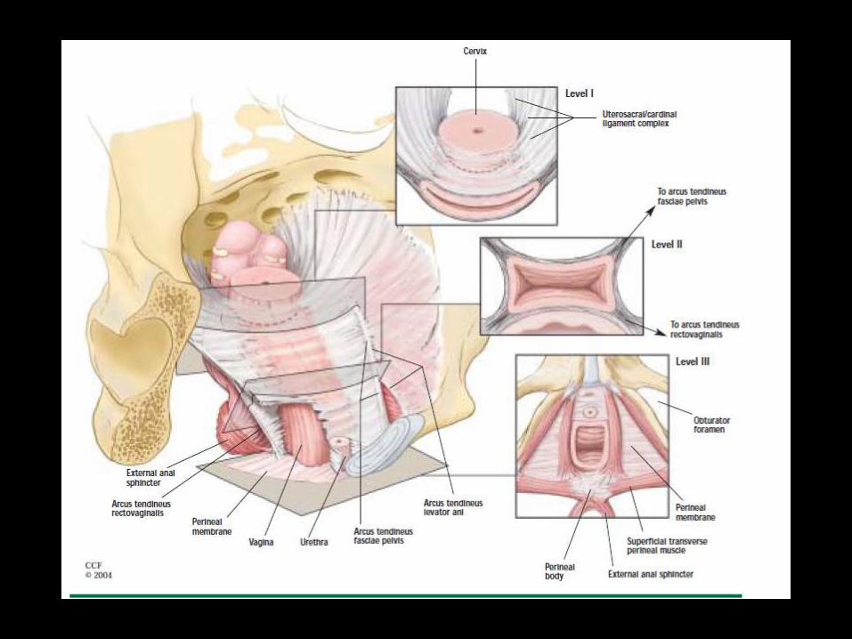
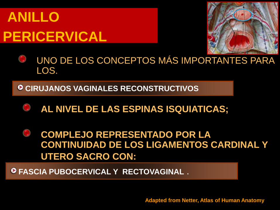
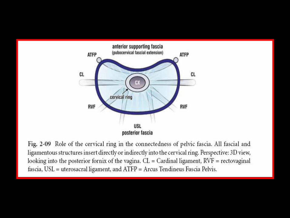
ANILLO
PERICERVICAL
ANILLO
PERICERVICAL
AL NIVEL DE LAS ESPINAS ISQUIATICAS
COMPLEJO REPRESENTADO POR LA CONTINUIDAD DE LOS LIGAMENTOS CARDINAL Y
UTERO SACRO CON
FASCIA PUBOCERVICAL Y RECTOVAGINAL
UNO DE LOS CONCEPTOS MAacuteS IMPORTANTES PARA LOS
CIRUJANOS VAGINALES RECONSTRUCTIVOS
Adapted from Netter Atlas of Human Anatomy
Davila GueretteIUGA 2004
DEFECTOS
POSIBLES
CENTRAL
TRANSVERSO
ANTERIOR
LATERAL
TRANSVERSO
POSTERIOR
Anillo Pericervical
Posterior vaginal w all Prolapse
Rectovaginal FasciaRectovaginal Fascia
RectumRectum
Septo
Rectovaginal
P
Reto
Utero
Vagina
Enfoque Quiruacutergico
TEORIA INTEGRAL DE LA
DISFUNCIOgraveN DEL PISO PEacuteLVICO
TEORIgraveA
INTEGRAL
PETROS Y ULMSTEN
TODOS LOS SIgraveNTOMAS DE DISFUNCIOgraveN DEL PISO
PEgraveLVICO SON INTERDEPENDIENTES CON UN
PROCESO FISIOPATOLOgraveGICO COMUgraveN
ALTERACIONES DEL SOPORTE SUBURETRAL DE
LOS LIGAMENTOS NERVIOS Y DE LOS MUgraveSCULOS
DEL PISO PEgraveLVICO SON RESPONSABLES DE LOS
SINTOMAS Y SIGNOS DE LA DISFUNCIOgraveN DEL PISO
PEgraveLVICO
TEORIA
INTEGRAL
ldquoLOS SINTOMAS DE DISFUNCIOgraveN DEL
PISO PEgraveLVICO DERIVAN MAYORMENTE
POR DIFERENTES RAZONES DE LA
LAXITUD DE LA VAGINA O DE SUS
LIGAMENTOS DE SOPORTE RESULTADO
DE UN TEJIDO CONECTIVO ALTERADO ldquo
TEORIgraveA INTEGRAL
ANALOGIgraveA DEL
PUENTE
BIOMECAacuteNICA DE LA
VAGINA
TEORIgraveA INTEGRAL
ldquo ANALOGIgraveA DEL PUENTErdquo
LOS LIGAMENTOS NERVIOS Y MUgraveSCULOS
TRABAJAN SINEgraveRGICAMENTE PARA
MANTENER LA ESTRUCTURA Y FUNCIOgraveN
DEL PISO PEgraveLVICO
EL DEBILITAMIENTO DE CUALQUIER PARTE
DE LA ESTRUCTURA PUEDE ALTERAR EL
EQUILIBRIO FORTALEZA Y FUNCIOgraveN DEL
TODO
TEORIgraveA
INTEGRAL
EN RELACIOgraveN A LA ANALOGIA LA FORMA
SE OBTIENE DEBIDO A QUE LA VAGINA Y
LA VEJIGA ESTAN SUSPENDIDAS DESDE
LA PORCION OgraveSEA POR LIGAMENTOS
(ligamentos pubouretrales-PUL ligamento
uacutetero sacro-USL y arco tendineo-ATFP)
LA DIMENSIOgraveN ESTRUCTURAL SE OBTIENE
POR EL ESTIRAMIENTO DE LAS FUERZAS
MUSCULARES
RELACIOgraveN DEL SEGMENTO APICAL CON
LOS OTROS SEGMENTOS
RELACION DEL AgravePEX CON
SEGMENTO POSTERIOR
P
Recto
Uacutetero
Vagina
Defecto Posterior
PROLAPSO VAGINAL POSTERIOR
PROLAPSO VAGINAL POSTERIOR
P
Recto
Utero
Vagina
Prolapso pared vaginal posterior
Evolucioacuten DefectoPericervical
PROLAPSO PARED VAGINAL POSTERIOR
P
Recto
Utero
Vagina
Prolapso pared vaginal posterior
ldquoRectocelerdquo
PROLAPSO VAGINAL POSTERIOR
ENFOQUE QUIRUacuteRGICO
P
RectoUtero
Vagina
Reparacioacuten anillo Pericervical
P
Recto
Utero
Vagina
ENFOQUE QUIRUacuteRGICO
BIOMECANICA DEL DANtildeO DEL TEJIDO CONECTIVO
DE LA ZONA POSTERIOR
El cuerpo perineal y los ligamentos uacutetero
sacros son los puntos de anclaje para el
estiramiento de la vagina en un vector de
fuerzas hacia atraacutes y abajo
Estas fuerzas suspenden la pared vaginal
anterior y la pared rectal anterior a manera de
cuerdas tensionadas por las fuerzas
musculares
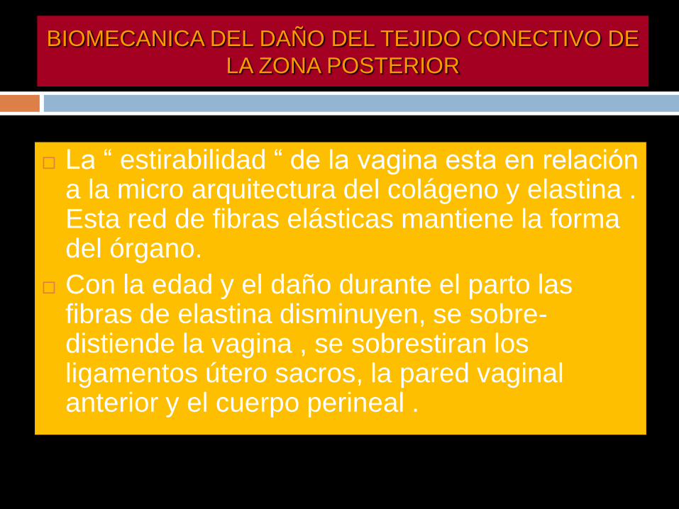
La ldquo estirabilidad ldquo de la vagina esta en relacioacuten a la micro arquitectura del colaacutegeno y elastina Esta red de fibras elaacutesticas mantiene la forma del oacutergano
Con la edad y el dantildeo durante el parto las fibras de elastina disminuyen se sobre-distiende la vagina se sobrestiran los ligamentos uacutetero sacros la pared vaginal anterior y el cuerpo perineal
BIOMECANICA DEL DANtildeO DEL TEJIDO CONECTIVO DE
LA ZONA POSTERIOR
BIOMECAacuteNICA DE
LA REPARACION
BIOMECAacuteNICA DE LA
REPARACIOacuteN
EL CUERPO PERINEAL (CP) Y LOS LIGAMENTOS
UTEROSACROS (LUS) SON AL MENOS SEIS VECES
MAS FUERTES QUE EL TEJIDO QUE SOSTIENEN
EL CUERPO PERINEAL OCUPA EL 50 DE LA PARED
VAGINAL POSTERIOR Y SE COMPORTA COMO
ELEMENTO CLAVE ACTIVO EN EL SOPORTE
ESTRUCTURAL
TANTO EL CP COMO LOS LUS SON PUNTOS DE IN
SERCION CLAVES DE LOS VECTORES
MUSCULARES DE FUERZA
BIOMECANICA DE LA
REPARACION
NO SOLO SE DEBE REPARAR EL DEFECTO FASCIAL SINO LOS ELEMENTOS DE SOPORTE ( CP Y LUS)
SI EL LUS ES DEBIL SLING POSTERIOR DE POgraveLIPROPILENO
DEBE PRESERVARSE EL ESPACIO RECTO VAGINAL PARA PERMITIR EL MOVIMIENTO INDEPENDIENTE DE LA VAGINA Y EL RECTO
CONSERVAR EL TEJIDO VAGINAL EN LO POSIBLE Y REPARACION SEPARADA DE LAS FASCIAS ESPECIALMENTE SI SE UTILIZA MALLA UNA VAGINA ESTRECHA PIERDE SUS PROPIEDADES DE ANGULACION Y ESTIRAMIENTO EN RELACION AL CP
BIOMECANICA DE LA
REPARACION
SE REQUIERE UN CUERPO PERINEAL Y
LIGAMENTOS UTEROSACROS
COMPETENTES PARA RESTAURAR EL
SOPORTE ESTRUCTURAL ACTIVO Y
PASICO AL RECTO Y VAGINA
SI SE DEBE USAR MALLA DEBE
COLOCARSE DESPUES DE LA
REPARACION ANATOMICA
RELACION DEL AgravePEX
CON SEGMENTO
ANTERIOR
Advanced anterior vaginal wall prolapse is highly
correlated with apical prolapse
Kristin Rooney MD Kimberly Kenton MD MS Elizabeth
R Mueller MD Mary Pat FitzGerald MD Linda Brubaker
MD MS
Volume 195 Issue 6 Pages 1837-1840 (December 2006)
yesp latfo rm +mauthorauthor
Objective
The purpose of this study was to determine the relationship between
the most prolapsed portion of the anterior and posterior vaginal walls
and the apex
Conclusion
Anterior vaginal wall prolapse is
associated strongly with apical
prolapse Anterior vaginal wall defects
that are surgically repaired usually
require a concomitant repair of the
apex
Interaction Among Apical
Support Levator Ani Impairment and
Anterior Vaginal Wall Prolapse
Chen Luyun MS Ashton-Miller James
A PhD Hsu Yvonne MD
DeLancey John O L MDObstetrics amp Gynecology
August 2006 - Volume 108 - Issue 2 - pp 324-332
doi 10109701AOG000022778669257a8
OBJECTIVE To use a biomechanical model to explore how impairment
of the pubovisceral portion of the levator ani muscle the apical vaginal
suspension complex or both might interact to affect anterior vaginal wall
prolapse severity
RESULTS Under raised intra-abdominal pressure the magnitude of
anterior vaginal wall prolapse was shown to be a combined function of
both pubovisceral muscle and uterosacral and cardinal ligament (apical
supports) impairment Once a certain degree of pubovisceral
impairment was reached the genital hiatus opened and a prolapse
developed The larger the pubovisceral impairment the larger the
anterior wall prolapse became A 90 impairment of apical support led
to an increase in anterior wall prolapse from 03 cm to 19 cm (a 530
increase) at 60 pubovisceral muscle impairment and from 07 cm to
24 cm (a 240 increase) at 80 pubovisceral muscle impairment
CONCLUSION These results suggest that a prolapse can develop as a
result of impairment of the muscular and apical supports of the anterior
vaginal wall
LEVEL OF EVIDENCE II-2
A 3D finite element model of anterior vaginal wall
Support to evaluate mechanisms underlying
cystocele formation
Luyun Chen James A Ashton-Miller
John OL DeLancey
Volume 42 Issue 10 Pages 1371-1377
(22 July 2009)
yesp latfo rm +mautho rautho r
A 3D finite element model of anterior vaginal wall support to
evaluate mechanisms underlying cystocele formation
Chen L Ashton-Miller JA DeLancey JO
Biomechanics Research Laboratory Department of Biomedical
Engineering University of Michigan Ann Arbor MI 48109-2125 USA
luyun_chenhotmailcom
OBJECTIVES To develop a 3D computer model of the anterior
vaginal wall and its supports validate that model and then use it to
determine the combinations of muscle and connective tissue
impairments that result in cystocele formation as observed on
dynamic magnetic resonance imaging (MRI)
RESULTS Cystocele size was sensitive to abdominal pressure and
impairment of connective tissue and muscle Larger cystocele formed
in the presence of impairments in muscular and apical connective
tissue support compared to either support element alone Apical
impairment resulted in a larger cystocele than paravaginal
impairment Levator ani muscle impairment caused a larger
urogenital hiatus size longer length of the distal vagina exposed to a
pressure differential larger apical descent and resulted in a larger
cystocele size
CONCLUSIONS Development of a cystocele requires a levator
muscle impairment an increase in abdominal pressure and apical
and paravaginal support defects
El modelo sugiere que el deterioro del elevador
del ano y del soporte apical tienen un papel
importante en la geacutenesis del cistocele Un
deterioro del 80 del soporte del aacutepex implica un
incremento en 33 del tamantildeo del cistocele
comparativamente con un soporte apical normal
El cistocele es el resultante de muacuteltiples
mecanismos El soporte apical defectuoso
produce un cistocele mayor comparado con un
defecto paravaginal
CONCEPTO DE LA PIEDRA ANGULAR
PARED VAGINAL
ANILLO PERICERVICAL
AN
TE
RIO
R
PO
ST
ER
IOR
PIEDRA ANGULAR
PARED VAGINAL
AN
TE
RIO
R
PO
ST
ER
IOR
PIEDRA ANGULAR
PARED VAGINAL
AN
TE
RIO
R
PO
ST
ER
IOR
PROLAPSOS
PIEDRA ANGULAR
PARED VAGINAL
AN
TE
RIO
R
PO
ST
ER
IOR
PR
OL
AP
SO
S
PIEDRA ANGULAR
CONCLUSIONE
S
DESINSERCIOgraveN DE LAS FASCIAS DESDE ELANILLO PERICERVICAL PROVOCA ELDEFECTO RESPONSABLE DE LOSPROLAPSOS
DEFECTOS TRANSVERSOS PARECEN SERLOS MAS IMPORTANTES
LOS DEFECTOS FASCIALES PUEDEN SERUgraveNICOS O MULTIPLES EN UNO O VARIOSCOMPARTIMIENTOS
REPARACIOgraveN SITIO ESPECIFICA CIRUGIACOMPENSADORA
CONCLUSION
ES
EXISTEN NUEVOS PARADIGMAS EN LA CONCEPCIOgraveN FISIOPATOLOgraveGICA DE LOS DEFECTOS DEL PISO PEgraveLVICO
LA REINSERCIOgraveN DE LAS FASCIAS AL ANILLO PERICERVICAL DEBE SER UN PASO INDISPENSABLE EN LA REPARACIOgraveN DE DEFECTOS DEL PISO PEgraveLVICO
ANILLO
PERICERVICAL
ANILLO
PERICERVICAL
AL NIVEL DE LAS ESPINAS ISQUIATICAS
COMPLEJO REPRESENTADO POR LA CONTINUIDAD DE LOS LIGAMENTOS CARDINAL Y
UTERO SACRO CON
FASCIA PUBOCERVICAL Y RECTOVAGINAL
UNO DE LOS CONCEPTOS MAacuteS IMPORTANTES PARA LOS
CIRUJANOS VAGINALES RECONSTRUCTIVOS
Adapted from Netter Atlas of Human Anatomy
Davila GueretteIUGA 2004
DEFECTOS
POSIBLES
CENTRAL
TRANSVERSO
ANTERIOR
LATERAL
TRANSVERSO
POSTERIOR
Anillo Pericervical
Posterior vaginal w all Prolapse
Rectovaginal FasciaRectovaginal Fascia
RectumRectum
Septo
Rectovaginal
P
Reto
Utero
Vagina
Enfoque Quiruacutergico
TEORIA INTEGRAL DE LA
DISFUNCIOgraveN DEL PISO PEacuteLVICO
TEORIgraveA
INTEGRAL
PETROS Y ULMSTEN
TODOS LOS SIgraveNTOMAS DE DISFUNCIOgraveN DEL PISO
PEgraveLVICO SON INTERDEPENDIENTES CON UN
PROCESO FISIOPATOLOgraveGICO COMUgraveN
ALTERACIONES DEL SOPORTE SUBURETRAL DE
LOS LIGAMENTOS NERVIOS Y DE LOS MUgraveSCULOS
DEL PISO PEgraveLVICO SON RESPONSABLES DE LOS
SINTOMAS Y SIGNOS DE LA DISFUNCIOgraveN DEL PISO
PEgraveLVICO
TEORIA
INTEGRAL
ldquoLOS SINTOMAS DE DISFUNCIOgraveN DEL
PISO PEgraveLVICO DERIVAN MAYORMENTE
POR DIFERENTES RAZONES DE LA
LAXITUD DE LA VAGINA O DE SUS
LIGAMENTOS DE SOPORTE RESULTADO
DE UN TEJIDO CONECTIVO ALTERADO ldquo
TEORIgraveA INTEGRAL
ANALOGIgraveA DEL
PUENTE
BIOMECAacuteNICA DE LA
VAGINA
TEORIgraveA INTEGRAL
ldquo ANALOGIgraveA DEL PUENTErdquo
LOS LIGAMENTOS NERVIOS Y MUgraveSCULOS
TRABAJAN SINEgraveRGICAMENTE PARA
MANTENER LA ESTRUCTURA Y FUNCIOgraveN
DEL PISO PEgraveLVICO
EL DEBILITAMIENTO DE CUALQUIER PARTE
DE LA ESTRUCTURA PUEDE ALTERAR EL
EQUILIBRIO FORTALEZA Y FUNCIOgraveN DEL
TODO
TEORIgraveA
INTEGRAL
EN RELACIOgraveN A LA ANALOGIA LA FORMA
SE OBTIENE DEBIDO A QUE LA VAGINA Y
LA VEJIGA ESTAN SUSPENDIDAS DESDE
LA PORCION OgraveSEA POR LIGAMENTOS
(ligamentos pubouretrales-PUL ligamento
uacutetero sacro-USL y arco tendineo-ATFP)
LA DIMENSIOgraveN ESTRUCTURAL SE OBTIENE
POR EL ESTIRAMIENTO DE LAS FUERZAS
MUSCULARES
RELACIOgraveN DEL SEGMENTO APICAL CON
LOS OTROS SEGMENTOS
RELACION DEL AgravePEX CON
SEGMENTO POSTERIOR
P
Recto
Uacutetero
Vagina
Defecto Posterior
PROLAPSO VAGINAL POSTERIOR
PROLAPSO VAGINAL POSTERIOR
P
Recto
Utero
Vagina
Prolapso pared vaginal posterior
Evolucioacuten DefectoPericervical
PROLAPSO PARED VAGINAL POSTERIOR
P
Recto
Utero
Vagina
Prolapso pared vaginal posterior
ldquoRectocelerdquo
PROLAPSO VAGINAL POSTERIOR
ENFOQUE QUIRUacuteRGICO
P
RectoUtero
Vagina
Reparacioacuten anillo Pericervical
P
Recto
Utero
Vagina
ENFOQUE QUIRUacuteRGICO
BIOMECANICA DEL DANtildeO DEL TEJIDO CONECTIVO
DE LA ZONA POSTERIOR
El cuerpo perineal y los ligamentos uacutetero
sacros son los puntos de anclaje para el
estiramiento de la vagina en un vector de
fuerzas hacia atraacutes y abajo
Estas fuerzas suspenden la pared vaginal
anterior y la pared rectal anterior a manera de
cuerdas tensionadas por las fuerzas
musculares
La ldquo estirabilidad ldquo de la vagina esta en relacioacuten a la micro arquitectura del colaacutegeno y elastina Esta red de fibras elaacutesticas mantiene la forma del oacutergano
Con la edad y el dantildeo durante el parto las fibras de elastina disminuyen se sobre-distiende la vagina se sobrestiran los ligamentos uacutetero sacros la pared vaginal anterior y el cuerpo perineal
BIOMECANICA DEL DANtildeO DEL TEJIDO CONECTIVO DE
LA ZONA POSTERIOR
BIOMECAacuteNICA DE
LA REPARACION
BIOMECAacuteNICA DE LA
REPARACIOacuteN
EL CUERPO PERINEAL (CP) Y LOS LIGAMENTOS
UTEROSACROS (LUS) SON AL MENOS SEIS VECES
MAS FUERTES QUE EL TEJIDO QUE SOSTIENEN
EL CUERPO PERINEAL OCUPA EL 50 DE LA PARED
VAGINAL POSTERIOR Y SE COMPORTA COMO
ELEMENTO CLAVE ACTIVO EN EL SOPORTE
ESTRUCTURAL
TANTO EL CP COMO LOS LUS SON PUNTOS DE IN
SERCION CLAVES DE LOS VECTORES
MUSCULARES DE FUERZA
BIOMECANICA DE LA
REPARACION
NO SOLO SE DEBE REPARAR EL DEFECTO FASCIAL SINO LOS ELEMENTOS DE SOPORTE ( CP Y LUS)
SI EL LUS ES DEBIL SLING POSTERIOR DE POgraveLIPROPILENO
DEBE PRESERVARSE EL ESPACIO RECTO VAGINAL PARA PERMITIR EL MOVIMIENTO INDEPENDIENTE DE LA VAGINA Y EL RECTO
CONSERVAR EL TEJIDO VAGINAL EN LO POSIBLE Y REPARACION SEPARADA DE LAS FASCIAS ESPECIALMENTE SI SE UTILIZA MALLA UNA VAGINA ESTRECHA PIERDE SUS PROPIEDADES DE ANGULACION Y ESTIRAMIENTO EN RELACION AL CP
BIOMECANICA DE LA
REPARACION
SE REQUIERE UN CUERPO PERINEAL Y
LIGAMENTOS UTEROSACROS
COMPETENTES PARA RESTAURAR EL
SOPORTE ESTRUCTURAL ACTIVO Y
PASICO AL RECTO Y VAGINA
SI SE DEBE USAR MALLA DEBE
COLOCARSE DESPUES DE LA
REPARACION ANATOMICA
RELACION DEL AgravePEX
CON SEGMENTO
ANTERIOR
Advanced anterior vaginal wall prolapse is highly
correlated with apical prolapse
Kristin Rooney MD Kimberly Kenton MD MS Elizabeth
R Mueller MD Mary Pat FitzGerald MD Linda Brubaker
MD MS
Volume 195 Issue 6 Pages 1837-1840 (December 2006)
yesp latfo rm +mauthorauthor
Objective
The purpose of this study was to determine the relationship between
the most prolapsed portion of the anterior and posterior vaginal walls
and the apex
Conclusion
Anterior vaginal wall prolapse is
associated strongly with apical
prolapse Anterior vaginal wall defects
that are surgically repaired usually
require a concomitant repair of the
apex
Interaction Among Apical
Support Levator Ani Impairment and
Anterior Vaginal Wall Prolapse
Chen Luyun MS Ashton-Miller James
A PhD Hsu Yvonne MD
DeLancey John O L MDObstetrics amp Gynecology
August 2006 - Volume 108 - Issue 2 - pp 324-332
doi 10109701AOG000022778669257a8
OBJECTIVE To use a biomechanical model to explore how impairment
of the pubovisceral portion of the levator ani muscle the apical vaginal
suspension complex or both might interact to affect anterior vaginal wall
prolapse severity
RESULTS Under raised intra-abdominal pressure the magnitude of
anterior vaginal wall prolapse was shown to be a combined function of
both pubovisceral muscle and uterosacral and cardinal ligament (apical
supports) impairment Once a certain degree of pubovisceral
impairment was reached the genital hiatus opened and a prolapse
developed The larger the pubovisceral impairment the larger the
anterior wall prolapse became A 90 impairment of apical support led
to an increase in anterior wall prolapse from 03 cm to 19 cm (a 530
increase) at 60 pubovisceral muscle impairment and from 07 cm to
24 cm (a 240 increase) at 80 pubovisceral muscle impairment
CONCLUSION These results suggest that a prolapse can develop as a
result of impairment of the muscular and apical supports of the anterior
vaginal wall
LEVEL OF EVIDENCE II-2
A 3D finite element model of anterior vaginal wall
Support to evaluate mechanisms underlying
cystocele formation
Luyun Chen James A Ashton-Miller
John OL DeLancey
Volume 42 Issue 10 Pages 1371-1377
(22 July 2009)
yesp latfo rm +mautho rautho r
A 3D finite element model of anterior vaginal wall support to
evaluate mechanisms underlying cystocele formation
Chen L Ashton-Miller JA DeLancey JO
Biomechanics Research Laboratory Department of Biomedical
Engineering University of Michigan Ann Arbor MI 48109-2125 USA
luyun_chenhotmailcom
OBJECTIVES To develop a 3D computer model of the anterior
vaginal wall and its supports validate that model and then use it to
determine the combinations of muscle and connective tissue
impairments that result in cystocele formation as observed on
dynamic magnetic resonance imaging (MRI)
RESULTS Cystocele size was sensitive to abdominal pressure and
impairment of connective tissue and muscle Larger cystocele formed
in the presence of impairments in muscular and apical connective
tissue support compared to either support element alone Apical
impairment resulted in a larger cystocele than paravaginal
impairment Levator ani muscle impairment caused a larger
urogenital hiatus size longer length of the distal vagina exposed to a
pressure differential larger apical descent and resulted in a larger
cystocele size
CONCLUSIONS Development of a cystocele requires a levator
muscle impairment an increase in abdominal pressure and apical
and paravaginal support defects
El modelo sugiere que el deterioro del elevador
del ano y del soporte apical tienen un papel
importante en la geacutenesis del cistocele Un
deterioro del 80 del soporte del aacutepex implica un
incremento en 33 del tamantildeo del cistocele
comparativamente con un soporte apical normal
El cistocele es el resultante de muacuteltiples
mecanismos El soporte apical defectuoso
produce un cistocele mayor comparado con un
defecto paravaginal
CONCEPTO DE LA PIEDRA ANGULAR
PARED VAGINAL
ANILLO PERICERVICAL
AN
TE
RIO
R
PO
ST
ER
IOR
PIEDRA ANGULAR
PARED VAGINAL
AN
TE
RIO
R
PO
ST
ER
IOR
PIEDRA ANGULAR
PARED VAGINAL
AN
TE
RIO
R
PO
ST
ER
IOR
PROLAPSOS
PIEDRA ANGULAR
PARED VAGINAL
AN
TE
RIO
R
PO
ST
ER
IOR
PR
OL
AP
SO
S
PIEDRA ANGULAR
CONCLUSIONE
S
DESINSERCIOgraveN DE LAS FASCIAS DESDE ELANILLO PERICERVICAL PROVOCA ELDEFECTO RESPONSABLE DE LOSPROLAPSOS
DEFECTOS TRANSVERSOS PARECEN SERLOS MAS IMPORTANTES
LOS DEFECTOS FASCIALES PUEDEN SERUgraveNICOS O MULTIPLES EN UNO O VARIOSCOMPARTIMIENTOS
REPARACIOgraveN SITIO ESPECIFICA CIRUGIACOMPENSADORA
CONCLUSION
ES
EXISTEN NUEVOS PARADIGMAS EN LA CONCEPCIOgraveN FISIOPATOLOgraveGICA DE LOS DEFECTOS DEL PISO PEgraveLVICO
LA REINSERCIOgraveN DE LAS FASCIAS AL ANILLO PERICERVICAL DEBE SER UN PASO INDISPENSABLE EN LA REPARACIOgraveN DE DEFECTOS DEL PISO PEgraveLVICO
ANILLO
PERICERVICAL
AL NIVEL DE LAS ESPINAS ISQUIATICAS
COMPLEJO REPRESENTADO POR LA CONTINUIDAD DE LOS LIGAMENTOS CARDINAL Y
UTERO SACRO CON
FASCIA PUBOCERVICAL Y RECTOVAGINAL
UNO DE LOS CONCEPTOS MAacuteS IMPORTANTES PARA LOS
CIRUJANOS VAGINALES RECONSTRUCTIVOS
Adapted from Netter Atlas of Human Anatomy
Davila GueretteIUGA 2004
DEFECTOS
POSIBLES
CENTRAL
TRANSVERSO
ANTERIOR
LATERAL
TRANSVERSO
POSTERIOR
Anillo Pericervical
Posterior vaginal w all Prolapse
Rectovaginal FasciaRectovaginal Fascia
RectumRectum
Septo
Rectovaginal
P
Reto
Utero
Vagina
Enfoque Quiruacutergico
TEORIA INTEGRAL DE LA
DISFUNCIOgraveN DEL PISO PEacuteLVICO
TEORIgraveA
INTEGRAL
PETROS Y ULMSTEN
TODOS LOS SIgraveNTOMAS DE DISFUNCIOgraveN DEL PISO
PEgraveLVICO SON INTERDEPENDIENTES CON UN
PROCESO FISIOPATOLOgraveGICO COMUgraveN
ALTERACIONES DEL SOPORTE SUBURETRAL DE
LOS LIGAMENTOS NERVIOS Y DE LOS MUgraveSCULOS
DEL PISO PEgraveLVICO SON RESPONSABLES DE LOS
SINTOMAS Y SIGNOS DE LA DISFUNCIOgraveN DEL PISO
PEgraveLVICO
TEORIA
INTEGRAL
ldquoLOS SINTOMAS DE DISFUNCIOgraveN DEL
PISO PEgraveLVICO DERIVAN MAYORMENTE
POR DIFERENTES RAZONES DE LA
LAXITUD DE LA VAGINA O DE SUS
LIGAMENTOS DE SOPORTE RESULTADO
DE UN TEJIDO CONECTIVO ALTERADO ldquo
TEORIgraveA INTEGRAL
ANALOGIgraveA DEL
PUENTE
BIOMECAacuteNICA DE LA
VAGINA
TEORIgraveA INTEGRAL
ldquo ANALOGIgraveA DEL PUENTErdquo
LOS LIGAMENTOS NERVIOS Y MUgraveSCULOS
TRABAJAN SINEgraveRGICAMENTE PARA
MANTENER LA ESTRUCTURA Y FUNCIOgraveN
DEL PISO PEgraveLVICO
EL DEBILITAMIENTO DE CUALQUIER PARTE
DE LA ESTRUCTURA PUEDE ALTERAR EL
EQUILIBRIO FORTALEZA Y FUNCIOgraveN DEL
TODO
TEORIgraveA
INTEGRAL
EN RELACIOgraveN A LA ANALOGIA LA FORMA
SE OBTIENE DEBIDO A QUE LA VAGINA Y
LA VEJIGA ESTAN SUSPENDIDAS DESDE
LA PORCION OgraveSEA POR LIGAMENTOS
(ligamentos pubouretrales-PUL ligamento
uacutetero sacro-USL y arco tendineo-ATFP)
LA DIMENSIOgraveN ESTRUCTURAL SE OBTIENE
POR EL ESTIRAMIENTO DE LAS FUERZAS
MUSCULARES
RELACIOgraveN DEL SEGMENTO APICAL CON
LOS OTROS SEGMENTOS
RELACION DEL AgravePEX CON
SEGMENTO POSTERIOR
P
Recto
Uacutetero
Vagina
Defecto Posterior
PROLAPSO VAGINAL POSTERIOR
PROLAPSO VAGINAL POSTERIOR
P
Recto
Utero
Vagina
Prolapso pared vaginal posterior
Evolucioacuten DefectoPericervical
PROLAPSO PARED VAGINAL POSTERIOR
P
Recto
Utero
Vagina
Prolapso pared vaginal posterior
ldquoRectocelerdquo
PROLAPSO VAGINAL POSTERIOR
ENFOQUE QUIRUacuteRGICO
P
RectoUtero
Vagina
Reparacioacuten anillo Pericervical
P
Recto
Utero
Vagina
ENFOQUE QUIRUacuteRGICO
BIOMECANICA DEL DANtildeO DEL TEJIDO CONECTIVO
DE LA ZONA POSTERIOR
El cuerpo perineal y los ligamentos uacutetero
sacros son los puntos de anclaje para el
estiramiento de la vagina en un vector de
fuerzas hacia atraacutes y abajo
Estas fuerzas suspenden la pared vaginal
anterior y la pared rectal anterior a manera de
cuerdas tensionadas por las fuerzas
musculares
La ldquo estirabilidad ldquo de la vagina esta en relacioacuten a la micro arquitectura del colaacutegeno y elastina Esta red de fibras elaacutesticas mantiene la forma del oacutergano
Con la edad y el dantildeo durante el parto las fibras de elastina disminuyen se sobre-distiende la vagina se sobrestiran los ligamentos uacutetero sacros la pared vaginal anterior y el cuerpo perineal
BIOMECANICA DEL DANtildeO DEL TEJIDO CONECTIVO DE
LA ZONA POSTERIOR
BIOMECAacuteNICA DE
LA REPARACION
BIOMECAacuteNICA DE LA
REPARACIOacuteN
EL CUERPO PERINEAL (CP) Y LOS LIGAMENTOS
UTEROSACROS (LUS) SON AL MENOS SEIS VECES
MAS FUERTES QUE EL TEJIDO QUE SOSTIENEN
EL CUERPO PERINEAL OCUPA EL 50 DE LA PARED
VAGINAL POSTERIOR Y SE COMPORTA COMO
ELEMENTO CLAVE ACTIVO EN EL SOPORTE
ESTRUCTURAL
TANTO EL CP COMO LOS LUS SON PUNTOS DE IN
SERCION CLAVES DE LOS VECTORES
MUSCULARES DE FUERZA
BIOMECANICA DE LA
REPARACION
NO SOLO SE DEBE REPARAR EL DEFECTO FASCIAL SINO LOS ELEMENTOS DE SOPORTE ( CP Y LUS)
SI EL LUS ES DEBIL SLING POSTERIOR DE POgraveLIPROPILENO
DEBE PRESERVARSE EL ESPACIO RECTO VAGINAL PARA PERMITIR EL MOVIMIENTO INDEPENDIENTE DE LA VAGINA Y EL RECTO
CONSERVAR EL TEJIDO VAGINAL EN LO POSIBLE Y REPARACION SEPARADA DE LAS FASCIAS ESPECIALMENTE SI SE UTILIZA MALLA UNA VAGINA ESTRECHA PIERDE SUS PROPIEDADES DE ANGULACION Y ESTIRAMIENTO EN RELACION AL CP
BIOMECANICA DE LA
REPARACION
SE REQUIERE UN CUERPO PERINEAL Y
LIGAMENTOS UTEROSACROS
COMPETENTES PARA RESTAURAR EL
SOPORTE ESTRUCTURAL ACTIVO Y
PASICO AL RECTO Y VAGINA
SI SE DEBE USAR MALLA DEBE
COLOCARSE DESPUES DE LA
REPARACION ANATOMICA
RELACION DEL AgravePEX
CON SEGMENTO
ANTERIOR
Advanced anterior vaginal wall prolapse is highly
correlated with apical prolapse
Kristin Rooney MD Kimberly Kenton MD MS Elizabeth
R Mueller MD Mary Pat FitzGerald MD Linda Brubaker
MD MS
Volume 195 Issue 6 Pages 1837-1840 (December 2006)
yesp latfo rm +mauthorauthor
Objective
The purpose of this study was to determine the relationship between
the most prolapsed portion of the anterior and posterior vaginal walls
and the apex
Conclusion
Anterior vaginal wall prolapse is
associated strongly with apical
prolapse Anterior vaginal wall defects
that are surgically repaired usually
require a concomitant repair of the
apex
Interaction Among Apical
Support Levator Ani Impairment and
Anterior Vaginal Wall Prolapse
Chen Luyun MS Ashton-Miller James
A PhD Hsu Yvonne MD
DeLancey John O L MDObstetrics amp Gynecology
August 2006 - Volume 108 - Issue 2 - pp 324-332
doi 10109701AOG000022778669257a8
OBJECTIVE To use a biomechanical model to explore how impairment
of the pubovisceral portion of the levator ani muscle the apical vaginal
suspension complex or both might interact to affect anterior vaginal wall
prolapse severity
RESULTS Under raised intra-abdominal pressure the magnitude of
anterior vaginal wall prolapse was shown to be a combined function of
both pubovisceral muscle and uterosacral and cardinal ligament (apical
supports) impairment Once a certain degree of pubovisceral
impairment was reached the genital hiatus opened and a prolapse
developed The larger the pubovisceral impairment the larger the
anterior wall prolapse became A 90 impairment of apical support led
to an increase in anterior wall prolapse from 03 cm to 19 cm (a 530
increase) at 60 pubovisceral muscle impairment and from 07 cm to
24 cm (a 240 increase) at 80 pubovisceral muscle impairment
CONCLUSION These results suggest that a prolapse can develop as a
result of impairment of the muscular and apical supports of the anterior
vaginal wall
LEVEL OF EVIDENCE II-2
A 3D finite element model of anterior vaginal wall
Support to evaluate mechanisms underlying
cystocele formation
Luyun Chen James A Ashton-Miller
John OL DeLancey
Volume 42 Issue 10 Pages 1371-1377
(22 July 2009)
yesp latfo rm +mautho rautho r
A 3D finite element model of anterior vaginal wall support to
evaluate mechanisms underlying cystocele formation
Chen L Ashton-Miller JA DeLancey JO
Biomechanics Research Laboratory Department of Biomedical
Engineering University of Michigan Ann Arbor MI 48109-2125 USA
luyun_chenhotmailcom
OBJECTIVES To develop a 3D computer model of the anterior
vaginal wall and its supports validate that model and then use it to
determine the combinations of muscle and connective tissue
impairments that result in cystocele formation as observed on
dynamic magnetic resonance imaging (MRI)
RESULTS Cystocele size was sensitive to abdominal pressure and
impairment of connective tissue and muscle Larger cystocele formed
in the presence of impairments in muscular and apical connective
tissue support compared to either support element alone Apical
impairment resulted in a larger cystocele than paravaginal
impairment Levator ani muscle impairment caused a larger
urogenital hiatus size longer length of the distal vagina exposed to a
pressure differential larger apical descent and resulted in a larger
cystocele size
CONCLUSIONS Development of a cystocele requires a levator
muscle impairment an increase in abdominal pressure and apical
and paravaginal support defects
El modelo sugiere que el deterioro del elevador
del ano y del soporte apical tienen un papel
importante en la geacutenesis del cistocele Un
deterioro del 80 del soporte del aacutepex implica un
incremento en 33 del tamantildeo del cistocele
comparativamente con un soporte apical normal
El cistocele es el resultante de muacuteltiples
mecanismos El soporte apical defectuoso
produce un cistocele mayor comparado con un
defecto paravaginal
CONCEPTO DE LA PIEDRA ANGULAR
PARED VAGINAL
ANILLO PERICERVICAL
AN
TE
RIO
R
PO
ST
ER
IOR
PIEDRA ANGULAR
PARED VAGINAL
AN
TE
RIO
R
PO
ST
ER
IOR
PIEDRA ANGULAR
PARED VAGINAL
AN
TE
RIO
R
PO
ST
ER
IOR
PROLAPSOS
PIEDRA ANGULAR
PARED VAGINAL
AN
TE
RIO
R
PO
ST
ER
IOR
PR
OL
AP
SO
S
PIEDRA ANGULAR
CONCLUSIONE
S
DESINSERCIOgraveN DE LAS FASCIAS DESDE ELANILLO PERICERVICAL PROVOCA ELDEFECTO RESPONSABLE DE LOSPROLAPSOS
DEFECTOS TRANSVERSOS PARECEN SERLOS MAS IMPORTANTES
LOS DEFECTOS FASCIALES PUEDEN SERUgraveNICOS O MULTIPLES EN UNO O VARIOSCOMPARTIMIENTOS
REPARACIOgraveN SITIO ESPECIFICA CIRUGIACOMPENSADORA
CONCLUSION
ES
EXISTEN NUEVOS PARADIGMAS EN LA CONCEPCIOgraveN FISIOPATOLOgraveGICA DE LOS DEFECTOS DEL PISO PEgraveLVICO
LA REINSERCIOgraveN DE LAS FASCIAS AL ANILLO PERICERVICAL DEBE SER UN PASO INDISPENSABLE EN LA REPARACIOgraveN DE DEFECTOS DEL PISO PEgraveLVICO
P
Reto
Utero
Vagina
Enfoque Quiruacutergico
TEORIA INTEGRAL DE LA
DISFUNCIOgraveN DEL PISO PEacuteLVICO
TEORIgraveA
INTEGRAL
PETROS Y ULMSTEN
TODOS LOS SIgraveNTOMAS DE DISFUNCIOgraveN DEL PISO
PEgraveLVICO SON INTERDEPENDIENTES CON UN
PROCESO FISIOPATOLOgraveGICO COMUgraveN
ALTERACIONES DEL SOPORTE SUBURETRAL DE
LOS LIGAMENTOS NERVIOS Y DE LOS MUgraveSCULOS
DEL PISO PEgraveLVICO SON RESPONSABLES DE LOS
SINTOMAS Y SIGNOS DE LA DISFUNCIOgraveN DEL PISO
PEgraveLVICO
TEORIA
INTEGRAL
ldquoLOS SINTOMAS DE DISFUNCIOgraveN DEL
PISO PEgraveLVICO DERIVAN MAYORMENTE
POR DIFERENTES RAZONES DE LA
LAXITUD DE LA VAGINA O DE SUS
LIGAMENTOS DE SOPORTE RESULTADO
DE UN TEJIDO CONECTIVO ALTERADO ldquo
TEORIgraveA INTEGRAL
ANALOGIgraveA DEL
PUENTE
BIOMECAacuteNICA DE LA
VAGINA
TEORIgraveA INTEGRAL
ldquo ANALOGIgraveA DEL PUENTErdquo
LOS LIGAMENTOS NERVIOS Y MUgraveSCULOS
TRABAJAN SINEgraveRGICAMENTE PARA
MANTENER LA ESTRUCTURA Y FUNCIOgraveN
DEL PISO PEgraveLVICO
EL DEBILITAMIENTO DE CUALQUIER PARTE
DE LA ESTRUCTURA PUEDE ALTERAR EL
EQUILIBRIO FORTALEZA Y FUNCIOgraveN DEL
TODO
TEORIgraveA
INTEGRAL
EN RELACIOgraveN A LA ANALOGIA LA FORMA
SE OBTIENE DEBIDO A QUE LA VAGINA Y
LA VEJIGA ESTAN SUSPENDIDAS DESDE
LA PORCION OgraveSEA POR LIGAMENTOS
(ligamentos pubouretrales-PUL ligamento
uacutetero sacro-USL y arco tendineo-ATFP)
LA DIMENSIOgraveN ESTRUCTURAL SE OBTIENE
POR EL ESTIRAMIENTO DE LAS FUERZAS
MUSCULARES
RELACIOgraveN DEL SEGMENTO APICAL CON
LOS OTROS SEGMENTOS
RELACION DEL AgravePEX CON
SEGMENTO POSTERIOR
P
Recto
Uacutetero
Vagina
Defecto Posterior
PROLAPSO VAGINAL POSTERIOR
PROLAPSO VAGINAL POSTERIOR
P
Recto
Utero
Vagina
Prolapso pared vaginal posterior
Evolucioacuten DefectoPericervical
PROLAPSO PARED VAGINAL POSTERIOR
P
Recto
Utero
Vagina
Prolapso pared vaginal posterior
ldquoRectocelerdquo
PROLAPSO VAGINAL POSTERIOR
ENFOQUE QUIRUacuteRGICO
P
RectoUtero
Vagina
Reparacioacuten anillo Pericervical
P
Recto
Utero
Vagina
ENFOQUE QUIRUacuteRGICO
BIOMECANICA DEL DANtildeO DEL TEJIDO CONECTIVO
DE LA ZONA POSTERIOR
El cuerpo perineal y los ligamentos uacutetero
sacros son los puntos de anclaje para el
estiramiento de la vagina en un vector de
fuerzas hacia atraacutes y abajo
Estas fuerzas suspenden la pared vaginal
anterior y la pared rectal anterior a manera de
cuerdas tensionadas por las fuerzas
musculares
La ldquo estirabilidad ldquo de la vagina esta en relacioacuten a la micro arquitectura del colaacutegeno y elastina Esta red de fibras elaacutesticas mantiene la forma del oacutergano
Con la edad y el dantildeo durante el parto las fibras de elastina disminuyen se sobre-distiende la vagina se sobrestiran los ligamentos uacutetero sacros la pared vaginal anterior y el cuerpo perineal
BIOMECANICA DEL DANtildeO DEL TEJIDO CONECTIVO DE
LA ZONA POSTERIOR
BIOMECAacuteNICA DE
LA REPARACION
BIOMECAacuteNICA DE LA
REPARACIOacuteN
EL CUERPO PERINEAL (CP) Y LOS LIGAMENTOS
UTEROSACROS (LUS) SON AL MENOS SEIS VECES
MAS FUERTES QUE EL TEJIDO QUE SOSTIENEN
EL CUERPO PERINEAL OCUPA EL 50 DE LA PARED
VAGINAL POSTERIOR Y SE COMPORTA COMO
ELEMENTO CLAVE ACTIVO EN EL SOPORTE
ESTRUCTURAL
TANTO EL CP COMO LOS LUS SON PUNTOS DE IN
SERCION CLAVES DE LOS VECTORES
MUSCULARES DE FUERZA
BIOMECANICA DE LA
REPARACION
NO SOLO SE DEBE REPARAR EL DEFECTO FASCIAL SINO LOS ELEMENTOS DE SOPORTE ( CP Y LUS)
SI EL LUS ES DEBIL SLING POSTERIOR DE POgraveLIPROPILENO
DEBE PRESERVARSE EL ESPACIO RECTO VAGINAL PARA PERMITIR EL MOVIMIENTO INDEPENDIENTE DE LA VAGINA Y EL RECTO
CONSERVAR EL TEJIDO VAGINAL EN LO POSIBLE Y REPARACION SEPARADA DE LAS FASCIAS ESPECIALMENTE SI SE UTILIZA MALLA UNA VAGINA ESTRECHA PIERDE SUS PROPIEDADES DE ANGULACION Y ESTIRAMIENTO EN RELACION AL CP
BIOMECANICA DE LA
REPARACION
SE REQUIERE UN CUERPO PERINEAL Y
LIGAMENTOS UTEROSACROS
COMPETENTES PARA RESTAURAR EL
SOPORTE ESTRUCTURAL ACTIVO Y
PASICO AL RECTO Y VAGINA
SI SE DEBE USAR MALLA DEBE
COLOCARSE DESPUES DE LA
REPARACION ANATOMICA
RELACION DEL AgravePEX
CON SEGMENTO
ANTERIOR
Advanced anterior vaginal wall prolapse is highly
correlated with apical prolapse
Kristin Rooney MD Kimberly Kenton MD MS Elizabeth
R Mueller MD Mary Pat FitzGerald MD Linda Brubaker
MD MS
Volume 195 Issue 6 Pages 1837-1840 (December 2006)
yesp latfo rm +mauthorauthor
Objective
The purpose of this study was to determine the relationship between
the most prolapsed portion of the anterior and posterior vaginal walls
and the apex
Conclusion
Anterior vaginal wall prolapse is
associated strongly with apical
prolapse Anterior vaginal wall defects
that are surgically repaired usually
require a concomitant repair of the
apex
Interaction Among Apical
Support Levator Ani Impairment and
Anterior Vaginal Wall Prolapse
Chen Luyun MS Ashton-Miller James
A PhD Hsu Yvonne MD
DeLancey John O L MDObstetrics amp Gynecology
August 2006 - Volume 108 - Issue 2 - pp 324-332
doi 10109701AOG000022778669257a8
OBJECTIVE To use a biomechanical model to explore how impairment
of the pubovisceral portion of the levator ani muscle the apical vaginal
suspension complex or both might interact to affect anterior vaginal wall
prolapse severity
RESULTS Under raised intra-abdominal pressure the magnitude of
anterior vaginal wall prolapse was shown to be a combined function of
both pubovisceral muscle and uterosacral and cardinal ligament (apical
supports) impairment Once a certain degree of pubovisceral
impairment was reached the genital hiatus opened and a prolapse
developed The larger the pubovisceral impairment the larger the
anterior wall prolapse became A 90 impairment of apical support led
to an increase in anterior wall prolapse from 03 cm to 19 cm (a 530
increase) at 60 pubovisceral muscle impairment and from 07 cm to
24 cm (a 240 increase) at 80 pubovisceral muscle impairment
CONCLUSION These results suggest that a prolapse can develop as a
result of impairment of the muscular and apical supports of the anterior
vaginal wall
LEVEL OF EVIDENCE II-2
A 3D finite element model of anterior vaginal wall
Support to evaluate mechanisms underlying
cystocele formation
Luyun Chen James A Ashton-Miller
John OL DeLancey
Volume 42 Issue 10 Pages 1371-1377
(22 July 2009)
yesp latfo rm +mautho rautho r
A 3D finite element model of anterior vaginal wall support to
evaluate mechanisms underlying cystocele formation
Chen L Ashton-Miller JA DeLancey JO
Biomechanics Research Laboratory Department of Biomedical
Engineering University of Michigan Ann Arbor MI 48109-2125 USA
luyun_chenhotmailcom
OBJECTIVES To develop a 3D computer model of the anterior
vaginal wall and its supports validate that model and then use it to
determine the combinations of muscle and connective tissue
impairments that result in cystocele formation as observed on
dynamic magnetic resonance imaging (MRI)
RESULTS Cystocele size was sensitive to abdominal pressure and
impairment of connective tissue and muscle Larger cystocele formed
in the presence of impairments in muscular and apical connective
tissue support compared to either support element alone Apical
impairment resulted in a larger cystocele than paravaginal
impairment Levator ani muscle impairment caused a larger
urogenital hiatus size longer length of the distal vagina exposed to a
pressure differential larger apical descent and resulted in a larger
cystocele size
CONCLUSIONS Development of a cystocele requires a levator
muscle impairment an increase in abdominal pressure and apical
and paravaginal support defects
El modelo sugiere que el deterioro del elevador
del ano y del soporte apical tienen un papel
importante en la geacutenesis del cistocele Un
deterioro del 80 del soporte del aacutepex implica un
incremento en 33 del tamantildeo del cistocele
comparativamente con un soporte apical normal
El cistocele es el resultante de muacuteltiples
mecanismos El soporte apical defectuoso
produce un cistocele mayor comparado con un
defecto paravaginal
CONCEPTO DE LA PIEDRA ANGULAR
PARED VAGINAL
ANILLO PERICERVICAL
AN
TE
RIO
R
PO
ST
ER
IOR
PIEDRA ANGULAR
PARED VAGINAL
AN
TE
RIO
R
PO
ST
ER
IOR
PIEDRA ANGULAR
PARED VAGINAL
AN
TE
RIO
R
PO
ST
ER
IOR
PROLAPSOS
PIEDRA ANGULAR
PARED VAGINAL
AN
TE
RIO
R
PO
ST
ER
IOR
PR
OL
AP
SO
S
PIEDRA ANGULAR
CONCLUSIONE
S
DESINSERCIOgraveN DE LAS FASCIAS DESDE ELANILLO PERICERVICAL PROVOCA ELDEFECTO RESPONSABLE DE LOSPROLAPSOS
DEFECTOS TRANSVERSOS PARECEN SERLOS MAS IMPORTANTES
LOS DEFECTOS FASCIALES PUEDEN SERUgraveNICOS O MULTIPLES EN UNO O VARIOSCOMPARTIMIENTOS
REPARACIOgraveN SITIO ESPECIFICA CIRUGIACOMPENSADORA
CONCLUSION
ES
EXISTEN NUEVOS PARADIGMAS EN LA CONCEPCIOgraveN FISIOPATOLOgraveGICA DE LOS DEFECTOS DEL PISO PEgraveLVICO
LA REINSERCIOgraveN DE LAS FASCIAS AL ANILLO PERICERVICAL DEBE SER UN PASO INDISPENSABLE EN LA REPARACIOgraveN DE DEFECTOS DEL PISO PEgraveLVICO
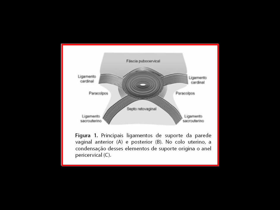
TEORIgraveA
INTEGRAL
PETROS Y ULMSTEN
TODOS LOS SIgraveNTOMAS DE DISFUNCIOgraveN DEL PISO
PEgraveLVICO SON INTERDEPENDIENTES CON UN
PROCESO FISIOPATOLOgraveGICO COMUgraveN
ALTERACIONES DEL SOPORTE SUBURETRAL DE
LOS LIGAMENTOS NERVIOS Y DE LOS MUgraveSCULOS
DEL PISO PEgraveLVICO SON RESPONSABLES DE LOS
SINTOMAS Y SIGNOS DE LA DISFUNCIOgraveN DEL PISO
PEgraveLVICO
TEORIA
INTEGRAL
ldquoLOS SINTOMAS DE DISFUNCIOgraveN DEL
PISO PEgraveLVICO DERIVAN MAYORMENTE
POR DIFERENTES RAZONES DE LA
LAXITUD DE LA VAGINA O DE SUS
LIGAMENTOS DE SOPORTE RESULTADO
DE UN TEJIDO CONECTIVO ALTERADO ldquo
TEORIgraveA INTEGRAL
ANALOGIgraveA DEL
PUENTE
BIOMECAacuteNICA DE LA
VAGINA
TEORIgraveA INTEGRAL
ldquo ANALOGIgraveA DEL PUENTErdquo
LOS LIGAMENTOS NERVIOS Y MUgraveSCULOS
TRABAJAN SINEgraveRGICAMENTE PARA
MANTENER LA ESTRUCTURA Y FUNCIOgraveN
DEL PISO PEgraveLVICO
EL DEBILITAMIENTO DE CUALQUIER PARTE
DE LA ESTRUCTURA PUEDE ALTERAR EL
EQUILIBRIO FORTALEZA Y FUNCIOgraveN DEL
TODO
TEORIgraveA
INTEGRAL
EN RELACIOgraveN A LA ANALOGIA LA FORMA
SE OBTIENE DEBIDO A QUE LA VAGINA Y
LA VEJIGA ESTAN SUSPENDIDAS DESDE
LA PORCION OgraveSEA POR LIGAMENTOS
(ligamentos pubouretrales-PUL ligamento
uacutetero sacro-USL y arco tendineo-ATFP)
LA DIMENSIOgraveN ESTRUCTURAL SE OBTIENE
POR EL ESTIRAMIENTO DE LAS FUERZAS
MUSCULARES
RELACIOgraveN DEL SEGMENTO APICAL CON
LOS OTROS SEGMENTOS
RELACION DEL AgravePEX CON
SEGMENTO POSTERIOR
P
Recto
Uacutetero
Vagina
Defecto Posterior
PROLAPSO VAGINAL POSTERIOR
PROLAPSO VAGINAL POSTERIOR
P
Recto
Utero
Vagina
Prolapso pared vaginal posterior
Evolucioacuten DefectoPericervical
PROLAPSO PARED VAGINAL POSTERIOR
P
Recto
Utero
Vagina
Prolapso pared vaginal posterior
ldquoRectocelerdquo
PROLAPSO VAGINAL POSTERIOR
ENFOQUE QUIRUacuteRGICO
P
RectoUtero
Vagina
Reparacioacuten anillo Pericervical
P
Recto
Utero
Vagina
ENFOQUE QUIRUacuteRGICO
BIOMECANICA DEL DANtildeO DEL TEJIDO CONECTIVO
DE LA ZONA POSTERIOR
El cuerpo perineal y los ligamentos uacutetero
sacros son los puntos de anclaje para el
estiramiento de la vagina en un vector de
fuerzas hacia atraacutes y abajo
Estas fuerzas suspenden la pared vaginal
anterior y la pared rectal anterior a manera de
cuerdas tensionadas por las fuerzas
musculares
La ldquo estirabilidad ldquo de la vagina esta en relacioacuten a la micro arquitectura del colaacutegeno y elastina Esta red de fibras elaacutesticas mantiene la forma del oacutergano
Con la edad y el dantildeo durante el parto las fibras de elastina disminuyen se sobre-distiende la vagina se sobrestiran los ligamentos uacutetero sacros la pared vaginal anterior y el cuerpo perineal
BIOMECANICA DEL DANtildeO DEL TEJIDO CONECTIVO DE
LA ZONA POSTERIOR
BIOMECAacuteNICA DE
LA REPARACION
BIOMECAacuteNICA DE LA
REPARACIOacuteN
EL CUERPO PERINEAL (CP) Y LOS LIGAMENTOS
UTEROSACROS (LUS) SON AL MENOS SEIS VECES
MAS FUERTES QUE EL TEJIDO QUE SOSTIENEN
EL CUERPO PERINEAL OCUPA EL 50 DE LA PARED
VAGINAL POSTERIOR Y SE COMPORTA COMO
ELEMENTO CLAVE ACTIVO EN EL SOPORTE
ESTRUCTURAL
TANTO EL CP COMO LOS LUS SON PUNTOS DE IN
SERCION CLAVES DE LOS VECTORES
MUSCULARES DE FUERZA
BIOMECANICA DE LA
REPARACION
NO SOLO SE DEBE REPARAR EL DEFECTO FASCIAL SINO LOS ELEMENTOS DE SOPORTE ( CP Y LUS)
SI EL LUS ES DEBIL SLING POSTERIOR DE POgraveLIPROPILENO
DEBE PRESERVARSE EL ESPACIO RECTO VAGINAL PARA PERMITIR EL MOVIMIENTO INDEPENDIENTE DE LA VAGINA Y EL RECTO
CONSERVAR EL TEJIDO VAGINAL EN LO POSIBLE Y REPARACION SEPARADA DE LAS FASCIAS ESPECIALMENTE SI SE UTILIZA MALLA UNA VAGINA ESTRECHA PIERDE SUS PROPIEDADES DE ANGULACION Y ESTIRAMIENTO EN RELACION AL CP
BIOMECANICA DE LA
REPARACION
SE REQUIERE UN CUERPO PERINEAL Y
LIGAMENTOS UTEROSACROS
COMPETENTES PARA RESTAURAR EL
SOPORTE ESTRUCTURAL ACTIVO Y
PASICO AL RECTO Y VAGINA
SI SE DEBE USAR MALLA DEBE
COLOCARSE DESPUES DE LA
REPARACION ANATOMICA
RELACION DEL AgravePEX
CON SEGMENTO
ANTERIOR
Advanced anterior vaginal wall prolapse is highly
correlated with apical prolapse
Kristin Rooney MD Kimberly Kenton MD MS Elizabeth
R Mueller MD Mary Pat FitzGerald MD Linda Brubaker
MD MS
Volume 195 Issue 6 Pages 1837-1840 (December 2006)
yesp latfo rm +mauthorauthor
Objective
The purpose of this study was to determine the relationship between
the most prolapsed portion of the anterior and posterior vaginal walls
and the apex
Conclusion
Anterior vaginal wall prolapse is
associated strongly with apical
prolapse Anterior vaginal wall defects
that are surgically repaired usually
require a concomitant repair of the
apex
Interaction Among Apical
Support Levator Ani Impairment and
Anterior Vaginal Wall Prolapse
Chen Luyun MS Ashton-Miller James
A PhD Hsu Yvonne MD
DeLancey John O L MDObstetrics amp Gynecology
August 2006 - Volume 108 - Issue 2 - pp 324-332
doi 10109701AOG000022778669257a8
OBJECTIVE To use a biomechanical model to explore how impairment
of the pubovisceral portion of the levator ani muscle the apical vaginal
suspension complex or both might interact to affect anterior vaginal wall
prolapse severity
RESULTS Under raised intra-abdominal pressure the magnitude of
anterior vaginal wall prolapse was shown to be a combined function of
both pubovisceral muscle and uterosacral and cardinal ligament (apical
supports) impairment Once a certain degree of pubovisceral
impairment was reached the genital hiatus opened and a prolapse
developed The larger the pubovisceral impairment the larger the
anterior wall prolapse became A 90 impairment of apical support led
to an increase in anterior wall prolapse from 03 cm to 19 cm (a 530
increase) at 60 pubovisceral muscle impairment and from 07 cm to
24 cm (a 240 increase) at 80 pubovisceral muscle impairment
CONCLUSION These results suggest that a prolapse can develop as a
result of impairment of the muscular and apical supports of the anterior
vaginal wall
LEVEL OF EVIDENCE II-2
A 3D finite element model of anterior vaginal wall
Support to evaluate mechanisms underlying
cystocele formation
Luyun Chen James A Ashton-Miller
John OL DeLancey
Volume 42 Issue 10 Pages 1371-1377
(22 July 2009)
yesp latfo rm +mautho rautho r
A 3D finite element model of anterior vaginal wall support to
evaluate mechanisms underlying cystocele formation
Chen L Ashton-Miller JA DeLancey JO
Biomechanics Research Laboratory Department of Biomedical
Engineering University of Michigan Ann Arbor MI 48109-2125 USA
luyun_chenhotmailcom
OBJECTIVES To develop a 3D computer model of the anterior
vaginal wall and its supports validate that model and then use it to
determine the combinations of muscle and connective tissue
impairments that result in cystocele formation as observed on
dynamic magnetic resonance imaging (MRI)
RESULTS Cystocele size was sensitive to abdominal pressure and
impairment of connective tissue and muscle Larger cystocele formed
in the presence of impairments in muscular and apical connective
tissue support compared to either support element alone Apical
impairment resulted in a larger cystocele than paravaginal
impairment Levator ani muscle impairment caused a larger
urogenital hiatus size longer length of the distal vagina exposed to a
pressure differential larger apical descent and resulted in a larger
cystocele size
CONCLUSIONS Development of a cystocele requires a levator
muscle impairment an increase in abdominal pressure and apical
and paravaginal support defects
El modelo sugiere que el deterioro del elevador
del ano y del soporte apical tienen un papel
importante en la geacutenesis del cistocele Un
deterioro del 80 del soporte del aacutepex implica un
incremento en 33 del tamantildeo del cistocele
comparativamente con un soporte apical normal
El cistocele es el resultante de muacuteltiples
mecanismos El soporte apical defectuoso
produce un cistocele mayor comparado con un
defecto paravaginal
CONCEPTO DE LA PIEDRA ANGULAR
PARED VAGINAL
ANILLO PERICERVICAL
AN
TE
RIO
R
PO
ST
ER
IOR
PIEDRA ANGULAR
PARED VAGINAL
AN
TE
RIO
R
PO
ST
ER
IOR
PIEDRA ANGULAR
PARED VAGINAL
AN
TE
RIO
R
PO
ST
ER
IOR
PROLAPSOS
PIEDRA ANGULAR
PARED VAGINAL
AN
TE
RIO
R
PO
ST
ER
IOR
PR
OL
AP
SO
S
PIEDRA ANGULAR
CONCLUSIONE
S
DESINSERCIOgraveN DE LAS FASCIAS DESDE ELANILLO PERICERVICAL PROVOCA ELDEFECTO RESPONSABLE DE LOSPROLAPSOS
DEFECTOS TRANSVERSOS PARECEN SERLOS MAS IMPORTANTES
LOS DEFECTOS FASCIALES PUEDEN SERUgraveNICOS O MULTIPLES EN UNO O VARIOSCOMPARTIMIENTOS
REPARACIOgraveN SITIO ESPECIFICA CIRUGIACOMPENSADORA
CONCLUSION
ES
EXISTEN NUEVOS PARADIGMAS EN LA CONCEPCIOgraveN FISIOPATOLOgraveGICA DE LOS DEFECTOS DEL PISO PEgraveLVICO
LA REINSERCIOgraveN DE LAS FASCIAS AL ANILLO PERICERVICAL DEBE SER UN PASO INDISPENSABLE EN LA REPARACIOgraveN DE DEFECTOS DEL PISO PEgraveLVICO
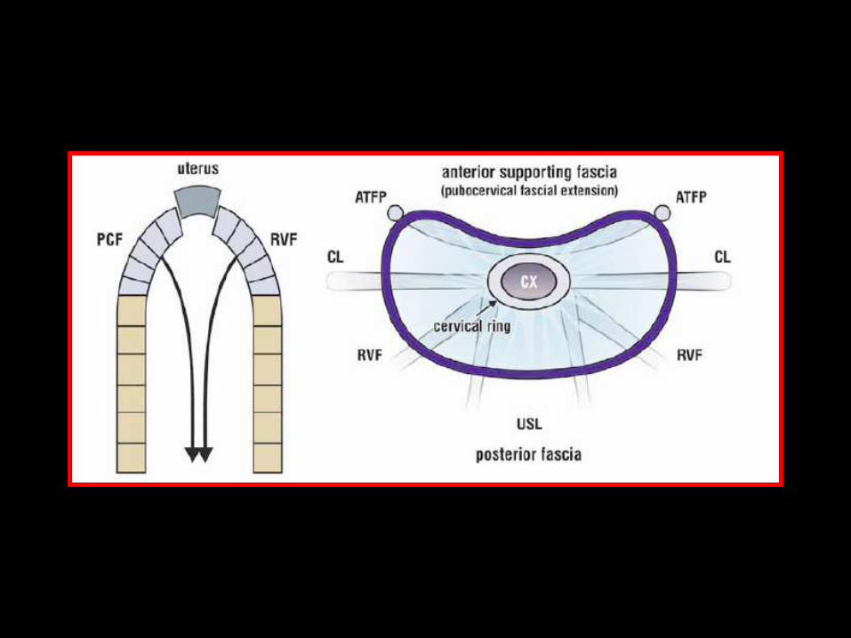
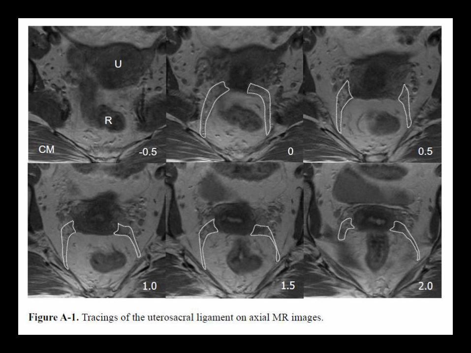
TEORIgraveA INTEGRAL
ANALOGIgraveA DEL
PUENTE
BIOMECAacuteNICA DE LA
VAGINA
TEORIgraveA INTEGRAL
ldquo ANALOGIgraveA DEL PUENTErdquo
LOS LIGAMENTOS NERVIOS Y MUgraveSCULOS
TRABAJAN SINEgraveRGICAMENTE PARA
MANTENER LA ESTRUCTURA Y FUNCIOgraveN
DEL PISO PEgraveLVICO
EL DEBILITAMIENTO DE CUALQUIER PARTE
DE LA ESTRUCTURA PUEDE ALTERAR EL
EQUILIBRIO FORTALEZA Y FUNCIOgraveN DEL
TODO
TEORIgraveA
INTEGRAL
EN RELACIOgraveN A LA ANALOGIA LA FORMA
SE OBTIENE DEBIDO A QUE LA VAGINA Y
LA VEJIGA ESTAN SUSPENDIDAS DESDE
LA PORCION OgraveSEA POR LIGAMENTOS
(ligamentos pubouretrales-PUL ligamento
uacutetero sacro-USL y arco tendineo-ATFP)
LA DIMENSIOgraveN ESTRUCTURAL SE OBTIENE
POR EL ESTIRAMIENTO DE LAS FUERZAS
MUSCULARES
RELACIOgraveN DEL SEGMENTO APICAL CON
LOS OTROS SEGMENTOS
RELACION DEL AgravePEX CON
SEGMENTO POSTERIOR
P
Recto
Uacutetero
Vagina
Defecto Posterior
PROLAPSO VAGINAL POSTERIOR
PROLAPSO VAGINAL POSTERIOR
P
Recto
Utero
Vagina
Prolapso pared vaginal posterior
Evolucioacuten DefectoPericervical
PROLAPSO PARED VAGINAL POSTERIOR
P
Recto
Utero
Vagina
Prolapso pared vaginal posterior
ldquoRectocelerdquo
PROLAPSO VAGINAL POSTERIOR
ENFOQUE QUIRUacuteRGICO
P
RectoUtero
Vagina
Reparacioacuten anillo Pericervical
P
Recto
Utero
Vagina
ENFOQUE QUIRUacuteRGICO
BIOMECANICA DEL DANtildeO DEL TEJIDO CONECTIVO
DE LA ZONA POSTERIOR
El cuerpo perineal y los ligamentos uacutetero
sacros son los puntos de anclaje para el
estiramiento de la vagina en un vector de
fuerzas hacia atraacutes y abajo
Estas fuerzas suspenden la pared vaginal
anterior y la pared rectal anterior a manera de
cuerdas tensionadas por las fuerzas
musculares
La ldquo estirabilidad ldquo de la vagina esta en relacioacuten a la micro arquitectura del colaacutegeno y elastina Esta red de fibras elaacutesticas mantiene la forma del oacutergano
Con la edad y el dantildeo durante el parto las fibras de elastina disminuyen se sobre-distiende la vagina se sobrestiran los ligamentos uacutetero sacros la pared vaginal anterior y el cuerpo perineal
BIOMECANICA DEL DANtildeO DEL TEJIDO CONECTIVO DE
LA ZONA POSTERIOR
BIOMECAacuteNICA DE
LA REPARACION
BIOMECAacuteNICA DE LA
REPARACIOacuteN
EL CUERPO PERINEAL (CP) Y LOS LIGAMENTOS
UTEROSACROS (LUS) SON AL MENOS SEIS VECES
MAS FUERTES QUE EL TEJIDO QUE SOSTIENEN
EL CUERPO PERINEAL OCUPA EL 50 DE LA PARED
VAGINAL POSTERIOR Y SE COMPORTA COMO
ELEMENTO CLAVE ACTIVO EN EL SOPORTE
ESTRUCTURAL
TANTO EL CP COMO LOS LUS SON PUNTOS DE IN
SERCION CLAVES DE LOS VECTORES
MUSCULARES DE FUERZA
BIOMECANICA DE LA
REPARACION
NO SOLO SE DEBE REPARAR EL DEFECTO FASCIAL SINO LOS ELEMENTOS DE SOPORTE ( CP Y LUS)
SI EL LUS ES DEBIL SLING POSTERIOR DE POgraveLIPROPILENO
DEBE PRESERVARSE EL ESPACIO RECTO VAGINAL PARA PERMITIR EL MOVIMIENTO INDEPENDIENTE DE LA VAGINA Y EL RECTO
CONSERVAR EL TEJIDO VAGINAL EN LO POSIBLE Y REPARACION SEPARADA DE LAS FASCIAS ESPECIALMENTE SI SE UTILIZA MALLA UNA VAGINA ESTRECHA PIERDE SUS PROPIEDADES DE ANGULACION Y ESTIRAMIENTO EN RELACION AL CP
BIOMECANICA DE LA
REPARACION
SE REQUIERE UN CUERPO PERINEAL Y
LIGAMENTOS UTEROSACROS
COMPETENTES PARA RESTAURAR EL
SOPORTE ESTRUCTURAL ACTIVO Y
PASICO AL RECTO Y VAGINA
SI SE DEBE USAR MALLA DEBE
COLOCARSE DESPUES DE LA
REPARACION ANATOMICA
RELACION DEL AgravePEX
CON SEGMENTO
ANTERIOR
Advanced anterior vaginal wall prolapse is highly
correlated with apical prolapse
Kristin Rooney MD Kimberly Kenton MD MS Elizabeth
R Mueller MD Mary Pat FitzGerald MD Linda Brubaker
MD MS
Volume 195 Issue 6 Pages 1837-1840 (December 2006)
yesp latfo rm +mauthorauthor
Objective
The purpose of this study was to determine the relationship between
the most prolapsed portion of the anterior and posterior vaginal walls
and the apex
Conclusion
Anterior vaginal wall prolapse is
associated strongly with apical
prolapse Anterior vaginal wall defects
that are surgically repaired usually
require a concomitant repair of the
apex
Interaction Among Apical
Support Levator Ani Impairment and
Anterior Vaginal Wall Prolapse
Chen Luyun MS Ashton-Miller James
A PhD Hsu Yvonne MD
DeLancey John O L MDObstetrics amp Gynecology
August 2006 - Volume 108 - Issue 2 - pp 324-332
doi 10109701AOG000022778669257a8
OBJECTIVE To use a biomechanical model to explore how impairment
of the pubovisceral portion of the levator ani muscle the apical vaginal
suspension complex or both might interact to affect anterior vaginal wall
prolapse severity
RESULTS Under raised intra-abdominal pressure the magnitude of
anterior vaginal wall prolapse was shown to be a combined function of
both pubovisceral muscle and uterosacral and cardinal ligament (apical
supports) impairment Once a certain degree of pubovisceral
impairment was reached the genital hiatus opened and a prolapse
developed The larger the pubovisceral impairment the larger the
anterior wall prolapse became A 90 impairment of apical support led
to an increase in anterior wall prolapse from 03 cm to 19 cm (a 530
increase) at 60 pubovisceral muscle impairment and from 07 cm to
24 cm (a 240 increase) at 80 pubovisceral muscle impairment
CONCLUSION These results suggest that a prolapse can develop as a
result of impairment of the muscular and apical supports of the anterior
vaginal wall
LEVEL OF EVIDENCE II-2
A 3D finite element model of anterior vaginal wall
Support to evaluate mechanisms underlying
cystocele formation
Luyun Chen James A Ashton-Miller
John OL DeLancey
Volume 42 Issue 10 Pages 1371-1377
(22 July 2009)
yesp latfo rm +mautho rautho r
A 3D finite element model of anterior vaginal wall support to
evaluate mechanisms underlying cystocele formation
Chen L Ashton-Miller JA DeLancey JO
Biomechanics Research Laboratory Department of Biomedical
Engineering University of Michigan Ann Arbor MI 48109-2125 USA
luyun_chenhotmailcom
OBJECTIVES To develop a 3D computer model of the anterior
vaginal wall and its supports validate that model and then use it to
determine the combinations of muscle and connective tissue
impairments that result in cystocele formation as observed on
dynamic magnetic resonance imaging (MRI)
RESULTS Cystocele size was sensitive to abdominal pressure and
impairment of connective tissue and muscle Larger cystocele formed
in the presence of impairments in muscular and apical connective
tissue support compared to either support element alone Apical
impairment resulted in a larger cystocele than paravaginal
impairment Levator ani muscle impairment caused a larger
urogenital hiatus size longer length of the distal vagina exposed to a
pressure differential larger apical descent and resulted in a larger
cystocele size
CONCLUSIONS Development of a cystocele requires a levator
muscle impairment an increase in abdominal pressure and apical
and paravaginal support defects
El modelo sugiere que el deterioro del elevador
del ano y del soporte apical tienen un papel
importante en la geacutenesis del cistocele Un
deterioro del 80 del soporte del aacutepex implica un
incremento en 33 del tamantildeo del cistocele
comparativamente con un soporte apical normal
El cistocele es el resultante de muacuteltiples
mecanismos El soporte apical defectuoso
produce un cistocele mayor comparado con un
defecto paravaginal
CONCEPTO DE LA PIEDRA ANGULAR
PARED VAGINAL
ANILLO PERICERVICAL
AN
TE
RIO
R
PO
ST
ER
IOR
PIEDRA ANGULAR
PARED VAGINAL
AN
TE
RIO
R
PO
ST
ER
IOR
PIEDRA ANGULAR
PARED VAGINAL
AN
TE
RIO
R
PO
ST
ER
IOR
PROLAPSOS
PIEDRA ANGULAR
PARED VAGINAL
AN
TE
RIO
R
PO
ST
ER
IOR
PR
OL
AP
SO
S
PIEDRA ANGULAR
CONCLUSIONE
S
DESINSERCIOgraveN DE LAS FASCIAS DESDE ELANILLO PERICERVICAL PROVOCA ELDEFECTO RESPONSABLE DE LOSPROLAPSOS
DEFECTOS TRANSVERSOS PARECEN SERLOS MAS IMPORTANTES
LOS DEFECTOS FASCIALES PUEDEN SERUgraveNICOS O MULTIPLES EN UNO O VARIOSCOMPARTIMIENTOS
REPARACIOgraveN SITIO ESPECIFICA CIRUGIACOMPENSADORA
CONCLUSION
ES
EXISTEN NUEVOS PARADIGMAS EN LA CONCEPCIOgraveN FISIOPATOLOgraveGICA DE LOS DEFECTOS DEL PISO PEgraveLVICO
LA REINSERCIOgraveN DE LAS FASCIAS AL ANILLO PERICERVICAL DEBE SER UN PASO INDISPENSABLE EN LA REPARACIOgraveN DE DEFECTOS DEL PISO PEgraveLVICO
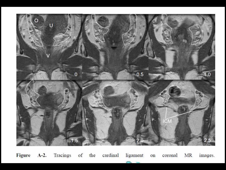
BIOMECAacuteNICA DE LA
VAGINA
TEORIgraveA INTEGRAL
ldquo ANALOGIgraveA DEL PUENTErdquo
LOS LIGAMENTOS NERVIOS Y MUgraveSCULOS
TRABAJAN SINEgraveRGICAMENTE PARA
MANTENER LA ESTRUCTURA Y FUNCIOgraveN
DEL PISO PEgraveLVICO
EL DEBILITAMIENTO DE CUALQUIER PARTE
DE LA ESTRUCTURA PUEDE ALTERAR EL
EQUILIBRIO FORTALEZA Y FUNCIOgraveN DEL
TODO
TEORIgraveA
INTEGRAL
EN RELACIOgraveN A LA ANALOGIA LA FORMA
SE OBTIENE DEBIDO A QUE LA VAGINA Y
LA VEJIGA ESTAN SUSPENDIDAS DESDE
LA PORCION OgraveSEA POR LIGAMENTOS
(ligamentos pubouretrales-PUL ligamento
uacutetero sacro-USL y arco tendineo-ATFP)
LA DIMENSIOgraveN ESTRUCTURAL SE OBTIENE
POR EL ESTIRAMIENTO DE LAS FUERZAS
MUSCULARES
RELACIOgraveN DEL SEGMENTO APICAL CON
LOS OTROS SEGMENTOS
RELACION DEL AgravePEX CON
SEGMENTO POSTERIOR
P
Recto
Uacutetero
Vagina
Defecto Posterior
PROLAPSO VAGINAL POSTERIOR
PROLAPSO VAGINAL POSTERIOR
P
Recto
Utero
Vagina
Prolapso pared vaginal posterior
Evolucioacuten DefectoPericervical
PROLAPSO PARED VAGINAL POSTERIOR
P
Recto
Utero
Vagina
Prolapso pared vaginal posterior
ldquoRectocelerdquo
PROLAPSO VAGINAL POSTERIOR
ENFOQUE QUIRUacuteRGICO
P
RectoUtero
Vagina
Reparacioacuten anillo Pericervical
P
Recto
Utero
Vagina
ENFOQUE QUIRUacuteRGICO
BIOMECANICA DEL DANtildeO DEL TEJIDO CONECTIVO
DE LA ZONA POSTERIOR
El cuerpo perineal y los ligamentos uacutetero
sacros son los puntos de anclaje para el
estiramiento de la vagina en un vector de
fuerzas hacia atraacutes y abajo
Estas fuerzas suspenden la pared vaginal
anterior y la pared rectal anterior a manera de
cuerdas tensionadas por las fuerzas
musculares
La ldquo estirabilidad ldquo de la vagina esta en relacioacuten a la micro arquitectura del colaacutegeno y elastina Esta red de fibras elaacutesticas mantiene la forma del oacutergano
Con la edad y el dantildeo durante el parto las fibras de elastina disminuyen se sobre-distiende la vagina se sobrestiran los ligamentos uacutetero sacros la pared vaginal anterior y el cuerpo perineal
BIOMECANICA DEL DANtildeO DEL TEJIDO CONECTIVO DE
LA ZONA POSTERIOR
BIOMECAacuteNICA DE
LA REPARACION
BIOMECAacuteNICA DE LA
REPARACIOacuteN
EL CUERPO PERINEAL (CP) Y LOS LIGAMENTOS
UTEROSACROS (LUS) SON AL MENOS SEIS VECES
MAS FUERTES QUE EL TEJIDO QUE SOSTIENEN
EL CUERPO PERINEAL OCUPA EL 50 DE LA PARED
VAGINAL POSTERIOR Y SE COMPORTA COMO
ELEMENTO CLAVE ACTIVO EN EL SOPORTE
ESTRUCTURAL
TANTO EL CP COMO LOS LUS SON PUNTOS DE IN
SERCION CLAVES DE LOS VECTORES
MUSCULARES DE FUERZA
BIOMECANICA DE LA
REPARACION
NO SOLO SE DEBE REPARAR EL DEFECTO FASCIAL SINO LOS ELEMENTOS DE SOPORTE ( CP Y LUS)
SI EL LUS ES DEBIL SLING POSTERIOR DE POgraveLIPROPILENO
DEBE PRESERVARSE EL ESPACIO RECTO VAGINAL PARA PERMITIR EL MOVIMIENTO INDEPENDIENTE DE LA VAGINA Y EL RECTO
CONSERVAR EL TEJIDO VAGINAL EN LO POSIBLE Y REPARACION SEPARADA DE LAS FASCIAS ESPECIALMENTE SI SE UTILIZA MALLA UNA VAGINA ESTRECHA PIERDE SUS PROPIEDADES DE ANGULACION Y ESTIRAMIENTO EN RELACION AL CP
BIOMECANICA DE LA
REPARACION
SE REQUIERE UN CUERPO PERINEAL Y
LIGAMENTOS UTEROSACROS
COMPETENTES PARA RESTAURAR EL
SOPORTE ESTRUCTURAL ACTIVO Y
PASICO AL RECTO Y VAGINA
SI SE DEBE USAR MALLA DEBE
COLOCARSE DESPUES DE LA
REPARACION ANATOMICA
RELACION DEL AgravePEX
CON SEGMENTO
ANTERIOR
Advanced anterior vaginal wall prolapse is highly
correlated with apical prolapse
Kristin Rooney MD Kimberly Kenton MD MS Elizabeth
R Mueller MD Mary Pat FitzGerald MD Linda Brubaker
MD MS
Volume 195 Issue 6 Pages 1837-1840 (December 2006)
yesp latfo rm +mauthorauthor
Objective
The purpose of this study was to determine the relationship between
the most prolapsed portion of the anterior and posterior vaginal walls
and the apex
Conclusion
Anterior vaginal wall prolapse is
associated strongly with apical
prolapse Anterior vaginal wall defects
that are surgically repaired usually
require a concomitant repair of the
apex
Interaction Among Apical
Support Levator Ani Impairment and
Anterior Vaginal Wall Prolapse
Chen Luyun MS Ashton-Miller James
A PhD Hsu Yvonne MD
DeLancey John O L MDObstetrics amp Gynecology
August 2006 - Volume 108 - Issue 2 - pp 324-332
doi 10109701AOG000022778669257a8
OBJECTIVE To use a biomechanical model to explore how impairment
of the pubovisceral portion of the levator ani muscle the apical vaginal
suspension complex or both might interact to affect anterior vaginal wall
prolapse severity
RESULTS Under raised intra-abdominal pressure the magnitude of
anterior vaginal wall prolapse was shown to be a combined function of
both pubovisceral muscle and uterosacral and cardinal ligament (apical
supports) impairment Once a certain degree of pubovisceral
impairment was reached the genital hiatus opened and a prolapse
developed The larger the pubovisceral impairment the larger the
anterior wall prolapse became A 90 impairment of apical support led
to an increase in anterior wall prolapse from 03 cm to 19 cm (a 530
increase) at 60 pubovisceral muscle impairment and from 07 cm to
24 cm (a 240 increase) at 80 pubovisceral muscle impairment
CONCLUSION These results suggest that a prolapse can develop as a
result of impairment of the muscular and apical supports of the anterior
vaginal wall
LEVEL OF EVIDENCE II-2
A 3D finite element model of anterior vaginal wall
Support to evaluate mechanisms underlying
cystocele formation
Luyun Chen James A Ashton-Miller
John OL DeLancey
Volume 42 Issue 10 Pages 1371-1377
(22 July 2009)
yesp latfo rm +mautho rautho r
A 3D finite element model of anterior vaginal wall support to
evaluate mechanisms underlying cystocele formation
Chen L Ashton-Miller JA DeLancey JO
Biomechanics Research Laboratory Department of Biomedical
Engineering University of Michigan Ann Arbor MI 48109-2125 USA
luyun_chenhotmailcom
OBJECTIVES To develop a 3D computer model of the anterior
vaginal wall and its supports validate that model and then use it to
determine the combinations of muscle and connective tissue
impairments that result in cystocele formation as observed on
dynamic magnetic resonance imaging (MRI)
RESULTS Cystocele size was sensitive to abdominal pressure and
impairment of connective tissue and muscle Larger cystocele formed
in the presence of impairments in muscular and apical connective
tissue support compared to either support element alone Apical
impairment resulted in a larger cystocele than paravaginal
impairment Levator ani muscle impairment caused a larger
urogenital hiatus size longer length of the distal vagina exposed to a
pressure differential larger apical descent and resulted in a larger
cystocele size
CONCLUSIONS Development of a cystocele requires a levator
muscle impairment an increase in abdominal pressure and apical
and paravaginal support defects
El modelo sugiere que el deterioro del elevador
del ano y del soporte apical tienen un papel
importante en la geacutenesis del cistocele Un
deterioro del 80 del soporte del aacutepex implica un
incremento en 33 del tamantildeo del cistocele
comparativamente con un soporte apical normal
El cistocele es el resultante de muacuteltiples
mecanismos El soporte apical defectuoso
produce un cistocele mayor comparado con un
defecto paravaginal
CONCEPTO DE LA PIEDRA ANGULAR
PARED VAGINAL
ANILLO PERICERVICAL
AN
TE
RIO
R
PO
ST
ER
IOR
PIEDRA ANGULAR
PARED VAGINAL
AN
TE
RIO
R
PO
ST
ER
IOR
PIEDRA ANGULAR
PARED VAGINAL
AN
TE
RIO
R
PO
ST
ER
IOR
PROLAPSOS
PIEDRA ANGULAR
PARED VAGINAL
AN
TE
RIO
R
PO
ST
ER
IOR
PR
OL
AP
SO
S
PIEDRA ANGULAR
CONCLUSIONE
S
DESINSERCIOgraveN DE LAS FASCIAS DESDE ELANILLO PERICERVICAL PROVOCA ELDEFECTO RESPONSABLE DE LOSPROLAPSOS
DEFECTOS TRANSVERSOS PARECEN SERLOS MAS IMPORTANTES
LOS DEFECTOS FASCIALES PUEDEN SERUgraveNICOS O MULTIPLES EN UNO O VARIOSCOMPARTIMIENTOS
REPARACIOgraveN SITIO ESPECIFICA CIRUGIACOMPENSADORA
CONCLUSION
ES
EXISTEN NUEVOS PARADIGMAS EN LA CONCEPCIOgraveN FISIOPATOLOgraveGICA DE LOS DEFECTOS DEL PISO PEgraveLVICO
LA REINSERCIOgraveN DE LAS FASCIAS AL ANILLO PERICERVICAL DEBE SER UN PASO INDISPENSABLE EN LA REPARACIOgraveN DE DEFECTOS DEL PISO PEgraveLVICO
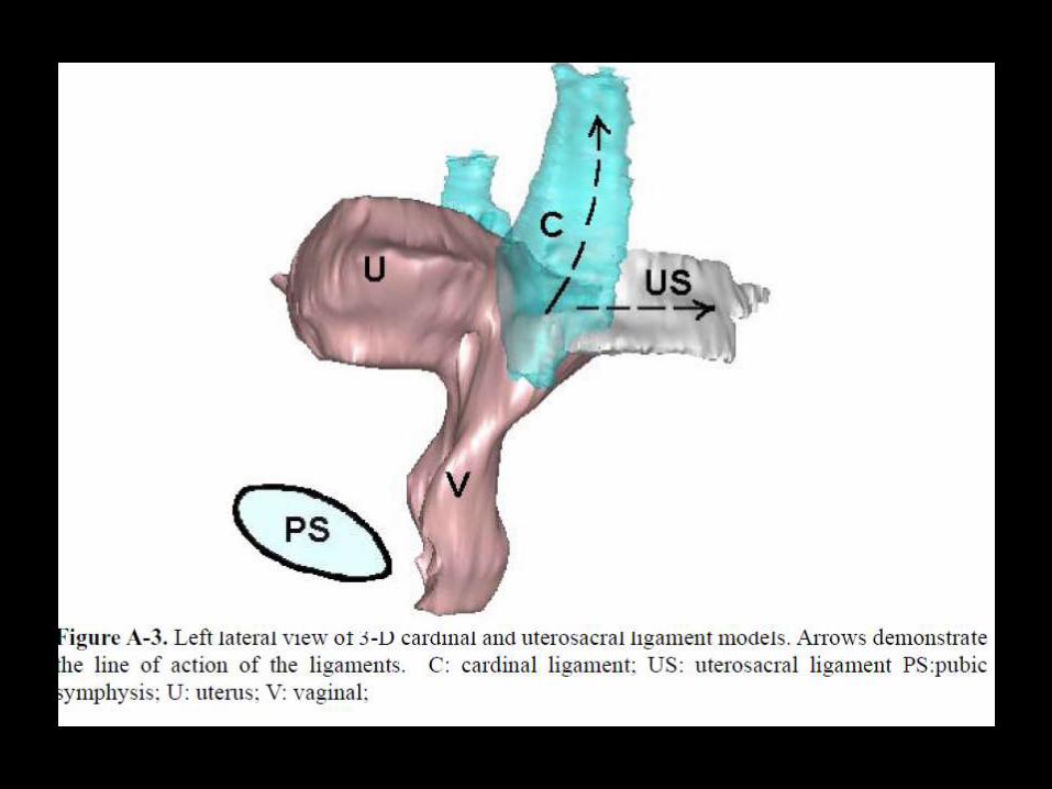
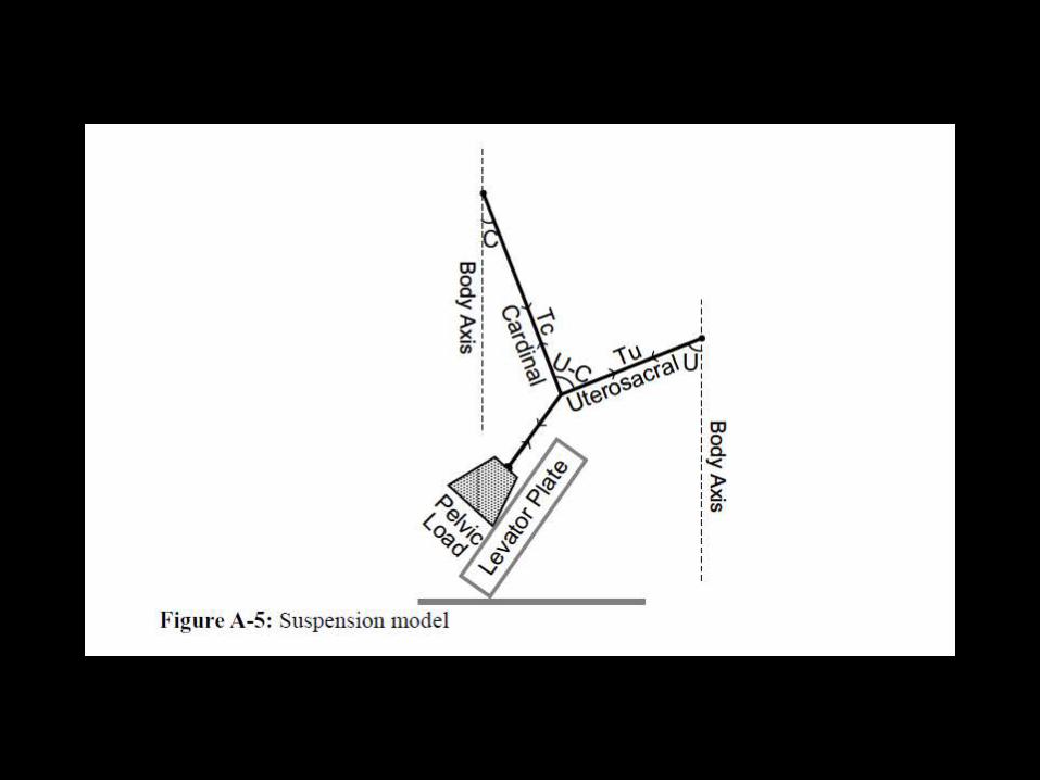
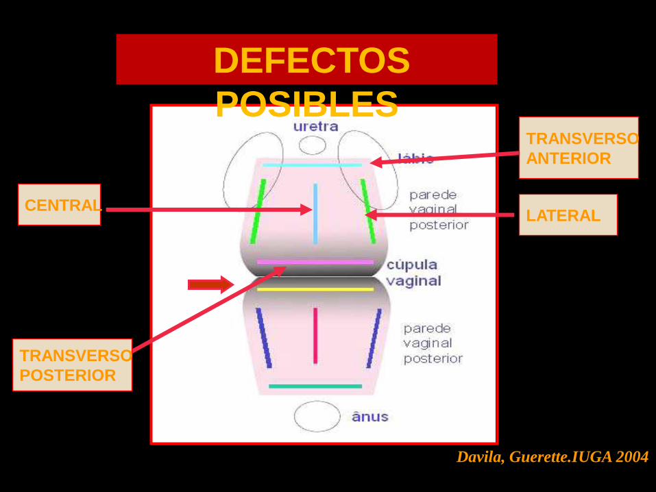
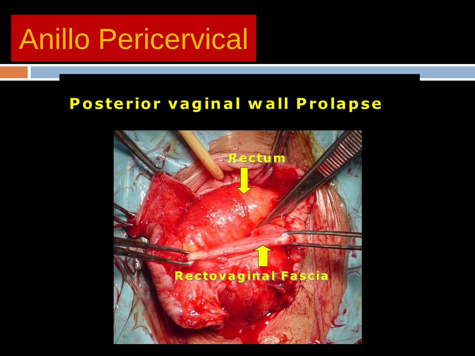
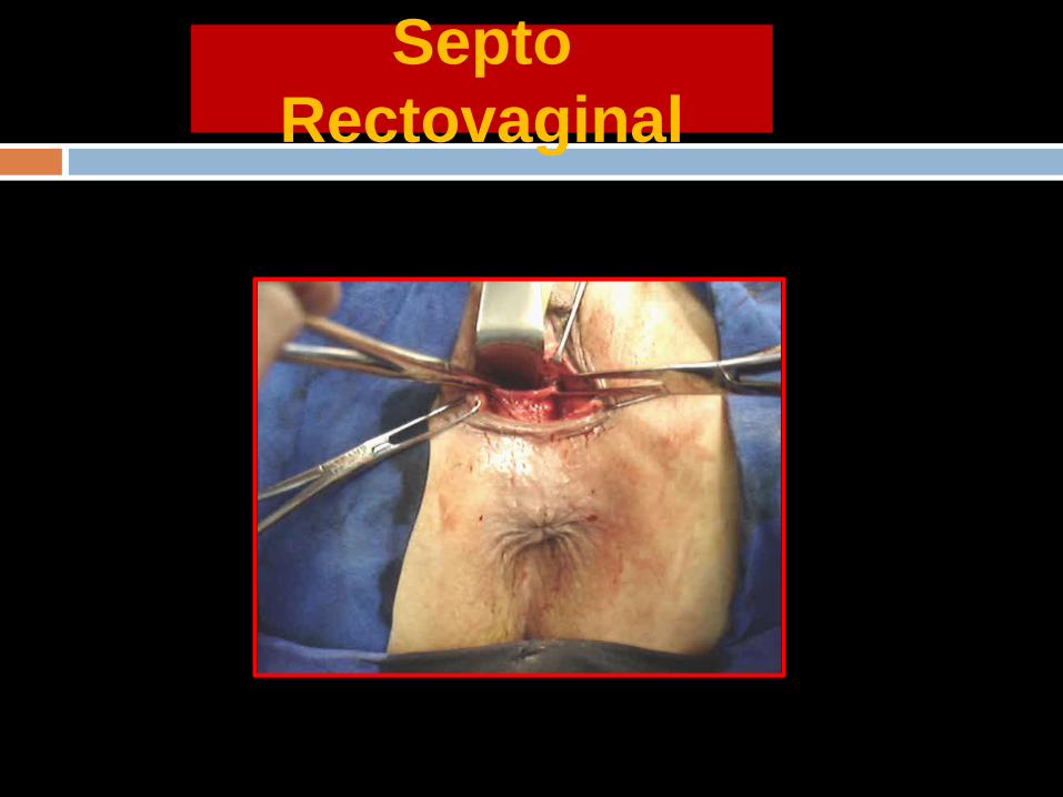
P
Recto
Uacutetero
Vagina
Defecto Posterior
PROLAPSO VAGINAL POSTERIOR
PROLAPSO VAGINAL POSTERIOR
P
Recto
Utero
Vagina
Prolapso pared vaginal posterior
Evolucioacuten DefectoPericervical
PROLAPSO PARED VAGINAL POSTERIOR
P
Recto
Utero
Vagina
Prolapso pared vaginal posterior
ldquoRectocelerdquo
PROLAPSO VAGINAL POSTERIOR
ENFOQUE QUIRUacuteRGICO
P
RectoUtero
Vagina
Reparacioacuten anillo Pericervical
P
Recto
Utero
Vagina
ENFOQUE QUIRUacuteRGICO
BIOMECANICA DEL DANtildeO DEL TEJIDO CONECTIVO
DE LA ZONA POSTERIOR
El cuerpo perineal y los ligamentos uacutetero
sacros son los puntos de anclaje para el
estiramiento de la vagina en un vector de
fuerzas hacia atraacutes y abajo
Estas fuerzas suspenden la pared vaginal
anterior y la pared rectal anterior a manera de
cuerdas tensionadas por las fuerzas
musculares
La ldquo estirabilidad ldquo de la vagina esta en relacioacuten a la micro arquitectura del colaacutegeno y elastina Esta red de fibras elaacutesticas mantiene la forma del oacutergano
Con la edad y el dantildeo durante el parto las fibras de elastina disminuyen se sobre-distiende la vagina se sobrestiran los ligamentos uacutetero sacros la pared vaginal anterior y el cuerpo perineal
BIOMECANICA DEL DANtildeO DEL TEJIDO CONECTIVO DE
LA ZONA POSTERIOR
BIOMECAacuteNICA DE
LA REPARACION
BIOMECAacuteNICA DE LA
REPARACIOacuteN
EL CUERPO PERINEAL (CP) Y LOS LIGAMENTOS
UTEROSACROS (LUS) SON AL MENOS SEIS VECES
MAS FUERTES QUE EL TEJIDO QUE SOSTIENEN
EL CUERPO PERINEAL OCUPA EL 50 DE LA PARED
VAGINAL POSTERIOR Y SE COMPORTA COMO
ELEMENTO CLAVE ACTIVO EN EL SOPORTE
ESTRUCTURAL
TANTO EL CP COMO LOS LUS SON PUNTOS DE IN
SERCION CLAVES DE LOS VECTORES
MUSCULARES DE FUERZA
BIOMECANICA DE LA
REPARACION
NO SOLO SE DEBE REPARAR EL DEFECTO FASCIAL SINO LOS ELEMENTOS DE SOPORTE ( CP Y LUS)
SI EL LUS ES DEBIL SLING POSTERIOR DE POgraveLIPROPILENO
DEBE PRESERVARSE EL ESPACIO RECTO VAGINAL PARA PERMITIR EL MOVIMIENTO INDEPENDIENTE DE LA VAGINA Y EL RECTO
CONSERVAR EL TEJIDO VAGINAL EN LO POSIBLE Y REPARACION SEPARADA DE LAS FASCIAS ESPECIALMENTE SI SE UTILIZA MALLA UNA VAGINA ESTRECHA PIERDE SUS PROPIEDADES DE ANGULACION Y ESTIRAMIENTO EN RELACION AL CP
BIOMECANICA DE LA
REPARACION
SE REQUIERE UN CUERPO PERINEAL Y
LIGAMENTOS UTEROSACROS
COMPETENTES PARA RESTAURAR EL
SOPORTE ESTRUCTURAL ACTIVO Y
PASICO AL RECTO Y VAGINA
SI SE DEBE USAR MALLA DEBE
COLOCARSE DESPUES DE LA
REPARACION ANATOMICA
RELACION DEL AgravePEX
CON SEGMENTO
ANTERIOR
Advanced anterior vaginal wall prolapse is highly
correlated with apical prolapse
Kristin Rooney MD Kimberly Kenton MD MS Elizabeth
R Mueller MD Mary Pat FitzGerald MD Linda Brubaker
MD MS
Volume 195 Issue 6 Pages 1837-1840 (December 2006)
yesp latfo rm +mauthorauthor
Objective
The purpose of this study was to determine the relationship between
the most prolapsed portion of the anterior and posterior vaginal walls
and the apex
Conclusion
Anterior vaginal wall prolapse is
associated strongly with apical
prolapse Anterior vaginal wall defects
that are surgically repaired usually
require a concomitant repair of the
apex
Interaction Among Apical
Support Levator Ani Impairment and
Anterior Vaginal Wall Prolapse
Chen Luyun MS Ashton-Miller James
A PhD Hsu Yvonne MD
DeLancey John O L MDObstetrics amp Gynecology
August 2006 - Volume 108 - Issue 2 - pp 324-332
doi 10109701AOG000022778669257a8
OBJECTIVE To use a biomechanical model to explore how impairment
of the pubovisceral portion of the levator ani muscle the apical vaginal
suspension complex or both might interact to affect anterior vaginal wall
prolapse severity
RESULTS Under raised intra-abdominal pressure the magnitude of
anterior vaginal wall prolapse was shown to be a combined function of
both pubovisceral muscle and uterosacral and cardinal ligament (apical
supports) impairment Once a certain degree of pubovisceral
impairment was reached the genital hiatus opened and a prolapse
developed The larger the pubovisceral impairment the larger the
anterior wall prolapse became A 90 impairment of apical support led
to an increase in anterior wall prolapse from 03 cm to 19 cm (a 530
increase) at 60 pubovisceral muscle impairment and from 07 cm to
24 cm (a 240 increase) at 80 pubovisceral muscle impairment
CONCLUSION These results suggest that a prolapse can develop as a
result of impairment of the muscular and apical supports of the anterior
vaginal wall
LEVEL OF EVIDENCE II-2
A 3D finite element model of anterior vaginal wall
Support to evaluate mechanisms underlying
cystocele formation
Luyun Chen James A Ashton-Miller
John OL DeLancey
Volume 42 Issue 10 Pages 1371-1377
(22 July 2009)
yesp latfo rm +mautho rautho r
A 3D finite element model of anterior vaginal wall support to
evaluate mechanisms underlying cystocele formation
Chen L Ashton-Miller JA DeLancey JO
Biomechanics Research Laboratory Department of Biomedical
Engineering University of Michigan Ann Arbor MI 48109-2125 USA
luyun_chenhotmailcom
OBJECTIVES To develop a 3D computer model of the anterior
vaginal wall and its supports validate that model and then use it to
determine the combinations of muscle and connective tissue
impairments that result in cystocele formation as observed on
dynamic magnetic resonance imaging (MRI)
RESULTS Cystocele size was sensitive to abdominal pressure and
impairment of connective tissue and muscle Larger cystocele formed
in the presence of impairments in muscular and apical connective
tissue support compared to either support element alone Apical
impairment resulted in a larger cystocele than paravaginal
impairment Levator ani muscle impairment caused a larger
urogenital hiatus size longer length of the distal vagina exposed to a
pressure differential larger apical descent and resulted in a larger
cystocele size
CONCLUSIONS Development of a cystocele requires a levator
muscle impairment an increase in abdominal pressure and apical
and paravaginal support defects
El modelo sugiere que el deterioro del elevador
del ano y del soporte apical tienen un papel
importante en la geacutenesis del cistocele Un
deterioro del 80 del soporte del aacutepex implica un
incremento en 33 del tamantildeo del cistocele
comparativamente con un soporte apical normal
El cistocele es el resultante de muacuteltiples
mecanismos El soporte apical defectuoso
produce un cistocele mayor comparado con un
defecto paravaginal
CONCEPTO DE LA PIEDRA ANGULAR
PARED VAGINAL
ANILLO PERICERVICAL
AN
TE
RIO
R
PO
ST
ER
IOR
PIEDRA ANGULAR
PARED VAGINAL
AN
TE
RIO
R
PO
ST
ER
IOR
PIEDRA ANGULAR
PARED VAGINAL
AN
TE
RIO
R
PO
ST
ER
IOR
PROLAPSOS
PIEDRA ANGULAR
PARED VAGINAL
AN
TE
RIO
R
PO
ST
ER
IOR
PR
OL
AP
SO
S
PIEDRA ANGULAR
CONCLUSIONE
S
DESINSERCIOgraveN DE LAS FASCIAS DESDE ELANILLO PERICERVICAL PROVOCA ELDEFECTO RESPONSABLE DE LOSPROLAPSOS
DEFECTOS TRANSVERSOS PARECEN SERLOS MAS IMPORTANTES
LOS DEFECTOS FASCIALES PUEDEN SERUgraveNICOS O MULTIPLES EN UNO O VARIOSCOMPARTIMIENTOS
REPARACIOgraveN SITIO ESPECIFICA CIRUGIACOMPENSADORA
CONCLUSION
ES
EXISTEN NUEVOS PARADIGMAS EN LA CONCEPCIOgraveN FISIOPATOLOgraveGICA DE LOS DEFECTOS DEL PISO PEgraveLVICO
LA REINSERCIOgraveN DE LAS FASCIAS AL ANILLO PERICERVICAL DEBE SER UN PASO INDISPENSABLE EN LA REPARACIOgraveN DE DEFECTOS DEL PISO PEgraveLVICO
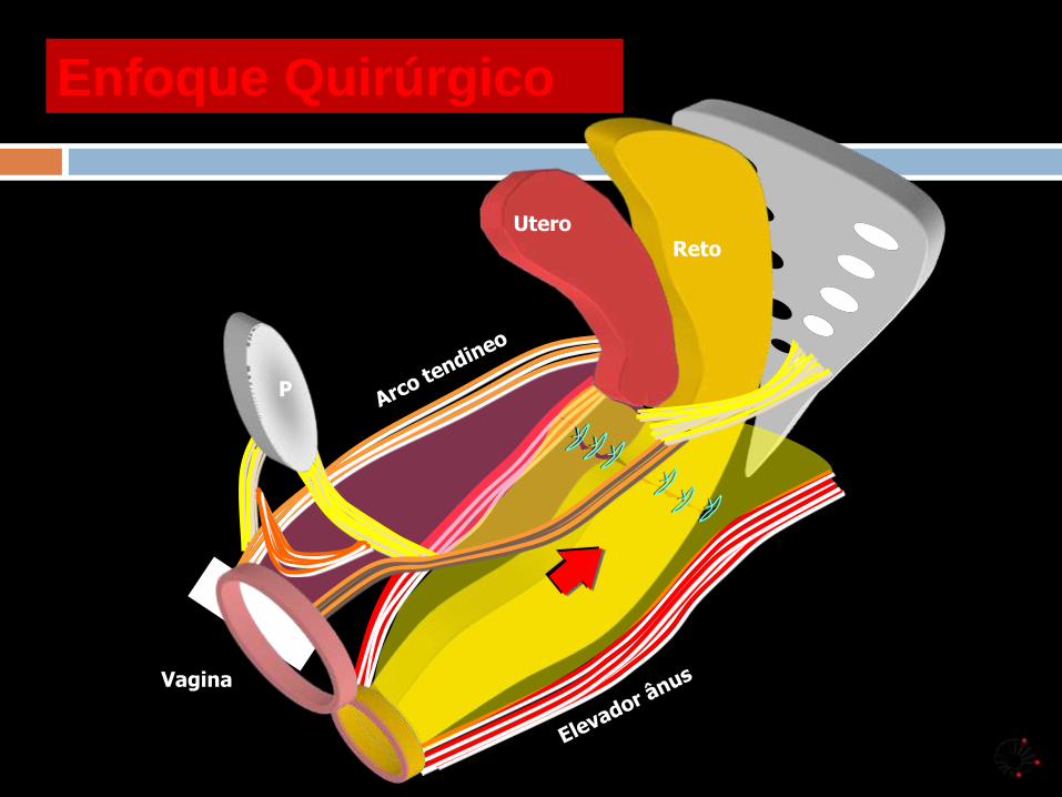
PROLAPSO VAGINAL POSTERIOR
P
Recto
Utero
Vagina
Prolapso pared vaginal posterior
Evolucioacuten DefectoPericervical
PROLAPSO PARED VAGINAL POSTERIOR
P
Recto
Utero
Vagina
Prolapso pared vaginal posterior
ldquoRectocelerdquo
PROLAPSO VAGINAL POSTERIOR
ENFOQUE QUIRUacuteRGICO
P
RectoUtero
Vagina
Reparacioacuten anillo Pericervical
P
Recto
Utero
Vagina
ENFOQUE QUIRUacuteRGICO
BIOMECANICA DEL DANtildeO DEL TEJIDO CONECTIVO
DE LA ZONA POSTERIOR
El cuerpo perineal y los ligamentos uacutetero
sacros son los puntos de anclaje para el
estiramiento de la vagina en un vector de
fuerzas hacia atraacutes y abajo
Estas fuerzas suspenden la pared vaginal
anterior y la pared rectal anterior a manera de
cuerdas tensionadas por las fuerzas
musculares
La ldquo estirabilidad ldquo de la vagina esta en relacioacuten a la micro arquitectura del colaacutegeno y elastina Esta red de fibras elaacutesticas mantiene la forma del oacutergano
Con la edad y el dantildeo durante el parto las fibras de elastina disminuyen se sobre-distiende la vagina se sobrestiran los ligamentos uacutetero sacros la pared vaginal anterior y el cuerpo perineal
BIOMECANICA DEL DANtildeO DEL TEJIDO CONECTIVO DE
LA ZONA POSTERIOR
BIOMECAacuteNICA DE
LA REPARACION
BIOMECAacuteNICA DE LA
REPARACIOacuteN
EL CUERPO PERINEAL (CP) Y LOS LIGAMENTOS
UTEROSACROS (LUS) SON AL MENOS SEIS VECES
MAS FUERTES QUE EL TEJIDO QUE SOSTIENEN
EL CUERPO PERINEAL OCUPA EL 50 DE LA PARED
VAGINAL POSTERIOR Y SE COMPORTA COMO
ELEMENTO CLAVE ACTIVO EN EL SOPORTE
ESTRUCTURAL
TANTO EL CP COMO LOS LUS SON PUNTOS DE IN
SERCION CLAVES DE LOS VECTORES
MUSCULARES DE FUERZA
BIOMECANICA DE LA
REPARACION
NO SOLO SE DEBE REPARAR EL DEFECTO FASCIAL SINO LOS ELEMENTOS DE SOPORTE ( CP Y LUS)
SI EL LUS ES DEBIL SLING POSTERIOR DE POgraveLIPROPILENO
DEBE PRESERVARSE EL ESPACIO RECTO VAGINAL PARA PERMITIR EL MOVIMIENTO INDEPENDIENTE DE LA VAGINA Y EL RECTO
CONSERVAR EL TEJIDO VAGINAL EN LO POSIBLE Y REPARACION SEPARADA DE LAS FASCIAS ESPECIALMENTE SI SE UTILIZA MALLA UNA VAGINA ESTRECHA PIERDE SUS PROPIEDADES DE ANGULACION Y ESTIRAMIENTO EN RELACION AL CP
BIOMECANICA DE LA
REPARACION
SE REQUIERE UN CUERPO PERINEAL Y
LIGAMENTOS UTEROSACROS
COMPETENTES PARA RESTAURAR EL
SOPORTE ESTRUCTURAL ACTIVO Y
PASICO AL RECTO Y VAGINA
SI SE DEBE USAR MALLA DEBE
COLOCARSE DESPUES DE LA
REPARACION ANATOMICA
RELACION DEL AgravePEX
CON SEGMENTO
ANTERIOR
Advanced anterior vaginal wall prolapse is highly
correlated with apical prolapse
Kristin Rooney MD Kimberly Kenton MD MS Elizabeth
R Mueller MD Mary Pat FitzGerald MD Linda Brubaker
MD MS
Volume 195 Issue 6 Pages 1837-1840 (December 2006)
yesp latfo rm +mauthorauthor
Objective
The purpose of this study was to determine the relationship between
the most prolapsed portion of the anterior and posterior vaginal walls
and the apex
Conclusion
Anterior vaginal wall prolapse is
associated strongly with apical
prolapse Anterior vaginal wall defects
that are surgically repaired usually
require a concomitant repair of the
apex
Interaction Among Apical
Support Levator Ani Impairment and
Anterior Vaginal Wall Prolapse
Chen Luyun MS Ashton-Miller James
A PhD Hsu Yvonne MD
DeLancey John O L MDObstetrics amp Gynecology
August 2006 - Volume 108 - Issue 2 - pp 324-332
doi 10109701AOG000022778669257a8
OBJECTIVE To use a biomechanical model to explore how impairment
of the pubovisceral portion of the levator ani muscle the apical vaginal
suspension complex or both might interact to affect anterior vaginal wall
prolapse severity
RESULTS Under raised intra-abdominal pressure the magnitude of
anterior vaginal wall prolapse was shown to be a combined function of
both pubovisceral muscle and uterosacral and cardinal ligament (apical
supports) impairment Once a certain degree of pubovisceral
impairment was reached the genital hiatus opened and a prolapse
developed The larger the pubovisceral impairment the larger the
anterior wall prolapse became A 90 impairment of apical support led
to an increase in anterior wall prolapse from 03 cm to 19 cm (a 530
increase) at 60 pubovisceral muscle impairment and from 07 cm to
24 cm (a 240 increase) at 80 pubovisceral muscle impairment
CONCLUSION These results suggest that a prolapse can develop as a
result of impairment of the muscular and apical supports of the anterior
vaginal wall
LEVEL OF EVIDENCE II-2
A 3D finite element model of anterior vaginal wall
Support to evaluate mechanisms underlying
cystocele formation
Luyun Chen James A Ashton-Miller
John OL DeLancey
Volume 42 Issue 10 Pages 1371-1377
(22 July 2009)
yesp latfo rm +mautho rautho r
A 3D finite element model of anterior vaginal wall support to
evaluate mechanisms underlying cystocele formation
Chen L Ashton-Miller JA DeLancey JO
Biomechanics Research Laboratory Department of Biomedical
Engineering University of Michigan Ann Arbor MI 48109-2125 USA
luyun_chenhotmailcom
OBJECTIVES To develop a 3D computer model of the anterior
vaginal wall and its supports validate that model and then use it to
determine the combinations of muscle and connective tissue
impairments that result in cystocele formation as observed on
dynamic magnetic resonance imaging (MRI)
RESULTS Cystocele size was sensitive to abdominal pressure and
impairment of connective tissue and muscle Larger cystocele formed
in the presence of impairments in muscular and apical connective
tissue support compared to either support element alone Apical
impairment resulted in a larger cystocele than paravaginal
impairment Levator ani muscle impairment caused a larger
urogenital hiatus size longer length of the distal vagina exposed to a
pressure differential larger apical descent and resulted in a larger
cystocele size
CONCLUSIONS Development of a cystocele requires a levator
muscle impairment an increase in abdominal pressure and apical
and paravaginal support defects
El modelo sugiere que el deterioro del elevador
del ano y del soporte apical tienen un papel
importante en la geacutenesis del cistocele Un
deterioro del 80 del soporte del aacutepex implica un
incremento en 33 del tamantildeo del cistocele
comparativamente con un soporte apical normal
El cistocele es el resultante de muacuteltiples
mecanismos El soporte apical defectuoso
produce un cistocele mayor comparado con un
defecto paravaginal
CONCEPTO DE LA PIEDRA ANGULAR
PARED VAGINAL
ANILLO PERICERVICAL
AN
TE
RIO
R
PO
ST
ER
IOR
PIEDRA ANGULAR
PARED VAGINAL
AN
TE
RIO
R
PO
ST
ER
IOR
PIEDRA ANGULAR
PARED VAGINAL
AN
TE
RIO
R
PO
ST
ER
IOR
PROLAPSOS
PIEDRA ANGULAR
PARED VAGINAL
AN
TE
RIO
R
PO
ST
ER
IOR
PR
OL
AP
SO
S
PIEDRA ANGULAR
CONCLUSIONE
S
DESINSERCIOgraveN DE LAS FASCIAS DESDE ELANILLO PERICERVICAL PROVOCA ELDEFECTO RESPONSABLE DE LOSPROLAPSOS
DEFECTOS TRANSVERSOS PARECEN SERLOS MAS IMPORTANTES
LOS DEFECTOS FASCIALES PUEDEN SERUgraveNICOS O MULTIPLES EN UNO O VARIOSCOMPARTIMIENTOS
REPARACIOgraveN SITIO ESPECIFICA CIRUGIACOMPENSADORA
CONCLUSION
ES
EXISTEN NUEVOS PARADIGMAS EN LA CONCEPCIOgraveN FISIOPATOLOgraveGICA DE LOS DEFECTOS DEL PISO PEgraveLVICO
LA REINSERCIOgraveN DE LAS FASCIAS AL ANILLO PERICERVICAL DEBE SER UN PASO INDISPENSABLE EN LA REPARACIOgraveN DE DEFECTOS DEL PISO PEgraveLVICO
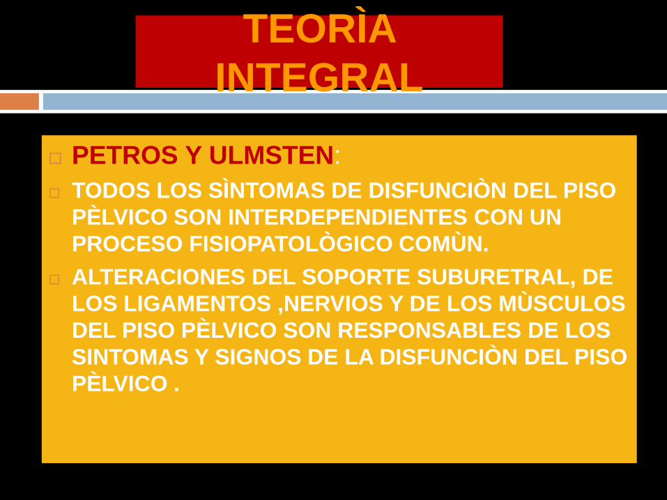
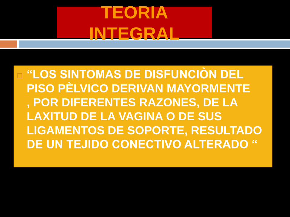
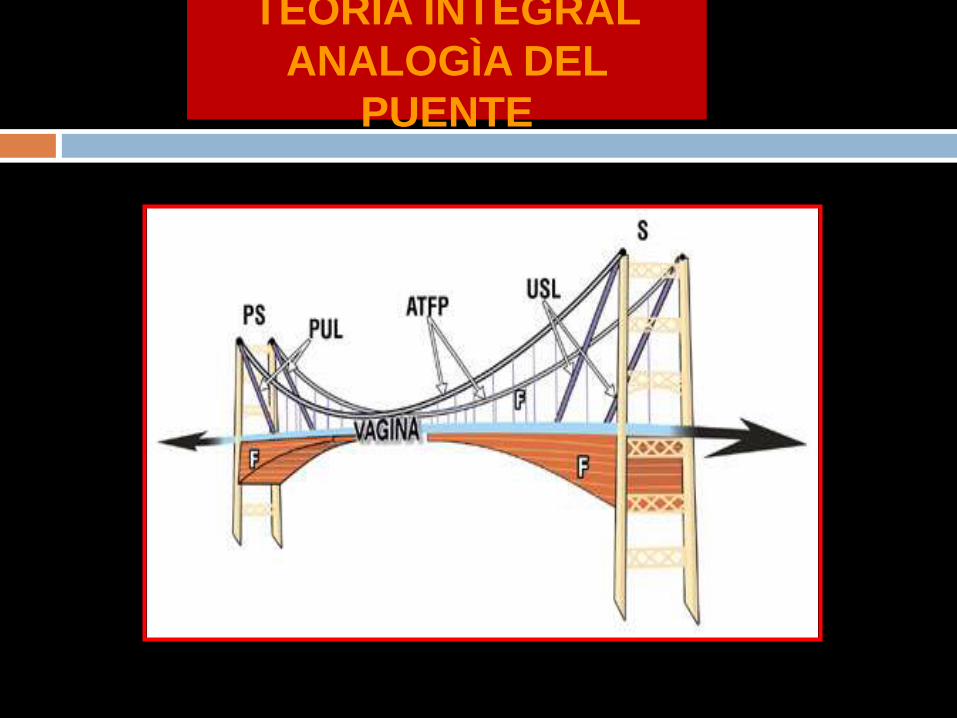
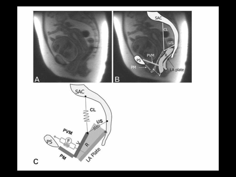
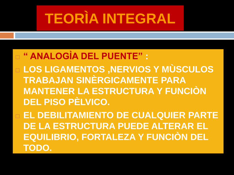
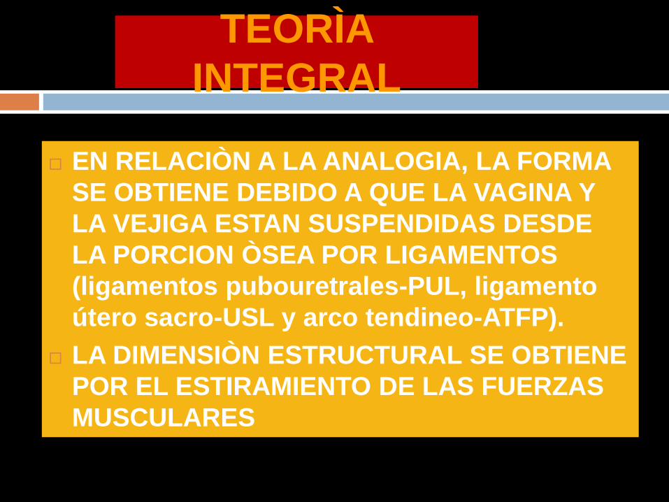
BIOMECANICA DE LA
REPARACION
NO SOLO SE DEBE REPARAR EL DEFECTO FASCIAL SINO LOS ELEMENTOS DE SOPORTE ( CP Y LUS)
SI EL LUS ES DEBIL SLING POSTERIOR DE POgraveLIPROPILENO
DEBE PRESERVARSE EL ESPACIO RECTO VAGINAL PARA PERMITIR EL MOVIMIENTO INDEPENDIENTE DE LA VAGINA Y EL RECTO
CONSERVAR EL TEJIDO VAGINAL EN LO POSIBLE Y REPARACION SEPARADA DE LAS FASCIAS ESPECIALMENTE SI SE UTILIZA MALLA UNA VAGINA ESTRECHA PIERDE SUS PROPIEDADES DE ANGULACION Y ESTIRAMIENTO EN RELACION AL CP
BIOMECANICA DE LA
REPARACION
SE REQUIERE UN CUERPO PERINEAL Y
LIGAMENTOS UTEROSACROS
COMPETENTES PARA RESTAURAR EL
SOPORTE ESTRUCTURAL ACTIVO Y
PASICO AL RECTO Y VAGINA
SI SE DEBE USAR MALLA DEBE
COLOCARSE DESPUES DE LA
REPARACION ANATOMICA
RELACION DEL AgravePEX
CON SEGMENTO
ANTERIOR
Advanced anterior vaginal wall prolapse is highly
correlated with apical prolapse
Kristin Rooney MD Kimberly Kenton MD MS Elizabeth
R Mueller MD Mary Pat FitzGerald MD Linda Brubaker
MD MS
Volume 195 Issue 6 Pages 1837-1840 (December 2006)
yesp latfo rm +mauthorauthor
Objective
The purpose of this study was to determine the relationship between
the most prolapsed portion of the anterior and posterior vaginal walls
and the apex
Conclusion
Anterior vaginal wall prolapse is
associated strongly with apical
prolapse Anterior vaginal wall defects
that are surgically repaired usually
require a concomitant repair of the
apex
Interaction Among Apical
Support Levator Ani Impairment and
Anterior Vaginal Wall Prolapse
Chen Luyun MS Ashton-Miller James
A PhD Hsu Yvonne MD
DeLancey John O L MDObstetrics amp Gynecology
August 2006 - Volume 108 - Issue 2 - pp 324-332
doi 10109701AOG000022778669257a8
OBJECTIVE To use a biomechanical model to explore how impairment
of the pubovisceral portion of the levator ani muscle the apical vaginal
suspension complex or both might interact to affect anterior vaginal wall
prolapse severity
RESULTS Under raised intra-abdominal pressure the magnitude of
anterior vaginal wall prolapse was shown to be a combined function of
both pubovisceral muscle and uterosacral and cardinal ligament (apical
supports) impairment Once a certain degree of pubovisceral
impairment was reached the genital hiatus opened and a prolapse
developed The larger the pubovisceral impairment the larger the
anterior wall prolapse became A 90 impairment of apical support led
to an increase in anterior wall prolapse from 03 cm to 19 cm (a 530
increase) at 60 pubovisceral muscle impairment and from 07 cm to
24 cm (a 240 increase) at 80 pubovisceral muscle impairment
CONCLUSION These results suggest that a prolapse can develop as a
result of impairment of the muscular and apical supports of the anterior
vaginal wall
LEVEL OF EVIDENCE II-2
A 3D finite element model of anterior vaginal wall
Support to evaluate mechanisms underlying
cystocele formation
Luyun Chen James A Ashton-Miller
John OL DeLancey
Volume 42 Issue 10 Pages 1371-1377
(22 July 2009)
yesp latfo rm +mautho rautho r
A 3D finite element model of anterior vaginal wall support to
evaluate mechanisms underlying cystocele formation
Chen L Ashton-Miller JA DeLancey JO
Biomechanics Research Laboratory Department of Biomedical
Engineering University of Michigan Ann Arbor MI 48109-2125 USA
luyun_chenhotmailcom
OBJECTIVES To develop a 3D computer model of the anterior
vaginal wall and its supports validate that model and then use it to
determine the combinations of muscle and connective tissue
impairments that result in cystocele formation as observed on
dynamic magnetic resonance imaging (MRI)
RESULTS Cystocele size was sensitive to abdominal pressure and
impairment of connective tissue and muscle Larger cystocele formed
in the presence of impairments in muscular and apical connective
tissue support compared to either support element alone Apical
impairment resulted in a larger cystocele than paravaginal
impairment Levator ani muscle impairment caused a larger
urogenital hiatus size longer length of the distal vagina exposed to a
pressure differential larger apical descent and resulted in a larger
cystocele size
CONCLUSIONS Development of a cystocele requires a levator
muscle impairment an increase in abdominal pressure and apical
and paravaginal support defects
El modelo sugiere que el deterioro del elevador
del ano y del soporte apical tienen un papel
importante en la geacutenesis del cistocele Un
deterioro del 80 del soporte del aacutepex implica un
incremento en 33 del tamantildeo del cistocele
comparativamente con un soporte apical normal
El cistocele es el resultante de muacuteltiples
mecanismos El soporte apical defectuoso
produce un cistocele mayor comparado con un
defecto paravaginal
CONCEPTO DE LA PIEDRA ANGULAR
PARED VAGINAL
ANILLO PERICERVICAL
AN
TE
RIO
R
PO
ST
ER
IOR
PIEDRA ANGULAR
PARED VAGINAL
AN
TE
RIO
R
PO
ST
ER
IOR
PIEDRA ANGULAR
PARED VAGINAL
AN
TE
RIO
R
PO
ST
ER
IOR
PROLAPSOS
PIEDRA ANGULAR
PARED VAGINAL
AN
TE
RIO
R
PO
ST
ER
IOR
PR
OL
AP
SO
S
PIEDRA ANGULAR
CONCLUSIONE
S
DESINSERCIOgraveN DE LAS FASCIAS DESDE ELANILLO PERICERVICAL PROVOCA ELDEFECTO RESPONSABLE DE LOSPROLAPSOS
DEFECTOS TRANSVERSOS PARECEN SERLOS MAS IMPORTANTES
LOS DEFECTOS FASCIALES PUEDEN SERUgraveNICOS O MULTIPLES EN UNO O VARIOSCOMPARTIMIENTOS
REPARACIOgraveN SITIO ESPECIFICA CIRUGIACOMPENSADORA
CONCLUSION
ES
EXISTEN NUEVOS PARADIGMAS EN LA CONCEPCIOgraveN FISIOPATOLOgraveGICA DE LOS DEFECTOS DEL PISO PEgraveLVICO
LA REINSERCIOgraveN DE LAS FASCIAS AL ANILLO PERICERVICAL DEBE SER UN PASO INDISPENSABLE EN LA REPARACIOgraveN DE DEFECTOS DEL PISO PEgraveLVICO
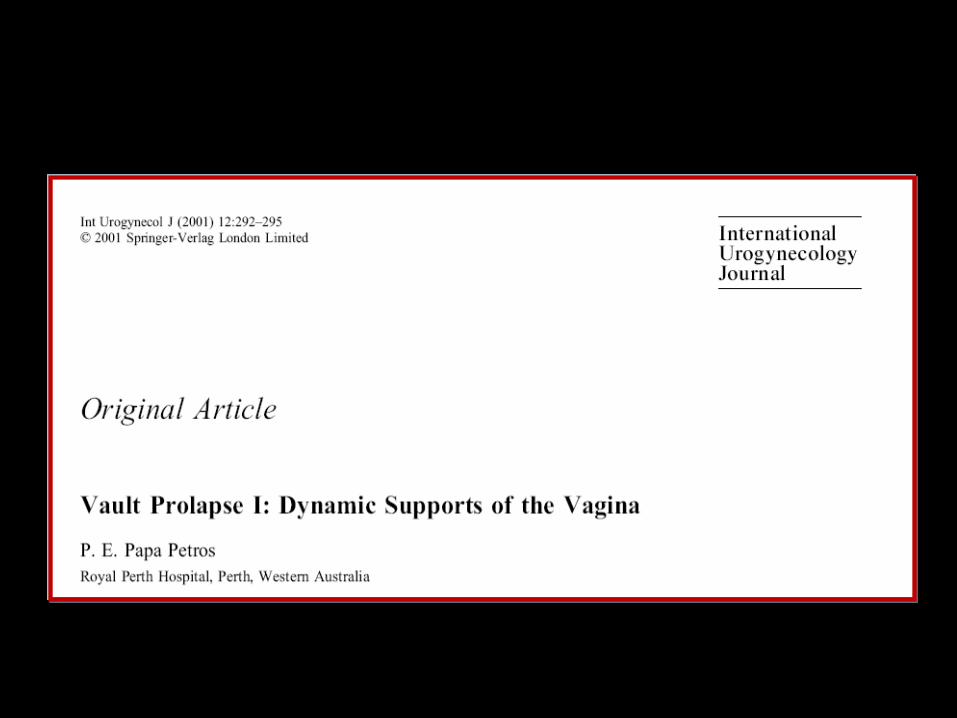
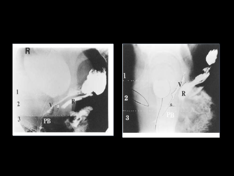
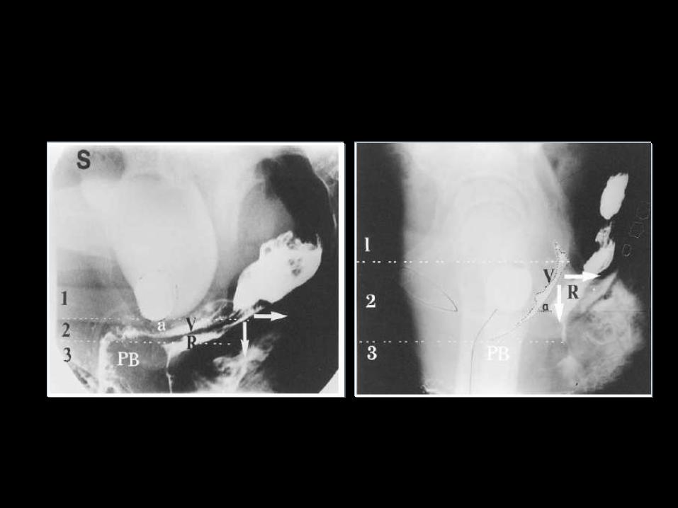
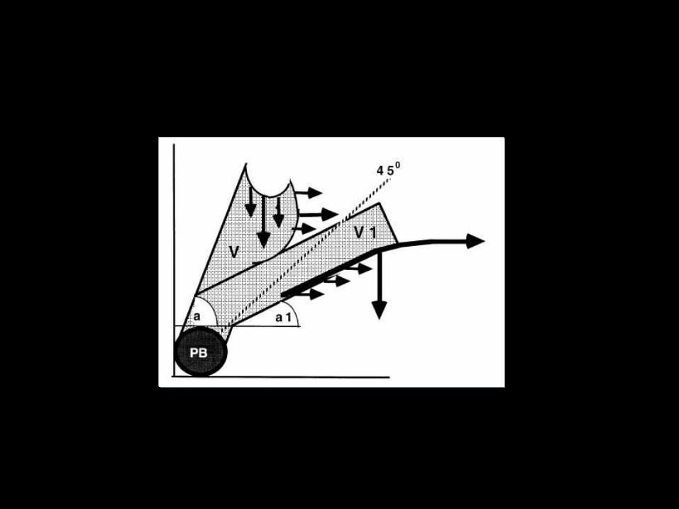
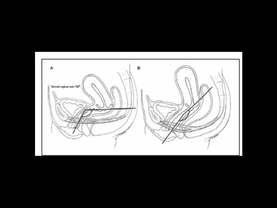
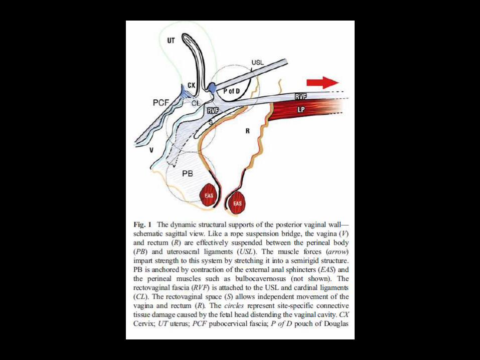
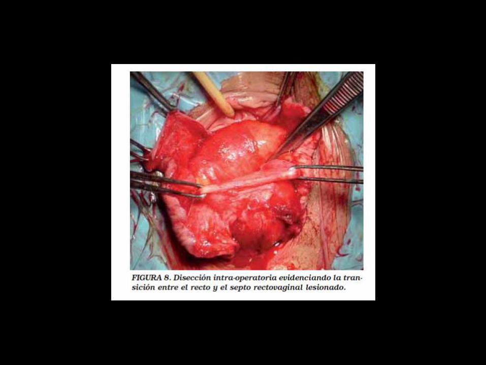
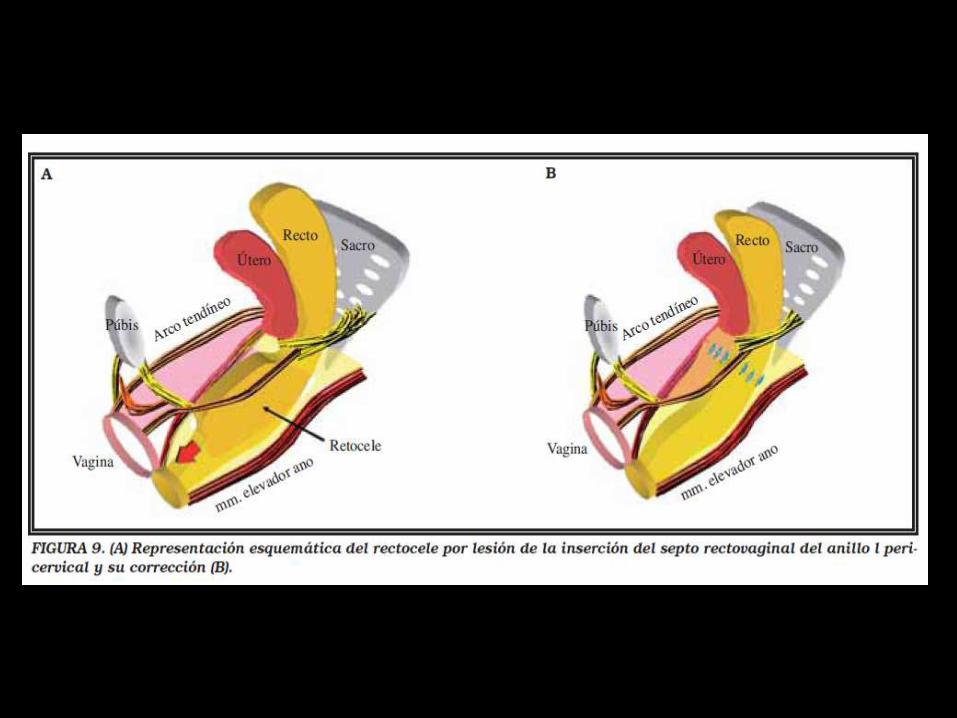
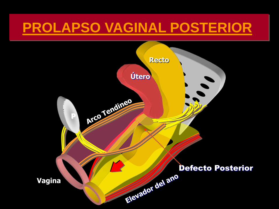
PARED VAGINAL
ANILLO PERICERVICAL
AN
TE
RIO
R
PO
ST
ER
IOR
PIEDRA ANGULAR
PARED VAGINAL
AN
TE
RIO
R
PO
ST
ER
IOR
PIEDRA ANGULAR
PARED VAGINAL
AN
TE
RIO
R
PO
ST
ER
IOR
PROLAPSOS
PIEDRA ANGULAR
PARED VAGINAL
AN
TE
RIO
R
PO
ST
ER
IOR
PR
OL
AP
SO
S
PIEDRA ANGULAR
CONCLUSIONE
S
DESINSERCIOgraveN DE LAS FASCIAS DESDE ELANILLO PERICERVICAL PROVOCA ELDEFECTO RESPONSABLE DE LOSPROLAPSOS
DEFECTOS TRANSVERSOS PARECEN SERLOS MAS IMPORTANTES
LOS DEFECTOS FASCIALES PUEDEN SERUgraveNICOS O MULTIPLES EN UNO O VARIOSCOMPARTIMIENTOS
REPARACIOgraveN SITIO ESPECIFICA CIRUGIACOMPENSADORA
CONCLUSION
ES
EXISTEN NUEVOS PARADIGMAS EN LA CONCEPCIOgraveN FISIOPATOLOgraveGICA DE LOS DEFECTOS DEL PISO PEgraveLVICO
LA REINSERCIOgraveN DE LAS FASCIAS AL ANILLO PERICERVICAL DEBE SER UN PASO INDISPENSABLE EN LA REPARACIOgraveN DE DEFECTOS DEL PISO PEgraveLVICO
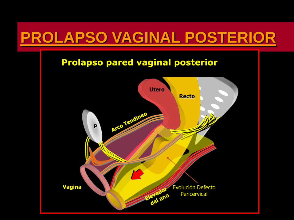
PARED VAGINAL
AN
TE
RIO
R
PO
ST
ER
IOR
PIEDRA ANGULAR
PARED VAGINAL
AN
TE
RIO
R
PO
ST
ER
IOR
PROLAPSOS
PIEDRA ANGULAR
PARED VAGINAL
AN
TE
RIO
R
PO
ST
ER
IOR
PR
OL
AP
SO
S
PIEDRA ANGULAR
CONCLUSIONE
S
DESINSERCIOgraveN DE LAS FASCIAS DESDE ELANILLO PERICERVICAL PROVOCA ELDEFECTO RESPONSABLE DE LOSPROLAPSOS
DEFECTOS TRANSVERSOS PARECEN SERLOS MAS IMPORTANTES
LOS DEFECTOS FASCIALES PUEDEN SERUgraveNICOS O MULTIPLES EN UNO O VARIOSCOMPARTIMIENTOS
REPARACIOgraveN SITIO ESPECIFICA CIRUGIACOMPENSADORA
CONCLUSION
ES
EXISTEN NUEVOS PARADIGMAS EN LA CONCEPCIOgraveN FISIOPATOLOgraveGICA DE LOS DEFECTOS DEL PISO PEgraveLVICO
LA REINSERCIOgraveN DE LAS FASCIAS AL ANILLO PERICERVICAL DEBE SER UN PASO INDISPENSABLE EN LA REPARACIOgraveN DE DEFECTOS DEL PISO PEgraveLVICO
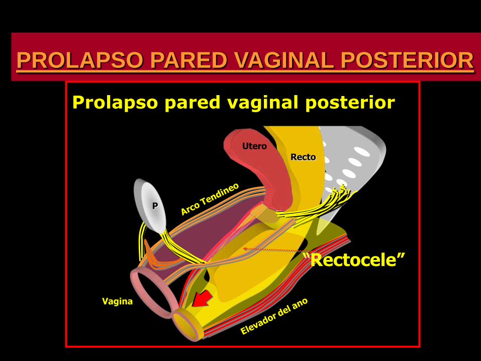
PARED VAGINAL
AN
TE
RIO
R
PO
ST
ER
IOR
PROLAPSOS
PIEDRA ANGULAR
PARED VAGINAL
AN
TE
RIO
R
PO
ST
ER
IOR
PR
OL
AP
SO
S
PIEDRA ANGULAR
CONCLUSIONE
S
DESINSERCIOgraveN DE LAS FASCIAS DESDE ELANILLO PERICERVICAL PROVOCA ELDEFECTO RESPONSABLE DE LOSPROLAPSOS
DEFECTOS TRANSVERSOS PARECEN SERLOS MAS IMPORTANTES
LOS DEFECTOS FASCIALES PUEDEN SERUgraveNICOS O MULTIPLES EN UNO O VARIOSCOMPARTIMIENTOS
REPARACIOgraveN SITIO ESPECIFICA CIRUGIACOMPENSADORA
CONCLUSION
ES
EXISTEN NUEVOS PARADIGMAS EN LA CONCEPCIOgraveN FISIOPATOLOgraveGICA DE LOS DEFECTOS DEL PISO PEgraveLVICO
LA REINSERCIOgraveN DE LAS FASCIAS AL ANILLO PERICERVICAL DEBE SER UN PASO INDISPENSABLE EN LA REPARACIOgraveN DE DEFECTOS DEL PISO PEgraveLVICO
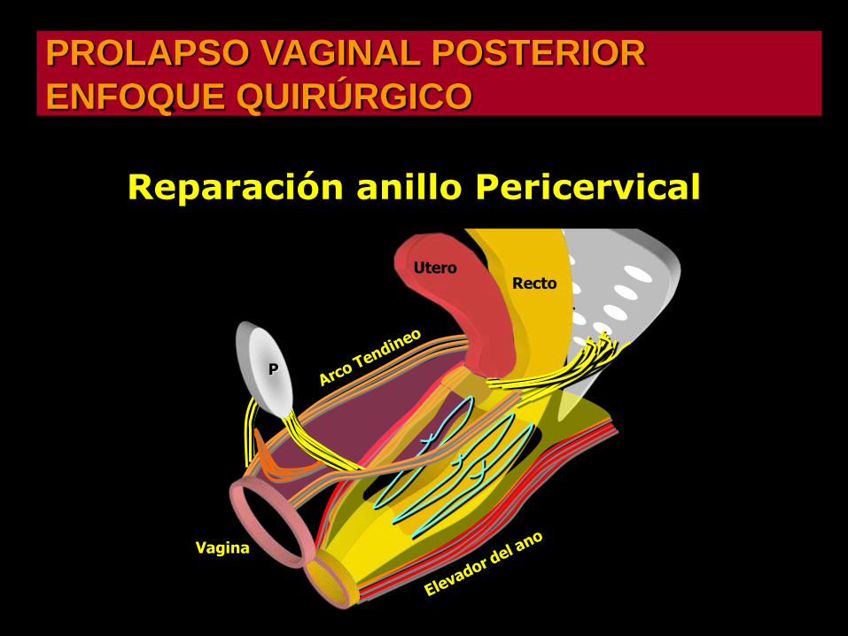
PARED VAGINAL
AN
TE
RIO
R
PO
ST
ER
IOR
PR
OL
AP
SO
S
PIEDRA ANGULAR
CONCLUSIONE
S
DESINSERCIOgraveN DE LAS FASCIAS DESDE ELANILLO PERICERVICAL PROVOCA ELDEFECTO RESPONSABLE DE LOSPROLAPSOS
DEFECTOS TRANSVERSOS PARECEN SERLOS MAS IMPORTANTES
LOS DEFECTOS FASCIALES PUEDEN SERUgraveNICOS O MULTIPLES EN UNO O VARIOSCOMPARTIMIENTOS
REPARACIOgraveN SITIO ESPECIFICA CIRUGIACOMPENSADORA
CONCLUSION
ES
EXISTEN NUEVOS PARADIGMAS EN LA CONCEPCIOgraveN FISIOPATOLOgraveGICA DE LOS DEFECTOS DEL PISO PEgraveLVICO
LA REINSERCIOgraveN DE LAS FASCIAS AL ANILLO PERICERVICAL DEBE SER UN PASO INDISPENSABLE EN LA REPARACIOgraveN DE DEFECTOS DEL PISO PEgraveLVICO
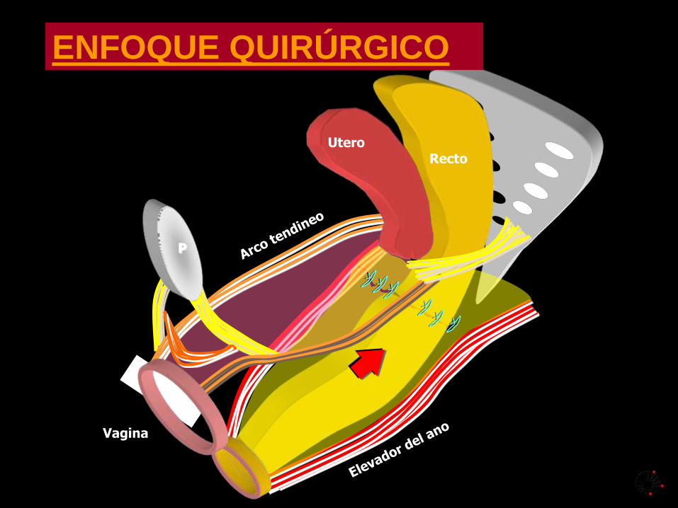
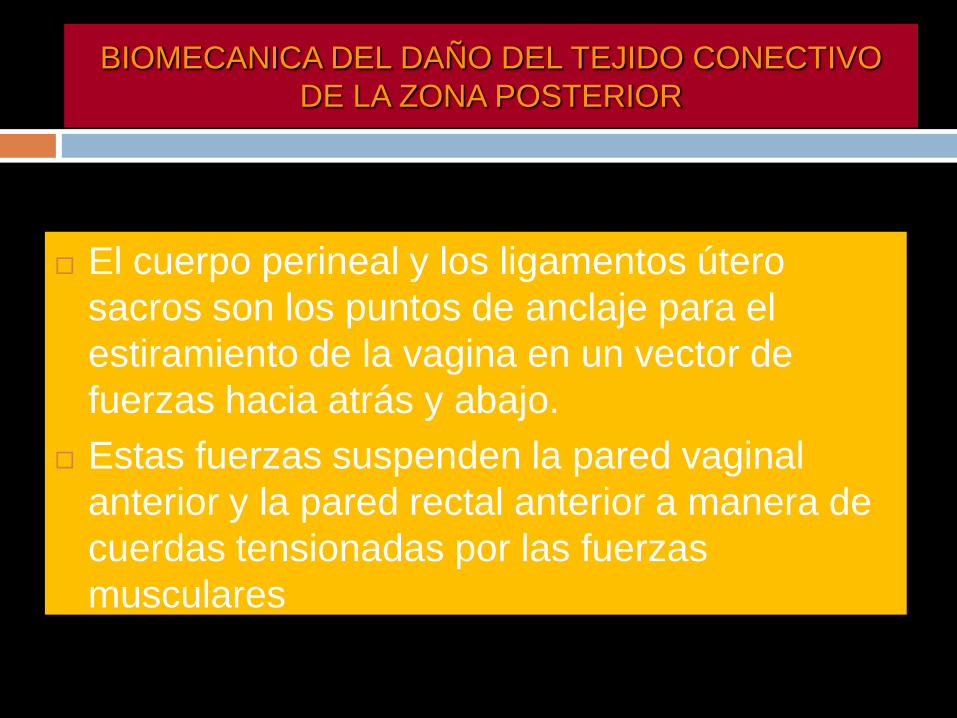
CONCLUSION
ES
EXISTEN NUEVOS PARADIGMAS EN LA CONCEPCIOgraveN FISIOPATOLOgraveGICA DE LOS DEFECTOS DEL PISO PEgraveLVICO
LA REINSERCIOgraveN DE LAS FASCIAS AL ANILLO PERICERVICAL DEBE SER UN PASO INDISPENSABLE EN LA REPARACIOgraveN DE DEFECTOS DEL PISO PEgraveLVICO