INFECÇÃO EXPERIMENTAL DE COELHOS COM …w3.ufsm.br/ppgmv/images/dissertacoes2009/Sara Campos da...
45
UNIVERSIDADE FEDERAL DE SANTA MARIA CENTRO DE CIÊNCIAS RURAIS PROGRAMA DE PÓS-GRADUAÇÃO EM MEDICINA VETERINÁRIA INFECÇÃO EXPERIMENTAL DE COELHOS COM RECOMBINANTES DO HERPESVÍRUS BOVINO TIPO 5 DEFECTIVOS NOS GENES DA TIMIDINA QUINASE E DA GLICOPROTEÍNA E DISSERTAÇÃO DE MESTRADO Sara Campos da Silva Santa Maria, RS, Brasil 2009
Transcript of INFECÇÃO EXPERIMENTAL DE COELHOS COM …w3.ufsm.br/ppgmv/images/dissertacoes2009/Sara Campos da...

0
UNIVERSIDADE FEDERAL DE SANTA MARIA
CENTRO DE CIÊNCIAS RURAIS
PROGRAMA DE PÓS-GRADUAÇÃO EM MEDICINA VETERINÁRIA
INFECÇÃO EXPERIMENTAL DE COELHOS COM
RECOMBINANTES DO HERPESVÍRUS BOVINO TIPO
5 DEFECTIVOS NOS GENES DA TIMIDINA QUINASE
E DA GLICOPROTEÍNA E
DISSERTAÇÃO DE MESTRADO
Sara Campos da Silva
Santa Maria, RS, Brasil
2009
1
INFECÇÃO EXPERIMENTAL DE COELHOS COM
RECOMBINANTES DO HERPESVÍRUS BOVINO TIPO 5
DEFECTIVOS NOS GENES DA TIMIDINA QUINASE E DA
GLICOPROTEÍNA E
por
Sara Campos da Silva
Dissertação apresentada ao curso de Mestrado do Programa de Pós-graduação
em Medicina Veterinária, área de concentração em Medicina Veterinária
Preventiva da Universidade Federal de Santa Maria (UFSM, RS) como requisito
parcial para obtenção do grau de Mestre em Medicina Veterinária
Orientador: Prof. Rudi Weiblen
Santa Maria, RS, Brasil
2009
2
S586i Silva, Sara Campos da
Infecção experimental de coelhos com recombinantes
do herpesvírus bovino tipo 5 defectivos nos genes da
timidina quinase e da glicoproteina E / por Sara Campos
da Silva. – 2009.
44 f. : il. ; 30 cm.
Orientador: Rudi Weiblen.
Dissertação (mestrado) – Universidade Federal de
Santa Maria, Centro de Ciências Rurais, Programa de
Pós-Graduação em Medicina Veterinária, RS, 2009.
1. Medicina veterinária 2. Coelhos 3. Patogenia
4. Vírus 5. Herpesvírus bovino tipo 5 6. Recombinantes
I. Weiblen, Rudi II. Título.
CDU 619:636.92
Ficha catalográfica elaborada por
Maristela Eckhardt - CRB-10/737
3
Universidade Federal de Santa Maria
Centro de Ciências Rurais
Programa de Pós-graduação em Medicina Veterinária
Departamento de Medicina Veterinária Preventiva
A Comissão Examinadora, abaixo assinada,
aprova a dissertação de Mestrado
INFECÇÃO EXPERIMENTAL DE COELHOS COM
RECOMBINANTES DO HERPESVÍRUS BOVINO TIPO 5
DEFECTIVOS NOS GENES DA TIMIDINA QUINASE E DA
GLICOPROTEÍNA E
elaborada por
Sara Campos da Silva
Como requisito parcial para obtenção do título de
Mestre em Medicina Veterinária
COMISSÃO EXAMINADORA
Rudi Weiblen, PhD
(Presidente/Orientador)
Ivomar Oldoni, PhD (Brasil Foods)
Eduardo Furtado Flores, PhD (UFSM)
Santa Maria, 9 de dezembro de 2009.
4
RESUMO
Dissertação de Mestrado
Programa de Pós-graduação em Medicina Veterinária
Universidade Federal de Santa Maria
INFECÇÃO EXPERIMENTAL DE COELHOS COM
RECOMBINANTES DO HERPESVÍRUS BOVINO TIPO 5
DEFECTIVOS NOS GENES DA TIMIDINA QUINASE E DA
GLICOPROTEÍNA E AUTORA: SARA CAMPOS DA SILVA
ORIENTADOR: RUDI WEIBLEN
Santa Maria, 9 de dezembro de 2009.
O herpesvírus bovino tipo 5 (BoHV-5), agente da meningoencefalite herpética bovina,
possui grande importância na América do Sul e tem motivado pesquisas para o
desenvolvimento de vacinas eficazes e seguras. Essa dissertação relata a investigação da
virulência em coelhos de três recombinantes do BoHV-5, candidatos vacinais, contendo
deleções nos genes da glicoproteína E (gE) (BoHV-5gE∆), da enzima timidina quinase (TK)
(BoHV-5TK∆) e deleção dupla nos genes da gE e TK (BoHV-5gE∆TK∆). Para isso, quatro
grupos de oito coelhos cada foram inoculados pela via intranasal com um dos recombinantes
ou com a cepa parental (SV507/99) e monitorados nos dias seguintes à inoculação. No dia 42
pós-inoculação (pi) realizou-se a administração de dexametasona (Dx) para reativar a infecção
latente e no dia 70 pi a eutanásia para a coleta do encéfalo para a pesquisa de DNA latente por
PCR. Os coelhos inoculados com o vírus parental (SV507/99) excretaram vírus nas secreções
nasais entre os dias 2 e 8 pós-inoculação (pi), e todos (n=8) desenvolveram doença
neurológica e morreram ou foram submetidos à eutanásia in extremis entre os dias 7 e 13 pi.
Excreção viral entre os dias 2 e 10 pi também foi detectada em 7 de 8 coelhos inoculados com
o BoHV-5gE∆; 6 de 8 inoculados com BoHV-5TK∆ e 3 de 8 inoculados com BoHV-
5gE∆TK∆. Apesar dos níveis variáveis de excreção viral, os animais inoculados com os três
recombinantes soroconverteram, apresentando anticorpos neutralizantes em títulos entre 2 e
256 no dia 42 pi. Nos animais inoculados com o vírus parental, o vírus foi detectado
amplamente disseminado no encéfalo, incluindo o bulbo olfatório, córtices, bulbo, ponte,
mesencéfalo e tálamo. Dentre os animais inoculados com o recombinante BoHV-5gE∆, três
desenvolveram doença neurológica, nos dias 10 e 15 pi. Uma distribuição viral restrita aos
córtices e tálamo foi detectada no encéfalo desses animais. Os coelhos inoculados com os
recombinantes BoHV-5TK∆ (n=8) e BoHV-5gE∆TK∆ (n=8) permaneceram saudáveis. A
administração de dexametasona (Dx) no dia 42 pi não resultou em reativação da infecção por
nenhum dos recombinantes, demonstrada por ausência de soroconversão ou excreção viral em
secreções. Entretanto, o DNA viral latente foi detectado no gânglio trigêmeo ou no bulbo
olfatório de todos esses animais no dia 28 pDx (70dpi), demonstrando o estabelecimento da
infecção latente. Esses resultados demonstram que os recombinantes são capazes de
estabelecer a infecção latente, mas não são facilmente reativados pela administração de Dx.
Em resumo, os recombinantes BoHV-5TK∆ e BoHV-5gE∆TK∆ são atenuados para coelhos
constituindo-se, assim, em candidatos vacinais em potencial.
Palavras-chave: BoHV-5; recombinantes; patogenia; virulência, candidatos vacinais.
5
ABSTRACT
Master’s Dissertation
Programa de Pós-graduação em Medicina Veterinária
Universidade Federal de Santa Maria
EXPERIMENTAL INFECTION OF RABBITS WITH BOVINE
HERPESVIRUS 5 RECOMBINANTS DEFECTIVE IN THYMIDINE
KINASE AND GLYCOPROTEIN E GENES AUTHOR: Sara Campos da Silva
ADVISER: Rudi Weiblen
Santa Maria, December 9th
, 2009.
Bovine herpesvirus 5 (BoHV-5) – the agent of meningoencephalitis in cattle - is an
important pathogen of cattle in South America and efforts have been made to produce safer
and more effective vaccines. This dissertation relates an investigation of the virulence in
rabbits of three BoHV-5 recombinants, vaccine candidates, defective in the glycoprotein E
(BoHV-5gEΔ), thymidine kinase (BoHV-5TKΔ) and both genes (BoHV-5gE∆TKΔ). To this,
four groups of eight rabbits each were inoculated intranasally with each recombinant or the
parental strain (SV507/99) and monitored thereafter. At day 42 post inoculation (pi) the
inoculated animals were submitted to dexamethasone (Dx) administration to reactivate latent
infection. At day 70 pi, all animals were euthanized and the brain was collected for
investigation of latent viral DNA by PCR. Rabbits inoculated with the parental virus shed
virus between days 2 and 8 pi and all rabbits (n=8) developed neurological disease and died or
were euthanized in extremis, between days 7 and 13 pi. Among the animals inoculated with
the recombinants, viral shedding was detected between days 2 and 10 pi, in 7 out of 8 rabbits
of the BoHV-5gE∆ group, in 6 out of 8 rabbits of the BoHV-5TK∆ group and in 3 out of 8 of
the BoHV-5gE∆TK∆ group. In spite of variable levels of virus shedding, all rabbits
inoculated with the recombinants seroconverted, developing virus-neutralizing antibodies in
titers from 2 to 256 at day 42 pi. The rabbits inoculated with the parental virus showed a wide
distribution of the virus in their brains, including the olfactory bulbs, cortices, medulla
oblongata, pons, midbrain and thalamus. Three out of eight rabbits inoculated with the
recombinant BoHV-5gEΔ developed neurological signs at days 10 and 15pi. A more
restricted virus distribution, confined mainly to cerebral cortices and thalamus was detected in
the brain of these animals. Rabbits inoculated with the recombinants BoHV-5TKΔ (n=8) or
BoHV-5gEΔTKΔ (n=8) remained healthy during the experiment. Dx administration to rabbits
inoculated with the three recombinants at day 42 pi did not result in viral reactivation, as
demonstrated by lack of seroconversion or virus shedding. Nevertheless, viral DNA was
detected in the trigeminal ganglia or olfactory bulbs of all these animals at day 28pDx,
demonstrating they were latently infected. These results showed that the three recombinants
were able to establish latent infection yet they were not easily reactivated by Dx
administration. In summary, the recombinants BoHV-5TKΔ and BoHV-5gEΔTKΔ are
attenuated for rabbits and constitute potential vaccine candidates.
Keywords: BoHV-5; recombinants; pathogenesis; virulence; vaccine candidate.
6
LISTA DE TABELAS
CAPÍTULO 1
TABELA 1 (Table 1) - Virological, clinical and serological findings during acute
infection in rabbits inoculated with the parental virus and recombinants of bovine
herpesvirus 5 (BoHV-5)…………………………………………………………………... 36
TABELA 2 (Table 2) - Distribution of infectivity in the brain of rabbits inoculated with
the parental virus or with the recombinant BoHV-5gE∆…………………………………. 37
TABELA 3 (Table 3) - Findings after dexamethasone (Dx) administration in rabbits
inoculated with the parental virus and recombinants of bovine herpesvirus 5 (BoHV-5)... 38
7
LISTA DE FIGURAS
CAPÍTULO 1
FIGURA 1 (Figure 1) - Illustrative results of a PCR performed to confirm the identity
of viruses shed in nasal secretions by rabbits of the respective groups. Panel A. PCR for
the TK gene (285 bp). Panel B. PCR for the glycoprotein E gene (269 bp). Panel C. PCR
for the glycoprotein B gene (control) (444 bp). MWM: molecular weight marker. Lane
1: negative control (DNA template from the brain of a BoHV-5 seronegative cow); lane
2: positive control, DNA extracted from the supernatant CRIB cells infected with the
parental virus; lane 3-6: pool of nasal secretions of rabbits inoculated with recombinants
- double mutant BoHV-5gEΔTKΔ (lane 3); BoHV-5gEΔ (lane 4); BoHV-5TKΔ (lane
5); parental virus (SV507/99; lane 6). Ethidium bromide stained 1.5% agarose gel; the
sizes of the corresponding markers are indicated……………………………………… 39

8
SUMÁRIO
1. INTRODUÇÃO.............................................................................................................. 9
2. CAPÍTULO 1. A bovine herpesvirus 5 recombinant defective in thymidine
kinase (TK) gene and a double mutant lacking TK and glycoprotein E gene are
fully attenuated for rabbits……………………………………………………………... 13
Abstract……………………………………………………………………………….…... 14
Introduction………………………………………………………………………….……. 15
Material and methods………………………………………………………………….…. 18
Results………………………………………………………………………………….…. 22
Discussion………………………………………………………………………………… 26
References………………………………………………………………………………… 30
3. REFERÊNCIAS……………………………………………………………………... 40
9
1. INTRODUÇÃO
A infecção pelo herpesvírus bovino tipo 5 (BoHV-5) está associada com
meningoencefalite de curso geralmente fatal em bovinos jovens (ROIZMAN et al., 1992). A
enfermidade causada pelo BoHV-5 possui maior importância na América do Sul, onde tem
sido relatada com freqüência no Brasil e na Argentina (CARRILLO et al., 1983;
SALVADOR et al., 1998, RISSI et al., 2007). O BoHV-5 apresenta grande similaridade
genética e antigênica com o herpesvírus bovino tipo 1 (BoHV-1), agente da rinotraqueíte
infecciosa (IBR) e da vulvovaginite/balanopostite pustular bovina (IPV/IBP) (SCHWYZER;
ACKERMANN, 1996; DELHON et al., 2003). Devido a reatividade sorológica cruzada entre
estes agentes, e a inexistência de um teste que os diferencie sorologicamente, a prevalência da
infecção pelo BoHV-5 ainda permanece desconhecida (FLORES et al., 2009).
O BoHV-5 pertence à família Herpesviridae, subfamília Alphaherpesvirinae, gênero
Varicellovirus (ROIZMAN et al., 1992). Os vírions são pleomórficos, com 150 a 200 nm de
diâmetro, envelopados e possuem como genoma uma molécula de DNA de fita dupla com
aproximadamente 138 quilobases (Kb) (ROIZMAN et al., 1992; DELHON et al., 2003). Os
alfaherpesvírus são reconhecidos por infectar várias espécies animais, replicar de forma
rápida e lítica em células de cultivo celular, possuir neurotropismo e estabelecer infecções
latentes em gânglios do sistema nervoso periférico e em outros tecidos neurais (SCHWYZER;
ACKERMANN, 1996). Mesmo sendo semelhantes genética e antigenicamente, e
estabelecerem infecções latentes em gânglios sensoriais, o BoHV-1 e o BoHV-5 diferem na
habilidade de invadir o sistema nervoso central (SNC) e causar doença neurológica
(METZLER et al., 1986; BELKNAP et al., 1994; DELHON et al., 2003). Embora o BoHV-1
tenha sido ocasionalmente isolado de casos de meningoencefalite, a maioria dos casos de
doença neurológica são associados com o BoHV-5 (SILVA et al., 2007).
Os bovinos são os hospedeiros naturais do BoHV-5, embora experimentalmente a
infecção já tenha sido reproduzida em outras espécies de mamíferos (SILVA et al., 1999;
BELTRÃO et al., 2000; ABRIL et al., 2004; DIEL et al., 2007). Nesse sentido, coelhos
jovens têm constituído um modelo experimental adequado para o estudo da infecção pelo
BoHV-5, podendo desenvolver doença aguda e fatal, semelhante à observada em bovinos; ou
ainda estabelecer infecção latente (MEYER et al., 1996; LEE et al., 1999; BELTRÃO et al.,
2000, CARON et al., 2002). A doença neurológica desenvolvida pelos animais infectados
com o BoHV-5 é consequência da replicação e invasão viral progressiva no encéfalo (LEE et
10
al., 1999). Inicialmente, observa-se depressão e secreção nasal e ocular, que progridem para
emagrecimento, dificuldade respiratória, tremores, bruxismo, andar em círculos,
incoordenação, cegueira, nistagmo e disfagia, chegando a convulsão e morte em estágios mais
avançados (CHOWDHURY et al., 1997; PEREZ et al., 2002). Embora classicamente o
BoHV-5 esteja associado à doença neurológica, este agente já foi isolado de amostras de
sêmen, de casos de doença respiratória e de infecções generalizadas em bezerros jovens
(SILVA et al., 2007; KIRKLAND et al., 2009).
Durante a infecção aguda, o BoHV-5 replica nas células da mucosa nasal, invadindo
as terminações dos nervos autonômicos e sensoriais que inervam a mucosa, sobretudo as
terminações dos nervos olfatório e ramo maxilar do nervo trigêmeo (LEE et al., 1999). A
seguir, os vírions invadem o SNC, principalmente pela via olfatória, no sentido retrógrado a
partir da mucosa nasal (CHOWDHURY et al., 1997; LEE et al., 1999; DIEL et al., 2005). A
disseminação viral no encéfalo pode ter como consequência o desenvolvimento de doença
neurológica ou o estabelecimento de infecção latente (LEE et al., 1999; PEREZ et al., 2002;
VOGEL et al., 2003). A infecção latente pode ser reativada naturalmente, durante episódios
de estresse, ou artificialmente, pela administração de corticóides, sendo geralmente
acompanhada de excreção viral e, ocasionalmente, de recrudescência clínica (CARON et al.,
2002; VOGEL et al., 2003). Animais portadores da infecção latente são epidemiologicamente
importantes, pois representam a principal fonte para introdução, disseminação e manutenção
do vírus nos rebanhos (ACKERMANN; ENGELS, 2006).
A vacinação é uma medida complementar ao manejo e amplamente utilizada no
controle das infecções pelos herpesvírus (VAN OIRSCHOT, 1999). Devido à importância
que o BoHV-5 possui na América do Sul e a existência de apenas uma vacina específica para
este agente no Brasil, vacinas para o BoHV-1 tem sido utilizadas no controle da infecção pelo
BoHV-5 (RISSI et al., 2007). A grande maioria das vacinas disponíveis comercialmente no
Brasil são inativadas. Apesar destas vacinas apresentarem eficácia satisfatória reduzindo a
severidade da doença e a excreção viral, serem seguras e não apresentarem riscos de reversão
à virulência, elas estimulam preferencialmente a resposta humoral e necessitam de reforços
periódicos para manter níveis adequados de anticorpos (VAN OIRSCHOT, 1999; VAN
DRUNEN LITTEL-VAN DEN HURK, 2007). Além disso, estas vacinas são classificadas
como convencionais, não permitindo a diferenciação sorológica entre animais naturalmente
infectados e vacinados, aspecto importante para o controle e erradicação da infecção do
rebanho (VAN OIRSCHOT, 1996; VAN OIRSCHOT, 1999). Como alternativa, esforços têm
sido empregados no desenvolvimento de vacinas diferenciais, baseadas na deleção de um ou
11
mais genes não-essenciais que além de resultar em redução da virulência podem ser utilizados
como marcadores antigênicos (ACKERMANN; ENGELS, 2006).
O genoma do BoHV-5 possui aproximadamente 70 genes, cujos produtos podem ser
classificados em essenciais e não-essenciais, de acordo com a sua necessidade para a
replicação viral em cultivo celular. Entretanto, todos os genes provavelmente possuem papel
importante na infecção viral in vivo (SCHWYZER; ACKERMANN, 1996; DELHON et al.,
2003; METTENLEITER, 2003). A deleção de alguns genes não-essenciais dos herpesvírus
pode resultar em redução ou incapacidade do vírus causar doença, sem afetar a capacidade de
replicação in vitro (ENQUIST et al., 1998). Proteínas estruturais, glicoproteínas e enzimas
têm sido alvo de deleção para a produção de cepas defectivas com o objetivo de desenvolver
vacinas atenuadas e diferenciais ou, ainda, para estudos de patogenia (ENQUIST et al., 1998;
METTENLEITER, 2003).
A glicoproteína E (gE) é uma proteína estrutural do envelope do BoHV-5, conservada
entre os isolados de campo, e não-essencial para a replicação viral em células de cultivo
(ENQUIST et al., 1998). Além disso, a gE está envolvida na neuropatogênese da infecção,
desempenhando um importante papel na neuroinvasidade e neurovirulência
(METTENLEITER, 2003). Estas características fazem da gE um adequado alvo para deleção
e produção de cepas atenuadas de herpesvírus neurovirulentos, como o vírus do herpes
simplex tipo 1 (HSV-1), vírus da doença de Aujeszky (PRV) e BoHV-5 (MULDER et al.,
1994; DINGWELL et al., 1995; CHOWDHURY et al., 2000). Um recombinante do BoHV-5
com deleção no gene da gE manteve a capacidade de penetrar no SNC pela via olfatória,
porém apresentou uma redução significativa na replicação, disseminação e conseqüente
neurovirulência (CHOWDHURY et al., 2000). As causas para a deficiente disseminação
transneuronal do BoHV-5 na ausência da gE ainda não foram investigadas. Entretanto,
acredita-se que, assim como proposto para o HSV-1 e o PRV, o transporte axonal das
glicoproteínas de envelope e o capsídeo viral tegumentado é realizado separadamente
(TOMISHIMA; ENQUIST, 2001; SNYDER et al., 2008). Na ausência da gE, a vesícula
contendo as glicoproteínas não seria transportada ao longo do axônio, não ocorrendo a
maturação final dos vírions junto à sinapse e a sua transmissão trans-sináptica (TOMISHIMA;
ENQUIST, 2001; SNYDER et al., 2008).
Os vírus da subfamília Alphaherpesvirinae codificam a sua própria enzima timidina
quinase (TK), cuja função é fornecer deoxiribonucleotídeos para a síntese de DNA viral em
células pós-mitóticas, como os neurônios (ENQUIST et al., 1998; ROIZMAN; KNIPE, 2001).
Nestes vírus, a TK é absolutamente necessária para a expressão completa da virulência in vivo

12
(ENQUIST et al., 1998). Assim, a deleção da TK do BoHV-1, PRV e HSV-1 está associada
com uma significativa redução da virulência (CHOWDHURY, 1996; FERRARI et al., 2000;
CHEN et al., 2004). Entretanto, o envolvimento da TK na neuropatogênese do BoHV-5 ainda
não foi totalmente elucidado, pois cepas do BoHV-5 defectivas nesta enzima ainda não foram
avaliadas in vivo. Estudos demonstram que variantes do BoHV-5 resistentes à brivudina e,
provavelmente, deficientes na atividade da enzima TK foram atenuados quando inoculados
em coelhos (BRUM, 2009).
A capacidade de estabelecer a infecção latente parece não ser afetada nas cepas
contendo deleções na gE ou na TK (CHOWDHURY et al., 2000; FERRARI et al., 2000;
CHEN et al., 2004; LIU et al., 2008). Entretanto, após administração de corticóides em
animais inoculados com cepas gE deletadas, não é possível detectar indícios de reativação,
como excreção viral ou sinais clínicos (VAN ENGELENBURG et al., 1995; KAASHOEK et
al., 1998). Um estudo utilizando uma cepa do BoHV-1 deletada na gE demonstrou que o vírus
reativa a infecção latente no gânglio trigêmeo, porém não é transportado no sentido
anterógrado em direção as terminações nervosas na mucosa olfatória e ocular (LIU et al.,
2008; BRUM et al., 2009). Os dados de reativação viral de cepas TK deletadas são bastante
controversos, indicando provavelmente as diferentes condições em que foram realizados os
experimentos (MENGELING, 1991; WHETSTONE et al., 1992; CHEN et al., 2004).
Neste trabalho são relatados os aspectos clínicos e virológicos da infecção aguda e
latente em coelhos inoculados com três recombinantes do BoHV-5, individualmente,
defectivos no gene da gE, da TK e ambos gE e TK. Estes recombinantes poderão ser
utilizados para a produção de vacinas diferenciais para o BoHV-5, além da possibilidade da
realização de estudos sobre importantes aspectos da patogenia deste agente.
13
2. CAPÍTULO 1
A bovine herpesvirus 5 recombinant defective in thymidine kinase (TK) gene and a
double mutant lacking TK and glycoprotein E gene are fully attenuated for rabbits
Sara Campos da Silva, Mário Celso Sperotto Brum, Rudi Weiblen, Eduardo Furtado Flores e
Shafqul I. Chowdhury
Artigo aceito para publicação na revista Brazilian Journal of Medical and Biological
Research, 2009.
14
A bovine herpesvirus 5 recombinant defective in thymidine kinase (TK) gene and a
double mutant lacking TK and glycoprotein E gene are fully attenuated for rabbits
S. C. Silva1, M.C.S. Brum
1, R. Weiblen
1, E.F. Flores
1 S.I. Chowdhury
2
1Setor de Virologia, Departamento de Microbiologia e Parasitologia e Departamento de
Medicina Veterinária Preventiva, Universidade Federal de Santa Maria, Santa Maria, RS,
Brasil. 2Department of Pathobiological Sciences, School of Veterinary Medicine, Louisiana
State University, Baton Rouge, LA, United States.
Correspondence to: E.F. Flores, Departamento de Medicina Veterinária Preventiva, Centro de
Ciências Rurais, Universidade Federal de Santa Maria, Santa Maria, RS, Brasil, CEP 97105-
900. Fone/fax +(55) 55 3220-8034. E-mail: [email protected]
Running title: Attenuation of BoHV-5 recombinants in rabbits
Abstract
Bovine herpesvirus 5 (BoHV-5) – the agent of herpetic meningoencephalitis in cattle -
is an important pathogen of cattle in South America and efforts have been made to produce
safer and more effective vaccines. In this article, we investigated the virulence in rabbits of
three recombinant viruses constructed out of a neurovirulent Brazilian BoHV-5 strain
(SV507/99). The recombinants are defective in the glycoprotein E (BoHV-5gEΔ), enzyme
thymidine kinase (BoHV-5TKΔ) and both proteins (BoHV-5gEΔTKΔ). Rabbits inoculated
with the parental virus (n=8) developed neurological disease and died or were euthanized in
extremis between days 7 and 13 pi (post infection). Infectivity was detected in several areas of
their brains. Three out of eight rabbits inoculated with the recombinant BoHV-5gEΔ
15
developed neurological signs between days 10 and 15 pi and were also euthanized. A more
restricted virus distribution was detected in the brain of these animals. Rabbits inoculated with
the recombinants BoHV-5TKΔ (n=8) or BoHV-5gEΔTKΔ (n=8) remained healthy
throughout the experiment, in spite of variable levels of virus replication in the nose.
Dexamethasone (Dx) administration to rabbits inoculated with the three recombinants at day
42 pi did not result in viral reactivation, as ascertained by absence of virus shedding and/or
increase in VN titers. Nevertheless, viral DNA was detected in the trigeminal ganglia or
olfactory bulbs of all animals at day 28 pDx (post Dx), demonstrating they were latently
infected. These results show that recombinants BoHV-5TKΔ and BoHV-5gEΔTKΔ are
attenuated for rabbits and constitute potential vaccine candidates upon the confirmation of this
phenotype in cattle.
Key words: BoHV-5, gE and TK deletion mutants, pathogenesis, virulence, vaccine
candidate.
Introduction
Bovine herpesvirus type 5 (BoHV-5) is a neurovirulent alphaherpesvirus associated
with meningoencephalitis generally fatal in cattle (1). BoHV-5 infection and disease have
been occasionally described in several countries, yet the disease is noticeably more frequent
in Brazil and Argentina where outbreaks are reported every year (2-6). BoHV-5 is related
antigenically and genetically to bovine herpesvirus 1 (BoHV-1), the agent of respiratory
(infectious bovine rhinotracheitis, IBR) and genital disease in cattle (infectious pustular
vulvovaginits/balanopostitis, IPV/IBP) (1). These viruses belong to the family Herpesviridae,
subfamily Alphaherpesvirinae, genus Varicellovirus (7, 8). BoHV-1 and BoHV-5 genomes
display the same genomic organization, share a nucleotide similarity 85% and 82% at the
16
nucleotide and protein level, respectively (8). The major biological difference between these
viruses seems to be the neurovirulent potential: BoHV-5 is highly neurovirulent for the
natural hosts and for experimental animals (9), whereas BoHV-1 is far less neurovirulent and
only occasionally has been associated with neurological infection (2). As other
alphaherpesviruses, BoHV-5 and BoHV-1 establish lifelong latent infection in sensory nerve
ganglia (9, 10).
The BoHV-5 genome consists of a linear, double stranded DNA molecule of
approximately 138kb in length and encodes at least 70 gene products (8). Alphaherpesviruses
(and other herpesviruses as well) encode a number of gene products that are non-essential
(NE) for virus replication in tissue culture, although they are probably required for virus
maintenance in nature (7, 11). Deletions of such genes have been used to study the role of
individual proteins in virus replication in vitro and in vivo (12, 13) and for the production of
recombinant strains for vaccine use (14, 15). In this sense, the genes encoding the envelope
glycoprotein E (gE) and the enzyme thymidine kinase (TK) are among the most frequent
targets for deletion towards vaccine production (16).
The envelope gE has been shown to be important for invasion and dissemination of
BoHV-5 within the brain and for anterograde transport of BoHV-1 from the trigeminal
ganglia to peripheric sites after reactivation (12, 17, 18). Furthermore, deletion of gE gene
from BoHV-1 and BoHV-5 genomes contributes for virus attenuation in calves and rabbits
(16-18). In addition, the deletion of gE from vaccine strains provides an antigenic marker
which would allow serological differentiation of vaccinated from naturally infected animals
(19). Vaccines with antigenic markers (also called DIVA vaccines – for differentiating
infected from vaccinated animals) have constituted the basis for eradication of BoHV-1 and
pseudorabies virus (PRV) from European countries and US (1) and are beginning to be used
in South America (20).

17
The full expression of virulence of neurovirulent alphaherpesviruses depends upon the
function of the enzyme TK (12). Herpesvirus-encoded TK is an enzyme involved in the
metabolism of deoxyribonucleotides (dNTPs), which is necessary for viral DNA synthesis
and genome replication in non-dividing cells such as neurons (12, 21). Deletion of TK gene in
neurovirulent alphaherpesviruses, namely human herpes simplex virus (HSV) and
pseudorabies virus (PRV), has been associated with deficient replication in neurons and
reduced neurovirulence (22-25). For these reasons, BoHV-1 mutants lacking TK activity are
also attenuated, albeit to a lesser extent (16, 26). For this reason, the TK gene has been an
attractive target for gene deletion towards the production of attenuated strains for vaccine use
(16).
Reactivation of latent infection by TK-defective alphaherpesviruses is also
significantly impaired or even abolished, especially for HSV in mice (22, 23). On the other
hand, attempts to reactivate latent infection by TK-negative BoHV-1 and PRV strains have
yielded conflicting results, probably reflecting differences in the viruses, animals and
experimental procedures (16, 27-30). Despite these conflicting results, it is generally accepted
that TK-defective alphaherpesviruses replicate poorly (if so) in neurons and, therefore, are not
expected to reactivate efficiently from latency.
The vaccines marketed in South America contain either inactivated or modified live
BoHV-1 strains, none of them containing antigenic markers (31). Following the trend of
European countries and predicting future sanitary restrictions for trading of animals and
products, some South American countries are now embarking on the development and use of
DIVA vaccines (20). Our group recently described the construction and in vitro
characterization of three recombinants (BoHV-5gEΔ, BoHV-5TKΔ and BoHV-5gEΔTKΔ)
out of a highly neurovirulent Brazilian BoHV-5 strain (31). These recombinants were
constructed for vaccine purposes yet they may also be useful for pathogenesis studies since
18
they are defective in specific gene products involved in different stages of virus infection. The
TK deletion, in particular, would provide an interesting means of attenuation for such
neurovirulent agent.
The present article describes studies on the pathogenesis of acute and latent infections
by these recombinants in a well characterized rabbit model (32). Our results show that the
recombinants BoHV-5TKΔ and BoHV-5gEΔTKΔ are fully attenuated for rabbits upon
intranasal inoculation and induce a virus-neutralizing antibody response. Although these
recombinants retained the ability to establish latent infection, they did not reactivate readily
upon Dx treatment. Thus, these results are promising towards the use of these recombinants as
vaccine strains, depending on equivalent phenotype in cattle.
Material and Methods
Experimental design
Groups of weanling rabbits were inoculated intranasally (IN) with each BoHV-5
recombinant or the parental virus and submitted to clinical, virological and serological
monitoring during acute infection. The identity of viruses shed in nasal secretions by rabbits
of each group was confirmed by PCR. Rabbits dying of neurological disease during acute
infection had sections of their brains examined for infectivity. At day 42 post-infection (pi),
rabbits were submitted to administration of dexamethasone (Dx) for reactivating latent
infection and were monitored thereafter as described for acute infection. Twenty eight days
after Dx treatment, the rabbits were euthanized and trigeminal ganglia (TG) and olfactory
bulbs (OB) were examined for viral DNA by PCR.
Viruses and cells
The Brazilian BoHV-5 strain SV507/99 - isolated from a cow with neurological
disease in southern Brazil and submitted to sequencing of the entire genome (8) was used as
19
the parental virus to construct the recombinants. The construction and characterization in vitro
of recombinants defective in the glycoprotein E (BoHV-5gEΔ), thymidine kinase (BoHV-
5TKΔ) and both genes (BoHV-5gEΔTKΔ) was described previously (31). The viruses were
propagated in a MDBK-derived cell line named CRIB (ATCC-CRL 11883) maintained in
MEM (minimum essential medium, Gibco, Brazil), supplemented with 10% fetal bovine
serum (Cultilab, Brazil) and 100 U/mL of penicillin, 100 µg/mL of streptomycin (Nutricell,
Brazil).
In vitro growth kinetics
Before rabbit inoculation, the recombinants were evaluated in their ability to replicate
in rabbit kidney cells (RK-13; ATCC-CCL 37), in an one step growth curve according to
Brum et al. (31). RK-13 cell monolayers were inoculated with each recombinant at
multiplicity of infection of 5. At intervals after virus inoculation (0, 4, 10, 18 and 24 hours)
aliquots of culture supernatants were harvested and submitted to virus quantitation by limiting
dilution. Virus titers were calculated according to Reed & Muench (33) and expressed as log10
median tissue culture infectious dose per mililiter (TCID50/mL).
Animals, virus inoculation, monitoring and dexamethasone treatment
Thirty-six New Zealand rabbits (28-30 days-old) weighting 300-400 g were used for
virus inoculation. The animals were randomly divided in four groups of eight rabbits each
plus a control group of four animals. Each group was maintained in separated cages, without
contact with each other, receiving water and food ad libitum. Rabbits of each experimental
group (n=8) were inoculated with one virus: parental SV-507/99; gE negative (BoHV-5gEΔ),
TK-deleted (BoHV-5TKΔ) and defective on both genes (BoHV-5gEΔTKΔ). The inoculum
consisted of supernatants of CRIB cells infected with each virus, containing approximately
107.5
TCID50/mL. The animals were inoculated IN with 1 mL of virus suspension into the
20
paranasal sinuses (0.5 mL in each side) through nephrine openings (34) after tranquilization
with tiletamine/zolazepan (30 mg/kg IM; Zoletil, Virbac, Brazil). Controls (n=4) were
inoculated IN with culture medium.
Following virus inoculation the rabbits were monitored twice a day for clinical signs.
Nasal swabs for virus isolation were collected every two days up to day 14 pi (acute infection)
and up to day 10 pDx (after Dx treatment). Swabs were vortexed, drained and the supernatant
was inoculated onto CRIB cell monolayers and submitted to three passages of five days each
with the cultures being monitored for cytopathic effect (CPE). Pools of nasal secretions of
each group were subsequently submitted to PCR to confirm the identity of the inoculated
viruses. Rabbits developing severe neurological signs during acute infection were euthanized
in extremis; brain sections were aseptically collected and tissue homogenates (10% w/v) were
submitted to virus isolation in CRIB cells. Serum samples were collected at days 0 and 42 pi
and submitted to a standard virus neutralizing assay (VN) for neutralizing antibodies, using
two-fold dilutions of sera against 100-200 TCID50 of the parental virus (SV507/99), 2h of
incubation of the virus-serum mixture and 72h of incubation before to reading. CRIB cells
were used as indicators of virus replication. Beginning at day 42 pi, the rabbits were
submitted to five daily administrations of dexamethasone (2.6 mg/kg/day, Decadronal, Achè,
Brazil) by the intramuscular route. In the days following Dx treatment the animals were
monitored as described for acute infection. Serum samples collected at the day of the first Dx
administration (42 pi/0 pDx) and 28 days later (day 28 pDx) were submitted to a VN assay as
described above. At day 28 pDx, inoculated and control animals were euthanized, the
olfactory bulbs (OB) and trigeminal ganglia (TGs) were collected for detection of viral DNA
by PCR.
All procedures of animal handling and experimentation were performed according to
recommendations by the Brazilian Committee on Animal Experimentation (COBEA; law

21
#6.638 of 8th May 1979). The animal experiments were approved by an Institutional Ethics
and Animal Welfare Committee (Comitê de Ética e Bem Estar Animal, Universidade Federal
de Santa Maria, UFSM, approval #44/2008; process #23081.010078/2008-41).
Identity of the viruses shed by inoculated rabbits
In order to confirm the identity of each recombinant virus being shed for the
respective groups, nasal secretions collected daily from the animals of each group were polled
and submitted to DNA extraction. Total DNA extracted from secretions was submitted to
PCR for detection of TK and gE deletions. A PCR for the gene of glycoprotein B was used as
control (35). Detection of gE gene (or its absence), was performed by using the following
primers: forward 5’-ACGAGACGTGCATCTTCC-3’ (position 124.888 on the BoHV-5
genome) and reverse – 5’-CAGCACGAAGACGTAGAG-3’ (position 125.156), giving rise to
a 269 bp product. The TK gene (or its absence) was detected by using the primers: forward
5’-GACGTCGTGACCCTCGTGTTTG-3’ (position 64.971) and reverse 5’-
TAGGAAGGCGCACGTGTTCG-3’ (position 65.255), which amplify a 286 bp product. PCR
reactions were performed essentially as described by Brum et al. (31). Total DNA extracted
from CRIB cells infected with BoHV-5TK∆ or BoHV-5gE∆ and SV-507/99 were used as
negative and positive controls, respectively.
DNA extraction from tissues and PCR
Neural tissues collected at day 28 pDx (TGs, BOs) were submitted to total DNA
extraction using DNAzol reagent (Invitrogen, Carlsbad, CA, USA). The extracted DNA was
solubilized in buffer Tris-EDTA (0.1 – 0.2 mL) and stored at -20°C until testing. DNA
concentration was determined in a UV spectrophotometer at 260 nm.
Total DNA was submitted to a nested PCR using two set of primers corresponding to
positions 57.338 and 57.782 (primers 1 and 2) and 57.372 and 57.666 (primers 3 and 4) of the
22
glycoprotein B gene coding region of the BoHV-5 strain SV-507 (8, 35). The external primers
(primers 1 and 2) used in the first reaction were -forward: 5’ CTC GAA AGC CGA GTA
CCT GCG 3’ and reverse: 5’-5’ CCA GTC CCA GGC AAC CGT CAC 3’. The internal
primers (primers 3 and 4) used in the second reaction were - forward: 5’ GTG GTG GCC
TTT GAC CGC GAC 3’ and reverse: 5’5’ GCT CCG GCG AGT AGC TGG TGT G 3’. The
first PCR reaction amplifies a 444 bp DNA fragment and the second reaction results in a 295
bp amplicon. The PCR reactions were performed in 25 µL, using 2 µL template DNA
(corresponding to approximately 1 µg of total DNA), 100 ng of each primer, 2 mM MgCl2, 10
mM dNTPs, 10% DMSO, 1x reaction buffer, and 2.5 units of Taq DNA Polymerase
(Invitrogen, Carlsbad, CA, USA). The PCR conditions were: initial denaturation at 94°C for 5
min, following by 35 cycles at 94°C for 45 sec for DNA denaturation, 56°C for 45 sec for
primer annealing and 72°C for 45 sec for primer extension, and final extension at 72°C for 10
min. Total DNA extracted from the brain of a control non-infected rabbit, and from a rabbit
with acute BoHV-5 infection were used as negative and positive controls, respectively. PCR
products were analysed under UV light after electrophoresis in an 1.5% agarose gel stained
with ethidium bromide. To determine the sensitivity of the PCR, 10-fold dilutions of strain
SV-507 DNA were prepared with DNA (1 µg/µl) extracted from the brain of a BoHV-5-
seronegative cow and used as templates for PCR. Based on the average size of the BHV-5
genome (137 kb), an estimate of the number of genome copies detected in the PCR was
calculated (18).
Results
Kinetics of virus replication in rabbit cells
As the recombinants have been amplified and characterized in bovine cells (MDBK)
and BoHV-5gE∆ and BoHV-5gE∆TK∆ produced slightly smaller plaques, before inoculation
into rabbits we investigated their ability to replicate in cultured rabbit cells. An one step
23
growth curve experiment performed in RK-13 cells demonstrated that the recombinants
BoHV-5TK∆ and BoHV-5gE∆ replicated with similar kinetics and to equivalent titers to
those of the parental virus in rabbit cells (not shown). The double deletion mutant (BoHV-
5gE∆TK∆) replicated with a slightly lower kinetics and to a lower titer. In a previous
experiment in MDBK cells, the three recombinants were shown to retain their ability to
replicate with about the same efficiency as the parental virus (31). Thus, a possible
impairment in replicating in rabbits in vivo would not be attributable to a gross defect in
replicating in rabbit cells.
Animal experiment – acute infection
All rabbits inoculated with the parental virus SV507/99 shed virus in nasal secretions
(between days 2 and 8 pi) and developed typical neurological signs (Table 1). The
neurological disease started from day 7 to 13 pi, depending on the animal, and was
characterized by depression/excitation, ptialism, bruxism, opisthotonus, seizures and
blindness in some cases. Animals showing severe neurological signs were euthanized.
Rabbits inoculated with the recombinant BoHV-5gE∆ shed virus between days 2 and
8 and three (# 1, 2 and 7) developed neurological signs similar to those of the rabbits
inoculated with the parental virus. These rabbits were euthanized in extremis at days 10 and
15 pi. Infectious virus was not recovered from nasal swabs collected from rabbit # 7, yet
infectivity was detected in several areas of the brain of this animal (Table 2). The rabbits
surviving acute infection developed VN titers from 8 to 256 at day 42 pi.
The group inoculated with the recombinant BoHV-5TK∆ remained healthy throughout
acute infection. Virus shedding was detected in 6/8 rabbits and lasted from one to eight days
(Table 1). These rabbits, including those from which virus shedding was not detected (# 1 and
4) developed VN titers from 2 to 64 in sera collected at day 42 pi, indicating they were
efficiently infected.
24
None of the rabbits inoculated with the double mutant (BoHV-5gE∆TK∆) developed
clinical signs during the period of monitoring. Virus shedding (in swabs collected every two
days) was detected in 3 out of 8 animals. All inoculated animals seroconverted by day 42 pi
(titers ranging from 2 to 32), indicating that virus replication took place.
These results showed that the recombinants BoHV-5TK∆ and BoHV-5gE∆TK∆ were
fully attenuated for rabbits upon IN inoculation. Apparently, these viruses (especially the
double mutant) replicated with a lower efficiency in the nasal mucosa. Nevertheless, data of
serology demonstrated that the viruses replicated in all animals in levels that sufficed to
stimulate the immune response. In contrast, the recombinant BoHV-5gE∆ retained part of its
neurovirulence, producing neurological disease in 3 out of 8 rabbits. The duration of virus
shedding by this recombinant in most animals was similar to that of the parental virus and
virus replication resulted in moderate to high VN titers (Table 1).
Confirming the identity of virus shed by inoculated rabbits
The experimental groups were maintained in separate cages without direct or indirect
contact among them to avoid possible cross-infection. Nonetheless, in order to confirm the
identity of each recombinant virus being shed for their respective groups, nasal secretions
collected daily from the animals of each group were pooled, submitted to DNA extraction and
to a specific PCR designed to detect each and both gene deletions (31). This procedure was
repeated from day 2 to 8 pi, the period of virus shedding by most animals. A representative
result of such PCRs is shown on Figure 1. Nasal secretions of parental, SV507/99 strain-
inoculated rabbits contained virus with both deleted genes; individual (gE∆, TK∆) and double
deletions (gE∆TK∆) were detected in nasal secretions collected from the respective groups.
These results showed that each group excreted the respective virus and discarded any possible
cross-infection among the groups.
25
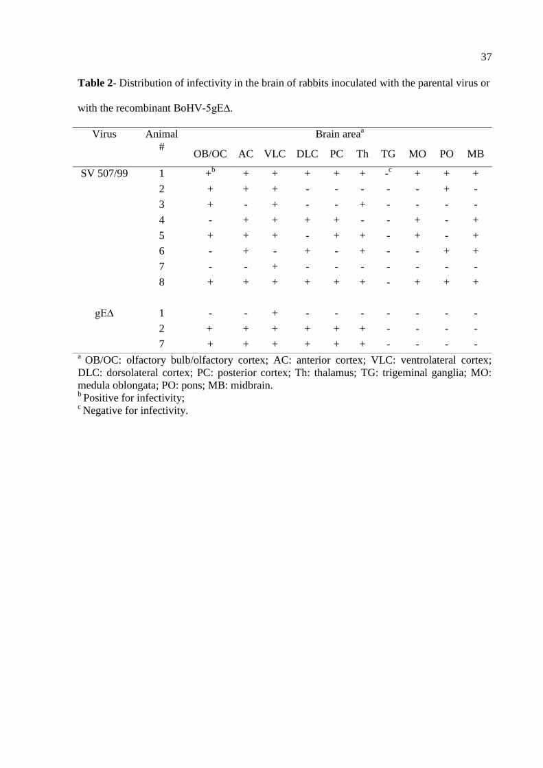
Distribution of parental virus and recombinant BoHV-5gE∆ in the brain
Three out of eight rabbits inoculated with the recombinant BoHV-5gE∆ developed
neurological disease clinically undistinguishable to that developed by rabbits inoculated with
the parental virus. In order to determine the extent of dissemination and distribution of each
virus, brain sections of rabbits from both groups (SV507/99 and BoHV-5gE∆) were submitted
to virus isolation. As shown in Table 2, the parental virus was more widely distributed in the
brain than the recombinant virus. It reached deeper areas of the brain, considering the
olfactory pathway of virus invasion, being detected in all frontal structures, cortices, thalamus
and also in the brain stem. In contrast, the recombinant virus was detected in the anterior,
median sections and barely reached the posterior areas. These results demonstrated that the
recombinant BoHV-5gE∆ invaded and replicated within the brain yet showed a relatively
restricted distribution compared to the parental virus.
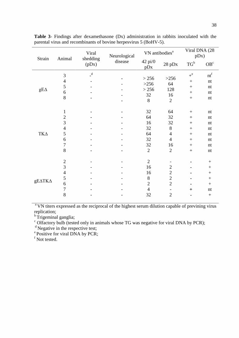
Latent infection – attempts of reactivation and molecular detection
At day 42 pi, all rabbits that survived the acute infection with the recombinants
BoHV-5gE∆ (n=5), BoHV-5TK∆ (n=8) and BoHV-5gE∆TK∆ (n=8) were submitted to Dx
treatment trying to reactivate a putative latent infection. At that day, all inoculated animals
had VN antibodies, in titers ranging from 2 to 256, indicating they harbored virus replication
in the nose during acute infection. However, five daily administrations of Dx to these animals
did not result in detectable virus shedding or in increase in VN titers (Table 3). Likewise,
none of the animals showed clinical signs indicative of clinical recrudescence. Thus, these
results demonstrated the recombinants were not reactivated upon Dx treatment. Since all
rabbits inoculated with the parental virus died during acute infection, reactivation attempts
were not possible. Nevertheless, previous studies have achieved virus reactivation by Dx
treatment ranging from 56.8 to 100% of the BoHV-5 inoculated rabbits, even those inoculated
with relatively low virus titers (106.5-7.0
TCID50) (36, 37).
26
The BoHV-5TK∆ recombinant, and to a lesser extent the BoHV-5gE∆TK∆ virus,
replicated with an apparent low efficiency in the nose during acute infection – in spite of the
VN titers developed by these animals at day 42 pi – and were not reactivated upon Dx
treatment. Then, we argued whether acute replication by these viruses – and by recombinant
BoHV-5gE∆ as well, was sufficient to assure the establishment of latency. Then, a nested
PCR (the test sensitivity was estimated to be around 10 to 100 genome copies per reaction)
performed on total DNA extracted from the TGs of rabbits from groups inoculated with each
single mutant (gE∆ and TK∆) confirmed they all harbored latent viral DNA, albeit it failed in
detecting viral DNA in the TGs of 7 out of 8 rabbits of the group inoculated with the double
mutant (Table 3). A second round of PCR was then performed on total DNA extracted from
the olfactory bulbs (OB) of this last group. This PCR confirmed the presence of latent viral
DNA in the OB of all rabbits inoculated with the double mutant (Table 3). Previous studies
have demonstrated the presence of BoHV-5 latent DNA in the BOs of experimentally infected
calves (9) and rabbits (36). Taken together these results showed that all three recombinants
were able to establish latent infection in the TGs and/or in the OB of inoculated rabbits, yet
they were not reactivated by Dx treatment.
Discussion
We herein investigated the virulence of three BoHV-5 recombinants in rabbits. The
recombinants defective in the glycoprotein E (BoHV-5gE∆), enzyme TK (BoHV-5TK∆) and
both TK and gE (BoHV-5gE∆TK∆) were constructed out of a neurovirulent Brazilian BoHV-
5 strain (SV507/99) as part of a strategy to produce an attenuated, differential vaccine for use
in South America. The recombinant BoHV-5TK∆ and the double deletion mutant (BoHV-
5gE∆TK∆) were fully attenuated for rabbits after IN inoculation. These viruses – especially
the double mutant -replicated less efficiently than the parental virus in the nasal cavity yet
27
were able to induce a VN antibody response in the inoculated rabbits. In contrast, the
recombinant defective in gE (BoHV-5gE∆) retained partially its neurovirulence for rabbits.
All three recombinants conserved their ability to establish latent infection in neural tissues
(TGs and/or OBs) albeit were not capable of reactivating from latency upon Dx
administration. These results open the way for further testing the recombinants BoHV-5TK∆
and BoHV-5gE∆TK∆ in cattle towards a potential use of these viruses as vaccine strains.
These recombinants may also be useful to study the role of each deleted gene product in the
biology of BoHV-5 infection and its interaction with the host.
The pathogenesis of BoHV-5 neurological disease in cattle is still poorly understood
and several aspects of acute and latent infection have been studied in a rabbit model (32, 34).
The neurological infection involves an initial viral replication in the site of entry (nasal
epithelium), axonal transport to and replication in second, third and fourth-order neurons in
several areas of the brain (38). Regardless the biological and molecular mechanisms
underlying the development of neurological disease – which are merely hypothetical at this
time -, massive virus replication in several areas of the brain is a consistent finding during
acute BoHV-5 infection in rabbits and calves (34, 38, 39). Thus, the full expression of BoHV-
5 neurovirulence likely depends upon the ability of the virus to reach the brain from
peripheric sites (neuroinvasiveness) and to replicate to high titers in neuronal cells
(neurogrowth).
Based on pathogenesis studies with other neuropathogenic alphaherpesviruses - HSV,
PRV, BoHV-1 and BoHV-5 as well - the envelope glycoprotein E (gE) plays an important
role in virus transport along circuits of synaptically connected neurons and, consequently,
influence virus invasion of the brain (17). Glycoprotein E-defective BoHV-5 strains show
reduced neuroinvasiveness and neurovirulence in rabbits (17); while a BoHV-1 gE-negative
recombinant is poorly transported anterogradely from the TGs to the nose after virus
28
reactivation (18, 40). Deletion of gE in BoHV-1 strains is associated with significant – yet
partial - attenuation for calves and has been also used as the antigenic marker in differential
vaccines (16). Our recombinant BoHV-5gE∆ replicated efficiently in the nose and was
partially neurovirulent for rabbits, producing neurological disease in 3/8 animals. Although
the recombinant was less widely distributed in the brain than the parental virus, it was able to
reach and replicate in the anterior areas of the CNS. A similar phenotype has been observed
for other BoHV-5 gE negative recombinant (17). Virus replication in these anterior areas has
been implicated in the production of seizures, a hallmark of BoHV-5 neurological infection
(38). Thus, gE deletion in the strain SV507/99 reduced the virulence but did not suffice to
abolish the ability of the virus to invade and replicate in the brain of rabbits producing
neurological disease. This recombinant established latent infection in TG/OB albeit it was not
reactivated upon Dx treatment. The lack of excretion of this virus after Dx treatment – taken
as indicative of virus reactivation - may be related to deficient anterograde axonal transport as
demonstrated for a gE-negative BoHV-1 strain in calves (18, 40). Alternatively, this virus was
not reactivated in the ganglia by corticosteroid treatment. Further studies will determine the
impact of gE deletion on the phenotype of this virus in cattle and whether a single gE- mutant
is sufficiently attenuated for use as a live vaccine.
The alphaherpesvirus TK is an enzyme involved in the metabolism of
deoxyribonucleotides (dNTPs) and is absolutely necessary for efficient viral replication in
non-dividing cells such as neurons (28). Consequently, TK activity is required for efficient
replication in neurons during acute infection and reactivation from latency but is not
necessary for virus replication in epithelial cells or in cell culture. PRV, HSV - and to a lesser
extent BoHV-1 - defective in TK gene product display reduced virulence and a relative
inability to reactivate from latent infection (12, 25, 28, 30). As BoHV-5 neuropathogenesis is
associated which virus replication and dissemination within the brain, we assumed that
29
functional TK would be necessary for the full expression of virus neurovirulence in vivo. Our
results confirmed this prediction: deletion of TK gene in SV507/99 resulted in a dramatic
reduction in neurovirulence for rabbits. In a previous study, our group reported the production
of a brivudin-resistant BoHV-5 variant – likely defective in TK activity – that displayed a
similar, attenuated phenotype in rabbits (31). Studies to determine whether these mutants are
impaired in invasion, dissemination or productive replication in the brain are underway and
may shed light on the exact role of TK in the biology of BoHV-5 and the effects of its
deletion on the viral phenotype. Thus, our BoHV-5TK∆ recombinant was shown to be fully
attenuated for rabbits after IN inoculation and would be worthwhile to investigate whether it
presents the same phenotype in cattle.
On the other hand, a search for BoHV-5TK∆ DNA by PCR at day 28 pDx
demonstrated the presence of latent viral DNA in TGs, demonstrating that the recombinant
virus was capable of reaching these sites during acute infection. However, the TK-negative
virus was not reactivated from latent infection, confirming findings observed for other TK-
defective alphaherpesviruses (22, 23, 25). The lack of reactivation by TK-defective
alphaherpesvirus is probably related to their inability to replicate productively in neurons, a
condition necessary for virus reactivation and excretion (21).
The double deletion mutant BoHV-5gE∆TK∆ was also fully attenuated for rabbits.
The replication of this recombinant in the nose was noticeably reduced what could partially
explain its inability to produce neurological disease. Nevertheless, the relatively low
replication levels in the nose did not abolish the ability of the virus to induce a VN response.
Whether this magnitude of VN response would protect upon challenge is not known and
would deserve investigation. Virus replication at the site of entry also sufficed to assure virus
transport to the OB and/or TGs where latent infection was established. Again, as
demonstrated for the other two recombinants, the double mutant was not capable of
30
reactivating the latent infection, probably reflecting a combined deleterious effect of lacking
both TK and gE. Thus, this recombinant would be a candidate to be used as an attenuated,
antigenically marked vaccine strain for cattle. Subsequent studies are needed to determine the
replication efficiency, attenuation and immunogenicity of this recombinant in cattle.
In summary, the BoHV-5 recombinants defective in TK alone or in combination with
gE are attenuated for rabbits and constitute potential vaccine candidates, depending upon the
confirmation of this phenotype in cattle. The single mutant gE- retained part of its virulence
for rabbits yet its phenotype awaits confirmation in cattle before discarding it for vaccine use.
Regardless their potential use in vaccine formulations, the recombinants may represent useful
tools to study the function of each gene product in the biology of BoHV-5 infection.
Acknowledgements
S. C. da Silva is a MS student recipient of a scholarship from CAPES. M. C. S. Brum
was recipient of doctoral scholarships from CNPq and CAPES (PDEE/UFSM). E. F. Flores
(301666/04-0) and R. Weiblen (301339/04-0) are recipients of CNPq (Brazilian Council for
Research) scholarships. The project was developed in collaboration with S. I. Chowdhury,
Kansas State University, Manhattan, KS, USA and funded by PRONEX, FAPERGS, CNPq
and CAPES.
References
1. Engels M, Ackermann M. Pathogenesis of ruminant herpesvirus infections. Vet Microbiol
1996; 53: 3-15.
2. Silva MS, Brum MC, Loreto EL, Weiblen R, Flores EF. Molecular and antigenic
characterization of Brazilian bovine herpesvirus type 1 isolates recovered from the brain of
cattle with neurological disease. Virus Res 2007; 129: 191-199.
31
3. Silva MS, Brum MCS, Weiblen R, Flores EF. Identificação e diferenciação de herpesvírus
bovino tipos 1 e 5 isolados de amostras clínicas no Centro-Sul do Brasil, Argentina e Uruguai
(1987-2006). Pesqui Vet Bras 2007; 27: 403-408.
4. Carrillo BJ, Ambrogi A, Schudel AA, Vazquez M, Dahme E, Pospischil A.
Meningoencephalitis caused by IBR virus in calves in Argentina. Zentralbl Veterinarmed B
1983; 30: 327-332.
5. d'Offay JM, Ely RW, Baldwin CA, Whitenack DL, Stair EL, Collins JK. Diagnosis of
encephalitic bovine herpesvirus type 5 (BHV-5) infection in cattle: virus isolation and
immunohistochemical detection of antigen in formalin-fixed bovine brain tissues. J Vet Diagn
Invest 1995; 7: 247-251.
6. Engels M, Giuliani C, Wild P, Beck TM, Loepfe E, Wyler R. The genome of bovine
herpesvirus 1 (BHV-1) strains exhibiting a neuropathogenic potential compared to known
BHV-1 strains by restriction site mapping and cross-hybridization. Virus Res 1986; 6: 57-73.
7. Roizmann B, Desrosiers RC, Fleckenstein B, Lopez C, Minson AC, Studdert MJ. The
family Herpesviridae: an update. The Herpesvirus Study Group of the International
Committee on Taxonomy of Viruses. Arch Virol 1992; 123: 425-449.
8. Delhon G, Moraes MP, Lu Z, Afonso CL, Flores EF, Weiblen R, Kutish GF, Rock DL.
Genome of bovine herpesvirus 5. J Virol 2003; 77: 10339-10347.
9. Vogel FS, Caron L, Flores EF, Weiblen R, Winkelmann ER, Mayer SV, Bastos RG.
Distribution of bovine herpesvirus type 5 DNA in the central nervous systems of latently,
experimentally infected calves. J Clin Microbiol 2003; 41: 4512-4520.
10. Rock DL. Latent Infection with Bovine Herpesvirus Type-1. Semin Virol 1994; 5: 233-
240.
11. Schwyzer M, Ackermann M. Molecular virology of ruminant herpesviruses. Vet
Microbiol 1996; 53: 17-29.
32
12. Enquist LW, Husak PJ, Banfield BW, Smith GA. Infection and spread of
alphaherpesviruses in the nervous system. Adv Virus Res 1998; 51: 237-347.
13. Mettenleiter TC. Pathogenesis of neurotropic herpesviruses: role of viral glycoproteins in
neuroinvasion and transneuronal spread. Virus Res 2003; 92: 197-206.
14. Belknap EB, Walters LM, Kelling C, Ayers VK, Norris J, McMillen J, Hayhow C,
Cochran M, Reddy DN, Wright J, Collins JK. Immunogenicity and protective efficacy of a
gE, gG and US2 gene-deleted bovine herpesvirus-1 (BHV-1) vaccine. Vaccine 1999; 17:
2297-2305.
15. Flores EF, Osorio FA, Zanella EL, Kit S, Kit M. Efficacy of a deletion mutant bovine
herpesvirus-1 (BHV-1) vaccine that allows serologic differentiation of vaccinated from
naturally infected animals. J Vet Diagn Invest 1993; 5: 534-540.
16. Kaashoek MJ, van Engelenburg FA, Moerman A, Gielkens AL, Rijsewijk FA, van
Oirschot JT. Virulence and immunogenicity in calves of thymidine kinase- and glycoprotein
E-negative bovine herpesvirus 1 mutants. Vet Microbiol 1996; 48: 143-153.
17. Chowdhury SI, Lee BJ, Ozkul A, Weiss ML. Bovine herpesvirus 5 glycoprotein E is
important for neuroinvasiveness and neurovirulence in the olfactory pathway of the rabbit. J
Virol 2000; 74: 2094-2106.
18. Liu ZF, Brum MC, Doster A, Jones C, Chowdhury SI. A bovine herpesvirus type 1
mutant virus specifying a carboxyl-terminal truncation of glycoprotein E is defective in
anterograde neuronal transport in rabbits and calves. J Virol 2008; 82: 7432-7442.
19. van Oirschot JT, Kaashoek MJ, Rijsewijk FA, Stegeman JA. The use of marker vaccines
in eradication of herpesviruses. J Biotechnol 1996; 44: 75-81.
20. Franco AC, Hübner SdO, Oliveira APd, Batista HBdCR, Roehe PM, Rijsewijk FAM.
Construction and characterization of a bovine herpesvirus 5 mutant with a deletion of the gI,
gE and US9 genes. Braz J Microbiol 2007; 38: 667-673.
33
21. Tikoo SK, Campos M, Babiuk LA. Bovine herpesvirus 1 (BHV-1): biology, pathogenesis,
and control. Adv Virus Res 1995; 45: 191-223.
22. Coen DM, Kosz-Vnenchak M, Jacobson JG, Leib DA, Bogard CL, Schaffer PA, Tyler
KL, Knipe DM. Thymidine kinase-negative herpes simplex virus mutants establish latency in
mouse trigeminal ganglia but do not reactivate. Proc Natl Acad Sci 1989; 86: 4736-4740.
23. Chen SH, Pearson A, Coen DM. Failure of thymidine kinase-negative herpes simplex
virus to reactivate from latency following efficient establishment. J Virol 2004; 78: 520-523.
24. Moormann RJ, de Rover T, Briaire J, Peeters BP, Gielkens AL, van Oirschot JT.
Inactivation of the thymidine kinase gene of a gI deletion mutant of pseudorabies virus
generates a safe but still highly immunogenic vaccine strain. J Gen Virol 1990; 71: 1591-
1595.
25. Ferrari M, Mettenleiter TC, Romanelli MG, Cabassi E, Corradi A, Dal Mas N, Silini R. A
comparative study of pseudorabies virus (PRV) strains with defects in thymidine kinase and
glycoprotein genes. J Comp Pathol 2000; 123: 152-163.
26. Kit S, Qavi H, Gaines JD, Billingsley P, McConnell S. Thymidine kinase-negative bovine
herpesvirus type 1 mutant is stable and highly attenuated in calves. Arch Virol 1985; 86: 63-
83.
27. Gilliam SE, Thackray AM, Brown GA, Field HJ. The pathogenesis of wild type and drug
resistant mutant strains of bovine herpesvirus-1 (BHV-1) in the natural host. Arch Virol 1993;
128: 43-54.
28. Whetstone CA, Miller JM, Seal BS, Bello LJ, Lawrence WC. Latency and reactivation of
a thymidine kinase-negative bovine herpesvirus 1 deletion mutant. Arch Virol 1992; 122: 207-
214.
29. Mengeling WL. Virus reactivation in pigs latently infected with a thymidine kinase
negative vaccine strain of pseudorabies virus. Arch Virol 1991; 120: 57-70.

34
30. Chowdhury SI. Construction and characterization of an attenuated bovine herpesvirus type
1 (BHV-1) recombinant virus. Vet Microbiol 1996; 52: 13-23.
31. Brum MCS, Weiblen R, Flores EF, Chowdhury SI. Construction and characterization in
vitro of bovine herpesvirus type 5 recombinants defective in the glycoprotein E (gE),
thymidine kinase (TK) and gE-TK genes. Braz.J.Med.Biol.Res.(accepted).
32. Chowdhury SI, Lee BJ, Mosier D, Sur JH, Osorio FA, Kennedy G, Weiss ML.
Neuropathology of bovine herpesvirus type 5 (BHV-5) meningo-encephalitis in a rabbit
seizure model. J Comp Pathol 1997; 117: 295-310.
33. Reed LJ, Muench H. A simple method for estimating fifty per cent end points. Am J Hyg
1938; 27: 493-497.
34. Silva AMd, Flores EF, Weiblen R, Canto MC, Irigoyen LF, Roehe PM, Sousa RSd.
Pathogenesis of meningoencephalitis in rabbits by bovine herpesvirus type-5 (BHV-5). Braz J
Microbiol 1999; 30: 22-31.
35. Diel DG, Almeida SR, Brum MC, Dezengrini R, Weiblen R, Flores EF. Acute and latent
infection by bovine herpesvirus type 5 in experimentally infected goats. Vet Microbiol 2007;
121: 257-267.
36. Mayer SV, Quadros VL, Vogel FS, Winkelmann ER, Arenhart S, Weiblen R, Flores EF.
Dexamethasone-induced reactivation of bovine herpesvirus type 5 latent infection in
experimentally infected rabbits results in a broader distribution of latent viral DNA in the
brain. Braz J Med Biol Res 2006; 39: 335-343.
37. Caron L, Flores EF, Weiblen R, Scherer CF, Irigoyen LF, Roehe PM, Odeon A, Sur JH.
Latent infection by bovine herpesvirus type-5 in experimentally infected rabbits: virus
reactivation, shedding and recrudescence of neurological disease. Vet Microbiol 2002; 84:
285-295.
35
38. Lee BJ, Weiss ML, Mosier D, Chowdhury SI. Spread of bovine herpesvirus type 5 (BHV-
5) in the rabbit brain after intranasal inoculation. J Neurovirol 1999; 5: 474-484.
39. Dezengrini R, Weiss M, Torres FD, Oliveira MS, Furian F, Mello CF, Weiblen R, Flores
EF. Bovine herpesvirus 5 induces an overproduction of nitric oxide in the brain of rabbits that
correlates with virus dissemination and precedes the development of neurological signs. J
Neurovirol 2009; 15: 153-163.
40. Brum MC, Coats C, Sangena RB, Doster A, Jones C, Chowdhury SI. Bovine herpesvirus
type 1 (BoHV-1) anterograde neuronal transport from trigeminal ganglia to nose and eye
requires glycoprotein E. J Neurovirol 2009; 15: 196-201.

36
Table 1- Virological, clinical and serological findings during acute infection in rabbits
inoculated with parental virus and recombinants of bovine herpesvírus 5 (BoHV-5).
a Period of virus shedding in nasal secretions;
b VN titers expressed as the reciprocal of the highest serum dilution capable of previning virus
replication; c Development of neurological signs;
d Day post-inoculation in which the animals died or were submitted to euthanasia;
e Not tested;
f Absence of virus shedding or neurological signs.
Strain Animal
#
Viral sheddinga
(day pi)
Neurological
disease (day pi)
VN antibodiesb
(42 pi)
SV-507/99
1
2
3
4
5
6
7
8
2-8
2-8
2-6
2-6
2-6
2-6
2-8
2-8
+c (8)
d
+ (8)
+ (13)
+ (13)
+ (7)
+ (8)
+ (10)
+ (9)
nte
nt
nt
nt
nt
nt
nt
nt
gE∆
1
2
3
4
5
6
7
8
2-4
2-6
2-6
2-8
4-6
2-6
-
4-8
+ (15)
+ (10)
-f
-
-
-
+ (10)
-
nt
nt
256
256
256
32
nt
8
TK∆
1
2
3
4
5
6
7
8
-
8
6
-
4-8
4-8
4
2-10
-
-
-
-
-
-
-
-
32
64
16
32
64
32
32
2
gE∆TK∆
1
2
3
4
5
6
7
8
6
4
-
-
6
-
-
-
-
-
-
-
-
-
-
-
2
2
16
16
8
2
4
32
37
Table 2- Distribution of infectivity in the brain of rabbits inoculated with the parental virus or
with the recombinant BoHV-5gE∆.
a OB/OC: olfactory bulb/olfactory cortex; AC: anterior cortex; VLC: ventrolateral cortex;
DLC: dorsolateral cortex; PC: posterior cortex; Th: thalamus; TG: trigeminal ganglia; MO:
medula oblongata; PO: pons; MB: midbrain. b
Positive for infectivity; c Negative for infectivity.
Virus Animal
#
Brain areaa
OB/OC AC VLC DLC PC Th TG MO PO MB
SV 507/99 1 +b
+ + + + + -c
+ + +
2 + + + - - - - - + -
3 + - + - - + - - - -
4 - + + + + - - + - +
5 + + + - + + - + - +
6 - + - + - + - - + +
7 - - + - - - - - - -
8 + + + + + + - + + +
gE∆ 1 - - + - - - - - - -
2 + + + + + + - - - -
7 + + + + + + - - - -
38
Table 3- Findings after dexamethasone (Dx) administration in rabbits inoculated with the
parental virus and recombinants of bovine herpesvirus 5 (BoHV-5).
a VN titers expressed as the reciprocal of the highest serum dilution capable of previning virus
replication; b
Trigeminal ganglia; c Olfactory bulb (tested only in animals whose TG was negative for viral DNA by PCR);
d Negative in the respective test;
e Positive for viral DNA by PCR;
f Not tested.
Strain Animal
Viral
shedding
(pDx)
Neurological
disease
VN antibodiesa Viral DNA (28
pDx)
42 pi/0
pDx 28 pDx TG
b OB
c
gE∆
3
4
5
6
8
-d
-
-
-
-
-
-
-
-
-
> 256
>256
> 256
32
8
>256
64
128
16
2
+e
+
+
+
+
ntf
nt
nt
nt
nt
TK∆
1
2
3
4
5
6
7
8
-
-
-
-
-
-
-
-
-
-
-
-
-
-
-
-
32
64
16
32
64
32
32
2
64
32
32
8
4
4
16
2
+
+
+
+
+
+
+
+
nt
nt
nt
nt
nt
nt
nt
nt
gE∆TK∆
2
3
4
5
6
7
8
-
-
-
-
-
-
-
-
-
-
-
-
-
-
2
16
16
8
2
4
32
-
2
2
2
2
-
2
-
-
-
-
-
+
-
+
+
+
+
+
nt
+
39
Figure 1. Illustrative results of a PCR performed to confirm the identity of viruses shed in
nasal secretions by rabbits of the respective groups. Panel A. PCR for the TK gene (285 bp).
Panel B. PCR for the glycoprotein E gene (269 bp). Panel C. PCR for the glycoprotein B gene
(control) (444 bp). MWM: molecular weight marker. Lane 1: negative control (DNA template
from the brain of a BoHV-5 seronegative cow); lane 2: positive control, DNA extracted from
the supernatant CRIB cells infected with the parental virus; lane 3-6: pool of nasal secretions
of rabbits inoculated with recombinants - double mutant BoHV-5gEΔTKΔ (lane 3); BoHV-
5gEΔ (lane 4); BoHV-5TKΔ (lane 5); parental virus (SV507/99; lane 6). Ethidium bromide
stained 1.5% agarose gel; the sizes of the corresponding markers are indicated

40
3. REFERÊNCIAS
ABRIL, C. et al. Both viral and host factors contribute to neurovirulence of bovine
herpesviruses 1 and 5 in interferon receptor-deficient mice. Journal of Virology,
Washington, v.78, n.7, p.3644-3653, Apr. 2004.
ACKERMANN, M.; ENGELS. M. Pro and contra IBR-eradication. Veterinary
Microbiology, Amsterdam, v.113, n.3-4, p.293-302, Mar. 2006.
BELKNAP, E. B. et al. Experimental infection of neonatal calves with neurovirulent bovine
herpesvirus type 1.3. Veterinary Pathology, Middleton, v.31, n.3, p.358-365, May. 1994.
BELTRÃO, N. et al. Infecção e enfermidade neurológica pelo herpesvírus bovino tipo 5
(BHV-5): coelhos como modelo experimental. Pesquisa Veterinária Brasileira, Rio de
Janeiro, v.20, p.144-150, out-dez. 2000.
BRUM, M. C. Produção e caracterização de cepas recombinantes do herpesvírus bovino tipo
5 defectivas na enzima timidina quinase e glicoproteína E. 2009. 88f. Tese (Doutorado em
Medicina Veterinária) – Universidade Federal de Santa Maria, Santa Maria, 2009.
BRUM, M. C. et al. Bovine herpesvirus type 1 (BoHV-1) anterograde neuronal transport from
trigeminal ganglia to nose and eye requires glycoprotein E. Journal of Neurovirology,
London, v.15, n.2, p.196-201, Apr. 2009.
CARON, L. et al. Latent infection by bovine herpesvirus type-5 in experimentally infected
rabbits: virus reactivation, shedding and recrudescence of neurological disease. Veterinary
Microbiology, Amsterdam, v.84, n.4, p.285-295, Feb. 2002.
CARRILLO, B. J. et al. Meningoencephalitis caused by IBR virus in calves in Argentina.
Zentralblatt Veterinarmedizin Reihe B, Berlin, v.30, n.5, p.327-332, Jun. 1983.
CHEN, S. H. et al. Failure of thymidine kinase-negative herpes simplex virus to reactivate
from latency following efficient establishment. Journal of Virology, Washington, v.78, n.1,
p.520-523, Jan. 2004.
CHOWDHURY, S. I. Construction and characterization of an attenuated bovine herpesvirus
type 1 (BHV-1) recombinant virus. Veterinary Microbiology, Amsterdam, v.52, n.1-2, p.13-
23, Sep. 1996.
41
CHOWDHURY, S. I. et al. Neuropathology of bovine herpesvirus type 5 (BHV-5) meningo-
encephalitis in a rabbit seizure model. Journal of Comparative Pathology, London, v.117,
n.4, p.295-310, Nov. 1997.
______.Bovine herpesvirus 5 glycoprotein E is important for neuroinvasiveness and
neurovirulence in the olfactory pathway of the rabbit. Journal of Virology, Washington,
v.74, n.5, p.2094-2106, Mar. 2000.
DELHON, G. et al. Genome of bovine herpesvirus 5. Journal of Virology, Washington,
v.77, n.19, p.10339-10347, Oct. 2003.
DIEL, D. G. et al. O Herpesvírus bovino tipo 5 (BoHV-5) pode utilizar as rotas olfatória ou
trigeminal para invadir o sistema nervoso central de coelhos, dependendo da via de
inoculação. Pesquisa Veterinária Brasileira, Rio de Janeiro, v.25, p.164-170, jul-set. 2005.
DIEL, D. G. et al. Acute and latent infection by bovine herpesvirus type 5 in experimentally
infected goats. Veterinary Microbiology, Amsterdam, v.121, n.3-4, p.257-267, Apr. 2007.
DINGWELL, K. S. et al. Glycoproteins E and I facilitate neuron-to-neuron spread of herpes
simplex virus. Journal of Virology, Washington, v.69, n.11, p.7087-7098, Nov. 1995.
ENQUIST, L. W. et al. Infection and spread of alphaherpesviruses in the nervous system.
Advances in Virus Research, New York, v.51, p.237-347, Jul. 1998.
FERRARI, M. et al. A comparative study of pseudorabies virus (PRV) strains with defects in
thymidine kinase and glycoprotein genes. Journal of Comparative Pathology, London,
v.123, n.2-3, p.152-163, Aug-Oct. 2000.
FLORES, E. F. et al. Neuropatogênese experimental da infecção pelo herpesvírus bovino tipo
5 em coelhos. Pesquisa Veterinária Brasileira, Rio de Janeiro, v.29, n.1, p.1-16, jan. 2009.
KAASHOEK, M. J. et al. Virulence, immunogenicity and reactivation of bovine herpesvirus
1 mutants with a deletion in the gC, gG, gI, gE, or in both the gI and gE gene. Vaccine,
Amsterdam, v.16, n.8, p.802-809, May. 1998.
KIRKLAND, P. D. et al. Infertility and venereal disease in cattle inseminated with semen
containing bovine herpesvirus type 5. Veterinary Record, London, v.165, n. 4, p.111-113,
Jul. 2009.

42
LEE, B. J. et al. Spread of bovine herpesvirus type 5 (BHV-5) in the rabbit brain after
intranasal inoculation. Journal of Virology, Washington, v.5, n.5, p.474-484, Oct. 1999.
LIU, Z. F. et al. A bovine herpesvirus type 1 mutant virus specifying a carboxyl-terminal
truncation of glycoprotein E is defective in anterograde neuronal transport in rabbits and
calves. Journal of Virology, Washington, v.82, n.15, p.7432-7442, Aug. 2008.
MENGELING, W. L. Virus reactivation in pigs latently infected with a thymidine kinase
negative vaccine strain of pseudorabies virus. Archives of Virology, Wien, v.120, n.1-2,
p.57-70, Jan. 1991.
METTENLEITER, T. C. Pathogenesis of neurotropic herpesviruses: role of viral
glycoproteins in neuroinvasion and transneuronal spread. Virus Research, Amsterdam, v.92,
n.2, p.197-206, Apr. 2003.
METZLER, A. E. et al. Bovine herpesvirus 1: molecular and antigenic characteristics of
variant viruses isolated from calves with neurological disease. Archives of Virology, Wien,
v.87, n.3-4, p.205-217, May 1986.
MEYER, G. et al. Establishment of a rabbit model for bovine herpesvirus type 5 neurological
acute infection. Veterinary Microbiology, Amsterdam, v.51, n.1-2, p.27-40, Jul. 1996.
MULDER, W. A. et al. Glycoprotein gE-negative pseudorabies virus has a reduced capability
to infect second- and third-order neurons of the olfactory and trigeminal routes in the porcine
central nervous system. Journal of General Virology, London, v.75, p.3095-3106, Nov.
1994.
PEREZ, S. E. et al. Primary infection, latency, and reactivation of bovine herpesvirus type 5
in the bovine nervous system. Veterinary Pathology, Middleton, v.39, n.4, p.437-444, Jul.
2002.
RISSI, D. R. et al. Meningoencefalite por herpesvírus bovino-5. Pesquisa Veterinária
Brasileira, Rio de Janeiro, v.27, p.251-260, jul. 2007.
ROIZMAN, B. et al. The family Herpesviridae: an update. The herpesvirus study group of the
international committee on taxonomy of viruses. Archives of Virology, Wien, v.123, n.3-4,
p.425-449, Sep. 1992.
43
ROIZMAN, B.; KNIPE, D.M. Herpes simplex viruses and their replication. In: D. M. Knipe e
P. M. Howley (Ed.). Fields virology. Philadelphia: Lippincott Williams & Wilkins, 2001, v.2,
p.2399-2459.
SALVADOR, S. C. et al. Meningoencefalite em bovinos causada por herpesvírus bovino-5 no
Mato Grosso do Sul e São Paulo. Pesquisa Veterinária Brasileira, Rio de Janeiro, v.18,
p.76-83, abr-jun. 1998.
SCHWYZER, M.; ACKERMANN, M. Molecular virology of ruminant herpesviruses.
Veterinary Microbiology, Amsterdam, v.53, n.1-2, p.17-29, Nov. 1996.
SILVA, A. M. et al. Experimental infection of sheep with bovine herpesvirus type-5 (BHV-
5): acute and latent infection. Veterinary Microbiology, Amsterdam, v.66, n.2, p.89-99, Apr.
1999.
SILVA, M. S. et al. Identificação e diferenciação de herpesvírus bovino tipos 1 e 5 isolados
de amostras clínicas no Centro-Sul do Brasil, Argentina e Uruguai (1987-2006). Pesquisa
Veterinária Brasileira, Rio de Janeiro, v.27, p.403-408, out. 2007.
SNYDER, A. et al. Herpes simplex virus gE/gI and US9 proteins promote transport of both
capsids and virion glycoproteins in neuronal axons. Journal of Virology, Washington, v.82,
n.21, p.10613-10624, Nov. 2008.
TOMISHIMA, M. J.; ENQUIST, L. W. A conserved alpha-herpesvirus protein necessary for
axonal localization of viral membrane proteins. Journal of Cell Biology, New York, v.154,
n.4, p.741-752, Aug. 2001.
VAN DRUNEN LITTEL-VAN DEN HURK, S. Cell-mediated immune responses induced by
BHV-1: rational vaccine design. Expert Review of Vaccines, London, v. 6, n. 3, p.369-80,
Jun. 2007.
VAN ENGELENBURG, F. A. et al. A glycoprotein E deletion mutant of bovine herpesvirus
1 infects the same limited number of tissues in calves as wild-type virus, but for a shorter
period. Journal of General Virology, London, v.76, p.2387-2392, Sep. 1995.
VAN OIRSCHOT, J. T. The use of marker vaccines in eradication of herpesviruses. Journal
of Biotechnology, Amsterdan, v. 44, n. 1-3, p.75-81, Jan. 1996.
VAN OIRSCHOT, J. T. Diva vaccines that reduce virus transmission. Journal of
Biotechnology, Amsterdan, v. 73, n. 2-3, p.195-205, Aug. 1999.
44
VOGEL, F. S. et al. Distribution of bovine herpesvirus type 5 DNA in the central nervous
systems of latently, experimentally infected calves. Journal of Clinical Microbiology,
Washington, v.41, n.10, p.4512-4520, Oct. 2003.
WHETSTONE, C. A. et al. Latency and reactivation of a thymidine kinase-negative bovine
herpesvirus 1 deletion mutant. Archives of Virology, Wien, v.122, n.1-2, p.207-214, Mar.
1992.