Molecular Bases of Diseaseauthors.library.caltech.edu/47517/1/J. Biol. Chem.-2014...Ming Liu...
14
Ming Liu Hutchison, Shu-ou Shan, Peter Arvan and Roberto Lara-Lemus, Leena Haataja, Kathryn Jingqing Cui, Ling-jia Wang, Jinhong Sun, Huan Guo, Yi Xiong, Piotr Witkowski, and Late-onset Diabetes Cell Failure β Contributes to Pancreatic Inefficient Translocation of Preproinsulin Molecular Bases of Disease: doi: 10.1074/jbc.M114.562355 originally published online April 25, 2014 2014, 289:16290-16302. J. Biol. Chem. 10.1074/jbc.M114.562355 Access the most updated version of this article at doi: . JBC Affinity Sites Find articles, minireviews, Reflections and Classics on similar topics on the Alerts: When a correction for this article is posted • When this article is cited • to choose from all of JBC's e-mail alerts Click here http://www.jbc.org/content/289/23/16290.full.html#ref-list-1 This article cites 62 references, 33 of which can be accessed free at at CALIFORNIA INSTITUTE OF TECHNOLOGY on July 24, 2014 http://www.jbc.org/ Downloaded from at CALIFORNIA INSTITUTE OF TECHNOLOGY on July 24, 2014 http://www.jbc.org/ Downloaded from
Transcript of Molecular Bases of Diseaseauthors.library.caltech.edu/47517/1/J. Biol. Chem.-2014...Ming Liu...
Ming LiuHutchison, Shu-ou Shan, Peter Arvan and Roberto Lara-Lemus, Leena Haataja, KathrynJingqing Cui, Ling-jia Wang, Jinhong Sun, Huan Guo, Yi Xiong, Piotr Witkowski, and Late-onset Diabetes
Cell FailureβContributes to Pancreatic Inefficient Translocation of PreproinsulinMolecular Bases of Disease:
doi: 10.1074/jbc.M114.562355 originally published online April 25, 20142014, 289:16290-16302.J. Biol. Chem.
10.1074/jbc.M114.562355Access the most updated version of this article at doi:
.JBC Affinity SitesFind articles, minireviews, Reflections and Classics on similar topics on the
Alerts:
When a correction for this article is posted•
When this article is cited•
to choose from all of JBC's e-mail alertsClick here
http://www.jbc.org/content/289/23/16290.full.html#ref-list-1
This article cites 62 references, 33 of which can be accessed free at
at CA
LIFO
RN
IA IN
STIT
UT
E O
F TE
CH
NO
LO
GY
on July 24, 2014http://w
ww
.jbc.org/D
ownloaded from
at C
AL
IFOR
NIA
INST
ITU
TE
OF T
EC
HN
OL
OG
Y on July 24, 2014
http://ww
w.jbc.org/
Dow
nloaded from

Inefficient Translocation of Preproinsulin Contributes toPancreatic � Cell Failure and Late-onset Diabetes*
Received for publication, March 2, 2014, and in revised form, March 26, 2014 Published, JBC Papers in Press, April 25, 2014, DOI 10.1074/jbc.M114.562355
Huan Guo‡1, Yi Xiong‡1, Piotr Witkowski§2, Jingqing Cui‡¶, Ling-jia Wang§, Jinhong Sun‡, Roberto Lara-Lemus‡�,Leena Haataja‡, Kathryn Hutchison‡, Shu-ou Shan**3, Peter Arvan‡4, and Ming Liu‡¶5
From the ‡Division of Metabolism, Endocrinology, and Diabetes, University of Michigan Medical School, Ann Arbor,Michigan 48105, the §Division of Organ Transplantation, University of Chicago, Chicago, Illinois 60637, the ¶Division ofMetabolism, Tianjin Medical University General Hospital, Tianjin, China 300052, the �Department of Research in Biochemistry,National Institute of Respiratory Diseases “Ismael Cosío Villegas” , Mexico City 14080, Mexico, and the **Division of Chemistry andChemical Engineering, California Institute of Technology, Pasadena, California 91125
Background: Preproinsulin signal peptide mutations have been linked to human diabetes.Results: Preproinsulin mutants fail to be fully translocated across the endoplasmic reticulum membrane, accumulate in thecytosol, and lead to � cell death.Conclusion: Cytosolic preproinsulin accumulation contributes to � cell failure.Significance: This reveals a novel pathogenesis of diabetes associated with inefficient preproinsulin translocation and cytosolicaccumulation.
Among the defects in the early events of insulin biosynthesis,proinsulin misfolding and endoplasmic reticulum (ER) stresshave drawn increasing attention as causes of � cell failure. How-ever, no studies have yet addressed potential defects at the cyto-solic entry point of preproinsulin into the secretory pathway.Here, we provide the first evidence that inefficient translocationof preproinsulin (caused by loss of a positive charge in the nregion of its signal sequence) contributes to � cell failure anddiabetes. Specifically, we find that, after targeting to the ERmembrane, preproinsulin signal peptide (SP) mutants associ-ated with autosomal dominant late-onset diabetes fail to be fullytranslocated across the ER membrane. The newly synthesized,untranslocated preproinsulin remains strongly associated withthe ER membrane, exposing its proinsulin moiety to the cytosol.Rather than accumulating in the ER and inducing ER stress,untranslocated preproinsulin accumulates in a juxtanuclearcompartment distinct from the Golgi complex, induces theexpression of heat shock protein 70 (HSP70), and promotes �cell death. Restoring an N-terminal positive charge to the
mutant preproinsulin SP significantly improves the transloca-tion defect. These findings not only reveal a novel molecularpathogenesis of � cell failure and diabetes but also provide thefirst evidence of the physiological and pathological significanceof the SP n region positive charge of secretory proteins.
In pancreatic � cells, insulin biosynthesis begins within thecytosol, where insulin mRNA is translated into the precursorpreproinsulin. Preproinsulin comprises, sequentially, signalpeptide (SP),6 insulin B chain, C peptide, and A chain (1, 2). Toenter the secretory pathway, nascent preproinsulin is deliveredby the signal recognition particle (SRP) to the endoplasmicreticulum (ER) membrane, where preproinsulin is translocatedto the luminal side of the ER and cleaved by signal peptidase,forming proinsulin (3, 4). Within the oxidizing environment ofthe ER lumen, proinsulin undergoes rapid oxidative folding,forming three evolutionarily conserved disulfide bonds (5– 8).Among these early events, proinsulin misfolding in the ERlumen and the consequent ER stress have been shown to play animportant role in the autosomal dominant syndrome of mutantinsulin (INS) gene-induced diabetes of youth (MIDY) (9 –13).However, no studies have addressed earlier defects at the pointof entry of preproinsulin into the ER. Recently, two preproin-sulin SP mutants, preproinsulin R6C and R6H (here simplycalled R6C or R6H) have been linked to autosomal dominantlate-onset diabetes in humans (14 –16). The molecular mecha-nism by which these mutants bring about diabetes has beenspeculated to involve ER stress (14), but the actual defect hasremained unknown.
* This work was supported, in whole or in part, by National Institutes ofHealth Grants RO1-DK088856 (to M. L.), RO1-DK-48280 (to P. A.), RO1GM078024 (to S. S.), and P60-DK-20572 (to the Molecular Biology and DNASequencing Core of the Diabetes Research and Training Center). This workwas also supported by National Natural Science Foundation of ChinaResearch Grant 81070629 (to M. L.), by a research grant from the March ofDimes Foundation (to M. L.), and by the Protein Folding Disease Initiativesof the University of Michigan. Human pancreatic islet processing was sup-ported by Illinois Department of Public Health Grant “Pancreatic IsletsTransplantation.”
1 These authors contributed equally to this work.2 Supported by University of Chicago DRTC Grant P30-DK020595.3 To whom correspondence may be addressed: Div. of Chemistry and Chem-
ical Engineering, California Institute of Technology. E-mail: [email protected].
4 To whom correspondence may be addressed: Div. of Metabolism, Endocri-nology, and Diabetes, University of Michigan. E-mail: [email protected].
5 To whom correspondence may be addressed: Div. of Metabolism, Endocri-nology, Diabetes University of Michigan, Brehm Tower, 1000 Wall St., AnnArbor, MI 48105. Tel.: 734-232-8164; Fax:734-232-8162; E-mail: [email protected].
6 The abbreviations used are: SP, signal peptide; SRP, signal recognitionparticle; ER, endoplasmic reticulum; Tricine, N-[2-hydroxy-1,1-bis(hydroxy-methyl)ethyl]glycine; PK, proteinase K; BiP, immunoglobulin-binding pro-tein; PDI, protein disulfide isomerase; Dox, doxycycline; MIDY, mutant insu-lin (INS) gene-induced diabetes of youth.
THE JOURNAL OF BIOLOGICAL CHEMISTRY VOL. 289, NO. 23, pp. 16290 –16302, June 6, 2014© 2014 by The American Society for Biochemistry and Molecular Biology, Inc. Published in the U.S.A.
16290 JOURNAL OF BIOLOGICAL CHEMISTRY VOLUME 289 • NUMBER 23 • JUNE 6, 2014
at CA
LIFO
RN
IA IN
STIT
UT
E O
F TE
CH
NO
LO
GY
on July 24, 2014http://w
ww
.jbc.org/D
ownloaded from
Like other secretory proteins targeted to the ER lumen, pre-proinsulin SP has three distinct regions: a positively charged “nregion,” a central hydrophobic “h region,” and a “c region” con-taining the signal peptidase cleavage site. Mutations R6C andR6H eliminate the highly conserved n region positive charge,which forms the basis for the “positive-inside” rule that isthought to facilitate SP orientation at the ER membrane duringmembrane protein translocation (17–19). Nevertheless, todate, no human disease has been shown to be caused by a loss ofthe n region positive charge. Indeed, a recent study reportedthat neither R6C nor R6H results in defective insulin granuletargeting of a preproinsulin-GFP chimera, thus leaving the dia-betes linked to these mutations largely unexplained (14).
Here we demonstrate that, upon targeting to the ER mem-brane, preproinsulin R6C and R6H fail to be fully translocatedacross the ER membrane. The newly synthesized untranslo-cated preproinsulin remains strongly associated with the ERmembrane, exposing its proinsulin moiety to the cytosol.Untranslocated preproinsulin accumulates in a juxtanuclearcytoplasmic compartment distinct from the Golgi complex,induces the expression of heat shock protein 70 (HSP70), andleads to � cell death. Restoring the N-terminal positive chargeof mutant preproinsulin SP significantly improves the translo-cation defect. This study reveals a novel mechanism leading to� cell failure and diabetes and highlights the pathological con-sequences of loss of the SP n region positive charge of secretoryproteins.
EXPERIMENTAL PROCEDURES
Materials—Guinea pig anti-porcine insulin antibody and thehuman insulin-specific RIA kit (catalog no. HI-14 K) was fromMillipore. Rabbit anti-Myc and anti-GFP antibodies were fromImmunology Consultant Laboratories. Anti-GM130 antibodywas from BD Biosciences. Anti-calnexin antibody was fromEnzo Life Sciences. Zysorbin was from Zymed LaboratoriesInc.. 35S-amino acid mixture (Met � Cys) and pure [35S]Metwere from PerkinElmer Life Sciences. DTT, protein A-agarose,digitonin, N-ethylmaleimide, brefeldin A, and MG132 werefrom Sigma-Aldrich. Peptide-N-glycosidase F (PNGaseF) wasfrom New England Biolabs. 4 –12% NuPage gel, Met/Cys-defi-cient DMEM, and all other tissue culture reagents were fromInvitrogen. The INS1 r9 cells were from Dr. Claes B. Wollhein(University of Geneva, Switzerland).
Human Islet Study—Institutional review board approval forresearch use of isolated human islets was obtained from theUniversity of Michigan. Human islets were isolated from previ-ously healthy, nondiabetic organ donors by the University ofChicago Transplant Center. Three independent human isletbatches from two male donors aged 20 and 58 and one femaledonor aged 48 were used in this study. The islets were dividedinto two groups incubated in CMRL medium (Invitrogen) con-taining either 5.5 or 25.5 mM glucose for 20 h before metabolicradiolabeling with either [35S]Met/Cys mixtures or pure[35S]Met, as indicated, for 10 min. Normalized by DNA, theislet lysates were immunoprecipitated using anti-insulin andanalyzed as indicated in the figure legends.
Mutagenesis, Cell Culture, Transfection, Metabolic Labeling,and Immunoprecipitation—Human and mouse preproinsulinmutants were generated using a site-directed mutagenesis kit(Agilent). INS1 rat � cells, Min6 mouse � cells, or 293T humanembryonic kidney cells were plated onto 12-well plates 1 daybefore transfection with Lipofectamine 2000 (Invitrogen) using1–2 �g of plasmid DNA. 48 h after transfection, cells werepulse-labeled with [35S]Met/Cys or pure [35S]Met with or with-out chase as indicated. Cells used for analysis of (pre)proinsulinoxidative folding were preincubated with 20 mM N-ethylma-leimide in PBS on ice for 10 min before lysis. Immunoprecipi-tation and non-reducing or reducing Tris-Tricine urea SDS-PAGE analyses were performed as described previously (5, 13).For Western blotting, 20 �g of total protein lysates was boiledin SDS sample buffer with or without 100 mM DTT, resolved by4 –12% NuPage, electrotransferred to nitrocellulose, and blot-ted with appropriate first antibodies, followed by appropriatesecondary antibodies conjugated with HRP, with developmentby enhanced chemiluminescence. The BiP promoter-drivenfirefly luciferase assay for Min6 cells was performed asdescribed previously (9).
In Vitro Targeting and Translocation Study—The cotransla-tional protein targeting and translocation assay has beendescribed previously in detail (20, 21). Briefly, wheat germtranslation extract, free of endogenous SRP and SRP receptor,was used to synthesize WT and mutant preproinsulin in thepresence of [35S]Met (for the in vitro assay, methionine residueswere added to the C terminus of preproinsulin). To best mimica single round of cotranslational protein targeting, a cap analog,7-methyl-GTP, was added 1–2 min after translation initiationto inhibit additional rounds of synthesis. Purified SRP, SRPreceptor, and ER microsomal membranes in which the endog-enous SRP and SRP receptor had been removed by high-saltwash and partial trypsin digestion were then added within 1min. Translation was continued for 20 –30 min at 26 °C to allowcompletion of preproinsulin synthesis, at which point the reac-tions were stopped and analyzed. The targeting and transloca-tion efficiency was assessed by two approaches (cleavage of thesignal sequence and protection of proinsulin from proteinase Kdigestion) and analyzed by SDS-PAGE and autoradiography.The localization of preproinsulin and proinsulin was assessedusing a sedimentation assay. The targeting and translocationreactions were carried out as described in the previous para-graph. A 30-�l reaction mixture was layered onto a 50-�l cush-ion of 0.5 M sucrose and ultracentrifuged at 55,000 rpm at 4 °Cfor 5 min (TLA100, Beckman Coulter). The supernatant wasTCA-precipitated. This and the microsomal pellet were dis-solved and analyzed on SDS-PAGE.
Selective Plasma Membrane Permeabilization by Digitonin,Proteinase K Digestion, and Sodium Carbonate Extraction—For the ER targeting experiments, after labeling with [35S]Cys/Met, the plasma membranes of 293T cells transfected with pre-proinsulin WT or mutants were partially permeabilized with0.01% digitonin as described previously (13). For proteinase K(PK) digestion, 2 days after transfection, 293T cells expressingMyc-tagged R6C or A24D were incubated on ice with eitherPBS only, PBS plus 0.01% digitonin and 10 �g/ml PK, or PBSplus 1% Triton X-100 and 10 �g/ml PK for 30 min. 2 �M PMSF
Preproinsulin Translocation Defects and � Cell Failure
JUNE 6, 2014 • VOLUME 289 • NUMBER 23 JOURNAL OF BIOLOGICAL CHEMISTRY 16291
at CA
LIFO
RN
IA IN
STIT
UT
E O
F TE
CH
NO
LO
GY
on July 24, 2014http://w
ww
.jbc.org/D
ownloaded from
and SDS sample buffer were added, boiled, and analyzed byWestern blotting using anti-Myc antibody. For sodium carbon-ate extraction, after pulse-labeling with [35S]Met/Cys, trans-fected cells were suspended in 0.1 M sodium carbonate (pH 12),homogenized, and incubated on ice for 1 h, followed by sedi-mentation at 50,000 rpm at 4 °C for 1 h. The supernatants andpellets were collected and immunoprecipitated with anti-Myc,anti-calnexin, and anti-PDI antibodies.
Generation of Inducible � Cell Lines Expressing Mouse Ins2Wild-type, R6C, and A24D; Cell Proliferation; and Cell DeathAssay—The INS-r9 cells carrying the reverse tetracycline/doxycycline-dependent transactivator (22) were cotransfectedwith a puromycin resistance plasmid and pTRE plasmidsencoding Myc-tagged mouse Ins2 WT, R6C, or A24D. Puromy-cin-resistant clones were isolated and tested for the expressionof Myc-tagged preproinsulin WT or R6C by both pulse labelingand Western blotting after induction with 2 �g/ml doxycycline(Dox). For determining cell proliferation, 3000 cells of induc-ible clones were seeded into 96-well plates and incubated withor without 2 �g/ml Dox for 4 days. BrdU incorporation wasmeasured using a BrdU cell proliferation kit (Millipore). Forexamining cell death, the cells of inducible clones were seededinto 8-well chamber slides (LabTek) and incubated with orwithout 2 �g/ml Dox for 4 days. Cell apoptosis was measured bylabeling DNA strand breaks (TUNEL) using an in situ cell deathdetection kit (Roche) with DAPI counterstaining to identify thenuclei. A total of more than 4500 cells expressing the wild typeor R6C were counted from three independent experiments.
Confocal Imaging—INS1-inducible cell lines expressingmouse preproinsulin WT, R6C, and A24D were induced with 2�g/ml Dox for 4 days. The cells were preincubated with 5 �g/mlcycloheximide for 1 h, followed by either permeabilization with0.01% digitonin on ice for 3 min (selectively permeabilizing theplasma membrane) or with 0.5% Triton X-100 (fully permeabi-lizing all membranes), as indicated. The cells were then fixedwith formaldehyde, blocked, and stained with anti-Myc, anti-PDI (an ER marker), and anti-GM130 (a Golgi marker), fol-lowed by secondary antibodies conjugated to fluorophores, andimaged by epifluorescence in an Olympus FV500 confocalmicroscope with a �60 oil objective. For disrupting the Golgistructure (23), the cells were pretreated with 5 �M brefeldin Afor 30 min before permeabilization with 0.01% digitonin for 3min.
Statistical Analyses—Statistical analyses were carried out byanalysis of variance followed by Newman-Keuls multiple com-parison test using GraphPad Prism 6. p � 0.05 was consideredto be statistically significant.
RESULTS
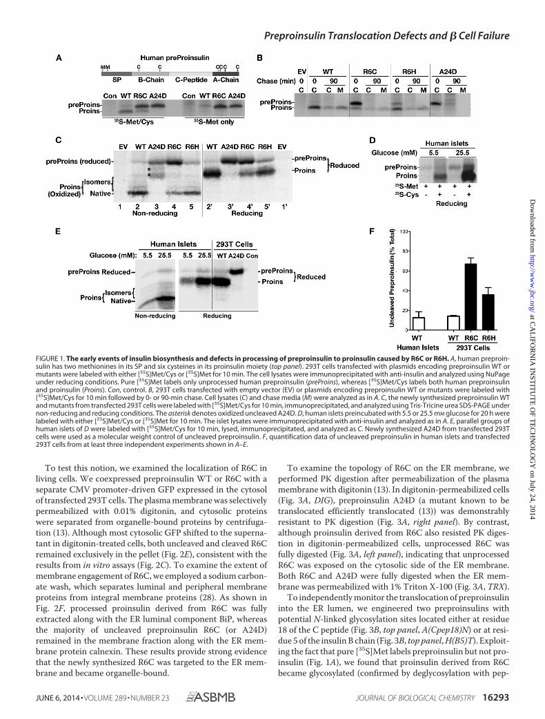
R6C/R6H Causes Early Defects in Preproinsulin to ProinsulinConversion—We developed a method that could specificallydistinguish processed and unprocessed human preproinsulin(13). Because human preproinsulin has two methionines in theSP and six cysteines in the proinsulin moiety (Fig. 1A, toppanel), pure [35S]Met labels exclusively unprocessed preproin-sulin, whereas a [35S]Met/Cys mixture labels both preproinsu-lin and proinsulin. Using this method, we first examined theprocessing of preproinsulin to proinsulin of newly synthesized
preproinsulin WT and mutants in transfected 293T cells. Com-pared with the WT, R6C exhibited significantly more unpro-cessed preproinsulin labeled by [35S]Met (Fig. 1A, bottom pan-el; preproinsulin-A24D, which is defective in SP cleavage,serves as a positive control of uncleaved preproinsulin (13)).After a 90-min chase, although the modest fraction of proinsu-lin derived from R6C was secreted with normal efficiency,unprocessed preproinsulin R6C largely disappeared withoutconversion to proinsulin (Fig. 1B). The defect of R6H was sim-ilar to that of R6C but less severe.
To examine whether unprocessed R6C and R6H wereexposed to the ER lumen, we analyzed newly synthesized pre-proinsulin using Tris-Tricine urea SDS-PAGE under both non-reducing and reducing conditions. Unlike proinsulin anduncleaved A24D, which underwent oxidative folding in the ERlumen (13), unprocessed R6C and R6H remained as fullyreduced forms (Fig. 1C), suggesting that their proinsulin moietyis not exposed to the oxidizing environment of the ER lumen.
Because a small fraction of unprocessed preproinsulin WTwas observed in transfected 293T cells (Fig. 1, A–C and F), wedecided to evaluate the translocation/processing efficiency ofwild-type preproinsulin in primary � cells. After 10 min oflabeling of isolated human pancreatic islets, �10% of the spe-cific immunoprecipitable product remained as unprocessedpreproinsulin, as demonstrated by specific labeling of this bandwith pure [35S]Met (Fig. 1D), by comigration of this band onSDS-PAGE with SP cleavage-defective A24D (Fig. 1E), and byits enhanced intensity upon stimulation with high glucose (Fig.1, D–E). Unprocessed preproinsulin WT was reduced fully (Fig.1E), suggesting that, in normal human islets, some fully trans-lated preproinsulin molecules are present in the cytosol prior toentry into the oxidizing environment of the ER lumen.
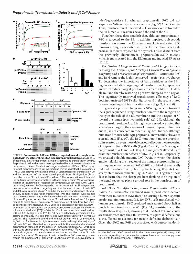
R6C Is Targeted to and Strongly Associated with the ER Mem-brane but Exhibits Impaired Polypeptide Translocation—Mu-tations of Arg-6 eliminate a highly conserved n region positivecharge, which is generally thought to be important for the tar-geting and translocation of secretory and membrane proteins(18, 24 –27). To determine whether R6C affects these earlysteps during insulin biosynthesis, we first tested the SRP-de-pendent targeting and translocation of WT and mutant prepro-insulins into ER microsomal membranes in a reconstituted invitro assay (20). Analysis of the reaction on the basis of both SPcleavage (Fig. 2A) and sensitivity to PK digestion (Fig. 2B) dem-onstrated that R6C mutation caused a 2-fold reduction in theefficiency of targeting and translocation of preproinsulin.
To uncouple the effects of targeting from translocationacross the ER, we performed a sedimentation analysis to exam-ine the localization of preproinsulin and proinsulin after thetargeting/translocation reaction was completed. As expected,in the absence of SRP, only a small fraction of preproinsulin wasassociated with the microsomal membrane, and the presence ofSRP significantly increased the amount of proinsulin mole-cules, which were primarily associated with the microsome.Unexpectedly, in the presence of SRP, the majority of prepro-insulin R6C also became microsome-bound (Fig. 2, C and D).This suggests that R6C was targeted to the ER and that themutation affected its translocation across the ER membrane.
Preproinsulin Translocation Defects and � Cell Failure
16292 JOURNAL OF BIOLOGICAL CHEMISTRY VOLUME 289 • NUMBER 23 • JUNE 6, 2014
at CA
LIFO
RN
IA IN
STIT
UT
E O
F TE
CH
NO
LO
GY
on July 24, 2014http://w
ww
.jbc.org/D
ownloaded from
To test this notion, we examined the localization of R6C inliving cells. We coexpressed preproinsulin WT or R6C with aseparate CMV promoter-driven GFP expressed in the cytosolof transfected 293T cells. The plasma membrane was selectivelypermeabilized with 0.01% digitonin, and cytosolic proteinswere separated from organelle-bound proteins by centrifuga-tion (13). Although most cytosolic GFP shifted to the superna-tant in digitonin-treated cells, both uncleaved and cleaved R6Cremained exclusively in the pellet (Fig. 2E), consistent with theresults from in vitro assays (Fig. 2C). To examine the extent ofmembrane engagement of R6C, we employed a sodium carbon-ate wash, which separates luminal and peripheral membraneproteins from integral membrane proteins (28). As shown inFig. 2F, processed proinsulin derived from R6C was fullyextracted along with the ER luminal component BiP, whereasthe majority of uncleaved preproinsulin R6C (or A24D)remained in the membrane fraction along with the ER mem-brane protein calnexin. These results provide strong evidencethat the newly synthesized R6C was targeted to the ER mem-brane and became organelle-bound.
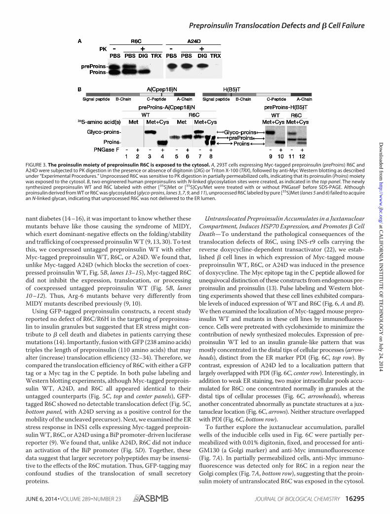
To examine the topology of R6C on the ER membrane, weperformed PK digestion after permeabilization of the plasmamembrane with digitonin (13). In digitonin-permeabilized cells(Fig. 3A, DIG), preproinsulin A24D (a mutant known to betranslocated efficiently translocated (13)) was demonstrablyresistant to PK digestion (Fig. 3A, right panel). By contrast,although proinsulin derived from R6C also resisted PK diges-tion in digitonin-permeabilized cells, unprocessed R6C wasfully digested (Fig. 3A, left panel), indicating that unprocessedR6C was exposed on the cytosolic side of the ER membrane.Both R6C and A24D were fully digested when the ER mem-brane was permeabilized with 1% Triton X-100 (Fig. 3A, TRX).
To independently monitor the translocation of preproinsulininto the ER lumen, we engineered two preproinsulins withpotential N-linked glycosylation sites located either at residue18 of the C peptide (Fig. 3B, top panel, A(Cpep18)N) or at resi-due 5 of the insulin B chain (Fig. 3B, top panel, H(B5)T). Exploit-ing the fact that pure [35S]Met labels preproinsulin but not pro-insulin (Fig. 1A), we found that proinsulin derived from R6Cbecame glycosylated (confirmed by deglycosylation with pep-
FIGURE 1. The early events of insulin biosynthesis and defects in processing of preproinsulin to proinsulin caused by R6C or R6H. A, human preproin-sulin has two methionines in its SP and six cysteines in its proinsulin moiety (top panel). 293T cells transfected with plasmids encoding preproinsulin WT ormutants were labeled with either [35S]Met/Cys or [35S]Met for 10 min. The cell lysates were immunoprecipitated with anti-insulin and analyzed using NuPageunder reducing conditions. Pure [35S]Met labels only unprocessed human preproinsulin (preProins), whereas [35S]Met/Cys labels both human preproinsulinand proinsulin (Proins). Con, control. B, 293T cells transfected with empty vector (EV) or plasmids encoding preproinsulin WT or mutants were labeled with[35S]Met/Cys for 10 min followed by 0- or 90-min chase. Cell lysates (C) and chase media (M) were analyzed as in A. C, the newly synthesized preproinsulin WTand mutants from transfected 293T cells were labeled with [35S]Met/Cys for 10 min, immunoprecipitated, and analyzed using Tris-Tricine urea SDS-PAGE undernon-reducing and reducing conditions. The asterisk denotes oxidized uncleaved A24D. D, human islets preincubated with 5.5 or 25.5 mM glucose for 20 h werelabeled with either [35S]Met/Cys or [35S]Met for 10 min. The islet lysates were immunoprecipitated with anti-insulin and analyzed as in A. E, parallel groups ofhuman islets of D were labeled with [35S]Met/Cys for 10 min, lysed, immunoprecipitated, and analyzed as C. Newly synthesized A24D from transfected 293Tcells were used as a molecular weight control of uncleaved preproinsulin. F, quantification data of uncleaved preproinsulin in human islets and transfected293T cells from at least three independent experiments shown in A–E.
Preproinsulin Translocation Defects and � Cell Failure
JUNE 6, 2014 • VOLUME 289 • NUMBER 23 JOURNAL OF BIOLOGICAL CHEMISTRY 16293
at CA
LIFO
RN
IA IN
STIT
UT
E O
F TE
CH
NO
LO
GY
on July 24, 2014http://w
ww
.jbc.org/D
ownloaded from
tide-N-glycosidase F), whereas preproinsulin R6C did notacquire an N-linked glycan at either site (Fig. 3B, lanes 5 and 6).Thus, translocation of uncleaved R6C was not even delivered tothe ER lumen 3–5 residues beyond the end of the SP.
Together, these data establish that, although preproinsulin-R6C is targeted to the ER, it exhibits impaired polypeptidetranslocation across the ER membrane. Untranslocated R6Cremains strongly associated with the ER membranes with itsproinsulin moiety exposed to the cytosol. This is distinct fromthe previously characterized preproinsulin-A24D mutant,which is translocated into the ER lumen and induced ER stress(12, 13).
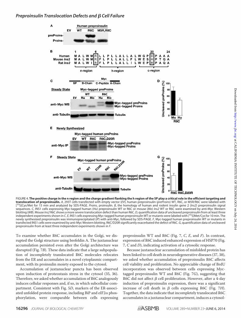
The Positive Charge in the N Region and Charge GradientFlanking the H Region of the SP Play a Critical Role in EfficientTargeting and Translocation of Preproinsulin—Mutations R6Cand R6H remove the highly conserved n region positive charge.To determine the importance of basic residues in the SP nregion for mediating targeting and translocation of preproinsu-lin, we introduced Arg at position 5 to create a M5R/R6C dou-ble mutant, thereby restoring a positive charge to the n region.This significantly improved translocation efficiency of R6C,both in transfected 293T cells (Fig. 4A) and in the reconstitutedin vitro targeting and translocation assay (Figs. 2, A and B).
In general, a positive charge in the SP n region helps to orientthe signal sequence during translocation, with the n region onthe cytosolic side of the ER membrane and the c region of SPtoward the lumen (positive-inside rule) (27, 29). Although thepreproinsulin residue Arg-6 is highly conserved, we noted thata negative charge in the c region of human preproinsulin (resi-due 20) is not conserved in rodents (Fig. 4B). Indeed, althoughhuman and mouse wild-type preproinsulin were fully cleaved ata steady state (Fig. 4C), the R6C mutation in mouse preproin-sulin exerted an even more deleterious effect on the processingof preproinsulin in INS1 cells (Fig. 4, C and D; the Myc-taggedpreproinsulin WT and R6C (Fig. 4C, top panel) allowed us toexamine translocation defects of R6C in � cells). Reciprocally,we created a double mutant, R6C/D20R, in which the chargegradient flanking the h region of the human preproinsulin sig-nal sequence was reversed. R6C/D20R exhibited dramaticallyreduced translocation by both pulse labeling (Fig. 4E) andsteady-state measurements (Fig. 4, F and G). Together, thesedata indicate that the charge gradient flanking the h region ofthe signal sequence plays a critical role in the translocation ofpreproinsulin.
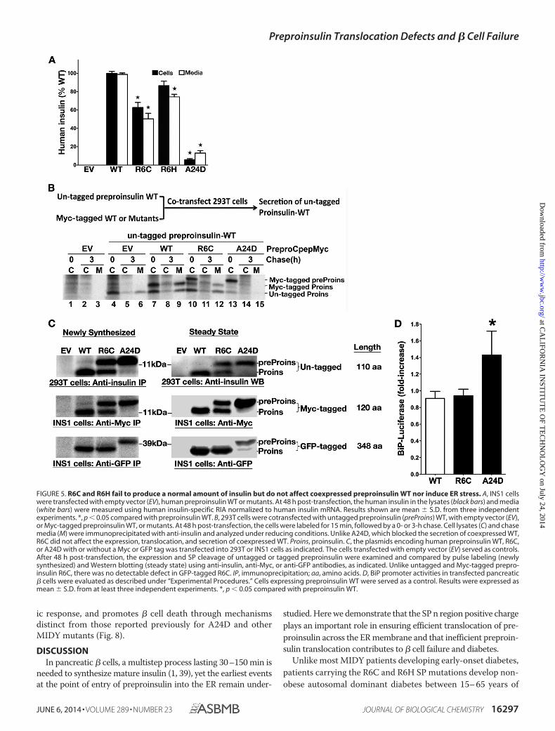
R6C Does Not Affect Coexpressed Preproinsulin WT norInduce ER Stress—We examined insulin production derivedfrom these mutants in INS1 (rat) � cells using a human-specificinsulin radioimmunoassay (13, 30). INS1 cells transfected withhuman preproinsulin R6C produced and secreted about half asmuch human insulin as the WT (Fig. 5A), consistent with theresults above (Figs. 1– 4) showing that �50% of R6C moleculesare translocated into the ER. However, this partial defect aloneis insufficient to account for insulin-deficient diabetes (31).Given that R6C and R6H are associated with autosomal domi-
FIGURE 2. Preproinsulin R6C and R6H are targeted to and strongly asso-ciated with the ER membrane but exhibit impaired translocation. A and B,effect of R6C on SRP-dependent protein targeting and translocation in vitro.Preproinsulin WT and mutants were synthesized by in vitro translation in thepresence of [35S]Met. The ability of exogenously added SRP and SRP receptorto target preproinsulins to salt-washed and trypsin-digested ER microsomes(TKRM) was assayed by cleavage of the SP upon successful translocation (A)and by protection of the translocated protein from PK digestion (B), asdescribed under “Experimental Procedures.” The translocation efficiency ofthe mutant proteins was normalized to that of preproinsulin WT, whose trans-location at saturating SRP receptor concentration was set to be 100%. C, pre-proinsulin (preProins) R6C is targeted to the microsomes in an SRP-dependentmanner. In vitro synthesis, targeting, and translocation of preproinsulin WTand R6C were carried out as in A and B in the presence and absence of exog-enously added SRP and rough microsomes (RM). The reactions were loadedon a 0.5 M sucrose cushion, and microsomal membranes were sedimented byultracentrifugation as described under “Experimental Procedures.” S, super-natant; P, pellet; Proins, proinsulin. D, quantification of data from two inde-pendent experiments shown in C. E, 293T cells coexpressing preproinsulin WTor mutants with a separate CMV promoter-driven GFP expressed in the cyto-sol were labeled with [35S]Cys/Met for 15 min and then incubated with orwithout 0.01% digitonin in PBS for 10 min to selectively permeabilize theplasma membrane. The cells transfected with empty vector (EV) served ascontrols. The membrane-bound and luminal proteins were sedimented at14,000 rpm for 10 min. In digitonin-treated cells, although the majority ofcytosolic GFP was liberated into supernatant, both cleaved and uncleavedpreproinsulin remained in the pellet. IP, immunoprecipitation. F, 293T cellsexpressing preproinsulin R6C and A24D were labeled with [35S]Cys/Met for 20min and subjected to the carbonate extraction as described under “Experi-mental Procedures.” Although proinsulin derived from R6C was mostly recov-ered in the supernatant (S) along with BiP, the majority of uncleaved prepro-
insulin R6C and A24D remained in the membrane pellet (P) along withcalnexin, suggesting that uncleaved preproinsulin mutants are strongly asso-ciated with the ER membrane. T, total lysate.
Preproinsulin Translocation Defects and � Cell Failure
16294 JOURNAL OF BIOLOGICAL CHEMISTRY VOLUME 289 • NUMBER 23 • JUNE 6, 2014
at CA
LIFO
RN
IA IN
STIT
UT
E O
F TE
CH
NO
LO
GY
on July 24, 2014http://w
ww
.jbc.org/D
ownloaded from
nant diabetes (14 –16), it was important to know whether thesemutants behave like those causing the syndrome of MIDY,which exert dominant-negative effects on the folding/stabilityand trafficking of coexpressed proinsulin WT (9, 13, 30). To testthis, we coexpressed untagged preproinsulin WT with eitherMyc-tagged preproinsulin WT, R6C, or A24D. We found that,unlike Myc-tagged A24D (which blocks the secretion of coex-pressed proinsulin WT, Fig. 5B, lanes 13–15), Myc-tagged R6Cdid not inhibit the expression, translocation, or processingof coexpressed untagged preproinsulin WT (Fig. 5B, lanes10 –12). Thus, Arg-6 mutants behave very differently fromMIDY mutants described previously (9, 10).
Using GFP-tagged preproinsulin constructs, a recent studyreported no defect of R6C/R6H in the targeting of preproinsu-lin to insulin granules but suggested that ER stress might con-tribute to � cell death and diabetes in patients carrying thesemutations (14). Importantly, fusion with GFP (238 amino acids)triples the length of preproinsulin (110 amino acids) that mayalter (increase) translocation efficiency (32–34). Therefore, wecompared the translocation efficiency of R6C with either a GFPtag or a Myc tag in the C peptide. In both pulse labeling andWestern blotting experiments, although Myc-tagged preproin-sulin WT, A24D, and R6C all appeared identical to theiruntagged counterparts (Fig. 5C, top and center panels), GFP-tagged R6C showed no detectable translocation defect (Fig. 5C,bottom panel, with A24D serving as a positive control for themobility of the uncleaved precursor). Next, we examined the ERstress response in INS1 cells expressing Myc-tagged preproin-sulin WT, R6C, or A24D using a BiP promoter-driven luciferasereporter (9). We found that, unlike A24D, R6C did not inducean activation of the BiP promoter (Fig. 5D). Together, thesedata suggest that larger secretory polypeptides may be insensi-tive to the effects of the R6C mutation. Thus, GFP-tagging mayconfound studies of the translocation of small secretoryproteins.
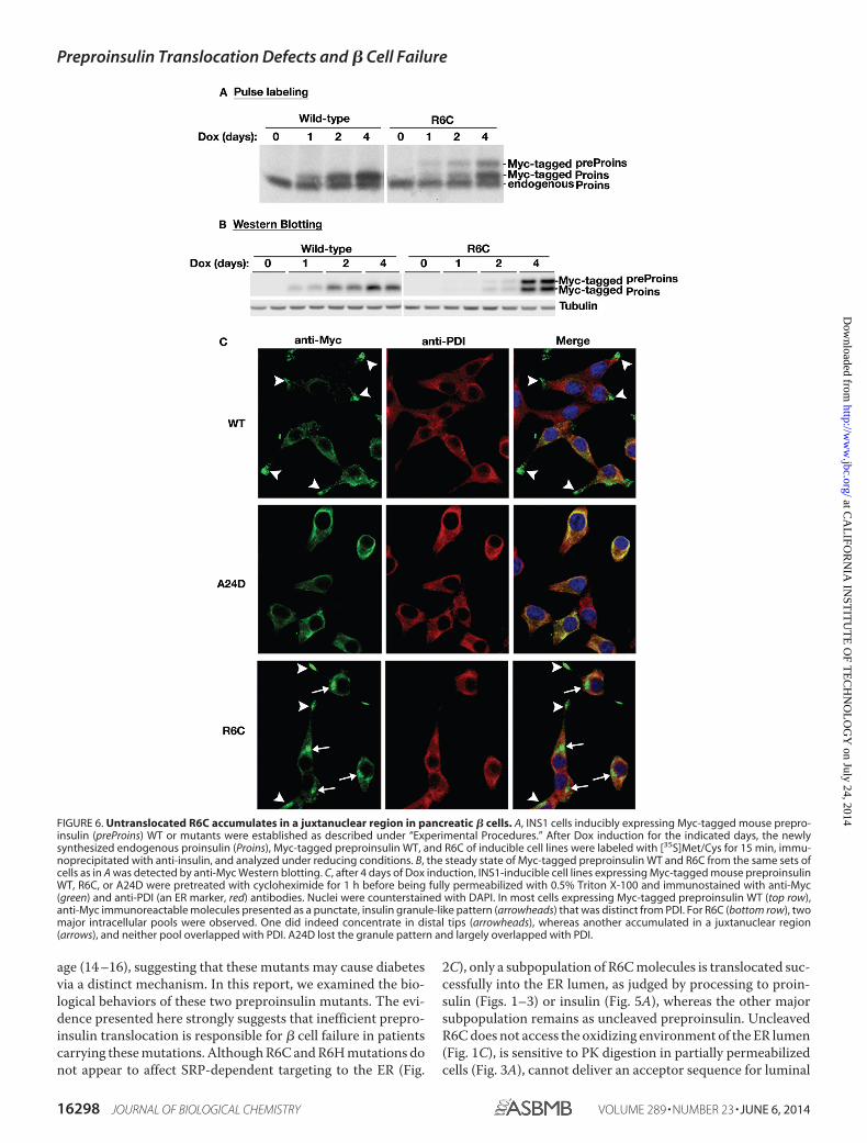
Untranslocated Preproinsulin Accumulates in a JuxtanuclearCompartment, Induces HSP70 Expression, and Promotes � CellDeath—To understand the pathological consequences of thetranslocation defects of R6C, using INS-r9 cells carrying thereverse doxycycline-dependent transactivator (22), we estab-lished � cell lines in which expression of Myc-tagged mousepreproinsulin WT, R6C, or A24D was induced in the presenceof doxycycline. The Myc epitope tag in the C peptide allowed forunequivocal distinction of these constructs from endogenous pre-proinsulin and proinsulin (13). Pulse labeling and Western blot-ting experiments showed that these cell lines exhibited compara-ble levels of induced expression of WT and R6C (Fig. 6, A and B).We then examined the localization of Myc-tagged mouse prepro-insulin WT and mutants in these cell lines by immunofluores-cence. Cells were pretreated with cycloheximide to minimize thecontribution of newly synthesized molecules. Expression of pre-proinsulin WT led to an insulin granule-like pattern that wasmostly concentrated in the distal tips of cellular processes (arrow-heads), distinct from the ER marker PDI (Fig. 6C, top row). Bycontrast, expression of A24D led to a localization pattern thatlargely overlapped with PDI (Fig. 6C, center row). Interestingly, inaddition to weak ER staining, two major intracellular pools accu-mulated for R6C: one concentrated normally in granules at thedistal tips of cellular processes (Fig. 6C, arrowheads), whereasanother concentrated abnormally as punctate structures at a jux-tanuclear location (Fig. 6C, arrows). Neither structure overlappedwith PDI (Fig. 6C, bottom row).
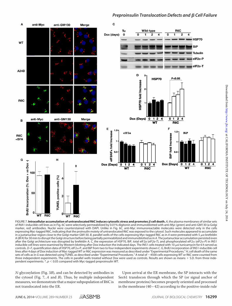
To further explore the juxtanuclear accumulation, parallelwells of the inducible cells used in Fig. 6C were partially per-meabilized with 0.01% digitonin, fixed, and processed for anti-GM130 (a Golgi marker) and anti-Myc immunofluorescence(Fig. 7A). In partially permeabilized cells, anti-Myc immuno-fluorescence was detected only for R6C in a region near theGolgi complex (Fig. 7A, bottom row), suggesting that the proin-sulin moiety of untranslocated R6C was exposed in the cytosol.
FIGURE 3. The proinsulin moiety of preproinsulin R6C is exposed to the cytosol. A, 293T cells expressing Myc-tagged preproinsulin (preProins) R6C andA24D were subjected to PK digestion in the presence or absence of digitonin (DIG) or Triton X-100 (TRX), followed by anti-Myc Western blotting as describedunder “Experimental Procedures.” Unprocessed R6C was sensitive to PK digestion in partially permeabilized cells, indicating that its proinsulin (Proins) moietywas exposed to the cytosol. B, two engineered human preproinsulins with N-linked glycosylation sites were created, as indicated in the top panel. The newlysynthesized preproinsulin WT and R6C labeled with either [35S]Met or [35S]Cys/Met were treated with or without PNGaseF before SDS-PAGE. Althoughproinsulin derived from WT or R6C was glycosylated (glyco-proins, lanes 3, 7, 9, and 11), unprocessed R6C labeled by pure [35S]Met (lanes 5 and 6) failed to acquirean N-linked glycan, indicating that unprocessed R6C was not delivered to the ER lumen.
Preproinsulin Translocation Defects and � Cell Failure
JUNE 6, 2014 • VOLUME 289 • NUMBER 23 JOURNAL OF BIOLOGICAL CHEMISTRY 16295
at CA
LIFO
RN
IA IN
STIT
UT
E O
F TE
CH
NO
LO
GY
on July 24, 2014http://w
ww
.jbc.org/D
ownloaded from
To examine whether R6C accumulates in the Golgi, we dis-rupted the Golgi structure using brefeldin A. The juxtanuclearaccumulation persisted even after the Golgi architecture wasdisrupted (Fig. 7B). These data indicate that a large subpopula-tion of incompletely translocated R6C molecules relocatesfrom the ER and accumulates in a novel cytoplasmic compart-ment, with its proinsulin moiety exposed to the cytosol.
Accumulation of juxtanuclear puncta has been observedupon induction of proteostasis stress in the cytosol (35, 36).Therefore, we asked whether accumulation of R6C analogouslyinduces cellular responses and, if so, in which subcellular com-partment. Consistent with Fig. 5D, markers of the ER-associ-ated unfolded protein response, including BiP and eIF2� phos-phorylation, were comparable between cells expressing
preproinsulin WT and R6C (Fig. 7, C, E, and F). In contrast,expression of R6C induced enhanced expression of HSP70 (Fig.7, C and D), indicating activation of a cytosolic response.
Because juxtanuclear accumulation of misfolded protein hasbeen linked to cell death in neurodegenerative diseases (37, 38),we asked whether accumulation of preproinsulin R6C affectscell viability and proliferation. No appreciable change of BrdUincorporation was observed between cells expressing Myc-tagged preproinsulin WT and R6C (Fig. 7G), suggesting thatR6C did not affect � cell proliferation. However, after a 4-dayinduction of preproinsulin expression, there was a significantincrease of cell death in � cells expressing R6C (Fig. 7H).Together, the data indicate that incompletely translocated R6Caccumulates in a juxtanuclear compartment, induces a cytosol-
FIGURE 4. The positive charge in the n region and the charge gradient flanking the h region of the SP play a critical role in the efficient targeting andtranslocation of preproinsulin. A, 293T cells transfected with empty vector (EV), human preproinsulin (preProins) WT, R6C, or M5R/R6C were labeled with[35S]Cys/Met for 15 min and analyzed by SDS-PAGE. Proins, proinsulin. B, the homology of human and rodent insulin gene 2 (Ins2) preproinsulin signalsequences. C, INS1 cells expressing Myc-tagged human (Hu) preproinsulin WT or R6C or mouse (Mo) Ins2 WT or R6C were examined by anti-Myc Westernblotting (WB). Mouse Ins2 R6C shows a more severe translocation defect than human R6C. D, quantification data of uncleaved preproinsulin from at least threeindependent experiments shown in C. E, INS1 cells expressing Myc-tagged human preproinsulin WT or mutants were labeled with [35S]Met/Cys for 10 min. Thenewly synthesized preproinsulin was immunoprecipitated (IP) with anti-Myc, followed by SDS-PAGE. F, Myc-tagged human preproinsulin WT or mutants intransfected INS1 cells were examined by anti-Myc Western blotting. R6C/D20R significantly exacerbated the defect of R6C. G, quantification data of uncleavedpreproinsulin from at least three independent experiments shown in F.
Preproinsulin Translocation Defects and � Cell Failure
16296 JOURNAL OF BIOLOGICAL CHEMISTRY VOLUME 289 • NUMBER 23 • JUNE 6, 2014
at CA
LIFO
RN
IA IN
STIT
UT
E O
F TE
CH
NO
LO
GY
on July 24, 2014http://w
ww
.jbc.org/D
ownloaded from
ic response, and promotes � cell death through mechanismsdistinct from those reported previously for A24D and otherMIDY mutants (Fig. 8).
DISCUSSIONIn pancreatic � cells, a multistep process lasting 30–150 min is
needed to synthesize mature insulin (1, 39), yet the earliest eventsat the point of entry of preproinsulin into the ER remain under-
studied. Here we demonstrate that the SP n region positive chargeplays an important role in ensuring efficient translocation of pre-proinsulin across the ER membrane and that inefficient preproin-sulin translocation contributes to � cell failure and diabetes.
Unlike most MIDY patients developing early-onset diabetes,patients carrying the R6C and R6H SP mutations develop non-obese autosomal dominant diabetes between 15– 65 years of
FIGURE 5. R6C and R6H fail to produce a normal amount of insulin but do not affect coexpressed preproinsulin WT nor induce ER stress. A, INS1 cellswere transfected with empty vector (EV), human preproinsulin WT or mutants. At 48 h post-transfection, the human insulin in the lysates (black bars) and media(white bars) were measured using human insulin-specific RIA normalized to human insulin mRNA. Results shown are mean � S.D. from three independentexperiments. *, p � 0.05 compared with preproinsulin WT. B, 293T cells were cotransfected with untagged preproinsulin (preProins) WT, with empty vector (EV),or Myc-tagged preproinsulin WT, or mutants. At 48 h post-transfection, the cells were labeled for 15 min, followed by a 0- or 3-h chase. Cell lysates (C) and chasemedia (M) were immunoprecipitated with anti-insulin and analyzed under reducing conditions. Unlike A24D, which blocked the secretion of coexpressed WT,R6C did not affect the expression, translocation, and secretion of coexpressed WT. Proins, proinsulin. C, the plasmids encoding human preproinsulin WT, R6C,or A24D with or without a Myc or GFP tag was transfected into 293T or INS1 cells as indicated. The cells transfected with empty vector (EV) served as controls.After 48 h post-transfection, the expression and SP cleavage of untagged or tagged preproinsulin were examined and compared by pulse labeling (newlysynthesized) and Western blotting (steady state) using anti-insulin, anti-Myc, or anti-GFP antibodies, as indicated. Unlike untagged and Myc-tagged prepro-insulin R6C, there was no detectable defect in GFP-tagged R6C. IP, immunoprecipitation; aa, amino acids. D, BiP promoter activities in transfected pancreatic� cells were evaluated as described under “Experimental Procedures.” Cells expressing preproinsulin WT were served as a control. Results were expressed asmean � S.D. from at least three independent experiments. *, p � 0.05 compared with preproinsulin WT.
Preproinsulin Translocation Defects and � Cell Failure
JUNE 6, 2014 • VOLUME 289 • NUMBER 23 JOURNAL OF BIOLOGICAL CHEMISTRY 16297
at CA
LIFO
RN
IA IN
STIT
UT
E O
F TE
CH
NO
LO
GY
on July 24, 2014http://w
ww
.jbc.org/D
ownloaded from
age (14 –16), suggesting that these mutants may cause diabetesvia a distinct mechanism. In this report, we examined the bio-logical behaviors of these two preproinsulin mutants. The evi-dence presented here strongly suggests that inefficient prepro-insulin translocation is responsible for � cell failure in patientscarrying these mutations. Although R6C and R6H mutations donot appear to affect SRP-dependent targeting to the ER (Fig.
2C), only a subpopulation of R6C molecules is translocated suc-cessfully into the ER lumen, as judged by processing to proin-sulin (Figs. 1–3) or insulin (Fig. 5A), whereas the other majorsubpopulation remains as uncleaved preproinsulin. UncleavedR6C does not access the oxidizing environment of the ER lumen(Fig. 1C), is sensitive to PK digestion in partially permeabilizedcells (Fig. 3A), cannot deliver an acceptor sequence for luminal
FIGURE 6. Untranslocated R6C accumulates in a juxtanuclear region in pancreatic � cells. A, INS1 cells inducibly expressing Myc-tagged mouse prepro-insulin (preProins) WT or mutants were established as described under “Experimental Procedures.” After Dox induction for the indicated days, the newlysynthesized endogenous proinsulin (Proins), Myc-tagged preproinsulin WT, and R6C of inducible cell lines were labeled with [35S]Met/Cys for 15 min, immu-noprecipitated with anti-insulin, and analyzed under reducing conditions. B, the steady state of Myc-tagged preproinsulin WT and R6C from the same sets ofcells as in A was detected by anti-Myc Western blotting. C, after 4 days of Dox induction, INS1-inducible cell lines expressing Myc-tagged mouse preproinsulinWT, R6C, or A24D were pretreated with cycloheximide for 1 h before being fully permeabilized with 0.5% Triton X-100 and immunostained with anti-Myc(green) and anti-PDI (an ER marker, red) antibodies. Nuclei were counterstained with DAPI. In most cells expressing Myc-tagged preproinsulin WT (top row),anti-Myc immunoreactable molecules presented as a punctate, insulin granule-like pattern (arrowheads) that was distinct from PDI. For R6C (bottom row), twomajor intracellular pools were observed. One did indeed concentrate in distal tips (arrowheads), whereas another accumulated in a juxtanuclear region(arrows), and neither pool overlapped with PDI. A24D lost the granule pattern and largely overlapped with PDI.
Preproinsulin Translocation Defects and � Cell Failure
16298 JOURNAL OF BIOLOGICAL CHEMISTRY VOLUME 289 • NUMBER 23 • JUNE 6, 2014
at CA
LIFO
RN
IA IN
STIT
UT
E O
F TE
CH
NO
LO
GY
on July 24, 2014http://w
ww
.jbc.org/D
ownloaded from
N-glycosylation (Fig. 3B), and can be detected by antibodies inthe cytosol (Fig. 7, A and B). Thus, by multiple independentmeasures, we demonstrate that a major subpopulation of R6C isnot translocated into the ER.
Upon arrival at the ER membrane, the SP interacts with theSec61 translocon through which the SP (or signal anchor ofmembrane proteins) becomes properly oriented and processedin the membrane (40 – 42) according to the positive-inside rule
FIGURE 7. Intracellular accumulation of untranslocated R6C induces cytosolic stress and promotes � cell death. A, the plasma membranes of similar setsof INA1-inducible cell lines as in Fig. 6C were selectively permeabilized by 0.01% digitonin and immunoblotted with anti-Myc (green) and anti-GM130 (a Golgimarker, red) antibodies. Nuclei were counterstained with DAPI. Unlike in Fig. 6C, anti-Myc immunoreactable molecules were detected only in the cellsexpressing Myc-tagged R6C, indicating that the proinsulin moiety of untranslocated R6C was exposed to the cytosol. Such molecules appeared to accumulatein a juxtanuclear region close to the Golgi marker GM130. B, parallel wells of the cells expressing Myc-tagged R6C as in A were pretreated with 5 �M brefeldinA (BFA) for 30 min to disrupt the Golgi structure before being partially permeabilized and immunoblotted as in A. The juxtanuclear accumulation persisted evenafter the Golgi architecture was disrupted by brefeldin A. C, the expression of HSP70, BiP, total eIF2a (eIF2a-T), and phosphorylated eIF2� (eIF2�-P) in INS1inducible cell lines were examined by Western blotting after Dox induction the indicated days. The INS1 cells treated with 10 �M tunicamycin for 6 h served ascontrols. D–F, quantification data of HSP70, eIF2�-P, and BiP from two to four independent experiments shown C. G, BrdU incorporation of INS1-inducible celllines after 4 days of Dox induction of Myc-tagged WT or R6C expression was measured as described under “Experimental Procedures.” H, cell death of the samesets of cells as in G was detected using TUNEL as described under “Experimental Procedures.” A total of �4500 cells expressing WT or R6C were counted fromthree independent experiments. The cells in parallel wells treated without Dox were used as controls. Results are shown as means � S.D. from three inde-pendent experiments. *, p � 0.05 compared with Myc-tagged preproinsulin WT.
Preproinsulin Translocation Defects and � Cell Failure
JUNE 6, 2014 • VOLUME 289 • NUMBER 23 JOURNAL OF BIOLOGICAL CHEMISTRY 16299
at CA
LIFO
RN
IA IN
STIT
UT
E O
F TE
CH
NO
LO
GY
on July 24, 2014http://w
ww
.jbc.org/D
ownloaded from
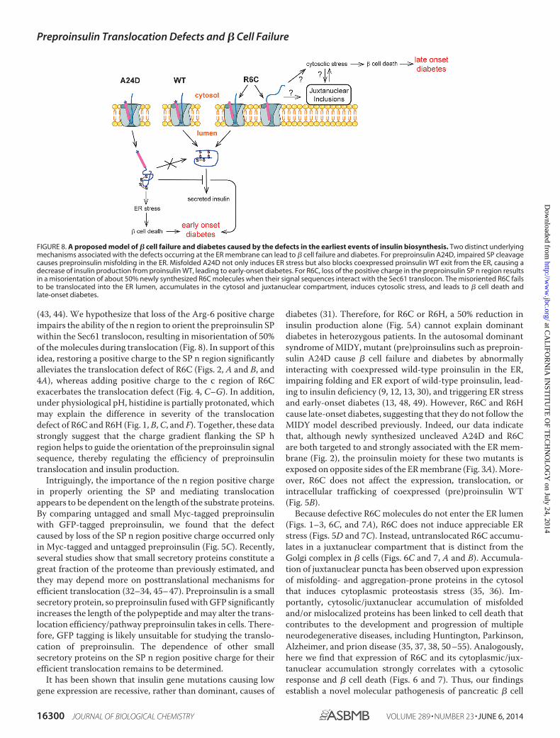
(43, 44). We hypothesize that loss of the Arg-6 positive chargeimpairs the ability of the n region to orient the preproinsulin SPwithin the Sec61 translocon, resulting in misorientation of 50%of the molecules during translocation (Fig. 8). In support of thisidea, restoring a positive charge to the SP n region significantlyalleviates the translocation defect of R6C (Figs. 2, A and B, and4A), whereas adding positive charge to the c region of R6Cexacerbates the translocation defect (Fig. 4, C–G). In addition,under physiological pH, histidine is partially protonated, whichmay explain the difference in severity of the translocationdefect of R6C and R6H (Fig. 1, B, C, and F). Together, these datastrongly suggest that the charge gradient flanking the SP hregion helps to guide the orientation of the preproinsulin signalsequence, thereby regulating the efficiency of preproinsulintranslocation and insulin production.
Intriguingly, the importance of the n region positive chargein properly orienting the SP and mediating translocationappears to be dependent on the length of the substrate proteins.By comparing untagged and small Myc-tagged preproinsulinwith GFP-tagged preproinsulin, we found that the defectcaused by loss of the SP n region positive charge occurred onlyin Myc-tagged and untagged preproinsulin (Fig. 5C). Recently,several studies show that small secretory proteins constitute agreat fraction of the proteome than previously estimated, andthey may depend more on posttranslational mechanisms forefficient translocation (32–34, 45– 47). Preproinsulin is a smallsecretory protein, so preproinsulin fused with GFP significantlyincreases the length of the polypeptide and may alter the trans-location efficiency/pathway preproinsulin takes in cells. There-fore, GFP tagging is likely unsuitable for studying the translo-cation of preproinsulin. The dependence of other smallsecretory proteins on the SP n region positive charge for theirefficient translocation remains to be determined.
It has been shown that insulin gene mutations causing lowgene expression are recessive, rather than dominant, causes of
diabetes (31). Therefore, for R6C or R6H, a 50% reduction ininsulin production alone (Fig. 5A) cannot explain dominantdiabetes in heterozygous patients. In the autosomal dominantsyndrome of MIDY, mutant (pre)proinsulins such as preproin-sulin A24D cause � cell failure and diabetes by abnormallyinteracting with coexpressed wild-type proinsulin in the ER,impairing folding and ER export of wild-type proinsulin, lead-ing to insulin deficiency (9, 12, 13, 30), and triggering ER stressand early-onset diabetes (13, 48, 49). However, R6C and R6Hcause late-onset diabetes, suggesting that they do not follow theMIDY model described previously. Indeed, our data indicatethat, although newly synthesized uncleaved A24D and R6Care both targeted to and strongly associated with the ER mem-brane (Fig. 2), the proinsulin moiety for these two mutants isexposed on opposite sides of the ER membrane (Fig. 3A). More-over, R6C does not affect the expression, translocation, orintracellular trafficking of coexpressed (pre)proinsulin WT(Fig. 5B).
Because defective R6C molecules do not enter the ER lumen(Figs. 1–3, 6C, and 7A), R6C does not induce appreciable ERstress (Figs. 5D and 7C). Instead, untranslocated R6C accumu-lates in a juxtanuclear compartment that is distinct from theGolgi complex in � cells (Figs. 6C and 7, A and B). Accumula-tion of juxtanuclear puncta has been observed upon expressionof misfolding- and aggregation-prone proteins in the cytosolthat induces cytoplasmic proteostasis stress (35, 36). Im-portantly, cytosolic/juxtanuclear accumulation of misfoldedand/or mislocalized proteins has been linked to cell death thatcontributes to the development and progression of multipleneurodegenerative diseases, including Huntington, Parkinson,Alzheimer, and prion disease (35, 37, 38, 50 –55). Analogously,here we find that expression of R6C and its cytoplasmic/jux-tanuclear accumulation strongly correlates with a cytosolicresponse and � cell death (Figs. 6 and 7). Thus, our findingsestablish a novel molecular pathogenesis of pancreatic � cell
FIGURE 8. A proposed model of � cell failure and diabetes caused by the defects in the earliest events of insulin biosynthesis. Two distinct underlyingmechanisms associated with the defects occurring at the ER membrane can lead to � cell failure and diabetes. For preproinsulin A24D, impaired SP cleavagecauses preproinsulin misfolding in the ER. Misfolded A24D not only induces ER stress but also blocks coexpressed proinsulin WT exit from the ER, causing adecrease of insulin production from proinsulin WT, leading to early-onset diabetes. For R6C, loss of the positive charge in the preproinsulin SP n region resultsin a misorientation of about 50% newly synthesized R6C molecules when their signal sequences interact with the Sec61 translocon. The misoriented R6C failsto be translocated into the ER lumen, accumulates in the cytosol and juxtanuclear compartment, induces cytosolic stress, and leads to � cell death andlate-onset diabetes.
Preproinsulin Translocation Defects and � Cell Failure
16300 JOURNAL OF BIOLOGICAL CHEMISTRY VOLUME 289 • NUMBER 23 • JUNE 6, 2014
at CA
LIFO
RN
IA IN
STIT
UT
E O
F TE
CH
NO
LO
GY
on July 24, 2014http://w
ww
.jbc.org/D
ownloaded from
failure and diabetes distinct from mechanisms described previ-ously (Fig. 8).
Integrating these findings, we speculate that inefficienttranslocation of preproinsulin might have implications for dia-betes even in the absence of INS gene mutations. Certainly inneurons, inefficient translocation of the wild-type prion proteinprecursor can trigger neurodegeneration (52, 56 –58). With thisin mind, it is important to note that insulin biosynthesis aloneaccounts for more than 10% of total protein synthesis underbasal condition in � cells. This percentage can further increaseto 30 –50% upon high glucose stimulation (59, 60), and up-reg-ulated preproinsulin production can occur in both type 1 andtype 2 diabetes (61, 62). Given the fact that a small fraction offully translated newly synthesized preproinsulin is present inthe cytosol in normal human islets (Fig. 1, D–F) and that R6Hwith a less severe translocation defect can cause diabetes, thereis the possibility that untranslocated preproinsulin WT canbecome a dangerous byproduct under certain pathological con-ditions. Studies are now needed to examine the efficiency ofpreproinsulin translocation in � cells in “garden variety” type 1and type 2 diabetes.
Acknowledgments—We thank Bill and Dee Brehm for helping toestablish the Brehm Center for Diabetes Research at the University ofMichigan and Drs. Donald F. Steiner, Michael A. Weiss, Graeme Bell,and Yanzhuang Wang for encouragement and discussions. We alsothank Dr. Martin Jendrisak and the entire team of the Gift of HopeOrgan and Tissue Donor Network in Chicago for the human pancreastissues used in this study.
REFERENCES1. Dodson G, Steiner, D. (1998) The role of assembly in insulin’s biosynthe-
sis. Curr. Opin. Struct. Biol. 8, 189 –1942. Patzelt, C., Labrecque, A. D., Duguid, J. R., Carroll, R. J., Keim, P. S., Hei-
nrikson, R. L., and Steiner, D. F. (1978) Detection and kinetic behavior ofpreproinsulin in pancreatic islets. Proc. Natl. Acad. Sci. U.S.A. 75,1260 –1264
3. Wolin, S. L., and Walter, P. (1993) Discrete nascent chain lengths arerequired for the insertion of presecretory proteins into microsomal mem-branes. J. Cell Biol. 121, 1211–1219
4. Okun, M. M., and Shields, D. (1992) Translocation of preproinsulin acrossthe endoplasmic reticulum membrane. The relationship between nascentpolypeptide size and extent of signal recognition particle-mediated inhi-bition of protein synthesis. J. Biol. Chem. 267, 11476 –11482
5. Liu, M., Li, Y., Cavener D, and Arvan P. (2005) Proinsulin disulfide matu-ration and misfolding in the endoplasmic reticulum. J. Biol. Chem. 280,13209 –13212
6. Zhang, B. Y., Liu, M., and Arvan, P. (2003) Behavior in the eukaryoticsecretory pathway of insulin-containing fusion proteins and single-chaininsulins bearing various B-chain mutations. J. Biol. Chem. 278, 3687–3693
7. Liu, M., Ramos-Castañeda, J., and Arvan, P. (2003) Role of the connectingpeptide in insulin biosynthesis. J. Biol. Chem. 278, 14798 –14805
8. Huang, X. F., and Arvan, P. (1995) Intracellular transport of proinsulin inpancreatic �-cells. Structural maturation probed by disulfide accessibility.J. Biol. Chem. 270, 20417–20423
9. Liu, M., Haataja, L., Wright, J., Wickramasinghe, N. P., Hua, Q. X., Phillips,N. F., Barbetti, F., Weiss, M. A., and Arvan, P. (2010) Mutant INS-geneinduced diabetes of youth: proinsulin cysteine residues impose dominant-negative inhibition on wild-type proinsulin transport. PLoS ONE 5,e13333
10. Liu, M., Hodish, I., Haataja, L., Lara-Lemus, R., Rajpal, G., Wright, J., andArvan, P. (2010) Proinsulin misfolding and diabetes: mutant INS gene-
induced diabetes of youth. Trends Endocrinol. Metab. 21, 652– 65911. Weiss, M. A. (2013) Diabetes mellitus due to the toxic misfolding of pro-
insulin variants. FEBS Lett. 587, 1942–195012. Park, S.-Y., Ye, H., Steiner, D. F., and Bell, G. I. (2010) Mutant proinsulin
proteins associated with neonatal diabetes are retained in the endoplasmicreticulum and not efficiently secreted. Biochem. Biophys. Res. Commun.391, 1449 –1454
13. Liu, M., Lara-Lemus, R., Shan, S.-O., Wright, J., Haataja, L., Barbetti, F.,Guo, H., Larkin, D., and Arvan, P. (2012) Impaired cleavage of preproin-sulin signal peptide linked to autosomal-dominant diabetes. Diabetes 61,828 – 837
14. Meur, G., Simon, A., Harun, N., Virally, M., Dechaume, A., Bonnefond, A.,Fetita, S., Tarasov, A. I., Guillausseau, P. J., Boesgaard, T. W., Pedersen, O.,Hansen, T., Polak, M., Gautier, J. F., Froguel, P., Rutter, G. A., and Vaxil-laire, M. (2010) Insulin gene mutations resulting in early-onset diabetes:marked differences in clinical presentation, metabolic status, and patho-genic effect through endoplasmic reticulum retention. Diabetes 59,653– 661
15. Boesgaard, T. W., Pruhova, S., Andersson, E. A., Cinek, O., Obermannova,B., Lauenborg, J., Damm, P., Bergholdt, R., Pociot, F., Pisinger, C., Barbetti,F., Lebl, J., Pedersen, O., and Hansen, T. (2010) Further evidence thatmutations in INS can be a rare cause of maturity-onset diabetes of theyoung (MODY). BMC Medical Genetics 11, 42
16. Edghill, E. L., Flanagan, S. E., Patch, A.-M., Boustred, C., Parrish, A.,Shields, B., Shepherd, M. H., Hussain, K., Kapoor, R. R., Malecki, M.,MacDonald, M. J., Støy, J., Steiner, D. F., Philipson, L. H., Bell, G. I., Neo-natal Diabetes International Collaborative Group, Hattersley, A. T., andEllard, S. (2008) Insulin mutation screening in 1,044 patients with diabe-tes. Diabetes 57, 1034 –1042
17. Peterson, J. H., Woolhead, C. A., and Bernstein, H. D. (2003) Basic aminoacids in a distinct subset of signal peptides promote interaction with thesignal recognition particle. J. Biol. Chem. 278, 46155– 46162
18. Sasaki, S., Matsuyama, S., and Mizushima, S. (1990) In vitro kinetic anal-ysis of the role of the positive charge at the amino-terminal region of signalpeptides in translocation of secretory protein across the cytoplasmicmembrane in Escherichia coli. J. Biol. Chem. 265, 4358 – 4363
19. Iino, T., Takahashi, M., and Sako, T. (1987) Role of amino-terminal posi-tive charge on signal peptide in staphylokinase export across the cytoplas-mic membrane of Escherichia coli. J. Biol. Chem. 262, 7412–7417
20. Powers, T., and Walter, P. (1997) Co-translational protein targeting cata-lyzed by the Escherichia coli signal recognition particle and its receptor.EMBO J. 16, 4880 – 4886
21. Shan, S.-O., Chandrasekar, S., and Walter, P. (2007) Conformationalchanges in the GTPase modules of the signal reception particle and itsreceptor drive initiation of protein translocation. J. Cell Biol. 178,611– 620
22. Wang, H., Kouri, G., and Wollheim, C. B. (2005) ER stress and SREBP-1activation are implicated in �-cell glucolipotoxicity. J. Cell Sci. 118,3905–3915
23. Feng, Y., Jadhav, A. P., Rodighiero, C., Fujinaga, Y., Kirchhausen, T., andLencer, W. I. (2004) Retrograde transport of cholera toxin from theplasma membrane to the endoplasmic reticulum requires the trans-Golginetwork but not the Golgi apparatus in Exo2-treated cells. EMBO Rep. 5,596 – 601
24. Fujita, H., Kida, Y., Hagiwara, M., Morimoto, F., and Sakaguchi, M. (2010)Positive charges of translocating polypeptide chain retrieve an upstreammarginal hydrophobic segment from the endoplasmic reticulum lumen tothe translocon. Mol. Biol. Cell 21, 2045–2056
25. Parks, G. D., and Lamb, R. A. (1991) Topology of eukaryotic type II mem-brane proteins: importance of N-terminal positively charged residuesflanking the hydrophobic domain. Cell 64, 777–787
26. Goder, V., and Spiess, M. (2001) Topogenesis of membrane proteins: de-terminants and dynamics. FEBS Lett. 504, 87–93
27. Sakaguchi, M., Tomiyoshi, R., Kuroiwa, T., Mihara, K., and Omura, T.(1992) Functions of signal and signal-anchor sequences are determined bythe balance between the hydrophobic segment and the N-terminal charge.Proc. Natl. Acad. Sci. U.S.A. 89, 16 –19
28. Fujiki, Y., Hubbard, A. L., Fowler, S., and Lazarow, P. B. (1982) Isolation of
Preproinsulin Translocation Defects and � Cell Failure
JUNE 6, 2014 • VOLUME 289 • NUMBER 23 JOURNAL OF BIOLOGICAL CHEMISTRY 16301
at CA
LIFO
RN
IA IN
STIT
UT
E O
F TE
CH
NO
LO
GY
on July 24, 2014http://w
ww
.jbc.org/D
ownloaded from
intracellular membranes by means of sodium carbonate treatment: appli-cation to endoplasmic reticulum. J. Cell Biol. 93, 97–102
29. von Heijne, G. (1986) Net N-C charge imbalance may be important forsignal sequence function in bacteria. J. Mol. Biol. 192, 287–290
30. Liu, M., Hodish, I., Rhodes, C. J., and Arvan, P. (2007) Proinsulin matura-tion, misfolding, and proteotoxicity. Proc. Natl. Acad. Sci. U.S.A. 104,15841–15846
31. Garin, I., Edghill, E. L., Akerman, I., Rubio-Cabezas, O., Rica, I., Locke,J. M., Maestro, M. A., Alshaikh, A., Bundak, R., del Castillo, G., Deeb, A.,Deiss, D., Fernandez, J. M., Godbole, K., Hussain, K., O’Connell, M., Klupa,T., Kolouskova, S., Mohsin, F., Perlman, K., Sumnik, Z., Rial, J. M., Ugarte,E., Vasanthi, T., Neonatal Diabetes International Group, Johnstone, K.,Flanagan, S. E., Martínez, R., Castaño, C., Patch, A. M., Fernández-Re-bollo, E., Raile, K., Morgan, N., Harries, L. W., Castaño, L., Ellard, S.,Ferrer, J., Perez de Nanclares, G., and Hattersley AT. (2010) Recessivemutations in the INS gene result in neonatal diabetes through reducedinsulin biosynthesis. Proc. Natl. Acad. Sci. U.S.A. 107, 3105–3110
32. Lakkaraju, A. K., Thankappan, R., Mary, C., Garrison, J. L., Taunton, J., andStrub, K. (2012) Efficient secretion of small proteins in mammalian cellsrelies on Sec62-dependent posttranslational translocation. Mol. Biol. Cell23, 2712–2722
33. Johnson, N., Vilardi, F., Lang, S., Leznicki, P., Zimmermann, R., and High,S. (2012) TRC40 can deliver short secretory proteins to the Sec61 translo-con. J. Cell Sci. 125, 3612–3620
34. Shao, S., and Hegde, R. S. (2011) A calmodulin-dependent translocationpathway for small secretory proteins. Cell 147, 1576 –1588
35. Kaganovich, D., Kopito, R., and Frydman, J. (2008) Misfolded proteinspartition between two distinct quality control compartments. Nature 454,1088 –1095
36. Kocik, L., Junne, T., and Spiess, M. (2012) Orientation of internal signal-anchor sequences at the Sec61 translocon. J. Mol. Biol. 424, 368 –378
37. Debure, L., Vayssiere, J.-L., Rincheval, V., Loison, F., Le Drean, Y., andMichel, D. (2003) Intracellular clusterin causes juxtanuclear aggregate for-mation and mitochondrial alteration. J. Cell Sci. 116, 3109 –3121
38. Viswanathan, J., Haapasalo, A., Böttcher, C., Miettinen, R., Kurkinen,K. M., Lu, A., Thomas, A., Maynard, C. J., Romano, D., Hyman, B. T.,Berezovska, O., Bertram, L., Soininen, H., Dantuma, N. P., Tanzi, R. E., andHiltunen, M. (2011) Alzheimer’s disease-associated ubiquilin-1 regulatespresenilin-1 accumulation and aggresome formation. Traffic 12, 330 –348
39. Alarcón, C., Leahy, J. L., Schuppin, G. T., and Rhodes, C. J. (1995) In-creased secretory demand rather than a defect in the proinsulin conver-sion mechanism causes hyperproinsulinemia in a glucose-infusion ratmodel of non-insulin-dependent diabetes mellitus. J. Clin. Invest. 95,1032–1039
40. Pilon, M., Römisch, K., Quach, D., and Schekman, R. (1998) Sec61p servesmultiple roles in secretory precursor binding and translocation into theendoplasmic reticulum membrane. Mol. Biol. Cell 9, 3455–3473
41. Plath, K., Mothes, W., Wilkinson, B. M., Stirling, C. J., and Rapoport, T. A.(1998) Signal sequence recognition in posttranslational protein transportacross the yeast ER membrane. Cell 94, 795– 807
42. Goder, V., Junne, T., and Spiess, M. (2004) Sec61p contributes to signalsequence orientation according to the positive-inside rule. Mol. Biol. Cell15, 1470 –1478
43. von Heijne, G., and Gavel, Y. (1988) Topogenic signals in integral mem-brane proteins. Eur. J. Biochem. 174, 671– 678
44. Andersson, H., and von Heijne, G. (1994) Membrane protein topology:effects of � � H� on the translocation of charged residues explain the
“positive inside” rule. EMBO J. 13, 2267–227245. Bionda, T., Tillmann, B., Simm, S., Beilstein, K., Ruprecht, M., and Schleiff,
E. (2010) Chloroplast import signals: the length requirement for translo-cation in vitro and in vivo. J. Mol. Biol. 402, 510 –523
46. Frith, M. C., Forrest, A. R., Nourbakhsh, E., Pang, K. C., Kai, C., Kawai, J.,Carninci, P., Hayashizaki, Y., Bailey, T. L., and Grimmond, S. M. (2006)The abundance of short proteins in the mammalian proteome. PLoSGenet. 2, e52
47. Johnson, N., Powis, K., and High, S. (2013) Post-translational transloca-tion into the endoplasmic reticulum. Biochim. Biophys. Acta 1833,2403–2409
48. Izumi, T., Yokota-Hashimoto, H., Zhao, S., Wang, J., Halban, P. A., andTakeuchi, T. (2003) Dominant negative pathogenesis by mutant proinsu-lin in the Akita diabetic mouse. Diabetes 52, 409 – 416
49. Oyadomari, S., Koizumi, A., Takeda, K., Gotoh, T., Akira, S., Araki, E., andMori, M. (2002) Targeted disruption of the Chop gene delays endoplasmicreticulum stress-mediated diabetes. J. Clin. Invest. 109, 525–532
50. Schaffar, G., Breuer, P., Boteva, R., Behrends, C., Tzvetkov, N., Strippel, N.,Sakahira, H., Siegers, K., Hayer-Hartl, M., and Hartl, F. U. (2004) Cellulartoxicity of polyglutamine expansion proteins: mechanism of transcriptionfactor deactivation. Mol. Cell 15, 95–105
51. Rubinsztein, D. C. (2006) The roles of intracellular protein-degradationpathways in neurodegeneration. Nature 443, 780 –786
52. Rane, N. S., Chakrabarti, O., Feigenbaum, L., and Hegde, R. S. (2010)Signal sequence insufficiency contributes to neurodegeneration caused bytransmembrane prion protein. J. Cell Biol. 188, 515–526
53. Miesbauer, M., Rambold, A. S., Winklhofer, K. F., and Tatzelt, J. (2010)Targeting of the prion protein to the cytosol: mechanisms and conse-quences. Curr. Issues Mol. Biol. 12, 109 –118
54. Strom, A., Wang, G.-S., Reimer, R., Finegood, D. T., and Scott, F. W. (2007)Pronounced cytosolic aggregation of cellular prion protein in pancreatic�-cells in response to hyperglycemia. Lab. Invest. 87, 139 –149
55. Reiner, T., Thurber, G., Gaglia, J., Vinegoni, C., Liew, C. W., Upadhyay, R.,Kohler, R. H., Li, L., Kulkarni, R. N., Benoist, C., Mathis, D., andWeissleder, R. (2011) Accurate measurement of pancreatic islet �-cellmass using a second-generation fluorescent exendin-4 analog. Proc. Natl.Acad. Sci. U.S.A. 108, 12815–12820
56. Rane, N. S., Kang, S.-W., Chakrabarti, O., Feigenbaum, L., and Hegde, R. S.(2008) Reduced translocation of nascent prion protein during ER stresscontributes to neurodegeneration. Dev. Cell 15, 359 –370
57. Hegde, R. S., and Ploegh, H. L. (2010) Quality and quantity control at theendoplasmic reticulum. Curr. Opin. Cell Biol. 22, 437– 446
58. Hegde, R. S., and Kang, S.-W. (2008) The concept of translocational reg-ulation. J. Cell Biol. 182, 225–232
59. Schuit, F. C., In’t Veld, P. A., and Pipeleers, D. G. (1988) Glucose stimulatesproinsulin biosynthesis by a dose-dependent recruitment of pancreatic �
cells. Proc. Natl. Acad. Sci. U.S.A. 85, 3865–386960. Scheuner, D., and Kaufman, R. J. (2008) The unfolded protein response: a
pathway that links insulin demand with �-cell failure and diabetes. Endocr.Rev. 29, 317–333
61. Tersey, S. A., Nishiki, Y., Templin, A. T., Cabrera, S. M., Stull, N. D.,Colvin, S. C., Evans-Molina, C., Rickus, J. L., Maier, B., and Mirmira, R. G.(2012) Islet B-cell endoplasmic reticulum stress precedes the onset of type1 diabetes in the nonobese diabetic mouse model. Diabetes 61, 818 – 827
62. Kaufman, R. J. (2011) �-Cell failure, stress, and type 2 diabetes. N. Engl.J. Med. 365, 1931–1933
Preproinsulin Translocation Defects and � Cell Failure
16302 JOURNAL OF BIOLOGICAL CHEMISTRY VOLUME 289 • NUMBER 23 • JUNE 6, 2014
at CA
LIFO
RN
IA IN
STIT
UT
E O
F TE
CH
NO
LO
GY
on July 24, 2014http://w
ww
.jbc.org/D
ownloaded from