PONTIFÍCIA UNIVERSIDADE CATÓLICA DO RIO GRANDE DO … · p.o. – via oral NMDA – N-metil D ......
62
PONTIFÍCIA UNIVERSIDADE CATÓLICA DO RIO GRANDE DO SUL PROGRAMA DE PÓS-GRADUAÇÃO EM MEDICINA E CIÊNCIAS DA SAÚDE ÁREA DE CONCENTRAÇÃO: FARMACOLOGIA BIOQUÍMICA E MOLECULAR KAREN OLIVIA BAZZO ANÁLISE DOS EFEITOS DO TRANS-RESVERATROL SOBRE A NOCICEPÇÃO ESPONTÂNEA INDUZIDA PELA CAPSAICINA E GLUTAMATO EM CAMUNDONGOS PORTO ALEGRE 2011
Transcript of PONTIFÍCIA UNIVERSIDADE CATÓLICA DO RIO GRANDE DO … · p.o. – via oral NMDA – N-metil D ......
PONTIFÍCIA UNIVERSIDADE CATÓLICA DO RIO GRANDE DO SUL
PROGRAMA DE PÓS-GRADUAÇÃO EM MEDICINA E CIÊNCIAS DA SAÚDE
ÁREA DE CONCENTRAÇÃO: FARMACOLOGIA BIOQUÍMICA E MOLECULAR
KAREN OLIVIA BAZZO
ANÁLISE DOS EFEITOS DO TRANS-RESVERATROL SOBRE A NOCICEPÇÃO
ESPONTÂNEA INDUZIDA PELA CAPSAICINA E GLUTAMATO EM
CAMUNDONGOS
PORTO ALEGRE
2011
2
KAREN OLIVIA BAZZO
ANÁLISE DOS EFEITOS DO TRANS-RESVERATROL SOBRE A NOCICEPÇÃO
ESPONTÂNEA INDUZIDA PELA CAPSAICINA E GLUTAMATO EM
CAMUNDONGOS
Dissertação apresentada como requisito para
obtenção do grau de Mestre pelo Programa de
Pós-Graduação em Medicina e Ciências da
Saúde, Área de Concentração em Farmacologia
Bioquímica e Molecular, da Pontifícia
Universidade Católica do Rio Grande do Sul.
Orientadora: Profª. Dra. Maria Martha Campos
Co-orientadora: Profa. Dra. Alessandra Hubner de Souza
Porto Alegre
2011

3
KAREN OLIVIA BAZZO
ANÁLISE DOS EFEITOS DO TRANS-RESVERATROL SOBRE A NOCICEPÇÃO
ESPONTÂNEA INDUZIDA PELA CAPSAICINA E GLUTAMATO EM
CAMUNDONGOS
Dissertação apresentada como requisito para
obtenção do grau de Mestre pelo Programa de
Pós-Graduação em Medicina e Ciências da
Saúde, Área de Concentração em Farmacologia
Bioquímica e Molecular, da Pontifícia
Universidade Católica do Rio Grande do Sul.
Aprovada em _____de_______________de______
BANCA EXAMINADORA:
Examinador 1
Dr. Jarbas Rodrigues de Oliveira
___________________________
Examinador 2
Dra. Rejane Giacomelli Tavares
___________________________
Examinador 3
Dra. Fernanda Bueno Morrone
___________________________
Examinador 4
Dr. Rafael Fernandes Zanin
___________________________
4
Dados Internacionais de Catalogação na Publicação (CIP)
Índice para o catálogo sistemático:
1. Farmacologia – Aplicação experimental 615.038
2. Trans-resveratrol - Camundongos 615.03:599.323
3. Nocicepção 616.8-009.7
Catalogação na fonte elaborada pela bibliotecária
Michele Marques Baptista – CRB 10/1633
B364a Bazzo, Karen Olivia
Análise dos efeitos do trans-resveratrol sobre a nocicepção
espontânea induzida pela capsaicina e glutamato em camundongos /
Karen Olivia Bazzo. - Porto Alegre, RS, 2011.
61 p. : il. ; 30 cm.
Dissertação (Mestrado) - Pontifícia Universidade Católica do Rio
Grande do Sul, Programa de Pós-Graduação em Medicina e Ciências
da Saúde, 2011.
―Orientação: Profª. Dra. Maria Martha Campos .‖
―Co-orientadora: Profª. Dra. Alessandra Hubner de Souza.‖
Apresenta bibliografia.
1. Farmacologia – Aplicação experimental. 2. Trans-resveratrol
Camundongos 3. Nocicepção. I. Título.
CDU: 615.038

5
Agradeço a todas as pessoas que tornaram
esse trabalho possível, especialmente
à minha família, meu namorado e
minha orientadora Maria Martha.
6
AGRADECIMENTOS
À Profa. Dr
a. Maria Martha Campos, amiga e orientadora, que me recebeu
abertamente desde o primeiro instante, agradeço por acreditar em meu potencial e
capacidade de tornar este projeto uma realidade e pela compreensão acima de tudo.
Agradeço por todo suporte e atenção dedicados ao longo do tempo.
À Profa. Dr
a. Alessandra Hubner de Souza, por colaborar de forma tão expressiva
e significante ao trabalho, pelo incentivo, apoio e ensinamentos.
Aos colegas Fernanda, Geferson e Tatiana pela amizade e carinho, e por tornarem
todos os momentos agradáveis com sua companhia.
À equipe do laboratório de Farmacologia Aplicada, pela recepção, disponibilidade
e compreensão. À Juliano Soares, pelo profissionalismo e excelente assistência.
Às amigas Juliana Comerlato, Francine Bonatto e Fernanda Machado pelo apoio e
amizade.
Ao meu namorado Douglas, pelo apoio incondicional e pela compreensão
dedicada.
E principalmente, à minha família, que sempre me incentivou de todas as formas,
que sempre acreditou na minha capacidade e que sempre torceu pelo meu sucesso.
Muito obrigada a tudo e a todos.
7
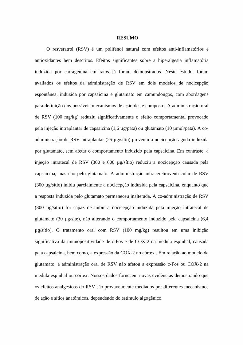
RESUMO
O resveratrol (RSV) é um polifenol natural com efeitos anti-inflamatórios e
antioxidantes bem descritos. Efeitos significantes sobre a hiperalgesia inflamatória
induzida por carragenina em ratos já foram demonstrados. Neste estudo, foram
avaliados os efeitos da administração de RSV em dois modelos de nocicepção
espontânea, induzida por capsaicina e glutamato em camundongos, com abordagens
para definição dos possíveis mecanismos de ação deste composto. A administração oral
de RSV (100 mg/kg) reduziu significativamente o efeito comportamental provocado
pela injeção intraplantar de capsaicina (1,6 µg/pata) ou glutamato (10 µmol/pata). A co-
administração de RSV intraplantar (25 µg/sítio) preveniu a nocicepção aguda induzida
por glutamato, sem afetar o comportamento induzido pela capsaicina. Em contraste, a
injeção intratecal de RSV (300 e 600 µg/sítio) reduziu a nocicepção causada pela
capsaicina, mas não pelo glutamato. A administração intracerebroventricular de RSV
(300 µg/sítio) inibiu parcialmente a nocicepção induzida pela capsaicina, enquanto que
a resposta induzida pelo glutamato permaneceu inalterada. A co-administração de RSV
(300 µg/sítio) foi capaz de inibir a nocicepção induzida pela injeção intratecal de
glutamato (30 µg/site), não alterando o comportamento induzido pela capsaicina (6,4
µg/sítio). O tratamento oral com RSV (100 mg/kg) resultou em uma inibição
significativa da imunopositividade de c-Fos e de COX-2 na medula espinhal, causada
pela capsaicina, bem como, a expressão da COX-2 no córtex . Em relação ao modelo de
glutamato, a administração oral de RSV não afetou a expressão c-Fos ou COX-2 na
medula espinhal ou córtex. Nossos dados fornecem novas evidências demostrando que
os efeitos analgésicos do RSV são provavelmente mediados por diferentes mecanismos
de ação e sítios anatômicos, dependendo do estímulo algogênico.
8
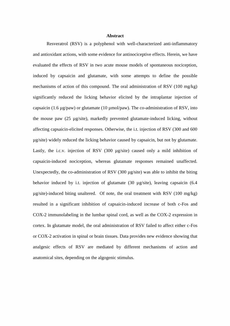
Abstract
Resveratrol (RSV) is a polyphenol with well-characterized anti-inflammatory
and antioxidant actions, with some evidence for antinociceptive effects. Herein, we have
evaluated the effects of RSV in two acute mouse models of spontaneous nociception,
induced by capsaicin and glutamate, with some attempts to define the possible
mechanisms of action of this compound. The oral administration of RSV (100 mg/kg)
significantly reduced the licking behavior elicited by the intraplantar injection of
capsaicin (1.6 µg/paw) or glutamate (10 µmol/paw). The co-administration of RSV, into
the mouse paw (25 µg/site), markedly prevented glutamate-induced licking, without
affecting capsaicin-elicited responses. Otherwise, the i.t. injection of RSV (300 and 600
µg/site) widely reduced the licking behavior caused by capsaicin, but not by glutamate.
Lastly, the i.c.v. injection of RSV (300 µg/site) caused only a mild inhibition of
capsaicin-induced nociception, whereas glutamate responses remained unaffected.
Unexpectedly, the co-administration of RSV (300 µg/site) was able to inhibit the biting
behavior induced by i.t. injection of glutamate (30 µg/site), leaving capsaicin (6.4
µg/site)-induced biting unaltered. Of note, the oral treatment with RSV (100 mg/kg)
resulted in a significant inhibition of capsaicin-induced increase of both c-Fos and
COX-2 immunolabeling in the lumbar spinal cord, as well as the COX-2 expression in
cortex. In glutamate model, the oral administration of RSV failed to affect either c-Fos
or COX-2 activation in spinal or brain tissues. Data provides new evidence showing that
analgesic effects of RSV are mediated by different mechanisms of action and
anatomical sites, depending on the algogenic stimulus.

9
ABREVIAÇÕES
RSV – Resveratrol
CAP – Capsaicina
GLU – Glutamato
i.c.v. – Intracerebroventrical
i.t. – intrathecal
i.pl. – intraplantar
p.o. – via oral
NMDA – N-metil D-Aspartato
TRPV – Receptor Vanilóide
TRPV1 – Receptor Vanilóide do tipo 1
NO- Óxido nítrico
ATP- Adenosina trifosfato
GMP–PKG- Guanina monofosfato - protein quinase G
CNS – Central Nervous Sistem
COX-2 – Ciclooxigenase 2
NOS – Óxido nítrico sintase
10
SUMÁRIO
1. INTRODUÇÃO.................................................................................................11
2. OBJETIVOS......................................................................................................20
2.1.Objetivo geral......................................................................................................20
2.2. Objetivos específicos..........................................................................................20
3. ARTIGO.............................................................................................................21
4. CONSIDERAÇÕES FINAIS............................................................................53
5. REFERÊNCIAS.................................................................................................57
11
1. INTRODUÇÃO
Dor e Nocicepção
A dor pode ser definida como ―uma experiência sensorial e emocional
desagradável associada com dano tecidual real ou potencial, ou descrita em termos de
tais danos" (Cheng et al., 2003). A nocicepção compreende toda a transmissão e
processamento da informação dolorosa. A dor é um sistema complexo que está
vinculado ao aspecto comportamental, pois o processamento da informação não ocorre
apenas no sistema somatossensorial, mas também no sistema límbico (Millan, 1999).
O início da transmissão dolorosa se dá em neurônios sensoriais primários de alto
limiar, que também exercem a função de receptores e são chamados nociceptores. Estão
subdivididos em mielinizados do tipo A e, não mielinizados do tipo C, ambos altamente
especializados em transmitir informações dolorosas, apresentando corpos celulares
localizados no gânglio da raiz dorsal. As fibras mielínicas com maior espessura Aβ
estão envolvidas na percepção tátil. As fibras mielínicas finas Aδ e não-mielinizadas do
tipo C transmitem a nocicepção e estímulos térmicos, mecânicos e químicos. A
nocicepção normal envolve a detecção de temperaturas altas e baixas, assim como
estímulos mecânicos intensos. Ambos nociceptores originam-se da pele e órgãos e
terminam na camada superficial do corno dorsal na medula espinhal. Os axônios das
células do corno dorsal transmitem impulsos nociceptivos para o tálamo e através deste
ao córtex cerebral, que processa a consciência dolorosa (Baron, 2006; Gilron et al.,
2006).
A estimulação nociva a algum tecido intacto causa uma dor de caráter protetor.
Por outro lado, a dor patológica ocorre quando há inflamação ou lesão tecidual. Neste
12
caso, ocorre sensibilização periférica e central, com a liberação de mediadores químicos
no local da lesão e em determinadas regiões do sistema nervoso central (SNC), o que
intensifica a transmissão nociceptiva. A partir destas alterações, há possibilidade de
ocorrer modificações sensoriais peculiares da dor patológica, que são observadas
através da existência de hiperalgesia, que traduz um aumento da sensibilidade local a
estímulos potencialmente nocivos, além de alodínia, que representa uma resposta
dolorosa a estímulos considerados inócuos (Millan, 1999).
Neste contexto, a dor pode ser classificada como: aguda, que é sinal de alerta
para a presença de lesão tecidual, real ou potencial e, crônica, que é resultante da
persistência de estímulos nociceptivos, sendo que esta última não pode ser considerada
uma dor aguda de longa duração (Ribeiro et al., 2002).
Nocicepção Aguda
Os principais mediadores liberados na presença de lesão ou inflamação tecidual
são a substância P e o glutamato (Zhuo, 2007). Quando ocorre dano, esta informação é
transmitida para o sistema SNC sem ocasionar nenhuma alteração. Porém, a inflamação
instalada ativa receptores silenciosos, tornando os mesmos sensíveis aos estímulos
mecânicos. Assim, ocorre persistência da neurotransmissão nociceptiva e, finalmente, a
hipersensibilidade desenvolvida no local da lesão gera alodínia .
A neurotransmissão glutamatérgica representa um grande passo iniciador das
alterações ocasionadas pela dor, pois o glutamato se liga a seus receptores NMDA,
AMPA e cainato, provocando a despolarização pós-sináptica e propagação da
informação nociceptiva (Baron, 2006). Outro evento que corrobora com aparecimento
de hiperalgesia e alodínia é a ativação das enzimas óxido nítrico sintases (NOS), que
são responsáveis pela formação de óxido nítrico. Este, por sua vez, interfere na
13
nocicepção atuando como mensageiro retrógrado, pois se difunde para os neurônios pré-
sinápticos do corno dorsal, estimulando a liberação de neurotransmissores nos terminais
aferentes primários (Aimar et al., 1998; Meller and Gebhart, 1993; Vetter et al., 2001;
Xu et al., 2007).
A glia também apresenta importância na modulação da nocicepção. Esta contém
receptores de neurotransmissores, como glutamato e substância P, que ao se ligarem aos
receptores correspondentes na glia, induzem a liberação de diversos mediadores como
citocinas, fatores de crescimento, espécies reativas de oxigênio, prostaglandinas, óxido
nítrico e outros (Costigan et al., 2009; Watkins et al., 2001).
Os receptores glutamatérgicos participam na transmissão nociceptiva,
principalmente na informação dolorosa neurogênica e nos aspectos relacionados à
hiperalgesia. A ativação prolongada e repetitiva das fibras aferentes C produzem um
aumento progressivo da magnitude da resposta, ocasionando um aumento na
excitabilidade, um fenômeno conhecido como wind up . O glutamato liberado pelas
fibras C estimula seus receptores ionotrópicos, NMDA e AMPA, causando uma
resposta pós-sináptica rápida. As fibras C também liberam substância P, que é um
neuropeptídeo capaz de estimular a liberação de glutamato. Esse conjunto de eventos
desencadeia diversas despolarizações pós-sinápticas e, assim, ativação das óxido nítrico
sintases, que também têm ação estimulatória sobre a secreção de glutamato. Portanto,
diversos feedbacks positivos elevam a concentração de glutamato na fenda sináptica
(Zhuo, 2007).
Atualmente, existem diversos modelos animais de dor aguda que reproduzem os
efeitos centrais e comportamentais ocorridos em humanos, dentre estes, destaca-se o
modelo de nocicepção aguda induzida pela injeção intraplantar de capsaicina, o
principal agente pungente contido nas pimentas vermelhas (Komatsu et al., 2009).
14
Receptores TRPV1
Os receptores TRP foram originalmente observados em moscas do gênero
Drosophila, onde funcionam como fotorreceptores. A resposta à luz mediada por estes
receptores é reduzida após uma exposição prolongada; desta forma, esta família de
receptores foi chamada de transient receptor potencial, TRP (Fein, 2009).
A família TRP é dividida em seis subtipos: TRPC, TRPM, TRPP, TRPML,
TRPA e TRPV1, ou receptor vanilóide. O TRPV1 é um canal seletivo para Ca+2
, que
pode ser ativado pela capsaicina, além de ser estimulado por altas temperaturas e por
faixas de pH menores que 6,5. Este receptor foi caracterizado através de testes de
influxo de Ca+2
em células não-neuronais transfectadas com cDNA de um RNA de
neurônios do gânglio da raiz dorsal (Caterina et al., 1997). O receptor TRPV1 é
produzido nos neurônios do gânglio da raiz dorsal e transportado até a periferia (Cho
and Valtschanoff, 2008).
A ativação dos receptores TRPV1 está relacionada a eventos periféricos e
centrais (Chen et al., 2009). Durante o processo de hipersensibilidade central, uma
variedade de alterações celulares e moleculares, tanto ao nível transcricional, quanto de
tradução, pode ser desencadeada pela injeção intraplantar de capsaicina em pequenos
roedores, através de mecanismos que envolvem a estimulação destes receptores .
Foi demonstrado que a administração intradérmica aguda de capsaicina modula
a fosforilação do receptor NMDA de glutamato, especialmente das subunidades NR1 e
NR2B. A NR1 é uma subunidade essencial para a formação do receptor NDMA,
enquanto a subunidade NR2B é a mais expressa na espinha dorsal de ratos, sendo
também muito importante em alterações dolorosas. Ademais, sabe-se que a ativação do
15
TRPV1 leva ao aumento de glutamato, tanto em níveis periféricos, quanto centrais,
através de mecanismos que envolvem a produção de óxido nítrico (Zhang et al., 2005).
Outro neuromodulador que age nas sinapses glutamatérgicas é o BDNF (fator
neurotrófico derivado do cérebro), uma neurotrofina bastante relevante nos processos
dolorosos, que atua como mediador no corno dorsal da medula espinhal. O BDNF
exerce a maior parte dos seus efeitos através da interação com os receptores TrkB
(tirosina quinase B). A ativação destes receptores modula as sinapses primárias na
medula espinhal e no hipocampo, liberando glutamato na área CA1 do hipocampo
(Malcangio and Lessmann, 2003). Além destas ações, dados recentes da literatura
demonstram que a ativação dos receptores TRPV1 causa intensificação de atividade
central da glia, por meio da ativação das fibras C (Hathway et al., 2009). A glia atua
liberando diversos mediadores, incluindo bradicinina, fator de crescimento neural
(NGF) e prostaglandinas, que por sua vez, aumentam a mediação nociceptiva térmical
do receptor TRPV1 (Katanosaka et al., 2008). Foi demonstrado que o antagonista de
receptores TRPV1, o SB-366791 é capaz de inibir as sinapses glutamatérgicas (Lappin
et al., 2006).
Um receptor envolvido com os efeitos decorrentes da ativação do TRPV1 é o
receptor sigma-1, pertencente ao sistema opióide. Agonistas destes receptores causam
efeito nociceptivo relacionado à ativação de vias glutamatérgicas. A ocorrência alodínia
é observada após a ativação de receptores sigma (Entrena et al., 2009; Guitart et al.,
2004). Recentemente foi evidenciado em camundongos knockout para o receptor sigma
1, a capacidade da injeção intraplantar de capsaicina não produzir hipersensibilidade,
indicando uma forte relação entre a atividade dos receptores TRPV1 e sigma-1 (Entrena
et al., 2009).

16
Outro estudo bastante relevante conduzido por (Chuang et al., 2007) demonstrou
que a injeção intra-prostática de capsaicina produz aumento da expressão da enzima
ciclo-oxigenase-2 (COX-2), além do aumento de células inflamatórias, dentre outros
eventos. Acredita-se que a injeção intraplantar de capsaicina produz os mesmos efeitos
em níveis centrais; porém, até o presente momento, não há trabalhos que demonstrem
essa ação e sua correlação com a enzima COX-2.
Estresse Oxidativo e Neuromodulação
Radicais livres são espécies químicas com elétrons desemparelhados na sua
configuração eletrônica, tornando estes compostos altamente reativos e lesivos aos
constituintes normais. Embora a presença de oxigênio seja fundamental para o
metabolismo celular, sua estrutura eletrônica favorece a formação de intermediários
ativos ao oxigênio que podem ter efeitos nocivos (Fridovich, 1998; Gabbita et al.,
2000).
O sistema nervoso tem particularidades que o leva a ser mais suscetível ao
estresse oxidativo, incluindo as altas concentrações de ferro e o grande consumo de
oxigênio. Além destes fatos, a grande concentração de ácidos graxos poliinsaturados no
SNC faz com que estes sejam maiores alvos de lipoperoxidação, uma reação onde as
moléculas orgânicas perdem um átomo de hidrogênio e um grupamento químico,
alterando sua composição bioquímica (Dugan and Choi, 1999; Warner et al., 2004). As
lesões nervosas periféricas ocasionam uma produção exacerbada de mediadores
químicos inflamatórios e essa resposta é intensificada pela produção de radicais livres
(Khalil and Khodr, 2001; Khalil et al., 1999). A concentração aumentada de
aminoácidos excitatórios, um evento usual na nocicepção aguda, também pode acarretar
estresse oxidativo no sistema nervoso central (Halliwell and Gutteridge, 1989). Estudos
17
demonstram que a atividade excessiva dos receptores NMDA está relacionada com o
aparecimento de estresse oxidativo no sistema nervoso, levando à morte celular
(Gabbita et al., 2000).
A dor aguda causada pela capsaicina intraplantar induz diversos eventos centrais
e periféricos, neuromodulando sinapses e levando ao aparecimento do estado de estresse
oxidativo central e periférico. Desta forma, o tratamento com antioxidantes pode ser
uma alternativa viável para aliviar e alterações sensoriais através de ações em nível
central.
Antioxidantes
O tratamento com substâncias antioxidantes diminui a degeneração hiperalgésica
(Crisp et al., 2006; Khalil and Khodr, 2001; Kim et al., 2004; Naik et al., 2006).
Entretanto, os mecanismos oxidantes não estão só presentes nas regiões periféricas, já
que a injeção intratecal de Vitamina E reduz significantemente as alterações sensoriais
após lesão nervosa periférica, possivelmente por diminuir a fosforilação do receptor
NMDA no corno dorsal espinhal (Kim et al., 2004).
Em diversos modelos experimentais de dor neuropática, tanto por lesões, quanto
por substâncias pró-nociceptivas, também foi demonstrado um aumento na produção de
espécies reativas de oxigênio (ERO) nas regiões centrais de processamento nociceptivo
e esta ação é revertida por antioxidantes (Lee et al., 2007; Viggiano et al., 2005).
Atualmente, substâncias antioxidantes vêm sendo apontadas para o manejo de
diversas doenças. O resveratrol (RSV), um polifenol obtido do vinho tinto, das cascas
de uvas vermelhas e de alguns grãos, vem sendo muito estudado devido ao seu alto
potencial antioxidante. A síntese deste composto ocorre naturalmente nas formas cis e
trans; a forma trans está mais associada com as ações benéficas do resveratrol. O trans-
18
resveratrol pode ser convertido à forma cis na presença de luz, sendo assim um
composto fotossensível (Doré, 2005).
Os principais órgãos alvo do RSV são fígado, rins, coração, ovários, pulmões e o
cérebro. Devido à alta lipossolubilidade, este composto consegue cruzar a barreira
hemato-encefálica, atingindo assim o SNC, justificando suas ações neuroprotetoras
(Wang et al., 2004), A ação antioxidante do RSV deve-se ao fato dos grupamentos
hidroxila do composto servirem de doadores de elétrons, neutralizando e sequestrando o
radical hidroxila e o ânion superóxido, prevenindo a peroxidação lipídica (Leonard et
al., 2003; Lopez-Velez et al., 2003; Mokni et al., 2007).
Há alguns estudos que demonstram efeitos benéficos marcantes para o RSV na
reversão da neuropatia diabética. Sua ação benéfica também foi demonstrada em outros
modelos de dor, como por exemplo, no modelo de ligação do nervo espinhal, atuando
na atividade e expressão da enzima óxido nítrico sintase (Pérez-Severiano et al., 2008;
Sharma et al., 2007b). A atividade antinociceptiva do RSV também parece estar
relacionada com a modulação da COX-2 e do receptor aril-hidrocarbono (AhR), que
quando ativado produz aumento transcrição celular. Outros mecanismos envolvem a
diminuição das correntes de sódio, com aumento da permeabilidade ao potássio,
diminuindo a despolarização neuronal (Gupta et al., 2004; Kim et al., 2004).
Foi demonstrado que a ação antinociceptiva do resveratrol pode ser revertida
pela administração de naloxona, um antagoniosta opioide. Entretanto, não há evidências
conclusivas sobre os efeitos do RSV em modelos de nocicepção aguda, como o modelo
da capsaicina, bem como, sobre qual seriam os efeitos deste composto sobre os
receptores TRPV1. (Gupta et al., 2004)

19
A realização do presente estudo baseia-se nas evidências da literatura que são
apresentadas abaixo, que permitem correlacionar a nocicepção espontânea induzida pela
injeção intraplantar de capsaicina e as ações anti-nociceptivas do resveratrol:
1. Cicloxigenase-2: O resveratrol atua inibindo a atividade e expressão da COX-2;
ademais, a injeção intraprostática de capsaicina aumenta a expressão desta
enzima (Chuang et al., 2007; Gentilli et al., 2001);
2. c-Fos: A expressão da proteína Fos, induzida por estímulo nocivo, pode também
ter relação com neuroplasticidade à dor patológica e hiperalgesia produzidas
também pela injeção de capsaicina e glutamato (Prado and Del Bel, 1998)
3. Glutamato: A administração de resveratrol modula as sinapses glutamatérgicas
indiretamente, através da regulação da expressão e atividade da óxido nítrico
sintase induzida. Já, a ativação de receptores TRPV1 ocasiona a facilitação da
sinapse glutamatérgica (Pérez-Severiano et al., 2008; Peters et al., 2010; Sharma
et al., 2007a).
Neste sentido, a investigação dos efeitos do resveratrol sobre as respostas
centrais evocadas pela aplicação periférica de capsaicina e glutamato pode ser de
extrema relevância para elucidar os mecanismos de ação deste composto anti-oxidante.
Os principais objetivos do presente projeto foram desenhados com base nas evidências
apresentadas acima e são apresentados a seguir.

20
2. OBJETIVOS
2.1. Objetivo Geral
Investigar possíveis mecanismos de ação envolvidos no efeito antinociceptivo de
trans-resveratrol no modelo de nocicepção espontânea induzida pela capsaicina e pelo
glutamato na pata de camundongos.
2.2. Objetivos Específicos
Verificar atividade antinociceptiva de RSV em diferentes testes de dor aguda;
Verificar sítio de ação analgésica do RSV em diferentes testes de nocicepção;
Verificar mecanismo de ação envolvido na atividade antinociceptiva de RSV
através de imunohistoquímica, em fatias de córtex e medula, para COX-2 e c-
Fos.
21
3. ARTIGO
Os resultados do presente trabalho foram submetidos à revista Neuropharmacology,
fator de impacto 4.677.

22

23
24
New evidence on the mechanisms of action of resveratrol in acute models on
nociception in mice
Karen O. Bazzo1, André A. Souto
2, Thiago G. Lopes
3, Marcus V. Gomez
4, Alessandra
H. Souza1, 4,5
, Maria M. Campos1, 4,6*
1Postgraduate Program in Medicine and Health Sciences, PUCRS, Porto Alegre, RS,
Brazil; 2School of Chemistry, PUCRS, Porto Alegre, RS, Brazil;
3Department of
Pathology, PUCRS, Porto Alegre, RS, Brazil; 4Faculty of Medicine, UFMG, Belo
Horizonte, MG, Brasil; 5Institute of Toxicology and Pharmacology, PUCRS, Porto
Alegre, RS, Brazil; 6School of Dentistry, PUCRS, Porto Alegre, RS, Brazil.
Authors e-mail and address:
Karen Bazzo: E-mail: [email protected] Address: Rua Olavo Bilac, B. Rio
Branco, 293, Caxias do Sul, RS, Brazil.
André A. Souto: E-mail: [email protected] Address: PUCRS, Av. Ipiranga, 6681,
Porto Alegre, RS, Brazil.
Thiago G. Lopes: E-mail: [email protected] Address: Avenida Ipiranga, 6690,
Jardim Botânico, Porto Alegre, RS, Brazil.
Marcus V. Gomez: E-mail: [email protected] Address: Alfredo Balena, 190,
Sala 114, B. Santa Efigênia, Belo Horizonte, MG, Brazil.
Alessandra H. Souza: E-mail: [email protected] Address: Alfredo
Balena, 190, Sala 114, B. Santa Efigênia, Belo Horizonte, MG, Brazil.
Maria M. Campos: E-mail: [email protected] Address: PUCRS, Av. Ipiranga,
6681, Porto Alegre, RS, Brazil.
25
*Corresponding Author: Maria Martha Campos, School of Dentistry/Institute of
Toxicology, Pontifícia Universidade Católica do Rio Grande do Sul, Avenida Ipiranga,
6681, Partenon, 90619-900, Porto Alegre, RS, Brazil. Phone number: 55 51 3320 3562;
Fax number: 55 51 3320 3626.
E-mail: [email protected]; [email protected]

26
1. Introduction
Pain is an unpleasant sensory and emotional experience associated with actual or
potential tissue damage, or described in terms of such damage (Cheng et al., 2003;
Merskey and Bogduk, 1994; Muralidharan and Smith, 2011) . Pain is a major health
problem that substantially reduces life quality and imparts high health costs and
economic loss to society. Pain usually starts with activation of sensory receptors called
nociceptors, which convey nociceptive information to the central nervous system
(CNS). Nociceptor activation involves many pathways and mediators, such as
glutamate, which is a pivotal neurotransmitter released in CNS (Young et al., 1995).
Glutamate is responsible for major central alterations that occur in acute pain, mainly by
activating ionotropic N-methyl-D-aspartate (NDMA) receptors (Gold and Gebhart,
2010; Haley et al., 1992; Pereira et al., 2011; Woolf, 2010). TRPV1 receptors are non-
selective cation channels that serve as receptors for noxious heat and vanilloids, such as
capsaicin. This class of receptors is associated with central hipersensivity, and
molecular alterations at transcriptional and translational levels (Komatsu et al., 2009).
The activation of both NMDA and TRPV1 receptors is deemed to be involved in central
mechanisms of acute pain.
Resveratrol (RSV) is a polyphenol compound, which is found in a wide variety
of plant species, being abundantly present in the seeds and skin of grapes. This
compound has been reported to have multiple biological activities such as anti-
inflammatory and anti-aging, antioxidant, anti-atherosclerotic, and anti-tumor activities
(Azorín-Ortuño et al., 2011; Li et al., 2011; Yan-Shi, 2011; Yoon et al., 2011).
Nevertheless, only some few studies have investigated the antinociceptive activities of
RSV. For instance, significant antinociceptive effects have been demonstrated for RSV
in the inflammatory hyperalgesia induced by carrageenan in rats (Gentilli et al., 2001).

27
Of note, it was demonstrated that antinociceptive activity of RSV can be reversed by the
opioid antagonist naloxone (Gupta et al., 2004).
Animal models of acute pain, employing the administration of irritant agents
such as capsaicin and glutamate, have been frequently used to study nociception
mechanisms. Drug discovery and development continue to be a challenge, and new
approaches for pain management are urgently required (Burgess and Williams, 2010;
Enza et al., 2010). Following this rational, in the present study, we have evaluated, for
the first time, the effects of RSV in two acute models of spontaneous nociception,
induced by capsaicin and glutamate in mice, with special attempts to determine the
possible sites of action of RSV, as well as, some of the mechanisms implicated in its
effects.
28
2. Methods
2.1. Animals
All animal care and experimental procedures were in accordance with the
current guidelines of the National Institutes of Health (NIH). Animal experiments were
approved by the local Animal Ethics Committee (protocol number: 10/00175). The
number of animals and intensities of noxious stimuli used were the minimum necessary
to demonstrate consistent effects of drug treatments.
Male Swiss mice (30 to 35 g, n=8 per group) were used. The animals were kept
on a 12-h light/dark cycle (light on at 7:00) at 22 ± 1°C, housed in plastic cages (six per
cage), with filtered water and commercial food ad libitum, under controlled humidity
(60 to 70 %) and temperature (22 ± 2 oC). In all experiments, the animals were
acclimatized to the laboratory for at least 1 h before testing.
2.2. Drug administration protocols
Thirty min before the experimental sessions, the animals were placed
individually in observation chambers. After this adaptation period, RSV treatments
were given as follows: by oral route (p.o.) (50-200 mg/kg), intraplantarly (i.pl.) (25
µg/paw), intrathecally (i.t.) (150-600 µg/site, in 5 µl), all 30 min before testing, or by
intracerebroventricular pathway (i.c.v.) (300 µg/site, in 5 µl), 10 min before. Control
groups received saline solution at the same schedules of treatment. The doses and
intervals of drug administration were selected on the basis of published data or pilot
experiments (Falchi et al., 2010a; Granados-Soto et al., 2002; Ribas et al., 2008).

29
2.3. Nociceptive Behavior
2.3.1. Capsaicin-induced nociception
The method used for capsaicin-induced licking was similar to previously
described (Sakurada et al., 1993). Following the appropriate intervals of time after RSV
administration, 20 µl of capsaicin (1.6 µg per paw) were injected under the plantar
surface of the right hindpaw (i.pl.). Animals were observed individually for 5 min for
the time spent licking the injected paw (in s), which was considered as indicative of
nociception.
2.3.2. Glutamate-induced nociception
The procedure adopted in the present study was similar to previously described
study (Beirith et al., 2002). The animals were pre-treated with RSV at the suitable time
intervals, and received an i.pl. injection of glutamate solution (10 µmol per paw, 20 µl)
into the right hindpaw. Mice were observed individually for 15 min after glutamate
injection, and the amount of time spent in licking the injected paw (in s) was considered
as indicative of nociception.
2.3.4. Glutamate- or capsaicin-induced biting
This series of experiments was aimed at further investigating the possible central
effects of RSV. For this purpose, we used the methodology previously described. (Ribas
et al., 2008). Briefly, the animals received an i.t. injection containing glutamate (30 µg
in 5µl) or capsaicin (6.4 µg in 5µl), into the subdural space of the L5–L6 spinal
segments. The biting behavior was defined as a single head movement directed at the
flanks or hind limbs, resulting in contact of the animal’s snout (Hunskaar et al., 1986).
30
RSV was administrated orally (100 mg/kg), 30 min before the nociception test, or it was
co-injected with glutamate or capsaicin, by i.t. route (300 µg in 5 µl).
2.4. Immunohistochemistry
The expression of c-Fos and COX-2, which are known biochemical markers of
nociception and inflammation, respectively, was measured by immunohistochemistry,
as previously described by. The spinal cords and the brains were rapidly excised 60 min
after capsaicin or glutamate application), and fixed in buffered neutral formalin.
Sections were mounted onto gelatine-coated slides. Rabbit polyclonal antibodies raised
against c-Fos and COX-2 (1:1000; Santa Cruz Biotechnology, Santa Cruz, CA, USA)
were diluted in Tris-buffered saline containing 0.3% Triton X-100, 2% donkey serum
and 1% BSA, and the sections were incubated overnight at room temperature, before
being incubated for 2 h with biotinylated donkey anti-rabbit antibody (1:1000;
Amersham Pharmacia Biotech Europe, Freiburg, Germany), for 2 h with avidin-biotin
peroxidase complex (1:1000; Vectastain ABC kit, Vector laboratories, Burlingame, CA,
USA), and finally revealed with diaminobenzidine via the nickel-enhanced glucose-
oxidase method. The procedure also included negative controls with omission of the
primary antibody, which did not show any immunoreaction. The images were captured
by a digital camera (DS-5 M-L1, Nikon, NY, USA), connected to an optical microscope
(Nikon Eclipse 50i) and analysed through the Image NIH Image J 1.36b Software. The
number of c-Fos and COX-2 positive cells was quantified and expressed as the positive
area per field (Labrousse et al., 2009). For this series of experiments, we have used four
animals per group.
31
2.5. Statistical analysis
The results are presented as the mean ± standard error mean of 8 animals per
group. The percentages of inhibition were calculated as the mean of inhibitions obtained
for each individual experiment. Statistical comparison of the data was performed by t
student test or one-way analysis of variance (ANOVA) followed by Dunnet’s test or
Tukey test. P-values less than 0.05 (P <0.05) were considered significant.
32
3. Results
3.1. Oral RSV reduces nociception induced by intraplantar injection of capsaicin or
glutamate
The results demonstrate that oral administration of RSV was able to significantly
reduce capsaicin-induced spontaneous nociception, at the doses of 50 and 100 mg/kg
(25 ± 11 % and 36 ± 10 %, respectively) (Fig. 1A). Furthermore, the oral treatment with
RSV (100 mg/kg) also produced a significant reduction of nociceptive behavior elicited
by glutamate (39 ± 9 %) (Fig. 1B). These findings extend previous evidence on the
systemic antinociceptive effects of RSV. Therefore, we decided to investigate whether
RSV might be effective on capsaicin- or glutamate-induced nociception, when dosed by
other routes of administration.
3.2. Intraplantar RSV reduces nociception induced by glutamate but not by capsaicin
In this experimental set, RSV was co-injected with capsaicin or glutamate into
the hind paw. The results demonstrate that i.pl. administration of RSV was able to
markedly reduce glutamate-induced spontaneous nociception (Fig. 1D) (p<0.05; 67 ± 9
%), although it failed to significantly affect capsaicin-induced nociception (Fig. 1C).
We might surmise that RSV acts via different pathways on capsaicin- and glutamate-
caused painful reactions.
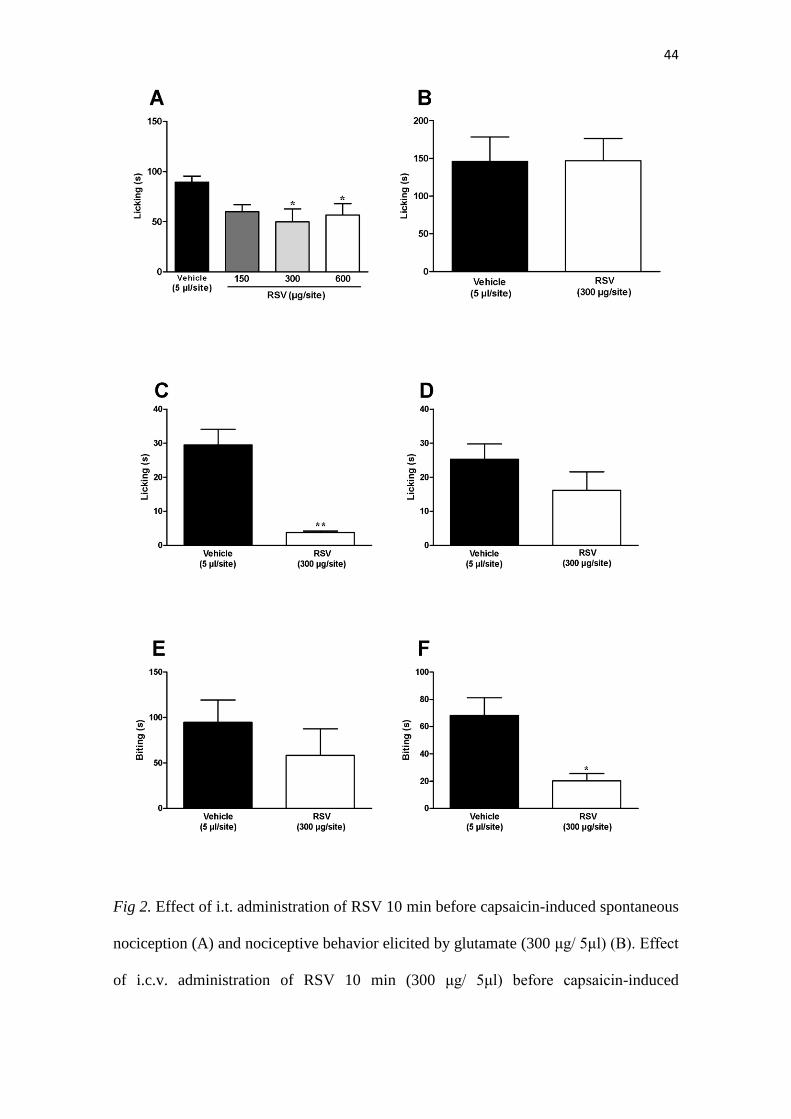
3.3. Intrathecal RSV reduces nociception induced by capsaicin but not by glutamate
When injected by i.t. route, RSV significantly reversed the nociception induced
by capsaicin, either at the doses of 300 µg or 600 µg per site (p<0.05), with percentages
of inhibition 52 ± 15% and 63 ± 13 %, correspondingly (Fig. 2A). On the other hand,
33
the i.t. administration of RSV did not elicit any alteration of glutamate-induced licking
(Fig. 2B).
3.4. Intracerebroventricular RSV inhibits nociception induced by capsaicin but not by
glutamate
The results show that i.c.v. administration of RSV displayed a partial inhibition
of capsaicin-induced spontaneous nociception (Fig. 2C) (p<0.01; 13± 2 %), without
affecting glutamate-induced nociception (Fig. 2D). This evidence corroborates our
hypothesis, indicating that RSV acts by different pathways depending on the
nociceptive stimuli.
3.5. Intrathecal RSV reduces biting nociception induced by i.t. glutamate but not by i.t.
capsaicin
When tested by i.t route, RSV was found able to significantly reduce glutamate-
induced biting behavior (Fig. 2F) (p<0.05, 29 ± 8 %), whereas it was not effective in
changing capsaicin induced-biting (Fig. 2E). This indicates that RSV displays different
effects on acute pain, depending on the route of administration of algogenic agents.
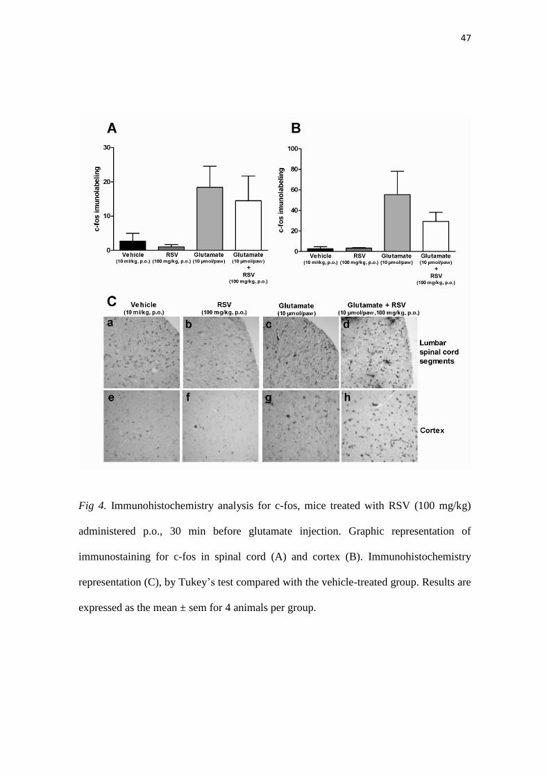
3.6. Immunohistochemistry for c-Fos following i.pl. injection of capsaicin or glutamate
To investigate whether RSV inhibition would be able to reduce the expression
of the neuronal activation marker c-Fos, we have performed an immunohistochemistry
analysis of lumbar spinal cord and cortex sections. RSV was administrated orally, at the
dose of 100 mg/kg. This scheme of treatment resulted in a significant inhibition of
capsaicin-induced increased of c-Fos expression, according to assessment in L3-L6
spinal segments (Fig. 3A) (p<0.01). Otherwise, RSV was not capable of significantly
34
inhibiting capsaicin-induced increased in c-Fos levels at the brain cortex, although the
immunolabeling was virtually reduced in RSV-treated animals (Fig. 3B, Fig. 3
representative images 3C panels a to h). The oral administration of RSV (100 mg/kg)
also failed to affect the increase of c-Fos labeling in glutamate-injected mice, either in
spinal cord (Fig. 4A) or brain sections (Fig. 4B). The representative images for these
experiments are depicted in the Figure 4C, panels a to h.
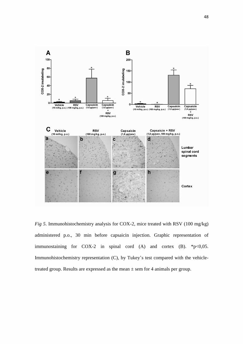
3.7. COX-2 Immunohistochemistry
The administration of RSV (100 mg/kg, p.o.) resulted in a significant
diminishment of COX-2 expression at L3-L6 spinal segments (Fig. 5A) and also in
cerebral cortex sections from capsaicin-injected mice (p<0.05) (Fig. 5B, representative
images Figure 5C, panels a to h). Conversely, RSV did not interfere with COX-2
immunopositivity allied to glutamate i.pl. injection, in neither the spinal cord (Figure
6A) or cortex brain sections (Fig. 6B). The representative images for this series of
experiments are depicted in Fig. 6C, panels a to h.

35
4. Discussion
The investigation of new molecules that are effective in management of
nociception and inflammatory processes represents a very attractive field of research.
The current analgesic therapeutic arsenal have been found to be only partially effective,
and their use is commonly associated with serious side-effects (Jones, 2011).
RSV is a polyphenol, well-known by its anti-inflammatory and anti-tumoral
actions (Busquets et al., 2007; Das and Das, 2007). Some few studies have reported
analgesic effects for RSV, according to assessment in animal models of diabetic
neuropathy, or in formalin and carrageenan tests, although its mechanisms of action
remain to be determined (Pham-Marcou et al., 2008; Sharma et al., 2007; Torres-López
et al., 2002). Therefore, the present study was designed to investigate the effects of RSV
on the nociceptive responses induced by capsaicin and glutamate, when these
nociceptive agents were injected into the mouse paw or by i.t. route. Some efforts have
been made to determine the possible anatomical sites related to RSV effects on
capsaicin- or glutamate-elicited pain behavior. We have also evaluated some of the
mechanisms implicated in the systemic analgesic effects of RSV.
Initially, RSV was tested orally, and this compound was administered 30 min
before the spontaneous nociception induced by capsaicin or glutamate, in doses ranging
from 50 to 200 mg/kg. Of note, the oral treatment with RSV was able to produce a
significant reduction of paw licking caused by either capsaicin or glutamate, without
clear dose-related effects. Recent studies demonstrated a great analgesic effect for RSV
at the dose of 100 mg/kg, p.o., in the capsaicin test, confirming our findings (Montiel-
Ruiz et al., 2009). Nevertheless, to the present moment, there are no previous studies
investigating the effects of RSV in the nociception induced by glutamate. Concerning
the mechanisms related to the analgesic activity observed for RSV, it has been
36
suggested that RSV analgesia is partly mediated via opioid release, as this compound
produces crossed-tolerance with morphine (Gupta et al., 2004).
In an attempt to verify the possible sites of action of this compound, we decided
to evaluate the effects of RSV when given by other pathways of administration.
Interestingly, the co-injection of RSV into the mouse paw, at the dose of 200 µg/site,
was able to largely reverse the nociceptive responses elicited by glutamate, although it
failed to significantly affect the pain-like behavior induced by capsaicin. It is worth
mentioning that this dose of RSV was selected on the basis of previous studies
(Granados-Soto et al., 2002), and it was found active in the formalin test. Our data on
the capsaicin model allow us to suggest that RSV does not act peripherally in relation to
TRPV1 activation. Otherwise, it is feasible to consider that RSV might interfere with
glutamate receptors, namely NMDA, at periphery sites. In a previous study conducted
in rats, it was demonstrated that peripheral antinociceptive effects of RSV in the
formalin model are likely related to the opening of large and small conductance Ca2+
activated K+ channels, but not ATP-sensitive K
+ channels (Granados-Soto et al., 2002).
This might well explain the actions of RSV on glutamate-induced pain, when this
algogenic agent is injected into the mouse hindpaw.
As another experimental approach, we have further evaluated the actions of
RSV, when this compound was given by i.t. route. The doses used by us were based on
the previous study published by (Pérez-Severiano et al., 2008). Significant effects were
observed for RSV at the doses of 300 and 600 µg/site, when nociception was induced
by capsaicin, but not by glutamate. This is highly suggestive for a central
antinociceptive action for RSV in the capsaicin model. We might suggest that RSV
might interfere with TRPV1 activation at the spinal levels of pain processing.
Accordingly, some studies on chronic nociception have suggested that RSV could
37
activate the proteins of the NO–cyclic GMP–PKG pathway or large-conductance Ca2+
-
activated, but not ATP-sensitive, K+ channels at the spinal cord, restoring altered NOS
activity and expression, what led to a reduction of allodynia in neuropathic rats (Pérez-
Severiano et al., 2008).
To additionally assess the central effects of RSV, this compound was injected
i.c.v., at the dose of 300 µg/site. Again, a partial, but significant antinociceptive effect
was found in the capsaicin model, but not in the glutamate-induced pain behavior. To
our knowledge, until the moment, there are no studies showing the effects of RSV when
injected by i.c.v. route, in the nociception context. Of note, a previous publication has
demonstrated that i.c.v. administration of RSV was able to exhibit anti-diabetic actions
in mice (Ramadori et al., 2009). Nevertheless, this series of experiments reinforce the
previous hypothesis that RSV probably interferes with central pain transmission via
TRPV1 receptor activation, although the spinal cord seems to be a most important site
of action for RSV.
To further investigate the analgesic effects of RSV, we have analyzed the biting
behavior evoked by the i.t. injection of glutamate or capsaicin, by application into the
subdural space of the L5–L6 spinal segments. Unexpectedly, the combination of RSV
was able to reverse the biting behavior induced by glutamate, whilst it failed to
significantly alter the biting activity elicited by i.t. injection of capsaicin. Nevertheless,
at the CNS, RSV is known to inhibit Na+ currents in the dorsal root ganglion neurons
(Kim et al., 2005). It might be hypothesized that RSV exerts suppression or antagonism
on the kinetics of Na+ currents, affecting the excitability of the dorsal root neurons, and
consequently glutamate-induced biting. Of note, it was recently demonstrated that i.t.
injection of guanosine, a guanine based-purine, inhibited the nociceptive response
induced by i.t. injection of glutamate, although it failed to affect capsaicin-mediated
38
biting response in mice (Schmidt et al., 2009), as showed for RSV in the present study.
It is tempting to propose that RSV is able to interfere with the signaling pathways
activated by glutamate in the spinal cord, following the activation of either ionotropic or
metabotropic receptors. Concerning the capsaicin model, our data is highly suggestive
for a central antinociceptive action for RSV, when this compound was dosed 10 min
before capsaicin-induced spontaneous nociception. Otherwise, RSV failed to
significantly alter the biting behavior elicited by i.t. injection of capsaicin, when
administered concurrently at the spinal level. To interpret our data, we also need to
consider that the capsaicin concentration and/or the site of injection of this pungent
agent might directly influence the open and closed TRPV1 channel states, likely via
PKC-mediated phosphorylation, what can be responsible for the differences in the
effects of RSV, as observed by us (Hui et al., 2003; Studer and McNaughton, 2010).
The systemic analgesic action of RSV observed in the two models of acute pain
led us to further investigate the possible mechanisms underlying these effects. COX-2 is
responsible for the elevated production of prostanoids at the inflammatory sites (Warner
and Mitchell, 2004). Previous studies have demonstrated that RSV is effective in
blocking both the expression and the activity of COX-2 (Pham-Marcou et al., 2008).
The analgesic activity of RSV seems to be related to various pathways of the pain
control. COX-2 inhibition and K+ channel opening have been suggested as the main
underlying mechanisms (Bertelli et al., 2008; Falchi et al., 2010b). In the present study,
we clearly demonstrate that i.pl. injection of capsaicin into the mouse paw resulted in a
marked increase of COX-2 immunopositivity in spinal cord and cortex sections, which
likely contributes to development of central sensitization. It has been previously shown
that intraprostatic injection of capsaicin resulted in increased central expression of
COX-2 (Chuang et al., 2007). Interestingly, the oral administration of RSV to mice was
39
able to significantly reduce capsaicin-induced COX-2 expression in both the spinal cord
and the brain cortex. These findings allow us to suggest that RSV might affect COX-2
induction, when nociception involves the activation of TRPV1 receptors by capsaicin.
A recent publication demonstrated that excitotoxic events lead to increased
COX-2 expression, supporting our results on the increased central levels of COX-2 after
glutamate peripheral injection. However, the systemic treatment with RSV failed to
affect the up-regulation of COX-2, according to assessment at the spinal cord or the
brain cortex of glutamate-injected mice (Stark and Bazan, 2011). This might explain, at
least in part, the absence of effects of RSV on glutamate-induced licking, when this
compound was administered by i.t. or i.c.v. routes.
Nociceptive stimulus modulates several pathways and influence transcriptional
and translational levels of many molecules, including the activation of immediate genes,
such as c-Fos, which has been markedly associated to nociception (Liu et al., 2011).
Thus, we also decided to evaluate whether the systemic administration of RSV could
affect c-Fos expression in the CNS. It was previously demonstrated that TRPV1
peripheral activation induced by capsaicin lead to modifications on spinal cord glia in
mice (Chen et al., 2009), resulting in increased c-Fos expression (Hossaini et al., 2011).
These pieces of evidence corroborate our data showing that capsaicin injection into the
mouse paw resulted in a significant increase of c-Fos immunelabeling at both the spinal
cord and brain cortex segments. Notably, in our study, RSV significantly reduced the c-
Fos expression-induced by capsaicin in the spinal cord, but not in the cortex. On the
basis of this data, it is tempting to suggest that RSV mechanisms of action probably
involve the modulation of TRPV1 activation in the spinal cord, allied to changes in c-
Fos expression. We can also infer that the most prominent effects of RSV when injected
40
by i.t., in comparison to i.c.v route, in the capsaicin model, might be consequence of
modulation of spinal c-Fos expression.
It was previously demonstrated that i.pl. injection of glutamate induced an
elevation of c-Fos levels in the mouse spinal cord (Lin et al., 2009). Our results confirm
and extend this evidence showing that application of glutamate into the mouse paw
resulted in a marked increase of c-Fos immunopositivity in either the spinal cord or the
brain cortex. Nevertheless, the oral administration of RSV failed to affect glutamate-
induced c-Fos activation in both anatomical sites. This is in accord with functional data
obtained in the licking model, further confirming that RSV acts peripherally, when the
nociception is induced by glutamate. However, additional studies are necessary to
confirm this hypothesis.
4.1. Conclusions
Collectively, our data bring novel evidence on the possible mechanisms of
action of RSV in pain processing. Therefore, the oral administration of RSV elicited
antinociceptive effects in the licking models induced by both capsaicin and glutamate.
From the experiments using different routes of administration, it is possible to conclude
that in the case of capsaicin, RSV appears to affect the spinal TRPV1 activation, via
mechanisms likely involving c-Fos activation and COX-2 expression. Otherwise, in the
glutamate-induce licking, the actions of RSV seem to take place mainly at peripheral
sites. Nevertheless, RSV also displayed antinociceptive effects in glutamate-induced
biting behavior, indicating the relevance of additional mechanisms of action for that
naturally-based compound. An overall analysis of our results allows us to suggest that
mechanisms of action of RSV might be different depending on the algogenic stimulus,
as well as the site of administration.
41
5. Financial Support: CAPES, AUX-PE Toxinologia, CNpq and PUCRS
Abbreviations:
RSV – resveratrol; i.c.v. – intracerebroventricular; i.t. – intrathecal; i.pl. – intraplantar;
p.o. – oral route; NMDA – N-metil D-Aspartate; TRPV – Transient Receptor Potential
Vanilloid 1; TRPV1 – Transient Receptor Potential Vanilloid type 1; NO- nitric oxide;
ATP- adenosine triphosphate; GMP–PKG- guanine monophosphate- G kinase protein ;
CNS – Central Nervous Sistem; COX-2 – Ciclooxigenase 2; NOS – nitric oxide
sinthase.
42
Acknowledgements
K. O. B. is a mastership postgraduate student in Medicine and Health Sciences
receiving grants from Capes (Coordenação de Aperfeiçoamento de Pessoal de Nível
Superior). A.H.S. is a post-doc student receiving financial support from Capes. We
would like to thank Mr. Juliano Soares by his technical assistance.

43
Fig 1. Effect of p.o. administration of RSV (50-200 mg/kg) 30 min before capsaicin-
induced spontaneous nociception (A) and nociceptive behavior elicited by glutamate
(B). Effect of i.pl. administration of RSV 30 min (200 μg in 25μl) before capsaicin-
induced spontaneous nociception (C) and nociceptive behavior elicited by glutamate
(D). Results are expressed as the mean ± sem for 8 animals per group.*p<0.05 by
Dunnett’s test compared with the vehicle-treated group.
44
Fig 2. Effect of i.t. administration of RSV 10 min before capsaicin-induced spontaneous
nociception (A) and nociceptive behavior elicited by glutamate (300 μg/ 5μl) (B). Effect
of i.c.v. administration of RSV 10 min (300 μg/ 5μl) before capsaicin-induced

45
spontaneous nociception (C) and nociceptive behavior elicited by glutamate (D). Effect
of i.t. administration of RSV with capsaicin-induced spontaneous nociception (6.4
μg/5μl) (E) and nociceptive behavior elicited by glutamate (6 μg/5μl) (F). Each column
represents the mean ± sem for 8 animals per group. *p<0.05, **p<0.01, by Dunnett’s
test compared with the vehicle-treated group.

46
Fig 3. Immunohistochemistry analysis for c-fos, mice treated with RSV (100 mg/kg)
administered p.o., 30 min before capsaicin injection. Graphic representation of
immunostaining for c-fos in spinal cord (A) and cortex (B). *p<0,05.
Immunohistochemistry representation (C) by Tukey’s test compared with the vehicle-
treated group. Results are expressed as the mean ± sem for 4 animals per group.
47
Fig 4. Immunohistochemistry analysis for c-fos, mice treated with RSV (100 mg/kg)
administered p.o., 30 min before glutamate injection. Graphic representation of
immunostaining for c-fos in spinal cord (A) and cortex (B). Immunohistochemistry
representation (C), by Tukey’s test compared with the vehicle-treated group. Results are
expressed as the mean ± sem for 4 animals per group.
48
Fig 5. Immunohistochemistry analysis for COX-2, mice treated with RSV (100 mg/kg)
administered p.o., 30 min before capsaicin injection. Graphic representation of
immunostaining for COX-2 in spinal cord (A) and cortex (B). *p<0,05.
Immunohistochemistry representation (C), by Tukey’s test compared with the vehicle-
treated group. Results are expressed as the mean ± sem for 4 animals per group.

49
Fig 6. Immunohistochemistry analysis for COX-2, mice treated with RSV (100 mg/kg)
administered p.o., 30 min before glutamate injection. Graphic representation of
immunostaining for COX-2 in spinal cord (A) and cortex (B). Immunohistochemistry
representation (C), by Tukey’s test compared with the vehicle-treated group. Results are
expressed as the mean ± sem for 4 animals per group.
50
6. References
Azorín-Ortuño, M., Yáñez-Gascón, M. J., González-Sarrías, A., Larrosa, M., Vallejo,
F., Pallarés, F. J., Lucas, R., Morales, J. C., Tomás-Barberán, F. A., García-Conesa, M.
T., 2011. Effects of long-term consumption of low doses of resveratrol on diet-induced
mild hypercholesterolemia in pigs: a transcriptomic approach to disease prevention. J
Nutr Biochem.
Beirith, A., Santos, A. R. S., Calixto, J. B., 2002. Mechanisms underlying the
nociception and paw oedema caused by injection of glutamate into the mouse paw.
Brain Res 924, 219-228.
Bertelli, A., Falchi, M., Dib, B., Pini, E., Mukherjee, S., Das, D. K., 2008. Analgesic
resveratrol? Antioxid Redox Signal 10, 403-404.
Burgess, G., Williams, D., 2010. The discovery and development of analgesics: new
mechanisms, new modalities. J Clin Invest 120, 3753-3759.
Busquets, S., Ametller, E., Fuster, G., Olivan, M., Raab, V., Argilés, J. M., López-
Soriano, F. J., 2007. Resveratrol, a natural diphenol, reduces metastatic growth in an
experimental cancer model. Cancer Lett 245, 144-148.
Chen, Y., Willcockson, H. H., Valtschanoff, J. G., 2009. Influence of the vanilloid
receptor TRPV1 on the activation of spinal cord glia in mouse models of pain. Exp
Neurol 220, 383-390.
Cheng, S. F., Foster, R. L., Huang, C., 2003. Concept Analysis of Pain. Tzu Chi
Nursing Journal 2, 20-29.
Chuang, Y. C., Yoshimura, N., Wu, M., Huang, C. C., Chiang, P. H., Tyagi, P.,
Chancellor, M. B., 2007. Intraprostatic capsaicin injection as a novel model for
nonbacterial prostatitis and effects of botulinum toxin A. Eur Urol 51, 1119-1127.
Das, S., Das, D. K., 2007. Anti-inflammatory responses of resveratrol. Inflamm Allergy
Drug Targets 6, 168-173.
Enza, P., Livio, L., de Novellis Vito, B. L., Francesco, R., Sabatino, M., 2010. Moving
towards supraspinal TRPV1 receptors for chronic pain relief. Mol Pain 6.
Falchi, M., Bertelli, A., Galazzo, R., Vigano, P., Dib, B., 2010a. Central antalgic
activity of resveratrol. Arch Ital Biol 148, 389-396.
Falchi, M., Bertelli, A., Galazzo, R., Viganò, P., Dib, B., 2010b. Central antalgic
activity of resveratrol. Arch Ital Biol 148, 389-396.
Gentilli, M., Mazoit, J. X., Bouaziz, H., Fletcher, D., Casper, R. F., Benhamou, D.,
Savouret, J. F., 2001. Resveratrol decreases hyperalgesia induced by carrageenan in the
rat hind paw. Life Sci 68, 1317-1321.
Gold, M. S., Gebhart, G. F., 2010. Nociceptor sensitization in pain pathogenesis. Nat
Med 16, 1248-1257.
51
Granados-Soto, V., Argüelles, C., Ortiz, M., 2002. The peripheral antinociceptive effect
of resveratrol is associated with activation of potassium channels. Neuropharmacology
43, 917-923.
Gupta, Y. K., Sharma, M., Briyal, S., 2004. Antinociceptive effect of trans-resveratrol
in rats: Involvement of an opioidergic mechanism. Methods Find Exp Clin Pharmacol
26, 667-672.
Haley, J., Dickenson, A., Schachter, M., 1992. Electrophysiological evidence for a role
of nitric oxide in prolonged chemical nociception in the rat. Neuropharmacology 31,
251-258.
Hossaini, M., Sara , Jongen, J., Holstege, J., 2011. C-fos activation of spinal glycinergic
and GABAergic neurons is increased after capsaicin stimulation in rats with
contralateral chronic pain. Neuroscience 24, 265-275.
Hui, K., Liu, B., Qin, F., 2003. Capsaicin activation of the pain receptor, VR1: multiple
open states from both partial and full binding. Biophys J 84, 2957-2968.
Hunskaar, S., Post, C., Fasmer, O., Arwestrom, E., 1986. Intrathecal injection of
capsaicin can be used as a behavioural nociceptive test in mice. Neuropharmacology 25,
1149-1153.
Jones, A. W., 2011. Early drug discovery and the rise of pharmaceutical chemistry.
Drug Test Anal 3, 337-344.
Kim, H. I., Kim, T. H., Song, J. H., 2005. Resveratrol inhibits Na+ currents in rat dorsal
root ganglion neurons. Brain Res 1045, 134-141.
Komatsu, T., Sasaki, M., Sanai, K., Kuwahata, H., Sakurada, C., Tsuzuki, M., Iwata, Y.,
Sakurada, S., Sakurada, T., 2009. Intrathecal substance P augments morphine-induced
antinociception: Possible relevance in the production of substance P N-terminal
fragments. Peptides 30, 1689-1696.
Labrousse, V. F., Costes, L., Aubert, A., Darnaudéry, M., Ferreira, G., Amédée, T.,
Layé, S., 2009. Impaired Interleukin-1 and c-Fos Expression in the Hippocampus Is
Associated with a Spatial Memory Deficit in P2X7 Receptor-Deficient Mice. PloS one
4, e6006.
Li, H., Yan, Z., Zhu, J., Yang, J., He, J., 2011. Neuroprotective effects of resveratrol on
ischemic injury mediated by improving brain energy metabolism and alleviating
oxidative stress in rats. Neuropharmacology 60, 252-258.
Lin, Y. R., Chen, H. H., Lin, Y. C., Ko, C. H., Chan, M. H., 2009. Antinociceptive
actions of honokiol and magnolol on glutamatergic and inflammatory pain. J Biomed
Sci 16, 94.
Liu, C. R., Duan, Q. Z., Wang, W., Wei, Y. Y., Zhang, H., Li, Y. Q., Wu, S. X., Xu, L.
X., 2011. Effects of intrathecal isoflurane administration on nociception and Fos
expression in the rat spinal cord. Eur J Anaesthesiol 28, 112-119.
52
Merskey, H., Bogduk, N., 1994. Classification of chronic pain, IASP Task Force on
Taxonomy. Seattle: IASP Press.
Montiel-Ruiz, R. M., Reyes-García, G., Flores-Murrieta, F., Déciga-Campos, M., 2009.
Antinociceptive Interaction Between Benfotiamine and Resveratrol in Capsaicin-
Induced Licking. Proc West Pharmacol Soc 52, 67-71.
Muralidharan, A., Smith, M. T., 2011. Pain, analgesia and genetics. J Pharm Pharmacol
63, 1387–1400.
Pereira, P. J. S., Lazarotto, L. F., Leal, P. C., Lopes, T. G., Morrone, F. B., Campos, M.
M., 2011. Inhibition of phosphatidylinositol-3 kinase γ reduces pruriceptive,
inflammatory, and nociceptive responses induced by trypsin in mice. Pain 152, 2861-
2869.
Pérez-Severiano, F., Bermúdez-Ocaña, D. Y., López-Sánchez, P., Ríos, C., Granados-
Soto, V., 2008. Spinal nerve ligation reduces nitric oxide synthase activity and
expression: Effect of resveratrol. Pharmacol Biochem Behav 90, 742-747.
Pham-Marcou, T. A., Beloeil, H., Sun, X., Gentili, M., Yaici, D., Benoit, G.,
Benhamou, D., Mazoit, J. X., 2008. Antinociceptive effect of resveratrol in
carrageenan-evoked hyperalgesia in rats: prolonged effect related to COX-2 expression
impairment. Pain 140, 274-283.
Ramadori, G., Gautron, L., Fujikawa, T., Vianna, C. R., Elmquist, J. K., Coppari, R.,
2009. Central administration of resveratrol improves diet-induced diabetes.
Endocrinology 150, 5326-5333.
Ribas, C. M., Meotti, F. C., Nascimento, F. P., Jacques, A. V., Dafre, A. L., Rodrigues,
A. L. S., Farina, M., Soldi, C., Mendes, B. G., Pizzolatti, M. G., 2008. Antinociceptive
effect of the Polygala sabulosa hydroalcoholic extract in mice: evidence for the
involvement of glutamatergic receptors and cytokine pathways. Basic Clin Pharmacol
Toxicol 103, 43-47.
Sakurada, T., Katsumata, K., Yogo, H., Tan-No, K., Sakurada, S., Kisara, K., 1993.
Antinociception induced by CP 96,345, a non-peptide NK-1 receptor antagonist, in the
mouse formalin and capsaicin tests. Neurosci Lett 151, 142-145.
Schmidt, A. P., Böhmer, A. E., Schallenberger, C., Antunes, C., Pereira, M. S. L., Leke,
R., Wofchuk, S. T., Elisabetsky, E., Souza, D. O., 2009. Spinal mechanisms of
antinociceptive action caused by guanosine in mice. Eur J Pharmacol 613, 46-53.
Sharma, S., Kulkarni, S. K., Chopra, K., 2007. Effect of resveratrol, a polyphenolic
phytoalexin, on thermal hyperalgesia in a mouse model of diabetic neuropathic pain.
Fundam Clin Pharmacol 21, 89-94.
Stark, D. T., Bazan, N. G., 2011. Synaptic and Extrasynaptic NMDA Receptors
Differentially Modulate Neuronal Cyclooxygenase-2 Function, Lipid Peroxidation, and
Neuroprotection. J Neurosci 31, 13710-13721.
53
Studer, M., McNaughton, P. A., 2010. Modulation of single-channel properties of
TRPV1 by phosphorylation. J Physiol 588, 3743-3756.
Torres-López, J. E., Ortiz, M. I., Castañeda-Hernández, G., Alonso-López, R.,
Asomoza-Espinosa, R., Granados-Soto, V., 2002. Comparison of the antinociceptive
effect of celecoxib, diclofenac and resveratrol in the formalin test. Life Sci 70, 1669-
1676.
Warner, T. D., Mitchell, J. A., 2004. Cyclooxygenases: new forms, new inhibitors, and
lessons from the clinic. FASEB J 18, 790-804.
Woolf, C. J., 2010. What is this thing called pain? J Clin Invest 120, 3742.
Yan-Shi, G., 2011. Recent Anti-aging Studies on Caloric Restriction and Resveratrol.
Sheng Li Ke Xue Jin Zhan 42, 161-164.
Yoon, D. H., Kwon, O. Y., Mang, J. Y., Jung, M. J., Kim, D. Y., Park, Y. K., Heo, T.
H., Kim, S. J., 2011. Protective potential of resveratrol against oxidative stress and
apoptosis in Batten disease lymphoblast cells. Biochem Biophys Res Commun 14, 49-
52.
Young, M., Fleetwood-Walker, S., Mitchell, R., Dickinson, T., 1995. The involvement
of metabotropic glutamate receptors and their intracellular signalling pathways in
sustained nociceptive transmission in rat dorsal horn neurons. Neuropharmacology 34,
1033-1041.

54
CONSIDERAÇÕES FINAIS
A cada ano, novos medicamentos são lançados no mercado, favorecendo o
aumento da expectativa de seus usuários e o melhor controle das doenças. Os agentes
terapêuticos medicamentosos modernos têm contribuído favoravelmente contra as
várias moléstias que acometem a humanidade. No entanto, em algumas situações, seu
uso cria efeitos indesejáveis à saúde (Mahmud et al., 2006).
Estudos e registros sobre intoxicações e reações adversas em vários países,
incluindo o Brasil, demonstram que os medicamentos são responsáveis por grande parte
dos atendimentos nos Centros de Controle de Intoxicações e, em especial, os
analgésicos. Desta forma, o desenvolvimento de novos fármacos com minimização de
efeitos adversos torna-se uma abordagem relevante para a realidade atual (Gandolfi and
Andrade, 2006; Kawano et al., 2006; Santos and Nitrini, 2004).
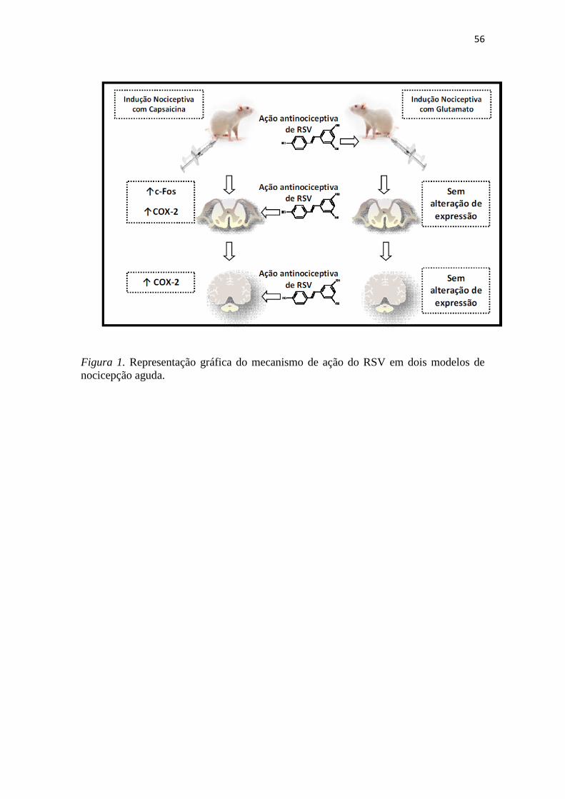
Neste estudo, demonstramos o efeito antinociceptivo do RSV em dois modelos
de nocicepção aguda induzida por capsaicina e glutamato. Interessantemente, uma única
dose via oral mostrou-se efetiva em ambos modelos, indicando uma ação farmacológica
satisfatória para o composto. Quando testado por outras vias de administração, o RSV
apresentou diferentes sítios de ação, conforme o estímulo nociceptivo. No caso da
nocicepção induzida pela capsaicina, que envolve a ativação de receptores TRPV1, o
efeito analgésico foi central; já, na nocicepção induzida pela ativação de canais NDMA,
após a injeção de glutamato, o RSV parece exercer uma ação periférica.
Para investigar o mecanismo de ação do RSV, o marcador de ativação neural, c-
Fos, e a enzima ciclooxigenase 2, COX-2, foram quantificados em fatias de medula
espinhal e cortex através de imunohistoquímica. Os níveis de c-Fos mostraram-se
elevados nos segmentos da medula espinhal quando a nocicepção foi induzida pela

55
capsaicina; neste modelo; foi possível observar a modulação de COX-2 no córtex e na
medula espinhal, confirmando os dados comportamentais que indicam a ação central de
RSV no modelo de nocicepção causado pela aplicação i.p.l. induzida pela capsaicina
(Figura 1).
Já, no modelo de nocicepção causada por glutamato, os níveis de c-Fos e COX-2
não foram significativamente elevados em fatias de medula espinhal e córtex, embora
haja uma tendência vista na quantificação destes marcadores, confirmando assim, os
dados obtidos nas análises comportamentais que indicaram ação periférica de RSV,
quando a nocicepção foi induzida pela ativação de receptores NDMA (Figura 1).
Baseado no fato de que atualmente poucos estudos esclarecem a ação do RSV
em modelos de nocicepção, o presente trabalho evidenciou novos mecanismos de ação
para este composto. De fato, na literatura, são propostos diversos mecanismos de ação
distintos para o RSV, embora existam poucas evidências que expliquem os seus efeitos
antinociceptivos;
Com base nos dados apresentados, pode-se concluir que: (i) o RSV tem efeito
antinociceptivo nos modelos de nocicepção espontânea induzida por capsaicina e
glutamato; (ii) quando a nocicepção envolve a ativação de receptores TRPV1, este
efeito tem como sítio de ação o SNC, envolvendo o aumento da expressão de c-Fos e
COX-2; (iii) no caso da nocicepção que envolve a ativação de receptores NDMA, este
mecanismo de ação ocorre na região periférica, ou, referente ao local de indução, não
envolvendo modificações de expressão de c-Fos ou COX-2. Desta forma, o presente
trabalho contribui para os conhecimentos atuais sobre este composto, fornecendo
evidências importantes sobre o RSV.
56
Figura 1. Representação gráfica do mecanismo de ação do RSV em dois modelos de
nocicepção aguda.
57
4. REFERÊNCIAS
Aimar, P., Pasti, L., Carmignoto, G., Merighi, A., 1998. Nitric oxide-producing islet
cells modulate the release of sensory neuropeptides in the rat substantia gelatinosa. The
Journal of neuroscience 18, 10375.
Baron, R., 2006. Mechanisms of disease: neuropathic pain—a clinical perspective.
Nature Clinical Practice Neurology 2, 95-106.
Caterina, M. J., Schumacher, M. A., Tominaga, M., Rosen, T. A., Levine, J. D., Julius,
D., 1997. The capsaicin receptor: a heat-activated ion channel in the pain pathway.
Nature 389, 816-824.
Chen, Y., Willcockson, H. H., Valtschanoff, J. G., 2009. Influence of the vanilloid
receptor TRPV1 on the activation of spinal cord glia in mouse models of pain. Exp
Neurol 220, 383-390.
Cheng, S. F., Foster, R. L., Huang, C., 2003. Concept Analysis of Pain. Tzu Chi
Nursing Journal 2, 20-29.
Cho, W. G., Valtschanoff, J. G., 2008. Vanilloid receptor TRPV1-positive sensory
afferents in the mouse ankle and knee joints. Brain Research 1219, 59-65.
Chuang, Y. C., Yoshimura, N., Wu, M., Huang, C. C., Chiang, P. H., Tyagi, P.,
Chancellor, M. B., 2007. Intraprostatic capsaicin injection as a novel model for
nonbacterial prostatitis and effects of botulinum toxin A. European urology 51, 1119-
1127.
Costigan, M., Scholz, J., Woolf, C. J., 2009. Neuropathic pain: a maladaptive response
of the nervous system to damage. Annual review of neuroscience 32, 1.
Crisp, T., Minus, T. O., Coleman, M. L., Giles, J. R., Cibula, C., Finnerty, E. P., 2006.
Aging, peripheral nerve injury and nociception: effects of the antioxidant 16-
desmethyltirilazad. Behavioural brain research 166, 159-165.
Doré, S., 2005. Unique properties of polyphenol stilbenes in the brain: more than direct
antioxidant actions; gene/protein regulatory activity. Neurosignals 14, 61-70.
58
Dugan, L., Choi, D., 1999. Hypoxic-ischemic brain injury and oxidative stress.
Lippincott-Raven Publishers, Philadelphia, pp. 711-729.
Entrena, J. M., Cobos, E. J., Nieto, F. R., Cendán, C. M., Baeyens, J. M., Del Pozo, E.,
2009. Antagonism by haloperidol and its metabolites of mechanical hypersensitivity
induced by intraplantar capsaicin in mice: role of sigma-1 receptors.
Psychopharmacology 205, 21-33.
Fein, A., 2009. Nociceptors: the cells that sense pain. Nociceptors.
Fridovich, I., 1998. Oxygen toxicity: a radical explanation. Journal of experimental
biology 201, 1203.
Gabbita, S. P., Robinson, K. A., Stewart, C. A., Floyd, R. A., Hensley, K., 2000. Redox
regulatory mechanisms of cellular signal transduction. Archives of biochemistry and
biophysics 376, 1.
Gandolfi, E., Andrade, M. G. G., 2006. Eventos toxicológicos relacionados a
medicamentos no Estado de São Paulo. Revista de Saúde Pública 40, 1056-1064.
Gentilli, M., Mazoit, J. X., Bouaziz, H., Fletcher, D., Casper, R. F., Benhamou, D.,
Savouret, J. F., 2001. Resveratrol decreases hyperalgesia induced by carrageenan in the
rat hind paw. Life sciences 68, 1317-1321.
Gilron, I., Watson, C. P. N., Cahill, C. M., Moulin, D. E., 2006. Neuropathic pain: a
practical guide for the clinician. Canadian Medical Association Journal 175, 265.
Guitart, X., Codony, X., Monroy, X., 2004. Sigma receptors: biology and therapeutic
potential. Psychopharmacology 174, 301-319.
Gupta, Y. K., Sharma, M., Briyal, S., 2004. Antinociceptive effect of trans-resveratrol
in rats: Involvement of an opioidergic mechanism. Methods Find Exp Clin Pharmacol
26, 667-672.
Halliwell, B., Gutteridge, J. M. C., 1989. Free radicals in biology and medicine.
Clarendon Press Oxford.
59
Hathway, G. J., Vega-Avelaira, D., Moss, A., Ingram, R., Fitzgerald, M., 2009. Brief,
low frequency stimulation of rat peripheral C-fibres evokes prolonged microglial-
induced central sensitization in adults but not in neonates. Pain 144, 110-118.
Katanosaka, K., Banik, R. K., Giron, R., Higashi, T., Tominaga, M., Mizumura, K.,
2008. Contribution of TRPV1 to the bradykinin-evoked nociceptive behavior and
excitation of cutaneous sensory neurons. Neuroscience research 62, 168-175.
Kawano, D. F., Pereira, L. R. L., Ueta, J. M., Freitas, O., 2006. Acidentes com os
medicamentos: como minimizá-los? Rev Bras Ciênc Farm 42.
Khalil, Z., Khodr, B., 2001. A role for free radicals and nitric oxide in delayed recovery
in aged rats with chronic constriction nerve injury. Free Radical Biology and Medicine
31, 430-439.
Khalil, Z., Liu, T., Helme, R. D., 1999. Free radicals contribute to the reduction in
peripheral vascular responses and the maintenance of thermal hyperalgesia in rats with
chronic constriction injury. Pain 79, 31-37.
Kim, H. K., Park, S. K., Zhou, J. L., Taglialatela, G., Chung, K., Coggeshall, R. E.,
Chung, J. M., 2004. Reactive oxygen species (ROS) play an important role in a rat
model of neuropathic pain. Pain 111, 116-124.
Komatsu, T., Sasaki, M., Sanai, K., Kuwahata, H., Sakurada, C., Tsuzuki, M., Iwata, Y.,
Sakurada, S., Sakurada, T., 2009. Intrathecal substance P augments morphine-induced
antinociception: Possible relevance in the production of substance P N-terminal
fragments. Peptides 30, 1689-1696.
Lappin, S. C., Randall, A. D., Gunthorpe, M. J., Morisset, V., 2006. TRPV1 antagonist,
SB-366791, inhibits glutamatergic synaptic transmission in rat spinal dorsal horn
following peripheral inflammation. Eur J Pharmacol 540, 73-81.
Lee, I., Kim, H. K., Kim, J. H., Chung, K., Chung, J. M., 2007. The role of reactive
oxygen species in capsaicin-induced mechanical hyperalgesia and in the activities of
dorsal horn neurons. Pain 133, 9-17.
Leonard, S. S., Xia, C., Jiang, B. H., Stinefelt, B., Klandorf, H., Harris, G. K., Shi, X.,
2003. Resveratrol scavenges reactive oxygen species and effects radical-induced
60
cellular responses. Biochemical and biophysical research communications 309, 1017-
1026.
Lopez-Velez, M., Martinez-Martinez, F., Del Valle-Ribes, C., 2003. The study of
phenolic compounds as natural antioxidants in wine. Critical reviews in food science
and nutrition 43, 233-244.
Mahmud, S. D. P., Martinbiancho, J. K., Zuckermann, J., Jacoby, T. S., dos Santos, L.,
da Silva, D., 2006. Assistência Farmacêutica: Ações de apoio à qualidade assistencial
Malcangio, M., Lessmann, V., 2003. A common thread for pain and memory synapses?
Brain-derived neurotrophic factor and trkB receptors. Trends in pharmacological
sciences 24, 116-121.
Meller, S., Gebhart, G., 1993. Nitric oxide (NO) and nociceptive processing in the
spinal cord. Pain 52, 127-136.
Millan, M. J., 1999. The induction of pain: an integrative review. Progress in
neurobiology 57, 1-164.
Mokni, M., Elkahoui, S., Limam, F., Amri, M., Aouani, E., 2007. Effect of resveratrol
on antioxidant enzyme activities in the brain of healthy rat. Neurochemical Research 32,
981-987.
Naik, A. K., Tandan, S. K., Dudhgaonkar, S. P., Jadhav, S. H., Kataria, M., Prakash, V.
R., Kumar, D., 2006. Role of oxidative stress in pathophysiology of peripheral
neuropathy and modulation by N-acetyl-L-cysteine in rats. European Journal of Pain 10,
573-579.
Pérez-Severiano, F., Bermúdez-Ocaña, D. Y., López-Sánchez, P., Ríos, C., Granados-
Soto, V., 2008. Spinal nerve ligation reduces nitric oxide synthase activity and
expression: Effect of resveratrol. Pharmacology Biochemistry and Behavior 90, 742-
747.
Peters, J. H., McDougall, S. J., Fawley, J. A., Smith, S. M., Andresen, M. C., 2010.
Primary afferent activation of thermosensitive TRPV1 triggers asynchronous glutamate
release at central neurons. Neuron 65, 657-669.

61
Prado, P. T. C., Del Bel, E. A., 1998. C-fos, um gene de ativaçäo imediata como
marcador neural de nocicepçäo; C-fos, an immediate early gene as a neuromarker for
nociception. Medicina (Ribeiräo Preto) 31, 424-433.
Ribeiro, S., Schmidt, A. P., Schmidt, S. R. G., 2002. O uso de opióides no tratamento da
dor crônica não oncológica: o papel da metadona. Rev Bras Anestesiol 52, 644-651.
Santos, V., Nitrini, S. M., 2004. Indicadores do uso de medicamentos prescritos e de
assistência ao paciente de serviços de saúde. Revista de Saúde Pública 38, 819-834.
Sharma, S., Chopra, K., Kulkarni, S. K., 2007a. Effect of insulin and its combination
with resveratrol or curcumin in attenuation of diabetic neuropathic pain: participation of
nitric oxide and TNF-alpha. Phytother Res 21, 278-283.
Sharma, S., Kulkarni, S. K., Chopra, K., 2007b. Effect of resveratrol, a polyphenolic
phytoalexin, on thermal hyperalgesia in a mouse model of diabetic neuropathic pain.
Fundamental & clinical pharmacology 21, 89-94.
Vetter, G., Geisslinger, G., Tegeder, I., 2001. Release of glutamate, nitric oxide and
prostaglandin E2 and metabolic activity in the spinal cord of rats following peripheral
nociceptive stimulation. Pain 92, 213-218.
Viggiano, A., Monda, M., Viggiano, D., Viggiano, E., Chiefari, M., Aurilio, C., De
Luca, B., 2005. Trigeminal pain transmission requires reactive oxygen species
production. Brain Research 1050, 72-78.
Wang, Q., Yu, S., Simonyi, A., Rottinghaus, G., Sun, G. Y., Sun, A. Y., 2004.
Resveratrol protects against neurotoxicity induced by kainic acid. Neurochemical
Research 29, 2105-2112.
Warner, D. S., Sheng, H., Batini -Haberle, I., 2004. Oxidants, antioxidants and the
ischemic brain. Journal of experimental biology 207, 3221.
Watkins, L. R., Milligan, E. D., Maier, S. F., 2001. Glial activation: a driving force for
pathological pain. TRENDS in Neurosciences 24, 450-455.
Xu, L., Mabuchi, T., Katano, T., Matsumura, S., Okuda-Ashitaka, E., Sakimura, K.,
Mishina, M., Ito, S., 2007. Nitric oxide (NO) serves as a retrograde messenger to

62
activate neuronal NO synthase in the spinal cord via NMDA receptors. Nitric Oxide 17,
18-24.
Zhang, X., Wu, J., Lei, Y., Fang, L., Willis, W. D., 2005. Protein phosphatase
modulates the phosphorylation of spinal cord NMDA receptors in rats following
intradermal injection of capsaicin. Molecular brain research 138, 264-272.
Zhuo, M., 2007. Neuronal mechanism for neuropathic pain. Molecular pain 3, 14.