RESEARCH ARTICLE Open Access Lactobacillus plantarum …
16
Huang et al. BMC Complement Med Ther (2021) 21:259 https://doi.org/10.1186/s12906-021-03426-8 RESEARCH ARTICLE Lactobacillus plantarum PS128 prevents cognitive dysfunction in Alzheimer’s disease mice by modulating propionic acid levels, glycogen synthase kinase 3 beta activity, and gliosis Hei‑Jen Huang 1 , Jie‑Ling Chen 2 , Jian‑Fu Liao 3 , Yu‑Hsin Chen 1 , Min‑Wei Chieu 1 , Ya‑Yun Ke 2 , Chih‑Chieh Hsu 4 , Ying‑Chieh Tsai 3* and Hsiu Mei Hsieh‑Li 2* Abstract Background: According to recent evidence, psychobiotics exert beneficial effects on central nervous system‑related diseases, such as mental disorders. Lactobacillus plantarum PS128 (PS128), a novel psychobiotic strain, improves motor function, depression, and anxiety behaviors. However, the psychobiotic effects and mechanisms of PS128 in Alzhei‑ mer’s disease (AD) remain to be explored. Objectives: The goal of the current study was to evaluate the beneficial effects of PS128 and to further elucidate its mechanism in AD mice. Methods: PS128 (10 10 colony‑forming unit (CFU)/ml) was administered via oral gavage (o.g.) to 6‑month‑old male wild‑type B6 and 3 × Tg‑AD mice (harboring the PS1M146V, APPswe and TauP30IL transgenes) that received an intrac‑ erebroventricular injection of streptozotocin (icv‑STZ, 3 mg/kg) or vehicle (saline) for 33 days. After serial behavioral tests, fecal short‑chain fatty acid levels and AD‑related pathology were assessed in these mice. Results: Our findings show that intracerebroventricular injection of streptozotocin accelerated cognitive dysfunction associated with increasing levels of glycogen synthase kinase 3 beta (GSK3β) activity, tau protein phosphorylation at the T231 site (pT231), amyloid‑β (Aβ) deposition, amyloid‑β protein precursor (AβPP), β‑site AβPP‑cleaving enzyme (BACE1), gliosis, fecal propionic acid (PPA) levels and cognition‑related neuronal loss and decreasing postsynaptic density protein 95 (PSD95) levels in 3 × Tg‑AD mice. PS128 supplementation effectively prevented the damage induced by intracerebroventricular injection of streptozotocin in 3 × Tg‑AD mice. Conclusions: Based on the experimental results, intracerebroventricular injection of streptozotocin accelerates the progression of AD in the 3 × Tg‑AD mice, primarily by increasing the levels of gliosis, which were mediated by the propionic acid and glycogen synthase kinase 3 beta pathways. PS128 supplementation prevents damage induced by © The Author(s) 2021. Open Access This article is licensed under a Creative Commons Attribution 4.0 International License, which permits use, sharing, adaptation, distribution and reproduction in any medium or format, as long as you give appropriate credit to the original author(s) and the source, provide a link to the Creative Commons licence, and indicate if changes were made. The images or other third party material in this article are included in the article’s Creative Commons licence, unless indicated otherwise in a credit line to the material. If material is not included in the article’s Creative Commons licence and your intended use is not permitted by statutory regulation or exceeds the permitted use, you will need to obtain permission directly from the copyright holder. To view a copy of this licence, visit http://creativecommons.org/licenses/by/4.0/. The Creative Commons Public Domain Dedication waiver (http://creativeco mmons.org/publicdomain/zero/1.0/) applies to the data made available in this article, unless otherwise stated in a credit line to the data. Open Access BMC Complementary Medicine and Therapies *Correspondence: [email protected]; [email protected] 2 Department of Life Science, National Taiwan Normal University, Taipei 11677, Taiwan 3 Institute of Biochemistry and Molecular Biology, National Yang Ming Chiao Tung University, Taipei 11221, Taiwan Full list of author information is available at the end of the article
Transcript of RESEARCH ARTICLE Open Access Lactobacillus plantarum …

Huang et al. BMC Complement Med Ther (2021) 21:259 https://doi.org/10.1186/s12906-021-03426-8
RESEARCH ARTICLE
Lactobacillus plantarum PS128 prevents cognitive dysfunction in Alzheimer’s disease mice by modulating propionic acid levels, glycogen synthase kinase 3 beta activity, and gliosisHei‑Jen Huang1, Jie‑Ling Chen2, Jian‑Fu Liao3, Yu‑Hsin Chen1, Min‑Wei Chieu1, Ya‑Yun Ke2, Chih‑Chieh Hsu4, Ying‑Chieh Tsai3* and Hsiu Mei Hsieh‑Li2*
Abstract
Background: According to recent evidence, psychobiotics exert beneficial effects on central nervous system‑related diseases, such as mental disorders. Lactobacillus plantarum PS128 (PS128), a novel psychobiotic strain, improves motor function, depression, and anxiety behaviors. However, the psychobiotic effects and mechanisms of PS128 in Alzhei‑mer’s disease (AD) remain to be explored.
Objectives: The goal of the current study was to evaluate the beneficial effects of PS128 and to further elucidate its mechanism in AD mice.
Methods: PS128 (1010 colony‑forming unit (CFU)/ml) was administered via oral gavage (o.g.) to 6‑month‑old male wild‑type B6 and 3 × Tg‑AD mice (harboring the PS1M146V, APPswe and TauP30IL transgenes) that received an intrac‑erebroventricular injection of streptozotocin (icv‑STZ, 3 mg/kg) or vehicle (saline) for 33 days. After serial behavioral tests, fecal short‑chain fatty acid levels and AD‑related pathology were assessed in these mice.
Results: Our findings show that intracerebroventricular injection of streptozotocin accelerated cognitive dysfunction associated with increasing levels of glycogen synthase kinase 3 beta (GSK3β) activity, tau protein phosphorylation at the T231 site (pT231), amyloid‑β (Aβ) deposition, amyloid‑β protein precursor (AβPP), β‑site AβPP‑cleaving enzyme (BACE1), gliosis, fecal propionic acid (PPA) levels and cognition‑related neuronal loss and decreasing postsynaptic density protein 95 (PSD95) levels in 3 × Tg‑AD mice. PS128 supplementation effectively prevented the damage induced by intracerebroventricular injection of streptozotocin in 3 × Tg‑AD mice.
Conclusions: Based on the experimental results, intracerebroventricular injection of streptozotocin accelerates the progression of AD in the 3 × Tg‑AD mice, primarily by increasing the levels of gliosis, which were mediated by the propionic acid and glycogen synthase kinase 3 beta pathways. PS128 supplementation prevents damage induced by
© The Author(s) 2021. Open Access This article is licensed under a Creative Commons Attribution 4.0 International License, which permits use, sharing, adaptation, distribution and reproduction in any medium or format, as long as you give appropriate credit to the original author(s) and the source, provide a link to the Creative Commons licence, and indicate if changes were made. The images or other third party material in this article are included in the article’s Creative Commons licence, unless indicated otherwise in a credit line to the material. If material is not included in the article’s Creative Commons licence and your intended use is not permitted by statutory regulation or exceeds the permitted use, you will need to obtain permission directly from the copyright holder. To view a copy of this licence, visit http:// creat iveco mmons. org/ licen ses/ by/4. 0/. The Creative Commons Public Domain Dedication waiver (http:// creat iveco mmons. org/ publi cdoma in/ zero/1. 0/) applies to the data made available in this article, unless otherwise stated in a credit line to the data.
Open Access
BMC ComplementaryMedicine and Therapies
*Correspondence: [email protected]; [email protected] Department of Life Science, National Taiwan Normal University, Taipei 11677, Taiwan3 Institute of Biochemistry and Molecular Biology, National Yang Ming Chiao Tung University, Taipei 11221, TaiwanFull list of author information is available at the end of the article
Page 2 of 16Huang et al. BMC Complement Med Ther (2021) 21:259
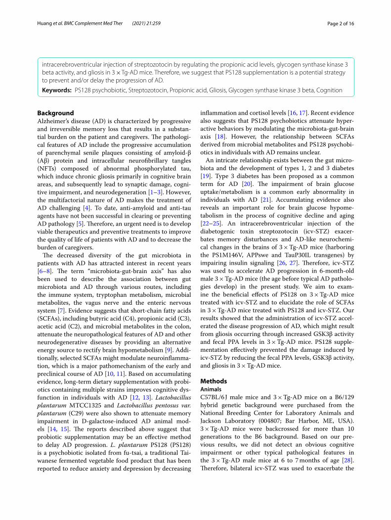
BackgroundAlzheimer’s disease (AD) is characterized by progressive and irreversible memory loss that results in a substan-tial burden on the patient and caregivers. The pathologi-cal features of AD include the progressive accumulation of parenchymal senile plaques consisting of amyloid-β (Aβ) protein and intracellular neurofibrillary tangles (NFTs) composed of abnormal phosphorylated tau, which induce chronic gliosis primarily in cognitive brain areas, and subsequently lead to synaptic damage, cogni-tive impairment, and neurodegeneration [1–3]. However, the multifactorial nature of AD makes the treatment of AD challenging [4]. To date, anti-amyloid and anti-tau agents have not been successful in clearing or preventing AD pathology [5]. Therefore, an urgent need is to develop viable therapeutics and preventive treatments to improve the quality of life of patients with AD and to decrease the burden of caregivers.
The decreased diversity of the gut microbiota in patients with AD has attracted interest in recent years [6–8]. The term “microbiota-gut-brain axis” has also been used to describe the association between gut microbiota and AD through various routes, including the immune system, tryptophan metabolism, microbial metabolites, the vagus nerve and the enteric nervous system [7]. Evidence suggests that short-chain fatty acids (SCFAs), including butyric acid (C4), propionic acid (C3), acetic acid (C2), and microbial metabolites in the colon, attenuate the neuropathological features of AD and other neurodegenerative diseases by providing an alternative energy source to rectify brain hypometabolism [9]. Addi-tionally, selected SCFAs might modulate neuroinflamma-tion, which is a major pathomechanism of the early and preclinical course of AD [10, 11]. Based on accumulating evidence, long-term dietary supplementation with probi-otics containing multiple strains improves cognitive dys-function in individuals with AD [12, 13]. Lactobacillus plantarum MTCC1325 and Lactobacillus pentosus var. plantarum (C29) were also shown to attenuate memory impairment in D-galactose-induced AD animal mod-els [14, 15]. The reports described above suggest that probiotic supplementation may be an effective method to delay AD progression. L. plantarum PS128 (PS128) is a psychobiotic isolated from fu-tsai, a traditional Tai-wanese fermented vegetable food product that has been reported to reduce anxiety and depression by decreasing
inflammation and cortisol levels [16, 17]. Recent evidence also suggests that PS128 psychobiotics attenuate hyper-active behaviors by modulating the microbiota-gut-brain axis [18]. However, the relationship between SCFAs derived from microbial metabolites and PS128 psychobi-otics in individuals with AD remains unclear.
An intricate relationship exists between the gut micro-biota and the development of types 1, 2 and 3 diabetes [19]. Type 3 diabetes has been proposed as a common term for AD [20]. The impairment of brain glucose uptake/metabolism is a common early abnormality in individuals with AD [21]. Accumulating evidence also reveals an important role for brain glucose hypome-tabolism in the process of cognitive decline and aging [22–25]. An intracerebroventricular injection of the diabetogenic toxin streptozotocin (icv-STZ) exacer-bates memory disturbances and AD-like neurochemi-cal changes in the brains of 3 × Tg-AD mice (harboring the PS1M146V, APPswe and TauP30IL transgenes) by impairing insulin signaling [26, 27]. Therefore, icv-STZ was used to accelerate AD progression in 6-month-old male 3 × Tg-AD mice (the age before typical AD patholo-gies develop) in the present study. We aim to exam-ine the beneficial effects of PS128 on 3 × Tg-AD mice treated with icv-STZ and to elucidate the role of SCFAs in 3 × Tg-AD mice treated with PS128 and icv-STZ. Our results showed that the administration of icv-STZ accel-erated the disease progression of AD, which might result from gliosis occurring through increased GSK3β activity and fecal PPA levels in 3 × Tg-AD mice. PS128 supple-mentation effectively prevented the damage induced by icv-STZ by reducing the fecal PPA levels, GSK3β activity, and gliosis in 3 × Tg-AD mice.
MethodsAnimalsC57BL/6 J male mice and 3 × Tg-AD mice on a B6/129 hybrid genetic background were purchased from the National Breeding Center for Laboratory Animals and Jackson Laboratory (004807; Bar Harbor, ME, USA). 3 × Tg-AD mice were backcrossed for more than 10 generations to the B6 background. Based on our pre-vious results, we did not detect an obvious cognitive impairment or other typical pathological features in the 3 × Tg-AD male mice at 6 to 7 months of age [28]. Therefore, bilateral icv-STZ was used to exacerbate the
intracerebroventricular injection of streptozotocin by regulating the propionic acid levels, glycogen synthase kinase 3 beta activity, and gliosis in 3 × Tg‑AD mice. Therefore, we suggest that PS128 supplementation is a potential strategy to prevent and/or delay the progression of AD.
Keywords: PS128 psychobiotic, Streptozotocin, Propionic acid, Gliosis, Glycogen synthase kinase 3 beta, Cognition

Page 3 of 16Huang et al. BMC Complement Med Ther (2021) 21:259
progression of AD, as described in previous studies [26, 27]. The mice were housed at 20–25 °C with 50–60% relative humidity on a 12 h light/dark cycle, and food and water were available ad libitum. The body weight of mice was approximately 26–27 g. All experiments were performed during the light phase between 7:00 a.m. and 7:00 p.m. To minimize animal suffering, the mice were deeply anesthetized with avertin (0.4 g/kg body weight, Sigma; St. Louis, MO, USA) and then sacrificed by perfu-sion or cervical dislocation for pathological analyses after a series of behavioral tasks. All experimental procedures were approved by an ethics committee at the Institutional Animal Care and Use Committee (IACUC) of National Taiwan Normal University (Permit Number: 106036).
Experimental designThe experimental timeline is shown in Fig. S1A. Wild-type B6 and 3 × Tg-AD mice aged 6 months were ran-domly divided into 6 groups (15–20 mice/group) according to the sigmoid distribution of body weight: (i) B6 mice treated with saline (oral gavage, o.g.)/saline (icv) as control group; (ii) B6 mice treated with PS128 (o.g.)/saline (icv); (iii) 3 × Tg-AD mice treated with saline (o.g.)/saline (icv); (iv) 3 × Tg-AD mice treated with PS128 (o.g.)/saline (icv); (v) 3 × Tg-AD mice treated with saline (o.g.)/STZ (icv); and (vi) 3 × Tg-AD mice treated with PS128 (o.g.)/STZ (icv). After adaption to handling and o.g. for 7 days (days 8–14), the mice were administered 100 μL of PS128 (o.g., daily, 1010 CFU/ml; Bened Bio-medical; Taipei, Taiwan) or the saline vehicle for 33 days (days 8–41). The concentration of PS128 was selected according to previous studies [16, 17]. Seven days after PS128 administration, the mice were anesthetized with avertin (0.4 g/kg of body weight; Sigma) and received a single icv injection of 2 μL of STZ (3 mg/kg; Sigma) or vehicle (saline) into the bilateral ventricle (0.3 mm with respect to the bregma, 1.0 mm right and left of the cen-tral suture, 2.5 mm deep). Serial behavioral tests, namely, the open field test (OFT), elevated plus maze (EPM), Y maze and Morris water maze (MWM), were conducted on days 22–23, 26–27, 28–29, and 34–40, respectively. Finally, the mice were sacrificed for western blot, immu-nohistochemical, and SCFA analyses of fecal boli on day 41. Before sacrifice, mouse blood samples were obtained by a tail prick to measure glucose levels using a commer-cial glucometer (Accu-CheckActive; Roche, Mannheim, Germany).
Behavioral assessmentsMice were subjected to a series of behavioral tasks (n = 15–20/group). All mice were habituated in the behavioral room for 30 min before testing, and all behav-ioral apparatuses were carefully cleaned between subjects
with 70 and 30% ethanol sequentially to remove any olfactory cues. Behavioral data were recorded using a video camera and analyzed using an automatic track-ing system (EthoVision-XT; Noldus, Wageningen, The Netherlands).
Open field test (OFT)The OFT was conducted using a recently described method [29]. Briefly, mice were individually placed in the central zone (15 × 15 cm) of a white acrylic box (30 cm long × 30 cm wide × 30 cm high) and then recorded for 10 min. The total distance travelled by the mice was meas-ured as an index of exploratory activity, and a decrease in the time spent in the center of the open field arena was considered an index of anxiety-like phenotypes.
Elevated plus maze (EPM)The EPM was carried out as described in a recent study [29]. The EPM apparatus consisted of a plus-shaped maze constructed of white opaque acrylic with two opposing open arms (30 cm long × 5 cm wide) and two oppos-ing closed arms (30 cm long × 5 cm wide × 15 cm high) connected by a central platform (10 cm long × 10 cm wide) and elevated 50 cm above the floor. The mouse was placed in the central zone with its head directed toward an open arm and allowed to explore the maze for 5 min. The time spent in the open arms was recorded with a camera and considered an index of an anxiolytic effect.
Y mazeThe Y maze testing procedure was performed as previ-ously reported [27]. The Y maze apparatus consisted of 3 identical symmetrical arms (46 cm long × 3 cm wide × 17 cm high) with white acrylic walls. During an 8 min test session, each mouse was initially placed in the central space and allowed to freely explore in the Y maze. Arm entry was defined as the entry of all four paws into one arm. The spontaneous alternation percentage, an indi-cator of short-term memory, was calculated using the following equation: [the number of sequential triplets containing entries in the three arms/(total arm entries - 2) × 100].
Morris water maze (MWM)The MWM task was used to evaluate the spatial learn-ing and memory abilities of mice as previously described [29]. The MWM assay consisted of four stages: pretrain-ing (day 34), training (days 35–38), testing (day 39), and probe (day 40). The swimming ability of the mice was tested on day 34. Mice that floated in the pool were not included in the task. The MWM consisted of a large circular white pool (100 cm in diameter and 47 cm in height) filled with water (21 ± 2 °C) and divided into four

Page 4 of 16Huang et al. BMC Complement Med Ther (2021) 21:259
quadrants. A hidden platform with a diameter of 10 cm was located 1 cm below the water level in the center of the target quadrant in the pool during the pretraining, training, and testing periods. The time to climb onto the hidden platform within 60 s was recorded as the escape latency. During days 35–38, mice were trained four times per day and allowed to rest for 15 min between trials. The mouse was allowed to search for the platform for 60 s and was allowed to remain on the platform for 20 s. If a mouse did not find the platform within 60 s, it was gently guided to the platform, where it remained for 20 s, and the escape latency was recorded as 60 s. The train-ing curve from days 35 to 38 was recognized as an index of spatial learning ability. On day 39 (testing), three test-ing trials were conducted as an index of spatial learning acquisition. Twenty-four hours after the last testing trial, a probe test (day 40, two trials) was performed by remov-ing the platform and allowing each mouse to swim freely for 60 s in the pool. The amount of time spent in the tar-get quadrant was calculated as an index of long-term spa-tial memory.
Analysis of fecal short chain fatty acid (SCFA) contentsBefore sacrifice (on day 41), fresh feces (100 mg) were collected from individual mice (n = 5/group) and sus-pended in 1 ml of 0.5% phosphoric acid with 200 ppm 2-ethylbutyric acid as a surrogate standard. The fecal sus-pensions were homogenized by vortexing for 2 min and centrifuging for 10 min at 17000×g. The supernatant was extracted with an equal volume of ethyl acetate by vor-texing for 10 min and centrifuging for 10 min at 17000×g. The organic extracts were stored at − 80 °C before analy-sis [30]. Prior to analysis, 600 μL of the organic extracts were transferred into a sample vial, and 4-methyl valeric acid was added as an internal standard at a final concen-tration of 200 ppm. A GC-MS analysis was performed using an Agilent 6890 N GC system equipped with an Agilent 5973 N mass detector (Agilent; Wilmington, Del-aware, USA). A fused-silica capillary column DB-FFAP (30 m length, 0.25 mm inner diameter, 0.25 μm film thick-ness, J&W Scientific, Agilent) was used. Helium was sup-plied as the carrier gas at a rate of 1 ml/min. The injection was performed in splitless mode with an injection volume of 1 μL and an injector temperature of 250 °C. The ini-tial oven temperature was maintained at 80 °C for 1 min, increased to 100 °C at 4 °C/min, maintained at 100 °C for 1 min, increased to 180 °C at 8 °C/min, maintained at 180 °C for 1 min, increased to 220 °C at 20 °C/min, and finally held at 220 °C for 5 min. The temperatures of the ion source, quadrupole, and interface were 230 °C, 150 °C, and 280 °C, respectively. The detector was operated in electron impact ionization mode (electron energy, 70 eV), scanning the 35–250 m/z range. The solvent delay was
3.5 min. SCFA mixtures consisting of acetic acid, propi-onic acid, and butyric acid were prepared at a series of concentrations (10, 20, 50, 100, 200, 300 and 400 ppm) with a 200 ppm internal standard to construct the cali-bration curves. The concentration of SCFAs was calcu-lated from the internal standard curve.
Immunohistochemistry (IHC)Mice (n = 3/group) were transcardially perfused with saline followed by 4% paraformaldehyde in 0.1 M phos-phate buffer (pH 7.4) under deep anesthesia induced by an intraperitoneal injection of avertin (0.4 g/kg; Sigma). The brains were transferred to a gradient of 10, 20, and 30% sucrose until they sank to the bottom of the con-tainer. Then, the brain samples were embedded in clear frozen sectioning compound (OCT, Sakura, USA) and cut into 30 μm coronal sections with a cryostat microtome (CMS3050S, Leica Microsystems; Nussloch, Germany). IHC was performed essentially as described in our recent report [29]. The free-floating sections (3–4 sections per mouse) were incubated with primary anti-bodies (Table 1) overnight at room temperature. After three washes with PBS, the sections were incubated with the appropriate biotinylated secondary antibodies (1:200, Vector Laboratories; CA, USA) for 1 h at room tempera-ture, followed by an incubation with an avidin-biotin-peroxidase complex (ABC kit; Vector Laboratories) for 1 h at room temperature. The sections were washed with PBS (10 min, three times), and the signal was developed by a diaminobenzidine (DAB) kit (Vector Laboratories). After DAB staining, the slices were observed using a light microscope (Leica; Wetzlar, Germany). Images were obtained using Image-Pro Plus 5.1 software (Image-Pro Plus Media Cybernetics; Washington, MD, USA), and the threshold intensity was manually set according to a standard signal for all images. Pixel counts were derived from the average of three adjacent sections per animal.
Western blot analysesWestern blot analyses were completed as previously described [29]. Briefly, hippocampal samples were col-lected (n = 5–8 mice/group) and homogenized in RIPA lysis buffer (1:2, w/v). A bicinchoninic acid (BCA) protein assay kit (Pierce, Rockford, USA) was utilized to deter-mine the protein concentration. Subsequently, 25 μg of protein were loaded and separated on SDS-PAGE gels and transferred to a polyvinylidene fluoride (PVDF) membrane (Millipore). The membranes were blocked with TBS containing 0.05% Tween 20 (TBST) and 5% skim milk for 1 h at 37 °C and then incubated with pri-mary antibodies (Table 1) for 16 h at 4 °C. After washes with TBST, the membranes were incubated with the secondary antibodies (1:10000, Amersham Pharmacia

Page 5 of 16Huang et al. BMC Complement Med Ther (2021) 21:259
Biotech; Piscataway, NJ, USA) corresponding to the type of primary antibody for 1 h at room temperature. Glyceraldehyde-3-phosphate dehydrogenase (GAPDH) was used as a loading control. The membranes were developed using an enhanced chemiluminescence (ECL) detection reagent kit (Amersham Pharmacia Biotech) and visualized using an LAS-4000 chemiluminescence detection system (Fujifilm; Tokyo, Japan). Band densities were quantified using Gel-Pro Analyzer software (Gel-Pro32, version 4.0; Media Cybernetics, Inc.; Rockville, MD, USA).
Statistical analysisThe data are presented as the means ± standard errors of the means (SEM), and the statistical analysis was per-formed using one-way analysis of variance (ANOVA) followed by a post hoc least significant difference (LSD) test. Statistical and data analyses were carried out with IBM SPSS software 20.0 (SPSS; Chicago, IL, USA). A P value of 0.05 or less was considered significant.
ResultsPS128 supplementation prevented cognitive dysfunction in 3 × Tg‑AD mice administered icv‑STZNo differences identified in the blood glucose and anxi-ety levels were observed among all groups (Fig. S1B-E). We measured short-term memory with the Y maze and spatial cognition with the MWM to further investigate
the ability of the PS128 pretreatment to protect against the cognitive dysfunction induced by icv-STZ in mice. Based on the results of the Y maze, the administration of PS128 or/and icv-STZ did not affect the total entry fre-quency, indicating that all groups of mice had the same ability to explore novel environments (Fig. 1A). Subse-quently, the spontaneous alternation rate was decreased in the icv-STZ group compared to the icv-saline group of 3 × Tg-AD mice (P < 0.05, Fig. 1B). However, pretreat-ment with PS128 significantly prevented the decrease in the spontaneous alternation rate in 3 × Tg-AD mice treated with icv-STZ (P < 0.01, Fig. 1B). The results indi-cate that the PS128 pretreatment prevented the short-term memory deficit induced by icv-STZ in 3 × Tg-AD mice. In addition to short-term memory, we further analyzed spatial learning and memory with the MWM test. First, we did not observe a significant difference in swimming ability among all groups of mice, suggest-ing that all groups of mice had the same level of motor activity (Fig. 1C). From the results of the learning curve, the time required to climb onto the hidden platform (escape latency) was decreased following training in B6 (F(3, 30) = 6.68, P < 0.001; Fig. 1D) and 3 × Tg-AD (F(3, 55) = 4.45, P < 0.01; Fig. 1D) mice in the saline/icv-saline group. However, the escape latency was not sig-nificantly reduced following training in 3 × Tg-AD mice treated with saline/icv-STZ (F(3, 39) = 1.641, P > 0.05; Fig. 1D). The PS128 pretreatment prevented the impaired
Table 1 List of primary antibodies
WB Western blot, IHC Immunohistochemistry
Antibody Species Supplier IHC dilution WB dilution Catalogue no.
Iba‑1 Rabbit Wako 1:1000 019–19,741
GFAP Mouse Millipore 1:1000 MAB360
NeuN Mouse Millipore 1:3000 MAB377
ChAT Rabbit COVANCE 1:1000 AB143
5‑HT Rat Millipore 1:200 MAB352
TH Rabbit Millipore 1:1000 AB152
6E10 Mouse Biolegend 1:1000 SIG‑39320
AβPP Rabbit Sigma‑Aldrich 1:1000 SAB3500274
Beta‑secretase 1 (BACE1) Rabbit Cell Signaling 1:1000 5606
Insulin‑degrading enzyme (IDE) Mouse Abcam 1:1000 Ab32216
Neprilysin (NEP) Mouse Abcam 1:1000 Ab81688
PSD95 Rabbit Abcam 1:1000 Ab18258
synaptophysin Rabbit Abcam 1:1000 Ab14692
GSK3β Rabbit Cell Signaling 1:1000 9315S
GSK3β (pS9) Rabbit Cell Signaling 1:1000 9336
Tau (pS202) Rabbit Anaspec 1:1000 AS‑28017
Tau (pT231) Rabbit Invitrogen 1:1000 44746G
HT7 Mouse Thermo 1:1000 MN1000
GAPDH Mouse Arigobio 1:1000 ARG10112

Page 6 of 16Huang et al. BMC Complement Med Ther (2021) 21:259
learning curve in 3 × Tg-AD mice induced by icv-STZ (F(3, 59) = 4.975, P < 0.01; Fig. 1D). For spatial learning acquisition (testing), the escape latencies were not sig-nificantly different between B6 and 3 × Tg-AD mice in the saline/icv-saline group (P > 0.05; Fig. 1E). However, 3 × Tg-AD mice in the saline/icv-STZ group required more time to reach the platform (escape latency) than mice in the saline/icv-saline group (P < 0.001; Fig. 1E), and PS128 significantly prevented the impairment in spatial learning acquisition induced by icv-STZ (P < 0.05; Fig. 1E). In the probe trials, the time spent in the target quadrant was not significantly different between B6 and 3 × Tg-AD mice in the saline/icv-saline group (P > 0.05; Fig. 1F). However, the length of time spent in the target quadrant was significantly decreased in 3 × Tg-AD mice
in the saline/icv-STZ group compared with those in the saline/icv-saline group (P < 0.05; Fig. 1F). On the other hand, PS128 prevented the decrease in the length of time spent in the target quadrant by 3 × Tg-AD mice treated with icv-STZ (P < 0.001; Fig. 1F). Taken together, the PS128 pretreatment prevented the cognitive dysfunction induced by icv-STZ in 3 × Tg-AD mice.
PS128 supplementation prevented the increase in GSK3β activity and Aβ production in 3 × Tg‑AD mice treated with icv‑STZThe early administration of icv-STZ induces cogni-tive dysfunction by increasing GSK3β activity and levels of the phosphorylated tau protein [31]. There-fore, the levels of inactive GSk3β (pS9) and tau protein
Fig. 1 The effects of PS128 supplementation on the cognitive function of 3 × Tg‑AD mice treated with icv‑STZ. A The total entry frequencies of mice in the Y maze were not different among the groups, which indicated no specific preferences for any the arms among the mice. B The spontaneous alternation rate of 3 × Tg‑AD and B6 mice treated with PS128 or/and icv‑STZ. For 3 × Tg‑AD mice, short‑term memory retrieval was better in the PS128/icv‑STZ group than in the saline/icv‑STZ group. C Swimming velocity of the mice in the MWM. No difference in the swimming ability of the mouse groups was observed. D Learning curves of the mice over the 4 training days of the MWM. PS128 prevented the retarded learning curve induced by icv‑STZ in 3 × Tg‑AD mice during the training period. E Spatial learning acquisition in the testing phase. PS128 prevented the impairment of spatial learning acquisition induced by icv‑STZ in 3 × Tg‑AD mice. F The long‑term memory retrieval results. A probe trial was conducted 24 h after the last testing trial to evaluate long‑term memory retrieval. PS128 prevented the impairment in long‑term memory retrieval induced by icv‑STZ in 3 × Tg‑AD mice. The data are presented as the means ± SEM (n = animals 15–20/group). * P < 0.05 and *** P < 0.001 compared with the 3 × Tg‑AD mice in the saline/icv‑saline group. # P < 0.05, ## P < 0.01, and ### P < 0.001 compared with the 3 × Tg‑AD mice in the saline/icv‑STZ group

Page 7 of 16Huang et al. BMC Complement Med Ther (2021) 21:259
phosphorylation at T231 (pT231) and S202 (pS202) sites were measured in this study. In 3 × Tg-AD mice, the levels of GSK3β (pS9) were significantly decreased in the saline/icv-STZ group compared to the saline/icv-saline group (P < 0.05; Fig. 2A-B). However, the PS128 pretreatment significantly prevented the decrease in GSK3β (pS9) activation induced by icv-STZ in 3 × Tg-AD mice (P < 0.01; Fig. 2A-B). The levels of pT231, but not pS202, were significantly increased in 3 × Tg-AD mice treated with saline/icv-STZ compared to saline/icv-saline mice (P < 0.05; Fig. 2A and C and Fig. S2A-B), and PS128 supplementation prevented the increase in levels the pT231 tau protein (P < 0.05; Fig. 2A and C). In addition, the levels of GSK3β (pS9) and phosphorylated tau proteins were not significantly different between B6 and 3 × Tg-AD mice treated with saline/icv-saline conditions (P > 0.05; Fig. 2A-C and Fig. S2A-B). Except the changes in GSK3β activity and tau pT231 phosphorylation levels, the abnormal
deposition of Aβ produced by the sequential cleav-age of β-site AβPP-cleaving enzyme (BACE1) and γ-secretase from AβPP is a major pathological feature of AD [32]. Evidence has also suggested that an imbal-ance in Aβ metabolic enzymes, including BACE1 for Aβ production, insulin-degrading enzyme (IDE) and neprilysin (NEP) for Aβ clearance, causes Aβ deposi-tion [33, 34]. Therefore, the levels of the 6E10, BACE1, AβPP, IDE, and NEP proteins in the mouse hippocam-pus were examined using western blot analysis. Based on the immunoblot results, we did not observe sig-nificant differences in levels of the 6E10, BACE1, APP, IDE, and NEP proteins between B6 and 3 × Tg-AD mice in the saline/icv-saline group (P > 0.05; Fig. 3 and Fig. S2A, C and D). The levels of 6E10 (P < 0.01; Fig. 3A-B) and BACE1 (P < 0.05; Fig. 3A and C) were significantly increased in 3 × Tg-AD mice treated with saline/icv-STZ compared to mice treated with saline/icv-saline. PS128 prevented the significant increase in
Fig. 2 The effects of PS128 supplementation on the levels of the phosphorylated tau protein and other related proteins in 3 × Tg‑AD mice treated with icv‑STZ. A Representative image of western blots. B and C Quantitative densitometry results of the ratios of GSK3β (pS9)/GSK3β and pT231/HT7 (total tau). GAPDH served as the internal control. The quantitative data are presented as the means ± SEM (n = 5–8 animals/group). * P < 0.05 compared with the 3 × Tg‑AD mice in the saline/icv‑saline group. # P < 0.05 and ### P < 0.001 compared with the 3 × Tg‑AD mice in the saline/icv‑STZ group

Page 8 of 16Huang et al. BMC Complement Med Ther (2021) 21:259
6E10 (P < 0.01; Fig. 3A-B) and BACE1 (P < 0.001; Fig. 3A and C) protein levels induced by icv-STZ in 3 × Tg-AD mice. However, significant changes in the levels of APP, IDE, and NEP proteins were not observed in all mouse groups (P > 0.05; Fig. 3A and D and Fig. S2A, C, and D). These data suggest that PS128 prevented Aβ deposi-tion mediated by BACE1 and GSK3β (pS9) to protect against the toxicity induced by icv-STZ in 3 × Tg-AD mice.
PS128 supplementation reduced the fecal propionic acid (PPA) levels in 3 × Tg‑AD mice treated with icv‑STZThe gas chromatography-mass spectrometry analy-sis of SCFAs revealed that the levels of PPA, ace-tic acid, and butyric acid were not different between 3 × Tg-AD mice and B6 mice treated with saline/icv-saline (Fig. 4). The administration of icv-STZ signifi-cantly increased the fecal PPA levels in 3 × Tg-AD mice (P < 0.05; Fig. 4A). In addition, PS128 supplementation prevented the increase in fecal PPA levels induced by icv-STZ in 3 × Tg-AD mice (P < 0.01; Fig. 4A). However,
the administration of PS128 and/or icv-STZ did not alter the fecal acetic acid and butyric acid levels in 3 × Tg-AD mice (Fig. 4B-C). Therefore, we suggest that PS128 supplementation prevented cognitive dysfunc-tion in icv-STZ-treated 3 × Tg-AD mice potentially through the modulation of PPA levels, but not acetic acid or butyric acid levels.
PS128 supplementation prevented gliosis in 3 × Tg‑AD mice treated with icv‑STZThe activation of astrocytes and microglia aggravates the neurodegenerative milieu through the release of proinflammatory mediators, which results in cogni-tive impairment [35, 36]. We performed staining with antibodies against glial fibrillary acidic protein (GFAP) and Iba1 to assess astrogliosis and microgliosis, respec-tively. The immunostaining results revealed signifi-cantly increased numbers of activated microglia, but not astrocytes, in 3 × Tg-AD mice compared to B6 mice treated with saline/icv-saline (P < 0.05; Fig. 5A-B). In 3 × Tg-AD mice, the gliosis of astrocytes and microglia
Fig. 3 The effects of PS128 supplementation on the levels of Aβ deposition‑related proteins in 3 × Tg‑AD mice treated with icv‑STZ. A Representative image of western blots. B‑D Quantitative densitometry results for the ratios of 6E10/GAPDH, BACE1/GAPDH, and AβPP/GAPDH. GAPDH served as the internal control. The quantitative data are presented as the means ± SEM (n = 5–8 animals/group). * P < 0.05 and ** P < 0.05 compared with the 3 × Tg‑AD mice in the saline/icv‑saline group. ## P < 0.01 and ### P < 0.001 compared with the 3 × Tg‑AD mice in the saline/icv‑STZ group

Page 9 of 16Huang et al. BMC Complement Med Ther (2021) 21:259
was significantly increased in the saline/icv-STZ group compared to the saline/icv-saline group (P < 0.001; Fig. 5A-C). However, PS128 prevented the gliosis induced by icv-STZ in 3 × Tg-AD mice (P < 0.001; Fig. 5A-C). In addition, in 3 × Tg-AD mice, microglio-sis was significantly decreased in the PS128/icv-saline
group compared to the saline/icv-saline group (P < 0.01; Fig. 5A-B). Based on these data, PS128 prevented icv-STZ-induced gliosis in 3 × Tg-AD mice.
0
50
100
150
200
250
300
SalinePS128 *
##
Pro
pio
nic
aci
d (
pp
m)
icv-Saline icv-Saline icv-STZB6 3xTg-AD
0
200
400
600
800
1000
1200
1400
1600
1800
SalinePS128
Ace
tic
acid
(p
pm
)
icv-Saline icv-Saline icv-STZB6 3xTg-AD
0
50
100
150
200
250
SalinePS128
Bu
tyri
c ac
id (
pp
m)
icv-Saline icv-Saline icv-STZB6 3xTg-AD
A B C
Fig. 4 The effects of PS128 supplementation on the fecal SCFA levels in 3 × Tg‑AD mice treated with icv‑STZ. A The levels of PPA in fecal boli. The administration of icv‑STZ increased the fecal PPA levels, while PS128 prevented this increase. B and C The levels of acetic acid and butyric acid in fecal boli. The administration of PS128 or/and icv‑STZ had no effects on the levels of fecal acetic acid and butyric acid in mice. The quantitative data are presented as the means ± SEM (n = 5 per group). * P < 0.05 compared with the 3 × Tg‑AD mice in the saline/icv‑saline group. ## P < 0.01 compared with the 3 × Tg‑AD mice in the saline/icv‑STZ group
Iba1G
FAP
200 µm
Saline/icv-Saline PS128/icv-Saline Saline/icv-STZ PS128/icv-STZ
A B6 3xTg
Saline/icv-Saline PS128/icv-Saline
a b c d e f
g h i j k l
0
200
400
600
800
1000
1200
1400SalinePS128
Nu
mb
ers
of
acit
vate
d a
stro
cyte
s
icv-Saline icv-Saline icv-STZB6 3xTg-AD
***
###
0
20
40
60
80
100
SalinePS128
icv-Saline icv-Saline icv-STZB6 3xTg-AD
Nu
mb
ers
of
acti
vate
d m
icro
glia
** ###
***
B C
Fig. 5 The effects of PS128 supplementation on the levels of gliosis in 3 × Tg‑AD mice treated with icv‑STZ. A Representative images of immunostaining of microglia with Iba1 antibodies and astrocytes with GFAP antibodies in the hippocampus of different groups. The quantitative results for activated microglia (B) and astrocytes (C). Scale bar = 200 μm. Arrowheads, positive staining signals. The quantitative data are presented as the means ± SEM (n = 3 animals per group). † P < 0.05 compared with the B6 mice in the saline/icv‑saline group. ** P < 0.05 and *** P < 0.001 compared with the 3 × Tg‑AD mice in the saline/icv‑saline group. ### P < 0.001 compared with the 3 × Tg‑AD mice in the saline/icv‑STZ group
Page 10 of 16Huang et al. BMC Complement Med Ther (2021) 21:259
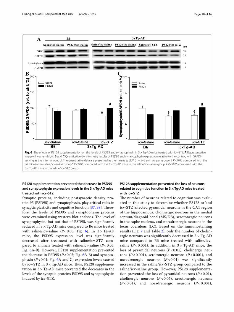
PS128 supplementation prevented the decrease in PSD95 and synaptophysin expression levels in the 3 × Tg‑AD mice treated with icv‑STZSynaptic proteins, including postsynaptic density pro-tein 95 (PSD95) and synaptophysin, play critical roles in synaptic plasticity and cognitive function [37, 38]. There-fore, the levels of PSD95 and synaptophysin proteins were examined using western blot analyses. The level of synaptophysin, but not that of PSD95, was significantly reduced in 3 × Tg-AD mice compared to B6 mice treated with saline/icv-saline (P < 0.05; Fig. 6). In 3 × Tg-AD mice, the PSD95 expression level was significantly decreased after treatment with saline/icv-STZ com-pared to animals treated with saline/icv-saline (P < 0.05; Fig. 6A-B). However, PS128 supplementation prevented the decrease in PSD95 (P < 0.05; Fig. 6A-B) and synapto-physin (P < 0.01; Fig. 6A and C) expression levels caused by icv-STZ in 3 × Tg-AD mice. Thus, PS128 supplemen-tation in 3 × Tg-AD mice prevented the decreases in the levels of the synaptic proteins PSD95 and synaptophysin induced by icv-STZ.
PS128 supplementation prevented the loss of neurons related to cognitive function in 3 × Tg‑AD mice treated with icv‑STZThe number of neurons related to cognition was evalu-ated in this study to determine whether PS128 or/and icv-STZ affected pyramidal neurons in the CA1 region of the hippocampus, cholinergic neurons in the medial septum/diagnoid band (MS/DB), serotonergic neurons in the raphe nucleus, and noradrenergic neurons in the locus coeruleus (LC). Based on the immunostaining results (Fig. 7 and Table 2), only the number of cholin-ergic neurons was significantly decreased in 3 × Tg-AD mice compared to B6 mice treated with saline/icv-saline (P < 0.001). In addition, in 3 × Tg-AD mice, the loss of pyramidal neurons (P < 0.01), cholinergic neu-rons (P < 0.001), serotonergic neurons (P < 0.001), and noradrenergic neurons (P < 0.01) was significantly increased in the saline/icv-STZ group compared to the saline/icv-saline group. However, PS128 supplementa-tion prevented the loss of pyramidal neurons (P < 0.01), cholinergic neurons (P < 0.05), serotonergic neurons (P < 0.01), and noradrenergic neurons (P < 0.001),
Fig. 6 The effects of PS128 supplementation on the levels of PSD95 and synaptophysin in 3 × Tg‑AD mice treated with icv‑STZ. A Representative image of western blots. B and C Quantitative densitometry results of PSD95 and synaptophysin expression relative to the control, with GAPDH serving as the internal control. The quantitative data are presented as the means ± SEM (n = 5–8 animals per group). † P < 0.05 compared with the B6 mice in the saline/icv‑saline group.* P < 0.05 compared with the 3 × Tg‑AD mice in the saline/icv‑saline group. # P < 0.05 compared with the 3 × Tg‑AD mice in the saline/icv‑STZ group

Page 11 of 16Huang et al. BMC Complement Med Ther (2021) 21:259
protecting against the damage induced by icv-STZ in 3 × Tg-AD mice. These results show that PS128 supple-mentation prevented the loss of neurons related to cog-nitive function in 3 × Tg-AD mice treated with icv-STZ.
DiscussionAccording to our findings, the administration of icv-STZ exacerbated the disease progression of AD via gliosis that resulted from an increases in the levels of fecal PPA and the inactive form of GSK3β in 3 × Tg-AD mice. However, PS128 supplementation prevented the deleterious effects of icv-STZ on 3 × Tg-AD mice. In addition, we did not
Fig. 7 The effects of PS128 supplementation on cognition‑related neurons in 3 × Tg‑AD mice treated with icv‑STZ. Representative images of immunostaining of mature pyramidal neurons with NeuN antibodies in the CA1 subregion of the hippocampus (a‑f), cholinergic neurons with ChAT antibodies in the MS/DB (g‑l), serotonergic neurons in the raphe nucleus (m‑r), and noradrenergic neurons in the LC region (s‑x) of different groups. Scale bars = 200 μm for NeuN, 5‑HT, and TH and 500 μm for ChAT. Arrowheads, positive staining signals. Mature neuron (NeuN), serotonergic neuron (5‑HT), noradrenergic neuron (TH), cholinergic neuron (ChAT), medial septum/diagnoid band (MS/DB), and locus coeruleus (LC)
Table 2 Quantification of the numbers of cognition‑related neurons in mice
† Compared to B6 mice in the icv-saline/saline group* Compared to 3xTg-AD mice in the icv-saline/saline group# Compared to 3xTg-AD mice in the icv-STZ/saline group††† P < 0.001
**P < 0.01; ***P < 0.001# P < 0.05; ##P < 0.01; ###P < 0.001
B6 3xTg‑AD
icv‑saline icv‑saline icv‑STZ
Saline PS128 Saline PS128 Saline PS128
NeuN 1299.8 ± 119.4 1576.6 ± 98.1 1356.5 ± 157.2 1266.3 ± 81.3 784.3 ± 109.7** 1305.2 ± 64.0##
ChAT 830.3 ± 68.7 944.7 ± 24.1 547.5 ± 13.3††† 491.0 ± 15.3 234.0 ± 36.5*** 390.7 ± 60.4#
5‑HT 136.0 ± 17.2 107.5 ± 7.8 63.3 ± 29.2 125.3 ± 7.1 46.0 ± 1.6*** 118.3 ± 27.8##
TH 324.9 ± 22.3 271.0 ± 31.8 289.8 ± 33.2 266.5 ± 22.5 162.8 ± 9.2** 296.7 ± 25.5###
Page 12 of 16Huang et al. BMC Complement Med Ther (2021) 21:259
observe a difference in cognitive function between B6 wild-type and 3 × Tg-AD mice, as only a few changes in microgliosis, synaptophysin protein expression lev-els, and cholinergic neurons were observed at 7 months of age. Furthermore, PS128 supplementation alone did not exert significant toxic effects on B6 wild-type mice and only decreased the level of the number of activated microglia in the hippocampus of naïve 3 × Tg-AD mice. Gliosis resulting from PPA and GSK3β was considered one of the possible mechanisms that attenuated disease progression in the AD mouse model. Therefore, PS128 supplementation is a safe and potential preventive strat-egy for AD progression.
In 3 × Tg-AD mice, the administration of icv-STZ induced cognitive dysfunction resulting from gliosis by increasing the levels of GSK3β activity and PPA. In addi-tion, gliosis was associated with increased Aβ deposition, phosphorylation of tau at site T231 (pT231), and BACE1 levels and decreased levels of the postsynaptic pro-tein PSD95 and numbers of cognition-related neurons, including pyramidal neurons in the CA1 subregion of the hippocampus, cholinergic neurons in the MS/DB, sero-tonergic neurons in the raphe nucleus, and noradrenergic neurons in the LC. These findings of the progression of cognitive dysfunction induced by icv-STZ in 3 × Tg-AD mice are consistent with many other studies [27, 39, 40]. In addition, the administration of icv-STZ triggers insulin resistance and then impairs insulin signaling in the brain [41], but the peripheral glucose level was not changed in 3 × Tg-AD mice, as shown in previous study [23]. Insulin resistance is associated with the modulation of amyloid production, tau phosphorylation and neuro-inflammation through the regulation of GSK3β activity [42, 43]. Furthermore, GSK3β activation subsequently leads to a number of changes, among which tau hyper-phosphorylation [44] and increased Aβ production by activated BACE1 [45] play a significant role in AD patho-genesis. The phosphorylation of tau at site T231 precedes phosphorylation at the S202 site in individuals with AD [46, 47], and pT231 was the primary site phosphoryl-ated by GSK3β [48]. In addition, the administration of icv-STZ to 3 × Tg-AD mice increased the expression levels of BACE1, but not AβPP, IDE or NEP. Many stud-ies have also suggested that icv-STZ increases GSK3β activity and subsequently regulates Aβ deposition by modulating BACE1 expression [27, 31, 49, 50]. The sub-stantially increased deposition of Aβ might be mediated by the regulation of both BACE1 and GSK3β activity in the 3 × Tg-AD mice treated with icv-STZ. The deposition of Aβ and phosphorylated tau protein triggers a series of reactions, including gliosis and synaptic dysfunction, resulting in neurodegeneration [51–53]. Accumulating evidence has also indicated that excess PPA crosses the
blood-brain barrier and then induces gliosis [54, 55]. A reduction in the neuron number and increase in gliosis were identified in the PPA-treated brain [56]. Excess PPA exerts many negative effects, including gliosis and ASDs [55, 57–59]. The association described above indicates that increased GSK3β activity and fecal PPA levels might increase gliosis. A previous meta-analysis also reported the loss of cholinergic neurons in the nucleus basalis, ser-otonergic neurons in the raphe nucleus, and noradrener-gic neurons in the LC of patients with AD [60, 61]. Based on the combination of the genetic (3 × Tg-AD mice) and environmental (the type 3 AD) factors, and the pheno-typic features described above, icv-STZ administration to 3 × Tg-AD mice was proven to be a reliable animal model to evaluate the effects and mechanisms of PS128 in AD mice.
Results from a recent randomized, double-blind, pla-cebo-controlled trial of younger (aged 7–15) individuals with autism spectrum disorder (ASD) in Taiwan showed that PS128 ameliorates opposition/defiance behaviors [62]. PS128 also exerts anxiolytic effects on stressed mice [16, 17]. In addition, PS128 is safe for human con-sumption, according to toxicological assessments [63]. In this study, we assessed the effect of PS128 on the icv-STZ-treated 3 × Tg-AD model and showed that long-term supplementation with PS128 prevented cognitive dysfunction induced by icv-STZ in the 3 × Tg-AD male mice. The beneficial effects of PS128 supplementation on attenuating cognitive deficits were consistent with many other probiotic studies [14, 64–67]. Therefore, we suggest that PS128 supplementation is safe and has the potential to prevent cognitive dysfunction in individuals with AD.
PS128 supplementation alters hyperactive and emo-tional behaviors by modulating the levels of monoamine neurotransmitters and systemic inflammation [16, 17, 68]. Researchers have not clearly determined whether changes in metabolites such as SCFAs, the main metabo-lites of gut microbiota, are modulated by the beneficial effect of PS128 on AD. SCFAs are often considered criti-cal candidate mediators of gut-brain communication, and altered SCFA production has been reported in a variety of neuropathologies [69]. In the current study, PS128 sup-plementation prevented the increase in fecal PPA levels induced by icv-STZ in 3 × Tg-AD mice. The results seem inconsistent with some previous evidence suggesting that SCFAs, including acetic acid, butyric acid, and PPA, exerted positive effects on individuals with AD [70, 71]. However, the results of a recent bioinformatics analysis indicated that PPA specifically plays an important role in AD pathogenesis [54]. PPA levels also play an important role in insulin resistance [72] and are increased in sub-jects with type 2 diabetes mellitus [73] due to alterations in the environment of the gut microbiota [74, 75]. PPA

Page 13 of 16Huang et al. BMC Complement Med Ther (2021) 21:259
readily crosses the gut–blood barrier and then enters the central nervous system to impair spatial cognitive behav-ior [76–78]. Furthermore, probiotics restore the normal gut microbiota and protect against damage induced by PPA [79]. Probiotics significantly improve the cogni-tive function of 3 × Tg-AD mice by increasing neuronal activity, attenuating gliosis, mitigating synaptic deficits, and reducing the levels of phosphorylated tau though the inhibition of GSK3β activity [80]. Therefore, the modu-lation of the levels of PPA and GSK3β activity by PS128 supplementation may play a role in preventing cognitive dysfunction and promoting memory consolidation in the 3 × Tg-AD mice treated with icv-STZ, as shown in Fig. 8. However, further studies are needed to elucidate whether the other mechanisms mediating the beneficial effects of PS128 on cognition are involved.
ConclusionsIn summary, the administration of icv-STZ substan-tially increased levels of gliosis resulting from increased GSK3β activity and fecal PPA levels in 3 × Tg-AD mice. PS128 supplementation prevented the damage induced by icv-STZ by reducing the levels of fecal PPA, GSK3β activity, and gliosis in 3 × Tg-AD mice. Therefore, the PS128 supplementation represents a potential strategy to prevent and/or delay the progression of AD.
AbbreviationsPS128: Lactobacillus plantarum PS128; AD: Alzheimer’s disease; CFU: Colony‑forming unit; icv: Intracerebroventricular; STZ: Streptozotocin; o.g.: Oral gavage; Aβ: Amyloid‑β; NFTs: Neurofibrillary tangles; AβPP: Amyloid‑β protein precursor; BACE1: β‑site AβPP‑cleaving enzyme; GSK3β: Glycogen synthase kinase 3 beta; SCFAs: Short‑chain fatty acids; PPA: Propionic acid levels; PSD95: Postsynaptic density protein 95; MS/DB: Medial septum/diagnoid band; ChAT: Choline acetyltransferase; OFT: Open field test; EPM: Elevated plus maze;
Fig. 8 The proposed effect of PS128 supplementation on 3 × Tg‑AD mice treated with icv‑STZ. The icv‑STZ treatment substantially increased gliosis by increasing GSK3β activity and fecal PPA levels, resulting in cognitive dysfunction in 3 × Tg‑AD mice. However, PS128 supplementation prevented the damage induced by icv‑STZ, possibly by reducing PPA levels, GSK3β activity, and gliosis in 3 × Tg‑AD mice, which further ameliorated the behavioral and pathological features of AD
Page 14 of 16Huang et al. BMC Complement Med Ther (2021) 21:259
MWM: Morris water maze; IHC: Immunohistochemistry; DAB: Diaminobenzi‑dine; GFAP: Glial fibrillary acidic protein; Iba1: Ionized calcium binding adaptor molecule 1; LC: Locus coeruleus; TH: Thyroxine hydroxylase; 5‑HT: 5‑hydroxy‑tryptamine; PVDF: Polyvinylidene fluoride; GAPDH: Glyceraldehyde‑3‑phos‑phate dehydrogenase; IDE: Insulin‑degrading enzyme; NEP : Neprilysin.
Supplementary InformationThe online version contains supplementary material available at https:// doi. org/ 10. 1186/ s12906‑ 021‑ 03426‑8.
Additional file 1: Figure S1. The effects of PS128 supplementation on anxiety behavior in 3 × Tg‑AD mice treated with icv‑STZ. Figure S2. The effects of PS128 supplementation on the levels of tau (pS202), IDE, and NEP protein expression in 3 × Tg‑AD mice treated with icv‑STZ. Figure S3. Original Uncropped Western blots for GSK3β related protein. Figure S4. Original Uncropped Western blots for phosphorylated Tau protein. Figure S5. Original Uncropped Western blots for 6E10 protein. Figure S6. Original Uncropped Western blots for BACE1 protein. Figure S7. Original Uncropped Western blots for AβPP protein. Figure S8. Original Uncropped Western blots for PSD95 protein. Figure S9. Original Uncropped Western blots for synaptophysin protein.
AcknowledgmentsWe thank Min‑Yu Chang, Yun‑Fang Cheng, and Chin‑Lin Huang for their assis‑tance in collecting samples from the mice. We also appreciate the assistance provided by the Molecular Imaging Core Facility of National Taiwan Normal University under the auspices of the Ministry of Science and Technology. PS128 was provided by Bened Biomedical Co., Ltd., Taipei, Taiwan.
Authors’ contributionsHJH designed the experiments, wrote the manuscript, and executed the statistical analysis. JLC, YHC, MWC, YYK, and JFL conducted the experiments. CCH provided the experimental materials. YCT designed the experiments and reviewed the manuscript. HMHL designed the experiments, wrote the manuscript, and organized the final version of the manuscript. All authors have reviewed and approved this final manuscript.
FundingFinancial assistance was provided by grants from the Ministry of Science and Technology (MOST; 107‑2320‑B‑003‑007 and 108‑2320‑B‑003‑002) and MacKay Junior College of Medicine, Nursing and Management (MKC107R01 and MKC1070412). The funding bodies had no role in designing the study, the collection, analysis, and interpretation of data, or in writing the manuscript.
Availability of data and materialsThe dataset used and/or analyzed during the current study are available from the corresponding author upon reasonable request.
Declarations
Ethics approval and consent to participateThe animals were maintained and treated in accordance with the principles of the Institutional Animal Care and Use Committee (IACUC) of National Taiwan Normal University, Taipei, Taiwan (Permit Number: 106036).
Consent for publicationNot applicable.
Competing interestsThe authors have no competing interests to declare.
Author details1 Department of Nursing, MacKay Junior College of Medicine, Nursing and Management, Taipei 11260, Taiwan. 2 Department of Life Science, National Taiwan Normal University, Taipei 11677, Taiwan. 3 Institute of Biochemistry and Molecular Biology, National Yang Ming Chiao Tung University, Tai‑pei 11221, Taiwan. 4 Bened Biomedical Co., Ltd., Taipei 10448, Taiwan.
Received: 5 January 2021 Accepted: 27 September 2021
References 1. Au A, Feher A, Mcphee L, Jessa A, Oh S, Einstein G. Estrogens, inflamma‑
tion and cognition. Front Neuroendocrinol. 2016;40:87–100. https:// doi. org/ 10. 1016/j. yfrne. 2016. 01. 002.
2. Lane CA, Hardy J, Schott JM. Alzheimer’s disease. Eur J Neurol. 2018;25(1):59–70. https:// doi. org/ 10. 1111/ ene. 13439.
3. Nebel RA, Aggarwal NT, Barnes LL, Gallagher A, Goldstein JM, Kantarci K, et al. Understanding the impact of sex and gender in Alzheimer’s disease: a call to action. Alzheimers Dement. 2018;14(9):1171–83. https:// doi. org/ 10. 1016/j. jalz. 2018. 04. 008.
4. Scheltens P, Blennow K, Breteler MM, De Strooper B, Frisoni GB, Salloway S, et al. Alzheimer’s disease. Lancet. 2016;388(10043):505–17. https:// doi. org/ 10. 1016/ S0140‑ 6736(15) 01124‑1.
5. Cummings JL, Morstorf T, Zhong K. Alzheimer’s disease drug‑develop‑ment pipeline: few candidates, frequent failures. Alzheimers Res Ther. 2014;6(4):37. https:// doi. org/ 10. 1186/ alzrt 269.
6. Bostanciklioglu M. The role of gut microbiota in pathogenesis of Alzhei‑mer’s disease. J Appl Microbiol. 2019;127(4):954–67. https:// doi. org/ 10. 1111/ jam. 14264.
7. Cryan JF, O’riordan KJ, Cowan CSM, Sandhu KV, Bastiaanssen TFS, Boehme M, et al. The microbiota‑gut‑brain axis. Physiol Rev. 2019;99(4):1877–2013. https:// doi. org/ 10. 1152/ physr ev. 00018. 2018.
8. Li B, He Y, Ma J, Huang P, Du J, Cao L, et al. Mild cognitive impairment has similar alterations as Alzheimer’s disease in gut microbiota. Alzheimers Dement. 2019;15(10):1357–66. https:// doi. org/ 10. 1016/j. jalz. 2019. 07. 002.
9. Zilberter Y, Zilberter M. The vicious circle of hypometabolism in neuro‑degenerative diseases: ways and mechanisms of metabolic correction. J Neurosci Res. 2017;95(11):2217–35. https:// doi. org/ 10. 1002/ jnr. 24064.
10. Caracciolo B, Xu W, Collins S, Fratiglioni L. Cognitive decline, dietary fac‑tors and gut‑brain interactions. Mech Ageing Dev. 2014;136‑137:59–69. https:// doi. org/ 10. 1016/j. mad. 2013. 11. 011.
11. Erny D, Hrabe De Angelis AL, Jaitin D, Wieghofer P, Staszewski O, David E, et al. Host microbiota constantly control maturation and function of microglia in the CNS. Nat Neurosci. 2015;18(7):965–77. https:// doi. org/ 10. 1038/ nn. 4030.
12. Akbari E, Asemi Z, Daneshvar Kakhaki R, Bahmani F, Kouchaki E, Tamtaji OR, et al. Effect of probiotic supplementation on cognitive function and metabolic status in Alzheimer’s disease: a randomized, double‑blind and controlled trial. Front Aging Neurosci. 2016;8:256. https:// doi. org/ 10. 3389/ fnagi. 2016. 00256.
13. O’hagan C, Li JV, Marchesi JR, Plummer S, Garaiova I, Good MA. Long‑term multi‑species Lactobacillus and Bifidobacterium dietary supplement enhances memory and changes regional brain metabolites in middle‑aged rats. Neurobiol Learn Mem. 2017;144:36–47. https:// doi. org/ 10. 1016/j. nlm. 2017. 05. 015.
14. Nimgampalle M, Kuna Y. Anti‑alzheimer properties of probiotic, Lactoba‑cillus plantarum MTCC 1325 in Alzheimer’s disease induced albino rats. J Clin Diagn Res. 2017;11(8):KC01–5. https:// doi. org/ 10. 7860/ JCDR/ 2017/ 26106. 10428.
15. Woo JY, Gu W, Kim KA, Jang SE, Han MJ, Kim DH. Lactobacillus pentosus var. plantarum C29 ameliorates memory impairment and inflammaging in a D‑galactose‑induced accelerated aging mouse model. Anaerobe. 2014;27:22–6. https:// doi. org/ 10. 1016/j. anaer obe. 2014. 03. 003.
16. Liu WH, Chuang HL, Huang YT, Wu CC, Chou GT, Wang S, et al. Alteration of behavior and monoamine levels attributable to Lactobacillus plan‑tarum PS128 in germ‑free mice. Behav Brain Res. 2016;298(Pt B):202–9. https:// doi. org/ 10. 1016/j. bbr. 2015. 10. 046.
17. Liu YW, Liu WH, Wu CC, Juan YC, Wu YC, Tsai HP, et al. Psychotropic effects of Lactobacillus plantarum PS128 in early life‑stressed and naive adult mice. Brain Res. 2016;1631:1–12. https:// doi. org/ 10. 1016/j. brain res. 2015. 11. 018.
18. Liao JF, Cheng YF, Li SW, Lee WT, Hsu CC, Wu CC, et al. Lactobacillus plan‑tarum PS128 ameliorates 2,5‑Dimethoxy‑4‑iodoamphetamine‑induced tic‑like behaviors via its influences on the microbiota‑gut‑brain‑axis. Brain Res Bull. 2019;153:59–73. https:// doi. org/ 10. 1016/j. brain resbu ll. 2019. 07. 027.
Page 15 of 16Huang et al. BMC Complement Med Ther (2021) 21:259
19. Bekkering P, Jafri I, Van Overveld FJ, Rijkers GT. The intricate association between gut microbiota and development of type 1, type 2 and type 3 diabetes. Expert Rev Clin Immunol. 2013;9(11):1031–41. https:// doi. org/ 10. 1586/ 17446 66X. 2013. 848793.
20. Steen E, Terry BM, Rivera EJ, Cannon JL, Neely TR, Avares R, et al. Impaired insulin and insulin‑like growth factor expression and signaling mecha‑nisms in Alzheimer’s disease‑‑is this type 3 diabetes? J Alzheimers Dis. 2005;7(1):63–80. https:// doi. org/ 10. 3349/ ymj. 2017. 58.3. 479.
21. Mosconi L, Sorbi S, De Leon MJ, NB LY, Myoung PS, Tsui W, et al. Hypo‑metabolism exceeds atrophy in presymptomatic early‑onset familial Alzheimer’s disease. J Nucl Med. 2006;47(11):1778–86.
22. Dash SK. Cognitive impairment and diabetes. Recent Pat Endocr Metab Immune Drug Discov. 2013;7(2):155–65. https:// doi. org/ 10. 2174/ 18722 14811 30702 0009.
23. Grieb P. Intracerebroventricular streptozotocin injections as a model of Alzheimer’s disease: in search of a relevant mechanism. Mol Neurobiol. 2016;53(3):1741–52. https:// doi. org/ 10. 1007/ s12035‑ 015‑ 9132‑3.
24. Leibson CL, Rocca WA, Hanson VA, Cha R, Kokmen E, O’brien PC, et al. Risk of dementia among persons with diabetes mellitus: a population‑based cohort study. Am J Epidemiol. 1997;145(4):301–8. https:// doi. org/ 10. 1093/ oxfor djour nals. aje. a0091 06.
25. Xu J, Begley P, Church SJ, Patassini S, Mcharg S, Kureishy N, et al. Elevation of brain glucose and polyol‑pathway intermediates with accompanying brain‑copper deficiency in patients with Alzheimer’s disease: metabolic basis for dementia. Sci Rep. 2016;6:27524. https:// doi. org/ 10. 1038/ srep2 7524.
26. Chen Y, Liang Z, Tian Z, Blanchard J, Dai CL, Chalbot S, et al. Intracer‑ebroventricular streptozotocin exacerbates Alzheimer‑like changes of 3xTg‑AD mice. Mol Neurobiol. 2014;49(1):547–62. https:// doi. org/ 10. 1007/ s12035‑ 013‑ 8539‑y.
27. Huang HJ, Chen SL, Chang YT, Chyuan JH, Hsieh‑Li HM. Administration of Momordica charantia enhances the neuroprotection and reduces the side effects of LiCl in the treatment of Alzheimer’s disease. Nutrients. 2018;10(12). https:// doi. org/ 10. 3390/ nu101 21888.
28. Huang HJ, Chen SL, Hsieh‑Li HM. Administration of NaHS attenuates footshock‑induced pathologies and emotional and cognitive dysfunction in triple transgenic Alzheimer’s mice. Front Behav Neurosci. 2015;9:312. https:// doi. org/ 10. 3389/ fnbeh. 2015. 00312.
29. Huang HJ, Huang HY, Hsieh‑Li HM. MGCD0103, a selective histone deacetylase inhibitor, coameliorates oligomeric Abeta25–35 ‑induced anxiety and cognitive deficits in a mouse model. CNS Neurosci Ther. 2019;25(2):175–86. https:// doi. org/ 10. 1111/ cns. 13029.
30. Garcia‑Villalba R, Gimenez‑Bastida JA, Garcia‑Conesa MT, Tomas‑Barberan FA, Carlos Espin J, Larrosa M. Alternative method for gas chromatography‑mass spectrometry analysis of short‑chain fatty acids in faecal samples. J Sep Sci. 2012;35(15):1906–13. https:// doi. org/ 10. 1002/ jssc. 20110 1121.
31. Xu ZP, Gan GS, Liu YM, Xiao JS, Liu HX, Mei B, et al. Adiponectin attenu‑ates streptozotocin‑induced tau hyperphosphorylation and cognitive deficits by rescuing PI3K/Akt/GSK‑3beta pathway. Neurochem Res. 2018;43(2):316–23. https:// doi. org/ 10. 1007/ s11064‑ 017‑ 2426‑2.
32. Essawy AE, Abdou HM, Ibrahim HM, Bouthahab NM. Soybean isoflavone ameliorates cognitive impairment, neuroinflammation, and amyloid β accumulation in a rat model of Alzheimer’s disease. Environ Sci Pollut Res. 2019. https:// doi. org/ 10. 1007/ s11356‑ 019‑ 05862‑z.
33. Gilbert BJ. The role of amyloid β in the pathogenesis of Alzheimer’s disease. J Clin Pathol. 2013;66(5):362. https:// doi. org/ 10. 1136/ jclin path‑ 2013‑ 201515.
34. Yamamoto N, Fujii Y, Kasahara R, Tanida M, Ohora K, Ono Y, et al. Simv‑astatin and atorvastatin facilitates amyloid beta‑protein degradation in extracellular spaces by increasing neprilysin secretion from astrocytes through activation of MAPK/Erk1/2 pathways. Glia. 2016;64(6):952–62. https:// doi. org/ 10. 1002/ glia. 22974.
35. Birch AM, Katsouri L, Sastre M. Modulation of inflammation in transgenic models of Alzheimer’s disease. J Neuroinflammation. 2014;11:25. https:// doi. org/ 10. 1186/ 1742‑ 2094‑ 11‑ 25.
36. Wyss‑Coray T. Inflammation in Alzheimer disease: driving force, bystander or beneficial response? Nat Med. 2006;12(9):1005–15. https:// doi. org/ 10. 1038/ nm1484.
37. Ju IG, Kim N, Choi JG, Lee JK, Oh MS. Cuscutae Japonicae semen amelio‑rates memory dysfunction by rescuing synaptic damage in Alzheimer’s
disease models. Nutrients. 2019;11(11). https:// doi. org/ 10. 3390/ nu111 12591.
38. Nelson PT, Alafuzoff I, Bigio EH, Bouras C, Braak H, Cairns NJ, et al. Correla‑tion of Alzheimer disease neuropathologic changes with cognitive status: a review of the literature. J Neuropathol Exp Neurol. 2012;71(5):362–81. https:// doi. org/ 10. 1097/ NEN. 0b013 e3182 5018f7.
39. Mehla J, Pahuja M, Gupta YK. Streptozotocin‑induced sporadic Alzheimer’s disease: selection of appropriate dose. J Alzheimers Dis. 2013;33(1):17–21. https:// doi. org/ 10. 3233/ JAD‑ 2012‑ 120958.
40. Salkovic‑Petrisic M, Osmanovic J, Grünblatt E, Riederer P, Hoyer S. Modeling sporadic Alzheimer’s disease: the insulin resistant brain state generates multiple long‑term morphobiological abnormalities including hyperphosphorylated tau protein and amyloid‑beta. J Alzheimers Dis. 2009;18(4):729–50. https:// doi. org/ 10. 3233/ JAD‑ 2009‑ 1184.
41. Salkovic‑Petrisic M, Knezovic A, Hoyer S, Riederer P. What have we learned from the streptozotocin‑induced animal model of sporadic Alzheimer’s disease, about the therapeutic strategies in Alzheimer’s research. J Neural Transm (Vienna). 2013;120(1):233–52. https:// doi. org/ 10. 1007/ s00702‑ 012‑ 0877‑9.
42. Jope RS, Yuskaitis CJ, Beurel E. Glycogen synthase kinase‑3 (GSK3): inflam‑mation, diseases, and therapeutics. Neurochem Res. 2007;32(4–5):577–95. https:// doi. org/ 10. 1007/ s11064‑ 006‑ 9128‑5.
43. Yang W, Ma J, Liu Z, Lu Y, Hu B, Yu H. Effect of naringenin on brain insulin signaling and cognitive functions in ICV‑STZ induced dementia model of rats. Neurol Sci. 2014;35(5):741–51. https:// doi. org/ 10. 1007/ s10072‑ 013‑ 1594‑3.
44. Ishiguro K, Shiratsuchi A, Sato S, Omori A, Arioka M, Kobayashi S, et al. Glycogen synthase kinase 3 beta is identical to tau protein kinase I generating several epitopes of paired helical filaments. FEBS Lett. 1993;325(3):167–72. https:// doi. org/ 10. 1016/ 0014‑ 5793(93) 81066‑9.
45. Ly PT, Wu Y, Zou H, Wang R, Zhou W, Kinoshita A, et al. Inhibition of GSK3β‑mediated BACE1 expression reduces Alzheimer‑associated phe‑notypes. J Clin Invest. 2013;123(1):224–35. https:// doi. org/ 10. 1172/ JCI64 516.
46. Augustinack JC, Schneider A, Mandelkow EM, Hyman BT. Specific tau phosphorylation sites correlate with severity of neuronal cytopathology in Alzheimer’s disease. Acta Neuropathol. 2002;103(1):26–35. https:// doi. org/ 10. 1007/ s0040 10100 423.
47. Luna‑Munoz J, Garcia‑Sierra F, Falcon V, Menendez I, Chavez‑Macias L, Mena R. Regional conformational change involving phosphorylation of tau protein at the Thr231, precedes the structural change detected by Alz‑50 antibody in Alzheimer’s disease. J Alzheimers Dis. 2005;8(1):29–41. https:// doi. org/ 10. 3233/ jad‑ 2005‑ 8104.
48. Lin YT, Cheng JT, Liang LC, Ko CY, Lo YK, Lu PJ. The binding and phos‑phorylation of Thr231 is critical for Tau’s hyperphosphorylation and functional regulation by glycogen synthase kinase 3beta. J Neurochem. 2007;103(2):802–13. https:// doi. org/ 10. 1111/j. 1471‑ 4159. 2007. 04792.x.
49. Ly PT, Wu Y, Zou H, Wang R, Zhou W, Kinoshita A, et al. Inhibition of GSK3beta‑mediated BACE1 expression reduces Alzheimer‑associated phenotypes. J Clin Invest. 2013;123(1):224–35. https:// doi. org/ 10. 1172/ JCI64 516.
50. Zhou D, Liu H, Li C, Wang F, Shi Y, Liu L, et al. Atorvastatin ameliorates cog‑nitive impairment, Abeta1–42 production and Tau hyperphosphorylation in APP/PS1 transgenic mice. Metab Brain Dis. 2016;31(3):693–703. https:// doi. org/ 10. 1007/ s11011‑ 016‑ 9803‑4.
51. Ahmad A, Ali T, Park HY, Badshah H, Rehman SU, Kim MO. Neuroprotec‑tive effect of fisetin against amyloid‑beta‑induced cognitive/synaptic dysfunction, neuroinflammation, and neurodegeneration in adult mice. Mol Neurobiol. 2017;54(3):2269–85. https:// doi. org/ 10. 1007/ s12035‑ 016‑ 9795‑4.
52. Craft JM, Watterson DM, Marks A, Van Eldik LJ. Enhanced susceptibility of S‑100B transgenic mice to neuroinflammation and neuronal dysfunction induced by intracerebroventricular infusion of human beta‑amyloid. Glia. 2005;51(3):209–16. https:// doi. org/ 10. 1002/ glia. 20194.
53. Oakley H, Cole SL, Logan S, Maus E, Shao P, Craft J, et al. Intraneuronal beta‑amyloid aggregates, neurodegeneration, and neuron loss in trans‑genic mice with five familial Alzheimer’s disease mutations: potential factors in amyloid plaque formation. J Neurosci. 2006;26(40):10129–40. https:// doi. org/ 10. 1523/ JNEUR OSCI. 1202‑ 06. 2006.
54. Aliashrafi M, Nasehi M, Zarrindast M‑R, Joghataei MT, Zali H, Siadat SD. Association of microbiota‑derived propionic acid and Alzheimer’s

Page 16 of 16Huang et al. BMC Complement Med Ther (2021) 21:259
• fast, convenient online submission
•
thorough peer review by experienced researchers in your field
• rapid publication on acceptance
• support for research data, including large and complex data types
•
gold Open Access which fosters wider collaboration and increased citations
maximum visibility for your research: over 100M website views per year •
At BMC, research is always in progress.
Learn more biomedcentral.com/submissions
Ready to submit your researchReady to submit your research ? Choose BMC and benefit from: ? Choose BMC and benefit from:
disease; bioinformatics analysis. J Diabetes Metab Disord. 2020. https:// doi. org/ 10. 1007/ s40200‑ 020‑ 00564‑7.
55. Al‑Lahham SH, Peppelenbosch MP, Roelofsen H, Vonk RJ, Venema K. Biological effects of propionic acid in humans; metabolism, poten‑tial applications and underlying mechanisms. Biochim Biophys Acta. 2010;1801(11):1175–83. https:// doi. org/ 10. 1016/j. bbalip. 2010. 07. 007.
56. Lobzhanidze G, Lordkipanidze T, Zhvania M, Japaridze N, Macfabe DF, Pochkidze N, et al. Effect of propionic acid on the morphology of the amygdala in adolescent male rats and their behavior. Micron. 2019;125:102732. https:// doi. org/ 10. 1016/j. micron. 2019. 102732.
57. Al‑Owain M, Kaya N, Al‑Shamrani H, Al‑Bakheet A, Qari A, Al‑Muaigl S, et al. Autism spectrum disorder in a child with propionic acidemia. JIMD Rep. 2013;7:63–6. https:// doi. org/ 10. 1007/ 8904_ 2012_ 143.
58. Macfabe DF. Short‑chain fatty acid fermentation products of the gut microbiome: implications in autism spectrum disorders. Microb Ecol Health Dis. 2012;23. https:// doi. org/ 10. 3402/ mehd. v23i0. 19260.
59. Wajner M, Latini A, Wyse AT, Dutra‑Filho CS. The role of oxidative damage in the neuropathology of organic acidurias: insights from animal studies. J Inherit Metab Dis. 2004;27(4):427–48. https:// doi. org/ 10. 1023/B: BOLI. 00000 37353. 13085. e2.
60. Lyness SA, Zarow C, Chui HC. Neuron loss in key cholinergic and aminergic nuclei in Alzheimer disease: a meta‑analysis. Neurobiol Aging. 2003;24(1):1–23. https:// doi. org/ 10. 1016/ s0197‑ 4580(02) 00057‑x.
61. Selkoe DJ. Alzheimer’s disease is a synaptic failure. Science. 2002;298(5594):789–91. https:// doi. org/ 10. 1126/ scien ce. 10740 69.
62. Liu YW, Liong MT, Chung YE, Huang HY, Peng WS, Cheng YF, et al. Effects of Lactobacillus plantarum PS128 on children with autism spectrum disorder in Taiwan: a randomized, double‑blind, placebo‑controlled trial. Nutrients. 2019;11(4). https:// doi. org/ 10. 3390/ nu110 40820.
63. Liao PL, Wu CC, Chen TY, Tsai YC, Peng WS, Yang DJ, et al. Toxicity studies of Lactobacillus plantarum PS128(TM) isolated from spontaneously fermented mustard greens. Foods. 2019;8(12). https:// doi. org/ 10. 3390/ foods 81206 68.
64. Abraham D, Feher J, Scuderi GL, Szabo D, Dobolyi A, Cservenak M, et al. Exercise and probiotics attenuate the development of Alzheimer’s dis‑ease in transgenic mice: role of microbiome. Exp Gerontol. 2019;115:122–31. https:// doi. org/ 10. 1016/j. exger. 2018. 12. 005.
65. Bonfili L, Cecarini V, Cuccioloni M, Angeletti M, Berardi S, Scarpona S, et al. SLAB51 probiotic formulation activates SIRT1 pathway promoting antioxi‑dant and neuroprotective effects in an AD mouse model. Mol Neurobiol. 2018;55(10):7987–8000. https:// doi. org/ 10. 1007/ s12035‑ 018‑ 0973‑4.
66. Shukla PK, Delotterie DF, Xiao J, Pierre JF, Rao R, Mcdonald MP, et al. Alterations in the gut‑microbial‑inflammasome‑brain axis in a mouse model of Alzheimer’s disease. Cells. 2021;10(4). https:// doi. org/ 10. 3390/ cells 10040 779.
67. Xu HM, Huang HL, Zhou YL, Zhao HL, Xu J, Shou DW, et al. Fecal micro‑biota transplantation: a new therapeutic attempt from the gut to the brain. Gastroenterol Res Pract. 2021;2021:6699268. https:// doi. org/ 10. 1155/ 2021/ 66992 68.
68. Liao JF, Cheng YF, You ST, Kuo WC, Huang CW, Chiou JJ, et al. Lactoba‑cillus plantarum PS128 alleviates neurodegenerative progression in 1‑methyl‑4‑phenyl‑1,2,3,6‑tetrahydropyridine‑induced mouse models of Parkinson’s disease. Brain Behav Immun. 2020;90:26–46. https:// doi. org/ 10. 1016/j. bbi. 2020. 07. 036.
69. Silva YP, Bernardi A, Frozza RL. The role of short‑chain fatty acids from gut microbiota in gut‑brain communication. Front Endocrinol (Lausanne). 2020;11:25. https:// doi. org/ 10. 3389/ fendo. 2020. 00025.
70. Ho L, Ono K, Tsuji M, Mazzola P, Singh R, Pasinetti GM. Protective roles of intestinal microbiota derived short chain fatty acids in Alzheimer’s disease‑type beta‑amyloid neuropathological mechanisms. Expert Rev Neurother. 2018;18(1):83–90. https:// doi. org/ 10. 1080/ 14737 175. 2018. 14009 09.
71. Zhang L, Wang Y, Xiayu X, Shi C, Chen W, Song N, et al. Altered gut microbiota in a mouse model of Alzheimer’s disease. J Alzheimers Dis. 2017;60(4):1241–57. https:// doi. org/ 10. 3233/ JAD‑ 170020.
72. Tirosh A, Calay ES, Tuncman G, Claiborn KC, Inouye KE, Eguchi K, et al. The short‑chain fatty acid propionate increases glucagon and FABP4 production, impairing insulin action in mice and humans. Sci Transl Med. 2019;11(489). https:// doi. org/ 10. 1126/ scitr anslm ed. aav01 20.
73. Koh A, Molinaro A, Stahlman M, Khan MT, Schmidt C, Manneras‑Holm L, et al. Microbially produced imidazole propionate impairs insulin signaling through mTORC1. Cell. 2018;175(4):947–961.e17. https:// doi. org/ 10. 1016/j. cell. 2018. 09. 055.
74. Karlsson FH, Tremaroli V, Nookaew I, Bergstrom G, Behre CJ, Fagerberg B, et al. Gut metagenome in European women with normal, impaired and diabetic glucose control. Nature. 2013;498(7452):99–103. https:// doi. org/ 10. 1038/ natur e12198.
75. Larsen N, Vogensen FK, Van Den Berg FW, Nielsen DS, Andreasen AS, Pedersen BK, et al. Gut microbiota in human adults with type 2 diabetes differs from non‑diabetic adults. PLoS One. 2010;5(2):e9085. https:// doi. org/ 10. 1371/ journ al. pone. 00090 85.
76. Mepham JR, Boon FH, Foley KA, Cain DP, Macfabe DF, Ossenkopp KP. Impaired spatial cognition in adult rats treated with multiple intrac‑erebroventricular (ICV) infusions of the enteric bacterial metabolite, propionic acid, and return to baseline after 1 week of no treatment: contribution to a rodent model of ASD. Neurotox Res. 2019;35(4):823–37. https:// doi. org/ 10. 1007/ s12640‑ 019‑ 0002‑z.
77. Mirza R, Sharma B. Selective modulator of peroxisome proliferator‑activated receptor‑alpha protects propionic acid induced autism‑like phenotypes in rats. Life Sci. 2018;214:106–17. https:// doi. org/ 10. 1016/j. lfs. 2018. 10. 045.
78. Karuri AR, Dobrowsky E, Tannock IF. Selective cellular acidification and toxicity of weak organic acids in an acidic microenvironment. Br J Cancer. 1993;68(6):1080–7. https:// doi. org/ 10. 1038/ bjc. 1993. 485.
79. El‑Ansary A, Bacha AB, Bjorklund G, Al‑Orf N, Bhat RS, Moubayed N, et al. Probiotic treatment reduces the autistic‑like excitation/inhibition imbal‑ance in juvenile hamsters induced by orally administered propionic acid and clindamycin. Metab Brain Dis. 2018;33(4):1155–64. https:// doi. org/ 10. 1007/ s11011‑ 018‑ 0212‑8.
80. Zhang ZH, Wen L, Wu QY, Chen C, Zheng R, Liu Q, et al. Long‑term dietary supplementation with selenium‑enriched yeast improves cognitive impairment, reverses synaptic deficits, and mitigates tau pathology in a triple transgenic mouse model of Alzheimer’s disease. J Agric Food Chem. 2017;65(24):4970–9. https:// doi. org/ 10. 1021/ acs. jafc. 7b014 65.
Publisher’s NoteSpringer Nature remains neutral with regard to jurisdictional claims in pub‑lished maps and institutional affiliations.