Responses to Suppress Colorectal Adenocarcinoma Limonin ...
15
Page 1/15 Limonin Modulated Immune and Inammatory Responses to Suppress Colorectal Adenocarcinoma in Mice Model Nur Iliyani Mohd Ishak Universiti Putra Malaysia Institute of Bioscience: University Putra Malaysia Institut Biosains SUHAILA MOHAMED ( [email protected] ) University Putra Malaysia Institut Biosains https://orcid.org/0000-0002-2954-3821 Iffah Nadhira Madzuki Universiti Putra Malaysia Institute of Bioscience: University Putra Malaysia Institut Biosains Noordin Mohamed Mustapha Putra University Malaysia: Universiti Putra Malaysia Norhaizan Mohd Esa Putra University Malaysia: Universiti Putra Malaysia Research Article Keywords: Limonin, Colorectal cancer, inammation, immune responses, avonoid Posted Date: April 19th, 2021 DOI: https://doi.org/10.21203/rs.3.rs-367298/v1 License: This work is licensed under a Creative Commons Attribution 4.0 International License. Read Full License
Transcript of Responses to Suppress Colorectal Adenocarcinoma Limonin ...
Page 1/15
Limonin Modulated Immune and In�ammatoryResponses to Suppress Colorectal Adenocarcinomain Mice ModelNur Iliyani Mohd Ishak
Universiti Putra Malaysia Institute of Bioscience: University Putra Malaysia Institut BiosainsSUHAILA MOHAMED ( [email protected] )
University Putra Malaysia Institut Biosains https://orcid.org/0000-0002-2954-3821Iffah Nadhira Madzuki
Universiti Putra Malaysia Institute of Bioscience: University Putra Malaysia Institut BiosainsNoordin Mohamed Mustapha
Putra University Malaysia: Universiti Putra MalaysiaNorhaizan Mohd Esa
Putra University Malaysia: Universiti Putra Malaysia
Research Article
Keywords: Limonin, Colorectal cancer, in�ammation, immune responses, �avonoid
Posted Date: April 19th, 2021
DOI: https://doi.org/10.21203/rs.3.rs-367298/v1
License: This work is licensed under a Creative Commons Attribution 4.0 International License. Read Full License
Page 2/15
AbstractPurpose. In�ammation and compromised immune responses often increases colorectal cancer (CRC)risk. The immune-modulating effects of limonin on carcinogen/in�ammation-induced colorectal cancer(CRC) were studied in mice.
Methods: Male Balb/c mice were randomly assorted into three groups (n=6): healthy control, non-treatedCRC-induced (azoxymethane/dextran-sulfate-sodium AOM/DSS) control, and CRC-induced+50 mglimonin /kg body-weight. The CRC development were monitored via macroscopic, histopathological,ELISA and mRNA expression analysis.
Results: Limonin downregulated in�ammation (TNF-α, tumor necrosis factor-α), enhanced the adaptiveimmune responses (CD8, CD4, and CD19), and upregulated antioxidant defense (Nrf2, SOD2) mRNAexpressions. Limonin reduced serum malondialdehyde (MDA, lipid peroxidation biomarker),prostaglandin-E2 and histopathology in�ammation scores, while increasing reduced-glutathione (GSH) inthe CRC-induced mice. Limonin signi�cantly (p<0.05) increased T cells (CD4 and CD8) and B cells (CD19)in spleen tissues. The CD335 (natural killer cells) were increased in the CRC-induced mice and limonintreatment restored it to normal levels suggesting reinstatement to normal colon conditions.
Conclusion: Limonin apparently mitigated CRC development, by ameliorating adaptive immuneresponses (CD8, CD4, and CD19), reducing in�ammation (serum prostaglandin-E2; TNF-α, innate immuneresponses), oxidative stress and enhancing the endogenous anti-oxidation defense reactions (GSH) in theCRC-induced mice.
IntroductionColorectal cancer (CRC) is amongst the top three killer cancer worldwide, second most common cancerfor men (10.9% of the total cancer cases) and third in women (9.5% of the total cancer cases) (Arnold etal. 2017). Chronic in�ammatory bowel disease increases CRC risk (Axelrad et al. 2016). Colitis-associated-cancer (CAC) is a serious in�ammatory bowel disease (IBD) complications which canprogress from the chronic in�amed mucosa to dysplasia and ultimately, colorectal cancer. Surgery,neoadjuvant chemotherapy and radiotherapy are the most common therapy used with many painful andundesirable side effects. The FDA approved chemotherapeutical drug for CRC includes Capecitabine,Fluorouracil (5-FU), Irinotecan, Oxaliplatin, Tri�uridine/tipiricil. Although chemotherapy is the foremostselected therapy, it usually causes nausea, vomiting, diarrhea, neuropathy, mouth sores, fatigue, hair loss,increased infection risks and often extend the lifespan for only a short time.
Dietary limonin was reported to signi�cantly reduce the incidence of colonic adenocarcinoma inazoxymethane-initiated CRC-induced rats (Tanaka et al. 2001). Limonin was also reported to causeapoptosis and inhibit colon adenocarcinoma cells proliferation (Chidambara Murthy et al. 2011). Limoninwas previously reported to have anti-cancer and preneoplastic lesions suppressive effects in vivo(Shimizu et al., 2015). To the best of our knowledge, there is to date not much animal study reports on the
Page 3/15
immuno-modulatory effects of limonin against colorectal cancer development. Previously, Tanaka et al.,(2001) studied the effects of citrus limonoids obacunone and limonin on azoxymethane (AOM)-inducedcolon tumorigenesis in male F344 rats. In this study, we examined the effects of limonin on in�ammation,mRNA expressions and immune related responses in colorectal adenocarcinoma mice usingAOM/dextran sulfate sodium (DSS) for the adenocarcinoma induction. This study provides indicationsthat dietary limonin suppressed colon adenocarcinoma, at least in part by enhancing immune, anti-in�ammatory and endogenous anti-oxidant responses in Balb/c mice.
Materials And MethodsChemicals and drugs
Azoxymethane (AOM) and dextran sulfate sodium (DSS) salt (Mr ~40,000) were purchased from theSigma-Aldrich (St. Louis, MO, USA). Limonin (95% purity) were obtained from Xi'an Xin Sheng Bio-chemCo.,Ltd. (Xi'an city,Shaanxi province, China).
Animals
Five weeks old male Balb/c mice (25-30 g) were purchased from the Animal Resource Unit, Faculty ofVeterinary Medicine, Universiti Putra Malaysia (Serdang, Selangor, Malaysia), given commercial rat chow(Gold Coin, Malaysia) and tap water ad libitum, in plastic cages (3 mice/cage) with a 12-h light-dark cycleat room temperature. Male mice were used because males are more prone to CRC than females, andfemales may be in�uenced by �uctuating hormones or estrus cycle. The animal study protocol wasapproved by the Institutional Animal Care and Use Committee (IACUC), Universiti Putra Malaysia(UPM/IACUC/AUP-R069/2017).
Experimental design
Mice were divided into three groups (n=6) G1: healthy control, G2: non-treated CRC-induced(Azoxymethane/Dextran Sulfate Sodium AOM/DSS) control, G3: CRC-induced + 50 mg Limonin /kg body-weight. The dose for limonin was chosen based on previous studies (Shimizu et al., 2015).(The CRC was induced by a single intraperitoneal injection of azoxymethane AOM (10 mg/kg bodyweight) to the mice in groups 2 and 3. After a week of AOM injection, mice received 2% dextran sulfate-sodium (DSS) in the drinking water for 7 days, followed by regular drinking water for recovery thereafter(Tanaka et al. 2000). Limonin were completely homogenized in distilled water (0.25 mg/ml) and given asdrinking water to G3. The average daily Balb/c mice drinking water intake was 0.16 ml/g body weight or5±1 ml/mouse/day. The similar (insigni�cantly different) progressive body weight gains indicated thatthe mice consumed about the same amount of food and water throughout the 20 weeks experimentalduration after the CRC-induction (AOM/DSS). Oral gavage was not used to avoid any additional stress tothe mice, which may increase their mortality. Limonin did not seem to affect the water or food intake ofthe mice in the treatment group.
Page 4/15
Mice were weighed monthly and the disease activity index (DAI) was scored based on the calculated sumof the individual scores of stool consistency and blood in stool as follows: stool consistency score = 0:normal, 2: loose, 4: diarrhea; blood in stool score = 0: normal, 2: reddish, 4: bloody (Li, Shen and Luo,2016). Following an intraperitoneal injection of ketamine: xylazine (100 mg/kg:10 mg/kg), the mice weresacri�ced by exsanguination (intracardiac puncture) 17 weeks post-treatment. The intracardiac punctureallowed for su�cient blood to be collected for the subsequent biochemical analysis. The blood wasallowed to clot for 30 min, centrifuged at 3000 g for 10 min at 4°C, and the serum stored at -20°C untilanalyses. Colon and spleen were collected and either stored at -80ºC or �xed in 10% formalin for furtheruse.
Macroscopy and histopathology
The colon of each mouse was cut, cleaned, colon length measured and tumor incidence/numbers wascounted. The colon length was measured using a ruler, measured and recorded to the nearest cm. Thetumor incidence (%) was determined as the percentage of mice having at least one tumor when examinedunder the macroscope. Colon samples were �xed in 10% buffered formalin for 24 h and handled throughautomated programmed tissue processing machine. The tissue was embedded in para�n and tissueblocks sectioned at thickness of 5 µm and stained with hematoxylin and eosin (H&E) for lightmicroscopic examination. Due to the short CRC developmental period, only a few tumors were present ineach AOM/DSS mouse. The in�ammation score was graded as previously described (Saadatdoust et al.2015). The in�ammation thickness ranged from 0 to 3 (0 = no in�ammation, 1 = mucosa, 2 = mucosaplus submucosa and 3 = transmural). The thickness of an in�ammation is measured throughout thelayers of the colon wall, such as the mucosa, sub-mucosa or transmural tissues. The in�ammation scorewas calculated based on the average scores from 0 to 3 (0 - no in�ammation; 1 – mild (in�ltration ofin�ammatory cells into the mucosa); 2 – moderate (in�ltration of in�ammatory cells into the mucosa andsubmucosa); 3 – severe (in�ltration of in�ammatory cells into the transmural layer).
Enzyme-linked immunosorbent (ELISA) assay
The serum malondialdehyde (MDA), reduced glutathione (GSH) and prostaglandinE2 (PGE2) levels weredetermined using commercial ELISA kits (Elabscience Biotechnology, Wuhan, China) following themanufacturer’s protocol.
RNA extraction and RT-qPCR analysis
Total RNA from large intestine of the mice was extracted with RNeasy Mini Kit (Qiagen, Hilden, Germany).Brie�y, ~20 mg of large intestine was disrupted in liquid nitrogen utilizing mortar and pestle andhomogenized in 600 μL of Buffer RLT. The homogenate was transferred to a new tube and centrifuged for3 min at full speed. The supernatant was transferred to a new tube, added 70% ethanol, mixed withpipetting, and passed through RNeasy mini column (Qiagen, Germany). The column was washed withwash buffer and contaminating DNA was digested on the column with DNase 1 (Qiagen, Hilden,Germany). RNA was eluted off the column using 30 μL of RNase free water. The RNA yield was
Page 5/15
determined by measuring absorbance at 260 nm and purity was assessed according to the ratio ofabsorbance readings at 260 nm to 280 nm using the Nano Drop TM ND-1000 (Thermo Fisher, USA).
Total RNA of each sample (772 ng) was reverse transcribed with RT2 First Strand kit (Qiagen, Hilden,Germany). Quantitative PCR for selected genes (Table S2) was performed according to Custom RT2Pro�ler PCR array using RT2 SYBR Green qPCR Mastermix (Qiagen, Hilden, Germany). Thermal cyclingand �uorescence detection were performed using a CFX96 Touch qPCR System (Bio-Rad, California,USA). The average relative mRNA expressed for each experimental group (n=3) was calculated using 2^ (-Avg. (Delta (Ct)) normalized to β-actin, since β-actin was the most stable gene compared to the otherhouse-keeping genes analyzed in this study. Fold change (2^(-∆∆Ct)) were calculated by dividing theaverage normalized gene expression (2^(-∆Ct)) in the test group with the average normalized geneexpression (2^(-∆Ct)) in the control healthy group. The target genes analyzed, and primer design are as inSupplementary Materials (Table S2).
Flow cytometry (immunophenotyping) analysis
The spleen was prepared by passage through a 70 μm cell strainer to obtain single-cell suspensions. Thecell suspension was washed with 5 mL of phosphate buffered saline (PBS) and 5 mL of complete DMEMmedium (10% FBS and 1% penicillin/streptomycin). The cell suspension was centrifuged at 500 g, 4ºC for5 minutes. Then, the pellet was washed with 3 mL of PBS, followed by incubation with 3 mL of ACK lysisbuffer (0.15 M NH4Cl, 1.0 mM KHCO3, 0.1 mM EDTA) for 3 minutes and centrifuged at 500g for 5minutes. The single-cell suspension was washed with BSA solution twice and centrifuged at 300 g, 4ºCfor 5 minutes. The BSA (fetal bovine serum albumin) is a nutrient, stabilizer and surfactant in cellcultures, to protect enzymes/proteins and to prevent their adhesion to the surfaces of reaction vesselwalls.
The cell pellets were stained with 5 μl of monoclonal antibodies speci�c for mouse (CD4+, CD8+ andCD19+) on ice for 20 minutes. The antibodies were from BD Pharmingen™ and they were (i) PerCP-Cy™5.5Rat Anti-Mouse CD4, Cat No: 550954, Concentration: 0.2 mg/ml, Isotype: Rat DA, also known as DA/HAIgG2a, κ; (ii) APC Rat Anti-Mouse CD8a, Concentration: 0.2 mg/ml, Isotype: Rat LOU, also known asLouvain, LOU/C, LOU/M IgG2a, κ, Cat No: 553035, (iii) PerCP-Cy™5.5 Rat Anti-Mouse CD335 (NKp46),Concentration: 0.2 mg/ml, Isotype: Rat IgG2a, κ, Cat No: 560800; (iv) PE Rat Anti-Mouse CD19,Concentration: 0.2 mg/ml, Isotype: Rat LEW, also known as Lewis IgG2a, κ, Cat No 557399.
The spleen cells were washed with 1 mL of BSA solution and centrifuged at 300 g for 5 minutes. The cellpellets were suspended in BSA solution and analysed by BD LSRFortessaTM Cell Analyzer (BDBiosciences, San Jose, USA) and BD FACS DivaTM software (BD Biosciences, San Jose, USA).
Statistical analysis
All animals in all treatment groups were included in the data collection and analysis are presented as themean ± S.D, unless otherwise stated (Please refer to the raw data provided as Supplementary materials).
Page 6/15
Comparisons between CRC and CRC + limonin were analyzed by t-test. Multiple group comparisons wereperformed by one-way ANOVA followed by Duncan post hoc test. Data was analyzed by SPSS 22.0software and p<0.05 was considered signi�cant. Correlations were evaluated using the Spearman testand using best curve �t online software at https://mycurve�t.com/.
ResultsPre-clinical animal model macroscopic observations and histopathology of colon
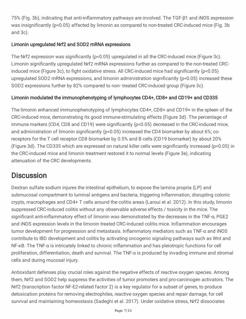
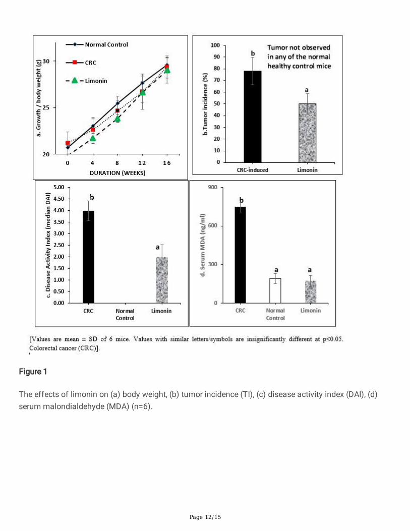
The limonin was given through the drinking water to reduce handling and stress to the mice throughoutthe experiment. The CRC-induced mice showed signi�cantly (p<0.05) lower net weight gains than thenormal control mice (Fig. 1a). Supplements with limonin improved the body weight gains. The nearnormal weight gains indicated that they consumed similar amount of food and water, indicating thatlimonin did not affect the appetite of the mice. The CRC-induced mice showed serious signs of colitis,(diarrhea, weight loss and bloody stool) indicating chronic in�ammation in the colon. The disease activityindex (DAI) score for bloody stools and consistency was highest in the non-treated CRC- induced miceand the treatment with limonin signi�cantly (p<0.05) reduced DAI and tumor incidence / number (Figure1b and 1c). The sizes of the tumor were too small to be measurable.
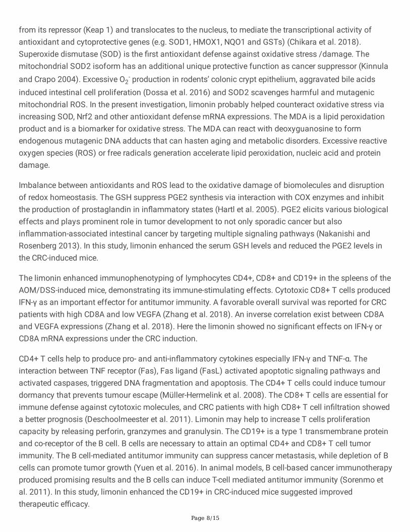
The oxidative stress biomarker MDA was signi�cantly elevated in the CRC-induced mice, and the limoninsigni�cantly attenuated the serum MDA levels (p<0.05) (Figure 1d). The colon tissues had precancerouschanges including gland distortion, ulceration of the mucosa, abundant in�ammatory cell in�ltration,high grade dysplasia, necrotic debris, loss of goblet cells, and severe in�ammation. In mice treated withlimonin, these lesions were either absent or signi�cantly reduced. The histopathology results showed thatthe limonin suppressed CRC-development (Fig 2 a-c). The e�cacy was supported by evidences from theanalyzed colon carcinogenesis and in�ammation biomarkers. Limonin treated mice showed tubularadenoma, low-grade dysplasia and moderate in�ammation in the colon mucosa.
The colorectal-cancer was con�rmed from adenocarcinoma structures on the control non-treated CRC-induced mice. The limonin mitigated the carcinogen/in�ammation-induced CRC and alleviated thecolonic mucosal dysplasia, adenoma and adenocarcinoma formation (Figure 2). The limonin reduced thehistological scores towards normal values (Figure 2d-e). The control non-treated CRC mice showedabnormal colon length and spleen weight, which were mitigated by the limonin treatment (Figure 2f-g).
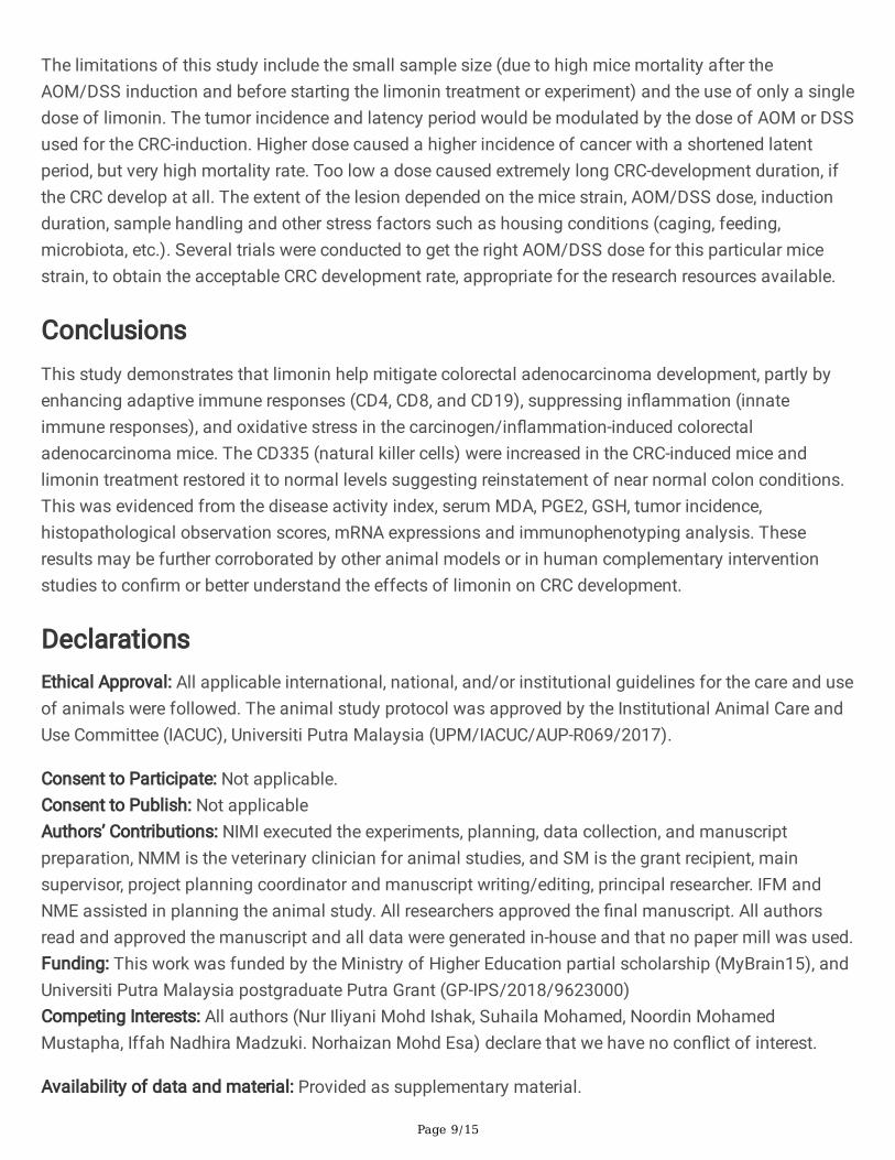
The limonin enhanced serum GSH and suppressed serum PGE2 levels under the carcinogen/in�ammation induced conditions (Figure 3a). Reduced glutathione (GSH) is an endogenous antioxidantresponse constituent that protects cells from oxidative damage and free radicals, a biomarker for anti-oxidant against oxidative stress. Prostaglandin E2 (PGE2) is a pro-in�ammatory lipid mediator that causein�ammation, pain and fever in response to infection and injury.
The pro-in�ammatory mRNA expressions for TNF-α were signi�cantly (p<0.05) upregulated in the CRC-induced mice. The treatment with limonin signi�cantly (p<0.05) down-regulated the expressions by about
Page 7/15
75% (Fig. 3b), indicating that anti-in�ammatory pathways are involved. The TGF-β1 and iNOS expressionwas insigni�cantly (p<0.05) affected by limonin as compared to non-treated CRC-induced mice (Fig. 3band 3c).
Limonin upregulated Nrf2 and SOD2 mRNA expressions
The Nrf2 expression was signi�cantly (p<0.05) upregulated in all the CRC-induced mice (Figure 3c).Limonin signi�cantly upregulated Nrf2 mRNA expressions further as compared to the non-treated CRC-induced mice (Figure 3c), to �ght oxidative stress. All CRC-induced mice had signi�cantly (p<0.05)upregulated SOD2 mRNA expressions, and limonin administration signi�cantly (p<0.05) increased theseSOD2 expressions further by 82% compared to non- treated CRC-induced group (Figure 3c).
Limonin modulated the immunophenotyping of lymphocytes CD4+, CD8+ and CD19+ and CD335
The limonin enhanced immunophenotyping of lymphocytes CD4+, CD8+ and CD19+ in the spleen of theCRC-induced mice, demonstrating its good immune-stimulating effects (Figure 3d). The percentage ofimmune markers (CD4, CD8 and CD19) were signi�cantly (p<0.05) decreased in the CRC-induced mice,and administration of limonin signi�cantly (p<0.05) increased the CD4 biomarker by about 6%; co-receptors for the T cell receptor CD8 biomarker by 0.5% and B cells (CD19 biomarker) by about 20%(Figure 3d). The CD335 which are expressed on natural killer cells were signi�cantly increased (p<0.05) inthe CRC-induced mice and limonin treatment restored it to normal levels (Figure 3e), indicatingattenuation of the CRC developments.
DiscussionDextran sulfate sodium injures the intestinal epithelium, to expose the lamina propria (LP) andsubmucosal compartment to luminal antigens and bacteria, triggering in�ammation, disrupting coloniccrypts, macrophages and CD4+ T cells around the colitis areas (Laroui et al. 2012). In this study, limoninsuppressed CRC-induced colitis without any observable adverse effects / toxicity in the mice. Thesigni�cant anti-in�ammatory effect of limonin was demonstrated by the decreases in the TNF-α, PGE2and iNOS expression levels in the limonin treated CRC-induced colitis mice. Inflammation encouragestumor development for progression and metastasis. In�ammatory mediators such as TNF-α and iNOScontribute to IBD development and colitis by activating oncogenic signaling pathways such as Wnt andNF-κB. The TNF-α is intricately linked to chronic in�ammation and has pleiotropic functions for cellproliferation, differentiation, death and survival. The TNF-α is produced by invading immune and stromalcells and during mucosal injury.
Antioxidant defenses play crucial roles against the negative effects of reactive oxygen species. Amongthem, Nrf2 and SOD2 help suppress the activities of tumor promoters and pro-carcinogen activators. TheNrf2 (transcription factor NF-E2-related factor 2) is a key regulator for a subset of genes, to producedetoxication proteins for removing electrophiles, reactive oxygen species and repair damage, for cellsurvival and maintaining homeostasis (Sadeghi et al. 2017). Under oxidative stress, Nrf2 dissociates
Page 8/15
from its repressor (Keap 1) and translocates to the nucleus, to mediate the transcriptional activity ofantioxidant and cytoprotective genes (e.g. SOD1, HMOX1, NQO1 and GSTs) (Chikara et al. 2018).Superoxide dismutase (SOD) is the �rst antioxidant defense against oxidative stress /damage. Themitochondrial SOD2 isoform has an additional unique protective function as cancer suppressor (Kinnulaand Crapo 2004). Excessive O2
- production in rodents’ colonic crypt epithelium, aggravated bile acidsinduced intestinal cell proliferation (Dossa et al. 2016) and SOD2 scavenges harmful and mutagenicmitochondrial ROS. In the present investigation, limonin probably helped counteract oxidative stress viaincreasing SOD, Nrf2 and other antioxidant defense mRNA expressions. The MDA is a lipid peroxidationproduct and is a biomarker for oxidative stress. The MDA can react with deoxyguanosine to formendogenous mutagenic DNA adducts that can hasten aging and metabolic disorders. Excessive reactiveoxygen species (ROS) or free radicals generation accelerate lipid peroxidation, nucleic acid and proteindamage.
Imbalance between antioxidants and ROS lead to the oxidative damage of biomolecules and disruptionof redox homeostasis. The GSH suppress PGE2 synthesis via interaction with COX enzymes and inhibitthe production of prostaglandin in in�ammatory states (Hartl et al. 2005). PGE2 elicits various biologicaleffects and plays prominent role in tumor development to not only sporadic cancer but alsoin�ammation-associated intestinal cancer by targeting multiple signaling pathways (Nakanishi andRosenberg 2013). In this study, limonin enhanced the serum GSH levels and reduced the PGE2 levels inthe CRC-induced mice.
The limonin enhanced immunophenotyping of lymphocytes CD4+, CD8+ and CD19+ in the spleens of theAOM/DSS-induced mice, demonstrating its immune-stimulating effects. Cytotoxic CD8+ T cells producedIFN-γ as an important effector for antitumor immunity. A favorable overall survival was reported for CRCpatients with high CD8A and low VEGFA (Zhang et al. 2018). An inverse correlation exist between CD8Aand VEGFA expressions (Zhang et al. 2018). Here the limonin showed no signi�cant effects on IFN-γ orCD8A mRNA expressions under the CRC induction.
CD4+ T cells help to produce pro- and anti-in�ammatory cytokines especially IFN-γ and TNF-α. Theinteraction between TNF receptor (Fas), Fas ligand (FasL) activated apoptotic signaling pathways andactivated caspases, triggered DNA fragmentation and apoptosis. The CD4+ T cells could induce tumourdormancy that prevents tumour escape (Müller-Hermelink et al. 2008). The CD8+ T cells are essential forimmune defense against cytotoxic molecules, and CRC patients with high CD8+ T cell in�ltration showeda better prognosis (Deschoolmeester et al. 2011). Limonin may help to increase T cells proliferationcapacity by releasing perforin, granzymes and granulysin. The CD19+ is a type 1 transmembrane proteinand co-receptor of the B cell. B cells are necessary to attain an optimal CD4+ and CD8+ T cell tumorimmunity. The B cell-mediated antitumor immunity can suppress cancer metastasis, while depletion of Bcells can promote tumor growth (Yuen et al. 2016). In animal models, B cell-based cancer immunotherapyproduced promising results and the B cells can induce T-cell mediated antitumor immunity (Sorenmo etal. 2011). In this study, limonin enhanced the CD19+ in CRC-induced mice suggested improvedtherapeutic e�cacy.
Page 9/15
The limitations of this study include the small sample size (due to high mice mortality after theAOM/DSS induction and before starting the limonin treatment or experiment) and the use of only a singledose of limonin. The tumor incidence and latency period would be modulated by the dose of AOM or DSSused for the CRC-induction. Higher dose caused a higher incidence of cancer with a shortened latentperiod, but very high mortality rate. Too low a dose caused extremely long CRC-development duration, ifthe CRC develop at all. The extent of the lesion depended on the mice strain, AOM/DSS dose, inductionduration, sample handling and other stress factors such as housing conditions (caging, feeding,microbiota, etc.). Several trials were conducted to get the right AOM/DSS dose for this particular micestrain, to obtain the acceptable CRC development rate, appropriate for the research resources available.
ConclusionsThis study demonstrates that limonin help mitigate colorectal adenocarcinoma development, partly byenhancing adaptive immune responses (CD4, CD8, and CD19), suppressing in�ammation (innateimmune responses), and oxidative stress in the carcinogen/in�ammation-induced colorectaladenocarcinoma mice. The CD335 (natural killer cells) were increased in the CRC-induced mice andlimonin treatment restored it to normal levels suggesting reinstatement of near normal colon conditions.This was evidenced from the disease activity index, serum MDA, PGE2, GSH, tumor incidence,histopathological observation scores, mRNA expressions and immunophenotyping analysis. Theseresults may be further corroborated by other animal models or in human complementary interventionstudies to con�rm or better understand the effects of limonin on CRC development.
DeclarationsEthical Approval: All applicable international, national, and/or institutional guidelines for the care and useof animals were followed. The animal study protocol was approved by the Institutional Animal Care andUse Committee (IACUC), Universiti Putra Malaysia (UPM/IACUC/AUP-R069/2017).
Consent to Participate: Not applicable.Consent to Publish: Not applicableAuthors’ Contributions: NIMI executed the experiments, planning, data collection, and manuscriptpreparation, NMM is the veterinary clinician for animal studies, and SM is the grant recipient, mainsupervisor, project planning coordinator and manuscript writing/editing, principal researcher. IFM andNME assisted in planning the animal study. All researchers approved the �nal manuscript. All authorsread and approved the manuscript and all data were generated in-house and that no paper mill was used.Funding: This work was funded by the Ministry of Higher Education partial scholarship (MyBrain15), andUniversiti Putra Malaysia postgraduate Putra Grant (GP-IPS/2018/9623000)Competing Interests: All authors (Nur Iliyani Mohd Ishak, Suhaila Mohamed, Noordin MohamedMustapha, Iffah Nadhira Madzuki. Norhaizan Mohd Esa) declare that we have no con�ict of interest.
Availability of data and material: Provided as supplementary material.
Page 10/15
Acknowledgements: We thank the Ministry of Higher Education for the partial scholarship (MyBrain15),Universiti Putra Malaysia for the seed research Putra Grant (GP-IPS/2018/9623000) and the staff fromComparative Medicine and Technology (COMeT), Institute of Bioscience, Universiti Putra Malaysia (UPM)for the help provided in handling the animals throughout the study. Financial support: This work wassupported by the Ministry of Higher Education partial scholarship (MyBrain15), and UPM postgraduatePutra Grant (GP-IPS/2018/9623000)
References1. Arnold M, Sierra MS, Laversanne M, et al (2017) Global patterns and trends in colorectal cancer
incidence and mortality. Gut 66:683–691. https://doi.org/10.1136/gutjnl-2015-310912
2. Axelrad JE, Lichtiger S, Yajnik V (2016) In�ammatory bowel disease and cancer: The role ofin�ammation, immunosuppression, and cancer treatment. World J Gastroenterol 22:4794.https://doi.org/10.3748/wjg.v22.i20.4794
3. Chidambara Murthy KN, Jayaprakasha GK, Kumar V, et al (2011) Citrus limonin and its glucosideinhibit colon adenocarcinoma cell proliferation through apoptosis. J Agric Food Chem 59:2314–2323. https://doi.org/10.1021/jf104498p
4. Chikara S, Nagaprashantha LD, Singhal J, et al (2018) Oxidative stress and dietary phytochemicals:Role in cancer chemoprevention and treatment. Cancer Lett 413:122–134.https://doi.org/10.1016/j.canlet.2017.11.002
5. Deschoolmeester V, Baay M, Lardon F, et al (2011) Immune cells in colorectal cancer: Prognosticrelevance and role of MSI. Cancer Microenviron. 4:377–392
�. Dossa AY, Escobar O, Golden J, et al (2016) Bile acids regulate intestinal cell proliferation bymodulating EGFR and FXR signaling. Am J Physiol Gastrointest Liver Physiol 310:G81-92.https://doi.org/10.1152/ajpgi.00065.2015
7. Hartl D, Starosta V, Maier K, et al (2005) Inhaled glutathione decreases PGE2 and increaseslymphocytes in cystic �brosis lungs. Free Radic Biol Med 39:463–472.https://doi.org/10.1016/j.freeradbiomed.2005.03.032
�. Kinnula VL, Crapo JD (2004) Superoxide dismutases in malignant cells and human tumors. FreeRadic Biol Med 36:718–744. https://doi.org/10.1016/j.freeradbiomed.2003.12.010
9. Laroui H, Ingersoll SA, Liu HC, et al (2012) Dextran sodium sulfate (dss) induces colitis in mice byforming nano-lipocomplexes with medium-chain-length fatty acids in the colon. PLoS One 7:.https://doi.org/10.1371/journal.pone.0032084
10. Li Y, Shen L, Luo H (2016) Luteolin ameliorates dextran sulfate sodium-induced colitis in micepossibly through activation of the Nrf2 signaling pathway. Int Immunopharmacol 40:24–31.https://doi.org/10.1016/j.intimp.2016.08.020
11. Müller-Hermelink N, Braumüller H, Pichler B, et al (2008) TNFR1 Signaling and IFN-γ SignalingDetermine whether T Cells Induce Tumor Dormancy or Promote Multistage Carcinogenesis. Cancer
Page 11/15
Cell 13:507–518
12. Nakanishi M, Rosenberg DW (2013) Multifaceted roles of PGE2 in in�ammation and cancer. Semin.Immunopathol. 35:123–137
13. Saadatdoust Z, Pandurangan AK, Ananda Sadagopan SK, et al (2015) Dietary cocoa inhibits colitisassociated cancer: a crucial involvement of the IL-6/STAT3 pathway. J Nutr Biochem 26:1547–1558.https://doi.org/10.1016/j.jnutbio.2015.07.024
14. Sadeghi MR, Jeddi F, Soozangar N, et al (2017) The role of Nrf2-Keap1 axis in colorectal cancer,progression, and chemoresistance. Tumor Biol 39:101042831770551.https://doi.org/10.1177/1010428317705510
15. Shimizu S, Miyamoto S, Fujii G, et al (2015) Suppression of intestinal carcinogenesis in Apc-mutantmice by limonin. J Clin Biochem Nutr 57:39–43. https://doi.org/10.3164/jcbn.15-28
1�. Sorenmo KU, Krick E, Coughlin CM, et al (2011) CD40-activated B cell cancer vaccine improvessecond clinical remission and survival in privately owned dogs with non-Hodgkin’s lymphoma. PLoSOne 6:. https://doi.org/10.1371/journal.pone.0024167
17. Tanaka T, Kohno H, Kawabata K, et al (2000) Citrus limonoids obacunone and limonin inhibit thedevelopment of a precursor lesion, aberrant crypt foci, for colon cancer in rats
1�. Tanaka T, Maeda M, Kohno H, et al (2001) Inhibition of azoxymethane-induced colon carcinogenesisin male F344 rats by the citrus limonoids obacunone and limonin. Carcinogenesis 22:193–198.https://doi.org/10.1093/carcin/22.1.193
19. Yuen GJ, Demissie E, Pillai S (2016) B Lymphocytes and Cancer: A Love–Hate Relationship. Trendsin Cancer 2:747–757
20. Zhang L, Zhao Y, Dai Y, et al (2018) Immune Landscape of Colorectal Cancer TumorMicroenvironment from Different Primary Tumor Location. Front Immunol 9:1578.https://doi.org/10.3389/�mmu.2018.01578
Figures
Page 12/15
Figure 1
The effects of limonin on (a) body weight, (b) tumor incidence (TI), (c) disease activity index (DAI), (d)serum malondialdehyde (MDA) (n=6).

Page 13/15
Figure 2
(a) Photomicrograph of the effects of limonin on the colon of CRC-induced mice (H&E, X20), (b-c)Histological in�ammation score and in�ammation thickness score from the histology of the mice colon,(d) colon length, (e) spleen weight (n=6).

Page 14/15
Figure 3
The effects of limonin on (a) serum prostaglandin E2 (PGE2) and reduced glutathione (GSH) levels (n=4),(b) the fold change in mRNA expressions in the colon tissues for TNF-α and TGF-β1 (c) iNOS, Nrf2 andSOD2 (d) immunophenotyping analysis of CD4, CD19 and CD8, and CD335 in the spleen (single-cellsuspensions stained with monoclonal antibodies and analysed by �ow cytometry) (n=3).

Page 15/15
Supplementary Files
This is a list of supplementary �les associated with this preprint. Click to download.
SUPPLEMENTARYMATERIALS.docx