UNIVERSIDADE ESTADUAL DO CEARÁ PRÓ-REITORIA DE … · Jales de Hollanda Celestino, Prof....
202
i UNIVERSIDADE ESTADUAL DO CEARÁ PRÓ-REITORIA DE PÓS-GRADUAÇÃO E PESQUISA FACULDADE DE VETERINÁRIA PROGRAMA DE PÓS-GRADUAÇÃO EM CIÊNCIAS VETERINÁRIAS FRANCISCO LÉO NASCIMENTO DE AGUIAR CULTIVO IN VITRO DE FOLÍCULOS PRÉ-ANTRAIS EQUINOS INCLUSOS EM TECIDO OVARIANO: BENEFÍCIOS DA SUPLEMENTAÇÃO CONCENTRAÇÃO- DEPENDENTE DA INSULINA, FSH E FATOR DE CRESCIMENTO EPIDERMAL FORTALEZA 2016
Transcript of UNIVERSIDADE ESTADUAL DO CEARÁ PRÓ-REITORIA DE … · Jales de Hollanda Celestino, Prof....

i
UNIVERSIDADE ESTADUAL DO CEARÁ
PRÓ-REITORIA DE PÓS-GRADUAÇÃO E PESQUISA
FACULDADE DE VETERINÁRIA
PROGRAMA DE PÓS-GRADUAÇÃO EM CIÊNCIAS VETERINÁRIAS
FRANCISCO LÉO NASCIMENTO DE AGUIAR
CULTIVO IN VITRO DE FOLÍCULOS PRÉ-ANTRAIS EQUINOS INCLUSOS EM
TECIDO OVARIANO: BENEFÍCIOS DA SUPLEMENTAÇÃO CONCENTRAÇÃO-
DEPENDENTE DA INSULINA, FSH E FATOR DE CRESCIMENTO EPIDERMAL
FORTALEZA
2016

ii
FRANCISCO LÉO NASCIMENTO DE AGUIAR
CULTIVO IN VITRO DE FOLÍCULOS PRÉ-ANTRAIS EQUINOS INCLUSOS EM TECIDO
OVARIANO: BENEFÍCIOS DA SUPLEMENTAÇÃO CONCENTRAÇÃO-DEPENDENTE DA
INSULINA, FSH E FATOR DE CRESCIMENTO EPIDERMAL
FORTALEZA
2016
Tese apresentada ao Programa de Pós-Graduação em
Ciências Veterinárias da Faculdade de Veterinária da
Universidade Estadual do Ceará, como requisito parcial para
a obtenção do grau de Doutor em Ciências Veterinárias.
Área de Concentração: Reprodução e Sanidade Animal.
Linha de Pesquisa: Reprodução e Sanidade de Carnívoros,
Onívoros, Herbívoros e Aves.
Orientador: Prof. Dr. José Ricardo de Figueiredo.

iii

iv
FRANCISCO LÉO NASCIMENTO DE AGUIAR
CULTIVO IN VITRO DE FOLÍCULOS PRÉ-ANTRAIS EQUINOS INCLUSOS EM TECIDO
OVARIANO: BENEFÍCIOS DA SUPLEMENTAÇÃO CONCENTRAÇÃO-DEPENDENTE DA
INSULINA, HORMÔNIO FOLÍCULO ESTIMULANTE E FATOR DE CRESCIMENTO
EPIDERMAL
Aprovada em: 26/07/2016
BANCA EXAMINADORA
Tese apresentada ao Programa de Pós-Graduação em
Ciências Veterinárias da Faculdade de Veterinária da
Universidade Estadual do Ceará, como requisito parcial para
a obtenção do grau de Doutor em Ciências Veterinárias.

v
Dedico,
A Deus, que faz tudo ganhar sentido.
Ao meu filho Estevão Carvalho de Aguiar,
Que fala comigo seu amor sem palavras.
À minha esposa Luzelena dos Santos Carvalho Aguiar,
Adjuntora fiel mais rara que um rubi, meu suporte para execução desta tese.
À minha mãe,
Fonte inspiradora e minha eterna amiga.

vi
AGRADECIMENTOS
À Universidade Estadual do Ceará (UECE) e ao Programa de Pós-Graduação em Ciências
Veterinárias (PPGCV), aos professores e funcionários, aos quais dedico minha formação acadêmica
durante a graduação e pós-graduação.
Ao Laboratório de Manipulação de Oócitos e Folículos Pré-Antrais (LAMOFOPA) da UECE,
por dar-me toda a guarida para a realização dessa tese.
À Fundação Cearense de Apoio ao Desenvolvimento Científico e Tecnológico (FUNCAP)
pelo suporte financeiro, através da bolsa de doutorado.
À Southern Illinois University (SIU), pela acolhida e suporte durante o Doutorado Sanduíche.
À Mississipi State University (MSU), pela estrutura concedida durante a execução de parte
desta tese.
Ao meu orientador, professor Dr. José Ricardo de Figueiredo pela inspiração profissional, por
me orientar na execução desta tese e por me incentivar a galgar passos cada vez maiores como ser
humano.
Ao meu Co-orientador, professor Dr. Eduardo Leite Gastal, pela oportunidade de realizar
doutorado sanduíche em seu laboratório, pela confiança depositada, por todos os ensinamentos, por
acreditar no meu trabalho, desafiando-me a ser um pesquisador melhor.
À minha Co-orientadora Profa. Dra. Ana Paula Ribeiro Rodrigues, que através do seu exemplo
profissional foi de grande importância no meu crescimento junto à equipe do LAMOFOPA.
Ao meu Co-orientador, Dr. Jean Magloire Nguekam Feugang, por toda a gentileza de me
receber na MSU e pelos ensinos e amizade compartilhados durante o doutorado sanduíche.
Aos membros da banca Profa. Dra. Débora de Melo Magalhães-Padilha, Profa. Dra. Juliana
Jales de Hollanda Celestino, Prof. Dr.Dárcio Ítalo Alves Teixeira, Dr. Luís Alberto Vieira e Prof. Dr.
Eduardo Leite Gastal, pelas as correções desta tese contribuindo para torná-la ainda melhor.
À Doutora Jamily Bezerra Bruno, pela sua amizade e exemplo como profissional, no suporte
à realização de grande parte dos experimentos desta tese, bem como ajuda na redação de artigos e por
ter abraçado minha co-orientação em um momento delicado da mesma.
À Doutora Francieli Osmarini Lunardi, a primeira amiga de trabalho que “abraçou” esta tese,
que me deu palavras de incentivo em momentos de fraqueza, que contribuiu ativamente em meus
experimentos no LAMOFOPA, participando de coletas de ovário inclusive grávida. Meu carinho por
você transcede o lado profissional, pois a tenho como uma amiga mais chegada que uma irmã. Muito
obrigado.
Sou grato às doutoras Laritza Ferreira Lima e Rebeca Magalhães Pedrosa Rocha (as quais
coloquei juntas de propósito) que me ensinaram as análises histológicas e foram cruciais na execução

vii
dos experimentos de cultivo in vitro e auxílio nas redações dos artigos. Muito obrigado pelos seus
exemplos profissionais.
Ao Dr. Benner Geraldo Alves, pelas valiosas contribuições feitas ao meu terceiro artigo,
amizade e exemplo profissional.
Ao doutorando Gustavo Desires Antunes Gastal, que me auxiliou imensamente na execução
e redação do meu quarto artigo técnico, além de sua amizade durante meu período nos Estados
Unidos, que foi vital para a minha adaptação. Um amigo que será guardado no coração para sempre.
À Dra. Melba O. Gastal, que me auxiliou na interpretação dos dados estatísticos, confecção
de figuras e discussão de artigos desta tese, além de sua gentileza pessoal e profissional para comigo.
Ao Dr. Gary A. Apgar pelo auxílio na tradução e revisão dos artigos confeccionados, bem
como sua amizade e exemplo profissional inspirador.
À minha querida amiga Alanna Ferreira da Costa Pessoa, que foi uma parceira querida durante
boa parte deste doutorado.
À Livia Brunetti Apolloni por sua amizade e cuidado, bem como palavras de serenidade e
conselho, que foram em muitos momentos fontes de inspiração durante o doutorado.
À Erica Suzanne Soares Leal, por sua amizade diferenciada, pelos seus conselhos e por tudo
de bom que fez por mim durante a execução desta tese.
À Johanna Leiva Revilla, pela sua amizade e carinho, nos quais sempre pude encontram um
ombro amigo.
À Andrea Moreira Sampaio da Silva, pela sua amizade e por tudo que representou em minha
vida profissional durante a execução desta tese.
À Lindemara Rodrigues, pela amizade, carinho e por todas as inúmeras ajudas que me deu no
setor de histologia.
Aos alunos de iniciação científica Lorena Andrade, Ívila Lorrine, Renato Félix da Silva,
Arnaldo, Luana, e Paula Correia pela enorme ajuda na histologia clássica durante os três primeiros
artigos desta tese.
Aos membros do grupo de estudo bíblico “UBUNTU”: Naíza Arcângela de Sá, Luciana
Mascena Silva, Julian Pontes, Marcela Pinheiro Paz, e Daniela, que por seu suporte mútuo, foram de
grande ajuda para tornar a convivência no LAMOFOPA algo de grande valor pessoal e espiritual.
À toda equipe do LAMOFOPA que me auxiliou a tornar o ambiente de trabalho algo ímpar:
Lindemara Rodrigues, Priscilla de Melo Campos, Rita Kelly, Nathalie Jiatsa, Gildas Mbenia
Tetaping, Rosane Oliveira, Lidiane Sales, Carolina Maside, Antônia Debora Sales, Giovanna Quirino
Rodrigues, Kele Amaral Alves, Geovânia Canafístula, Carlos Lobo, César Camelo, Seu João, Anna
Clara Accioly Ferreira, Victor Macêdo Paes, Jesus de los Reyes Cadenas Moreno, Hudson Henrique
Vieira Correia, Denise Damasceno Guerreiro, Rafael Rossetto, Gerlane Modesto da Silva, Michelle

viii
Karen Brasil Serafin, Valesca Barreto Luz, Roberta Nogueira Chaves, Anderson Pinto, Cláudio
Afonso Pinho Lopes, Rafael Rossetto e Fabricio de Sousa Martins.
Aos membros da Agência de Defesa Agropecuária (Adagri) que me forneceram os ovários a
campo para subsidiar meus experimentos, em especial aos Médicos Veterinários Eudson Almeida dos
Santos, Arquelau Nobre, Herisvaldo Bezerra da Silva, Djanira Gouveia e Aline Lima de Souza.
E um agradecimento especial à minha esposa Luzelena, amada de minh’alma, que suportou
minha ausência e soube pacientemente dar-me o suporte fundamental para execução dos
experimentos. Por cuidar do nosso filho com muito carinho e dedicação, mitigando os efeitos da
minha ausência familiar e proporcionando o ambiente de equilíbrio para nosso lar. Amo-te, mas tu
me amaste primeiro.
Ao meu filho amado Estevão, que sempre alegra meu coração e é o combustível para lembrar-
me do que realmente importa nessa vida. Sua especialidade é uma ferramenta para me fazer alguém
melhor. Você é uma dádiva.
Ao meu irmão Thiago Nascimento de Aguiar, grande parceiro de caminhada que sempre me
impulsionou para ser uma pessoa melhor.
E por fim, à minha mãe Tereza Nascimento de Aguiar, que constituiu todo o alicerce que
possuo como indivíduo, sem a qual a realização desta tese não faria sentido, e demonstrou seu amor
incondicional para comigo.

ix
"...dura coisa te é recalcitrar contra os aguilhões. Atos 28:14 "
Jesus

x
RESUMO
O principal objetivo desta tese foi estudar o efeito concentração-dependente dos hormônios
insulina, hormônio folículo estimulante (FSH), bem como do fator de crescimento epidermal (EGF)
no cultivo in vitro (CIV) de folículos pré-antrais equinos inclusos em tecido ovariano, avaliando os
parâmetros morfologia e desenvolvimento folicular, produção hormonal (todas as fases), espécies
reativas de oxigênio (ROS) (Fases I, II, III), perfil metabolômico (Fase III), níveis de apoptose,
expressão proteíca para Ki-67 e para o receptor de EGF, bem como níveis de mRNA para GDF-9,
BMP-15 e Cyclin-D2 (Fase IV). Para isso, fragmentos ovarianos equinos foram cultivados em 4
diferentes condições: CIV utilizando meio de base (α-MEM+) na ausência ou presença de
suplementação com insulina em concentração fisiológica (10 ng/mL) ou suprafisiológica (10 µg/mL)
(Fase I); CIV em meio de base suplementado com diferentes concentrações (0, 10, 50 e 100 ng/mL)
de FSH (Fase II) ou EGF (Fase III); e CIV usando um meio de base (α-MEM+) enriquecido com
insulina (10 ng/mL) e EGF (50 ng/mL), na ausência ou presença de FSH (50 ng/mL) (Fase IV). A
duração do cultivo foi de até 7 dias (Fases I, II e III) e de 15 dias (Fase IV). Na Fase I, observou-se
que a concentração fisiológica de insulina (10 ng/mL) apresentou maiores (P < 0,05) percentagens de
folículos morfologicamente normais e em desenvolvimento quando comparado aos demais
tratamentos, após 7 dias de cultivo. Independente do período de cultivo, a produção de ROS foi menor
(P < 0,05) no tratamento 10 ng/mL de insulina. O hormônio folículo estimulante na concentração de
50 ng/mL apresentou maior (P < 0,05) percentagem de folículos morfologicamente normais e em
desenvolvimento, bem como maior diâmetro folicular do que os demais tratamentos após 7 dias de
cultivo (Fase II). Adicionalmente, esta concentração de FSH manteve (P > 0,05) a produção de
estradiol e de ROS ao longo do cultivo. De maneira geral, o uso de EGF na concentração de 50 ng/mL
resultou em maior (P < 0,05) percentagem de folículos morfologicamente normais e em
desenvolvimento, bem como maiores (P < 0,05) diâmetros folicular e oocitário após 7 dias de cultivo
(Fase III). Além disso, a referida concentração de EGF manteve a produção de ROS ao longo do
cultivo. A análise do perfil metabolômico do meio de cultivo após 7 dias revelou a presença de três
substâncias (Dinex, Leonuriside A e Avobenzene) com potencial efeito negativo na sobrevivência,
bem como sobre o diâmetro folicular e oocitário. Finalmente, independente da suplementação com
FSH, o uso de um meio enriquecido contendo concentrações apropriadas de insulina e EGF manteve
os níveis de apoptose do tecido ovariano cultivado similares ao controle fresco não cultivado após 15
dias de cultivo (Fase IV). A expressão proteíca para EGFR, Ki-67 e os níveis de RNAm para GDF-
9 e Cyclin-D2 não diferiram entre os grupos tratados após 15 dias. Concluiu-se assim que: as
concentrações de insulina (10 ng/mL), FSH (50 ng/mL) e EGF (50 ng/mL) são benéficas ao CIV
FOPAs equinos inclusos em tecido ovariano por até 7 dias; e que um meio enriquecido contendo

xi
insulina e EGF manteve a morfologia e funcionalidade do tecido ovariano equino após 15 dias de
cultivo, independente da adição de FSH.
Palavras - chave: Folículo pré-antral. Tecido Ovariano Equino, Insulina, FSH, EGF, Cultivo in vitro.

xii
ABSTRACT
The main goal of this dissertation was to study the concentration-dependent effect of the hormones
insulin and follicle stimulating hormone (FSH), as well as of the epidermal growth factor (EGF) on
the in vitro culture (IVC) of equine preantral follicles (PAFs) enclosed in ovarian tissue. The
following endpoints were evaluated: morphology, follicle development, hormonal (all phases),
reactive oxygen species (ROS) production (Phase I, II, III), metabolomics profile (Phase III), and
apoptotic levels, expression (protein) of EGF receptors and Ki-67, as well as mRNA levels of GDF-
9, BMP-15 and Cyclin-D2 (Phase IV). Hence, equine ovarian fragments were cultured in vitro in four
different conditions: IVC using base medium (α-MEM+) in the absence or presence of insulin
supplementation in a physiological concentration (10 ng/mL) or supraphysiological (10 µg/mL)
(Phase I); IVC in base medium supplemented with different concentrations (0, 10, 50 and 100 ng/mL)
of FSH (Phase II) or EGF (Phase III); and IVC using a base medium (α-MEM+) enriched with
insulin (10 ng/mL) and EGF (50 ng/mL), in the absence or presence of FSH (50 ng/mL) (Phase IV).
The culture time lasted 7 days (Phases I, II, and III), and 15 days (Phase IV). In the Phase I, we
observed that physiological concentration of insulin (10 ng/mL) had higher rates (P < 0.05)
percentage of both morphologically normal and developing follicles when compared to the other
treatments after 7 days of culture. Regardless culture time, ROS production was lower (P < 0.05) in
the 10 ng/mL insulin treatment. Follicle stimulating hormone at 50 ng/mL had higher (P < 0.05)
percentage of both morphologically normal and development follicles, as well as greater (P < 0.05)
follicular diameter than the other treatments after 7 days of culture (Phase II). In addition, this FSH
concentration maintained (P > 0.05) estradiol and ROS during culture. Overall, the use of EGF at 50
ng/mL resulted in higher (P < 0.05) percentage of morphologically normal and developing follicles,
greater (P < 0.05) follicular and oocyte diameters after 7 days of culture (Phase III). In addition, the
aforementioned concentration of EGF maintained ROS production during culture. The metabolomics
profile of culture medium from Day 7 of culture demonstrated the presence of three substances
(Dinex, Leonuriside A, and Avobenzene) with a potential negative effect on follicle survival, as well
as follicular and oocyte diameters. Finally, regardless the FSH supplementation, the use of an
enriched medium containing appropriate concentration of insulin and EGF maintained the apoptotic
levels in the ovarian cultured tissue similar to fresh noncultured control after 15 days of culture (Phase
IV). The protein expression for EGFR, Ki-67 and the mRNA levels of GDF-9 and Cyclin-D2 did not
differ between the treated groups after 15 days of culture. In conclusion, the concentrations of insulin
(10 ng/mL), FSH (50 ng/mL), and EGF (50 ng/mL) were beneficial for IVC of equine PAF enclosed
in ovarian tissue at least for seven days, and an enriched medium containing insulin and EGF
maintained the morphology and functionality of the ovarian tissue after 15 days of culture regardless
the FSH addition.

xiii
Keywords: Preantral follicle. Equine Ovarian Tissue, Insulin, FSH, EGF, In vitro culture.

xiv
LISTA DE FIGURAS
Capítulo 1
Figure 1 Percentage of primordial and developing follicles (transitional, primary, and
secondary) in a fresh non-cultured control group and after in vitro culture for
1 or 7 days in the absence or presence of different concentrations of insulin (0
ng/mL, 10 ng/mL, or 10 µg/mL). a,b Within each treatment, values without a
common letter differed (P < 0.02). A,B Within days (day 0 = fresh non-cultured
control group; days 1 and 7 = insulin treated groups), values without a common
letter differed (P < 0.05).…………...…......……………………… 59
Figure 2
Mean (± SEM) diameters (μm) of preantral follicles (primordial and primary
combined) and oocytes in a fresh non-cultured control group and after in vitro
culture for 1 or 7 days in the absence or presence of different concentrations of
insulin (0 ng/mL, 10 ng/mL, or 10 µg/mL). a,b Within each treatment, values
without a common letter differed (P < 0.03-0.0001). A,B Within days (day 0 =
fresh non-cultured control group; days 1 and 7 = insulin treated groups), values
without a common letter differed (P < 0.0001)............. 60
Figure 3
Mean (± SEM) concentrations of estradiol (pg/mL) or progesterone (ng/mL)
produced in culture medium after 1 or 7 days of culture of equine ovarian
follicles enclosed in ovarian tissue in the absence or presence of different
concentrations of insulin. a,b Within each treatment, values without a common
letter differed (P < 0.05). A Within each day, no difference was observed among
treatments for estradiol and progesterone….…….……………………. 61
Figure 4
Mean (± SEM) production of reactive oxygen species (relative fluorescence
units) produced in cultured medium after 1 or 7 days of culture of equine
preantral follicles enclosed in ovarian tissue in the absence or presence of
different concentrations of insulin. a,b Within each treatment, values without a
common letter differed (P < 0.0001). A,B Within days, values without a
common letter differed (P < 0.0001).……….................................................... 62
Capítulo 2
Figure 1 Morphological aspects of preantral follicles after seven days of culture in
FSH 50 ng/mL treatment. (A) normal primordial follicle, (B) abnormal
transitional follicle, and (C) secondary normal follicle. Bars = 25 µm (A, B)
and 50 µm (C)…………...…………………………......………………... 80

xv
Figure 2
Percentage of primordial and developing follicles (transitional, primary, and
secondary; n = 779) in fresh non-cultured ovarian tissue fragments and after
in vitro culture for one or seven days in media supplemented with different
concentrations of rbFSH (mean, 86.5 follicles/treatment/day).a,b Within each
treatment, values without a common letter differed (P < 0.005). A,B,C Within
days, values without a common letter differed (P < 0.05). # Tended (P <
0.09) to differ from 0 ng/mL FSH treatment at one day.* Differed (P < 0.02)
from fresh non-cultured control…….................... 84
Figure 3
Mean (± SEM) concentrations of estradiol (pg/mL; n= 160 samples)
produced by equine ovarian tissue fragments after in vitro culture for one or
seven days in media supplemented with different concentrations of rbFSH
(mean, 20 samples/treatment/day). a,b Within each treatment, non-common
superscripts differed (P < 0.0001). No difference within days was observed
among treatment…….……………………………………………. 86
Capítulo 3
Figure 1 Percentage of primordial and developing follicles (transitional, primary, and
secondary) in a fresh non-cultured control group and after in vitro culture
for one or seven days using different concentrations of EGF. a,b Within each
treatment, values without a common letter differed (P < 0.05). A,B Within
days, values without a common letter differed (P < 0.05). * Differed (P <
0.05) from fresh non-cultured control..……………………… 136
Figure 2
(A) Pairwise score plots between the selected principal compounds (PCs).
The explained variance of each PC is presented in the corresponding
diagonal cell. (B) Scree plot showing the variance explained by the PCs. The
green line on top shows the accumulated variance explained; the blue line
underneath shows the variance explained by individual PC…............... 137
Figure 3
Two-dimensional score plots between selected PCs 1 and 2 using (A)
principal component analysis (PCAs), and (B) partial least squares -
discriminant analysis (PLS-DA). The explained variances are shown in
parentheses in both axes. Color dots represent different treatments as
indicated……………………………………………………………………. 138

xvi
Figure 4
Important features identified with the partial least square discrimination
analysis (PLS-DA) are shown by variable importance in projection (VIP
scores). The colored boxes on the right indicate the relative concentrations
of the corresponding metabolite in each experimental treatment by EGF (0
ng/ml, 10 ng/ml, 50 ng/ml, and 100 ng/ml) under study………………........ 139
Figure 5
Relationship of (A) normal preantral follicles, (B) follicle diameter, and (C)
oocyte diameter with principal component 5 (scores). Regardless of
treatment, each point on the graph represents a medium sample collected
after seven days of in vitro culture (n = 32). A linear regression is
represented by the equation and the line (black) for (A) [normal preantral
follicles = 32.975 ˗ (0.933 × component score), R2 = 0.15, r = ˗0.38, P <
0.05]; (B) [follicle diameter = 31.225 ˗ (0.604 × component score), R2 =
0.19, r = ˗0.44, P < 0.01]; and (C) [oocyte diameter = 24.050 ˗ (0.592 ×
component score), R2 = 0.19, r = ˗0.44, P < 0.01]. …………..…………… 140
Capítulo 4
Figure 1 Morphological representative aspects of preantral follicles in equine ovarian
tissue after treatment with an enriched medium with or without FSH during
seven and fifteen days of culture. (A) normal primordial follicle, (B)
abnormal transitional follicle, Bars = 25 µm. .………………... 170
Figure 2
Mean (± SEM) estradiol concentrations (pg/mL x 100; n = 60 samples)
produced by equine ovarian biopsy tissue after in vitro culture for two, four,
seven, ten and fifteen days in spent enriched media with or without FSH (n
= 3 pooled samples/treatment/day; 9 mares). a,b,c Within each treatment,
values without a common letter differed (P < 0.002). No difference (P >
0.05) within days was observed between treatments. …...... 171
Figure 3
Mean (± SEM) fluorescence intensity detected by TUNEL analysis in
equine ovarian biopsy tissue in the noncultured control group, and treated
groups with or without FSH during seven and fifteen days of culture. No
difference (P > 0.05) was observed between treatments..................……….. 172
Figure 4
Representative immunofluorescence staining of TUNEL in equine ovarian
biopsy tissue in the noncultured control group, and treated groups with or
without FSH during seven and fifteen days of culture. (A) Noncultured
control, (B) Cultured Day 7, (C) Cultured Day 15, (D) Cultured + FSH Day
7, (E) Cultured + FSH Day 15, (F) Positive control, (G) Negative control.
Green Fluorescen Protein, green; DAPI, blue. Bars = 200 µm......... 173

xvii
Figure 5
Mean (± SEM) detection of epidermal growth factor receptor (EGFR) in
equine ovarian biopsy tissue in the noncultured control group, and treated
groups with or without FSH during seven and fifteen days of culture. a,b
Values without a common letter differed (P < 0.05). † Tended to differ (P <
0.07) from noncultured control group. .......................................................... 174
Figure 6
Representative immunofluorescence staining of epidermal growth factor
receptor in equine ovarian biopsy tissue in the noncultured control group,
and treated groups with or without FSH during seven and fifteen days of
culture. (A) Noncultured control, (B) Negative control, (C) Cultured Day 7,
(D) Cultured Day 15, (E) Cultured + FSH Day 7, (F) Cultured + FSH Day
15. Green Fluorescen Protein, green; DAPI, blue. Bars = 200 µm........ 175
Figure 7
Mean (± SEM) fluorescence detection of Ki-67 in equine ovarian biopsy
tissue in the noncultured control group, and treated groups with or without
FSH during seven and fifteen days of culture. a,b,c Values without a common
letter differed (P < 0.05)…………………………………....…….. 176
Figure 8
Representative immunofluorescence staining of Ki-67 in equine ovarian
biopsy tissue in the noncultured control group, and treated groups with or
without FSH during seven and fifteen days of culture. (A) Noncultured
control, (B) Negative control, (C) Cultured Day 7, (D) Cultured Day 15, (E)
Cultured + FSH Day 7, (F) Cultured + FSH Day 15. Green Fluorescen
Protein, green; DAPI, blue. Bars = 200 µm................................................... 177
Figure 9
Mean (± SEM) relative amounts of mRNA for (A) GDF-9, (B) BMP-15, (C)
Cyclin-D2 (Cyclin-D2), in equine ovarian biopsy tissue in the noncultured
control group and treated groups with or without FSH during seven and
fifteen days of culture genes……………..……………………… 178

xviii
LISTA DE TABELAS
Capítulo 1
Table 1 Percentage of morphologically normal equine preantral follicles in
fresh non-cultured control group and after in vitro culture for 1 or 7
days in the absence or presence of different concentrations of
insulin………………………………………………………………. 58
Capítulo 2
Table 1 Percentage of morphologically normal equine preantral follicles in
fresh non-cultured ovarian tissue fragments and after in vitro culture
for one or seven days in media supplemented with different
concentrations of rbFSH………………………….……………..…. 83
Table 2
Mean (± SEM) diameters of equine preantral follicles (primordial,
transitional, and primary combined) in fresh non-cultured ovarian
tissue fragments and after in vitro culture for one or seven days in
media supplemented with different concentrations of rbFSH...…… 85
Table 3
Mean (± SEM) diameters of equine oocytes from preantral follicles
(primordial, transitional, and primary combined) in fresh non-
cultured ovarian tissue fragments and after in vitro culture for one
or seven days in media supplemented with different concentrations
of rbFSH…………………………………………… 85
Table 4
Mean (± SEM) reactive oxygen species (relative fluorescence units)
produced by equine ovarian tissue fragments after in vitro culture
for one or seven days in media supplemented with different
concentrations of rbFSH…………………………………………… 87
Capítulo 3
Table 1 Percentage of morphologically normal equine preantral follicles in
fresh non-cultured control group and after one and seven days of
culture using different concentrations of EGF.................................. 130
Table 2
Mean (± SEM) diameter (µm) of preantral follicles and oocytes
(primordial and primary combined) in fresh non-cultured group and
after one and seven days of culture using different concentrations of
EGF…..…............................................................
131
Table 3
Mean (± SEM) concentrations of estradiol (pg/ml) produced in
spent medium after one or seven days of culture of equine preantral

xix
follicles enclosed in ovarian tissue using different concentrations of
EGF………..………………................................
132
Table 4
Mean (± SEM) reactive oxygen species (relative fluorescence units)
produced in spent medium after one or seven days of culture of
equine preantral follicles enclosed in ovarian tissue using different
concentrations of EGF…………………………………… 133
Table 5
Most representative ions identified in VIP score………………….. 134
Table 6
Most representative ions identified in PC5…...………………….... 135
Table S1
VIP score components listed in research database after PLS-DA.... 135
Table S2
Factor loadings of five principal components (PCs) extracted by
PCA showing the metabolites (mass) found in spent medium after
seven days of in vitro culture of equine preantral follicles….…….. 135
Table S3
Components listed after research in database for PC5 m/z after
regression analysis…………………………..................................... 135
Capítulo 4
Table 1
Primers sequences and characteristics……....................................... 169

xx
LISTA DE ABREVIATURAS E SIGLAS
Akt Protein Kinase B (proteina quinase B)
ANOVA Análise de variância
AP-1 Activator protein 1
BAD Bcl-2-associated death promoter
Bax BCL2 Associated X Protein
Bcl-2 B-cell lymphoma 2
Bim BH3-containing protein
BMP-15 Bone morphogenetic protein 15
BPU Biopsy Pick-Up method
BSA Bovine serum albumin
cAMP Cyclic adenosine monophosphate
Ccnd-d2 G1/S-specific cyclin-D2
CEUA Comitê de Ética no Uso de Animais
CGP Células Germinativas Primordiais
CIV Cultivo In vitro
CO2 Dióxido de Carbono
CREB cAMP response element-binding protein
Cyclin D1 G1/S-specific cyclin-D1
Cyclin-D2 G1/S-specific cyclin-D2
DCHF-DA 2',7'-dihidrodiclorofluorescein diacetate
DNA deoxyribonucleic acid (ácido desoxirribonucleico)
EGF Epidermal growth factor (fator de crescimento epidermal)
EGFR Epidermal growth factor Receptor (receptor do fator de crescimento
epidermal)
EIA Equine Infectious Anemia
ERK extracellular signal–regulated kinases
EROs Espécies Reativas de Oxigênio
FasL Fas ligand
FOXO Forkhead box
FSH Follicle-Stimulating Hormone
GDF-9 Growth/differentiation factor 9
h hours
HC Histologia Clássica

xxi
HEPES 4-(2-hydroxyethyl)-1-piperazineethanesulfonic acid
IGF-I Insulin-Like Growth Factor 1 (IGF-1)
ITS Insulin-Transferrin-Selenium
IVC In Vitro Culture
JAK Janus Kinase
KL Kit Ligand
LH Luteinizing Hormone
MAPK Mitogen Activated Protein Kinases
MEM Minimum Essential Medium
METLIN Metabolomics Database
min minutos
mL mililitros
mm milímetros
MOIFOPA Manipulação de Oócitos Inclusos em Folículos Ovarianos Pré-Antrais
mRNA Messenger RNA
NFkB Nuclear factor kappa B
ng nanogramas
P450arom Cytochrome P450 aromatase
P450scc Cholesterol side-chain cleavage enzyme P450
PAF Preantral Follicles
PAS Periodic Acid-Schiff (ácido periódico-Schiff)
PC Principal Component
PCA Principal Component Analysis
PI3-K phosphatidylinositol 3-kinase
PLS-DA partial least square discrimination analysis
qPCR real-time PCR; quantitative PCR
R2 Linear regression
rbFSH Recombinant bovine Follicle Stimulating Hormone
RNA Ribonucleic Acid
ROS Reactive Oxygen Species
SEM Standard Error of the Mean
SNRPD3 Small Nuclear Ribonucleoprotein D3 Polypeptide
STAT Signal Transducer and Activator of Transcription
TCM-199 Tissue Culture Media -199
TGF-β Transforming Growth Factor beta

xxii
TUNEL Terminal deoxynucleotidyl transferase dUTP nick end labeling
VIP Variable Importance in Projection
α-MEM Alpha Minimum Essential Medium
α-MEM+ Alpha Minimum Essential Medium with supplementation
μm micrômetros
°C Celsius degree (Graus Celsius)
17β-HSD 17 – beta - hydroxylsteroid dehydrogenase

xxiii
SUMÁRIO
1. INTRODUÇÃO....................................................................................................................... 24
2 REVISÃO DE LITERATURA............................................................................................... 26
2.1 O ovário dos mamíferos com destaque para o ovário da égua: Sítio promotor da oogênese
e foliculogênese............................................................................................................................
26
2.2 Atresia folicular .................................................................................................................... 28
2.3 A biotécnica de manipulação de oócitos inclusos em folículos ovarianos pré-antrais
(MOIFOPA)................................................................................................................................. 29
2.3.1 Recuperação de tecido ovariano equino e estudo da população, morfologia e
densidade folicular......................................................................................................................
29
2.3.2 Cultivo in vitro de folículos pré-antrais............................................................................. 31
2.4 A composição do meio e o desenvolvimento folicular in vitro.............................................. 33
2.4.1 Insulina................................................................................................................................ 34
2.4.2 Hormônio Folículo Estimulante (FSH)............................................................................... 36
2.4.3 Fator de Crescimento Epidermal (EGF).............................................................................. 38
2.5 Avaliação da Morfologia e Funcionalidade Folicular Após Cultivo In Vitro......................... 39
3 JUSTIFICATIVA..................................................................................................................... 44
4 HIPÓTESES CIENTÍFICAS.................................................................................................. 46
5 OBJETIVOS............................................................................................................................. 47
5.1 Objetivo geral........................................................................................................................ 47
5.2 Objetivos específicos............................................................................................................. 47
6 CAPÍTULO 1
Insulina melhora a sobrevivência in vitro de folículos pré-antrais inclusos em tecido
ovariano e reduz a produção de espécies reativas de oxigênio após cultivo..........................
49
7 CAPÍTULO 2
Hormônio folículo estimulante mantém a sobrevivência e promove o desenvolvimento de
folículos pré-antrais equinos inclusos em tecido ovariano......................................................
74
8 CAPÍTULO 3
Papel do EGF no cultivo in situ de folículos pré–antrais equinos e perfil metabolômico.... 100
9 CAPÍTULO 4
Efeitos da adição de FHS em um meio enriquecido contendo insulina e EGF após cultivo
de longa duração na funcionalidade de biópsias de tecido ovariano equino........................
143
10 CONCLUSÕES..................................................................................................................... 182
11 PERSPECTIVAS.................................................................................................................. 183
12 REFERÊNCIAS BIBLIOGRÁFICAS................................................................................ 184

24
1. INTRODUÇÃO
A importância do estudo dos folículos pré-antrais (FOPAs) tem se tornado mais perceptível
nas últimas duas décadas. Em condições fisiológicas, os folículos ovarianos pré-antrais têm três
possíveis destinos: permanecer em estado de dormência sobre a influência de fatores inibitórios,
iniciar o desenvolvimento após ativação folicular, ou morrer pelo processo de atresia. Somente
uma minoria (0,01%) alcançará o estágio pré-ovulatório, e consequentemente, irá liberar um oócito
fertilizável. Portanto, a manipulação in vitro de FOPAs, os quais representam 90% da população
folicular, visando evitar a atresia e promover o desenvolvimento desses folículos até estádios mais
avançados, é um atrativo desafio no campo da pesquisa reprodutiva (XU et al., 2013; GRENN;
SHIKANOV, 2016).
Apesar dos excelentes resultados alcançados até agora em camundongos com à produção
de oócitos maturos a partir de FOPAs cultivados in vitro (O’BRIEN e EPPIG 2003), a
repetibilidade destes resultados em animais de produção é baixa (ARUNAKUMARI et al., 2010;
MAGALHÃES et al., 2011).
Grupos de pesquisa têm avaliado modelos animais para estudo comparativo com a
foliculogênese humana, sendo portanto uma alternativa devido a barreiras éticas para o estudo de
material biológico humano. Neste cenário, a égua surge como um modelo atrativo para o estudo
da foliculogênese comparativa com a mulher, por compartilhar similaridades, tais como a dinâmica
folicular (GINTHER, 2012), eventos endócrinos (GINTHER et al., 2004a, MIHM e EVANS;
2008), efeito da idade (CARNEVALE, 2008), e mais recentemente, a dinâmica de folículos pré-
antrais (ALVES et al., 2016b). Contudo, estudos da foliculogênese ovariana em éguas têm alguns
obstáculos para se transpor, uma vez que em alguns países, tais como os Estados Unidos, o abate
de equinos é proibido desde 2007. Para superar este problema, uma alternativa é o uso do método
de biópsia ovariana por pick-up (BPU; HAAG et al., 2013d; ALVES et al., 2015; 2016a,b), que
representa uma ferramenta valiosa para recuperar o tecido ovariano para diversas abordagens.
Quando aplicável, ovários de abatedouro (GOMES et al., 2015) ou oriundos de animais
eutanasiados, também podem ser utilizados, provendo material biológico para diversos estudos.
Tal material biológico poderá então fornecer folículos ovarianos pré-antrais, e os mesmos
serem cultivados in vitro. A eficiência do cultivo in vitro vai depender de diferentes fatores,

25
podendo-se destacar dentre eles a composição de meio. Neste sentido, a adição de substâncias
importantes para o sucesso do cultivo in vitro, capazes de permitir o desenvolvimento de um
grande número de folículos pré-antrais, melhorando assim a taxa de produção de embriões no
futuro, são de grande importância. Neste contexto pelo seu papel como importantes reguladores
da foliulogênese ovariana, destacam-se a insulina, o FSH e o EGF.
Para uma melhor compreensão do tema investigado nesta tese, a revisão de literatura a
seguir fará uma breve abordagem sobre ovário equino, o cultivo in vitro de folículos pré-antrais
na espécie equina, com destaque para a utilização da insulina, FSH e EGF no meio de cultivo e
sua ação, bem como as principais técnicas de avaliação da qualidade folicular.

26
2 REVISÃO DE LITERATURA
2.1 O ovário dos mamíferos com destaque para o ovário da égua: Sítio promotor da oogênese e
foliculogênese
O ovário constitui-se um órgão do trato reprodutivo da fêmea capaz de produzir oócitos e
liberar diferente fatores, incluindo hormônios. Assim, o ovário atua, fornecendo os gametas
femininos através da ovulação, para a produção de descendentes, além de produzir moléculas
bioativas, tais como esteroides (principalmente estradiol e progesterona) e fatores de crescimento
capazes de regular diferentes aspectos da fisiologia reprodutiva da fêmea (EDSON et al., 2009).
O ovário possui como unidade funcional o folículo ovariano, composto por um oócito
circundado por células somáticas, incluindo células da granulosa e da teca, cuja função é
proporcionar um ambiente ideal para a sobrevivência, o crescimento e maturação do oócito, bem
como produzir hormônios (MCGEE et al., 2000) e outros peptídeos (PENG et al., 2010). O folículo
ovariano é uma estrutura resultante dos processos de oogênese e foliculogênese que ocorrem no
ovário ao longo da vida reprodutiva da fêmea. O ovário é constituído por duas regiões: o córtex e
a medula. O córtex consiste na região funcional do órgão, e é formado por tecido conjuntivo
(fibroblastos, colágeno e fibras reticulares), folículos ovarianos e corpos lúteos em diversos
estádios de crescimento ou regressão. A medula é composta primariamente de vasos sanguíneos e
linfáticos, nervos e tecido conjuntivo, responsáveis pela nutrição e estruturação do ovário
(SAMUELSON, 2007).
A égua tem um ovário em formato anatômico semelhante ao rim, que mede
aproximadamente 6-8 cm de comprimento e 3-4 cm de largura durante a estação de cobertura.
Éguas maduras tendem a ter ovários maiores, algumas vezes alcançando 10 cm de comprimento.
Durante o anestro, o ovário reduz de tamanho, apresentando de 2-4 cm de comprimento por 2-3
cm de largura. O córtex ovariano da égua, ao contrário da maioria das espécies mamíferas, é
localizado na área central do ovário, circundado pela área medular (MOSSMAN e DUKE, 1973).
Esta zona parenquimatosa contém folículos e, com o crescimento dos folículos dentro do ovário,
uma ampla cavidade repleta de fluído pode ser percebida via palpação retal. Inicialmente este

27
folículo tem consistência firme à palpação, mas tende a diminuir a sua consistência com a
aproximação da ovulação.
Precedendo a ovulação, o folículo pré-ovulatório torna-se triangular no formato,
posicionando seu ápice em direção à fossa ovulatória. A fossa ovulatória forma uma área côncava
no ovário onde a ovulação ocorre. Essa região é mais parenquimatosa do que as áreas circundadas
e o epitélio germinal é exposto para o exterior do ovário equino (GINTHER, 1992), garantindo
que a ovulação ocorra somente neste local (WITHERSPOON e TALBOT, 1970; STABENFELDT
et al., 1975). O verdadeiro propósito pelo qual a fossa ovulatória existe na égua é ainda
desconhecido, mas uma especulação é a de que a fossa reduz a possibilidade de ocorrência de
múltiplas ovulações que colocariam a égua em risco de desenvolver gestação gemelar. Durante a
estação de anestro, os ovários tornam-se mais firmes à palpação, devido a um decréscimo no
desenvolvimento dos folículos ovarianos e da perfusão vascular ovariana (GINTHER, 1992).
A oogênese consiste na etapa pela qual as células germinativas primordiais (CGP)
transformam-se até oócitos maduros, ou aptos a fertilização, tendo seu início ainda na vida intra-
uterina, e concluindo-se após o fim da maturidade sexual (OLIVEIRA, 2009). Na vida fetal, as
CGPs migram a partir do endoderma do saco vitelínico para a gônada primitiva, onde iniciam um
processo de multiplicação, através de sucessivas mitoses, originando as oogônias meioticamente
ativas (SUH et al., 2002). Quando estas oogônias entram em prófase I da meiose, estas são agora
denominadas oócitos primários. Estes oócitos são encontrados em “ninhos”, sendo envoltos por
células somáticas planas conhecidas como células da pré-granulosa. Em seguida, estes oócitos
circundados pelas células da pré-granulosasaem dos ninhos, formando os folículos primordiais
(HARTSHORE et al., 2009). Oócitos inclusos em folículos primordiais interrompem seu
desenvolvimento e entram em um período de quiescência. A retomada da divisão meiótica e a
completa maturação oocitária, tanto nuclear quanto citoplasmática, ocorrerão somente a partir da
puberdade. Em equinos, não há pico pré-ovulatório de LH, e sim um aumento progressivo do FSH
durante os dias de estro (HINRICHS et al., 1993a, BERGFELT e GINTHER, 1993). Se este oócito
for ovulado e posteriormente fecundado pelo espermatozoide, ocorrerá a formação do oócito
haploide fecundado, finalizando assim a oogênese (FIGUEIREDO et al., 2008).
A foliculogênese é um processo que vai desde a formação dos folículos primordiais até o
estádio de folículos pré-ovulatórios. A população folicular na espécie equina é de em média 36,000
folículos, apresentando grande variação individual entre os animais (DRIANCOURT et al., 1982).
28
Durante a vida reprodutiva da fêmea, um pequeno grupo de folículos é gradualmente estimulado
a crescer, iniciando o processo de ativação folicular. A ativação é um processo que ocorre através
da passagem dos folículos primordiais quiescentes para os diferentes estádios de desenvolvimento
folicular (transição, primário, secundário, terciário e pré-ovulatório), sendo este o maior evento
biológico que controla o potencial reprodutivo das fêmeas.
2.2 Atresia folicular
A atresia folicular consiste na morte dos folículos após a sua formação, através da ativação
de alguma via de morte celular. A atresia folicular pode ocorrer em qualquer estádio de
desenvolvimento folicular, ocorrendo por via degenerativa ou apoptótica. A morte celular por
degeneração, conhecida como uma morte celular passiva, ocorre geralmente como consequência
de estresse físico-químico extremo associado ao calor, choque osmótico, estresse mecânico,
congelação-descongelação e altas concentrações de peróxido de hidrogênio (KRYSKO et al.,
2008), ou ainda por exemplo, por isquemia em que o ovário sofre restrição dos suprimentos
adequados de oxigênio ou nutrientes (MIKKELSEN et al., 2001). Esse tipo de morte celular é
caracterizado morfologicamente pelo aumento do volume celular, desorganização do citoplasma,
disfunção mitocondrial, colapso de organelas e perda da integridade da membrana plasmática.
Consequentemente, ocorre a ruptura da célula com liberação de seu conteúdo para o meio
extracelular, causando dano às células vizinhas e uma reação inflamatória no local (ZONG e
THOMPSON, 2006).
Já a apoptose, também conhecida como morte celular programada, é um processo
determinado geneticamente, e como tal, dependente da expressão de genes pró e anti-apoptóticos.
Este processo é morfologicamente caracterizado pela condensação da cromatina (picnose nuclear),
fragmentação específica do DNA, perda de volume celular e formação de protuberâncias na
membrana plasmática e de corpos celulares condensados, conhecidos como corpos apoptóticos
(HUSSEIN, 2005).
Apesar de ser um fenômeno natural, a atresia reduz significativamente o número de oócitos
que seriam ovulados, diminuindo assim o potencial reprodutivo do animal. Entretanto, o cultivo
in vitro pode se caracterizar como uma excelente estratégia para reverter ou reduzir o impacto da
perda folicular. Esses folículos podem então ser cultivados in vitro para a obtenção de oócitos

29
maturos, aptos à fecundação, garantindo assim a manutenção da função reprodutiva de um
determinado animal, ou até mesmo a multiplicação de animais de alto valor genético ou em vias
de extinção.
2.3 A biotécnica de Manipulação de Oócitos Inclusos em Folículos Ovarianos Pré-Antrais
(MOIFOPA)
Conforme visto anteriormente, existe uma grande perda folicular que ocorre naturalmente
in vivo. Assim, a disponibilidade de oócitos é um fator limitante para o desenvolvimento de novas
técnicas reprodutivas (SMITZ e CORTVRINDT, 2002). Os métodos atuais para a produção in
vitro de embriões dependem de uma oferta de oócitos competentes provenientes de grandes
folículos antrais ou pré-ovulatórios, os quais estão presentes no ovário em número reduzido
(TELFER, 1998). Dessa forma, a possibilidade de desenvolver sistemas in vitro que explorem o
grande número de oócitos provenientes de folículos pré-antrais deve ser considerada. Neste
contexto, a biotécnica de Manipulação de Oócitos Inclusos em Folículos Ovarianos Pré-Antrais
(MOIFOPA) visa previnir a atresia observada in vivo, maximizando a recuperação de oócitos
potencialmente fertilizáveis. Tal biotécnica consiste no isolamento, conservação (resfriamento e
criopreservação) e/ou cultivo in vitro de folículos ovarianos pré-antrais, visando a estocagem,
ativação, crescimento e maturação in vitro do folículo primordial até o folículo pré-ovulatório
(FIGUEIREDO et al., 2008). Dentre as possíveis aplicações da MOIFOPA, podem-se
exemplificar: a pequisa fundamental, com o aumento dos conhecimentos acerca da foliculogênese;
a criopreservação de material biológico para a produção de biobancos de células germinativas;
fonte para testes toxicológicos para a indústria farmacêutica; incremento na produtividade de
animais de alto valor genético, bem como preservação de espécies ameaçadas de extinção (HAAG
et al., 2013d).
2.3.1 Recuperação de tecido ovariano equino e estudo da população, morfologia e densidade
folicular.
Em equinos, estudos recentes demonstraram que diversos fatores podem influenciar na
qualidade do tecido ovariano recuperado, no que tange a integridade morfológica folicular e no

30
número de folículos obtidos para análise. Sabe-se que a população folicular no ovário equino
apresenta uma alta variabilidade, estimada em média de 35.950 folículos primordiais e 100
folículos em crescimento, variando entre 5.600 a 75.000 folículos primordiais, com 20 a 300
folículos em crescimento (DRIANCOURT et al., 1982). Além disso, o diâmetro folicular e
oocitário médio são de 31.0 ± 0.5 e 27.6 ± 0.6, respectivamente (HAAG et al., 2013d). Após a
recuperação tecidual, o tempo, a temperatura, bem como o meio de manutenção, pode influenciar
na qualidade dos folículos após recuperação do tecido ovariano. Em relato prévio, demonstrou-se
que a temperatura de 4ºC por até 4 horas em meio PBS foi capaz de preservar maiores percentagens
de folículos morfologicamente normais do que o meio MEM (GOMES et al., 2012).
Outro fator limitante para a manipulação de tecido ovariano equino consiste na obtenção
do tecido ovariano, uma vez que em alguns países, como por exemplo nos Estados Unidos, o abate
de equinos está proibido desde 2007. Neste contexto, uma excelente alternativa consiste no uso da
técnica de biópsia ovariana por pick-up (BPU; AERTS et al., 2005; 2008). Tal procedimento foi
validado na espécie equina com sucesso (HAAG et al., 2013a), com a obtenção de fragmentos de
biópsia contendo folículos ovarianos em diferentes estádios de desenvolvimento, sendo aptos a
manipulações subsequentes, como o isolamento mecânico por tissue chopper (HAAG et al.,
2013b), com a manutenção da viabilidade folicular.
Após a manipulação, o fragmento de biópsia geralmente é destinado à análise por histologia
clássica. A eficácia da análise morfológica vai depender do correto processamento do material
obtido. Desta maneira, alguns fatores podem afetar o processamento, como por exemplo, o tipo de
fixador escolhido. A fixação é uma das etapas mais importantes da técnica histológica, pois visa
interromper o metabolismo celular, estabilizando os componentes bioquímicos e estruturas intra e
extracelulares, além de permitir a penetração de substâncias subsequentes à fixação (O`LEARY,
2001). Em estudo prévio avaliando diferentes tipos de fixadores para o tecido ovariano equino,
demonstrou-se que para fragmentos ovarianos (5 x 5 x 1 mm), o fixador Carnoy utilizado por 24
horas foi o mais benéfico para a manutenção da morfologia folicular, quando comparado ao Bouin
e à formalina a 10%. Em outros estudos, o Bouin (HAAG et al., 2013a,b,c), bem como o
paraformaldeído a 4 % (AGUIAR et al., 2016a,b), têm sido utilizados com sucesso para a
classificação morfológica folicular.
Adicionalmente, a espessura de corte do tecido ovariano pode interferir na identificação de
estruturas morfológicas após processamento histológico. Analisando diferente espessuras de corte

31
(3 µm, 5 µm e 7 µm; ALVES et al., 2015) verificou-se que um maior número de folículos
morfologicamente normais foram encontrados na espessura de 7 µm (ALVES et al., 2015).
O ovário equino pode sofrer alterações na densidade folicular por conta da idade, bem como
devido à heterogeneidade da distribuição folicular no ovário (HAAG et al., 2013a). Neste contexto,
um estudo anterior demonstrou que a densidade folicular difere entre animais e dentro de cada
fragmento ovariano. Adicionalmente, a morfologia folicular é afetada negativamente pelo aumento
da idade (11-17 anos), sendo necessários 3 a 4 fragmentos ovarianos, combinados com 65 secções
histológicas, para detectar a densidade folicular do tecido ovariano equino, independentemente da
heterogeneidade (ALVES et al., 2016a).
Mais recentemente, a influência de estruturas ovarianas (corpo lúteo versus folículo pré-
ovulatório), bem como da sazonalidade reprodutiva (diestro versus anestro) foram avaliadas
(ALVES et al., 2016b). Neste estudo, a presença de corpo lúteo teve efeito positivo na qualidade
dos folículos pré-antrais, apresentando maior densidade folicular e de células do estroma. Além
disso, o diestro apresentou maior percentagem de folículos morfologicamente normais, folículos
em desenvolvimento e densidade de células estromais quando comparado ao anestro (ALVES et
a., 2016b).
Por fim, um estudo avaliou o efeito do tempo de exposição do tecido ovariano equino a
diferentes agentes crioprotetores (dimetilsufóxido, DMSO; etilenoglicol, EG; e propanodiol,
PROH). Como principal resultado, o etilenoglicol (EG) demonstrou ser o agente crioprotetor
menos prejudicial ao tecido ovariano equino nos diferentes tempos de exposição avaliados (0, 10,
15 e 20 minutos), não afetando a densidade celular (GASTAL et al., 2016).
2.3.2 Cultivo in vitro de folículos pré-antrais
O cultivo in vitro de folículos ovarianos é uma importante etapa da biotécnica de
MOIFOPA, e tem por objetivo assegurar um ambiente ideal capaz de proporcionar o
desenvolvimento in vitro dos oócitos até a produção de um oócito maturo, capaz de ser utilizado
na produção de embriões em larga escala. Através do cultivo in vitro pode-se avaliar o efeito de
diferentes substâncias, em concentrações variáveis durante as diferentes fases do desenvolvimento
folicular. Existem basicamente dois tipos de sistemas de cultivo onde os folículos pré-antrais

32
podem ser cultivados: inclusos no fragmento de córtex ovariano (cultivo in situ), ou na forma
isolada (cultivo de folículos isolados) (ARAUJO et al., 2014).
O cultivo in situ tem a vantagem de promover a manutenção do contato de diferentes
folículos entre si, bem como com o estroma circundante. Além disso, permite a investigação do
efeito de diversas substâncias sobre a ativação e crescimento folicular, até o estádio de folículo
secundário (PENG, 2010). Uma das desvantagens deste modelo é que os folículos não conseguem
crescer até a fase final da foliculogênese, sendo que para esta finalidade, tais folículos devem ser
isolados do tecido, e cultivados na forma isolada até a formação de antro e maturação oocitária
(TELFER e ZELINSKI, 2013). Neste sentido, o cultivo de folículos na forma isolada são
previamente isolados de forma mecânica (utilizando-se um Tissue chopper, um homogeneizador
ou microdissecção utilizando-se agulhas de seringa), ou ainda na forma enzimática, utilizando-se
de enzimas como a colagenase e DNase, e cuidados de forma bidimensional, diretamente sobre a
superfície plástica da placa de cultivo, ou sobre uma matrix (composta por exemplo de células
fibroblásticas), e ainda na forma tridimensional sendo inserido em uma matrix, como por exemplo
o gel de agarose (FIGUEIREDO et al., 2008).
Grandes progressos já foram obtidos com o cultivo in vitro de folículos pré-antrais em
folículos em diferentes espécies animais. Em felinos (JEWGENOW, 1998) e marsupiais
(BUTCHER E ULLMAN, 1996) foi observado o crescimento de folículos ovarianos pré-antrais
isolados após o cultivo in vitro, porém, sem a formação de antro. Nas espécies bovina
(GUTIERREZ et al., 2000; MCCAFFERY et al., 2000), canina (SERAFIM et al., 2010) e humana
(ROY e TREACY, 1993), os folículos pré-antrais isolados desenvolveram-se in vitro até o estádio
antral. Em suínos, os folículos secundários crescidos in vitro chegaram até a maturação e tiveram
seus oócitos fecundados in vitro (HIRAO et al., 1994) com desenvolvimento até o estádio de
blastocisto (WU et al., 2001). Mais recentemente, foram obtidos embriões de búfalos (GUPTA et
al., 2008), cabras (SARAIVA et al., 2010; MAGALHÃES et al., 2011) e ovelhas
(ARUNAKUMARI et al., 2010) a partir de folículos pré-antrais cultivados in vitro.
Na espécie equina, somente três estudos realizaram cultivo in vitro de folículos pré-antrais.
Em estudo prévio (SZLACHTA e TISCHNER, 2000), avaliou-se o efeito da suplementação de
FSH em folículos isolados enzimaticamente. Como resultado, houve aumento na taxa de atresia
observada após quatro dias de cultivo. Posteriormente, testou-se a eficácia de dois diferentes meios
de cultivo (Menezo B2 e Waymouth MB 752/1), com ou sem a suplementação de FSH no cultivo

33
in vitro de folículos pré-antrais isolados enzimaticamente (SZLACHTA e TISCHNER, 2004). O
meio Menezo B2 apresentou melhor resultado de sobrevivência folicular quando comparado ao
meio Waymouth MB 752/1. Contudo, a suplementação de FSH não beneficiou a taxa de
crescimento e de sobrevivência folicular durante os quatro dias de cultivo.
Em outro estudo realizado por Haag et al. ( 2013c), utilizando tecido ovariano recuperado
pela técnica de BPU, investigou o efeito de dois meios de base (α-MEM e TCM-199) após cultivo
de 1 e 7 dias. Neste trabalho, o meio α-MEM obteve maior taxa de ativação folicular (27%) após
7 dias de cultivo, não sendo observados folículos viáveis no cultivo em TCM-199 no final do
cultivo.
Em estudo mais recente, avaliou-se o papel da concentração-dependente (0, 10, 50 e 100
ng/mL) do ácido ascórbico sob tecido ovariano equino oriundo de abatedouro (GOMES et al.,
2015). As concentrações de 50 ng/mL e 100 ng/mL de ácido ascórbico apresentaram maiores
percentagens de folículos em desenvolvimento após 6 dias de cultivo em comparação com as
concentrações de 0 e 25 ng/mL de ácido ascórbico.
Assim, a realização de mais trabalhos referentes ao cultivo in situ de folículos pré-antrais
equinos são de grande importância, especialmente devido à escassez de resultados relativos à essa
espécie. Com o objetivo de obter-se melhores resultados para o cultivo in vitro, diversos
suplementos vêm sendo adicionados aos meios de cultivo de base. A seguir, será realizada uma
breve abordagem da importância da composição do meio de base e de alguns suplementos para
esse meio como a insulina, hormônio folículo estimulante (FSH) e o fator de crescimento
epidermal (EGF).
2.4 A composição do meio e o desenvolvimento folicular in vitro
A composição do meio é um importante fator para o sucesso do cultivo in vitro de folículos
ovarianos. Estudos prévios demonstraram que a sobrevivência de folículos pré-antrais bovinos in
vitro foi reduzida na ausência de hipoxantina e substratos energéticos, tais como piruvato e
glutamina (FIGUEIREDO et al., 1994). Adicionalmente, a suplementação de insulina, transferrina
e selênio (ITS) têm assegurado o crescimento oocitário e a formação de antro (GORE-LANGTON
e DANIEL, 1990). Desta forma, a adição de piruvato, glutamina, ITS e hipoxantina ao meio de

34
cultivo tem sido essencial para o crescimento de folículos pré-antrais in vitro (JEWGENOW et al.,
1998).
Os antioxidantes, selênio e transferrina, são relatados como substâncias importantes a
serem adicionadas ao meio de cultivo. Alguns autores sugerem que o processo de maturação
folicular está relacionado aos altos níveis de transferrina e seus receptores na célula, e que o selênio
pode ser adicionado ao meio de cultivo para ativar enzimas envolvidas na detoxificação e
eliminação de radicais livres (DEMEESTERE et al., 2005). O desenvolvimento de um sistema de
cultivo básico que garanta a ativação e o crescimento folicular até um estádio em que os oócitos
possam ser maturados e fecundados in vitro é importante para estudar os fatores que controlam o
crescimento oocitário e a multiplicação das células da granulosa (CORTVRINDT et al., 1996).
Além disso, sabe-se que o crescimento dos folículos presentes no ovário mamífero é regulado por
gonadotrofinas e por fatores intra-ovarianos (FORTUNE, 1998). Assim, estudos têm investigado
o efeito de vários componentes no cultivo in vitro de folículos pré-antrais, tanto de animais de
laboratórios como animais domésticos (SMITZ e CORTVRINDT, 2002). A seguir, serão descritos
como os hormônios (insulina e FSH) e do fator de crescimento epidermal (EGF) influenciam no
cultivo in vitro de folículos pré-antrais.
2.4.1 Insulina
A insulina é um hormônio fundamentalmente relacionado com a regulação da concentração
circulante de glicose, tendo seu papel no crescimento e na diferenciação de diversos tipos celulares,
e atuando como regulador da atividade ovariana (HERNANDEZ et al., 1988). A insulina no ovário
estimula a proliferação das células da granulosa, a produção de esteróides (SPICER et al., 1993),
a atividade da aromatase (GARZO e DORRINGTON, 1984), bem como regula a maturação
oocitária (PAWSHE et al., 1998).
O efeito direto da insulina no ovário pode ser comprovado pela presença de seus receptores.
O receptor de insulina pertence a uma família de receptores de fatores de crescimento que têm
atividade tirosina-quinase (CARVALHEIRA et al., 2002). Este receptor específico de membrana
é uma proteína heterotetramérica com atividade quinase, composta por duas subunidades α (massa
molecular 135 kDa) e duas subunidades β (massa molecular 95 kDa), unidas por uma ponte
dissulfídica (LAWRENCE et al., 2007). A subunidade α dos receptores de insulina são estruturas

35
extracelulares que servem como sítio para ligação deste hormônio. Já a subunidade β do receptor
possui um domínio transmembranário e um intracelular, o qual é responsável pela transmissão do
sinal (BELFIORE et al., 2009). A ligação da insulina à subunidade α permite que a subunidade β
adquira atividade quinase, levando a alteração conformacional (IRS-1/-2: Insulin receptor
substrate-1/-2) que aumenta ainda mais a atividade quinase do receptor (LAWRENCE et al.,
2007). Uma vez fosforilado, o IRS- 1/-2 interage com uma série de proteínas intracelulares,
desencadeando uma cascata complexa de reações de fosforilação e desfosforilação (CHEATHAM
e KAHN, 1995). Em adição à ativação da fosfatidilinositol 3-quinase (PI-3 quinase), a proteína
quinase mitogenicamente ativada (MAPK) também é fosforilada após a ligação da insulina ao seu
receptor (CHEATHAM e KAHN, 1995; WHITE, 1996). A ativação do MAPK é responsável pelos
efeitos no crescimento promovidos pela insulina (LAWRENCE et al., 2007). Tanto em modelos
humanos como animais, os receptores de insulina são amplamente distribuídos em todos os
compartimentos ovarianos, incluindo células da granulosa, células da teca, estroma e oócito
(PORETSKY et al., 1988). No entanto, a expressão de RNAm para receptor de insulina nas células
da granulosa e teca de folículos pré-ovulatórios foi maior do que em todos os outros estágios de
desenvolvimento (SHIMIZU et al., 2008).
Quando a insulina liga-se ao seu receptor, esta promove uma série de efeitos metabólicos,
destacando-se a estimulação do transporte de glicose para o interior das células, a principal fonte
energética para o ovário (SHIMIZU et al., 2008). Estudos mostram que a insulina possui
importante papel na regulação da responsividade do ovário, podendo também atuar sinergicamente
junto às gonadotrofinas hipofisárias (LH e FSH), aumentando a produção de hormônio do
crescimento e estimulando a proliferação e diferenciação das células da granulosa juntamente com
o FSH (KAWAUCHI e SOWER, 2006).
Com relação ao papel da insulina na função ovariana, especificamente na foliculogênese in
vitro, consiste na manutenção da viabilidade e crescimento dos folículos primordiais e primários,
aumentando a formação de folículos primários em baixas concentrações (LOUHIO et al., 2000;
CHAVES et al., 2011). Evidências apontam para o fato da insulina estimular o fator inibidor de
leucemia, Kit Ligand e IGF-I, atuando como co-reguladora no padrão de sinalização da transição
de folículos primordiais para primários durante a foliculogênese inicial (VAN DEN HURK e
ZHAO, 2005). Estudos in vitro têm mostrado que a insulina estimulou a formação de folículos

36
primários em tecido ovariano cultivado em diferentes espécies, como em humanos (LOUHIO et
al., 2000) e murinos neonatais (KEZELE et al., 2002).
A insulina quando utilizada em concentrações fisiológicas (10-20 ng/mL), atuou no
crescimento folicular e oocitário de folículos pré-antrais bovinos, com alta porcentagem (acima de
60%) de formação de antro após 13 dias de cultivo (MCLAUGHLIN et al., 2010). Em ovinos, a
suplementação de insulina na concentração de 10 ng/mL contribuiu para o desenvolvimento de
folículos secundários (ARUNAKUMARI et al., 2010). Em caprinos reportou-se que folículos pré-
antrais cultivados in vitro quando inseridos in situ ou isolados mecanicamente, apresentaram maior
crescimento e sobrevivência com a utilização de insulina 10-ng/mL associada ao FSH em
concentrações crescentes (sequencial: Dia 0 = 100 ng/mL; Dia 6 = 500 ng/mL; Dia 12 = 1000
ng/mL; CHAVES et al., 2012). Quando adicionada ao meio de cultivo juntamente com o piruvato,
glutamina e hipoxantina, a insulina como componente do ITS (Insulina, Transferrina e Selênio)
aumentou o percentual de folículos morfologicamente normais, e estimulou o crescimento
folicular em roedores (DEMEESTERE et al., 2005). As ações da insulina quando adicionada ao
meio de cultivo estão relacionadas possivelmente a um melhor aproveitamento das fontes
energéticas presentes no meio, atuando assim como fator de sobrevivência, e garantindo aporte de
precursores metabólicos como aminoácidos e glicose (CHAVES et al., 2011).
2.4.2 Hormônio Folículo Estimulante (FSH)
O FSH é uma gonadotrofina secretada pela hipófise anterior, sendo um dos principais
hormônios adicionados ao meio de cultivo. Sua principal função é no desenvolvimento e
maturação gonadal durante a puberdade, bem como no desenvolvimento de folículos durante a
fase inicial da onda folicular (GINTHER et al., 1992). O receptor do FSH é composto de um grande
domínio extracelular N-terminal, sete domínios transmembranários e um domínio C- terminal
intracelular acoplado à proteína G (SALESSE et al., 1991). Após ligar-se ao receptor, ocorre a
conversão de guanosina difosfato (GDP) em guanosina trifosfato (GTP), que se liga à subunidade
α da proteína G, estimulando a adenilciclase (AC) a gerar AMP cíclico (cAMP). Este, por sua vez,
aciona uma cascata de fosforilação nas proteínas quinases dependentes de cAMP (PK-A). Desta
forma, a ativação da PK-A controla múltiplos aspectos da função celular por meio da fosforilação
de substratos proteicos. Uma vez que a interação receptor-ligante tenha se estabelecido, o
37
complexo é internalizado por endocitose e degradado pelos lisossomos, sendo o receptor reciclado
à membrana celular por exocitose (HILLIER, 1996). A interação do FSH com seu receptor inicia
uma cadeia de reações intracelulares que incluem a ativação de mais de 100 genes que codificam
diferentes respostas (HUNZICKER-DUNN e MAIZELS, 2006), tais como a estimulação da
proliferação celular, a síntese de estereoides e a expressão de receptores para o Fator de
Crescimento Epidermal (EGF), Fator de Crescimento Semelhante à Insulina 1 (IGF-1) e LH (VAN
DEN HURK e ZHAO, 2005).
Desta forma, estudos in vitro da ação do FSH tem merecido atenção especial,
principalmente pelo seu envolvimento na proliferação celular, síntese de esteroides e expressão de
receptores para outras substâncias importantes, como por exemplo, o LH, o kit ligand (KL) e o
GDF-9 (NILSSON e SKINNER, 2004). No ovário mamífero, embora os folículos pré-antrais
sejam independentes de FSH para seu crescimento inicial, os receptores de FSH estão presentes
nas células da granulosa foliculares (O’SHAUGHNESSY et al., 1996). Por conta disto, o FSH é
capaz de exercer efeito benéfico sobre os folículos mesmo quando adicionado ao cultivo antes da
formação da cavidade antral (ADRIENS et al., 2004). Desta forma, estudos têm demonstrado que
o FSH promove aumento no crescimento folicular in vitro (WU e TIAN; 2007).
Os melhores resultados relatados na literatura relativo ao cultivo de FOPAs tem em comum
a presença de FSH no meio de desenvolvimento, como por exemplo, o nascimento de animais
vivos em camundongos a partir de folículos primordiais (EPPIG et al., 1996), bem como a
produção de embriões oriundos de folículos pré-antrais em ovinos (ARUNAKUMARI et al.,
2010), caprinos (SARAIVA et al., 2010) e búfalas (GUPTA et al., 2008). No cultivo de folículos
primários e secundários isolados enzimaticamente, o FSH promoveu aumento no diâmetro,
sobrevivência folicular, e secreção de hormônios esteroides e aumento na taxa de formação de
antro (GUTIERREZ et al., 2000; ITOH et al., 2002). Estudos relataram que o FSH promove a
formação de antro em diferentes espécies (Ovinos: ARUNAKUMARI et al., 2010; Bovinos:
GUTIERREZ et al., 2000; Suínos: WU e TIAN., 2007; Caprinos: MATOS et al., 2007). Em
equinos, trabalhos avaliando a adição de FSH ao meio de cultivo são escassos. No cultivo in vitro
de folículos isolados em meio na presença de FSH, observou-se que após 4 dias de cultivo, o FSH
não afetou as taxas de crescimento ou sobrevivência folicular (SZLACHTA e TISCHNER, 2000,
2004).

38
2.4.3 Fator de Crescimento Epidermal (EGF)
O EGF é considerado um fator de crescimento proteico pertencente à família EGF, a qual
consiste de no mínimo oito membros (CONTI et al., 2006). Sua atividade biológica é mediada por
receptores de membrana EGF-R do tipo tirosina-quinase, pertencentes à superfamília ErbB
(YARDEN, 2001). Na década de 80, estudos já demonstravam que o EGF atua sobre o crescimento
de folículos ovarianos, além de modular a função das células da granulosa (KNECHT e CATT,
1983; FENG et al., 1986). É considerado um potente fator mitogênico que estimula a proliferação
de diferentes tipos celulares (DAS, 1984; MULLIN e MCGINNET, 1988). Tem sido demonstrado
que o EGF atua como importante regulador da fisiologia ovariana, estando envolvido na regulação
de diversos processos, incluindo a ativação folicular, a proliferação e diferenciação das células da
granulosa, esteroidogênese e maturação oocitária (CELESTINO et al., 2009). Além disso, o EGF
tem recebido notável atenção por inibir a apoptose, garantindo assim maior sobrevivência folicular
em condições in vitro (MARKSTRÖM et al., 2002).
O EGF é conhecido como um fator de sobrevivência tanto in vivo como in vitro
(SILVA et al., 2006). Quando utilizado in vitro na concentração de 10 ng/mL, o EGF inibiu
apoptose das células da granulosa e proporcionou aumento da formação de antro após o
cultivo de folículos isolados suínos (MAO et al., 2004). Porém, em folículos pré-antrais de
camundongos, a utilização de EGF não demonstrou nenhum efeito na supressão da
apoptose (DEMEESTERE et al., 2005). Em bovinos, quando o EGF foi utilizado em uma
concentração de 0,5 ng/mL, resultou na redução dos níveis de atresia em folículos pré-
antrais cultivados in vitro (GUTIERREZ et al., 2000). Por outro lado, nesta mesma espécie,
utilizando a concentração de 10 ng/mL, o EGF não influenciou na sobrevivência de
folículos primários e secundários cultivados in vitro (DERRAR et al., 2000).
Posteriormente, estudos de ZHOU e ZHANG (2005a,b) relataram que o EGF na
concentração de 50 ng/mL estimulou a sobrevivência de oócitos caprinos após o cultivo in
vitro. Além disso, baixas concentrações de EGF (1 ou 10 ng/mL) no cultivo in vitro de
folículos pré-antrais caprinos mantiveram a morfologia e a ultraestrutura folicular após 7
dias de cultivo (CELESTINO et al. 2009). No entanto, em altas concentrações (100 ng/mL),
não houve efeito do EGF sobre a sobrevivência de folículos pré-antrais caprinos após 5

39
dias de cultivo (SILVA et al. 2004). Na espécie ovina investigou-se o efeito de diferentes
concentrações de EGF (0, 25, 50, 75 ou 100 ng/mL) sobre folículos pré-antrais cultivados
in vitro, demonstrando que maiores concentrações (75 e 100 ng/mL) induziram a
degeneração de todos os folículos pré- antrais após cultivo de 6 dias (TALMIMANI et al.
2005). Resultados similares foram descritos em folículos pré-antrais caprinos cultivados in
vitro por 7 dias, onde altas concentrações de EGF (100 ou 200 ng/mL) não induziram
ativação e apresentaram altas taxas de degeneração folicular (CELESTINO et al. 2009).
A ação do EGF sobre a ativação, diferenciação, proliferação e esteroidogênese das células
da granulosa tem se mostrado controversa na literatura. Alguns estudos têm mostrado que embora
o EGF não seja essencial para ativação de folículos primordiais (BRAW-TAL e YOSSEFI, 1997;
FORTUNE et al., 1998), o mesmo parece ser importante para os estádios mais avançados de
desenvolvimento folicular (GUTIERREZ et al., 2000; PENG et al., 2010). Em caprinos, o EGF na
concentração de 100 ng/mL promoveu a ativação de folículos primordiais após cultivo in situ após
3 dias, bem como promoveu efeito benéfico no crescimento de oócitos de folículos primários
(SILVA et al. 2004). Em ovinos, sua utilização nesta mesma concentração promoveu a ativação
de folículos primordiais in situ e a manutenção da viabilidade por até 6 dias de cultivo (ANDRADE
et al., 2005). Em pequenos e médios folículos pré-antrais bovinos (60-179 μm), a utilização de 50
ng/mL de EGF foi capaz de garantir a sobrevivência e promover o crescimento e produção de
progesterona, bem como estimulou a formação de antro em grandes folículos pré-antrais (166 μm),
mas não o crescimento do oócito (GUTIERREZ et al., 2000). Em suínos, o EGF em baixas
concentrações (0,75, 1,5 ou 3 ng/mL) quando associado ao FSH, melhorou a qualidade dos oócitos,
levando a maior taxa de desenvolvimento embrionário (WU e TIAN, 2007).
2.5 Avaliação da morfologia e funcionalidade folicular após cultivo in vitro
Diversas técnicas podem ser empregadas para análise folicular após o cultivo in vitro de
folículos pré-antrais. Os parâmetros mais utilizados nesta análise são aqueles inerentes à avaliação
da morfologia e funcionalidade folicular.
Avaliação morfológica

40
A histologia clássica (HC) constitui-se de uma importante ferramenta para avaliação de
folículos pré-antrais in vitro permitindo uma análise quantitativa de um grande número de folículos
cultivados, e a verificação da morfologia e número de células foliculares, bem como integridade e
viabilidade das mesmas após ativação (normais ou atrésicos). Diante destes parâmetros, pode-se
também classificar os folículos com relação ao seu estádio de desenvolvimento (primordial,
transição, primário ou secundário). Todavia, a HC possui como limitação a incapacidade de avaliar
a integridade de membranas e organelas citoplasmáticas (MATOS et al., 2007).
A técnica de microscopia eletrônica de transmissão pussui um sistema de captação de
imagens de altíssima resolução (0,1 nm), e se vale de parâmetros morfológicos para visualização
de estruturas biológicas detalhadas não visíveis por histologia clássica, com o auxílio de um
microscópio eletrônico (SALEHNIA et al., 2002). As mudanças ultraestruturais ocorridas após
cultivo in vitro, como por exemplo, pequenos danos em membranas nucleares (oocitária e das
células da granulosa), podem ser identificados através da interação entre elétrons e átomos
presentes nas células. Entretanto, essa técnica é por vezes laboriosa quando comparada a histologia
clássica, limitando o número de folículos a serem analisados, sendo assim uma técnica
essencialmente qualitativa.
A técnica de TUNEL (terminal deoxynucleotidil transferase-mediated deoxyuridine
triphosphate biotin nick end-labeling) utiliza uma enzima (tranferase deoxynucleotidil terminal)
para adicionar nucleotídeos aos fragmentos das fitas de DNA quebradas nas células apoptóticas.
Essa técnica é utilizada para avaliar a fragmentação do DNA em secções histológicas, permitindo
a avaliação histológica, bem como a detecção do grau de apoptose (LOO et al., 2011).
De maneira geral, ao longo do cultivo in vitro, a taxa de células apoptóticas aumenta ao
longo do cultivo. MAO et al., 2004 relataram que a percentagem de células da granulosa
apoptóticas em folículos frescos foi 0,1%, contudo após quatro dias de cultivo, essa porcentagem
aumentou significativamente para 3,4%. Em bovinos, também se observou que não havia células
da granulosa coradas por TUNEL em folículos isolados frescos ou em folículos cultivados na
presença de ácido ascórbico. Por outro lado, havia um número significativamente maior de células
da granulosa apoptóticas em folículos cultivados na ausência do ácido ascórbico (THOMAS et al.,
2001).

41
Avaliação da funcionalidade
A funcionalidade dos folículos ovarianos pode ser avaliada in vitro de diferentes formas,
como por exemplo, através da proliferação celular de células da granulosa (CUILING et al., 2005),
da atividade esteroidogênica (BILLIG et al., 1993), espécies reativas de oxigênio (BEHRMAN et
al., 2001), avaliação do perfil metabolômico (GOOK et al., 2014), e da expressão gênica (OKTAY
et al., 1997).
No tocante à proliferação de células da granulosa, a proteína Ki-67 é fortemente associada
com a proliferação celular (SCHOLZEN e GERDES, 2000). Seu antígeno pode ser identificado
exclusivamente no núcleo. Esta proteína está presente durante todas as fases do ciclo celular (G(1),
S, G (2), e mitose, mas está ausente na fase G (0), tornando esta proteína um excelente marcador
para determinar a fração em crescimento de uma dada população celular. Sua aplicação na
avaliação de folículos pré-antrais já foi demonstrada em estudos prévios (SCALERCIO et al., 2015;
PAULINI et al., 2016).
A atividade esteroidogênica ou produção de hormônios esteroides tem sido amplamente
utilizada para avaliar a funcionalidade do folículo cultivados in vitro (SONGSASEN et al., 2011).
Os hormônios esteróides são originários do colesterol por meio de uma série clássica de reações
enzimáticas. O colesterol é transportado para a membrana interna da mitocôndria sendo alvo da
enzima citocromo P450 side chain cleavage (P450scc) nas células da teca, e alvo do citocromo
P450 aromatase (P450arom) nas células da granulosa (XU et al., 1995; TAMURA et al., 2007). A
P450scc converte o colesterol em pregnenolona, a qual pode ser convertida em progesterona ou
em andrógeno. O andrógeno pode ser convertido, formando androstenediona, a qual pode ser
convertida no andrógeno mais ativo, a testosterona, pela enzima 17β hidroxisteróide desidrogenase
(17β-HSD). Finalmente, a testosterona pode ser convertida a estradiol pela ação da enzima
P450arom (CONLEY e BIRD, 1997). De uma forma geral, o produto esteróide final secretado
pelos folículos depende do perfil das enzimas esteroidogênicas expressas pelo mesmo. Além disso,
o estradiol é requerido para a indução da expressão de receptores para o hormônio luteinizante
(LH) nas células da granulosa, o que é um pré-requisito para a ovulação. Assim, a alta atividade
estrogênica associada com a alta atividade da aromatase é um bom indicador da dominância
fisiológica folicular, do mesmo modo que a inibição da atividade da aromatase pode resultar na
atresia folicular (BERGFELT et al., 1999).

42
As ERO’s incluem todos os radicais e não radicais derivados do oxigênio, os quais são
eletronicamente instáveis e, por isso, altamente reativos, tendo a capacidade de reagir com um
grande número de compostos que estejam próximos. Eles podem exercer a função de agentes
oxidantes, atuando como receptores de elétrons, ou de agentes redutores, atuando como doadores
de elétrons (AGARWAL et al., 2005). Quando há um desequilíbrio entre a produção e a
degradação de espécies reativas de oxigênio, ocorre o fenômeno chamado de estresse oxidativo
(BEHRMAN, 2001).
O papel das ERO’s no estresse oxidativo das fêmeas domésticas permanece incerto.
Acredita-se que a atividade enzimática antioxidante esteja relacionada, por exemplo, com a
esteroidogênese folícular, uma vez que o folículo dentro do ambiente ovariano encontra-se
naturalmente exposto a um certo nível de estresse oxidativo durante a produção hormonal
(SUGINO, 2005). Altas concentrações de estrógeno contribuem para uma maior atividade
antioxidante, evidenciando que o estresse oxidativo apresenta impacto na produção de hormônios
esteróides produzidos pelas células da granulosa, principalmente o estrógeno (LUND et al., 1999).
A peroxidação lipídica parece estar envolvida nesse processo, influenciando também a produção
de outras glicoproteínas produzidas pelas células da granulosa, como a inibina A, a inibina B, a
ativina A e o hormônio antimuelleriano, os quais têm sido investigados como marcadores da
resposta ovariana e sua reserva folicular (APPASAMY et al., 2008).
Durante o cultivo in vitro, diferentes metabólitos oriundos das células cultivadas podem
ser monitorados através da técnica de metabolômica. Estas substâncias podem ser utilizadas como
marcadores do perfil celular (GRIFFITHS e WANG, 2009). Os estudos na área de metabolômica
fazem parte de uma pesquisa relativamente nova, e servem como um indicador da atividade celular.
(PATTI et al., 2012). Aplicações desta técnica consistem em identificar patologias, alvos
terapêuticos e investigar mecanismos de processos biológicos fundamentais. Assim, aplicações da
metabolômica na biotécnica de MOIFOPA podem estar relacionadas a identificação de diferentes
substâncias, as quais podem ser necessárias na suplementação do cultivo, sendo uma nova
estratégia para os métodos tradicionais (GOOK et al., 2014).
A sobrevivência, o crescimento e a diferenciação celular podem também ser monitorados
pela alteração dos padrões da expressão gênica utilizando-se a biologia molecular
(MAZERBOURG e HSUEH, 2006). As técnicas de biologia molecular possibilitam a
identificação da expressão de genes e/ou proteínas relacionados ao processo de foliculogênese.

43
Genes relacionados ao desenvolvimento folicular, como por exemplo, o GDF-9 e o BMP-15, os
quais são apontados como importantes reguladores ovarianos (VITT et al., 2002, PENG et al.,
2013), atuando no desenvolvimento folicular e oogênese (PAULINI e MELO, 2011). Assim, a
quantificação dos níveis de transcrição de genes específicos é fundamental para garantir uma maior
avaliação das funções foliculares.

44
3 JUSTIFICATIVA
A espécie equina possui uma grande importância econômica e social, por compartilhar
diversas similaridades com a foliculogênese humana, no que tange a dinâmica das ondas
foliculares, mudanças hormonais durante o ciclo estral, relação entre declínio da fertilidade e
idade, bem como difunções anovulatórias (GINTHER et al., 2004a, GASTAL et al., 2011,
GINTHER et al., 2012), e mais recentemente, em relação a dinâmica de folículos pré-antrais
(ALVES et al., 2016b). Neste contexto, a espécie equina emerge como uma excelente alternativa
de modelo animal visando auxiliar o desenvolvimento de protocolos no tratamento de infertilidade
na espécie humana. Nesse sentido, torna-se de fundamental importância o emprego de biotécnicas
que permitam um melhor entendimento do processo de foliculogênese equina, capaz de assegurar
a otimização da utilização dos milhares de oócitos presentes no ovário. Além disso, o emprego da
biotécnica de MOIFOPA em equinos poderá favorecer a multiplicação de animais de interesse
econômico ou ameaçados de extinção.
Dentre as biotécnicas capazes de maximizar o potencial reprodutivo das fêmeas domésticas,
a MOIFOPA disponta não somente pelos resultados promissores obtidos em camundongos e em
outras espécies domésticas, mas também pela possibilidade de subsidiar outras biotécnicas
reprodutivas, como a fertilização e produção in vitro de embriões (FIGUEIREDO et al., 2008). Na
espécie equina, trabalhos relativos à manipulação in vitro de folículos pré-antrais podem ser
considerados apenas insipientes. Uma etapa crucial para o sucesso da biotécnica de MOIFOPA é
o cultivo in vitro de folículos pré-antrais. Neste sentido, a composição do meio incluindo as
corretas concentrações dos diferentes suplementos adicionados ao meio de base é essencial para
assegurar a sobrevivência e o crescimento folicular, dada a complexa interação entre
concentrações, associações e tempos de adição dos fatores no meio de cultivo. Dentre as principais
substâncias adicionadas ao meio de cultivo de base, merecem destaque a insulina, o FSH e o EGF,
que vem sendo amplamente utilizadas por diferentes pesquisadores, entretanto, apresentando
resultados variáveis entre as diferentes espécies (GUTIERREZ et al., 2000; GUPTA et al., 2002;
MAO et al., 2004; MATOS et al., 2007; CHAVES et al., 2011; CELESTINO et al., 2009). Apesar
da importância dos referidos suplementos, bem como dos achados da literatura referente a
influência marcante concentração-dependente de diferentes suplementos utilizados, inexistia

45
trabalhos na literatura investigando curvas concentração-resposta de insulina, FSH e EGF no
cultivo in vitro de folículos pré-antrais equinos inclusos em tecido ovariano, constituindo, portanto,
a originalidade da presente tese.
Em conjunto, este trabalho se justificou pela necessidade de estabelecer qual as melhores
concentrações de insulina, FSH e EGF, bem como a associação destes fatores, a fim de melhorar
os resultados obtidos anteriormente na espécie equina, envolvendo o cultivo in vitro de folículos
pré-antrais. A originalidade dos achados do presente trabalho e, consequentemente, sua
importância científica, pode ser comprovada pela publicação de dois artigos científicos em relação
ao emprego da insulina (Capítulo I) e FSH (Capítulo II).

46
4 HIPÓTESES CIENTÍFICAS
Diante do exposto, foram formuladas as seguintes hipóteses científicas:
O uso de uma concentração de insulina similar a fisiológica (10 ng/mL), melhora a
sobrevivência e o desenvolvimento de folículos pré-antrais equinos quando comparada a
concentração (10 µg/mL) previamente utilizada na literatura (Fase I).
A suplementação com FSH (Fase II) e EGF (Fase III) no cultivo in vitro, promove a
manutenção da sobrevivência e desenvolvimento de folículos pré-antrais equinos de forma
concentração-dependente.
Um meio enriquecido, contendo a associação da insulina, FSH e EGF nas melhores
concentrações definidas na presente tese, mantém a morfologia folicular e funcionalidade tecidual
após o cultivo in vitro de longa duração de fragmento de biópsia ovariana equina (Fase IV).

47
5. OBJETIVOS
5.1 Objetivo Geral
Avaliar o efeito de diferentes concentrações de insulina, FSH e EGF, bem como da
associação destes componentes no cultivo in vitro de folículos pré-antrais equinos inclusos em
fragmentos de tecido ovariano.
5.2 Objetivos Específicos
1) Investigar o efeito da diferentes concentração fisiológica (10 ng/mL) e suprafisiológica
(10 µg/mL) de insulina sobre o percentual de folículos pré-antrais morfologicamente normais,
ativação folicular, diâmetro folicular e oocitário, produção hormonal (estradiol e progesterona) e
de ROS após 1 e 7 dias de cultivo;
2) Avaliar o efeito concentração-dependente do FSH recombinante bovino (0, 10, 50 e 100
ng/mL) sobre o percentual de folículos pré-antrais morfologicamente normais, ativação folicular,
diâmetro folicular e oocitário, produção hormonal (estradiol e progesterona) e de ROS após 1 e 7
dias de cultivo;
3) Estudar o efeito concentração-dependente do EGF (0, 10, 50 e 100 ng/mL) sobre o
percentual de folículos pré-antrais morfologicamente normais, ativação folicular, diâmetro
folicular e oocitário, produção hormonal (estradiol e progesterona) e de ROS após 1 e 7 dias de
cultivo, e perfil metabolômico do meio de cultivo após 7 dias;
4) Avaliar o efeito de um meio de cultivo enriquecido contendo insulina (10 ng/mL) e EGF
(50 ng/mL), suplementado ou não com FSH (50 ng/mL), no cultivo in vitro de tecido ovariano
equino biopsado, tendo como parâmetros a morfologia folicular, produção de estradiol, níveis de

48
apoptose, expressão para o receptor de EGF e proteína Ki-67, e níveis de RNAm para os genes
GDF-9, BMP-15 e Cyclin-D2 após 7 ou 15 dias de cultivo.

49
6 CAPÍTULO 1
Insulina melhora a sobrevivência in vitro de folículos pré-antrais inclusos em tecido
ovariano e reduz a produção de espécies reativas de oxigênio após cultivo.
“Insulin improves in vitro survival of equine preantral follicles enclosed in ovarian tissue and
reduces reactive oxygen species production after culture”
Periódico: Theriogenology, v. 85 (6) p. 1063 - 1069, 2016, (ISSN: 0093-691-X) Qualis A2

50
RESUMO
Este estudo invetigou o efeito da concentração de insulina no cultivo in vitro de folículos pré-
antrais equinos inclusos em tecido ovariano. Fragmentos ovarianos foram imediatamente fixados
(controle não cultivado) ou cultivados por 1 ou 7 dias em α-MEM+suplementado com 0 ng/mL,
10 ng/mL, or 10 µg/mL de insulina. Os fragmentos ovarianos foram processados e analisados para
histologia clássica. Amostras de meio de cultivo foram coletadas após 1 e 7 dias para análise de
esteróides e espécies reativas de oxigênio (EROs). A percentagem de folículos morfologicamente
normais foi maior (P < 0.001) nos grupos tratados com insulina após 1 dia de cultivo;
Similarmente, mais (P < 0.02) folículos normais foram observados após 7 dias de cultivo em meio
suplementado com 10 ng/mL de insulina. Além disso, um aumento (P < 0.01) nos folículos em
crescimento (transição, primários e secundários) entre os dias 1 e 7 de cultivo foi observado
somente no tratamento 10 ng/mL de insulina. A produção de EROs após 1 ou 7 dias de cultivo foi
menor (P < 0.0001) no meio com 10 ng/mL de insulina do que nos demais tratamentos. Os
fragmentos ovarianos contendo folículos pré-antrais foram capazes de produzir estradiol e
progesterona após 1 e 7 dias de cultivo; contudo os tratamentos não diferiram na produção de
esteróides. Como conclusão, o uso de uma concentração fisiológica (10 ng/mL) de insulina, ao
invés da concentração previamente relatada (10 µg/mL) para o cultivo in vitro de folículos pré-
antrais equinos, melhorou a sobrevivência e o crescimento folicular, além de reduzir o estresse
oxidativo. Os resultados deste estudo abre novas perspectivas para a produção de meios de cultivo
apropriados capazes de incrementar a sobrevivência e o crescimento de folículos pré-antrais
equinos.
Palavras - chave: Insulina. Cultivo in vitro, folículo pré–antral, espécies reativas de oxigênio,
ovário equino.

51
Insulin improves in vitro survival of equine preantral follicles enclosed in
ovarian tissue and reduces reactive oxygen species production afterculture
F.L.N. Aguiar a, F.O. Lunardia, L.F. Lima a, R.M.P. Rocha a,J.B. Bruno a, D.M. Magalhães-
Padilha b, F.W. Santos c, A.P.R. Rodrigues a, M.O. Gastal d, E.L. Gastald,*, J.R. Figueiredo a
a Laboratory of Manipulation of Oocytes and Preantral Follicles (LAMOFOPA), Faculty of
Veterinary Medicine, State University of Ceará, Fortaleza, Ceará, Brazil
b Biotechnology Graduate School, Potiguar University/Laureate International Universities,
Natal, Rio Grande do Norte, Brazil
c Federal University of Pampa, Uruguaiana, Rio Grande do Sul, Brazil
d Department of Animal Science, Food and Nutrition, Southern Illinois University, Carbondale,
Illinois, USA
*Corresponding author: Eduardo Gastal, Department of Animal Science, Food and Nutrition,
Southern Illinois University, 1205 Lincoln Drive, MC 4417, Carbondale, IL, 62901, USA. Tel.:
+ 618 4531774; fax: + 618 453 5231. E-mail address: [email protected] (E.L. Gastal).

52
Abstract
This study investigated the effect of insulin concentration on in vitro culture of equine preantral
follicles enclosed in ovarian tissue. Ovarian fragments were immediately fixed (non-cultured
control) or cultured for 1 or 7 days in α-MEM+ supplemented with 0 ng/mL, 10 ng/mL, or 10
µg/mL insulin. Ovarian fragments were processed and analyzed by classical histology. Culture
medium samples were collected after 1 and 7 days of culture for steroid and reactive oxygen
species (ROS) analyses. The percentage of morphologically normal follicles was greater (P <
0.001) in insulin-treated groups after 1 day of culture; likewise, more (P < 0.02) normal follicles
were observed after 7 days of culture in medium supplemented with 10 ng/mL insulin.
Furthermore, an increase (P < 0.01) in growing (transition, primary, and secondary) follicles
between days 1 and 7 of culture was observed only with the 10 ng/mL insulin treatment. ROS
production after 1 or 7 days of culture was lower (P < 0.0001) in medium with 10 ng/mL insulin
than the other treatments. Ovarian fragments containing preantral follicles were able to produce
estradiol and progesterone after 1 and 7 days of culture; however, treatments did not differ in
steroid production. In conclusion, the use of a physiological concentration (10 ng/mL) of insulin
rather than the previously reported concentration (10 µg/mL) for in vitro culture of equine
preantral follicles improved follicular survival and growth, and reduced oxidative stress. Results
from this study shed light on new perspectives for producing an appropriate medium to improve
equine preantral follicle in vitro survival and growth.
Keywords: Insulin, in vitro culture, preantral follicle, ROS, equine ovary

53
1. Introduction
The progress of assisted reproductive biotechnologies in horses has been slower than in
other domestic animals, mainly because of some technical barriers not present in other species
and the deficient acceptance in many breed registries [1]. However, the high economic value of
individual animals coupled with changing registry attitudes has resulted in a resurgence of
interest and advances on horse assisted reproductive techniques in recent years [1,2]. Studies
using equine oocytes have been done with limited numbers of oocytes because of the failure of
mares to respond to superovulatory regimes and the scarce availability of horse abattoirs to
collect ovaries for research projects. In this regard, the use of matured equine oocytes from in
vitro cultured preantral follicles (the main oocyte reserve) will potentially contribute to the
preservation of genetic material in horses [3] and an increase in the reproductive efficiency of
genetically superior animals [4].
Contrary to what has been observed in mice, in which offspring have been produced from
in vitro cultured preantral follicles [5,6], in farm domestic animals only the production of a few
variable number of matured oocytes and embryos has been reported (rat: [7]; pig: [8]; buffalo:
[9]; ewe: [10]; goat: [11]). In horses, there are only a few studies evaluating the in vitro culture
of preantral follicles [12,13,14]. Recently, Haag et al. [14] used in vitro culture of ovarian
biopsies in medium α-MEM+containing10 µg/mL of insulin and reported that after 7 days of
culture 27% of preantral follicleswere morphologically normal.
Insulin plays an important role in the regulation of ovarian function in several species.
The presence of the insulin receptor has been identified in different cell types of the ovary [15].
This hormone maintains the viability and growth of ovarian follicles in humans [16], stimulates
the production of steroids in ovarian interstitial cells of rats [17], and inhibits gene expressions

54
that induce apoptosis under conditions of oxidative stress in mice [18]. However, the
concentration of insulin used in the medium to culture preantral follicles presents a large
variation among species as well as research groups (mice: 0.2 and 0.5 µg/mL [19]; dogs:10
µg/mL [20]; goats:10 ng/mL [21] or 10 µg/mL [22,23]; sheep: 10 µg/mL [24,25]; cattle: 10
ng/mL [26]; and horses: 10 µg/mL [14]). In general, in the majority of studies, including those
on horses, a supraphysiological concentration of insulin has routinely been added to a basic
culture medium through the use of a commercial product called ITS (10 μg/mL insulin, 5.5
μg/mL transferrin, and 5 ng/mL sodium selenite).
In this context, we hypothesized that the use of lower concentrations of insulin similar to
physiological concentrations found in the horse plasma [27] would improve the survival and
development of equine preantral follicles. In addition, the effect of insulin on steroid (estradiol
and progesterone) and reactive oxygen species (ROS) production after in vitro culture by equine
preantral follicles enclosed in ovarian tissue has never been investigated up to now.
The aim of this study was to identify what concentration of insulin (10 ng/mL,
physiological or 10 μg/mL, supraphysiological) is the most suitable for the in vitro culture of
equine preantral follicles enclosed in ovarian tissue. To accomplish this goal, the following end
points were evaluated: follicular survival, activation of primordial follicles, follicular and oocyte
growth, and estradiol, progesterone, and ROS production.
2. Materials and methods
2.1. Chemicals

55
Unless otherwise noted, the culture media and other chemicals used in the present study
were purchased from Sigma Chemical Co. (St. Louis, Mo., USA).
2.2. Animals and ovaries
The research protocol (#12637266-7) was approved by the Ethics and Animal Use
Committee (CEUA) of State University of Ceará (UECE), Fortaleza, CE. The ovaries were
obtained from euthanized mares (n = 5) positive for Equine Infectious Anemia (EIA). The mares
were crossbreed, 3 to 11 years of age, and with body condition scores between 4 and 6 (1,
emaciated to 9, obese; [28]).
Immediately after euthanasia, the ovaries were collected and washed in 70% alcohol,
followed by two washes in minimum essential medium alpha (α-MEM) supplemented with 25
mM HEPES. The ovaries were placed into tubes containing 20 mL of α-MEM supplemented
with 100 μg/mL penicillin and 100 μg/mL streptomycin and transported to the laboratory at 4°C
[29] within 4 h. In the laboratory, both ovaries of each animal were stripped of surrounding fat
tissue and ligaments. Subsequently, ovarian cortex tissue samples from each pair of ovaries were
cut into 8 slices (approximate size, 3 x 3 x 1 mm) under sterile conditions using a scalpel blade.
2.3. Culture of preantral follicles and experimental design
Ovarian tissue was placed in 24-well culture plate containing 1 mL of culture media.
Culture was performed at 39°C in a humidified atmosphere with 5% CO2 in air. Fresh medium
was prepared immediately before use and incubated for at least 1 h. The basic culture medium
consisted of α-MEM (pH 7.2–7.4) supplemented with 5.5 μg/mL transferrin, 5.0 ng/mL sodium

56
selenite, 2 mM glutamine, 2 mM hypoxanthine, 1.25 mg/mL bovine serum albumin (BSA), and
100 µg/mL penicillin, and 100 µg/mL streptomycin, which was called α-MEM+.
To test the effect of insulin in cultures of preantral follicles, the basic medium was
supplemented with different concentrations of insulin, generating the following groups: 0ng/mL
insulin, 10 ng/mL insulin, and 10 µg/mL insulin. The ovarian fragment pieces were then either
fixed for histological analyses (fresh, non-cultured control group) or placed in culture for 1 or 7
days. Five replicates of each treatment were performed. The culture medium was replaced every
other day, and before each replacement, 1mL of medium was collected at days 1 and 7 of culture
and stored at -80°C until hormonal and ROS analyses.
2.4. Morphological evaluation and follicle development
Follicular morphology and development of preantral follicles were assessed in situ
(ovarian fragments) before and after in vitro culture of ovarian tissue. Once harvested, ovarian
tissue to be submitted to histological analysis was fixed in paraformaldehyde solution at 4oC for
12 h and then kept in 70% alcohol. Ovarian fragments were embedded in paraffin wax and cut
into serial sections of 7 μm. Samples were stained with periodic acid-Schiff (PAS) and
counterstained with hematoxylin. Histology slides were analyzed using light microscopy (Nikon,
Tokyo, Japan) at 400X magnification. The end points evaluated were number of follicles, follicle
class (primordial, one layer of flattened granulosa cells surrounding the oocyte; transitional, one
layer of flattened and cuboidal granulosa cells; primary, one layer of cuboidal granulosa cells;
and secondary, two or more layers of cuboidal granulosa cells), follicle morphology, and
diameter of follicles and oocytes. To evaluate follicular development, the percentages of normal
primordial and developing follicles (transitional, primary, and secondary) were calculated before

57
(fresh non-cultured control group) and after culture for each treatment. Regarding morphology,
follicles were classified as normal (follicle containing an intact oocyte and granulosa cells well
organized in layers without pyknotic nucleus) or abnormal (oocyte with pyknotic nucleus,
retracted cytoplasm, or disorganized granulosa cells detached from the basement membrane).
Only sections where the oocyte nucleus was visualized were considered. Every follicle was
examined in each section in which it appeared and matched with the same follicle on adjacent
sections to avoid double counting, thereby ensuring that each follicle was counted only once.
Follicle and oocyte diameters were measured only in morphologically normal follicles using
software (Nis-Element AR 3.0) coupled with an epifluorescence microscope (Zeiss, Cologne,
Germany).
2.5. Hormone analyses
To evaluate follicular steroidogenesis in vitro, concentrations of estradiol and
progesterone were measured in reserved culture media against standard dilutions using
competitive immunoassay commercial kits: IMMULITE 2000 Estradiol (catalog No: L2KE22;
Siemens Medical Solutions Diagnostics, 5210 Pacific Concourse Drive Los Angeles, CA, 90045-
6900 USA) and IMMULITE 2000 Progesterone (catalog No: L2KPW2; Siemens Medical
Solutions Diagnostics, 5210 Pacific Concourse Drive Los Angeles, CA, 90045-6900 USA). The
analytical sensitivity of the assay was 15 pg/mL (assay range, 20-2000 pg/mL) for estradiol and
0.1 ng/mL (assay range, 0.2 to 40 ng/mL) for progesterone.
2.6. ROS analysis

58
The levels of reactive oxygen species were determined in thawed cultured media using a
spectrofluorimetric method [30]. Culture media from fresh control and treated groups were
incubated with 10 μL of 2',7'-dihidrodiclorofluorescein diacetate (DCHF-DA; 1 mM). The
oxidation of DCHF-DA to dichlorofluorescein was measured for detection of reactive species in
the medium. The intensity of fluorescence emission was recorded at 520 nm (with 480 nm
excitation) for 2 h after addition of DA-DCHF to the medium.
2.7. Statistical analyses
Data for follicular and hormonal end points that were not normally distributed according
to a Kolmogorov–Smirnov test were transformed to natural logarithms or rank. Diameters of
preantral follicles and oocytes, and concentrations of hormones and ROS were analyzed to
determine the main effects of treatment groups, day and for their interaction. The SAS MIXED
procedure was used (9.3 Version; SAS Institute Inc., Cary, NC, USA). If a significant effect of
treatment or treatment-by-day interaction was detected, the Duncan test for multiple comparisons
was used to locate differences in means among groups. If a significant day effect was obtained,
differences between means within a group were examined by unpaired Student’s t-tests. Chi-
square analysis was used to compare the percentage of morphologically normal and growing
preantral follicles among groups within each day, and between days within each group. A
probability of P ≤ 0.05 indicated that a difference was significant, and probabilities between P >
0.05 and ≤ 0.1 indicated that a difference approached significance. Data are presented as the
mean ± SEM, unless otherwise indicated.
3. Results

59
3.1. Number of follicles evaluated
A total of 167, 385, 444, and 341 preantral follicles were evaluated on the fresh non-
cultured control, 0 ng/mL insulin, 10 ng/mL insulin, and 10 µg/mL insulin treatment groups,
respectively. On average, 267.4 ± 23.5 follicles were evaluated per replicate.
3.2. Effect of insulin on follicular morphology
The percentage of morphologically normal follicles in the 0 ng/mL insulin treatment was
reduced (P < 0.05) after 1 or 7 days of culture when compared to the non-cultured control group
(Table 1). Regarding the 10 ng/mL and 10 µg/mL insulin treatments, a similar reduction (P <
0.05) was observed only on day 7 of culture. After 1 day of culture, both 10 ng/mL and 10
µg/mL insulin-treated groups had a greater (P < 0.002) percentage of normal follicles than the 0
ng/mL insulin treatment. After 7 days of culture, the 10 ng/mL insulin treatment had a greater (P
< 0.002) percentage of normal follicles than the other two treatments tested. A reduction (P <
0.05) in the percentage of normal follicles between days 1 and 7 of culture was observed in all
treatments tested.

60
3.3. Follicular development after in vitro culture
The percentages of primordial and developing follicles are shown (Fig. 1). After 1 and 7
days of culture, all treatments had a reduction (P < 0.0001) in the percentage of primordial
follicles and an increase (P < 0.0001) in the percentage of growing follicles when compared to
the fresh non-cultured control. However, no difference (P > 0.05) was observed within days for
the percentage of primordial and growing follicles among the treatments tested. An increase (P <
0.01) in the percentage of developing follicles from day 1 to day 7 of culture was observed only
with the 10 ng/mL insulin treatment.

61
Figure 1. Percentage of primordial and developing follicles (transitional, primary, and secondary)
in a fresh non-cultured control group and after in vitro culture for 1 or 7 days in the absence or
presence of different concentrations of insulin (0 ng/mL, 10 ng/mL, or 10 µg/mL). a,bWithin each
treatment, values without a common letter differed (P < 0.02). A,BWithin days (day 0 =fresh non-
cultured control group; days 1 and 7 = insulin treated groups), values without a common letter
differed (P < 0.05).
After days 1 and 7 of culture, the follicular and oocyte diameters were smaller (P <
0.0001) for all treatments when compared to the fresh non-cultured control group, except for the
10 µg/mL insulin treatment on day 1 of culture (Fig. 2). The 10 µg/mL insulin group also had
greater (P < 0.05) follicle and oocyte diameters than the other treatments on day 1 of culture.
However, at day 7of culture the 10 ng/mL and 10 µg/mL insulin treatments had greater (P <
0.0001) follicular diameter when compared with the 0 ng/mL insulin treatment. When comparing
days 1 and 7 of culture, the 10 ng/mL insulin treatment was the only one that maintained the

62
follicular diameter; however, the oocyte diameter decreased (P < 0.05) only in the 10 µg/mL
insulin treatment between days 1 and 7 of culture.
Figure 2. Mean (± SEM) diameters (μm) of preantral follicles (primordial and primary combined)
and oocytes in a fresh non-cultured control group and after in vitro culture for 1 or 7 days in the
absence or presence of different concentrations of insulin (0 ng/mL, 10 ng/mL, or 10 µg/mL).
a,bWithin each treatment, values without a common letter differed (P < 0.03-0.0001). A,BWithin
days (day 0 = fresh non-cultured control group; days 1 and 7 = insulin treated groups), values
without a common letter differed (P <0.0001).
3.4. Hormone production

63
Estradiol and progesterone production obtained from the culture media after days 1 and 7
of culture of equine preantral follicles enclosed in ovarian tissue are shown (Fig. 3).
Concentrations of estradiol and progesterone were not different (P > 0.05) within each day of
culture for all treatments evaluated. However, a significant reduction (P < 0.0001) of both
hormones was observed between days 1 and 7 of culture in all treatments.
Figure 3. Mean (± SEM) concentrations of estradiol (pg/mL) or progesterone (ng/mL) produced
in culture medium after 1 or 7 days of culture of equine ovarian follicles enclosed in ovarian tissue
in the absence or presence of different concentrations of insulin. a,bWithin each treatment, values
without a common letter differed (P < 0.05). AWithin each day, no difference was observed among
treatments for estradiol and progesterone.

64
3.5. Reactive oxygen species (ROS) production
A decrease (P < 0.0001) in ROS production was observed between days 1 and 7 of culture
in all treatments (Fig. 4). However, on days 1 and 7 of culture, the 10 ng/mL insulin treatment
showed a lower (P < 0.0001) production of ROS when compared to the other treatments.
Moreover, at day 1 of culture, the 10 µg/mL insulin treatment had a higher (P < 0.0001) production
of ROS when compared to the other treatments.
Figure 4. Mean (± SEM) production of reactive oxygen species (relative fluorescence units)
produced in cultured medium after 1 or 7 days of culture of equine preantral follicles enclosed in
ovarian tissue in the absence or presence of different concentrations of insulin. a,bWithin each
treatment, values without a common letter differed (P < 0.0001). A,BWithin days, values without a
common letter differed (P < 0.0001).
4. Discussion
This study showed for the first time that the concentration of insulin affects the in vitro
culture of equine preantral follicles enclosed in ovarian tissue. Moreover, it revealed that equine

65
ovarian cortex containing preantral follicles, when cultured in vitro, had steroidogenic activity
(estradiol and progesterone production).
The addition of a lower (physiological; 10 ng/mL) concentration of insulin to the culture
medium induced greater follicular survival when compared to the treatment without insulin and
the treatment with higher (supraphysiological; 10 µg/mL) concentration of insulin. Even though
insulin has been used as an important constituent of culture medium for several studies in
different species [31,32,33,34,35], the suitable concentration of this hormone is still
controversial. In mares, Haag et al. [14] obtained only a 27% of follicular survival rate after 7
days of culture using a similar type of medium of the current study but with insulin at a
concentration of 10 µg/mL. The present study improved the follicular survival rate (37%) and
reduced the oxidative stress (ROS) using a lower concentration (physiological) of insulin. This
finding corroborates previous reported results in goats [21] and cattle [36], in which the in vitro
culture of preantral follicles in the presence of 10 ng/mL insulin resulted in higher rates of
follicular survival. Sun et al. [19] demonstrated that insulin concentrations ranging from 0.2 – 0.5
µg/mL favored follicular survival and growth in vitro, and higher insulin concentrations (>1
µg/mL) resulted in deleterious effects on folliculogenesis and oogenesis in mice. However, in
dogs [20], a 10µg/mL insulin concentration increased the percentage of viable follicles when
compared to the medium without insulin or with lower (5 and 10 ng/mL) concentrations of
insulin. Therefore, as stated above, the results of an ideal concentration of insulin for culture of
preantral follicles have been controversial and might be influenced by several factors, such as in
vitro culture conditions.
In the present study, the highest concentration of insulin tested (10 µg/mL) only tended to
improve follicular survival. The binding of insulin to its receptors decreases in the presence of

66
high concentrations of this hormone [37]. Thus, high concentrations of insulin may reduce the
ability for insulin-receptors binding in cultured ovarian tissue and consequently reduce follicular
survival. Therefore, the addition of an appropriate concentration of insulin to the culture medium
is important because it promotes cell survival through the modulation of intracellular kinases,
such as Akt [38,39]. The Akt phosphorylates members of the forkheadbox protein transcription
factor (FOXO), which inhibits the transcription of genes involved in apoptosis (e.g., Bim, Trail,
FasL, Caspase-3; [18]).
Herein, the maintenance of follicular survival seemed to be related to a decrease in
production of ROS, because ROS production was lower in the 10 ng/mL insulin treatment when
compared to the other treatments. This novel finding might be explained because moderate
concentrations of ROS have been able to stimulate the proliferation of theca and interstitial
ovarian cells [40]. The production of high concentrations of ROS in the in vitro culture
environment, as observed in the 10 µg/mL insulin treatment in the present study, might have
caused deleterious effects to the follicles [41,42]. Furthermore, as previously reported [43], in
cases of hyperinsulinemia there is an increase of ROS production with a reduction in the
production of glutathione and induction of apoptosis in oocytes.
After 7 days of culture, all treatments had a significant reduction in the percentage of
primordial follicles and a concomitant significant increase in the percentage of developing
follicles. However, no difference was detected within days 1 and 7 for the concentrations of
insulin tested, demonstrating that insulin did not affect follicular development. It has been known
that in vivo follicular development is a process modulated by inhibitory factors that keep the
preantral follicles dormant [44]. However, in in vitro culture, a reduction of the inhibitory action
of some growth factors and/or hormones, such as the anti-Müllerian hormone, can occur and lead

67
to spontaneous preantral follicle activation by the action of autocrine and paracrine mechanisms
[45]. Moreover, the medium used in the current experiment (α-MEM+) contained a rich
composition of vitamins, inorganic salts, amino acids [46], and essential substances (e.g.,
glutamine, hypoxanthine, BSA, transferrin, and selenium) for follicular development. This
complex supplementation may also have been responsible for follicular development observed in
all treatments.
This study showed for the first time that fragments of equine ovarian tissue containing
preantral follicles when cultured in vitro exhibited steroidogenic activity. In the present study,
after in vitro culture for 7 days, there was a significant reduction in the concentration of these
hormones in all treatments. This finding might be explained by the fact that larger follicles,
which have a higher steroidogenic activity, degenerate before smaller follicles, which have a
lower steroidogenic capacity. Although aromatase activity has been reported in small preantral
follicles, estradiol production at this developmental stage is limited by the inability of those
follicles to produce substrates required for androgen aromatization for estradiol [47]. In addition,
it has been reported [48] that cells of the ovarian stroma are capable of producing steroids.
However, some stromal cells may degenerate during the culture period, decreasing the
production of steroids.
In conclusion, the use of a physiological concentration (10 ng/mL) of insulin rather than
the previously reported concentration (10 µg/mL) for in vitro culture of equine preantral follicles
improved follicular survival and growth, and reduced oxidative stress. Results from this study
shed light on new perspectives for producing an appropriate medium to improve equine preantral
follicle in vitro survival and growth.

68
Acknowledgments
Research supported by The National Council for Scientific and Technological
Development (CNPq), Brazil. Francisco Léo N. de Aguiar is the recipient of doctoral scholarship
from Fundação Cearense de Apoio ao Desenvolvimento Científico e Tecnológico (Funcap). The
authors thank Dr. Diana Célia Sousa Nunes-Pinheiro, PhD, for technical support during the
hormonal analyses.

69
References
[1] Carnevale EM. Clinical considerations regarding assisted reproductive procedures in
horses. J Equine Vet Sci 2008;28:686–90.
[2] Hinrichs K. Assisted reproduction techniques in the horse. ReprodFertil Dev 2012;25:80–
93.
[3] Haag KT, Magalhães-Padilha DM, Gastal MO, Figueiredo JR, Gastal EL. Equine
preantral follicle harvesting, processing, and in vitro culture: the journey has already
started. AnimReprod 2013;10:187–98.
[4] Figueiredo JR, Rodrigues AP, Silva JR, Santos RR. Cryopreservation and in vitro culture
of caprine preantral follicles. ReprodFertil Dev 2011;23:40–7.
[5] O’Brien MJ, Pendola JK, Eppig JJ. A revised protocol for in vitro development of mouse
oocytes from primordial follicles dramatically improves their developmental competence.
BiolReprod 2003;68:1682–6.
[6] Kim IW, Gong SP, Yoo CR, Choi JH, Kim DY, Lim JM. Derivation of developmentally
competent oocytes by the culture of preantral follicles retrieved from adult ovaries:
maturation, blastocyst formation, and embryonic stem cell transformation. FertilSteril
2009;92:1716–24.
[7] Cortvrindt R, Smitz J, Van Steirteghem AC. In-vitro maturation, fertilization and embryo
development of immature oocytes from early preantral follicles from prepuberal mice in a
simplified culture system. Hum Reprod 1996;11:2656–66.
[8] Wu J, Emery BR, Carrell DT. In vitro growth, maturation, fertilization, and embryonic
development of oocytes from porcine preantral follicles. BiolReprod 2001;64:375–81.
[9] Gupta PS, Ramesh HS, Manjunatha BM, Nandi S, Ravindra JP. Production of buffalo
embryos using oocytes from in vitro grown preantral follicles. Zygote 2008;16:57–63.
[10] Arunakumari G, Shanmugasundaram N, Rao VH. Development of morulae from the
oocytes of cultured sheep preantral follicles. Theriogenology 2010;74:884–94.
[11] Magalhães DM, Duarte AB, Araújo VR, Brito IR, Soares TG, Lima IM, et al. In vitro
production of a caprine embryo from a preantral follicle cultured in media supplemented
with growth hormone. Theriogenology 2011;75:182–8.
[12] Szlachta M, Tischner M. Isolation and short-term culture of preantral follicles from mare
ovaries. Proceedings from the 14th International Congress on Animal Reproduction,
Stockholm, Sweden 2000;2:224.

70
[13] Szlachta M, Tischner M. Isolation and in-vitro culture of preantral follicles in mares.
MedycynaWeterynaryjna 2004;60:1177–80.
[14] Haag KT, Magalhães-Padilha DM, Fonseca GR, Wischral A, Gastal MO, King SS, et al.
In vitro culture of equine preantral follicles obtained via the Biopsy Pick-Up method.
Theriogenology 2013;79:911–7.
[15] Myers MG, Backer JM, Siddle K, White MF. The insulin receptor functions normally in
Chinese hamster ovary cells after truncation of the C terminus. J BiolChem
1991;266:10616–23.
[16] Louhio H, Hovatta O, Sjöberg J, Tuuri T. The effects of insulin, and insulin-like growth
factors I and II on human ovarian follicles in long-term culture. Mol Hum Reprod
2000;6:694–8.
[17] Duleba AJ, Spaczynski RZ, Olive DL, Behrman HR. Effects of insulin and insulin-Like
growth factors on proliferation of rat ovarian theca-interstitial cells. BiolReprod
1997;56:891–7.
[18] Shen M, Lin F, Zhang J, Tang Y, Chen WK, Liu H. Involvement of the up-regulated
FoxO1 expression in follicular granulosa cell apoptosis induced by oxidative stress. J
BiolChem 2012;287:25727–40.
[19] Sun LL, Sun ZY, Zhang P, Zhai XW, Tang J, Pan QJ, et al. Effect of insulin on oogenesis
from mouse fetal germ cells in a serum-free 3D culture system. Reprod Biomed Online
2010;20:11–25.
[20] Serafim MK, Silva GM, Duarte AB, Araújo VR, Silva TF, Lima AK, et al. High insulin
concentrations promote the in vitro growth and viability of canine preantral follicles.
ReprodFertil Dev 2013;25:927–34.
[21] Chaves RN, Alves AM, Faustino LR, Oliveira KP, Campello CC, Lopes CA, et al. How
the concentration of insulin affects the development of preantral follicles in goats. Cell
Tissue Res 2011;346:451–6.
[22] Araújo VR, Silva GM, Duarte ABG, Magalhães DM, Almeida AP, Gonçalves RFB, et al.
Vascular endothelial growth factor-A(165) (VEGF-A(165)) stimulates the in vitro
development and oocyte competence of goat preantral follicles. Cell Tissue Res
2011;346:273–81.
[23] CelestinoJJ, Bruno JB, Lima-Verde IB, Matos MH, Saraiva MV, Chaves RN, et al.
Recombinant epidermal growth factor maintains follicular ultrastructure and promotes the
transition to primary follicles in caprine ovarian tissue cultured in vitro. ReprodSci
2009;16:239–46.

71
[24] Lunardi FO, Araújo VR, Faustino LR, Carvalho AA, Gonçalves RFB, Bass CS, et al.
Morphologic, viability and ultrastructural analysis of vitrified sheep preantral follicles
enclosed in ovarian tissue. Small Rum Res 2012;107:121–30.
[25] Luz VB, Araújo VR, Duarte AB, Celestino JJ, Silva TF, Magalhaes-Padilha DM, et al.
Eight-cell parthenotes originated from in vitro grown sheep preantral follicles. ReprodSci
2012;19:1219–25.
[26] McLaughlin M, Bromfield JJ, Albertini DF, Telfer EE. Activin promotes follicular
integrity and oogenesis in cultured pre-antral bovine follicles. Mol Hum Reprod
2010;16:644–53.
[27] Noble GK, Sillence MN. Diurnal rhythm and effects of feeding, exercise and recombinant
equine growth hormone on serum insulin concentrations in the horse. Equine Vet J
2013;45:745–50.
[28] Henneke DR, Potter GD, Kreider JL, Yeates BF. Relationship between condition score,
physical measurements and body fat percentage in mares. Equine Vet J 1983;15:371–2.
[29] Chaves RN, Martins FS, Saraiva MV, Celestino JJ, Lopes CA, Correia JC, et al. Chilling
ovarian fragments during transportation improves viability and growth of goat preantral
follicles cultured in vitro. ReprodFertil Dev 2008;20:640–7.
[30] Loetchutinat C, Kothan S, Dechsupa S, Meesungnoen J, Jay-Gerin JP, Mankhetkorn S.
Spectrofluorometric determination of intracellular levels of reactive oxygen species in
drug-sensitive and drug-resistant cancer cells using the 2’,7’-dichlorofluorescein
diacetateassay. RadiatPhysChem 2005;72:323–31.
[31] Wandji SA, Srsen V, Voss AK, Eppig JJ, Fortune JE. Initiation in vitro of growth of
bovine primordial follicles. BiolReprod 1996;55:942–8.
[32] van den Hurk R, Spek ER, Hage WJ, Fair T, Ralph JH, Schotanus K. Ultrastructure and
viability of isolated bovine preantral follicles. Hum Reprod Update 1998;4:833–41.
[33] Itoh T, Kacchi M, Abe H, Sendai Y, Hoshi H. Growth, antrum formation, and estradiol
production of bovine preantral follicles cultured in a serum-free medium. BiolReprod
2002;67:1099–105.
[34] Yang MY, Fortune JE. Vascular endothelial growth factor stimulates the primary to
secondary follicle transition in bovine follicles in vitro. MolReprod Dev 2007;74:1095–
104.
[35] McLaughlin M, Telfer EE. Oocyte development in bovine primordial follicles is promoted
by activin and FSH within a two-step serum-free culture system. Reproduction
2010;139:971–8.

72
[36] RossettoR, Saraiva MV, dos Santos RR, da Silva CM, Faustino LR, Chaves RN, et al.
Effect of medium composition on the in vitro culture of bovine pre-antral follicles:
morphology and viability do not guarantee functionality. Zygote 2013;21:125–8.
[37] Gavin JR 3rd, Roth J, Neville DM Jr, de Meyts P, Buell DN. Insulin-dependent regulation
of insulin receptor concentrations: a direct demonstration in cell culture. Proc Natl
AcadSci U S A 1974;71:84–8.
[38] Brunet A, Bonni A, Zigmond MJ, Lin MZ, Juo P, Hu LS, et al. Akt promotes cell survival
by phosphorylating and inhibiting a Forkhead transcription factor. Cell 1999;96:857–68.
[39] Brunet A, Sweeney LB, Sturgill JF, Chua KF, Greer PL, Lin Y, et al. Stress-dependent
regulation of FOXO transcription factors by the SIRT1 deacetylase. Science
2004;303:2011–5.
[40] Kwintkiewicz J, Spaczynski RZ, Foyouzi N, Pehlivan T, Duleba AJ. Insulin and oxidative
stress modulate proliferation of rat ovarian theca-interstitial cells through diverse signal
transduction pathways. BiolReprod 2006;74:1034–40.
[41] Behrman HR, Kodaman PH, Preston SL, Gao S. Oxidative stress and the ovary. J
SocGynecolInvestig2001;8:S40–2.
[42] Talebi A, Zavareh S, Kashani MH, Lashgarbluki T, Karimi I. The effect of alpha lipoic
acid on the developmental competence of mouse isolated preantral follicles. J Assist
Reprod Genet 2012;29:175–83.
[43] Ou XH, Li S, Wang ZB, Li M, Quan S, Xing F, et al. Maternal insulin resistance causes
oxidative stress and mitochondrial dysfunction in mouse oocytes. Hum Reprod
2012;27:2130–45.
[44] Wright CS, Hovatta O, Margara R, Trew G, Winston RM, Franks S, et al. Effects of
follicle-stimulating hormone and serum substitution on the in-vitro growth of human
ovarian follicles. Hum Reprod 1999;14:1555–62.
[45] Reddy P, Liu L, Adhikari D, Jagarlamudi K, Rajareddy S, Shen Y, et al. Oocyte-specific
deletion of Pten causes premature activation of the primordial follicle pool. Science
2008;319:611–3.
[46] Silva JR, van den Hurk R, de Matos MH, dos Santos RR, Pessoa C, de Moraes MO, et al.
Influences of FSH and EGF on primordial follicles during in vitro culture of caprine
ovarian cortical tissue. Theriogenology 2004;61:1691–704.
[47] Drummond AE, Findlay JK. The role of estrogen in folliculogenesis. Mol Cell Endocrinol
1999;151:57–64.

73
[48] McNatty KP, Makris A, Camillo DE, Osathanondh R, Ryan KJ. The production of
progesterone, androgens, and estrogens by granulosa cells, thecal tissue, and stromal
tissue from human ovaries in vitro. J ClinEndocrinolMetab 1979;49:687–99.

74
7. CAPÍTULO 2
Hormônio folículo estimulante mantém a sobrevivência e promove o desenvolvimento de
folículos pré-antrais equinos inclusos em tecido ovariano.
“Follicle stimulating hormone maintains in vitro survival and promotes development of equine
preantral follicles enclosed in ovarian tissue”
Periódico: Theriogenology, v. 85 (6) p. 1106 - 1112, 2016, (ISSN: 0093-691-X) Qualis A2

75
RESUMO
Este estudo investigou o efeito da adição de diferentes concentrações de hormônio folículo
estimulante bovino no cultivo in vitro de folículos pré-antrais equinos inclusos em fragmentos de
tecido ovariano. Fragmentos ovarianos aleatórios foram fixados imediatamente (controle fresco
não cultivado) ou cultivados por 1 ou 7 dias em α-MEM+ suplementado com 0, 10, 50, e 100
ng/mL de FSH, os quais foram posteriormente analisados por histologia clássica. O meio de cultivo
coletado no dia 1 ou 7 foi analizado para os esteróides estradiol e progesterona e para as espécies
reativas de oxigênio (EROs). Após o dia 1 e dia 7 de cultivo, 50 ng/mL de FSH teve uma maior (P
< 0.05) percentagem de folículos morfologicamente normais quando comparado com os outros
grupos, exceto o tratamento 10 ng/mL de FSH no dia 1 de cultivo. A percentagem de folículos em
desenvolvimento (transição, primário e secundário) e o diâmetro folicular e oocitário foram
maiores (P < 0.05) no tratamento 50 ng/mL de FSH quando comparados com de outros grupos
após 7 dias de cultivo. Adicionalmente, a secreção de estradiol e a produção de espécies reativas
de oxigênio foram mantidas (P > 0.05) ao longo do cultivo no tratamento 50 ng/mL de FSH. Como
conclusão, a adição de 50 ng/mL de FSH promoveu a ativação de folículos primordiais para
folículos em desenvolvimento, aumentou a sobrevivência de folículos pré-antrais e manteve a
produção de estradiol e EROs de tecido ovariano equino após 7 dias de cultivo.
Palavras - Chave: Ovário equino, FSH, Cultivo in vitro, folículo pré–antral.

76
FSH supplementation to culture medium is beneficial for activation and
survival of preantral follicles enclosed in equine ovarian tissue
F.L.N. Aguiar a*, F.O. Lunardi a, L.F. Lima a, R.M.P. Rocha a; J.B. Bruno a, D.M. Magalhães-
Padilha b, F.W.S. Cibin c, D.C.S Nunes-Pinheiro a, M.O. Gastal d, A.P.R. Rodrigues a, G.A. Apgar
d, E.L. Gastal d,*, J.R. Figueiredo a
a Laboratory of Manipulation of Oocytes and Preantral Follicles (LAMOFOPA), State University
of Ceará, Fortaleza, Ceará, Brazil
b Postgraduate biotechnology, Potiguar University/Laureate International Universities, Natal,
RN, Brazil.
c Federal Universityof Pampa, Uruguaiana, RS, Brazil.
d Department of Animal Science, Food and Nutrition, Southern Illinois University, Carbondale,
Illinois, USA
*Corresponding author: Eduardo Gastal, Department of Animal Science, Food and Nutrition,
Southern Illinois University, 1205 Lincoln Drive, MC 4417, Carbondale, IL, 62901, USA. Tel.:
+ 618 453 1774; fax: + 618 453 5231. E-mail address: [email protected] (E.L. Gastal).

77
Abstract
This study investigated the effect of adding different concentrations of bovine recombinant follicle
stimulating hormone (rbFSH) on the in vitro culture of equine preantral follicles enclosed in
ovarian tissue fragments. Randomized ovarian fragments were fixed immediately (fresh non-
cultured control) or cultured for one or seven days in α-MEM+supplemented with 0, 10, 50, and
100 ng/mL FSH, and subsequently analyzed by classical histology. Culture media collected on
Day 1 or Day 7 and were analyzed for steroids (estradiol and progesterone) and reactive oxygen
species (ROS). After Day 1 and Day 7 of culture, FSH 50 ng/mL treatment had a greater (P < 0.05)
percentage of morphologically normal follicles when compared to the other groups, except the
FSH 10 ng/mL treatment at Day 1 of culture. The percentage of developing follicles (transition,
primary, and secondary), and follicular and oocyte diameters were higher (P < 0.05) in the 50
ng/mL FSH treatment compared to the other groups after Day 7 of culture. Furthermore, estradiol
secretion and ROS production were maintained (P > 0.05) throughout the culture in the 50 ng/mL
FSH treatment. In conclusion, the addition of 50 ng/mL of FSH promoted activation of primordial
follicles to developing follicles, improved survival of preantral follicles, and maintained estradiol
and ROS production of equine ovarian tissue after seven days of culture.
Keywords: FSH, in vitro culture, preantral follicles, equine ovary

78
1. Introduction
Assisted reproductive biotechnologies in horses have largely been used to maximize
reproductive potential in valuable or endangered horses [1,2]. However, the efficiency of assisted
reproduction is hampered due to reduced recovery of fertilizable oocytes. One approach to improve
reproductive efficiency is the use of in vitro culture of ovarian preantral follicles [3].
Several substances have been shown to improve in vitro survival and development of
preantral follicles in domestic animals, including hormones (FSH [4]; insulin [5]) and growth
factors (growth differentiation factor-9 (GDF-9[6]); bone morphogenetic protein-15 (BMP-15[7]);
epidermal growth factor (EGF[8]). Among these substances, the role of FSH in in vitro follicle
culture [9,10,11] is remarkable.
FSH elicits effects by binding to its receptors located in primordial and primary preantral
follicles in goats [12] and sheep [13], and in horses, in primordial, primary, secondary, and tertiary
folliclesas well as in corpus luteum [14]. These findings suggest that FSH seems to be important
even during early folliculogenesis (preantral follicular phase). Reports have shown that FSH added
to culture medium maintains follicle viability and promotes the development of isolated preantral
follicles and antrum formation in a concentration-dependent manner in different species
(primate:[15]; bovine:[16]; human:[17]; caprine:[18]; ovine:[19]). Furthermore, FSH can act
indirectly by stimulating the expression of kit-ligand (KL), GDF-9 and BMP-15 [20,21] which
play important roles in folliculogenesis.
In vitro culture studies of preantral follicles enclosed in ovarian tissue fragments (in situ
culture) have shown that the ideal concentration of FSH to be added in culture to improve follicle
survival and development varies according to species (50 ng/mL, caprine:[22,23]; 100 ng/mL,
ovine:[24]; 100 ng/mL, canine:[25]; 50 ng/mL, bovine:[26]; 25 mIU/mL, murine:[27]). However,

79
despite the importance of FSH on early folliculogenesis in many species, the impact of FSH on in
vitro development of equine preantral follicles enclosed in ovarian tissue remains unknown.
Therefore, the objective of this paper was to evaluate the effect of different concentrations
of FSH (0, 10, 50, and 100 ng/mL) on the survival, activation, hormone production (estradiol and
progesterone), and reactive oxygen species (ROS) generation after in vitro culture of equine
preantral follicles enclosed in ovarian tissue fragments.
2. Materials and methods
2.1. Chemicals
Unless otherwise noted, the culture media and other chemicals used in the present study
were purchased from Sigma Chemical Co. (St. Louis, Mo., USA).
2.2. Animals and ovaries
This research protocol (#12637266-7) was approved by the Ethics and Animal Use
Committee of the State University of Ceará (UECE), Fortaleza, CE. The ovaries were harvested
from euthanized mares (n=5) positive but asymptomatic for Equine Infectious Anemia (EIA),
during September to January, in the state of Ceará, Brazil. The mares were crossbred, 6 to 10 years
old (mean, 7.2 ± 1.9 years), and had body condition scores between 4 and 6 (1, emaciated to 9,
obese [28]). Dental characteristics [29] were used to estimate the age of the mares. At the time of
ovary harvesting, mares were non-pregnant and cycling, based on the presence of large
(maturing/growing) or small (regressing) corpus luteum and preovulatory (> 30 mm) follicle in
one or both ovaries.

80
Immediately after euthanasia, the ovaries were collected and washed in 70% alcohol,
followed by two washes in minimum essential medium (MEM) supplemented with 25 mM
HEPES. The ovaries were placed into tubes containing 100 mL of MEM supplemented with 100
µg/mL penicillin and 100 μg/mL streptomycin and transported to the laboratory at 4°C within 4 h
[30]. In the laboratory, the ovaries of each animal were stripped of surrounding fat tissue and
ligaments. Subsequently, 72 ovarian tissue samples (approximate size, 3 x 3 x 1 mm) were obtained
from each pair of ovaries under sterile conditions using a scalpel blade. Eight ovarian tissues
samples were distributed for each treatment per day in five replicates.
2.3. Culture of preantral follicles and experimental design
Ovarian tissue was placed in 24-well culture plate containing 1 mL of culture media.
Culture was performed at 39°C in a humidified atmosphere with 5%CO2 in air. Fresh medium was
prepared and incubated for at least 1 h before use. The basic culture medium consisted of α-MEM
(pH 7.2–7.4) supplemented with 2 mM glutamine, 2 mM hypoxanthine, 1.25 mg/mL bovine serum
albumin (BSA), 10 ng/mL insulin, 5.5 µg/mL transferrin, 5 ng/mL selenium, 100 µg/mL penicillin,
and 100 µg/mL streptomycin, which is called α-MEM+.
To test the effect of FSH in the culture of preantral follicles, the basic medium (control)
was supplemented with increasing concentrations of bovine recombinant FSH (rbFSH®;
NANOCORE, Campinas, SP, Brazil) generating the following treatments: 0 ng/mL, 10 ng/mL, 50
ng/mL, and 100 ng/mL. The ovarian fragment pieces were then either fixed for histological
analyses (fresh, non-cultured control group) or placed in culture for one or seven days. Five
replicates were performed for each treatment. The culture media were replaced every other day,

81
and before each replacement 1 mL of medium was collected at Day 1 and Day 7 of culture, and
stored at -80°C for hormonal and ROS analyses.
2.4. Morphological evaluation and follicle development
Follicular morphology and development of preantral follicles were assessed in situ (ovarian
fragments) before and after in vitro culture of ovarian tissue. Once harvested, ovarian tissue
submitted to histological analysis was fixed in paraformaldehyde solution at 4oC for 12 h and kept
in 70% alcohol. Ovarian fragments were dehydrated by use of a graded series of alcohol, embedded
in paraffin wax, and cut into serial sections at 10 μm. Samples were stained with periodic acid-
Schiff (PAS) and counterstained with hematoxylin. Histology slides were analyzed using light
microscopy (Nikon, Tokyo, Japan) at × 400 magnification. The histological evaluation and
classification of the follicles were performed by only one operator who was blind to the treatments,
although other five trained helpers, also blind to the treatments, have been used to find preantral
follicles on the histological sections of each fragment.
The end points evaluated were number of follicles, follicle class (primordial, one layer of
flattened pre-granulosa cells surrounding the oocyte; transitional, one layer of flattened and
cuboidal granulosa cells; primary, one layer of cuboidal granulosa cells; and secondary, two or
more layers of cuboidal granulosa cells;[17]), follicle morphology, and diameter of follicles and
oocytes.
To evaluate follicular development, the percentage of both normal primordial and
developing follicles (transitional, primary, and secondary; Fig. 1) were calculated before (fresh
non-cultured control group) and after culture for each treatment. Regarding morphology, follicles
were classified as normal (follicle containing an intact oocyte and granulosa cells well-organized

82
in layers without pyknotic nucleus) or abnormal (oocyte with pyknotic nucleus, retracted
cytoplasm, or disorganized granulosa cells detached from the basement membrane). Every follicle
was examined in each section in which it appeared and matched with the same follicle on adjacent
sections to avoid double counting, thereby ensuring that each follicle was counted only once.
Figure 1. Morphological aspects of preantral follicles after seven days of culture in FSH 50 ng/mL
treatment. (A) normal primordial follicle, (B) abnormal transitional follicle, and (C) secondary
normal follicle. Bars = 25 µm (A, B) and 50 µm (C).
Follicle and oocyte diameters were measured only in morphologically normal follicles
using software (Nis-Element AR 3.0) coupled with an epifluorescence microscope (Zeiss,
Cologne, Germany). Follicle diameter was measured from one edge to the other edge of the
outermost layer of granulosa cells. Oocyte diameter was measured from one edge of the oocyte
membrane to the other. Two perpendicular diameters were recorded for each measurement and the
average of those two values was calculated.
2.5. Hormone analyses
To evaluate follicular steroidogenesis in vitro, concentrations of estradiol and progesterone
were measured in reserved culture media against standard dilutions, according to manufacturer’s
instructions, using competitive immunoassay commercial kits: IMMULITE 2000 Estradiol

83
(catalog No: L2KE22 Siemens Medical Solutions Diagnostics, 5210 Pacific Concourse Drive, Los
Angeles, CA, USA) and IMMULITE 2000 Progesterone (catalog No: L2KPW2 Siemens Medical
Solutions diagnostics, 5210 Pacific Concourse Drive, Los Angeles, CA, USA). The two assays
have been previously used and validated for different species, including the horse [31,32]. The
analytical sensitivity of the assay was 15 pg/mL (assay range, 20-2000 pg/mL) for estradiol and
0.1 ng/mL (assay range, 0.2 to 40 ng/mL) for progesterone.
2.6. ROS analysis
The levels of reactive oxygen species were determined in reserved culture media using a
spectrofluorimetric method [33]. Culture media from all FSH-treated groups were incubated with
10 μl of 2', 7'-dichlorofluorescin diacetate (DCHF-DA; 1 mM). The oxidation of DCHF-DA to
dichlorofluorescein was measured for detection of reactive species in the medium. The intensity
of fluorescence emission was recorded at 520 nm (with 480 nm excitation) for 2 h after addition
of the DCHF-DA to the medium.
2.7. Statistical analyses
Follicular and oocyte diameter end points were not normally distributed according to a
Kolmogorov–Smirnov test and were transformed to natural logarithms and ranks, respectively.
Diameters of preantral follicles and oocytes, and concentrations of hormones and ROS were
analyzed to determine the main effects of treatment groups, day, and their interaction. One-way
ANOVA and SAS PROC MIXED procedure were used (9.3 Version; SAS Institute Inc., Cary,
NC, USA). If a significant effect of treatment or treatment-by-day interaction was detected, a
Duncan test for multiple comparisons was used to discern differences in means among groups. If

84
a significant day effect was obtained, differences between means within a group were examined
by unpaired Student’s t-tests. Chi-square analysis was used to compare the percentage of
morphologically normal and growing preantral follicles among groups within each day, and
between days within each group. A probability of P ≤ 0.05 indicated that a difference was
significant, and probabilities between P > 0.05 and ≤ 0.1 indicated that a difference approached
significance. Data are presented as the mean ± SEM, unless otherwise indicated.
3. Results
3.1. Number of fragments and follicles evaluated
A total of 3,600 slides with 36,000 tissue sections were produced from 360 ovarian fragments. At
least 30 follicles (range, 30-54 follicles) were evaluated per treatment and per day in each
replicate, allowing each animal to contribute in a similar way to the FSH treatments. A total of
206, 337, 373, 345, and 342 preantral follicles were evaluated on the fresh non-cultured control,
0 ng/mL FSH, 10 ng/mL FSH, 50 ng/mL FSH, and 100 ng/mL FSH treatment groups,
respectively. Altogether, 1603 follicles were evaluated with an average, 320.6 ± 29.3 follicles
were evaluated per treatment.
3.2. Effect of FSH on follicular morphology
The percentage of morphologically normal follicles was reduced after Day 1 and Day 7 of
culture (P < 0.01) when compared to fresh non-cultured control (Table 1). Moreover, after Day 1
and Day 7 of culture, the 50 ng/mL FSH treatment had a higher (P < 0.05) percentage of normal
follicles than the other treatments, except for the 10 ng/mL FSH at Day 1 of culture. After Day 7

85
of culture, FSH 50 ng/mL tended (P < 0.07) to differ from the 10 ng/mL treatment. A reduction (P
< 0.003) in the percentage of normal follicles was observed in all treatments from Day 1 to Day 7
of culture.
3.3. Follicular development after in vitro culture
The percentages of primordial and developing follicles are shown (Fig. 2). After Day 1 of
culture, the 10 ng/mL FSH and 50 ng/mL FSH treatments decreased (P < 0.02) the percentage of
primordial follicles and increased the percentage of growing follicles when compared to the fresh
non-cultured control group. In all treatments, after Day 7 of culture, the percentage of primordial
follicles decreased (P < 0.02) and the percentage of growing follicles increased (P < 0.02) when
compared to fresh non-cultured control. The 50 ng/mL FSH treatment had the highest (P < 0.05)
rate of growing follicles on Day 7 of culture among all treatments. All treatments had a decrease
(P < 0.005) in percentage of primordial follicles and an increase in developing follicles during in
vitro culture.

86
Figure 2. Percentage of primordial and developing follicles (transitional, primary, and
secondary; n=779) in fresh non-cultured ovarian tissue fragments and after in vitro culture for
one or seven days in media supplemented with different concentrations of rbFSH (mean, 86.5
follicles/treatment/day). a,bWithin each treatment, values without a common letter differed (P <
0.005). A,B,CWithin days, values without a common letter differed (P < 0.05). #Tended (P < 0.09)
to differ from 0 ng/mL FSH treatment at one day. *Differed (P < 0.02) from fresh non-cultured
control.
After Day 1 of culture, the 50 ng/mL FSH was the treatment that maintained follicular and
oocyte diameters similar (P > 0.05) to fresh non-cultured control group. In addition, the 50 ng/mL
FSH treatment showed greater (P < 0.0001) follicular and oocyte diameter than the other FSH
treatments, except for the oocyte diameter in the 0 ng/mL FSH treatment (Table 2, 3). However,
after Day 7 of culture, all treatments had lower (P < 0.0001) follicular and oocyte diameters when

87
compared to the fresh non-cultured control. After Day 7of culture, the FSH 50 ng/mL treatment
had greater (P < 0.05) follicular and oocyte diameters than all other FSH treatments. Regardless
of treatment, follicular and oocyte diameters decreased (P < 0.03) from Day 1 to Day 7 of culture.
3.4. Estradiol and progesterone analyses
Estradiol production was not different among treatments (P > 0.05; Fig.3). However,
estradiol concentration decreased (P < 0.0001) in all FSH treatments from Day 1 to Day 7 of
culture, except for the 50 ng/mL treatment. Progesterone production in the culture media was

88
detected in a small number of samples (19 out of 160; data not shown). Therefore, data were not
analyzed statistically.
Figure 3. Mean (± SEM) concentrations of estradiol (pg/mL; n= 160 samples) produced by
equine ovarian tissue fragments after in vitro culture for one or seven days in media
supplemented with different concentrations of rbFSH (mean, 20 samples/treatment/day).a,b
Within each treatment, non-common superscripts differed (P < 0.0001). No difference within
days was observed among treatments.
3.5. Reactive oxygen species production
After Day 1 of culture, no differences (P > 0.05) among treatments were observed in ROS
production. However, after Day 7 of culture, the 50 ng/mL FSH treatment produced more (P <
0.05) ROS only when compared to the 0 ng/mL FSH treatment (Table 4). The concentrations of
ROS decreased from Day 1 to Day 7 of culture in the 0 ng/mL (P < 0.07) and in the 10 ng/mL (P
< 0.05) FSH treatments. On the other hand, the 50 ng/mL and 100 ng/mL FSH treatments
maintained ROS levels during the culture time.

89

90
4. Discussion
This study demonstrated for the first time that adding FSH in a concentration-dependent
manner improved both in vitro follicular survival, and activation of equine primordial follicles
enclosed in ovarian tissue.
The addition of 50 ng/mL of FSH was able to provide a greater percentage of
morphologically normal follicles than all tested concentrations after Day 7 of culture. In goats,
Magalhães et al. [23] used a concentration-response curve of rbFSH (0, 10, 50, 100, and 1000
ng/mL), and reported that 50 ng/mL maintained survival and follicular ultrastructure, and promoted
the activation and growth of primordial follicles after Day 7 of culture. FSH receptors have been
reported to be expressed in oocytes of primordial follicles of porcine and primary follicles in humans
[34], and in granulosa cells from the primary follicle stage onward in horses [14]. The positive effect
of FSH observed on in vitro follicle culture in the present study might have been due to its direct
and/or indirect action. Among the direct effects is the activation of genes that code for the stimulation
of cell proliferation and steroid synthesis [35]. Indirectly, FSH regulates the expression of some of
the many important substances that play a role on folliculogenesis, such as KL, GDF-9, and BMP-
15 [20].
The addition of 50 ng/mL of FSH to the culture medium reduced the proportion of
primordial follicles and caused a concomitant increase in the proportion of growing follicles at
Day 7of culture, indicating that primordial follicle activation had occurred. Such an effect is
possibly due to the stimulatory effect of FSH on the expression of genes involved in proliferation
and differentiation of granulosa cells [36], which in turn induce multiple signaling cascades [37]
and can quickly stimulate the activation of MAPK pathways and phosphatidylinositol 3-kinase
(PI3-K), which impact cell proliferation [38].

91
Follicular and oocyte diameters were greater after Day 7 of culture in the 50 ng/mL FSH
treatment when compared to all other treatments. Similar results have been found for goat [22],
and dog [25] preantral follicles. Compared with the fresh non-cultured control, FSH treatment in
this study was not efficient in promoting follicular growth, which is in agreement with findings
using horse preantral follicle culture without supplemental FSH [39]. In addition, a possible
explanation for the decrease in follicle and oocyte diameters from Day 1 to Day 7 of culture was
that primary and secondary follicles are more sensitive to degeneration than primordial follicles
[40]. This could explain why only few primary and secondary follicles contributed to the mean
follicle and oocyte diameters in this study. Thus, studies aimed toward developing a culture system
for equine preantral follicles that promotes continuous follicular and oocyte growth to late stages
of folliculogenesis are warranted.
This study showed that fragments of equine ovarian tissue containing preantral follicles,
when cultured in vitro, exhibited steroidogenic activity. Irrespective of culture time, estradiol and
progesterone production were not affected by FSH concentrations. However, after Day 7 of
culture, while the 50 ng/ml FSH treatment maintained estradiol levels, all other FSH treatments
significantly reduced estradiol concentration. This finding might be explained by the fact that
larger follicles, which have a higher steroidogenic activity, degenerate before smaller follicles [40],
which have a lower steroidogenic capacity [41]. In addition, we cannot rule out that other cell types
such as ovarian stromal cells could contribute to estradiol production under our culture conditions,
as it has been reported that these cells are capable of producing steroids [42,43].
In this study, 50 and 100 ng/mL FSH treatments maintained ROS production between Day 1
and Day 7of culture. However, the 50 ng/mL FSH treatment was the only one that differed
(significant higher levels) from the 0 ng/mL FSH treatment (FSH control group). The evaluation of
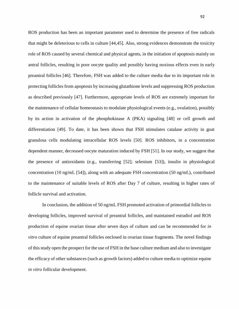
92
ROS production has been an important parameter used to determine the presence of free radicals
that might be deleterious to cells in culture [44,45]. Also, strong evidences demonstrate the toxicity
role of ROS caused by several chemical and physical agents, in the initiation of apoptosis mainly on
antral follicles, resulting in poor oocyte quality and possibly having noxious effects even in early
preantral follicles [46]. Therefore, FSH was added to the culture media due to its important role in
protecting follicles from apoptosis by increasing glutathione levels and suppressing ROS production
as described previously [47]. Furthermore, appropriate levels of ROS are extremely important for
the maintenance of cellular homeostasis to modulate physiological events (e.g., ovulation), possibly
by its action in activation of the phosphokinase A (PKA) signaling [48] or cell growth and
differentiation [49]. To date, it has been shown that FSH stimulates catalase activity in goat
granulosa cells modulating intracellular ROS levels [50]. ROS inhibitors, in a concentration
dependent manner, decreased oocyte maturation induced by FSH [51]. In our study, we suggest that
the presence of antioxidants (e.g., transferring [52]; selenium [53]), insulin in physiological
concentration (10 ng/mL [54]), along with an adequate FSH concentration (50 ng/mL), contributed
to the maintenance of suitable levels of ROS after Day 7 of culture, resulting in higher rates of
follicle survival and activation.
In conclusion, the addition of 50 ng/mL FSH promoted activation of primordial follicles to
developing follicles, improved survival of preantral follicles, and maintained estradiol and ROS
production of equine ovarian tissue after seven days of culture and can be recommended for in
vitro culture of equine preantral follicles enclosed in ovarian tissue fragments. The novel findings
of this study open the prospect for the use of FSH in the base culture medium and also to investigate
the efficacy of other substances (such as growth factors) added to culture media to optimize equine
in vitro follicular development.

93
Acknowledgments
Research supported by The National Council for Scientific and Technological Development
(CNPq), Brazil. Francisco Léo N. de Aguiar is the recipient of a doctoral scholarship from
Fundação Cearense de Apoio ao Desenvolvimento Científico e Tecnológico (Funcap).
94
References
1. Smits K, Hoogewijs M, Woelders H, Daels P, Van Soom A. Breeding or assisted
reproduction? Relevance of the horse model applied to the conservation of endangered
equids. Reprod Domest Anim 2012;47:239-48.
2. Galli C, Colleoni S, Duchi R, Lagutina I, Lazzari G. Equine assisted reproduction and
embryo technologies. Anim Reprod 2013;10:334-43.
3. Figueiredo JR, Rodrigues APR, Silva JRV, Santos RR. Cryopreservation and in vitro culture
of caprine preantral follicles. Reprod Fertil Dev 2011;23:40-7.
4. Gutierrez CG, Ralph JH, Telfer EE, Wilmut I, Webb R. Growth and antrum formation of
bovine preantral follicles in long-term culture in vitro. Biol Reprod 2000;62:1322-8
5. Chaves RN, Alves AMCV, Faustino LR, Oliveira KPL, Campello CC, Lopes CAP et al.
How the concentration of insulin affects the development of preantral follicles in goats. Cell
Tissue Res. 2011;3463:451-6.
6. Martins FS, Celestino JJH, Saraiva MVA, Matos MHT, Bruno JB, Rocha-Junior CMC,et al.
Growth and differentiation factor-9 stimulates activation of goat primordial follicles in vitro
and their progression to secondary follicles. Reprod Fertil Dev 2008;20:916-24.
7. Celestino JJH, Lima-Verde IB, Bruno JB, Matos MHT, Chaves RN, Saraiva MVA, et al.
Steady-state level of bone morphogenetic protein-15 in goat ovaries and its influence on in
vitro development and survival of preantral follicles. Mol Cell Endocrinol 2011;338:1-9.
8. Celestino JJH, Bruno JB, Saraiva MVA, Rocha RMP, Brito IR, Duarte ABG, et al. Steady-
state level of epidermal growth factor (EGF) mRNA and effect of EGF on in vitro culture of
caprine preantral follicles. Cell Tissue Res 2011;344:539-50.
9. Saha S, Shimizu M, Geshi M, Izaike Y. In vitro culture of bovine preantral follicles. Anim
Reprod Sci 2000;63:27-39.
10. Sun J, Li X. Growth and antrum formation of bovine primary follicles in long-term culture in
vitro. Reprod Biol 2013;13:221-8.
11. Serafim MKB, Duarte ABG, Silva GM, Souza CEA, Magalhães-Padilha DM, Moura AAA,
et al. Impact of growth hormone (GH) and follicle stimulating hormone (FSH) on in vitro
canine preantral follicle development and estradiol production. Growth Horm IGF Res
2015;25:85-9.

95
12. Barros VRP, Cavalcante AYP, Macedo TJS, Barberino RS, Lins TLB, Gouveia BB, et al.
Immunolocalization of melatonin and follicle-stimulating hormone receptors in caprine
ovaries and their effects during in vitro development of isolated pre-antral follicles. Reprod
Domest Anim 2013;48:1025-33.
13. Patel H, Bhartiya D, Parte S, Gunjal P, Yedurkar S, Bhatt M. Follicle stimulating hormone
modulates ovarian stem cells through alternately spliced receptor variant FSH-R3. J Ovarian
Res 2013; 20:6:52-66
14. Scarlet D, Walter I, Hlavaty J, Aurich C. Expression and immunolocalisation of follicle-
stimulating hormone receptors in gonads of newborn and adult female horses. Reprod Fertil
Dev. 2015
15. Xu J, Lawson MS, Yeoman RR, Pau KY, Barrett SL, Zelinski MB, et al. Secondary follicle
growth and oocyte maturation during encapsulated three-dimensional culture in rhesus
monkeys: effects of gonadotrophins, oxygen and fetuin. Hum Reprod 2011;26:1061-72.
16. McLaughlin M, Bromfield JJ, Albertini DF, Telfer EE. Activin promotes follicular integrity
and oogenesis in cultured pre-antral bovine follicles. Mol Hum Reprod 2010;16:644-53.
17. Wright CS, Hovatta O, Margara R, Trew G, Winston RM, Franks S, et al. Effects of follicle-
stimulating hormone and serum substitution on the in-vitro growth of human ovarian
follicles. Hum Reprod 1999;14:1555-62.
18. Magalhães DM, Fernandes DD, Mororó MB, Silva CM, Rodrigues GQ, Bruno JB, et al.
Effect of the medium replacement interval on the viability, growth and in vitro maturation of
isolated caprine and ovine pre-antral follicles. Reprod Domest Anim 2011;46:134-40.
19. Rodrigues GQ, Silva CMG, Faustino L, Bruno JB, Pinto LC, Lopes CAP, et al. Efeito de
diferentes concentrações de hormônio folículo-estimulante recombinante sobre o
desenvolvimento in vitro de folículos pré-antrais caprinos e ovinos isolados. Acta Veterinaria
Brasilica 2010; 4:144-52.
20. Thomas FH, Ethier JF, Shimasaki S, Vanderhyden BC. Follicle-stimulating hormone
regulates oocyte growth by modulation of expression of oocyte and granulosa cell factors.
Endocrinology 2005;146:941-9.
21. hen AQ, Yu SD, Wang ZG, Xu ZR, Yang ZG. Stage-specific expression of bone
morphogenetic protein type I and type II receptor genes: Effects of follicle-stimulating
hormone on ovine antral follicles. Anim Reprod Sci 2009;111:391-9.

96
22. Matos MHM, Lima-Verde IB, Luque MC, Maia JE Jr, Silva JR, Celestino JJH, et al.
Essential role of follicle stimulating hormone in the maintenance of caprine preantral follicle
viability in vitro. Zygote 2007;15:173-82.
23. Magalhães DM, Araújo VR, Lima-Verde IB, Matos MHT, Silva RC, Lucci CM, et al.
Different follicle-stimulating hormone (FSH) sources influence caprine preantral follicle
viability and development in vitro. Braz J Vet Res Anim Sci 2009;46:378-86.
24. Costa SHF, Santos RR, Rondina D, Andrade ER, Ohashi OM, Rodrigues APR, et al., Effects
of IAA in combination with FSH on in vitro culture of ovine preantral follicles. Zygote
2010;18:89-92.
25. Serafim MKB, Araújo VR, Silva GM, Duarte ABG, Almeida AP, Chaves RN et al. Canine
preantral follicles cultured with various concentrations of follicle-stimulating hormone
(FSH).Theriogenology 2010:74:749-55.
26. Itoh T, Kacchi M, Abe H, Sendai Y, Hoshi H. Growth, antrum formation, and estradiol
production of bovine preantral follicles cultured in a serum-free medium. Biol Reprod
2002;67:1099-105.
27. Sánchez F, Romero S, Smitz J. Oocyte and Cumulus Cell Transcripts from Cultured Mouse
Follicles Are Induced to Deviate from Normal In Vivo Conditions by Combinations of
Insulin, Follicle-Stimulating Hormone, and Human Chorionic Gonadotropin. Biol Reprod
2011;85:565-74.
28. Henneke DR, Potter GD, Kreider JL, Yeates BF. Relationship between condition score,
physical measurements and body fat percentage in mares. Equine Vet J 1983;15:371–2.
29. American Association of Equine Practitioners. Official Guide for Determining the Age of the
Horse. Lexington KY, USA: AAEP, 2002:1–26.
30. Chaves RN, Martins FS, Saraiva MV, Celestino JJH, Lopes CAP, Correia JC, et al. Chilling
ovarian fragments during transportation improves viability and growth of goat preantral
follicles cultured in vitro. Reprod Fertil Dev 2008;20:640–7.
31. Relave F, Lefebvre RC, Beaudoin S, Price C. Accuracy of a rapid enzyme-linked
immunosorbent assay to measure progesterone in mares. Can Vet J 2007;48:823-6.
32. Claes A, Ball BA, Troedsson MH, Curry TE, Squires EL, Scoggin KE. Molecular changes in
the equine follicle in relation to variations in antral follicle count and anti-Müllerian hormone
concentrations. Equine Vet J 2015;Sep 25. doi: 10.1111/evj.12514. [Epub ahead of print]

97
33. Loetchutinat C, Kothan S, Dechsupa S, Meesungnoen J, Jay-Gerin JP, Mankhetkorn S.
Spectrofluorometric determination of intracellular levels of reactive oxygen species in drug-
sensitive and drug-resistant cancer cells using the 2’,7’- dichlorofluorescein.
RadiatPhysChem 2005;72:323–31.
34. Méduri G, Charnaux N, Driancourt MA, Combettes L, Granet P, Vannier B, et al., Follicle-
stimulating hormone receptors in oocytes? J Clin Endocrinol Metab 2002;87:2266-76.
35. Navalakhe RM, Jagtap DD, Nayak SU, Nandedkar TD, Mahale SD. Effect of FSH receptor-
binding inhibitor-8 on FSH-mediated granulosa cell signaling and proliferation. ChemBiol
Drug Des 2013;82:178-88.
36. Ji Q, Liu PI, Chen PK, Aoyama C. Follicle stimulating hormone-induced growth promotion
and gene expression profiles on ovarian surface epithelial cells. Int J Cancer 2004;112:803-
14.
37. Wayne CM, Fan HY, Cheng X, Richards JS. Follicle-stimulating hormone induces multiple
signaling cascades: evidence that activation of Rous sarcoma oncogene, RAS, and the
epidermal growth factor receptor are critical for granulosa cell differentiation. Mol
Endocrinol 2007;21:1940-57.
38. Hunzicker-Dunn ME, Lopez-Biladeau B, Law NC, Fiedler SE, Carr DW, Maizels ET. PKA
and GAB2 play central roles in the FSH signaling pathway to PI3K and AKT in ovarian
granulosa cells. Proc Natl Acad Sci U S A. 2012;109:2979-88.
39. Haag KT, Magalhães-Padilha DM, Fonseca GR, Wischral A, Gastal MO, King SS, et al. In
vitro culture of equine preantral follicles obtained via the Biopsy Pick-Up method.
Theriogenology 2013;79:911-7.
40. Silva JRV, Ferreira MAL, Costa SHF, Santos RR, Carvalho FCA, Rodrigues APR, et al.
Degeneration rate of preantral follicles in the ovaries of goats. Small RuminantRes
2002;43:203-9.
41. Drummond AE, Findlay JK. The role of estrogen in folliculogenesis. Mol Cell Endocrinol
1999;151:57–64.
42. Qiu M, Liu J, Han C, Wu B, Yang Z, Su F, et al. The influence of ovarian stromal/theca cells
during in vitro culture on steroidogenesis, proliferation and apoptosis of granulosa cells
derived from the goat ovary. Reprod Domest Anim 2014;49:170-6.

98
43. McNatty KP, Makris A, Camillo DE, Osathanondh R, Ryan KJ. The production of
progesterone, androgens, and estrogens by granulosa cells, thecal tissue, and stromal tissue
from human ovaries in vitro. J Clin Endocrinol Metab 1979;49:687–99.
44. Talebi A, Zavareh S, Kashani MH, Lashgarbluki T, Karimi I. The effect of alpha lipoic acid
on the developmental competence of mouse isolated preantral follicles. J Assist Reprod
Genet 2012;29:175-83.
45. Martino NA, Dell'Aquila ME, Uranio MF, Rutigliano L, Nicassio M, Lacalandra GM, et al.
Effect of holding equine oocytes in meiosis inhibitor-free medium before in vitro maturation
and of holding temperature on meiotic suppression and mitochondrial energy/redox potential.
Reprod Biol Endocrinol 2014;12:99.
46. Luderer U. Ovarian toxicity from reactive oxygen species. Vitam Horm 2014;94:99-127.
47. Tsai-Turton M, Luderer U. Opposing effects of glutathione depletion and follicle-stimulating
hormone on reactive oxygen species and apoptosis in cultured preovulatory rat follicles.
Endocrinology 2006;147:1224-36.
48. Shkolnik K, Tadmor A, Ben-Dor S, Nevo N, Galiani D, Dekel N. Reactive oxygen species
are indispensable in ovulation.Proc Natl Acad Sci U S A 2011;108:1462-7.
49. Sauer H, Wartenberg M, Hescheler J. Reactive oxygen species as intracellular messengers
during cell growth and differentiation. Cell Physiol Biochem 2001;11:173-186.
50. Behl R, Pandey RS. FSH induced stimulation of catalase activity in goat granulosa cells in
vitro. Anim Reprod Sci 2002;70:215-21.
51. Chen Q, Zhang W, Ran H, Feng L, Yan H, Um X. PKCδ and θ Possibly Mediate FSH-
Induced Mouse Oocyte Maturation via NOX-ROS-TACE Cascade Signaling Pathway. PLoS
One 2014;9:1-8.
52. Eppig JJ, Schroeder AC. Capacity of mouse oocytes from preantral follicles to undergo
embryogenesis and development to live young after growth, maturation, and fertilization in
vitro. BiolReprod 1989;41:268-76.
53. Ebert R, Ulmer M, Zeck S, Meissner-Weigl J, Schneider D, Stopper H, et al., Selenium
supplementation restores the antioxidative capacity and prevents cell damage in bone marrow
stromal cells in vitro. Stem Cells 2006;24:1226-35.

99
54. Noble GK, Sillence MN. Diurnal rhythm and effects of feeding, exercise and recombinant
equine growth hormone on serum insulin concentrations in the horse. Equine Vet J
2013;45:745-50.

100
8. CAPÍTULO 3
Papel do EGF no cultivo in situ de folículos pré – antrais equinos e perfil metabolômico
“Role of EGF on in situ culture of equine preantral follicles and metabolomics profile”
Artigo Submetido Periódico: Animal Reproduction Science, (ISSN: 0378-4320) Qualis A2

101
-----Original Message-----
From: [email protected]
[mailto:[email protected]] On Behalf Of ANIREP
Sent: Tuesday, June 21, 2016 6:26 PM
To: Eduardo Leite Gastal
Subject: Revision Confirmation for ANIREP-D-16-6322R1
Ms. No. ANIREP-D-16-6322R1
Role of EGF on in situ culture of equine preantral follicles and metabolomics profile
Dear Dr. Gastal,
Thank you for the revised version of your submission to the journal Animal Reproduction
Science.
You will be able to check on the progress of your paper by logging onto the Elsevier Editorial
Systems as an Author using the following information:
http://ees.elsevier.com/anirep/
Your username is: [email protected]
If you need to retrieve password details, please go to:
http://ees.elsevier.com/anirep/automail_query.asp

102
Kind regards,
Editorial Office Staff
Animal Reproduction Science
***********************************************************

103
RESUMO
Os efeitos das concentrações do fator de crescimento epidermal (EGF - 0, 10, 50 e 100 ng/mL)
foram avaliados no cultivo in vitro (CIV) de folículos pré – antrais equinos inclusos em tecido
ovariano, usando histologia, produção hormonal, espécies reativas de oxigênio (EROs), e
metabolômica. Após o CIV, a percentagem de folículos normais foi menor (P < 0.05) para todos
os tratamentos, quando comparados com o controle não cultivado. O tratamento EGF 50 ng/mL
teve maior (P < 0.05) número de folículos normais após 7 dias de cultivo quando comparado com
o tratamento 0 ng/mL e 100 ng/mL de EGF. Os diâmetros foliculares e oocitários foram maiores
(P < 0.05) com 50 ng/mL do que os outros tratamentos cultivados, mas similares (P > 0.05) ao
controle não-cultivado. O tratamento 50 ng/mL de EGF foi o único tratamento que manteve a
produção de EROs através do CIV. O perfil metabolômico do meio de cultivo indicou que onze
íons avaliados por importância da projeção variável (VIP escores) foram hiper representados no
tratamento 50 ng/mL de EGF. Em conclusão, o tratamento EGF 50 ng/mL manteve a
sobrevivência folicular, a produção de EROs e promoveu a ativação folicular de folículos pré –
antrais equinos inclusos em tecido ovariano.
Palavras - chave: Cultivo in vitro. Folículos Pré – Antrais Equinos, EGF, Metabolômica, Espécies
Reativas de Oxigênio.

104
Role of EGF on in situ culture of equine preantral follicles and
metabolomics profile
F.L.N. Aguiara, F.O. Lunardia, L.F. Limaa, J.B. Brunoa, B.G. Alvesa, D.M. Magalhães-Padilhab,
F.W.S. Cibinc, L. Berionid, G.A. Apgare, E.G. Lo Turcod, E.L. Gastale,*, J.R. Figueiredoa
a Laboratory of Manipulation of Oocytes and Preantral Follicles (LAMOFOPA), State University
of Ceará, Fortaleza, Ceará, Brazil
b Postgraduate Biotechnology, Potiguar University/Laureate International Universities, Av.
Senador Salgado Filho, 1610, Lagoa Nova, Natal, 59056-000, CE, Brazil
c Federal University of Pampa, Uruguaiana, Rio Grande do Sul, Brazil.
d Department of Surgery, Division of Urology, Human Reproduction Section, Sao Paulo Federal
University, Sao Paulo, SP, Brazil.
e Department of Animal Science, Food and Nutrition, Southern Illinois University, Carbondale,
Illinois, USA
Running head: EGF effects on equine preantral follicles
*Corresponding author: Eduardo Gastal, Department of Animal Science, Food and Nutrition,
Southern Illinois University, 1205 Lincoln Drive, MC 4417, Carbondale, IL, 62901, USA.
Telephone: (618) 453-1774; FAX: (618) 453-5231; e-mail: [email protected]

105
ABSTRACT
The effects of EGF concentrations (0, 10, 50, and 100 ng/ml) on in vitro culture (IVC) of equine
preantral follicles were evaluated using histology, estradiol and reactive oxygen species (ROS)
production and metabolomics. After IVC, the percentage of normal follicles was lower (P <
0.05) for all treatments when compared to non-cultured control. EGF 50 ng/ml treatment had
more (P < 0.05) normal follicles after Day 7 of culture when compared with EGF 0 and 100
ng/ml. EGF 50 ng/ml had more (P < 0.05) developing follicles than the 0 ng/ml and 10 ng/ml
EGF treatments. Follicular and oocyte diameters were greater (P< 0.05) with EGF 50 ng/ml than
the other cultured treatments, but similar (P > 0.05) to the non-cultured control. From Day 1 to
Day 7 estradiol production increased (P < 0.05) in all EGF treatments. EGF 50 ng/ml was the
only treatment that maintained ROS production through IVC. Metabolomics profiles of the spent
media indicated that eleven ions from VIP score were higher represented in the EGF 50 ng/ml
treatment. In conclusion, EGF 50 ng/ml treatment maintained follicle survival and ROS
production, and promoted activation of cultured equine preantral follicles enclosed in ovarian
tissue.
Keywords: In vitro culture, equine preantral follicles, EGF, metabolomics, reactive oxygen
species.

106
1. Introduction
An important strategy to maximize the reproductive potential of domestic animals consists of
in vitro culture (IVC) of preantral follicles, aiming to preserve follicular survival and
development, and minimize oocyte losses observed in vivo (Xu et al., 2013; Haag et al., 2013a;
Araújo et al., 2014).
Considering the limited efficiency in assisted reproduction technologies in mares (e.g.,
superovulation and in vitro fertilization) studies using IVC of preantral follicles emerge as a
future alternative source of meiotically competent oocytes. Moreover, mares share similarities
with women in reproductive dynamic events, as previously reported for follicular waves, changes
in hormonal levels, and age-related reduction in fertility (Ginther et al., 2004; Carnevale, 2008;
Gastal, 2011; Cox et al., 2015). Particularly in horses, studies on IVC of preantral follicles are
scarce because of the prohibition of slaughterhouses in some countries that could provide ovaries
as a biological source. More recently, the mare has also been suggested as an interesting model
for studies related to preantral follicles (Haag et al., 2013a, 2013b, 2013c; Alves et al., 2015).
Previous reports on IVC of equine preantral follicles have produced limited results using follicles
chemically isolated (Szlachta and Tischner, 2000, 2004) or enclosed in ovarian tissue (Haag et
al., 2013a; Alves et al., 2015).
A base medium (α-MEM) has been used recently with limited success for culture of equine
preantral follicles enclosed in ovarian tissue (Haag et al., 2013c). Furthermore, insulin at a
physiological concentration (10 ng/ml) improved survival and development of equine preantral
follicles (Aguiar et al., 2016a). In addition, 50 ng/ml of FSH promoted activation of primordial
follicles, improved survival of preantral follicles, and maintained estradiol and ROS production
of equine ovarian tissue after seven days of culture (Aguiar et al., 2016b). Ascorbic acid

107
supplementation (50 and 100 μg/ml) has also improved the development of equine preantral
follicles after 6 days of IVC (Gomes et al., 2015). However, the role of different substances,
including intraovarian factors such as epidermal growth factor (EGF), which has potent mitogen
activity (Silva et al., 2004; Wu and Tian, 2007; Celestino et al., 2009; Fujihara et al., 2014), is
still unknown on in vitro survival and development of equine preantral follicles. EGF belongs to
the transforming growth factor β protein family (TGF-β; Silva et al., 2013). Once bound to its
tyrosine kinase-like receptor, EGF induces a series of intracellular events, with an activation of
second messengers responsible for regulating cell growth and differentiation (Ma et al., 2015).
Previous reports have shown that EGF, in a dose dependent manner (range, 0.5 to 200
ng/ml), affected survival, activation, and growth of IVC of preantral follicles [e.g., ovine: 100
ng/ml (Andrade et al., 2005), and 75 ng/ml (Peng et al., 2010), caprine: 1 to 200 ng/ml (Celestino
et al., 2009), human: 50 ng/ml (Roy and Kole, 1998)], proliferation of granulosa cells [porcine:
10 ng/ml (Mao et al., 2004)], and reduced the rate of atresia of preantral follicles [bubaline: 50
ng/ml (Gupta et al., 2002), porcine: 1.5 ng/ml (Wu and Tian, 2007), and caprine: 100 ng/ml
(Silva et al., 2004)].
Metabolomics may provide timely information about molecular interactions and metabolic
signaling, indicating new biomarkers and specific supplies for successful IVC and physiological
modulation (Montani et al., 2012). Molecules secreted in the medium during IVC have been
investigated recently as an alternative approach (metabolomics) to determine oocyte maturation
and embryo development potential in several species [e.g., humans (Wallace et al., 2012;
Montani et al., 2014), murine (Preis et al., 2005), and bovine (Matoba et al., 2014). However, to
date evaluation of metabolites from preantral follicles isolated or enclosed in ovarian tissue in
spent culture medium has been performed only in a few species, demonstrating different

108
requirements of carbohydrates and amino acids for follicles cultured in different oxygen tensions
(Gook et al., 2014). Matrix-assisted laser desorption/ionization time-of-flight mass spectrometry
(MALDI-TOF MS) has been efficiently employed as a tool to analyze lipid composition,
providing a study of highly complex structures (Schiller et al., 1999).
Studies investigating the dose-dependent effect of EGF on IVC of equine preantral follicles
and the metabolic characterization of the spent culture media remain unknown. Thus, the aim of
this study was to evaluate the effect of different concentrations of EGF (0, 10, 50 and 100 ng/ml)
on IVC of preantral follicles enclosed in ovarian tissue fragments, using the following end
points: (i) follicular survival, (ii) follicular growth, (iii) hormonal production, (iv) reactive
oxygen species (ROS), and (v) metabolomics profile of the spent culture media.
2. Materials and methods
2.1. Chemicals
Unless otherwise noted, the culture media and other chemicals used in the present study were
purchased from Sigma Chemical Co. (St. Louis, Mo., USA).
2.2. Animals and ovaries
The research protocol (#12637266-7) was approved by the Ethics and Animal Use
Committee (CEUA) of State University of Ceará (UECE), Fortaleza, CE. The ovaries were
harvested from euthanized mares (n = 5) positive but asymptomatic for Equine Infectious
Anemia (EIA), during February to May, in the state of Ceará, Brazil. The mares were crossbred,
6 to 10 years old (mean, 7.2 ± 1.9 years), and had body condition scores between 4 and 6 [1,
emaciated to 9, obese; Henneke et al., 1983]. Dental characteristics (American Association of

109
Equine Practitioners, 2002) were used to estimate the age of the mares. At the time of ovary
harvesting, mares were non-pregnant and cycling, based on the presence of large
(maturing/growing) or small (regressing) corpus luteum and a preovulatory (> 30 mm) follicle in
one or both ovaries.
Immediately after euthanasia, the ovaries were collected and washed in 70% alcohol,
followed by two washes in minimum essential medium (MEM) supplemented with 25 mM
HEPES. The ovaries were placed into tubes containing 150 ml of MEM supplemented with 100
μg/ml penicillin and 100 μg/ml streptomycin and transported to the laboratory at 4°C (Chaves et
al., 2008) within 4 hours. In the laboratory, both ovaries of each animal were stripped of
surrounding fat tissue and ligaments. Subsequently, 54 ovarian cortical tissue samples
(approximate size, 3 x 3 x 1 mm) were obtained from each pair of ovaries under sterile
conditions using a scalpel blade. Cortical samples were recovered avoiding adjacent areas to a
corpus luteum and preovulatory follicle. A pool of six similar ovarian fragments randomly
chosen from the same animal were distributed in nine treatments. Five replicates (mares) were
performed for each treatment.
2.3. Culture of preantral follicles and experimental design
Each ovarian tissue fragment was placed in a well of a 24-well culture plate containing 1 ml
of culture media. Culture was performed at 39°C in a humidified atmosphere with 5% CO2.
Fresh medium was prepared and incubated for at least 1 h before use. The basic culture medium
consisted of α-MEM (pH 7.2–7.4) supplemented with 2 mM glutamine, 2 mM hypoxanthine,
1.25 mg/ml bovine serum albumin (BSA), 10 ng/ml insulin, 5.5 μg/ml transferrin, 5.0 ng/ml
sodium selenite, 100 µg/ml penicillin, and 100 µg/ml streptomycin, which was called α-MEM+.

110
To test the effect of EGF in cultures of preantral follicles, the basic medium (control) was
supplemented with increasing concentrations of EGF, generating the following treatments: EGF
0 ng/ml, EGF 10 ng/ml, EGF 50 ng/ml, and EGF 100 ng/ml.
The ovarian tissue fragments were then either fixed for histological analyses (fresh, non-
cultured control group) or placed in culture for one (Day 1) or seven (Day 7) days. The whole
culture medium was replaced every other day, and before each replacement, 1 ml of medium was
collected after 24 h (Days 1 and Days 7 of culture), and stored at -80°C for hormonal and ROS
analyses.
2.4. Follicle morphology and development
Follicular morphology and development of preantral follicles were assessed in ovarian
fragments in situ before (fresh, non-cultured control group) and after IVC of ovarian tissue. Once
harvested, ovarian tissue was fixed in paraformaldehyde solution at 4°C for 12 h and kept in 70%
alcohol, and submitted to histological analysis. Ovarian fragments were dehydrated by use of a
graded series of alcohol, embedded in paraffin wax, and cut into serial sections of 10 μm.
Samples were stained with periodic acid-Schiff (PAS) and counterstained with hematoxylin. A
total of 2,700 slides with 27,000 tissue sections were produced from 270 ovarian fragments.
Histology slides were analyzed using light microscopy (Nikon, Tokyo, Japan) at 400X
magnification. The histological evaluation and classification of the follicles were performed by
only one operator, who was blind to the treatments. However, five other trained helpers, also
blind to the treatments, were used to find preantral follicles on the histological sections of each
fragment.

111
Once that equine preantral follicles had a similar morphology to other species (Haag et al.,
2013a), the end points evaluated were number of follicles, follicle class (primordial, one layer of
flattened pre-granulosa cells surrounding the oocyte; transitional, one layer of flattened and
cuboidal granulosa cells; primary, one layer of cuboidal granulosa cells; and secondary, two or
more layers of cuboidal granulosa cells; Maciel et al., 2004), follicle morphology, and diameter
of follicles and oocytes.
To evaluate follicular development, the percentages of both normal primordial and
developing follicles (transitional, primary, and secondary) were calculated before (fresh non-
cultured control group) and after culture for each treatment. Regarding morphology, follicles
were classified as normal (follicle containing an intact oocyte and granulosa cells well-organized
in layers without pyknotic nucleus) or abnormal (oocyte with pyknotic nucleus, retracted
cytoplasm, or disorganized granulosa cells detached from the basement membrane; Haag et al.,
2013b). Every follicle was examined in each section in which it appeared and matched with the
same follicle on adjacent sections to avoid double counting, thereby ensuring that each follicle
was counted only once.
Follicle and oocyte diameters were measured only in morphologically normal follicles using
software (Nis-Element AR 3.0) coupled with an epifluorescence microscope (Zeiss, Cologne,
Germany). Follicle diameter was measured from one edge to the other edge of the outermost
layer of granulosa cells. Oocyte diameter was measured from one edge of the oocyte membrane
to the other. Two perpendicular diameters were recorded for each measurement and the average
of those two values was calculated.
2.5. Hormone analyses

112
To evaluate follicular steroidogenesis in vitro, concentrations of estradiol and progesterone
were measured in spent culture media against standard dilutions according to manufacturer’s
instructions using competitive immunoassay commercial kits: IMMULITE 2000 Estradiol
(catalog No: L2KE22; Siemens Medical Solutions Diagnostics, 5210 Pacific Concourse Drive,
Los Angeles, CA, USA) and IMMULITE 2000 Progesterone (catalog No: L2KPW2; Siemens
Medical Solutions Diagnostics, 5210 Pacific Concourse Drive, Los Angeles, CA, USA). The two
hormonal assay kits for estradiol and progesterone in this study have been used previously and
validated for different species, including the horse (Relave et al., 2007; Claes et al., 2015). The
analytical sensitivity of the assay was 15 pg/ml (range, 20 to 2000 pg/ml) for estradiol and 0.1
ng/ml (range, 0.2 to 40 ng/ml) for progesterone.
2.6. ROS analysis
The levels of reactive oxygen species were determined in spent cultured media using a
spectrofluorimetric method (Loetchutinat et al., 2005). Culture media from all EGF-treated
groups were incubated with 10 μl of 2',7'-dihidrodiclorofluorescein diacetate (DCHF-DA; 1
mM). The oxidation of DCHF-DA to dichlorofluorescein was measured for detection of reactive
species in the medium. The intensity of fluorescence emission was recorded at 520 nm (with 480
nm excitation) for 2 h after addition of DCHF-DA to the medium.
2.7. Metabolomics analysis
Thirty-two samples of spent culture media (eight samples in each EGF treatment) were
analyzed in four replicates. Mass spectra from the spent culture media of the different treatments
at Day 7 were acquired using MALDI equipment. The mass spectra were acquired in positive ion

113
mode using a MALDI AUTO FLEX SPEEDY TOF/TOF (Bruker Daltonics, Billerica, Ma,
USA) mass spectrometer equipped with a 1 kHz solid-state laser with a wavelength of 355 nm
and a range of 600–1200 m/z in the reflectron mode. Typical operating conditions for both modes
were as follows: laser energy, 750 (arbitrary units); sample plate, 22.5 Kv; and laser irradiation
of 100 shots in the region where the sample had been placed on the target plate for 60–90 sec in
the positive ion mode. The quality of the spectra was individually evaluated with respect to ion
count. To avoid potential noise influence on the assays, only spectra presenting an ion count >
800 were included in the study. The mass spectrum of each sample was acquired using Flex
Control software (Bruker Daltonics, Billerica, MA, USA) and the m/z values and ion intensities
were exported to an Excel table. The ions presenting zero values in > 50% of the samples were
removed, and the whole data set was standardized to a common peak (most intense) present in all
samples. The ions were identified by using METLIN metabolite database from Scripps Center
for Metabolomics (http://metlin.scripps.edu/index.php). The mass error was calculated and only
assignments with an error < 50 ppm were considered.
2.8. Statistical analyses
Statistical analyses were carried out using R statistical software version 3.0.2 (R Foundation
for Statistical Computing, Vienna, Austria). Data for end points that were not normally
distributed according to a Kolmogorov–Smirnov test were transformed to natural logarithms or
ranks. Chi-square test was used to compare the percentage of morphologically normal and
growing preantral follicles among and within treatments. Follicle and oocyte diameters, estradiol,
progesterone, and ROS were compared among treatments by one-way ANOVA. For
metabolomics analyses of the spent culture media after seven days of culture, the data were

114
analyzed using the principal component analysis (PCA), followed by a partial least square
discrimination analysis (PLS-DA), and combined with variable influence in the projection (VIP)
scores. The statistical analysis was performed using the Metabo-Analyst 3.0
(http://www.metaboanalyst.ca). According to the results, one-way ANOVA and subsequently t-
or Fisher-tests were used when appropriate. Thereafter, statistical ions were identified in the
online database, through the m/z values detected by the equipment. A probability of P < 0.05
indicated that a difference was significant, and P > 0.05 and ≤ 0.1 indicated that a difference
approached significance. Data are presented as the mean ± SEM, unless otherwise indicated.
3. Results
3.1. Number of follicles evaluated
A total of 207, 317, 373, 345, and 342 preantral follicles were evaluated in the fresh non-
cultured control, EGF 0 ng/ml, EGF 10 ng/ml, EGF 50 ng/ml, and EGF 100 ng/ml treatment
groups, respectively. Altogether, 1,584 follicles were evaluated, with an average of 236.0 ± 26.6
follicles evaluated per treatment.
3.2. Effect of EGF on follicular morphology
The percentage of morphologically normal follicles was reduced (P < 0.05) after one and
seven days of culture when compared to the fresh non-cultured control group (Table 1). Overall,
after each culture time, EGF 50 ng/ml treatment had a greater (P <0.05) percentage of normal
follicles than the other treatments, except for the EGF 100 ng/ml (Day 1) and EGF 10 ng/ml
(Day 7) treatments. A reduction (P < 0.05) in the percentage of normal follicles was observed in
all treatments from Day 1 to Day 7 of culture.

115
3.3. Follicular development after in vitro culture
The percentages of primordial and developing follicles are shown (Fig. 1 A,B). After one day
of culture, only the EGF 0 ng/ml and 10 ng/ml treatments decreased (P < 0.05) the percentage of
primordial follicles and increased (P < 0.05) the percentage of developing follicles when
compared to the fresh non-cultured control group. In all treatments, after seven days of culture,
the percentage of primordial follicles decreased (P < 0.05) and the percentage of developing
follicles increased (P < 0.05) when compared to fresh non-cultured control group. The EGF 50
ng/ml treatment had a greater (P < 0.05) rate of developing follicles on Day 7 of culture, except
when compared with the EGF 100 ng/ml treatment. Regarding to follicle activation, except for
the EGF 0 ng/ml treatment, all EGF treatments had a reduction (P < 0.05) in the percentage of
primordial follicles and an increase (P < 0.05) in developing follicles between Day 1 and Day 7
of culture.
After one and seven days of culture, the EGF 50 ng/ml treatment maintained follicular and
oocyte diameters similar to fresh non-cultured control group (P > 0.05; Table 2). In addition,
regardless of culture time, the EGF 50 ng/ml treatment had greater (P < 0.05) follicular and
oocyte diameters than the other EGF treatments. Moreover, only the EGF 50 ng/ml treatment
maintained (P > 0.05) follicular and oocyte diameters between Day 1 and Day 7 of culture.
3.4. Hormonal production
Estradiol production was not different (P > 0.05) among treatments (Table 3). However,
estradiol concentration in the spent culture medium increased (P < 0.05) in the EGF 10 ng/ml, 50
ng/ml, and 100 ng/ml treatments between Day 1 and Day 7 of culture. Progesterone production

116
in the culture media was detected in a small number of samples (20 out of 158; data not shown).
Therefore, data were not analyzed statistically.
3.5. Reactive oxygen species production
After one day of culture, no difference (P > 0.05) among treatments was observed in ROS
production (Table 4). However, after seven days of culture, the EGF 50 ng/ml treatment
produced more (P < 0.05) ROS than the other treatments, except when compared with the EGF
100 ng/ml treatment. After seven days of culture, the EGF 50 ng/ml treatment was the only
treatment that maintained (P > 0.05) ROS production similar to Day 1 of culture.
3.6. Metabolomics analysis
A total of 32 samples of spent culture media were analyzed. These samples produced an
average of 7,356 peaks, with 229.9 peaks per sample. Data submitted to PCA generated 32
components. Pairwise score plots providing overviews of the various separation patterns among
the most significant PCs are presented (Fig. 2A). Furthermore, a scree plot showing the variances
explained by the selected PCs is shown (Fig. 2B). The five principal components (PC1, PC2,
PC3, PC4, and PC5; Fig. 2A) represented > 53.9% of the variance observed in the data (Fig. 2B).
Two-dimensional score plots between selected PCs 1 and 2 using principal component analysis
(PCAs; Fig. 3A), and partial least squares - discriminant analysis (PLS-DA; Fig. 3B) are shown.
Important features identified with the partial least square discrimination analysis (PLS-DA) are
shown by the variable importance in projection (VIP scores; Fig. 4). The colored boxes on the
right indicate the relative concentrations of the corresponding metabolite on Day 7 in each EGF
treatment (0 ng/ml, 10 ng/ml, 50 ng/ml, and 100 ng/ml). A complete separation of ions was

117
conducted using VIP scores to obtain the most important ions responsible for the variance among
the groups (Table 5; Table S1). Ten out of 15 VIP ions were identified. For the 50 ng/ml
treatment, 6 ions were listed by the METLIN metabolite database mainly as chemical groups of
amino acids, carbohydrates, lipids, and steroids (Table S1)
After linear regression, PC5 showed a negative correlation with survival (R2 = 0.15, r =
˗0.38, P < 0.05), follicle diameter (R2 = 0.19, r = ˗0.44, P < 0.01), and oocyte diameter (R2 =
0.19, r = ˗0.44, P < 0.01; Fig. 5). Because the other PCs (e.g., PC1, PC2, PC3, and PC4) were not
correlated (P > 0.05) with survival rate, follicle diameter, and oocyte diameter, data were not
further evaluated and presented. The factor loadings extracted by the principal component
analysis of metabolites found 13 relevant ions for PC5 (Table S2), and six of those ions were
identified by the METLIN database. From the six ions identified in PC5, three ions were
considered the most representative (Table 6). Furthermore, from the three most representative
ions, three metabolites belonging to the classes of phenols, Glycosyl, and Benzophenone were
identified (Table 6; Table S3).
4. Discussion
To our knowledge, this is the first study to demonstrate the beneficial effect of EGF for
maintenance of in vitro follicular survival (i.e., morphologically normal follicles), and activation
of equine preantral follicles enclosed in ovarian tissue and its subsequent metabolomics profile in
the spent media.
This study considered IVC in situ (i.e., preantral follicles enclosed in ovarian tissue) because
of the low efficiency of isolating early preantral follicles (Telfer and Watson, 2000; Szlachta and
Tischner, 2000, 2004; Haag et al., 2013a). Because of this, a “two-step culture system”, where

118
follicles are cultured in situ for some days until the secondary stage, and then are isolated
mechanically or enzymatically, has been proposed for other species (Telfer and Zelinski, 2013).
Therefore, knowledge regarding hormonal interactions, ROS production, and nutrient
requirements for preantral follicles and stromal cells using an in situ model is imperative. In this
study, because of ovarian fragments from the same animal were randomly allocated among
treatments, a similar contribution of preantral follicles and stromal cells from each fragment
(same number and comparable size in each treatment) was expected.
In the present study, greater follicular survival was obtained with EGF 50 ng/ml after seven
days of culture, except for the EGF 10 ng/ml treatment. After binding to its receptor tyrosine
kinase, EGF stimulates cell survival by acting on intracellular signaling pathways such as
PIK3/AKT, MAPK/ERK, and JAK/STAT (Henson and Gibson, 2006). EGF reduces apoptosis
of preantral follicles (porcine: Mao et al., 2004) and follicular cells (rats: Tilly et al., 1992), and
inhibits spontaneous cleavage of DNA through stimulation of anti-apoptotic genes, such as BAD
(Sastry et al., 2006), survivin (Peng et al., 2006), and NFkB (Sethi et al., 2006).
Previous studies demonstrated the importance of supplementation of an appropriate
concentration of EGF on in vitro culture of preantral follicles either enclosed in ovarian tissue
(caprine: 100 ng/ml, Silva et al., 2004, 1 and 10 ng/ml, Celestino et al., 2009; ovine: 100 ng/ml,
Andrade et al., 2005) or in an isolated form (caprine: 50 ng/ml, Zhou and Zhang, 2005a, 50
ng/ml, Silva et al., 2013; ovine: 50 ng/ml, Hemamalini et al., 2003, 50 ng/ml, Santos et al., 2014;
bubaline: 50 ng/ml, Gupta et al., 2002; bovine: 50 ng/ml, Wandji et al., 1996, 0.5 ng/ml,
Gutierrez et al., 2000). In a previous study in goats (Celestino et al., 2009), the concentration of 1
and 10 ng/ml EGF maintained follicular survival similar to the control group after seven days of
culture, but no differences were observed between the 10 and 50 ng/ml EGF treatments.

119
The percentage of developing follicles, as an indicative of follicle activation, was greater
after seven days of culture on the EGF 50 ng/ml treatment, when compared with the other
treatments, except for the EGF 100 ng/ml treatment. Similar results have been reported
previously (caprine: Celestino et al., 2009, ovine: Andrade et al., 2005). In our study, follicle
activation possibly occurred through EGF action in cell cycle progression (Luo et al., 2007;
L'Hortet et al., 2012; Wee et al., 2015), promoting cell differentiation through the transcription
factor stimulus such as activator protein 1 (AP-1), Cyclin D1, (Mitsudomi and Watanabe, 2010),
and cAMP response element-binding protein (CREB; Rodrigues et al., 2010).
In this study, only the EGF 50 ng/ml treatment maintained follicular and oocyte diameters
after seven days of culture. EGF has increased follicular diameters when associated with
hormones (e.g., FSH: Wu and Tian, 2007; Celestino et al., 2011; LH: Saraiva et al., 2010) or
growth factors (e.g., IGF-I: Zhou and Zhang, 2005b). Therefore, it seems reasonable to assume
that the association of EGF with other growth factors or hormones is necessary to promote
follicular growth in mares.
In our study, irrespective of culture time, estradiol production by cultured ovarian tissue was
not affected by the different EGF concentrations. Moreover, it revealed that fragments of equine
ovarian tissue containing preantral follicles when cultured in vitro exhibited steroidogenic
activity (estradiol production). However, besides follicular cells, we cannot rule out the
possibility that other cells such as ovarian stromal cells could have contributed to estradiol
production under our culture conditions, since it has been reported that these cells are capable of
producing steroids (McNatty et al., 1979; Qiu et al., 2014). Our results are in agreement with a
previous study (Jones et al., 1982) reporting that the addition of EGF in different concentrations

120
(0.1, 1, and 10 ng/ml) to the culture medium did not affect estradiol production in rat granulosa
cells.
The treatment EGF 50 ng/ml maintained ROS production throughout the culture period in
our study. Production of ROS has been used to evaluate in vitro oocyte quality (Ou et al., 2012;
Martinho et al., 2014), and the balance between the production and degradation of ROS is an
indicator of oxidative control (Rizzo et al., 2012; Winterbourn, 2014). Furthermore, ROS might
be important due to its modulatory role as a second messenger in intracellular pathways such as
MAPK/PI3K (McCubrey et al., 2006), and blockage or inactivation of inhibitory phosphatases
(Shkolnik et al., 2011).
This study shows for the first time the metabolomics profile in the spent culture medium
obtained on Day 7 from equine preantral follicles enclosed in ovarian tissue cultured in vitro. In
the current study, eleven ions from VIP score were higher represented in the EGF 50 ng/ml
treatment, and three ions in the EGF 0 ng/ml treatment. Moreover, negative correlations with
follicle survival and follicle and oocyte diameters were observed for the PC5 component. In
addition, from the ions identified, chemical groups such as amino acids, carbohydrates, lipids,
and steroids were listed.
To support the in vitro culture of preantral follicles, the base medium α-MEM with the most
enriched formulation containing substances such as aminoacids, vitamins, and inorganic salts,
was used in the present study. Previous studies demonstrated that aminoacids (Figueiredo et al.,
1994), vitamins (Eppig and O’brien, 1996), and inorganic salts (Wright et al., 1997) are essential
to promote the in vitro culture of preantral follicles in different species. However, in our study,
the negative correlation observed in PC5 led us to hypothesize the potential substances present in
the culture medium that could have been responsible for this effect. After a METLIN metabolite

121
database search, substances such as Dinex (Phenol group), Leonuriside A (Glycosyl group), and
Avobenzone were identified as having a potential deleterious effect on in vitro cell culture assay
systems. Dinex metabolite acts as an antagonist of steroidogenic receptors (PubChem
AID=743078; National Center for Biotechnology Information, 2015a), Leonuriside A possesses
an anti-steroidogenic activity that may lead to a reduction in cell proliferation (PubChem
AID=470167; National Center for Biotechnology Information, 2015b). Avobenzone, a sunscreen
blocker, has an antagonistic action on the estrogen alpha-receptor signaling pathway (PubChem
AID=743091; National Center for Biotechnology Information, 2015c). This fact may support the
findings that EGF supplementation in this study, regardless of day of culture, did not have a
positive effect on estradiol production or follicular and oocyte growth.
5. Conclusions
In conclusion, 50 ng/ml of EGF maintained follicle survival and ROS production, and
promoted follicle activation in equine preantral follicles enclosed in ovarian tissue after in vitro
culture. In addition, a metabolomics profile for the spent culture media identified three
substances (Dinex, Leonuriside A, and Avobenzene) that had a potential negative effect on
follicle survival, and follicle and oocyte diameters at Day 7 of culture. Therefore, further
research is warranted into the association between EGF with other growth factors and hormones
to improve in vitro culture of equine preantral follicles.
Conflict of interest
The authors declare that there is no conflict of interest that could be perceived as prejudicing
the impartiality of the research reported.

122
Acknowledgments
Research supported by The National Council for Scientific and Technological Development
(CNPq; Grant #407594/2013-2), Brazil. Francisco Léo N. de Aguiar is the recipient of a doctoral
scholarship from Fundação Cearense de Apoio ao Desenvolvimento Científico e Tecnológico
(FUNCAP).
Figure legends
Fig. 1. Percentage of primordial and developing follicles (transitional, primary, and secondary)
in a fresh non-cultured control group and after in vitro culture for one or seven days using
different concentrations of EGF. a,b Within each treatment, values without a common letter
differed (P < 0.05). A,B Within days, values without a common letter differed (P < 0.05). *
Differed (P < 0.05) from fresh non-cultured control.
Fig. 2. (A) Pairwise score plots between the selected principal compounds (PCs). The explained
variance of each PC is presented in the corresponding diagonal cell. (B) Scree plot showing the
variance explained by the PCs. The green line on top shows the accumulated variance explained;
the blue line underneath shows the variance explained by individual PC.
Fig. 3. Two-dimensional score plots between selected PCs 1 and 2 using (A) principal
component analysis (PCAs), and (B) partial least squares - discriminant analysis (PLS-DA). The
explained variances are shown in parentheses in both axes. Color dots represent different
treatments as indicated.

123
Fig. 4. Important features identified with the partial least square discrimination analysis (PLS-
DA) are shown by variable importance in projection (VIP scores). The colored boxes on the right
indicate the relative concentrations of the corresponding metabolite in each experimental
treatment by EGF (0 ng/ml, 10 ng/ml, 50 ng/ml, and 100 ng/ml) under study.
Fig. 5. Relationship of (A) normal preantral follicles, (B) follicle diameter, and (C) oocyte
diameter with principal component 5 (scores). Regardless of treatment, each point on the graph
represents a medium sample collected after seven days of in vitro culture (n = 32). A linear
regression is represented by the equation and the line (black) for (A) [normal preantral follicles =
32.975 ˗ (0.933 × component score), R2 = 0.15, r = ˗0.38, P < 0.05]; (B) [follicle diameter =
31.225 ˗ (0.604 × component score), R2 = 0.19, r = ˗0.44, P < 0.01]; and (C) [oocyte diameter =
24.050 ˗ (0.592 × component score), R2 = 0.19, r = ˗0.44, P < 0.01].

124
References
Alves, K.A., Alves, B.G., Rocha, C.D., Visonna, M., Mohallem, R.F., Gastal, M.O., Jacomini,
J.O., Beletti, M.E., Figueiredo, J.R., Gambarini, M.L., Gastal, E.L., 2015. Number and
density of equine preantral follicles in different ovarian histological section thicknesses.
Theriogenology83, 1048-1055.
Aguiar, F.L.N., Lunardi, F.O., Lima, L.F., Rocha, R.M.P., Bruno, J.B., Magalhães-Padilha,
D.M., Cibin, F.W.S., Rodrigues, A.P.R., Gastal, M.O., Gastal, E.L., Figueiredo, J.R., 2016a.
Insulin improves in vitro survival of equine preantral follicles enclosed in ovarian tissue and
reduces reactive oxygen species production after culture. Theriogenology85, 1063-1069.
Aguiar, F.L.N., Lunardi, F.O., Lima, L.F., Rocha, R.M.P., Bruno, J.B., Magalhães-Padilha,
D.M., Cibin, F.W.S., Nunes-Pinheiro, D.C.S., Gastal, M.O., Rodrigues, A.P.R., Apgar, G.A.,
Gastal, E.L., Figueiredo, J.R., 2016b. FSH supplementation to culture medium is beneficial
for activation and survival of preantral follicles enclosed in equine ovarian tissue.
Theriogenology 85, 1106-1112.
American Association of Equine Practitioners, 2002. Official Guide for Determining the Age of
the Horse. Lexington KY, USA: AAEP, 1-26.
Andrade, E.R., Seneda, M.M., Alfieri, A.A., Oliveira, J.A., Bracarense, A.P., Figueiredo, J.R.,
Toniolli R., 2005. Interactions of indole acetic acid with EGF and FSH in the culture of ovine
preantral follicles. Theriogenology 64, 1104-1113.
Araujo, V.R., Gastal, M.O., Figueiredo, J.R., Gastal, E.L., 2014. In vitro culture of bovine
preantral follicles: a review. Reprod. Biol. Endocrinol. 12, 78.
Celestino, J.J.H., Bruno, J.B., Lima-Verde, I.B., Matos, M.H.T., Saraiva, M.V.A., Chaves, R.N.,
Martins, F.S., Lima, L.F., Name, K.P.O., Campello, C.C., Silva, J.R.V., Bao, S.N.,
Figueiredo, J.R., 2009. Recombinant epidermal growth factor maintains follicular
ultrastructure and promotes the transition to primary follicles in caprine ovarian tissue
cultured in vitro. Reprod. Sci. 16, 239-246.
Celestino, J.J.H., Bruno, J.B., Saraiva, M.V.A., Rocha, R.M.P., Brito, I.R., Duarte, A.B., Araújo,
V.R., Silva, C.M., Matos, M.H.T., Campello, C.C., Silva, J.R.V., Figueiredo, J.R.,
2011.Steady-state level of epidermal growth factor (EGF) mRNA and effect of EGF on in
vitro culture of caprine preantral follicles. Cell Tissue Res.344, 539-550.

125
Chaves, R.N., Martins, F.S., Saraiva, M.V.A., Celestino, J.J.H., Lopes, C.A.P., Correia, J.C.,
Lima-Verde, I.B., Matos, M.H.T., Báo, S.N., Name, K.P.O., Campello, C.C., Silva, J.R.V.,
Figueiredo, J.R., 2008. Chilling ovarian fragments during transportation improves viability
and growth of goat preantral follicles cultured in vitro. Reprod. Fertil. Dev. 20, 640-647.
Claes, A., Ball, B.A., Troedsson, M.H., Curry, T.E., Squires, E.L., Scoggin, K.E., 2015.
Molecular changes in the equine follicle in relation to variations in antral follicle count and
anti-Müllerian hormone concentrations. Equine Vet. J. [Epub ahead of print].doi:
10.1111/evj.12514
Cox, L., Vanderwall, D.K., Parkinson, K.C., Sweat, A., Isom, C., 2015. Expression profiles of
selected genes in cumulus–oocyte complexes from young and aged mares. Reprod. Fertil.
Dev. 27, 914-924.
Eppig, J.J., O'Brien, M., Wigglesworth, K., 1996. Mammalian oocyte growth and development
in vitro. Mol. Reprod. Dev. 44, 260-273.
Figueiredo, J.R., Hulshof, S.C., Van den Hurk, R., Nusgens, B., Bevers, M.M., Ectors, F.J.,
Beckers, J.F., 1994. Preservation of oocyte and granulosa cell morphology in bovine preantral
follicles cultured in vitro. Theriogenology41, 1333-1346.
Fujihara, M., Comizzoli, P., Keefer, C.L., Wildt, D.E., Songsasen, N., 2014. Epidermal growth
factor (EGF) sustains in vitro primordial follicle viability by enhancing stromal cell
proliferation via MAPK and PI3K pathways in the prepubertal, but not adult cat ovary. Biol.
Reprod. 90, 1-10.
Gastal, E.L., 2011. Ovulation: Part 1. Follicle development and endocrinology during the
periovulatory period, in: McKinnon, A.O., Squires, E.L., Vaalaand, W.E., Varner, D.D.,
(Eds.), Equine Reproduction, 2nd edn. Ames: Wiley-Blackwell, pp. 2020-2031.
Ginther, O.J., Gastal, E.L., Gastal, M.O., Bergfelt, D.R., Baerwald, A.R., Pierson, R.A., 2004.
Comparative study of the dynamics of follicular waves in mares and women. Biol. Reprod.
71, 1195-1201.
Gomes, R.G., Lisboa, L.A., Silva, C.B., Max, M.C., Marino, P.C., Oliveira, R.L., González,
S.M., Barreiros, T.R., Marinho, L.S., Seneda, M.M., 2015. Improvement of development of
equine preantral follicles after 6 days of in vitro culture with ascorbic acid supplementation.
Theriogenology 84, 750-755.

126
Gook, D.A., Edgar, D.H., Lewis, K., Sheedy, J.R., Gardner, D.K., 2014. Impact of oxygen
concentration on adult murine pre-antral follicle development in vitro and the corresponding
metabolic profile. Mol. Reprod. Dev. 20, 31-41.
Gupta, P.S., Nandi, S., Ravindranatha, B.M., Sarma, P.V., 2002. In vitro culture of buffalo
(Bubalus bubalis) preantral follicles. Theriogenology 57, 1839-1854.
Gutierrez, C.G., Ralph, J.H., Telfer, E.E., Wilmut, I., Webb, R., 2000. Growth and antrum
formation of bovine preantral follicles in long-term culture in vitro. Biol. Reprod. 62, 1322-
1328.
Haag, K.T., Magalhaes-Padilha, D.M., Fonseca, G.R., Wischral, A., Gastal, M.O., King, S.S.,
Jones, K.L., Figueiredo, J.R., Gastal, E.L., 2013a. Equine preantral follicles obtained via the
Biopsy Pick-Up method: histological evaluation and validation of a mechanical isolation
technique. Theriogenology 79, 735-743.
Haag, K.T., Magalhaes-Padilha, D.M., Fonseca, G.R., Wischral, A., Gastal, M.O., King, S.S.,
Jones, K.L., Figueiredo, J.R., Gastal, E.L., 2013b. Quantification, morphology, and viability
of equine preantral follicles obtained via the Biopsy Pick-Up method. Theriogenology 79,
599-609.
Haag, K.T., Magalhaes-Padilha, D.M., Fonseca, G.R., Wischral, A., Gastal, M.O., King, S.S.,
Jones, K.L., Figueiredo, J.R., Gastal, E.L. 2013c. In vitro culture of equine preantral follicles
obtained via the Biopsy Pick-Up method. Theriogenology 79, 911-917.
Hemamalini, N.C., Rao, B.S., Tamilmani, G., Amarnath, D., Vagdevi, R., Naidu, K.S., Reddy,
K.K., Rao, V.H., 2003. Influence of transforming growth factor-α, insulin-like growth factor-
II, epidermal growth factor or follicle stimulating hormone on in vitro development of
preantral follicles in sheep. Small Rumin. Res. 50, 11-22.
Henneke, D.R., Potter, G.D., Kreider, J.L., Yeates, B.F., 1983. Relationship between condition
score, physical measurements and body fat percentage in mares. Equine Vet. J. 15, 371-372.
Henson, E.S., Gibson, S.B., 2006. Surviving cell death through epidermal growth factor (EGF)
signal transduction pathways: implications for cancer therapy. Cell. Signal.18, 2089-2097.
Jones, P.B., Welsh, T.H., Hsueh, A.J., 1982. Regulation of ovarian progestin production by
epidermal growth factor in cultured rat granulosa cells. J. Biol. Chem. 19, 11268-11273.
L'Hortet, A.C., Gilgenkrantz, H., Guidotti, J.E., 2012. EGFR: a master piece in G1/S phase
transition of liver regeneration. Int. J. Hepatol. 2012, 476910.

127
Loetchutinat, C., Kothan, S., Dechsupa, S., Meesungnoen, J., Jay-Gerin, J.P., Mankhetkorn, S.,
2005. Spectrofluorometric determination of intracellular levels of reactive oxygen species in
drug-sensitive and drug-resistant cancer cells using the 2’,7’- dichlorofluorescein. Radiat.
Phys. Chem. 72, 323-331.
Luo, Y., Dixon, C.J., Hall, J.F., White, P.J., Boarder, M.R., 2007. A role for Akt in epidermal
growth factor-stimulated cell cycle progression in cultured hepatocytes: generation of a
hyperproliferative window after adenoviral expression of constitutively active Akt. J.
Pharmacol. Exp. Ther. 321, 884-891.
Ma, C.X., Song, Y.L., Xiao, L., Xue, L.X., Li, W.J., Laforest, B., Komati, H., Wang, W.P., Jia,
Z.Q., Zhou, C.Y., Zou, Y., Nemer, M., Zhang, S.F., Bai, X., Wu, H., Zang, M.X., 2015. EGF
is required for cardiac differentiation of P19CL6 cells through interaction with GATA-4 in a
time- and dose-dependent manner. Cell. Mol. Life Sci. 721, 2005-2022.
Maciel, G.A., Baracat, E.C., Benda, J.A., Markham, S.M., Hensinger, K., Chang, R.J., Erickson,
G.F., 2004. Stockpiling of transitional and classic primary follicles in ovaries of women with
polycystic ovary syndrome. J. Clin. Endocrinol. Metab.89, 5321-5327.
Mao, J., Smith, M.F., Rucker, E.B., Wu, G.M., McCauley, T.C., Cantley, T.C., Prather, R.S.,
Didion, B.A., Day, B.N., 2004. Effect of epidermal growth factor and insulin-like growth
factor I on porcine preantral follicular growth, antrum formation, and stimulation of
granulosal cell proliferation and suppression of apoptosis in vitro. J. Anim. Sci.82, 1967-
1975.
Martino, N.A., Dell'Aquila, M.E., Uranio, M.F., Rutigliano, L., Nicassio, M., Lacalandra, G.M.,
Hinrichs, K., 2014. Effect of holding equine oocytes in meiosis inhibitor-free medium before
in vitro maturation and of holding temperature on meiotic suppression and mitochondrial
energy/redox potential. Reprod. Biol. Endocrinol. 12, 99.
Matoba, S., Bender, K., Fahey, A.G., Mamo, S., Brennan, L., Lonergan, P., Fair, T., 2014.
Predictive value of bovine follicular components as markers of oocyte developmental
potential. Reprod. Fertil. Dev. 26, 337-345.
McCubrey, J.A., Lahair, M.M., Franklin, R.A., 2006. Reactive oxygen species-induced
activation of the MAP kinase signaling pathways. Antioxid. Redox Signal. 8, 1775-1789.

128
McNatty, K.P., Makris, A., Camillo, D.E., Osathanondh, R., Ryan, K.J., 1979. The production of
progesterone, androgens, and estrogens by granulosa cells, thecal tissue, and stromal tissue
from human ovaries in vitro. J. Clin. Endocrinol. Metab.49, 687-699.
Mitsudomi, T., Yatabe, Y., 2010. Epidermal growth factor receptor in relation to tumor
development: EGFR gene and cancer. FEBS J.277, 301-308.
Montani, D.A., Cordeiro, F.B., Regiani, T., Victorino, A.B., Pilau, E.J., Gozzo F.C., Ferreira,
C.R., Fraietta, R., Lo Turco, E.G., 2012. The follicular microenvironment as a predictor of
pregnancy: MALDI-TOF MS lipid profile in cumulus cells. J. Assist. Reprod. Genet.29,
1289-1297.
Montani, D.A., Regiani, T., Victorino, A.B., Camillo, J., Pilau, E.J., Gozzo, F.C., Zylbersztejn,
D.S., Ferreira, C.R., Lo Turco, E.G., 2014. Hyaluronidase alters the lipid profile of cumulus
cells as detected by MALDI-TOF MS and multivariate analysis. Lipids49, 957-962.
National Center for Biotechnology Information, 2015a. PubChem BioAssay Database;
AID=743078. https://pubchem.ncbi.nlm.nih.gov/bioassay/743078 (accessed 10.16.15).
National Center for Biotechnology Information, 2015b. PubChem BioAssay Database; AID
470167. https://pubchem.ncbi.nlm.nih.gov/bioassay/470167 (accessed 10.16.15).
National Center for Biotechnology Information, 2015c. PubChem BioAssay Database; AID
743091. https://pubchem.ncbi.nlm.nih.gov/bioassay/743091 (accessed 10.16.15).
Ou, X.H., Li, S., Wang, Z.B., Li, M., Quan, S., Xing, F., Guo, L., Chao, S.B., Chen, Z., Liang,
X.W., Hou, Y., Schatten, H., Sun, Q.Y., 2012. Maternal insulin resistance causes oxidative
stress and mitochondrial dysfunction in mouse oocytes. Hum. Reprod. 27, 30-45.
Peng, X.H., Karna, P., Cao, Z., Jiang, B.H., Zhou, M., Yang, L., 2006. Cross-talk between
epidermal growth factor receptor and hypoxia-inducible factor-1alpha signal pathways
increases resistance to apoptosis by up-regulating survivin gene expression. J. Biol. Chem.
281, 25903-25914.
Peng, X., Wang, M., Wang, L., Tong, C., Guo, Z., 2010. In vitro culture of sheep lamb ovarian
cortical tissue in a sequential culture medium. J. Assist. Reprod. Genet. 27, 247-257.
Preis, K.A., Seidel, G. Jr., Gardner, D.K., 2005. Metabolic markers of developmental
competence for in vitro-matured mouse oocytes. Reproduction 130, 475-483.

129
Qiu, M., Liu, J., Han, C., Wu, B., Yang, Z., Su, F., Quan, F., Zhang, Y., 2014. The influence of
ovarian stromal/theca cells during in vitro culture on steroidogenesis, proliferation and
apoptosis of granulosa cells derived from the goat ovary. Reprod. Domest. Anim. 49, 170-
176.
Relave, F., Lefebvre, R.C., Beaudoin, S., Price, C., 2007. Accuracy of a rapid enzyme-linked
immunosorbent assay to measure progesterone in mares. Can. Vet. J. 48, 823-826.
Rizzo, A., Roscino, M.T., Binetti, F., Sciorsci, R.L., 2012. Roles of reactive oxygen species in
female reproduction. Reprod. Domest. Anim. 47, 344-352.
Rodrigues, M., Griffith, L.G., Wells, A., 2010. Growth factor regulation of proliferation and
survival of multipotential stromal cells. Stem Cell Res. Ther.26, 32.
Roy, S.K., Kole, A.R., 1998. Ovarian transforming growth factor-beta (TGF-beta) receptors: in-
vitro effects of follicle stimulating hormone, epidermal growth factor and TGF-beta on
receptor expression in human preantral follicles. Mol. Hum. Reprod. 4, 207-214.
Santos, L.P., Barros, V.R., Cavalcante, A.Y., Menezes, V.G., Macedo, T.J., Santos, J.M., Araújo,
V.R., Queiroz, M.A., Matos M.H.T., 2014. Protein localization of epidermal growth factor in
sheep ovaries and improvement of follicle survival and antrum formation in vitro. Reprod.
Domest. Anim.49, 783-789.
Saraiva, M.V.A., Rossetto, R., Brito, I.R., Celestino, J.J.H., Silva, C.M., Faustino L.R., Almeida,
A.P., Bruno, J.B., Magalhães-Padilha, D.M., Matos, M.H.T., Campelo, C.C., Figueiredo, J.R.,
2010. Dynamic medium produces caprine embryo from preantral follicles grown in vitro.
Reprod. Sci.17, 1135-1143.
Sastry, K.S., Karpova, Y., Kulik, G., 2006. Epidermal growth factor protects prostate cancer
cells from apoptosis by inducing BAD phosphorylation via redundant signaling pathways. J.
Biol. Chem. 281, 27367-27377.
Schiller, J., Arnhold, J., Benard, S., Müller, M., Reichl, S., Arnold, K., 1999. Lipid analysis by
matrix-assisted laser desorption and ionization mass spectrometry: a methodological
approach. Anal. Biochem.267, 46-56.
Sethi, G., Ahn, K.S., Chaturvedi, M.M., Aggarwal, B.B., 2007. Epidermal growth factor (EGF)
activates nuclear factor-kappaB through IkappaBalpha kinase-independent but EGF receptor-
kinase dependent tyrosine 42 phosphorylation of IkappaBalpha. Oncogene26, 7324-7332.

130
Shkolnik, K., Tadmor, A., Ben-Dor, S., Nevo, N., Galiani, D., Dekel, N., 2011. Reactive oxygen
species are indispensable in ovulation. Proc. Natl. Acad. Sci. U.S.A. 108, 1462-1467.
Silva, C.M., Castro, S.V., Faustino, L.R., Rodrigues, G.Q., Brito, I.R., Rossetto, R., Saraiva,
M.V.A., Campello, C.C., Lobo, C.H., Souza, C.E.A., Moura, A.A., Donato, M.A., Peixoto,
C.A., Figueiredo, J.R., 2013. The effects of epidermal growth factor (EGF) on the in vitro
development of isolated goat secondary follicles and the relative mRNA expression of EGF,
EGF-R, FSH-R and P450 aromatase in cultured follicles. Res. Vet. Sci. 94, 453-461.
Silva, J.R.V., van den Hurk, R., Matos, M.H.T., dos Santos, R.R., Pessoa, C., Moraes, M.O.,
Figueiredo, J.R., 2004. Influences of FSH and EGF on primordial follicles during in vitro
culture of caprine ovarian cortical tissue. Theriogenology 61, 1691-1704.
Szlachta, M., Tischner, M., 2000. Isolation and short-term culture of preantral follicles from
mare ovaries. Proceedings from the 14th International Congress on Animal Reproduction
Stockholm, Sweden, 2, 224. [Abstract].
Szlachta, M., Tischner, M., 2004. Isolation and in-vitro culture of preantral follicles in mares.
Med. Weter. 60, 1177-1180.
Telfer, E.E., Watson, E.D., 2000. Method for isolating preantral follicles from mare ovaries. J.
Reprod. Fertil. Suppl. 56, 447-453.
Telfer, E.E., Zelinski, M.B., 2013. Ovarian follicle culture: advances and challenges for human
and nonhuman primates. Fertil. Steril.99, 1523-1533.
Tilly, J.L., Billig, H., Kowalski, K.I., Hsueh, A.J., 1992. Epidermal growth factor and basic
fibroblast growth factor suppress the spontaneous onset of apoptosis in cultured rat ovarian
granulosa cells and follicles by a tyrosine kinase-dependent mechanism. Mol. Endocrinol. 6,
1942-1950.
Wallace, M., Cottell, E., Gibney, M.J., McAuliffe, F.M., Wingfield, M., Brennan, L., 2012. An
investigation into the relationship between the metabolic profile of follicular fluid, oocyte
developmental potential, and implantation outcome. Fertil. Steril. 97, 1078-1084.
Wandji, S.A., Eppig, J.J., Fortune, J.E., 1996. FSH and growth factors affect the growth and
endocrine function in vitro of granulosa cells of bovine preantral follicles. Theriogenology 45,
817-832.

131
Wee, P., Shi, H., Jiang, J., Wang, Y., Wang, Z., 2015. EGF stimulates the activation of EGF
receptors and the selective activation of major signaling pathways during mitosis. Cell.
Signal. 27, 638-651.
Winterbourn, C.C., 2014. The challenges of using fluorescent probes to detect and quantify
specific reactive oxygen species in living cells. Biochim. Biophys. Acta.1840, 730-738.
Wright, C.S., Hovatta, O., Margara, R., Trew, G., Winston, R.M., Franks, S., Hardy, K., 1999.
Effects of follicle-stimulating hormone and serum substitution on the in-vitro growth of
human ovarian follicles. Hum. Reprod. 14, 1555-1562.
Wu, J., Tian, Q., 2007. Role of follicle stimulating hormone and epidermal growth factor in the
development of porcine preantral follicle in vitro. Zygote15, 233-240.
Xu, J., Xu, M., Bernuci, M.P., Fisher, T.E., Shea, L.D., Woodruff, T.K., Zelinski, M.B., Stouffer,
R.L., 2013. Primate follicular development and oocyte maturation in vitro. Adv. Exp. Med.
Biol. 761, 43-67.
Zhou, H., Zhang, Y., 2005a. Impact of growth factors on in vitro development of caprine oocytes
at pre-antral stage. Reprod. Domest. Anim. 40, 161-165.
Zhou, H., Zhang, Y., 2005b. Effect of growth factors on in vitro development of caprine
preantral follicle oocytes. Anim. Reprod. Sci. 90, 265-272.

132

133

134

135

136

137
Table S1: VIP score components listed in research database after PLS-DA
https://docs.google.com/spreadsheets/d/17hpEraVdEZbYlrlDUJspEy97a_CwiX2XBfkYa5
BcuIo/edit?usp=sharing
Table S2: Factor loadings of five principal components (PCs) extracted by PCA showing the
metabolites (mass) found in spent medium after seven days of in vitro culture of equine preantral
follicles
https://docs.google.com/spreadsheets/d/148j3beQdBcAl1pLC63btPw7SE36Dv5aOL3JGFf
HqzE4/edit?usp=sharing
Table S3: Components listed after research in database for PC5 m/z after regression analysis
https://docs.google.com/spreadsheets/d/1Uv0obztyFT-
S0UkghXSPNNHUDK8zHs6nHYHwtdG8bYc/edit?usp=sharing
https://docs.google.com/spreadsheets/d/17hpEraVdEZbYlrlDUJspEy97a_CwiX2XBfkYa5BcuIo/edit?usp=sharing
https://docs.google.com/spreadsheets/d/17hpEraVdEZbYlrlDUJspEy97a_CwiX2XBfkYa5BcuIo/edit?usp=sharing
https://docs.google.com/spreadsheets/d/148j3beQdBcAl1pLC63btPw7SE36Dv5aOL3JGFfHqzE4/edit?usp=sharing
https://docs.google.com/spreadsheets/d/148j3beQdBcAl1pLC63btPw7SE36Dv5aOL3JGFfHqzE4/edit?usp=sharing

138
Figure 1

139
Figure 2

140
Figure 3

141
Figure 4

142
Figure 5

143
9 CAPÍTULO 4
Efeitos da adição de FSH em um meio enriquecido contendo insulina e EGF após cultivo de
longa duração na funcionalidade de biópsias de tecido ovariano equino.
“Effects of FSH addition to an enriched medium containing insulin and EGF after long-term
culture on functionality of equine ovarian biopsy tissue”
Artigo Submetido Periódico: Theriogenology, (ISSN:0093-691-X) Qualis A2

144
Theriogenology <[email protected]>
14 de jul
Dear Dr. Francisco Aguiar,
You have been listed as a Co-Author of the following submission:
Journal: Theriogenology
Corresponding Author: Eduardo Gastal
Co-Authors: Francisco L Aguiar, DVM, MS; Gustavo D Gastal, DVM, MS; Ghassan M Ishak,
DVM, MS; Melba O Gastal, DVM, MS, PhD; Dárcio I Teixeira, DVM, MS, PhD; Jean M
Feugang, DVM, MS, PhD; José R Figueiredo, DVM, MS, PhD;
Title: Effects of FSH addition to an enriched medium containing insulin and EGF after long-
term culture on functionality of equine ovarian biopsy tissue
If you did not co-author this submission, please contact the Corresponding Author of this
submission at [email protected]; do not follow the link below.
An Open Researcher and Contributor ID (ORCID) is a unique digital identifier to which you can
link your published articles and other professional activities, providing a single record of all your
research.
We would like to invite you to link your ORCID ID to this submission. If the submission is
accepted, your ORCID ID will be linked to the final published article and transferred to
CrossRef. Your ORCID account will also be updated.
To do this, visit our dedicated page in EES. There you can link to an existing ORCID ID or
register for one and link the submission to it:
http://ees.elsevier.com/therio/l.asp?i=95155&l=5TR721NF
More information on ORCID can be found on the ORCID website, http://www.ORCID.org, or
on our help page: http://help.elsevier.com/app/answers/detail/a_id/2210/p/7923

145
Like other Publishers, Elsevier supports ORCID - an open, non-profit, community based effort -
and has adapted its submission system to enable authors and co-authors to connect their
submissions to their unique ORCID IDs.
Thank you,
Theriogenology
146
RESUMO
Os efeitos da suplementação de FSH (50 ng/mL) em um meio de cultivo enriquecido contendo
insulina (10 ng/mL) e EGF (50 ng/mL) foi investigado no cultivo in vitro (CIV) de biópias de
tecido ovariano. Dez éguas foram usadas para recuperar os tecidos ovarianos usando o método de
biópsia por pick-up (BPU), e distribuído nos seguintes tratamentos: 1. grupo Controle não
cultivado, 2. Controle cultivado e 3. Cultivado + FSH. Ambos grupos tratados foram cultivados
por 7 ou 15 dias. Os parâmetros avaliados foram: morfologia folicular, níveis de estradiol no meio
de cultivo, intensidade de fluorescência para o TUNEL, detecção de EGFR, Ki-67 e expressão
gênica do GDF-9, BMP-15, Cyclin-D2, no tecido ovariano. Após 7 dias de cultivo, o grupo
suplementado com FSH teve similar (P < 0.05) percentagem de folículos morfologicamente
normais quando comparado ao grupo Controle não cultivado. Os níveis de estradiol aumentaram
(P < 0.05) do Dia 7 para o Dia 15 de cultivo em ambos os grupos tratados. Nenhuma diferença (P
> 0.05) foi observada para a intensidade de TUNEL e EGFR entre os grupos controle Controle não
cultivado e os grupos tratados após 15 dias de cultivo. A intensidade de Ki-67 não diferiu entre os
grupos tratados após 15 dias de cultivo, mas diminuiu (P < 0.05) quando comparado com o grupo
Controle não Cultivado. Similares (P > 0.05) níveis de mRNA foi encontrado para os genes GDF-
9, BMP-15 e Cyclin-D2, entre todos os tratamentos após 15 dias de cultivo. Em conclusão, um
meio enriquecido suplementado ou não com FSH foi capaz de manter a funcionalidade da biópsia
de tecido ovariano equino após um CIV de longa duração.
Palavras - chave: biópsia ovariana, Células estromais ovarianas, Folículos pré-antrais, meio
enriquecido, cultivo in vitro de longa duração.

147
Highlights
- In vitro culture (IVC) of equine ovarian biopsy tissue for 7 or 15 days
- Enriched medium supplemented or not with FSH sustained IVC for 15 days
- Apoptosis rate was similar to fresh control after IVC for 15 days in both treatments
- Ovarian tissue estradiol production and gene expression were preserved for 15 days

148
Effects of FSH addition to an enriched medium containing insulin and EGF
after long-term culture on functionality of equine ovarian biopsy tissue
F.L.N. Aguiara, G.D.A. Gastalb, G.M. Ishakb, M.O. Gastalb, D.I.A. Teixeiraa, J.M.N. Feugangc,
J.R. Figueiredoa, E.L. Gastalc,*
a Laboratory of Manipulation of Oocytes and Preantral Follicles (LAMOFOPA), State University
of Ceará, Fortaleza, Ceará, Brazil
b Department of Animal Science, Food and Nutrition, Southern Illinois University, Carbondale,
Illinois, USA
c Department of Animal and Dairy Sciences, Mississippi State University, Mississippi State,
Mississippi, USA.
Running head: Effects of FSH to an enriched medium on equine ovarian tissue
*Corresponding author: Eduardo Gastal, Department of Animal Science, Food and Nutrition,
Southern Illinois University, 1205 Lincoln Drive, MC 4417, Carbondale, IL, 62901, USA.
Telephone: (618) 453-1774; FAX: (618) 453-5231; e-mail: [email protected]

149
Abstract
The effect of FSH supplementation on an enriched cultured medium containing insulin (10
ng/mL) and EGF (50 ng/mL) was investigated on in vitro culture of equine ovarian biopsy tissue.
Ovarian tissue fragments were collected from mares (n = 10) and distributed in the following
treatments: noncultured control, cultured control, and cultured + FSH. Both treated groups were
cultured for 7 or 15 days. The end points evaluated were: follicular morphology, estradiol levels
in the culture medium, fluorescence intensity for TUNEL, EGFR and Ki-67 detection, and gene
expression of GDF-9, BMP-15, and Cyclin-D2 in the ovarian tissue. After seven days of culture,
medium supplemented with FSH had a similar (P > 0.05) percentage of morphologically normal
follicles to the noncultured control group. Estradiol levels increased (P < 0.05) from Day 7 to
Day 15 of culture for both treated groups. No difference (P > 0.05) was observed for TUNEL
and EGFR intensity between the noncultured control group and the treated groups after 15 days
of culture. Ki-67 intensity did not differ (P > 0.05) between treated groups after 15 days of
culture, but decreased (P < 0.05) when compared with the noncultured control group. Similar (P
> 0.05) mRNA expression for GDF-9, BMP-15, and Cyclin-D2 was observed among all
treatments after 15 days of culture. In conclusion, an enriched medium supplemented or not with
FSH was able to maintain the functionality of equine ovarian biopsy tissue after a long-term in
vitro culture.
Keywords: Ovarian biopsy; Ovarian stromal cells; Preantral follicles; Enriched medium; Long
term in vitro culture

150
1. Introduction
In vitro culture (IVC) of preantral follicles enclosed in ovarian stromal cells has been a major
topic in the literature due to its potential to supply fertilizable oocytes [1,2,3,4,5]. Stromal cells
have a crucial importance in supporting the architecture and plasticity of the ovary [6], and serve
as a reserve for recruitment and differentiation of theca cells [7]. Stromal cells are responsible for
the biosynthesis of hormones such as estradiol [8], and for interacting with the follicles to
produce competent oocytes [9]. Few studies have investigated the requirements of ovarian
stromal cells during in vitro culture [10,11]. Therefore, a suitable IVC condition that ensures the
maintenance of stroma cell survival and proliferation is essential for new and established IVC of
preantral follicles (PAFs) enclosed in ovarian tissue protocols.
The base culture medium to maintain the survivability of equine preantral follicles enclosed
in ovarian tissue has been already reported [12]. However, additional studies to improve the
culture condition of equine preantral follicles enclosed in the ovarian tissue are needed to allow
more preantral follicles to grow and achieve later stages. For instance, the supplementation of the
physiological insulin concentration (10 ng/mL) to the culture medium maintained high PAF
survival rates [13]. Moreover, FSH supplementation (50 ng/mL) improved PAF viability and
follicle development [14], and EGF supplementation (50 ng/mL) enhanced follicle survivability
up to 7 days [15].
EGF and FSH have been proved to be important for proper in vitro folliculogenesis in several
species [16,17,18,19]. Nevertheless, to the best of our knowledge, there is no information related
to the association effect of EGF and FSH on in vitro survival and proliferation of equine ovarian
tissue. Therefore, the comprehension of factors capable of supporting the survivability of ovarian

151
cells during IVC is crucial to allow advancements in knowledge regarding adequate development
of equine PAFs [20].
Thus, the aim of this study was to evaluate the effect of FSH supplementation on an enriched
culture medium containing insulin and EGF on the survival and proliferation of equine ovarian
tissue for up to 15 days of IVC. The main end points evaluated were: (i) estradiol level in the
culture medium, (ii) follicular morphology, (iii) rate of apoptosis, (iv) immunostaining of EGFR
and Ki-67, and (v) gene expression of GDF-9, BMP-15, and Cyclin-D2 in ovarian tissue. The
hypothesis tested in this study was that the association of FSH with EGF is not superior to
treatment with EGF only to maintain ovarian tissue survival in vitro.
2. Materials and methods
2.1. Chemicals
Unless mentioned otherwise, medium and other chemicals were purchased from Sigma
Chemical Co. (St. Louis, MO, USA), and antibodies and mounting media from Santa Cruz
Biotechnology (Santa Clara, CA, USA).
2.2. Animals and ovarian tissue collection
All experimental procedures were performed according to the United States Government
Principles for the Utilization and Care of Vertebrate Animals Used in Testing, Research and
Training (http://grants.nih.gov/grants/olaw/references/phspol.htm) and were approved by the
Institutional Animal Care and Use Committee of Southern Illinois University. Healthy
reproductive light Quarter Horse mares (n = 10; 4 to 11 years old) were kept on pasture with

152
orchard grass/alfalfa mixed hay, with free access to mineral salt and fresh water. No hormonal
treatments were administered during the experimental period.
Ovaries and uteri of mares were scanned using a transrectal ultrasound scanner (Aloka SSD-
900, Aloka Co., LTD., Wallingford, CT, USA) equipped with a multi-frequency 5–10 MHz
linear array transducer (Aloka UST-5821-7.5). Ovaries were biopsied and an average of nine
ovarian fragments (size = 1.5 x 1.5 x10 mm) were obtained from each mare via the biopsy pick-
up (BPU) method as previously described for horses [21]. Harvested biopsy fragments were
immediately washed three times at room temperature (20°C) in 100 µL drops of α-MEM
buffered with 25 mM HEPES, supplemented with 100 µg/mL penicillin and 100 µg/mL
streptomycin. Biopsy fragments were cut into smaller pieces (1.5 x 1.5 x 2 mm), transferred into
vials containing 1 mL α-MEM HEPES, and transported to the laboratory in a container at 20°C
within 3 h. Each mare was considered a replicate.
2.3. Experimental design and in vitro culture
Small cut biopsy pieces (n = 40 per animal) were placed in a 24-well culture plate containing
culture medium at 39°C in a humidified atmosphere with 5% CO2. The control culture medium
consisted of α-MEM (pH 7.2 – 7.4) supplemented with 10 ng/mL insulin, 5.5 µg/mL transferrin,
5 ng/mL selenium, 2 mM glutamine, 2 mM hypoxanthine, 1.25 mg/mL bovine serum albumin,
100 µg/mL penicillin, 100 µg/mL streptomycin, and 50 ng/mL of EGF, which was called α-
MEM+. To test the effect of FSH on ovarian tissue morphology and function, the following
experimental groups were evaluated: fresh noncultured control; cultured control, and cultured +
FSH. Ovarian fragments were immediately fixed (fresh noncultured control group) or cultured
for 7 or 15 days (cultured treated groups). Every other day culture medium was completely
renewed and stored at -80°C for hormonal analyses. The composition of the base control medium

153
[15] as well as the FSH concentration [14] chosen for the treated group have been recently
reported.
2.4. Histology
At the end of IVC (Day 7 or Day 15), all ovarian biopsies were immediately fixed in 10%
neutral buffered formalin (12 h) and kept in 70% alcohol until standard histological processing.
Briefly, biopsies were embedded in paraffin wax, and cut in 7.0 µm serial sections, and placed
on histology microscope slides. A subset of slides was stained with Periodic Acid-Schiff (PAS)
and counterstained with hematoxylin, while the other subset was used for immunohistochemistry
assays.
Histological slides were examined, and preantral follicles were categorized as
morphologically normal (follicles containing an intact oocyte and granulosa cells well-organized
in layers without pyknotic nucleus) or abnormal (oocytes with pyknotic nucleus, retracted
cytoplasm, or disorganized granulosa cells detached from the basement membrane) [22]. Every
follicle was examined in each section in which it appeared and matched with the same follicle on
adjacent sections, thereby ensuring that each follicle was counted only once. Representative
morphologically normal and abnormal follicles are shown (Fig. 1).
2.5. Estradiol analysis
On Days 2, 4, 7, 10, and 15 of culture, spent culture media from each treatment were
collected and frozen at -80°C for estradiol assay using the commercial Estradiol (Horse) ELISA
kit (Abnova Corporation, 1697 Walnut, CA, USA) as recommended by the manufacturer. The
analytical sensitivity of the assay was 0.001 pg/mL (range, 0.001 to 0.219 pg/mL). The plate was

154
read using the SpectraMaxPlus Microplate Reader (Molecular Devices, LLC, CA USA), and the
data were analyzed by the software SoftMax Pro 6.5.1.
2.6. Detection of apoptosis
The TUNEL detection kit of apoptosis and the positive and negative controls were used
according to the manufacturer’s instruction (In Situ Cell Death Detection kit, Fluorescein; Roche
Applied Science, Mannheim, Germany). Tissue sections were counterstained using UltraCruz®
mounting medium with DAPI. Samples were evaluated under fluorescence microscope (EVOS
FLAuto Imaging System, Life Technologies, Grand Island, NY) and four to six images
(magnification, 20X; 1228800 square pixels per image) of each section were randomly taken for
image analysis using ImageJ software version 1.50i (imagej.nih.gov/ij).
2.7. Immunohistochemistry
The in situ immunofluorescence technique was used to assess the expression levels of EGFR
and Ki-67 proteins. The employed protocols have been described previously [23]. Briefly, all
slides were subjected to antigen retrieval (Dako North America, Carpinteria, CA, USA),
followed by permeabilization (X-100 detergent diluted in PBS; 30 min), binding to non-specific
sites (1% BSA in PBS; 1 h), and incubation with primary antibodies diluted 100x (anti-rabbit
EGFR and anti-rabbit Ki-67; 1 h). Thereafter, samples were incubated with secondary antibody
(anti-Rabbit IgG, FITC) conjugated for 1 h [dilution 1:200]. All procedures were undertaken at
room temperature and samples were washed three times with PBS. The tissue sections were
counterstained using UltraCruz® mounting medium with DAPI. Negative control was prepared
155
by incubating the tissue sections without the primary antibodies. Images capturing and analyses
were performed as described above for the TUNEL analysis.
2.8. RNA isolation and real time-qPCR
Real time-qPCR analysis was performed as previously described [23] with some
modifications. Briefly, samples were stored at -80°C in RNAlater®Stabilization Solution
(AmbionTM ThermoFisher) until analysis. Total RNA samples were isolated after frozen-thawed
samples using RNeasy Mini Kit (Qiagen Inc., Valencia, CA, USA), and quantified in Nanodrop
1000. Three pools were constituted according to the quality and quantity of RNA extracted from
each sample. Subsequently, synthesis of cDNA of each pool was performed using Quantitect®
Reverse Transcription Kit (Qiagen Inc., Valencia, CA, USA) and high quality samples
(A260/A280) were used after normalization in a concentration of 250 µg/µL of total cDNA.
The real-time qPCR (2x Rotor-Gene SYBR® Green PCR Kit; Qiagen Inc., Valencia, CA,
USA) was performed using 2.0 μL of cDNA, in a final concentration of 10 μM of each primer.
The PCR conditions were the following: (1) initial denaturation of 5 min at 95°C; (2) two step
cycling of 5 sec at 95°C for denaturation, and 10 sec at 60°C for the optimal annealing and
extension. A total number of 40 cycles were performed. Relative gene expression was calculated
using the 2(-ΔΔCt) method and expressed in arbitrary units. Three replicates were performed and
primers for the equine target genes GDF-9, BMP-15, SNRPD3 (housekeeping; [24]), and
Cyclin–D2 [25] were used as previously designed.
2.9. Statistical analyses

156
Data on fluorescent intensities and mRNA expression were normalized after transformation
to natural logarithms or ranks for statistical analyses. The SAS MIXED procedure was used (9.3
Version; SAS Institute Inc., Cary, NC, USA). If a significant effect of treatment or treatment-by-
day interaction was detected, a Duncan test for multiple comparisons was used to distinguish
differences in means among groups. Chi-square analysis was used to compare the percentage of
morphologically normal follicles among groups within each day, and between days within each
group. For estradiol analysis, data were rank transformed and tested using a two-way ANOVA
comparing treatments, days, and the interaction of treatment by day of culture, followed by a
Duncan test for multiple comparisons. Data are expressed as mean ± SEM, with P < 0.05 set as
the threshold of significance and P values between 0.05 and 0.1 indicating tendency for a
significance.
3. Results
3.1. Number of fragments and follicles evaluated
A total of 325 slides with 5,200 tissue sections were produced from 407 small cut ovarian
fragments. Altogether, 947 preantral follicles were evaluated, with an average of 189.4 ± 60.1
follicles per treatment. The number of preantral follicles evaluated was 196, 419, 134, 95, and
103 for the fresh noncultured control group, cultured Day 7, cultured Day 15, cultured + FSH
Day 7, and cultured + FSH Day 15 treatments, respectively.
3.2. Follicular morphology
A similar distribution of morphologically normal follicles was observed for both cultured
treatments (48%) when compared with the noncultured control group (52%) regardless of culture

157
time (data not shown). After seven days of culture, medium supplemented with FSH was the
only treatment that maintained a percentage of morphologically normal follicles similar (P >
0.05) to the noncultured control group. However, after 15 days of culture, both treated groups
had a reduction (P < 0.05) in the percentage of normal follicles compared with the noncultured
control group.
3.3. Estradiol production
Although estradiol production was detected in both cultured groups, no difference (P > 0.05)
within days of culture was observed (Fig. 2). Within each cultured group, similar levels (P >
0.05) of estradiol were observed among Days 2, 4, and 7 of culture, as well as between Days 10
and 15 of culture. In the cultured control group, estradiol production increased (P < 0.05) from
Day 4 to Day 10 and remained unchanged. An increase (P < 0.05) in estradiol production was
observed between Days 7 and 10 of culture for the medium supplemented with FSH.
3.4. Incidence of apoptosis
The level of TUNEL fluorescence intensity was measured per pixel area for each image as
represented (Fig. 3) and illustrated (Fig. 4). Apoptosis levels were similar (P > 0.05) among
treatments and culture times.
3.5. Immunohistochemistry for EGFR and Ki-67
The staining of EGFR and Ki-67 was measured (Figs. 5-8) and expressed using fluorescence
intensity per pixel area (106), as illustrated (Fig. 6 and Fig. 8). When compared with the
noncultured control group, EGFR fluorescence intensity levels were similar (P > 0.05) to all

158
treated groups. However, the FSH supplemented medium after 15 days of culture tended (P <
0.06) to have higher fluorescence intensity than the noncultured control group. No difference (P
> 0.05) was observed between the treated groups after 15 days of culture. However, the FSH
supplemented medium was the only treatment in which an increase (P < 0.05) in fluorescence
intensity was observed between Days 7 and 15 of culture.
Except for the cultured control medium at Day 7, a decrease in the expression (P < 0.05) of
Ki-67 was observed for the treated groups when compared with the nonculture control group.
Even though Ki-67 had lower levels of expression in the FSH treatment at Day 7 when compared
to the cultured control medium, such a difference between these treatments was no longer
observed at Day 15 of culture.
3.6. Gene expression
Gene expression of GDF-9, BMP-15, and Cyclin-D2 for the noncultured control group and
treated groups are shown (Fig. 9). GDF-9 and Cyclin-D2 mRNA expression was observed in the
noncultured control group and in all cultured treated groups; however, no difference (P > 0.05)
was observed among groups. In contrast, BMP-15 gene expression was observed only in the non-
cultured control group and in the cultured control treatment after 15 days of culture.
Discussion
The present study shows for the first time that equine ovarian biopsy tissue can be cultured
successfully for up to 15 days, while maintaining the survival and functionality of enclosed
ovarian stromal cells/preantral follicles. To the best of our knowledge, there is no study reporting
the effect of FSH addition in a medium containing insulin plus EGF. Overall, the addition of

159
FSH to an enriched culture medium containing insulin and EGF did not affect the studied end
points.
Estradiol production has been a viable tool to evaluate tissue functionality and differentiation
for long-term culture in sheep [26]. In the present study, estradiol production from equine
ovarian tissue was detected in every time point and increased significantly over time for up to 15
days of culture. However, estradiol production in every time point was not affected by the
addition of FSH. The source of estradiol in our culture system was potentially the stromal cells,
since preantral follicle number and density in equine ovarian biopsy fragments has been reported
to be very low [12,21,27,28,29]. In addition, the estradiol production by stromal cells has
previously been described in humans [8]. The estradiol levels found in the present study were
lower than our previous results in in vitro culture of equine ovarian tissue [14,15]; this might
have been due to the use of different types of estradiol assays in our previous studies.
Furthermore, the current study was performed during the anestrous equine season, in which
lower levels of steroids are expected to be produced [30,31]. Lastly, the lower levels of estradiol
found in our study might have been caused by the smaller size (1.5 x 1.5 x 2 mm) of the cultured
biopsied fragment compared to those used in our previous studies (3 x 3 x 1 mm) recovered from
euthanatized animals [14,15].
Even though the addition of FSH to an enriched culture medium containing insulin and EGF
did not improve the percentage of morphologically normal follicles, medium supplemented with
FSH was the only treatment that maintained the percentage of morphologically normal follicles
similar to the noncultured control group on Day 7 of culture. The best concentrations of insulin
(10 ng/mL; [13]), EGF (50 ng/mL; [15]), and FSH (50 ng/mL; [14]) to improve the percentage of
morphologically normal equine preantral follicles in culture have been established in previous

160
studies performed by our group. Nevertheless, to the best of our knowledge, there was no study
reporting the effect of FSH addition in a medium containing insulin plus EGF in equine preantral
follicles. Although the combination of insulin, EGF, and FSH has previously been used for some
species, the benefit of this association has been controversial (mouse: [32]; bovine: [16,33];
bubaline: [34]; caprine: [17]; ovine: [35]. The effect of the addition of several factors to the
culture medium can be strongly influenced by the concentration [36] and composition [37,38] of
the base medium. Therefore, follicular cell survival and function depend on a fine balance among
the substances present in the culture medium to control properly metabolic events, such as gene
expression, and protein synthesis [15,36,38]. Our study supported the hypothesis that the
association of FSH with EGF was not superior to treatment with EGF only to maintain ovarian
tissue survival in vitro. However, further combinations of lower concentrations of insulin, EGF,
and FSH should also be tested for the equine species.
An important finding in the present work was that the apoptotic levels detected by TUNEL
assay and the expression of EGFR until 15 days of culture for both treated groups were
statistically similar to the noncultured control group. The role of insulin [39], EGF [40], and FSH
[41] on cell survival has previously been described. As a result, our findings clearly demonstrate
that the concentrations of insulin, EGF, and FSH were appropriate for the survival and
functionality of equine ovarian tissue even in the absence of FSH.
Despite the similarity of mRNA levels for Cyclin-D2 between treated groups and
noncultured control, the expression for Ki-67 as a proliferative marker was lower in the treated
groups at 15 days of culture. The later finding indicates a reduction in proliferative activity of
cultured ovarian cells, suggesting that mRNA level does not necessarily correlate with its protein
production [42]. These results clearly demonstrate that cell survival requirements differ from cell
161
proliferation. Hence, we have postulated that ovarian tissue cells decrease their metabolism, and
consequently proliferation, with the final goal of surviving without jeopardizing cell
functionality. Previous study [43] has reported that under certain culture conditions, i.e. after
vitrification of mouse ovarian tissue, cell survival was maintained with a decrease in cell
proliferation.
GDF-9 and BMP-15, belonging to the TGF-β family [44], are oocyte–specific proteins
secreted by growing oocytes of several species [45,46,47], and have been highly correlated with
follicle development [48,49]. However, BMP-15 was only expressed in the noncultured control
and cultured control Day 15 groups. This finding most likely was due to the low relative mRNA
expression of BMP-15 in primordial and primary follicles [50], which represent the majority of
the follicular population in equine ovaries [6,12,21,27,29,51]. Contrary to the BMP-15 findings,
the mRNA levels for GDF-9 were detected in all groups after culture and were similar to the
noncultured control group. This finding indicates the presence of oocytes enclosed in preantral
follicles after the culture periods. This is the first description of mRNA expression for GDF-9
and BMP-15 in equine ovarian tissue. In humans, the expression of GDF-9 and BMP-15 in
primordial and primary follicles has previously been reported [52,53].
In conclusion, an enriched medium with insulin and EGF, supplemented or not with FSH,
was able to maintain the functionality of equine ovarian biopsy tissue for up to 15 days of
culture. The medium composition of both treated groups was beneficial to increase estradiol
production, modulate cell proliferation to avoid a significant rise of apoptosis, and sustain EGFR
and mRNA expression for GDF-9, BMP-15, and Cyclin-D2 genes. The findings of this study
warrant future investigations on the evaluation of longer culture periods, as well as new culture
systems with isolated ovarian cells, and the use of different concentrations of combined

162
supplemented substances to enhance knowledge in ovarian tissue manipulation of equine
preantral follicles.
Conflict of interest
The authors declare that there is no conflict of interest that could be perceived as prejudicing
the impartiality of the research reported.
Acknowledgments
Research supported by Coordination for the Improvement of Higher Education Personnel
(CAPES; Grant # 88881.064955/2014-01), Brazil and by USDA-ARS Biophotonics Research
Initiative grant (to JM Feugang; Grant#58-6402-3-018). Francisco Léo N. de Aguiar is the
recipient of a doctoral scholarship from Fundação Cearense de Apoio ao Desenvolvimento
Científico e Tecnológico (FUNCAP). The authors thank the undergraduate students Gabriella
Kawahisa Piquini and Maria Eduarda Magalhães de Souza for the handling of the animals during
the experiments.

163
Figure legends
Fig. 1. Morphological representative aspects of preantral follicles in equine ovarian tissue after
treatment with an enriched medium with or without FSH during seven and fifteen days of
culture. (A) normal primordial follicle, (B) abnormal transitional follicle, Bars = 25 µm.
Fig. 2. Mean (± SEM) estradiol concentrations (pg/mL x 100; n = 60 samples) produced by
equine ovarian biopsy tissue after in vitro culture for two, four, seven, ten, and fifteen days in
spent enriched media with or without FSH (n = 3 pooled samples/treatment/day; 9 mares). a,b,c
Within each treatment, values without a common letter differed (P < 0.002). No difference (P >
0.05) within days was observed between treatments.
Fig. 3. Mean (± SEM) fluorescence intensity detected by TUNEL analysis in equine ovarian
biopsy tissue in the noncultured control group, and treated groups with or without FSH during
seven and fifteen days of culture. No difference (P > 0.05) was observed between treatments.
Fig. 4. Representative immunofluorescence staining of TUNEL in equine ovarian biopsy tissue
in the noncultured control group, and treated groups with or without FSH during seven and
fifteen days of culture. (A) noncultured control, (B) cultured Day 7, (C) cultured Day 15, (D)
cultured + FSH Day 7, (E) cultured + FSH Day 15, (F) positive control, (G) negative control.
FITC revealed TUNEL positive nuclei in green and DAPI, counterstained all nuclei in blue. Bars
= 200 µm.

164
Fig. 5. Mean (± SEM) detection of epidermal growth factor receptor (EGFR) in equine ovarian
biopsy tissue in the noncultured control group, and treated groups with or without FSH during
seven and fifteen days of culture. a,b Values without a common letter differed (P < 0.05). †
Tended to differ (P < 0.07) from noncultured control group.
Fig. 6. Representative immunofluorescence staining of epidermal growth factor receptor in
equine ovarian biopsy tissue in the noncultured control group, and treated groups with or without
FSH during seven and fifteen days of culture. (A) noncultured control, (B) negative control, (C)
cultured Day 7, (D) cultured Day 15, (E) cultured + FSH Day 7, (F) cultured + FSH Day 15.
FITC revealed TUNEL positive nuclei in green and DAPI, counterstained all nuclei in blue. Bars
= 200 µm.
Fig. 7. Mean (± SEM) fluorescence detection of Ki-67 in equine ovarian biopsy tissue in the
noncultured control group, and treated groups with or without FSH during seven and fifteen days
of culture. a,b,c Values without a common letter differed (P < 0.05).
Fig. 8. Representative immunofluorescence staining of Ki-67 in equine ovarian biopsy tissue in
the noncultured control group, and treated groups with or without FSH during seven and fifteen
days of culture. (A) noncultured control, (B) negative control, (C) cultured Day 7, (D) cultured
Day 15, (E) cultured + FSH Day 7, (F) cultured + FSH Day 15. FITC revealed TUNEL positive
nuclei in green and DAPI, counterstained all nuclei in blue. Bars = 200 µm.

165
Fig. 9. Mean (± SEM) relative amounts of mRNA for (A) GDF-9, (B) BMP-15, and (C) Cyclin-
D2 (Ccnd2) in equine ovarian biopsy tissue in the noncultured control group and treated groups
with or without FSH during seven and fifteen days of culture. No difference (P > 0.05) was
observed among treatments in the GDF-9, BMP-15, and Cyclin-D2 genes.

166
References
[1] Parrott JA, Skinner MK. Kit ligand actions on ovarian stromal cells: effects on theca cell
recruitment and steroid production. Mol Reprod Dev 2000;55:55-64.
[2] Jabara S, Christenson LK, Wang CY, McAllister JM, Javitt NB, Dunaif A, Strauss III JR.
Stromal cells of the human postmenopausal ovary display a distinctive biochemical and
molecular phenotype. J Clin Endocrinol Metab 2003;88:484-92.
[3] Qiu M, Quan F, Han C, Wu B, Liu J, Yang Z, Su F, Zhang Y. Effects of granulosa cells on
steroidogenesis, proliferation and apoptosis of stromal cells and theca cells derived from the goat
ovary. J Steroid Biochem Mol Biol 2013;138:325-33.
[4] Qiu M, Liu J, Han C, Wu B, Yang Z, Su F, Quan F, Zhang Y. The influence of ovarian
stromal/theca cells during in vitro culture on steroidogenesis, proliferation and apoptosis of
granulosa cells derived from the goat ovary. Reprod Domest Anim 2014;49:170-6.
[5] Soares M, Sahrari K, Chiti MC, Amorim CA, Ambroise J, Donnez J, Dolmans MM. The best
source of isolated stromal cells for the artificial ovary: medulla or cortex, cryopreserved or fresh?
Hum Reprod 2015;30:1589-98.
[6] Alves KA, Alves BG, Gastal GD, de Tarso SG, Gastal MO, Figueiredo JR, Gambarini ML,
Gastal EL. The mare model to study the effects of ovarian dynamics on preantral follicle
features. PLoS One 2016;11:e0149693.
[7] Young JM, McNeilly AS. Theca: the forgotten cell of the ovarian follicle. Reproduction
2010;140:489-504.
[8] McNatty KP, Makris A, Degrazia C, Rapin O, Ryan KJ. The production of progesterone,
androgens, and estrogens by granulosa cells, thecal tissue, and stromal tissue from human ovaries
in vitro. J Clin Endocrinol Metab 1979;49:687-99.
[9] Huang Z, Wells D. The human oocyte and cumulus cells relationship: new insights from the
cumulus cell transcriptome. Mol Hum Reprod 2010;16:715-25.
[10] Fu D, Lv X, Hua G, He C, Dong J, Lele SM, Li DW, Zhai Q, Davis JS, Wang C. YAP
regulates cell proliferation, migration, and steroidogenesis in adult granulosa cell tumors. Endocr
Relat Cancer 2014;21:297-310.
[11] Castro SV, Carvalho AA, Silva CM, Santos FW, Campello CC, de Figueiredo JR,
Rodrigues AP. Frozen and fresh ovarian tissue require different culture media to promote in vitro
development of bovine preantral follicles. Biopreserv Biobank 2014;12:317-24.

167
[12] Haag KT, Magalhaes-Padilha DM, Fonseca GR, Wischral A, Gastal MO, King SS, Jones
KL, Figueiredo JR, Gastal EL. In vitro culture of equine preantral follicles obtained via the
Biopsy Pick-Up method. Theriogenology 2013;79:911-7.
[13] Aguiar FLN, Lunardi FO, Lima LF, Rocha RMP, Bruno JB, Magalhães-Padilha DM, Cibin
FWS, Rodrigues APR, Gastal MO, Gastal EL, Figueiredo JR. Insulin improves in vitro survival
of equine preantral follicles enclosed in ovarian tissue and reduces reactive oxygen species
production after culture. Theriogenology 2016;85:1063-9.
[14] Aguiar FLN, Lunardi FO, Lima LF, Rocha RMP, Bruno JB, Magalhães-Padilha DM, Cibin
FWS, Nunes-Pinheiro DCS, Gastal MO, Rodrigues APR, Apgar GA, Gastal EL, Figueiredo JR.
FSH supplementation to culture medium is beneficial for activation and survival of preantral
follicles enclosed in equine ovarian tissue. Theriogenology 2016;85:1106-12.
[15] Aguiar FLN, Lunardi FO, Lima LF, Bruno JB, Alves BG, Magalhães-Padilha DM, Cibin
FWS, Berioni L, Apgar GA, Lo Turco EG, Gastal EL, Figueiredo JR. Role of EGF on in situ
culture of equine preantral follicles and metabolomics profile. Anim Reprod Sci (submitted).
[16] Gutierrez CG, Ralph JH, Telfer EE, Wilmut I, Webb R. Growth and antrum formation of
bovine preantral follicles in long-term culture in vitro. Biol Reprod 2000;62:1322-8.
[17] Silva JRV, van den Hurk R, de Matos MHT, dos Santos RR, Pessoa C, de Moraes MO, de
Figueiredo JR. Influences of FSH and EGF on primordial follicles during in vitro culture of
caprine ovarian cortical tissue. Theriogenology 2004;61:1691-704.
[18] Zhou H, Zhang Y. Effect of growth factors on in vitro development of caprine preantral
follicle oocytes. Anim Reprod Sci 2005;90:265-72.
[19] Wu J, Tian Q. Role of follicle stimulating hormone and epidermal growth factor in the
development of porcine preantral follicle in vitro. Zygote 2007;15:233-40.
[20] Magalhães-Padilha DM, Fonseca GR, Haag KT, Wischral A, Gastal MO, Jones KL,
Geisler-Lee J, Figueiredo JR, Gastal EL. Long-term in vitro culture of ovarian cortical tissue in
goats: effects of FSH and IGF-I on preantral follicular development and FSH and IGF-I receptor
mRNA expression. Cell Tissue Res 2012;350:503-11.
[21] Haag KT, Magalhães-Padilha DM, Fonseca GR, Wischral A, Gastal MO, King SS, Jones
KL, Figueiredo JR, Gastal EL. Quantification, morphology, and viability of equine preantral
follicles obtained via the Biopsy Pick-Up method. Theriogenology 2013;79:599-609.

168
[22] Maciel GAR, Baracat EC, Benda JA, Markham SM, Hensinger K, Chang RJ, Erickson GF.
Stockpiling of transitional and classic primary follicles in ovaries of women with polycystic
ovary syndrome. J Clin Endocrinol Metab 2004;89:5321-7.
[23] Feugang JM, Rodriguez-Munoz JC, Willard ST, Bathgate RA, Ryan PL. Examination of
relaxin and its receptors expression in pig gametes and embryos. Reprod Biol Endocrinol
2011;20:1-11.
[24] Scarlet D, Ille N, Ertl R, Alves BG, Gastal GDA, Paiva SO, Gastal MO, Gastal EL, Aurich
C. Glucocorticoid metabolism in equine follicles and oocytes. Domest Anim Endocrinol
(submitted).
[25] Remacha AR, Barrachina L, Álvarez-Arguedas S, Ranera B, Romero A, Vázquez FJ,
Zaragoza P, Yañez R, Martín-Burriel I, Rodellar C. Expression of genes involved in immune
response and in vitro immunosuppressive effect of equine MSCs. Vet Immunol Immunopathol
2015;165:107-18.
[26] Peng X, Yang M, Wang L, Tong C, Guo Z. In vitro culture of sheep lamb ovarian cortical
tissue in a sequential culture medium. J Assist Reprod Genet 2010;27:247-57.
[27] Haag KT, Magalhaes-Padilha DM, Fonseca GR, Wischral A, Gastal MO, King SS, Jones
KL, Figueiredo JR, Gastal EL. Equine preantral follicles obtained via the Biopsy Pick-Up
method: histological evaluation and validation of a mechanical isolation technique.
Theriogenology 2013;79:735-43.
[28] Gomes RG, Lisboa LA, Silva CB, Max MC, Marino PC, Oliveira RL, González SM,
Barreiros TRR, Marinho LSR, Seneda MM. Improvement of development of equine preantral
follicles after 6 days of in vitro culture with ascorbic acid supplementation. Theriogenology
2015;84:750-5.
[29] Alves KA, Alves BG, Gastal GDA, Haag KT, Gastal MO, Figueiredo JR, Gambarini ML,
Gastal EL. Preantral follicle density in ovarian biopsy fragments and effects of mare age. Reprod
Fertil Dev 2016; doi: 10.1071/RD15402.
[30] Ginther OJ, Gastal EL, Gastal MO, Beg MA. Seasonal influence on equine follicle
dynamics. Anim Reprod 2004;1:31-44.
[31] Gastal EL, Gastal MO, Donadeu FX, Acosta TJ, Beg MA, Ginther OJ. Temporal
relationships among LH, estradiol, and follicle vascularization preceding the first compared with
later ovulations during the year in mares. Anim Reprod Sci 2007;102:314-21.

169
[32] O'Brien MJ, Pendola JK, Eppig JJ. A revised protocol for in vitro development of mouse
oocytes from primordial follicles dramatically improves their developmental competence. Biol
Reprod 2003;68:1682-6.
[33] Saha S, Shimizu M, Geshi M, Izaike Y. In vitro culture of bovine preantral follicles. Anim
Reprod Sci 2000;63:27-39.
[34] Gupta PSP, Nandi S, Ravindranatha BM, Sarma PV. In vitro culture of buffalo (Bubalus
bubalis) preantral follicles. Theriogenology 2002;57:1839-54.
[35] Andrade ER, Seneda MM, Alfieri AA, Oliveira JA, Bracarense APFRL, Figueiredo JR,
Toniolli R. Interactions of indole acetic acid with EGF and FSH in the culture of ovine preantral
follicles. Theriogenology 2005;64:1104-13.
[36] Silva JRV, van den Hurk R, Figueiredo JR. Ovarian follicle development in vitro and oocyte
competence: advances and challenges for farm animals. Domest Anim Endocrinol 2016;55:123-
35.
[37] Rossetto R, Saraiva MVA, dos Santos RR, da Silva CMG, Faustino LR, Chaves RN, Brito
IR, Rodrigues GQ, Lima IMT, Donato MAM, Peixoto CA, de Figueiredo JR. Effect of medium
composition on the in vitro culture of bovine pre-antral follicles: morphology and viability do not
guarantee functionality. Zygote 2013;21:125-8.
[38] Araújo VR, Gastal MO, Wischral A, Figueiredo JR, Gastal EL. Long-term in vitro culture
of bovine preantral follicles: Effect of base medium and medium replacement methods. Anim
Reprod Sci 2015;161:23-31.
[39] Chaves RN, Alves AMCV, Faustino LR, Oliveira KPL, Campello CC, Lopes CAP, Báo SN,
Figueiredo JR. How the concentration of insulin affects the development of preantral follicles in
goats. Cell Tissue Res 2011;346:451–6.
[40] Luciano AM, Pappalardo A, Ray C, Peluso JJ. Epidermal growth factor inhibits large
granulosa cell apoptosis by stimulating progesterone synthesis and regulating the distribution of
intracellular free calcium. Biol Reprod 1994;51:646-54.
[41] Matos MHT, Lima-Verde IB, Luque MCA, Maia Jr JE, Silva JRV, Celestino JJH, Martins
FS, Báo SN, Lucci CM, Figueiredo JR. Essential role of follicle stimulating hormone in the
maintenance of caprine preantral follicle viability in vitro. Zygote 2007;15:173-82.
[42] Tian Q, Stepaniants SB, Mao M, Weng L, Feetham MC, Doyle MJ, Eugene CY, Dai H,
Thorsson V, Eng J, Goodlett D, Berger JP, Gunter B, Linseley PS, Stoughton RB, Aebersold R,

170
Collins SJ, Hanlon WA, Hood LE. Integrated genomic and proteomic analyses of gene
expression in Mammalian cells. Mol Cell Proteomics 2004;3:960-9.
[43] Choi J, Lee B, Lee E, Yoon BK, Bae D, Choi D. Cryopreservation of ovarian tissues
temporarily suppresses the proliferation of granulosa cells in mouse preantral follicles.
Cryobiology 2008;56:36-42.
[44] Otsuka F, McTavish KJ, Shimasaki S. Integral role of GDF-9 and BMP-15 in ovarian
function. Mol Reprod Dev 2011;78:9-21.
[45] Sadeu JC, Smitz J. Growth differentiation factor-9 and anti-Müllerian hormone expression
in cultured human follicles from frozen-thawed ovarian tissue. Reprod Biomed Online
2008;17:537-48.
[46] Kedem A, Fisch B, Garor R, Ben-Zaken A, Gizunterman T, Felz C, Ben-Haroush A,
Kravarusic D, Abir R. Growth differentiating factor 9 (GDF9) and bone morphogenetic protein
15 both activate development of human primordial follicles in vitro, with seemingly more
beneficial effects of GDF9. J Clin Endocrinol Metab 2011;96:1246-54.
[47] Shimasaki S, Moore RK, Otsuka F, Erickson GF. The bone morphogenetic protein system
in mammalian reproduction. Endocr Rev 2004;25:72-101.
[48] McGrath SA, Esquela AF, Lee SJ. Oocyte-specific expression of growth/differentiation
factor-9. Mol Endocrinol 1995;9:131-6.
[49] Laitinen M, Vuojolainen K, Jaatinen R, Ketola I, Aaltonen J, Lehtonen E, Heikinheimo M,
Ritvos O. A novel growth differentiation factor-9 (GDF-9) related factor is co-expressed with
GDF-9 in mouse oocytes during folliculogenesis. Mech Dev 1998;78:135-40.
[50] Celestino JJH, Lima-Verde IB, Bruno JB, Matos MHT, Chaves RN, Saraiva MVA, Silva
CMG, Faustino LR, Rossetto R, Lopes CAP, Donato MAM, Peixoto CA, Campello CC, Silva
JRV, Figueiredo JR. Steady-state level of bone morphogenetic protein-15 in goat ovaries and its
influence on in vitro development and survival of preantral follicles. Mol Cell Endocrinol
2011;338:1-9.
[51] Alves KA, Alves BG, Rocha CD, Visonna M, Mohallem RFF, Gastal MO, Jacomini JO,
Beletti ME, Figueiredo JR, Gambarini ML, Gastal EL. Number and density of equine preantral
follicles in different ovarian histological section thicknesses. Theriogenology 2015;83:1048-55.
[52] Margulis S, Abir R, Felz C, Nitke S, Krissi H, Fisch B. Bone morphogenetic protein 15
expression in human ovaries from fetuses, girls, and women. Fertil Steril 2009;92:1666-73.

171
[53] Oron G, Fisch B, Ao A, Zhang XY, Farhi J, Haroush AB, Kesseler-Icekson G, Abir R.
Expression of growth-differentiating factor 9 and its type 1 receptor in human ovaries. Reprod
Biomed Online 2010;21:109-17.
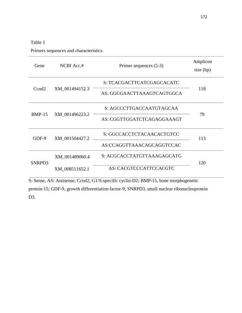
172
Table 1
Primers sequences and characteristics.
Gene NCBI Acc.# Primer sequences (5-3) Amplicon
size (bp)
Ccnd2 XM_001494152.3
S: TCACGACTTCATCGAGCACATC
118 AS: GGCGAACTTAAAGTCAGTGGCA
BMP-15 XM_001496223.2
S: AGCCCTTGACCAATGTAGCAA
79 AS: CGGTTGGATCTCAGAGGAAAGT
GDF-9 XM_001504427.2 S: GGCCACCTCTACAACACTGTCC
113
AS:CCAGGTTAAACAGCAGGTCCAC
SNRPD3
XM_001489060.4
XM_008511652.1
S: ACGCACCTATGTTAAAGAGCATG
120 AS: CACGTCCCATTCCACGTC
S: Sense, AS: Antisense; Ccnd2, G1/S-specific cyclin-D2; BMP-15, bone morphogenetic
protein-15; GDF-9, growth differentiation factor-9; SNRPD3, small nuclear ribonucleoprotein
D3.

173
Figure 1

174
Figure 2

175
Figure 3

176
Figure 4

177
Figure 5
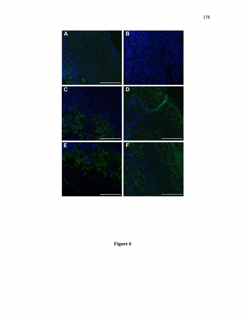
178
Figure 6

179
Figure 7

180
Figure 8

181
Figure 9

182
10 CONCLUSÕES
• O uso de uma concentração fisiológica (10 ng/mL) de insulina para o cultivo in vitro de
folículos pré-antrais equinos melhorou a sobrevivência e o desenvolvimento folicular, com uma
redução no estresse oxidativo (Fase I);
• A adição de 50 ng/mL de FSH promoveu a ativação de folículos primordiais, aumentou a
sobrevivência de folículos pré-antrais e manteve a produção de estradiol e de ROS do tecido
ovariano equino do dia 1 para 7 de cultivo (Fase II);
• A concentração de 50 ng/mL de EGF manteve a sobrevivência folicular e a produção de
ROS do dia 1 para o dia 7 de cultivo, bem como promoveu a ativação de folículos pré-antrais
equinos inclusos em tecido ovariano após cultivo in vitro. Além disso, a análise do perfil
metabolômico do meio de cultivo revelou a presença de três substâncias (Dinex, Leonuriside A e
Avobenzene) com potencial efeito negativo sobre a sobrevivência, bem como sobre diâmetro
folicular e oocitário após 7 dias de cultivo (Fase III).
• Um meio enriquecido com EGF (50 ng/mL) e insulina (10 ng/mL), suplementado ou não
com FSH (50 ng/mL), foi capaz de manter a sobrevivência e a funcionalidade do tecido ovariano
equino biopsado, após cultivo in vitro de longa duração (15 dias) (Fase IV).

183
11 PERSPECTIVAS
Este trabalho definiu uma suplementação ao meio de cultivo de base capaz de promover a
sobrevivência e desenvolvimento de folículos pré-antrais iniciais inclusos em fragmentos de tecido
ovariano equino cultivados in vitro. Isso representou um marco importante no desenvolvimento da
biotécnica de MOIFOPA na espécie equina, a qual possui grande importância socioeconômica,
além de constituir um valioso modelo animal para o estudo da foliculogênese humana.
Apesar dos avanços relatados na espécie equina relativos ao cultivo in vitro de folículos
pré-antrais, esses resultados podem ser considerados modestos quando comparados aos relatados
em outras espécies domésticas, como por exemplo, ovinos e caprinos. Nesse sentido, em função
da complexidade do cultivo in vitro de tecido ovariano equino, devido às características inerentes
à sua população folicular, a avaliação de novas combinações de fatores em diferentes
concentrações e associações deve ser realizada visando no futuro incrementar a obtenção de
oócitos maturos e, posteriormente, a produção in vitro de embriões a partir de folículos pré-
antrais.

184
12 REFERÊNCIAS BIBLIOGRÁFICAS
ADRIAENS, I.; CORTVRINDT, R.; SMITZ, J.; 2004: Differential FSH exposure in preantral
follicle culture has marked effects on folliculogenesis and oocyte developmental competence.
Human Reproduction, 19, 398-408.
AERTS, J. M. J., MARTINEZ‐MADRID, B.; FLOTHMANN, K.; DE CLERCQ, J. B. P.; VAN
AELST, S.; BOLS, P. E. J.; 2008: Quantification and Viability Assessment of Isolated
Bovine Primordial and Primary Ovarian Follicles Retrieved Through a Standardized Biopsy
Pick‐Up Procedure. Reproduction in Domestic Animals, 43, 360-366. AERTS, J. M. J.; OSTE, M.; BOLS, P. E. J.; 2005: Development and practical applications of a
method for repeated transvaginal, ultrasound-guided biopsy collection of the bovine ovary.
Theriogenology, 64, 947-957.
AGARWAL, A.; GUPTA, S.; SHARMA, R. K.; 2005: Role of oxidative stress in female
reproduction. Reproductive Biology and Endocrinology, 3, 1.
AGUIAR, F. L. N.; LUNARDI, F. O.; LIMA, L. F.; BRUNO, J. B.; ALVES, B. G.;
MAGALHÃES-PADILHA, D. M.; CIBIN, F. W. S; BERIONI, L.; APGAR, G. A.; LO
TURCO, E. G.; GASTAL, E. L.; FIGUEIREDO, J. R.; Role of EGF on in situ culture of
equine preantral follicles and metabolomics profile. Animal Reproduction Science, submitted.
AGUIAR, F. L. N.; LUNARDI, F. O.; LIMA, L. F.; ROCHA, R. M. P.; BRUNO, J. B.;
MAGALHÃES-PADILHA, D. M.; CIBIN, F. W. S.; RODRIGUES, A. P. R.; GASTAL, M.
O.; GASTAL, E. L.; FIGUEIREDO, J. R.; 2016a: Insulin improves in vitro survival of
equine preantral follicles enclosed in ovarian tissue and reduces reactive oxygen species
production after culture. Theriogenology, 85,1063-1069.
AGUIAR, F. L. N.; LUNARDI, F. O.; LIMA, L. F.; ROCHA, R. M. P.; BRUNO, J. B.;
MAGALHÃES-PADILHA, D. M.; CIBIN, F. W. S.; NUNES-PINHEIRO, D. C. S.;
GASTAL, M. O.; RODRIGUES, A. P. R.; APGAR, G. A.; GASTAL, E. L.; FIGUEIREDO,
J. R.; 2016b: FSH supplementation to culture medium is beneficial for activation and
survival of preantral follicles enclosed in equine ovarian tissue. Theriogenology, 85, 1106-
1112.
ALVES, K. A.; ALVES, B. G.; GASTAL, G. D. A.; DE TARSO, S. G.; GASTAL, M. O.;
FIGUEIREDO, J. R.; GAMBARINI, M. L.; GASTAL, E. L.; 2016b: The Mare Model to
Study the Effects of Ovarian Dynamics on Preantral Follicle Features.PLoS One 22, 11.
ALVES, K. A.; ALVES, B. G.; GASTAL, G. D.; HAAG, K. T.; GASTAL, M. O.;
FIGUEIREDO, J. R; GAMBARINI, M. L.; GASTAL, E. L.; 2016a: Preantral follicle density
in ovarian biopsy fragments and effects of mare age. Reproduction, Fertility and
Development, 5.
ALVES, K. A.; ALVES, B. G.; ROCHA, C. D; VISONNA, M.; MOHALLEM, R. F. F.;
GASTAL, M. O.; JACOMINI, J. O.; BELETTI, M. E.; FIGUEIREDO, J. R.; GAMBARINI,
M. L.; GASTAL, E. L.; 2015: Number and density of equine preantral follicles in different
ovarian histological section thicknesses. Theriogenology, 83,1048-1055.
American Association of Equine Practitioners. Official Guide for Determining the Age of the
Horse. Lexington KY, USA: AAEP, 2002, 1-26.

185
ANDRADE, E. R.; SENEDA, M. M.; ALFIERI, A. A.; OLIVEIRA, J. A.; BRACARENSE, A.
P.; FIGUEIREDO, J. R.; TONIOLLI, R.; 2005: Interactions of indole acetic acid with EGF
and FSH in the culture of ovine preantral follicles. Theriogenology, 64, 1104-1113.
APPASAMY, M.; JAUNIAUX, E.; SERHAL, P.; AL-QAHTANI, A.; GROOME, N. P.;
MUTTUKRISHNA, S.; 2008: Evaluation of the relationship between follicular fluid
oxidative stress, ovarian hormones, and response to gonadotropin stimulation. Fertility and
Sterility, 89, 912-921.
ARAUJO, V. R.; GASTAL, M. O.; FIGUEIREDO, J. R.; GASTAL, E. L.; 2014: In vitro culture
of bovine preantral follicles: a review. Reproductive Biology and Endocrinology 12, 78.
ARAÚJO, V. R.; GASTAL, M. O.; WISCHRAL, A.; FIGUEIREDO, J. R.; GASTAL, E. L.;
2015: Long-term in vitro culture of bovine preantral follicles: Effect of base medium and
medium replacement methods. Animal Reproduction Science, 161, 23-31.
ARAÚJO, V. R.; SILVA, G. M.; DUARTE, A. B. G.; MAGALHÃES, D. M.; ALMEIDA, A. P.;
GONÇALVES, R. F. B.; BRUNO, J. B.; SILVA, T. F. P.; CAMPELLO, C. C.;
RODRIGUES, A. P. R.; FIGUEIREDO, J. R.; 2011: Vascular endothelial growth factor-A
(165) (VEGF-A(165)) stimulates the in vitro development and oocyte competence of goat
preantral follicles. Cell Tissue Research, 346, 273–281.
ARUNAKUMARI, G.; SHANMUGASUNDARAM, N.; RAO, V. H.; 2010: Development of
morulae from the oocytes of cultured sheep preantral follicles. Theriogenology, 74, 884–894.
BARROS, V. R. P.; CAVALCANTE, A. Y. P.; MACEDO, T. J. S.; BARBERINO, R. S.; LINS,
T. L. B.; GOUVEIA, B. B.; MENEZES, V. G.; QUEIROZ, M. A. A.; ARAÚJO, V. R.;
PALHETA, R.C.; LEITE, M. C. P.; 2013: Immunolocalization of melatonin and follicle-
stimulating hormone receptors in caprine ovaries and their effects during in vitro
development of isolated pre-antral follicles. Reproduction in Domestic Animals, 48, 1025-
1033.
BEHL, R.; PANDEY, R. S.; 2002: FSH induced stimulation of catalase activity in goat
granulosa cells in vitro. Animal Reproduction Science, 70, 215-221.
BEHRMAN, H. R.; KODAMAN, P. H.; PRESTON, S. L.; GAO, S.; 2001: Oxidative stress and
the ovary. Journal of the Society for Gynecologic Investigation, 8, 40-42.
BELFIORE, A.; FRASCA, F.; PANDINI, G.; SCIACCA, L.; VIGNERI, R.; 2009: Insulin
receptor isoforms and insulin receptor/insulin-like growth factor receptor hybrids in
physiology and disease. Endocrine Reviews, 30, 586-623.
BERGFELT, D. R.; GINTHER, O. J.; 1993: Relationships between FSH surges and follicular
waves during the estrous cycle in mares. Theriogenology, 39, 781-796.
BERGFELT, D. R.; KOT, K.; GINTHER, O. J.; 1999: Intrafollicular injection of an aromatase
inhibitor in mares. Theriogenology, 51, 297.
BILLIG, H.; FURUTA, I.; HSUEH, A. J.; 1993: Estrogens inhibit and androgens enhance
ovarian granulosa cell apoptosis. Endocrinology, 133, 2204-2212.
BRAW-TAL, R.; YOSSEFI, S.; 1997: Studies in vivo and in vitro on the initiation of follicle
growth in the bovine ovary. Journal of Reproduction and Fertility, 109, 165-171.
BRUNET, A.; BONNI, A.; ZIGMOND, M. J.; LIN, M. Z.; JUO, P.; HU, L. S.; ANDERSON,
M. J.; ARDEN, K. C.; BLENIS, J.; GREENBERG, M. E.; 1999: Akt promotes cell survival
by phosphorylating and inhibiting a Forkhead transcription factor. Cell, 96, 857–868.
BRUNET, A.; SWEENEY, L. B.; STURGILL, J. F.; CHUA, K. F.; GREER, P. L.; LIN, Y.;
TRAN, H.; ROSS, S. E.; MOSTOSLAVSKY, R.; COHEN, H. Y.; HU, L. S.; 2004: Stress-

186
dependent regulation of FOXO transcription factors by the SIRT1 deacetylase. Science, 303,
2011–2015.
BUTCHER, L.; ULLMANN, S. L.; 1996: Culture of preantral ovarian follicles in the grey, short-
tailed opossum, Monodelphis domestica. Reproduction, Fertility and Development, 8, 535–
539.
CARNEVALE, E. M.; 2008: Clinical considerations regarding assisted reproductive procedures
in horses. Journal of Equine Veterinary Science, 28, 686–690.
CARVALHEIRA, J. B. C.; ZECCHIN, H. G.; SAAD, M. J. A.; 2002: Vias de sinalização da
insulina. Arquivos Brasileiros de Endocrinologia & Metabologia, 46, 419-425.
CASTRO, S. V.; CARVALHO, A. A.; SILVA, C. M. G.; SANTOS, F. W. C.; CAMPELLO, C.
C.; DE FIGUEIREDO, J. R.; RODRIGUES, A. P.; 2014: Frozen and fresh ovarian tissue
require different culture media to promote in vitro development of bovine preantral follicles.
Biopreservation and Biobanking, 12, 317-324.
CELESTINO, J. J. H.; BRUNO, J. B.; LIMA-VERDE, I. B.; MATOS, M. H. T.; SARAIVA, M.
V. A.; CHAVES, R. N.; MARTINS, F. S.; LIMA, L. F.; CAMPELLO, C. C.; SILVA, J. R.
V.; BÁO, S. N.; FIGUEIREDO, J. R.; 2009: Recombinant epidermal growth factor maintains
follicular ultrastructure and promotes the transition to primary follicles in caprine ovarian
tissue cultured in vitro. Reproductive Sciences, 16, 239–246.
CELESTINO, J. J. H.; BRUNO, J. B.; SARAIVA, M. V. A.; ROCHA, R. M.; BRITO, I. R.;
DUARTE, A. B. G.; ARAÚJO, V. R.; SILVA, C. M. G.; MATOS, M. H. T.; CAMPELLO,
C. C. SILVA, J. R. V.; 2011: Steady-state level of epidermal growth factor (EGF) mRNA
and effect of EGF on in vitro culture of caprine preantral follicles. Cell and Tissue Research,
344, 539-550.
CELESTINO, J. J. H.; LIMA-VERDE, I. B.; BRUNO, J. B.; MATOS, M. H. T.; CHAVES, R.
N.; SARAIVA, M. V. A.; SILVA, C. M. G.; FAUSTINO, L. R.; ROSSETTO, R.; LOPES,
C. A. P.; DONATO, M. A. M.; PEIXOTO, C. A.; CAMPELLO, C. C.; SILVA, J. R. V.;
FIGUEIREDO, J. R.; 2011: Steady-state level of bone morphogenetic protein-15 in goat
ovaries and its influence on in vitro development and survival of preantral follicles.
Molecular and Cellular Endocrinology, 338, 1-9.
CHAVES, R. N.; ALVES, A. M.; FAUSTINO, L. R.; OLIVEIRA, K. P.; CAMPELLO, C. C.;
LOPES, C. A. P.; BÁO, S. N.; FIGUEIREDO, J. R.; 2011: How the concentration of insulin
affects the development of preantral follicles in goats. Cell and Tissue Research, 346, 451–
456.
CHAVES, R. N.; DUARTE, A. B. G.; RODRIGUES, G. Q.; CELESTINO, J. J. H.; SILVA, G.
M. G.; LOPES, C. A. P.; ALMEIDA, A. P.; DONATO, M. A. M.; PEIXOTO, C. A.;
MOURA, A. A. A.; LOBO, C. H.; LOCATELLI, Y.; MERMILLOD, P.; CAMPELLO, C.
C.; FIGUEIREDO, J. R.; 2012: The effects of insulin and follicle-stimulating hormone (FSH)
during in vitro development of ovarian goat preantral follicles and the relative mRNA
expression for insulin and FSH receptors and cytochrome P450 aromatase in cultured
follicles. Biology of Reproduction, 87, 69.
CHAVES, R. N.; MARTINS, F. S.; SARAIVA, M. V. A.; CELESTINO, J. J. H.; LOPES, C. A.
P.; CORREIA, J. C.; VERDE, I. L.; MATOS, M. H. T.; BÁO, S. N.; CAMPELLO, C. C.;
SILVA, J. R. V.; 2008: Chilling ovarian fragments during transportation improves viability
and growth of goat preantral follicles cultured in vitro. Reproduction, Fertility and
Development, 20, 640-647.

187
CHEATHAM, B.; KAHN, C. R.; 1995: Insulin Action and the Insulin Signaling Network.
Endocrine Reviews, 16, 117-142.
CHEN, A. Q.; YU, S. D.; WANG, Z. G.; XU, Z. R.; YANG, Z. G.; 2009: Stage-specific
expression of bone morphogenetic protein type I and type II receptor genes: Effects of
follicle-stimulating hormone on ovine antral follicles. Animal Reproduction Science, 111,
391-399.
CHEN, Q.; ZHANG, W.; RAN, H.; FENG, L.; YAN, H.; MU, X.; HAN, Y.; LIU, W.; XIA, G.;
WANG, C.; 2014: PKCδ and θ possibly mediate FSH-induced mouse oocyte maturation via
NOX-ROS-TACE cascade signaling pathway. PloS one, 9, p.e111423.
CHOI, J.; LEE, B.; LEE, E.; YOON, B. K.; BAE, D.; CHOI, D.; 2008: Cryopreservation of
ovarian tissues temporarily suppresses the proliferation of granulosa cells in mouse preantral
follicles. Cryobiology, 56, 36-42.
CLAES, A.; BALL, B. A.; TROEDSSON, M. H. T.; CURRY, T. E.; SQUIRES, E. L.;
SCOGGIN, K. E.; 2015: Molecular changes in the equine follicle in relation to variations in
antral follicle count and anti‐Müllerian hormone concentrations. Equine Veterinary Journal.
doi: 10.1111/evj.12514
CONLEY, A. J.; BIRD, I. M.; 1997: The role of cytochrome P450 17 alpha-hydroxylase and 3
beta-hydroxysteroid dehydrogenase in the integration of gonadal and adrenal steroidogenesis
via the delta 5 and delta 4 pathways of steroidogenesis in mammals. Biology of
Reproduction, 56, 789.
CONTI, M.; HSIEH, M.; PARK, J. Y.; SU, Y. Q.; 2006: Role of the epidermal growth factor
network in ovarian follicles. Molecular Endocrinology, 20, 715-723.
CORTVRINDT, R; SMITZ, J.; VAN STEIRTEGHEM, A. C; 1996: In-vitro maturation,
fertilization and embryo development of immature oocytes from early preantral follicles from
prepuberal mice in a simplified culture system. Human Reproduction, 11, 2656–2666.
COSTA, S. H. F.; SANTOS, R. R.; RONDINA, D.; ANDRADE, E. R.; OHASHI, O. M.;
RODRIGUES, A. P. R.; FIGUEIREDO, J. R.; 2010: Effects of IAA in combination with
FSH on in vitro culture of ovine preantral follicles. Zygote, 18, 89-92.
COX, L.; VANDERWALL, D. K.; PARKINSON, K. C.; SWEAT, A.; ISOM, C.; 2015:
Expression profiles of selected genes in cumulus–oocyte complexes from young and aged
mares. Reproduction, Fertility and Development, 27, 914-924.
CUILING, L.; WEI, Y.; ZHAOYUAN, H.; YIXUN, L.; 2005: Granulosa cell proliferation
differentiation and its role in follicular development. Chinese Science Bulletin, 50, 2665-
2671.
DAS, M.; KNOWLES, B.; BISWAS, R.; BISHAYEE, S.; 1984: Receptor modulating properties
of an antibody directed against the epidermal growth factor receptor. European Journal of
Biochemistry, 141, 429-434.
DEMEESTERE, I.; CENTNER, J.; GERVY, Y.; DELBAERE, A.; 2005: Impact of various
endocrine and paracrine factors on culture of preantral follicles in rodents. Reproduction, 130,
147-156.
DERRAR, N.; PRICE, C. A.; SIRARD, M. A.; 2000: Effect of growth factors and co-culture
with ovarian medulla on the activation of primordial follicles in explants of bovine ovarian
cortex. Theriogenology, 54, 587-598.
DRIANCOURT, M. A.; PARIS, A.; ROUX, C.; MARIANA, J. C.; PALMER, E.; 1982: Ovarian
follicular populations in pony and saddle type mares. Reproduction, Nutrition, and
Development, 22, 1035-1047.

188
DRUMMOND, A. E.; FINDLAY, J. K.; 1999: The role of estrogen in folliculogenesis.
Molecular and Cellular Endocrinology, 151, 57–64.
DULEBA, A. J.; SPACZYNSKI, R. Z.; OLIVE, D. L.; BEHRMAN, H. R.; 1997: Effects of
insulin and insulin-Like growth factors on proliferation of rat ovarian theca-interstitial cells.
Biology of Reproduction, 56, 891–897.
EBERT, R.; ULMER, M.; ZECK, S.; MEISSNER‐WEIGL, J.; SCHNEIDER, D.; STOPPER,
H.; SCHUPP, N.; KASSEM, M.; JAKOB, F.; 2006: Selenium supplementation restores the
antioxidative capacity and prevents cell damage in bone marrow stromal cells in vitro. Stem
Cells, 24, 1226-1235.
EDSON, M. A.; NAGARAJA, A. K.; MATZUK, M. M.; 2009: The mammalian ovary from
genesis to revelation. Endocrine Reviews, 30, 624-712.
EPPIG, J. J.; SCHROEDER, A. C.; 1989: Capacity of mouse oocytes from preantral follicles to
undergo embryogenesis and development to live young after growth, maturation, and
fertilization in vitro. Biology of Reproduction,41, 268-276.
EPPIG, J.J.; O'BRIEN, M.; WIGGLESWORTH, K.; 1996: Mammalian oocyte growth and
development in vitro. Molecular Reproduction and Development, 44, 260-273.
FENG, P.; CATT, K. J.; KNECHT, M.; 1986: Transforming growth factor beta regulates the
inhibitory actions of epidermal growth factor during granulosa cell differentiation. Journal of
Biological Chemistry, 261, 14167-14170.
FEUGANG, J. M. N.; RODRIGUEZ-MUNOZ, J. C.; WILLARD, S. T.; BATHGATE, R. A.;
RYAN, P. L.; 2011: Examination of relaxin and its receptors expression in pig gametes and
embryos. Reproductive Biology and Endocrinology, 20, 1-11.
FIGUEIREDO, J. R.; HULSHOF, S. C.; VAN DEN HURK, R.; NUSGENS, B.; BEVERS, M.
M.; ECTORS, F. J.; BECKERS, J. F.; 1994: Preservation of oocyte and granulosa cell
morphology in bovine preantral follicles cultured in vitro. Theriogenology, 41, 1333-1346.
FIGUEIREDO, J. R.; RODRIGUES, A. P. R.; AMORIM, C. A.; SILVA, J. R. V.; 2008:
Manipulação De Oócitos Inclusos em Folículos Pré-Antrais. In: Gonçalves, P.B.D.,
Figueiredo, J.R.; Freitas, V. J. F. Biotécnicas aplicadas à reprodução animal. São Paulo:
Editora Roca, p. 303-327.
FIGUEIREDO, J. R.; RODRIGUES, A. P. R.; SILVA, J. R. V.; SANTOS, R. R.; 2011:
Cryopreservation and in vitro culture of caprine preantral follicles. Reproduction, Fertility
and Development, 23, 40-47.
FORTUNE, J. E.; KITO, S.; WANDJI, S. A.; SRŠEŇ, V.; 1998: Activation of bovine and baboon
primordial follicles in vitro. Theriogenology, 49, 441-449.
FU, D.; LV, X.; HUA, G.; HE, C.; DONG, J.; LELE, S. M.; LI, D. W.; ZHAI, Q.; DAVIS, J. S.;
WANG, C.; 2014: YAP regulates cell proliferation, migration, and steroidogenesis in adult
granulosa cell tumors. Endocrine-Related Cancer 21, 297-310.
FUJIHARA, M.; COMIZZOLI, P.; KEEFER, C.L.; WILDT, D.E.; SONGSASEN, N.; 2014:
Epidermal growth factor (EGF) sustains in vitro primordial follicle viability by enhancing
stromal cell proliferation via MAPK and PI3K pathways in the prepubertal, but not adult cat
ovary. Biology of Reproduction, 90, 1-10.
GALLI, C.; COLLEONI, S.; DUCHI, R.; LAGUTINA, I.; LAZZARI, G.; 2013: Equine assisted
reproduction and embryo technologies. Animal Reproduction, 10, 334-343.
GARZO, V. G.; DORRINGTON, J. H.; 1984: Aromatase activity in human granulosa cells
during follicular development and the modulation by follicle-stimulating hormone and
insulin. American Journal of Obstetrics and Gynecology, 148, 657-662.

189
GASTAL, E. L.; 2011: Ovulation: Part 1. Follicle development and endocrinology during the
periovulatory period, in: McKinnon, A.O., Squires, E.L., Vaalaand, W.E., Varner, D.D.,
(Eds.), Equine Reproduction, 2nd edn. Ames: Wiley-Blackwell, pp. 2020-2031.
GASTAL, E. L.; GASTAL, M. O.; DONADEU, F. X.; ACOSTA, T. J.; BEG, M. A.;
GINTHER, O. J.; 2007: Temporal relationships among LH, estradiol, and follicle
vascularization preceding the first compared with later ovulations during the year in mares.
Animal Reproduction Science, 102, 314-321.
GASTAL, G. D. A.; ALVES, B. G.; ALVES, K. A.; PAIVA, S. O.; DE TARSO, S. G. S.;
ISHAK, G. M.; BASHIR, S. T.; GASTAL, E. L.; 2016: Effects of cryoprotectant agents on
equine ovarian biopsy fragments in preparation for cryopreservation. Journal of Equine
Veterinary Science.
GAVIN, J. R.; ROTH, J.; NEVILLE, D. M.; DE MEYTS, P.; BUELL, D. N.; 1974: Insulin-
dependent regulation of insulin receptor concentrations: a direct demonstration in cell
culture. Proceedings of the National Academy of Sciences, 71, 84–88.
GINTHER, O. J.; 1992: Reproductive Biology of the Mare. Basic and Applied Aspects, 2nd edn.
Equiservices Publications, Cross Plains, WI.
GINTHER, O. J.; 2012: The mare: a 1000-pound guinea pig for study of the ovulatory follicular
wave in women. Theriogenology, 77, 818-828.
GINTHER, O. J.; GASTAL, E. L.; GASTAL, M. O.; BERGFELT, D. R.; BAERWALD, A. R.;
PIERSON, R. A.; 2004a: Comparative study of the dynamics of follicular waves in mares
and women. Biology of Reproduction, 71, 1195-1201.
GINTHER, O. J.; GASTAL, E. L.; GASTAL, M. O.; BEG, M. A.; 2004b: Seasonal influence on
equine follicle dynamics. Animal Reproduction, 1, 31-44.
GOMES, R. G.; ANDRADE, E. R.; LISBOA, L. A.; CIQUINI, A.; BARREIROS, T. R. R.;
FONSECA, N. A. N.; SENEDA, M. M.; 2012: Effect of holding medium, temperature and
time on structural integrity of equine ovarian follicles during the non-breeding season.
Theriogenology, 78, 731-736.
GOMES, R. G.; LISBOA, L. A.; SILVA, C. B.; MAX, M. C.; MARINO, P. C; OLIVEIRA, R.
L.; GONZÁLEZ, S. M.; BARREIROS, T. R.; MARINHO, L. S.; SENEDA, M. M.; 2015:
Improvement of development of equine preantral follicles after 6 days of in vitro culture with
ascorbic acid supplementation. Theriogenology 84, 750-755.
GOOK, D. A.; EDGAR, D. H.; LEWIS, K.; SHEEDY, J. R.; GARDNER, D. K.; 2014: Impact
of oxygen concentration on adult murine pre-antral follicle development in vitro and the
corresponding metabolic profile. Molecular Human Reproduction, 20, 31-41.
GORE-LANGTON, R. E.; DANIEL, S. A.; 1990: Follicle-stimulating hormone and estradiol
regulate antrum-like reorganization of granulosa cells in rat preantral follicle cultures.
Biology of Reproduction, 43, 65-72.
GREEN, L. J.; SHIKANOV, A.; 2016: In vitro culture methods of preantral follicles.
Theriogenology, 86, 229-238.
GRIFFITHS, W. J.; WANG, Y.; 2009: Mass spectrometry: from proteomics to metabolomics
and lipidomics. Chemical Society Reviews, 38, 1882-1896.
GUDERMANN, T.; NÜRNBERG, B.; SCHULTZ, G; 1995: Receptors and G proteins as
primary components of transmembrane signal transduction. Journal of Molecular Medicine,
73, 51-63.
GUPTA, P. S.; NANDI, S.; RAVINDRANATHA, B. M.; SARMA, P. V.; 2002: In vitro culture
of buffalo (Bubalus bubalis) preantral follicles. Theriogenology, 57, 1839-1854.

190
GUPTA, P. S.; RAMESH, H. S.; MANJUNATHA, B. M.; NANDI, S.; RAVINDRA, J. P.;
2008: Production of buffalo embryos using oocytes from in vitro grown preantral follicles.
Zygote, 16, 57–63.
GUTIERREZ, C. G.; RALPH, J. H.; TELFER, E. E.; WILMUT, I.; WEBB, R.; 2000: Growth
and antrum formation of bovine preantral follicles in long-term culture in vitro. Biology of
Reproduction, 62,1322-1328.
HAAG, K. T.; MAGALHÃES-PADILHA, D. M.; FONSECA, G. R.; WISCHRAL, A.;
GASTAL, M. O.; KING, S. S.; JONES, K. L.; FIGUEIREDO, J. R.; GASTAL, E. L.; 2013a:
Quantification, morphology, and viability of equine preantral follicles obtained via the
Biopsy Pick-Up method. Theriogenology, 79, 599-609.
HAAG, K. T.; MAGALHAES-PADILHA, D. M.; FONSECA, G. R.; WISCHRAL, A.;
GASTAL, M. O.; KING, S. S.; JONES, K. L; FIGUEIREDO, J. R.; GASTAL, E. L.; 2013b:
Equine preantral follicles obtained via the Biopsy Pick-Up method: histological evaluation
and validation of a mechanical isolation technique. Theriogenology, 79, 735-743.
HAAG, K. T.; MAGALHAES-PADILHA, D. M.; FONSECA, G. R.; WISCHRAL, A.;
GASTAL, M. O.; KING, S. S.; JONES, K. L.; FIGUEIREDO, J. R.; GASTAL, E. L.; 2013c:
In vitro culture of equine preantral follicles obtained via the Biopsy Pick-Up method.
Theriogenology, 79, 911-917.
HAAG, K. T.; MAGALHÃES-PADILHA, D. M.; GASTAL, M. O.; FIGUEIREDO, J. R.;
GASTAL, E. L.; 2013d: Equine preantral follicle harvesting, processing, and in vitro culture:
the journey has already started. Animal Reproduction, 10, 187-198.
HARTSHORE, G. M.; LYRAKOU, S.; HAMODA, H.; OLOTO, E.; GHAFARI, F.; 2009:
Oogenesis and cell death in human prenatal ovaries: what are the criteria for oocyte selection?
Molecular Human Reproduction, 15, 805-819.
HEMAMALINI, N.C.; RAO, B. S.; TAMILMANI, G.; AMARNATH, D.; VAGDEVI, R.;
NAIDU, K.S.; REDDY, K. K.; RAO, V. H.; 2003: Influence of transforming growth factor-
α, insulin-like growth factor-II, epidermal growth factor or follicle stimulating hormone on in
vitro development of preantral follicles in sheep. Small Ruminant Research, 50, 11-22.
HENNEKE, D. R.; POTTER, G. D.; KREIDER, J. L.; YEATES, B. F.; 1983: Relationship
between condition score, physical measurements and body fat percentage in mares. Equine
Veterinary Journal, 15, 371–372.
HENSON, E. S.; GIBSON, S. B.; 2006: Surviving cell death through epidermal growth factor
(EGF) signal transduction pathways: implications for cancer therapy. Cell Signal,18, 2089-
2097.
HERNANDEZ, E. R.; RESNICK, C. E.; HOLTZCLAW, W. D.; PAYNE, D. W.; ADASHI, E.
Y.; 1988: Insulin as a Regulator of Androgen Biosynthesis by Cultured Rat Ovarian Cells:
Cellular Mechanism (s) Underlying Physiological and Pharmacological Hormonal Actions.
Endocrinology, 122, 2034-2043.
HILLIER, S. G.; 1996: Roles of follicle stimulating hormone and luteinizing hormone in
controlled ovarian hyperstimulation. Human Reproduction, 11, 113-121.
HINRICHS, K.; 2012: Assisted reproduction techniques in the horse. Reproduction, Fertility and
Development, 25, 80–93.
HINRICHS, K.; SCHMIDT, A. L.; FRIEDMAN, P. P.; SELGRATH, J. P.; MARTIN, M. G.;
1993: In vitro maturation of horse oocytes: characterization of chromatin configuration using
fluorescence microscopy. Biology of Reproduction, 48, 363-370.

191
HIRAO, Y.; NAGAI, T.; KUBO, M.; MYANO, T.; MIYAKE, M.; KATO, S.; 1994: In vitro
growth and maturation of pig oocytes. Journal of Reproduction and Fertility, 100, 333-339.
HUANG, Z.; WELLS, D.; 2010: The human oocyte and cumulus cells relationship: new insights
from the cumulus cell transcriptome. Molecular Human Reproduction, 16, 715-725.
HUNZICKER-DUNN, M. E.; LOPEZ-BILADEAU, B.; LAW, N. C.; FIEDLER, S. E.; CARR,
D. W.; MAIZELS, E. T.; 2012: PKA and GAB2 play central roles in the FSH signaling
pathway to PI3K and AKT in ovarian granulosa cells. Proceedings of the National Academy
of Sciences, 109, 2979-2988.
HUNZICKER-DUNN, M.; MAIZELS, E. T.; 2006: FSH signaling pathways in immature
granulosa cells that regulate target gene expression: branching out from protein kinase A.
Cellular Signalling, 18, 1351-1359.
HUSSEIN, M. R.; 2005: Apoptosis in the ovary: molecular mechanisms. Human Reproduction
Update, 11, 162–177.
ITOH, T.; KACCHI, M; ABE, H.; SENDAI, Y.; HOSHI, H.; 2002: Growth, antrum formation,
and estradiol production of bovine preantral follicles cultured in a serum-free medium.
Biology of Reproduction, 67, 1099-1105.
JABARA, S.; CHRISTENSON, L. K.; WANG, C. Y.; MCALLISTER, J. M.; JAVITT, N. B.;
DUNAIF, A.; STRAUSS III, J. F.; 2003: Stromal cells of the human postmenopausal ovary
display a distinctive biochemical and molecular phenotype. The Journal of Clinical
Endocrinology & Metabolism, 88, 484-492.
JEWGENOW, K.; PENFOLD, L. M.; MEYER, H. H. D.; WILDT, D. E.; 1998: Viability of small
preantral ovarian follicles from domestic cats after cryoprotectant exposure and
cryopreservation. Journal of Reproduction and Fertility, 112, 39-47.
JI, Q.; LIU, P. I.; CHEN, P. K.; AOYAMA, C.; 2004: Follicle stimulating hormone-induced
growth promotion and gene expression profiles on ovarian surface epithelial cells.
International Journal of Cancer,112, 803-814.
JONES, P. B.; WELSH, T. H.; HSUEH, A. J.; 1982: Regulation of ovarian progestin production
by epidermal growth factor in cultured rat granulosa cells. Journal of Biological Chemistry,
19, 11268-11273.
KAWAUCHI, H.; SOWER, S. A.; 2006: The dawn and evolution of hormones in the
adenohypophysis. General and Comparative Endocrinology, 148, 3-14.
KEDEM, A.; FISCH, B.; GAROR, R.; BEN-ZAKEN, A.; GIZUNTERMAN, T.; FELZ, C.;
BEN-HAROUSH, A.; KRAVARUSIC, D.; ABIR, R.; 2011: Growth differentiating factor 9
(GDF9) and bone morphogenetic protein 15 both activate development of human primordial
follicles in vitro, with seemingly more beneficial effects of GDF9. The Journal of Clinical
Endocrinology & Metabolism, 96: 246-254.
KEZELE, P. R.; NILSSON, E. E.; SKINNER, M. K.; 2002: Insulin but not insulin-like growth
factor-1 promotes the primordial to primary follicle transition. Molecular and Cellular
Endocrinology, 192, 37-43.
KIM, I. W.; GONG, S. P.; YOO, C. R.; CHOI, J. H.; KIM, D. Y.; LIM, J. M.; 2009: Derivation
of developmentally competent oocytes by the culture of preantral follicles retrieved from
adult ovaries: maturation, blastocyst formation, and embryonic stem cell transformation.
Fertility and Sterility, 92, 1716–1724.
KNECHT, M.; CATT, K. J.; 1983: Modulation of cAMP-mediated differentiation in ovarian
granulosa cells by epidermal growth factor and platelet-derived growth factor. Journal of
Biological Chemistry, 258, 2789-2794.

192
KRYSKO, D. V.; DIEZ-FRAILE, A.; CRIEL, G.; SVISTUNOV, A. A.; VANDENABEELE, P.;
D’HERDE, K. 2008: Life and death of female gametes during oogenesis and folliculogenesis.
Apoptosis, 13, 1065-1087.
KWINTKIEWICZ, J.; SPACZYNSKI, R. Z.; FOYOUZI, N.; PEHLIVAN, T.; DULEBA, A. J.;
2006: Insulin and oxidative stress modulate proliferation of rat ovarian theca-interstitial cells
through diverse signal transduction pathways. Biology of Reproduction, 74, 1034–1040.
LAITINEN, M.; VUOJOLAINEN, K.; JAATINEN, R.; KETOLA, I.; AALTONEN, J;
LEHTONEN, E.; HEIKINHEIMO, M.; RITVOS, O.; 1998: A novel growth differentiation
factor-9 (GDF-9) related factor is co-expressed with GDF-9 in mouse oocytes during
folliculogenesis. Mechanisms of Development, 78; 135-140.
LAWRENCE, M. C.; MCKERN, N. M.; WARD, C. W.; 2007: Insulin receptor structure and its
implications for the IGF-1 receptor. Current Opinion in Structural Biology, 17, 699-705.
L'HORTET, A. C.; GILGENKRANTZ, H.; GUIDOTT, J. E.; 2012: EGFR: a master piece in
G1/S phase transition of liver regeneration. International Journal of Hepatology.
LOETCHUTINAT, C.; KOTHAN, S.; DECHSUPA, S.; MEESUNGNOEN, J.; JAY-GERIN, J.
P.; MANKHETKORN, S.; 2005: Spectrofluorometric determination of intracellular levels of
reactive oxygen species in drug-sensitive and drug-resistant cancer cells using the 2′, 7′-
dichlorofluorescein diacetate assay. Radiation Physics and Chemistry, 72, 323-331.
LOO, D.T.; 2011: In situ detection of apoptosis by the TUNEL assay: an overview of techniques.
DNA Damage Detection In Situ, Ex Vivo, and In Vivo: Methods and Protocols, 3-13.
LOUHIO, H.; HOVATTA, O.; SJÖBERG, J.; TUURI, T.; 2000: The effects of insulin, and
insulin-like growth factors I and II on human ovarian follicles in long-term culture.
Molecular Human Reproduction, 6, 694–698.
LUCIANO, A. M.; PAPPALARDO, A.; RAY, C.; PELUSO, J. J.; 1994: Epidermal growth
factor inhibits large granulosa cell apoptosis by stimulating progesterone synthesis and
regulating the distribution of intracellular free calcium. Biology of Reproduction, 51, 646-
654.
LUDERER, U.; 2014: Ovarian toxicity from reactive oxygen species. Vitamins and Hormones,
94, 99-127.
LUNARDI, F. O.; ARAÚJO, V. R.; FAUSTINO, L. R.; DE ANDRADE CARVALHO, A.;
GONÇALVES, R. F. B.; BASS, C. S.; BÁO, S. N.; NAME, K. P. O.; CAMPELLO, C. C.;
DE FIGUEIREDO, J. R.; RODRIGUES, A. P. R.; 2012: Morphologic, viability and
ultrastructural analysis of vitrified sheep preantral follicles enclosed in ovarian tissue. Small
Ruminant Research, 107, 121-130.
LUND, E. K.; WHARF, S. G.; FAIRWEATHER-TAIT, S. J.; JOHNSON, I. T.; 1999: Oral
ferrous sulfate supplements increase the free radical–generating capacity of feces from
healthy volunteers. The American Journal of Clinical Nutrition, 69, 250-255.
LUO, Y.; DIXON, C. J.; HALL, J. F.; WHITE, P. J.; BOARDER, M. R.; 2007: A role for Akt in
epidermal growth factor-stimulated cell cycle progression in cultured hepatocytes: generation
of a hyperproliferative window after adenoviral expression of constitutively active Akt.
Journal of Pharmacology and Experimental Therapeutics, 321, 884-891.
LUZ, V. B.; ARAUJO, V. R.; DUARTE, A. B. G.; CELESTINO, J. J. H.; SILVA, T. F. P.;
MAGALHAES-PADILHA, D. M.; CHAVES, R. N.; BRITO, I. R.; ALMEIDA, A. P.;
CAMPELLO, C. C.; FELTRIN, C.; BERTOLINI, M.; SANTOS, R. R.; FIGUEIREDO, J.

193
R.; 2012: Eight-cell parthenotes originated from in vitro grown sheep preantral follicles.
Reproductive Sciences, 19, 1219-1225.
MA, C. X.; SONG, Y. L.; XIAO, L.; XUE, L. X.; LI, W. J.; LAFOREST, B.; KOMATI, H.;
WANG, W. P.; JIA, Z. Q.; ZHOU, C. Y.; ZOU, Y.; 2015: EGF is required for cardiac
differentiation of P19CL6 cells through interaction with GATA-4 in a time-and dose-
dependent manner. Cellular and Molecular Life Sciences, 72, 2005-2022.
MACIEL, G. A.; BARACAT, E. C.; BENDA, J. A., MARKHAM, S. M.; HENSINGER, K.;
CHANG, R. J.; ERICKSON, G. F.; 2004: Stockpiling of transitional and classic primary
follicles in ovaries of women with polycystic ovary syndrome. The Journal of Clinical
Endocrinology & Metabolism, 89, 5321-5327.
MAGALHÃES, D. M.; ARAÚJO, V. R.; LIMA-VERDE, I. B.; MATOS, M. H. T.; SILVA, R.
C.; LUCCI, C. M.; BÁO, S. N.; CAMPELLO, C. C.; FIGUEIREDO, J. R.; 2009: Different
follicle-stimulating hormone (FSH) sources influence caprine preantral follicle viability and
development in vitro. Brazilian Journal of Veterinary Research and Animal Science, 46, 378-
386.
MAGALHÃES, D. M.; DUARTE, A. B.; ARAÚJO, V. R.; BRITO, I. R.; SOARES, T. G.;
LIMA, I. M.; LOPES, C. A.; CAMPELLO, C. C.; RODRIGUES, A. P.; FIGUEIREDO, J.
R.; 2011: In vitro production of a caprine embryo from a preantral follicle cultured in media
supplemented with growth hormone. Theriogenology,75,182–188.
MAGALHÃES, D. M.; FERNANDES, D. D.; MORORÓ, M. B. S.; SILVA, C. M. G.;
RODRIGUES, G. Q.; BRUNO, J. B.; MATOS, M. H. T.; CAMPELLO, C. C.
FIGUEIREDO, J. R.; 2011: Effect of the Medium Replacement Interval on the Viability,
Growth and In Vitro Maturation of Isolated Caprine and Ovine Pre‐Antral Follicles.
Reproduction in Domestic Animals, 46, 134-140.
MAGALHÃES-PADILHA, D. M.; FONSECA, G. R.; HAAG, K. T.; WISCHRAL, A.;
GASTAL, M. O.; JONES, K. L.; GEISLER-LEE, J.; FIGUEIREDO, J. R.; GASTAL, E. L.;
2012: Long-term in vitro culture of ovarian cortical tissue in goats: effects of FSH and IGF-I
on preantral follicular development and FSH and IGF-I receptor mRNA expression. Cell and
Tissue Research, 350, 503-511.
MAO, J.; SMITH, M. F.; RUCKER, E. B.; WU, G. M.; MCCAULEY, T. C.; CANTLEY, T. C.;
PRATHER, R. S.; DIDION, B. A.; DAY, B. N.; 2004: Effect of epidermal growth factor and
insulin-like growth factor I on porcine preantral follicular growth, antrum formation, and
stimulation of granulosal cell proliferation and suppression of apoptosis in vitro. Journal of
Animal Science, 82, 1967-1975.
MARGULIS, S.; ABIR, R.; FELZ, C.; NITKE, S.; KRISSI, H.; FISCH, B.; 2009: Bone
morphogenetic protein 15 expression in human ovaries from fetuses, girls, and women.
Fertility and Sterility, 92, 1666-1673.
MARKSTRÖM, E.; SVENSSON, E.; SHAO, R.; SVANBERG, B.; BILLIG, H.; 2002: Survival
factors regulating ovarian apoptosis--dependence on follicle differentiation. Reproduction,
123, 23-30.
MARTINO, N. A.; DELL’AQUILA, M. E.; URANIO, M. F.; RUTIGLIANO, L.; NICASSIO,
M.; LACALANDRA, G. M.; HINRICHS, K.; 2014: Effect of holding equine oocytes in
meiosis inhibitor-free medium before in vitro maturation and of holding temperature on
meiotic suppression and mitochondrial energy/redox potential. Reproductive Biology and
Endocrinology, 12, 1.

194
MARTINS, F. S.; CELESTINO, J. J. H.; SARAIVA, M. V. A.; MATOS, M. H. T.; BRUNO, J.
B.; ROCHA-JUNIOR, C. M. C.; LIMA-VERDE, I. B.; LUCCI, C. M.; BÁO, S. N.;
FIGUEIREDO, J. R.; 2008: Growth and differentiation factor-9 stimulates activation of goat
primordial follicles in vitro and their progression to secondary follicles. Reproduction,
Fertility and Development, 20, 916-924.
MATOBA, S.; BENDER, K.; FAHEY, A. G.; MAMO, S.; BRENNAN, L.; LONERGAN, P.;
FAIR, T.; 2014: Predictive value of bovine follicular components as markers of oocyte
developmental potential. Reproduction, Fertility and Development, 26, 337-345.
MATOS, M. H. T.; LIMA-VERDE, I. B.; LUQUE, M. C. A.; MAIA JR, J. E.; SILVA, J. R. V.;
CELESTINO, J. J. H.; MARTINS, F. S.; BÁO, S. N.; LUCCI, C. M.; FIGUEIREDO, J.
R.;2007: Essential role of follicle stimulating hormone in the maintenance of caprine
preantral follicle viability in vitro. Zygote, 15, 173-182.
MAZERBOURG, S. HSUEH, A. J.; 2006: Genomic analyses facilitate identification of receptors
and signalling pathways for growth differentiation factor 9 and related orphan bone
morphogenetic protein/growth differentiation factor ligands. Human Reproduction Update,
12, 373-383.
MCCAFFERY, F. H.; LEASK, R.; RILEY, S. C.; TELFER, E. E. 2000: Culture of bovine
preantral follicles in a serum-free system: markers for assessment of growth and development.
Biology of Reproduction, 63, 267-273.
MCCUBREY, J. A.; LAHAIR, M. M.; FRANKLIN, R. A.; 2006: Reactive oxygen species-
induced activation of the MAP kinase signaling pathways. Antioxidants & Redox Signaling,
8, 1775-1789.
MCGEE, E. A.; HSUEH, A. J.; 2000: Initial and cyclic recruitment of ovarian follicles. Endocrine
Reviews, 21, 200-214.
MCGRATH, S. A.; ESQUELA, A. F.; LEE, S. J.; 1995: Oocyte-specific expression of
growth/differentiation factor-9. Molecular Endocrinology, 9, 131-136.
MCLAUGHLIN, M.; BROMFIELD, J. J.; ALBERTINI, D. F.; TELFER, E. E.; 2010: Activin
promotes follicular integrity and oogenesis in cultured pre-antral bovine follicles. Molecular
Human Reproduction, 16, 644-653.
McNATTY, K. P.; MAKRIS, A.; DEGRAZIA, C.; RAPIN, O.; RYAN, K. J.; 1979: The
Production of Progesterone, Androgens, and Estrogens by Granulosa Cells, Thecal Tissue,
and Stromal Tissue from Human Ovaries in Vitro. The Journal of Clinical Endocrinology &
Metabolism, 49, 687-699.
MÉDURI, G.; CHARNAUX, N.; DRIANCOURT, M. A.; COMBETTES, L.; GRANET, P.;
VANNIER, B.; LOOSFELT, H.; MILGROM, E.; 2002: Follicle-stimulating hormone
receptors in oocytes?. The Journal of Clinical Endocrinology & Metabolism, 87, 2266-2276.
MIHM, M.; EVANS, A. C. O.; 2008: Mechanisms for dominant follicle selection in
monovulatory species: a comparison of morphological, endocrine and intraovarian events in
cows, mares and women. Reproduction in Domestic Animals, 43, 48-56.
MIKKELSEN, A. L.; HOST, E.; LINDENBERG, S.; 2001: Incidence of apoptosis in granulosa
cells from immature human follicles. Reproduction, 122, 481-486.
MITSUDOMI, T.; YATABE, Y.; 2010: Epidermal growth factor receptor in relation to tumor
development: EGFR gene and cancer. Federation of European Biochemical Societies
Journal, 277, 301-308.

195
MONTANI, D. A.; CORDEIRO, F. B.; REGIANI, T.; VICTORINO, A. B.; PILAU, E. J.;
GOZZO, F. C.; FERREIRA, C. R.; FRAIETTA, R. TURCO, E. G. L.; 2012. The follicular
microenviroment as a predictor of pregnancy: MALDI-TOF MS lipid profile in cumulus
cells. Journal of Assisted Reproduction and Genetics, 29, 1289-1297.
MONTANI, D. A.; REGIANI, T.; VICTORINO, A. B.; CAMILLO, J.; PILAU, E. J.; GOZZO,
F. C.; ZYLBERSZTEJN, D. S.; FERREIRA, C. R. TURCO, E. G. L.; 2014: Hyaluronidase
alters the lipid profile of cumulus cells as detected by MALDI-TOF MS and multivariate
analysis. Lipids, 49, 957-962.
MOSSMAN, H. W.; DUKE, K. L.; 1973: Comparative Morphology of the Mammalian Ovary.
Madison, Wisconsin: University of Wisconsin Press.
MULLIN, J. M.; MCGINN, M. T.; 1988: Epidermal growth factor-induced mitogenesis in
kidney epithelial cells (LLC-PK1). Cancer Research, 48, 4886-4891.
MYERS, M. G.; BACKER, J. M.; SIDDLE, K.; WHITE, M. F.; 1991: The insulin receptor
functions normally in Chinese hamster ovary cells after truncation of the C terminus. Journal
of Biological Chemistry, 266, 10616-10623.
National Center for Biotechnology Information, 2015a: PubChem BioAssay Database;
AID=743078. https://pubchem.ncbi.nlm.nih.gov/bioassay/743078 (accessed 10.16.15).
National Center for Biotechnology Information, 2015b: PubChem BioAssay Database; AID
470167. https://pubchem.ncbi.nlm.nih.gov/bioassay/470167 (accessed 10.16.15).
National Center for Biotechnology Information, 2015c: PubChem BioAssay Database; AID
743091. https://pubchem.ncbi.nlm.nih.gov/bioassay/743091 (accessed 10.16.15).
NAVALAKHE, R. M.; JAGTAP, D. D.; NAYAK, S. U.; NANDEDKAR, T. D.; MAHALE, S.
D.; 2013: Effect of FSH Receptor‐Binding Inhibitor‐8 on FSH‐Mediated Granulosa Cell
Signaling and Proliferation. Chemical Biology & Drug Design, 82, 178-188.
NILSSON, E. E.; SKINNER, M. K.; 2004: Kit ligand and basic fibroblast growth factor
interactions in the induction of ovarian primordial to primary follicle transition. Molecular
and Cellular Endocrinology, 214, 19-25.
NOBLE, G. K.; SILLENCE, M. N.; 2013. Diurnal rhythm and effects of feeding, exercise and
recombinant equine growth hormone on serum insulin concentrations in the horse. Equine
Veterinary Journal, 45, 745-750.
O`LEARY, T. J.; 2001: Standardization in immunohistochemistry. Applied
Immunohistochemistry & Molecular Morphology, 9, 3-8.
O’BRIEN, M. J.; PENDOLA, J. K.; EPPIG J. J.; 2003: A revised protocol for in vitro
development of mouse oocytes from primordial follicles dramatically improves their
developmental competence. Biology of Reproduction, 68,1682–1686.
OKTAY, K.; BRIGGS, D; GOSDEN, R. G.; 1997: Ontogeny of follicle-stimulating hormone
receptor gene expression in isolated human ovarian follicles 1. The Journal of Clinical
Endocrinology & Metabolism, 82, 3748-3751.
OLIVEIRA, E. G.; POLISSENI, J.; GUERRA, M. O.; PETERS, V. M.; 2009: Mecanismos
fisiológicos e bioquímicos envolvidos na ovogênese. Revista Interdiciplinar de Estudos
Experimentais, 1, 29-33.
ORON, G.; FISCH, B.; AO, A.; ZHANG, X. Y.; FARHI, J.; HAROUSH, A. B.; KESSELER-
ICEKSON, G.; ABIR, R.; 2010: Expression of growth-differentiating factor 9 and its type 1
receptor in human ovaries. Reproductive Biomedicine Online, 21, 109-117.

196
O'SHAUGHNESSY, P. J.; DUDLEY, K.; RAJAPAKSHA, W. R. A. K. J. S.; 1996: Expression
of follicle stimulating hormone-receptor mRNA during gonadal development. Molecular and
Cellular Endocrinology, 125, 169-175.
OTSUKA, F.; MCTAVISH, K. J.; SHIMASAKI, S.; 2011: Integral role of GDF‐9 and BMP‐15
in ovarian function. Molecular Reproduction and Development, 78, 9-21.
OU, X. H.; LI, S.; WANG, Z. B.; LI, M.; QUAN, S.; XING, F.; GUO, L.; CHAO, S. B.; CHEN,
Z.; LIANG, X. W.; HOU, Y.; 2012: Maternal insulin resistance causes oxidative stress and
mitochondrial dysfunction in mouse oocytes. Human Reproduction, 27, 2130-2145.
PARROTT, J. A.; SKINNER, M. K.; 2000: Kit ligand actions on ovarian stromal cells: effects
on theca cell recruitment and steroid production. Molecular Reproduction and Development,
55, 55-64.
PATEL, H.; BHARTIYA, D.; PARTE, S.; GUNJAL, P.; YEDURKAR, S.; BHATT, M.; 2013:
Follicle stimulating hormone modulates ovarian stem cells through alternately spliced
receptor variant FSH-R3. Journal of Ovarian Research, 6, 52-66.
PATTI, G. J.; YANES, O.; SIUZDAK, G.; 2012: Innovation: Metabolomics: the apogee of the
omics trilogy. Nature Reviews Molecular Cell Biology, 13, 263-269.
PAULINI, F.; MELO, E. O.; 2011: The Role of Oocyte‐Secreted Factors GDF9 and BMP15 in Follicular Development and Oogenesis. Reproduction in Domestic Animals, 46, 354-361.
PAULINI, F.; VILELA, J. M.; CHITI, M. C.; DONNEZ, J.; JADOUL, P.; DOLMANS, M. M.;
AMORIM, C. A.; 2016: Survival and growth of human preantral follicles after
cryopreservation of ovarian tissue, follicle isolation and short-term xenografting.
Reproductive Biomedicine Online.
PAWSHE, C. H.; APPA RAO, K. B. C.; TOTEY, S. M.; 1998: Effect of insulin‐like growth factor I and its interaction with gonadotropins on in vitro maturation and embryonic
development, cell proliferation, and biosynthetic activity of cumulus‐oocyte complexes and
granulosa cells in buffalo. Molecular Reproduction and Development, 49, 277-285.
PENG, J.; LI, Q.; WIGGLESWORTH, K.; RANGARAJAN, A.; KATTAMURI, C.;
PETERSON, R. T.; EPPIG, J. J.; THOMPSON, T. B.; MATZUK, M. M.; 2013: Growth
differentiation factor 9: bone morphogenetic protein 15 heterodimers are potent regulators of
ovarian functions. Proceedings of the National Academy of Sciences, 110, 776-785.
PENG, X. H.; KARNA, P.; CAO, Z.; JIANG, B. H.; ZHOU, M.; YANG, L.; 2006: Cross-talk
between epidermal growth factor receptor and hypoxia-inducible factor-1α signal pathways
increases resistance to apoptosis by up-regulating survivin gene expression. Journal of
Biological Chemistry, 281, 25903-25914.
PENG, X.; YANG, M.; WANG, L.; TONG, C.; GUO, Z.; 2010: In vitro culture of sheep lamb
ovarian cortical tissue in a sequential culture medium. Journal of Assisted Reproduction and
Genetics, 27, 247-257.
PORETSKY, L.; BHARGAVA, G.; KALIN, M. F.; WOLF, S. A.; 1988: Regulation of Insulin
Receptors in the Human Ovary: In Vitro Studies. The Journal of Clinical Endocrinology &
Metabolism, 67, 774-778.
PREIS, K. A.; SEIDEL, G.; GARDNER, D. K.; 2005: Metabolic markers of developmental
competence for in vitro-matured mouse oocytes. Reproduction, 130, 475-483.
QIU, M.; LIU, J.; HAN, C.; WU, B.; YANG, Z.; SU, F.; QUAN, F.; ZHANG, Y.; 2014: The
influence of ovarian stromal/theca cells during in vitro culture on steroidogenesis,
proliferation and apoptosis of granulosa cells derived from the goat ovary. Reproduction in
Domestic Animals, 49, 170-176.

197
QIU, M.; QUAN, F.; HAN, C.; WU, B.; LIU, J.; YANG, Z.; SU, F.; ZHANG, Y.; 2013: Effects
of granulosa cells on steroidogenesis, proliferation and apoptosis of stromal cells and theca
cells derived from the goat ovary. The Journal of Steroid Biochemistry and Molecular
Biology, 138, 325-333.
REDDY, P.; LIU, L.; ADHIKARI, D.; JAGARLAMUDI, K.; RAJAREDDY, S.; SHEN, Y.;
DU, C.; TANG, W.; HÄMÄLÄINEN, T.; PENG, S. L.; LAN, Z. J.; 2008: Oocyte-specific
deletion of Pten causes premature activation of the primordial follicle pool. Science, 319,
611-613.
RELAVE, F.; LEFEBVRE, R. C.; BEAUDOIN, S.; PRICE, C.; 2007: Accuracy of a rapid
enzyme-linked immunosorbent assay to measure progesterone in mares. The Canadian
Veterinary Journal, 48, 823-826.
REMACHA, A. R.; BARRACHINA, L.; ÁLVAREZ-ARGUEDAS, S.; RANERA, B.;
ROMERO, A.; VÁZQUEZ, F. J.; ZARAGOZA, P.; YAÑEZ, R.; MARTÍN-BURRIEL, I.;
RODELLAR, C.; 2015: Expression of genes involved in immune response and in vitro
immunosuppressive effect of equine MSCs. Veterinary Immunology and Immunopathology,
165, 107-118.
RIZZO, A.; ROSCINO, M. T.; BINETTI, F.; SCIORSCI, R. L.; 2012: Roles of reactive oxygen
species in female reproduction. Reproduction in Domestic Animals, 47: 344-352.
RODRIGUES, G. Q.; FAUSTINO, L. R.; DA SILVA, C. M. G.; BRUNO, J. B.; LOPES, C. A.
P.; PINTO, L. C.; CAMPELLO, C. C.; DE FIGEIREDO, J. R.; 2010: Efeito de diferentes
concentrações de hormônio folículo-estimulante recombinante sobre o desenvolvimento in
vitro de folículos pré-antrais caprinos e ovinos isolados. Acta Veterinaria Brasilica, 4, 144-
152.
RODRIGUES, M.; GRIFFITH, L. G.; WELLS, A.; 2010: Growth factor regulation of
proliferation and survival of multipotential stromal cells. Stem Cell Research & Therapy, 1,
1.
ROSSETTO, R.; SARAIVA, M. V. A.; DOS SANTOS, R. R.; DA SILVA, C. M. G.;
FAUSTINO, L. R.; CHAVES, R. N.; BRITO, I. R.; RODRIGUES, G. Q.; LIMA, I. M. T.;
DONATO, M. A. M.; PEIXOTO, C. A.; FIGUEIREDO, J. R.; 2013: Effect of medium
composition on the in vitro culture of bovine pre-antral follicles: morphology and viability do
not guarantee functionality. Zygote, 21, 125-128.
ROY, S. K.; KOLE, A. R.; 1998: Ovarian transforming growth factor-beta (TGF-beta) receptors:
in-vitro effects of follicle stimulating hormone, epidermal growth factor and TGF-beta on
receptor expression in human preantral follicles. Molecular Human Reproduction, 4, 207-
214.
ROY, S. K.; TREACY, B. J.; 1993: Isolation and long term culture of human preantral follicles.
Fertility and Sterility, 59, 783–790.
SADEU, J. C.; SMITZ, J.; 2008: Growth differentiation factor-9 and anti-Müllerian hormone
expression in cultured human follicles from frozen–thawed ovarian tissue. Reproductive
Biomedicine Online, 17, 537-548.
SAHA, S.; SHIMIZU, M.; GESHI, M.; IZAIKE, Y.; 2000: In vitro culture of bovine preantral
follicles. Animal Reproduction Science, 63, 27-39.
SALESSE, R.; REMY, J. J.; LEVIN, J. M.; JALLAL, B.; GARNIER, J.; 1991: Towards
understanding the glycoprotein hormone receptors. Biochimie, 73, 109-120.
SAMUELSON, D. A.; 2007: Textbook of veterinary histology, Saunders-Elservier, 546.

198
SÁNCHEZ, F.; ROMERO, S.; SMITZ, J.; 2011: Oocyte and cumulus cell transcripts from
cultured mouse follicles are induced to deviate from normal in vivo conditions by
combinations of insulin, follicle-stimulating hormone, and human chorionic gonadotropin.
Biology of Reproduction, 85, 565-574.
SANTOS, L. P.; BARROS, V. R. P.; CAVALCANTE, A. Y. P.; MENEZES, V. G.; MACEDO,
T. J. S.; SANTOS, J. M. S.; ARAÚJO, V. R.; QUEIROZ, M. A. A.; MATOS, M. H. T.;
2014: Protein localization of epidermal growth factor in sheep ovaries and improvement of
follicle survival and antrum formation in vitro. Reproduction in Domestic Animals, 49, 783-
789.
SARAIVA, M. V. A.; ROSSETTO, R.; BRITO, I. R.; CELESTINO, J. J. H.; SILVA, C. M.;
FAUSTINO, L. R.; ALMEIDA, A. P.; BRUNO, J. B.; MAGALHÃES-PADILHA, D. M.;
MATOS, M. H. T.; CAMPELO, C. C.; FIGUEIREDO, J. R.; 2010: Dynamic medium
produces caprine embryo from preantral follicles grown in vitro. Reproductive Sciences,
17,1135-1143.
SASTRY, K. S.; KARPOVA, Y.; KULIK, G.; 2006: Epidermal growth factor protects prostate
cancer cells from apoptosis by inducing BAD phosphorylation via redundant signaling
pathways. Journal of Biological Chemistry, 281, 27367-27377.
SAUER, H.; WARTENBERG, M.; HESCHELER, J.; 2001: Reactive oxygen species as
intracellular messengers during cell growth and differentiation. Cellular Physiology and
Biochemistry, 11, 173-186.
SCALERCIO, S. R. R. A.; BRITO, A. B.; DOMINGUES, S. F. S.; SANTOS, R. R.; AMORIM,
C. A.; 2015: Immunolocalization of growth, inhibitory, and proliferative factors involved in
initial ovarian folliculogenesis from adult common squirrel monkey (Saimiri collinsi).
Reproductive Sciences, 22: 68-74.
SCARLET, D.; ILLE, N.; ERTL, R.; ALVES, B. G.; GASTAL, G. D. A.; PAIVA, S. O.;
GASTAL, M. O.; GASTAL, E. L.; AURICH, C.; Glucocorticoid Metabolism in Equine
Follicles and Oocytes. Biology of Reproductionsubmitted.
SCARLET, D.; WALTER, I.; HLAVATY, J.; AURICH, C.; 2015: Expression and
immunolocalisation of follicle-stimulating hormone receptors in gonads of newborn and
adult female horses. Reproduction, Fertility and Development.
SCHILLER, J.; ARNHOLD, J.; BENARD, S.; MÜLLER, M.; REICHL, S.; ARNOLD, K.;
1999: Lipid analysis by matrix-assisted laser desorption and ionization mass spectrometry: a
methodological approach. Analytical Biochemistry, 267, 46-56.
SCHOLZEN, T.; GERDES, J.; 2000: The Ki‐67 protein: from the known and the unknown.
Journal of Cellular Physiology, 182, 311-322.
SERAFIM, M. K. B.; ARAÚJO, V. R.; SILVA, G.M.; DUARTE, A. B. G.; ALMEIDA, A. P.;
CHAVES, R. N.; CAMPELLO, C. C.; LOPES, C. A. P.; DE FIGUEIREDO, J. R. DA
SILVA, L. D. M.; 2010: Canine preantral follicles cultured with various concentrations of
follicle-stimulating hormone (FSH). Theriogenology, 74, 749-755.
SERAFIM, M. K. B.; DUARTE, A. B. G.; SILVA, G. M.; SOUZA, C. E. A.; MAGALHÃES-
PADILHA, D. M.; MOURA, A. A. A.; SILVA, L. D. M.; CAMPELLO, C. C.;
FIGUEIREDO, J. R.; 2015: Impact of growth hormone (GH) and follicle stimulating
hormone (FSH) on in vitro canine preantral follicle development and estradiol production.
Growth Hormone & IGF Research, 25, 85-89.
SERAFIM, M. K. B.; ARAÚJO, V. R.; SILVA, G. M.; DUARTE, A. B.; ALMEIDA, A. P.;
CHAVES, R. N.; CAMPELLO, C. C.; LOPES, C. A.; FIGUEIREDO, J. R.; DA SILVA, L.
199
D.; 2010: Canine preantral follicles cultured with various concentrations of follicle-stimulating
hormone (FSH). Theriogenology, 74, 749–755.
SERAFIM, M. K. B.; SILVA, G. M.; DUARTE, A. B.; ARAÚJO, V. R.; SILVA, T. F. P.;
LIMA, A. K. F.; CHAVES, R. N.; CAMPELLO, C. C.; SILVA, L. D. M.; FIGUEIREDO, J.
R.; 2013: High insulin concentrations promote the in vitro growth and viability of canine
preantral follicles. Reproduction, Fertility and Development, 25, 927-934.
SETHI, G.; AHN, K. S.; CHATURVEDI, M. M.; AGGARWAL, B. B.; 2007: Epidermal growth
factor (EGF) activates nuclear factor-kappaB through IkappaBalpha kinase-independent but
EGF receptor-kinase dependent tyrosine 42 phosphorylation of IkappaBalpha. Oncogene,26,
7324-7332.
SHEN, M.; LIN, F.; ZHANG, J.; TANG, Y.; CHEN, W. K.; LIU, H.; 2012: Involvement of the
up-regulated FoxO1 expression in follicular granulosa cell apoptosis induced by oxidative
stress. Journal of Biological Chemistry, 287, 25727-25740.
SHIMASAKI, S.; MOORE, R. K.; OTSUKA, F. ERICKSON, G. F.; 2004: The bone
morphogenetic protein system in mammalian reproduction. Endocrine Reviews, 25, 72-101.
SHIMIZU, T.; MURAYAMA, C.; SUDO, N.; KAWASHIMA, C.; TETSUKA, M.;
MIYAMOTO, A.; 2008: Involvement of insulin and growth hormone (GH) during follicular
development in the bovine ovary. Animal Reproduction Science, 106, 143–152.
SHKOLNIK, K.; TADMOR, A.; BEN-DOR, S.; NEVO, N.; GALIANI, D.; DEKEL, N.; 2011:
Reactive oxygen species are indispensable in ovulation. Proceedings of the National
Academy of Sciences, 108, 1462-1467.
SILVA, C. M. G.; CASTRO, S. V.; FAUSTINO, L. R.; RODRIGUES, G. Q.; BRITO, I. R.;
ROSSETTO, R.; SARAIVA, M. V. A.; CAMPELLO, C. C.; LOBO, C. H.; SOUZA, C. E.
A. MOURA, A. A. A.; DONATO, M. A. M.; PEIXOTO C. A.; FIGUEIREDO, J. R.; 2013:
The effects of epidermal growth factor (EGF) on the in vitro development of isolated goat
secondary follicles and the relative mRNA expression of EGF, EGF-R, FSH-R and P450
aromatase in cultured follicles. Research in Veterinary Science, 94, 453-461.
SILVA, J. R. V.; FERREIRA, M. A. L.; COSTA, S. H. F.; SANTOS, R. R.; CARVALHO, F. C.
A.; RODRIGUES, A. P. R.; LUCCI, C. M.; BÁO, S. N.; FIGUEIREDO, J. R.; 2002:
Degeneration rate of preantral follicles in the ovaries of goats. Small Ruminant Research, 43,
203-209.
SILVA, J. R. V.; VAN DEN HURK, R.; DE MATOS, M. H. T.; DOS SANTOS, R. R.;
PESSOA, C.; DE MORAES, M. O.; DE FIGUEIREDO, J. R.; 2004: Influences of FSH and
EGF on primordial follicles during in vitro culture of caprine ovarian cortical tissue.
Theriogenology, 61, 1691–1704.
SILVA, J. R. V.; VAN DEN HURK, R.; FIGUEIREDO, J. R.; 2016: Ovarian follicle
development in vitro and oocyte competence: advances and challenges for farm animals.
Domestic Animal Endocrinology, 55, 123-135.
SMITS, K.; HOOGEWIJS, M.; WOELDERS, H.; DAELS, P.; VAN SOOM, A.; 2012: Breeding
or assisted reproduction? Relevance of the horse model applied to the conservation of
endangered equids. Reproduction in Domestic Animals, 47, 239-248.
SMITZ, J. E.; CORTVRINDT, R. G.; 2002: The earliest stages of folliculogenesis in vitro.
Reproduction, 123, 185-202.
SOARES, M.; SAHRARI, K.; CHITI, M. C.; AMORIM, C. A.; AMBROISE, J.; DONNEZ, J.
DOLMANS, M. M.; 2015: The best source of isolated stromal cells for the artificial ovary:
medulla or cortex, cryopreserved or fresh?. Human Reproduction, 30, 1589-1598.

200
SONGSASEN, N.; WOODRUFF, T. K.; WILDT, D. E.; 2011: In vitro growth and
steroidogenesis of dog follicles are influenced by the physical and hormonal
microenvironment. Reproduction, 142, 113-122.
SPICER, L. J.; ALPIZAR, E.; ECHTERNKAMP, S. E; 1993: Effects of insulin, insulin-like
growth factor I, and gonadotropins on bovine granulosa cell proliferation, progesterone
production, estradiol production, and (or) insulin-like growth factor I production in vitro.
Journal of Animal Science, 71, 1232-1241.
STABENFELDT, G. H.; HUGHES, J. P.; EVANS, J. W.; GESCHWIND, I. I.; 1975: Unique
aspects of the reproductive cycle of the mare. Reproduction and Fertility Supplement, 23, 155-
160.
SUGINO, N., 2005: Reactive oxygen species in ovarian physiology. Reproductive Medicine and
Biology, 4, 31-44.
SUH, C. S.; SONNTAG, B.; ERICKSON, G. F.; 2002: The ovarian life cycle: a contemporary
view. Reviews in Endocrine & Metabolic Disorders, 3, 5-12.
SUN, J.; LI, X.; 2013: Growth and antrum formation of bovine primary follicles in long-term
culture in vitro. Reproductive Biology, 13, 221-228.
SUN, L. L.; SUN, Z. Y.; ZHANG, P.; ZHAI, X. W.; TANG, J.; PAN, Q. J.; SHI, Q. H.; SHEN,
W.; 2010: Effect of insulin on oogenesis from mouse fetal germ cells in a serum-free 3D
culture system. Reproductive Biomedicine Online, 20, 11-25.
SZLACHTA, M.; TISCHNER, M.; 2000: Isolation and short-term culture of preantral follicles
from mare ovaries. Proceedings from the 14th International Congress on Animal
Reproduction Stockholm, Sweden, 2, 224. [Abstract].
SZLACHTA, M.; TISCHNER, M.; 2004: Isolation and in-vitro culture of preantral follicles in
mares. Medycyna Weterynaryjna, 60, 1177–1180.
TALEBI, A.; ZAVAREH, S.; KASHANI, M. H.; LASHGARBLUKI, T.; KARIMI, I.; 2012:
The effect of alpha lipoic acid on the developmental competence of mouse isolated preantral
follicles. Journal of Assisted Reproduction and Genetics, 29, 175-183.
TAMILMANI, G.; RAO, B. S.; VAGDEVI, R.; AMARNATH, D.; NAIK, B. R.; MUTHARAO,
M.; RAO, V. H.; 2005: Nuclear maturation of ovine oocytes in cultured preantral follicles.
Small Ruminant Research, 60, 295-305.
TAMURA, K.; MATSUSHITA, M.; ENDO, A.; KUTSUKAKE, M.; KOGO, H.; 2007: Effect of
insulin-like growth factor-binding protein 7 on steroidogenesis in granulosa cells derived
from equine chorionic gonadotropin-primed immature rat ovaries. Biology of Reproduction,
77, 485-491.
TELFER, E. E.; 1998: In vitro models for oocyte development. Theriogenology, 49, 451-460.
TELFER, E. E.; WATSON, E. D.; 2000: Method for isolating preantral follicles from mare
ovaries. Journal of Reproduction and Fertility. Supplement, 56, 447-453.
TELFER, E. E.; ZELINSKI, M. B.; 2013: Ovarian follicle culture: advances and challenges for
human and nonhuman primates. Fertility and Sterility,99, 1523-1533.
THOMAS, F. H.; ETHIER, J. F.; SHIMASAKI, S.; VANDERHYDEN, B. C.; 2005: Follicle-
stimulating hormone regulates oocyte growth by modulation of expression of oocyte and
granulosa cell factors. Endocrinology, 146, 941-949.
THOMAS, F. H.; LEASK, R.; SRSEN, V.; RILEY, S. C.; SPEARS, N.; TELFER, E. E.; 2001:
Effect of ascorbic acid on health and morphology of bovine preantral follicles during long-
term culture. Reproduction, 122, 487-495.

201
TIAN, Q.; STEPANIANTS, S. B.; MAO, M.; WENG, L.; FEETHAM, M. C.; DOYLE, M. J.;
EUGENE, C. Y.; DAI, H.; THORSSON, V.; ENG, J.; GOODLETT, D.; 2004: Integrated
genomic and proteomic analyses of gene expression in mammalian cells. Molecular &
Cellular Proteomics, 3, 960-969.
TILLY, J. L.; BILLIG, H.; KOWALSKI, K. I.; HSUEH, A. J.; 1992: Epidermal growth factor
and basic fibroblast growth factor suppress the spontaneous onset of apoptosis in cultured rat
ovarian granulosa cells and follicles by a tyrosine kinase-dependent mechanism. Molecular
Endocrinology, 6, 1942-1950.
TSAI-TURTON, M.; LUDERER, U.; 2006: Opposing effects of glutathione depletion and
follicle-stimulating hormone on reactive oxygen species and apoptosis in cultured
preovulatory rat follicles. Endocrinology, 147, 1224-1236.
VAN DEN HURK, R.; SPEK, E. R.; HAGE, W. J.; FAIR, T.; RALPH, J. H.; SCHOTANUS, K.;
1998: Ultrastructure and viability of isolated bovine preantral follicles. Human Reproduction
Update, 4, 833-841.
VAN DEN HURK, R.; ZHAO, J.; 2005: Formation of mammalian oocytes and their growth,
differentiation and maturation within ovarian follicles. Theriogenology, 63, 1717-1751.
VITT, U. A.; MAZERBOURG, S.; KLEIN, C.; HSUEH, A. J.; 2002: Bone morphogenetic
protein receptor type II is a receptor for growth differentiation factor-9. Biology of
Reproduction, 67, 473-480.
WALLACE, M.; COTTELL, E.; GIBNEY, M. J.; MCAULIFFE, F. M.; WINGFIELD, M.;
BRENNAN, L.; 2012: An investigation into the relationship between the metabolic profile of
follicular fluid, oocyte developmental potential, and implantation outcome. Fertility and
Sterility, 97, 1078-1084.
WANDJI, S. A.; EPPIG, J. J.; FORTUNE, J. E.; 1996: FSH and growth factors affect the growth
and endocrine function in vitro of granulosa cells of bovine preantral follicles.
Theriogenology, 45, 817-832.
WANDJI, S. A.; SRSEN, V.; VOSS, A. K.; EPPIG, J. J.; FORTUNE, J. E.; 1996: Initiation in
vitro of growth of bovine primordial follicles. Biology of Reproduction, 55, 942-948.
WAYNE, C. M.; FAN, H. Y.; CHENG, X.; RICHARDS, J. S.; 2007: Follicle-stimulating
hormone induces multiple signaling cascades: evidence that activation of Rous sarcoma
oncogene, RAS, and the epidermal growth factor receptor are critical for granulosa cell
differentiation. Molecular Endocrinology, 21, 1940-1957.
WEE, P.; SHI, H.; JIANG, J.; WANG, Y.; WANG, Z., 2015: EGF stimulates the activation of
EGF receptors and the selective activation of major signaling pathways during mitosis.
Cellular Signalling, 27, 638-651.
WHITE, M. F.; 1996: The role of IRS-1 during insulin signaling. In: LeRoith D., Taylor, S. I.;
Olefsky, J. M. Diabetes Mellitus: A Fundamental and Clinical Text. Philadelphia: Lippincott-
Raven, p. 154-160.
WINTERBOURN, C. C.; 2014: The challenges of using fluorescent probes to detect and
quantify specific reactive oxygen species in living cells. Biochimica et Biophysica Acta
(BBA)-General Subjects, 1840, 730-738.
WITHERSPOON, D. M.; TALBOT, R. B.; 1970: Ovulation site in the mare. Journal of American
Veterinary Medical Association, 157, 1452-1459.
WRIGHT, C. S.; HOVATTA, O.; MARGARA, R.; TREW, G.; WINSTON, R. M. L.; FRANKS,
S.; HARDY, K.; 1999: Effects of follicle-stimulating hormone and serum substitution on the
in-vitro growth of human ovarian follicles. Human Reproduction, 14, 1555-1562.

202
WU, J.; EMERY, B, R.; CARRELL, D, T.; 2001: In vitro growth, maturation, fertilization, and
embryonic development of oocytes from porcine preantral follicles. Biology of Reproduction,
64, 375–381.
WU, J.; TIAN, Q.; 2007: Role of follicle stimulating hormone and epidermal growth factor in the
development of porcine preantral follicle in vitro. Zygote, 15, 233-240.
XU, J.; LAWSON, M. S.; YEOMAN, R. R.; PAU, K. Y.; BARRETT, S. L.; ZELINSKI, M. B.;
STOUFFER, R. L.; 2011: Secondary follicle growth and oocyte maturation during
encapsulated three-dimensional culture in rhesus monkeys: effects of gonadotrophins,
oxygen and fetuin. Human Reproduction, 26, 1061-1072.
XU, J.; XU, M.; BERNUCI, M. P.; FISHER, T. E.; SHEA, L. D.; WOODRUFF, T. K.;
ZELINSKI, M. B.; STOUFFER, R. L.; 2013: Primate follicular development and oocyte
maturation in vitro. In Oocyte Biology in Fertility Preservation, 43-67.
XU, Z. H.; GARVERICK, H. A.; SMITH, G. W.; SMITH, M. F.; HAMILTON, S. A.;
YOUNGQUIST, R. S.; 1995: Expression of messenger ribonucleic acid encoding
cytochrome P450 side-chain cleavage, cytochrome p450 17 alpha-hydroxylase, and
cytochrome P450 aromatase in bovine follicles during the first follicular wave.
Endocrinology, 136, 981-989.
YANG, M. Y.; FORTUNE, J. E.; 2007: Vascular endothelial growth factor stimulates the
primary to secondary follicle transition in bovine follicles in vitro. Molecular Reproduction
and Development, 74, 1095-1104.
YARDEN, Y.; 2001: The EGFR family and its ligands in human cancer: signalling mechanisms
and therapeutic opportunities. European Journal of Cancer, 37, 3-8.
YOUNG, J. M.; MCNEILLY, A. S.; 2010: Theca: the forgotten cell of the ovarian follicle.
Reproduction 140, 489-504.
ZHOU, H. M.; ZHANG, Y.; 2005a: Effect of growth factors on in vitro development of caprine
preantral follicle oocytes. Animal Reproduction Science, 90, 265-272.
ZHOU, H. M.; ZHANG, Y.; 2005b: Impact of growth factors on in vitro development of caprine
oocytes at pre-antral stage. Reproduction in Domestic Animals, 40, 161-165.
ZONG, W. X.; THOMPSON, C. B.; 2006: Necrotic death as a cell fate. Genes & Development,20,
1-15.