Funcionalidades Localizadas do Contas a Receber PeopleSoft 9.1

UNIVERSIDADE FEDERAL DA BAHIA FACULDADE DE MEDICINA
FUNDAÇÃO OSWALDO CRUZ - FIOCRUZ
INSTITUTO GONÇALO MONIZ
Curso de Pós Graduação em Patologia
TESE DE DOUTORADO
FOTOINATIVAÇÃO DE Leishmania APLICADA À
IMUNOPROFILAXIA DE LEISHMANIOSE CUTÂNEA
SAYONARA DE MELO VIANA
Salvador – Bahia
2018
UNIVERSIDADE FEDERAL DA BAHIA
FACULDADE DE MEDICINA
FUNDAÇÃO OSWALDO CRUZ
INSTITUTO GONÇALO MONIZ
Curso de Pós Graduação em Patologia
FOTOINATIVAÇÃO DE Leishmania APLICADA À
IMUNOPROFILAXIA DE LEISHMANIOSE CUTÂNEA
SAYONARA DE MELO VIANA
Orientadora: Profa Dra Camila Indiani de
Oliveira.
Tese apresentada ao Curso de Pós-Graduação
em Patologia Humana, como pré-requisito
obrigatório para obtenção do grau de Doutor.
Salvador – Bahia
2018
Ficha Catalográfica elaborada pela Biblioteca do
Instituto Gonçalo Moniz / FIOCRUZ - Salvador - Bahia.
Viana, Sayonara de Melo
V614f Fotoinativação de Leishmania aplicada à imunoprofilaxia de Leishmaniose
Cutânea. / Sayonara de melo Viana. - 2018.
180 f. : il. ; 30 cm.
Orientador: Profª Drª Camila Indiani de Oliveira, Laboratório de
Enfermidades Infecciosas Transmitidas por Vetores.
Tese (Doutorado em Patologia) – Universidade Federal da Bahia, Faculdade
de Medicina. Fundação Oswaldo Cruz, Instituto Gonçalo Moniz, Salvador,
2018.
1. Leishmaniose cutânea. 2. Fotossensibilizadores. 3. Leishmania
Amazonenses. 4. Leishmania braziliensis. 5 Uroporfirinas. I. Título.
CDU 616.993.161
Título da Tese: "FOTOINATIVAÇAO DE Leishmanla APLICADA A IMUNOPROFILAXIA DELEISHMANIOSE CUTÂNEA."
SAYONARADEMELOVIANA
FOLHADEAPROVAÇAO
Salvador, 15 de agosto de 2018
COMISSÃOEXAMINADORA
PesquisadoraUNIVERSIDADEDESAOPAULO
Dr. Lacas Pedreira de CarvalhoPesquisador
IGM/FIOCRUZ
Dr. Leonardo Paiva FariasPesquisadorIGM/Fiocruz
lera. Natália Machado TavaresPesquisadoraIGM/Fiocruz
Dra. Camila Indiani de Oliveira
PesquisadoraIGM/Fiocruz

Aos meus pais, Sônia e Almeida pelo amor, incentivo e investimento na minha educação.
Essa conquista é nossa!
AGRADECIMENTOS
À Dra. Camila Indiani de Oliveira, pela orientação, incentivo e dedicação junto à minha
formação nesses quase cinco anos.
Ao Dr. Kwang Poo Chang pelas lições de bancada, valiosa colaboração e dedicação nas
discussões.
À Dra. Aldina Barral, chefe do LEITV, pela acolhida em seu laboratório.
Aos colegas de equipe e amigos Francys Avendaño, Gabriele Cajaty, Larissa Costa,
Laíse Brandão, Juqueline Cristal, Taíse Queiroz e Vinícius Costa pela amizade, ajuda nos
experimentos e companhia nos cafés vespertinos.
Aos colegas Fabiana Celes, Laura Ramírez e Rohit Sharma pelo trabalho colaborativo,
discussões científicas e co-autoria dos manuscritos desta tese.
Aos demais membros do LEITV e LIB pelas discussões enriquecedoras e convivência
amigável de todos os dias.
A Andrezza Miranda e Juliana Oliveira, pelo apoio administrativo e logístico.
Aos funcionários da secretaria e coordenação da PGPAT, por todo apoio fornecido e
presteza na resolução de problemas.
Aos funcionários do Biotério, em especial ao colega Valdomiro Moitinho, pela boa
relação e fornecimento dos animais.
Aos funcionários da Biblioteca do IGM-Fiocruz, especialmente à Sra. Ana Maria
Fiscina, pela atenção na correção da versão final desta tese.
Aos professores da pós-graduação, pelo bom desempenho em suas atividades de ensino.
Ao CNPq, à CAPES e à FIOCRUZ, pelo apoio financeiro.
À Universidade Federal da Bahia.
VIANA, Sayonara de Melo. Fotoinativação de Leishmania aplicada à imunoprofilaxia de
Leishmaniose Cutânea. 180 f. il. Tese (Doutorado em Patologia) – Universidade Federal da
Bahia. Fundação Oswaldo Cruz, Instituto Gonçalo Moniz, Salvador, 2018.
RESUMO
INTRODUÇÃO. A leishmaniose é uma doença global que afeta 12 milhões de pessoas e para
a qual não existe uma vacina. A administração de substâncias fotossensibilizadoras e luz torna
formas de leishmania inviáveis através da geração de espécies reativas de oxigênio, mas
preserva seu uso efetivo para imunização. Os parasitas podem ser fotoinativados através do
acúmulo de fotossensibilizadores externos, captados pela da via endocítica, ou pela indução de
porfirinas endógenas com o uso delta-aminolevulinato (ALA) em parasitas transgênicos.
OBJETIVOS. Neste trabalho empregamos a fotoinativação para a geração de parasitas
atenuados/inativados, de modo a induzir imunidade contra a leishmaniose cutânea.
MATERIAL E MÉTODOS / RESULTADOS. Inicialmente, mostramos que a sensibilização
exógena de Leishmania amazonensis com aminoftalocianina 2 (PC2) e posterior exposição à
luz vermelha diminuiu significativamente a viabilidade parasitária e a taxa de infecção de
macrófagos. Camundongos inoculados com parasitas fotoinativados por PC2 apresentaram
menor carga de doença quando comparados aos controles, inoculados com parasitas viáveis,
além de proteção parcial após o desafio. Em seguida, mostramos que uma cepa de L.
amazonensis geneticamente complementada com os genes que codificam a porfobilinogênio
deaminase (PBGD) e a aminolevulinato desidratase (ALAD) acumula uorporfirina 1 (URO1)
após exposição ao ácido delta-aminolevulínico (ALA) e URO1 atua como sensibilizador
quando exposta à luz. A fotoinativação endógena de L. amazonensis com ALA-URO1 também
reduziu a viabilidade dos parasitas e a taxa de infecção de macrófagos. Diante desses
resultados, testamos o efeito da inoculação de camundongos com parasitas duplamente
sensibilizados, empregando PC2 e ALA-URO1, simultaneamente. Nestes ensaios, a
fotoinativação foi realizada in vivo, após exposição do local da inoculação dos parasitas à luz.
Os parasitas não causaram lesão e nem foram detectados por carga parasitária. A imunização
induziu uma proteção parcial pois foi capaz atrasar o aparecimento da lesão após o desafio com
parasitas vivos. Em seguida, testamos a fotoinativação de L. braziliensis e, para isso, geramos
uma cepa geneticamente complementada e capaz de expressar ALAD e PBGD. Os parasitas
transgênicos também acumularam porfirinas após exposição ao ALA e foram inativados por
exposição à luz. Os parasitas fotoinativados foram internalizados por macrófagos murinos em
taxas semelhantes aos parasitas controle, embora sua replicação tenha sido menor. Macrófagos
infectados com L. braziliensis fotoinativada produziram IL-6, TNF e IL-10 e aumentaram a
expressão das moléculas co-estimulatórias CD40 e CD86. CONCLUSÕES. Estes dados
indicam que as linhagens transgênicas de L. amazonensis e L. braziliensis podem ser
fotoinativadas, permitindo a geração de parasitas atenuados, capazes de induzir proteção parcial
em modelos de leishmaniose cutânea.
Palavras-chave: Fotossensibilizador; leishmaniose cutânea; Leishmania amazonensis;
Leishmania braziliensis; fotoinativação; ALA; Uroporfirina I.
VIANA, Sayonara de Melo. Fotoinativação de Leishmania aplicada à imunoprofilaxia de
Leishmaniose Cutânea. 180 f. il. Tese (Doutorado em Patologia) – Universidade Federal da
Bahia. Fundação Oswaldo Cruz, Instituto Gonçalo Moniz, Salvador, 2018.
ABSTRACT
INTRODUCTION. Leishmaniasis is a global disease that affects 12 million people and despite
its severity, there is no effective vaccine to prevent the onset of disease. The cellular uptake of
photosensitizers and light exposure renders leishmania susceptible to photolysis through the
generation of reactive oxygen species while preserving their use as vaccines. External
photosensitizers are taken up by leishmania through the endocytic pathway while endogenous
porphyrins are induced in transgenic parasites with the use of delta-aminolevulinate (ALA).
AIM. In this work we used photoinactivation for the generation of attenuated / inactivated
parasites, aiming to induce immunity against cutaneous leishmaniasis. MATERIAL AND
METHODS / RESULTS. Initially, we showed that the exogenous photosensitization of
Leishmania amazonensis with aminophthalocyanine 2 (PC2) and subsequent exposure to red
light significantly decreased parasite viability and macrophage infection rates. Mice inoculated
with PC2-photoinactivated parasites displayed lower disease burden when compared to
controls, inoculated with viable parasites, and partial protection after challenge. Next, we
showed that a strain of L. amazonensis genetically supplemented with the genes for
porphobilinogen deaminase (PBGD) and aminolevulinate dehydratase (ALAD) accumulates
uroporphyrin 1 (URO1) when exposed to delta-aminolevulinic acid (ALA). URO1 acts as a
photosensitizer when exposed to UVA light; the endogenous photoinactivation of L.
amazonensis with ALA-URO reduced parasite viability and macrophage infection rates. Mice
inoculated with parasites photoinactivated by the endogenous strategy presented no lesions. In
view of these results, we tested the effect of the inoculation of mice with doubly photosensitized
parasites using both PC2 and ALA-URO1. Photoinactivation was performed in vivo, with
exposure of parasite inoculation site to light. Parasites did not cause injury and were not
detected by limiting dilution. Immunization induced partial protection as it was able to delay
the onset of the lesion after challenge with live parasites. Next, we tested L. braziliensis
photoinactivation through genetic complementation with the genes for ALAD and PBGD.
Genetically complemented parasites accumulated porphyrins after incubation with delta-
aminolevulinate (ALA) and were photoinactivated upon light exposure. Photoinactivated
parasites were internalized by murine macrophages at rates similar to photosensitized control
parasites, although their replication was lower. Macrophages infected with photoinactivated L.
braziliensis produced IL-6, TNF and IL-10 and increased expression of co-stimulatory
molecules. CONCLUSION. Data indicate that the transgenic lines of L. amazonensis and L.
braziliensis are sensitive to photoinactivation, allowing the generation of attenuated parasites,
capable of inducing partial protection in cutaneous leishmaniasis models.
Keywords: Photodynamic therapy; cutaneous leishmaniasis; Leishmania amazonensis;
Leishmania braziliensis; photodynamic vaccination; ALA; Uroporphyrin I.

SUMÁRIO
1. INTRODUÇÃO .................................................................................................................. 8
1.1 AS LEISHMANIOSES ..................................................................................................... 8
1.2 MECANISMOS DE PROTEÇÃO NA LEISHMANIOSE CUTÂNEA
EXPERIMENTAL .................................................................................................................. 9
1.3 VACINAS CONTRA A LEISHMANIOSE ................................................................... 11
1.4 TERAPIA FOTODINÂMICA E LEISHMANIOSE ...................................................... 17
1.5 FOTOINATIVAÇÃO DE LEISHMANIA APLICADA À PROFILAXIA ................... 18
2. JUSTIFICATIVA ................................................................................................................. 23
3. OBJETIVOS ......................................................................................................................... 24
3.1 OBJETIVO GERAL ....................................................................................................... 24
3.2 OBJETIVOS ESPECÍFICOS ......................................................................................... 24
4. MANUSCRITO I ................................................................................................................. 25
5. MANUSCRITO II ................................................................................................................ 38
6. DISCUSSÃO ........................................................................................................................ 63
7. CONCLUSÃO ...................................................................................................................... 71
REFERÊNCIAS ....................................................................................................................... 72

8
1. INTRODUÇÃO
1.1 AS LEISHMANIOSES
As leishmanioses são antropozoonoses causadas por protozoários do gênero Leishmania
e representam a segunda doença parasitária que mais causa óbitos no mundo (revisado em
BOER et al., 2011). Segundo estimativas da Organização Mundial de Saúde, as leishmanioses
estão presentes em quatro continentes, com cerca de 12 milhões de pessoas infectadas e 2
milhões de novos casos por ano, sendo 1,5 milhão de casos de leishmaniose tegumentar (LT)
e 500 mil de leishmaniose visceral (LV) (WHO EXPERT COMITTEE, 2010).
A infecção em mamíferos ocorre através do inóculo de parasitas na pele por fêmeas de
flebotomíneos vetores. As manifestações clínicas podem ser localizadas ou generalizadas, de
acordo com a espécie de leishmania envolvida. Além disso, a capacidade infectiva do parasita
bem como o estado imunológico e nutricional do hospedeiro desempenham importante papel
para o desenvolvimento da doença (PEARSON; SOUSA, 1996). Os casos de LV são os mais
prováveis de evoluir para óbito caso não sejam tratados, pois resultam da disseminação dos
parasitas do local de infecção na pele para o sistema retículo-endotelial de órgãos como fígado,
baço, medula óssea e linfonodos (WILSON; JERONIMO; PEARSON, 2005).
A LT apresenta-se sob um amplo espectro de manifestações clínicas, sendo assim
dividida em: leishmaniose cutânea localizada (LCL), leishmaniose cutânea mucosa (LCM),
leishmaniose disseminada (LD) e leishmaniose cutânea-difusa (LCD). A LT representa
importante problema de saúde pública devido à sua alta incidência, assim como pelo risco de o
paciente desenvolver deformidades, podendo ser considerada uma doença ocupacional com
impacto nos campos social e econômico (BRASIL, 2017). No Brasil, a LT é causada
majoritariamente por Leishmania braziliensis e L. amazonensis (GONTIJO; CARVALHO,
2003). As regiões Norte e Nordeste concentram a maior parte das ocorrências de LT, com 45,7
e 26,8% do número de casos registrados entre 2007 e 2015, respectivamente. Na Bahia, no
mesmo período foram registrados 30.126 casos de LT, o que representa 14,9% de todos os casos
ocorridos no país (BRASIL, 2016).
Cerca de 90 a 95% dos casos de LT se manifestam como LCL (revisado em SCORZA;
CARVALHO; WILSON, 2017), que caracteriza-se pela presença de lesão (lesões) ulcerada(s),
de bordas elevadas e com fundo granuloso (GUIMARÃES et al., 2005). Após um período de
incubação assintomático a lesão se instala no sítio de entrada do parasita. Na fase inicial da

9
doença, a pápula ou nódulo é precedida ou acompanhada por linfadenopatia regional,
especialmente na infecção por L. braziliensis (BARRAL et al., 1995b). A LCL se caracteriza
primordialmente pela presença de resposta imunológica celular ativa contra os parasitas. Em
regiões endêmicas para L. braziliensis, 1 a 10% dos casos de LCL progridem para LCM, a qual
é caracterizada por lesões altamente inflamatórias. A LCM é descrita como uma complicação
metastática de LCL, causada majoritariamente por L. braziliensis (AMATO et al., 2008;
MARSDEN, 1986). A LD também é uma forma metastática de LCL, porém menos prevalente
que a LCM. Apresenta-se sob a forma de numerosas lesões acneiformes/papulares em duas ou
mais regiões anatômicas não contíguas (TURETZ et al., 2002). Finalmente, a LCD, causada
por L. amazonensis, caracteriza-se pela presença de múltiplas lesões nodulares que podem se
unir em placas, cobrindo grandes regiões de pele. As lesões contém amastigotas em abundância
e não ulceram, o que é atribuído à anergia da resposta imune celular contra o parasita
(BARRAL et al., 1995a).
1.2 MECANISMOS DE PROTEÇÃO NA LEISHMANIOSE CUTÂNEA EXPERIMENTAL
Na leishmaniose experimental, a resposta imunológica inata é mediada primariamente
por neutrófilos, macrófagos e células dendríticas, sendo os macrófagos a principal célula
hospedeira. Após a fagocitose, ocorre a formação de fagolisossomos e a diferenciação das
formas promastigotas em amastigotas, mais resistentes ao ambiente ácido. A explosão
respiratória e a formação de espécies reativas de oxigênio (ROS) ocorrem inicialmente após a
fagocitose mas as formas amastigotas podem evitar ou resistir ao dano (revisado em CECILIO
et al., 2014). A eliminação do parasita depende essencialmente da ativação de uma resposta de
células T auxiliares CD4+ do subtipo Th1, produtoras de IFN-γ. Esta citocina induz a expressão
de óxido nítrico sintase induzível (iNOS ou NOS2), catalisadora da produção de óxido nítrico
(NO) que, conjuntamente com ROS, destroem os parasitas intracelulares (revisado em SCOTT;
NOVAIS, 2016).
Grande parte dos estudos de resistência e susceptibilidade em leishmaniose foram
desenvolvidos em modelo experimental de infecção por L. major (revisado em SACKS;
NOBEN-TRAUTH, 2002). Em camundongos C57BL/6 infectados por L. major ocorre a
diferenciação de células T CD4+ em Th1, a partir da IL-12 produzida principalmente por células
dendríticas, o que os torna resistentes à infecção. Por outros lado, camundongos BALB/c
desenvolvem a resposta Th2, caracterizada pela produção de IL-4, a qual impede a ativação dos

10
macrófagos infectados, levando à proliferação do parasita e progressão da doença (revisado em
SACKS; NOBEN-TRAUTH, 2002). O TNF também tem um papel importante na indução da
produção de NO, atuando em sinergia com o IFN-γ (revisado em MASPI; ABDOLI;
GHAFFARIFAR, 2016). No entanto, o paradigma de susceptibilidade versus resistência
observado na infecção experimental por L. major não é completamente aplicável para explicar
a doença humana e nem mesmo outros modelos experimentais de leishmanioses (MCMAHON-
PRATT; ALEXANDER, 2004).
Na infecção experimental por L. braziliensis, tanto os camundongos C57BL/6 quanto
os BALB/c são considerados resistentes, pois desenvolvem lesões dérmicas progressivas
autolimitadas (CHILDS et al., 1984). A infecção subcutânea de BALB/c com L. braziliensis
induz a formação de lesões pequenas e nodulares (ROCHA et al., 2007), enquanto a infecção
intradérmica produz úlceras semelhantes àquelas observadas em pacientes (MOURA et al.,
2005) e em ambos os casos a cura é espontânea. Camundongos BALB/c infectados com L.
braziliensis produzem menos IL-4 comparado àqueles infectados com L. major e o tratamento
com anti-IFN-γ leva à formação de lesões rápidas, progressivas e que o animal é incapaz de
resolver (DEKREY; LIMA; TITUS, 1998). Camundongos deficientes em IL-12 também
desenvolvem lesões grandes, incontroláveis e com parasitas que visceralizam (ROCHA et al.,
2007), assim como camundongos deficientes em STAT, a principal molécula de transdução de
sinal ativada por meio da ligação entre IL-12 e seu receptor (ROCHA et al., 2007). Assim, o
controle de L. braziliensis em BALB/c também é dependente da produção de IFN-γ, mas a
resposta de células T CD4+ é mista Th1/Th2, com a presença de células secretoras de IFN-γ,
IL-4 e IL-10 (MOURA et al., 2005), diferente da resposta polarizada Th2 que se observa após
a infecção com L. major.
A infecção experimental por L. amazonensis leva a lesões progressivas, incuráveis e
com alta carga parasitária em grande parte das cepas de camundongos isogênicos (revisado em
SOONG, 2012). Quando infectados por L. amazonensis, camundongos C57BL/6 podem
apresentar lesões progressivas com grande quantidade de parasitas e baixa produção de IFN-γ
e proliferação de linfócitos (AFONSO; SCOTT, 1993; MAIOLI et al., 2004). De fato, quando
se compara a infecção por L. amazonensis e L. braziliensis em camundongos C57BL/6 é
possível observar que L. amazonensis induz a produção de IL-10 e IL-17 enquanto que L.
braziliensis induz a produção de IFN-γ (XIN et al., 2011). Por outro lado, Velasquez e
colaboradores (2016) compararam a infecção por L. amazonensis em camundongos C57BL/6
e BALB/c e observaram que os primeiros, mais resistentes à infecção, apresentaram maior

11
resposta de linfócitos T CD4+IFN-γ+ no ápice da lesão (VELASQUEZ et al., 2016) enquanto
que camundongos BALB/c apresentam presença de TGF-β, IL-10 e inibição de NO,
correlacionados com a maior susceptibilidade e lesões progressivas (AFONSO; SCOTT, 1993;
WANDERLEY et al., 2006). A resposta mista Th1/Th2 e de baixa amplitude observada na
infecção por L. amazonensis é particularmente relevante por ser semelhante àquela observada
em infecções humanas (SILVEIRA et al., 2009).
De maneira geral, a resolução de uma infecção primária com leishmania confere
imunidade de longo prazo à reinfecção, a qual é mediada primariamente por células T CD4+
(LIEW; HALE; HOWARD, 1982). No entanto, a cura não é esterilizante e parasitos
permanecem no hospedeiro devido à presença de uma resposta reguladora caracterizada pela
presença de IL-10 (BELKAID et al., 2002). Os parasitas persistentes estimulam a população de
células T CD4+ Th1 efetoras e específicas, as quais são capazes de responder rapidamente a um
novo desafio, impedindo o aparecimento da lesão. Na infecção crônica por L. major, células T
CD4+ efetoras migram rapidamente para o local da reinfecção produzindo IFN-γ (PETERS et
al., 2014). De forma semelhante, também se observa imunidade concomitante em infecção
experimental por L. braziliensis. O desafio secundário não gera desenvolvimento de lesão após
a cura de uma infecção inicial, no entanto, há aumento da população de células T CD4+ IFN-γ+
no linfonodo de drenagem da lesão inicial, indicando a reativação da resposta efetora (FALCÃO
et al., 2012).
1.3 VACINAS CONTRA A LEISHMANIOSE
Na leishmaniose humana, os indivíduos infectados por Leishmania desenvolvem
imunidade à reinfecção após a cura espontânea ou quimioterapêutica, mostrando que é possível
induzir imunidade específica anti-leishmania. Na verdade, a vacinação com parasitas virulentos
vivos, também conhecida como leishmanização, provou ser eficaz para prevenir a doença. A
técnica foi utilizada como uma vacina profilática em Israel e no Irã nas décadas de 1970 e 1980,
utilizando parasitas derivados de lesões ativas e crescidos in vitro (revisado em
KHAMESIPOUR et al., 2006). Entretanto, o fato de o parasita perder virulência após seguidas
subculturas in vitro comprometeu sua capacidade de causar lesões e, consequentementemente,
a eficácia da leishmanização como estratégia profilática. Outros problemas relatados após a
leishmanização foram o desenvolvimento de lesões crônicas (KHAMESIPOUR et al., 2012), a
falta de padronização e controle de qualidade, além do risco de desenvolvimento de

12
leishmaniose disseminada em indivíduos imunocomprometidos (revisado em GILLESPIE et
al., 2016). Essas dificuldades inviabilizaram o uso da leishmanização com método de
imunoprofilaxia e o foco do desenvolvimento de vacinas voltou-se para o uso de subunidades,
parasitas mortos ou vivos e atenuados.
As vacinas que utilizam o parasita completo expõem o indivíduo a toda a capacidade
antigênica do parasita, incluindo os padrões moleculares, os quais são necessários para a
ativação adequada do sistema imunológico. A alternativa mais segura para esse tipo de
vacinação seria a utilização de parasitas mantidos in vitro e mortos por meio de incubação com
timerosal (DE LUCA et al., 1999; MARZOCHI et al., 1998; MENDONCA et al., 1995) ,
aquecimento e autoclavagem, por exemplo (ARMIJOS et al., 2004; DE LUCA et al., 1999;
VÉLEZ et al., 2000).
Estudos pioneiros com voluntários no Brasil mostraram que a imunização com
promastigotas mortas levou à conversão do teste de Montenegro para positivo (indicador de
geração de hipersensibilidade do tipo tardia, DTH) em pelo menos 74% dos voluntários
(MAYRINK et al., 1985; MENDONCA et al., 1995). Entretanto, a imunização rendeu pouca
proteção (50%) nos estudos clínicos de fase II subsequentes, o que pôde ser explicado pela
baixa incidência de leishmaniose na área estudada naquele período (MAYRINK et al., 1985).
Além disso, a imunização com parasitas mortos ou autoclavados induziu a geração de resposta
imunológica majoritariamente composta por células T CD8+ (DE LUCA et al., 1999;
MENDONCA et al., 1995), enquanto indivíduos com infecção ativa apresentam resposta
predominante de células T CD4+. Esse achado evidencia que a imunização com parasitas mortos
induz uma resposta imunológica diferente daquela induzida por infecção ativa, o que,
novamente, poderia explicar a baixa eficácia observada nos ensaios clínicos. Desta maneira,
adjuvantes como BCG (MOMENI et al., 1999), hidróxido de alumínio (alum) (KENNEY et al.,
199 9) ou oligonucleotídeos de CpG (CpG-ODN) (VERTHELYI et al., 2002) passaram a ser
incorporados à formulação de vacinas mortas, na tentativa de melhorar a imunogenicidade
observada. Em um estudo clínico realizado no Equador, a imunização com três cepas locais de
Leishmania preservadas com fenol mais BCG (utilizado como adjuvante) conferiu alta proteção
(72,9%) em crianças (ARMIJOS et al., 1998). No entanto, resultados diferentes foram
observados em dois estudos realizados com populações naturalmente expostas a Leishmania
spp., nos quais a imunização com L. major autoclavada + BCG (MOMENI et al., 1999) ou L.
amazonensis morta por timerosal + BCG (VÉLEZ et al., 2005) não induziu proteção maior que
a observada no grupo que recebeu apenas BCG. Além dos resultados contraditórios obtidos em

13
ensaios clínicos, existem dificuldades para a produção de vacinas mortas de parasita total
respeitando bons padrões de fabricação clínica, reforçando a necessidade de busca por
alternativas.
Em modelo experimental, a imunização de camundongos BALB/c com L. major
autoclavada (ALM) mais CpG-ODN induziu proteção de curta duração (OKWOR; LIU;
UZONNA, 2009; RHEE et al., 2002) mas esta foi ineficaz frente ao desafio com flebotomíneos
infectados (PETERS et al., 2009). Os animais vacinados com ALM+CpG e desafiados com L.
major montaram uma resposta específica anti-Leishmania caracterizada por células T CD4+
produtoras de IFN-γ e TNF-α mas esta resposta foi menor e retardada comparada àquela
observada em camundongos curados de infecção prévia com L. major (PETERS et al., 2009).
Foi sugerido que a resposta imunológica efetora desencadeada pela imunização com parasitas
mortos não tem magnitude suficiente para contrabalancear a modulação imunológica gerada
pelos produtos do inseto, inoculados durante a picada. Os autores sugerem que qualquer
candidato a vacina deverá ser avaliado em modelo experimental envolvendo a transmissão
natural da leishmania, ou seja, pela picada do inseto, de modo a avaliar sua eficácia no contexto
natural da infecção. Até o momento, nenhuma preparação utilizando parasitas mortos
demonstrou eficácia suficiente para ser utilizada amplamente como vacina profilática em
humanos (NOAZIN et al., 2009).
As vacinas de subunidades constituem-se de peptídeos, proteínas e componentes não-
protéicos dos parasitas ou DNA recombinante, por exemplo. A imunização utilizando proteínas
recombinantes geralmente induz uma fraca resposta de células T a qual pode ser fortalecida
pela presença de adjuvantes, utilização de coquetéis de proteínas ou por meio de estratégias de
prime-boost (revisado em DUTHIE et al., 2012). Dentre os muitos antígenos de leishmania,
destacamos a glicoproteína gp63 e a proteína LACK, as quais estão entre as mais estudadas em
diferentes plataformas de vacinação. A gp63 é uma glicoproteína de 63 kDa expressa na
superfície celular de Leishmania (HANDMAN; BUTTON; MCMASTER, 1990). A
imunização com gp63 encapsulada em lipossomas ou complexos de nanopartículas + CpG-
ODN protegeu camundongos BALB/c contra infecção por L. major, (FIROUZMAND et al.,
2018; JAAFARI et al., 2007) por meio da indução de resposta Th1. A imunização utilizando
plasmídeos com DNA codificando gp63 também induziu proteção substancial em
camundongos BALB/c contra L. major (XU; LIEW, 1995) e, assim, trabalhos subsequentes
passaram a utilizar DNA para a imunização. A imunização com DNA codificando gp63 (prime)
seguido de reforço (boost) com proteína recombinante na presença de CpG-ODN induziu

14
proteção contra L. donovani em camundongos (MAZUMDER et al., 2011), assim como a
vacina de DNA codificando gp63 e Hsp70 (KAUR; KAUR; JOSHI, 2016). Na LV, a proteção
foi associada a uma menor carga parasitária, desenvolvimento de resposta DTH, produção de
IFN-γ e IL-2 e supressão na produção de IL-4 e IL-10.
A proteína LACK, homóloga do receptor para quinase C ativada (MOUGNEAU et al.,
1995) também se mostrou protetora contra a infecção por L. major em BALB/c quando
administrada na presença de IL-12 recombinante. A imunização induziu células T CD4+
produtoras de IFN-γ, mas não levou ao desenvolvimento de imunidade de longa duração
(HUGENTOBLER et al., 2012). A vacinação com DNA codificando LACK se mostrou
parcialmente protetora na infecção experimental com L. major (AHMED et al., 2004) ou com
L. chagasi (GOMES et al., 2007). Já a vacina combinada com DNA codificando LACK, LeIF
(fator de iniciação eucariótico) e TSA (antioxidante tiol-específico) conferiu proteção superior
contra a infecção por L. major comparada àquela obtida com cada antígeno separadamente,
induzindo maior razão IFN-γ/IL-4 (MASPI et al., 2018). Uma outra forma de aumentar a
imunogenicidade de LACK consistiu em vacinação com DNA seguida de reforço com o vírus
vacínia Ankara não-replicante modificado (MVA) expressando a mesma proteína. Essa
estratégia induziu proteção contra a infecção por L. major (SÁNCHEZ-SAMPEDRO et al.,
2013) e contra L. infantum (FERNÁNDEZ et al., 2018).
Recentemente, um peptídeo derivado da proteína PEPCK (fosfoenolpiruvato
carboxilase glicossomal) foi identificado como imunodominante para a resposta de células T
CD4+ na infecção por L. major. A imunização com a proteína recombinante + CpG-ODN ou
com DNA codificando a mesma proteína +CpG-ODN conferiu ampla proteção contra L. major
em camundongos C57BL/6, caracterizada por menor lesão, carga parasitária diminuída e o
desenvolvimento de células T CD4+ IFN-γ+ produtoras de IL-2 e TNF-α (MOU et al., 2015).
Apesar da imunização com DNA ser mais imunogênica, estas vacinas ainda não foram
licenciadas para uso humano, favorecendo ainda o uso de proteínas recombinantes
(GILLESPIE et al., 2016).
A Leish-111f é uma poliproteína formada pela fusão de três antígenos de Leishmania:
o anti-oxidante tiol-específico (TSA, de L. major), a proteína induzível por estresse 1 de L.
major (LmSTI1) e o fator de elongamento e iniciação (LeIF) de L. braziliensis. A Leish- 111f
apresentou eficácia contra infecção de camundongos com L. major e com L. amazonensis
(COLER et al., 2002) e proteção a longo prazo contra L. major em camundongos BALB/c
susceptíveis, quando administrada juntamente com a toxina de cólera como adjuvante (SAKAI
15
et al., 2010). Uma formulação de Leish 111f com o adjuvante MPL-SE tornou-se a primeira
vacina a ser investigada em ensaios clínicos e se mostrou segura e bem tolerada em indivíduos
com e sem evidência de infecção subclínica prévia em áreas endêmicas para LC e LV
(CHAKRAVARTY et al., 2011; VÉLEZ et al., 2010). Os trabalhos citados também
evidenciaram a imunogenicidade da vacina, a qual induziu a produção de IFN-γ e resposta DTH
em maior magnitude em indivíduos vacinados. Uma segunda proteína de fusão, constituída pela
nucleosídeo hidrolase (NH) e esterol 24-c metiltransferase (SMT), protetoras contra a LV
experimental, também progrediu para ensaios clínicos do tipo I em formulação com uma nano
emulsão (GLA-SE). A vacina LEISH-F3+GLA-SE se mostrou segura e imunogênica em
indivíduos de uma área não-endêmica para leishmaniose, e induziu a produção de IFN-γ, IL-2
e TNF-α mas também IL-5 e IL-10 (COLER et al., 2015). No entanto, ambas as proteínas de
fusão precisam ser avaliadas em ensaios clínicos subsequentes de modo a demonstrar eficácia
em regiões endêmicas.
Diante do exposto acima, a utilização de parasitas atenuados para a imunização surge
como mais uma alternativa para a baixa imunogenicidade de parasitas mortos ou de vacinas de
subunidade. Parasitas atenuados entregam uma coleção de antígenos a células apresentadoras
de antígenos, induzindo uma ativação mais potente e possivelmente imitando o curso natural
da infecção, o que pode otimizar a polarização de células T CD4+ para o subtipo Th1
(SALJOUGHIAN; TAHERI; RAFATI, 2014). Abordagens químicas, físicas, bem como
genéticas; por meio da deleção de fatores de virulência e/ou essenciais para a sobrevivência da
leishmania vem sendo utilizadas para gerar parasitas atenuados. Quando promastigotas são
cultivadas de forma axênica e a longo prazo ocorre perda espontânea de virulência (ALI et al.,
2013; DE SOUZA et al., 2010; MOREIRA et al., 2012) e a imunização com parasitas atenuados
desta maneira induziu proteção contra L. major (MITCHELL; HANDMAN; SPITHILL, 1984),
mas não contra L. chagasi (STREIT et al., 2001). Promastigotas de L. donovani irradiadas (raios
γ) induziram proteção na LV experimental juntamente com a proliferação de células T e indução
de uma resposta Th1 (DATTA et al., 2012; DATTA; ROY; MANNA, 2015). No entanto, apesar
de mostrarem efeitos protetores promissores, estes métodos de inativação são inespecíficos e
podem gerar uma inativação incompleta, com potencial para a reversão.
Outra forma de atenuação é a genética, por meio da deleção de genes que codificam
fatores de virulência ou moléculas essenciais para a sobrevivência intracelular do parasita. Os
parasitas gerados ainda são capazes de infectar o hospedeiro e de induzir resposta imunológica
mas sem patologia associada (revisado em SALJOUGHIAN; TAHERI; RAFATI, 2014).

16
Dentre vários estudos nessa área, destacamos os trabalhos pioneiros utilizando L. major
deficiente em dihidrofolato redutase-timidilato sintase, DHFR-TS (TITUS et al., 1995) ou em
lipofosfoglicanos (LIU et al., 2013; UZONNA et al., 2004). A deleção do gene DHFR-TS,
essencial para o metabolismo da Leishmania, conferiu resistência parcial à infecção com L.
major virulenta em camundongos BALB/c (TITUS et al., 1995), mas não foi capaz de proteger
contra o desafio homólogo em macacos Rhesus (AMARAL et al., 2002). A imunização com L.
major deficiente no gene LPG2 conferiu resistência ao desafio homólogo de forma dependente
da geração de células T de memória e de produção de IFN-γ (LIU et al., 2013; UZONNA et al.,
2004). Mais recentemente, verificou-se que L. infantum deficiente na proteína HSP do tipo 2
(CARRIÓN et al., 2011) induziu proteção contra infecção por L. major associada à presença de
células T produtoras de IFN-γ (SOLANA et al., 2017). Selvapandian e colaboradores (2009)
mostraram que a imunização com L. donovani deficiente em Centrina 1 (LdCen-/-) garantiu
proteção contra desafio homólogo em BALB/c. A proteção foi dependente de IFN-γ, associada
a células T CD4+ e CD8+ ativadas, produtoras também de TNF e IL-2 (SELVAPANDIYAN et
al., 2009). LdCen-/- também foi capaz de conferir proteção duradoura contra infecção por L.
donovani em hamsters quando administrado com LJM19, uma proteína presente na saliva do
flebotomíneo (FIUZA et al., 2016). Pesquisadores do mesmo grupo desenvolveram L. donovani
deficiente na proteína p27 (Ldp27-/-) e a imunização com Ldp27-/- conferiu proteção duradoura
contra desafio com L. major e L. braziliensis (DEY et al., 2013). Além disso, LdCen-/- e Ldp27-
/- induziram resposta predominantemente Th1 após a infecção de células mononucleares
derivadas de sangue periférico (PBMCs) de pacientes curados de leishmaniose visceral
(AVISHEK et al., 2016), o que indica a indução de resposta similar a observada após a infecção
natural.
Entretanto, a atenuação de parasitas por manipulação genética não elimina totalmente a
presença de alelos selvagens e estes parasitas não atenuados permanecem capazes de causar
doença. Mesmo parasitas deficientes em um gene apresentam um risco de reativação,
especialmente quando inoculados em indivíduos imunocomprometidos (SUNDAR; SINGH,
2014). De fato, já foi relatada a ocorrência de mutação compensatória, a qual reverte a
capacidade do parasito de expressar o gene deletado, como ocorrido com L. major deficiente
em lpg2- (SPATH et al., 2004) que, portanto, voltou a ser virulento. Por isso, torna-se
interessante o desenvolvimento de estratégias de atenuação que mantenham a capacidade de
infecção da Leishmania mas que eliminem a sua capacidade de causar doença.

17
1.4 TERAPIA FOTODINÂMICA E LEISHMANIOSE
A terapia fotodinâmica (TFD) consiste em um tratamento em duas fases no qual um
paciente recebe primeiramente um composto fotossensibilizador (FS) e, em seguida, é exposto
a energia luminosa, na presença de oxigênio. O FS (inativo quando protegido da luz) absorve
um fóton quando exposto à radiação com comprimento de onda adequado e passa do seu estado
fundamental para um estado excitado instável (CASTANO; DEMIDOVA; HAMBLIN, 2004).
Durante esse momento a molécula reage com o oxigênio do meio, entre outras moléculas
aceptoras de elétrons, através de duas reações fotoquímicas. Na reação do tipo I ocorre a
transferência de um elétron para o substrato, levando à formação de ânion superóxido e radicais,
enquanto na reação do tipo II, o FS transfere energia para o oxigênio molecular em estado
fundamental, gerando o oxigênio singleto (1O2) (CASTANO; DEMIDOVA; HAMBLIN, 2004;
OLEINICK; EVANS, 1998). A geração de ROS via reação do tipo II é muito mais simples que
a do tipo I, e acredita-se que a maior parte dos FSs opere desta maneira, produzindo 1O2. A TFD
tem aplicação clínica no tratamento de doenças da pele como psoríase (CHEN et al., 2017),
queratose actínica (CANAVAN et al., 2017) e também no tratamento de carcinoma (WANG
et al., 2015) e de tumores como de bexiga e de pulmão (revisado em DOLMANS;
FUKUMURA; JAIN, 2003).
Na leishmaniose, os estudos inicias envolvendo a TFD empregaram o ácido delta-
aminolevulínico (ALA) ou o seu derivado éster, o metil-aminolevulinato (MAL) para o
tratamento clínico de lesões (ENK et al., 2003, 2015; GARDLO et al., 2003; SOHL et al., 2007),
como já vinha sendo feito no tratamento de neoplasias. A exposição celular ao ALA leva ao
acúmulo intracelular de protoporfirina IX (ppIX), um potente FS endógeno (STRAKA; RANK;
BLOOMER, 1990), induzindo a formação de ROS e, consequentemente, dano celular, quando
em contato com a luz UV (revisado por KOŘENÝ; OBORNÍK; LUKEŠ, 2013). Recentemente,
Enk e colaboradores (2015) observaram que o tratamento empregando MAL levou à diminuição
de lesões causadas por L. major e L. tropica em pacientes após apenas uma semana, efeito
atribuído à rápida destruição de amastigotas. Em estudos com camundongos BALB/c
infectados com L. major (AKILOV et al., 2007) ou com L. braziliensis (SOUZA et al., 2016),
a TFD com ALA também induziu um controle significativo da carga parasitária . No entanto,
no modelo de L. major, a morte dos parasitas foi associada a uma vigorosa inflamação,
indicando que o efeito leishmanicida de ALA-PDT é inespecífico e promove dano ao
hospedeiro (AKILOV et al., 2007).

18
Além de ALA, outros FSs também foram avaliados para a fotoinativação de Leishmania
em estudos experimentais in vitro e in vivo. O tratamento com azul de metileno e luz LED
diminuiu as lesões e a carga parasitária de forma significativa em hamsters infectados com L.
braziliensis ou com L. amazonensis (PELOI et al., 2011; SBEGHEN et al., 2015), mostrando
que esse fotossensibilizador pode ser usado de forma terapêutica. A PPA904, uma fenotiazina,
controlou a carga parasitária quando administrada como um creme em camundongos infectados
por L. major, mas também induziu uma irritação cutânea (AKILOV et al., 2009; LATORRE-
ESTEVES et al., 2010). Outros FSs como rosa bengala (NAVASCONI et al., 2017), porfirinas
de zinco (ANDRADE et al., 2018) e ftalocianinas de zinco (SILVA et al., 2015) também
possuem baixo índice de seletividade em relação ao alvo terapêutico, causando danos às células
do hospedeiro in vitro e, assim, surgiram novos compostos como as ftalocianinas catiônicas de
silício (DUTTA et al., 2011) e as aminoftalocianinas (AL-QAHTANI et al., 2016), as quais
foram desenhadas para o uso terapêutico pois apresentam atividade cerca de 100 vezes mais
eficaz contra parasitas que células hospedeiras. As ftalocianinas de cloreto de alumínio
(AlPhCl) e a de hidróxido de alumínio (AlPhOH) também foram eficazes no controle da
infecção de macrófagos com leishmania, mediando citotoxicidade modesta (NESI-REIS et al.,
2018). Por fim, o encapsulamento de FSs em formulações lipossômicas confere maior
seletividade e efetividade para o tratamento, representando uma alternativa ao desenvolvimento
de novas moléculas (PEREZ et al., 2014; RIBEIRO et al., 2016).
1.5 FOTOINATIVAÇÃO DE LEISHMANIA APLICADA À PROFILAXIA
O ALA e a protoporfirina IX são moléculas produzidas durante a biossíntese do heme,
uma molécula essencial para a oxidação fosforilativa e da cadeia transportadora de elétrons;
ativação do oxigênio para participação em reações biológicas como a síntese de óxido nítrico e
para o transporte de oxigênio molecular e dióxido de carbono entre tecidos (BONKOVSKY et
al., 2013). As porfirinas, moléculas intermediárias na via, são FSs constituídos por quatro anéis
pirrólicos ligados a um elemento metálico no centro. Os compostos são pigmentados e exibem
intensa fluorescência vermelha quando expostos à luz ultravioleta de ondas longas. Além das
porfirinas, porfirinogênios (porfirinas reduzidas) também são moléculas intermediárias que
podem ser rapidamente oxidadas para a forma ativa (STRAKA; RANK; BLOOMER, 1990) .
O acúmulo de FSs tais como as porfirinas pode ser induzido tanto visando o tratamento de
doenças, como na TFD, ou pode ocorrer devido a deficiências enzimáticas como ocorre nas
doenças metabólicas conhecidas como Porfírias (BONKOVSKY et al., 2013).

19
O heme é sintetizado na maioria das células heterotróficas em uma via que envolve a
ação de oito enzimas e inicia-se com a condensação de succinil coenzima A e glicina sob ação
da enzima Ácido 5-aminolevulínico sintase (ALA-sintase), originando o ALA (Figura 1). Esta
é a reação mais regulada e limitante da via e ocorre no interior da mitocôndria, de onde as
moléculas de ALA se deslocam para o citoplasma. Uma vez no citoplasma, a enzima ALA
desidratase (ALAD) une duas moléculas de ALA em uma reação de síntese por desidratação,
formando porfobilinogênio (PBG), uma unidade do anel pirrólico. O hidroximetilbilano é então
formado através da condensação de quatro moléculas de PBG pela enzima PBG desaminase
(PBGD) e pode se converter de forma não enzimática em um composto cíclico chamado
uroporfirinogênio I ou pode sofrer ação da uroporfirinogênio III sintase (URO III Sintase),
formando uroporfirinogênio III (URO III). Este composto sofre descarboxilação através da
uroporfirinogênio descarboxilase (UROD) formando coproporfirinogênio III (COPRO III). O
COPRO III é transportado de volta para a mitocôndria, onde sofre oxidação mediada por
coproporfirinogênio oxidase, formando protoporfirinogênio IX, o qual é convertido em
protoporfirina IX na presença de oxigênio. A molécula de heme é então formada a partir da
inserção de um átomo de Ferro (Fe2+) no centro do macrociclo da protoporfirina IX, sob ação
da ferroquelatase (PONKA, 1999; STRAKA; RANK; BLOOMER, 1990 ).
Figura 1. Esquema das enzimas envolvidas na síntese do heme em eucariotos heterotróficos. Fonte:
adaptado de Fisher & Lilge, 2015.

20
Diferentemente de outros eucariotos, sabe-se que L. amazonensis, L. infantum, L. major
e L. tropica não possuem a via biossintética do heme completa, e há evidência de que as cinco
enzimas iniciais da via estejam ausentes ou não-funcionais (DUTTA et al., 2008a; KOŘENÝ;
OBORNÍK; LUKEŠ, 2013). Os genes que codificam para as últimas três enzimas (CPO, PPO
e FeCH) da via foram descritos em L. major através de sequenciamento completo do parasita,
mas não há evidência de sua funcionalidade in vivo (OPPERDOES; COOMBS, 2007). Diante
deste achado e sabendo que as porfirinas, moléculas intermediárias na via do heme, podem
atuar como FSs, Sah e colaboradores (2002) geraram uma L. amazonensis capaz de expressar
as enzimas Ácido Delta-Aminolevulínico Desidratase (ALAD) e Porfobilinogênio Deaminase
(PBGD) e Dutta e colaboradores (2008a) também fizeram o mesmo com L. major, L. infantum
e L. tropica. Acredita-se que, quando incubados com ALA, estes parasitas geneticamente
complementados produzem Hidroximetilbilano o qual, na ausência de Uroporfirinogênio III
Sintase (UROS), é oxidado em Uroporfirinogênio I e em Uroporfirina I (URO I), este último
um poderoso FS (Figura 2). Como o parasita não possui a enzima Uroporfirinogênio III Sintase
(UROS), a URO1 se acumula no interior da célula, tornando o parasita fotossensível, apesar da
suposta presença das três últimas enzimas da via (DUTTA et al., 2008a, 2008b). Quando
parasitas fotossensíveis pelo acúmulo de URO1 são expostos à luz ultravioleta de ondas longas
ou à luz branca, ocorre a formação de oxigênio singleto (O2-) entre outros ROS, levando ao
dano intracelular imediato e destruição do parasita, levando à sua inativação (DUTTA et al.,
2008b; SAH et al., 2002).

21
Figura 2. Esquema mostrando a complementação genética de Leishmania spp. com ALAD e PBGD e a
produção de URO1 por meio do fornecimento de ALA. Fonte: adaptado de Fisher & Lilge, 2015.
Segundo Dutta e colaboradores (2008b), esta estratégia de sensibilização e posterior
fotoinativação não induz resistência nos parasitas geneticamente complementados, mesmo após
repetidos ciclos de tratamento. Um estudo posterior mostrou que a inoculação de L.
amazonensis geneticamente complementada em hamsters e fotoinativada por exposição ao
ALA e à luz branca induziu proteção contra um desafio com L. donovani, na ausência do
desenvolvimento de lesões. Apesar de serem extremamente susceptíveis à leishmaniose
visceral, os animais imunizados e desafiados não apresentaram caquexia ou
hepatoesplenomegalia, e parasitas não foram detectados após 120 dias de infecção. Além disso,
a imunização induziu proliferação de linfócitos, resposta DTH específica e níveis aumentados
de iNOS, IFN-γ, IL-12 e IgG2, moléculas características de resposta de células T CD4+ Th1.
Os hamsters imunizados também apresentaram menor expressão de IL-4, TGF-β e IL-10. Por
fim, a transferência de linfócitos T para animais naive induziu proteção similar à observada nos
a animais imunizados (KUMARI et al., 2009). Esse trabalho mostrou que a vacinação com
parasitas fotoinativados pode induzir resposta imunológica específica contra Leishmania
mesmo na ausência de parasitas persistentes, representando importante alternativa de
imunoprofilaxia.

22
Além da fotoinativação endógena, a Leishmania também pode ser fotoinativada
utilizando FSs exógenos, tais como as ftalocianinas (PCs). Dutta e colaboradores (2005)
observaram que os macrófagos podem ser infectados com promastigotas fotossensibilizadas
com AlPhCl de forma que uma exposição subsequente à luz faz com que a fotoinativação ocorra
no ambiente intracelular (DUTTA et al., 2005). Pesquisadores do mesmo grupo mostraram que
células dendríticas infectadas com Leishmania transgênica expressando OVA e fotoinativada
com PC15 apresentaram epítopos da proteína e levaram à ativação de células T de forma mais
eficiente comparado a células dendríticas expostas a parasitas autoclavados (DUTTA et al.,
2011). Este estudo indica que a fotoinativação de Leishmania utilizando PCs de nova geração
preserva os antígenos do parasita. Dessa forma, a sensibilização de Leishmania, seja pela via
endógena (parasitas geneticamente complementados e exposição ao ALA para o acúmulo de
porfirinas), seja pela via exógena (exposição a PCs), seguida de exposição à luz é uma forma
eficiente de inativação dos parasitas. A hipótese desse trabalho é que a fotoinativação de
Leishmania pelas vias endógena e/ou exógena pode gerar parasitas competentes para
indução de proteção contra LC causada por L. amazonensis ou L. braziliensis.

23
2. JUSTIFICATIVA
O tratamento para a leishmaniose consiste em drogas que apresentam toxicidade
significativa e o aumento da incidência de resistência está sendo relatado (YASINZAI et al.,
2013), tornando o desenvolvimento de uma vacina eficaz ainda mais urgente. Neste sentido, a
fotoinativação é uma estratégia capaz de atenuar os parasitas, permitindo a preservação dos
antígenos, tornando-se uma alternativa interessante para o desenvolvimento de vacinas. No
presente estudo, avaliamos a fotoinativação de Leishmania através de estratégias endógena e
exógena. Em seguida, desenvolvemos e caracterizamos uma linhagem de L. braziliensis
geneticamente complementada capaz de produzir porfirinas endógenas para fotoinativação após
o tratamento com ALA, consistindo em mais uma ferramenta a ser explorada para a profilaxia
da CL experimental.

24
3. OBJETIVOS
3.1 OBJETIVO GERAL
Estabelecer um modelo de fotoinativação de L. amazonensis e de L. braziliensis e avaliar o
efeito protetor de promastigotas fotoinativadas in vitro e in vivo.
3.2 OBJETIVOS ESPECÍFICOS
1. Avaliar a taxa de fotoinativação de L. amazonensis com fotossensibilizadores
endógenos e exógenos.
2. Quantificar a taxa de infecção de macrófagos com L. amazonensis sensibilizada e
fotoinativada.
3. Avaliar a segurança do inóculo com L. amazonensis fotoinativada e o efeito protetor
contra um desafio com parasitas vivos.
4. Desenvolver L. braziliensis geneticamente complementada e avaliar a taxa de
fotoinativação.
5. Quantificar a taxa de infecção de macrófagos com L. braziliensis duplamente
sensibilizada e fotoinativada e a produção de radicais livres.
6. Avaliar o perfil de expressão de moléculas de superfície e a produção de citocinas por
macrófagos expostos a L. braziliensis duplamente sensibilizada e fotoinativada.
25
4. MANUSCRITO I
Título: Photodynamic Vaccination of BALB/c Mice for Prophylaxis of Cutaneous
Leishmaniasis Caused by Leishmania amazonensis
Fotossensibilizadores (FS), como porfirinas e ftalocianinas (PC) são excitáveis pela luz e geram
oxigênio singleto citotóxico e outras espécies reativas de oxigênio na presença de O2
atmosférico. A fotoinativação de Leishmania por esta estratégia inativa os parasitas mas
preserva sua imunogenicidade. A Leishmania pode ser fotoinativada após sensibilização com o
FS, por meio da captação de PC ou através da geração de URO1 após exposição ao ácido delta-
aminolevulínico (ALA). Neste trabalho, a sensibilização por FS e fotoinativação de Leishmania
amazonensis foram examinadas in vitro e in vivo para vacinação contra a leishmaniose cutânea
(LC). Promastigotas de L. amazonensis foram sensibilizadas e fotoinativadas in vitro por meio
da absorção de PC seguida de exposição à luz vermelha (1-2 J/cm2) ou por meio do acúmulo
de URO1 após o tratamento dos parasitas com ALA e exposição a luz UV. Quando aplicadas
individualmente, ambas as estratégias de fotoinativação diminuíram significativamente as
atividades de redução de MTT das promastigotas e a sua entrada em macrófagos derivados da
medula óssea, além de sua infectividade in vivo. Uma combinação de ambas as estratégias foi
usada para a inativação completa de Leishmania, de forma a obter parasitas inertes, mas
imunogênicos para a imunização de camundongos BALB/c. Diferentes locais foram avaliados
quanto à eficácia desse método de vacinação fotodinâmica in vivo. Inicialmente, o camundongo
foi inoculado em diversos locais com promastigotas duplamente sensibilizadas in vitro e os
animais foram subsequentemente iluminados com luz branca (50 J/cm2), para a fotoinativação
in situ. Apenas os parasitas inoculados na derme da orelha foram fotoinativados de forma a não
serem detectáveis. Dessa forma, os camundongos foram imunizados uma vez na derme da
orelha e desafiados após 3 semanas na base da cauda com L. amazonensis virulenta. A ação
profilática foi observada em camundongos inoculados com parasitas duplamente
fotoinativados, como indicado pelo atraso significativo no estabelecimento da lesão e
diminuição substancial na carga parasitária. Dessa forma, a Leishmania duplamente
sensibilizada e fotoinativada in situ se mostrou segura e eficaz quando utilizada como forma de
imunização, como indicado pela proteção observada em camundongos BALB/c, inerentemente
susceptíveis a LC.

fmicb-09-00165 February 3, 2018 Time: 13:27 # 1
ORIGINAL RESEARCHpublished: 06 February 2018
doi: 10.3389/fmicb.2018.00165
Edited by:Celio Geraldo Freire De Lima,Universidade Federal do Rio
de Janeiro, Brazil
Reviewed by:Marisa Mariel Fernandez,
Instituto de Estudios de la InmunidadHumoral (IDEHU), Argentina
Cecilia Parodi,Institute of Experimental Pathology
(IPE), Argentina
*Correspondence:Camila I. de Oliveira
[email protected] P. Chang
Specialty section:This article was submitted to
Infectious Diseases,a section of the journal
Frontiers in Microbiology
Received: 16 November 2017Accepted: 24 January 2018
Published: 06 February 2018
Citation:Viana SM, Celes FS, Ramirez L,Kolli B, Ng DKP, Chang KP and
de Oliveira CI (2018) PhotodynamicVaccination of BALB/c Mice
for Prophylaxis of CutaneousLeishmaniasis Caused by Leishmaniaamazonensis. Front. Microbiol. 9:165.
doi: 10.3389/fmicb.2018.00165
Photodynamic Vaccination ofBALB/c Mice for Prophylaxis ofCutaneous Leishmaniasis Caused byLeishmania amazonensisSayonara M. Viana1, Fabiana S. Celes1, Laura Ramirez1, Bala Kolli2, Dennis K. P. Ng3,Kwang P. Chang2* and Camila I. de Oliveira1,4*
1 Instituto Gonçalo Muniz (IGM), FIOCRUZ, Salvador, Brazil, 2 Department of Microbiology/Immunology, Chicago MedicalSchool, Rosalind Franklin University of Medicine and Science, North Chicago, IL, United States, 3 Department of Chemistry,The Chinese University of Hong Kong, Hong Kong, Hong Kong, 4 Instituto Nacional de Ciência e Tecnologia (iii-INCT) -Instituto de Investigação em Imunologia, São Paulo, Brazil
Background: Photosensitizers (PS), like porphyrins and phthalocyanines (PC) areexcitable by light to generate cytotoxic singlet oxygen and other reactive oxygenspecies in the presence of atmospheric O2. Photodynamic inactivation of Leishmaniaby this means renders them non-viable, but preserves their effective use as vaccines.Leishmania can be photo-inactivated after PS-sensitization by loading via their endocyticuptake of PC or endogenous induction of transgenic mutants with delta-aminolevulinate(ALA) to accumulate cytosolic uroporphyrin I (URO). Here, PS-sensitization and photo-inactivation of Leishmania amazonensis was further examined in vitro and in vivo forvaccination against cutaneous leishmaniasis (CL).
Methods and Results: Leishmania amazonensis promastigotes werephotodynamically inactivated in vitro by PC-loading followed by exposure to redlight (1–2 J/cm2) or ALA-induction of uroporphyrinogenic transfectants to accumulatecytosolic URO followed by longwave UV exposure. When applied individually, bothstrategies of photodynamic inactivation were found to significantly, albeit incompletelyabolish the MTT reduction activities of the promastigotes, their uptake by mousebone marrow-derived macrophages in vitro and their infectivity to mouse ear dermisin vivo. Inactivation of Leishmania to completion by using a combination of bothstrategies was thus used for the sake of safety as whole-cell vaccines for immunizationof BALB/c mice. Different cutaneous sites were assessed for the efficacy of suchphotodynamic vaccination in vivo. Each site was inoculated first with in vitro doubly PS-sensitized promastigotes and then spot-illuminated with white light (50 J/cm2) for theirphoto-inactivation in situ. Only in ear dermis parasites were photo-inactivated beyonddetection. Mice were thus immunized once in the ear and challenged 3 weeks later at thetail base with virulent L. amazonensis. Prophylaxis was noted in mice photodynamicallyvaccinated with doubly photo-inactivated parasites, as indicated by a significant delayin the onset of lesion development and a substantial decrease in the parasite loads.
Frontiers in Microbiology | www.frontiersin.org 1 February 2018 | Volume 9 | Article 165

fmicb-09-00165 February 3, 2018 Time: 13:27 # 2
Viana et al. Photodynamic Vaccine for Leishmaniasis
Conclusion: Leishmania doubly PS-sensitized and in situ photo-inactivated asdescribed proved to be safe and effective when used for one-time immunization ofear dermis, as indicated by its significant protection of the inherently very susceptibleBALB/c mice against CL.
Keywords: Leishmania, leishmaniasis, potosensitizer, phthalocyanine, uroporphyrin, photodynamic vaccination,suicidal vaccination, cutaneous leishmaniasis
INTRODUCTION
Cutaneous leishmaniasis (CL) is caused by protozoan parasitesin the genus of Leishmania and is a wide-spread disease, withestimated 1.5 million new cases per year (WHO, 2010). CLpresents a varied spectrum of clinical manifestations that aredetermined presumably by both the type and magnitude of thehuman immune responses as well as by the differences of thecausative agents (Reithinger et al., 2007). Leishmania infectionfrequently produces no clinical symptom, but sometimes causes alocalized lesion, characteristic of simple CL and also more severediseases, i.e., diffused CL and mucosal leishmaniasis [reviewed in(Bittencourt et al., 1993)]. Clinical management of leishmaniasishas been based solely on treatment of patients by chemotherapywith antiquated and toxic drugs that elicits resistance (Yasinzaiet al., 2013), thus making the development of an effective vaccineall the more urgent.
Immunologically competent individuals after recoveryfrom leishmaniasis develop lifelong immunity, indicative ofthe feasibility to develop an effective prophylactic vaccine.It is possible to elicit protective immunity to human CLby leishmanization, i.e., inoculation of healthy individualswith a low dose of live Leishmania (Nadim et al., 1983).Leishmanization is, however, unacceptable because of itsassociation with the development of non-healing lesions,especially in immunocompromised individuals [reviewedin (Palatnik-De-Sousa, 2008)]. Attempts to overcome thesedifficulties included the use of parasites after attenuation via, forexample, long-term in vitro cultivation (Daneshvar et al., 2003),genetic modifications (Alexander et al., 1998; Spath et al., 2000;Uzonna et al., 2004; Selvapandiyan et al., 2009; Dey et al., 2013;Bhattacharya et al., 2015) and gamma irradiation (Alexander,1982). Although such attenuated parasites immunologicallyprotect susceptible animals against experimental challenges, therisk of potential reactivation remains to be a concern for theirclinical use, especially among immunocompromised individuals(Sundar and Singh, 2014).
We have explored the principle of photodynamic therapy(PDT) as a new strategy for Leishmania inactivation in vitroto develop non-viable, but immunologically competent wholecell vaccines and vaccine carriers (Sah et al., 2002; Dutta et al.,2005; Chang and Kolli, 2016). PDT uses photosensitizers (PS)that are excitable by light at a specific wavelength to producereactive oxygen species (ROS) for the clinical treatment of skindiseases, such as psoriasis (Chen et al., 2017), actinic keratosis(Canavan et al., 2017), carcinoma (Wang et al., 2016) andCL (Enk et al., 2015). Our attention to PDT started with thework on Leishmania genetic deficiency in the enzymes of heme
biosynthesis. Leishmania spp., e.g., Leishmania amazonensis weregenetically complemented to express the 2nd and 3rd enzymesin this biosynthetic pathway, i.e., delta-aminolevulinate (ALA)dehydratase (ALAD) and porphobilinogen deaminase (PBGD).Upon exposure of these mutants to ALA, uroporphyrin I (URO)accumulates in the cytosol, rendering them light sensitive asa PS to generate cytotoxic singlet oxygen (1O2) and otherROS (Sah et al., 2002; Dutta et al., 2008). This strategy ofphoto-inactivation, especially in combination with additionalsensitization with exogenous phthalocyanines (PC) irreparablydamages all Leishmania cells. Significantly, repeated cycles ofPDT selected no PDT-resistant mutants (Dutta et al., 2011).These and other properties of PDT argue strongly in favorof its use to generate inactivated parasites for vaccination,especially for eliciting cell-mediated immunity via oxidativeand proteolytic processing of vaccines in macrophages andother antigen-presenting cells (APC) for epitope presentationto the immune system. Indeed, vaccination of hamsterswith porphyrinogenic L. amazonensis followed by in vivoALA treatment and light exposure conferred protection onthese susceptible animals against the challenge with virulentL. donovani (Kumari et al., 2009). Significantly, this immunityis adaptively transferrable from immunized hamsters to naïveanimals.
In the present study, we have evaluated initially bothendogenous and exogenous strategies separately for photo-inactivation of L. amazonensis based on parasite viability,parasite uptake in vitro and lesion development in mice.Only when doubly PS-sensitized with exogenously provided PCtogether with endogenously generated URO, were promastigotesrendered susceptible to complete photo-inactivation by spot-illumination in situ, but only in the ear dermis. Ear dermis wasthus the site chosen for immunization of BALB/c mice. Thisphotodynamic vaccination prophylactically protects the highlysusceptible strain of mice against challenge infection, as indictedby the delay in lesion development and reduction in parasiteloads.
MATERIALS AND METHODS
Ethics StatementsFemale BALB/c mice, 6–8 weeks of age, were obtained fromCPqGM/FIOCRUZ animal facility where they were maintainedunder pathogen-free conditions. All animal work was conductedaccording to the Guidelines for Animal Experimentation ofthe Colégio Brasileiro de Experimentação Animal and of theConselho Nacional de Controle de Experimentação Animal. The
Frontiers in Microbiology | www.frontiersin.org 2 February 2018 | Volume 9 | Article 165
fmicb-09-00165 February 3, 2018 Time: 13:27 # 3
Viana et al. Photodynamic Vaccine for Leishmaniasis
local Ethics Committee on Animal Care and Utilization (CEUA)approved all procedures involving animals (CEUA-003/2014-IGM/FIOCRUZ).
ParasitesLeishmania amazonensis (MPRO/BR/72/M1845/LV78) clone 12-1 was maintained as promastigotes in Medium 199 (SIGMA)containing 10% heat-inactivated fetal bovine serum (FBS),2 mM L-glutamine and antibiotics (penicillin 100 IU/mL andstreptomycin 100 µg/mL) (all from Invitrogen). Geneticallycomplemented L. amazonensis expressing ALAD and PBGD (Sahet al., 2002) were grown as described above in the presenceof G418 (100 µg/mL) (Sigma) and tunicamycin (20 µg/mL)(CalBiochem). Before exposure of the transfectants to ALA foruroporphyrinogenesis, they were grown for one-cycle in drug-free medium to avoid potential cytotoxicity of the carryover drugsto macrophages during in vitro and in vivo infection.
In Vitro PS-Sensitization andPhoto-Inactivation of LeishmaniaThe exogenous PS used for this study included amino-phthalocyanines, e.g., PC2 (Al-Qahtani et al., 2016) andaluminum phthalocyanine chloride (AlPhCl, Sigma) (Dutta et al.,2005). Photosensitizers were dissolved in dimethyl sulfoxide(DMSO) (SIGMA) as 1 mM stock solutions. For exogenoussensitization, L. amazonensis promastigotes were grown to late-log phase, washed and resuspended in Hank’s Balanced SaltSolution (Invitrogen)/0.01% bovine serum albumin (HBSS-BSA),pH 7.4, in presence of the PC (0.1–10 µM). Cells exposed todiluent (DMSO) equivalent to the highest PC concentration wereused as controls. After overnight incubation at 26◦C in the dark,PC-sensitized and control cells were washed and illuminatedwith red light (RL) until the cessation of their flagellar motility(1–2 J/cm2) as described (Dutta et al., 2011).
For endogenous PS-sensitization, L. amazonensis geneticallycomplemented to express ALAD and PBGD (Sah et al., 2002)were exposed to 1 mM ALA (SIGMA) in HBSS-BSA for24–48 h at 26◦C in the dark for accumulation of cytosolic URO(Dutta et al., 2008). URO-loaded L. amazonensis were washed,placed in unlidded wells and then exposed to longwave UV(λmax = 365 nm) from the top for 20 min as before. Uroporphyriccells kept in the dark served as controls.
Microscopy, MTT Reduction and GrowthAssaysThe effect of exogenous (PC) and endogenous (ALA-URO)PS-sensitization with and without photo-inactivation onL. amazonensis was examined by phase contrast and fluorescencemicroscopy for PC and porphyrin using filter sets previouslydescribed (Dutta et al., 2008). After incubation for PS-loading(overnight for PC and 24–48 h for ALA-URO), one set ofsamples were kept in the dark and the other set exposed to lightat the excitation wavelengths specific to PC or URO, also asdescribed before (Sah et al., 2002; Dutta et al., 2011). All cellsamples were subjected to MTT [3-(4,5-dimethylthiazol-2-yl)-2,5-diphenyltetrazolium bromide] reduction assay
(SIGMA) according to the manufacturer’s protocol. Treatedand control cells (2 × 10⁵) were also inoculated into Schneidermedium containing 20% FBS for growth, as determined bydaily enumeration of cell density in a haemacytometer inquintuplicate.
In Vitro Uptake of PS-Sensitized andPhoto-Inactivated L. amazonensis byBone Marrow-Derived Macrophages(BMDM)The macrophages were obtained as previously described(Weischenfeldt and Porse, 2008), resuspended in RPMI 1640medium (SIGMA) supplemented with 100 U/ml penicillin,100 µg/ml streptomycin and 10% FBS for seeding onto glasscoverslips at 3 × 10⁵ cells/coverslip placed in 24-well plates.Monolayers formed on the coverslips were each infected with3 × 106 control or experimental cell samples (10:1 parasite/hostcell) in RPMI 1640 containing 20% FBS at 35◦C, 5% CO2.After 4 h, monolayers were extensively washed to remove non-internalized parasites, fixed and stained with hematoxylin andeosin. Parasite uptake was determined by microscopic countingof 200 macrophages in quintuplicate for the number of infectedcells, non-infected cells and intracellular Leishmania.
Inoculation of BALB/c Mouse Ear Dermiswith in Vitro Singly PS-Sensitized andPhoto-Inactivated L. amazonensisPhotosensitizers-sensitized promastigotes of L. amazonensis withand without photo-inactivation in vitro were inoculated into theear dermis of BALB/c mice, each with ∼106 cells using a 27.5-gauge needle. Ear thickness was measured periodically by using adigital caliper (Thomas Scientific).
Inoculation of BALB/c Mice at DifferentCutaneous Sites with in Vitro DoublyPS-Sensitized Leishmania for in SituPhoto-Inactivation to Select Suitable Sitefor ImmunizationPromastigotes of the mutant L. amazonensis, which were doublysensitized in the dark with PC (AlPhCl, 0.1 µg/ml) andALA/URO were inoculated into four groups of BALB/c miceat different cutaneous sites: ear dermis, shaved flank or back,footpad and tail base. Each group consisted of four mice,each inoculated subcutaneously in the given location with 106
parasites. The use of this cell number was chosen as the mostadequate size of inoculation based on prior testing of 103 to 107
per site. After 24 h, each site received an additional injectionof 100 mM ALA (100 µl) to boost uroporphyrinogenesis ofthe inoculated transfectants in situ. After another 36 h, a set ofmice was spot-illuminated (individually at the inoculation site)with white light generated from a probe, consisting of heatlessfiber optic end-point emitter at 50 J/cm2 (LumaCare modelLC122, MGB Technologies, Inc.); the other set of mice receivedno spot-illumination. All mice were inspected every other dayat the inoculated sites for lesion development. After 3 weeks,
Frontiers in Microbiology | www.frontiersin.org 3 February 2018 | Volume 9 | Article 165

fmicb-09-00165 February 3, 2018 Time: 13:27 # 4
Viana et al. Photodynamic Vaccine for Leishmaniasis
mice were euthanized and tissues surrounding the injectionsites were removed and homogenized. The homogenates weresubjected to limiting dilution assay in 96 wells for growthto estimate the number of surviving parasites (Dutta et al.,2012).
Ear Dermis Immunization of BALB/cMice with in Vitro Doubly PS-SensitizedL. amazonensis for Their in SituPhoto-Inactivation Followed byChallenge InfectionThe choice of immunization site and dosage was based onthe outcome of the experiments described in the precedingsection (see section “Results”). Porphyrinogenic transfectants ofL. amazonensis were doubly PS-sensitized in vitro with ALA(1 mM) and AlPhCl (0.1 µM). The PS-sensitized cells werewashed and resuspended to 109 cells/ml in HBSS-BSA. Controlswere similarly prepared, consisting of six different groups:untreated cells exposed to light (La+Light), singly PS-sensitizedcells without light (La+ALA; La+AlPhCl), singly PS-sensitizedcells with light (La+ALA+Light; La+AlPhCl+Light) and bothPS alone with light (ALA+AlPhCl+Light). There were thus sevengroups, each consisting of six BALB/c mice. Each mouse wasimmunized once in the ear dermis with the experimental orone of the six control cell samples at 106 parasites/10 µl HBSS-BSA/mouse. One day later, an additional volume (∼100 µl intotal) of 100 mM ALA was injected into the ear dermis. After36 h, ear dermis was exposed to white light (50 J/cm2) in five ofthe seven groups according to the experimental designs indicated.Experimental and control mice were each challenged 3 weekslater with 107 stationary-phase L. amazonensis promastigotes atthe tail base. Lesion size in diameter was measured periodicallyfor a total period of ∼10 weeks post challenge. Parasite loadsat the challenge sites were determined by limiting dilutionassay of the parasites in the tissues at the end point of theexperiment.
Data AnalysisComparisons between two groups were performed by Mann–Whitney (non-parametric t-test) and comparisons amongmore than two groups were performed by Kruskal–Wallis.Analyses were conducted using Prism (GraphPad, V 5.0) and ap-value ≤ 0.05 was considered significant. The course of diseasefor mice in all experimental and control groups was plottedindividually. Disease burden was calculated in some cases as thediseased area under the curve (AUC). Lesion development wasassessed by measuring thickness or lesion diameter, dependingon the site of inoculation in ear dermis or tail base. Data arepresented as mean± standard deviation.
RESULTS
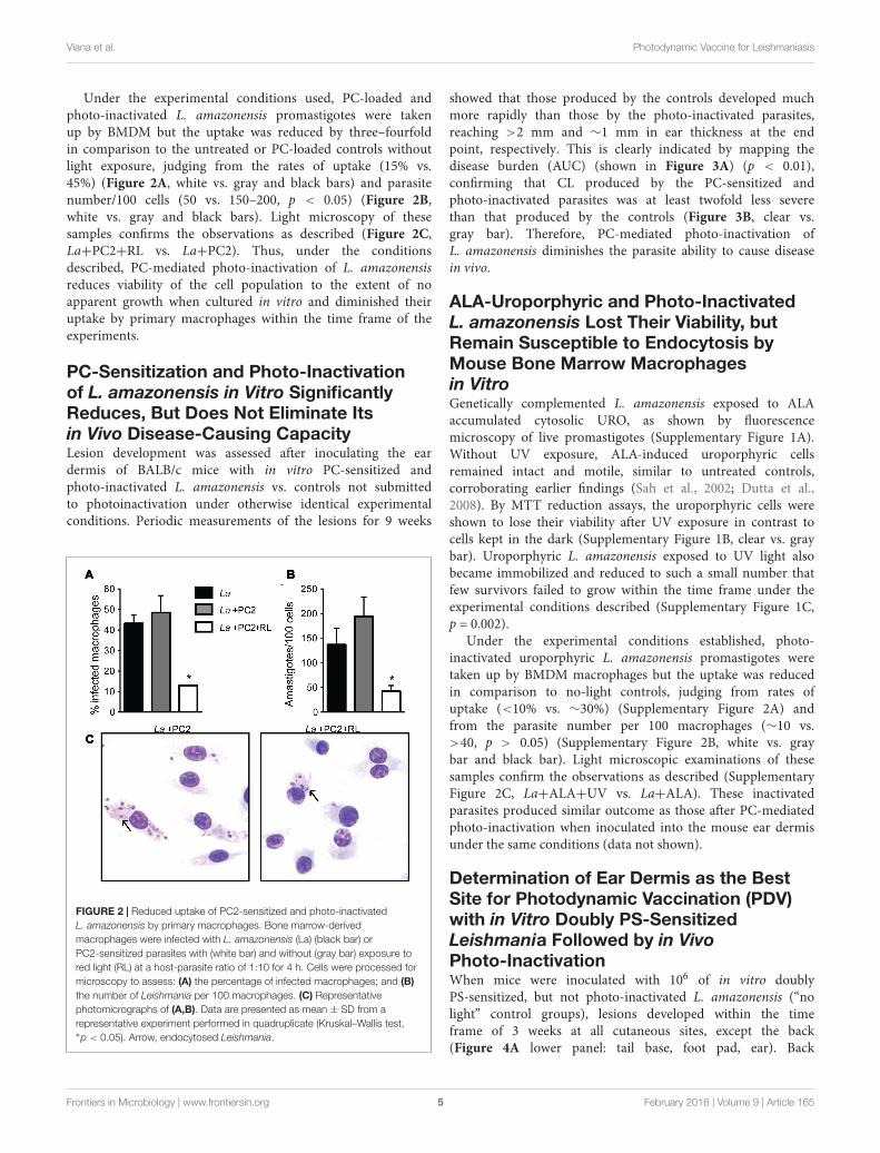
PC-Sensitized and Photo-InactivatedL. amazonensis Lost Their ViabilitySubstantially, but Remain Susceptible toEndocytosis by Mouse Bone MarrowMacrophages in VitroLeishmania amazonensis were PC-sensitized and exposed tored light for photo-inactivation, e.g., amino-PC2. Fluorescencemicroscopy of live promastigotes showed PC2 localization incytoplasmic vacuoles (Figure 1A). Without light exposure, thesePC-loaded cells remained, as expected, intact and motile, justlike the untreated controls. By MTT reduction assays, PC-loaded(e.g., 0.1–10 µM PC2) cells were shown to lose their viabilityonly after light exposure in contrast to parasites kept in thedark (Figure 1B, white bars vs. gray bars). When sensitized with0.1 µM PC and exposed to light, cells lost flagellar motility; exceptvery few, which failed to grow up on further incubation under theexperimental conditions described (p = 0.0286; Figure 1C). TheED50 of the amino-PC for photo-inactivation of these cells falls inbetween 10–100 nM according to the results with PC2 from bothcell viability assays.
FIGURE 1 | PC2-mediated photo-inactivation of Leishmania amazonensis. Promastigotes (La) were incubated in the dark with PC2 for 16 h and exposed to red-light(RL) (=1–2 J/cm2). (A) Merged phase contrast and Cy5 fluorescence images, showing the uptake of PC2 by cells sensitized in the dark overnight with 1 µM PC2.(B) MTT reduction activities of cells sensitized with graded concentrations of PC2, as indicated, in the dark (gray bar) and after light exposure (white bar), the valuesbeing expressed in % of untreated controls (black bar). (C) Disparity in cell density between PC2-sensitized cells with (white bar) and without photo-inactivation (graybar) after inoculation into culture medium. Similar results were obtained after PS-sensitization with either 0.1 or 1 µM PC2. Data are presented as mean ± SD from arepresentative set of experiments performed in quadruplicate.
Frontiers in Microbiology | www.frontiersin.org 4 February 2018 | Volume 9 | Article 165
fmicb-09-00165 February 3, 2018 Time: 13:27 # 5
Viana et al. Photodynamic Vaccine for Leishmaniasis
Under the experimental conditions used, PC-loaded andphoto-inactivated L. amazonensis promastigotes were takenup by BMDM but the uptake was reduced by three–fourfoldin comparison to the untreated or PC-loaded controls withoutlight exposure, judging from the rates of uptake (15% vs.45%) (Figure 2A, white vs. gray and black bars) and parasitenumber/100 cells (50 vs. 150–200, p < 0.05) (Figure 2B,white vs. gray and black bars). Light microscopy of thesesamples confirms the observations as described (Figure 2C,La+PC2+RL vs. La+PC2). Thus, under the conditionsdescribed, PC-mediated photo-inactivation of L. amazonensisreduces viability of the cell population to the extent of noapparent growth when cultured in vitro and diminished theiruptake by primary macrophages within the time frame of theexperiments.
PC-Sensitization and Photo-Inactivationof L. amazonensis in Vitro SignificantlyReduces, But Does Not Eliminate Itsin Vivo Disease-Causing CapacityLesion development was assessed after inoculating the eardermis of BALB/c mice with in vitro PC-sensitized andphoto-inactivated L. amazonensis vs. controls not submittedto photoinactivation under otherwise identical experimentalconditions. Periodic measurements of the lesions for 9 weeks
FIGURE 2 | Reduced uptake of PC2-sensitized and photo-inactivatedL. amazonensis by primary macrophages. Bone marrow-derivedmacrophages were infected with L. amazonensis (La) (black bar) orPC2-sensitized parasites with (white bar) and without (gray bar) exposure tored light (RL) at a host-parasite ratio of 1:10 for 4 h. Cells were processed formicroscopy to assess: (A) the percentage of infected macrophages; and (B)the number of Leishmania per 100 macrophages. (C) Representativephotomicrographs of (A,B). Data are presented as mean ± SD from arepresentative experiment performed in quadruplicate (Kruskal–Wallis test,∗p < 0.05). Arrow, endocytosed Leishmania.
showed that those produced by the controls developed muchmore rapidly than those by the photo-inactivated parasites,reaching >2 mm and ∼1 mm in ear thickness at the endpoint, respectively. This is clearly indicated by mapping thedisease burden (AUC) (shown in Figure 3A) (p < 0.01),confirming that CL produced by the PC-sensitized andphoto-inactivated parasites was at least twofold less severethan that produced by the controls (Figure 3B, clear vs.gray bar). Therefore, PC-mediated photo-inactivation ofL. amazonensis diminishes the parasite ability to cause diseasein vivo.
ALA-Uroporphyric and Photo-InactivatedL. amazonensis Lost Their Viability, butRemain Susceptible to Endocytosis byMouse Bone Marrow Macrophagesin VitroGenetically complemented L. amazonensis exposed to ALAaccumulated cytosolic URO, as shown by fluorescencemicroscopy of live promastigotes (Supplementary Figure 1A).Without UV exposure, ALA-induced uroporphyric cellsremained intact and motile, similar to untreated controls,corroborating earlier findings (Sah et al., 2002; Dutta et al.,2008). By MTT reduction assays, the uroporphyric cells wereshown to lose their viability after UV exposure in contrast tocells kept in the dark (Supplementary Figure 1B, clear vs. graybar). Uroporphyric L. amazonensis exposed to UV light alsobecame immobilized and reduced to such a small number thatfew survivors failed to grow within the time frame under theexperimental conditions described (Supplementary Figure 1C,p = 0.002).
Under the experimental conditions established, photo-inactivated uroporphyric L. amazonensis promastigotes weretaken up by BMDM macrophages but the uptake was reducedin comparison to no-light controls, judging from rates ofuptake (<10% vs. ∼30%) (Supplementary Figure 2A) andfrom the parasite number per 100 macrophages (∼10 vs.>40, p > 0.05) (Supplementary Figure 2B, white vs. graybar and black bar). Light microscopic examinations of thesesamples confirm the observations as described (SupplementaryFigure 2C, La+ALA+UV vs. La+ALA). These inactivatedparasites produced similar outcome as those after PC-mediatedphoto-inactivation when inoculated into the mouse ear dermisunder the same conditions (data not shown).
Determination of Ear Dermis as the BestSite for Photodynamic Vaccination (PDV)with in Vitro Doubly PS-SensitizedLeishmania Followed by in VivoPhoto-InactivationWhen mice were inoculated with 106 of in vitro doublyPS-sensitized, but not photo-inactivated L. amazonensis (“nolight” control groups), lesions developed within the timeframe of 3 weeks at all cutaneous sites, except the back(Figure 4A lower panel: tail base, foot pad, ear). Back
Frontiers in Microbiology | www.frontiersin.org 5 February 2018 | Volume 9 | Article 165
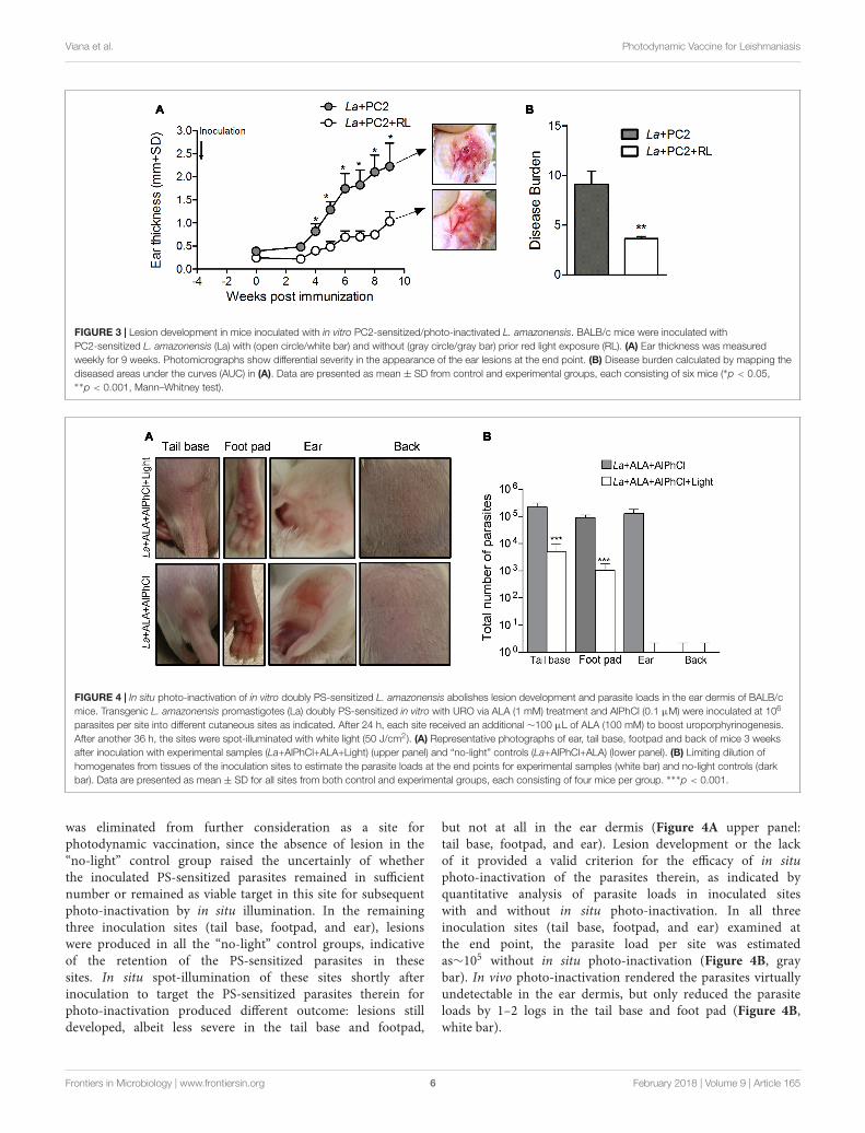
fmicb-09-00165 February 3, 2018 Time: 13:27 # 6
Viana et al. Photodynamic Vaccine for Leishmaniasis
FIGURE 3 | Lesion development in mice inoculated with in vitro PC2-sensitized/photo-inactivated L. amazonensis. BALB/c mice were inoculated withPC2-sensitized L. amazonensis (La) with (open circle/white bar) and without (gray circle/gray bar) prior red light exposure (RL). (A) Ear thickness was measuredweekly for 9 weeks. Photomicrographs show differential severity in the appearance of the ear lesions at the end point. (B) Disease burden calculated by mapping thediseased areas under the curves (AUC) in (A). Data are presented as mean ± SD from control and experimental groups, each consisting of six mice (∗p < 0.05,∗∗p < 0.001, Mann–Whitney test).
FIGURE 4 | In situ photo-inactivation of in vitro doubly PS-sensitized L. amazonensis abolishes lesion development and parasite loads in the ear dermis of BALB/cmice. Transgenic L. amazonensis promastigotes (La) doubly PS-sensitized in vitro with URO via ALA (1 mM) treatment and AlPhCl (0.1 µM) were inoculated at 106
parasites per site into different cutaneous sites as indicated. After 24 h, each site received an additional ∼100 µL of ALA (100 mM) to boost uroporphyrinogenesis.After another 36 h, the sites were spot-illuminated with white light (50 J/cm2). (A) Representative photographs of ear, tail base, footpad and back of mice 3 weeksafter inoculation with experimental samples (La+AlPhCl+ALA+Light) (upper panel) and “no-light” controls (La+AlPhCl+ALA) (lower panel). (B) Limiting dilution ofhomogenates from tissues of the inoculation sites to estimate the parasite loads at the end points for experimental samples (white bar) and no-light controls (darkbar). Data are presented as mean ± SD for all sites from both control and experimental groups, each consisting of four mice per group. ∗∗∗p < 0.001.
was eliminated from further consideration as a site forphotodynamic vaccination, since the absence of lesion in the“no-light” control group raised the uncertainly of whetherthe inoculated PS-sensitized parasites remained in sufficientnumber or remained as viable target in this site for subsequentphoto-inactivation by in situ illumination. In the remainingthree inoculation sites (tail base, footpad, and ear), lesionswere produced in all the “no-light” control groups, indicativeof the retention of the PS-sensitized parasites in thesesites. In situ spot-illumination of these sites shortly afterinoculation to target the PS-sensitized parasites therein forphoto-inactivation produced different outcome: lesions stilldeveloped, albeit less severe in the tail base and footpad,
but not at all in the ear dermis (Figure 4A upper panel:tail base, footpad, and ear). Lesion development or the lackof it provided a valid criterion for the efficacy of in situphoto-inactivation of the parasites therein, as indicated byquantitative analysis of parasite loads in inoculated siteswith and without in situ photo-inactivation. In all threeinoculation sites (tail base, footpad, and ear) examined atthe end point, the parasite load per site was estimatedas∼105 without in situ photo-inactivation (Figure 4B, graybar). In vivo photo-inactivation rendered the parasites virtuallyundetectable in the ear dermis, but only reduced the parasiteloads by 1–2 logs in the tail base and foot pad (Figure 4B,white bar).
Frontiers in Microbiology | www.frontiersin.org 6 February 2018 | Volume 9 | Article 165
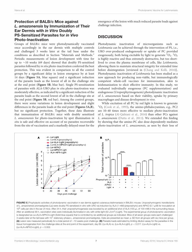
fmicb-09-00165 February 3, 2018 Time: 13:27 # 7
Viana et al. Photodynamic Vaccine for Leishmaniasis
Protection of BALB/c Mice againstL. amazonensis by Immunization of TheirEar Dermis with in Vitro DoublyPS-Sensitized Parasites for in VivoPhoto-InactivationGroups of BALB/c mice were photodynamically vaccinatedonce accordingly in the ear dermis with multiple controlsand challenged 3 weeks later at the tail base under theconditions as described in Section “Materials and Methods.”Periodic measurements of lesion development with time forup to ∼10 weeks (69 days) showed that doubly PS-sensitizedparasites followed by in situ photo-inactivation conferred the bestprotection. This was evident in comparison to all the controlgroups by a significant delay in lesion emergence by at least14 days (Figure 5A, blue square) and a significant reductionof the parasite loads as the lowest of all in the challenge siteat the end point (Figure 5B, blue bar). Single PS-sensitizationof parasites with ALA-URO plus in situ photo-inactivation wasmoderately effective, as indicated by a significant reduction of theparasite loads as the second lowest of all in the challenge site atthe end point (Figure 5B, red bar). Among the control groups,there were some variations in lesion development and slightdifferences in the parasite loads at the end point (Figures 5A,B),but no significant protection. Together, these data illustratethat immunization of BALB/c mice with doubly sensitizedL. amazonensis for photo-inactivation by spot illumination insitu is safe and effective on account of no parasites recoverablefrom the site of vaccination and a markedly delayed onset for the
emergence of the lesion with much reduced parasite loads againstchallenge infection.
DISCUSSION
Photodynamic inactivation of microorganisms such asLeishmania can be achieved through the intervention of PS, i.e.,URO over-produced endogenously or uptake of PC providedexogenously, both being excitable by light to generate 1O2. 1O2is highly reactive and thus extremely destructive, but too short-lived to cross the plasma membrane of cells, like Leishmania,allowing them to maintain structural integrity for extended timebefore disintegration [reviewed in (Chang and Kolli, 2016)].Photodynamic inactivation of Leishmania has been studied as anew approach for producing non-viable, but immunologicallycompetent whole-cell vaccines for immunization, akin toleishmanization to elicit effective immunity. In this study, weevaluated individually exogenous (PC supplementation) andendogenous (Uroporphyrinogenesis) photodynamic inactivationof L. amazonensis based on their viability, uptake by primarymacrophages and disease development in vivo.
While excitation of all PC by red light is known to generate1O2 (Cook et al., 1995), the amino-phthalocyanines, e.g., PC2are 10–40 times more effective to mediate photo-inactivationof L. tropica (Al-Qahtani et al., 2016) than other PCs againstL. amazonensis (Dutta et al., 2011). We extended this findingby showing that the amino-PC also dose-dependently mediatesphoto-inactivation of L. amazonensis, as seen by their loss of
FIGURE 5 | Prophylactic activities of photodynamic vaccination in ear dermis against cutaneous leishmaniasis in BALB/c mouse. Uroporphyrinogenic transfectantsof L. amazonensis promastigotes (La) were doubly PS-sensitized in vitro with URO via induction by ALA (1 mM) (preporphyric) and AlPhCl (0.1 µM) for inoculation at106 cells per site in the ear of mice. After 24 h, their uroporphyrinogenesis was boosted by an additional shot of ALA (100 µL of 100 mM) in the surrounding tissue.After an additional 36 h, vaccination sites were individually illuminated in situ with white light (50 J/cm2) (Light). The experimental group of photodynamic vaccinationis designated as La+ALA+AlPhCl+Light (Solid blue square) that is controlled by six additional groups as indicated. Mice of all seven groups were each challenged3 weeks later at the tail base with 107 stationary-phase L. amazonensis promastigotes. Data are presented as mean ± SD from all groups with six mice per group.(A) Lesion size measured periodically for a total period of ∼10 weeks post challenge. (B) Parasite loads estimated by limiting dilution assay for the parasites in thetissue homogenates from the challenge sites at the end point of the experiment, day 69. (La-ALA) vs. (La+ALA+Light), p < 0.017. (La+ALA+Light) vs.(La-ALA+AlPhCl+Light), p < 0.002.
Frontiers in Microbiology | www.frontiersin.org 7 February 2018 | Volume 9 | Article 165

fmicb-09-00165 February 3, 2018 Time: 13:27 # 8
Viana et al. Photodynamic Vaccine for Leishmaniasis
cell motility and viability based on microscopic observations andMTT reduction assay, respectively (Figure 1). PC-sensitizationand photo-inactivation of L. amazonensis also reduced itsuptake by primary macrophages (Figure 2). As shown here,amino-PC-sensitization of L. amazonensis is stochastic, leavinga small number of parasites un-sensitized, hence escapingfrom the fate of photo-inactivation. Stochasticity of single-PSsensitization has been reported previously, including the useof other PC (Dutta et al., 2005, 2008) and ALA induction ofcytosolic URO accumulation (Sah et al., 2002; Dutta et al.,2008). This is the case despite amino-PC being much morepotent than other PS to mediate Leishmania photo-inactivation.The development of lesion, albeit with a delayed onset andsignificantly reduced disease burden (Figure 3) is thus notunexpected after inoculation of BALB/c mice with amino-PC-sensitized and photo-inactivated L. amazonensis.
The potential use of single PS-sensitized and photo-inactivated Leishmania for PDV raises concern in consideringtheir safety, but not efficacy. The safety is clearly a concernwhen lesions emerge after inoculation of these inactivatedLeishmania into mice (Figure 3). The reduction of their uptakeby primary macrophages in vitro (Figure 2) also may be takento indicate diminished “vaccine” loading of the APC whenapplied in vivo. However, after such single-PS photo-inactivation,Leishmania transgenically modified to express OVA was shownto effectively deliver this T-cell antigen to macrophages anddendritic cells (DC) for processing and presentation to OVA-epitope specific CD8+ T cells in vitro (Dutta et al., 2011,2012). Interestingly, immunization of Syrian Golden hamsterswith single PS-sensitized and photo-inactivated L. amazonensisin vivo was shown to protect them against challenge infectionwith L. donovani: the splenic parasite load was drasticallyreduced by 99%, concomitant with significant increase in theexpression of iNOS, IFN-γ, and IL-12 (Kumari et al., 2009).Significantly, this immunity is adaptively transferable with Tcells from immunized hamsters to naïve animals, indicatingthat it requires no stimulation by persisting parasites, as theyare unlikely to exist in the T-cell recipients. In contrast,persistence of parasites in small number cannot be ruled outin the hamsters after primary immunization with Leishmaniasubjected to single PS-sensitization/photo-inactivation, whichinvariably leaves behind few survivors, regardless of the PSused. Thus, while the use of incompletely photo-inactivatedLeishmania is unacceptable for safety consideration, the resultsobtained point to the efficacy of PDV as a right path tovaccination.
In keeping with the synergism of two differentphotosensitizers in combination to enhance PDT efficacyagainst cancer (Schneider-Yin et al., 2009; Villanueva et al., 2010;Gyenge et al., 2013; Acedo et al., 2014), we previously showedthat L. amazonensis doubly sensitized with URO+PC in vitrowas fully susceptible to in vivo photo-inactivation in BALB/c XC57BL/6 mice, producing no lesion and no detectable parasites8 weeks after inoculation with 106 cells per site into their eardermis (Dutta et al., 2012). Here, we were able to duplicatethis finding in BALB/c mice and found it specific to the eardermis, but not footpad or tail base. Translucency of ear due to
its thinness may facilitate the efficiency of in situ illuminationwith white light, especially the short wavelength of the spectrum,i.e., ∼400 nm optimal for excitation of URO. Ear dermis wasthus chosen as the site for PDV against challenge infection in thetail base. This choice is based not solely on the consideration ofsafety, but on that of efficacy. Leishmania subjected to doublePS-sensitization alone without photo-inactivation in vitrowas found to persist in comparable abundance in all threecutaneous sites (ear dermis, footpad, and tail base), but onlythose in the ear dermis were photo-inactivated beyond detectionby in situ illumination (Figure 4). Thus, at this site there isan optimal amount of “vaccines” made available to APC viaphoto-inactivation of Leishmania therein and, more importantly,little or no immunosuppression by live parasites expected dueto their virtual absence. By both accounts, PDV appears tocreate a microenvironment more favorably to elicit immunityin the ear dermis than tail base and foot pad. This immunityproduced by PDV of the ear dermis is manifested againstchallenge infection by significantly delaying the onset of lesiondevelopment and by the marked reduction of parasite load seenin the challenging site at the end point of day 69 (Figure 5).This level of protection is significant, considering that mostmouse lineages, including C3H, C57BL/6, BDA, and CBA allfail to heal or control L. amazonensis infection (Soong et al.,1997; Cortes et al., 2010), to which a susceptible phenotype ofmixed Th1–Th2 response is often developed (Afonso and Scott,1993; Mcmahon-Pratt and Alexander, 2004). BALB/c mice areespecially susceptible to the infection by this species, presentingprogressive development of non-healing necrotic lesion (Pereiraand Alves, 2008). More complete protection by PDV is expectedby optimizing the experimental conditions, e.g., reduction of theparasite dose for challenge infection, increasing the frequencyof immunization more than once and/or adjustment of thetime intervals between inoculation of doubly PS-sensitizedLeishmania and in situ photo-inactivation. Optimization of thisinterval is expected to maximize photo-inactivation of doublyPS-sensitized Leishmania, thereby minimizing their migrationfrom ear dermis to the draining lymph nodes. Minimizationof this migration reduces the parasite population that escapesphoto-inactivation by spot-illumination of the inoculation site,thereby reducing their immunosuppressive activities in thelymph nodes and facilitating immune clearance. The importanceof immune clearance has been discussed previously and cannotbe over-emphasized, considering its apparent necessity forpost chemotherapeutic cure of leishmaniasis, since no drug isexpected to reach every parasite in the patients, regardless ofdosages used and treatment duration (Chang, 2014).
The current working hypothesis is that macrophages and DCtake up in vitro doubly PS-sensitized Leishmania for subsequentcytolysis to release antigens therein after in vivo photo-inactivation, while the host APC remains viable and functional(Dutta et al., 2012). Therefore, antigen presentation and ensuingcellular-based immune response can be induced effectively, inkeeping with the development of life long immunity known tooccur after spontaneous or chemotherapeutic cure of humanleishmaniasis. In summary, results presented demonstrate thatPS-sensitization and photo-inactivation of L. amazonensis
Frontiers in Microbiology | www.frontiersin.org 8 February 2018 | Volume 9 | Article 165
fmicb-09-00165 February 3, 2018 Time: 13:27 # 9
Viana et al. Photodynamic Vaccine for Leishmaniasis
based on a combination of endogenous and exogenous strategiesrenders them non-viable, but immunologically competent againstCL. The results presented remain to be a proof-of-principle,pending further investigation to reduce the complexity of PS-sensitization and photo-inactivation. Work is underway towardthe simplification of these steps to facilitate the standardizationand scale-up production of such photodynamically inactivatedvaccines.
AUTHOR CONTRIBUTIONS
Conceived and designed the experiments: SV, CdO,and KC. Performed the experiments: SV, FC, and LR.Analyzed the data: SV, FC, BK, CdO, and KC. Contributedreagents/materials/analysis tools: DN, BK, and KC. Wrote themanuscript: SV, BK, KC, and CdO.
ACKNOWLEDGMENTS
We thank to the support of NIH-NIAID grant # AI-68835,AI-7712375, and AI-097830 to KC. Technical assistance ofSujoy Dutta to portion of this work is gratefully acknowledged.We also acknowledge the support of Conselho Nacional deDesenvolvimento Científico e Tecnológico (CNPq) – (ProgramaVisitante Especial) Processo 402312/2012-0 to CdO. SV, FC, andLR were supported by CNPq fellowships and CdO is a seniorinvestigator for CNPq.
SUPPLEMENTARY MATERIAL
The Supplementary Material for this article can be foundonline at: https://www.frontiersin.org/articles/10.3389/fmicb.2018.00165/full#supplementary-material
REFERENCESAcedo, P., Stockert, J. C., Cañete, M., and Villanueva, A. (2014). Two combined
photosensitizers: a goal for more effective photodynamic therapy of cancer. CellDeath Dis. 5:e1122. doi: 10.1038/cddis.2014.77
Afonso, L. C., and Scott, P. (1993). Immune responses associated with susceptibilityof C57BL/10 mice to Leishmania amazonensis. Infect. Immun. 61, 2952–2959.
Alexander, J. (1982). A radioattenuated Leishmania major vaccine markedlyincreases the resistance of CBA mice to subsequent infection with Leishmaniamexicana mexicana. Trans. R. Soc. Trop. Med. Hyg. 76, 646–649. doi: 10.1016/0035-9203(82)90232-2
Alexander, J., Coombs, G. H., and Mottram, J. C. (1998). Leishmania mexicanacysteine proteinase-deficient mutants have attenuated virulence for mice andpotentiate a Th1 response. J. Immunol. 161, 6794–6801.
Al-Qahtani, A., Alkahtani, S., Kolli, B., Tripathi, P., Dutta, S., Al-Kahtane, A. A.,et al. (2016). Aminophthalocyanine-mediated photodynamic inactivation ofLeishmania tropica. Antimicrob. Agents Chemother. 60, 2003–2011. doi: 10.1128/AAC.01879-15
Bhattacharya, P., Dey, R., Dagur, P. K., Kruhlak, M., Ismail, N., Debrabant, A., et al.(2015). Genetically modified live attenuated L. donovani parasites induce innateimmunity through classical activation of macrophages that direct Th1 responsein mice. Infect. Immun. 83, 3800–3815. doi: 10.1128/IAI.00184-15
Bittencourt, A. L., Costa, J. M., Carvalho, E. M., and Barral, A. (1993).Leishmaniasis recidiva cutis in American cutaneous leishmaniasis. Int. J.Dermatol. 32, 802–805. doi: 10.1111/j.1365-4362.1993.tb02767.x
Canavan, T. N., De La Feld, S. F., Huang, C., and Sami, N. (2017). Photodynamictherapy effective for the treatment of actinic keratosis and basal cell carcinomain bullous pemphigoid patients. Photodiagnosis Photodyn. Ther. 18, 257–259.doi: 10.1016/j.pdpdt.2017.03.019
Chang, K. P. (2014). Vaccination for disease prevention and control: the necessityof renewed emphasis and new approaches. J. Immunol. Immunotech. 1, 1–4.doi: 10.17653/2374-9105.SSe0001
Chang, K. P., and Kolli, B. K. (2016). New "light" for one-world approach towardsafe and effective control of animal diseases and insect vectors from leishmaniacperspectives. Parasit. Vectors 9, 396–408. doi: 10.1186/s13071-016-1674-3
Chen, T., Zhang, L. W., Fu, L. X., Wu, Y. B., Liu, X. Y., and Guo, Z. P. (2017).Systemic ALA-PDT effectively blocks the development of psoriasis-like lesionsand alleviates leucocyte infiltration in the K14-VEGF transgenic mouse. Clin.Exp. Dermatol. 42, 849–856. doi: 10.1111/ced.13148
Cook, M. J., Chambrier, I., Cracknell, S. J., Mayes, D. A., and Russell, D. A.(1995). Octa-alkyl zinc phthalocyanines: potential photosensitizers for usein the photodynamic therapy of cancer. Photochem. Photobiol. 62, 542–545.doi: 10.1111/j.1751-1097.1995.tb02381.x
Cortes, D. F., Carneiro, M. B., Santos, L. M., Souza, T. C., Maioli, T. U., Duz, A. L.,et al. (2010). Low and high-dose intradermal infection with Leishmania major
and Leishmania amazonensis in C57BL/6 mice. Mem. Inst. Oswaldo Cruz 105,736–745. doi: 10.1590/S0074-02762010000600002
Daneshvar, H., Coombs, G. H., Hagan, P., and Phillips, R. S. (2003). Leishmaniamexicana and Leishmania major: attenuation of wild-type parasites andvaccination with the attenuated lines. J. Infect. Dis. 187, 1662–1668. doi: 10.1086/374783
Dey, R., Dagur, P. K., Selvapandiyan, A., Mccoy, J. P., Salotra, P., Duncan, R.,et al. (2013). Live attenuated Leishmania donovani p27 gene knockout parasitesare nonpathogenic and elicit long-term protective immunity in BALB/c mice.J. Immunol. 190, 2138–2149. doi: 10.4049/jimmunol.1202801
Dutta, S., Kolli, B. K., Tang, A., Sassa, S., and Chang, K.-P. (2008). TransgenicLeishmania model for delta-aminolevulinate-inducible monospecificuroporphyria: cytolytic phototoxicity initiated by singlet oxygen-mediatedinactivation of proteins and its ablation by endosomal mobilization of cytosolicuroporphyrin. Eukaryot. Cell 7, 1146–1157. doi: 10.1128/EC.00365-07
Dutta, S., Ongarora, B. G., Li, H., Da Vicente, M. G. H., Kolli, B. K., and Chang, K. P.(2011). Intracellular targeting specificity of novel phthalocyanines assessed ina host-parasite model for developing potential photodynamic medicine. PLOSONE 6:e20786. doi: 10.1371/journal.pone.0020786
Dutta, S., Ray, D., Kolli, B. K., and Chang, K. P. (2005). Photodynamic sensitizationof Leishmania amazonensis in both extracellular and intracellular stages withaluminum phthalocyanine chloride for photolysis in vitro. Antimicrob. AgentsChemother. 49, 4474–4484. doi: 10.1128/AAC.49.11.4474-4484.2005
Dutta, S., Waki, K., and Chang, K. P. (2012). Combinational sensitization ofLeishmania with uroporphyrin and aluminum phthalocyanine synergisticallyenhances their photodynamic inactivation in vitro and in vivo. Photochem.Photobiol. 88, 620–625. doi: 10.1111/j.1751-1097.2012.01076.x
Enk, C. D., Nasereddin, A., Alper, R., Dan-Goor, M., Jaffe, C. L., and Wulf, H. C.(2015). Cutaneous leishmaniasis responds to daylight-activated photodynamictherapy: proof of concept for a novel self-administered therapeutic modality.Br. J. Dermatol. 172, 1364–1370. doi: 10.1111/bjd.13490
Gyenge, E. B., Luscher, D., Forny, P., Antoniol, M., Geisberger, G., Walt, H., et al.(2013). Photodynamic mechanisms induced by a combination of hypericin anda chlorin based-photosensitizer in head and neck squamous cell carcinoma cells.Photochem. Photobiol. 89, 150–162. doi: 10.1111/j.1751-1097.2012.01217.x
Kumari, S., Samant, M., Khare, P., Misra, P., Dutta, S., Kolli, B. K., et al. (2009).Photodynamic vaccination of hamsters with inducible suicidal mutants ofLeishmania amazonensis elicits immunity against visceral leishmaniasis. Eur.J. Immunol. 39, 178–191. doi: 10.1002/eji.200838389
Mcmahon-Pratt, D., and Alexander, J. (2004). Does the Leishmania majorparadigm of pathogenesis and protection hold for New World cutaneousleishmaniases or the visceral disease? Immunol. Rev. 201, 206–224.
Nadim, A., Javadian, E., Tahvildar-Bidruni, G., and Ghorbani, M. (1983).Effectiveness of leishmanization in the control of cutaneous leishmaniasis. Bull.Soc. Pathol. Exot. Filiales 76, 377–383.
Frontiers in Microbiology | www.frontiersin.org 9 February 2018 | Volume 9 | Article 165

fmicb-09-00165 February 3, 2018 Time: 13:27 # 10
Viana et al. Photodynamic Vaccine for Leishmaniasis
Palatnik-De-Sousa, C. B. (2008). Vaccines for leishmaniasis in the fore coming 25years. Vaccine 26, 1709–1724. doi: 10.1016/j.vaccine.2008.01.023
Pereira, B. A., and Alves, C. R. (2008). Immunological characteristics ofexperimental murine infection with Leishmania (Leishmania) amazonensis.Vet. Parasitol. 158, 239–255. doi: 10.1016/j.vetpar.2008.09.015
Reithinger, R., Dujardin, J. J.-C. J., Louzir, H., Pirmez, C., Alexander, B., Brooker, S.,et al. (2007). Cutaneous leishmaniasis. Lancet 8, 8–9. doi: 10.1016/S1473-3099(07)70209-8
Sah, J. F., Ito, H., Kolli, B. K., Peterson, D. A., Sassa, S., and Chang, K. P.(2002). Genetic rescue of Leishmania deficiency in porphyrin biosynthesiscreates mutants suitable for analysis of cellular events in uroporphyria andfor photodynamic therapy. J. Biol. Chem. 277, 14902–14909. doi: 10.1074/jbc.M200107200
Schneider-Yin, X., Kurmanaviciene, A., Roth, M., Roos, M., Fedier, A., Minder, E. I.,et al. (2009). Hypericin and 5-aminolevulinic acid-induced protoporphyrin IXinduce enhanced phototoxicity in human endometrial cancer cells with non-coherent white light. Photodiagnosis Photodyn. Ther. 6, 12–18. doi: 10.1016/j.pdpdt.2009.02.001
Selvapandiyan, A., Dey, R., Nylen, S., Duncan, R., Sacks, D., and Nakhasi, H. L.(2009). Intracellular replication-deficient Leishmania donovani induces longlasting protective immunity against visceral leishmaniasis. J. Immunol. 183,1813–1820. doi: 10.4049/jimmunol.0900276
Soong, L., Chang, C. H., Sun, J., Longley, BJ Jr, Ruddle, N. H., Flavell, R. A.,et al. (1997). Role of CD4+ T cells in pathogenesis associated with Leishmaniaamazonensis infection. J. Immunol. 158, 5374–5383.
Spath, G. F., Epstein, L., Leader, B., Singer, S. M., Avila, H. A., Turco, S. J.,et al. (2000). Lipophosphoglycan is a virulence factor distinct from relatedglycoconjugates in the protozoan parasite Leishmania major. Proc. Natl. Acad.Sci. U.S.A. 97, 9258–9263. doi: 10.1073/pnas.160257897
Sundar, S., and Singh, B. (2014). Identifying vaccine targets for anti-leishmanialvaccine development. Expert Rev. Vaccines 13, 489–505. doi: 10.1586/14760584.2014.894467
Uzonna, J. E., Spath, G. F., Beverley, S. M., and Scott, P. (2004). Vaccination withphosphoglycan-deficient Leishmania major protects highly susceptible micefrom virulent challenge without inducing a strong Th1 response. J. Immunol.172, 3793–3797. doi: 10.4049/jimmunol.172.6.3793
Villanueva, A., Stockert, J. C., Canete, M., and Acedo, P. (2010). A new protocolin photodynamic therapy: enhanced tumour cell death by combining twodifferent photosensitizers. Photochem. Photobiol. Sci. 9, 295–297. doi: 10.1039/b9pp00153k
Wang, Y., Lin, Y., Zhang, H. G., and Zhu, J. (2016). A photodynamic therapycombined with topical 5-aminolevulinic acid and systemic hematoporphyrinderivative is more efficient but less phototoxic for cancer. J. Cancer Res. Clin.Oncol. 142, 813–821. doi: 10.1007/s00432-015-2066-3
Weischenfeldt, J., and Porse, B. (2008). Bone Marrow-Derived Macrophages(BMM): isolation and applications. CSH Protoc. 2008:pdb.prot5080. doi: 10.1101/pdb.prot5080
WHO (2010). Control of the Leishmaniases: Report of a Meeting of theWHO ExpertCommittee on the Control of Leishmaniases. Geneva: WHO, 1–186.
Yasinzai, M., Khan, M., Nadhman, A., and Shahnaz, G. (2013). Drug resistancein leishmaniasis: current drug-delivery systems and future perspectives. FutureMed. Chem. 5, 1877–1888. doi: 10.4155/fmc.13.143
Conflict of Interest Statement: The authors declare that the research wasconducted in the absence of any commercial or financial relationships that couldbe construed as a potential conflict of interest.
Copyright © 2018 Viana, Celes, Ramirez, Kolli, Ng, Chang and de Oliveira. This is anopen-access article distributed under the terms of the Creative Commons AttributionLicense (CC BY). The use, distribution or reproduction in other forums is permitted,provided the original author(s) and the copyright owner are credited and that theoriginal publication in this journal is cited, in accordance with accepted academicpractice. No use, distribution or reproduction is permitted which does not complywith these terms.
Frontiers in Microbiology | www.frontiersin.org 10 February 2018 | Volume 9 | Article 165
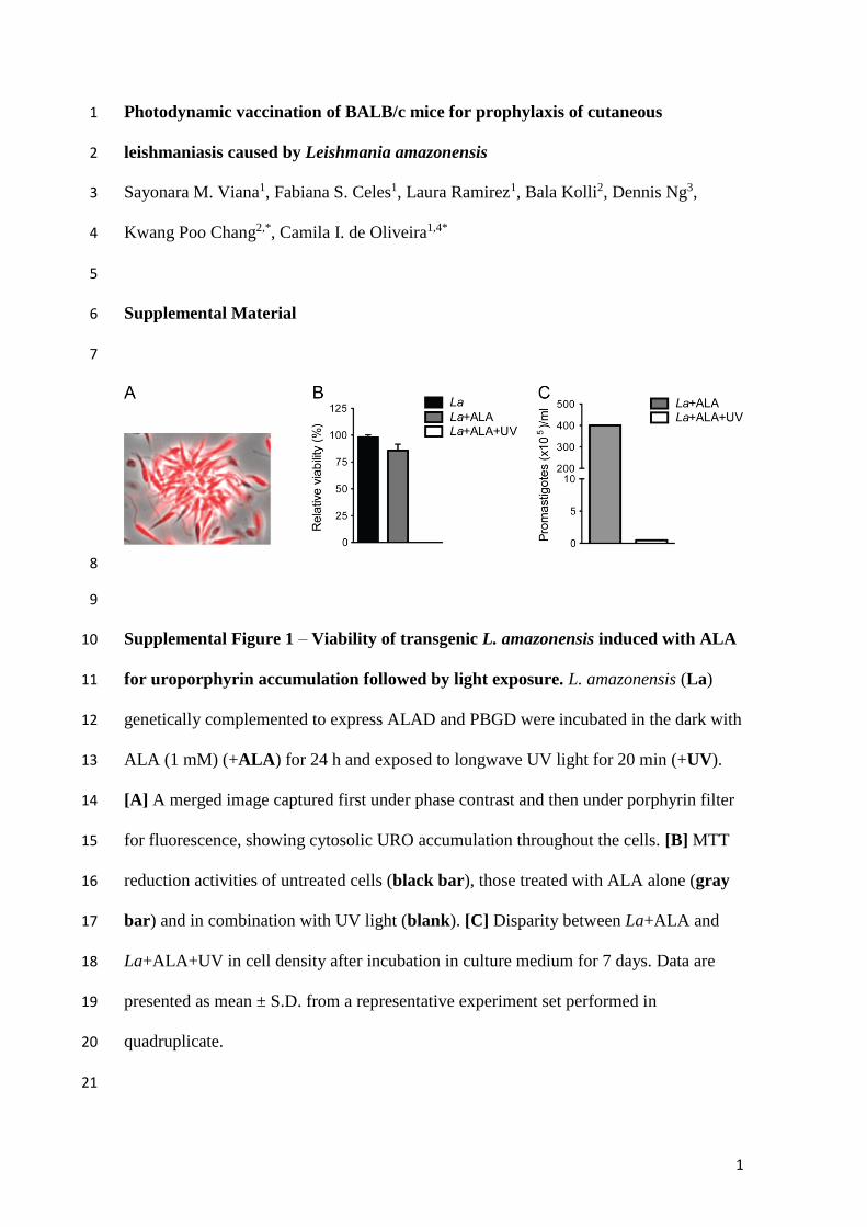
1
Photodynamic vaccination of BALB/c mice for prophylaxis of cutaneous 1
leishmaniasis caused by Leishmania amazonensis 2
Sayonara M. Viana1, Fabiana S. Celes1, Laura Ramirez1, Bala Kolli2, Dennis Ng3, 3
Kwang Poo Chang2,*, Camila I. de Oliveira1,4* 4
5
Supplemental Material 6
7
8
9
Supplemental Figure 1 – Viability of transgenic L. amazonensis induced with ALA 10
for uroporphyrin accumulation followed by light exposure. L. amazonensis (La) 11
genetically complemented to express ALAD and PBGD were incubated in the dark with 12
ALA (1 mM) (+ALA) for 24 h and exposed to longwave UV light for 20 min (+UV). 13
[A] A merged image captured first under phase contrast and then under porphyrin filter 14
for fluorescence, showing cytosolic URO accumulation throughout the cells. [B] MTT 15
reduction activities of untreated cells (black bar), those treated with ALA alone (gray 16
bar) and in combination with UV light (blank). [C] Disparity between La+ALA and 17
La+ALA+UV in cell density after incubation in culture medium for 7 days. Data are 18
presented as mean ± S.D. from a representative experiment set performed in 19
quadruplicate. 20
21
2
22
Supplemental Figure 2 – Reduced uptake of uroporphyric and photo-inactivated 23
L. amazonensis by primary macrophages. Bone marrow-derived macrophages were 24
infected at a host to parasite ratio of 1:10 for 4h with untreated L. amazonensis (black 25
bar), those ALA-sensitized alone (gray bar) and in combination with exposure to UV 26
light (blank bar). Cells were processed for microscopy to determine: [A] the 27
percentage of infected macrophages; and [B] the number of Leishmania per 100 28
macrophages. [C] Representative photomicrographs of cultures shown in A and B. Data 29
are presented as mean ± S.D from a representative experiment set performed in 30
quadruplicate (Kruskal-Wallis test, *p<0.05). Arrow, Endocytosed Leishmania. 31
32
33

38
5. MANUSCRITO II
Título: Photodynamic inactivation of Leishmania braziliensis doubly sensitized with
porphyrin and diamino-phthalocyanine for loading primary macrophages to activate
vaccination-favorable effector functions in vitro
Fotossensibilizadores, como as porfirinas, são excitáveis pela luz e geram oxigênio singleto
citotóxico entre outras espécies reativas de oxigênio na presença de O2 atmosférico. A
fotoinativação de Leishmania por esse meio torna os parasitas inviáveis, preservando sua
antigenicidade. Neste trabalho geramos Leishmania braziliensis transgênica capaz de acumular
porfirinas citosólicas a partir da exposição ao ácido delta-aminolevulínico (ALA). Inicialmente,
a L. braziliensis foi transfectada com os genes que codificam porfobilinogênio desaminase
(PBGD) e aminolevulinato desidratase (ALAD) e a expressão destas enzimas foi confirmada
de maneira funcional por meio do acúmulo de porfirinas no citoplasma após exposição ao ácido
delta-aminolevulínico (ALA). A análise de citometria de fluxo mostrou uma porcentagem
variável de células positivas para porfirinas entre os diferentes clones gerados, enquanto os
ensaios de redução de MTT confirmaram a viabilidade diminuída de L. braziliensis
geneticamente complementada após sensibilização e fotoinativação com luz branca. Após a
fotoinativação, a L. braziliensis foi fagocitada em taxa semelhante àquela observada com
parasitos não fotoinativados, mas a capacidade de se replicar dentro de macrófagos murinos
derivados da medula óssea foi prejudicada. A fagocitose de parasitas duplamente sensibilizados
com porfirinas e amino-ftalocianina (PC2) e, posteriormente, fotoinativados aumentou a
produção de superóxido, óxido nítrico, TNF e IL-6 e a expressão de moléculas co-
estimulatórias, comparado à infecção com parasitas controle. Em conjunto, estes dados sugerem
que a infecção por L. braziliensis sensibilizada com porfirinas endógenas e com PC2 induz a
ativação de macrófagos e representa uma ferramenta potencial para a vacinação contra a
leishmaniose cutânea.
39
Photodynamic inactivation of Leishmania braziliensis doubly sensitized with porphyrin
and diamino-phthalocyanine for loading primary macrophages to activate vaccination-
favorable effector functions in vitro
Rohit Sharma1, Sayonara M. Viana1, Dennis K. P. Ng2, Bala Kolli3, Kwang Poo Chang3, Camila
I. de Oliveira1,4*
1Instituto Gonçalo Moniz, FIOCRUZ, Salvador, BA, Brazil; 2Chinese University of Hong
Kong, Sha Tin, Hong Kong; 3Rosalind Franklin University of Medicine and Science,
Chicago, USA; 4INCT - Instituto de Investigação em Imunologia
Abstract
Photosensitizers, like porphyrins are excitable by light to generate cytotoxic singlet oxygen and
other reactive oxygen species in the presence of atmospheric O2. Photodynamic inactivation of
Leishmania by this means renders parasites non-viable while preserving their antigenicity.
Here, we have generated transgenic Leishmania braziliensis for photo-inactivation with delta-
aminolevulinate (ALA) to accumulate cytosolic porphyrins. L. braziliensis was transfected and
expression of the two enzymes [porphobilinogen deaminase (PBGD) and aminolevulinate
dehydratase (ALAD)] was functionally confirmed by accumulation of porphyrins in the
cytoplasm upon exposure to delta-aminolevulinate (ALA). Flow cytometry analysis showed a
variable percentage of porphyrin-fluorescent cells among the different L. braziliensis clones
generated whereas MTT reduction assays confirmed diminished viability of genetically
complemented L. braziliensis following porphyrin sensitization and photo-inactivation with
white light. Porphyrin-sensitized and photo-inactivated L. braziliensis showed phagocytosis
rate close to non-photoinactivated parasites, but impaired ability to replicate inside bone-
marrow derived murine macrophages. Uptake of doubly sensitized with porphyrin and amino-
phthalocyanine (PC2) and photoinactivated parasites increased macrophage production of
superoxide, nitric oxide, TNF and IL-6 as well as expression of co-stimulatory markers. Taken
together, data suggests that the infection with porphyrinogenic and PC2-sensitized L.
braziliensis induces macrophage activation and represents a potential tool for vaccination
against cutaneous leishmaniasis.

40
Introduction
Leishmaniasis are neglected tropical diseases (NTDs) caused by protozoan pathogen of
Leishmania spp. Human leishmaniasis alone has a prevalence of 14 million cases and an
estimated 350 million people are at risk of infection worldwide. Nearly 1.3 million new cases
and 20,000-30,000 deaths occur annually 1, second only to malaria 2. In Brazil, Leishmania
braziliensis is the leading cause of cutaneous leishmaniasis (CL), a disease characterized by the
development of skin ulcers. Infection by L. braziliensis may metastasize into mucosal sites,
originating mucocutaneous leishmaniasis (MCL), a debilitating form of leishmaniasis
associated with extensive tissue destruction 3.
The control of Leishmania infection relies mainly on chemotherapy with pentavalent
antimonials, which have toxic side effects 4 or prove ineffective due to emergence of resistant
strains 5. Thus, the development of an effective vaccine remains a crucial area of research.
Individuals infected once with Leishmania become resistant to re-infection after recovery,
driving efforts to develop prophylactic vaccines. Indeed, there are numerous attempts at
developing a successful vaccine against leishmaniasis including DNA vaccines, killed parasite
vaccines and recombinant protein vaccines (rev. in 6). Vaccination with live attenuated parasites
is an appealing approach as it closely mimics natural infection and may lead to the development
of similarly effective immune responses, without the inherent danger associated with live
virulent or disease-causing infection. Leishmania attenuation has been accomplished by long-
term in vitro culture known to select for avirulence 7 and genetic modifications 8-10 or gamma
irradiation 11 for producing live, but non-replicative Leishmania. Although live attenuated
vaccines have shown substantial potential to protect animal models against challenges, their
potential reversion to virulence is a concern, precluding human application.
We have been exploring Photodynamic therapy or treatment (PDT) of Leishmania as a
strategy of their inactivation for use as whole-cell vaccines or vaccine carriers 12,13.
Photodynamic therapy (PDT) involves the use of photosensitizers that are photoactivable with
light of specific wavelength to produce reactive oxygen species (ROS). We genetically
complemented L. amazonensis parasites for the expression of delta-aminolevulinate
dehydratase (ALAD) and porphobilinogen deaminase (PBGD). Exposure of these genetically
modified parasites to δ-aminolevulinic acid (ALA) leads to their cytosolic accumulation of
uroporphyrin I (URO1) - a PS excitable with light of 405 nm in wavelength to produce cytotoxic
singlet oxygen and other ROS for their inactivation 14,15. Moreover, repeated cycles of ALA-
treatment selected no PDT-resistant mutants 13,16,17. These and other properties of such PDT

41
strongly argue in favor of its use to generate non-viable parasites for vaccination purposes.
Vaccination of hamsters with porphyrinogenic L. amazonensis followed by ALA treatment and
light exposure in vivo conferred protection against the challenge with virulent L. donovani and
the immunity is adaptively transferrable from immunized hamsters to naïve animals 18.
Combinational approaches using both exogenously supplemented PS and endogenous
accumulation of URO1 have shown synergistic enhancement in the photo-inactivation and
photolysis of L. amazonensis 19. More importantly, immunization with doubly sensitized L.
amazonensis confers protection of BALB/c mice against a homologous challenge, as seen by a
significant reduction in parasites burden as well as a significant delay in lesion development 20.
In this study, we generated porphyrinogenic mutant clones of L. braziliensis, rendering
them similarly ALA-inducible to accumulate porphyrins for UVA-inactivation. Uptake of such
photo-inactivated parasites by bone marrow derived macrophages increased their oxidative
burst, immunity-enhancing cytokine production and expression of co-stimulatory markers,
especially after additional amino-phthalocyanine (PC2)-mediated photo-inactivation.
Methods
Ethics statements
Female BALB/c mice, 6–8 weeks of age, were obtained from IGM/FIOCRUZ animal
facility where they were maintained under pathogen-free conditions. All animal work was
conducted according to the Guidelines for Animal Experimentation of the Colégio Brasileiro
de Experimentação Animal and of the Conselho Nacional de Controle de Experimentação
Animal. The local Ethics Committee on Animal Care and Utilization (CEUA) approved all
procedures involving animals (CEUA-003/2014-IGM/FIOCRUZ).
Cells
Leishmania braziliensis promastigotes (MHOM/BR/00/BA788) 21 were maintained in
Schneider’s insect medium (SIGMA) supplemented with 10% heat-inactivated FCS, 100 U/ml
penicillin, 100 μg/ml streptomycin (all from Invitrogen) or in Medium 199 (SIGMA)
supplemented with 20% heat-inactivated fetal calf serum (FCS), Hepes (40mM, pH 7.4),
Adenine (0.1mM), Hemin (5 μg/ml), Biotin (1μg/ml) and antibiotics (penicillin 100 IU/mL and
streptomycin 100 µg/mL) (all from Invitrogen). Porphyrinogenic L. amazonensis were grown
under similar conditions with 20 ug/ml tunicamycin and 100 ug/ml G418 {Sah, 2002 #2459}.

42
Primary macrophages were obtained from mouse bone marrow as previously described
22. Cells were resuspended and maintained in RPMI 1640 medium (SIGMA) supplemented with
100 U/ml penicillin, 100 ug/ml streptomycin and 10% FBS.
Construction of recombinant expression vectors
pX-NEO-alad 14 and pXG-HYG-pbgd were used or constructed for transfection of L.
braziliensis (MHOM/BR/00/BA788) to express mammalian ALAD and PBGD. For the
construction of pXG-HYG-pbgd, p6.5-pbgd 14 was BamHI-digested to remove the entire pbgd
coding sequence, which was gel-purified and subcloned at the BamHI sites of the pXG-HYG
expression vector. The correct plasmid orientation was verified by restriction mapping and used
to transform E. coli (DH5α).
Transfection and selection of genetically complemented L. braziliensis
Mid-log phase promastigotes were harvested by centrifugation, resuspended in Tb-BSF
buffer 23 at a density of 108 cells/ml. A mixture of 0.4 ml of the cell suspension and pX-NEO-
alad (5 μg) was placed in a pre-chilled 2mm electroporation cuvette (SIGMA) for
electroporation using a Bio-Rad Gene Pulser 24. Electroporated cells were kept in an ice-bath
for exactly 10 minutes and diluted with 10 ml culture medium for shock recovery at 26°C for
24 h. An ALAD-positive clone of the transfectants was subjected to a second round of
transfection with pXG-HYG-pbgd as described above. Doubly transfected cells were drug-
selected and cloned by limiting dilution with culture medium supplemented with G418 (50
μg/ml) and/or HYG (50 μg/ml) in 96-well tissue culture plates. The cultures monitored for 2-4
weeks. Emerging parasites in the wells of the highest dilutions were taken for further expansion
in selective medium with increasing drug pressures to G418 (100 ug/mL) and Hygromycin
(HYG) (100 μg/ml).
ALA-induced uroporphyrinogenesis of genetically complemented L. braziliensis clones
Transfectant clones were grown in Schneider’s medium plus G418 (100 ug/mL) and
(HYG) (100 μg/ml) (SIGMA). Cells of 12 independent clones were harvested, washed and
resuspended to 5x10⁷ cells/ml for exposure in the dark for 48h at 26° C to 1.6 mM delta-
aminolevulinate (ALA) (SIGMA) in RPMI 1640 medium (Gibco)/0.01% bovine serum
albumin (RPMI-BSA), pH 7.4. ALA-exposed cells were washed and examined by phase
43
contrast and fluorescence microscopy for integrity and porphyrin fluorescence using an
Olympus FluoView confocal microscope equipped with a krypton/argon-mixed gas laser.
Specimens were illuminated with the 488 nm excitation line. The specific fluorescent emission
of the porphyrin was collected by a photomultiplier tube after passing through a 605 nm
bandpass emission filter. Parental wild-type L. braziliensis was used as negative control and L.
amazonensis porphyrinogenic mutants simultaneously and similarly exposed to ALA were used
as a positive control. All cells of the 12 clones so processed were assessed for their
porphyrinogenesis by flow cytometry (BD-LSR Fortessa) at an excitation wavelength of 405
nm and emission wavelength of 610 nm. Data from 30,000 events were acquired to determine
the % of Porphyrin-positive population for each clone. The specificity of fluorescence settings
was defined by using non-fluorescent negative controls (wild-type parental L. braziliensis) and
fluorescent positive uroporphyric cells. Data were analyzed using FlowJo software (Tree Star,
Version 10.2).
Photo-inactivation of porphyric cells by UVA exposure and their viability assessed by
MTT reduction assay
Porphyric cell populations of the 12 clones obtained after ALA treatments as described
above were each suspended to 5x10⁷ cells/ml in RPMI-BSA and placed in 500 µl/well in 24-
well culture plates in two sets. One set was unlidded and exposed to UVA (longwave UV,
λmax=365 nm) from the top for 30 min. The 2nd set was kept covered in the dark. Cell viability
of all samples was assessed by MTT (3-(4,5-dimethylthiazol-2-yl)-2,5-diphenyltetrazolium
bromide) reduction assay (SIGMA) according to the manufacturer’s protocol. Wild-type
parental L. braziliensis was included as a control.
In vitro uptake of ALA-sensitized and photo-inactivated L. braziliensis by bone marrow-
derived macrophages (BMDM)
The macrophages suspended in culture medium were seeded onto glass coverslips
placed in 24-well plates at 3x10⁵ cells/500 μl RPMI 1640 /coverslip. Monolayers formed on the
coverslips were washed to remove non-adherent cells. Each received experimental or control
Leishmania at 3x10⁶/500μl RPMI 1640 containing 10% FBS (10:1 parasite/host cell ratio). The
plates were covered and incubated at 35 °C, 5% CO2. After incubation for 6 and 24 hours,
coverslips with monolayers were extensively washed to remove non-internalized Leishmania,
methanol-fixed and stained with hematoxylin and eosin. The extent of uptake was
44
microscopically determined in quintuplicate samples by scanning 200 macrophages in each for
enumeration of cells with and without Leishmania, and the total number of intracellular
Leishmania.
Exogenous sensitization L. braziliensis with diamino-phthalocyanine (PC2) for their
photo-inactivation with red light
L. braziliensis and its porphyrinogenic clones were sensitized with PC2 under the
conditions, as previously described 16. Promastigotes were grown to late-log phase, washed and
resuspended at 5x10⁷ cells/ml in RPMI-BSA in presence of PC2 at graded concentrations of
0.001 to 5 μM. Cells exposed to the concentration of DMSO equivalent to that of the highest
PC2 concentration were used to serve as the solvent control. After overnight incubation at 26°C
in the dark, PC2-sensitized and unsensitized control cells were washed and resuspended to its
original cell density for exposure to red light (RL) from the bottom of the culture plates for 60
min until the cessation of their flagellar motility (1-2 J/cm2) 17. The viability of all cell samples
was assessed by MTT reduction assay, as described in the preceding section.
Double photo-sensitization and -inactivation of porphyrinogenic L. braziliensis
L. braziliensis promastigotes of the porphyrinogenic Clone #4 were subjected to double
photo-sensitization and -inactivation at 5x10⁷/ml in RPMI-BSA, pH 7.4. They were first
cytosolically sensitized with porphyrins by ALA exposure and then endosomally PC2 (1 uM)-
sensitized, as described above. These doubly sensitized cells were washed and resuspended in
RPMI-BSA to their original cell density for photo-inactivation under above-described
conditions, i.e. exposure first with lid off to UVA (λmax=365 nm) from the top for 30 min and
then to red light (λmax=~600 nm) from the bottom for 60 min until flagellar immobilization (1-
2 J/cm2).
Effector responses of bone marrow-derived macrophages (BMDM) to doubly photo -
sensitized and -inactivated L. braziliensis
Bone marrow derived macrophages obtained were seeded in 24-well culture plates at a
density of 106 cells per well. The seeded macrophages were incubated with URO1-PC2 double
sensitized L. braziliensis using two different strategies. In Strategy #1, seeded macrophages
were incubated (for 6 and 24h) with doubly-sensitized L. braziliensis illuminated with UVA
and red light, as described above (Mac+ [Lb+2x PS+L]). In Strategy #2, seeded macrophages
45
were first incubated with the doubly-sensitized L. braziliensis for 6h and then infected
macrophages were exposed to longwave UV and red light. After illumination, cells were
cultured for another 18h ([Mac+Lb+2x PS] +L).
Quantification of nitric oxide, superoxide and cytokines in culture supernatants of Lb-
exposed BMDM
The culture supernatants were collected for these assays from Lb-exposed BMDM
according to Strategy #1 and Strategy #2. Prior to sample collections, BMDM were further
incubated for 24h with 0.5 mM hydroxylamine hydrochloride (Acros Organics) and primed
with LPS (40 ng/mL) and IFN-γ (10 ng/mL) (all from Invivogen) for 24h before use for
determining superoxide and NO, respectively. NO and Superoxide in the culture supernatants
were quantified using Griess reagent 25. Cytokine levels in the culture supernatants were
determined by ELISA following manufacturer’s instructions (eBioscience).
Flow cytometric analysis of BMDM for MHC II and co-stimulating molecules after
exposure to double PS-sensitized and photo-inactivated L. braziliensis
BMDM Lb-exposed according to both strategies for 24h were stained with
fluorochrome-labeled antibodies for CD40, CD86 and MHC II. FITC, PE and PerCP isotype
controls were included in all experiments. Initially, cells were blocked at 4 °C with rat anti-
mouse CD16/32 (5 mg/ml; BD Pharmingen) for 10 min and then stained with anti-mouse
antibodies for 30 min (1:200 dilution each molecule) at 4 °C. The cells were then stained with
Fixable Viability Dye (eBioscience/ThermoFisher), which stain dead cells. Cells were fixed in
PBS with 2% paraformaldehyde for 10 min and finally were washed with wash buffer and kept
at 4 °C in the dark until acquisition. Data were acquired in a Fortessa flow cytometer (BD
Biosciences, USA), for analysis by using FlowJo software (Tree Star, Version 10.2).
Statistical analysis
Comparisons between two groups were performed by using Mann-Whitney (non-
parametric t-test) and among more than two groups by using Kruskal Wallis. Analyses were
conducted using Prism (GraphPad, V 5.0) and a p value ≤ 0.05 was considered significant.
Data are presented as mean± standard deviation.
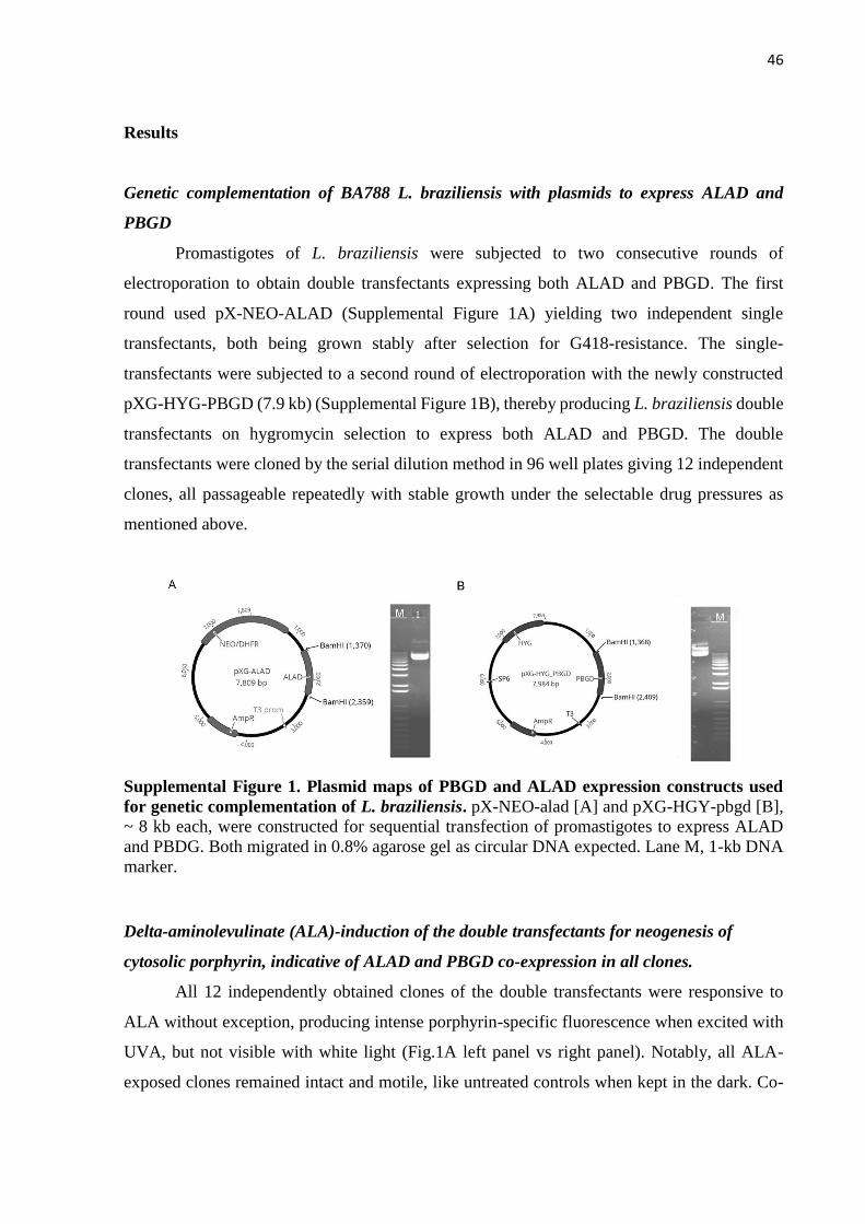
46
Results
Genetic complementation of BA788 L. braziliensis with plasmids to express ALAD and
PBGD
Promastigotes of L. braziliensis were subjected to two consecutive rounds of
electroporation to obtain double transfectants expressing both ALAD and PBGD. The first
round used pX-NEO-ALAD (Supplemental Figure 1A) yielding two independent single
transfectants, both being grown stably after selection for G418-resistance. The single-
transfectants were subjected to a second round of electroporation with the newly constructed
pXG-HYG-PBGD (7.9 kb) (Supplemental Figure 1B), thereby producing L. braziliensis double
transfectants on hygromycin selection to express both ALAD and PBGD. The double
transfectants were cloned by the serial dilution method in 96 well plates giving 12 independent
clones, all passageable repeatedly with stable growth under the selectable drug pressures as
mentioned above.
Supplemental Figure 1. Plasmid maps of PBGD and ALAD expression constructs used
for genetic complementation of L. braziliensis. pX-NEO-alad [A] and pXG-HGY-pbgd [B],
~ 8 kb each, were constructed for sequential transfection of promastigotes to express ALAD
and PBDG. Both migrated in 0.8% agarose gel as circular DNA expected. Lane M, 1-kb DNA
marker.
Delta-aminolevulinate (ALA)-induction of the double transfectants for neogenesis of
cytosolic porphyrin, indicative of ALAD and PBGD co-expression in all clones.
All 12 independently obtained clones of the double transfectants were responsive to
ALA without exception, producing intense porphyrin-specific fluorescence when excited with
UVA, but not visible with white light (Fig.1A left panel vs right panel). Notably, all ALA-
exposed clones remained intact and motile, like untreated controls when kept in the dark. Co-

47
migration of the fluorescence with sedimented cells after centrifugation is indicative of its cell-
association, just like the similarly treated double transfectants of L. amazonensis (Figure 1A, 1-
12 vs C2). Confocal microscopy of these live promastigotes showed porphyrin fluorescence
throughout the cells including flagella, indicative of its cytosolic localization, (Figure 1B),
consistent with previously reported findings in such genetically complemented Leishmania spp.
14,26. The fluorescence observed is due to the accumulation of intermediate porphyrins and
possibly uroporphyrin I (URO1) as the product, since wild type Leishmania have no URO1-
utilizing enzyme immediately downstream of PBGD. Indeed, at least ALAD and PBGD are
absent in the wildtype L. braziliensis, as indicated by the absence of any porphyrin fluorescence
after their exposure to ALA and UVA light (Fig. 1A, C1, right panel).
Figure 1. Aminolevulinate (ALA)-mediated uroporphyrinogenesis of genetically
complemented L. braziliensis clones. Twelve clones of L. braziliensis promastigotes
transfected to express PBGD and ALAD were exposed in the dark to ALA (1 mM) for 48 h.
[A] Cell samples were centrifuged, showing co-sedimentation of porphyrin fluorescence with
cell pellets by illumination with UVA (right panel), but not white light (left panel). Wild type
L. braziliensis (C1) and porphyrinogenic L. amazonensis (C2) were similarly processed as
negative control and positive control, respectively. [B] Microscopic images of porphyric L.
braziliensis, showing cytoplasmic porphyrin accumulation. DIC, Differential interference;
Porphyrin, Filter set for URO1 fluorescence.
Clonal variations in porphyrinogenesis was noted among the 12 clones, as shown by
flow cytometric analysis for URO1 fluorescence in a time course study from 24 to 72h after
ALA-exposure. Judging from the percentage of porphyric cell populations and their mean
fluorescence intensity (MFI), porphyrinogenesis of all clones was detectable at a low range at

48
24h (Figure 2A) and peaked 48 hours after ALA-exposure (Figure 2B). After further incubation
to 72h, the percentage of porphyric cells remained similar, while porphyrin MFI decreased
sharply in all 12 clones (Figure 2C, right panel). This is consistent with the release of URO1
from the porphyric cells, as noted before 14 and from the fluorescence in the supernatants
(Figure 1A, right panel). Clones 2 and 8-10 were substantially lower than the rest in the peak
levels of porphyrin. According to this kinetics analysis, clones # 4-6 and 11-12 are clearly more
porphyrinogenic than the others and clone #4 was chosen for further phenotypic analysis.
Figure 2. Porphyrinogenesis in transgenic L. braziliensis clones. Twelve independent L.
braziliensis clones expressing PBGD and ALAD were exposed to 1.6 mM ALA and the
resulting uroporphyrin (URO1) accumulation was evaluated by flow cytometry. Individual bars
show clonal variations in % porphyric cells (left panel) and fluorescent intensity of porphyrin
(right panel) during ALA-induced porphyrinogenesis for 24h [A], 48h [B] and 72h [C].
Porphyrinogenesis of all 12 clones renders them sensitive to inactivation with UVA
By MTT reduction assays, we observed a significant decrease in the viability of all
twelve L. braziliensis clones of porphyrinogenic cells after UVA exposure in contrast to their
counterpart kept in the dark (Figure 3, white bar vs. black bar). In contrast, the wild-type
parental L. braziliensis line remained viable, independent of UVA exposure (Figure 3, WT Lb
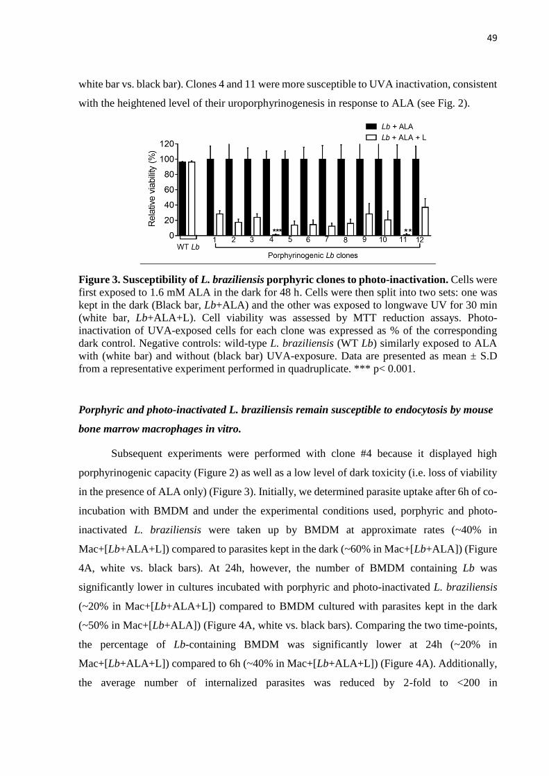
49
white bar vs. black bar). Clones 4 and 11 were more susceptible to UVA inactivation, consistent
with the heightened level of their uroporphyrinogenesis in response to ALA (see Fig. 2).
Figure 3. Susceptibility of L. braziliensis porphyric clones to photo-inactivation. Cells were
first exposed to 1.6 mM ALA in the dark for 48 h. Cells were then split into two sets: one was
kept in the dark (Black bar, Lb+ALA) and the other was exposed to longwave UV for 30 min
(white bar, Lb+ALA+L). Cell viability was assessed by MTT reduction assays. Photo-
inactivation of UVA-exposed cells for each clone was expressed as % of the corresponding
dark control. Negative controls: wild-type L. braziliensis (WT Lb) similarly exposed to ALA
with (white bar) and without (black bar) UVA-exposure. Data are presented as mean ± S.D
from a representative experiment performed in quadruplicate. *** p< 0.001.
Porphyric and photo-inactivated L. braziliensis remain susceptible to endocytosis by mouse
bone marrow macrophages in vitro.
Subsequent experiments were performed with clone #4 because it displayed high
porphyrinogenic capacity (Figure 2) as well as a low level of dark toxicity (i.e. loss of viability
in the presence of ALA only) (Figure 3). Initially, we determined parasite uptake after 6h of co-
incubation with BMDM and under the experimental conditions used, porphyric and photo-
inactivated L. braziliensis were taken up by BMDM at approximate rates (~40% in
Mac+[Lb+ALA+L]) compared to parasites kept in the dark (~60% in Mac+[Lb+ALA]) (Figure
4A, white vs. black bars). At 24h, however, the number of BMDM containing Lb was
significantly lower in cultures incubated with porphyric and photo-inactivated L. braziliensis
(~20% in Mac+[Lb+ALA+L]) compared to BMDM cultured with parasites kept in the dark
(~50% in Mac+[Lb+ALA]) (Figure 4A, white vs. black bars). Comparing the two time-points,
the percentage of Lb-containing BMDM was significantly lower at 24h (~20% in
Mac+[Lb+ALA+L]) compared to 6h (~40% in Mac+[Lb+ALA+L]) (Figure 4A). Additionally,
the average number of internalized parasites was reduced by 2-fold to <200 in

50
Mac+[Lb+ALA+L] from ~400 in Mac+[Lb+ALA], at 6h, and by 8-fold to ~50 in
Mac+[Lb+ALA+L], at 24h (Figure 4B, white vs. black bars). Again, a comparison of the two
time points showed that the number of intracellular parasites is significantly lower at 24h when
BMDM were cultured with porphyric and photo-inactivated L. braziliensis. Representative
photomicrographs illustrate a visible reduction in the number of intracellular parasites in
Mac+[Lb+ALA+L] compared to Mac+[Lb+ALA] at 24h (Figure 4C). These data confirm that
porphyric and photo-inactivated L. braziliensis remain susceptible to internalization by BMDM
but become more susceptible to killing.
Figure 4. Endocytosis of photo-inactivated porphyric L. braziliensis by primary
macrophages. Bone marrow-derived macrophages (BMDM) were co-cultured with wildtype
L. braziliensis (Lb) (gray bar), and its uroporphyric transfectants with (white bar) and without
(black bar) photo-inactivation by exposure to UVA light (L) for 30 min at a host-parasite ratio
of 1:10. Cells were processed at 6 and 24 h for microscopy to assess: [A] the percentage of
BMDM containing L. braziliensis; and [B] the number of Leishmania per 200 macrophages.
Data are presented as mean ±. S.D from a representative experiment performed in
quadruplicate, *p<0.05. (C) Representative photomicrographs of (A, B).
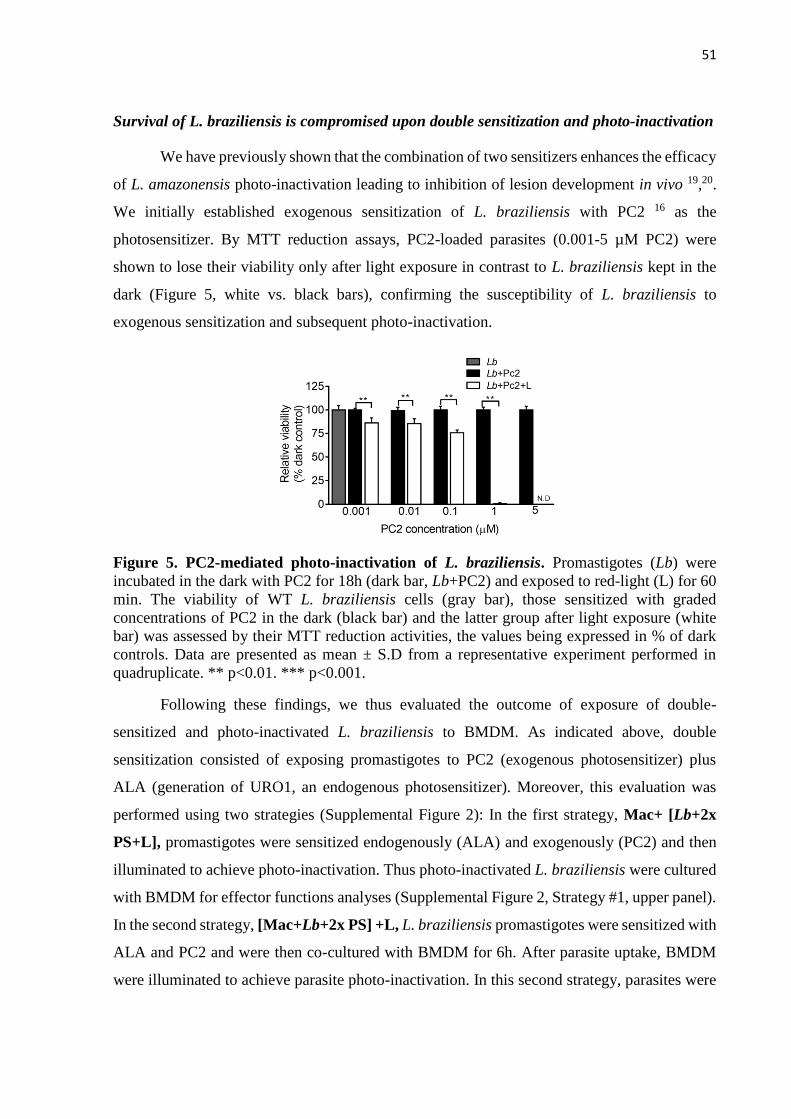
51
Survival of L. braziliensis is compromised upon double sensitization and photo-inactivation
We have previously shown that the combination of two sensitizers enhances the efficacy
of L. amazonensis photo-inactivation leading to inhibition of lesion development in vivo 19,20.
We initially established exogenous sensitization of L. braziliensis with PC2 16 as the
photosensitizer. By MTT reduction assays, PC2-loaded parasites (0.001-5 µM PC2) were
shown to lose their viability only after light exposure in contrast to L. braziliensis kept in the
dark (Figure 5, white vs. black bars), confirming the susceptibility of L. braziliensis to
exogenous sensitization and subsequent photo-inactivation.
Figure 5. PC2-mediated photo-inactivation of L. braziliensis. Promastigotes (Lb) were
incubated in the dark with PC2 for 18h (dark bar, Lb+PC2) and exposed to red-light (L) for 60
min. The viability of WT L. braziliensis cells (gray bar), those sensitized with graded
concentrations of PC2 in the dark (black bar) and the latter group after light exposure (white
bar) was assessed by their MTT reduction activities, the values being expressed in % of dark
controls. Data are presented as mean ± S.D from a representative experiment performed in
quadruplicate. ** p<0.01. *** p<0.001.
Following these findings, we thus evaluated the outcome of exposure of double-
sensitized and photo-inactivated L. braziliensis to BMDM. As indicated above, double
sensitization consisted of exposing promastigotes to PC2 (exogenous photosensitizer) plus
ALA (generation of URO1, an endogenous photosensitizer). Moreover, this evaluation was
performed using two strategies (Supplemental Figure 2): In the first strategy, Mac+ [Lb+2x
PS+L], promastigotes were sensitized endogenously (ALA) and exogenously (PC2) and then
illuminated to achieve photo-inactivation. Thus photo-inactivated L. braziliensis were cultured
with BMDM for effector functions analyses (Supplemental Figure 2, Strategy #1, upper panel).
In the second strategy, [Mac+Lb+2x PS] +L, L. braziliensis promastigotes were sensitized with
ALA and PC2 and were then co-cultured with BMDM for 6h. After parasite uptake, BMDM
were illuminated to achieve parasite photo-inactivation. In this second strategy, parasites were
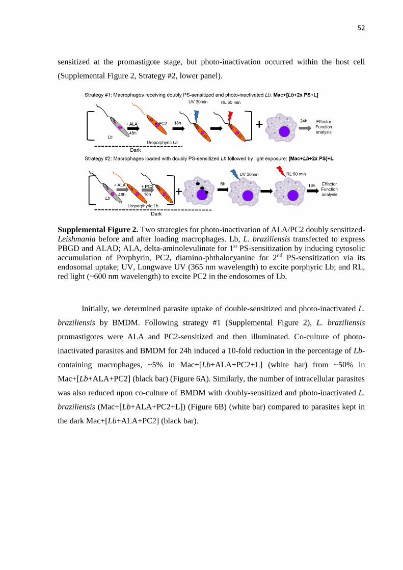
52
sensitized at the promastigote stage, but photo-inactivation occurred within the host cell
(Supplemental Figure 2, Strategy #2, lower panel).
Supplemental Figure 2. Two strategies for photo-inactivation of ALA/PC2 doubly sensitized-
Leishmania before and after loading macrophages. Lb, L. braziliensis transfected to express
PBGD and ALAD; ALA, delta-aminolevulinate for 1st PS-sensitization by inducing cytosolic
accumulation of Porphyrin, PC2, diamino-phthalocyanine for 2nd PS-sensitization via its
endosomal uptake; UV, Longwave UV (365 nm wavelength) to excite porphyric Lb; and RL,
red light (~600 nm wavelength) to excite PC2 in the endosomes of Lb.
Initially, we determined parasite uptake of double-sensitized and photo-inactivated L.
braziliensis by BMDM. Following strategy #1 (Supplemental Figure 2), L. braziliensis
promastigotes were ALA and PC2-sensitized and then illuminated. Co-culture of photo-
inactivated parasites and BMDM for 24h induced a 10-fold reduction in the percentage of Lb-
containing macrophages, ~5% in Mac+[Lb+ALA+PC2+L] (white bar) from ~50% in
Mac+[Lb+ALA+PC2] (black bar) (Figure 6A). Similarly, the number of intracellular parasites
was also reduced upon co-culture of BMDM with doubly-sensitized and photo-inactivated L.
braziliensis (Mac+[Lb+ALA+PC2+L]) (Figure 6B) (white bar) compared to parasites kept in
the dark Mac+[Lb+ALA+PC2] (black bar).

53
Figure 6. Uptake of doubly-sensitized and photo-inactivated L. braziliensis by primary
macrophages. Bone marrow-derived macrophages (BMDM) were exposed at a host-parasite
ratio of 1:10 to URO1/PC2 doubly photo-sensitized parasites with (white bar,
Mac+[Lb+ALA+Pc2+L]) and without (black bar, Mac+[Lb+ALA+PC2]) exposure to
UVA/red light. Alternatively, parasites were doubly sensitized and then cultured with
macrophages, which were exposed to UVA/red light (red bar, [Mac+Lb+ALA+Pc2] +L).
Macrophages infected with untreated Lb served as controls (checkered bar, Mac+Lb). Cells
were processed after 24h for microscopy to assess: [A] the percentage of Lb-containing
macrophages and [B] the number of parasites per 200 macrophages. Data are presented as mean
±. S.D from a representative experiment performed in quadruplicate, *p<0.05; **p<0.01
Equivalent results were obtained when photo-inactivation occurred after parasite
internalization by BMDM: both the percentage of Lb-containing macrophages (Figure 6A,
[Mac+Lb+ALA+PC2]+L, red bar) and the number of intracellular amastigotes (Figure 6B,
[Mac+Lb+ALA+PC2]+L, red bar) were significantly reduced compared to parasites kept in the
dark (Figure 6A and B, (Mac+[Lb+ALA+PC2], black bars). These results confirm that like
single sensitization with ALA only (Figure 4) double sensitization with ALA and PC2 does not
disturb parasite uptake by BMDM but impairs parasite survival within the host cell, regardless
of the stage at which photo-inactivation occurs (before or after parasite internalization).
Importantly, this outcome was unrelated to the ability of BMDM to uptake photo-
inactivated L. braziliensis since co-cultures performed for 6h showed that the percentage of Lb-
containing macrophages (Supplemental Figure 3A) and the number of internalized parasites
(Supplemental Figure 3B) was similar comparing doubly-sensitized and photo-inactivated L.
braziliensis (Mac+[Lb+ALA+PC2+L], white bars) vs. parasites kept in the dark
(Mac+[Lb+ALA+PC2], black bars).
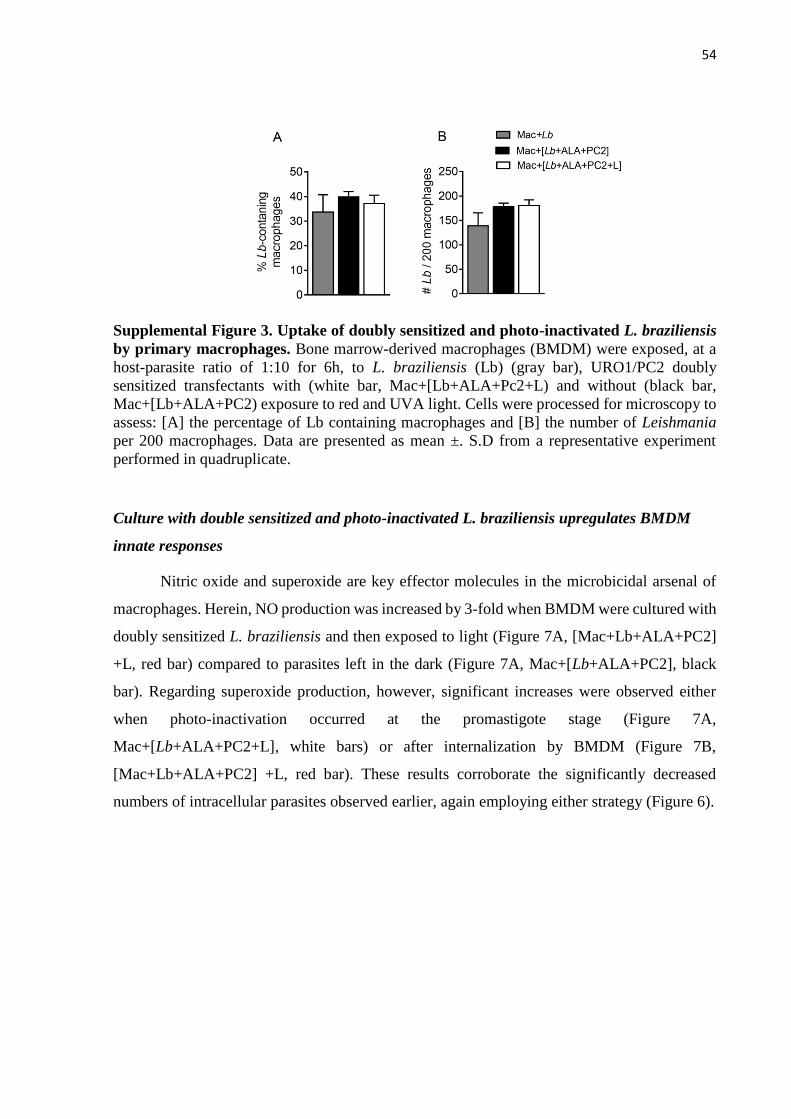
54
Supplemental Figure 3. Uptake of doubly sensitized and photo-inactivated L. braziliensis
by primary macrophages. Bone marrow-derived macrophages (BMDM) were exposed, at a
host-parasite ratio of 1:10 for 6h, to L. braziliensis (Lb) (gray bar), URO1/PC2 doubly
sensitized transfectants with (white bar, Mac+[Lb+ALA+Pc2+L) and without (black bar,
Mac+[Lb+ALA+PC2) exposure to red and UVA light. Cells were processed for microscopy to
assess: [A] the percentage of Lb containing macrophages and [B] the number of Leishmania
per 200 macrophages. Data are presented as mean ±. S.D from a representative experiment
performed in quadruplicate.
Culture with double sensitized and photo-inactivated L. braziliensis upregulates BMDM
innate responses
Nitric oxide and superoxide are key effector molecules in the microbicidal arsenal of
macrophages. Herein, NO production was increased by 3-fold when BMDM were cultured with
doubly sensitized L. braziliensis and then exposed to light (Figure 7A, [Mac+Lb+ALA+PC2]
+L, red bar) compared to parasites left in the dark (Figure 7A, Mac+[Lb+ALA+PC2], black
bar). Regarding superoxide production, however, significant increases were observed either
when photo-inactivation occurred at the promastigote stage (Figure 7A,
Mac+[Lb+ALA+PC2+L], white bars) or after internalization by BMDM (Figure 7B,
[Mac+Lb+ALA+PC2] +L, red bar). These results corroborate the significantly decreased
numbers of intracellular parasites observed earlier, again employing either strategy (Figure 6).

55
Figure 7. Respiratory burst of macrophages up-regulated after loading with doubly-
sensitized and photo-inactivated L. braziliensis. Bone marrow-derived macrophages
(BMDM) were infected at a host-parasite ratio of 1:10 with URO1/PC2 doubly sensitized
parasites with (white bar, Mac+[Lb+ALA+Pc2+L]) and without (black bar,
Mac+[Lb+ALA+PC2]) exposure to UVA/red light. Alternatively, parasites were doubly
sensitized and delivered to macrophages, which were then exposed to UVA/red light (red bar,
[Mac+Lb+ALA+Pc2] +L). Macrophages infected with untreated Lb (checkered bar, Mac+Lb)
or exposed to light only (striped bar, Mac+L) served as controls. [A] Nitric oxide and [B]
superoxide levels were quantified in culture supernatants 24 hours after priming of BMDM
with LPS+IFN-γ. Data are presented as mean ±. S.D from a representative experiment
performed in quadruplicate, *p<0.05
Next, we probed for the expression of CD40 and CD86, two co-stimulatory molecules
involved in priming cellular immune responses. As before, L. braziliensis were sensitized and
photo-inactivated prior to co-culture with BMDM or macrophages were co-cultured with
sensitized parasites and later illuminated and the percentage of CD40 and CD86 cells was
evaluated by flow cytometry. We observed a 2-fold increase in CD40 population upon co-
culture of BMDM with doubly sensitized and photoinactivated L. braziliensis (~50%,
Mac+[Lb+ALA+PC2+L], white bars) compared to parasites left in the dark (<20%,
Mac+[Lb+ALA+PC2], black bar) (Figure 8B). Photo-inactivation of L. braziliensis after
BMDM uptake induced a similar outcome ([Mac+Lb+ALA+PC2] +L, red bar, Figure 8B).
Again, using either photo-inactivation strategy, we also observed a significant increase in the
percentage of CD86 population for BMDM co-cultured with photoinactivated parasites (~30%,
Mac+[Lb+ALA+PC2+L], white bar and [Mac+Lb+ALA+PC2+L], red bar compared to
parasites left in the dark (~20%, [Mac+Lb+ALA+PC2]+L, black bar) (Figure 8C). These results
indicate that BMDM upregulate co-stimulatory properties upon contact with photo-inactivated
L. braziliensis, suggesting an increased capacity of antigen presentation.

56
Figure 8. Expression of co-stimulatory molecules up-regulated in primary macrophages
after loading with doubly-sensitized and photo-inactivated L. braziliensis. Bone marrow-
derived macrophages (BMDM) were exposed at a host-parasite ratio of 1:10 to URO1/PC2
doubly sensitized parasites with (white bar, Mac+[Lb+ALA+Pc2+L]) and without (black
bar, Mac+[Lb+ALA+PC2]) exposure to red and UVA light. Alternatively, parasites were
doubly photo-sensitized and used to infect macrophages followed by exposure to UVA/red light
(red bar, [Mac+Lb+ALA+Pc2] +L). Macrophages infected with untreated Lb (checkered
bar, Mac+Lb) or exposed to light only (striped bar, Mac+L) served as controls. Bar graphs
showing the percentage of cells expressing [A] CD40 and [B] CD86, as determined after
incubation for 24h by flow cytometry. Data are presented as mean ±. S.D from a representative
experiment performed in quadruplicate, *p<0.05.
Lastly, we investigated the cytokine production induced by the two strategies of L.
braziliensis photo-inactivation. Culture of BMDM with doubly sensitized and photo-
inactivated parasites did not alter TNF production (Figure 9A, Mac+[Lb+ALA+PC2+L], white
bars white bar) but increase IL-6 secretion by ~3-fold (Figure 9B, white bar) while decreasing
IL-10 production by 2-fold (Figure 9C, white bar). On the other hand, photo-inactivation of
doubly sensitized L. braziliensis after uptake by BMDM modulated all three cytokines
evaluated (Figure 9, [Mac+Lb+ALA+PC2] +L, red bars). These results show that, in this
strategy, BMDM can clear the infection more efficiently, while upregulating co-stimulatory
and cytokine production functions.

57
Figure 9. Production of immunity-favorable cytokines by primary macrophages after
loading with doubly-sensitized and photo-inactivated L. braziliensis. Bone marrow-derived
macrophages (BMDM) were exposed at a host-parasite ratio of 1:10 with to URO1/PC2 doubly
photo-sensitized parasites with (white bar, Mac+[Lb+ALA+Pc2+L]) and without (black bar,
Mac+[Lb+ALA+PC2]) exposure to red and UVA light. Alternatively, parasites were doubly
photo-sensitized and delivered to macrophages, which were then exposed to UVA/red light
(red bar, [Mac+Lb+ALA+Pc2] +L). Macrophages infected with untreated Lb (checkered
bar, Mac+Lb) served as controls. The presence of TNF [A], IL-6 [B] and IL-10 [C] into culture
supernatants after incubation for 24 h was determined by ELISA. Data are presented as mean
±. S.D from a representative experiment performed in quadruplicate, *p<0.05
Discussion
Vaccination providing lasting immunity is fundamental to the control of leishmaniasis.
An immunization strategy involving live parasites would enable the presentation of the entire
antigenic spectrum to the immune system; however, immunosuppression and parasite
persistence currently limit this approach. To overcome this limitation, physical, chemical and
genetic attenuation have been pursued to generate a safe yet efficient vaccine for leishmaniasis.
In the present study, we developed a genetically complemented L. braziliensis cell line that
could be endogenously sensitized and photoinactivated upon illumination. We show that these
transgenic parasites become inactivated upon light exposure but remain capable of infecting
macrophages. Within the host cell, parasites are readily susceptible to the oxidative burst while
the macrophage upregulates cytokine production and co-stimulatory markers.
Photosensitizers are light-excitable ring compounds that produce cytotoxic ROS in
response to illumination. Photodynamic vaccination refers to the use of PS to sensitize
Leishmania, thusly rendering them photoinactivated through light exposure and allowing such
parasites to then be employed as whole-cell vaccines. The photodynamic inactivation of
parasites can be accomplished in two ways, either through the uptake of PS, such as
phthalocyanines 27, or through the endogenous production of PS, such as URO1, via a
58
transgenic approach 15. To pursue the latter, we initially generated genetically complemented
L. braziliensis expressing ALAD and PBGD by two consecutive transfections. Upon ALA
exposure, we successfully induced uroporphyrinogenesis in all the resulting clones. The
percentage of uroporphyric cells seen herein was not as high as that described for L.
amazonensis 14. The ability to transition from a porphyric to an aporphyric state is not a stable
trait in L. amazonensis, suggesting that genetically complemented L. braziliensis may behave
similarly and have a reduced capacity to uptake ALA as opposed to an increased ability to efflux
URO1, as was reported earlier 26. We thus selected one genetically complemented L.
braziliensis clone for further characterization.
We functionally demonstrated that the sensitization of L. braziliensis with ALA induced
URO1 accumulation that, upon excitation by white or UVA light, resulted in parasite
inactivation. Photoinactivation rates were consistent with experimentation performed in L.
amazonensis 15,26. The ability of photoinactivated L. braziliensis promastigotes to infect bone
marrow-derived macrophages and subsequently differentiate into amastigotes remained
uncompromised. The infectivity of attenuated uroporphyrinogenic L. braziliensis exposed to
light was comparable to that of wild-type parasites. However, after internalization and
differentiation into amastigotes, the attenuated parasites were unable to persist as long as wild-
type counterparts. Similar results have been reported upon the attenuation of L. chagasi with
amotosalen followed by low doses of UVA radiation 28 and upon the deletion of centrin and
p27 in L. donovani 29.
Immunization of Syrian hamsters with L. amazonensis submitted to single photo-
inactivation, that is using one photosensitizer only, conferred protection against heterologous
infection 18, indicating that photodynamic vaccination against leishmaniasis can be readily
achievable. Nonetheless, in the mouse model, we have shown that the use of a single
photosensitizer such as endogenous ALA may not induce complete photo-inactivation of the
light-exposed parasite population and dermal lesions develop, raising safety concerns 20. To
address both safety and efficacy issues, we evaluated the outcome of double sensitization, using
endogenous ALA-URO1 and exogenous PC2 as photosensizers. It has been shown that PC2 is
10-40 times more effective at mediating photo-inactivation of L. tropica 16 and, in our hands, it
was also effective for photo-inactivation of L. amazonensis promastigotes and L. braziliensis.
Therefore, PC2 was employed as the external photosensitizer besides ALA-URO that acted as
the endogenous photosensitizer. Additionally, we also investigated the outcome of macrophage
exposure to light, after internalization of doubly photosensitized L. braziliensis. The rationale

59
for such strategy is that photo-inactivation inactivates intracellular Leishmania leading to the
release of antigens within the host macrophage, without compromising the antigen presenting
cell (APC) itself 19. That is because the host cell is not sensitized at the time of light exposure
and oxygen radicals produced during that step are limited to the parasite and do not cause
oxidative damage to the host cell. Indeed, double sensitization allowed efficient parasite uptake
by BMDM but compromised its replication and this outcome was regardless photoinactivation
occurring before or after parasite internalization.
We speculate that a vaccination strategy involving internalization of photo-inactivated
L. braziliensis by BMDM would result in the release of antigens into phagolysosomes and the
cytosol of viable macrophages, thusly enabling downstream events related to the development
of cellular immunity. Accordingly, using Strategy #2, we found a significant increase in the
production of reactive oxygen and nitrogen species, which may account for parasite killing, but,
at the same time, the host cell upregulated cytokine production and co-stimulatory effectors
such as CD40 and CD86. CD86 is a co-stimulatory molecule expressed on activated APCs that
binds CD28 expressed on the T cell. CD40 is also expressed on APCs and binds CD40L
expressed in activated T cells, delivering signals important to enhance expression of CD80 and
CD86, for example, on the APCs. We also observed TNF production which, together with IFN-
γ, activates the macrophage to kill intracellular parasites 30. TNF induces both NO and SO
production by the macrophage and these mediators can be accounted for the L. braziliensis
killing we observed following photo-inactivation, using both strategies. Moreover, IL-10 levels
were decreased in cultures of BMDM exposed to photo-inactivated L. braziliensis. In L. major
experimental infection, IL-10 produced by infected macrophages prevented parasite killing and
pre-treatment of cultures with recombinant IL-10 enhances intracellular parasite survival 31.
Collectively, our results suggest that BMDM exposure to photo-inactivated L. braziliensis
enhanced parasite killing by the infected cell, advocating for efficacy of the double sensitization
and photo-inactivation strategies evaluated here. More importantly, we also show that these
strategies enhance the antigen presenting capacity of BMDM exposed to photo-inactivated L.
braziliensis which, we speculate, shall induce the development of protective cellular responses,
in vivo.
Immunization with attenuated parasites obtained by the targeted deletion of genes
coding for lipophosphoglycan 32, biopetrin transporter 33, HSP70 34, ALO 35 and centrin 36 was
shown to induce long-lasting protective immunity in experimental models. Nonetheless, while
attenuated vaccines achieved by genetic deletion offer an interesting approach for

60
immunization, parasites may revert to their virulent form, and parasite persistence may induce
disease reactivation in immunosuppressed individuals. Moreover, whole genome sequencing of
L. donovani centrin-deficient parasites showed that in addition to knocking out the centrin gene,
additional deletions were also observed 37. While such deletions did not impact the attenuation
of the centrin-deficient parasites, this emphasizes the need for comprehensive characterization
of attenuated parasite lines. In conclusion, our generation of transgenic mutants of L.
braziliensis demonstrated a potential for applicability in photodynamic vaccination against
cutaneous leishmaniasis caused by L. braziliensis.
Acknowledgments
Thanks are due to the support of NIH-NIAID grant # AI-68835, AI-7712375 and AI-097830
to KPC. We also acknowledge the support of Conselho Nacional deDesenvolvimento
Científico e Tecnológico (CNPq) – (Programa Visitante Especial) Processo 402312/2012-0 to
CIO. RS was supported by CAPES (Coordenação de Aperfeiçoamento de Pessoal de Nível
Superior) and SMV was supported by CNPq. CIO is a senior investigator for CNPq.
References
1 Alvar, J. et al. Leishmaniasis worldwide and global estimates of its incidence. PLoS
One 7, e35671, doi:10.1371/journal.pone.0035671 (2012).
2 Mathers, C. D., Ezzati, M. & Lopez, A. D. Measuring the burden of neglected tropical
diseases: the global burden of disease framework. PLoS Negl Trop Dis 1, e114,
doi:10.1371/journal.pntd.0000114 (2007).
3 Bittencourt, A. & Barral-Netto, M. in Tropical Pathology Vol. 8 (ed Seifert G Doerr
W) 597-651 (1995).
4 Oliveira, L. F. et al. Systematic review of the adverse effects of cutaneous
leishmaniasis treatment in the New World. Acta Tropica 118, 87-96 (2011).
5 Sundar, S. et al. Failure of pentavalent antimony in visceral leishmaniasis in India:
report from the center of the Indian epidemic. Clin Infect Dis 31, 1104-1107,
doi:10.1086/318121 (2000).
6 Srivastava, S., Shankar, P., Mishra, J. & Singh, S. Possibilities and challenges for
developing a successful vaccine for leishmaniasis. Parasites & vectors 9, 277,
doi:10.1186/s13071-016-1553-y (2016).
7 Daneshvar, H., Coombs, G. H., Hagan, P. & Phillips, R. S. Leishmania mexicana and
Leishmania major: attenuation of wild-type parasites and vaccination with the
attenuated lines. J Infect Dis 187, 1662-1668, doi:10.1086/374783 (2003).
8 Alexander, J., Coombs, G. H. & Mottram, J. C. Leishmania mexicana cysteine
proteinase-deficient mutants have attenuated virulence for mice and potentiate a Th1
response. J Immunol 161, 6794-6801 (1998).
9 Uzonna, J. E., Spath, G. F., Beverley, S. M. & Scott, P. Vaccination with
phosphoglycan-deficient Leishmania major protects highly susceptible mice from

61
virulent challenge without inducing a strong Th1 response. J Immunol 172, 3793-3797
(2004).
10 Selvapandiyan, A. et al. Intracellular replication-deficient Leishmania donovani
induces long lasting protective immunity against visceral leishmaniasis. Journal of
immunology (Baltimore, Md. : 1950) 183, 1813-1820, doi:10.4049/jimmunol.0900276
(2009).
11 Alexander, J. A radioattenuated Leishmania major vaccine markedly increases the
resistance of CBA mice to subsequent infection with Leishmania mexicana mexicana.
Trans R Soc Trop Med Hyg 76, 646-649 (1982).
12 Sah, J. F. et al. Genetic rescue of Leishmania deficiency in porphyrin biosynthesis
creates mutants suitable for analysis of cellular events in uroporphyria and for
photodynamic therapy. J Biol Chem 277, 14902-14909, doi:10.1074/jbc.M200107200
(2002).
13 Dutta, S., Ray, D., Kolli, B. K. & Chang, K. P. Photodynamic sensitization of
Leishmania amazonensis in both extracellular and intracellular stages with aluminum
phthalocyanine chloride for photolysis in vitro. Antimicrobial Agents and
Chemotherapy 49, 4474-4484, doi:10.1128/AAC.49.11.4474-4484.2005 (2005).
14 Sah, J. F. et al. Genetic rescue of Leishmania deficiency in porphyrin biosynthesis
creates mutants suitable for analysis of cellular events in uroporphyria and for
photodynamic therapy. Journal of Biological Chemistry 277, 14902-14909,
doi:10.1074/jbc.M200107200 (2002).
15 Dutta, S., Kolli, B. K., Tang, A., Sassa, S. & Chang, K.-P. Transgenic Leishmania
model for delta-aminolevulinate-inducible monospecific uroporphyria: cytolytic
phototoxicity initiated by singlet oxygen-mediated inactivation of proteins and its
ablation by endosomal mobilization of cytosolic uroporphyrin. Eukaryotic cell 7,
1146-1157, doi:10.1128/EC.00365-07 (2008).
16 Al-qahtani, A. et al. Aminophthalocyanine-Mediated Photodynamic Inactivation of.
Antimicrobial Agents and Chemotherapy 60, 2003-2011, doi:10.1128/AAC.01879-
15.Address (2016).
17 Dutta, S. et al. Intracellular targeting specificity of novel phthalocyanines assessed in a
host-parasite model for developing potential photodynamic medicine. PLoS ONE 6,
doi:10.1371/journal.pone.0020786 (2011).
18 Kumari, S. et al. Photodynamic vaccination of hamsters with inducible suicidal
mutants of Leishmania amazonensis elicits immunity against visceral leishmaniasis.
European journal of immunology 39, 178-191, doi:10.1002/eji.200838389 (2009).
19 Dutta, S., Waki, K. & Chang, K. P. Combinational sensitization of Leishmania with
uroporphyrin and aluminum phthalocyanine synergistically enhances their
photodynamic inactivation in vitro and in vivo. Photochemistry and Photobiology 88,
620-625, doi:10.1111/j.1751-1097.2012.01076.x (2012).
20 Viana, S. M. et al. Photodynamic Vaccination of BALB/c Mice for Prophylaxis of
Cutaneous Leishmaniasis Caused by Leishmania amazonensis. Front Microbiol 9,
165, doi:10.3389/fmicb.2018.00165 (2018).
21 de Moura, T. R. et al. Toward a novel experimental model of infection to study
American cutaneous leishmaniasis caused by Leishmania braziliensis. Infect Immun
73, 5827-5834, doi:73/9/5827 [pii]10.1128/IAI.73.9.5827-5834.2005 (2005).
22 Weischenfeldt, J. & Porse, B. Bone Marrow-Derived Macrophages (BMM): Isolation
and Applications. CSH Protoc 2008, pdb prot5080, doi:10.1101/pdb.prot5080 (2008).

62
23 Schumann Burkard, G., Jutzi, P. & Roditi, I. Genome-wide RNAi screens in
bloodstream form trypanosomes identify drug transporters. Mol Biochem Parasitol
175, 91-94, doi:10.1016/j.molbiopara.2010.09.002 (2011).
24 Kapler, G. M., Coburn, C. M. & Beverley, S. M. Stable transfection of the human
parasite Leishmania major delineates a 30-kilobase region sufficient for
extrachromosomal replication and expression. Mol Cell Biol 10, 1084-1094 (1990).
25 Khouri, R. et al. IFN-beta impairs superoxide-dependent parasite killing in human
macrophages: evidence for a deleterious role of SOD1 in cutaneous leishmaniasis. J
Immunol 182, 2525-2531, doi:10.4049/jimmunol.0802860182/4/2525 [pii] (2009).
26 Dutta, S., Furuyama, K., Sassa, S. & Chang, K.-P. Leishmania spp: Delta-
aminolevulinate-inducible neogenesis of porphyria by genetic complementation of
incomplete heme biosynthesis pathway. Experimental parasitology 118, 629-636,
doi:10.1016/j.micinf.2008.07.015.Toxoplasma (2008).
27 Jiang, X. J., Yeung, S. L., Lo, P. C., Fong, W. P. & Ng, D. K. Phthalocyanine-
polyamine conjugates as highly efficient photosensitizers for photodynamic therapy. J
Med Chem 54, 320-330, doi:10.1021/jm101253v (2011).
28 Bruhn, K. W. et al. Killed but metabolically active Leishmania infantum as a novel
whole-cell vaccine for visceral leishmaniasis. Clin Vaccine Immunol 19, 490-498,
doi:10.1128/CVI.05660-11 (2012).
29 Bhattacharya, P. et al. Genetically modified live attenuated <i>L.donovani</i>
parasites induce innate immunity through classical activation of macrophages that
direct Th1 response in mice. Infection and Immunity, IAI.00184-00115,
doi:10.1128/IAI.00184-15 (2015).
30 Liew, F. Y., Li, Y. & Millott, S. Tumor necrosis factor-alpha synergizes with IFN-
gamma in mediating killing of Leishmania major through the induction of nitric oxide.
J Immunol 145, 4306-4310 (1990).
31 Kane, M. M. & Mosser, D. M. The role of IL-10 in promoting disease progression in
leishmaniasis. J Immunol 166, 1141-1147 (2001).
32 Spath, G. F. et al. Lipophosphoglycan is a virulence factor distinct from related
glycoconjugates in the protozoan parasite Leishmania major. Proc Natl Acad Sci U S
A 97, 9258-9263, doi:10.1073/pnas.160257897 (2000).
33 Papadopoulou, B. et al. Reduced infectivity of a Leishmania donovani biopterin
transporter genetic mutant and its use as an attenuated strain for vaccination. Infect
Immun 70, 62-68 (2002).
34 Carrion, J., Folgueira, C., Soto, M., Fresno, M. & Requena, J. M. Leishmania
infantum HSP70-II null mutant as candidate vaccine against leishmaniasis: a
preliminary evaluation. Parasit Vectors 4, 150, doi:10.1186/1756-3305-4-150 (2011).
35 Anand, S. & Madhubala, R. Genetically Engineered Ascorbic acid-deficient Live
Mutants of Leishmania donovani induce long lasting Protective Immunity against
Visceral Leishmaniasis. Sci Rep 5, 10706, doi:10.1038/srep10706 (2015).
36 Selvapandiyan, A. et al. Intracellular replication-deficient Leishmania donovani
induces long lasting protective immunity against visceral leishmaniasis. J Immunol
183, 1813-1820, doi:10.4049/jimmunol.0900276 (2009).
37 Gannavaram, S. et al. Whole genome sequencing of live attenuated Leishmania
donovani parasites reveals novel biomarkers of attenuation and enables product
characterization. Sci Rep 7, 4718, doi:10.1038/s41598-017-05088-4 (2017).
63
6. DISCUSSÃO
Apesar de ainda não existir uma vacina eficaz contra a leishmaniose, indivíduos curados
não voltam a desenvolver a doença, evidenciando a indução de imunidade duradoura após a
cura clínica (revisado em GILLESPIE et al., 2016). Assim, diversos esforços vêm sendo
empreendidos pela comunidade científica para o desenvolvimento de vacinas para a
leishmaniose, tais como a utilização de parasitas mortos, proteínas recombinantes ou
plasmídeos de DNA que codificam proteínas parasitárias bem como parasitas atenuados, os
quais mimetizam a infecção natural, sem indução de doença (revisado em REZVAN; MOAFI,
2015).
A inativação da leishmania pode ser obtida por meio da exposição do parasita a
fotossensibilizadores (FSs), os quais se acumulam no seu interior. A consequente exposição
dos parasitas fotossensibilizados a uma fonte de luz induz a geração de ROS tais como
superóxido e oxigênio singleto (1O2), induzindo assim dano por oxidação. O alcance do 1O2 é
muito curto (20 Å) (JUZENAS; MOAN, 2006), provavelmente por ser altamente reativo e
destrutivo, e especula-se que uma vez gerado o 1O2 age no interior da célula, preservando a
estrutura e as moléculas de superfície por horas após a fotoinativação do parasita (revisado em
CHANG; KOLLI, 2016). Não se conhecem enzimas capazes de detoxificar 1O2, tornando o
dano oxidativo mediado por esta molécula letal (revisado em VATANSEVER et al., 2013). No
presente estudo, induzimos a atenuação de leishmania a partir de sua exposição a FSs seguido
da fotoinativação por exposição à luz. Neste sentido, hipotetizamos que a imunização de
camundongos com parasitas fotoinativados induz imunidade contra um desafio subsequente
com parasitas virulentos, conferindo proteção contra a LT.
As ftalocianinas (PCs) são FSs de segunda geração, conhecidas por induzir a produção
de 1O2 após a exposição à luz vermelha (COOK et al., 1995). A PC2 é uma aminoftalocianina
descrita como 10 a 40 vezes mais eficaz para a fotoinativação de L. tropica (AL-QAHTANI et
al., 2016) quando comparada a ftalocianinas ligadas a grupos anilina ou piridil usadas
inicialmente na fotoinativação de L. amazonensis (DUTTA et al., 2011). Os resultados obtidos
no presente trabalho mostram que a PC2 induz um fenótipo tóxico dose-dependente em L.
amazonensis, evidenciado pela perda de mobilidade e de viabilidade celular, conforme avaliado
pelo ensaio de MTT. Também mostramos que a fotoinativação de L. amazonensis mediada por
PC2 reduziu a sua captação por macrófagos em cerca de 5 vezes quando comparado a parasitas
controle apenas sensibilizados com PC2, mesmo em tempos iniciais de infecção (4h). Isso

64
sugere que a fotoinativação utilizando a dose de PC e/ou de iluminação empregadas em nossos
experimentos dificulta a entrada dos parasitas na célula hospedeira. O achado de que uma fração
de macrófagos permanece infectada com sucesso, apesar da fotoinativação dos parasitas, pode
ser atribuída ao modo estocástico de sensibilização por PC2. Ou seja, uma pequena
porcentagem de parasitas não absorve o sensibilizador e, portanto, não é fotoinativada,
permanecendo viável e infectiva (DUTTA et al., 2005, 2011).
Uma vez determinada a dose com a qual L. amazonensis pode ser sensibilizada e
fotoinativada de maneira exógena, avaliamos a fotoinativação de L. amazonensis por via
endógena. Nesta estratégia, utilizamos parasitas geneticamente complementados com ALAD e
PBGD (SAH et al., 2002), as quais estão ausentes em parasitas selvagens. A presença destas
enzimas leva à produção de URO, a qual se acumula no citosol e funciona como um FS,
semelhante a PC2 (DUTTA et al., 2008a; SAH et al., 2002). A exposição de L. amazonensis
uroporfírica à luz UV de ondas longas gera oxigênio singleto (DUTTA et al., 2008a; SAH et
al., 2002), causando dano irreparável a estes parasitas. Em nossos experimentos, a
fotoinativação endógena de L. amazonensis induzida por URO reduziu a quantidade de células
infectadas em cerca de 2,5 vezes, mas assim como a fotoinativação exógena com PC2,
aproximadamente 10% dos macrófagos permaneceram infectados.
Em seguida, avaliamos o desfecho da inoculação de parasitas fotoinativados em
camundongos BALB/c. Observamos que os animais inoculados com L. amazonensis
fotoinativada de maneira exógena utilizando PC2 apresentaram lesões mais tardiamente
comparado àqueles inoculados com L. amazonensis apenas fotossensibilizada. Esse resultado
pode ser explicado por nossos ensaios in vitro, os quais mostraram redução na quantidade de
macrófagos infectados com os parasitas fotoinativados.
Dutta e colaboradores (2011) mostraram que células dendríticas infectadas com L.
amazonensis transgênica para a expressão de OVA e fotoinativada com uma ftalocianina foram
capazes de ativar linfócitos T CD8+ epítopo-específicos in vitro. Esse trabalho evidenciou o
potencial da utilização de parasitas atenuados por fotoinativação para a imunoprofilaxia. Um
outro estudo mostrou que a imunização de hamsters com L. amazonensis fotoinativada conferiu
proteção contra a infecção heteróloga com L. donovani (KUMARI et al., 2009). Neste estudo,
a imunização induziu uma resposta imune capaz de reduzir a carga parasitária em 99% e, em
paralelo, aumentou a expressão de iNOS, IFN-γ e IL-12 nos animais desafiados. Além disso, a
transferência de células T obtidas de hamsters imunizados conferiu proteção a animais naïve,
65
os quais foram infectados posteriormente, mostrando que a resposta celular induzida nos
animais imunizados pode ser transferida de maneira a permanecer protetora.
Dada a natureza estocástica da fotossensibilização simples, parasitas podem permanecer
viáveis mesmo após a exposição a luz, e, portanto, o risco de desenvolvimento de doença
permanece. Para minimizar este risco, testamos a combinação de FSs com o objetivo de
aumentar a eficácia da fotoinativação, pois dois FSs podem atuar em sinergia como mostrado
na terapia fotodinâmica aplicada a linhagens celulares tumorais e em modelo de melanoma em
camundongos (ACEDO et al., 2014; SCHNEIDER-YIN et al., 2009; VILLANUEVA et al.,
2010). Mesmo quando a aplicação de FSs combinados não aumenta a mortalidade celular, ela
pode reduzir a toxicidade inespecífica induzida por cada molécula, e também amplificar a
resposta imunológica contra o tumor (GYENGE et al., 2012). Com relação a Leishmania, Dutta
e colaboradores (2012) observaram que L. amazonensis fotossensibilizada com AlPhCl e com
URO1 in vitro tornou-se totalmente sensível à fotoinativação in vivo pois camundongos
BALB/c inoculados na derme da orelha não apresentaram lesões, mesmo 8 semanas após a
infecção (DUTTA; WAKI; CHANG, 2012). O acúmulo de AlPhCl e URO1 pela Leishmania
ocorre em diferentes sítios subcelulares; o URO1 se forma no citosol e é depois restrito a
vacúolos no citoplasma e vesículas endo- ou exocíticas (DUTTA et al., 2008a), enquanto que
AlPhCl é internalizada e aparentemente se liga a membranas intracelulares, graças à sua
natureza hidrofóbica (DUTTA; WAKI; CHANG, 2012). Uma vez que esses FSs são excitados
em diferentes comprimentos de onda, a exposição à luz branca (consistindo em todo o espectro
visível) pode resultar na produção simultânea de 1O2 em diferentes compartimentos do parasita,
resultando em maior eficácia da fotoinativação.
No presente trabalho replicamos esta estratégia, inoculando L. amazonensis
fotossensibilizada com ALA e AlPhCl na pata, base da cauda, dorso e derme da orelha.
Observamos que promastigotas duplamente fotossensibilizadas, mas não fotoinativadas in vitro
foram detectáveis nos diversos locais inoculados de forma comparável, com exceção daquelas
inoculadas na região dorsal do animal. A derme da orelha foi o único local onde os parasitas
foram fotoinativados de forma satisfatória após exposição à luz, pois não foi possível detectar
parasitas após três semanas. Embora não possamos descartar a possibilidade de migração dos
parasitas inoculados para os gânglios linfáticos regionais, propomos que a translucidez da
orelha seja particularmente adequada para a iluminação, aumentando assim o sucesso da
fotoinativação, diferentemente do ocorrido na base da cauda ou da pata. Podemos também
especular que a fotoinativação dos parasitas inoculados na orelha permitiu a indução de uma

66
resposta imune efetora com pouca ou nenhuma imunossupressão pois observamos atraso no
desenvolvimento da lesão e redução significativa na carga parasitária. O nível de proteção
observado foi significativo, considerando que a maior parte das linhagens de camundongos,
incluindo C3H, C57BL/6, BDA e CBA são suscetíveis à infecção por L. amazonensis,
desenvolvendo lesões progressivas (CÔRTES et al., 2010; SOONG, 2012) e uma resposta mista
Th1-Th2 após a infecção (AFONSO; SCOTT, 1993; MCMAHON-PRATT; ALEXANDER,
2004). Os camundongos BALB/c são particularmente suscetíveis a essa infecção e apresentam
lesões necróticas com crescimento progressivo (PEREIRA; ALVES, 2008).
Estes achados constituem forte evidência de que a fotoinativação é uma estratégia viável
para a geração de parasitas imunogênicos e seguros. Acreditamos que os níveis de proteção
possam ser melhorados a partir da otimização dos procedimentos experimentais tais como a
redução da dose de parasitas no desafio, o aumento da frequência de imunizações e ajuste do
intervalo entre o inóculo de parasitas fotossensibilizados e a iluminação in vivo. A otimização
do intervalo é necessária sobretudo para eliminar parasitas persistentes, os quais podem induzir
uma resposta supressora, contrabalanceando a resposta protetora e dificultando a eliminação
completa de parasitas após a infecção (ROSSI; FASEL, 2017). Em suma, os resultados
apresentados na primeira parte dessa tese demonstram que a fotoinativação de L. amazonensis
usando FSs endógenos ou exógenos reduz sua viabilidade e capacidade de infecção em
macrófagos. Além disso, a fotoinativação de L. amazonensis por meio da combinação de FSs
reduz a viabilidade do parasita, mas o mesmo permanece capaz de induzir uma resposta celular
parcialmente protetora, em modelo experimental.
Na segunda parte do trabalho, partimos para avaliar se L. braziliensis poderia ser
fotossensibilizada e inativada da mesma maneira, sobretudo porque L. braziliensis causa as
formas mais complicadas da LT é responsável pela maioria dos casos no Brasil.
Adicionalmente, o modelo de infecção intradérmica de camundongos BALB/c com L.
braziliensis é autolimitado, gerando lesões ulceradas que apresentam cura espontânea e resposta
imunológica mista Th1/Th2, semelhante ao que ocorre em pacientes com LCL (DE MOURA
et al., 2005).
Inicialmente, geramos uma linhagem de L. braziliensis complementada com os genes
ALAD e PBGD, de forma a torná-la fotossensível pela geração e acúmulo de porfirinas
intermediárias da via do heme e, posteriormente, suscetível a fotoinativação. Após duas
transfecções consecutivas, os genes ALAD e PBGD foram inseridos em L. braziliensis e os 12
clones resultantes cresceram de forma estável sob pressão seletiva, por várias passagens. A
67
expressão dos genes inseridos foi verificada funcionalmente através de intensa fluorescência
vermelha característica da presença de porfirinas tanto nas células quanto nos sobrenadantes
após a incubação com ALA e exposição à luz UV de ondas longas. Parasitas selvagem ou
transfectados apenas com ALAD não apresentaram fluorescência quando incubados com ALA.
Além disso, o fato de o acúmulo de porfirinas apenas ocorrer em L. braziliensis geneticamente
complementada quando as promastigotas foram incubadas com ALA confirma que essa espécie
também é naturalmente deficiente na enzima ALA Sintase. Em nosso modelo com a L.
braziliensis geneticamente complementada não pudemos determinar qual porfirina da via é a
responsável pela coloração vermelha e pela fototoxicidade observada. No entanto, Dutta e
colaboradores (2008a) verificaram que L. amazonensis, L. infantum, L. major e L. tropica
complementadas com ALAD e PBGD produzem apenas URO1 após a incubação com ALA,
logo são deficientes em pelo menos 5 enzimas da via de biossíntese do heme.
Em nosso modelo, todos os clones testados apresentaram fluorescência em proporções
variadas após exposição ao ALA, mas com valores menores ao descrito anteriormente para L.
amazonensis. É sabido que promastigotas de L. amazonensis contam com a geração de
"porfirinossomas" que realizam a detoxificação celular de URO1 (DUTTA et al., 2008b).
Acreditamos que um mecanismo similar pode ser responsável pela fluorescência de porfirinas
observada no sobrenadante de culturas de L. braziliensis após a incubação com ALA. A
capacidade reduzida de captar ALA, bem como a rápida mobilização de porfirinas podem ser
responsáveis pela população fluorescente menor, observada em L. braziliensis quando
comparado a L. amazonensis. Semelhante aos modelos de Porfíria desenvolvidos anteriormente
(DUTTA et al., 2008a; SAH et al., 2002), os clones de L. braziliensis apresentaram maior
população fluorescente e intensidade de fluorescência após 48h de incubação com ALA. É
interessante observar a clara heterogeneidade tanto na geração de fluorescência quando na taxa
de inativação entre os 12 clones gerados. Após a exposição à luz UVA, a taxa de fotoinativação
variou entre os clones de acordo com a proporção celular que havia se mostrado fluorescente
para porfirinas, confirmando a atividade fotossensibilizadora das moléculas geradas e que a
fluorescência pode ser interpretada como um indicador da susceptibilidade à fotoinativação.
Assim, demonstramos funcionalmente a atividade de ALAD e PBGD após a transfecção em L.
braziliensis, tornando-a capaz de gerar porfirinas intracelulares utilizando ALA exógeno, e a
subsequente fotoinativação após a exposição a luz branca ou UVA.
A complementação genética de L. braziliensis não alterou sua capacidade de infectar
macrófagos murinos e de transformar-se na forma amastigota nos períodos estudados. Na

68
verdade, mesmo após sensibilização com ALA ou após dupla sensibilização com ALA e Pc2 e
exposição à luz, os parasitas foram internalizados em taxa semelhante àqueles não
fotoinativados. Entretanto, após 24h houve redução significativa da taxa de infecção e do
número de amastigotas nas células. Resultados similares foram observados em macrófagos
humanos ou murinos infectados com L. donovani deficiente em p27 e centrina
(BHATTACHARYA et al., 2015; DEY et al., 2010; SELVAPANDIYAN et al., 2009). Apesar
da taxa de infecção diminuída, a infecção com essas linhagens atenuadas induziu a regulação
positiva de citocinas de perfil Th1 e de moléculas co-estimulatórias de superfície em
macrófagos murinos (BHATTACHARYA et al., 2015). Em nosso modelo, cerca de 10% das
amastigotas porfirinogênicas não foram eliminadas dos macrófagos após 24h, corroborando os
achados anteriores os quais mostraram que alguns parasitas são capazes de sobreviver à fotólise
devido à absorção reduzida de ALA e/ou ao efluxo excessivo de porfirinas formadas.
Entretanto, no modelo de L. amazonensis esses sobreviventes se mostraram tão sensíveis a uma
nova indução de uroporfirina quanto clones de parasitas parentais nunca expostos a ALA
(CHANG; KOLLI, 2016), o que permitiria sua posterior fotoinativação.
Em seguida, avaliamos a fotoinativação exógena de promastigotas de L. braziliensis
com PC2, que a 1uM induziu aproximadamente 100% de fotoinativação após exposição à luz
vermelha, se caracterizando como promissor para a dupla fotoinativação empregando também
ALA-URO1. A taxa de infecção de macrófagos com L. braziliensis duplamente fotoinativada
caiu até 4 vezes após 24h, quando comparado às células infectadas com parasitas fotoinativados
apenas por meio de ALA-URO1. A partir daí avaliamos a infecção de macrófagos com parasitas
previamente fotoinativados e a iluminação de macrófagos infectados com L. braziliensis
fotossensibilizada. Desta maneira, pudemos comparar os efeitos da infecção com parasitas
fotoinativados versus a infecção com parasitas sensibilizados e submetidos à fotoinativação
posteriormente (após a internalização pelo macrófago). Como mostrado na primeira etapa desta
tese, a fotoinativação in vivo, ou seja, após a inoculação dos parasitas fotossensibilizados em
animais conferiu proteção contra L. amazonensis e contra L. donovani (KUMARI et al., 2009).
De fato, a iluminação de macrófagos murinos infectados com L. braziliensis duplamente
fotossensibilizada induziu maior produção de NO e de superóxido, moléculas microbicidas
responsáveis pela destruição do parasita. Ao mesmo tempo, observamos aumento na expressão
de CD40 e de CD86, moléculas co-estimulatórias expressas na superfície de células
apresentadoras de antígeno e importantes para a iniciação e sustentação da resposta imunológica
adaptativa. CD86 liga-se a CD28 presente na superfície de linfócitos T fornecendo o sinal
69
secundário para sua ativação (KHAN et al., 2012). Já o CD40 liga-se ao CD40L presente em
células T ativadas e se mostrou crucial para a indução de imunidade contra L. major, por ser
responsável pela amplificação da secreção de IL-12p70 e diferenciação de células Th1
(CAMPBELL et al., 1996). Além disso, CD40 fornece sinais para a ativação de macrófagos e
amplificação da expressão de CD80 e CD86 em APCs (CAUX et al., 1994). A infecção por
Leishmania é capaz de diminuir a expressão de moléculas co-estimulatórias, fazendo com que
as células T se tornem anérgicas (CAMPBELL et al., 1996). Dessa forma, especulamos que a
infecção com L. braziliensis fotoinativada pode ser capaz de subverter a imunossupressão
desencadeada pela infecção, aumentando a capacidade de apresentação de antígenos em
macrófagos, in vivo.
Paralelamente, observamos a indução de TNF nos macrófagos infectados, que
juntamente com IFN-γ ativa os macrófagos para destruir os parasitas intracelulares através da
indução de NO e SO. Esses mediadores podem ser responsáveis pela morte de L. braziliensis
que observamos após a fotoinativação, usando ambas as estratégias. Além disso, a infecção com
parasitas fotoinativados inibiu a produção de IL-10 em até 4 vezes, dependendo da estratégia
de fotoinativação utilizada. Sabe-se que IL-10 é uma importante citocina regulatória e o pré-
tratamento de culturas de macrófagos com IL-10 recombinante aumenta a sobrevivência
intracelular de L. major (KANE; MOSSER, 2001). Nossos resultados sugerem que a exposição
de macrófagos a L. braziliensis fotoinativada aumentou a atividade efetora da célula hospedeira
contra o parasita, corroborando a eficácia das estratégias de dupla fotossensibilização utilizadas
aqui.
A imunização com parasitas atenuados por meio da deleção de genes codificando para
lipofosfoglicano (SPATH, 2000), transportador de biopterina (PAPADOPOULOU et al.,
2002), HSP70 (SOLANA et al., 2017), centrina (SELVAPANDIYAN et al., 2009) e p27 (DEY
et al., 2013) induziram proteção a longo prazo em modelos experimentais de leishmaniose. No
entanto, a imunidade gerada por essas vacinas é baseada na infecção natural e persistência de
Leishmania, e sabe-se que indivíduos curados de uma infecção inicial podem sofrer reativação
da doença quando tornam-se imunossuprimidos (MORTAZAVI; SALEHI; KAMYAB, 2014;
SOUZA et al., 2017; VAN GRIENSVEN et al., 2014). Em nosso trabalho, a L. amazonensis
fotossensibilizada infecta a célula hospedeira por um período limitado e é eliminada em seguida
pela própria célula quando previamente fotoinativada, ou através da fotoinativação in situ,
oferecendo menor chance de persistência que linhagens atenuadas. Além disso, parasitas
atenuados através da deleção de um grupo de genes podem sofrer reversão para a forma
70
virulenta, ou a deleção pode levar a rearranjos genômicos inesperados, como observado
recentemente através do sequenciamento de genoma de L. donovani deficiente em centrina
(GANNAVARAM et al., 2017). Nesse estudo foi observado que outros genes também foram
deletados e, apesar de isso não impactar na atenuação ou imunogenicidade do parasita, enfatiza-
se a necessidade de ampla caracterização de linhagens atenuadas de parasitas para fins de
imunização. Nesse trabalho, empregamos um sistema de expressão protéica epissomal para a
produção das enzimas ALAD e PBGD. Uma vez estabelecida a expressão das moléculas de
interesse, o sistema não tem efeitos inesperados no organismo transfectado (ROBERTS, 2011),
o que confere estabilidade para seu emprego como imunização.
Em suma, o presente estudo mostrou que L. braziliensis e L. amazonensis podem ser
fotoinativadas por FSs que atuam via endógena ou exógena, e que há sinergismo nos efeitos
induzidos. Também observamos que é possível induzir a porfirinogênese em L. braziliensis
duplamente complementada com ALAD e PBGD. Mesmo fotoinativados, os parasitas são
fagocitados por macrófagos, conferindo proteção em modelo de CL causada por L. amazonensis
e induzindo a ativação de macrófagos infectados por L. braziliensis. A fotoinativação após a
infecção com parasitas fotossensibilizados induziu proteção parcial in vivo e maior perfil de
ativação de funções efetoras e de apresentação de antígenos de macrófagos in vitro, sugerindo
a possibilidade de utilização dessa estratégia para imunoprofilaxia

71
7. CONCLUSÃO
A fotoinativação de L. amazonensis e de L. braziliensis fotossensibilizadas de maneira
endógena ou exógena reduziu a viabilidade dos parasitas e sua capacidade de infecção. A
imunização de camundongos com L. amazonensis duplamente fotoinativada reduziu a carga de
doença de maneira significativa após o desafio com parasitos vivos. Já a infecção de macrófagos
com L. braziliensis duplamente fotoinativada levou à ativação de suas funções efetoras. Assim,
propomos o uso da dupla fotossensibilização e inativação de leishmania como estratégia de
imunização para a leishmaniose tegumentar.

72
REFERÊNCIAS
Aqui estão listadas as referências utilizadas na introdução e discussão geral da tese. Aquelas
citadas apenas nos manuscritos não estão listadas nesta seção.
ACEDO, P. et al. Two combined photosensitizers: a goal for more effective photodynamic
therapy of cancer. Cell Death & Disease, v. 5, p. 1-12, 2014.
AFONSO, L. C.; SCOTT, P. Immune responses associated with susceptibility of C57BL/10
mice to Leishmania amazonensis. Infection and Immunity, v. 61, n. 7, p. 2952–2959, 1993.
AHMED, S. B. H. et al. A comparative evaluation of different DNA vaccine candidates
against experimental murine leishmaniasis due to L. major. Vaccine, v. 22, n. 13–14, p.
1631–1639, 2004.
AKILOV, O. E. et al. Parasiticidal effect of delta-aminolevulinic acid-based photodynamic
therapy for cutaneous leishmaniasis is indirect and mediated through the killing of the host
cells. Experimental Dermatology, v. 16, n. 8, p. 651–660, 2007.
AKILOV, O. E. et al. Optimization of Topical Photodynamic Therapy With 3,7-bis(di-n-
butylamino) Phenothiazin-5-ium Bromide for Cutaneous Leishmaniasis. Lasers in Surgery
and Medicine, v. 365, p. 358–365, 2009.
AL-QAHTANI, A. et al. Aminophthalocyanine-Mediated Photodynamic Inactivation of
Leishmania tropica. Antimicrobial Agents and Chemotherapy, v. 60, n. 4, p. 2003–2011,
2016.
ALI, K. S. et al. Virulence loss and amastigote transformation failure determine host cell
responses to Leishmania mexicana. Parasite Immunology, v. 35, n. 12, p. 441–456, 2013.
AMARAL, V. F. et al. Study of the safety, immunogenicity and efficacy of attenuated and
killed Leishmania (Leishmania) major vaccines in a rhesus monkey (Macaca mulatta) model
of the human disease. Memorias do Instituto Oswaldo Cruz, v. 97, n. 7, p. 1041–1048,
2002.
AMATO, V. S. et al. Mucosal leishmaniasis. Acta Tropica, v. 105, n. 1, p. 1–9, 2008.
ANDRADE, C. G. et al. Photodynamic effect of zinc porphyrin on the promastigote and
amastigote forms of: Leishmania braziliensis. Photochemical and Photobiological Sciences,
v. 17, n. 4, p. 482–490, 2018.
ARMIJOS, R. X. et al. Field trial of a vaccine against New World cutaneous leishmaniasis in
an at-risk child population: safety, immunogenicity, and efficacy during the first 12 months of
follow-up. The Journal of Infectious Diseases, v. 177, n. 5, p. 1352–1357, 1998.
ARMIJOS, R. X. et al. Safety, immunogenecity, and efficacy of an autoclaved Leishmania

73
amazonensis vaccine plus BCG adjuvant against New World cutaneous leishmaniasis.
Vaccine, v. 22, n. 9–10, p. 1320–1326, 2004.
AVISHEK, K. et al. Gene deleted live attenuated Leishmania vaccine candidates against
visceral leishmaniasis elicit pro-inflammatory cytokines response in human PBMCs.
Scientific Reports, v. 6, n. 8, p. 4–13, 2016.
BARRAL, A. et al. Polar and subpolar diffuse cutaneous leishmaniasis in Brazil: clinical and
immunopathologic aspects. International Journal of Dermatology, v. 34, n. 7, p. 474–479,
1995a.
BARRAL, A. et al. Lymphadenopathy as the First Sign of Human Cutaneous Infection by
Leishmania braziliensis. The American Journal of Tropical Medicine and Hygiene, v. 53,
n. 3, p. 256–259, 1995b.
BELKAID, Y. et al. CD4 1 CD25 1 regulatory T cells control Leishmania major persistence
and immunity. Nature, v. 420, n. 12, p. 502–507, 2002.
BHATTACHARYA, P. et al. Genetically modified live attenuated Leishmania donovani
parasites induce innate immunity through classical activation of macrophages that direct the
Th1 response in mice. Infection and Immunity, v. 83, n. 10, p. 3800–3815, 2015.
BOER, M. DEN et al. Leishmaniasis impact and treatment access. Clinical Microbiology
and Infection, v. 17, n. 10, p. 1471–1477, 2011.
BONKOVSKY, H. L. et al. Porphyrin and Heme Metabolism and the Porphyrias. In:
Comprehensive Physiology, v. 3, n.4, p. 365–401, 2013.
BRASIL. Ministério da Saúde. Manual de Vigilância da Leishmaniose Tegumentar
Americana. Brasília: Ministério da Saúde, 2017. 189p.
BRASIL. Ministério da Saúde. Leishmaniose Tegumentar Americana - casos confirmados
notificados no sistema de informação de agravos de notificação - Brasil. Brasília:
Ministério da Saúde, 2016. p. 7–8.
CAMPBELL, K. A. et al. CD40 ligand is required for protective cell-mediated immunity to
Leishmania major. Immunity, v. 4, n. 3, p. 283–9, 1996.
CANAVAN, T. N. et al. Photodynamic therapy effective for the treatment of actinic keratosis
and basal cell carcinoma in bullous pemphigoid patients. Photodiagnosis and Photodynamic
Therapy, v. 18, p. 257–259, 2017.
CARRIÓN, J. et al. Leishmania infantum HSP70-II null mutant as candidate vaccine against
leishmaniasis: a preliminary evaluation. Parasites & Vectors, v. 4, n. 1, p. 150, 2011.
CASTANO, A. P.; DEMIDOVA, T. N.; HAMBLIN, M. R. Mechanisms in photodynamic
therapy: Part one - Photosensitizers, photochemistry and cellular localization. Photodiagnosis
and Photodynamic Therapy, v. 1, n. 4, p. 279–293, 2004.

74
CAUX, C. et al. Activation of human dendritic cells through CD40 cross-linking. The
Journal of Experimental Medicine, v. 180, n. 4, p. 1263–72, 1994.
CECILIO, P. et al. Deception and manipulation, the arms of Leishmania. Frontiers in
Immunology, v. 5, n. 10, p. 1–16, 2014.
CHAKRAVARTY, J. et al. A clinical trial to evaluate the safety and immunogenicity of the
LEISH-F1+MPL-SE vaccine for use in the prevention of visceral leishmaniasis. Vaccine, v.
29, n. 19, p. 3531–3537, 2011.
CHANG, K. P.; KOLLI, B. K. New “light” for one-world approach toward safe and effective
control of animal diseases and insect vectors from leishmaniac perspectives. Parasites &
Vectors, v. 9, n. 396, p. 1-13, 2016.
CHEN, T. et al. Systemic ALA-PDT effectively blocks the development of psoriasis-like
lesions and alleviates leucocyte infiltration in the K14-VEGF transgenic mouse. Clinical and
Experimental Dermatology, v.42, n.8, p. 1–8, 2017.
CHILDS, G. E. et al. Inbred mice as model hosts for cutaneous leishmaniasis. Annals of
Tropical Medicine & Parasitology, v. 78, n. 1, p. 25–34, 1984.
COLER, R. N. et al. Immunization with a polyprotein vaccine consisting of the T-cell
antigens thiol-specific antioxidant, Leishmania major stress-inducible protein 1, and
Leishmania elongation initiation factor protects against leishmaniasis. Infection and
Immunity, v. 70, n. 8, p. 4215–4225, 2002.
COLER, R. N. et al. From mouse to man: safety, immunogenicity and efficacy of a candidate
leishmaniasis vaccine LEISH-F3+GLA-SE. Clinical & Translational Immunology, v. 4, n.
4, p. e35, 2015.
COOK, M. J. et al. Octa-Alkyl Zinc Phthalocyanines: Potential Photosensitizers for Use in the
Photodynamic Therapy of Cancer. Photochemistry and Photobiology, v. 62, n. 3, p. 542–
545, 1995.
CÔRTES, D. F. et al. Low and high-dose intradermal infection with Leishmania major and
Leishmania amazonensis in C57BL / 6 mice. v. 105, n. September, p. 736–745, 2010.
DATTA, S. et al. Radio-attenuated leishmanial parasites as immunoprophylactic agent against
experimental murine visceral leishmaniasis. Experimental Parasitology, v. 130, n. 1, p. 39–
47, 2012.
DATTA, S.; ROY, S.; MANNA, M. Therapy with radio-attenuated vaccine in experimental
murine visceral leishmaniasis showed enhanced T cell and inducible nitric oxide synthase
levels, suppressed tumor growth factor-beta production with higher expression of some
signaling molecules. The Brazilian Journal of Infectious Diseases, v. 19, n. 1, p. 36–42,
2015.
DE LUCA, P. M. et al. Evaluation of the stability and immunogenicity of autoclaved and
75
nonautoclaved preparations of a vaccine against American tegumentary leishmaniasis.
Vaccine, v. 17, n. 9–10, p. 1179–85, 1999.
DE SOUZA, M. C. et al. The influence of ecto-nucleotidases on Leishmania amazonensis
infection and immune response in C57B/6 mice. Acta Tropica, v. 115, n. 3, p. 262–269,
2010.
DEKREY, G. K.; LIMA, H. C.; TITUS, R. G. Analysis of the immune responses of mice to
infection with Leishmania braziliensis. Infection and Immunity, v. 66, n. 2, p. 827–9, 1998.
DEY, R. et al. Characterization of a Leishmania stage-specific mitochondrial membrane
protein that enhances the activity of cytochrome c oxidase and its role in virulence.
Molecular Microbiology, v. 77, n. 2, p. 399–414, 2010.
DEY, R. et al. Live attenuated Leishmania donovani p27 gene knockout parasites are
nonpathogenic and elicit long-term protective immunity in BALB/c mice. Journal of
Immunology, v. 190, n. 5, p. 2138–49, 2013.
DOLMANS, D. E. J. G. J.; FUKUMURA, D.; JAIN, R. K. Photodynamic therapy for cancer.
Nature Reviews Cancer, v. 3, n. 5, p. 380–387, 2003.
DUTHIE, M. S. et al. The Development and Clinical Evaluation of Second-Generation
Leishmaniasis Vaccines. Vaccine, v. 30, n. 2, p. 134–141, 2012.
DUTTA, S. et al. Photodynamic sensitization of Leishmania amazonensis in both
extracellular and intracellular stages with aluminum phthalocyanine chloride for photolysis in
vitro. Antimicrobial Agents and Chemotherapy, v. 49, n. 11, p. 4474–4484, 2005.
DUTTA, S. et al. Leishmania spp: Delta-aminolevulinate-inducible neogenesis of porphyria
by genetic complementation of incomplete heme biosynthesis pathway. Experimental
Parasitology, v. 118, n. 4, p. 629–636, 2008a.
DUTTA, S. et al. Transgenic Leishmania model for delta-aminolevulinate-inducible
monospecific uroporphyria: cytolytic phototoxicity initiated by singlet oxygen-mediated
inactivation of proteins and its ablation by endosomal mobilization of cytosolic uroporphyrin.
Eukaryotic Cell, v. 7, n. 7, p. 1146–57, 2008b.
DUTTA, S. et al. Intracellular targeting specificity of novel phthalocyanines assessed in a
host-parasite model for developing potential photodynamic medicine. PLoS ONE, v. 6, n. 6,
p. 1-14, 2011.
DUTTA, S.; WAKI, K.; CHANG, K. P. Combinational sensitization of Leishmania with
uroporphyrin and aluminum phthalocyanine synergistically enhances their photodynamic
inactivation in vitro and in vivo. Photochemistry and Photobiology, v. 88, n. 3, p. 620–625,
2012.
ENK, C. D. et al. Treatment of cutaneous leishmaniasis with photodynamic therapy. Archives
of Dermatology, v. 139, n. 4, p. 432–434, 2003.
ENK, C. D. et al. Cutaneous leishmaniasis responds to daylight-activated photodynamic

76
therapy : proof of concept for a novel self-administered therapeutic modality. British Journal
of Dermatology, v. 172, p. 1364–1370, 2015.
FALCÃO, S. C. et al. The presence of Tregs does not preclude immunity to reinfection with
Leishmania braziliensis. International Journal for Parasitology, v. 42, n. 8, p. 771–780,
2012.
FERNÁNDEZ, L. et al. Antigenicity of Leishmania-Activated C-Kinase Antigen (LACK) in
Human Peripheral Blood Mononuclear Cells, and Protective Effect of Prime-Boost
Vaccination With pCI-neo-LACK Plus Attenuated LACK-Expressing Vaccinia Viruses in
Hamsters. Frontiers in Immunology, v. 9, p. 843, 2018.
FIROUZMAND, H. et al. The role of LPD-nanoparticles containing recombinant major
surface glycoprotein of Leishmania (rgp63) in protection against leishmaniasis in murine
model. Immunopharmacology and Immunotoxicology, v. 40, n. 1, p. 72–82, 2018.
FIUZA, J. A. et al. Intradermal Immunization of Leishmania donovani Centrin Knock-Out
Parasites in Combination with Salivary Protein LJM19 from Sand Fly Vector Induces a
Durable Protective Immune Response in Hamsters. PLOS Neglected Tropical Diseases, v.
10, n. 1, p. 1-17, 2016.
GANNAVARAM, S. et al. Whole genome sequencing of live attenuated Leishmania
donovani parasites reveals novel biomarkers of attenuation and enables product
characterization. Scientific Reports, v. 7, n. 1, p. 4718, 2017.
GARDLO, K. et al. Treatment of cutaneous leishmaniasis by photodynamic therapy. Journal
of the American Academy of Dermatology, v. 48, n. 6, p. 893–6, 2003.
GILLESPIE, P. M. et al. Status of vaccine research and development of vaccines for
leishmaniasis. Vaccine, v. 34, n. 26, p. 2992–2995, 2016.
GOMES, D. C. DE O. et al. Intranasal delivery of naked DNA encoding the LACK antigen
leads to protective immunity against visceral leishmaniasis in mice. Vaccine, v. 25, n. 12, p.
2168–2172, 2007.
GONTIJO, B.; CARVALHO, M. L. R. Leishmaniose tegumentar americana = American
cutaneous leishmaniasis. Revista da Sociedade Brasileira de Medicina Tropical, v. 36, n. 1,
p. 71–80, 2003.
GUIMARÃES, L. H. et al. Aspectos Clínicos da Leishmaniose Tegumentar. Gazeta Médica
da Bahia, v. 75, n. 1, p. 66–74, 2005.
GYENGE, E. B. et al. Effects of hypericin and a chlorin based photosensitizer alone or in
combination in squamous cell carcinoma cells in the dark. Photochemistry and
Photobiology, v. 9, n. 4, p. 321–331, 2012.
HANDMAN, E.; BUTTON, L. L.; MCMASTER, R. W. Leishmania major: production of

77
recombinant gp63, its antigenicity and immunogenicity in mice. Experimental Parasitology,
v. 70, n. 4, p. 427–435, 1990.
HUGENTOBLER, F. et al. Immunization against Leishmania major infection using LACK-
and IL-12-expressing lactococcus lactis induces delay in footpad swelling. PLoS ONE, v. 7,
n. 2, p. 1-12, 2012.
JAAFARI, M. R. et al. The role of CpG ODN in enhancement of immune response and
protection in BALB/c mice immunized with recombinant major surface glycoprotein of
Leishmania (rgp63) encapsulated in cationic liposome. Vaccine, v. 25, n. 32, p. 6107–6117,
2007.
JUZENAS, P.; MOAN, J. Singlet Oxygen in Photosensitization. Journal of Environmental
Pathology, Toxicology and Oncology, v. 25, n. 1–2, p. 29–50, 2006.
KANE, M. M.; MOSSER, D. M. The Role of IL-10 in Promoting Disease Progression in
Leishmaniasis. The Journal of Immunology, v. 166, n. 2, p. 1141–1147, 2001.
KAUR, S.; KAUR, T.; JOSHI, J. Immunogenicity and protective efficacy of DNA vaccine
against visceral leishmaniasis in BALB/c mice. Journal of Biomedical Research, v. 30, n. 4,
p. 304–313, jul. 2016.
KENNEY, R. T. et al. Protective immunity using recombinant human IL-12 and alum as
adjuvants in a primate model of cutaneous leishmaniasis. Journal of Immunology, v. 163, n.
8, p. 4481–4488, 1999.
KHAMESIPOUR, A. et al. Leishmaniasis vaccine candidates for development: a global
overview. Indian Journal of Medical Research, v. 123, n.3, p. 423–438, 2006.
KHAMESIPOUR, A. et al. Treatment of cutaneous lesion of 20 years’ duration caused by
leishmanization. Indian Journal of Dermatology, v. 57, n. 2, p. 123–125, 2012.
KHAN, N. et al. Manipulation of costimulatory molecules by intracellular pathogens: Veni,
Vidi, Vici!! PLoS Pathogens, v. 8, n. 6, p.1-7, 2012.
KOŘENÝ, L.; OBORNÍK, M.; LUKEŠ, J. Make It, Take It, or Leave It: Heme Metabolism
of Parasites. PLoS Pathogens, v. 9, n. 1, p.1-6, 2013.
KUMARI, S. et al. Photodynamic vaccination of hamsters with inducible suicidal mutants of
Leishmania amazonensis elicits immunity against visceral leishmaniasis. European Journal
of Immunology, v. 39, n. 1, p. 178–191, 2009.
LATORRE-ESTEVES, E. et al. Monitoring the efficacy of antimicrobial photodynamic
therapy in a murine model of cutaneous leishmaniasis using L. major expressing GFP.
Journal of Biophotonics, v. 3, n. 5–6, p. 328–335, 2010.
LIEW, F. Y.; HALE, C.; HOWARD, J. G. Immunologic regulation of experimental cutaneous
78
leishmaniasis . V. Characterization of effector and specific suppressor T cells. The Journal of
Immunology, v. 128, n. 4, p. 1917–1922, 1982.
LIU, D. et al. Deficiency of Leishmania Phosphoglycans Influences the Magnitude but Does
Not Affect the Quality of Secondary (Memory) Anti-Leishmania Immunity. PLoS ONE, v. 8,
n. 6, p.1-11, 2013.
MAIOLI, T. U. et al. Immune response induced by New World Leishmania species in
C57BL/6 mice. Parasitology Research, v. 94, n. 3, p. 207–212, 2004.
MARSDEN, P. D. Mucosal leishmaniasis ("espundia"; Escomel, 1911). Transactions of the
Royal Society of Tropical Medicine and Hygiene, v. 80, n. 6, p. 859–876, 1986.
MARZOCHI, K. B. et al. Phase 1 Study of an Inactivated Vaccine against American
Tegumentary Leishmaniasis in Normal Volunteers in Brazil. Memórias do Instituto
Oswaldo Cruz, v. 93, n. 2, p. 205–212, 1998.
MASPI, N. et al. Comparative Assessment of Induced Immune Responses Following
Intramuscular Immunization with Fusion and Cocktail of LeIF, LACK and TSA Genes
Against Cutaneous Leishmaniasis in BALB/c Mice. Archivum Immunologiae et Therapiae
Experimentalis, v. 66, n. 1, p. 55–64, 2018.
MASPI, N.; ABDOLI, A.; GHAFFARIFAR, F. Pro- and anti-inflammatory cytokines in
cutaneous leishmaniasis: a review. Pathogens and Global Health, v. 110, n. 6, p. 247–260,
2016.
MAYRINK, W. et al. An experimental vaccine against American dermal leishmaniasis:
experience in the State of Espírito Santo, Brazil. Annals of Tropical Medicine and
Parasitology, v. 79, n. 3, p. 259–269, 1985.
MAZUMDER, S. et al. Potency, efficacy and durability of DNA/DNA, DNA/ protein and
protein/protein based vaccination using gp63 against Leishmania donovani in BALB/c mice.
PLoS ONE, v. 6, n. 2, p. 1-10, 2011.
MCMAHON-PRATT, D.; ALEXANDER, J. Does the Leishmania major paradigm of
pathogenesis and protection hold for New World cutaneous leishmaniases or the visceral
disease ? Immunological Reviews, v. 201, p. 206–224, 2004.
MENDONCA, S. C. et al. Characterization of human T lymphocyte-mediated immune
responses induced by a vaccine against American tegumentary leishmaniasis. The American
Journal of Tropical Medicine and Hygiene, v. 53, n. 2, p. 195–201, 1995.
MITCHELL, G. F.; HANDMAN, E.; SPITHILL, T. W. Vaccination Against Cutaneous
Leishmaniasis in Mice Using Nonpathogenic Cloned Promastigotes of Leishmania Major and
Importance of Route of Injection. Australian Journal of Experimental Biology and
Medical Science, v. 62, n. 2, p. 145–153, 1984.
MOMENI, A. Z. et al. A randomised, double-blind, controlled trial of a killed L. major

79
vaccine plus BCG against zoonotic cutaneous leishmaniasis in Iran. Vaccine, v. 17, n. 5, p.
466–472, 1999.
MOREIRA, D. et al. Impact of Continuous Axenic Cultivation in Leishmania infantum
Virulence. PLoS Neglected Tropical Diseases, v. 6, n. 1, p. e1469, 2012.
MORTAZAVI, H.; SALEHI, M.; KAMYAB, K. Reactivation of cutaneous leishmaniasis
after renal transplantation: a case report. Case Reports in Dermatological Medicine, v.
2014, p. 251423, 2014.
MOU, Z. et al. Identification of broadly conserved cross-species protective Leishmania
antigen and its responding CD4+ T cells. Science Translational Medicine, v. 7, n. 310, p. 1–
12, 2015.
MOUGNEAU, E. et al. Expression cloning of a protective Leishmania antigen. Science, v.
268, n. 5210, p. 563–566, 1995.
MOURA, T. R. et al. Toward a Novel Experimental Model of Infection To Study American
Cutaneous Leishmaniasis Caused by Leishmania braziliensis. Infection and Immunity, v.
73, n. 9, p. 5827–5834, 2005.
NAVASCONI, T. R. et al. Photodynamic Therapy With Bengal Rose and Derivatives Against
Leishmania amazonensis. Journal of Lasers in Medical Sciences, v. 8, n. 1, p. 46–50, 2017.
NESI-REIS, V. et al. Phototoxic effect of aluminium-chlorine and aluminium-hydroxide
phthalocyanines on Leishmania (l.) amazonensis. Photodiagnosis and Photodynamic
Therapy, v.21, p. 239-245, 2018.
NOAZIN, S. et al. Efficacy of killed whole-parasite vaccines in the prevention of
leishmaniasis-A meta-analysis. Vaccine, v.27, n.35, p. 4747-475, 2009.
OKWOR, I.; LIU, D.; UZONNA, J. Qualitative differences in the early immune response to
live and killed Leishmania major: Implications for vaccination strategies against
Leishmaniasis. Vaccine, v. 27, p. 2554–2562, 2009.
OLEINICK, N. L.; EVANS, H. H. The photobiology of photodynamic therapy: cellular
targets and mechanisms. Radiation Research, v. 150, p. S146–S156, 1998.
PAPADOPOULOU, B. et al. Reduced infectivity of a Leishmania donovani biopterin
transporter genetic mutant and its use as an attenuated strain for vaccination. Infection and
Immunity, v. 70, n. 1, p. 62–68, 2002.
PEARSON, R. D.; SOUSA, A. Q. Clinical spectrum of Leishmaniasis. Clinical Infectious
Diseases, v. 22, n. 1, p. 1–13, 1996.
PELOI, L. S. et al. Photodynamic therapy for American cutaneous leishmaniasis: The efficacy
of methylene blue in hamsters experimentally infected with Leishmania (Leishmania)
amazonensis. Experimental Parasitology, v. 128, n. 4, p. 353–356, 2011.
PEREIRA, B. A. S.; ALVES, C. R. Immunological characteristics of experimental murine
80
infection with Leishmania ( Leishmania ) amazonensis. Veterinary Parasitology, v. 158, p.
239–255, 2008.
PEREZ, A. P. et al. Enhanced photodynamic leishmanicidal activity of hydrophobic zinc
phthalocyanine within archaeolipids containing liposomes. International Journal of
Nanomedicine, v. 9, p. 3335–3345, 2014.
PETERS, N. C. et al. Vector Transmission of Leishmania Abrogates Vaccine- Induced
Protective Immunity. PLoS Pathogens, v. 5, n. 6, 2009.
PETERS, N. C. et al. Chronic parasitic infection maintains high frequencies of short-lived
Ly6C+CD4+ effector T cells that are required for protection against re-infection. PLoS
pathogens, v. 10, n. 12, p. e1004538, 2014.
PONKA, P. Cell Biology of Heme. American Journal of the Medical Sciences, v. 318, p.
241–256, 1999.
REZVAN, H.; MOAFI, M. An overview on Leishmania vaccines: A narrative review article.
Veterinary Research Forum, v. 6, n. 1, p. 1–7, 2015.
RHEE, E. G. et al. Vaccination with heat-killed leishmania antigen or recombinant
leishmanial protein and CpG oligodeoxynucleotides induces long-term memory CD4+ and
CD8+ T cell responses and protection against leishmania major infection. The Journal of
Experimental Medicine, v. 195, n. 12, p. 1565–1573, 2002.
RIBEIRO, J. B. P. et al. Evaluation of the efficacy of systemic miltefosine associated with
photodynamic therapy with liposomal chloroaluminium phthalocyanine in the treatment of
cutaneous leishmaniasis caused by Leishmania (L.) amazonensis in C57BL/6 mice.
Photodiagnosis and Photodynamic Therapy, v. 13, p. 282–290, 2016.
ROBERTS, S. C. The genetic toolbox for Leishmania parasites. Bioengineered Bugs, v. 2, n.
6, p. 320–326, 2011.
ROCHA, F. J. S. et al. Cytokines, signaling pathways, and effector molecules required for the
control of Leishmania (Viannia) braziliensis in mice. Infection and Immunity, v. 75, n. 8, p.
3823–3832, 2007.
ROSSI, M.; FASEL, N. How to master the host immune system? Leishmania parasites have
the solutions! International Immunology, v. 30, n. 3, p. 103–111, 2017.
SACKS, D.; NOBEN-TRAUTH, N. The immunology of susceptibility and resistance to
Leishmania major in mice. Nature reviews. Immunology, v. 2, n. 11, p. 845–858, 2002.
SAH, J. F. et al. Genetic rescue of Leishmania deficiency in porphyrin biosynthesis creates
mutants suitable for analysis of cellular events in uroporphyria and for photodynamic therapy.
Journal of Biological Chemistry, v. 277, n. 17, p. 14902–14909, 2002.
SAKAI, S. ICHI et al. Intranasal immunization with Leish-111f induces IFN-γ production and
81
protects mice from Leishmania major infection. Vaccine, v. 28, n. 10, p. 2207–2213, 2010.
SALJOUGHIAN, N.; TAHERI, T.; RAFATI, S. Live vaccination tactics: possible approaches
for controlling visceral leishmaniasis. Frontiers in Immunology, v. 5, n.3, p. 134, 2014.
SÁNCHEZ-SAMPEDRO, L. et al. Attenuated and replication-competent vaccinia virus
strains M65 and M101 with distinct biology and immunogenicity as potential vaccine
candidates against pathogens. Journal of Virology, v. 87, n. 12, p. 6955–74, 2013.
SBEGHEN, M. R. et al. Topical and Intradermal Efficacy of Photodynamic Therapy with
Methylene Blue and Light-Emitting Diode in the Treatment of Cutaneous Leishmaniasis
Caused by Leishmania braziliensis. Journal of Lasers in Medical Sciences, v. 6, n. 3, p.
106–111, 2015.
SCHNEIDER-YIN, X. et al. Hypericin and 5-aminolevulinic acid-induced protoporphyrin IX
induce enhanced phototoxicity in human endometrial cancer cells with non-coherent white
light. Photodiagnosis and Photodynamic Therapy, v. 6, n. 1, p. 12–18, 2009.
SCORZA, B.; CARVALHO, E.; WILSON, M. Cutaneous Manifestations of Human and
Murine Leishmaniasis. International Journal of Molecular Sciences, v. 18, n. 6, p. 1296,
2017.
SCOTT, P.; NOVAIS, F. O. Cutaneous leishmaniasis: immune responses in protection and
pathogenesis. Nature Reviews. Immunology, v. 16, n. 9, p. 581–592, 2016.
SELVAPANDIYAN, A. et al. Intracellular replication-deficient Leishmania donovani induces
long lasting protective immunity against visceral leishmaniasis. Journal of Immunology, v.
183, n. 3, p. 1813–1820, 2009.
SILVA, E. P. DE O. et al. Photodynamic effects of zinc phthalocyanines on intracellular
amastigotes of Leishmania amazonensis and Leishmania braziliensis. Lasers in Medical
Science, v. 30, n. 1, p. 347–354, 2015.
SILVEIRA, F. T. et al. Immunopathogenic competences of Leishmania (V.) braziliensis and
L. (L.) amazonensis in American cutaneous leishmaniasis. Parasite Immunology, v. 31, n. 8,
p. 423–431, 2009.
SOHL, S. et al. Photodynamic treatment of cutaneous leishmaniasis. Journal of the German
Society of Dermatology, v. 5, n. 2, p. 128–130, 2007.
SOLANA, J. C. et al. Vaccination with a Leishmania infantum HSP70-II null mutant confers
long-term protective immunity against Leishmania major infection in two mice models. PLoS
Neglected Tropical Diseases, v. 11, n. 5, p. 1–26, 2017.
SOONG, L. Subversion and utilization of host innate defense by Leishmania amazonensis.
Frontiers in Immunology, v. 3, n. 3, p. 1–7, 2012.
SOUZA, D. M. et al. 5-ALA-mediated Photodynamic Therapy Reduces the Parasite Load in

82
Mice Infected with Leishmania braziliensis. Parasite Immunology, v.39, n.3, p.e12403,
2016.
SOUZA, R. M. DE et al. Reactivation of cutaneous and mucocutaneous tegumentary
leishmaniasis in rheumatoid arthritis patients: an emerging problem? Revista do Instituto de
Medicina Tropical de São Paulo, v. 59, n. 1, p. 1-7, 2017.
SPATH, G. F. et al. Identification of a Compensatory Mutant (lpg2-REV) of Leishmania
major Able To Survive as Amastigotes within Macrophages without LPG2-Dependent
Glycoconjugates and Its Significance to Virulence and Immunization Strategies. Infection
and immunity, v. 72, n. 6, p. 3622–3627, 2004.
STRAKA, J. G.; RANK, J. M.; BLOOMER, J. R. Porphyria and porphyrin metabolism.
Annual Review of Medicine, v. 41, n. 1, p. 457–469, 1990.
STREIT, J. A. et al. Protective immunity against the protozoan Leishmania chagasi is induced
by subclinical cutaneous infection with virulent but not avirulent organisms. Journal of
Immunology, v. 166, n. 3, p. 1921–1929, 2001.
SUNDAR, S.; SINGH, B. Identifying vaccine targets for anti-leishmanial vaccine
development. Expert Review of Vaccines, v. 13, n. 4, p. 489–505, 2014.
TITUS, R. G. et al. Development of a safe live Leishmania vaccine line by gene replacement.
Proceedings of the National Academy of Sciences of the United States of America, v. 92,
n. 3, p. 10267–10271, 1995.
TURETZ, M. L. et al. Disseminated leishmaniasis: a new and emerging form of leishmaniasis
observed in northeastern Brazil. The Journal of Infectious Diseases, v. 186, n. 12, p. 1829–
1834, 2002.
UZONNA, J. E. et al. Vaccination with phosphoglycan-deficient Leishmania major protects
highly susceptible mice from virulent challenge without inducing a strong Th1 response.
Journal of Immunology, v. 172, p. 3793–3797, 2004.
VAN GRIENSVEN, J. et al. Leishmaniasis in immunosuppressed individuals. Clinical
Microbiology and Infection, v. 20, n. 4, p. 286–299, 2014.
VATANSEVER, F. et al. Antimicrobial strategies centered around reactive oxygen species -
bactericidal antibiotics, photodynamic therapy, and beyond. FEMS Microbiology Reviews,
v. 37, n. 6, p. 955–989, 2013.
VELASQUEZ, L. G. et al. Distinct courses of infection with Leishmania (L.) amazonensis are
observed in BALB/c, BALB/c nude and C57BL/6 mice. Parasitology, v. 4, n. 2, p. 1–12,
2016.
VÉLEZ, I. D. et al. Safety and immunogenicity of a killed Leishmania (L.) amazonensis
vaccine against cutaneous leishmaniasis in Colombia: a randomized controlled trial.
Transactions of the Royal Society of Tropical Medicine and Hygiene, v. 94, n. 6, p. 698–
703, 2000.
83
VÉLEZ, I. D. et al. Failure of a killed Leishmania amazonensis vaccine against American
cutaneous leishmaniasis in Colombia. Transactions of the Royal Society of Tropical
Medicine and Hygiene, v. 99, n. 8, p. 593–598, 2005.
VÉLEZ, I. D. et al. Safety and immunogenicity of a defined vaccine for the prevention of
cutaneous leishmaniasis. Vaccine, v. 28, n. 2, p. 329–337, 2010.
VERTHELYI, D. et al. CpG oligodeoxynucleotides as vaccine adjuvants in primates. Journal
of Immunology, v. 168, n. 4, p. 1659–1663, 2002.
VILLANUEVA, A. et al. A new protocol in photodynamic therapy: enhanced tumour cell
death by combining two different photosensitizers. Photochemical & Photobiological
Sciences, v. 9, n. 3, p. 295–297, 2010.
WANDERLEY, J. L. M. et al. Mimicry of apoptotic cells by exposing phosphatidylserine
participates in the establishment of amastigotes of Leishmania (L) amazonensis in mammalian
hosts. Journal of Immunology, v. 176, n. 3, p. 1834–1839, 2006.
WANG, Y. et al. A photodynamic therapy combined with topical 5-aminolevulinic acid and
systemic hematoporphyrin derivative is more efficient but less phototoxic for cancer. Journal
of Cancer Research and Clinical Oncology, v. 142, n. 4, p. 813–821, 2015.
WHO EXPERT COMITTEE. Control of the leishmaniases: report of a meeting of the WHO
Expert Committee on the Control of Leishmaniases. World Health Organization Technical
Report Series, n. 949, 1-186, 2010.
WILSON, M. E.; JERONIMO, S. M. B.; PEARSON, R. D. Immunopathogenesis of infection
with the visceralizing Leishmania species. Microbial Pathogenesis, v. 38, p. 147–160, 2005.
XIN, L. et al. The magnitude of CD4+T-cell activation rather than TCR diversity determines
the outcome of Leishmania infection in mice. Parasite Immunology, v. 33, n. 3, p. 170–180,
2011.
XU, D.; LIEW, F. Y. Protection against leishmaniasis by injection of DNA encoding a major
surface glycoprotein, gp63, of L. major. Immunology, v. 84, n. 2, p. 173–176, 1995.
YASINZAI, M. et al. Drug resistance in leishmaniasis: current drug-delivery systems and
future perspectives. Future Medicinal Chemistry, v. 5, n. 15, p. 1877–1888, 2013.

REVIEW Open Access
New "light" for one-world approach towardsafe and effective control of animaldiseases and insect vectors fromleishmaniac perspectivesKwang Poo Chang* , Bala K. Kolli and the New Light Group
Abstract
Light is known to excite photosensitizers (PS) to produce cytotoxic reactive oxygen species (ROS) in the presence ofoxygen. This modality is attractive for designing control measures against animal diseases and pests. Many PS havea proven safety record. Also, the ROS cytotoxicity selects no resistant mutants, unlike other drugs and pesticides.Photodynamic therapy (PDT) refers to the use of PS as light activable tumoricides, microbicides and pesticides inmedicine and agriculture.Here we describe “photodynamic vaccination” (PDV) that uses PDT-inactivation of parasites, i.e. Leishmania aswhole-cell vaccines against leishmaniasis, and as a universal carrier to deliver transgenic add-on vaccines againstother infectious and malignant diseases. The efficacy of Leishmania for vaccine delivery makes use of their inherentattributes to parasitize antigen (vaccine)-presenting cells. Inactivation of Leishmania by PDT provides safety for theiruse. This is accomplished in two different ways: (i) chemical engineering of PS to enhance their uptake, e.g.Si-phthalocyanines; and (ii) transgenic approach to render Leishmania inducible for porphyrinogenesis. Threedifferent schemes of Leishmania-based PDV are presented diagrammatically to depict the cellular events resulting incell-mediated immunity, as seen experimentally against leishmaniasis and Leishmania-delivered antigen in vitro andin vivo. Safety versus efficacy evaluations are under way for PDT-inactivated Leishmania, including those furtherprocessed to facilitate their storage and transport. Leishmania transfected to express cancer and viral vaccinecandidates are being prepared accordingly for experimental trials.We have begun to examine PS-mediated photodynamic insecticides (PDI). Mosquito cells take up rosebengal/cyanosine, rendering them light-sensitive to undergo disintegration in vitro, thereby providing a cellularbasis for the larvicidal activity seen by the same treatments. Ineffectiveness of phthalocyanines and porphyrins forPDI underscores its requirement for different PS. Differential uptake of PS by insect versus other cells to account forthis difference is under study.The ongoing work is patterned after the one-world approach by enlisting the participation of experts in medicinalchemistry, cell/molecular biology, immunology, parasitology, entomology, cancer research, tropical medicine andveterinary medicine. The availability of multidisciplinary expertise is indispensable for implementation of thenecessary studies to move the project toward product development.
Keywords: Photosensitizers, Leishmania, Mosquito, Photodynamic therapy, Photodynamic vaccination,Photodynamic insecticide
* Correspondence: [email protected] of Microbiology/Immunology, Chicago Medical School/RosalindFranklin University of Medicine and Science, 3333 Green Bay Rd, NorthChicago, IL 60064, USA
© 2016 The Author(s). Open Access This article is distributed under the terms of the Creative Commons Attribution 4.0International License (http://creativecommons.org/licenses/by/4.0/), which permits unrestricted use, distribution, andreproduction in any medium, provided you give appropriate credit to the original author(s) and the source, provide a link tothe Creative Commons license, and indicate if changes were made. The Creative Commons Public Domain Dedication waiver(http://creativecommons.org/publicdomain/zero/1.0/) applies to the data made available in this article, unless otherwise stated.
Chang and Kolli Parasites & Vectors (2016) 9:396 DOI 10.1186/s13071-016-1674-3

BackgroundPhotosensitizers (PS)These are ring compounds whose soluble form is light-excitable to produce cytotoxic reactive oxygen species(ROS) [1]. Naturally occurring PS include tetrapyrroles,e.g. corrins, chlorins and porphyrins – intermediates inthe biosynthesis of vitamin B12, chlorophyll and heme [2].The stoichiometry of these intermediates is stringentlyregulated by necessity to minimize their phototoxicity.Many plants produce PS as secondary metabolites for self-protection, e.g. psoralen and hypericin [3]. Other PS arechemically synthesized: the fluorescein analogues, rosebengal and cyanosine, and phthalocyanines (PC). Naturaland synthetic PS include Food and Drug Administration(FDA)-approved drugs, cosmetic, food and fabric dyes.
PDT-generated singlet oxygen (1O2) and -cellsusceptibilityPDT has been used to eliminate tumors, pathogens andpests with cytotoxic ROS that is produced by illuminationof targets treated with PS, e.g. porphyrins, PC and rosebengal, at their respective excitation wavelengths [4]. PDTinitially generates singlet oxygen (1O2) and/or hydroxylradicals, leading to the production of additional ROS, in-cluding peroxides and superoxides. 1O2 is highly reactiveand destructive, but too short-lived (2–3 μs) to cross thecell membrane. 1O2 is produced by plants during photo-synthesis, but not by non-photosynthetic mammals, in-sects and Leishmania. Cells from the latter group are thusmost susceptible to oxidative damage by 1O2 because theylack mechanisms of detoxification. 1O2 has the potentialfor strategic deployment to inflict maximal destruction ofspecific cell types with minimal collateral damage.PDT, especially using 1O2 generating PS for non-
photosynthetic cells, is unlikely to select for resistance,since neither light nor PS alone is cytotoxic. Their use incombination produces ROS inactivating multiple targets,minimizing the likelihood of selecting resistant traits. Insupport of this concept, no resistant Leishmania wereselected after six consecutive cycles of PDT, i.e. induceduroporphyrinogensis plus light (see below) [5, 6]. Fewsurvivors emerged after each PDT cycle as aporphyriccells, resulting from reduced uptake of the inducer and/or heightened efflux of uroporphyrin I (URO). Thesephenotypes are not stable traits, since populations fromthe survivors after each of the six PDT cycles remainequally sensitive to the same PDT. Total inactivation ofLeishmania by PDT is achievable when using two differ-ent PS, i.e. URO and PC (see below).
Cellular uptake and subcellular targeting of PS for effectivePDTThe effectiveness of PDT is a function of light intensitydelivered at a wavelength specific to the PS and its
quantum yield [4]. Under physiological conditions, PDTis critically dependent on the uptake of PS by the targetcells. The best example to illustrate this is the all-or-none phototoxicity of the 1O2 generating URO, depend-ing on its presence in the cytosol or in the extracellularmilieu [5, 6]. URO is highly water-soluble, but not takenup by cells, like Leishmania. These cells are thus light-insensitive and remain fully viable, as indicated by theiractive motility when bathed in URO-containing milieu[5]. This changes dramatically for uroporphyrinogenicLeishmania, which are transgenically modified to expressthe 2nd and 3rd enzymes in the heme biosynthetic path-way, rendering them inducible with the product of the1st enzyme in this pathway, i.e. delta-aminolevulinate(ALA) for cytosolic accumulation of URO [5–7]. DuringALA-induced uroporphyrinogenesis, these mutantscease flagellar motility abruptly when examined underdim light for microscopy as URO begins to form in thecytosol [5, 6]. Clearly, intracellular delivery of PS even ina minute amount is sufficient to sensitize cells to photo-inactivation.Cellular uptake of PS varies with their chemical
structures. PC have been chemically modified to en-hance such bioavailability. Modifications of their co-ordinating metals, side-chains and/or axial ligandsincrease cationicity for affinity to the negativelycharged cell surface and solubility for persistence in themilieu [8, 9]. Figure 1 shows some PS, which are local-ized to different subcellular sites of Leishmania. ALA-induced cytosolic accumulation of URO was discussedearlier (Fig. 1B, B’). The hydrophobic/lipophilic hyperi-cin (A, A’) and aluminum phthalocyanine (Al-PC) (C,C’) are taken up rapidly. These PS become associatedimmediately with and remain bound constantly to cel-lular membranes with undiminished fluorescence, butare transferrable from sensitized cells to the mem-branes of untreated cells [10]. How these phenomenaare related to the expected turnover of cellular mem-branes is a question of interest for investigation. In con-trast, the amino-PC [9] is endocytosed by Leishmania intotheir endosome-lysosome vacuolar system [11]. Other Si-or Zn-PC analogues [8] are either not taken up at all byLeishmania or are taken into the endosome-lysosome sys-tem or mitochondria [12].Illumination of the PC-sensitized Leishmania with
red light (~600 nm excitation wavelength) at low flu-ence (1–2 J cm2) generates enough 1O2 to inactivatethem [11, 12]. The inactivated cells lose their flagellarmotility and viability, but remain intact structurallyfor hours before disintegration. In many instances,Leishmania differ from mammalian cells in their re-sponse to different PS for PDT. Elucidation of thesedifferential mechanisms is of interest for optimizingthe utility of PS for targeted PDT.
Chang and Kolli Parasites & Vectors (2016) 9:396 Page 2 of 13
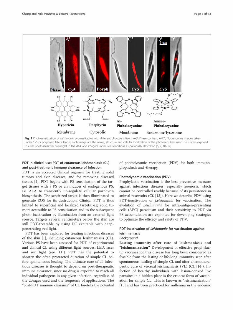
PDT in clinical use: PDT of cutaneous leishmaniasis (CL)and post-treatment immune clearance of infectionPDT is an accepted clinical regimen for treating solidtumors and skin diseases, and for removing diseasedtissues [4]. PDT begins with PS-sensitization of the tar-get tissues with a PS or an inducer of endogenous PS,i.e. ALA to transiently up-regulate cellular porphyrinbiosynthesis. The sensitized target is then illuminated togenerate ROS for its destruction. Clinical PDT is thuslimited to superficial and localized targets, e.g. solid tu-mors accessible to PS-sensitization and to the subsequentphoto-inactivation by illumination from an external lightsource. Targets several centimeters below the skin arestill PDT-treatable by using PC excitable with deep-penetrating red light.PDT has been explored for treating infectious diseases
of the skin [1], including cutaneous leishmaniasis (CL).Various PS have been assessed for PDT of experimentaland clinical CL using different light sources: LED, laserand sun light (see [11]). PDT has the potential toshorten the often protracted duration of simple CL be-fore spontaneous healing. The ultimate cure of all infec-tious diseases is thought to depend on post-therapeuticimmune clearance, since no drug is expected to reach allindividual pathogens in any given infection, regardless ofthe dosages used and the frequency of applications. The“post-PDT immune clearance” of CL foretells the potential
of photodynamic vaccination (PDV) for both immuno-prophylaxis and -therapy.
Photodynamic vaccination (PDV)Prophylactic vaccination is the best preventive measureagainst infectious diseases, especially zoonosis, whichcannot be controlled readily because of its persistence inanimal reservoirs (Cf. [13]). Here we describe PDV usingPDT-inactivation of Leishmania for vaccination. Theevolution of Leishmania for intra-antigen-presentingcells (APC) parasitism and their sensitivity to PDT viaPS accumulation are exploited for developing strategiesto optimize the efficacy and safety of PDV.
PDT-inactivation of Leishmania for vaccination againstleishmaniasisBackgroundLasting immunity after cure of leishmaniasis and“leishmanization” Development of effective prophylac-tic vaccines for this disease has long been considered asfeasible from the lasting or life-long immunity seen afterspontaneous healing of simple CL and after chemothera-peutic cure of visceral leishmaniasis (VL) (Cf. [14]). In-fection of healthy individuals with lesion-derived liveparasites in a hidden place is the crudest form of vaccin-ation for simple CL. This is known as “leishmanization”[15] and has been practiced for millennia in the endemic
Fig. 1 Photosensitization of Leishmania promastigotes with different photosensitizers. A-D, Phase contrast; A’-D”, Fluorescence images takenunder Cy5 or porphyrin filters. Under each image are the name, structure and cellular localization of the photosensitizer used. Cells were exposedto each photosensitizer overnight in the dark and imaged under live conditions as previously described [6, 7, 10–12]
Chang and Kolli Parasites & Vectors (2016) 9:396 Page 3 of 13
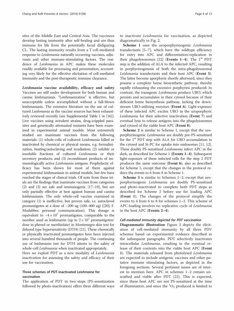
sites of the Middle East and Central Asia. The vaccineesdevelop lasting immunity after self-healing and are thusimmune for life from the potentially facial disfiguringCL. The lasting immunity results from a T cell-mediatedresponse to Leishmania naturally occurring vaccines, adju-vants and other immune-stimulating factors. The resi-dence of Leishmania in APC makes these moleculesreadily available for processing and presentation, account-ing very likely for the effective elicitation of cell-mediatedimmunity and the post-therapeutic immune clearance.
Leishmania vaccine availability, efficacy and safetyVaccines are still under development for both human andcanine leishmaniasis. “Leishmanization” is effective, butunacceptable unless accomplished without a full-blownleishmaniasis. The extensive literature on the use of cul-tured Leishmania as the vaccine sources has been exhaust-ively reviewed recently (see Supplemental Table 1 in [16]).Live vaccines using avirulent strains, drug-crippled para-sites and genetically attenuated mutants have been exam-ined in experimental animal models. Most extensivelystudied are inanimate vaccines from the followingmaterials: (1) whole-cells of cultured Leishmania killed orinactivated by chemical or physical means, e.g. formalini-zation, heating/autoclaving and irradiation; (2) soluble orinsoluble fractions of cultured Leishmania or theirsecretory products; and (3) recombinant products of im-munologically active Leishmania antigens. Prophylactic ef-ficacy has been shown for most of them againstexperimental leishmaniasis in animal models, but few havereached the stages of clinical trials. Of note from these tri-als are the findings that inanimate vaccines from categories(2) and (3) are safe and immunogenic [17–19], but areonly partially effective at best against human and canineleishmaniasis. The only whole-cell vaccine examined incategory (1) is ineffective, but proven safe, i.e. autoclavedpromastigotes at a dose of ~200 ug (100–400 ug) ([20]; F.Modabber, personal communication). This dosage isequivalent to ~4 × 10⁷ promastigotes, comparable to thenumber used as leishmanin (up to 2 × 10⁷ promastigotes/dose in phenol or merthiolate) in Montenegro skin test fordelayed type hypersensitivity (DTH) [21]. These chemicallyor physically inactivated promastigotes have been injectedinto several hundred thousands of people. The continuinguse of leishmanin test for DTH attests to the safety ofwhole-cell Leishmania when inactivated appropriately.Here we exploit PDT as a new modality of Leishmaniainactivation for assessing the safety and efficacy of theiruse for vaccination.
Three schemes of PDT-inactivated Leishmaina forvaccinationThe application of PDT in two steps (PS-sensitizationfollowed by photo-inactivation) offers three different ways
to inactivate Leishmania for vaccination, as depicteddiagramatically in Fig. 2.Scheme 1 uses the uroporphyrinogenic Leishmania
transfectants [5–7], which have the wildtype efficiencyfor entry into APC and differentiation/replication intheir phagolysosomes [22] (Events 1–4). The 1st PDTstep is the addition of ALA to the infected APC, resultingin porphyrinogenesis of both the intra-phagolysosomalLeishmania transfectants and their host APC (Event 5).The latter become aporphyric shortly afterward, since theypossess a complete heme biosynthetic pathway, therebyrapidly exhausting the excessive porphyrins produced; Incontrast, the transgenic Leishmania produce URO, whichpersists and accumulates in their cytosol because of theirdeficient heme biosynthesis pathway, lacking the down-stream URO-utilizing enzymes (Event 6). Light-exposureof these infected APC excites URO in the uroporphyricLeishmania for their selective inactivation (Event 7) andeventual lysis to release antigens into the phagolysosomesand cytosol of the viable host APC (Event 8).Scheme 2 is similar to Scheme 1, except that the uro-
porphyrinogenic Leishmania are doubly pre-PS-sensitizedfor the 1st PDT step with ALA for URO accumulation inthe cytosol and Si-PC for uptake into endosomes [11, 12].These doubly PS-sensitized Leishmania infect APC in thedark, as described for Scheme 1 (Events 1–4). Subsequentlight-exposure of these infected cells for the step 2 PDTproduces the same outcome (Event 6), also as describedfor Scheme 1, except that the changes in the protocol re-duce the events to 6 from 8 in Scheme 1.Scheme 3 is similar to Schemes 1–2, except that uro-
porphyrinogenic Leishmania are doubly PS-sensitizedand photo-inactivated to complete both PDT steps asdescribed for Scheme 2 before use for loading APC(Event 1). The changes of the protocol simplify theevents to 4 from 6 to 8 for schemes 1–2. This scheme ofAPC-loading involves no replicative cycle of Leishmaniain the host APC (Events 2–4).
Cell-mediated immunity depicted for PDT vaccinationDiagrammatic illustration Figure 3 depicts the elicit-ation of cell-mediated immunity by all three PDVschemes based on experimental evidence described inthe subsequent paragraphs. PDT selectively inactivatesintracellular Leishmania, resulting in the eventual re-lease of their contents into the viable host APC (Event1). The materials released from photolysed Leishmaniaare expected to include antigenic vaccines and other pu-tative immune stimulating factors, as depicted in theforegoing sections. Several pertinent issues are of inter-est to mention here. APC in schemes 1–2 remain un-scathed and viable after PDT [22]. This is expected,since these host APC are not PS-sensitized at the timeof illumination, and since the 1O2 produced is limited to
Chang and Kolli Parasites & Vectors (2016) 9:396 Page 4 of 13
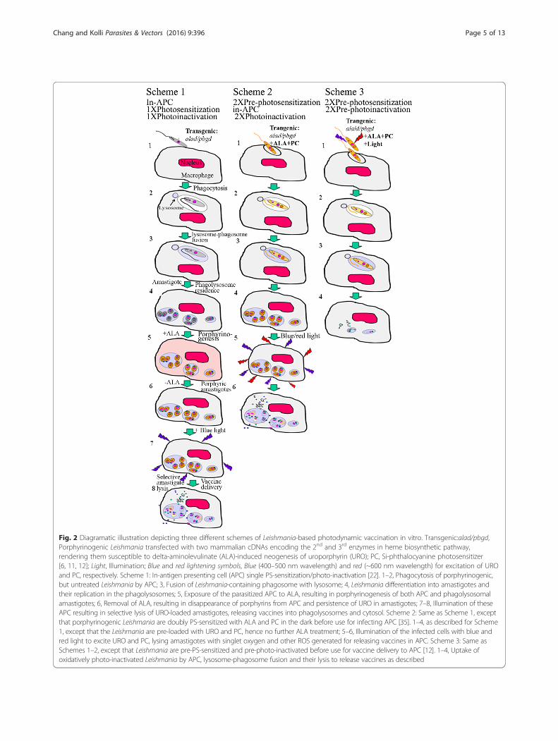
Fig. 2 Diagramatic illustration depicting three different schemes of Leishmania-based photodynamic vaccination in vitro. Transgenic:alad/pbgd,Porphyrinogenic Leishmania transfected with two mammalian cDNAs encoding the 2nd and 3rd enzymes in heme biosynthetic pathway,rendering them susceptible to delta-aminolevulinate (ALA)-induced neogenesis of uroporphyrin (URO); PC, Si-phthalocyanine photosensitizer[6, 11, 12]; Light, Illumination; Blue and red lightening symbols, Blue (400–500 nm wavelength) and red (~600 nm wavelength) for excitation of UROand PC, respectively. Scheme 1: In-antigen presenting cell (APC) single PS-sensitization/photo-inactivation [22]. 1–2, Phagocytosis of porphyrinogenic,but untreated Leishmania by APC; 3, Fusion of Leishmania-containing phagosome with lysosome; 4, Leishmania differentiation into amastigotes andtheir replication in the phagolysosomes; 5, Exposure of the parasitized APC to ALA, resulting in porphyrinogenesis of both APC and phagolysosomalamastigotes; 6, Removal of ALA, resulting in disappearance of porphyrins from APC and persistence of URO in amastigotes; 7–8, Illumination of theseAPC resulting in selective lysis of URO-loaded amastigotes, releasing vaccines into phagolysosomes and cytosol. Scheme 2: Same as Scheme 1, exceptthat porphyrinogenic Leishmania are doubly PS-sensitized with ALA and PC in the dark before use for infecting APC [35]. 1–4, as described for Scheme1, except that the Leishmania are pre-loaded with URO and PC, hence no further ALA treatment; 5–6, Illumination of the infected cells with blue andred light to excite URO and PC, lysing amastigotes with singlet oxygen and other ROS generated for releasing vaccines in APC. Scheme 3: Same asSchemes 1–2, except that Leishmania are pre-PS-sensitized and pre-photo-inactivated before use for vaccine delivery to APC [12]. 1–4, Uptake ofoxidatively photo-inactivated Leishmania by APC, lysosome-phagosome fusion and their lysis to release vaccines as described
Chang and Kolli Parasites & Vectors (2016) 9:396 Page 5 of 13

the PS-sensitized Leishmania, as these ROS are tooshort-lived to cross multiple membranes to cause oxida-tive damage to the host APC. The endogenous anti-oxidants of APC are expected to protect themselvesfrom other ROS generated secondarily from PDT. Inaddition, PDT may contribute positively to the APCfunctions in two ways: (1) Antigen processing by PDT-generated 1O2 and/or other ROS via oxidative modifica-tions of the APC proteases involved and/or the Leish-mania-released antigens as their substrates, e.g. 1O2
oxidation of their aromatic amino acid residues [23]; and(2) PDT-activation of ROS signal pathways favourable
for the elicitation of immunity [24]. Clearly, the selectivePDT-inactivation of intracellular Leishmania relievestheir host APC of immunosuppression caused by the in-fection [22]. The subsequent processing of Leishmaniavaccine antigens is predicted to follow the conventionallysosomal pathway (Event 2) and/or proteosomal path-way (Event 3) for co-presentation with MHC Class IIand Class I molecules to activate CD4+ and CD8+ Tcells, respectively (Events 2–3). The latter pathway isenvisioned to proceed via cross presentation of Leish-mania antigens, which are translocated from phagolyso-somes to the cytosol. Other Leishmaina-derived factors
Fig. 3 Diagrammatic depiction of processing and presentation of Leishmania-delivered vaccines by antigen-presenting cells. 1, Leishmania-releasedvaccines represented as dots and lines in phagolysosomes and cytosol and relevant organelles: RER, Golgi and proteasome stacks; 2, Antigenpresentation by lysosomal pathway via MHC Class II for activation of CD4+ T cells; 3, Antigen presentation by proteosomal pathway via MHCClass I for activation of CD8+ T cells; Bottom, Combination of both pathways shown in 2 and 3
Chang and Kolli Parasites & Vectors (2016) 9:396 Page 6 of 13
may further participate in the step of co-stimulation (notshown).
Experimental evidence The cell-mediated immunitydepicted (Figs. 2 and 3) is based on the experimentaloutcome from the PDV schemes carried out in differentexperimental models, as briefly summarized below:Scheme 1 was applied to immunization of Syrian
Golden hamsters, eliciting a Th1 response for prophy-laxis against Indian kala-azar produced by challengeswith virulent Leishmania donovani [25]. The vaccinationproduces lasting immunity, as shown by the analysis ofhepatosplenomegaly, parasite loads and cytokine profiles.Significantly, the immunity is adoptively transferable bysplenic T cells from immunized animals to naïve ham-sters, indicating that the immunity is cell-mediated andrequires no antigen stimulation from persistent para-sites, if any, at least in the recipients.Scheme 2 was used for immunization of BALB/c mice
against CL produced by challenges with Leishmaniaamazonensis. The observed prophylactic protection issignificant, albeit incomplete, as indicated by comparingimmunized mice versus the control groups. Immunizationdelayed the emergence of lesions for several weeks andsignificantly reduced the lesion size and their parasiteloads by 10-fold versus the controls (Unpublished data.See legend to Fig. 4, Experimental-in-brief). The vaccin-ation is considered effective, considering that BALB/cmice are known to bias toward Th2 with extreme levels ofgenetic susceptibility to cutaneous leishmaniasis.Scheme 3 PDV used PDT-inactivated Leishmania,
which were transfected to express ovalbumin (OVA) as amarker antigen or surrogate vaccine [12]. The cell-mediated immune responses to OVA delivered by PDT-inactivated transfectants were examined in in vitro andin vivo mouse models. APC loaded with the PDT-inactivated Leishmania were shown to deliver OVA,which was effectively processed for MHC Class I presen-tation of its specific peptide for activation of CD8+ T cellline [12]. In the in vivo studies, BL57 mice were immu-nized three times, each with ~106 PDT-inactivatedOVA-Leishmania. Splenic T cells of these immunizedmice were activated in response to CD4+ and CD8+ Tcell-specific OVA peptides that increased proportionallywith the number of immunizations (Unpublished data.See legend to Fig. 4, Experimental-in-brief). Most signifi-cantly, T cell activation is 6-fold higher with OVA deliv-ered by PDT-inactivated Leishmania than that deliveredby conventional means.The safety of Leishmania PDT-inactivation for vaccinationincreases in the order of Schemes 1 to 3. Leishmania weresingly and doubly PDT-inactivated for Scheme 1 andSchemes 2–3, respectively. They were completely inacti-vated by both PDT steps of PS-sensitization followed by
double photo-inactivation before loading APC in Scheme3 (see Fig. 4 and text for further discussion).
PDT-inactivation of Leishmania for vaccine deliveryagainst other infectious and malignant diseasesThe utility of PDT-inactivated Leishmania for delivery ofadd-on vaccines against other diseases is feasible, as in-dicated by the favourable outcome of the immune re-sponses seen in vitro and in vivo to OVA delivered bythis means. The successful delivery of OVA is significant,considering its expression at minuscule amount againsta background of Leishmania proteins in overwhelmingquantity and diversity in ~106 cells used for the delivery.This is taken to indicate that Leishmania creates noantigen-overload for vaccine delivery at least for OVA asa well-known T cell antigen.Leishmania are naturally endowed with favourable at-
tributes, making these parasites highly deployable as auniversal vaccine carrier [22]. Many Leishmania speciescan be cultured safely as promastigotes in serum-free,chemically defined media [26] and scaled up for expan-sion [27]. The biosynthetic machineries of Leishmaniaare capable of high capacity transcription, translationand correct post-translational modification of foreignproteins. A number of efficient vectors are available fortheir abundant expression episomally or chromosomallyas add-on vaccines in Leishmania - a favourable milieuof adjuvanticity and antigenicity conducive to elicit cell-mediated immunity.Efficient delivery of add-on vaccines by Leishmania is
due to their surface coat, consisting of unique lipid-saccharide-protein complexes [28]. In natural infection,they are known to protect Leishmania against the lytichumeral factors abundant in the animal body fluidsand to target them to the phagolysosomes of APC.This mode of parasitism is further facilitated by thesecretory products of Leishmania, e.g. nucleoside di-phosphate kinase [29]. Full deployment of these mo-lecular attributes by Leishmania is expected to protectthe payload of add-on vaccines for homing to APCwhen using non-sensitized or PS-sensitized Leish-mania for vaccine delivery according to Schemes 1–2(Fig. 2). Notably, Leishmania PDT-inactivated accord-ing to Scheme 3 are no longer viable, but remainOVA-delivery competent. The integrity of their surfacecoat may account for this, since it is unaffected by the1O2, which is generated in and limited to the cytosolof PDT-inactivated Leishmania.Uroporphyrinogenic Leishmania are being evaluated
for their ability to serve as a carrier of candidate vac-cines for trials against other infectious and malignantdiseases [30–33]. PDV with PDT-inactivated Leishmaniatransfectants will follow Schemes 1–3 (Fig. 2) to obtainsafety and efficacy data. In vitro vaccination of DCs will
Chang and Kolli Parasites & Vectors (2016) 9:396 Page 7 of 13
be pursued, as described [33, 34]. This presents a newapproach by using a eukaryotic vehicle for safe and ef-fective vaccine delivery.
Safety versus efficacy evaluation of five LeishmaniaPDT-inactivation formatsFigure 4 summaries the available data of Leishmania,which are PS-sensitized [A] and photo-inactivated [B]with or without additional treatments in different ways[1]–[5] for assessing their safety [C] and efficacy [D].The safety is assessed after PDT inactivation of Leish-mania by three different ways: microscopy for flagellar
motility, cultivation for growth (2 weeks) and inocula-tion of mouse ear or tail base for lesion development(~2 months). Not all preparations were assessed by allcriteria mentioned and the assessments for some sam-ples are on-going or planned. The available results arebriefly discussed below:
1. Single PDT of Leishmania by ALA-induceduropoprhyrinogensis [1] or PC-loading [2] aloneinactivated ~95 % of these cells, as determined by thecriteria described [C]. Interestingly, PDV based onprotocol [1] elicited adoptively transferable cell-mediated
Fig. 4 Safety and efficacy evaluations of PDT-inactivated Leishmania prepared under different conditions for immuno-prophylaxis and therapy.Horizontal: [1] and [2], Single PS-sensitization with ALA (+ALA) for uroporphyrin (URO) or Si-PC-loading (PC) followed by single photo-inactivationwith blue or red light illumination for generation of cytotoxic 1O2 (Symbol as shown), respectively [6, 7, 10–12]; [3], Double PS-sensitization/doublephoto-inactivation using a combination of [1] and [2] conditions [35]; [4–5], Singly or doubly PDT-inactivated Leishmania from [1-3B] stored frozenat −20 °C and lyophilized, respectively. See legend to Fig. 2 for other abbreviations used. Vertical: [A–B], PS-sensitization/photo-inactivation of Leishmaniaunder the conditions as described in [1–5]. [C], Safety evaluations of the samples examined by microscopic observation for promastigote flagellarmotility, for growth after inoculation into culture medium for 2 weeks, and lesion development after injection to BALB/c mouse ear dermis or tail basefor ~60 days; [D], Efficacy evaluation in vitro and/or in vivo briefly summarized from published, on-going or planned studies. See text for [1D] and [2D]efficacy. Experimental-in-brief: see [25] for [1D in vivo]; see [12] for [2D in vitro]; [2D in vivo]: Groups of BL57 mice (~30 gm, 15/group) were immunizedi.d. with 106 photo-inactivated OVA transfectants/10 ul PBS/ear for 3 times 1-week apart. Control groups were simultaneously and similarly immunizedwith un-treated, PC-sensitized, light-exposed, freeze-thawed OVA-transfectants, and 1 ug OVA. Splenic cells were collected from four mice from eachgroup 2-weeks after 1–3 immunization for in vitro activation with OVA CD4- and CD8-specific peptides for ~4 days. Proliferation of CFSE-labeledlymphocytes was assayed flow cytometrically, providing the results briefly described in the text; [3D in vivo]: Female BLAB/c mice (~30 g) wereimmunized exactly as described for [2D in vivo], except that doubly PS-sensitized Leishmania were used. Controls included 6 groups using untreated,single PS-sensitized, light-exposed samples. Day 3 after immunization, photo-inactivation of Leishmania was carried out in situ at ~5 J/cm2 usingLumaCare LC-122 white light probe. Mice were each challenged at the tailbase with 107 parasites. Lesion size was measured weekly in all groups.Experiments were terminated after ~10 weeks when parasite loads were determined by limiting dilution method. Preliminary results obtained werebriefly described in the text. Abbreviations: CanVL, canine leishmaniasis; Li, Leishmania infantum; Lt, Leishmania tropica
Chang and Kolli Parasites & Vectors (2016) 9:396 Page 8 of 13

immunity and produced no visible pathology of thevaccination sites in hamster [25].2. Double-PDT of Leishmania with a combination ofProtocols 1-2 [3] resulted in no viable cells, as assessedby all three criteria [C], indicative of a completeinactivation [35]. Immunization of BALB/c miceaccording to [3] is protective, although incompletedue to their inherent sensitivity to CL, as alreadydiscussed.
Products [4] and [5] prepared by freezing andlyophilization of PDT-inactivated Leishmania [1–3B], re-spectively, were undertaken to facilitate their storageand transport and to increase their safety at the expenseof their efficacy. Although still on-going, lyophilizedsamples [5] appear to have some prophylactic activitiesagainst CL challenges after immunization of BALB/cmice.From the available data, the double-PDT inactivation
of Leishmania by method [3] provides the best vaccinationformat for use with optimal safety and efficacy. The otherregimens are being optimized for further safety versus effi-cacy evaluation.
Photodynamic insecticides (PDI)BackgroundHistory PDT to control insect pests was first mentionedin the early 1900’s (see [36]). From 1980’s to1990’s, TheAmerican Chemical Society published several sympo-sium volumes on “Light-activated pesticides” [37–39].Since then, follow-up publications have been limited andwere summarized in the reviews [36, 40, 41]. Differentdyes were used in experimental and/or field trials as PDIagainst various insects, mainly mosquito larvae andMediterranean fruit flies. Industrial interests (PhotoDyeInternational, Inc) included aerial spray of dye mixtures(xanthenes) or “SureDye®” (Red Dye #28 and Yellow Dye#8) (http://www.cdpr.ca.gov/docs/emon/pubs/ehapreps/suredye.htm) in attempt to control the latter pest. Thework in the past decades showed some effectiveness ofPDI, but this area of research has not gained attention.
Preamble PDI has the potential as an effective measureto control disease-transmitting vectors and other harm-ful insects. Development of resistance by insect pests toinsecticides is a recurrent scenario [42], calling attentionto different approaches, like PDT, which is unlikely toelicit resistance. The potential of PDI to control differentinsect pests are briefly discussed below.Phytophagous insects cause substantial losses in cropsand livestock despite the use of genetically modified(GM) insect-resistant plants [43]. Phloem/xylem sap-feeding insects cause additional damage by transmittingplant diseases. These vectors are PDT-targetable, since
they engorge voluminous plant saps amenable to PS-loading and are translucent to light for photo-inactivation.The use of 1O2-generating PS for PDT has the poten-tial to discriminate these and other phytophagous in-sects for selective killing, sparing their photosyntheticand 1O2-resistant host plants.Many animal biting insects feed on blood and transmit
serious diseases, accounting for substantial morbidityand mortality of domestic animal and human popula-tions worldwide. Application of PDI to control suchinsect vectors is highly desirable, e.g. Anopheles mosqui-toes, which transmit malaria and Aedes spp., whichtransmit Chikungunya, Dengue and Zika fever, causingepidemics in the tropical/subtropical world today. Theonly new non-PDI approach to control these vectors isto release GM mosquitoes based on Wolbachia- ormale-induced infertility [44, 45]. For PDT of femalemosquitoes and other blood feeders (phototropic andday-light active species), PS is deliverable via the blood-stream of susceptible hosts or the use of suitable baitsto sensitize the insects for sun light inactivation. Thelarval stages of all mosquitoes (and also black flies) areaquatic and thus are receptive to water-soluble PS forPDT [46, 47].PS-sensitization of all insects is possible by direct
spraying for their uptake via surface contact and/or sys-tematically via the hosts, as used for the current insecti-cides. Direct incorporation of PS into the drinking andfood sources of insects will deliver them into the digest-ive tracts for sensitization of cells therein. In either case,accessibility of PS-sensitized cells to light is necessary togenerate cytotoxic ROS for target destruction. Nocturnaland darkness-loving insects are less amenable to PDTunless a light-emitter is provided with the PS for theirexcitation.Summarized below are some observations from our
preliminary studies of few insects on their uptake of se-lected PS and susceptibility to PDI.
Screening of PS for their PDI against selected insectsExposure of the 4th instar mosquito larvae (Culex pipiensquinquefasciatus) and adult sand flies (Phlebotomusdubosqi) [48] to rose bengal (RB) and cyanosine (CY)overnight resulted in the accumulation of these red dyesthat are visible in the gut of the larvae (Fig. 5a) and ofboth female and male flies (Fig. 5b, c). Their uptake of theother PS examined is less clear, including aluminum-phthalocyanine (Al-PC), protoporphyrin IX (PROTO) andNile blue sulfate (NB). Only RB- and CY-sensitized larvaelost their viability after light exposure based on their mo-bility (not shown). The sand fly response to the PDI is in-conclusive due to a high mortality of the control group,pending further investigation. This is also true for PDI ofthe plant-sucking insects, e.g. aphids, suggestive of a need
Chang and Kolli Parasites & Vectors (2016) 9:396 Page 9 of 13

to use membrane-feeding techniques instead of using cutor potted plants [49, 50].Our observations as described are preliminary, but rep-
resent the first study of PDI on sand flies, showing theiruptake of PS used. The mosquito larvicidal activities of thePDI seen are consistent with the results of an early work(see [36]) and the reports using marigold alpha-terthienylas the PS and different mosquito species [46, 47].
Uptake of PS by mosquito cells in vitroSince the uptake of PS by mammalian and Leishmaniacells is a prerequisite for their sensitization for PDT, wehave begun to assess this with insect cells, e.g. Aedesalbopictus clone C6/36 (ATCC CRL-1660). Figure 6shows the uptake of RB and CY by these mosquito cells,rendering them sensitive to photo-inactivation. Un-treated cells ([1]-None) are adherent (1A-DIF) and
Fig. 5 Uptake of rose bengal by selected insects and their photo-inactivation. a Culex pipiens quinquefasciatus 4th instar larvae exposed to roseBengal (10 ug/ml) (~20 larvae/5 ml water/well in 6-well plate) in dark for ~24 hours followed by exposure to white light for ~6 h at ~2500 lux;b-c Phlebotomus duboscqi female (b) and male (c) adult flies (~20 flies/screened paper cup) fed with 5 % sucrose solution and 500 ul of 5 mg/mlrose bengal in a cotton ball for ~20 h in the dark followed by exposure to ~2500 lux of white light for 3 h. Duplicate samples were prepared andkept in the dark as controls. Rose bengal is taken up by the flies of both sexes. Phototoxicity is evident for the mosquito larvae, but inconclusivefor the flies. The tests were done in Petr Volf’s lab
Fig. 6 Uptake of cyanosine and rose bengal by mosquito cells of the C6/36 line and their photosensitivity in vitro. The insect cells were exposedto both dyes overnight and illuminated with white light under conditions similar to those described for mammalian and Leishmania cells [10–12, 22].Images were captured first under differential interference (DIF) [A] and then under the filter sets for DAPI and Texas red [B]. [C] Chemical structure ofcyanosione and rose Bengal. Uptake of both dyes by the cells after incubation in the dark overnight (Dark, A2-3, B2-3) and cellular disintegration afterlight exposure for 4 h (Light, A2-3, B2-3) in contrast to the untreated controls (Dark and Light, A1, B1). Work done by Shin-Hong Shiao
Chang and Kolli Parasites & Vectors (2016) 9:396 Page 10 of 13

non-fluorescent (1B DAPI + TxR), irrespective of illu-mination (1 Dark and Light). Cells exposed to CY [2]and RB [3] show cytoplasmic fluorescence (2B, 3BDAPI-txR), indicative of dye uptake. Sensitized cellsremain adherent and intact (Dark, 2A, 3A-DIF), butbecome disintegrated after light-exposure (Light, 2A,3A-DIF). These results are consistent with the larvicidalactivities of RB and CY observed, providing a cellularbasis for their PDT activities. Notably, the mosquitocells were not sensitized for PDT with the following PS:Al-PC [10], PC3-4 [11], NB and a porphyrin analogue[51]. Insect cells are thus similar to other cells in theirrequirement of PS uptake for susceptibility to PDT, butrequire different PS for PDI.The preliminary data point to the feasibility of screen-
ing additional PS for PDT of cells from different insects,both harmful and beneficial, and from other life forms intheir environments. Such in vitro screening of PS for ac-tivities has the potential to identify PDI, which discrim-inate harmful pests from beneficial insects and otherfriendly organisms for selective killing of the former. Offurther interest is to elucidate the mechanisms of differ-ential PS-uptake by cells of different origin, providingclues for designing PS with specificity for PDI targeting.
ConclusionsPDT-inactivation of Leishmania offers the versatility andflexibility to balance safety versus efficacy for vaccinationagainst leishmaniasis and as potential carriers of vac-cines against other infectious and malignant diseases(PDV). The development of this new approach willbenefit from governmental and public acceptance andsupport. The ingenuity of the new leadership [52] isneeded for novel regulation that will ensure the safety ofvaccines with no barrier to disrupt innovation. The ad-vocacy groups also call attention to rectify the existingbarriers between science and cures, e.g. fasterCures(http://www.fastercures.org/). Development of vaccinesincluding PDV will further benefit from effective mea-sures against the anti-vaccination movement [53].PDI represents an alternative approach to control in-
sect pests. It is still in its early infancy of developmentdespite the idea first emerged almost 100 years ago.Many PS for PDI are innocuous compounds, which havelong been used among our everyday household products.Their application as PDI is not expected to select for re-sistance in contrast to the chemical pesticides in currentuse. PDI has the potential to complement the GM ap-proaches in the field of agriculture and medicine. It willbe particularly suitable for development in places wherethe population is sensitive to GM organisms.The lynchpin between PDV and PDI is the PS for light
excitation to generate cytotoxic ROS. The expertise inmedicinal chemistry is essential for synthesis and design
of novel PS. This depends on the input of biologists toelucidate the mechanisms of their cellular/molecular activ-ities. New PS need to be assessed by expert clinicians, veter-inarians, entomologists, cancer researchers, microbiologistsand immunologists, hence the consortium of collaboratorsenlisted.
AbbreviationsALA, delta-aminolevulinate; Al-PC, aluminum phthalycyanine; APC, antigen-presenting cells; CL, cutaneous leishmaniasis; CY, cyanosine; DIF, differentialinterference; DTH, delayed type hypersensitivity; GMO, genetically modifiedorganisms; NB, Nile blue; OVA, ovalbumin; PC, phthalocyanines;PDI, photodynamic insecticide; PDT, photodynamic therapy; PDV, photodynamicvaccination; PROTO, protoporphyrin IX; PS, photosensitizer; RB, rose bengal;ROS, reactive oxygen species; URO, uroporphyrin I; VL, visceral leishmaniasis
AcknowledgementsThanks are due to Joseph Reynolds and David Everly for reviewing thismanuscript. Publication of the CVBD 11 thematic series has been sponsoredby Bayer HealthCare - Animal Health division.
Collaboration groupRamesh B. Batchu, PhD, Director, Division of Surgical Oncology &Developmental, Therapeutics, Associate Professor, The Michael and MarianIlitch Department of Surgery, Wayne State University, 4646 John R Road,Detroit, MI 48201, USA. [email protected] (Winni) Chen, DVM, PhD (陳慧文), Assistant Professor, Departmentof Veterinary Medicine, National Taiwan University, 1 Sec. 4 Roosevelt Rd.,Taipei, 10617 Taiwan. [email protected] Ming C. Chow, ScD (周銘祥), Associate Head and Professor,Department of Applied Biology and Chemical Technology, Hong KongPolytechnic University, Hong Kong. [email protected] Elliott, MD/Jonathan F. Head, PhD, Elliott Mastology Center, 541Shadows Lane, Baton Rouge, LA 70806, USA. [email protected] Fan PhD (范家堃), Professor and Chairman, Department ofMolecular Parasitology and Tropical Diseases, School of Medicine and Centerfor International Tropical Medicine, College of Medicine, 250 Wu-Xing Street,Taipei Medical University, Taipei, Taiwan. [email protected] Hung, PhD (洪政雄), Investigator and Vice Director, Institute ofChemistry, Academia Sinica, 128, Sec. 2, Academia Road,, Nankang, Taipei,Taiwan 11529. [email protected] Ji, PhD (嵇達德), Associate Professor, Department of TropicalMedicine/International Health Programs, National Yang-Ming University,Beitou, Taipei 112, Taiwan. [email protected] Lun, PhD (伦照荣), Professor & Director, Center for ParasiticOrganisms, School of Life Sciences, Sun Yat-Sen University, Guangzhou510275, P.R. China. [email protected] Manna, PhD, DVM, School of Veterinary Medicine and AnimalProductions, University of Naples, Federico II, Naples, Italy. [email protected] Matsumoto, DVM, PhD, Professor, Laboratory of MolecularImmunology, School of Agricultural and Life Sciences, The University ofTokyo, Yayoi 1-1-1, Tokyo, Japan 113-8657 [email protected] KP Ng, DPhil (吳基培), Professor, Department of Chemistry, ChineseUniversity of Hong Kong, Sha Tin, N. T., Hong Kong. [email protected] I. de Oliveira PhD/Sayonara Melo, Centro de Pesquisas GonçaloMoniz. FIOCRUZ, R. Waldemar, Falcão, 121 Candeal, Salvador- BA -40296-710,Brazil. [email protected] Ozbel, MD, Professor, Ege University Faculty of Medicine, Departmentof Parasitology, 35100 Bornova, Izmir,TURKEY. [email protected] Özbilgin. PhD, Professor, Celal Bayar Üniversitesi, Tip FakültesiParazitoloji A.D, Dekanlik Binasi Uncubozköy, 45030, Manisa, Tü[email protected] Reynolds, PhD, Department of Microbiology/Immunology, ChicagoMedical School/RFUMS, N Chicago, IL 60064, [email protected] Sanjoba, PhD, Laboratory of Molecular Immunology, Graduate Schoolof Agricultural and Life Sciences, The University of Tokyo, 1-1-1Yayoi, Bunkyo-ku,Tokyo 113-8657 Japan. [email protected]
Chang and Kolli Parasites & Vectors (2016) 9:396 Page 11 of 13

Shin-Hong Shiao, PhD (蕭信宏), Assistant Professor, Department ofParasitology, Institute of Microbiology, School of Medicine, National TaiwanUniversity, Taipei, Taiwan. [email protected] Shih, PhD (施能耀), Associate Investigator, National Institute ofCancer Research, National Health Research Institutes, Tainan, [email protected] Tsai, PhD (蔡志偉), Associate Professor, Department of Entomology,College of Natural Resources and Agriculture, National Taiwan University,1 Sec. 4 Roosevelt Rd., Taipei, 10617 Taiwan. [email protected] da Graça H. Vicente, PhD, Distinguished Professor, Department ofChemistry, 249 Chemistry and Materials Building, Highland Rd, LouisianaState University, Baton Rouge, LA 70803, USA. [email protected] Volf, PhD, Professor, Department of Parasitology, Faculty of Science,Charles University Vinicna 7, 128 44 Praha 2, Czech Republic. [email protected] Wu, PhD (吳岳隆), Assistant Professor, Department ofEntomology, College of Natural Resources and Agriculture, National TaiwanUniversity, 1 Sec. 4 Roosevelt Rd., Taipei, 10617 Taiwan. [email protected] Yu, PhD (郁兆蘭), Professor, Department of Biomedical Sciences,Chang Gung University, No. 259, Wenhua 1st Rd., Guishan Dist., Taoyuan City33302, Taiwan. [email protected] Zhou, PhD (周晓农),Professor and Director, National Institute ofParasitic Diseases, Chinese Center for Disease Control and Prevention, 207Rui Jin Er Road, Shanghai 200025, P.R. China. [email protected];[email protected]
FundingWork described has received partial support by NIH/NIAID Grant # AI097830,AI-7712375, AI-68835 and other sources.
Availability of data and materialsNot applicable.
Authors’ contributionsKPC organized and wrote the first draft of this manuscript. BKK joined KPC torefine the science and language of the writing and the illustrations tocomplete the manuscript for submission. Both authors have read andapproved the final version of the manuscript.
Competing interestsThe authors declare that they have no competing interests.
Consent for publicationNot applicable.
Received: 1 April 2016 Accepted: 29 June 2016
References1. Abrahamse H, Hamblin MR. New photosensitizers for photodynamic
therapy. Biochem J. 2016;473:347–64.2. Battersby AR. Tetrapyrroles: the pigments of life. Nat Prod Rep.
2000;17:507–26.3. Berenbaum M. Phototoxicity of plant secondary metabolites: insect and
mammalian perspectives. Arch Insect Biochem Physiol. 1995;29:119–34.4. Benov L. Photodynamic therapy: current status and future directions. Med
Princ Pract. 2015;24 Suppl 1:14–28.5. Sah JF, Ito H, Kolli BK, Peterson DA, Sassa S, Chang KP. Genetic rescue of
Leishmania deficiency in porphyrin biosynthesis creates mutants suitable foranalysis of cellular events in uroporphyria and for photodynamic therapy. JBiol Chem. 2002;277:14902–9.
6. Dutta S, Kolli BK, Tang A, Sassa S, Chang KP. Transgenic Leishmania modelfor delta-aminolevulinate-inducible monospecific uroporphyria: cytolyticphototoxicity initiated by singlet oxygen-mediated inactivation of proteinsand its ablation by endosomal mobilization of cytosolic uroporphyrin.Eukaryot Cell. 2008;7:1146–57.
7. Dutta S, Furuyama K, Sassa S, Chang KP. Leishmania spp. delta-aminolevulinate-inducible neogenesis of porphyria by genetic complementationof incomplete heme biosynthesis pathway. Exp Parasitol.2008;118:629–36.
8. Li H, Jensen TJ, Fronczek FR, Vicente MG. Syntheses and properties of a seriesof cationic water-soluble phthalocyanines. J Med Chem. 2008;51:502–11.
9. Jiang XJ, Yeung SL, Lo PC, Fong WP, Ng DKP. Phthalocyanine-polyamineconjugates as highly efficient photosensitizers for photodynamic therapy. JMed Chem. 2011;54:320–30.
10. Dutta S, Ray D, Kolli BK, Chang KP. Photodynamic sensitization ofLeishmania amazonensis in both extracellular and intracellular stages withaluminum phthalocyanine chloride for photolysis in vitro. AntimicrobAgents Chemother. 2005;49:4474–84.
11. Al-Qahtani A, Alkahtani S, Kolli B, Tripathi P, Dutta S, Al-Kahtane AA, et al.Amino-phthalocyanine-mediated photodynamic inactivation of Leishmaniatropica. Antimicrob Agents Chemother. 2016;60:2003–11.
12. Dutta S, Ongarora BG, Li H, Vicente MG, Kolli BK, Chang KP. Intracellular targetingspecificity of novel phthalocyanines assessed in a host-parasite model fordeveloping potential photodynamic medicine. PLoS One. 2011;6:e20786.
13. Chang KP. Vaccination for disease prevention and control: The necessity ofrenewed emphasis and new approaches. J Immunol Immunotech.2014;1:1–4. doi:10.17653/2374-9105.SSe0001.
14. Chang KP. Leishmaniases. In: Brown M, editor. Encyclopedia of Life Sciences.New York: John Wiley & Sons, Ltd.; 2012. doi:10.1002/9780470015902.a0001954.pub3.
15. Khalil EAG, El Hassan AM, Zijlstra EE, Mukhtar MM, Ghalib HW, Musa B, et al.Autoclaved L. major vaccine for prevention of visceral leishmaniasis: arandomised, double-blind, BCG-controlled trial in Sudan. Lancet.2000;356:1565–9.
16. Costa CH, Peters NC, Maruyama SR, de Brito Jr EC, Santos IK. Vaccines forthe leishmaniases: proposals for a research agenda. Working Group onResearch Priorities for Development of Leishmaniasis Vaccines. PLoS NeglTrop Dis. 2011;5:e943.
17. Gradoni L. Canine Leishmania vaccines: still a long way to go. Vet Parasitol.2015;208:94–100.
18. Chakravarty J, Kumar S, Trivedi S, Rai VK, Singh A, Ashman JA, et al. A clinicaltrial to evaluate the safety and immunogenicity of the LEISH-F1+MPL-SEvaccine for use in the prevention of visceral leishmaniasis. Vaccine. 2011;29:3531–7.
19. Llanos-Cuentas A, Calderón W, Cruz M, Ashman JA, Alves FP, Coler RN,et al. A clinical trial to evaluate the safety and immunogenicity of theLEISH-F1+MPL-SE vaccine when used in combination with sodiumstibogluconate for the treatment of mucosal leishmaniasis. Vaccine.2010;28:7427–35.
20. Kamil AA, Khalil EA, Musa AM, Modabber F, Mukhtar MM, Ibrahim ME, et al.Alum-precipitated autoclaved Leishmania major plus Bacille Calmette-Guérrin,a candidate vaccine for visceral leishmaniasis: safety, skin-delayed typehypersensitivity response and dose finding in healthy volunteers. Trans R SocTrop Med Hyg. 2003;97:365–8.
21. Wilcocks C, Manson-Bahr PEC. Manson’s tropical diseases. 17th ed. London:Bailiere-Tindall; 1976.
22. Dutta S, Chang C, Kolli BK, Sassa S, Yousef M, Showe M, et al. Delta-aminolevulinate-induced host-parasite porphyric disparity for selectivephotolysis of transgenic Leishmania in the phagolysosomes of mononuclearphagocytes: a potential novel platform for vaccine delivery. Eukaryot Cell.2012;11:430–41.
23. Gracanin M, Hawkins CL, Pattison DI, Davies MJ. Singlet-oxygen-mediatedamino acid and protein oxidation: formation of tryptophan peroxides anddecomposition products. Free Radic Biol Med. 2009;47:92–102.
24. Kotsias F, Hoffmann E, Amigorena S, Savina A. Antioxid Reactive oxygenspecies production in the phagosome: impact on antigen presentation indendritic cells. Redox Signal. 2013;8:714–29.
25. Kumari S, Samant M, Khare P, Misra P, Dutta S, Kolli BK, et al. Photodynamicvaccination of hamsters with inducible suicidal mutants of Leishmaniaamazonensis elicits immunity against visceral leishmaniasis. Eur J Immunol.2009;39:178–91.
26. Chang KP, Hendricks DL. Laboratory cultivation and maintenance ofLeishmania. In: Chang KP, Bray RS, editors. Human parasitic diseases,Leishmaniasis, vol. 1. Amsterdam: Elsevier; 1985. p. 213–46.
27. Fritsche C, Sitz M, Weiland N, Breitling R, Pohl HD. Characterization of thegrowth behavior of Leishmania tarentolae: a new expression system forrecombinant proteins. J Basic Microbiol. 2007;47:384–93.
28. Chang KP, Reed SG, McGwire BS, Soong L. Leishmania model for microbialvirulence: the relevance of parasite multiplication and pathoantigenicity.Acta Trop. 2003;85:375–90.
29. Kolli BK, Kostal J, Zaborina O, Chakrabarty AM, Chang KP. Leishmania-released nucleoside diphosphate kinase prevents ATP-mediated cytolysis ofmacrophages. Mol Biochem Parasitol. 2008;158:163–75.
Chang and Kolli Parasites & Vectors (2016) 9:396 Page 12 of 13
30. Chen HW, Wang CH. A multiplex reverse transcriptase-PCR assay for thegenotyping of avian infectious bronchitis viruses. Avian Dis. 2010;54:104–8.
31. Elliott RL, Head JF. Adjuvant breast cancer vaccine improves disease specificsurvival of breast cancer patients with depressed lymphocyte immunity.Surg Oncol. 2013;22:172–7.
32. Shih NY, Chang GC, Tseng SW, Liu KJ, Hsiao KC, Lin HC. Targeting to tumor-associated antigen eno1 provides preventive and therapeutic effects ondisease progression of lung cancer. Ann Oncol. 2013;24 Suppl 1:i27–9.
33. Batchu RB, Gruzdyn OV, Moreno-Bost AM, Szmania S, Jayandharan G,Srivastava A, et al. Efficient lysis of epithelial ovarian cancer cells byMAGE-A3-induced cytotoxic T lymphocytes using rAAV-6 capsid mutantvector. Vaccine. 2014;32:938–43.
34. Batchu RB, Gruzdyn O, Potti RB, Weaver DW, Gruber SA. MAGE-A3 withcell-penetrating domain as an efficient therapeutic cancer vaccine. JAMASurg. 2014;149:451–7.
35. Dutta S, Waki K, Chang KP. Combinational sensitization of Leishmaniawith uroporphyrin and aluminum phthalocyanine synergisticallyenhances their photodynamic inactivation in vitro and in vivo.Photochem Photobiol. 2012;88:620–5.
36. Ben Amor T, Jori G. Sunlight-activated insecticides: historicalbackground and mechanisms of phototoxic activity. Insect Biochem MolBiol. 2000;30:915–25.
37. Heitz JR, Downum K. Light activated pest control. Am Chem Soc SympSeries. 1987;339. Doi: 10.1021/bk-1987-0339 (http://pubs.acs.org/isbn/9780841210264); Am Chem Soc Symp Series. 1995;616. (http://pubs.acs.org/doi/pdf/10.1021/bk-1995-0616.fw001).
38. Duke SO, Rebeiz CA. Porphyric pesticides: chemistry, toxicology andpharmaceutical applications. Washington DC. Am Chem Soc Symp Series.1994;559:317 (http://www.loc.gov/catdir/enhancements/fy0605/94016418-t.html).
39. Heitz JR. Pesticidal applications of halogenated xanthene dyes.Phytoparasitica Guest Editorial. 1997;25:89–92 (http://link.springer.com/article/10.1007/BF02981188).
40. Rebeiz CA, Guta LJ, Leea K, Juvika JA, Rebeiza CC, Boutona CE, et al.Photodynamics of porphyric insecticides. Crit Rev Plant Sci. 1995;14:329–66.
41. Mangan RL, Moreno DS. Photoactive dye insecticide formulations: adjuvantsincrease toxicity to Mexican fruit fly (Diptera: Tephritidae). J Econ Entomol.2001;94:150–6.
42. Owusu HF, Jančáryová D, Malone D, Müller P. Comparability betweeninsecticide resistance bioassays for mosquito vectors: time to review currentmethodology? Parasit Vectors. 2015;8:357. doi:10.1186/s13071-015-0971-6.
43. Lucht JM. Public acceptance of plant biotechnology and GM crops. Viruses.2015;7:4254–81.
44. Nguyen TH, Nguyen HL, Nguyen TY, Vu SN, Tran ND, Le TN, et al. Fieldevaluation of the establishment potential of wMelPop Wolbachia inAustralia and Vietnam for dengue control. Parasit Vectors. 2015;8:563.doi:10.1186/s13071-015-1174-x.
45. Winskill P, Carvalho DO, Capurro ML, Alphey L, Donnelly CA, McKemey AR.Dispersal of engineered male Aedes aegypti mosquitoes. PLoS Negl Trop Dis.2015;9:e0004156.
46. Hasspieler BM, Arnason JT, Downe AE. Toxicity, localization and eliminationof the phototoxin, alpha-terthienyl, in mosquito larvae. J Am Mosq ControlAssoc. 1988;4:479–84.
47. Nivsarkar M, Kumar GP, Laloraya M, Laloraya MM. Superoxide dismutase inthe anal gills of the mosquito larvae of Aedes aegypti: its inhibition by alpha-terthienyl. Arch Insect Biochem Physiol. 1991;16:249–55.
48. Volf P, Volfova V. Establishment and maintenance of sand fly colonies. JVector Ecol. 2011;36 Suppl 1:S1–9.
49. Tsai CW, Rowhani A, Golino DA, Daane KM, Almeida RP. Mealybugtransmission of Grapevine leafroll viruses: an analysis of virus-vectorspecificity. Phytopathology. 2010;100:830–4.
50. Akey DH, Beck SD. Continuous rearing of the pea aphid, Acyrthosiphonpisum, on a holidic diet. Ann Ent Soc Am. 1971;64:353–6.
51. Hung CH, Chang GF, Kumar A, Lin GF, Luo LY, Ching WM, et al.m-Benziporphodimethene: a new porphyrin analogue fluorescence zinc(II)sensor. Chem Commun (Camb). 2008;8:978–80.
52. Drazen JM. Califf for the FDA. New Engl J Med. 2016;374:176–7.53. Shelby A, Ernst K. Story and science: how providers and parents can utilize
storytelling to combat anti-vaccine misinformation. Hum VaccineImmunother. 2013;9:1795–801.
• We accept pre-submission inquiries
• Our selector tool helps you to find the most relevant journal
• We provide round the clock customer support
• Convenient online submission
• Thorough peer review
• Inclusion in PubMed and all major indexing services
• Maximum visibility for your research
Submit your manuscript atwww.biomedcentral.com/submit
Submit your next manuscript to BioMed Central and we will help you at every step:
Chang and Kolli Parasites & Vectors (2016) 9:396 Page 13 of 13