UNIVERSIDADE FEDERAL DE PERNAMBUCO · Efeito do extrato aquoso do chá verde e suas catecinas puras...
75
UNIVERSIDADE FEDERAL DE PERNAMBUCO CENTRO DE CIÊNCIAS BIOLÓGICAS DEPARTAMENTO DE FISIOLOGIA E FARMACOLOGIA MESTRADO EM CIÊNCIAS BIOLÓGICAS - FISIOLOGIA EFEITO DO EXTRATO AQUOSO DO CHÁ VERDE E SUAS CATECINAS PURAS SOBRE A PRODUÇÃO DE TESTOSTERONA PELAS CÉLULAS DE LEYDIG DE RATO IN VITRO MARINA DE SOUZA FIGUEIROA RECIFE 2008
Transcript of UNIVERSIDADE FEDERAL DE PERNAMBUCO · Efeito do extrato aquoso do chá verde e suas catecinas puras...
UNIVERSIDADE FEDERAL DE PERNAMBUCO CENTRO DE CIÊNCIAS BIOLÓGICAS
DEPARTAMENTO DE FISIOLOGIA E FARMACOLOGIA MESTRADO EM CIÊNCIAS BIOLÓGICAS - FISIOLOGIA
EFEITO DO EXTRATO AQUOSO DO CHÁ VERDE E SUAS CATECINAS PURAS SOBRE A PRODUÇÃO DE TESTOSTERONA PELAS CÉLULAS DE LEYDIG DE
RATO IN VITRO
MARINA DE SOUZA FIGUEIROA
RECIFE 2008

MARINA DE SOUZA FIGUEIROA
EFEITO DO EXTRATO AQUOSO DO CHÁ VERDE E SUAS CATECINAS PURAS SOBRE A PRODUÇÃO DE TESTOSTERONA PELAS CÉLULAS DE LEYDIG DE
RATO IN VITRO
Orientadora: Profa Dra. Maria Inês Wanderley
Co-orientador: Profo Dr. Daniel Pedro Udrisar
RECIFE 2008
Dissertação apresentada ao Curso de
Mestrado em Ciências Biológicas –
área de concentração Fisiologia – da
Universidade Federal de Pernambuco,
como parte dos pré-requisitos para a
obtenção do grau de Mestre em
Fisiologia.
Figueiroa, Marina de Souza
Efeito do extrato aquoso do chá verde e suas catecinas puras sobre a produção de testosterona pelas células de Leydig de rato in vitro / Marina de Souza Figueiroa. – Recife : O Autor, 2008.
73 folhas. Il: graf ., fig. e tab.
Dissertação (mestrado) – Universidade Federal de Pernambuco. CCB. Ciências Biológicas- Fisiologia, 2008.
Inclui bibliografia. 1. Chá Verde. 2. Testosterona. 3. Células d e
Leydig. 4. PKA. 5. PKC. I . Título. 615.322 CDU (2. ed) UFPE 615.321 CDD (22.ed.) CCS2008-Bib

Dedico este trabalho à minha família: minha mãe, meu pai, meu irmão
e, em especial, à Paulo Henrique Pimentel pelo amor, incentivo
e apoio constantes durante toda a sua realização.

AGRADECIMENTOS
Considerando este trabalho como resultado de uma caminhada que não começou na UFPE, agradecer pode não ser tarefa fácil, nem justa. Para não correr o risco de injustiças, agradeço de antemão a todas as pessoas que de alguma forma passaram pela minha vida e contribuíram para a construção desta dissertação. E agradeço, particularmente, a algumas pessoas pela contribuição direta na execução deste estudo: À meus pais, Lúcia e José Figueiroa, pessoas que sempre foram exemplos de amor, união, coragem, determinação e sucesso; agradeço pelo carinho e força que me dão e por estarmos sempre juntos nos momentos mais importantes da minha vida! Amo vocês! Ao meu amado noivo Paulo, que tantas vezes enfrentou o meu cansaço e ansiedade e com seu bom humor incansável me fez sorrir nos momentos mais difíceis. Obrigada amor! À minha orientadora professora Inês, que tornou possível a realização deste trabalho, agradeço pelo apoio e encorajamento contínuos na pesquisa, pelos conhecimentos transmitidos, pelo acolhimento no laboratório, pela confiança e amizade em mim depositadas, pelos conselhos e conversas compridas em dias de experimento e, principalmente, pelo exemplo de conduta ética, moral e científica. Ao meu co-orientador e professor Daniel, que busca sempre instigar a reflexão e crítica dos seus alunos frente às diversas questões da sociedade, pelos conselhos e palavras reconfortantes nas horas de ansiedade e insegurança, e, principalmente, pelo exemplo de pessoa sensível e humana. Obrigada pelas inúmeras discussões e reflexões sobre o mundo interior que muito me engrandeceram como pessoa! À minha amiga-irmã Cláudia Soares que, com muito carinho, não mediu esforços para que eu chegasse até esta etapa de minha vida. Muito obrigada por tudo amiga! Ao querido amigo e professor Mallison Vasconcelos, que me acompanha desde a graduação e cuja amizade, incentivo e discussões foram valiosas tanto nas questões acadêmicas quanto no meu amadurecimento como pessoa. Aos amigos Lauro, Juliany e Carine Wiesiolek pelo convívio, pelas dicas e pela amizade, essenciais no desenvolvimento desta pesquisa. Aos amigos Maurício Matos, Solange Barbosa e Cinthia Vasconcelos, e aos funcionários do Departamento de Fisiologia e Farmacologia da UFPE, não só pela amizade, mas também pelas facilidades oferecidas durante minha jornada, pelo incentivo e apoio constantes. Vocês representaram, para mim, união em momentos importantes!

“Quero, um dia, poder dizer às pessoas que nada foi em vão...
Que a vida é bela sim e que eu sempre dei o melhor de mim...
E que valeu a pena!”
(Mário Quintana)

RESUMO
Este estudo investigou os efeitos agudos do extrato aquoso do chá verde (GTE) e dos
seus constituintes polifenóis (-)-epigalocatecina-3-galato (EGCG) e (-)-epicatecina (EC)
sobre a produção de testosterona basal e estimulada, em células de Leydig de ratos in
vitro. Células de Leydig purificadas foram incubadas por 3 horas com GTE, EGCG ou
EC e com o precursor da testosterona androstenediona, na presença ou ausência de
ativadores da proteína quinase A (PKA) e da proteína quinase C (PKC). O GTE e a
EGCG, mas não a EC, inibiram ambas as produções de testosterona, basal e quinase-
estimuladas. Células pré-tratadas por 15 minutos com GTE ou EGCG e recuperadas por 1
hora foram submetidas a tratamento com gonadotrofina coriônica humana (hCG),
hormônio liberador de gonadotrofinas (LHRH), 22OHColesterol ou androstenediona.
Nestas condições o efeito inibitório do GTE/EGCG em suas maiores concentrações
utilizadas (69,2 e 100 µg/mL, respectivamente) sob a produção de testosterona estimulada
por hCG/LHRH ou 22OHColesterol se manteve, enquanto que a produção de testosterona
estimulada pela androstenediona retornou para os níveis do controle, indicando que o
efeito inibitório sob a função da enzima 17β-hidroxidesidrogenase (17β-HSD) foi
reversível. Nestas mesmas condições de pré-tratamento, porém utilizando menores
concentrações de GTE/EGCG (13,8 e 20 µg/mL, respectivamente) observou-se que o
efeito inibitório destes polifenóis sobre a produção de testosterona estimulada pelo
22OHColesterol foi revertida e até excedeu os níveis do controle, indicando que o efeito
inibitório dos polifenóis sob a função da enzima de clivagem da cadeia lateral (P450scc)
em mitocôndrias foi reversível. Conclui-se que os efeitos inibitórios do GTE podem ser
explicados, pelo menos em parte, pela ação da EGCG, seu principal componente, e que a
presença do grupo galato em sua estrutura parece ser importante para sua alta eficácia na
inibição da síntese de testosterona. Os mecanismos envolvidos nos efeitos do GTE e da
EGCG são provavelmente diversos e envolvem a inibição das cascatas de sinalização da
PKA/PKC, assim como a inibição da função das enzimas P450scc e 17β-HSD.
Palavras chave: Polifenóis do chá verde; Testosterona; Células de Leydig; PKA; PKC

ABSTRACT
This study investigated the acute effects of green tea extract (GTE) and its polyphenol
constituents (-)-epigallocatechin-3-gallate (EGCG) and (-)-epicatechin (EC) on basal and
stimulated testosterone production by rat Leydig cells in vitro. Purified Leydig cells were
incubated for 3 h with GTE, EGCG or EC and the testosterone precursor
androstenedione, in the presence or absence of either protein kinase A (PKA) or protein
kinase C (PKC) activators. GTE and EGCG, but not EC, inhibited both basal and kinase-
stimulated testosterone production. Cells pretreated for 15 min with GTE or EGCG and
allowed to recover for 1 hr were challenged with human chorionic gonadotropin (hCG),
luteinizing hormone releasing hormone (LHRH), 22(R)-hydroxycholesterol or
androstenedione. Under these conditions the inhibitory effect of GTE/EGCG at the higher
concentration used (69,2 and 100 µg/mL, respectively) on hCG/LHRH-stimulated or
22(R)-hydroxycholesterol-induced testosterone production was maintained whereas
androstenedione-supported testosterone production returned to control levels, indicating
that the inhibitory effect on 17β-hydroxysteroid dehydrogenase (17β-HSD) function was
reversible. At the same pretreatment conditions but using lower concentration of
GTE/EGCG (13,8 and 20 µg/mL, respectively) the inhibitory effect of these polyphenols
on 22(R)-hydroxycholesterol-supported testosterone production was reverted to and even
exceeded the control levels, indicating that the inhibitory effect of polyphenols on the
mitochondrial P450 side-chain cleavage enzyme (P450scc) function was reversible. We
conclude that the inhibitory effects of GTE may be explained, at least in part, by its
principal component EGCG and that the presence of a gallate group in its structure seems
important to its high efficacy in inhibiting testosterone production. The mechanisms
underlying the effects of GTE and EGCG are probably diverse and involve the inhibition
of the PKA/PKC signaling pathways, as well as the inhibition of the regulation of
P450scc and 17β-HSD function.
Key Words: Green tea polyphenols; Testosterone; Leydig cells; PKA; PKC
LISTA DE SIGLAS E ABREVIATURAS GTE: Extrato aquoso do chá verde
EGCG: Epigalocatecina-3-galato
EGC: Epigalocatecina
EC: Epicatecina
ECG: Epicatecina-3-galato
OH: Hidroxila
PKC: Proteína quinase C
PKA : Proteína quinase A
PI 3-quinase: Fosfoinositídeo 3-quinase
Akt/PKB : Akt/proteína quinase B
MAP-quinase: Proteína quinase ativada por mitógeno
P-60: Polifenona-60
P450scc: Enzima de clivagem da cadeia lateral
3β-HSD: ∆5-3β-Hidroxidesidrogenase
StAR: Proteína reguladora aguda da esteroidogênese
GnRH / LHRH : Hormônio liberador de gonadotrofinas
LH : Hormônio Luteinizante
ATP: Adenosina trifosfato
AMPc: Monofosfato cíclico de adenosina
DAG: Diacilglicerol
hCG: Gonadotrofina coriônica humana
AC: Adenilato ciclase
PPI: Fosfoinositídeo
PLCβ: Fosfolipase C beta
PC: Fosfatidilcolina
PIP2: Fosfatidilinositol 4,5-bifosfato
IP3: 1,4,5-trifosfato
OS: Fosfatidilserina
AA : Ácido aracdônico
PIP3: Fosfatidilinositol 3,4,5-trifosfato
CSCC: Sistema enzimático de clivagem da cadeia lateral do colesterol
Ca+2: Cálcio
RE: Retículo Endoplasmático Liso
17α-OH-IASE : 17α-hidroxilase
P45017α: C17-20-Liase
17β-HSD: 17β-hidroxidesidrogenase
BSA: Albumina sérica bovina
PBS: Tampão fosfato-salina
NaHCO3: Bicarbonato de Sódio
KCl : Cloreto de potássio
NaCl: Cloreto de sódio
PDBu: Forbol 12,13-dibutirato
PPO: 2,5-diphenyl-oxazole
dbcAMP: N6,2’-O-dibutiriladenosina-3’:5’-monofosfato cíclico
Meio 199: Meio de cultura 199
POPOP: 2,2’-p-phenylen-bis (5-phenyloxazol)
CaCl2: Cloreto de cálcio
KH 2PO4: Fosfato de potássio monobásico
Na2HPO4.7H2O: Fosfato de sódio dibásico
NaH2PO4.H2O: Fosfato de sódio monobásico
MTT : 3-(4,5-dimetiltialzolil)-2,5 difeniltetrazólio
DMSO: Dimetilsufóxido

SUMÁRIO Pág. LISTA DE SIGLAS E ABREVIATURAS...................................................... i
1. INTRODUÇÃO ........................................................................................... 12
1.1 Anatomia dos Testículos e Funções das Células de Leydig .......... 18
1.2 Vias de Trandução do Sinal Hormonal na Síntese de Testosterona
nas Células de Leydig ..................................................................................... 19
2. OBJETIVOS ................................................................................................ 25
2.1 Objetivo Geral................................................................................ 25
2.2 Objetivos Específicos..................................................................... 25
3. MATERIAL E MÉTODOS ......................................................................... 26
3.1 Animais ..........................................................................................26
3.2 Reagentes ....................................................................................... 26
3.3 Obtenção do Extrato Aquoso do Chá Verde (GTE)....................... 27
3.4 Obtenção e Purificação das Células de Leydig .............................. 27
3.5 Secreção de Testosterona in vitro .................................................. 28
3.6 Viabilidade Celular ........................................................................ 29
3.7 Radioimunoensaio da Testosterona................................................ 30
3.8 Tratamento Estatístico.................................................................... 30
REFERÊNCIAS BIBLIOGRÁFICAS............................................................. 32
Green tea polyphenols affect testosterone production in rat Leydig cells... .... 46
Abstract... ......................................................................................................... 47
Introduction...................................................................................................... 48
Material and Methods... ................................................................................... 49
Material... ............................................................................................. 49
Green tea extract (GTE) preparation.................................................... 50
Animals and Leydig cell-enriched preparation... ................................. 50
Cell Viability... ..................................................................................... 51
In vitro testosterone secretion... ........................................................... 52
RIA and statistical analysis... ............................................................... 53
Results.............................................................................................................. 54
Discussion... ..................................................................................................... 57
References........................................................................................................ 61
Legends... ......................................................................................................... 65
CONCLUSÕES... ............................................................................................ 73
12
1. INTRODUÇÃO
Após a água, o chá é considerado a bebida popular mais consumida em todo o
mundo. Derivado da planta Camellia sinensis, é consumido em variadas partes do globo
em suas três formas básicas: chá verde, chá preto e chá “oolong” (Wu et al., 2004). Da
quantidade total de chá produzido e consumido no mundo, 78% é de chá preto, 20% é de
chá verde e < 2% é de chá “oolong”. Historicamente o chá verde é o mais consumido no
Japão, China e Índia, tendo seu consumo, feito parte da cultura destes países nos últimos
5.000 anos (Mukhtar & Ahmad, 2000; Crespy & Williamson, 2004). Recentemente, este
chá tem atraído bastante atenção dos pesquisadores e de toda comunidade consumidora
de chá ao redor do mundo, uma vez que benefícios em uma variedade enorme de
patologias, que variam desde cânceres até perda de peso corporal, têm sido relacionados
ao seu consumo (Zaveri, 2006).
Os três tipos básicos de chá são processados de maneiras diferentes após a sua
colheita (Graham, 1992). O principal objetivo na produção do chá verde é a preservação
das catecinas das folhas em suas formas monoméricas; sendo este chá constituído de
folhas frescas colhidas de diferentes partes da planta Camellia sinensis, submetidas à
vapor e, posteriormente secas para prevenir a fermentação e a oxidação dos componentes
polifenóis. Já o chá preto passa por diversas etapas de processamento, dentre elas a de
fermentação, que consiste, na verdade, de uma oxidação enzimática dos flavanóis a
compostos poliméricos denominados teaflavinas, grupo característico deste tipo de chá, e
tearubiginas. O chá “oolong” é um produto parcialmente oxidado que contém uma
mistura de polifenóis monoméricos e uma grande quantidade de teaflavinas (Mukhtar &
Ahmad, 2000; Matsubara & Rodriguez-Amaya, 2006; Zaveri, 2006).
Segundo Liu (2003), os polifenóis fazem parte dos fitoquímicos que são
compostos bioativos presentes nas frutas, vegetais e grãos. Os fitoquímicos classificam-se
em carotenóides, fenólicos, alcalóides, compostos contendo nitrogênio e compostos
organo-sulfúricos, conforme esquema exposto na Figura 1.

13
Fig
ura
1: C
lass
ifica
ção
dos
Fito
qu
ímic
os.
Ext
raíd
a de
Liu
RH
. P
oten
tial S
iner
gy
of p
hyto
chem
ica
ls in
can
cer
pre
ven
tion
: mec
han
ism
of
actio
n.
J. N
utr.
20
04; 1
34
: 3
479
-34
85.
14
Os compostos fenólicos, bastante estudados, possuem um ou mais anel aromático
com um ou mais grupo hidroxila acoplado em sua estrutura e são categorizados em
ácidos fenólicos, flavonóides, “stilbenes”, “coumarins” e taninos. Os flavonóides
constituem um grupo dos compostos fenólicos com atividade antioxidante, presentes nas
frutas e vegetais, e relacionados com redução do risco para o desenvolvimento de
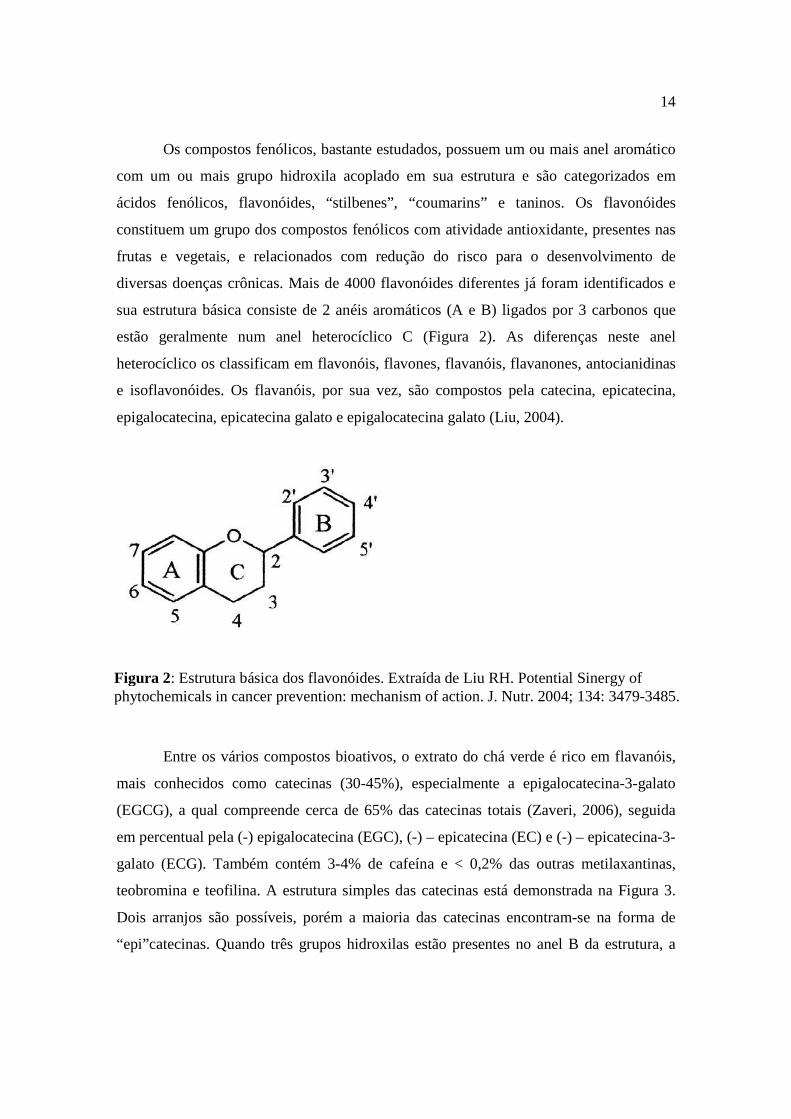
diversas doenças crônicas. Mais de 4000 flavonóides diferentes já foram identificados e
sua estrutura básica consiste de 2 anéis aromáticos (A e B) ligados por 3 carbonos que
estão geralmente num anel heterocíclico C (Figura 2). As diferenças neste anel
heterocíclico os classificam em flavonóis, flavones, flavanóis, flavanones, antocianidinas
e isoflavonóides. Os flavanóis, por sua vez, são compostos pela catecina, epicatecina,
epigalocatecina, epicatecina galato e epigalocatecina galato (Liu, 2004).
Entre os vários compostos bioativos, o extrato do chá verde é rico em flavanóis,
mais conhecidos como catecinas (30-45%), especialmente a epigalocatecina-3-galato
(EGCG), a qual compreende cerca de 65% das catecinas totais (Zaveri, 2006), seguida
em percentual pela (-) epigalocatecina (EGC), (-) – epicatecina (EC) e (-) – epicatecina-3-
galato (ECG). Também contém 3-4% de cafeína e < 0,2% das outras metilaxantinas,
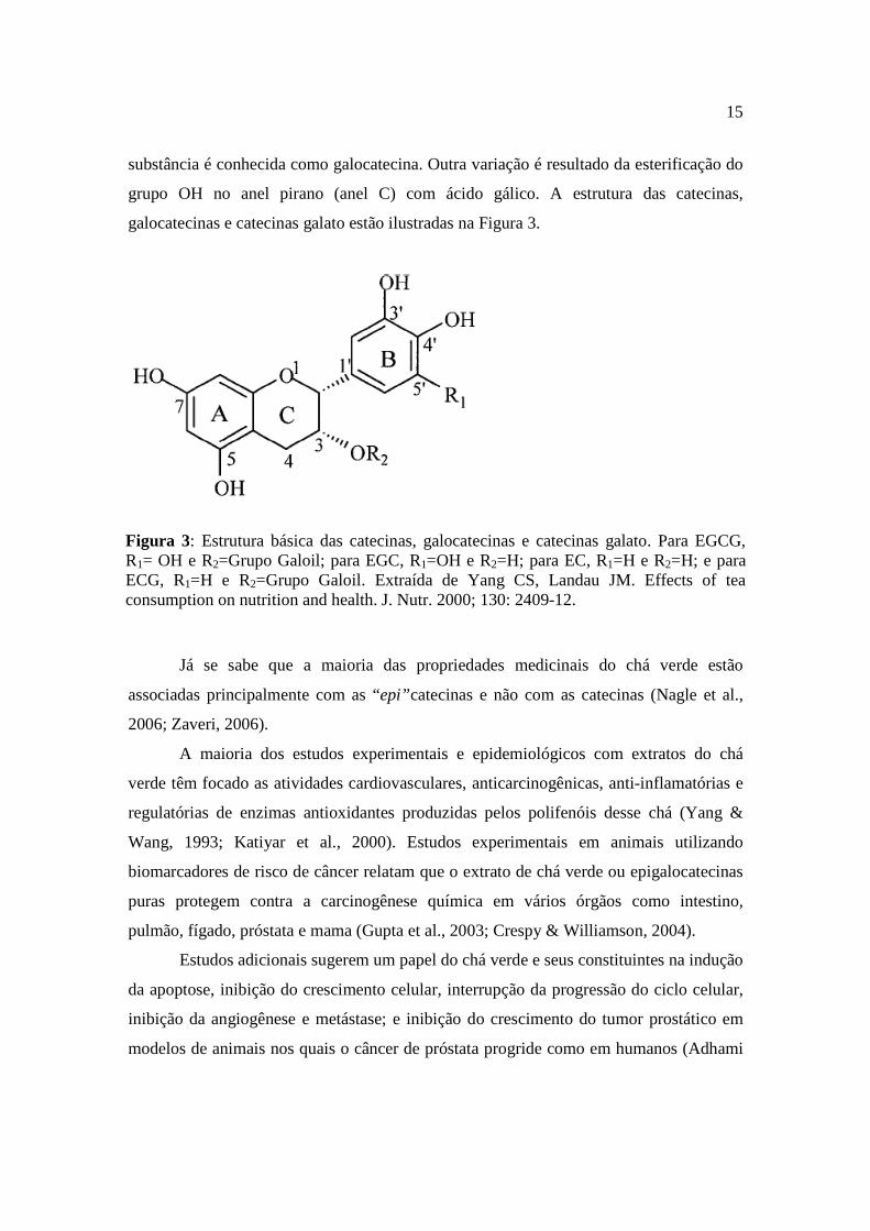
teobromina e teofilina. A estrutura simples das catecinas está demonstrada na Figura 3.
Dois arranjos são possíveis, porém a maioria das catecinas encontram-se na forma de
“epi”catecinas. Quando três grupos hidroxilas estão presentes no anel B da estrutura, a
Figura 2: Estrutura básica dos flavonóides. Extraída de Liu RH. Potential Sinergy of phytochemicals in cancer prevention: mechanism of action. J. Nutr. 2004; 134: 3479-3485.
15
substância é conhecida como galocatecina. Outra variação é resultado da esterificação do
grupo OH no anel pirano (anel C) com ácido gálico. A estrutura das catecinas,
galocatecinas e catecinas galato estão ilustradas na Figura 3.
Já se sabe que a maioria das propriedades medicinais do chá verde estão
associadas principalmente com as “epi”catecinas e não com as catecinas (Nagle et al.,
2006; Zaveri, 2006).
A maioria dos estudos experimentais e epidemiológicos com extratos do chá
verde têm focado as atividades cardiovasculares, anticarcinogênicas, anti-inflamatórias e
regulatórias de enzimas antioxidantes produzidas pelos polifenóis desse chá (Yang &
Wang, 1993; Katiyar et al., 2000). Estudos experimentais em animais utilizando
biomarcadores de risco de câncer relatam que o extrato de chá verde ou epigalocatecinas
puras protegem contra a carcinogênese química em vários órgãos como intestino,
pulmão, fígado, próstata e mama (Gupta et al., 2003; Crespy & Williamson, 2004).
Estudos adicionais sugerem um papel do chá verde e seus constituintes na indução
da apoptose, inibição do crescimento celular, interrupção da progressão do ciclo celular,
inibição da angiogênese e metástase; e inibição do crescimento do tumor prostático em
modelos de animais nos quais o câncer de próstata progride como em humanos (Adhami
Figura 3: Estrutura básica das catecinas, galocatecinas e catecinas galato. Para EGCG, R1= OH e R2=Grupo Galoil; para EGC, R1=OH e R2=H; para EC, R1=H e R2=H; e para ECG, R1=H e R2=Grupo Galoil. Extraída de Yang CS, Landau JM. Effects of tea consumption on nutrition and health. J. Nutr. 2000; 130: 2409-12.
16
et al., 2003). Em outros modelos animais, o consumo crônico das catecinas do chá verde
também diminuiu a incidência de artrite induzida pelo colágeno (Haqqi et al., 1999).
Com relação aos componentes puros do chá verde, demonstrou-se que as
catecinas inibem a atividade de uma variedade de enzimas, tais como: colagenase,
glutationa redutase, glutationa S-transferase hepática e 5α-redutase tipo 1 (Makimura et
al., 1993; Zhang & Das, 1994; Liao & Hiipakka, 1995; Zhang et al., 1997), são potentes
antioxidantes e alteram algumas propriedades de células cancerígenas em cultura (Wang
et al., 1996; Vinson et al., 2001; Sun et al., 2002; Chu et al., 2002; Adom & Liu, 2002;
Adom et al., 2003). Suas ações envolvem também a manutenção de condições
fisiológicas ideais para as células, uma vez que as protegem de dano oxidativo sendo
mais efetivas do que as vitaminas C e E (Rice-Evans et al., 1995). Em adição ao efeito
antioxidante, estudos demonstram uma atuação neuroprotetora das catecinas do chá verde
sobre os mecanismos envolvidos com morte e sobrevivência celular no cérebro, em
doenças como: Parkinson, Alzheimer, Huntington e Esclerose Lateral Amiotrófica
(Mandel et al., 2004; Gouni-Berthold & Sachinidis, 2004). Estudos in vivo e in vitro
apontam também um papel protetor das catecinas na neurodegeneração, envolvendo
alterações nas moléculas sinalizadoras celulares e nas vias do ciclo celular, ativação de
genes importantes para a sobrevivência celular e ativação das funções mitocondriais
(Mandel & Youdin, 2004). Esses compostos exercem ainda efeitos modulatórios na
célula por meio de ações seletivas em diferentes compostos de várias cascatas de
sinalização de proteínas quinase e lipídios quinase, alterando estados de fosforilação de
proteínas alvo como proteína quinase C (PKC), tirosina quinase, fosfoinositídeo 3-
quinase (PI 3-quinase), Akt/proteína quinase B (Akt/PKB) e proteína quinase ativada por
mitógeno (MAP-quinase), e/ou modulando a expressão gênica dessas proteínas (Williams
et al., 2004).
Outros benefícios do chá verde incluem ação bactericida (Stapleton et al., 2004),
anti-HIV (Nance & Shearer, 2003), anti-envelhecimento (Espósito et al., 2002) e
atividade anti-inflamatória (Dona et al., 2003).
O efeito das catecinas do chá verde no sistema reprodutor masculino já tem sido
estudado e demonstrado. Estudos laboratoriais e epidemiológicos sugerem que uma
associação entre dieta e níveis de andrógenos pode alterar o risco para câncer de próstata
17
(Ripple et al., 1997; Clinton & Giovannucci, 1998; Parkin, 2001). Foi demonstrado que
injeções parenterais de EGCG por 7 dias foram capazes de suprimir o crescimento de
tumores prostáticos e de mama em ratos atímicos (Liao et al., 1995). Em um estudo mais
recente, Kao et al. (2000) analisaram os efeitos da EGCG sobre os sistemas endócrinos de
ratos adultos e relataram que após 7 dias de injeções parenterais de EGCG observou-se
uma redução no peso dos testículos e dos órgãos sexuais acessórios (próstata, vesícula
seminal, glândulas prepuciais), assim como, nos níveis circulantes de LH e testosterona
em ratos machos intactos. Ainda no mesmo estudo, para determinar se a redução no peso
dos órgãos sexuais acessórios era devido a redução, induzida pela EGCG, nos níveis de
androgênios, os autores injetaram um grupo de ratos machos com androgênios e/ou
EGCG e observaram que não houve redução no peso prostático dos ratos tratados com
EGCG mais andrógenos, concluindo, então, que o efeito da catecina do chá verde,
EGCG, sobre o peso de órgãos sexuais acessórios masculinos era devido a diminuição
dos níveis circulantes de testosterona.
Satoh et al. (2002) estudaram os efeitos da administração oral de um extrato
contendo as catecinas do chá verde denominado Polifenona-60 (P-60) em ratos adultos e
relataram que após a ingesta oral deste extrato por 2 e 8 semanas os ratos machos
apresentaram redução significativa do peso corporal e do peso dos testículos e da
próstata, além de apresentarem elevados níveis sanguíneos de LH e testosterona. Os
mesmos autores demonstram um efeito inibitório importante da ingesta do extrato sobre a
atividade da enzima aromatase causado pela soma das ações dos vários constituintes do
P-60, principalmente, pela ação da EGCG a qual é responsável por 60% deste efeito
inibitório. Os autores sugerem que a inibição da enzima aromatase pelas catecinas pode
ser um dos mecanismos responsáveis pelas alterações endócrinas, elevação dos níveis
sanguíneos de LH e testosterona, observadas neste estudo.
Embora o efeito antigonadotrófico das catecinas tenha sido explicado como
secundário à redução na ingesta de alimentos (Kao et al., 2000) ou através da inibição da
atividade da enzima aromatase nas gônadas de ratos machos (Satoh et al., 2002), um
efeito modulatório pode estar presente a nível da esteroidogênese gonadal. Pesquisas
recentes demonstram a ação inibitória de outros polifenóis como os isoflavonóides,
também chamados de fitoestrógenos, sobre a atividade de enzimas importantes na síntese
18
de esteróides sexuais. Foi demonstrado que uma dieta rica em fitoestrógenos por 5
semanas é capaz de reduzir o peso corporal e prostático, e os níveis circulantes de
testosterona e androstenediona de ratos machos adultos (Weber et al., 2001),
isoflavonóides como a genisteína reduz a expressão da enzima de clivagem da cadeia
lateral (CYP11A1 ou P450scc) em mitocôndrias de células de Leydig de ratos adultos
(Svechnikov et al., 2005) e a daidzeína inibe a atividade da enzima ∆5-3β-
Hidroxidesidrogenase (3β-HSD) em células adrenais (Ohno et al., 2002); assim como a
genisteína e o resveratrol são capazes de inibir a expressão gênica do RNA mensageiro da
proteína reguladora aguda da esteroidogênese (StAR) (Chen et al., 2007). Entretanto,
ainda não existem evidências acerca do efeito direto das catecinas do chá verde sobre a
esteroidogênese testicular ou sobre as enzimas envolvidas na produção de andrógenos
nessas células.
Já foi bem descrito anteriormente o envolvimento das vias de sinalização da
proteína quinase A (PKA) e da PKC na produção de andrógenos nos testículos (Dehejia
et al., 1982; Wanderley & Negro-Vilar, 1996; Jo et al., 2005); entretanto não se sabe
ainda se as catecinas do chá verde podem modular estas vias. Estudos demonstram que a
EGCG e outros flavonóis podem modular a via de sinalização da PKC (Levites et al.,
2003) e da PKA (Lorenz et al., 2004) em outros modelos animais.
ANATOMIA DOS TESTÍCULOS E FUNÇÕES DAS CÉLULAS DE LE YDIG
Em todos os mamíferos, os testículos são órgãos pares, ovóides, envoltos pela
túnica albugínea e divididos em dois compartimentos funcionais: o tecido intersticial
vascularizado e os túbulos seminíferos avasculares. Os túbulos seminíferos são estruturas
contorcidas, conectadas em ambas às extremidades à rede testicular e onde ocorre a
espermatogênese. Na maioria dos mamíferos os túbulos seminíferos constituem,
aproximadamente, 80% do volume do testículo; os 20% remanescentes são compostos de
tecido conectivo de apoio, no qual as células de Leydig estão espalhadas. No interior
desses túbulos encontram-se as células de Sertoli e as células germinativas em diferentes
estágios de maturação. Os espermatozóides são produzidos nos túbulos seminíferos e são
19
drenados da rede testicular via ductos eferentes para o epidídimo (Christensen, 1975;
Odell, 1988; Setchell & Brooks, 1988).
O compartimento intersticial preenche os espaços entre os túbulos seminíferos e
contém todos os vasos sanguíneos e linfáticos, nervos, macrófagos, mastócitos e as
células de Leydig. Em todas as espécies com sistema linfático extenso, existe abundante
líquido intersticial banhando as células de Leydig, vasos sanguíneos e o exterior dos
túbulos seminíferos; é através desse líquido que todos os hormônios e nutrientes são
transportados do sangue para as células testiculares e entre essas células no interior dos
testículos (Fawcett et al., 1973).
As células de Leydig encontram-se, geralmente, agrupadas ao redor dos capilares
sanguíneos situados no tecido conectivo, o qual é drenado por vasos linfáticos; e sua
principal função é a produção de testosterona e outros hormônios esteróides necessários
para a iniciação e manutenção da espermatogênese, desenvolvimento e manutenção da
genitália externa e interna, aparecimento das características sexuais secundárias,
desenvolvimento do sistema imuno-esquelético e inibição, por retroalimentação, do eixo
hipotálamo-hipofisário (Fawcett et al., 1973; Christensen, 1975; Odell, 1988; Setchell &
Brooks, 1988).
A função normal do testículo depende das gonadotrofinas hipofisárias e, apesar de
já se saber que a regulação da função testicular está predominantemente sob o controle da
hipófise, o arranjo anatômico do testículo em dois compartimentos, o tecido intersticial e
os túbulos seminíferos, separados pela barreira hemato-testicular, indica uma interação
ativa entre as diferentes células testiculares (Saez, 1994).
VIAS DE TRADUÇÃO DO SINAL HORMONAL NA SÍNTESE DE
TESTOSTERONA NAS CÉLULAS DE LEYDIG
O controle da produção de esteróides pelas células de Leydig é realizado
primariamente pela ação da gonadotrofina hormônio luteinizante (LH), secretado em
pulsos de alta atividade biológica pela adenohipófise sob o controle da secreção episódica
do hormônio liberador de gonadotrofinas (GnRH ou LHRH) (Saez, 1994; De Kretser et
al., 1995; Evans, 1999).

20
Há dois sistemas principais de segundo mensageiros que parecem ser os efetores
da ação hormonal no testículo: o sistema do monofosfato cíclico de adenosina (AMPc) e
o sistema do diacilglicerol (DAG) e fosfato de inositol (IP3) (Steele & Leung, 1992).
O LH estimula a função esteroidogênica da célula de Leydig através da interação
com receptores de LH na superfície celular, resultando, principalmente, na estimulação
de eventos dependentes de AMPc (Dufau & Catt, 1978). Após a ocupação dos sítios
receptores de LH nas células de Leydig pelo LH/Gonadotrofina Coriônica Humana
(hCG), vários processos são iniciados na membrana plasmática através de uma proteína
ligante de GTP (Proteína G) e que levam à ativação da adenilato ciclase (AC) ligada à
membrana (Dufau et al., 1980, 1984; Dufau, 1988; Gore-Langton & Armstrong, 1988;
Ascoli & Segaloff, 1989). Esta enzima, por sua vez, catalisa a síntese de AMPc a partir
de adenosina trifosfato (ATP) no lado citoplasmático da membrana plasmática (Steele &
Leung, 1992).
O AMPc é o segundo mensageiro que media muitas modificações dependentes de
hormônio no metabolismo celular. Esta resposta à estimulação hormonal é mediada
através de um aumento nos níveis de AMPc o qual, por sua vez, ativa a PKA dependente
de AMPc (Rommerts et al., 1974; Marsh, 1975; Cooke et al., 1979; Benninghoff &
Thomas, 2006). O AMPc liga-se às subunidades regulatórias da PKA inativa e promove
sua dissociação do complexo, dessa forma permitindo o aumento da atividade da
subunidade catalítica a qual, então, fosforila uma proteína substrato em um resíduo
treonina, iniciando o processo de esteroidogênese estimulado pelo LH (Edelman et al.,
1987; Stocco & Clark, 1991; Arakane et al., 1997).
Apesar da regulação da esteroidogênese testicular estar predominantemente sob o
controle das gonadotrofinas hipofisárias, outros hormônios e fatores localmente
produzidos, dentre eles o LHRH, podem influenciar a diferenciação da célula de Leydig e
as ações agudas ou crônicas do LH na esteroidogênese (Dufau, 1988; Steele & Leung,
1992; Saez, 1994). O LHRH estimula um outro sistema de segundo mensageiro
envolvido na esteroidogênese no testículo; a via que utiliza a hidrólise de
fosfoinosotídeos (PPI’s), fosfolipídeos que contêm o açúcar mio-inositol como grupo
polar (Berridge & Irvine, 1989).
21
Os PPI’s ou lipídios de inositol, são compostos de um esqueleto de glicerol
contendo grupos de ácidos graxos nas posições 1- e 2-, e um grupo fosfato acoplado
através de uma ligação fosfodiéster na posição 3- ao mio-inositol (Berridge & Irvine,
1989). A hidrólise desses fosfolipídios ocorre por ação de uma fosfolipase; e no caso dos
PPI’s atua a fosfolipase C PPI-específica ou PLC PPI-específica, que não hidrolisa outros
fosfolipídios, tais como a fosfatidilcolina (PC) (Rhee & Choi, 1992). A fosfolipase C beta
(PLCβ) faz parte de um sistema de segundo mensageiro extensamente estudado ativado
por receptores que se acoplam por meio de interação com proteínas G (Dohlman et al.,
1991; Kaziro et al., 1991), e consiste em uma família de enzimas que hidrolizam
fosfolipídios na ligação fosfodiéster da posição 3- do esqueleto de glicerol (Rhee & Choi,
1992).
Quando o PIP2 (fosfadilinositol 4,5-bifosfato) é o substrato, como ocorre na via
que leva à produção de testosterona estimulada pelo LHRH, a ação da PLCβ leva a
formação de inositol 1,4,5-trifosfato (IP3) e 1,2-diacilglicerol (1,2-DAG) (Rhee & Choi,
1992; Steele & Leung, 1992; Gershengorn & Perlman, 1995).
O IP3 pode aumentar a concentração intracelular de Ca+2 pela mobilização dos
estoques no retículo endoplasmático e/ou estimulando o transporte através da membrana
plasmática (Berridge, 1984, 1987; Berridge & Irvine, 1989).
O diacilglicerol, em combinação com a fosfatidilserina (OS) e, dependendo do
subtipo de isoenzima, com ou sem elevação da concentração intracelular de Ca+2, ativa a
PKC dependente de fosfolipídeo (Nishizuka, 1984), resultando na formação de um
complexo quaternário altamente ordenado consistindo da Kinase, o 1,2-DAG, os
cofatores Ca+2 e fosfatidilserina, e o substrato (Hannun & Bell, 1986; Gershengorn &
Perlman, 1995). Outros produtos da membrana celular , tais como o ácido aracdônico
(AA) e o fosfatidilinositol 3,4,5-trifosfato (PIP3) também ativam fisiologicamente a PKC
e sinergizam com o 1,2-DAG para ativar a PKC de forma máxima (Liu, 1996).
Nos testículos, a atividade da PKC foi identificada nos túbulos seminíferos e
células de Leydig, e está envolvida na regulação destes compartimentos testiculares
(Kimura et al., 1984; Galdieri et al., 1986; Nikula et al., 1987), participando na ação do
LHRH e seus análogos agonistas na secreção de andrógeno pela célula de Leydig devido
a fosforilação e ativação de algumas proteínas implicadas na produção de esteróides
22
(Nikula & Huhtaniemi, 1988; Steinschneider et al., 1989; Wanderley & Negro-Villar,
1996).
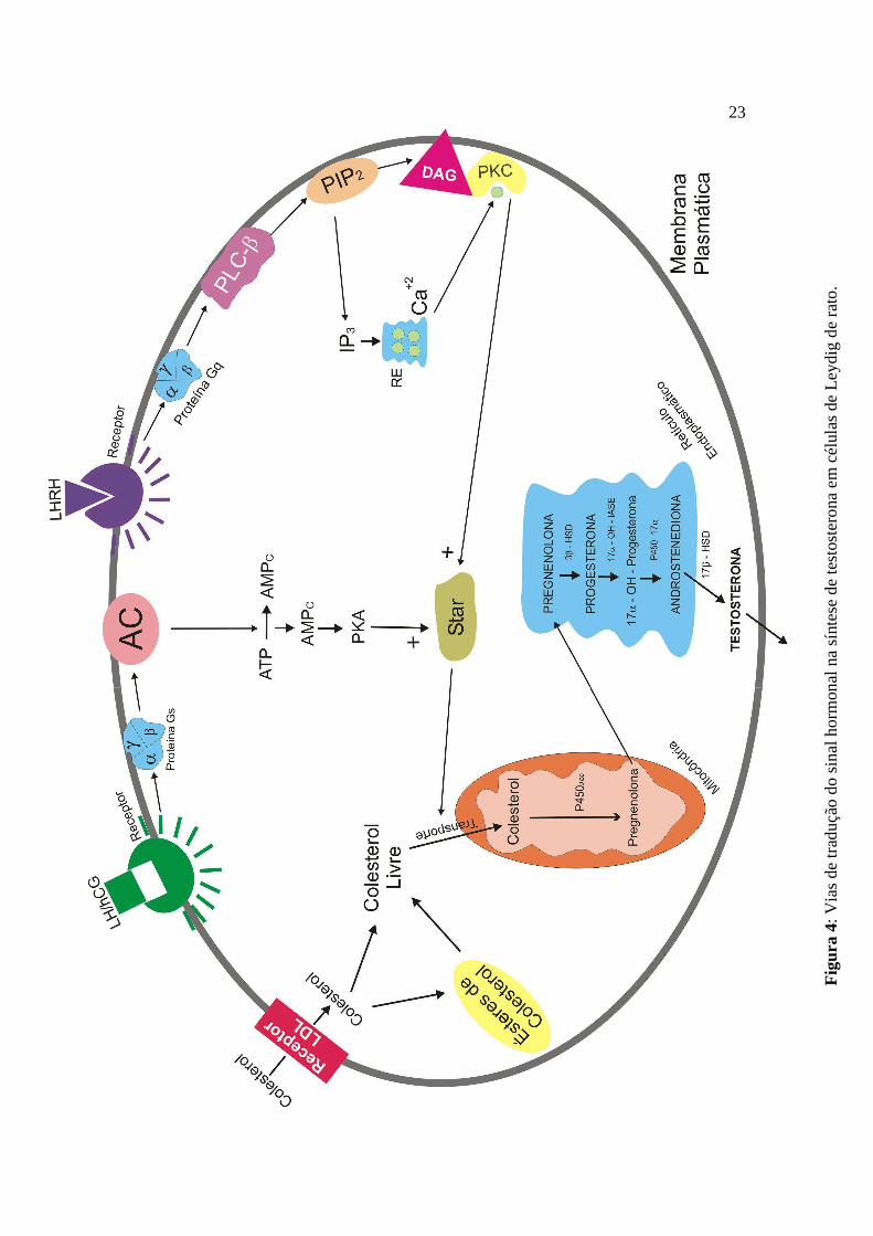
Já se sabe que a PKA e a PKC ativadas, atuam no transporte do colesterol
citoplasmático da camada mais externa da mitocôndria para a mais interna, através da
ativação da proteína reguladora aguda da esteroidogênese (StAR) (Juengel et al., 1995;
Arakane et al., 1997; Arakane et al., 1998; Jo et al., 2005). A principal reação enzimática
na biossíntese de esteróides é a conversão do colesterol livre em pregnenolona, a qual é
catalizada pela enzima de clivagem da cadeia lateral (CYP11A1 ou P450scc). Esta
enzima faz parte do sistema enzimático de clivagem da cadeia lateral do colesterol
(CSCC) e se encontra localizada no lado da matriz da membrana interna da mitocôndria
(Kahnt et al., 1974; Stocco & Kilgore, 1988; Stocco & Clark, 1996). Entretanto, o passo
mais relevante para a realização desta reação, e que controla a esteroidogênese, é o
transporte ou transferência do colesterol livre citoplasmático da membrana externa da
mitocôndria para a membrana mitocondrial interna, onde a P450scc se encontra (Stocco
& Chen, 1991; Stocco & Sodeman, 1991; Stocco & Clark, 1996). Estudos demonstram
que esta transferência é mediada pela ativação da proteína StAR (Clark et al., 1994; Luo
et al., 1998; Stocco, 2000a,b; Manna et al., 2001; Manna et al., 2006), a qual é sintetizada
no citoplasma celular em resposta a ação de hormônios gonadotróficos via AMPc
(Juengel et al., 1995; Sugawara et al., 1997). Após sintetizada, a StAR é importada para a
superfície externa da mitocôndria, onde interage com alguns componentes desta
membrana (proteínas, lipídios e outros fatores), permitindo o transporte facilitado do
colesterol citoplasmático da membrana externa para a membrana interna mitocondrial
(Kallen et al., 1998; Stocco, 2000b). Uma vez dentro da mitocôndria, o colesterol é
convertido à pregnenolona pela ação da P450scc, e esta é transferida para o retículo
endoplasmático liso da célula, onde acontecem os passos enzimáticos adicionais da
esteroidogênese, que levam à síntese da testosterona (Papadopoulos, 1993; Payne &
Hales, 2004; Haider, 2004; Haider, 2007) (Figura 4).
23
Fig
ura
4: V
ias
de
trad
uçã
o d
o s
inal
ho
rmon
al n
a sí
ntes
e d
e te
stos
tero
na
em
cél
ulas
de
Leyd
ig d
e ra
to.
24
Assim, no retículo endoplasmático liso da célula de Leydig, a pregnenolona é
convertida em progesterona pela ação das enzimas ∆5-3β-Hidroxidesidrogenase (3β-
HSD) e ∆5,4-isomerase. A progesterona sofre a ação das enzimas 17α-hidroxilase e C17-20-
Liase (P45017α) para produzir 17-hidroxiprogesterona (17α-OH-progesterona) e depois
androstenediona; e a androstenediona é, então, convertida a testosterona pela ação da
enzima 17β-hidroxidesidrogenase (17β-HSD), concluindo assim a síntese de testosterona
testicular (Luo et al., 1996; Payne & Hales, 2004).
Tendo em vista as considerações e os mecanismos anteriormente citados, é de
interesse estudarmos a ação modulatória direta das catecinas do chá verde sobre as vias
de tradução do sinal envolvidas na esteroidogênese testicular.
25
2. OBJETIVOS
2.1 Objetivo Geral
Investigar os efeitos agudos do extrato aquoso do chá verde (GTE) e seus
constituintes polifenóis (-)-epigalocatecina-3-galato (EGCG) e (-)-epicatecina (EC) sobre
a produção de testosterona basal e estimulada, em células de Leydig de ratos in vitro.
2.2 Objetivos Específicos
• Analisar os efeitos agudos do GTE, da EGCG e da EC sobre a produção de
testosterona em células de Leydig de ratos in vitro, na presença de ativadores
diretos e indiretos da PKA e da PKC.
• Estudar os efeitos agudos do GTE, da EGCG e da EC sobre a função das
enzimas 17β-HSD e P450scc, em células de Leydig de ratos in vitro.
• Verificar a possível reversibilidade dos efeitos do GTE, da EGCG e da EC
sobre a produção de testosterona basal e estimulada, em células de Leydig de
ratos in vitro.
26
3. MATERIAL E MÉTODOS
3.1 Animais
Ratos machos Wistar (Ratus norvegicus albinos) adultos, com aproximadamente
70-80 dias de idade, com pesos entre 200 e 300g foram utilizados nos experimentos. Os
animais foram criados no biotério do Departamento de Fisiologia e Farmacologia da
Universidade Federal de Pernambuco, recebendo alimentação livre (ração Purina para
ratos) e água filtrada ad libitum. Os animais foram mantidos em sala com iluminação
controlada (aproximadamente 12 horas de claro e 12 horas de escuro) e temperatura que
variava entre 25 e 29oC durante todo o ano.
3.2 Reagentes
Albumina sérica bovina (BSA) Fração V obtida de Miles (Naperville, IL); Ácido
tricloroacético (TCA), Álcool etílico P.A., Azida de sódio, Bicarbonato de Sódio
(NaHCO3), Cloreto de potássio (KCl) e Cloreto de sódio (NaCl) obtidos de VETEC; Azul
Tripan, carvão ativado, Colagenase tipo I, Dextran, Ácido etilenoglicol bis (β-
aminoetiléter)-N,N,N’,N’-tetracético (EGTA), Forbol 12,13-dibutirato (PDBu), Gelatina
tipo III, Inibidor da tripsina, Leupeptina, 2,5-diphenyl-oxazole (PPO), Gonadotrofina
coriônica humana (hCG), N6,2’-O-dibutiriladenosina-3’:5’-monofosfato cíclico
(dbcAMP), Androstenediona (4-androstene-3,17-dione), EC, EGCG e 22OHColesterol
obtidos de Sigma Chemical Co. (St. Louis, MO); Meio de cultura 199 (Meio 199) obtido
de GIBCO (Grand Island, NY); 2,2’-p-phenylen-bis (5-phenyloxazol) (POPOP) obtido de
MERCK; Percoll obtido de Pharmacia (Uppsala, Sweden); 1,2,6,7 [3H] Testosterona
obtida de Amersham International (Buckinghamshire, England); Hormônio liberador de
hormônio luteinizante (LHRH) obtido de Península (San Carlos, CA); Cloreto de cálcio
(CaCl2), Fosfato de potássio monobásico (KH2PO4), Fosfato de sódio dibásico
(Na2HPO4.7H2O), Fosfato de sódio monobásico (NaH2PO4.H2O), Glicerol e Tolueno
P.A. obtidos de Reagen, 3-(4,5-dimetiltialzolil)-2,5 difeniltetrazólio (MTT),
Dimetilsufóxido (DMSO) e folhas secas de chá verde obtidas em comércio local.
27
3.3 Obtenção do Extrato Aquoso do Chá Verde (GTE)
As folhas secas de chá verde (5,0g) foram infundidas por 5 minutos em 100mL de
água destilada a uma temperatura de 90ºC. Essa infusão foi misturada durante 30 minutos
por agitação magnética e, em seguida, filtrada; o sobrenadante foi considerado extrato
aquoso do chá verde e foi adicionado, em diferentes diluições, diretamente às suspensões
de células intersticiais testiculares in vitro. Para calcular a concentração do extrato, 10mL
do mesmo foi evaporado em um forno (100oC) durante uma noite e depois pesado
totalizando 69,2mg. Os extratos aquosos do chá verde foram preparados no dia dos
experimentos (Wang et al., 1992).
3.4 Obtenção e purificação das células de Leydig
A obtenção das células intersticiais foi realizada conforme descrito por Hedger e
Eddy (1986) modificado por Wanderley e Negro-Villar (1996). Os animais foram
sacrificados por anestesia com éter, e os testículos foram rapidamente removidos e
cuidadosamente descapsulados. Em seguida, os testículos descapsulados foram incubados
em uma solução enzimática (2mL/testículo) contendo colagenase (0,5mg/mL), inibidor
de tripsina (0,2mg/mL) e leupeptina (5µg/mL), todos dissolvidos em tampão fosfato-
salina (PBS) (136,9 mM NaCl, 2,68 mM KCl, 8,1 mM Na2HPO4.7H2O, 1,47 mM
KH2PO4) contendo albumina sérica bovina (BSA) (1mg/mL). O pH foi ajustado para 7,4
e a incubação durou 20-30 minutos em um banho-maria a 34ºC, sob agitação de 90
ciclos/minuto até ocorrer a dispersão dos túbulos em uma massa homogênea. O tecido
disperso foi imediatamente diluído à 50mL com PBS/BSA para diminuir o efeito da
enzima, os túbulos seminíferos foram sedimentados durante 5 minutos e o sobrenadante
foi filtrado através de malha de nylon (80µm) e lavado com 5mL de PBS/BSA. O filtrado
foi centrifugado em tubos plásticos de 50mL a 150xg, 20ºC, durante 15 minutos. O
sobrenadante foi descartado e as células sedimentadas foram ressuspendidas em 5 mL de
M199/BSA contendo NaHCO3 (2,2 mg/mL) e BSA (1mg/mL), com o auxílio de uma
pipeta plástica até que nenhum agregado celular fosse visível. Essa suspensão foi
cuidadosamente colocada sobre o gradiente de Percoll descontinuo (20%, 35%, 43%,
28
68% e 90%), em seguida, centrifugada a 800xg durante 30 min a 20oC. Os primeiros 8
mL foram aspirados com seringa de plástico (10 mL) a partir do fundo do tubo – fração
contendo as hemácias – e descartados. A fração das células de Leydig (interface 43-68%)
foi obtida através da aspiração dos 8 mL seguintes e lavada duas vezes com M199
contendo 0,1% de BSA e ressuspendidas em M199/0,1%BSA. Em seguida as células
foram contadas utilizando uma câmara de Neubauer. O número de células a serem
incubadas foi determinado previamente em 0,35 x 10⁶ células/0,5mL de meio de
incubação. Os procedimentos utilizados foram aprovados pelo comitê de ética em
experimentação animal da Universidade Federal de Pernambuco, Recife, PE, Brasil.
3.5 Secreção de testosterona in vitro
As preparações de células intersticiais testiculares, enriquecidas ou não em células
de Leydig (0,35 x 10⁶células/0,5mL ) foram incubadas por 3 horas com M199, com
extrato aquoso do chá verde (6,92 – 692,0 µg/mL), EGCG (5-200 µg/mL) ou EC (200
µg/mL), sem (produção basal de testosterona) ou com hCG (1mUI/mL), dbcAMP
(1mM), LHRH (10-7M), PDBu (200nM) ou androstenediona (1-100µM). As incubações
foram realizadas logo após a obtenção da suspensão celular, a 34ºC sob atmosfera de
95% de O2 e 5% de CO2, em banho-maria tipo Dubnoff com agitação de 60 ciclos por
minuto. Para determinarmos a reversibilidade dos efeitos do GTE e da EGCG sobre a
síntese de testosterona, as células também foram pré-incubadas por 15 minutos com
M199, GTE ou EGCG, depois lavadas com M199, recuperadas por 60 minutos e
incubadas novamente por 2hs com hCG (0,5mUI/mL), LHRH (10-7M), androstenediona
(10µM) ou 22OHColesterol (20µM). Após o término das incubações, as células foram
precipitadas por centrifugação (150xg por 15min) a 4ºC e o sobrenadante coletado e
armazenado a -20ºC para posterior determinação da testosterona por radioimunoensaio
(RIA).

29
3.6 Viabilidade Celular
Para avaliar os efeitos do GTE e da EGCG sobre a viabilidade celular, os métodos
do critério de exclusão do corante azul tripan e redução do brometo de 3-(4,5-
dimetiltialzolil)-2,5 difeniltetrazólio (MTT) foram utilizados. O critério de exclusão do
corante azul tripan e a redução do MTT são métodos muito utilizados para mensurar a
integridade da membrana plasmática e a funcionalidade da mitocôndria, respectivamente.
O teste de exclusão pelo corante azul tripan foi realizado após incubação das
células por 3 horas com diferentes doses de EGCG (10, 50 e 100µg/mL) e GTE
(69,2µg/mL). As células foram incubadas com o corante azul tripan (0,5%) por 20
minutos e a percentagem de células azuis, indicando a entrada do corante pela ruptura da
membrana plasmática dessas células, foi contada. Considerou-se normal a viabilidade
celular quando 90-95% das células não coravam. A leitura das células foi realizada na
câmara de Neubauer.
O método do MTT é baseado na redução do corante tetrazólio amarelo e solúvel
em um composto cristalizado azul escuro (púrpura) e insolúvel, denominado formazan.
Apenas células vivas, com a mitocôndria intacta e ativa, são capazes de converter o MTT
em formazan (Mosmann, 1983). Para o teste do MTT, células de Leydig (0,35 x
106células/mL) foram incubadas com GTE (69,2µg/mL) ou EGCG (10, 50 e 100µg/mL)
por 2 horas a 34ºC sob atmosfera de 95% de O2 e 5% de CO2, em banho-maria tipo
Dubnoff com agitação de 60 ciclos por minuto. Após este período de incubação, as
células foram lavadas duas vezes com M199 com o objetivo de remover todo o GTE e a
EGCG e depois foram ressuspendidas em M199 (100µL). Este procedimento evita a
reação direta entre os polifenóis e o MTT, uma vez que já se sabe que estas substâncias
são capazes de reduzir o MTT na ausência de células vivas e os resultados obtidos
poderiam não refletir a real viabilidade celular durante o teste para determinação dos
efeitos dos polifenóis sobre as células (Peng et al., 2005). Assim, 25 µL de MTT em PBS
(5mg/mL) foram adicionados à suspensão de células e seguido por mais 3 horas de
incubação a 37ºC. Após este tempo, as células foram precipitadas por centrifugação
(450xg por 5min). Depois o sobrenadante foi desprezado e 100µl de DMSO foram
adicionados a cada tubo por 12 horas para dissolver os cristais de formazan formados. A
30
leitura da absorbância foi mensurada a 630nm em leitor de ELISA. Múltiplos controles
foram incluídos (tubos sem células e com M199, MTT e DMSO (tubos brancos) e tubos
com células tratadas com o reagente tóxico saponina (0,1%)).
3.7 Radioimunoensaio da Testosterona
A testosterona foi determinada diretamente no meio de incubação (sem extração).
As amostras do meio de incubação foram diluídas e incubadas com anticorpo anti-
testosterona e 10.000 cpm de [3H]-testosterona a 4ºC, durante 16-18 horas, sendo o
volume final da reação de 0,32 mL. O tampão de ensaio utilizado foi o PBS – 0,1%
gelatina. O anticorpo anti-testosterona foi desenvolvido em coelhos e produzido no
laboratório da Dra. Maria Inês Wanderley (UFPE), e sua diluição final no ensaio foi de
1 : 5.000.
Uma mistura de carvão-dextrano na concentração de 0,625% de carvão e 0,0625
de dextrana foi utilizada para remover a testosterona livre. Os tubos foram centrifugados
a 1.300 xg, a 4ºC, durante 20 minutos. O traçador ligado ao anticorpo foi lido em 0,4 mL
do sobrenadante adicionado a frascos de cintilação contendo 5 mL de solução de
cintilação (3 g de PPO + 0,2 g de POPOP + 20 mL de metanol + 980 mL de tolueno). A
leitura foi realizada em um contador de cintilação líquida (Liquid Scintillation Analyzer).
Foram preparados padrões de testosterona de 8, 16, 32, 64, 128, 256 e 512 pg/tubo e
dosados em triplicata. A testosterona das amostras foi dosada em duplicata.
O coeficiente de variação intra-ensaio foi de 8,1% e o coeficiente inter-ensaio foi
de 15,1%.
3.8 Tratamento Estatístico
Os dados experimentais foram expressos como média + o desvio padrão (média +
DP) dos valores obtidos para cada grupo estudado em triplicata e representam os
resultados obtidos em, pelo menos, 2 experimentos similares.
A análise estatística dos dados foi realizada utilizando o teste t de Student para
comparações dos valores basais de cada grupo com os valores dos grupos com as
31
diferentes concentrações do GTE, EGCG e EC. Os valores de p < 0,05 (quando indicado)
foram considerados estatisticamente significativos.
32
REFERÊNCIAS BIBLIOGRÁFICAS
Ascoli M, Segaloff DL. On the structure of the luteinizing hormone/chorionic
gonadotropin receptor. Endocr Rev. 1989; 10: 27-44.
Adhami VM, Ahmad N, Mukthar H. Molecular targets for green tea in prostate câncer
prevention. The journal of nutrition. 2003; 133: 24175-245.
Adom KK & Liu RH. Antioxidant activity of grains. J. Agric. Food Chem. 2002; 50:
6182-7.
Adom KK, Sorrells ME, Liu RH. Phytochemicals and antioxidant activity of wheat
varieties. J. Agric. Food Chem. 2003; 51: 7825-34.
Arakane F, King SR, Du Y, Kallen CB, Walsh LP, Watari H, Stocco DM, Strauss JF 3rd.
Phosphorylation of steroidogenic acute regulatory protein (StAR) modulates its
steroidogenic activity. J Biol Chem. 1997; 272(51): 32656-62.
Arakane F, Kallen CB, Watari H, Foster JA, Sepuri NB, Pain D, Stayrook SE, Lewis M,
Gerton GL, Strauss JF 3rd. The mechanism of action of steroidogenic acute regulatory
protein (StAR). StAR acts on the outside of mitochondria to stimulate steroidogenesis. J
Biol Chem. 1998; 273(26): 16339-45.
Barry RZ, Chen H. Regulation of Leydig Cell Steroidogenic Function during Aging.
Biology of Reproduction. 2000; 63: 977-81.
Benninghoff AD, Thomas P. Gonadotropin regulation of testosterone production by
primary cultured theca and granulose cells of atlantic croaker: I. Novel role of CaMKs
and interactions between calcium- and adenylylciclase-dependent pathways. General and
comparative Endocrinology. 2006; 147: 276-87.

33
Berridge MJ. Inositol triphosphate and diacylglycerol as second messengers. Biochemical
Journal. 1984; 220: 345-60.
Berridge MJ. Inositol triphosphate as a second messenger in signal transduction. Annals
New York Academy of Sciences. 1987; 494: 39-51.
Berridge MJ, Irvine RF. Inositol phosphates and cell signaling. Nature. 1989; 341: 197-
205.
Clinton SK, Giovannucci E. Diet, nutrition and prostate cancer. Ann Rev Nutr. 1998; 18:
413-40.
Chen Y-C, Nagpal ML, Stocco DM, Lin T. Effects of genistein, resveratrol, and
quercetin on steroidogenesis and proliferation of MA-10 mouse Leydig tumor cells.
Journal of Endrocrinology. 2007; 192: 527-37.
Christensen AK. Leydig Cell. In: Greep RO, Astwood EB, Hamilton DW (eds).
Handbook of Physiology. Washington: American Physiological Society, 1975. Sec. 7,
v.5.
Chu YF, Sun J, Wu X, Liu RH. Antioxidant and antiproliferative activities of vegetables.
J. Agric. Food Chem. 2002; 50: 6910-6.
Clark BJ, Wells J, King SR, Stocco DM. The purification, cloning and expression of a
novel luteinizing hormone-induced mitochondrial protein in MA-10 mouse Leydig tumor
cells. The Journal of Biological Chemistry. 1994; 269(45): 28314-22.
Cooke BA, Janszen FHA, Van Driel MJA, Van der Molen HJ. Evidence for the
involvement of lutropin-independent RNA synthesis in Leydig cell steroidogenesis.
Molecular and cellular Endocrinology. 1979; 14: 181-9.
34
Cooke BA, Ashford L, Abayasekara DR, Choi M. The role of chloride ions in the
regulation of steroidogenesis in rat Leydig cells and adrenal cells. J. Steroid Biochem.
Mol. Biol. 1999; 69: 359-65.
Crespy V, Williamson G. A review of the health effects of green tea catechins in in vivo
animals models. The Journal of Nutrition. 2004; 134: 34315-405.
Dehejia A, Nozu K, Catt KJ, Dufau ML. Luteinizing hormone receptors and
gonadotropic activation of purified rat Leydig cells. J Biol Chem. 1982; 257: 13781-86.
De Kretser DM, Risbridger GP, Kerr JB. Basic endocrinology of the testis. In: DeGroot
LJ (ed) Endocrinology. 3.ed. Philadelphia: Saunders, 1995. v.3. p.2307-35.
Dona M, Dell’Aica I, Calabrese F, Benelli R, Morini M, Albini A, Garbisa S. Neutrophil
restraint by green tea: inhibition of inflammation, associated angiogenesis and pulmonary
fibrosis. Journal of Immunology. 2003; 170: 4335-41.
Dohlman HG, Thorner J, Caron MG, Lefkowitz RJ. Model systems for the study of
seven-transmembrane-segment receptors. Annual Review of Biochemistry. 1991; 60:
653-88.
Dufau ML, Catt KJ. Gonadotropin receptors and regulation of steroidogenesis in testis
and ovary. Vitamins and Hormones-Advances in Research and Applications. 1978; 36:
461-600.
Dufau ML, Baukal AJ, Catt KJ. Hormone-induced guanyl nucleotide binding and
activation of adenylate cyclase in the Leydig cell. Proceedings of the National Academy
of Sciences of the United States of America. 1980; 77: 5837-41.
35
Dufau ML, Winters CA, Hattori M, Aquilano D, Baranao JLS, Nozu K, Baukal AJ, Catt
KJ. Hormonal regulation of androgen production by the Leydig cell. Journal of Steroid
Biochemistry and Molecular Biology. 1984; 20: 161-73.
Dufau ML. Endocrine regulation and communication functions of the Leydig cell.
Annual Review of Physiology. 1988; 50: 483-508.
Edelman AM, Blumenthal DK, Krebs EG. Protein serine/threonine kinases. Annual
Review of Biochemistry. 1987; 56: 567-613.
Esposito E, Rotilio D, Di Matteo V, Di Giulio C, Cacchio M, Algeri S. A review of
specific dietary antioxidants and the effects on biochemical mechanisms related to
neurodegenerative processes. Neurobiology of Aging. 2002; 23: 719-35.
Evans JJ. Modulation of gonadotropin levels by peptides acting at the anterior pituitary
gland. Endocr Rev. 1999; 20: 46-67.
Fawcett DW, Neaves WB, Flores MN. Comparative observations on intertubular
lymphatics and the organization of the intersticial tissue of the mammalian testis. Biol
Reprod. 1973; 9: 500-32.
Galdieri M, Caporale C, Adam S. Calcium-dependent, phospholipid-dependent protein-
kinase activity of cultured rat Sertoli cells and its modification by vitamin-A. Molecular
and Cellular Endocrinology. 1986; 48: 213-20.
Gershengorn MC, Perlman JH. Second messenger signaling pathways: phosphatidyl
inositol and calcium. In: DeGroot LJ (ed) Endocrinology. 3.ed. Philadelphia: Saunders,
1995. v. 1. p.66-76.
Graham HN. Green tea composition and polyphenol chemistry. Prev Med. 1992; 21: 334-
50.
36
Gore-Langton RE, Armstrong DT. Follicular Steroidogenesis and its control. In: Knobil
E, Neill J (eds) The Physiology of Reproduction. Raven Press: New York. 1988. p.331-
385.
Gouni-Berthold L, Sachinidis A. Molecular mechanisms explaining the preventive effects
of catechins on the development of proliferative diseases. Current Pharmaceutical
Design. 2004; 10: 1261-71.
Gupta S, Hussain T, Mukhtar H. Molecular pathway for (-)-epigallocatechin-3-gallate
induced cell cycle arrest and apoptosis of human prostate carcinoma cells. Archives of
Biochemistry and Biophysics. 2003; 410: 177-85.
Haider SG. Cell biology of Leydig cells in the testis. Int Rev Cytol. 2004; 233: 181-241.
Haider SG. Leydig Cell Steroidogenesis: Unmasking the functional importance of
mitochondria. Endocrinology. 2007; 148 (6): 2581-82.
Haqqi TM, Anthiny DD, Gupta S, Ahmad N, Lee MS, Kumar GK, Mukthar H.
Prevention of collagen-induced arthritis in mice by a polyphenolic fraction from green
tea. Proc Natl Acad Sci USA. 1999; 96: 4524-29.
Hannum YA, Bell RM. Phorbol ester binding and activation of protein kinase C on triton
X-100 mixed micelles containing phosphatidylserine. Journal of Biological Chemistry.
1986; 261: 9341-47.
Hedger MP, Eddy EM. Monoclonal antibodies against rat Leydig cell surface antigens.
Biol Reprod. 1986; 35: 1309-19.
Jo Y, King SR, Khan SA, Stocco DM. Involvement of protein kinase C and cyclic
adenosine 3’,5’-monophosphate- dependent kinase in steroidogenic acute regulatory
37
protein expression and steroid biosynthesis in Leydig cells. Biology of Reproduction.
2005; 73: 244-55.
Juengel JL, Meberg BM, Turzillo AM, Nett TM, Niswender GD. Hormonal regulation of
messenger ribonucleic acid encoding steroidogenic acute regulatory protein in ovine
corpora lutea. Endocrinology. 1995; 136(12): 5423-29.
Kahnt FW, Milani A, Steffen H, Nehler R. The rate-limiting step of adrenal
steroidogenesis and adenosine 3’:5’-monophosphate. Eur J Biochem. 1974; 44: 243-50.
Kallen CB, Billheimer JT, Summers SA, Stayrook SE, Lewis M, Strauss JF 3rd.
Steroidogenic acute regulatory protein (StAR) is a sterol transfer protein. J Biol Chem.
1998; 273(41): 26285-88.
Kao Y-H, Hiipakka RH, Liao S. Modulation of endocrine system and food intake by
green tea epigallocatechin gallate. Endocrinology. 2000; 141: 980-87.
Kaziro Y, Itoh H, Kozasa T, Nakafuku M, Satoh T. Structure and function of signal-
transducing GTP-binding proteins. Annual Review of Biochemistry. 1991; 60: 349-400.
Katiyar SK, Ahmad N, Mukhtar H. Green tea and skin. Arch Dermatol. 2000; 136(8):
989-94.
Kimura K, Katoh N, Sakurada K, Kubo S. Phospholipid-sensitive Ca+2-dependent
protein-kinase system in testis – localization and endogenous substrates. Endocrinology.
1984; 115(6): 2391-99.
Lee S, Miselis R, Rivier C. Anatomical and Functional Evidence for a neural
hypothalamic-testicular pathway that is independent of the pituitary. Endocrinology.
2002; 143 (11): 4447-54.

38
Leung PCK, Steele GL. Intracellular signaling in the gonads. Endocr. Rev. 1992; 13:
476-98.
Levites Y, Amit T, Mandel S, Youdim MBH. Neuroprotection and neurorescue against
Abeta toxicity and PKC-dependent release of nonamyloidogenic soluble precursor
protein by green tea polyphenol (-)-epigallocatechin-3-gallate. FASEB J. 2003; 17: 952-
54.
Liao S, Hiipakka RA. Selective inhibition of steroid 5α-reductase isozymes by tea
epicatechin-3-gallate and epigallocatechin-3-gallate. Biochem Biophys Res Commun.
1995; 214: 833-38.
Liao, S, Umekita Y, Guo J, Kokontis JM, Hiipakka RA. Growth inhibition and regression
of human prostate and breast tumors in athymic mice by tea epigallocatechin gallate.
Cancer Lett. 1995; 96: 239-43.
Liu JP. Protein kinase C and its substrates. Molecular and Cellular Endocrinology. 1996;
116: 1-29.
Liu, RH. Health benefits of fruits and vegetables are from additive and synergistic
combination of phytochemicals. Am. J. Clin. Nutr 2003; 78: 517-520.
Liu, RH. Potential synergy of phytochemicals in cancer prevention: mechanism of action.
J. Nutr. 2004; 134: 3479-85.
Lorenz M, Wessle S, Follmann E, Michaelism W, Dusterhoft T, Baumann G, Stangl K,
Stangel V. A constituent of green tea, epigallocatechin-3-gallate, activates endotelial
nitric oxide synthase by a phosphatidylinosiyol-3-OH-kinase-, cAMP-dependent protein
kinase-, and Akt-dependent pathway and leads to endothelial-dependent vasorelaxation. J
Biol Chem. 2004; 279: 6190-95.
39
Luo L, Chen H, Stocco DM, Zirkin BR. Leydig cell protein synthesis and steroidogenesis
in response to acute stimulation by luteinizing hormone in rats. Biology of Reproduction.
1998; 59: 263-70.
Luo L, Chen H, Zirkin B. Are Leydig cell steroidogenic enzymes differentially regulated
with aging? Journal of Andrology. 1996; 17(5): 509-15.
Makimura M, Hirasawa M, Kobayashi K, Indo J, Sakanaka S, Taguchi T, Otake S.
Inhibitory effect of tea catechins on collagenase activity. J Periodotol. 1993; 64: 630-36.
Mandel S, Weineb O, Amit T, Youdim MB. Cell signaling pathways in the
neuroprotective actions of the green tea polyphenol (-)-epigallocatechin-3-gallate:
implications for neurodegenerative diseases. Journal of Neurochemistry. 2004; 88: 1555-
69.
Mandel S, Youdim MBH. Catechin polyphenols: neurodegeneration and neuroprotection
in neurodegenerative diseases. Free Radical Biology & Medicine. 2004; 36: 304-17.
Manna PR, Kero J, Tena-Sempere M, Pakarinen P, Stocco DM, Huhtaniemi IT.
Assessment of mechanisms of thyroid hormone action in mouse Leydig cells: Regulation
of the Steroidogenic acute regulatory protein, steroidogenesis, and luteinizing hormone
receptor function. Endocrinology. 2001; 142(1): 319-31.
Manna PR, Chandrala SP, Jo Y, Stocco DM. cAMP-independent signaling regulates
steroidogenesis in mouse Leydig cells in the absence of StAR phosphorylation. Journal of
Molecular Endocrinology. 2006; 37: 81-95.
Marsh JM. The role of cyclic AMP in gonadal function. Advances in Cyclic Nucleotide
Research. 1975; 6: 137-99.
40
Matsubara S, Rodriguez-Amaya DB. Conteúdo de Miricetina, Quercetina e Kaempferol
em chás comercializados no Brasil. Ciênc. Tecnol. Aliment. 2006; 26 (2): 380-85.
Mosmann, T. Rapid colorimetric assay for cellular growth and survival: application to
proliferation and cytotoxicity assays. J. Immunol. Meth. 1983; 65: 55-63.
Mukhtar H, Ahmad N. Tea polyphenols: prevention of cancer and optimizing health. Am
J Clin Nutr. 2000; 71(suppl): 1698-702.
Nance CL, Shearer WT. Is green tea good for HIV-1 infection? Journal of Allergy and
Clinical Immunology. 2003; 112: 851-53.
Nagle DG, Ferreira D, Zhou Yu-D. Epigallocatechin-3-gallate (EGCG): Chemical and
biomedical perspectives. Phytochemistry. 2006; 67: 1849-55.
Nikula H, Naor Z, Parvinen M, Huhtaniemi I. Distribution and activation of protein
kinase C in the rat testis tissue. Molecular and Cellular Endocrinology. 1987; 49: 39-49.
Nikula H, Huhtaniemi I. Gonadotropin-release hormone agonist activates protein kinase
C in rat Leydig cells. Molecular and Cellular Endocrinology. 1988; 55: 53-9.
Nishizuka Y. The role of protein kinase C in cell surface signal transduction and tumour
promotion. Nature. 1984; 308: 693-8.
Odell DW. The Leydig Cell. In: DeGroot LJ (ed) Endocrinology. 2.ed. Philadelphia:
Saunders, 1988. v.3, cap.129, p.2137-45.
Ohno S, Shinoda S, Toyoshima S, Nakazawa H, Makino T, Nakajin S. Effects of
flavonoid phytochemicals on cortisol production and on activities of steroidogenic
enzymes in human adrenocortical H295R cells. J Steroid Biochem Mol Biol. 2002; 80
(3): 355-63.

41
Parkin DM. Global cancer statistics in the year 2000. Lancet Oncol. 2001; 2: 533-43.
Papadopoulos, V. Peripheral-type benzodiazepine/diazepam binding inhibitor receptor:
Biological role in steroidogenic cell function. Endocr Rev. 1993; 14: 222-40.
Payne AH, Hales DB. Overview of steroidogenic enzymes in the pathway from
cholesterol to active steroid hormones. Endocr Rev. 2004; 25: 947-70.
Peng L, Wang B, Ren P. Reduction of MTT by flavonoids in the absence of cells.
Colloids Surf B Biointerfaces. 2005; 45: 108-111.
Rhee SG, Choi KD. Regulation of inositol phospholipid-specific phospholipase C
isozymes. Journal of Biological Chemistry. 1992; 267: 12393-96.
Rice-Evans CA, Miller NJ, Bolwell PG, Bramley PM, Pridham JB. The relative
antioxidant activities of plant-derived polyphenolic flavonoids. Free Radical Research.
1995; 22(4): 375-83.
Ripple MO, Henry WF, Rago RP, Wilding G. Prooxidant-antioxidant shift induced by
androgen treatment of human prostate carcinoma cells. J Natl Cancer Inst. 1997; 89: 40-
8.
Rommerts FFG, Cooke BA, Van der Molen HJ. The role of cyclic-AMP in regulation of
steroid biosynthesis in testis tissue. Journal of Steroid Biochemistry and Molecular
Biology. 1974; 5: 279-85.
Saez JM. Leydig Cells: Endocrine, Paracrine, and Autocrine Regulation. Endocrine
Reviews. 1994; 15(5): 574-626.
42
Satoh K, Sakamoto Y, Ogata A, Nagai F, Mikuriya H, Numazawa M, Yamada K, Aoki
N. Inhibition of aromatase activity by green tea extract catechins and their
endocrinological effects of oral administration in rats. Food Chem Toxicol. 2002; 40:
925-33.
Setchell BP, Brooks DE. Anatomy, vasculature, innervation and fluids of the male
reproductive tract. In: Knobil E, Neill J. The Physiology of Reproduction. New York:
Raven Press, 1988. v.1. p.753-836.
Stapleton PD, Shah S, Anderson JC, Hara Y, Hamilton-Miller JMT, Taylor PW.
Modulation of β-lactam resistance in Staphylococcus aureus by catechins and gallates.
Int J Antimicrob. Agents. 2004; 23(5): 462-7.
Steele GL, Leung PCK. Intragonadal signaling mechanisms in the control of steroid
hormone production. Journal of Steroid Biochemistry and Molecular Biology. 1992;
41(3-8): 515-22.
Steinschneider A, McLean MP, Billheimer JT, Azhar S, Gibori G. Protein kinase C-
Catalyzed phosphorylation of sterol carrier protein 2. Endocrinology. 1989; 125: 569-71.
Stocco DM, Kilgore MW. Induction of mitochondrial proteins in MA-10 Leydig tumour
cells with human choriogonadotropin. Biochem J. 1988; 249: 95-103.
Stocco DM, Clark BJ. The requirement of phosphorylation on a threonine residue in the
acute regulation of steroidogenesis in MA-10 mouse Leydig tumor cells. Journal of
Steroid Biochemistry and Molecular Biology. 1991; 46: 337-47.
Stocco DM, Chen W. Presence of identical mitochondrial proteins in unstimulated
constitutive steroid-producing R2C rat Leydig tumor and stimulated nonconstitutive
steroid-producing MA-10 mouse Leydig tumor cells. Endocrinology. 1991; 128: 1918-
26.
43
Stocco DM, Sodeman TC. The 30-kDa mitochondrial proteins induced by hormone
stimulation in MA-10 mouse Leydig tumor cells are processed from larger precursors. J
Biol Chem. 1991; 266(29): 19731-38.
Stocco DM, Clark BJ. Regulation of the acute production of steroids in steroidogenic
cells. Endocr Rev. 1996; 17: 221-44.
Stocco DM. Intramitochondrial cholesterol transfer. Biochim Biophys Acta. 2000a; 1486:
184-97.
Stocco DM. The role of the StAR protein in steroidogenesis: challenges for the future.
Journal of Endocrinology. 2000b; 164: 247-53.
Sugawara T, Kiriakidou M, McAllister JM, Holt JA, Arakane F, Strauss JF 3rd.
Regulation of expression of the steroidogenic acute regulatory protein (StAR) gene: a
central role for steroidogenic factor 1. Steroids. 1997; 62(4): 395-9.
Sun J, Chu YF, Wu X, Liu RH. Antioxidant and proliferative activities of fruits. J. Agric.
Food Chem. 2002; 50: 7449-54.
Svechnikov K, Supornsilchai V, Strand M-L, Wahlgren A, Seidlova-Wuttke D, Wuttke
W, Soder O. Influence of long-term dietary administration of procymidone, a fungicide
with anti-androgenic effects, or the phytoestrogen genistein to rats on the pituitary-
gonadal axis and Leydig cell steroidogenesis. Journal of Endocrinology. 2005; 187: 117-
124.
Vinson JA, Hao Y, Su X, Zubik L, Bose P. Phenol antioxidant quantity and quality in
foods : fruits. J. Agric. Food Chem. 2001; 49: 5315-21.
44
Wanderley MI, Negro-Vilar A. Pretreatment with phorbol ester and LHRH agonist
reduces testosterone production and protein kinase C activity in rat Leydig cells
challenged with PDBu and LHRH. Braz J Med Biol Res. 1996; 29: 1557-65.
Wang H, Cao GH, Prior RL. Total antioxidant capacity of fruits. J. Agric. Food Chem.
1996; 44: 701-5.
Wang ZY, Huang MT, Ferrara T, Wong CQ, Lou YR, Reuhl K, Latropoulos M, Yang
CS, Conney AH. Inhibition effect of green tea in the drinking water on tumorigenesis by
ultraviolet light and 12-O-tetradecanoylphorbol-13-acetate in the skin of SKH-1 mice.
Cancer Res. 1992; 52: 1162-70.
Weber KS, Setchell KDR, Stocco DM, Lephart ED. Dietary soy-phytoestrogens decrease
testosterone levels and prostate weight without altering LH, prostate 5α-reductase or
testicular steroidogenic acute regulatory peptide levels in adult male Sprague-Dawley
rats. Journal of Endocrinology. 2001; 170: 591-9.
Williams RJ, Spencer JPE, Rive-Evans C. Flavonoids: antioxidants or signaling
molecules? Free Radical & Medicine. 2004; 36: 838-49.
Wu L-Y, Juan C-C, Ho L-T, Hsu Y-P, Yung-Pei H, Hwang LS. Effect of green tea
supplementation on insulin sensitivity in Sprague-Dawley rats. J. Agric. Food Chem.
2004; 52: 643-8.
Yang CS, Landau JM. Effects of tea consumption on nutrition and health. J. Nutr. 2000;
130: 2409-12.
Yang CS, Wang ZY. Tea and Cancer. J. Natl. Cancer Inst. 1993; 85: 1038-1049.
Zaveri NT. Green tea and its polyphenolic catechins: Medicinal uses in cancer and
noncancer applications. Life Sciences 2006; 78: 2073-80.

45
Zhang K, Das NP. Inhibitory effects of plant polyphenols on rat liver glutathione S-
transferases. Biochem Pharmacol 1994; 47: 2063-8.
Zhang K, Yang EB, Tang WY, Wong KP, Mack P. Inhibition of glutathione reductase by
plant polyphenols. Biochem Pharmacol. 1997; 54 (9): 1047-53.
46
Green tea polyphenols affect testosterone production in rat Leydig cells
Marina S. Figueiroa1, Juliany S. B. César Vieira1, Disleide S. Leite1, Ruben C. O.
Andrade Filho1, Patrícia S. Gouveia1, Daniel P. Udrisar1 and Maria I. Wanderley1.
1 Departamento de Fisiologia e Farmacologia, Universidade Federal de Pernambuco,
50670-901 Recife, PE, Brasil.
Correspondence: Maria Inês Wanderley
Departamento de Fisiologia e Farmacologia
Universidade Federal de Pernambuco
Cidade Universitária
50670-901 Recife, PE, Brasil
Telephone: +55-81-2126-8530
Fax: +55-81-2126-8976
E-mail: [email protected]
Key words: Green tea polyphenols; Testosterone; Leydig cells; PKA; PKC
Running title: Green tea polyphenols inhibit testosterone in vitro
Research Article
47
Abstract
This study investigated the acute effects of green tea extract (GTE) and its polyphenol
constituents (-)-epigallocatechin-3-gallate (EGCG) and (-)-epicatechin (EC) on basal and
stimulated testosterone production by rat Leydig cells in vitro. Purified Leydig cells were
incubated for 3 h with GTE, EGCG or EC and the testosterone precursor
androstenedione, in the presence or absence of either protein kinase A (PKA) or protein
kinase C (PKC) activators. GTE and EGCG, but not EC, inhibited both basal and kinase-
stimulated testosterone production. Cells pretreated for 15 min with GTE or EGCG and
allowed to recover for 1 hr were challenged with human chorionic gonadotropin (hCG),
luteinizing hormone releasing hormone (LHRH), 22(R)-hydroxycholesterol or
androstenedione. Under these conditions the inhibitory effect of GTE/EGCG at the
higher concentration used (69.2 and 100 µg/mL, respectively) on hCG/LHRH-stimulated
or 22(R)-hydroxycholesterol-induced testosterone production was maintained whereas
androstenedione-supported testosterone production returned to control levels, indicating
that the inhibitory effect on 17β-hydroxysteroid dehydrogenase (17β-HSD) function was
reversible. At the same pretreatment conditions but using lower concentration of
GTE/EGCG (13.8 and 20 µg/mL, respectively) the inhibitory effect of these polyphenols
on 22(R)-hydroxycholesterol-supported testosterone production was reverted to and even
exceeded the control levels, indicating that the inhibitory effect of polyphenols on the
mitochondrial P450 side-chain cleavage enzyme (P450scc) function was reversible. We
conclude that the inhibitory effects of GTE may be explained, at least in part, by its
principal component EGCG and that the presence of a gallate group in its structure seems
important to its high efficacy in inhibiting testosterone production. The mechanisms

48
underlying the effects of GTE and EGCG are probably diverse and involve the inhibition
of the PKA/PKC signaling pathways, as well as the inhibition of the regulation of
P450scc and 17β-HSD function.
Introduction
Green tea (Camellia sinensis) is one of the most commonly consumed beverages
worldwide. Its active components are reported to have several biological properties,
including cancer chemoprevention, inhibition of the growth of tumor cells and antiviral
and anti-inflammatory activities (Yang et al. 2000), antioxidant activity (Morel et al.
1993; Guo et al. 1996), and inhibitory effects on several enzymes, such as aromatase
(Satoh et al. 2002; Goodin and Rosegren 2003), angiotensin converting enzyme (Actis-
Goretta et al. 2006) and thyroid peroxidase (Divi and Doerge 1996). Dried leaves of
Camellia sinensis contain polyphenols (30-36 %), principally flavanols, more commonly
known as catechins (Ahmad and Mukhtar 1999). The predominant catechins are
epigallocatechin-3-gallate (EGCG), epicatechin-3 gallate (ECG), epigallocatechin (EGC)
and epicatechin (EC).
The effects of catechins on the male reproductive system have been described.
Epidemiological and laboratory studies suggest an association between diet and
androgens that can alter prostate cancer risk (Ripple et al. 1997; Clinton and Giovannucci
1998; Parkin 2001; Wang et al. 2003). It has been shown that parenteral injection of
EGCG can suppress human prostate and breast tumor growth in athymic mice (Liao et al.
49
1995) and reduce the weight of testis and accessory reproductive organs, as well the
circulating level of LH and testosterone in intact rat (Kao et al. 2000). Although the anti-
gonadotropic effect of catechins is explained as a secondary effect of EGCG on food
intake (Kao et al. 2000) or on aromatase activity (Satoh et al. 2002; Godin and Rosegren
2003), a modulatory function could be present even at the gonadal level.
Currently there is no evidence for a direct effect of green tea catechins on the testicular
steroidogenesis or on the enzymes involved in androgen production. Although the
involvement of the protein kinase A (PKA) and protein kinase C (PKC) signalling
pathways on the testicular androgen production is well known (Dehejia et al. 1982;
Wanderley and Negro-Vilar 1996), it is not known whether green tea catechins modulate
these pathways in Leydig cells. There is evidence that EGCG and others flavonoids can
modulate the PKC (Lin 2002; Levites et al. 2003) or PKA signaling pathway in other
animal models (Lin 2002; Lorenz et al. 2004). The aim of the present study was to
investigate the direct in vitro effect of GTE and its purified catechins on the basal and the
PKA- or PKC-stimulated testosterone production by rat Leydig cells.
Material and Methods
Material Hank’s balanced salt solution (HBSS) and Medium 199 were obtained from Gibco
(Grand Island, NY). Collagenase (Type I), soybean trypsin inhibitor, leupeptin, phorbol
12,13-dibutyrate (PDBu), human chorionic gonadotropin (hCG), N6,2’-O-
50
dibutyryladenosine3’:5’-cyclic monophosphate (dbcAMP), EC, EGCG from green tea, 3-
(4,5-dimethylthiazol-2-yl)-2,5-diphenyltetrazolium bromide (MTT), dimethylsufoxide
(DMSO), 22(R)-hydroxycholesterol and 4-androstene-3,17-dione were obtained from
Sigma Chemical Co. (St. Louis, MO). Bovine serum albumin (BSA, fraction V) was
obtained from Miles (Naperville, IL). Luteinizing hormone releasing hormone (LHRH)
was purchased from Peninsula (San Carlos, CA). Percoll was purchased from Pharmacia
(Uppsala, Sweden).
Green tea extract (GTE) preparation
Green tea extract was prepared according to Wang et al. (1992) with slight
modifications. Five grams of dry green tea leaves were infused for 5 min in 100 ml of
boiling saline water (90ºC), allowed to cool to room temperature and then filtered. The
resulting clear solution is similar to tea brews consumed by humans. The amount of solid
matter present in the 5 % green tea extract was determined to be 6.92 mg per ml by
drying samples in an oven (overnight at 100 ºC) and weighing the dry residue. The
extracts were prepared fresh on a daily basis. This preparation gave approximately 27 %
of catechins, 8.0 % of caffeine and 0.4 % of theobromine. EGCG represent ≥ 50% of total
catechins (Wang et al.1992).
Animals and Leydig cell-enriched preparation Adult (70-80 days old) male Wistar rats weighing 200-300g were kept in a
controlled environment (temperature, 25-29ºC; lights on from 5:00 to 18:00 h) with free
access to standard laboratory chow and tap water. Isolation and purification of rat Leydig
cell-enriched preparations were performed as described by Hedger and Eddy (1986). The
51
rats were killed by ether anesthesia and the testes were quickly removed and
decapsulated. The decapsulated testes were incubated in an enzyme solution of 0.5 mg/ml
collagenase, 0.2 mg/ml soybean trypsin inhibitor and 5 µg/ml leupeptin in Hank’s
balanced salt containing 0.1% BSA (HBSS/BSA), pH 7.4, in a shaking water bath (20
min, 90 Hz, 34ºC). The dispersed testes were suspended in 50 mL (final volume)
HBSS/BSA and the dissociated tubules allowed to settle (5 min). The supernatant was
filtered and washed with 5 mL HBSS/BSA. The filtered cell suspension was centrifuged
(150 x g, 15 min, 20 ºC), and the pellet was re-suspended in 5 mL HBSS/BSA and loaded
onto the top of a discontinuous Percoll density gradient (20%, 35%, 43%, 68% and 90%)
and centrifuged at 800 x g for 30 min at 20 ºC.. Cells in the 43-68% interface (specific
gravity: 1.0640-1.0960 g/mL) were collected, washed twice with M199 containing 0.1%
BSA, and were re-suspended in M199/0.1% BSA and used immediately for the
experiments.
Cell viability
To assess the effects of GTE and EGCG on cell viability the trypan blue exclusion
and the MTT reduction assays were used. The trypan blue exclusion and the MTT
reduction assays are widely used screening methods to measure plasma membrane
integrity and an active mitochondrial function respectively. The trypan blue assay was
performed after the period of 3 h incubation of cells with the different doses of GTE or
EGCG. Cells were incubated with trypan blue (0,5%) for 20 min and the percentage of
blue cells, indicating a capture of the colorant due to plasma membrane rupture, were
counted. 90-95% of no colored cells was considered normal cell viability. The MTT
assay is based on the reduction of a soluble pale yellow tetrazolium dye to water-
52
insoluble purple formazan crystals in living cells (Mossman, 1983). Cells (0.3 x 10⁶/mL)
were incubated with GTE ( 69,2 µg/mL) or EGCG ( 10, 50 and 100 µg/mL) for 2 h at 34
ºC under 95 % O2 and 5 % CO2 . After this time, the cells were washed twice with M199
and incubated for 1 h with M199 in order to remove the GTE/EGCG and then the cells
re-suspended in fresh 100µL M199. This procedure avoid a direct reaction between the
polyphenols and the MTT. It is known that polyphenols can reduce MTT without living
cells, so that the results obtained may not reflect the true cell viability to determine the
effect of polyphenols (Peng et al., 2005). Then, 25 µL of MTT in PBS (5 mg/mL) was
added to the cell suspensions followed by 3 h of incubation at 37 ºC. After that, the cell
suspensions were centrifuged at 400 x g for 5 min. The supernatant was removed, and
100 µL DMSO were added to each tube to dissolve formazan crystals overnight.
Absorbance was measured at 630 nm in an ELISA reader. Multiple controls were
included (blank tubes (with no cells) into which medium, MTT dye, and DMSO were
added or cells treated with the toxic reagent saponin (0.1 %)). The animal care committee
of the Federal University of Pernambuco, Recife, PE, Brazil, approved all treatments.
In vitro testosterone secretion
Cells (0.3 ×10⁶ cells/0.5 ml) were treated (incubated) for 3 h with M199, green tea
extract (6.92 - 692.0 µg/ml), EGCG (5-200 µg/ml) or EC (200 µg/ml) in the absence or
presence of hCG (1 mIU/ml), dbcAMP (1 mM), LHRH (10-7 M), PDBu (200 nM) or
androstenedione (1-100 µM) in a shaking water bath (60 Hz, 34ºC) under an atmosphere
of 95% O2 and 5% CO2. It is noteworthy that a cup of green tea extract (100 mL) usually
contains 50-150 mg/mL of tea polyphenols (Sakanaka and Okada, 2004). To determine
reversibility of the inhibitory effect of GTE and EGCG, the cells were preincubated for
53
15 min with M199 GTE or EGCG, washed with fresh medium, allowed to recuperate for
60 min, and incubated again for 2 h with 0.5 mUI/ml hCG, 10-7 M LHRH, 20 µM 22-
hydroxycholesterol or 10 µM androstenedione. This 15 min preincubation period was
chosen because it was enough to detect the inhibitory effect of GTE or EGCG on
testosterone production as that seen in after 3 h incubation (data not shown). At the end
of incubation the cells were centrifuged and the supernatant was collected and stored at –
20ºC until the time for testosterone measurement by RIA.
RIA and statistical analysis
Testosterone was measured directly (without extraction) in the incubation medium
by a charcoal-dextran RIA (Niswender et al. 1975) that employs [3H]-testosterone as
tracer and a primary antiserum raised in rabbits in our laboratory against testosterone-3-
(0-carboxymethyl)oxime:BSA. Intra- and inter-assay coefficients of variation were 8.1 %
and 15.1 %, respectively. The testosterone antibody showed <0.1% cross-reactivity with
androstenedione, dehydroepiandrosterone, androsterone, 17α-hydroxyprogesterone, β-
estradiol or estrone. None of the substances tested interfered with the assays. The data
from the different analyses were reported as the mean ± SEM of triplicate determinations
and were representative of results obtained in at least two similar experiments. The data
were analyzed statistically using the Student t test. Values of p<0.05 were considered to
be statistically significant.

54
Results
GTE and EGCG inhibit basal and hCG-stimulated testosterone production
The modulatory effect of GTE and its pure constituents (EGCG and EC) was
tested by incubating Leydig cell-enriched preparations with GTE or EGCG followed by
determination of testosterone levels in the incubation medium. Cells were incubated with
various concentrations (three different orders of magnitude) of GTE or EGCG. In
Figures 1 and 2, Leydig cells were treated with and without GTE or the catechins EGCG
and EC, respectively, in the absence and presence of maximum stimulation by hCG. The
extract and the EGCG produced an inhibitory effect on basal and stimulated testosterone
production. At 6.92, 69.2 and 692 µg/ml, GTE inhibited basal testosterone levels by 27.5
± 4.8%, 60.5 ± 3.9% and 93.1 ± 0.2% respectively, while EGCG at 5, 50 and 200 µg/ml
produced 24.0 ± 6.7%, 37.9 ± 1.4% and 58.2 ± 1.4% inhibition, respectively. The
inhibitory effect of 6.92, 69.2 and 692 µg/ml GTE on hCG-stimulated testosterone
production were 11.2 ± 6.0%, 56.8 ± 1.5% and 99.4 ± 0.03% respectively, while the
inhibitory effect of 50 and 200 µg/ml EGCG upon stimulation with hCG was 46.5 ± 7.8%
and 98.8% ± 0.06, respectively. Exposure to EC at 200 µg/ml showed no significant
inhibitory effect on basal or stimulated testosterone production. In order to evaluate
whether the effect of GTE or EGCG on testosterone production was due to a decrease in
cell viability, the trypan blue exclusion and the MTT reduction assays were used. The
trypan blue exclusion test for cell viability demonstrated that inhibition was not due to the
toxicity of green tea extract per se at the concentrations of 6.92 µg/ml or 69.2 µg/ml or
due to the toxicity of EGCG per se at any concentration used. This test showed that at the

55
above cited concentrations, the percentages of integral cells obtained after 3 h of
incubation were comparable between the green tea extract- or EGCG-treated (91%) cells
and the controls (95%). At the maximum concentration of GTE (692 µg/ml), cell viability
was 80% of control; this difference (10-15 % less viable cells compared to the control
group) could have contributed for the observed decrease in testosterone production. For
this reason this concentration wasn’t used in further experiments in the present study.
When measured by MTT reduction, cell viability was not modified by any concentration
of GTE or EGCG used. The two assays for cell viability demonstrated that inhibition of
testosterone production was not due to the toxicity of GTE (6.92 µg/ml or 69.2 µg/ml) or
EGCG (10-100 µg/mL) per se.
GTE and EGCG inhibit testosterone production elicited by activation of PKA and PKC
The action of GTE and EGCG was further investigated in the presence of a direct
PKA activator, dbcAMP, and of both an indirect and direct PKC activator, LHRH and
PDBu, respectively. Table 1 shows testosterone production in the presence of each
maximum stimulus alone or with GTE or EGCG. Testosterone production was reduced
completely in all groups (to < 98 % stimulated-respective basal), regardless of the nature
of the stimulus.
EGCG inhibits the stimulatory effect of androstenedione on testosterone production
To further evaluate the effect of EGCG, the steroidogenic precursor
androstenedione was used to support testosterone production (a measure of 17β-HSD
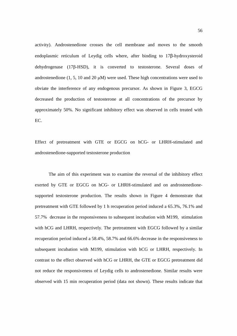
56
activity). Androstenedione crosses the cell membrane and moves to the smooth
endoplasmic reticulum of Leydig cells where, after binding to 17β-hydroxysteroid
dehydrogenase (17β-HSD), it is converted to testosterone. Several doses of
androstenedione (1, 5, 10 and 20 µM) were used. These high concentrations were used to
obviate the interference of any endogenous precursor. As shown in Figure 3, EGCG
decreased the production of testosterone at all concentrations of the precursor by
approximately 50%. No significant inhibitory effect was observed in cells treated with
EC.
Effect of pretreatment with GTE or EGCG on hCG- or LHRH-stimulated and
androstenedione-supported testosterone production
The aim of this experiment was to examine the reversal of the inhibitory effect
exerted by GTE or EGCG on hCG- or LHRH-stimulated and on androstenedione-
supported testosterone production. The results shown in Figure 4 demonstrate that
pretreatment with GTE followed by 1 h recuperation period induced a 65.3%, 76.1% and
57.7% decrease in the responsiveness to subsequent incubation with M199, stimulation
with hCG and LHRH, respectively. The pretreatment with EGCG followed by a similar
recuperation period induced a 58.4%, 58.7% and 66.6% decrease in the responsiveness to
subsequent incubation with M199, stimulation with hCG or LHRH, respectively. In
contrast to the effect observed with hCG or LHRH, the GTE or EGCG pretreatment did
not reduce the responsiveness of Leydig cells to androstenedione. Similar results were
observed with 15 min recuperation period (data not shown). These results indicate that
57
the inhibitory effect of GTE or EGCG on the steroidogenic process was reversible only in
the androstenedione to testosterone enzymatic step.
The 22(R)-hydroxycholesterol induced testosterone production was examined as
well, to further evidence the involvement of the earlier steps (before and after
mitochondrial P450 side-chain cleavage enzyme (P450scc)-catalyzed conversion of
cholesterol to pregnenolone) of the steroidogenic process in the GTE/EGCG effect.
22(R)-hydroxycholesterol bypasses all signal transduction pathways and has access
directly to mitochondria where it is a substrate for P450scc enzyme (Papadopoulos et al.
1992). The results showed in Figure 4 demonstrate that pretreatment with GTE or EGCG
followed by 1 h recuperation period induced a 38.4% and 49.2% decrease in the
responsiveness of Leydig cells to subsequent incubation with 22(R)-hydroxycholesterol,
respectively. These results evidence that GTE and EGCG also reduce the function of the
P450scc enzyme. However, when we pretreated the cells with lower concentration of
GTE (13.8 µg/mL) or EGCG (20 µg/mL) a complete revertion of the inhibitory effect of
the polyphenols on the 22(R)-hydroxycholesterol induced testosterone production was
observed, and even a stimulatory effect was observed (Table 2).
Discussion
In this study the ability of green tea extract or of individual catechins to inhibit
steroidogenesis by rat Leydig cells in vitro was examined through the modification of
testosterone production. It was found that GTE and EGCG inhibited basal and stimulated
testosterone production. This indicates that the effects of GTE or its pure catechin on
testis are direct and clearly independent of any effects of the drug on the secretion of
58
gonadotropin. The concentrations of GTE used in the present study (6.92 and 69.2
µg/mL) are within the range of those of pure EGCG (5-100 µg/mL). These EGCG
concentrations are similar to those used by most of the published in vitro tumor cell
cytotoxicity studies (4.6 to 458.4 µg/mL), the efficacy of inhibition depending on the cell
type used (IC50 = 10-60 µg/mL) (Yang et al. 2000; Nagle et al. 2006). Whereas
concentrations higher then 4.6 µg/mL have been reffered to decrease cell survival (Yang
et al. 2000; Levites et al. 2002; Nagle et al. 2006), the results of the trypan blue and MTT
tests in our eperimental conditions showed that the reduced testosterone production was
not a consequence of a decline in Leydig cell viability. These results were confirmed by
the absence of effect of EC (200 ug/ml). The absence of an inhibitory effect of EC on
testosterone production suggests a structure-activity relationship. Since EGCG and EC
only differ structurally from each other by the presence of a gallate group at 3’ position,
this group seems important for the high efficacy of EGCG in inhibiting testosterone
production. This structure-activity relationship has been observed by other authors (Nam
et al. 2001; Hiipakka et al. 2002; Doss et al. 2005). Since the inhibition is observed with
EGCG, but not with EC, it cannot be caused by the antioxidant properties of these
catechins. The activity of EGCG in whole cells indicates that the catechin
interacts/crosses the cell membrane. It has been demonstrated that tea catechins interact
with lipid bilayers by altering the membrane structure and that the presence of gallic acid
esters in the structure of EGCG is responsible for its high affinity with the lipid bilayer
and the high amounts incorporated (Hashimoto et al. 1999). Recently, a membrane
receptor for EGCG in cancer cells was proposed (Tachibana et al. 2004).
59
To investigate the mechanisms of the inhibitory effect of GTE or EGCG on
stimulated testosterone production, an established in vitro model was used based on the
ability of hCG/dbcAMP or LHRH/PDBu to stimulate testosterone production via
mechanisms involving PKA or PKC pathways activation, respectively (Dehejia et al.
1982; Wanderley and Negro-Vilar 1996). The present data showed that the GTE or
EGCG inhibits hCG-, dbcAMP-, LHRH- and PDBu-stimulated testosterone production.
These results suggest that GTE or EGCG act directly on Leydig cells to regulate
testosterone secretion at the PKA/PKC level and/or at a point distal to the activation of
PKA and PKC.
For further study of the effects of EGCG on testosterone production (the end
product of the steroidogenic process) the function of the last enzyme 17β-hydroxysteroid
dehydrogenase (17β-HSD) which converts androstenedione to testosterone, was also
examined. To that end, the Leydig cells were incubated with the direct precursor of the
last metabolic step, androstenedione. The inhibitory effect of EGCG was detected at all
concentrations of androstenedione, indicating that the regulation of the 17β-HSD is
affected by EGCG and contributes to the reduction in the cell’s steroidogenic capacity.
This effect of GTE or EGCG on the function of the 17β-HSD appears to be independent
of their effects on PKA/PKC signaling pathways or on the transference of cholesterol
from the outer to the inner mitochondrial membrane. This was demonstrated by the
revertion of the inhibitory effect of the GTE or EGCG on androstenedione-supported and
not on hCG/LHRH-stimulated or 22(R)-hydroxycholesterol-supported testosterone
production, after a recuperation period following the pretreatment with the higher
concentration of GTE(69.2 µg/mL) or EGCG(100 µg/mL) used. It is possible that the
60
reversibility on androstenedione-supported testosterone production is related to the
constitutively expressed high levels of 17β-HSD (Payne and O’Shaughnessy 1996). On
the other side, the results also indicate that the steroidogenic step in the mitochondrial
compartment (P450scc function) remained inhibited in this experimental condition.
However, after the pretreatment with lower concentration of GTE (13.8 µg/mL) or
EGCG (20 µg/mL) it was observed revertion of the P450 scc function, suggesting that the
effect of inhibition/revertion can be concentration dependent. Several studies have
addressed the ability of other subclasses of flavonoids to decrease the activity of a variety
of steroidogenic enzymes such as P450scc, P450c17, P450c21, P45011β, 3β-HSD (Wong
and Keung 1999; Le Bail et al. 2000; Krazeisen et al. 2001; Ohno et al. 2002; Lacey et al.
2005), aromatase and 17βHSD (Le Bail et al. 2000; Lacey et al. 2005). Additionally,
EGCG has been demonstrated to inhibit ovarian aromatase activity (Goodin and
Rosengren 2003) and estradiol and progesterone production by swine granulosa cells
(Basini 2005). We can not discard also the possibility of an inhibitory effect of
GTE/EGCG on the the regulation of steroidogenic acute regulatory protein (StAR
protein) (StAR transcription and/or turnover and binding to cholesterol).
Taken together, the present results show that the GTE, and its major constituent
EGCG, inhibit testosterone production by rat Leydig cells in vitro and that the inhibitory
effects observed with GTE may be explained, at least in part, by the EGCG present in this
extract. The results indicate that the mechanisms underlying these effects are probably
diverse and involve the inhibition of the PKA/PKC signaling pathways as well the
inhibition of the regulation of P450scc and 17β-HSD activity required for hormone
synthesis. The inhibition of the regulation of P450scc activity could explain the observed
61
inhibitory effect of GTE and EGCG on basal testosterone production, which is
maintained after a recuperation period of 1 h.
Finally, the data presented here could explain, at least in part, the decrease in
testosterone plasma levels observed in in vivo studies with green tea catechins (Kao et al.
2000) and provide insight into the protective effect that the catechins exert in androgen-
dependent disorders, such as benign prostatic hyperplasia and prostate cancer.
References Actis-Goretta, L., Ottaviani, J.I. and Fraga, C.G. (2006) Inhibition of angiotensin converting enzyme activity by flavanol-rich foods. J Agirc Food Chem 54: 229-234. Ahmad, N. and Mukhtar, H. (1999) Green tea polyphenols and cancer: biologic mechanisms and practical implications. Nutr Rev 57: 78-83. Basini, G., Bianco, F. and Grasselli, F. (2005) Epigallocatechin-3-gallate from tea negatively affects swine granulosa cell function. Domest Anim Endocrinol 28: 243-256. Clinton, S.K. and Giovannucci, E. (1998) Diet, nutrition, and prostate cancer. Ann Rev Nutr 18: 413-440. Dehejia, A., Nozu, K., Catt, K.J. and Dufau, M.L. (1982) Luteinizing hormone receptors and gonadotropic activation of purified rat Leydig cells. J Biol Chem 257: 13781-13786. Divi, R.L. and Doerge, D.R. (1996) Inhibition of thyroid peroxidase by dietary flavonoids. Chem Res Toxicol 9: 16-23. Doss, M.X., Potta, S.P., Hescheler, J. and Sachinidis, A. (2005) Trapping of growth factors by catechins: a possible therapeutical target for prevention of proliferative diseases. J Nutr Biochem 16: 259-266. Goodin, M.G. and Rosegren, R.J. (2003) Epigallocathecin gallate modulates CYP450 isoforms in the female Swiss-Webster mouse. Toxicol Sci 76: 262-270. Guo, Q., Zhao, B., Li, M., Shen, S. and Xin, W. (1996) Studies on protective mechanisms of four components of green tea polyphenols against lipid peroxidation in synaptosomes. Biochim Biophys Acta 1304: 210-222.
62
Hashimoto, T., Kumazawa, S., Nanjo, F., Hara, Y. and Nahayama, T. (1999) Interaction of tea catechins with lipid bilayers investigated with liposome systems. Biosc Biotecnol Biochem 63: 2252-2255. Hedger, M.P. and Eddy E.M. (1986) Monoclonal antibodies against rat Leydig cell surface antigens. Biol Reprod 35: 1309-1319. Hiipakka, R.H., Zhang, H-Z., Dai, W., Dai, Q. and Liao, S. (2002) Structure-activity relationships for inhibition of human 5α-reductases by polyphenols. Biochem Pharmacol 63: 1165-1176. Kao, Y-H., Hiipakka, R.H. and Liao, S. (2000) Modulation of endocrine systems and food intake by green tea epigallocatechin gallate. Endocrinology 141: 980-987. Krazeisen, A., Breitling, R., Möller, G. and Adamski, J. (2001) Phytoestrogens inhibit human 17β-hydroxysteroid dehydrogenase type 5. Mol Cell Endocrinol 171: 151-162. Lacey, M., Bohday, J, Fonseka, S.M.R., Ullah, A.I. and Whitehead, A.S. (2005) Dose-response effects of phytoestrogens on the activity and expression of 3β-hydroxysteroid dehydrogenase and aromatase in human granulosa-luteal cells. J Steroid Biochem Mol Biol 96: 279-86. Le Bail, J-C., Champavier, Y., Chulia, A-J. and Habrioux, G. (2000) Effect of phytoestrogens on aromatase, 3β- and 17β-hydroxysteroid dehydrogenase activities and human breast cancer cells. Life Sci 66: 1281-1291. Levites, Y., Amit, T., Mandel, S. and Youdim, M.B.H. (2003) Neuroprotection and neurorescue against Abeta toxicity and PKC-dependent release of nonamyloidogenic soluble precursor protein by green tea polyphenol (-)-epigallocatechin-3-gallate. FASEB J 17: 952-954. Levites, Y., Amit, T., Youdim, M.B.H. and Mandel S. (2002) Involvment of protein kinase C activation and cell survival/cell cycle genes in green tea polyphenol (-)-epigallocatechin-3-gallate neuroprotective action. J Biol Chem 277: 30574-30580. Liao, S., Umekita, Y., Guo, J., Kokontis, J.M. and Hiipakka, R.A. (1995) Growth inhibition and regression of human prostate and breast tumors in athymic mice by tea epigallocatechin gallate. Cancer Lett 96: 239-243. Lin, J.K. (2002) Cancer chemoprevention by tea polyphenols through modulating signal transduction pathways. Arch Pharm Res 25: 561-571. Lorenz, M., Wessle, S., Follmann, E., Michaelism W., Dusterhoft, T., Baumann, G., Stangl, K. and Stangel, V. (2004) A constituent of green tea, epigallocatechin-3-gallate, activates endotelial nitric oxide synthase by a phosphatidylinositol-3-OH-kinase-, cAMP-
63
dependent protein kinase-, and Akt-dependent pathway and leads to endothelial-dependent vasorelaxation. J Biol Chem 279: 6190-6195. Morel, L., Lescoat, G., Cogrel, P., Sergent, O., Pasdeloup, N., Brissot, P., Cillard, P. and Cillard, J. (1993) Antioxidant and iron-chelating activities of the flavonoids catechin, quercetin and diosmetin on iron-loaded rat hepatocyte cultures. Biochem Pharmacol 45: 13-19. Mosmann, T. (1983). Rapid colorimetric assay for cellular growth and survival: application to proliferation and citotoxicity assays. J Immunol. Meth 65: 55-63. Nagle, D.G., Ferreira, D. and Zhou, Y-D. (2006) Epigallocatechin-3-gallate (EGCG): chemical and biomedical perspectives. Phytochemistry 67: 1849-1855. Nam, S., Smith, D.M. and Ping Dou, Q. (2001) Ester bound-containing tea polyphenols potently inhibit proteassome activity in vitro and in vivo. J Biol Chem 276: 13322-13330. Niswender, G.D., Akbar, A.M. and Nett, T.M. (1975) Use of specific antibodies for quantification of steroid hormones. In Academic Press Inc. Methods in Enzymology ed. O’Malley, B.W. and Hardman, J.G. New York, pp. 16-34. Ohno, S., Shinoda, S., Toyoshima, S., Nakazawa, H., Makino, T. and Nakajin, S. (2002) Effects of flavonoid phytochemicals on cortisol production and on activities of steroidogenic enzymes in human adrenocortical H295R cells. J Steroid Biochem Mol Biol 80: 355-363. Papapadopoulos, V., Guarnieri, P., Krueger, K., E., Guidotti, A. and Costa, E. (1992) Pregnenolone biosynthesis in C6-2B glioma cell mitochondria: regulation by a mitochondrial diazepam binding inhibitor receptor. Proc Natl Acad Sci USA 89: 5113-5117. Parkin, D.M. (2001) Global cancer statistics in the year 2000. Lancet Oncol 2: 533-543. Payne, A.H. and O’Shaughnessy, P.J. (1996) Structure, function and regulation of steroidogenic enzymes in the Leydig cell. In Cache River Press. The Leydig cell ed. Payne, A.H., Hardy, M.P. and Russel, L.D. Vienna, IL pp 259-286. Peng, L., Wang, B. And Ren, P. (2005). Reduction of MTT by flavonoids in the absence of cells. Colloids Surf B Biointerfaces 45: 108-111. Ripple, M.O., Henry, W. F. and Rago, R.P. (1997). Wilding G. Prooxidant-antioxidant shift induced by androgen treatment of human prostate carcinoma cells. J Natl Cancer Inst 89: 40-48.

64
Sakanaka, S. and Okada, Y. (2004) Inhibitory effects of green tea polyphenols on the production of a virulence factor of the periodontal-disease-causing anaerobic bacterium porphyromonas gingivalis. J Agric Food Chem 52: 1688-1692. Satoh, K., Sakamoto, Y., Ogata, A., Nagai, F., Mikuriya, H., Numazawa, M., Yamada, K. and Aoki, N. (2002) Inhibition of aromatase activity by green tea extract catechins and their endocrinological effects of oral administration in rats. Food Chem Toxicol 40: 925-933. Tachibana, H., Koga, K., Fujimura, Y. and Yamada, K. (2004) A receptor for green tea polyphenol EGCG. Nat Struct Mol Biol 11: 380-381. Walsh L.P., Webster, D.R. and Stocco, D.M. (2000) Dimethoate inhibits steroidogenesis by disrupting transcription of the steroidogenic acute regulatory (StAR) gene. J Endocrinol 167: 253-263. Wanderley, M.I. and Negro-Vilar, A. (1996) Pretreatment with phorbol ester and LHRH agonist reduces testosterone production and protein kinase C activity in rat Leydig cells challenged with PDBu and LHRH. Braz J Med Biol Res 29: 1557-1565. Wang, Z-Y., Huang, M-T., Ferraro, T., Wong, C-Q., Lou, Y-R., Reuhl, K., Iatropoulos, M., Yang, C.S. and Conney, A.H. (1992) Inhibitory effect of green tea in the drinking water on tumorigenesis by ultraviolet light and 12-O-tetradecanoylphorbol-13-acatate in the skin of SKH-1 mice. Cancer Res 52: 1162-1170. Wong, C.K. and Keung, W.M. (1999) Bovine adrenal 3β-hydroxysteroid dehydrogenase (E.C. 1.1.1.145)/5-ene-4-ene isomerase (E.C. 5.3.3.1): characterization and its inhibition by isoflavones. J Steroid Biochem Mol Biol 71: 191-202. Yang, C.S., Chung, J.Y., Yang, G., Chhabra, S.K. and Lee, M.J. (2000). Tea and tea polyphenols in cancer prevention. J Nutr 130: 472S-478S.
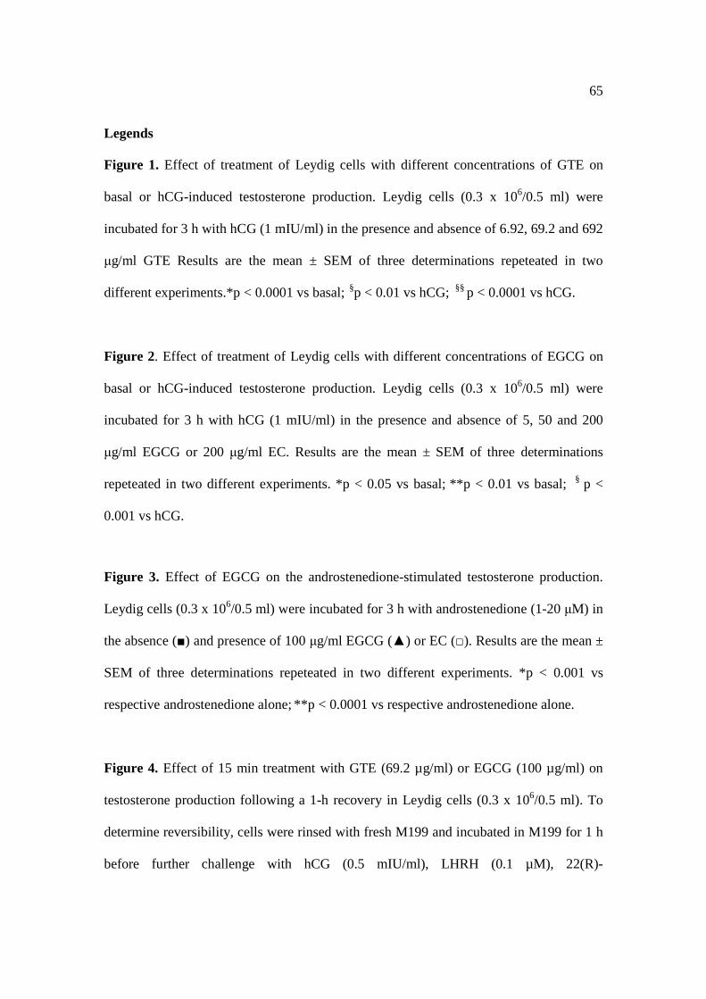
65
Legends Figure 1. Effect of treatment of Leydig cells with different concentrations of GTE on
basal or hCG-induced testosterone production. Leydig cells (0.3 x 10⁶/0.5 ml) were
incubated for 3 h with hCG (1 mIU/ml) in the presence and absence of 6.92, 69.2 and 692
µg/ml GTE Results are the mean ± SEM of three determinations repeteated in two
different experiments.*p < 0.0001 vs basal; §p < 0.01 vs hCG; §§ p < 0.0001 vs hCG.
Figure 2. Effect of treatment of Leydig cells with different concentrations of EGCG on
basal or hCG-induced testosterone production. Leydig cells (0.3 x 10⁶/0.5 ml) were
incubated for 3 h with hCG (1 mIU/ml) in the presence and absence of 5, 50 and 200
µg/ml EGCG or 200 µg/ml EC. Results are the mean ± SEM of three determinations
repeteated in two different experiments. *p < 0.05 vs basal; **p < 0.01 vs basal; § p <
0.001 vs hCG.
Figure 3. Effect of EGCG on the androstenedione-stimulated testosterone production.
Leydig cells (0.3 x 10⁶/0.5 ml) were incubated for 3 h with androstenedione (1-20 µM) in
the absence (■) and presence of 100 µg/ml EGCG (▲) or EC (□). Results are the mean ±
SEM of three determinations repeteated in two different experiments. *p < 0.001 vs
respective androstenedione alone; **p < 0.0001 vs respective androstenedione alone.
Figure 4. Effect of 15 min treatment with GTE (69.2 µg/ml) or EGCG (100 µg/ml) on
testosterone production following a 1-h recovery in Leydig cells (0.3 x 10⁶/0.5 ml). To
determine reversibility, cells were rinsed with fresh M199 and incubated in M199 for 1 h
before further challenge with hCG (0.5 mIU/ml), LHRH (0.1 µM), 22(R)-

66
hydroxicholesterol (20 µM) or androstenedione (10 µM) during 2 h. The data represent
the mean ± SEM of triplicate determinations.
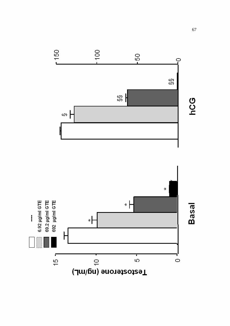
67

68
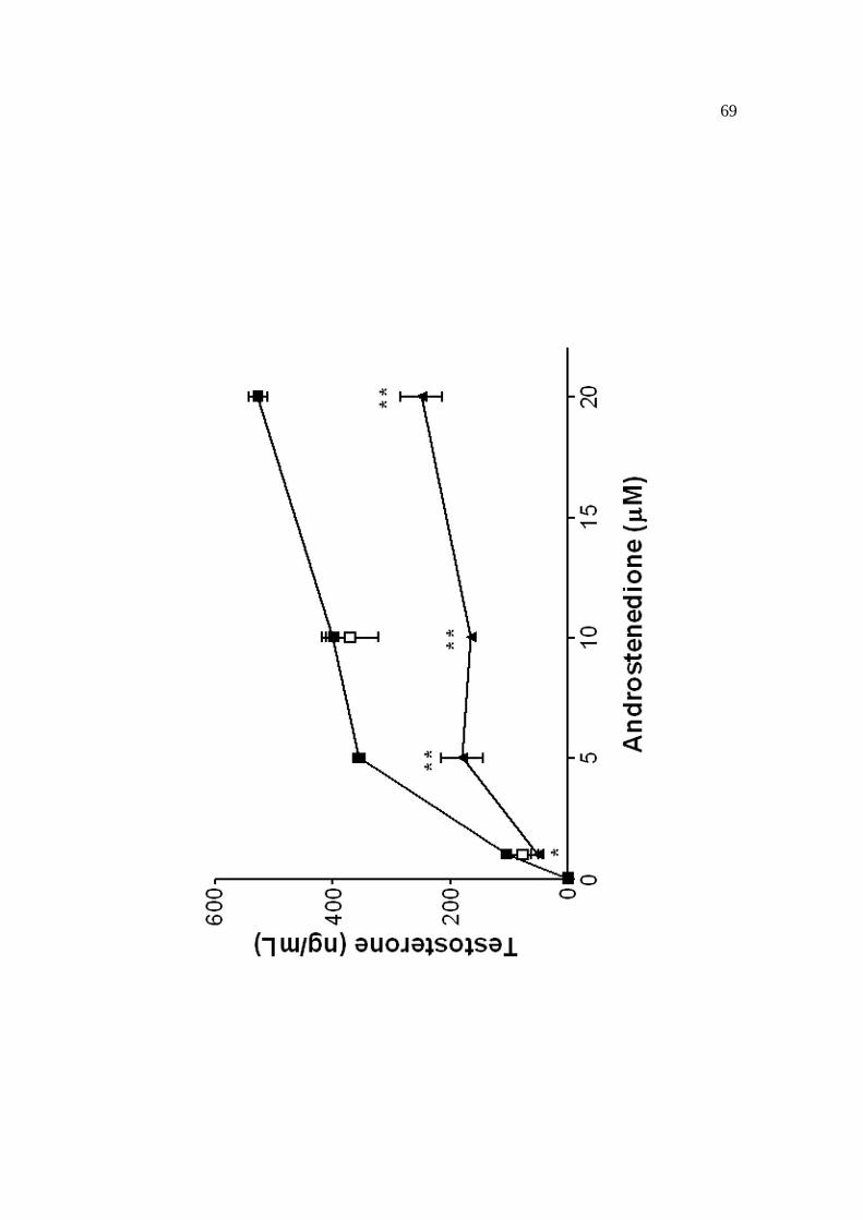
69
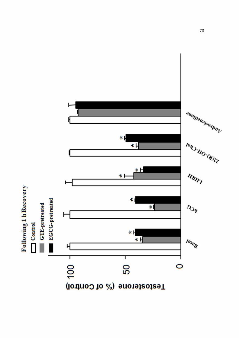
70

71

72
73
CONCLUSÕES
- O extrato aquoso do chá verde e a EGCG pura inibem a produção basal e estimulada de
testosterona em células de Leydig de ratos in vitro.
- O GTE e a EGCG inibem a função das enzimas P450scc e 17β-HSD contribuindo,
assim, na redução da capacidade esteroidogênica das células de Leydig.
- A presença do grupo galato na EGCG é importante para o seu efeito inibitório.
- Os efeitos inibitórios observados com o extrato aquoso do chá verde podem ser
explicados, ao menos em parte, pela presença da EGCG em seu extrato.