
UNIVERSIDADE FEDERAL DO PARANÁ SETOR DE CIÊNCIAS ...livros01.livrosgratis.com.br/cp122788.pdf ·...
107
UNIVERSIDADE FEDERAL DO PARANÁ SETOR DE CIÊNCIAS BIOLÓGICAS DEPARTAMENTO DE FARMACOLOGIA EDMAR MIYOSHI O HIPOCAMPO DORSAL E O ESTRIADO SÃO NECESSÁRIOS TANTO PARA A NAVEGAÇÃO BASEADA EM PISTAS QUANTO PARA A NAVEGAÇÃO ESPACIAL NO LABIRINTO AQUÁTICO DE MORRIS CURITIBA 2009
Transcript of UNIVERSIDADE FEDERAL DO PARANÁ SETOR DE CIÊNCIAS ...livros01.livrosgratis.com.br/cp122788.pdf ·...
UNIVERSIDADE FEDERAL DO PARANÁ SETOR DE CIÊNCIAS BIOLÓGICAS
DEPARTAMENTO DE FARMACOLOGIA
EDMAR MIYOSHI
O HIPOCAMPO DORSAL E O ESTRIADO SÃO NECESSÁRIOS TANTO PARA A
NAVEGAÇÃO BASEADA EM PISTAS QUANTO PARA A NAVEGAÇÃO
ESPACIAL NO LABIRINTO AQUÁTICO DE MORRIS
CURITIBA 2009

Livros Grátis
http://www.livrosgratis.com.br
Milhares de livros grátis para download.

EDMAR MIYOSHI
O HIPOCAMPO DORSAL E O ESTRIADO SÃO NECESSÁRIOS TANTO PARA A
NAVEGAÇÃO BASEADA EM PISTAS QUANTO PARA A NAVEGAÇÃO
ESPACIAL NO LABIRINTO AQUÁTICO DE MORRIS
Tese apresentado ao Curso de Pós-Graduação em Farmacologia, Setor de Ciências Biológicas, Universidade Federal do Paraná, como requisito parcial à obtenção do título de Doutor em Farmacologia.
Orientador: Prof. Dr. Cláudio Da Cunha
CURITIBA 2009

iii
EDMAR MIYOSHI
O HIPOCAMPO DORSAL E O ESTRIADO SÃO NECESSÁRIOS TANTO PARA A NAVEGAÇÃO BASEADA EM PISTAS QUANTO PARA A NAVEGAÇÃO
ESPACIAL NO LABIRINTO AQUÁTICO DE MORRIS
Tese aprovada como requisito parcial para obtenção do grau de Doutor em Farmacologia ao Curso de Pós-Graduação em Farmacologia, Setor de Ciências Biológicas, Universidade Federal do Paraná, pela seguinte banca examinadora:
Orientador: Prof. Dr. Cláudio Da Cunha Professor Livre Docente – Universidade Federal do Paraná Prof. Dr. Marcus Lira Brandão Professor Titular – Universidade de São Paulo – Ribeirão Preto Prof. Dr. Rui Daniel S. Prediger Professor Adjunto – Universidade Federal de Santa Catarina Profa. Dra. Maria Aparecida B. F. Vital Professor Associado – Universidade Federal do Paraná Prof. Dr. Marcelo Machado Ferro Professor Adjunto - Universidade Estadual de Ponta Grossa
Curitiba, 09 de outubro de 2009.

iv
Dedico aos meus pais, Masafiro e Matsue, pelo grande apoio e incentivo à realização
deste trabalho.

v
AGRADECIMENTOS
A Deus que sempre me guiou pelo caminho da vida, dando saúde e força para eu não
desistir mesmo que muitas vezes este caminho tenha sido cheio de obstáculos.
A Ana Paula que teve paciência nos momentos em que não pude estar presente e por
suportar a minha impaciência nos momentos em que estive presente. Além disso, pelo grande
amor sempre dedicado a mim.
Ao amigo e orientador Cláudio que sempre compartilhou seu vasto conhecimento e
permitiu a realização deste trabalho, além da confiança, atenção e amizade em mim
depositada.
Aos meus amigos de laboratório, Evellyn, Lucélia, Patrícia, Mariza, Suelen, Marcelo,
Jamile e Flávia, pelo auxílio nos experimentos, trocas de informações, pelos problemas e
sorrisos compartilhados.
Aos meus amigos, alunos e professores, da Pós-Graduação em Farmacologia pelas
conversas agradáveis realizadas no corredor do departamento e pela grande amizade e apoio,
tornando a execução deste trabalho muito agradável.
Aos meus colegas, professores e funcionários, da Universidade Estadual de Ponta
Grossa pelo apoio à realização deste trabalho que permitiu o afastamento de minhas
atividades didáticas.
A todos os meus amigos que me ajudaram nos momentos difíceis.
Aos funcionários do Departamento de Farmacologia da UFPR, Silvia, Lindacir, Nair,
Jorge e Erich que me auxiliaram, tanto na parte administrativa quanto na parte técnica, na
realização dos experimentos e no cuidado com os animais, além da grande amizade.
A CAPES, CNPq e Fundação Araucária pelo apoio financeiro que permitiu a
realização deste trabalho.
Enfim, a todas as pessoas que, de maneira direta ou indireta, contribuíram para a
realização deste trabalho.
Muito obrigado a todos!

vi
"É PRECISO COMEÇAR A PERDER A MEMÓRIA PARA PERCEBER QUE É ELA QUE FAZ A NOSSA VIDA. UMA VIDA SEM MEMÓRIA NÃO SERIA UMA VIDA."
LUIS BUÑUEL

vii
SUMÁRIO
LISTA DE ABREVIAÇÕES ................................................................................................ viii
RESUMO .................................................................................................................................. ix
ABSTRACT .............................................................................................................................. x
1 INTRODUÇÃO ............................................................................................................. 1
1.1 TIPOS DE MEMÓRIA ................................................................................................... 1
2 OBJETIVO GERAL .................................................................................................. 12
2.1 OBJETIVOS ESPECÍFICOS ....................................................................................... 12 2.1.1 Objetivo 1 ...................................................................................................................... 12 2.1.2 Objetivo 2 ...................................................................................................................... 12 2.1.3 Objetivo 3 ...................................................................................................................... 12
3 ARTIGO ...................................................................................................................... 13
4 DISCUSSÃO ............................................................................................................... 47
5 CONCLUSÕES ........................................................................................................... 51
REFERÊNCIAS BIBLIOGRÁFICAS ................................................................................. 52
ANEXOS ................................................................................................................................. 56
ANEXO 1 ................................................................................................................................. 57 ANEXO 2 ................................................................................................................................. 80

viii
LISTA DE ABREVIAÇÕES
DLS - estriado dorsolateral (do inglês dorsolateral striatum)
DMS - estriado dorsomedial (do inglês dorsomedial striatum)
R-O - resposta-consequência (do inglês response-outcome)
SNc - substância negra pars compacta
S-R - memória estímulo-resposta (do inglês stimulus-response)
S-R-O - memória estímulo-resposta-consequência (do inglês stimulus-response-
outcome)
S-S - memória estímulo-estímulo (do inglês stimulus-stimulus)
ix
RESUMO
Nesta tese buscamos explicar o papel do hipocampo dorsal e do estriado dorsolateral e a interação entre eles na navegação em um labirinto aquático. Dados na literatura têm mostrado a dissociação entre estas duas estruturas no aprendizado e na memória. Onde, o hipocampo dorsal é importante para desempenhar a versão espacial, mas não a versão com pista visual, do labirinto aquático. Enquanto que o estriado é importante para a versão com pista visual, mas não para a versão espacial. Entretanto, vários trabalhos mostram que estes sistemas não trabalham de forma isolada, eles podem interagir entre si. A natureza desta interação ainda é bastante controversa. Para contribuir na solução desta controvérsia, avaliamos a interação entre o estriado dorsolateral e o hipocampo no aprendizado em tarefas do labirinto aquático. Para isto, submetemos animais com lesões isoladas do estriado dorsolateral ou hipocampo e animais com lesões das duas estruturas em diferentes versões do labirinto aquático: com pista visual ou espacial, dependentes do estriado dorsolateral ou hipocampo respectivamente. Animais com lesões isoladas no hipocampo apresentaram um prejuízo no aprendizado da versão espacial, mas não na versão com pista visual do labirinto aquático. Mas, todos os animais conseguiram aprender a tarefa com mais sessões de treinamento. Os animais com lesão no estriado dorsolateral aprenderam as duas versões como os animais controle. Quando os animais foram pré-treinados em uma das versões e testados na outra versão, i.e. pré-treinados na versão com pista visual e testados na versão espacial e vice-versa, não foi observado este prejuízo. Entretanto, aqueles animais com lesão dupla (do estriado dorsolateral e do hipocampo) apresentaram um prejuízo severo em ambas as versões, tal como se não apresentassem nenhuma evidência de aprendizado e estes prejuízo não desapareceram ao longo das sessões de treinamento e nem com o pré-treinamento em outra versão. Estes resultados sugerem que tanto o estriado dorsolateral como o hipocampo dorsal são necessários para os dois tipos de aprendizados, contrariando a teoria vigente na literatura de que há uma dupla dissociação: versão espacial dependente do hipocampo, mas não do estriado e versão com pista visual dependente do estriado (dorsolateral) e não do hipocampo. Isto sugere que estes dois sistemas não só atuam de forma cooperativa, como que eles desempenham papéis complementares essenciais para a navegação e aprendizado espacial. As implicações destes resultados para o modelo do mosaico dos espelhos quebrados também é discutido nesta tese.

x
ABSTRACT
In this thesis we proposed an explanation for the role of the dorsal hippocampus and of the dorsolateral striatum and the interaction between them in the navigation in water maze task. Data in the literature have been showing the double dissociation between these structures in the learning and memory. Where, the dorsal hippocampus is important to perform the spatial version, but not the cued version, of the water maze task. While striatum is important for the cued version, but not for the spatial version. However, several works showed that these systems didn't work in an isolated way, they can interact between them. The nature of this interaction is still controversial. In order to contribute with a solution for this controversy, we evaluated the interaction between dorsolateral striatum and the hippocampus in the learning of water maze task. For this, we submitted animals with isolated lesions on the dorsolateral striatum or hippocampus and animals with lesions of both structures in different versions of the water maze: cued or spatial, dorsolateral striatum- or hippocampus-dependent, respectively. Animals with isolated lesions in the hippocampus showed impairment in the spatial version, but not in the cued version of the water maze task. But, all the animals learned this task when submitted to more training sessions. The animals with lesion in the dorsolateral striatum learned the two versions as the control animals. When the animals were pre-trained in one of the versions and tested in the other version, i.e. pretrained in the cued version and tested in the spatial version and vice-versa, this damage was not observed. However, those animals with double lesion (dorsolateral striatum and hippocampus) presented a severe impairment in both versions, as if they didn't present any learning evidence and this impairment didn't disappear along the training sessions nor with the pre-training in another version. These results suggest that both dorsolateral striatum and dorsal hippocampus are necessary for the two types of learning, contradicting the current theory in the literature that there is a double dissociation: spatial version dependent of the hippocampus but not of the striatum and cued version dependent of the striatum (dorsolateral) and not of the hippocampus. This suggests that these two systems not only interact in a cooperative way, but they play a complementary role, that is essential for the navigation and spatial learning. The implications of these results for the mosaic of broken mirrors model are also discussed in this thesis.

1
1 INTRODUÇÃO
A organização da memória no cérebro dos mamíferos e os sistemas neurais que
medeiam os processos de aprendizado e memória têm um papel importante nos nossos
pensamentos, emoções, escolhas, ações e personalidade. Perder a memória leva à perda de si
mesmo, à perda da história de uma vida e das interações duradouras com outros seres
humanos. O enfraquecimento normal da memória com a idade e o prejuízo causado pelas
doenças de Alzheimer, Parkinson e Huntington são apenas os exemplos mais conhecidos de
um grande número de doenças que afetam a memória.
Durante muito tempo debateu-se intensamente a possibilidade de a memória ser
considerada uma função unitária ou ser decomposta em diferentes sistemas. Rejeitada de
início pelos cientistas, a idéia de que podem existir várias formas ou tipos de memória hoje
afinal se impôs (Poldrack e Packard, 2003; Squire, 2004; Squire, Stark et al., 2004;
Voermans, Petersson et al., 2004; Doeller, King et al., 2008; Lee, Duman et al., 2008; Berke,
Breck et al., 2009). Esta hipótese dos vários tipos de memória recebeu um importante apoio
com o estudo de Scoville e Milner em 1957 (Scoville e Milner, 1957). Estes autores
estudaram o paciente H.M., um homem que se tornou amnésico após a retirada cirúrgica do
seu lobo temporal medial para melhorar suas crises epilépticas. H.M. apresentou um prejuízo
em algumas tarefas de memória (principalmente as memórias episódica), entretanto, ele ainda
conseguia aprender certas tarefas (traçar o contorno de uma estrela olhando por um espelho),
sugerindo que existiria outro tipo de memória.
1.1 TIPOS DE MEMÓRIA
Existem várias classificações diferentes para as memórias. Uma dessas classificações

2
que podem ser encontrada é a seguida por Izquierdo (Izquierdo, 2002), onde ele classifica as
memórias quanto ao tempo de duração em:
• Memória de longa duração - é aquela que dura muitas horas, dias ou
anos;
• Memória de curta duração - é o processo ou conjunto de processos que
mantém a memória funcionando durante estas horas iniciais em que a memória de
longa duração ainda não assumiu sua forma definitiva; e,
• Memória operacional (“working memory”) - que mantém a informação
“viva” durante segundos ou poucos minutos, enquanto ela está sendo percebida
conscientemente ou processada em uma operação mental.
Além dessa classificação, as memórias de longa duração podem ser subdivididas em
dois grandes grupos de memória (figura 1) (Salmon e Butters, 1995; Izquierdo, 2002; Squire,
2004):
1) as memórias declarativas ou explícitas
São aquelas que nós humanos podemos relatar e evocar de forma consciente. Elas
guardam informações factuais sobre eventos que vivenciamos no passado. Este tipo de
memória pode ser também subdividido em: memória episódica (representações de
experiências pessoais específicas que ocorreram em um contexto de tempo e espaço – saber o
que, onde e quando) e memória semântica (conjunto de conhecimentos generalizados sobre o
mundo sem nenhuma vinculação com uma experiência pessoal específica).

3
2) as memórias não-declarativas ou implícitas
São aquelas que podemos adquirir e evocar de forma automática ou inconsciente.
Este tipo de memória pode ser subdividido em: memória de procedimento, o priming
(aperfeiçoamento da capacidade de detectar ou identificar palavras ou objetos após uma
experiência recente com eles), o condicionamento e memórias formadas por aprendizado não-
associativo (habituação e sensibilização).
FIGURA 1 – ESQUEMA DA CLASSIFICAÇÃO DA MEMÓRIA DE LONGA DURAÇÃO E AS ESTRUTURAS CEREBRAIS IMPORTANTES PARA CADA TIPO DE MEMÓRIA.
FONTE: modificado de SQUIRE, L.R. Memory systems of the brain: a brief and current perspective. Neurobiology of Learning and Memory. v. 82, p. 171-177, 2004.
Vários autores sugerem que estes diferentes grupos de memórias sejam organizados e
controlados por sistemas neuroanatômicos distintos (Packard e White, 1990; Packard e
Mcgaugh, 1992; Mcdonald e White, 1994; Salmon e Butters, 1995; Oliveira, Bueno et al.,
1997; Packard e Teather, 1997; 1998; Eichenbaum, 2004; Squire, 2004; Doeller, King et al.,
4
2008; Lee, Duman et al., 2008; Berke, Breck et al., 2009). Dois exemplos bem definidos
destes sistemas de memória são: o sistema de memória declarativa, o qual tem o hipocampo
como estrutura central (Eichenbaum, 2004; Squire, 2004; Gold e Squire, 2006; Squire, 2009),
e o sistema de memória de procedimento, o qual tem os gânglios da base como núcleo central
(Knowlton, Mangels et al., 1996; Da Cunha, Gevaerd et al., 2001; Miyoshi, Wietzikoski et
al., 2002; Packard e Knowlton, 2002; Da Cunha, Wietzikoski et al., 2003; Da Cunha, Silva et
al., 2006; Prediger, Batista et al., 2006; Da Cunha, Wietzikoski et al., 2007; Da Cunha,
Wietzikoski et al., 2009; Prediger, Rial et al., 2009).
Em humanos, a memória declarativa episódica é sempre expressa na forma de
recordações conscientes de experiências pessoais específicas (o sujeito dentro de um contexto
espacial e temporal). Como os animais não possuem uma linguagem equivalente à de
humanos, estas propriedades (ser declarativa e consciente) não podem ser estudas em modelos
animais. Entretanto, a memória declarativa humana tem diversas outras propriedades além da
lembrança consciente e declarativa, podendo, muitas delas, serem estudadas.
Por exemplo, a memória declarativa é elaborada para representar objetos e eventos
no mundo externo e as relações (espaciais, temporais e lógicas) entre eles. Esta organização
associativa das memórias declarativas resulta em uma representação flexível (relacional) do
espaço e do tempo. Animais podem aprender relações entre itens armazenados e, então, ter
sua memória episódica testada em situações onde precisem usar estas relações de forma
flexível para resolver uma tarefa de aprendizagem e memória (Eichenbaum, 2004).
Já a memória de procedimento foi sempre fácil de entender intuitivamente com algo
especial, diferente da evocação comum de eventos recentes. Eles não são declarativos: não
precisamos “declarar” coisa alguma nem ser capazes de, mesmo quando pressionados, dizer
muito sobre o que estamos fazendo. Adquirimos muitos hábitos e habilidades no início da
vida, sem esforço óbvio e sem observarmos o momento em que tal aprendizado ocorreu.
5
Um modo de se estudar este tipo de memória em animais é expondo-os a uma tarefa
onde o animal, a partir de um determinado estímulo, deve exercer uma determinada resposta
para obter reforço. Este é um tipo de aprendizado é chamado de estímulo-resposta-
consequência (S-R-O).
Na formação de memórias resposta-consequência (R-O) ou ação-consequência
(action-outcome), o sujeito aprende que uma ação ou resposta têm como consequência um
estímulo (incondicionado) cuja percepção envolve uma avaliação hedônica de
“recompensador” ou “aversivo”. Na linguagem da psicologia experimental, o pareamento de
um estímulo reforçador, subsequente a uma resposta do sujeito resulta em uma maior
probabilidade de que ele emita esta resposta no futuro. Ainda segundo esta corrente teórica, a
apresentação de um estímulo punidor, subseqüente a uma resposta, diminui a probabilidade de
que esta resposta seja emitida no futuro. Porém, se após a aprendizagem, a consequência
reforçadora ou punidora não ocorrer, esta memória entra em processo de extinção (Yin e
Knowlton, 2006; Balleine, Liljeholm et al., 2009).
Já na formação da memória de hábito, um estímulo (condicionado) é repetidamente
pareado com uma resposta incondicionada, i.e. que é emitida de forma inata pelo sujeito em
reação ao estímulo (Thorndike, 1911; Hull, 1943). Ainda há muita divergência sobre a
natureza desta memória.
Nos primórdios da psicologia comportamental, este tipo de aprendizagem era
chamado de “controle por estímulo” e dizia-se que o que é aprendido é a associação entre o
estímulo condicionado e a resposta incondicionada, daí o nome de hábito estímulo-resposta
(S-R). Já os teóricos modernos, tais como o americano Balleine, dizem que a formação do
hábito é uma decorrência de um aprendizado instrumental muito prolongado (onde se pareia
resposta e consequência).
Segundo Balleine, em situações onde as consequências de uma resposta a um

6
estímulo não mudam, a repetição deste pareamento S-R-O leva a uma automação da resposta,
de forma que o indivíduo a escolhe e executa de forma automática. Após um treino extensivo,
a resposta (ação/comportamento) deixa de ser controlada pela consequência e passa a ser
controlada pelo estímulo condicionado (S-R) (Balleine, Liljeholm et al., 2009).
Um exemplo de experimento que pode ser utilizado para avaliar estes tipos de
memórias é o labirinto aquático de Morris. Esta tarefa do labirinto aquático foi desenvolvida
por Richard Morris em 1982 e consiste em colocar o animal em uma piscina circular com
água. Em algum lugar da piscina havia uma plataforma que permanecia logo abaixo do nível
da água (invisível para o rato).
Os ratos nadavam muito bem, mas preferiam subir na plataforma para fugir da água.
Subir na plataforma era uma recompensa eficiente, chamada na psicologia comportamental de
reforço negativo. Em cada tentativa, o animal foi colocado em pontos diferentes da borda da
piscina. Os ratos aprenderam a usar uma estratégia espacial (relação entre as pistas localizadas
fora do labirinto) para encontrar a plataforma. Isto pode ser observado pela redução na
latência para encontrar a plataforma.
Morris e seus colaboradores demonstraram que a lesão do hipocampo causa um
prejuízo no desempenho desta tarefa (Morris, Garrud et al., 1982). A partir deste trabalho de
Morris e colaboradores, esta tarefa do labirinto aquático passou a ser bastante útil para a
pesquisa de aprendizado e memória em animais. Isto pode ser observado através de uma
pesquisa no site da “Web of Science” com as seguintes palavras chaves: “Morris water maze”
AND (learning OR memory) no período de 1982 até 2009. Realizando esta busca,
encontramos 3014 artigos com estas características (pesquisa realizada no dia 16 de setembro
de 2009).
A flexibilidade da memória episódica e a relativa inflexibilidade de memórias não-
declarativas são vivamente ilustradas em um estudo sobre aprendizado e memória espacial em

7
ratos. Eichenbaum e colaboradores (Eichenbaum, Stewart et al., 1990) avaliaram o
desempenho de animais com lesão hipocampal em uma versão modificada do labirinto
aquático de Morris.
Neste estudo os animais foram liberados para nadar somente de um ponto de partida
e deveriam encontrar a plataforma submersa oculta. Tanto os animais com lesão hipocampal
como os animais controle aprenderam a localização da plataforma submersa, conforme
avaliado por reduções marcantes no tempo de natação e na distância percorrida até atingir a
plataforma. Assim, à medida que o aprendizado progredia, os ratos aprendiam a nadar
diretamente até a plataforma. Depois de completar-se o aprendizado, os animais foram
submetidos a testes adicionais para que se determinasse que tipo de informação haviam
adquirido sobre a localização da plataforma.
Nessas sessões, os ratos eram liberados de um novo ponto de partida. Os animais
intactos eram capazes de descobrir a plataforma rapidamente a partir de qualquer ponto
inicial, indicando que haviam adquirido uma representação flexível (declarativa) do espaço na
memória. Mais especificamente, eles haviam aprendido sobre as relações espaciais entre a
localização da plataforma e as várias dicas externas que estavam disponíveis nas paredes que
circundavam o tanque (mapa relacional).
Este tipo de aprendizado foi classificado por White como sendo do tipo estímulo-
estímulo (S-S). Em contraste, os ratos com lesões hipocampais eram incapazes de encontrar a
plataforma a partir de novos pontos de partida e tinham de recomeçar a busca empregando
uma estratégia do tipo tentativa-e-erro ao longo do labirinto.
Estudos posteriores mostraram que o aprendizado espacial em ratos depende
criticamente da integridade do hipocampo, mas não do estriado (Packard e Mcgaugh, 1992;
White e Mcdonald, 2002; Da Cunha, Wietzikoski et al., 2007; Goodrich-Hunsaker,
Livingstone et al., 2009; Xavier e Costa, 2009).

8
Resultados semelhantes foram obtidos por Da CUNHA e colaboradores (Da Cunha,
Wietzikoski et al., 2003) através da administração de lidocaína no hipocampo dorsal de ratos
submetidos à versão espacial (S-S) da tarefa do labirinto aquático. Os animais inicialmente
aprenderam a encontrar a plataforma submersa, pois o tempo de latência para encontrá-la
diminuiu. Entretanto, a administração de lidocaína intra-hipocampal, antes da exposição ao
labirinto, promoveu um aumento no tempo de latência, sugerindo um prejuízo na memória
espacial (S-S).
Outro trabalho que usou estas versões do labirinto aquático de Morris foi realizado
por PACKARD e McGAUGH (Packard e Mcgaugh, 1992). Eles mostraram que animais com
lesão do estriado dorsal conseguem aprender a desempenhar a versão espacial (S-S) da tarefa
do labirinto aquático. Mas estes animais têm um prejuízo na versão com pista visual (S-R-O)
da tarefa do labirinto aquático (Packard e Mcgaugh, 1992). Nesta versão, o animal deve
encontrar uma plataforma que possui uma pista visual sobre ela e visível ao animal, mas sua
posição se altera entre uma tentativa e outra.
Então, o estriado é visto como uma região importante para o aprendizado de relações
entre um único estímulo e uma resposta recompensada, ou seja, aprendizado S-R-O (White e
Mcdonald, 2002). Há evidências de que ocorra uma dissociação entre o estriado dorsolateral
(DLS, equivalente ao putamen de primatas) e o estriado dorsomedial (DMS, equivalente ao
núcleo caudado em primatas), onde o primeiro seria importante para o aprendizado S-R-O
(Devan, Mcdonald et al., 1999) e o último para o aprendizado espacial (S-S) (White, 2009).
Outros estudos também mostraram esta dissociação entre o sistema hipocampal
(memória declarativa) e o sistema dos gânglios da base (memória de procedimento), como por
exemplo um importante estudo realizado por PACKARD e colaboradores (Packard, Hirsh et
al., 1989). Eles treinaram ratos para realizar duas tarefas diferentes, que mostravam diferenças
chaves entre a memória de hábito e a memória declarativa episódica. Em uma tarefa, os

9
animais deveriam procurar por alimento nos oito braços de um labirinto radial. A cada dia,
durante diversos dias, os ratos eram colocados no labirinto e, após, retirados quando tivessem
recolhido uma recompensa de cada um dos oito braços do labirinto. Um erro era registrado
cada vez que o animal entrasse pela segunda vez em um braço no curso da coleta das oito
recompensas. O desempenho nessa tarefa de memória é prejudicado por lesão do sistema
hipocampal, porém, a lesão do estriado dorsal não tem efeito. Em uma tarefa semelhante que
utilizou o mesmo labirinto, os animais aprenderam a visitar quadro dos oito braços, os quais
eram sinalizados, através de uma luz, que continham o alimento como recompensa. Após duas
semanas de treino, os animais gradualmente aprenderam a entrar nos braços corretos. Nessa
tarefa, onde o animal deveria associar o estímulo (luz) com a resposta recompensada (entrar
para comer o alimento) o aprendizado foi prejudicado por lesão do estriado dorsal, mas não
por lesão do sistema hipocampal.
Algum tempo depois, um questionamento que começou a ser feito foi se estes
sistemas funcionavam de forma isolada ou eles poderiam interagir entre si? E, nas últimas
cinco décadas, pesquisadores têm focado seus estudos na dissociação entre os sistemas de
memórias e suas funções no armazenamento de informações e adaptação do comportamento
(Scoville e Milner, 1957; Packard e White, 1990; Packard e Mcgaugh, 1992; Mcdonald e
White, 1994; Packard e Teather, 1997; 1998; Miyoshi, Wietzikoski et al., 2002; Da Cunha,
Silva et al., 2006; Doeller, King et al., 2008).
Entretanto, pode ser que os sistemas de memória trabalhem de uma forma integrada,
e não de forma isolada (White e Mcdonald, 2002; Voermans, Petersson et al., 2004; Hartley e
Burgess, 2005; Albouy, Sterpenich et al., 2008; Doeller, King et al., 2008; Lee, Duman et al.,
2008; Berke, Breck et al., 2009).
A maioria destes estudos observou o prejuízo causado pela lesão de uma estrutura
cerebral no desempenho de tarefas (Da Cunha, Gevaerd et al., 2001; Da Cunha, Angelucci et

10
al., 2002; Da Cunha, Wietzikoski et al., 2003; Mcdonald, Hong et al., 2004). E, quando uma
lesão de determinada estrutura prejudicava o desempenho de determinada tarefa, concluíam
que aquela estrutura era importante para aquele tipo de aprendizado e memória. Por exemplo,
concluiu-se que a SNc é importante para a memória S-R-O e operacional (mas não para a
memória espacial) porque sua lesão causou um prejuízo no desempenho da versão com pista
visual e na versão da memória espacial operacional do labirinto aquático de Morris e não
prejudicou o desempenho na versão espacial (Miyoshi, Wietzikoski et al., 2002). Mas,
estudos recentes sugerem que ocorre uma interação (competição e/ou cooperação) entre os
diferentes sistemas neurais de memória (Poldrack e Packard, 2003; Voermans, Petersson et
al., 2004; Albouy, Sterpenich et al., 2008; Lee, Duman et al., 2008; Berke, Breck et al., 2009).
A interação competitiva entre os sistemas de memória pode ser revelada pelos
estudos em que a lesão de um dado sistema resulta em melhora na aprendizagem da tarefa
dependente da estrutura encefálica intacta (Poldrack e Packard, 2003). Por exemplo, animais
com lesão do hipocampo dorsal têm um desempenho melhor do que animais controles na
tarefa de esquiva de duas vias (dependente do estriado e da SNc) (Guillazo-Blanch, Nadal et
al., 2002; Torras-Garcia, Costa-Miserachs et al., 2003).
A interação cooperativa entre os sistemas de memória foi observada por Voermans e
colaboradores (Voermans, Petersson et al., 2004) através de um estudo de neuroimagens em
pacientes com doença de Huntington desempenhando uma tarefa de memória de navegação
espacial. Nesta tarefa, o participante navega em uma sequência de vídeos com uma visão em
primeira pessoa. Durante a fase de aquisição, o vídeo é parado em cinco pontos de decisão,
que são locais onde o participante deve escolher uma direção (esquerda ou direita) e esta
direção é indicada por setas. Os participantes devem relembrar a direção a ser seguida em
cada ponto de decisão. Durante a fase de navegação, o participante vê a mesma sequência e
deve indicar a direção a ser seguida em cada ponto de decisão (sem auxílio das setas). Esta
11
tarefa emprega um sistema que adquire gradualmente sequências de resposta para
determinada situação (i.e. seguir uma rota fixa repetidamente, S-R-O) e é dependente do
estriado. Os pacientes com doença de Huntington que estavam no estágio leve a moderado
tinham grande ativação do estriado durante a realização da tarefa, enquanto que os pacientes
nos estágios mais graves apresentaram maior ativação do hipocampo. Os pacientes
apresentaram escores semelhantes ao do grupo controle na realização desta tarefa, sugerindo
uma compensação hipocampal para desempenhar a tarefa.
Dados de nosso laboratório também sugerem uma interação cooperativa, onde os
animais com lesão da SNc que são previamente treinados na versão espacial (S-S) do labirinto
aquático (dependente do hipocampo) não tem prejuízo em desempenhar a versão com pista
visual (S-R-O, dependente dos gânglios da base) (Da Cunha, Wietzikoski et al., 2007). Isto
mostra a necessidade de se estudar mais sobre a função do hipocampo e dos gânglios da base
nos processos de aprendizado e memória e as interações que ocorrem entre estas estruturas.

12
2 OBJETIVO GERAL
Avaliar a interação entre os sistemas dos gânglios da base e do hipocampo na
navegação em um labirinto aquático.
2.1 OBJETIVOS ESPECÍFICOS
2.1.1 Objetivo 1
Avaliar o papel do DLS no aprendizado da versão com pista visual do labirinto
aquático de Morris, um modelo animal de memória de procedimento (S-R-O).
2.1.2 Objetivo 2
Avaliar o papel do hipocampo dorsal no aprendizado da versão espacial do labirinto
aquático de Morris, um modelo animal de memória relacional (S-S).
2.1.3 Objetivo 3
Avaliar a interação entre o DLS e o hipocampo dorsal no aprendizado das versões
com pista visual e espacial do labirinto aquático de Morris.

13
3 ARTIGO
Neste trabalho avaliamos o papel do hipocampo, do estriado dorsolateral e da
interação entre eles nos processos de aprendizado e memória. O manuscrito deste trabalho foi
submetido à revista “HIPPOCAMPUS” neste ano de 2009. Neste estudo apresentamos
resultados obtidos de animais com lesão do estriado dorsolateral e/ou do hipocampo
submetidos a diferentes versões (dependentes do estriado ou do hipocampo) do labirinto
aquático.
For Peer Review
1
Both the dorsal hippocampus and the dorsolateral striatum are needed for
rat navigation in the Morris water maze
Edmar Miyoshia,b, Evellyn Claudia Wietzikoskia,c, Mariza Bortolanzaa, Suelen
Lucio Boschena, Newton Sabino Canterasd, and Claudio Da Cunhaa,*
aLaboratório de Fisiologia e Farmacologia do Sistema Nervoso Central,
Departamento de Farmacologia, UFPR, Curitiba, Brazil; bDepartamento de
Ciências Farmacêuticas, UEPG, Ponta Grossa, Brazil. cLaboratório de
Neurosciências, UNIPAR, Francisco Beltrão, Brazil. dDepartamento de
Anatomia, Instituto de Ciências Biomédicas-3, USP, São Paulo, Brazil.
Running title: The role of the hippocampus and striatum in navigation
Number of text pages: 26
Number of figures: 7
Number of tables: 0
*Author for correspondence: Claudio Da Cunha, Laboratório de Fisiologia e
Farmacologia do Sistema Nervoso Central, Departamento de Farmacologia,
UFPR, C.P. 19.031, 81.531-980 Curitiba PR, Brazil. Tel: +55 41 3361-1717.
Fax: +55 41 3266-2042. E-mail: [email protected]
Grant sponsors: CNPq, CAPES, Fundação Araucária, FAPESP
KEY WORDS: basal ganglia, dopamine, memory, spatial learning, cued
learning
Page 1 of 33
John Wiley & Sons
Hippocampus
123456789101112131415161718192021222324252627282930313233343536373839404142434445464748495051525354555657585960

For Peer Review
2
ABSTRACT
The multiple memory systems theory proposes that the hippocampus and the
dorsolateral striatum are the core structures of the spatial/relational and
stimulus-response (S-R) memory systems, respectively. This theory is
supported by double dissociation studies showing that the spatial and cued
(stimulus-response) versions of the Morris water maze are impaired by lesions
in the dorsal hippocampus and dorsal striatum, respectively. In the present
study we further investigated this hypothesis by testing whether adult male
Wistar rats bearing double and bilateral electrolytic lesions in the dorsal
hippocampus and dorsolateral striatum were as impaired as rats bearing single
lesions in just one of these structures in learning both versions of the water
maze. Such prediction, based on the multiple memory systems theory, was not
confirmed by our findings. Although, compared to the controls, the latency to
find the escape platform of the animals with single lesions decreased more
slowly in one of the versions, the animals with double lesions presented no
improvement at all in both versions of the water maze. These results suggest
that both the dorsal hippocampus and the dorsolateral striatum are needed for
learning cue- and spatial-based navigation in the water maze. Therefore, it
seems that, instead of independent systems supporting S-R or spatial learning,
the hippocampus and dorsal striatum play critical roles in these two kinds of
learning.
Page 2 of 33
John Wiley & Sons
Hippocampus
123456789101112131415161718192021222324252627282930313233343536373839404142434445464748495051525354555657585960

For Peer Review
3
INTRODUCTION
In the recent past the hippocampus was taken as the brain structure
playing the main role in spatial navigation learning and performance.
Contributed to this reputation the discovery of the hippocampal place cells,
neurons that discharge when the animal is in a particular place of the
environment (Nadel and O’Keefe, 1978). The finding that rats bearing lesions in
the hippocampus are impaired to learn the Morris water maze task, also caused
a great impact and made this memory task to be considered a “gold standard”
test of the hippocampal function and as a model of spatial/relational memory
(Morris et al., 1982; Eichenbaum, 2002; Squire et al., 2004).
Nowadays, navigation learning and performance is seen as the result
of computations that involve not only the hippocampus, but also other brain
structures. An influential model proposes that the representation of the
environment and its reconstitution in the brain is based on a process called
pattern integration that points out the location of the animal based on its own
movements. According to this theory, an allocentric parahippocampal
representation of the environment is translated into an egocentric medial
parietal representation (Byrne et al., 2007). This process also depends on the
posterior parietal cortex and the retrosplenial cortex/parieto-occipital sulcus
(Bird and Burgess, 2008). Still according to this view, in addition to the
hippocampal place cells, pattern integration depends on the so called grid cells
of the enthorhinal cortex and on the head direction cells found along the
Papez’s circuit (Hafting et al., 2005; Bird and Burgess, 2008).
The striatum is not usually seen as playing a role in spatial navigation.
Contributed to this view, the seminal double dissociation studies reporting that
the lesion of the fimbria/fornix, but not of the dorsal striatum, impaired rats to
learn the spatial version of the Morris water maze and of the win-shift (spatial)
version of the 8-arm radial maze tasks, while lesions of the dorsal striatum, but
not of the fimbria/fornix, impaired learning of cued versions of these tasks
(Packard et al., 1989; Packard and McGaugh, 1992; McDonald and White,
1994). In order to explain these findings, some authors proposed that both the
hippocampus and the striatum can hold control over navigation by using
different strategies, and that in some instances they compete for the control
Page 3 of 33
John Wiley & Sons
Hippocampus
123456789101112131415161718192021222324252627282930313233343536373839404142434445464748495051525354555657585960

For Peer Review
4
over behavior (White and McDonald, 2002; Chavarriaga et al., 2005; Lee et al.,
2008; Berke et al., 2009). According to them, the hippocampus uses the
relations among environmental stimuli to form a kind of “cognitive map”, as
proposed by Tolman (1948) and supported by the hippocampal place cells
(O'keefe and Nadel, 1978), and uses it to plan flexible navigation strategies
(White and McDonald, 2002). The striatum, on its turn, learns the relations
between single environmental stimulus and rewarded responses and thus, can
guide navigation by approaching a specific individual cue that signalizes a
rewarding outcome (White and McDonald, 2002). These two strategies are
sometimes referred to as spatial (or S-S, stimulus-stimulus) and cue-based (or
S-R, stimulus-response) navigation, respectively.
Thus, this theory proposes that navigational behavior can be controlled
by two parallel memory systems that sometimes compete for control over
behavior: the hippocampal system mediating spatial/relational navigation and
the striatal system mediating cue-based navigation. However, some years after
this theory was proposed (see White and McDonald, 2002), another study
presented evidence that at least the dorsal medial part of the striatum (DMS) is
also needed for spatial learning (Devan et al., 1999). Then, the
hippocampal/striatal parallel memory systems theory was modified to
incorporate the DMS into the hippocampal-based spatial memory system and
restricted the memory system that supports the cue-based navigation learning
to the dorsolateral striatum (DLS) (White, 2009).
Some authors claim that a differential pattern of striatal inputs of the
DMS and DLS may allow them to play different roles in navigation: that the
inputs from the prefrontal cortex and hippocampus to the DMS may enable it to
elaborate flexible navigation strategies based on spatial/contextual information
and the inputs from the sensorymotor cortex to the DLS may enable it to
elaborate rigid and egocentric/cue-based strategies (Potegal et al., 1971;
Veening et al., 1980; McGeorge and Faull, 1989; Ramanathan et al., 2002;
Voorn et al., 2004). However, a recent study by Cenquizca and Swanson (2007)
showed that most projections from the rat field
CA1 to the caudate-putamen are indirect, mediated by the prefrontal cortex, and
is virtually impossible to differentiate between projections to the DMS or
DLS.
Page 4 of 33
John Wiley & Sons
Hippocampus
123456789101112131415161718192021222324252627282930313233343536373839404142434445464748495051525354555657585960

For Peer Review
5
Defining insensibility to reward devaluation as a feature that
distinguishes S-R habits and response-outcome (R-O) behaviors, Balleine,
Knowlton, Yin, and co-workers presented the following evidence that these
kinds of learning are mediated by the DLS and DMS, respectively: Overtraining
and interval schedules of reinforcement are known to convert instrumental R-O
responding into S-R habits (Yin and Knowlton, 2006; Balleine et al., 2009). In a
study by Yin et al. (2004), lesions in the rat DLS reversed this effect of the
overtraining, thus turning habitual into R-O responding. In addition, the
inactivation of the DLS of rats enhanced the sensitivity of their instrumental
responding to the omission of a rewarding outcome (Yin et al., 2006).
Conversely, inactivating the posterior DMS, but not the DLS, of rats prevented
the decrease of instrumental responding contingent of the devaluation of a
rewarding outcome (Yin et al., 2005) and the discrimination of two stimuli that
differentially signaled reinforcement for response to one or the other of two bars
(Balleine et al., 2009).
All these findings strengthen the hypothesis that the hippocampus and
the DMS mediate more flexible behavior, while the DLS supports S-R habit
learning. However, there are some inconsistencies between these hypotheses
and some findings reported in the literature. Although it was not tested whether
rats overtrained in the cued version of the water maze are insensitive to reward
devaluation, this task have been taken as a model of S-R learning (White, 2004;
Packard, 2009; White, 2009). It is also generally accepted that the spatial
version of this task is a good model of the spatial/relational learning, the kind of
learning supported by the hippocampus and DMS, as stressed above. Then, it
is expected that the lesion of the DLS, but not of the DMS, would impair
learning of the cued version. However, the study by Devan et al. (1999) did not
confirm this prediction: they found out that the lesion of the rat DMS impaired
learning of both versions while the lesion of the DLS did not impair any of them.
The assumption that the DLS is not involved in spatial navigation is also in
disagreement with the finding that some neurons in the striatum respond to the
animal location and head direction (Wiener, 1993; Mizumori et al., 2009). It is
important to mention that these neurons are not restricted to the DMS, but were
found in all regions of the striatum, including the DLS (Mizumori’s personal
communication).
Page 5 of 33
John Wiley & Sons
Hippocampus
123456789101112131415161718192021222324252627282930313233343536373839404142434445464748495051525354555657585960

For Peer Review
6
Such inconsistencies had lead to alternative theories about the
interactions between the hippocampus and striatum in learning and
performance of spatial navigation. Mizumori and co-workers (2009)
hypothesized that, in addition to the hippocampus, all regions of the striatum
contribute to spatial navigation. They proposed that, instead of competing for
control over behavior, while the hippocampus extracts a spatial/relational map
of the environment from sensory inputs, the striatum selects the proper actions
to navigate according to the directions that can be taken from this map and that
leads to a reward. According to them, the striatum performs this selection by
applying the same computational pattern to the different inputs arriving to
different parts of the striatum. Da Cunha and co-workers (2009) recently
proposed a model called “the mosaic of broken mirrors” to explain the
computational contribution of the striatum on learning and memory. In short, this
model proposes that objects and locations are represented in functional units of
the striatum, as well as the subject’s body (and body parts). The association of
these units encodes the action of the subject (or the subject’s body part)
towards a particular location or object of the environment. The indirect striatal
inputs from the hippocampus make it a likely candidate to feed the striatum with
information of near locations in relation to the subject. However, instead of
encoding these locations based on the spatial relations among them (like the
hippocampus does), according to the mosaic of broken mirrors model, they are
encoded as fragments of the environment that are individually related to specific
actions, but that cannot reconstitute the environment based on multiple relations
among the environmental pieces.
Thus, instead of parallel memory systems that sometimes compete for
the control over behavior, the hippocampus and the striatum may be systems
with complementary roles in spatial navigation. If this is true, the lesion of the
striatum plus the hippocampus would result in a deeper impairment in learning
of both the spatial and cued versions of the Morris water maze. Such prediction
is more particularly in confront with the prediction of the competition theory, if
the striatum lesion were restricted to the DLS, which lesion is known to not
affect the learning of both versions of this task (Devan et al., 1999). Testing this
prediction is the aim of the present study.
Page 6 of 33
John Wiley & Sons
Hippocampus
123456789101112131415161718192021222324252627282930313233343536373839404142434445464748495051525354555657585960

For Peer Review
7
MATERIALS AND METHODS
Subjects
Adult male Wistar rats from our own breeding stock weighing 280-320 g
at the beginning of the experiments were used. The animals were housed
individually in a temperature-controlled room (22 + 2oC) on a 12/12-h dark/light
cycle (lights on at 7:00 a.m.) with food and water available ad libitum. All
experimental procedures were in compliance with the guidelines laid down by
the National Institute of Health and the Brazilian Society for Neuroscience and
Behavior guidelines and were approved by the Institutional Animal Care and
Use Committee of the Federal University of Paraná State.
Surgery
Fourteen days before the beginning of the behavioral experiments, the
animals received atropine sulfate (0.4 mg/kg, i.p.) to suppress salivation,
penicillin G-procaine (20,000 U in 0.1 ml, i.m.) to prevent infection, and were
anesthetized with 3 ml/kg i.p. equithesin (1% sodium thiopental, 4.25% chloral
hydrate, 2.31% magnesium sulfate, 42.8% propylene glycol, and 3.7% ethanol
in water). The animals were randomly assigned to one of four lesion groups,
hereafter referred to as the dorsal hippocampus- (HIP), dorsolateral striatum-
(DLS), dorsal hippocampus plus dorsolateral striatum- (HIP+DLS), and SHAM-
lesioned groups (SHAM). The rats were placed in a stereotaxic frame (Kopf
Instruments, Tujunga, CA) with the nose bar at - 3.3 mm from the interaural line
and bilateral lesions in the HIP and/or DLS were performed by passing an
anodic current of 2 mA for 15 s (HIP) and 6 mA for 20 s (DLS) through an
stainless steel electrode insulated except for 0.7 mm from the tip. The following
coordinates were used: HIP, anteroposterior (AP), -2.5, -3.5, -4.5 and -5.2 mm
from the bregma; mediolateral (ML), ± 1.5, ± 2.0, ± 2.5 and ± 4.0 mm from the
midline; dorsoventral (DV), −3.5, -4.0, -4.0 and -4.0 mm from the skull,
respectively; DLS, AP = 0.0 and +1.0 mm, ML = ± 4.0 and ± 3.5 mm from the
midline, DV = −5.5 and -5.5 mm, respectively. The SHAM group underwent the
same procedures, with the electrode lowered to a position just to the target
areas, but no current passed through the electrode.
Page 7 of 33
John Wiley & Sons
Hippocampus
123456789101112131415161718192021222324252627282930313233343536373839404142434445464748495051525354555657585960

For Peer Review
8
Behavioral procedures
The experiments were conducted between 1:00 and 6:00 p.m. The
Morris water maze sessions were conducted in a round tank, 170 cm in
diameter and 40 cm deep, filled with water. The water temperature was
maintained at 22oC. Several distal visual cues were placed on the walls of the
water maze room. During the experiments, the tank was videotaped and the
traveled distance and latency to reach the escape platform, the swimming
speed, and the swimming paths were recorded by an image analyzer (HVS
System, Buckingham, UK).
The spatial version of the water maze task consisted of training the
animals for various consecutive days, 4 trials per day, during which each animal
was left in the tank facing the wall and allowed to swim freely to a transparent
acrylic escape platform (11 x 14 cm) placed at a fixed location in the center of
one of the quadrants of the tank, 35 cm away from the edge of the pool. The
platform location was kept constant throughout the training days. The platform
was submerged 2 cm under the water surface and could not be seen by the
rats. The initial position in which the animal was left in the tank was one of the 4
cardinal vertices of the pool quadrants and varied among trials in a
pseudorandom manner. If the animal did not find the platform during a period of
60 s it was gently guided to it. Then, it was allowed to remain on the platform for
20 s and removed from the tank, and this procedure was repeated with all the
other rats, each of them returning to the tank in the next initial starting position
until the 4 trials of that training day were completed. Scores of traveled
distances and latencies to find the platform for the individual trials were
averaged by a block of four trials conducted on the same day.
The cued version of the water maze task was similar to the previous
experimental procedure, except that the position of the escape platform was
cued by a 7-cm diameter white ball attached to the top of the platform and
protruding above the water. Furthermore, the location of the platform was
changed in a pseudorandom manner in each trial and was never repeated.
Test schedules
Page 8 of 33
John Wiley & Sons
Hippocampus
123456789101112131415161718192021222324252627282930313233343536373839404142434445464748495051525354555657585960

For Peer Review
9
Experiment 1 was planned to test the prediction (based on the
hypothesis that spatial learning depends on both the hippocampus and DLS)
that the HIP+DLS rats would present worse scores than HIP rats to learn the
spatial version of the Morris water maze.
Experiment 2 aimed to test the converse prediction, based on the
hypothesis that cued learning also depends on both the hippocampus and DLS:
it tested whether the HIP+DLS rats present worse scores than HIP or DSL rats
to learn the cued version of the water maze.
Experiment 3 was an extension of Experiment 1, and was aimed to test
the prediction that pretraining the HIP rats in the cued version would reverse
their deficit to learn the spatial version and that the HIP+DLS rats would not
have the same benefit. This prediction was also based on the hypothesis that
spatial learning depends on both the hippocampus and DSL.
Experiment 4 was an extension of Experiment 2, and was intended to
test the prediction that pretraining the STR, but not the HIP+DLS, rats in the
spatial version would reverse their deficit to learn the spatial version. This
prediction was based on the hypothesis that cued learning depends on both the
hippocampus and DSL.
In experiments 1 and 3, 10 SHAM, 10 HIP, 6 DLS, and 5 HIP+DLS rats
were given 5 days of training in the spatial version of the water maze, and then
2 more training days in the cued version. In experiments 2 and 4 other 10
SHAM, 7 HIP, 6 DLS, and 6 HIP+DLS rats were given 5 days of training in the
cued version and then 2 days in the spatial version.
Histology
At the end of the experimental procedures, all rats were killed with an
overdose of pentobarbital and were perfused transcardially with saline (NaCl
0.9%) followed by 4% paraformaldehyde in 0.1 M phosphate buffer, pH 7.4 ; the
brains were immediately removed and placed in the same paraformaldehyde
solution for 72 h before sectioning. The brains were then cut in the frontal plane
in 30 µm thick sections with a vibrating blade microtome (Leica, VT1000 S,
Bensheim, Germany). The sections were mounted on gelatin-coated slides and
stained with thionin. Only the animals with lesions limited to the DLS and the
Page 9 of 33
John Wiley & Sons
Hippocampus
123456789101112131415161718192021222324252627282930313233343536373839404142434445464748495051525354555657585960

For Peer Review
10
dorsal hippocampus were included in the present analysis. The lesions were
plotted with the aid of a camera lucida from the thionin-stained sections, and
transferred onto a series of standard rat brain drawings (Swanson, 1992).
Statistics
Escape latencies and traveled distances for the individual trials were averaged
by trial block and analyzed by two-way ANOVA with repeated measures (trial
block), followed by the Newman-Keuls test. Differences were considered to be
statistically significant when p ≤ 0.05.
RESULTS
Two weeks after surgery, when submitted to the behavioral tests, no
gross sensorimotor deficit was observed in the lesioned animals. They swam
normally and the mean swimming speed did not differ significantly among the
groups (SHAM = 20.3 ± 0.9 cm/s; HIP = 20.3 ± 1.0 cm/s; DLS= 21.2 ± 1.3 cm/s;
HIP+DLS= 21.8 ± 2.9 cm/s; F(3,25) = 0.22, P = 0.88 ANOVA). Therefore,
similar results were obtained for latencies or traveled distances to find the
platform. In order to avoid presenting unnecessary information, only latency
scores are shown.
Experiment 1 examined whether combined lesions of the dorsal
hippocampus and DLS of the rats cause a higher impairment to learn the spatial
version of the water maze than the lesion of just one of these structures. The
results presented in Figs. 1A and 4 show that this is the case. A two-way
ANOVA showed significant group (F(3,27) = 12.47, P < 0.001) and session
effects (F(4,108) = 20.89, P < 0.001), and a significant interaction between
these factors (F(12, 108) = 2.38, P < 0.01). The DLS rats learned the task as
the controls. The HIP rats took longer to find the hidden platform compared to
SHAM rats, but the HIP+DLS rats performed even worse (see Fig. 1A for
statistics). They presented no sign of learning at all. Although after the 3rd day
of training the HIP group no longer significantly differed from the SHAM group
(Fig. 1A), only the SHAM rats could swam directly to the hidden platform on the
Page 10 of 33
John Wiley & Sons
Hippocampus
123456789101112131415161718192021222324252627282930313233343536373839404142434445464748495051525354555657585960

For Peer Review
11
last training day (first column of the Fig. 4). The HIP rats typically swam a little
longer to find the platform and the HIP+DLS rats presented a random swimming
path, as if they were completely lost.
Experiment 2 examined the converse situation - whether the lesion of
the dorsal hippocampus, in addition to the lesion of the DLS, causes higher
impairment in the learning of the cued version of the water maze. This
prediction was also confirmed, as can be seen in Fig. 1B and Fig. 5. A two-way
ANOVA showed a significant group (F(3,25) = 8.15, P < 0.001) and session
effects (F(4,100) = 62.64, P < 0.001), and a significant interaction between
these factors (F(12, 100) = 3.77, P < 0.001). The HIP and DLS rats learned the
task as effectively as the SHAM rats, and only the HIP+DLS rats were deeply
impaired to learn this version. Along the 5 training days they barely decreased
the latency to find the cued platform (see Fig. 1B for post hoc statistics). As
shown in the first column of Fig. 5, SHAM, HIP, and DLS, but not the HIP+DLS,
rats swam directly to the cued platform in the last trial of the 5th training day.
Experiments 3 and 4 further tested the hypothesis that the
hippocampus and the DLS play complementary roles in spatial and cued
learning. Experiment 3 tested the prediction that pretraining the HIP, but not the
HIP+DLS, rats in the cued version would reverse their deficit to learn the spatial
version. The converse prediction was tested in Experiment 4: the prediction that
pretraining the HIP+DLS rats in the spatial version would not reverse their
deficit to learn the cued version. Both predictions were confirmed.
As shown in Figs. 2B and 5, pretraining the HIP rats in the cued version
reversed their deficit to learn the spatial version, but the HIP+DLS rats did not
get such benefit. A two-way ANOVA showed a significant group (F(3,25) = 4.37,
P < 0.05) and session (F(1,25) = 6.77, P < 0.05) effects. No significant
interaction between these factors was found (F(3, 25) = 1.26, P = 0.30). Data of
the first 2 trial blocks of the naive rats trained in the spatial version are repeated
in Fig. 2A just for comparison purpose. Post hoc statistics can be seen in Fig.
2B. Both the naive DLS rats and the DLS rats pretrained in the spatial version
were not impaired to learn the spatial version. The pretraining of the HIP+DLS
rats in the cued version did not reverse their deficit to learn the spatial version.
Conversely, HIP+DLS, but not DLS, rats were impaired to learn the
cued version - even those pretrained in the spatial version (see Figs. 3 and 4).
Page 11 of 33
John Wiley & Sons
Hippocampus
123456789101112131415161718192021222324252627282930313233343536373839404142434445464748495051525354555657585960
For Peer Review
12
A two-way ANOVA showed significant group (F(3,27) = 6.06, P < 0.001) and
session effects (F(1,27) = 20.38, P < 0.001). No significant interaction between
these factors was found (F(3, 27) = 0.02, P = 0.88). Further statistic details can
be seen in Fig. 3B. It is interesting to note that, as shown in Fig. 4, in their first
trial in the cued version, SHAM and DLS rats typically searched for the platform
in the place that it was during the previous pretraining sessions in the spatial
version. This behavior was not observed in HIP rats. Later on, the SHAM, HIP,
and DLS rats learned to swim more directly to the cued platform (Fig. 4). On the
other hand, the HIP+DLS rats presented a random (and many times
thigmotactic) swimming in their first trial in the cued version. Even in the last trial
in the cued version, they kept presenting a disoriented swimming pattern, many
times spending more time swimming near the starting location (Fig. 4).
The patterns of hippocampal and striatal lesions are presented in Fig. 6
and Fig. 7, respectively. The lesions of the hippocampus affected the dorsal
CA1 and dentate gyrus. For the striatal lesions, we excluded those centered in
the DMS, and kept the remaining lesions mostly restricted to the DLS. The
nucleus accumbens was always spared in the striatum-lesioned rats.
DISCUSSION
We replicated the results of previous studies showing that the learning
impairment of HIP rats was selective to the spatial version of the water maze
(Morris et al., 1982; Packard and McGaugh, 1992; Lee et al., 2008); while rats
bearing lesions in the dorsal striatum (sparing most parts of the DMS)
presented no impairment in both versions of the water maze (Whishaw et al.,
1987; McDonald and White, 1994). Our results are also in agreement with a
study by Devan et al. (1999) that showed that the lesion of the DMS, but not of
the DLS impaired rats to learn the cued version of the water maze (but see
Furtado and Mazurek, 1996). These findings have been taken as evidence of
the “multiple memory systems” theory that proposes that the hippocampus and
the dorsal striatum are, respectively, core structures in the memory systems
specialized in spatial/relational and in cued-based (S-R) learning and memory
(White and McDonald, 2002; Squire, 2004).
Page 12 of 33
John Wiley & Sons
Hippocampus
123456789101112131415161718192021222324252627282930313233343536373839404142434445464748495051525354555657585960

For Peer Review
13
However, such view is inconsistent with the present findings that
combined lesions of DLS and dorsal hippocampus caused a learning
impairment in both the spatial and cue-guided versions of the water maze that
was dramatic compared to the impairment caused by the lesion of just one of
them (see Fig. 1). Neither do our findings support the view of the hippocampus
and the striatum as two systems competing for the control over navigation
behavior (McDonald and White, 1993; Poldrack et al., 2001; Poldrack and
Packard, 2003; Avila et al., 2009). On the contrary, our finding confirms our
predictions based on the hypothesis that both the hippocampus and striatum
are critical for spatial and cue-based navigation, as has been proposed more
recently (Mizumori et al., 2009).
This hypothesis is also supported by our finding that pretraining HIP
rats in the cued version reversed their impairment to learn the spatial version
(see Fig. 2), a result similar to that reported in a study with DMS rats by Devan
et al. (1999). Conversely, we found that the pretraining of DLS rats in the spatial
version has improved their learning of the cued version (see Fig. 3). We have
also reported similar results in a previous study with substantia nigra pars
compacta (SNc)-lesioned rats (Da Cunha et al., 2007). However, the HIP+DLS
rats had no benefit from the pretraining treatment on either conditions (Figs. 2
and 3). These results suggest that a kind of latent learning mediated by the DLS
occurred during the pretraining sessions of the HIP rats in the cued version and
that it helped them to solve the spatial version. Conversely, it seems that a
latent learning mediated by the dorsal hippocampus occurred during the
pretraining sessions in the spatial task, helped the DLS rats to solve the cued
version.
Cooperative interactions between the hippocampus and the dorsal
striatum during learning of other tasks have also been reported in previous
studies (Hikosaka and Wurtz, 1983; Hikosaka et al., 1989; Gardiner and Kitai,
1992; Devan and White, 1999; Mizumori et al., 2000; Ragozzino et al., 2001;
White and McDonald, 2002; Tariot et al., 2004; Voermans et al., 2004;
Yeshenko et al., 2004; Gengler et al., 2005; Hartley et al., 2005; Eschenko and
Mizumori, 2007; Bonsi et al., 2008; Puryear and Mizumori, 2008; Tort et al.,
2008). In addition, both the hippocampus and striatum are active while humans
perform spatial and cued-based navigation tasks (Henke et al., 2003; Degonda
Page 13 of 33
John Wiley & Sons
Hippocampus
123456789101112131415161718192021222324252627282930313233343536373839404142434445464748495051525354555657585960

For Peer Review
14
et al., 2005; Schendan and Stern, 2008). However, there are also reports of
competitive interactions between the hippocampus and the dorsal striatum
during learning of some other tasks (McDonald and White, 1993; Poldrack et
al., 2001; Poldrack and Packard, 2003; Mizumori et al., 2004; Avila et al., 2009).
The main role of the hippocampus in navigation is to process sensory
information in order to map the subject’s environment (Wilson and McNaughton,
1993). However, no action is associated to this map. Therefore, the
hippocampus cannot provide an action solution while navigating to search for a
reward. It only provides the information necessary for another system to choose
the proper action to achieve such goal. The striatum, on the other hand, fulfills
the attributes to play this action-selection role (Frank and Claus, 2006). While
the hippocampal place cells do not encode actions and reward, the striatal cells
encode place-action and cue-action associations (Schmitzer-Torbert and
Redish, 2008). Striatal neurons also fire in response to specific locations,
egocentric movements, directional heading, and reward expectation (Wiener,
1993; Lavoie and Mizumori, 1994; Mizumori et al., 2000; Schultz, 2006;
Eschenko and Mizumori, 2007; Lau and Glimcher, 2007; Puryear and Mizumori,
2008; Schmitzer-Torbert and Redish, 2008; Mizumori et al., 2009).
Many studies also showed striatal neurons reorganization when the
spatial context is changed. However, a recent study by Berke et al. (2009)
reported not having found such place-related cells in the striatum of rats
performing a cued version of a plus maze task. In this task, thirsty animals keep
entering the arm signaled by a visual cue in order to get drops of sweet water.
This strategy, called win-stay, is considered to depend on the dorsal striatum,
but not on the dorsal hippocampus (Packard et al., 1989; McDonald and White,
1993). At Berke’s at al. study (2009), they recorded simultaneously from the
dorsal hippocampus and from different regions of the dorsal and ventral
striatum. They found more than 70% of the projection neurons recorded in the
CA1 region of the dorsal hippocampus firing unambiguously when the rat was in
a specific place in the maze (place cells), but they found no striatal neuron with
this firing pattern. Some striatal neurons fired when the animals were in the
center of the maze, when they arrived to the end of the baited arm, and when
they were at the same distance from the end of a baited arm. These results
Page 14 of 33
John Wiley & Sons
Hippocampus
123456789101112131415161718192021222324252627282930313233343536373839404142434445464748495051525354555657585960
For Peer Review
15
were taken as evidence against the theory that the striatum can encode spatial
locations, at least at that task.
Another recent study by Schmitzer-Torbert and Redish (2008) also
reported that they did not find neurons in the striatum that fired when a rat was
in a particular location during performance of the take-5 task. This task cannot
be solved with the use of a spatial strategy. However, by using ensembles of
striatal neurons, they could reconstitute the position of the rat in the maze when
it was performing a spatial task called multiple-T. Therefore, differently from the
place cells of the hippocampus, some striatal neurons seem to encode spatial
parameters only when performing a task in which the goal can be
unambiguously associated to a location. These neurons can also respond to the
stage of the task and to rewards, properties not found in the hippocampal place
cells. These findings may explain why Berke et al. (2009) did not find striatal
place-related cells, since they recorded from animals that were performing a
task that could not be unambiguously solved by using a spatial strategy.
However, this hypothesis cannot explain why, in the present study, the lesion of
the DLS plus the dorsal hippocampus caused impairment in the learning of the
cued version of the water maze that was dramatic, compared to impairment
caused by the lesion of the hippocampus, since this task can be solved with a
non-spatial strategy.
The “mosaic of broken mirrors model” can accommodate these
apparently contradictory findings. It proposes that the striatum does not encode
the space as a continuum. Instead, it breaks the environment into fragments,
i.e., objects or locations that the animal should approach to be rewarded (Da
Cunha et al., 2009). This may explain why Berke and his colleagues (2009)
found striatal neurons that fired when the rat was at the same distance from the
end of the maze, no matter in which arm it was. Remember that, in this task, the
reward is placed just in the end of the cued arm and the striatal neurons are
expected to fire to encode the distance between the animal and a cue that
signals the reward location. The “mosaic of broken mirrors model” can also
explain why the striatum cannot distinguish ambiguous locations without using a
visual cue as a landmark (White and McDonald, 2002). According to this model,
the striatal neurons are expected to fire as if they encoded the animal’s location
only in situations with different cues marking the place of a reward, a condition
Page 15 of 33
John Wiley & Sons
Hippocampus
123456789101112131415161718192021222324252627282930313233343536373839404142434445464748495051525354555657585960

For Peer Review
16
not available in the take-5 task (Schmitzer-Torbert and Redish, 2008). The
worse navigation of the HIP+DLS rats in relation to the DLS rats when they
performed the cued version of the water maze suggests that the striatum picks
up the fragmented locations of the environment from the hippocampal cognitive
map. This provides a relevant role for the projection neurons of the dorsal
hippocampus to sustain the encoding of the location of the reward by the striatal
neurons, even when the animal is performing a non-spatial task, as observed by
Berke et al. (2009). The encoding of the space in unrelated pieces (sometimes
cued by objects of the environment) also makes sense considering the
dimensionality reduction that occurs in cortical to striatal encoding of
sensorymotor information (Bar-Gad et al., 2003; Da Cunha et al., 2009).
According to the “mosaic of broken mirrors model”, during this process, the
cognitive map of the space, based on multiple relations among the objects of
the environment, is reduced into places cued only by a particular object or into
places that are at the same distance from a relevant cue.
According to this view, the results of the present study can be
compared to the situation of two guys looking for an address in Rio de Janeiro.
One of them, Hippocampus, has the map but cannot drive. The other, Striatum,
is a driver without the map. Hippocampus says to Striatum – turn right on
Copacabana Ave., go straight ahead for three blocks, turn left at Rodolfo
Dantas St., turn left again at Barata Ribeiro St., and stop at Cardeal Arco Verde
Square. Striatum looks for the names of the roads and uses egocentric
orientation to make the correct turns. Trial after trial, Striatum learns to relate
the corners to other sights – turn right at the Coffee place, turn left at the mall,
and so on. After habituation, he no longer needs the Hippocampus’ map to find
the address. He drives randomly if he cannot count on Hippocampus. He takes
much longer, but can eventually find the address by chance and, trial after trial,
he learns to find it by using cues in an egocentric strategy. However, he gets
lost when departing from the opposite side of the city. Hippocampus, on his
way, is in trouble to find the address without the driver. He can ask someone
else to drive him there, but this person is not used to his instructions and takes
longer to find the address. However, the more dramatic situation is when both
Striatum and Hippocampus are missing – then, the car is empty.
Page 16 of 33
John Wiley & Sons
Hippocampus
123456789101112131415161718192021222324252627282930313233343536373839404142434445464748495051525354555657585960

For Peer Review
17
In conclusion, instead of parallel memory systems competing for the
control over navigational behavior, the hippocampus should be seen as the
system that encodes the environmental/contextual space and the striatum as
another system that selects the action that heads navigation towards the reward
location, both systems with memory properties.
Acknowledgements: We thank Jamile Moreira Cugler, Silvia Cordazzo Genari,
and Lindacir R. Nascimento for technical assistance.
Page 17 of 33
John Wiley & Sons
Hippocampus
123456789101112131415161718192021222324252627282930313233343536373839404142434445464748495051525354555657585960

For Peer Review
18
REFERENCES
Avila I, Reilly MP, Sanabria F, Posadas-Sanchez D, Chavez CL, Banerjee N,
Killeen P and Castaneda E (2009) Modeling operant behavior in the
Parkinsonian rat. Behavioural Brain Research 198:298-305.
Balleine BW, Liljeholm M and Ostlund SB (2009) The integrative function of the
basal ganglia in instrumental conditioning. 199:43-52.
Bar-Gad I, Morris G and Bergman H (2003) Information processing,
dimensionality reduction and reinforcement learning in the basal ganglia.
Progress in Neurobiology 71:439-473.
Berke JD, Breck JT and Eichenbaum H (2009) Striatal Versus Hippocampal
Representations During Win-Stay Maze Performance. Journal of
Neurophysiology 101:1575-1587.
Bird CM and Burgess N (2008) The hippocampus and memory: Insights from
spatial processing. Nature Reviews Neuroscience 9:182-194.
Bonsi P, Platania P, Martella G, Madeo G, Vita D, Tassone A, Bernardi G and
Pisani A (2008) Distinct roles of group I mGlu receptors in striatal
function. Neuropharmacology 55:392-395.
Byrne P, Becker S and Burgess N (2007) Remembering the past and imagining
the future: a neural model of spatial memory and imagery. . Psychol.
Reviews 114:340-375.
Cenquizca LA and Swanson LW (2007) Spatial organization of direct
hippocampal field CA1 axonal projections to the rest of the cerebral
cortex. Brain Research Reviews 56:1-26.
Chavarriaga R, Strosslin T, Sheynikhovich D and Gerstner W (2005)
Competition between cue response and place response: a model of rat
navigation behaviour. Connection Science 17:167-183.
Da Cunha C, Wietzikoski EC, Dombrowski P, Santos LM, Bortolanza M,
Boschen SL and Miyoshi E (2009) Learning processing in the basal
ganglia: A mosaic of broken mirrors. Behavioural Brain Research
199:156-169.
Da Cunha C, Wietzikoski S, Wietzikoski EC, Silva MHC, Chandler J, Ferro MM,
Andreatini R and Canteras NS (2007) Pre-training to find a hidden
Page 18 of 33
John Wiley & Sons
Hippocampus
123456789101112131415161718192021222324252627282930313233343536373839404142434445464748495051525354555657585960
For Peer Review
19
platform in the Morris water maze can compensate for a deficit to find a
cued platform in a rat model of Parkinson's disease. Neurobiology of
Learning and Memory 87:451-463.
Degonda N, Mondadori CR, Bosshardt S, Schmidt CF, Boesiger P, Nitsch RM,
Hock C and Henke K (2005) Implicit associative learning engages the
hippocampus and interacts with explicit associative learning. Neuron
46:505-520.
Devan BD, McDonald RJ and White NM (1999) Effects of medial and lateral
caudate-putamen lesions on place- and cue-guided behaviors in the
water maze: relation to thigmotaxis. Behavioural Brain Research 100:5-
14.
Devan BD and White NM (1999) Parallel information processing in the dorsal
striatum: relation to hippocampal function. J Neurosci 19:2789-2798.
Eichenbaum H (2002) The Cognitive Neuroscience of Memory: An Introduction.
NY: Oxford University Press. Oxford University Press, NY.
Eschenko O and Mizumori SJY (2007) Memory influences on hippocampal and
striatal neural codes: Effects of a shift between task rules. Neurobiology
of Learning and Memory 87:495-509.
Frank MJ and Claus ED (2006) Anatomy of a decision: Striato-orbitofrontal
interactions in reinforcement learning, decision making, and reversal.
Psychological Review 113:300-326.
Furtado JCS and Mazurek MF (1996) Behavioral characterization of
quinolinate-induced lesions of the medial striatum: relevance for
Huntington’s disease. Experimental Neurology 138:158-168.
Gardiner TW and Kitai ST (1992) Single-unit activity in the globus pallidus and
neostriatum of the rat during performance of a trained head movement.
Exp Brain Res 88:517-530.
Gengler S, Mallot HA and Holscher C (2005) Inactivation of the rat dorsal
striatum impairs performance in spatial tasks and alters hippocampal
theta in the freely moving rat. Behavioural Brain Research 164:73-82.
Hafting T, Fyhn M, Molden S, Moser MB and Moser EI (2005) Microstructure of
a spatial map in the entorhinal cortex. Nature 436:801-806.
Page 19 of 33
John Wiley & Sons
Hippocampus
123456789101112131415161718192021222324252627282930313233343536373839404142434445464748495051525354555657585960

For Peer Review
20
Hartley M, Taylor N and Taylor J (2005) Subfield variations in hippocampal
processing - components of a spatial navigation system. Neural
Networks 18:611-619.
Henke K, Treyer V, Nagy ET, Kneifel S, Dursteler M, Nitsch RM and Buck A
(2003) Active hippocampus during nonconscious memories. Conscious
Cogn 12:31-48.
Hikosaka O, Sakamoto M and Usui S (1989) Functional properties of monkey
caudate neurons. III. Activities related to expectation of target and
reward. J Neurophysiol 61:814-832.
Hikosaka O and Wurtz RH (1983) Visual and oculomotor functions of monkey
substantia nigra pars reticulata. III. Memory-contingent visual and
saccade responses. J Neurophysiol 49:1268-1284.
Lau B and Glimcher PW (2007) Action and outcome encoding in the primate
caudate nucleus. Journal of Neuroscience 27:14502-14514.
Lavoie AM and Mizumori SJY (1994) Spatial, Movement-Sensitive and Reward-
Sensitive Discharge by Medial Ventral Striatum Neurons of Rats. Brain
Research 638:157-168.
Lee AS, Duman RS and Pittenger C (2008) A double dissociation revealing
bidirectional competition between striatum and hippocampus during
learning. Proceedings of the National Academy of Sciences of the United
States of America 105:17163-17168.
McDonald RJ and White NM (1993) A triple dissociation of memory systems:
hippocampus, amygdala, and dorsal striatum. Behav Neurosci 107:3-22.
McDonald RJ and White NM (1994) Parallel Information-Processing in the
Water Maze - Evidence for Independent Memory-Systems Involving
Dorsal Striatum and Hippocampus. Behavioral and Neural Biology
61:260-270.
McGeorge AJ and Faull RLM (1989) The Organization of the Projection from
the Cerebral-Cortex to the Striatum in the Rat. Neuroscience 29:503-537.
Mizumori SJ, Cooper BG, Leutgeb S and Pratt WE (2000) A neural systems
analysis of adaptive navigation. Mol Neurobiol 21:57-82.
Mizumori SJ, Yeshenko O, Gill KM and Davis DM (2004) Parallel processing
across neural systems: implications for a multiple memory system
hypothesis. Neurobiol Learn Mem 82:278-298.
Page 20 of 33
John Wiley & Sons
Hippocampus
123456789101112131415161718192021222324252627282930313233343536373839404142434445464748495051525354555657585960

For Peer Review
21
Mizumori SJY, Puryear CB and Martig AK (2009) Basal ganglia contributions to
adaptive navigation. Behavioural Brain Research 199:32-42.
Morris RG, Garrud P, Rawlins JN and O'Keefe J (1982) Place navigation
impaired in rats with hippocampal lesions. Nature 297:681-683.
Nadel L and O’Keefe J (1978) The hippocampus as a cognitive map. Oxford
University Press, Oxford, England.
O'keefe J and Nadel L (1978) The Hippocampus as a Cognitive Map. Clarendon
Press, Oxford.
Packard MG (2009) Exhumed from thought: Basal ganglia and response
learning in the plus-maze. Behavioural Brain Research 199:24-31.
Packard MG, Hirsh R and White NM (1989) Differential-Effects of Fornix and
Caudate-Nucleus Lesions on 2 Radial Maze Tasks - Evidence for
Multiple Memory-Systems. Journal of Neuroscience 9:1465-1472.
Packard MG and McGaugh JL (1992) Double dissociation of fornix and caudate
nucleus lesions on acquisition of two water maze tasks: further evidence
for multiple memory systems. Behav Neurosci 106:439-446.
Poldrack RA, Clark J, Pare-Blagoev EJ, Shohamy D, Creso Moyano J, Myers C
and Gluck MA (2001) Interactive memory systems in the human brain.
Nature 414:546-550.
Poldrack RA and Packard MG (2003) Competition among multiple memory
systems: converging evidence from animal and human brain studies.
Neuropsychologia 41:245-251.
Potegal M, Copack P, Jong J, Krautham.G and Gilman S (1971) Vestibular
Input to Caudate Nucleus. Experimental Neurology 32:448-&.
Puryear CB and Mizumori SJY (2008) Reward prediction error signals by
reticular formation neurons. Learning & Memory 15:895-898.
Ragozzino KE, Leutgeb S and Mizumori SJY (2001) Dorsal striatal head
direction and hippocampal place representations during spatial
navigation. Experimental Brain Research 139:372-376.
Ramanathan S, Hanley JJ, Deniau JM and Bolam JP (2002) Synaptic
convergence of motor and somatosensory cortical afferents onto
GABAergic interneurons in the rat striatum
Journal of Neuroscience 22:8158-5859.
Page 21 of 33
John Wiley & Sons
Hippocampus
123456789101112131415161718192021222324252627282930313233343536373839404142434445464748495051525354555657585960

For Peer Review
22
Schendan HE and Stern CE (2008) Where vision meets memory: Prefrontal-
posterior networks for visual object constancy during categorization and
recognition. Cerebral Cortex 18:1695-1711.
Schmitzer-Torbert NC and Redish AD (2008) Task-dependent encoding of
space and events by striatal neurons is dependent on neural subtype.
Neuroscience 153:349-360.
Schultz W (2006) Behavioral theories and the neurophysiology of reward.
Annual Review of Psychology 57:87-115.
Squire LR (2004) Memory systems of the brain: A brief history and current
perspective. Neurobiology of Learning and Memory 82:171-177.
Squire LR, Stark CE and Clark RE (2004) The medialtemporal lobe. Annual
Review of Neuroscience 27:279-306.
Swanson LW (1992) Brain Maps: structure of the rat brain. Elsevier,
Amesterdam.
Tariot PN, Farlow MR, Grossberg GT, Graham SM, McDonald S and Gergel I
(2004) Memantine treatment in patients with moderate to severe
Alzheimer disease already receiving donepezil - A randomized controlled
trial. Jama-Journal of the American Medical Association 291:317-324.
Tolman EC (1948) Cognitive maps in rats and men. Psychological Review
56:144-155.
Tort ABL, Kramer MA, Thorn C, Gibson DJ, Kubota Y, Graybiel AM and Kopell
NJ (2008) Dynamic cross-frequency couplings of local field potential
oscillations in rat striatum and hippocampus during performance of a T-
maze task. Proceedings of the National Academy of Sciences of the
United States of America 105:20517-20522.
Veening JG, Cornelissen FM and Lieven JM (1980) The topical organization of
the afferents to the caudatoputamen of the rat. A horseradish peroxidase
study. Neuroscience 5:1253–1268.
Voermans NC, Petersson KM, Daudey L, Weber B, Van Spaendonck KP,
Kremer HP and Fernandez G (2004) Interaction between the human
hippocampus and the caudate nucleus during route recognition. Neuron
43:427-435.
Page 22 of 33
John Wiley & Sons
Hippocampus
123456789101112131415161718192021222324252627282930313233343536373839404142434445464748495051525354555657585960
For Peer Review
23
Voorn P, Vanderschuren L, Groenewegen HJ, Robbins TW and Pennartz CMA
(2004) Putting a spin on the dorsal-ventral divide of the striatum. Trends
in Neurosciences 27:468-474.
Whishaw IQ, Mittleman G, Bunch ST and Dunnett SB (1987) Impairments in the
Acquisition, Retention and Selection of Spatial Navigation Strategies
after Medial Caudate-Putamen Lesions in Rats. Behavioural Brain
Research 24:125-138.
White NM (2004) The role of stimulus ambiguity and movement in spatial
navigation: A multiple memory systems analysis of location
discrimination. Neurobiology of Learning and Memory 82:216-229.
White NM (2009) Some highlights of research on the effects of caudate nucleus
lesions over thepast 200 years. Behavioural Brain Research 199:3-23.
White NM and McDonald RJ (2002) Multiple parallel memory systems in the
brain of the rat. Neurobiol Learn Mem 77:125-184.
Wiener SI (1993) Spatial and behavioral correlates of striatal neurons in rats
performing a self-initiated navigation task. .Journal of Neuroscience
13:3802-3817.
Wilson MA and McNaughton BL (1993) Dynamics of the Hippocampal
Ensemble Code for Space. Science 261:1055-1058.
Yeshenko O, Guazzelli A and Mizumori SJ (2004) Context-dependent
reorganization of spatial and movement representations by
simultaneously recorded hippocampal and striatal neurons during
performance of allocentric and egocentric tasks. Behav Neurosci
118:751-769.
Yin HH and Knowlton BJ (2006) The role of the basal ganglia in habit formation.
Nature Reviews Neuroscience 7:464-476.
Yin HH, Knowlton BJ and Balleine BW (2004) Lesions of dorsolateral striatum
preserve outcome expectancy but disrupt habit formation in instrumental
learning. European Journal of Neuroscience 19:181-189.
Yin HH, Knowlton BJ and Balleine BW (2005) Blockade of NMDA receptors in
the dorsomedial striatum prevents action-outcome learning in
instrumental conditioning. European Journal of Neuroscience 22:505-
512.
Page 23 of 33
John Wiley & Sons
Hippocampus
123456789101112131415161718192021222324252627282930313233343536373839404142434445464748495051525354555657585960

For Peer Review
24
Yin HH, Knowlton BJ and Balleine BW (2006) Inactivation of dorsolateral
striatum enhances sensitivity to changes in the action-outcome
contingency in instrumental conditioning. Behavioural Brain Research
166:189-196.
Page 24 of 33
John Wiley & Sons
Hippocampus
123456789101112131415161718192021222324252627282930313233343536373839404142434445464748495051525354555657585960

For Peer Review
25
FIGURE LEGENDS
Fig. 1. Effects of the bilateral lesion of the dorsal hippocampus (HIP) and/or
dorsolateral striatum (DLS) on learning the spatial (A) and cued (B) versions of
the water maze. Data are expressed as mean ± SEM. * P < 0.05 compared to
the SHAM group; # P < 0.05 compared to HIP group; Newman Keuls after two-
way ANOVA.
Fig. 2. Effect of pretraining in the cued version rats with bilateral lesions in the
dorsal hippocampus (HIP) and/or dorsolateral striatum (DLS) on learning the
spatial version of the water maze. The pretraining consisted of 4 trials in the
cued version for 5 days. Data are expressed as mean ± SEM to find the
platform before (A) and after (B) the pretraining sessions.* P < 0.05 compared
to the SHAM group; # P < 0.05 compared to the HIP group; Newman Keuls
after two-way ANOVA.
Fig. 3. Effect of pretraining in the spatial version rats with bilateral lesions in the
dorsal hippocampus (HIP) and/or dorsolateral striatum (DLS) on learning the
cued version of the water maze. The pretraining consisted of 4 trials in the
spatial version for 5 days. Data are expressed as mean ± SEM to find the
platform before (A) and after (B) the pretraining sessions. * P < 0.05 compared
to the SHAM group; # P < 0.05 compared to the DLS group; Newman Keuls
after two-way ANOVA.
Fig. 4. Individual swim paths of rats bearing bilateral lesions in the dorsal
hippocampus (HIP) and/or dorsolateral striatum (DLS), that were trained in the
spatial version of the water maze for 5 days and then in the cued version for 2
further days. The paths shown are representative of the last trial of the 5th
training day in the spatial version of the water maze, of the first trail in the cued
version, and of the last trial in the cued version. The black circle indicates the
location of the cued platform, the black square the location of the hidden
platform, and the dotted square the location in which the hidden platform was in
the previous trial.
Page 25 of 33
John Wiley & Sons
Hippocampus
123456789101112131415161718192021222324252627282930313233343536373839404142434445464748495051525354555657585960

For Peer Review
26
Fig. 5. Individual swim paths of rats bearing bilateral lesions in the dorsal
hippocampus (HIP) and/or dorsolateral striatum (DLS), that were trained in the
cued version of the water maze for 5 days and then in the spatial version for 2
further days. The paths shown are representative of the last trial of the 5th
training day in the spatial version of the water maze, of the first trail in the cued
version, and of the last trial in the cued version. The black circle indicates the
location of the cued platform and the black square the location of the hidden
platform.
Fig. 6. Reconstruction of coronal sections through the hippocampus showing
the smallest (dark gray) and the largest (gray) lesion centered in the dorsal CA1
and dentate gyrus. In the upper right corner of each figure, the approximate
distance (mm) from the bregma is indicated.
Fig. 7. Reconstruction of coronal sections through the striatum showing the
smallest (dark gray) and the largest (gray) lesion centered in the DLS. In the
upper right corner of each figure, the approximate distance (mm) from the
bregma is indicated.
Page 26 of 33
John Wiley & Sons
Hippocampus
123456789101112131415161718192021222324252627282930313233343536373839404142434445464748495051525354555657585960

For Peer Review
Fig. 1. Effects of the bilateral lesion of the dorsal hippocampus (HIP) and/or dorsolateral striatum (DLS) on learning the spatial (A) and cued (B) versions of the water maze. Data are expressed as mean ± SEM. * P < 0.05 compared to the SHAM group; # P < 0.05 compared to HIP group;
Newman Keuls after two-way ANOVA. 125x174mm (600 x 600 DPI)
Page 27 of 33
John Wiley & Sons
Hippocampus
123456789101112131415161718192021222324252627282930313233343536373839404142434445464748495051525354555657585960

For Peer Review
Fig. 2. Effect of pretraining in the cued version rats with bilateral lesions in the dorsal hippocampus
(HIP) and/or dorsolateral striatum (DLS) on learning the spatial version of the water maze. The pretraining consisted of 4 trials in the cued version for 5 days. Data are expressed as mean ± SEM to find the platform before (A) and after (B) the pretraining sessions.* P < 0.05 compared to the SHAM group; # P < 0.05 compared to the HIP group; Newman Keuls after two-way ANOVA.
99x65mm (600 x 600 DPI)
Page 28 of 33
John Wiley & Sons
Hippocampus
123456789101112131415161718192021222324252627282930313233343536373839404142434445464748495051525354555657585960

For Peer Review
Fig. 3. Effect of pretraining in the spatial version rats with bilateral lesions in the dorsal hippocampus (HIP) and/or dorsolateral striatum (DLS) on learning the cued version of the water maze. The pretraining consisted of 4 trials in the spatial version for 5 days. Data are expressed as mean ± SEM to find the platform before (A) and after (B) the pretraining sessions. * P < 0.05
compared to the SHAM group; # P < 0.05 compared to the DLS group; Newman Keuls after two-way ANOVA.
90x61mm (600 x 600 DPI)
Page 29 of 33
John Wiley & Sons
Hippocampus
123456789101112131415161718192021222324252627282930313233343536373839404142434445464748495051525354555657585960

For Peer Review
Fig. 4. Individual swim paths of rats bearing bilateral lesions in the dorsal hippocampus (HIP) and/or dorsolateral striatum (DLS), that were trained in the spatial version of the water maze for 5 days and then in the cued version for 2 further days. The paths shown are representative of the last trial of the 5th training day in the spatial version of the water maze, of the first trail in the cued
version, and of the last trial in the cued version. The black circle indicates the location of the cued platform, the black square the location of the hidden platform, and the dotted square the location in
which the hidden platform was in the previous trial. 200x194mm (600 x 600 DPI)
Page 30 of 33
John Wiley & Sons
Hippocampus
123456789101112131415161718192021222324252627282930313233343536373839404142434445464748495051525354555657585960

For Peer Review
Fig. 5. Individual swim paths of rats bearing bilateral lesions in the dorsal hippocampus (HIP) and/or dorsolateral striatum (DLS), that were trained in the cued version of the water maze for 5 days and then in the spatial version for 2 further days. The paths shown are representative of the last trial of the 5th training day in the spatial version of the water maze, of the first trail in the cued version, and of the last trial in the cued version. The black circle indicates the location of the cued
platform and the black square the location of the hidden platform. 196x190mm (600 x 600 DPI)
Page 31 of 33
John Wiley & Sons
Hippocampus
123456789101112131415161718192021222324252627282930313233343536373839404142434445464748495051525354555657585960
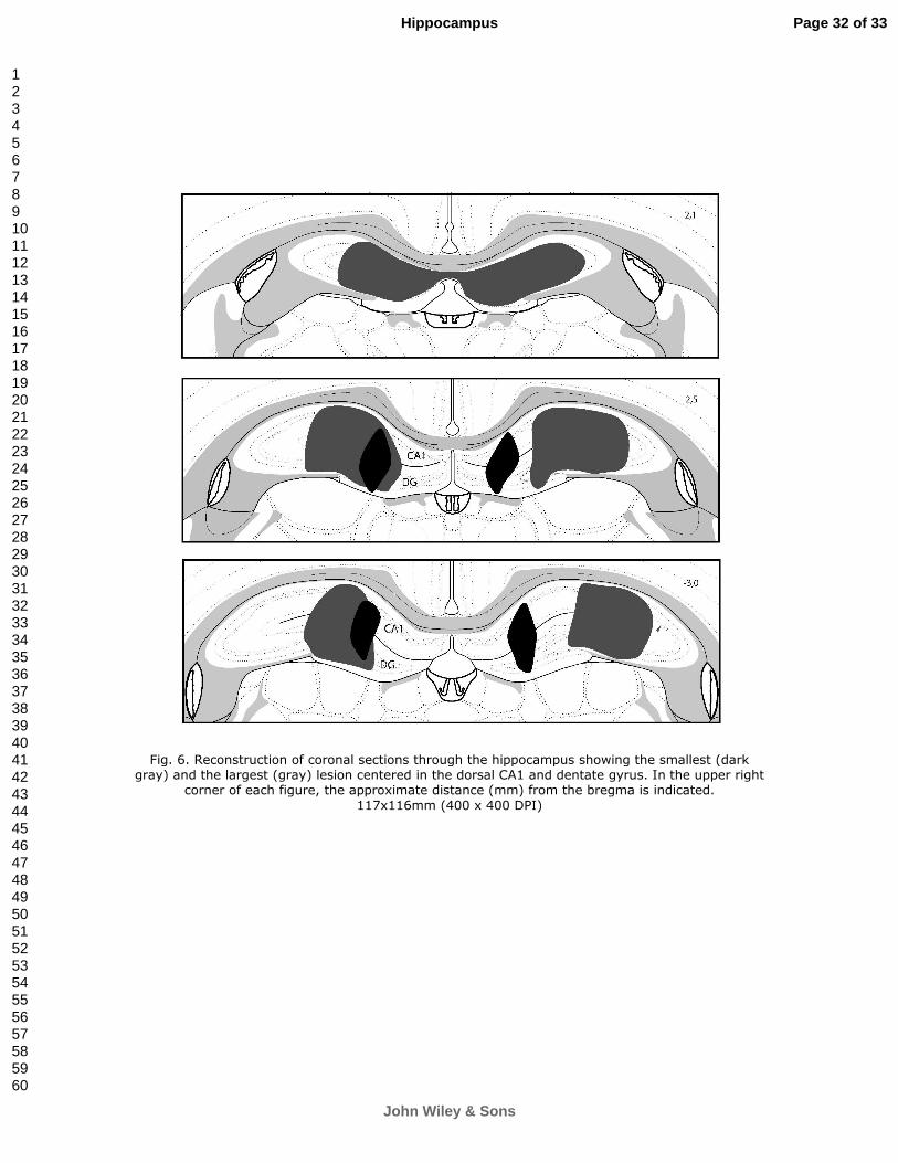
For Peer Review
Fig. 6. Reconstruction of coronal sections through the hippocampus showing the smallest (dark gray) and the largest (gray) lesion centered in the dorsal CA1 and dentate gyrus. In the upper right
corner of each figure, the approximate distance (mm) from the bregma is indicated.
117x116mm (400 x 400 DPI)
Page 32 of 33
John Wiley & Sons
Hippocampus
123456789101112131415161718192021222324252627282930313233343536373839404142434445464748495051525354555657585960

For Peer Review
Fig. 7. Reconstruction of coronal sections through the striatum showing the smallest (dark gray) and the largest (gray) lesion centered in the DLS. In the upper right corner of each figure, the
approximate distance (mm) from the bregma is indicated. 88x189mm (400 x 400 DPI)
Page 33 of 33
John Wiley & Sons
Hippocampus
123456789101112131415161718192021222324252627282930313233343536373839404142434445464748495051525354555657585960

47
4 DISCUSSÃO
Uma teoria bastante aceita na literatura é a dupla dissociação entre o sistema
hipocampal e o sistema estriatal nos processos de aprendizado e memória, onde o hipocampo
é importante para a memória declarativa, enquanto que o estriado é importante para a
memória de procedimento (Packard e Mcgaugh, 1992; Mcdonald e White, 1994; Packard e
Knowlton, 2002; White e Mcdonald, 2002; Packard, 2009; White, 2009). Os nossos
resultados reforçam a teoria de que o hipocampo dorsal é importante para a navegação
espacial no labirinto aquático de Morris, já que os animais com lesão apenas do hipocampo
dorsal apresentaram um prejuízo no desempenho desta versão do labirinto (Morris, Garrud et
al., 1982; Packard e Mcgaugh, 1992). Além disso, mostramos também que quando
lesionamos bilateralmente apenas o hipocampo dorsal não alteramos o desempenho dos
animais na versão com pista visual (Packard e Mcgaugh, 1992). Isto sugere que o hipocampo
dorsal é importante apenas para o aprendizado relacional (S-S) e não tem influência no
aprendizado S-R-O.
Entretanto, como os animais com lesão apenas do DLS não apresentaram prejuízo
em ambas as versões do labirinto aquático, estes resultados contradizem a teoria de Packard e
Mcgaugh (1992), a qual sugere que o estriado dorsal prejudica apenas a versão com pista
visual (Packard e Mcgaugh, 1992). Entretanto, no trabalho realizado por Packard e Mcgaugh
(1992) foram lesionados os estriados dorsais, sem delimitar estas lesões no DMS ou DLS. As
lesões foram tanto no DMS quanto no DLS.
Os nossos resultados estão de acordo com os trabalhos de DEVAN e colaboradores
(Devan, Mcdonald et al., 1999) que mostram uma dissociação entre o DMS e o DLS, onde a
lesão do DLS não prejudica nenhuma versão (espacial e com pista visual) do labirinto
aquático. Estes resultados diferentes encontrados com as lesões do DLS e do DMS levaram a

48
subdivisão funcional do estriado. Nesta nova visão, o DMS passou a ser considerado por estes
autores com parte do sistema hipocampal de memória espacial ou S-S (White, 2009) enquanto
o sistema estriatal, ou S-R-O, ficou restrito ao DLS (White, 2009). Porém, mesmo esta nova
visão não consegue explicar todos os resultados disponíveis, pois os animais com lesão
apenas do DLS não apresentaram prejuízo na versão com pista, sugerindo que o DLS não
seria importante para o aprendizado S-R-O.
Resultados interessantes foram obtidos quando submetemos os animais a um pré-
treinamento no labirinto aquático. Quando os animais com lesão do hipocampo dorsal são
pré-treinados, na versão com pista visual do labirinto aquático, e então testados na versão
espacial, eles apresentaram um desempenho semelhante aos animais do grupo SHAM. Isto
sugere que alguma informação adquirida pelo DLS no pré-treinamento foi utilizada para
auxiliar a navegação espacial no labirinto aquático.
Como sabemos que foi o DLS que participou na aquisição desta informação? É
porque quando lesionamos o hipocampo dorsal e o DLS (grupo HIP+DLS), mesmo com o
pré-treinamento os animais não conseguiram aprender a versão espacial do labirinto aquático.
Então, isto sugere que o DLS interage com o sistema hipocampal na navegação em um
labirinto aquático. Além disso, que esta interação é do tipo cooperativa (Mizumori, Cooper et
al., 2000; Voermans, Petersson et al., 2004; Hartley e Burgess, 2005). Isto também sugere
que não só o hipocampo dorsal, mas também o DLS tem um papel na navegação espacial no
labirinto aquático (Mizumori, Puryear et al., 2009).
Esta cooperação entre o hipocampo dorsal e o DLS também foi observada quando os
animais foram pré-treinados na versão espacial e depois testados na versão com pista visual.
Os animais SHAM, HIP e DLS apresentaram um desempenho semelhante na versão com
pista visual. Mas, os animais com lesão dupla (grupo HIP+DLS) apresentaram um
desempenho pior que estes três grupos. Ou seja, quando a lesão ocorre só no hipocampo ou só

49
no DLS, os animais desempenham normalmente a tarefa, mas quando a lesão ocorre em
ambas as estruturas estes animais apresentam um prejuízo. Isto sugere este papel cooperativo
entre o hipocampo dorsal e o DLS na navegação no labirinto aquático (Mizumori, Puryear et
al., 2009).
Como vários trabalhos mostram que o hipocampo dorsal tem a capacidade de
codificar o ambiente formando um mapa cognitivo através da atividade de neurônios
denominados de células de lugar ou place cells (Wilson e Mcnaughton, 1993). Uma
explicação alternativa é que o papel do hipocampo dorsal na navegação no labirinto aquático
é processar as informações sensoriais para mapear o ambiente através dos diversos estímulos
(objetos) presentes fora do labirinto aquático. Porém, o hipocampo não tem a capacidade de
coordenar as respostas motoras (ações) e nem detectar as recompensas obtidas durante a
realização desta navegação. Quem desempenharia esta seleção de quais as ações deveriam ser
tomadas para se conseguir uma recompensa seria o estriado ou ao menos uma parte dele, i.e.,
DLS. Isto porque o estriado dorsal apresenta neurônios que se ativam quando o animal
executa movimentos com orientação egocêntrica, direcionamento da cabeça para locais
específicos e também em função da expectativa de recompensa (Lavoie e Mizumori, 1994;
Mizumori, Cooper et al., 2000; Schultz, 2006; Schmitzer-Torbert e Redish, 2008; Mizumori,
Puryear et al., 2009).
Este papel do DLS na seleção de ações durante a navegação no labirinto aquático
pode ser explicado pelo modelo do mosaico dos espelhos quebrados (anexo 2) (Da Cunha,
Wietzikoski et al., 2009). Segundo este modelo, o córtex sensorial e motor enviam projeções
para o estriado de forma convergente e repetitiva. Em função destas projeções estímulos
sensoriais (partes do corpo, objetos e partes do ambiente) são representados de forma
fragmentada e repetida no estriado. Cada fragmento forma uma unidade funcional no estriado.
Quando uma unidade funcional do estriado é ativada ao mesmo tempo por projeções do

50
sensorial (representando uma parte do corpo e um objeto), por projeções do córtex motor
(representando a ação da parte do corpo sobre objeto) e por projeções de neurônios
dopaminérgicos mesencefálicos (ativados de forma fásica pela novidade) ocorrem fenômenos
de plasticidade sináptica que podem fortalecer ou enfraquecer estas associações. Este
fortalecimento desta associação na via direta (estriado – Gpi/SNr) e o enfraquecimento da via
indireta (estriado – Gpe – NST – Gpi/SNr) faz com que a aquela ação recompensada que foi
deflagrada por aquele estímulo seja reforçada.
Portanto, nossos resultados sugerem que a interação entre o hipocampo dorsal e o
DLS não é de forma cooperativa ou competitiva, mas complementar, ou seja, que estas duas
estruturas desempenhem funções diferentes, mas indispensáveis para a navegação espacial e
também na navegação orientada por pistas. Nesta complementariedade, o hipocampo
mapearia o ambiente enquanto que o estriado selecionaria quais são as ações a serem tomadas
durante a navegação.

51
5 CONCLUSÕES
Os resultados desta tese questionam de forma crítica a teoria de que o hipocampo
dorsal seja suficiente para controlar o comportamento de navegação espacial baseado em
memórias relacionais (S-S), tal como na versão espacial do labirinto aquático de Morris.
Assim como, de que o DLS seja suficiente para mediar a navegação baseada em pistas
(aprendizagem S-R-O), tal como na versão com pista visual do labirinto aquático de Morris.
Ou seja, esta tese apresenta evidências de que tanto o hipocampo dorsal quanto o DLS sejam
necessários para mediar a aquisição de comportamentos de navegação baseados tanto em
memórias S-S como S-R-O.

52
REFERÊNCIAS BIBLIOGRÁFICAS
Albouy, G., V. Sterpenich, et al. Both the hippocampus and striatum are involved in consolidation of motor sequence memory. Neuron, v.58, n.2, Apr 24, p.261-72. 2008. Balleine, B. W., M. Liljeholm, et al. The integrative function of the basal ganglia in instrumental conditioning. Behav Brain Res, v.199, n.1, Apr 12, p.43-52. 2009. Berke, J. D., J. T. Breck, et al. Striatal versus hippocampal representations during win-stay maze performance. J Neurophysiol, v.101, n.3, Mar, p.1575-87. 2009. Da Cunha, C., M. E. Angelucci, et al. The lesion of the rat substantia nigra pars compacta dopaminergic neurons as a model for Parkinson's disease memory disabilities. Cell Mol Neurobiol, v.22, n.3, Jun, p.227-37. 2002. Da Cunha, C., M. S. Gevaerd, et al. Memory disruption in rats with nigral lesions induced by MPTP: a model for early Parkinson's disease amnesia. Behav Brain Res, v.124, n.1, Sep 28, p.9-18. 2001. Da Cunha, C., M. H. Silva, et al. Place learning strategy of substantia nigra pars compacta-lesioned rats. Behav Neurosci, v.120, n.6, Dec, p.1279-84. 2006. Da Cunha, C., E. C. Wietzikoski, et al. Learning processing in the basal ganglia: a mosaic of broken mirrors. Behav Brain Res, v.199, n.1, Apr 12, p.157-70. 2009. Da Cunha, C., S. Wietzikoski, et al. Evidence for the substantia nigra pars compacta as an essential component of a memory system independent of the hippocampal memory system. Neurobiol Learn Mem, v.79, n.3, May, p.236-42. 2003. ______. Pre-training to find a hidden platform in the Morris water maze can compensate for a deficit to find a cued platform in a rat model of Parkinson's disease. Neurobiol Learn Mem, v.87, n.4, May, p.451-63. 2007. Devan, B. D., R. J. Mcdonald, et al. Effects of medial and lateral caudate-putamen lesions on place- and cue-guided behaviors in the water maze: relation to thigmotaxis. Behav Brain Res, v.100, n.1-2, Apr, p.5-14. 1999. Doeller, C. F., J. A. King, et al. Parallel striatal and hippocampal systems for landmarks and boundaries in spatial memory. Proc Natl Acad Sci U S A, v.105, n.15, Apr 15, p.5915-20. 2008. Eichenbaum, H. Hippocampus: cognitive processes and neural representations that underlie declarative memory. Neuron, v.44, n.1, Sep 30, p.109-20. 2004. Eichenbaum, H., C. Stewart, et al. Hippocampal representation in place learning. J Neurosci, v.10, n.11, Nov, p.3531-42. 1990. Gold, J. J. e L. R. Squire. The anatomy of amnesia: neurohistological analysis of three new cases. Learn Mem, v.13, n.6, Nov-Dec, p.699-710. 2006. Goodrich-Hunsaker, N. J., S. A. Livingstone, et al. Spatial deficits in a virtual water maze in amnesic participants with hippocampal damage. Hippocampus, Jun 24. 2009. Guillazo-Blanch, G., R. Nadal, et al. Effects of fimbria lesions on trace two-way active

53
avoidance acquisition and retention in rats. Neurobiol Learn Mem, v.78, n.2, Sep, p.406-25. 2002. Hartley, T. e N. Burgess. Complementary memory systems: competition, cooperation and compensation. Trends Neurosci, v.28, n.4, Apr, p.169-70. 2005. Hull, C. L. Principles of behavior. New York: Appleton. 1943 Izquierdo, I. Memória. Porto Alegre: Artmed Editora. 2002. 95 p. Knowlton, B. J., J. A. Mangels, et al. A neostriatal habit learning system in humans. Science, v.273, n.5280, Sep 6, p.1399-402. 1996. Lavoie, A. M. e S. J. Mizumori. Spatial, movement- and reward-sensitive discharge by medial ventral striatum neurons of rats. Brain Res, v.638, n.1-2, Feb 28, p.157-68. 1994. Lee, A. S., R. S. Duman, et al. A double dissociation revealing bidirectional competition between striatum and hippocampus during learning. Proc Natl Acad Sci U S A, v.105, n.44, Nov 4, p.17163-8. 2008. Mcdonald, R. J., N. S. Hong, et al. The challenges of understanding mammalian cognition and memory-based behaviours: an interactive learning and memory systems approach. Neurosci Biobehav Rev, v.28, n.7, Nov, p.719-45. 2004. Mcdonald, R. J. e N. M. White. Parallel information processing in the water maze: evidence for independent memory systems involving dorsal striatum and hippocampus. Behav Neural Biol, v.61, n.3, May, p.260-70. 1994. Miyoshi, E., S. Wietzikoski, et al. Impaired learning in a spatial working memory version and in a cued version of the water maze in rats with MPTP-induced mesencephalic dopaminergic lesions. Brain Res Bull, v.58, n.1, May, p.41-7. 2002. Mizumori, S. J., B. G. Cooper, et al. A neural systems analysis of adaptive navigation. Mol Neurobiol, v.21, n.1-2, Feb-Apr, p.57-82. 2000. Mizumori, S. J. Y., C. B. Puryear, et al. Basal ganglia contributions to adaptive navigation. Behavioural Brain Research, v.199, n.1, Apr 12, p.32-42. 2009. Morris, R. G., P. Garrud, et al. Place navigation impaired in rats with hippocampal lesions. Nature, v.297, n.5868, Jun 24, p.681-3. 1982. Oliveira, M. G., O. F. Bueno, et al. Strategies used by hippocampal- and caudate-putamen-lesioned rats in a learning task. Neurobiol Learn Mem, v.68, n.1, Jul, p.32-41. 1997. Packard, M. G. Exhumed from thought: Basal ganglia and response learning in the plus-maze. Behavioural Brain Research, v.199, n.1, Apr 12, p.24-31. 2009. Packard, M. G., R. Hirsh, et al. Differential effects of fornix and caudate nucleus lesions on two radial maze tasks: evidence for multiple memory systems. J Neurosci, v.9, n.5, May, p.1465-72. 1989. Packard, M. G. e B. J. Knowlton. Learning and memory functions of the Basal Ganglia. Annu Rev Neurosci, v.25, p.563-93. 2002.

54
Packard, M. G. e J. L. Mcgaugh. Double dissociation of fornix and caudate nucleus lesions on acquisition of two water maze tasks: further evidence for multiple memory systems. Behav Neurosci, v.106, n.3, Jun, p.439-46. 1992. Packard, M. G. e L. A. Teather. Double dissociation of hippocampal and dorsal-striatal memory systems by posttraining intracerebral injections of 2-amino-5-phosphonopentanoic acid. Behav Neurosci, v.111, n.3, Jun, p.543-51. 1997. ______. Amygdala modulation of multiple memory systems: hippocampus and caudate-putamen. Neurobiol Learn Mem, v.69, n.2, Mar, p.163-203. 1998. Packard, M. G. e N. M. White. Lesions of the caudate nucleus selectively impair "reference memory" acquisition in the radial maze. Behav Neural Biol, v.53, n.1, Jan, p.39-50. 1990. Poldrack, R. A. e M. G. Packard. Competition among multiple memory systems: converging evidence from animal and human brain studies. Neuropsychologia, v.41, n.3, p.245-51. 2003. Prediger, R. D., L. C. Batista, et al. The risk is in the air: Intranasal administration of MPTP to rats reproducing clinical features of Parkinson's disease. Exp Neurol, v.202, n.2, Dec, p.391-403. 2006. Prediger, R. D., D. Rial, et al. Risk is in the air: an intranasal MPTP (1-methyl-4-phenyl-1,2,3,6-tetrahydropyridine) rat model of Parkinson's disease. Ann N Y Acad Sci, v.1170, Jul, p.629-36. 2009. Salmon, D. P. e N. Butters. Neurobiology of skill and habit learning. Curr Opin Neurobiol, v.5, n.2, Apr, p.184-90. 1995. Schmitzer-Torbert, N. C. e A. D. Redish. Task-dependent encoding of space and events by striatal neurons is dependent on neural subtype. Neuroscience, v.153, n.2, May 2, p.349-360. 2008. Schultz, W. Behavioral theories and the neurophysiology of reward. Annual Review of Psychology, v.57, p.87-115. 2006. Scoville, W. B. e B. Milner. Loss of recent memory after bilateral hippocampal lesions. J Neurol Neurosurg Psychiatry, v.20, n.1, Feb, p.11-21. 1957. Squire, L. R. Memory systems of the brain: A brief history and current perspective. Neurobiology of Learning and Memory, v.82, n.3, Nov, p.171-177. 2004. ______. The legacy of patient H.M. for neuroscience. Neuron, v.61, n.1, Jan 15, p.6-9. 2009. Squire, L. R., C. E. Stark, et al. The medial temporal lobe. Annu Rev Neurosci, v.27, p.279-306. 2004. Thorndike, E. L. Animal intelligence: experimental studies. New York: Macmillan. 1911 Torras-Garcia, M., D. Costa-Miserachs, et al. Improvement of shuttle-box performance by anterodorsal medial septal lesions in rats. Behav Brain Res, v.141, n.2, May 15, p.147-58. 2003.

55
Voermans, N. C., K. M. Petersson, et al. Interaction between the human hippocampus and the caudate nucleus during route recognition. Neuron, v.43, n.3, Aug 5, p.427-35. 2004. White, N. M. Some highlights of research on the effects of caudate nucleus lesions over the past 200 years. Behavioural Brain Research, v.199, n.1, Apr 12, p.3-23. 2009. White, N. M. e R. J. Mcdonald. Multiple parallel memory systems in the brain of the rat. Neurobiol Learn Mem, v.77, n.2, Mar, p.125-84. 2002. Wilson, M. A. e B. L. Mcnaughton. Dynamics of the Hippocampal Ensemble Code for Space. Science, v.261, n.5124, Aug 20, p.1055-1058. 1993. Xavier, G. F. e V. C. Costa. Dentate gyrus and spatial behaviour. Prog Neuropsychopharmacol Biol Psychiatry, v.33, n.5, Aug 1, p.762-73. 2009. Yin, H. H. e B. J. Knowlton. The role of the basal ganglia in habit formation. Nat Rev Neurosci, v.7, n.6, Jun, p.464-76. 2006.
56
ANEXOS
Além deste trabalho realizado durante a tese, foram publicados outros artigos.
Apresento aqui, nos anexo, estes trabalhos que foram publicados. No primeiro trabalho
revisamos os trabalhos que mostram o papel da substância negra nos processos de
aprendizado e memória, exemplificando com o que acontece em pacientes com doença de
Parkinson. No segundo trabalho, a partir de trabalhos já publicados, elaboramos e
apresentamos uma nova teoria para explicar como os gânglios da base participam nos
processos de aprendizado de memórias de procedimentos.

57
ANEXO 1

REVISTA MEXICANA DE ANÁLISIS DE LA CONDUCTA 2006 NÚMERO 2 (DIC) MEXICAN JOURNAL OF BEHAVIOR ANALYSIS 32, 219-242 NUMBER 2 (DEC)
A RAT MODEL OF THE COGNITIVE IMPAIRMENTS IN PARKINSON’S DISEASE
UN MODELO EN RATA FRL DETERIORO COGNITIVO EN LA ENFERMEDAD DE PARKINSON
CLAUDIO DA CUNHA1* RUI D. S. PREDIGER2 EDMAR MIYOSHI1,3 REINALDO N. TAKAHASHI2
1LABORATÓRIO DE FISIOLOGIA E FARMACOLOGIA DO SISTEMA NERVOSO CENTRAL, DEPARTAMENTO DE FARMACOLOGIA, UFPR,
C.P. 19.031, 81.531-980, CURITIBA-PR, BRAZIL. 2DEPARTAMENTO DE FARMACOLOGIA, CENTRO DE CIÊNCIAS
BIOLÓGICAS, UNIVERSIDADE FEDERAL DE SANTA CATARINA, UFSC, FLORIANÓPOLIS-SC, BRAZIL.
3DEPARTAMENTO DE CIÊNCIAS FARMACÊUTICAS, UNIVERSIDADE ESTADUAL DE PONTA GROSSA, PONTA GROSSA-PR, BRAZIL.
ABSTRACT
Although Parkinson’s disease (PD) is classically considered to be a motor system disease, subtle cognitive impairments can be observed even during the early phases of PD. In this article we review behavioral and neurochemi-cal studies on the cognitive alterations observed in rats treated with intranigral infusion of the neurotoxin MPTP. The critical role of dopamine release in the dorsal striatum and its modulation by adenosine receptors is also reviewed as a potential strategy to treat the cognitive disabilities of PD patients who do not improve with levodopa therapy. Most of the impairments presented by rats treated with intranigral infusion of MPTP are similar to those observed during the early phase of PD, when a moderate loss of nigral dopamine neurons (40-70%) results in sensory and memory defi cits with no major motor impair-
* This work was supported by CNPq, Fundação Araucária, Institutos de Milênio, and CAPES. Corresponding author: Laboratório de Fisiologia e Farmacologia do Sistema Nervoso Central, Departamento de Farmacologia, UFPR, C.P. 19.031, 81.531-980, Curitiba-PR, Brazil. Phone 55
RMAC_dic_2006.indd 219RMAC_dic_2006.indd 219 21/05/2007 01:30:00 p.m.21/05/2007 01:30:00 p.m.
220 DA CUNHA, PREDIGER, MIYOSHI AND TAKAHASHI
ments. These animals also model the working memory and habit learning defi cits, with long-term spatial (episodic) memories being mostly spared as observed in non-demented PD patients. The intranigral infusion of MPTP in rats has led to the development of useful models, which do not present gross motor impairments that would otherwise compromise the interpretation of the performance of the animals in cognitive tasks.
Keywords: Parkinson’s disease, learning, memory, cognition, MPTP, rats, animal model.
RESUMEN
Aunque el mal de Parkinson (DP) es considerado clásicamente como un desorden del sistema motor, pueden observarse ligeros deterioros cognitivos aun en las fases iniciales del DP. En este artículo revisamos estudios conduc-tuales y neuroquímicos sobre alteraciones cognitivas observadas en ratas tratadas con infusiones intranigrales de la neurotoxina MPTP. El papel crítico de la liberación de dopamina en el estriado dorsal y su modulación por los receptores de adenosina también es revisada como una estrategia potencial para tratar los deterioros cognitivos en pacientes con desorden de Parkinson (PD) que no mejoran con la terapia de levo dopa. Resultados: La mayoría de de los daños presentados en ratas con infusiones intranigrales de MPTP son similares a los observados en las primeras fases de PD, una pérdida moderada de neuronas nigrales dopaminérgicas (40-70%) que causa défi -cits sensoriales y motores y poco deterioro motor. Estos animales también modelan los défi cits de memoria de trabajo y aprendizaje de hábitos, con la memoria de largo plazo espacial (episódica) mayormente preservada como se observa en los pacientes sin DP. La infusión intranigral de MPTP en ratas a llevado al desarrollo de modelos útiles, ya que no presentan un deterioro motor excesivo que podría de otra manera comprometer la interpretación de de la ejecución de los animales en tareas cognitivas.
Palabras clave: mal de Parkinson, aprendizaje, memoria, cognición, MPTP, ratas, modelo animal.
Parkinson’s disease (PD) is the second most common neurodegenerative disorder, following Alzheimer’s disease, affecting approximately 1% of the population older than 50 years (Duvoisin 1991). Current estimates from the American Parkinson’s Disease Foundation put the number of American citi-zens suffering from this disease at more than 1.5 million individuals. Since the incidence of the disease increases with age (the most important risk factor), it is likely that the number of people suffering from PD will rise as improved health care lengthens the average life span.
RMAC_dic_2006.indd 220RMAC_dic_2006.indd 220 21/05/2007 01:30:01 p.m.21/05/2007 01:30:01 p.m.

221A RAT MODEL OF THE COGNITIVE IMPAIRMENTS IN PARKINSON’S DISEASE
Classically, PD is considered to be a motor system disease and its diag-nosis is based on the presence of a set of cardinal motor signs (e.g. rigidity, bradykinesia, rest tremor and postural refl ex disturbance). These symptoms of PD mainly result from the progressive degeneration of dopamine neurons of the substantia nigra pars compacta (SNc) that project predominantly to the striatum (Hirsch et al. 1988), a fact that contributes to the prevailing view that the basal ganglia are mainly concerned with motor control functions (Heikkila et al. 1989). More recently, an increasing amount of evidence has suggested that this system is also critically involved in learning and memory processes (Brown et al. 1997), as indicated by the fact that many cognitive impairments, including memory defi cits, occur during the early stage of PD even before the development of its classical symptoms (Dubois and Pillon 1997; Owen et al. 1995). The non-motor symptoms that include cognitive defi cits can be more important than the motor defi cits to determine the patients’ quality of life and represent an important factor to determine the need for nursing home care.
On the other hand, animal models are an invaluable tool for studying the pathogenesis and progression of human diseases, as well as for testing new therapeutic intervention strategies. PD is one of many human diseases which do not appear to have arisen spontaneously in animals. The characteristic features of the disease can, however, be more or less faithfully mimicked in animals through genetic approaches and the administration of various neuro-toxic agents that interfere with dopaminergic neurotransmission. Despite the recent discovery of mutations in the alpha-synuclein gene (and some other genes) in a few PD patients that has led to the development of gene-based PD models (von Bohlen und Halbach et al. 2004), the administration of differ-ent neurotoxins such as 6-hydroxydopamine (6-OHDA) and 1-methyl-4-phe-nyl-1,2,3,6,-tetrahydropyridine (MPTP), which disrupt or destroy the dopami-nergic system, remains the most widely used animal model for the study of PD. Although these models have undoubtedly contributed to a better under-standing of many features of PD, most studies have focused on the ability of these models to induce nigrostriatal pathway damage and motor alterations associated with advanced phases of PD. However, until recently, no well-ac-cepted model of the early phase of PD was available in the literature. The present review seeks to document these challenges using our earlier review (Da Cunha et al. 2002) as a basis for integrating the subsequent behavioral and neurochemical studies showing that the intranigral infusion of MPTP into rats causes a partial loss of dopamine neurons in the SNc and depletion of striatal dopamine, resulting in sensory and memory defi cits with no major mo-tor impairments, thus representing a model of the early phase of PD.
Finally, the fact that most of the drugs currently available for the treatment of PD (such as levodopa) are more effi cient in alleviating motor than cogni-tive impairments has led many researchers to postulate non-dopaminergic
RMAC_dic_2006.indd 221RMAC_dic_2006.indd 221 21/05/2007 01:30:01 p.m.21/05/2007 01:30:01 p.m.
222 DA CUNHA, PREDIGER, MIYOSHI AND TAKAHASHI
mechanisms for the cognitive symptoms of this disease. Here, we will briefl y review clinical and non-clinical studies evaluating the potential of caffeine and other adenosine receptor antagonists to restore defective learning and mem-ory processes in PD.
COGNITIVE IMPAIRMENTS IN PARKINSON’S DISEASE
In addition to the characteristic motor symptoms, subtle cognitive impairments can be observed even during the early phases of PD (Dubois and Pillon 1997; Bosboom et al. 2004). They comprise a dysexecutive syndrome that includes attentional and working memory impairments accompanied by secondary defi cits in the internal representation of visuospatial stimuli and in the use of declarative memory storage (Bradley 1989; Owen et al. 1993; Dubois and Pil-lon 1997; Tamaru 1997; Bosboom et al. 2004). Skill and habit learning is also impaired in these patients (Knowlton et al. 1996). Almost one-third of patients may eventually progress to dementia (Aarsland et al. 1996).
Dysexecutive syndrome represents the core of the cognitive impairments and dementia observed in PD, and appears even during early stages of the disease (Dubois and Pillon 1997; Tamaru, 1997; Bosboom et al. 2004; Owen 2004; Zgaljardic et al. 2004). Executive function describes a wide range of cognitive functions required for goal-directed, adaptive behavior in response to new, challenging environmental situations, including planning, task man-agement, attention, inhibition, monitoring, and coding. All of these functions are attributable to the prefrontal cortex and therefore, PD cognitive disabilities resemble cognitive defi cits found in frontal cortex patients (Tamaru 1997; Ma-rie et al. 1999; Owen 2004).
A recent positron emission tomography study by Aalto et al. (2005) has shown increased dopamine release in the frontal cortex of human subjects performing a working memory task. Working memory (Stebbins et al. 1999; Marie and Defer 2003), especially spatial working memory (Pillon et al. 1996, 1997; Owen et al. 1997), fails in non-demented PD patients. The articulatory (verbal-phonological) component of the working memory is usually preserved, but when a verbal working memory task demands more attention, a defi ciency is also observed in these patients (Moreaud et al. 1997; Owen 2004). These impairments are possibly the consequence of failure of the central executive component that manages the short-term memory. Thus, these impairments appear when the working memory tasks present a higher demand on execu-tive functions such as planning and attention shifting (Bosboom et al. 2004).
Many studies have tested whether PD patients, who are known to have a striatal depletion of dopamine, present non-declarative learning and mem-ory defi cits (Bondi and Kaszniak 1991). These patients fail to improve mirror
RMAC_dic_2006.indd 222RMAC_dic_2006.indd 222 21/05/2007 01:30:02 p.m.21/05/2007 01:30:02 p.m.
223A RAT MODEL OF THE COGNITIVE IMPAIRMENTS IN PARKINSON’S DISEASE
reading of words that appeared only once during the test, an impairment at-tributable to a skill learning defi cit (Thomas et al. 1996; Koenig et al. 1999). PD patients have been shown to present impaired skill learning not only for visuoperceptual but also for motor skill tasks such as puzzle assembly, press-ing specifi c keys on a computer keyboard in response to a stimulus present-ed on the computer screen, and drawing lines in hidden mazes (Bondi and Kaszniak 1991; Thomas et al. 1996; Moreaud et al. 1997). Many defi cits of PD patients in performing non-declarative tasks relay on the initial learning phase (Dujardin and Laurent 2003). On the other hand, there is no consensus about whether PD spares declarative memory (Thomas et al. 1996; Bondi and Kaszniak 1991). PD patients are generally not impaired to encode and store consolidated new information, but they present diffi culties in retrieving this in-formation, particularly when they have to self-initiate remembering strategies (Dujardin and Laurent 2003). A failure in executive functions may explain this defi cit. However, the non-intentional and automatic nature of a non-declara-tive task, such as learning a list of words or matching pairs of words, may also determine whether it can be learned normally or not by PD patients (Faglioni et al. 1995, 1997; Roncacci et al. 1996). Some authors explain the declara-tive defi cits reported in some studies involving PD patients as resulting from the fact that they require a larger number of repetitions of the task to translate non-declarative (procedural) into declarative knowledge (Pascual-Leone et al. 1993).
Habits are by defi nition stimulus-response associations that are uncon-sciously learned through repetitively rewarded experiences. The main diffi cul-ty to model a habit task is to guarantee that the subjects will not respond con-sciously in order to receive the reward. One of the main well-designed studies planned to test whether non-demented PD patients are impaired in stimu-lus-response habit learning used a probabilistic classifi cation task (Knowlton et al. 1996). The probabilistic structure of the task permitted the subjects to learn the task unconsciously by trial-and-error. PD patients scored worse than Alzheimer’s disease patients and healthy subjects, but when asked about it, they remembered to have participated in the previous training sessions. Alzheimer’s disease patients, on the other hand, learned this task like healthy subjects, but barely remembered the training episode. This study supports the double dissociation proposed for the medial temporal- and basal ganglia-mediated declarative (episodic) and non-declarative (implicit habit learning) memory systems, respectively (Packard and Knowlton 2002).
The risk of developing dementia is up to six times higher in PD pa-tients than in healthy subjects of the same age (Aarsland et al. 1996). The core of the impairments lies in executive functions (e.g. set-shifting) (Girot-ti 1986). Mood (e.g. depression), and psychotic (e.g. visual hallucinations) symptoms are also common in demented PD patients. Other common im-
RMAC_dic_2006.indd 223RMAC_dic_2006.indd 223 21/05/2007 01:30:02 p.m.21/05/2007 01:30:02 p.m.

224 DA CUNHA, PREDIGER, MIYOSHI AND TAKAHASHI
pairments include visuospatial and visuoconstructive skills. Speech and lan-guage diffi culties, such as naming and sentence comprehension, are also common. Furthermore, poor verbal fl uency would be predictive of dementia in PD. Declarative memory impairments are present, but are less severe, as compared to Alzheimer’s disease. There is a defi cit in free recall, but it can be compensated for by semantic cueing. Furthermore, PD patients have more problems to recall than to encode declarative memories, i.e., their impairment relies on diffi culties in activating processes involved in the functional use of memory storages, probably as a consequence of the dysexecutive syndrome. Recognition memory is relatively intact (Bosboom et al. 2004). Some of these cognitive impairments, especially attention impairment, are aggravated by a degeneration of cholinergic neurons in the nucleus basalis of Meynert and of noradrenaline neurons in the locus ceruleus that also occur in PD. On the other hand, impairments in declarative memory, aphasia and apraxia, when present, are related to cortical pathology indicative of Alzheimer’s disease or Lewy body dementia. Regarding the last co-morbidity, it is noteworthy how many characteristics of PD dementia resemble Lewy body dementia. Addi-tionally, postmortem studies have revealed that many Lewy body disease pa-tients had been wrongly diagnosed in life as having PD patients and many PD patients develop Lewy body disease later on (Zgaljardic et al. 2004).
THE BASAL GANGLIA SYSTEM OF LEARNING AND MEMORY
As important as knowing the cause of PD is to know the normal function of the brain components affected by this disease. Dopamine neurons of the SNc modulate the basal ganglia, which are composed of the caudate nucleus and putamen (altogether called striatum) and the globus pallidus. Due to their re-ciprocal connections with these core structures of the basal ganglia, the sub-stantia nigra, ventral tegmental area and the subthalamic nucleus are con-sidered to be associated basal ganglia structures (Alexander and Crutcher 1990). Neurons from all parts of the neocortex project to the striatum. Striatal neurons, in turn, project to the globus pallidus or to the substantia nigra pars reticulata which projects to the ventrolateral thalamus that, in turn, projects back to the frontal cortex (Alexander and Crutcher 1990). Therefore, the ac-tivity of sensory and motor parts of the cortex affects the activity of the basal ganglia that, in turn, modulate the activity of motor and cognitive parts of the frontal cortex. The positive modulation exerted by glutamate thalamic neurons on the frontal cortex is under inhibitory control of GABAergic neurons of the globus pallidus and the substantia nigra pars reticulata. This inhibition can be either blocked by a direct pathway or increased by an indirect pathway of neurons that arise in the striatum. Midbrain dopamine neurons play a dual
RMAC_dic_2006.indd 224RMAC_dic_2006.indd 224 21/05/2007 01:30:03 p.m.21/05/2007 01:30:03 p.m.
225A RAT MODEL OF THE COGNITIVE IMPAIRMENTS IN PARKINSON’S DISEASE
role in the modulation of the activity of these striatal neurons. Acting on D1-like or D2-like dopamine receptors, the dopamine released by these neurons activates the direct pathway and inhibits the indirect pathway, respectively. Both actions result in a positive modulation of the motor and cognitive func-tions of the frontal cortex (Alexander et al. 1986). According to this view, it is clear that the loss of midbrain dopamine neurons that occurs in PD results in the impairment of both motor and cognitive functions.
How does the decrease of dopamine concentration in the striatum, and its consequent decrease in the positive modulation exerted by the basal gan-glia loop on the frontal cortex, causes the cognitive impairments observed in PD? Let us start with motor skills and habit learning. The primary motor cortex, supplementary motor area and somatosensory cortex neurons di-rectly control the fi ring of spinal motor neurons, leading to consciously willed movements. Motor programs are the orchestrated sequences of commands to functional groups of muscles that govern movements at or around the joints (Alexander and Crutcher 1990). Where are these motor programs encoded and stored? The striatum is in a strategic position to participate in the encod-ing of such motor programs that will constitute the framework of the motor skills and habits (Packard and Knowlton 2002). The ability to perform a skill demands the coordinated activity of muscle groups from different parts of the body, the continuous integration of information about the contraction state of these muscles, and the visual follow-up of the movement in order to make fi ne adjustments for proper movements. Habit learning consists of increasing the probability that a sensory stimulus triggers a motor program designed for a particular behavioral response (White and McDonald 2002). As mentioned above, both sensory and motor regions of the entire cortex project to the striatum. The primary motor cortex also presents a somatotopic organization. Inputs from regions of the primary motor (MI) and sensory (SI) cortex that rep-resent the same part of the body send projections to the same region within the striatum (Flaherty and Graybiel 1998). However, while the cortical regions form a single and continuous representation of the entire body, the represen-tation of these areas of the body in the striatum is broken into a mosaic and is redundant, i.e., each part of the body is represented by multiple striatal units called matrisomes. After these somatosensorimotor inputs are processed in the striatum, the multiple matrisomes representing the same body parts send overlapping projections to the globus pallidus, where a unique and continuous representation of the body is restored (Graybiel 1998). Primary cortex regions for other sensory modalities, i.e., vision, hearing and smell, also send projec-tions to the striatum (Calabresi et al. 1996). Notice that the multiple mosaic representation of sensory and motor information in the striatum allows the association of different stimuli with the activation of movement sequences in-volving different parts of the body. The capacity of dopamine neurons to either
RMAC_dic_2006.indd 225RMAC_dic_2006.indd 225 21/05/2007 01:30:03 p.m.21/05/2007 01:30:03 p.m.

226 DA CUNHA, PREDIGER, MIYOSHI AND TAKAHASHI
inhibit or stimulate the basal-cortical output and to induce fi ring-dependent plasticity in the corticostriatal synapses enables this system to form experi-ence-driven stimulus-response programs that are the basis of skills and habit learning (Graybiel et al. 1994; Graybiel 1998).
Working memory and executive functions, also affected in PD, depend on the activity of the prefrontal cortex (Dirnberger et al. 2005). There are loops integrating the dorsolateral and the orbitofrontal areas of the prefron-tal cortex with the basal ganglia (Alexander et al. 1986). A study by Postle and D’Esposito (1999) showed increased activity of these cortical regions and the dorsal striatum when subjects were performing spatial working memory tasks. Furthermore, Lewis et al. (2003) reported that cognitive impairments in early PD, including working memory, are accompanied by a reduced activity in the frontostriatal neural circuitry. The concept of working memory involves the integration and maintenance of information for its prospective use when selecting the appropriate behavior (Baddeley 2003). This process could in-volve the transformation of sensory cues into a code response. The prefrontal cortex is at the top hierarchy of the sensory and motor systems (Faw 2003). Like the striatum, it can receive information from all sensory modalities and control the motor output. While doing this, it works in consonance with basal ganglia loops. These corticobasal loops can run parallel subroutines that are unconsciously operated, while the prefrontal cortex is involved in solving con-scious demands for the ongoing behavior. Like the striatum, the prefrontal cortex is also modulated by dopamine neurons arising in the midbrain (Costa et al. 2003). Therefore, it is easy to understand how the abnormal depletion of dopamine levels in these brain regions as observed in PD can affect work-ing memory. In the same way, attention and other executive functions of the prefrontal cortex will be affected by dopamine depletion in the striatum and prefrontal cortex (Dubois and Pillon 1997; Owen 2004).
MPTP-LESIONED RAT AS AN ANIMAL MODEL OF COGNITIVE IMPAIRMENTS OBSERVED DURING THE EARLY PHASE OF
PARKINSON’S DISEASE
In the early 1980s, the dopaminergic neurotoxin MPTP was accidentally dis-covered when a group of young drug addicts in California developed an idio-pathic parkinsonian syndrome. Investigation revealed that the syndrome was caused by self-administration of a “synthetic heroin” analogue that had been contaminated with a byproduct (MPTP) during manufacturing (Davis et al. 1979; Langston et al. 1983). At present, MPTP represents the most important and most frequently used neurotoxin applied to animal models of PD, pre-senting advantages over all other toxic PD models since it causes a specifi c
RMAC_dic_2006.indd 226RMAC_dic_2006.indd 226 21/05/2007 01:30:04 p.m.21/05/2007 01:30:04 p.m.

227A RAT MODEL OF THE COGNITIVE IMPAIRMENTS IN PARKINSON’S DISEASE
loss of dopamine neurons and induces symptoms identical to PD in humans (Przedborski and Vila 2003).
MPTP is highly lipophilic and readily crosses the blood-brain barrier. It is then converted in the glia into its active metabolite, 1-methyl-4-phenyl-pyridinium cation (MPP+), by monoamine oxidase B, an enzyme involved in catecholamine degradation. MPP+ is taken up by the dopamine transporter and accumulates in dopamine neurons. Absorbed MPP+ concentrates in mi-tochondria where it inhibits complex I of the electron transport chain, thereby reducing ATP generation and causing the production of reactive oxygen spe-cies, inducing apoptotic death of dopamine neurons (see Beal 2001).
MPTP can be given in a variety of regimens (e.g. gavage or stereotactic injection), but the most common and reproducible form is systemic admin-istration (e.g. subcutaneous, intravenous, intraperitoneal or intramuscular) (Przedborski et al. 2001). In primates such as humans, monkeys and ba-boons, MPTP causes irreversible and severe parkinsonian symptoms that are indistinguishable from those of sporadic PD (Bezard et al. 1997, 2001; Pzed-borski et al. 2001). In contrast to primates, rodents are less sensitive to MPTP toxicity (Schmidt and Ferger 2001). Nevertheless, the C57 black mouse strain was found to be sensitive to systemic injection of MPTP and was signifi cantly more selective than other mouse strains in terms of affecting mesencephalic dopamine neurons (Sedelis et al. 2000, 2001; Schmidt and Ferger 2001). Therefore, because of the economical, logistic and ethical constraints related to experimental research in primates, the MPTP mouse model has become the most commonly used animal model of PD to study neuropathological and neurochemical changes (Schmidt and Ferger 2001; Schober 2004).
On the other hand, few studies have used MPTP-lesioned rats. The main reason for this is that shortly after the discovery that MPTP causes a parkin-sonian syndrome when systemically administered to humans and non-human primates (Langston et al. 1983), no susceptibility of rats to MPTP has been reported when the drug was administered systemically (Chiueh et al. 1984; Kalaria et al. 1987). The conspicuous insensitivity of rats to MPTP toxicity may be related to a species-specifi c MPTP metabolism and/or sequestration of MPP+, which could be different in rats compared to mice and monkeys (Jo-hannessen et al. 1985; Kalaria et al. 1987; Schmidt and Ferger 2001). For this reason some authors (see Schmidt and Ferger 2001; Schober 2004) did not recommend rats for MPTP research. Recently, this view has been re-evalu-ated following the fi ndings that the infusion of MPTP directly into the rat SNc causes a partial loss of dopamine neurons and depletion of striatal dopamine that result in sensory and memory defi cits (Harik et al. 1987; Da Cunha et al. 2001, 2002).
Rats with SNc lesion induced by intracerebral administration of 6-OHDA have been successfully used to study the physiology of nigrostriatal pathway
RMAC_dic_2006.indd 227RMAC_dic_2006.indd 227 21/05/2007 01:30:04 p.m.21/05/2007 01:30:04 p.m.

228 DA CUNHA, PREDIGER, MIYOSHI AND TAKAHASHI
disruption, and have become a very popular model of motor alterations re-lated to advanced phases of PD characterized by gross motor alterations (Un-gerstedt 1968; Shwarting and Huston 1996). However, until recently, no well-accepted model of the early phase of PD was available in the literature. Such model is very important to study the mechanisms of the defi cits characteristic of this phase and to screen putative drugs able to improve and maintain the quality of life of PD patients during a phase when they can better benefi t from treatment and be more effectively cared for.
Since the early phase of PD is characterized by only partial lesion of the SNc (less than 70% cell loss), mild motor impairment and cognitive defi cits, we have proposed that bilaterally MPTP-lesioned rats represent a good model of this early phase of the disease. This model of PD seems to be appropriate for this purpose because, in contrast to unilaterally SNc-lesioned rats, animals with bilateral lesions do not present gross motor alterations that would other-wise confound the interpretation of poor scores in memory tasks as indicative of cognitive impairment. Extensive tests have shown that 3 weeks after sur-gery these animals present no signifi cant sensorimotor disturbances. At this time, the animals are not aphagic or adipsic and their exploratory behavior scored in an open fi eld or in a shuttle-box, as well as their time of permanence in a rota-rod, is normal (Da Cunha et al. 2001; Miyoshi et al. 2002). The rea-son for this lack of motor impairment is probably due to a combination of the following factors: 1) the partial nature of the SNc lesion and striatal dopamine depletion induced by MPTP, 2) a compensatory neural plasticity in the basal ganglia circuit during the 3 weeks after surgery, and 3) the bilateral nature of the lesion.
Since bilateral lesion of the SNc by MPTP does not cause motor impair-ments in rats, the next step was to study what kinds of memory are affected in these animals. Nowadays, it is generally accepted that there are multiple memory systems. Two of the most studied examples are the hippocampal and the basal ganglia memory systems, which process and store information independently and in different styles. According to this view, the hippocampal system processes spatial-temporal memories involving relations among en-vironmental cues (e.g. episodic memory in humans), while the basal ganglia system is involved in habit learning in which a single stimulus is repeatedly associated with a response (Packard and Knowlton 2002; McDonald et al. 2004; White 2004). As pointed out above, there is evidence to support the idea that PD patients present defi cits to learn habit tasks (Knowlton et al. 1996; Dubois and Pillon 1997). Other studies consistently reported that PD patients are impaired in spatial working memory and other central executive functions (Owen et al. 1997; Owen 2004). Our studies using MPTP-induced SNc-lesioned rats as a model of PD are consistent with this view. Two differ-ent versions of the Morris water maze task proved to be particularly suitable
RMAC_dic_2006.indd 228RMAC_dic_2006.indd 228 21/05/2007 01:30:04 p.m.21/05/2007 01:30:04 p.m.
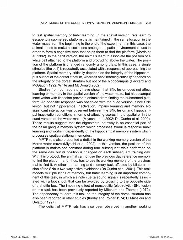
229A RAT MODEL OF THE COGNITIVE IMPAIRMENTS IN PARKINSON’S DISEASE
to test spatial memory or habit learning. In the spatial version, rats learn to escape to a submersed platform that is maintained in the same location in the water maze from the beginning to the end of the experiment. In this case, the animals need to make associations among the spatial environmental cues in order to form a cognitive map that helps them to fi nd the platform (Morris et al. 1982). In the habit version, the animals learn to associate the position of a white ball attached to the platform and protruding above the water. The posi-tion of the platform is changed randomly among trials. In this case, a single stimulus (the ball) is repeatedly associated with a response of approaching the platform. Spatial memory critically depends on the integrity of the hippocam-pus but not of the dorsal striatum, whereas habit learning critically depends on the integrity of the dorsal striatum but not of the hippocampus (Packard and McGaugh 1992; White and McDonald 2002).
Studies from our laboratory have shown that SNc lesion does not affect learning or memory in the spatial version of the water maze, but hippocampal inactivation with lidocaine prevents animals from fi nding the submersed plat-form. An opposite response was observed with the cued version, since SNc lesion, but not hippocampal inactivation, impairs learning and memory. No signifi cant interaction was observed between the SNc lesion and hippocam-pal inactivation conditions in terms of affecting scores in the spatial or in the cued version of the water maze (Miyoshi et al. 2002; Da Cunha et al. 2002). These results suggest that the nigrostriatal pathway is an essential part of the basal ganglia memory system which processes stimulus-response habit learning and works independently of the hippocampal memory system which processes spatial/relational memories.
MPTP rats also presented a defi cit in the working memory version of the Morris water maze (Miyoshi et al. 2002). In this version, the position of the platform is maintained constant during four subsequent trials performed on the same day, but its position is changed on each subsequent training day. With this protocol, the animal cannot use the previous day reference memory to fi nd the platform and, thus, has to use its working memory of the previous trial to fi nd it. Another rat learning and memory task affected by bilateral le-sion of the SNc is two-way active avoidance (Da Cunha et al. 2001). This task models multiple kinds of memory, but habit learning is an important compo-nent of this task, in which a single cue (a sound signal) is repeatedly associ-ated with a foot shock that can be avoided by crossing to the opposite side of a shuttle box. The impairing effect of nonspecifi c (electrolytic) SNc lesion on this task has been previously reported by Mitcham and Thomas (1972). The dependency to learn this task on the integrity of the dorsal striatum has also been reported in other studies (Kirkby and Polgar 1974; El Massioui and Delatour 1997).
The defi cit of MPTP rats has also been observed in another working
RMAC_dic_2006.indd 229RMAC_dic_2006.indd 229 21/05/2007 01:30:05 p.m.21/05/2007 01:30:05 p.m.

230 DA CUNHA, PREDIGER, MIYOSHI AND TAKAHASHI
memory task named delayed alternation in a Y-maze (Braga et al. 2005). In this task, the rats have to alternate between two arms of a Y-maze in order to fi nd a food pellet. During the 20-s intertrial intervals the animals have to main-tain in their working memory which arm they had previously visited in order to alternate correctly. SNc lesion with MPTP increased the number of errors in both pretrained and naive rats. In another study, we have shown that the left SNc seemed to be more critical than the right SNc for the performance of the working memory of rats in a version of the Morris water maze (Bellissimo et al. 2004).
EFFECTS OF DOPAMINERGIC DRUGS ON THE MPTP RAT MODEL OF MEMORY IMPAIRMENTS RELATED TO PD
Controversy exists regarding the dopaminergic nature of the cognitive impair-ments in PD. Since neurons producing other neurotransmitters (e.g. acetyl-choline, serotonin, noradrenaline) are also reported to degenerate in this dis-ease (Braak et al. 2003), some authors consider that they may cause some cognitive and behavioral dysfunction, especially in demented patients (Dujar-din and Laurent 2003; Zgaljardic et al. 2004). On the other hand, other inves-tigators have reported a correlation between the loss of dopamine neurons of the nigrostriatal pathway and the degree of dementia (Rinne et al. 1999) and performance in neuropsychological tests in PD patients (Marie et al. 1999; Bruck et al. 2001). Animal models can contribute to establish the specifi c im-plications of each neurotransmitter system in the cognitive impairments of PD. The role of dopamine can be studied by using models that are specifi c for dopamine depletion, such as the MPTP models, and by investigating the effects of dopamine receptor antagonists on cognition.
Ogren and Archer (1994) reported that haloperidol and other dopamine receptor antagonists impair acquisition and retention in the two-way active avoidance task, indicating that the performance of this task depends on nor-mal dopaminergic neurotransmission. The sensitivity of this task to SNc le-sions and striatal dopamine manipulations and the facility to perform this task – only two sessions are necessary in an automated apparatus – make it par-ticularly suitable to test drugs with a potential to treat the cognitive symptoms of PD. Thus, we tested the effect of the most effi cient drug used in the treat-ment of the motor symptoms of PD, levodopa, on SNc-lesioned rats. The administration of benserazide/levodopa to MPTP-lesioned rats, at a dose that restores the striatal level of dopamine, did not reverse the MPTP-induced learning and memory impairment (Gevaerd et al. 2001a).
In humans, the benefi cial effect of levodopa on improving the cognitive function affected in PD is controversial. While some studies indicate an im-
RMAC_dic_2006.indd 230RMAC_dic_2006.indd 230 21/05/2007 01:30:05 p.m.21/05/2007 01:30:05 p.m.

231A RAT MODEL OF THE COGNITIVE IMPAIRMENTS IN PARKINSON’S DISEASE
provement of cognitive functions in PD patients treated with levodopa (Beard-sley and Puletti 1971; Loranger et al. 1972; Girotti et al. 1986; Cooper et al. 1992; Cools et al. 2001), others have shown that this treatment may cause no or only mild improvement (Pillon et al. 1989; Growdon et al. 1998; Rektorova et al. 2005), or may even aggravate PD cognitive impairments (Huber et al. 1989; Poewe et al. 1991; Prasher and Findley 1991; Cools et al. 2001). Go-tham et al. (1988) proposed that the detrimental effects of levodopa observed in some cognitive tasks may be due to excessively high concentrations of dopamine in areas such as the prefrontal cortex where dopamine depletion is less severe. We showed that this was the case for MPTP rats treated with levodopa (Gevaerd et al. 2001a). The levodopa dose necessary to restore a normal striatal level of dopamine caused a large increase of dopamine levels in extrastriatal brain regions. Therefore, that study proves that, at least for the MPTP rat model of PD, levodopa therapy is not effective in improving the observed memory impairment because it appears to tilt the balance be-tween dopamine levels in the striatum and in extrastriatal regions such as the prefrontal cortex (and also limbic structures), resulting in a cognitive defi cit. In accordance with this idea, a recent work by Bruck et al. (2005) showed that the fi nding of early phase PD patients scoring poorly in tests measuring frontal lobe functions was positively correlated with increased cortical Fdopa uptake.
Furthermore, the various cognitive impairments of PD may depend on different brain areas that are differently depleted of dopamine, such as the dorsal striatum and prefrontal cortex. A study by Swainson et al. (2000) has shown that non-medicated PD patients performed better than medicated pa-tients in a reversal test that depends on the striatum and ventral frontal cortex. However, the same patients performed worse than medicated patients in a spatial recognition memory task that depends on the dorsolateral frontal cor-tex. The authors suggested that the levodopa treatment overdosed the dorso-lateral frontal cortex, which was less affected by the disease, at the same time that it restored a normal level of dopamine in the striatum and ventral frontal cortex. Cools et al. (2001) reported similar results showing that levodopa-treated patients can perform better or worse in tasks depending on different components of the frontostriatal circuitry. In that study, levodopa withdrawal improved performance in probabilistic reversal learning, a task that depends on the orbitofrontal cortex, ventral frontal cortex, and ventral striatum. How-ever, levodopa withdrawal impaired performance in a set-shifting task, which depends on the dorsolateral frontal cortex and dorsal caudate nucleus.
Therefore, although the above studies discourage the use of levodopa therapy to treat some PD cognitive symptoms, it does not imply that these cognitive symptoms are not related to the degeneration of the nigrostriatal pathway. In addition to the observed impairment in two-way active avoid-
RMAC_dic_2006.indd 231RMAC_dic_2006.indd 231 21/05/2007 01:30:06 p.m.21/05/2007 01:30:06 p.m.

232 DA CUNHA, PREDIGER, MIYOSHI AND TAKAHASHI
ance learning caused by the depletion of striatal dopamine in MPTP-lesioned rats, other important fi ndings suggest that mnemonic processes depend on a normal level of stimulation of the striatal dopamine receptors. Packard and White (1991) and Packard and McGaugh (1994) showed improved cognitive performance after intrastriatal administration of a D2 receptor agonist to rats. Also, Schneider et al. (1994) observed a cognitive improving effect of the systemic administration of a D1-receptor agonist to MPTP-lesioned monkeys. D1-receptor agonists have also been reported to release acetylcholine in the frontal cortex and dorsal striatum and to improve cognitive performance in rats (Steele et al. 1997). More recently, other authors have suggested that D1 receptor agonists can be useful in the treatment of cognitive impairments of PD (Nichols and Lewis 2004; Salmi et al. 2004), and it would be interesting to test them in an animal model such as the MPTP rat model used here.
The failure of levodopa to reverse the memory impairment of MPTP rats in the two-way active avoidance task is likely to be related, at least in part, to the failure of this treatment to improve the cognitive impairments of PD patients, as mentioned above. Since this was equally observed in some clini-cal studies and in our rat model of memory impairments related to PD, these results encouraged the use of the rat MPTP model in studies on alternative drug therapies for the treatment of the cognitive impairments of PD.
EFFECTS OF ADENOSINE RECEPTOR ANTAGONISTS ON THE MPTP RAT MODEL OF MEMORY IMPAIRMENTS RELATED TO PD
It is well known that adenosine receptors are densely expressed in the striatum and exert a modulatory infl uence on dopamine neurotransmission (Moreau and Huber 1999; Svenningsson et al. 1999). The understanding of the role of adenosine in basal ganglia and its anatomical and functional relationship with the striatal dopamine D1 and D2 receptors has increased over the last years, providing evidence of an antagonistic interaction between A(2A)/D2 and A(1)/D1 receptors in the striatum (Fuxe et al. 1998; Franco et al. 2000). Moreover, neuroprotective properties of caffeine and A(2A) adenosine receptor antago-nists have been reported for dopamine neurons in the SNc (Chen et al. 2001). Furthermore, adenosine receptor-related drugs seem to be promising candi-dates for the symptomatic treatment of PD, since there is evidence that caf-feine directly increases dopamine release from striatal nerve terminals (Okada 1997). This dopamine-releasing effect of caffeine was also observed with the A(2A) adenosine receptor antagonist, ZM 241385, in striatal synaptosomes (Da Cunha et al. 2002). All these putative anti-Parkinson effects may explain the fi nding that the risk of PD is signifi cantly reduced among coffee drinkers (Paganini-Hill 2001). Based on these promising effects, adenosine receptor
RMAC_dic_2006.indd 232RMAC_dic_2006.indd 232 21/05/2007 01:30:06 p.m.21/05/2007 01:30:06 p.m.
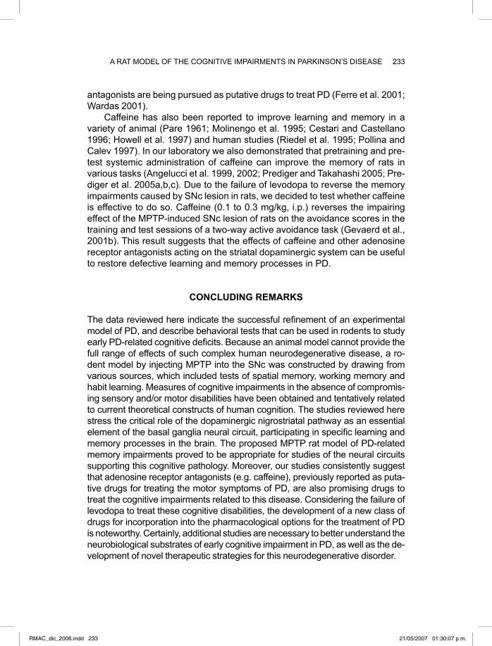
233A RAT MODEL OF THE COGNITIVE IMPAIRMENTS IN PARKINSON’S DISEASE
antagonists are being pursued as putative drugs to treat PD (Ferre et al. 2001; Wardas 2001).
Caffeine has also been reported to improve learning and memory in a variety of animal (Pare 1961; Molinengo et al. 1995; Cestari and Castellano 1996; Howell et al. 1997) and human studies (Riedel et al. 1995; Pollina and Calev 1997). In our laboratory we also demonstrated that pretraining and pre-test systemic administration of caffeine can improve the memory of rats in various tasks (Angelucci et al. 1999, 2002; Prediger and Takahashi 2005; Pre-diger et al. 2005a,b,c). Due to the failure of levodopa to reverse the memory impairments caused by SNc lesion in rats, we decided to test whether caffeine is effective to do so. Caffeine (0.1 to 0.3 mg/kg, i.p.) reverses the impairing effect of the MPTP-induced SNc lesion of rats on the avoidance scores in the training and test sessions of a two-way active avoidance task (Gevaerd et al., 2001b). This result suggests that the effects of caffeine and other adenosine receptor antagonists acting on the striatal dopaminergic system can be useful to restore defective learning and memory processes in PD.
CONCLUDING REMARKS
The data reviewed here indicate the successful refi nement of an experimental model of PD, and describe behavioral tests that can be used in rodents to study early PD-related cognitive defi cits. Because an animal model cannot provide the full range of effects of such complex human neurodegenerative disease, a ro-dent model by injecting MPTP into the SNc was constructed by drawing from various sources, which included tests of spatial memory, working memory and habit learning. Measures of cognitive impairments in the absence of compromis-ing sensory and/or motor disabilities have been obtained and tentatively related to current theoretical constructs of human cognition. The studies reviewed here stress the critical role of the dopaminergic nigrostriatal pathway as an essential element of the basal ganglia neural circuit, participating in specifi c learning and memory processes in the brain. The proposed MPTP rat model of PD-related memory impairments proved to be appropriate for studies of the neural circuits supporting this cognitive pathology. Moreover, our studies consistently suggest that adenosine receptor antagonists (e.g. caffeine), previously reported as puta-tive drugs for treating the motor symptoms of PD, are also promising drugs to treat the cognitive impairments related to this disease. Considering the failure of levodopa to treat these cognitive disabilities, the development of a new class of drugs for incorporation into the pharmacological options for the treatment of PD is noteworthy. Certainly, additional studies are necessary to better understand the neurobiological substrates of early cognitive impairment in PD, as well as the de-velopment of novel therapeutic strategies for this neurodegenerative disorder.
RMAC_dic_2006.indd 233RMAC_dic_2006.indd 233 21/05/2007 01:30:07 p.m.21/05/2007 01:30:07 p.m.

234 DA CUNHA, PREDIGER, MIYOSHI AND TAKAHASHI
REFERENCES
Aalto S, Bruck A, Laine M, Nagren K, Rinne JO (2005) Frontal and temporal dopamine release during working memory and attention tasks in healthy humans: a positron emission tomography study using the high-affi nity dopamine D-2 receptor ligand [C-11]FLB 457. J Neurosci 25:2471-2477
Aarsland D, Tandberg E, Larsen JP, Cummings JL (1996) Frequency of dementia in Parkinson disease. Arch Neurol 53:538–542
Alexander GE, Crutcher MD (1990) Functional architecture of basal ganglia circuits: neural substrates of parallel processing. Trends Neurosci 13:266-271
Alexander GE, DeLong MR, Strick PL (1986) Parallel organization of functionally seg-regated circuits linking basal ganglia and cortex. Annu Rev Neurosci 9:357-381
Angelucci ME, Vital MA, Cesario C, Zadusky CR, Rosalen PL, Da Cunha C (1999) The effect of caffeine in animal models of learning and memory. Eur J Pharmacol 373:135-140
Angelucci MEM, Cesario C, Hiroi RH, Rosalen P, Da Cunha C (2002) The effect of caffeine on learning and memory of the Morris water maze. Braz J Med Biol Res 35:1201-1208
Baddeley A (2003) Working memory: looking back and looking forward. Nat Rev Neu-rosci 4:829-839
Beal MF (2001) Experimental models of Parkinson’s disease. Nat Rev Neurosci 2:325-334
Beardsley JV, Puletti F (1971) Personality (MMPI) and cognitive (WAIS) changes after levodopa treatment. Arch Neurol 25:145-150
Bellissimo MI, Kouzmine I, Ferro MM, Oliveira BH, Canteras NS, Da Cunha C (2004) Is the unilateral lesion of the left substantia nigra pars compacta suffi cient to induce working memory impairment in rats? Neurobiol Learning Memory 82:150-158
Bezard E, Dovero S, Prunier C, Ravenscroft P, Chalon S, Guilloteau D, Crossman AR, Bioulac B, Brotchie JM, Gross CE (2001) Relationship between the appearance of symptoms and the level of nigrostriatal degeneration in a progressive 1-methyl-4-phenyl-1,2,3,6-tetrahydropyridine-lesioned macaque model of Parkinson’s dis-ease. J Neurosci 21:6853-6861
Bezard E, Imbert C, Deloire X, Bioulac B, Gross CE (1997) A chronic MPTP model reproducing the slow evolution of Parkinson’s disease: evolution of motor symp-toms in the monkey. Brain Res 766:107–112
Bondi MW, Kaszniak AW (1991) Implicit and explicit memory in Alzheimer’s disease and Parkinson’s disease. J Clin Exp Neuropsychol 13:339-358
Bosboom, JL, Stoffers D, Wolters ECh (2004) Cognitive dysfunction and dementia in Parkinson’s disease. J Neural Transm 111:1303–1315
Braak H, Del Tredici K, Rub U, de Vos RA, Jansen Steur EN, Braak E (2003) Stag-ing of brain pathology related to sporadic Parkinson’s disease. Neurobiol Aging 24:197–211
Bradley VA, Welch JL, Dick DJ (1989) Visuospatial working memory in Parkinson’s disease. J Neurol Neurosurg Psychiatry 52:1228–1235
Braga R, Kouzmine I, Canteras NS, Da Cunha C (2005) Lesion of the substantia nigra,
RMAC_dic_2006.indd 234RMAC_dic_2006.indd 234 21/05/2007 01:30:07 p.m.21/05/2007 01:30:07 p.m.

235A RAT MODEL OF THE COGNITIVE IMPAIRMENTS IN PARKINSON’S DISEASE
pars compacta impairs delayed alternation in a Y-maze in rats. Exp Neurol 192:134-141
Brown LL, Schneider JS, Lidsky TI (1997) Sensory and cognitive functions of the basal ganglia. Curr Opin Neurobiol 7:157-163
Bruck A, Portin R, Lindell A, Laihinen A, Bergman J, Haaparanta M, Solin O, Rinne JO (2001) Positron emission tomography shows that impaired frontal lobe function-ing in Parkinson’s disease is related to dopaminergic hypofunction in the caudate nucleus. Neurosci Lett 311:81–84
Calabresi P, Pisani A, Mercuri NB, Bernardi G (1996) The corticostriatal projection: from synaptic plasticity to dysfunctions of the basal ganglia. Trends Neurosci 19:19-24
Cestari V, Castellano C (1996) Caffeine and cocaine interaction on memory consolida-tion in mice. Arch Int Pharmacodyn Ther 331:94-104
Chen JF, Xu K, Petzer JP, Staal R, Xu YH, Beilstein M, Sonsalla PK, Castagnoli K, Castagnoli N Jr, Schwarzschild MA (2001) Neuroprotection by caffeine and A(2A) adenosine receptor inactivation in a model of Parkinson's disease. J Neurosci 21:RC143
Cools R, Barker RA, Sahakain BJ, Robbins TW (2001) Enhanced or impaired cogni-tive function in Parkinson’s disease as a function of dopaminergic medication and task demands. Cerebral Cortex 11:1136–1143
Cooper JA, Sagar HJ, Doherty SM, Jordan N, Tidswell P, Sullivan EV (1992) Differ-ent effects of dopaminergic and anticholinergic therapies on cognitive and motor function in Parkinson’s disease. Brain 115:1701–1725
Costa A, Peppe A, Dell'Agnello G, Carlesimo GA, Murri L, Bonuccelli U, Caltagirone C (2003) Dopaminergic modulation of visual-spatial working memory in Parkinson’s disease. Dement Geriatr Cogn Disord 15:55-66
Da Cunha C, Angellucci MEM, Canteras NS, Wonnacott S, Takahashi RN (2002) The lesion of the rat substantia nigra pars compacta dopamine neurons as a model for Parkinson’s disease memory disabilities. Cell Mol Neurobiol 22:227-237
Da Cunha C, Gevaerd MS, Vital MA, Miyoshi E, Andreatini R, Silveira R, Takahashi RN, Canteras NS (2001) Memory disruption in rats with nigral lesions induced by MPTP: a model for early Parkinson's disease amnesia. Behav Brain Res 124:9-18
Da Cunha C, Wietzikoski S, Wietzikoski EC, Miyoshi E, Ferro MM, Anselmo-Franci JA, Canteras NS (2003) Evidence for the substantia nigra pars compacta as an es-sential component of a memory system independent of the hippocampal memory system. Neurobiol Learning Mem 79:236-242
Davis GC, Williams AC, Markey SP, Ebert MH, Caine ED, Reichert CM, Kopin IJ (1979) Chronic parkinsonism secondary to intravenous injection of meperidine analogues. Psychiatry Res 1:249–254
Dirnberger G, Frith CD, Jahanshahi M (2005) Executive dysfunction in Parkinson's disease is associated with altered pallidal-frontal processing. Neuroimage 25:588-599
Dubois B, Pillon B (1997) Cognitive defi cits in Parkinson's disease. J Neurol 244:2-8Dujardin K, Laurent B (2003) Dysfunction of the human memory systems: role of the
dopaminergic transmission. Curr Opin Neurol 16: S11-S16Duvoisin RC (1991) Parkinson’s disease. Third Edition. Raven Press, New York
RMAC_dic_2006.indd 235RMAC_dic_2006.indd 235 21/05/2007 01:30:08 p.m.21/05/2007 01:30:08 p.m.

236 DA CUNHA, PREDIGER, MIYOSHI AND TAKAHASHI
El Massioui N, Delatour BV (1997) Contrasting effects of central nucleus of the amyg-dala and dorsal striatum lesions on active avoidance learning and its contextual modifi cation Neurosci Res Comm 21:103–111
Faglioni P, Botti C, Scarpa M, Ferrari V, Saetti MC (1997) Learning and forgetting pro-cesses in Parkinson’s disease: a model-based approach to disentangling storage, retention and retrieval contributions. Neuropsychologia 35:767-779
Faglioni P, Scarpa M, Botti C, Ferrari V (1995) Parkinson’s disease affects automatic and spares intentional verbal learning. A stochastic approach to explicit learning processes. Cortex 31:597-617
Faw B (2003) Pre-frontal executive committee for perception, working memory, atten-tion, long-term memory, motor control, and thinking: a tutorial review. Conscious Cogn 12:83-139
Ferre S, Popoli P, Gimenez-Llort L, Rimondini R, Muller CE, Stromberg I, Ogren SO, Fuxe K (2001) Adenosine/dopamine interaction: implications for the treatment of Parkinson's disease. 7:235-241
Flaherty AW, Graybiel AM (1998) Input-output organization of the sensorimotor stria-tum in the squirrel-monkey. J Neurosci 14:599-610
Franco R, Ferre S, Agnati L, Torwinen M, Hillion J, Casado V, Lledo P, Zoli M, Lluis C, Fuxe K (2000) Evidence for adenosine/dopamine receptor interactions. Indica-tions for heteromerization. Neuropharmacology 23:550–559
Fuxe K, Ferre S, Zoli M, Agnati LF (1998) Integrated events in central dopamine trans-mission as analyzed at multiple levels. Evidence for intramembrane adenosine A2A/dopamine D2 and adenosine A1/dopamine D1 receptor interactions in basal ganglia. Brain Res Brain Rev 26:258–273
Gevaerd MS, Miyoshi E, Silveira R, Canteras NS, Takahashi RN, Da Cunha C (2001a) L-Dopa restores striatal dopamine level but fails to reverse MPTP-induced mem-ory defi cits in rats. Int J Neuropsychopharmacol 4:361-370
Gevaerd MS, Takahashi RN, Da Cunha C (2001b). Caffeine reverses the memory dis-ruption induced by intra-nigral MPTP-injection in rats. Brain Res Bull 55:101-106
Girotti F, Carella F, Grassi MP, Soliveri P, Marano R, Caraceni T (1986) Motor and cognitive performances of parkinsonian patients in the on and off phases of the disease. J Neurol Neurosurg Psychiatry 49:657–660
Gotham AM, Brown RG, Marsden CD (1988) “Frontal” cognitive functions in patients with Parkinson’s disease “on” and “off ” levodopa. Brain 111:299–321
Graybiel AM (1998) The basal ganglia and chunking of action repertories. Neurobiol Learn Mem 70:119-36
Growdon JH, Kieburtz K, McDermott MP, Panisset M, Friedman JH (1998) Levodopa improves motor function without impairing cognition in mild non-demented Parkin-son's disease patients. Parkinson Study Group. Neurology 50:1327-1331
Heikkila RE, Sonsalla PK, Duvoisin RC (1989) Biochemical models of Parkinson’s disease. In: Boulton AA, Baker GB, Jurio AV (eds) Neuromethods. Drugs as Tools in Neurotransmitter Research, vol. 12, Human Press, New York, pp 351–384
Hirsch E, Graybiel AM, Agid YA (1988) Melanized dopamine neurons are differentially susceptible to degeneration in Parkinson's disease. Nature 334:345-348
Howell LL, Coffi n VL, Spealman RD (1997) Behavioral and physiological effects of xanthines in nonhuman primates. Psychopharmacology 129:1-14
RMAC_dic_2006.indd 236RMAC_dic_2006.indd 236 21/05/2007 01:30:08 p.m.21/05/2007 01:30:08 p.m.

237A RAT MODEL OF THE COGNITIVE IMPAIRMENTS IN PARKINSON’S DISEASE
Huber SJ, Shulman HG, Paulson GW, Shuttleworth EC (1989) Dose-dependent mem-ory impairment in Parkinson's disease. Neurology 39:438-440
Johannessen JN, Chiueh CC, Burns RS, Markey SP (1985) Differences in the me-tabolism of MPTP in the rodent and primate parallel differences in sensitivity to its neurotoxic effects. Life Sci 36:219-224
Kirkby RJ, Polgar S (1974). Active avoidance in laboratory rat following lesions of dor-sal or ventral caudate-nucleus. Physiol Psychol 2:301-306
Knowlton BJ, Mangels JA, Squire LR (1996) A neostriatal habit learning system in humans. Science 273:1399-1402
Koenig O, Thomas-Anterion C, Laurent B (1999) Procedural learning in Parkinson’s dis-ease: intact and impaired cognitive components. Neuropsychologia 37:1103-1109
Langston JW, Ballard P, Tetrud JW, Irwin I (1983) Chronic Parkinsonism in humans due to a product of meperidine-analog synthesis. Science 219:979–980
Lewis SJG, Dove A, Robbins TW, Barker RA, Owen AM (2003) Cognitive impairments in early Parkinson's disease are accompanied by reductions in activity in frontos-triatal neural circuitry. J Neurosci 23:6351-6356
Loranger AW, Goodell H, Lee JE, McDowell F (1972) Levodopa treatment of Parkin-son's syndrome. Improved intellectual functioning. Arch Gen Psychiatry 26:163-168
Marie RM, Barre L, Dupuy B, Viader F, Defer G, Baron JC (1999) Relationships be-tween striatal dopamine denervation and frontal executive tests in Parkinson’s disease. Neurosci Lett 260:77-80
Marie RM, Defer G (2003) Working memory and dopamine: clinical and experimental clues. Curr Opin Neurol 16:S29-S35
McDonald JR, Hong NS, Devan BD (2004) The challenges of understanding mamma-lian cognition and memory-based behaviours: an interactive learning and memory systems approach. Neurosci Biobehav Rev 28:719–745
Mitcham JC, Thomas RK Jr (1972) Effects of substantia nigra and caudate-nucleus lesions on avoidance-learning in rats. J Comp Physiol Psychol 81:101-107
Miyoshi E, Wietzikoski S, Camplessei M, Silveira R, Takahashi RN, Da Cunha C (2002) Impaired learning in a spatial working memory version and in a cued version of the water maze in rats with MPTP-induced mesencephalic dopaminergic lesions. Brain Res Bull 58:41-47
Molinengo L, Orsetti M, Pastorello B, Scordo I, Ghi P (1995) Habituation of exploratory activity in rats: action of N6phenylisopropyladenosine, caffeine and their combina-tion. Prog Neuropsychopharmacol Biol Psychiatry 19:1189-1200
Moreau JL, Huber G (1999) Central adenosine A(2A) receptors: an overview. Brain Res Brain Res Rev 31:65-82
Moreaud O, Fournet N, Roulin JL, Naegele B, Pellat J (1997) The phonological loop in medicated patients with Parkinson’s disease: presence of phonological similarity and word length effects. J Neurol Neurosurg Psychiatry 62:609-611
Morris RG, Garrud P, Rawlins JN, O'Keefe J (1982) Place navigation impaired in rats with hippocampal lesions. Nature 297:681-683
Nichols DE, Lewis MM (2004) Mechanisms and issues relating to the use of D1-like dopamine receptor agonists for age-related illnesses: Parkinson's disease and memory and cognition. Med Chem Res 13:105-114
RMAC_dic_2006.indd 237RMAC_dic_2006.indd 237 21/05/2007 01:30:09 p.m.21/05/2007 01:30:09 p.m.
238 DA CUNHA, PREDIGER, MIYOSHI AND TAKAHASHI
Ogren SO, Archer T (1994) Effects of typical and atypical antipsychotic drugs on two-way active avoidance. Relationship to DA receptor blocking profi le. Psychophar-macology 114:383-391
Okada M, Kiryu K, Kawata Y, Mizuno K, Wada K, Tasaki H, Kaneko S (1997) Determi-nation of the effects of caffeine and carbamazepine on striatal dopamine release by in vivo microdialysis. Eur J Pharmacol 321:181-188
Owen AM (2004) Cognitive dysfunction in Parkinson’s disease: the role of frontostria-tal circuitry. Neuroscientist 10:525–537
Owen AM, Beksinska M, James M, Leigh PN, Summers BA, Marsden CD, Quinn NP, Sahakian BJ, Robbins TW (1993) Visuospatial memory defi cits at different stages of Parkinson’s. Neuropsychologia 31:627-644
Owen AM, Sahakian BJ, Hodges JR, Summers BA, Polkey CE, Robins TW (1995) Dopamine-dependent frontostriatal planning in early Parkinson’s disease. Neuro-psychology 9:126–140
Packard MG, Knowlton BJ (2002) Learning and memory functions of the basal ganglia. Annu Rev Neurosci 25:563-593
Packard MG, McGaugh JL (1992) Double dissociation of fornix and caudate nucleus lesions on acquisition of two water maze tasks: further evidence for multiple mem-ory systems. Behav Neurosci 106:439-446
Paganini-Hill A (2001) Risk factors for Parkinson’s disease: the leisure world cohort study. Neuroepidemiology 20:118-124
Pare W (1961) Effect of caffeine and seconal on a visual discrimination task. J Comp Physiol Psychol 54: 506-509
Pascual-Leone A, Grafman J, Clark K, Stewart M, Massaquoi S, Lou J-S, Hallett M (1993) Procedural learning in Parkinson’s disease and cerebellar degeneration. Ann Neurol 34:594-602
Pillon B, Dubois B, Cusimano G, Bonnet AM, Lhermitte F, Agid Y (1989) Does cogni-tive impairment in Parkinson's disease result from non-dopaminergic lesions? J Neurol Neurosurg Psychiatry 52:201-206
Pillon B, Ertle S, Deweer B, Bonnet AM, Vidailhet M, Dubois B (1997) Memory for spa-tial location in ´de novo` parkinsonian patients. Neuropsychologia 35:221-228
Pillon B, Ertle S, Deweer B, Sarazin M, Agid Y, Dubois B (1996) Memory for spatial location is affected in Parkinson’s disease. Neuropsychologia 34:77-85
Poewe W, Berger W, Benke T, Schelosky L (1991) High-speed memory scanning in Parkinson's disease: adverse effects of levodopa. Ann Neurol 29:670-673
Pollina DA, Calev A (1997) Amnesia associated with electroconvulsive therapy - prog-ress in pharmacological prevention and treatment CNS Drugs 7:381-387
Postle BR, D’Esposito M (1999) Dissociation of human caudate nucleus activity in spatial and nonspatial working memory: an event-related fMRI study. Brain Res Cogn Brain Res 8:107-115
Prasher D, Findley L (1991) Dopaminergic induced changes in cognitive and motor processing in Parkinson's disease: an electrophysiological investigation. J Neurol Neurosurg Psychiatry 54:603-609
Prediger RD, Batista LC, Takahashi RN (2005a) Caffeine reverses age-related defi cits in olfactory discrimination and social recognition memory in rats. Involvement of adenosine A1 and A2A receptors. Neurobiol Aging 26:957-964
RMAC_dic_2006.indd 238RMAC_dic_2006.indd 238 21/05/2007 01:30:09 p.m.21/05/2007 01:30:09 p.m.

239A RAT MODEL OF THE COGNITIVE IMPAIRMENTS IN PARKINSON’S DISEASE
Prediger RD, Da Cunha C, Takahashi RN (2005b) Antagonistic interaction between adenosine A2A and dopamine D2 receptors modulates the social recognition memory in reserpine-treated rats. Behav Pharmacol 16:209-218
Prediger RD, Pamplona FA, Fernandes D, Takahashi RN (2005c) Caffeine improves spatial learning defi cits in an animal model of attention defi cit hyperactivity disor-der (ADHD) – the spontaneously hypertensive rat (SHR). Int J Neuropsychophar-macol 8:583-594
Prediger RD, Takahashi RN (2005) Modulation of short-term social memory in rats by adenosine A1 and A(2A) receptors. Neurosci Lett 376:160-165
Przedborski S, Jackson-Lewis V, Naini AB, Jakowec M, Petzinger G, Miller R, Akran M (2001) The parkinsonian toxin 1-methyl-4-phenyl-1,2,3,6-tetrahydropyridine (MPTP): a technical review of its utility and safety. J Neurochem 76:1265-1274
Przedborski S, Vila M (2003) The 1-methyl-4-phenyl-1,2,3,6-tetrahydropyridine mouse model: a tool to explore the pathogenesis of Parkinson’s disease. Ann NY Acad Sci 991:189–198
Rektorova I, Rektor I, Bares M, Dostal V, Ehler E, Fanfrdlova Z, Fiedler J, Klajblova H, Kulist'ak P, Ressner P, Svatova J, Urbanek K, Veliskova J (2005) Cognitive perfor-mance in people with Parkinson's disease and mild or moderate depression: ef-fects of dopamine agonists in an add-on to L-dopa therapy. Eur J Neurol 12:9-15
Riedel W, Hogervorst E, Leboux R, Verhey F, van Praag H, Jolles J (1995) Caffeine attenuates scopolamine-induced memory impairment in humans. Psychopharma-cology 122:158-168
Rinne JO, Rummukainen J, Paljarvi L, Rinne UK (1989) Dementia in Parkinson’s disease is related to neuronal loss in the medial substantia nigra. Ann Neurol 26:47–50
Roncacci S, Troisi E, Carlesimo GA, Nocentini U, Caltagirone C (1996) Implicit memory in parkinsonian patients: evidence for defi cient skill learning. Eur Neurol 36:154-159
Salmi P, Isacson R, Kull B (2004) Dihydrexidine - the fi rst full dopamine D1 receptor agonist. CNS Drug Rev 10:230-242
Schmidt N, Ferger B (2001) Neurochemical fi ndings in the MPTP model of Parkinson’s disease. J Neural Transm 108:1263–1282
Schneider JS, Sun ZQ, Roeltgen DP (1994) Effects of dihydrexidine, a full dopamine D-1 receptor agonist, on delayed response performance in chronic low dose MPTP-treated monkeys. Brain Res 663:140-144
Schober A (2004) Classic toxin-induced animal models of Parkinson’s disease: 6-OHDA and MPTP. Cell Tissue Res 318:215-224
Sedelis M, Hofele K, Auburger GW, Morgan S, Huston JP, Schwarting RK (2000) MPTP susceptibility in the mouse: behavioral, neurochemical, and histological analysis of gender and strain differences. Behav Genet 30:171-182
Sedelis M, Schwarting RK, Huston JP (2001) Behavioral phenotyping of the MPTP mouse model of Parkinson’s disease. Behav Brain Res 125:109-125
Shwarting RKW, Huston JP (1996) The unilateral 6-hydroxydopamine lesion model in behavioral brain research. Analysis of functional defi cits, recovery and treat-ments. Progr Neurobiol 50:275-331
Stebbins GT, Gabrieli JDE, Masciari F, Monti L, Goetz CG (1999) Delayed recognition
RMAC_dic_2006.indd 239RMAC_dic_2006.indd 239 21/05/2007 01:30:10 p.m.21/05/2007 01:30:10 p.m.

240 DA CUNHA, PREDIGER, MIYOSHI AND TAKAHASHI
memory in Parkinson’s disease: a role for working memory? Neuropsychologia 37:503-510
Steele TD, Hodges DB, Levesque TR, Locke KW (1997) D-1 agonist dihydrexidine releases acetylcholine and improves cognitive performance in rats. Pharmacol Biochem Behav 58:477-483
Svenningsson P, Le Moine C, Fisone G, Fredholm BB (1999) Distribution, biochemis-try and function of striatal adenosine A2A receptors. Prog Neurobiol 59:355-396
Swainson R, Rogers RD, Sahakian BJ, Summers BA, Polkey CE, Robbins TW (2000) Probabilistic learning and reversal defi cits in patients with Parkinson’s disease or frontal or temporal lobe lesions: possible adverse effects of dopaminergic medica-tion. Neuropsychologia 38:596–612
Tamaru F (1997) Disturbances in higher function in Parkinson’s disease. Eur Neurol 38:33-36
Thomas V, Reymann JM, Lieury A, Allain H (1996) Assessment of procedural memory in Parkinson’s disease. Prog NeuroPsychopharmacol Biol Psychiatry 20:641-650
Ungerstedt U (1968) 6-Hydroxydopamine induced degeneration of central monoamine neurons. Eur J Phamacol 5:107-110
von Bohlen und Halbach O, Schober A, Krieglstein K (2004) Genes, proteins, and neurotoxins involved in Parkinson’s disease. Prog Neurobiol 73:151-177
Wardas J, Konieczny J, Lorenc-Koci E (2001) SCH 58261, an A(2A) adenosine re-ceptor antagonist, counteracts parkinsonian-like muscle rigidity in rats. Synapse 41:160-171.
White NM, McDonald RJ (2002) Multiple parallel memory systems in the brain of the rat. Neurobiol Learn Mem 77:125-184
Zgaljardic DJ, Foldi NS, Borod JC (2004) Cognitive and behavioral dysfunction in Par-kinson’s disease: neurochemical and clinicopathological contributions. J Neural Transm 111:1287–1301
RMAC_dic_2006.indd 240RMAC_dic_2006.indd 240 21/05/2007 01:30:10 p.m.21/05/2007 01:30:10 p.m.

80
ANEXO 2

Behavioural Brain Research 199 (2009) 157–170
Contents lists available at ScienceDirect
Behavioural Brain Research
journa l homepage: www.e lsev ier .com/ locate /bbr
Review
Learning processing in the basal ganglia: A mosaic of broken mirrors
Claudio Da Cunhaa,∗, Evellyn Claudia Wietzikoskia, Patrícia Dombrowskia, Mariza Bortolanzaa,Lucélia Mendes Santosa, Suelen Lucio Boschena, Edmar Miyoshia,b
a Laboratório de Fisiologia e Farmacologia do Sistema Nervoso Central, Departamento de Farmacologia, UFPR, C.P. 19.031, 81.531-980 Curitiba PR, Brazilb Departamento de Ciências Farmacêuticas, UEPG, Ponta Grossa, Brazil
a r t i c l e i n f o
Article history:Received 16 May 2008Received in revised form 1 October 2008Accepted 2 October 2008Available online 11 October 2008
Keywords:Basal gangliaStriatumSubstantia nigraAssociative learningMemoryCognitionDopamineReinforcementReward
a b s t r a c t
In the present review we propose a model to explain the role of the basal ganglia in sensorimotor andcognitive functions based on a growing body of behavioural, anatomical, physiological, and neurochem-ical evidence accumulated over the last decades. This model proposes that the body and its surroundingenvironment are represented in the striatum in a fragmented and repeated way, like a mosaic consisting ofthe fragmented images of broken mirrors. Each fragment forms a functional unit representing articulatedparts of the body with motion properties, objects of the environment which the subject can approachor manipulate, and locations the subject can move to. These units integrate the sensory properties andmovements related to them. The repeated and widespread distribution of such units amplifies the com-binatorial power of the associations among them. These associations depend on the phasic release ofdopamine in the striatum triggered by the saliency of stimuli and will be reinforced by the rewardingconsequences of the actions related to them. Dopamine permits synaptic plasticity in the corticostriatalsynapses. The striatal units encoding the same stimulus/action send convergent projections to the inter-nal segment of the globus pallidus (GPi) and to the substantia nigra pars reticulata (SNr) that stimulate orhold the action through a thalamus-frontal cortex pathway. According to this model, this is how the basalganglia select actions based on environmental stimuli and store adaptive associations as nondeclarativememories such as motor skills, habits, and memories formed by Pavlovian and instrumental conditioning.
© 2008 Elsevier B.V. All rights reserved.
Contents
1. Introduction . . . . . . . . . . . . . . . . . . . . . . . . . . . . . . . . . . . . . . . . . . . . . . . . . . . . . . . . . . . . . . . . . . . . . . . . . . . . . . . . . . . . . . . . . . . . . . . . . . . . . . . . . . . . . . . . . . . . . . . . . . . . . . . . . . . . . . . . . 1582. The basal ganglia circuitry . . . . . . . . . . . . . . . . . . . . . . . . . . . . . . . . . . . . . . . . . . . . . . . . . . . . . . . . . . . . . . . . . . . . . . . . . . . . . . . . . . . . . . . . . . . . . . . . . . . . . . . . . . . . . . . . . . . . . . . . . . 1583. The ‘mosaic of broken mirrors’ model . . . . . . . . . . . . . . . . . . . . . . . . . . . . . . . . . . . . . . . . . . . . . . . . . . . . . . . . . . . . . . . . . . . . . . . . . . . . . . . . . . . . . . . . . . . . . . . . . . . . . . . . . . . . . . 159
3.1. Breaking the mirrors: functional convergence and widespread repetition . . . . . . . . . . . . . . . . . . . . . . . . . . . . . . . . . . . . . . . . . . . . . . . . . . . . . . . . . . . . . . . . . 1593.2. Building functional units . . . . . . . . . . . . . . . . . . . . . . . . . . . . . . . . . . . . . . . . . . . . . . . . . . . . . . . . . . . . . . . . . . . . . . . . . . . . . . . . . . . . . . . . . . . . . . . . . . . . . . . . . . . . . . . . . . . . 159
3.2.1. Body parts . . . . . . . . . . . . . . . . . . . . . . . . . . . . . . . . . . . . . . . . . . . . . . . . . . . . . . . . . . . . . . . . . . . . . . . . . . . . . . . . . . . . . . . . . . . . . . . . . . . . . . . . . . . . . . . . . . . . . . . . . . 1593.2.2. Objects . . . . . . . . . . . . . . . . . . . . . . . . . . . . . . . . . . . . . . . . . . . . . . . . . . . . . . . . . . . . . . . . . . . . . . . . . . . . . . . . . . . . . . . . . . . . . . . . . . . . . . . . . . . . . . . . . . . . . . . . . . . . . 1603.2.3. Locations . . . . . . . . . . . . . . . . . . . . . . . . . . . . . . . . . . . . . . . . . . . . . . . . . . . . . . . . . . . . . . . . . . . . . . . . . . . . . . . . . . . . . . . . . . . . . . . . . . . . . . . . . . . . . . . . . . . . . . . . . . . 1603.2.4. Other functional units of the striatum . . . . . . . . . . . . . . . . . . . . . . . . . . . . . . . . . . . . . . . . . . . . . . . . . . . . . . . . . . . . . . . . . . . . . . . . . . . . . . . . . . . . . . . . . . . . 162
3.3. Building associative units . . . . . . . . . . . . . . . . . . . . . . . . . . . . . . . . . . . . . . . . . . . . . . . . . . . . . . . . . . . . . . . . . . . . . . . . . . . . . . . . . . . . . . . . . . . . . . . . . . . . . . . . . . . . . . . . . . . 1623.3.1. Synaptic plasticity in the striatum . . . . . . . . . . . . . . . . . . . . . . . . . . . . . . . . . . . . . . . . . . . . . . . . . . . . . . . . . . . . . . . . . . . . . . . . . . . . . . . . . . . . . . . . . . . . . . . . . 1633.3.2. Dopamine-dependent synaptic plasticity . . . . . . . . . . . . . . . . . . . . . . . . . . . . . . . . . . . . . . . . . . . . . . . . . . . . . . . . . . . . . . . . . . . . . . . . . . . . . . . . . . . . . . . . . 1633.3.3. Novelty-driven reinforcement learning . . . . . . . . . . . . . . . . . . . . . . . . . . . . . . . . . . . . . . . . . . . . . . . . . . . . . . . . . . . . . . . . . . . . . . . . . . . . . . . . . . . . . . . . . . . 1633.3.4. Aversively motivated learning . . . . . . . . . . . . . . . . . . . . . . . . . . . . . . . . . . . . . . . . . . . . . . . . . . . . . . . . . . . . . . . . . . . . . . . . . . . . . . . . . . . . . . . . . . . . . . . . . . . . . 164
3.4. Building action units . . . . . . . . . . . . . . . . . . . . . . . . . . . . . . . . . . . . . . . . . . . . . . . . . . . . . . . . . . . . . . . . . . . . . . . . . . . . . . . . . . . . . . . . . . . . . . . . . . . . . . . . . . . . . . . . . . . . . . . . 1653.4.1. Driving MSNs to an ‘up’ or ‘down state’ . . . . . . . . . . . . . . . . . . . . . . . . . . . . . . . . . . . . . . . . . . . . . . . . . . . . . . . . . . . . . . . . . . . . . . . . . . . . . . . . . . . . . . . . . . . . 165
Abbreviations: CAR, conditioned avoidance response; CS, conditioned stimulus; GP, globus pallidus; GPe, external segment of the globus pallidus; GPi, internal segmentof the globus pallidus; LTD, long-term depression; LTP, long-term potentiation; MSNs, medium spiny neurons; NAc, nucleus accumbens; SNc, substantia nigra parscompacta; SNr, substantia nigra pars reticulata; S-R, stimulus-response; STN, subthalamic nucleus; TANs, called tonically active neurons; US, unconditioned stimulus.∗ Corresponding author. Tel.: +55 41 3361 1717; fax: +55 41 3266 2042.
E-mail address: [email protected] (C. Da Cunha).
0166-4328/$ – see front matter © 2008 Elsevier B.V. All rights reserved.doi:10.1016/j.bbr.2008.10.001

158 C. Da Cunha et al. / Behavioural Brain Research 199 (2009) 157–170
3.4.2. Go/NoGo units . . . . . . . . . . . . . . . . . . . . . . . . . . . . . . . . . . . . . . . . . . . . . . . . . . . . . . . . . . . . . . . . . . . . . . . . . . . . . . . . . . . . . . . . . . . . . . . . . . . . . . . . . . . . . . . . . . . . . 1653.5. Gathering action units . . . . . . . . . . . . . . . . . . . . . . . . . . . . . . . . . . . . . . . . . . . . . . . . . . . . . . . . . . . . . . . . . . . . . . . . . . . . . . . . . . . . . . . . . . . . . . . . . . . . . . . . . . . . . . . . . . . . . . . 165
4. Emergent properties of the ‘mosaic of broken mirrors’ model . . . . . . . . . . . . . . . . . . . . . . . . . . . . . . . . . . . . . . . . . . . . . . . . . . . . . . . . . . . . . . . . . . . . . . . . . . . . . . . . . . . . . 1655. Conclusion. . . . . . . . . . . . . . . . . . . . . . . . . . . . . . . . . . . . . . . . . . . . . . . . . . . . . . . . . . . . . . . . . . . . . . . . . . . . . . . . . . . . . . . . . . . . . . . . . . . . . . . . . . . . . . . . . . . . . . . . . . . . . . . . . . . . . . . . . . . 166
Acknowledgements . . . . . . . . . . . . . . . . . . . . . . . . . . . . . . . . . . . . . . . . . . . . . . . . . . . . . . . . . . . . . . . . . . . . . . . . . . . . . . . . . . . . . . . . . . . . . . . . . . . . . . . . . . . . . . . . . . . . . . . . . . . . . . . . . 167References . . . . . . . . . . . . . . . . . . . . . . . . . . . . . . . . . . . . . . . . . . . . . . . . . . . . . . . . . . . . . . . . . . . . . . . . . . . . . . . . . . . . . . . . . . . . . . . . . . . . . . . . . . . . . . . . . . . . . . . . . . . . . . . . . . . . . . . . . . . 167
1. Introduction
At the first half of the last century, Parkinson’s and Hunting-ton’s diseases were known by their motor disabilities. The discoverythat these diseases are caused by the degeneration of compo-nents of the basal ganglia led to the theory that this system isexclusively involved in motor functions [13,55,164]. Over the lastdecades a growing body of evidence has shown that Parkinson’sand Huntington’s disease patients also present marked cognitivedisabilities [78,112,127,142,155]. It also became evident that themalfunctioning of components of the basal ganglia contributes tocognitive disabilities in mental diseases such as schizophrenia [93],attention-deficit/hyperactivity disorder [24], and addiction [11,58].
The involvement of the basal ganglia in cognitive processes alsobecame evident from studies on learning and memory carried outafter the second half of the last century. Studies involving patientswho became amnesic after lesion to the medial temporal lobe(such as patient H.M.) have shown that these patients conservedsome learning and memory abilities later named nondeclarativeor procedural memories [190,196]. These clinical studies, com-plemented by investigations on animals with experimental brainlesions (i.e., the hippocampal formation and the dorsal striatum),supported the theory of multiple memory systems in the brain([136,137,157,159–162], see also Refs. [196,214] for a review). In thiscontext, the hippocampus and the adjacent cortex of the medialtemporal lobe were considered to be components of the declara-tive memory system and the striatum was considered to be a criticalcomponent of the nondeclarative or procedural memory system.
Nowadays there are many theories to explain the roleof the basal ganglia in cognitive and motor functions. Oneview accepted by many researchers is that the basal gangliaform a system selecting actions appropriate under specific cir-cumstances [6,30,64,83,102,108,114,135,174,191]. In this context,procedural memories are products of basal ganglia processing.Motor skills [51,52,95,189], Pavlovian conditioning [10,187], action-outcome instrumental conditioning [7,143,173,217,222], and habits[7,136,214,222] are examples of procedural memories processed bythe basal ganglia.
What kind of computation do the basal ganglia do that resultin these types of procedural memory? The term procedural mem-ory means knowing “how to do something” rather than “whatto do”, which is a kind of knowledge encoded as a declarativememory. As suggested by some authors, the expression of pro-cedural memories is the product of an action selection process[6,83,135,149,174] based on associations, i.e., sequential associa-tions of a chain of movements in skill learning; association ofan action-eliciting stimulus with a neutral stimulus in Pavlovianconditioning; association of a discrete stimulus with the out-come of a specific action in instrumental conditioning. In all ofthese cases, the choice of the most adaptive association in a givensituation is learned in a reinforcement-driven gradual process[53,158,214].
The present paper proposes a unified model to explain howthe basal ganglia process learning and memories. This model, herenamed the ‘mosaic of broken mirrors’, is based on the known cir-cuit and properties of the basal ganglia, most of them reviewed in
this special issue of Behavioural Brain Research. It explains how theassociative process occurs in the basal ganglia and how the choice ofthe most adaptive associations increases as a function of the noveltyand salience of a stimulus and the outcome of the action associatedwith it.
2. The basal ganglia circuitry
A detailed review of the anatomy, physiology, and biochem-istry of the basal ganglia is beyond the scope of this article andcan be found elsewhere [15,48,163]. The description that followsis a concise view of the basal ganglia components and proper-ties sufficient for readers to understand the model proposed inthe article to explain the basal ganglia processing of learning andmemory.
The core components of the basal ganglia are the dorsal andventral striatum and the globus pallidus (GP). The dorsal stria-tum is formed by the caudate nucleus and the putamen. Manyauthors refer to the ventral striatum as the nucleus accumbens(NAc), its main part. The GP consists of an internal (GPi) andan external (GPe) segment and of the ventral pallidum. Due totheir reciprocal connections with these core structures, the sub-stantia nigra, ventral tegmental area, and subthalamic nucleus(STN) are considered to be associated basal ganglia structures. Thesubstantia nigra comprises two parts: the substantia nigra parscompacta (SNc), and the substantia nigra pars reticulata (SNr) parts[163].
The basal ganglia nuclei form partially closed loops with theneocortex and thalamus (Fig. 1). Neurons from most parts of theneocortex project to the striatum [48]. Sensorimotor subthalamicstructures also project directly to the striatum or by innervatingother thalamic regions that project to the striatum [131]. Stri-atal neurons project to the GP or to the SNr which projects tospecific thalamic nuclei that, in turn, project back to the frontalcortex. Projection neurons of the neocortex, STN, and thalamusare excitatory (glutamatergic), whereas projection neurons of thestriatum, GP, and SNr are inhibitory (GABAergic). Therefore, theactivity of different regions of the neocortex affects the activityof the basal ganglia that, in turn, modulate motor and cognitiveparts of the frontal cortex. The positive modulation exerted bythalamic neurons in the frontal cortex is under inhibitory con-trol of the GPi and SNr. This inhibition can be either blockedby a direct pathway or can be increased by an indirect path-way of neurons that arise in the striatum. The direct pathway isa projection of the striatum to the GPi/SNr. The indirect path-way is formed by striatal neurons that project to the STN which,in turn, projects to the GPe. The latter then sends projections tothe GPi/SNr. Both the GPe and the STN present reciprocal projec-tions to many nuclei of this circuit, thus working as relay stations.Midbrain dopaminergic neurons project mainly to the striatum.Dopamine released by these neurons activates the direct path-way and inhibits the indirect pathway by acting on ‘D1-like’ (D1and D5) or on ‘D2-like’ (D2, D3, and D4) dopamine receptors,respectively. Both actions result in a positive modulation of themotor and cognitive functions of the frontal cortex [2,30,48,163].The segregation of the direct and indirect pathways seems to be

C. Da Cunha et al. / Behavioural Brain Research 199 (2009) 157–170 159
Fig. 1. An updated and simplified diagram of the Alexander et al. [2] cortico-basalganglia network. Glutamatergic synapses are indicated by green arrows, GABAergicsynapses by red arrows and dopaminergic synapses by blue arrows. Abbreviations:D, dopamine receptors; GPe, external globus pallidus; GPi, internal globus pallidus;SNc, substantia nigra pars compacta; SNr, substantia nigra pars reticulata; STN,subthalamic nucleus; VTA, ventral tegmental area.
incomplete, with many projection neurons of the striatum express-ing both D1 and D2 receptors [199]. In these cases, one family ofdopamine receptors may predominate in each subpopulation ofneurons.
Almost 95% of the neurons of the striatum consist ofGABAergic projection neurons called medium spiny neurons(MSNs). The other striatal neurons are interneurons that interactand modulate the activity of MSNs, including parvalbumin-containing, GABA-releasing interneurons; NADPH diaphorase-and somatostatin-positive interneurons, and giant cholinergicaspiny interneurons, also called tonically active neurons (TANs)[107,166,201].
The homogeneity of the cytoarchitecture of the striatumis only apparent. The MSNs of the direct and indirect path-ways are homogenously mixed [71,72]. However, the MSNs formpatches of acetylcholinesterase-poor but � opioid receptor-richregions, named striosomes. Striosomes are surrounded by a denseacetylcholinesterase-rich matrix [81].
The striatum is the input unit of the basal ganglia. Practi-cally all modalities of cortical regions project to the striatum.Elegant studies conducted by [62,63] regarding the projectionsof the primary somatosensory and motor cortices of monkeys tothe striatum have revealed that units of different modalities ofsomatosensory and motor information, encoded in different areasof the cortex, project to the same area of the striatal matrix. Theauthors called each region of the matrix representing a part ofthe body a matrisome. The cortical regions encoding, for example,the motor and sensory (pain, temperature, and pressure sensi-tivity) properties of a finger of a monkey overlap in the samematrisome. More intriguing, the authors found several matri-somes in the striatum encoding for the same functional part ofthe body. This indicates that a regions in the cortex that repre-sent a body part project to several matrisomes in the striatum.In this respect, the distribution of matrisomes in the striatum isa mosaic of multiple sensorimotor units that are repeatedly repre-sented.
The concept of corticostriatal convergence and disperse repeti-tion of matrisomes in the striatum is in contrast to the concept ofsegregated and parallel corticostriatal circuits. There is a currentdebate about which of these concepts better explains corticostri-atal functioning [22,72]. Many studies have shown convergent andoverlapping corticostriatal projections, including regions beyondthe somatosensorimotor areas such as the prefrontal [22,87,192],posterior parietal [28,175], secondary visual [28,175], and cingulatecortex [224], among others [123,150,179,221].
Zheng and Wilson [224] showed that the axonal arborizations ofcorticostriatal neurons form a pattern of multiple focal and denseinnervations dispersed within a vast area of the striatum, similar tothe matrisomes. The same pattern of multiple focal cortical projec-tions with widespread terminal fields in the striatum have also beenreported by other investigators [22,72]. In addition to these patchycorticostriatal projections, these authors also found diffuse projec-tions that would “broadcast” the cortical activity to different areasof the striatum, thus increasing the probability of corticostriatalconvergence.
However, corticostriatal convergence may not be complete andis certainly not homogeneous throughout the striatum. Areas ofpredominantly (but not absolutely segregated) sensorimotor, asso-ciative or limbic cortical projections in the striatum exist, asproposed by the parallel segregated loops model [2] and in agree-ment with experimental evidence [105,177].
3. The ‘mosaic of broken mirrors’ model
The model is inspired by the properties of the cortico-basal cir-cuitry described above. It proposes that the striatum processescortical information in an operation similar to the generation ofimages of a person and his environment in a mirror house. Theimages are repeatedly represented in the many mirrors. The mir-rors are broken into many pieces that conserve fragments of theimage. The repetition of the multiple pieces facilitates their com-bination into a mosaic. The mosaic is the product of a particularcombination.
3.1. Breaking the mirrors: functional convergence and widespreadrepetition
The first postulate of this model is based on the generalizationof the finding that corticostriatal projections from the somatosen-sory and motor cortex form matrisomes in the striatum [62,63].According to this postulate, all cortical projections to the striatumare functionally convergent and form ‘matrisome-like’ units widelydispersed within the striatum (see Figs. 3 and 4). The term matri-some was proposed by Flaherty and Graybiel because they foundout that all corticostriatal projections from the somatosensory andmotor cortices made synapses with MSNs of the matrix and not ofthe striosomes [62,63]. However, more recent studies have reportedfocal projections from other cortical regions forming ‘matrisome-like’ terminals in both the matrix and the striosomal compartmentsof the striatum [224]. Thus, these “matrisome-like” units will benamed here ‘functional units’ of the striatum.
3.2. Building functional units
3.2.1. Body partsThe first question is what do these ‘functional units’ represent?
Let us go back to the ‘functional units’ called matrisomes by Flahertyand Graybiel [62,63]. The matrisomes integrate different sensoryand motor properties of articulated parts of the animal’s body, i.e.,a functional part with motion properties. The model proposes thatfunctional units allow the striatum to program actions based on the
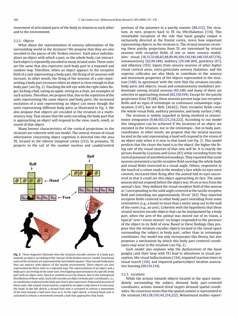
160 C. Da Cunha et al. / Behavioural Brain Research 199 (2009) 157–170
movement of articulated parts of the body in relation to each otherand to the environment.
3.2.2. ObjectsWhat about the representation of sensory information of the
surrounding world in the striatum? We propose that they are alsoencoded in the pieces of the ‘broken mirrors’. Each piece individu-alizes an object with which a part, or the whole body, can interact.Each object is repeatedly encoded in many striatal units. These unitsare the same that also represent each body part in a repeated andrandom way. Therefore, when an object appears in the receptivefield of a unit representing a body part, the firing of its neurons willincrease. In other words, the firing of the neurons of a unit repre-senting a body part increases when an object is close enough to thatbody part (see Fig. 2). Touching the left eye with the right index fin-ger, kicking a ball, eating an apple, sitting on a chair, are examples ofsuch actions. Therefore, we propose that, due to the repetition of theunits representing the same objects and body parts, the increasedexcitation of a unit representing an object can move though theunits representing different body parts as illustrated in Fig. 2. Wealso propose that objects are encoded in the striatum in a multi-sensory way. That means that the units encoding the body part thatis approaching an object will respond to the view, touch, smell, orsound of that object.
Many known characteristics of the cortical projections to thestriatum are coherent with our model. The ventral stream of visualinformation concerning object cognition is directed into the areaTE, located in the inferior temporal cortex [212]. In primates, TEprojects to the tail of the caudate nucleus and caudal/ventral
Fig. 2. These diagrams illustrate how the striatum encodes actions of a body parttowards an object, according to the ‘mosaic of the broken mirrors’ model. Functionalunits of the striatum are represented by interlinked squares. They encode body partsthat can interact with objects of the nearby environment. These objects are alsorepresented by these units in a repeated way. The representation of an object and abody part can overlap in the same unit. Overlapping representation of a specific bodypart with an object seen, heard or smelled occurs by chance, due to the widespreaddistribution of these units. Each unit encodes an object in body part-coordinates, i.e.,in coordinates centered in the body part that it also represents. Polymodal neurons ofthese units, like a hand-vision neuron, respond to an object only when it is seen nearthe hand. In the left sketch, a striatal foot-unit is activated to release a movementof the foot towards a ball seen close to it. In the right sketch, a striatal hand-unit isactivated to release a movement towards a ball that approaches that hand.
portions of the putamen in a patchy manner [88,212]. The stria-tum, in turn, projects back to TE via SNr/thalamus [134]. Thisremarkable exception of the rule that basal ganglia output isexclusively directed at the frontal cortex, stress how importantrepresenting objects in the striatum is. The striatal neurons receiv-ing these patchy projections from TE are intermixed by striatalneurons with receptive fields of one or more sensory modal-ities: visual [18,31,33,60,82,88,89,96,104,130,146,148,150,167,176],somatosensory [62,96,148], auditory [29,148,184], gustatory [67],and olfactory [193]. Inputs from sensory neurons of other highervisual cortical areas, extra-geniculate sensory thalamus, and thesuperior colliculus are also likely to contribute to the sensoryand movement properties of the objects represented in the stria-tum [148]. In agreement with the view that the striatum encodesbody parts and objects, visual and somatosensory modalities pre-dominate among striatal neurons [82,148] and many of them areselective to approaching stimuli [82,150,194]. Except for the patchyprojections from TE [88], these neurons present large size receptivefields and no signs of retinotopic or continuous somatotopic orga-nization ([147], but see Refs. [36,82]). Their receptive fields coverthe whole visual field, auditory perimeter, and body surface [148].
The striatum is widely regarded as being involved in sensori-motor integration [9,48,163,121,214,222]. According to our modelthis integration can be achieved if the locations of an object areencoded in the striatum, not in the retinotopic-, but in body part-coordinates. In other words, we propose that the striatal neuronslocated in the unit representing a hand will respond to the vision ofan object only when it is near to that hand (see Fig. 2). This modelpredicts that the closer the hand is to the object, the higher the fir-ing rate of the visual neurons of that unit will be. It is exactly thepicture found by Graziano and Gross [82] while recording from theventral putamen of anesthetized monkeys. They reported that someneurons presented a tactile receptive field covering the whole bodyand visual fields restricted to a visual angle. Others, responsive tothe touch of a cotton swab in the monkey’s face while its eyes werecovered, increased their firing after the animal had its eyes uncov-ered so that it could see this object approaching its face. The sameneuron did not respond before the object was 10 cm or less from theanimal’s face. They defined the visual receptive field of this neuronas “corresponding to the solid angle centered at the tactile receptivefield and extending out approximately 10 cm” [82]. They reportedreceptive fields centered in other body parts extending from somecentimeters (e.g., a hand) to more than a meter away out to the wallof the room (e.g., an arm). Coherent with the hypothesis that thesestriatal neurons encode objects that can be manipulated by a bodypart, when the arm of the animal was moved out of its vision, atypical “arm + vision neuron” no longer responded to the presenceof the object to its field of view. Based in these findings they pro-pose that the striatum encodes objects located in the visual spacesurrounding the subject in body part, rather than in retinotopiccoordinates. Our model not only incorporates this theory, but alsoproposes a mechanism by which this body part-centered coordi-nates may arise in the striatum (see Fig. 2).
Such model also explains why the dysfunctions of the basalganglia (and their loop with TE) lead to alterations in visual per-ception, like visual hallucinations [134], impaired reaction times invisual search [116], and impaired pattern/object location associa-tive learning [60,116,134].
3.2.3. LocationsWhile the actions towards objects located in the space imme-
diately surrounding the subject demand body part-centeredcoordinates, actions toward distal targets demand spatial coordi-nates. No consensus exists that the spatial context is represented inthe striatum [49,128,139,141,214,222]. Behavioural studies report-

C. Da Cunha et al. / Behavioural Brain Research 199 (2009) 157–170 161
ing a double dissociation between the dorsolateral striatum andthe hippocampus for spatial and stimulus-response (S-R) learn-ing tasks have initially led to the view that the striatum is notimportant for spatial tasks. These studies included spatial and cuedversions of the Morris water maze [156,159], radial maze [159],and plus-maze tasks [160]. Other studies from our group have alsoshown this dissociation between the SNc and the hippocampus[38,40,42,43,61,138].
However, even the cued tasks mentioned above require somedegree of spatial information to be solved. In those studies, the cue(i.e., a ball, a salient platform, a light) can be conceived as an objectwhich the animal needs to approach in order to be rewarded. Sincethis object is located in a specific place of the maze, the behaviourof the rat can be conceived as “to go to that object located in thatplace”. In some instances, such as in a plus-maze or T-maze, thereference is not an object but a hemi-side of the animal’s body(egocentric orientation) which permits encoding behaviours suchas making a right or left turn to be rewarded [16,106]. Even in thesecases, the task involves performing an action (turn) in a specificplace.
Evidence that the striatum encodes spatial information aboutthe environment came from studies reporting that, like the hip-pocampus [153], the striatum also contains place-related cells,neurons that discharge when the animal is in a particular place ofthe environment [57,139–141,172,213]. Compared to the hippocam-pal place cells, those found in the striatum are more influencedby other parameters of the task [111]: they also encode egocen-tric movements and are more sensitive to visual cues [141] andreward variables [111,126,140,141,194]. The striatum, as well as thehippocampus, also contains a subpopulation of neurons called headdirection cells that fire preferentially when the animal’s head isaligned with a particular orientation, irrespective of the animal’slocation [139,141]. These neurons are probably involved in egocen-tric movement.
The difference between the tasks depending on the dorsal stria-tum and those depending on the hippocampus is that, in the former,the location of the target does not need to be defined in terms ofmultiple relations between distal cues. In a recent study, we haveshown that inactivation or lesion of the striatum or of the SNc doesnot impair the ability of rats to navigate in a water maze when theyalways depart from the same starting point to find a hidden plat-form kept in the same place in the maze ([40], see Ref. [159]). Theanimals learn this task probably by using a single object of the envi-ronment as a distal cue. Animals with intact striatum and a lesionin the hippocampus may orient themselves in an environment, butthis orientation is not sufficient to disambiguate places equidis-tant to the same environmental object. This dissociation has beenshown by McDonald and White [128] in rats searching for food intwo adjacent arms of an 8-arm radial maze. Rats with a hippocam-pal disconnection, but with an intact striatum, were unable to solvethis task. However, the same rats were not impaired to discriminatein which of the two arms, separated by other two or more arms,they would find the food. In the latter case the animals probablyuse different distal cues to discriminate between arms.
According to the ‘mosaic of broken mirrors’ model, the represen-tation of space in the striatum may account for the characteristics ofthe tasks that can be learned with the participation of the striatum.This model postulates that cortical projections to the striatum arefragmented into pieces, with each piece representing a location. Inother words, this model assumes that, while the hippocampus rep-resents space as a continuum, the place fields in the striatum arerepeated and intermixed. This configuration facilitates the asso-ciation of objects (cues) with particular places, but breaks theorthogonal relationships among different locations. Therefore, thehippocampus is in a position to compare the current spatial context
Fig. 3. This diagram illustrates the redundancy and functional convergence prop-erties that the ‘mosaic of broken mirrors’ model proposes to the functional units ofthe striatum. The indirect pathway and the dopaminergic modulation are not repre-sented in order to simplify the diagram. Abbreviations: GPi, internal globus pallidus;SNr, substantia nigra pars reticulata.
of the environment with the context found in the past. On the otherhand, the striatum is in a position to choose an action that can movethe “pieces of the mosaic (the subject’s body, body’s parts, objects)to a particular location. According to this view, the hippocampalrepresentation of the environment is globally oriented, while thestriatal actions depend on breaking the environment into pieces inorder to move them. Hence, tasks such as the cued version of thewater maze or the win-stay version of the radial maze can be easilysolved by the striatum by associating the approaching action withthe place in which an object (cue) is located.
The action of approaching a location cannot be encoded in thehippocampus since it does not have direct connections with motorareas of the neocortex. This location-approaching action associa-tion is probably done in the striatum that receives direct inputsfrom the hippocampal formation to the shell region of the NAc, andindirect inputs to the core of the NAc through the prefrontal cortexand to the ventromedial striatum through the medial entorhinalcortex [66,119,129,202].
We recently obtained some curious results in experiments oflatent learning that can be explained by the assumption of the‘mosaic of broken mirrors’ model that the striatum represents spacein a fragmented way. We found that the impairment of SNc-lesionedrats to perform the cued version of the water maze disappearedwhen the animals were pre-trained in the spatial version of thistask [42]. Curiously, SNc-lesioned rats were not impaired to per-form the spatial version. A series of control experiments showedthat the presence of the hidden platform and the view of the distalcues during the pre-training sessions were critical for that benefi-cial effect. More intriguing was the finding that this improvementwas observed even when the locations of the distal cues (postersfixed on a curtain around the maze) were changed in relation tothe pre-training session. Our model explains these data by assum-ing that the spatial map formed during the pre-training sessionswas broken into pieces, each containing a distal cue. Hence, a par-ticular cue could be associated with the action of approaching it,irrespective of its relationship with the other cues.

162 C. Da Cunha et al. / Behavioural Brain Research 199 (2009) 157–170
Fig. 4. This diagram demonstrates the combinatorial, associative, learning, and action selection properties of the mosaic of broken mirrors model. Neurons are representedby boxes and circles. The colours of the arrows linking glutamatergic cortical neurons to striatal neurons denote their origins. Arrows linking the other components of thebasal ganglia circuit represent axons of GABAergic (red) or glutamatergic (green) neurons. (A) Before learning occurs, the circuit allows the association of any environmentalcue with any action. (B) After pairings of the salient Cue 1 with Action 1, coincident with a phasic release of dopamine (not shown), the following alterations occur, restrictedto the synapses between cortical neurons representing Cue 1 and those representing Action 1 that converge to the same striatal neurons: LTP in the direct pathway for Cue1; LTD in the indirect pathway for Cue 1; LTP in the indirect pathway for Action 1; LTD in the direct pathway for Action 1. Alterations in the synapses of Cue 1 increase theprobability that it will induce the choice of Action 1. The alterations in the synapses of Action 1 lead to the conclusion of the action. Abbreviations: GPe, external globuspallidus; GPi, internal globus pallidus; SNr, substantia nigra pars reticulata; St, striatum; STN, subthalamic nucleus; Th, thalamus.
In another study carried out in our laboratory, we found furtherevidence that units of the striatum encode actions directed at a goal(unpublished results). In that study, rats with complete hemilesionof the SNc induced by 6-hydroxydopamine were trained to enter thelighted arm of a radial maze in order to find a sucrose pellet. Thelesion prevented the animals from running directly to the lightedarm when it was located on the side contralateral to the lesion.However, these animals made ipsiversive turns in order to adjusttheir pathway and enter the lighted arm. This result suggests thatthe action of approaching a goal, but not the goal per se, dependson the release of dopamine in the striatum contralateral to the goallocation. Although SNc-hemilesioned rats have lost the basal gan-glia modulation that helps them to choose making contraversiveturns, they could still approach a goal located on their contralateralside by means of other actions (i.e., ipsiversive turns). When thedopaminergic receptors of the hemilesioned striatum were stim-ulated by the administration of a dopamine receptor agonist (i.e.,apomorphine), these animals did not only recover their ability toperform contraversive turns, but also overdid this action due tosupersensitization of D2 dopamine receptors [41]. These resultsare in agreement with the postulate of the “mosaic of broken mir-rors” model that the activation of specific actions (such as turns)directed at a goal is encoded by the functional units of the stria-tum. Other actions involved in the practice of innate behaviours,such as grooming [34] and predatory hunting [183], have also beenreported to depend on the striatum.
Therefore, the model proposes that not only the hippocampus,but also the striatum, is needed to solve spatial versions of waterand radial mazes. The poor performance of striatum-lesioned ratsin these tasks has been attributed to lesions more restricted to thedorsolateral striatum, sparing other regions that receive direct or
indirect projections from the hippocampus, i.e., the dorsomedialstriatum [49,141,222]. According to this view, spatial navigationdepends on both the hippocampus and the striatum. The hip-pocampus provides the map and the striatum the pathway tonavigate through it. Coherent with this postulate, neurons encod-ing for particular behaviours such as turns have been found in thestriatum, but not in the hippocampus [141]. Mulder et al. [145]reported the existence in the striatum of “goal”-like neurons thatfire continuously while a rat moves from one location to another ina plus-maze. These neurons may encode the paces of movementsbetween landmarks of a route made up by pieces of the spatial map.
3.2.4. Other functional units of the striatumThe inputs to the striatum are not restricted to sensory, spatial
or motor areas of the cortex. Prefrontal and limbic areas of the cor-tex also project to the striatum in a convergent and widespreadmanner. Convergence refers to afferents departing from differentregions of the cortex to overlap in restricted areas of the cortexforming ‘matrisome-like’ functional units. These units are widelydistributed in vast regions of the striatum. What is the functionalnature of these units? They may refer to affective meaning and toabstract information such as symbols, words, digits, thoughts, andplans. The processing of these functional units by the basal gangliawould explain the involvement of the latter in working memoryand executive and affective functions [26,132,154].
3.3. Building associative units
Once objects, locations, body parts, symbols, and associatedactions or plans are individualized into functional units in thestriatum, what is the function of their repeated representation?

C. Da Cunha et al. / Behavioural Brain Research 199 (2009) 157–170 163
The answer is associative learning. The body, the surroundingworld and the mental world can be combined into more flexibleassociations if they are broken into pieces (see Fig. 3). Repeti-tion increases the probability of association among pieces andexplains the involvement of the basal ganglia in different kindsof associative learning: Pavlovian conditioning for the associa-tion between a conditioned stimulus (CS) (a neutral stimulus)and an unconditioned stimulus (US) (a rewarding or aversiveoutcome) [10,187]; instrumental or operant conditioning for theassociation of a predictive cue with an action outcome (reinforce-ment or punishment) [143,173,217]; addiction for the associationbetween a drug with strong rewarding properties and its com-pulsive consumption [11,58]; skill learning for the association of asequence of motor actions [1,51,52,95,189]. The associative propertyof basal ganglia proposed by this model also permits the striatumto play a role in action selection based on reinforcement of pre-vious cue-action associations [6,7,30,64,114,191]. The ingredientsfor these associations are the synapses between the corticostri-atal neurons and the MSNs encoding the functional units of thestriatum.
3.3.1. Synaptic plasticity in the striatumWhat are the mechanisms underlying the association of func-
tional units of the striatum? The most likely candidates are thesynaptic plasticity phenomena known to occur in the striatum.Both long-term potentiation (LTP) and long-term depression (LTD)have been reported to occur in synapses between the corticos-triatal neurons and MSNs [20,50,218]. According to Hebb’s rule,LTP occurs when presynaptic and postsynaptic neurons are depo-larized at the same time. LTP can be induced in the striatum byrepeated activation of cortical terminals [27]. Therefore, corticos-triatal synapses are the binding elements associating informationarriving from different regions of the cortex. This associationmay occur when LTP is induced in the synapses of the two cor-ticostriatal neurons with the same MSN and requires a triplecoincidence: the two cortical neurons and the MSN must be depo-larized at the same time. Such coincidence fulfil the needs forthe induction of heterosynaptic associative LTP [124]. The partiallyclosed loops between the striatum–GPi–thalamus–striatum andthe striatum–GPi–thalamus–cortex–striatum (Fig. 1) may result inreverberant activation of MSNs, a factor contributing to keep theseneurons depolarized. Other loops involving the GPe and/or the STNmay also play a role in such reverberation and/or in the modulationof this circuit. High-frequency firing of the corticostriatal neuronsmay also induce LTD in their synapses with MSNs ([19,120,211], seealso Refs. [50,218] for a review). The concentration of dopamineand how dopamine receptors are distributed among MSNs are crit-ical factors to determine the induction of LTD or LTP, as will bediscussed in the next section. LTP and LTD of synapses associatingdifferent cortical inputs with the same MSNs may build the mem-ory trace of associative learning mediated by the basal ganglia (seeFigs. 3 and 4).
3.3.2. Dopamine-dependent synaptic plasticityThe synaptic plasticity necessary for the occurrence of associa-
tive learning in the striatum requires a learning signal, a messagethat signals when and how learning occurs. This message seems tobe the release of dopamine ([99,187], but see Ref. [218]). The activa-tion of dopamine receptors in MSNs is necessary for the inductionof LTP or LTD. D2-like and (maybe) D1-like dopamine receptors arerequired for the induction of LTD, but the activation of D2 recep-tors favours the induction of LTD over LTP in some instances ([21],see also Ref. [218] for a different view). Activation of DB1 cannabi-noid and adenosine A2A receptors also seems to be involved in theinduction of the striatal LTD [50,70,218]. On the other hand, LTP
requires the activation of D1 receptors [25] and is inhibited by theactivation of D2 receptors [21].
D1 receptors occur mainly in MSNs of the direct pathway(those projecting to the GPi/SNr), whereas D2 receptors are mainlyexpressed in neurons of the indirect pathway (those projecting tothe GPe) [71,72]. Therefore, in the presence of dopamine, LTP ismore likely to occur in the direct pathway and LTD in the indirectpathway. The direct pathway positively modulates actions encodedby the frontal cortex, while the indirect pathway inhibits theiroccurrence (see Section 2 above). According to the ‘mosaic of brokenmirrors’ model, in the presence of dopamine, the concomitant acti-vation of corticostriatal neurons encoding, for example, an objectand the action of approaching it, would induce LTP in their synapseswith MSNs of the direct pathway and LTD in synapses with MSNs ofthe indirect pathway. This feature would increase the firing prob-ability of MSNs encoding the association between the stimulus(object) and the action of approaching it [101].
The complete segregation of the direct and indirect pathwaysis currently a matter of debate [48,87,218]. Induction of LTD thatrequires the activation of D2 receptors occurs in most MSNs [19,50].In addition, there is evidence for the co-expression of D1 and D2receptors in a subpopulation of neurons [199]. In these neuronsthe induction of LTP or LTD depends on the level of dopamine andon the depolarization state of MSNs. D2 receptors present a higheraffinity for dopamine than D1 receptors [103]. As a consequence,lower levels of dopamine favour the induction of LTD and higherlevels favour the induction of LTP [25].
What happens when the act of approaching an object is rein-forced? The corticostriatal neurons encoding the object and theaction of approaching it are activated at the same time. As a conse-quence, LTP or LTD would occur in the connections of MSNs thatreceive overlapping projections from these active corticostriatalneurons, with the occurrence of LTP in MSNs of the direct pathwayand LTD in those of the indirect pathway (see above). This featurewould increase the firing probability of these MSNs and the conse-quent occurrence of the approaching action when the same objectis seen by the subject in the future.
3.3.3. Novelty-driven reinforcement learningMidbrain neurons release dopamine in the striatum in tonic
or phasic patterns [68,75–77]. A small amount of dopamine isspontaneously and continuously released by these neurons in atonic pattern, providing a baseline level of extrasynaptic dopaminerequired to run the motor programs already set up [75]. The pha-sic firing of dopaminergic neurons causes a transient and robustrelease of dopamine and serves as a learning signal, inducing neu-ral plasticity in the striatum. Coherent with this theory, the phasicrelease of dopamine is critical for Pavlovian conditioning [10,187]instrumental learning [143], and other types of associative and rein-forcement learning [114,185].
The influential studies by Schultz and other groups sug-gested that the phasic release of dopamine occurs in response tounpredicted rewarding stimuli [10,143,188], with the amount ofdopamine released being proportional to the difference betweenexpected and obtained reward [188]. This difference is calledreward prediction error. More recently, this theory has been con-tested by the argument that the latency for a stimulus to inducethe phasic release of dopamine is too short to permit the sen-sory processing necessary to evaluate the stimulus identity andreward value [173]. The fact that the unpredicted presentation ofnon-rewarding salient stimuli such as light flashes or tones elic-its a phasic dopamine response also disagrees with the rewardprediction error theory [99,100,118]. Habituation to a stimulus abol-ishes the phasic dopamine response [118,187]. The omission of anexpected reward causes a brief cessation in the firing of midbrain

164 C. Da Cunha et al. / Behavioural Brain Research 199 (2009) 157–170
dopaminergic neurons at the time the stimulus was expected tooccur [186]. Aversive or detrimental stimuli (usually those thatcause pain) induce a pause in the firing of dopaminergic neurons forthe duration of the event, followed by a rebound response [32,210].Therefore, the phasic dopamine response seems to signal the pres-ence of new biologically significant stimuli, with a positive response(increased release do dopamine) to non-harmful stimuli (neutral orrewarding) and a negative response to harmful stimuli [173].
As stressed above, striatal synaptic plasticity depends on theactivation of dopamine receptors. Therefore, the phasic release ofdopamine serves as a permissive signal for learning processes thatoccur in the striatum. The fragmentation of the sensory repre-sentation of the environmental world and functional parts of thebody involved in actions permits the individualization of theseelements and their repetition increases the combinatorial asso-ciation among them. After repeated presentation of novel stimuliassociated with actions, the continuous reinforcement of the asso-ciations between pairs of stimuli or stimulus-action units thatalways appear together causes them to be more strongly associatedthan the stimuli and actions that are associated only occasionally.According to the ‘mosaic of broken mirrors’ model, it is the prin-ciple of the associative learning that forms expectations based oncurrent stimuli and actions (see also [114,149,207,219]). After learn-ing, the occurrence of a salient stimulus can be predicted and it willno longer induce the phasic dopamine response. The memory forthis association becomes stable.
According to this model, the association of an action with its out-come depends on their representation in the striatum at the sametime as the concentration of dopamine in the synapses are highdue to the phasic response. Otherwise, the synaptic plasticity tostrengthen the synapses between overlapping corticostriatal neu-rons and MSNs would be lacking. The phasic dopamine responseseems to appear too early and to be too short [65,84,99,188] to per-mit the association of a stimulus with an action and its rewardingoutcome [173]. However, the clearance of dopamine released in thestriatum, particularly in the NAc and medial regions of the striatum,takes longer compared to the dorsolateral striatum [151,198,216].This fact would explain MSNs in the striatum responding to pre-vious actions and their reward outcome [35,92,97,114,115]. Theclearance of dopamine may range from a few hundreds of mil-liseconds in the dorsolateral striatum to several seconds in the NAc[151,198,216]. This difference can account for the higher involve-ment of the NAc in action-outcome reinforcement learning and ofthe dorsolateral striatum in S-R habits [149,222]. The fast clearanceof dopamine in the dorsolateral striatum opens a time window tootight to include the reward outcome to the S-R association. Thismight be the reason for the slow learning rate of S-R habits andfor the fact that these habits are relatively insensitive to rewarddevaluation. On the other hand, in the NAc the slow clearance ofdopamine after a phasic response is probably long enough to asso-ciate the outcome (reward) with the action, a fast learning thatfades more easily after reward withdrawal or devaluation.
This postulate is in line with imaging and electrophysiologicalstudies showing increased activity in the striatum in response toa reward [47,91,113,114] and reward prediction errors [90,152]. Itis also supported by studies reporting that the lesion or manip-ulation of the rat SNc or striatum disrupts associative reinforcedlearning in various tasks such as the cued version of the Mor-ris water maze [42,43,61,138], two-way active avoidance task[39,73,74,110], inhibitory avoidance [23,46,133,165,170,171,181],Pavlovian conditioning [168], and cued instrumental tasks[12,59,168,169]. Similar associative reinforced and habit learningdeficits have also been observed in mouse and monkey modelsof Parkinson’s disease, as well as in Parkinson’s disease patients[60,78,109,112,178,182,200].
3.3.4. Aversively motivated learningAssociative learning mediated by appetitive reinforcement can
be easily explained by the postulates of the ‘mosaic of the brokenmirrors’ model since a short latency phasic dopamine response fol-lows the reward presentation [186], as mentioned above. However,aversively motivated associative learning demands further elabora-tion since, as also mentioned above, aversive stimuli may induce apause in the firing of midbrain dopaminergic neurons for the dura-tion of the event, followed by a rebound response [101,210]. Howcan a reduction in the extracellular dopamine levels in the striatuminduce learning, a process that demands neuronal plasticity? Let usdiscuss two popular models of aversively motivated learning: theactive and the inhibitory avoidance tasks.
Learning the two-way active avoidance task, a kind of con-ditioned avoidance response (CAR), demands from a rodent toactively run away from a footshock (unconditioned stimulus) sig-nalled by a cue (usually a tone or the light of the chamber, i.e., theconditioned stimulus) [39]. Training is carried out by the pairing ofthe CS and US in a two-chamber shuttle box. The CS starts before andturns off together with the US. After many consecutive pairings, theanimal learns to avoid the US by crossing to the opposite chamberjust after the presentation of the CS. Electrophisiological studiesreported that most, if not all [210], midbrain dopamine neuronsrespond to noxious stimuli with a short latency increase in the fir-ing rate, followed by a rebound offset ([32,69,98,122,173,208,210],but see Ref. [125]). The temporal resolution of microdialysis stud-ies is not enough to detect the decrease in dopamine release in thestriatum after a footshock, but these studies consistently detect theincrease that may result from the rebound response that follows theending of the noxious stimulus [100,205,223].
Thus, the increase in the extracellular concentration ofdopamine probably coincide with the presentation of the “crossing”action that turns the US and CS off. The higher level of dopaminefavours the induction of LTP between the corticostriatal neuronsencoding the CS that converge to MSNs to which the corticostriatalneurons encoding the “crossing” action also project (see Section3.3.2). This “crossing response” of the animal may be seen as theaction of running away from the CS. Note that running away from apainful stimulus (US) is an innate behaviour, independent of learn-ing.
Inhibitory avoidance, also called passive avoidance, demandsthat the animal (usually a rodent) avoids entering a particular place.Inhibitory avoidance training may be performed in the same two-chamber box used for two-way active avoidance conditioning [3].The animal is placed in a lit chamber and receives a brief footshockwhen it enters the dark chamber. Usually only one session is neededfor the animal to learn to inhibit the innate tendency of enteringthe dark chamber. In other words, it learns not to go to that loca-tion. The novelty of exploring the lit chamber probably induces aphasic response of the midbrain dopaminergic neurons ([117,118],but see Ref. [44]). The footshock probably induces the cessation oftheir firing [32,173,187]. Therefore, the act of remaining in the litchamber will coincide with higher levels of extracellular striataldopamine and the act of entering the dark chamber with the low-ering in the level of dopamine. The former situation favours theinduction of LTP between the corticostriatal neurons encoding thelocation of the lit chamber and MSNs receiving projections of cor-ticostriatal neurons encoding the action of remaining there (seeSection 3.3.2).
Therefore, we propose that in aversively motivated learning, it isnot the reduction of the firing of midbrain dopamine neurons thatinduces learning, but the increase in the release of dopamine in thestriatum before and after the aversive stimulus. In both active andinhibitory avoidances, the action that coincides with higher levelsof dopamine is associated with the concomitant cue or location.

C. Da Cunha et al. / Behavioural Brain Research 199 (2009) 157–170 165
This hypothesis is coherent with the findings that manipulations inthe SNc [39,73,74] or in the striatum [23,46,110,133,165,170,171,181]impair learning of these tasks.
Note that inhibitory avoidance may be learned as the associationof an action with a place. However, such association would impairlearning of the two-way active avoidance task in which the animalmust successively return to the place in which it was punished. Inthis situation, the hippocampus, that encodes an environment as aplace [153], is expected to play a detrimental influence. This pre-diction is in agreement with studies reporting that the lesion of theseptum [180,206] or fimbria-fornix [85] improves learning of theinhibitory avoidance task. This illustrates a case in which the stria-tum and the hippocampus play competitive roles on learning [214].It is coherent with the present view that the striatum encodes dis-crete stimuli and locations (see Section 3.2.3). The representationof both discrete cues and locations in the striatum does not meanthat they will be always associated with the current actions. Onlythe activation of the striatal units that coincide with an action per-formed under high levels of striatal dopamine will be associated tothis action. During learning of the two-way active avoidance, theact of running to a specific location (chamber) will be coincidentwith the release of dopamine only in 50% of the trials. On the otherhand, the action of running from the CS will be reinforced by therelease of dopamine in all occasions. As a consequence, the compe-tition between the associations of the CS-“running from it” and thelocation-“avoid running to it” will be won by the former as trials goon. Such learning may be faster if the influence of the hippocampusis inhibited.
3.4. Building action units
3.4.1. Driving MSNs to an ‘up’ or ‘down state’The membrane potential of MSNs oscillates between ‘up’ (sub-
threshold depolarized) and ‘down’ (hyperpolarized) states [220].LTP is more likely to occur during the former and LTD during thelatter state [20,50]. The higher activity of corticostriatal neuronsrepresenting actions and current features of the external or internalenvironment favours the ‘up state’ in MSNs to which they converge[197]. Since these functional units are represented in a repeated way[62,63,87], at least some of them probably overlap, thus present-ing a higher probability to be in the ‘up state’ or depolarized. Thisprobability is increased by the diffuse corticostriatal projections toa broader area of the striatum [22].
3.4.2. Go/NoGo unitsThe result of striatal processing flows to the GPi and SNr, the
output doors of the basal ganglia through the direct or indirectpathway (see Figs. 1 and 4). They build the ‘Go’ and ‘NoGo’ prod-ucts of the basal ganglia processing [64] (see Figs. 3 and 4). Thedirect pathway is a GABAergic (inhibitory) connection between thestriatum and GPi/SNr. The indirect pathway connects the striatumto the GPi by a sequence of neurons that finally exert an excitatoryeffect. Therefore, the direct pathway (Go) relieves the thalamocor-tical neurons from the tonic inhibition of the GPi/SNr. The indirectpathway (NoGo) results in the opposite effect [2] (see Section 2above).
Since the ‘Go’ and ‘NoGo’ units affect almost exclusively thefrontal cortex (through thalamocortical projections) and subcorti-cal motor areas, they result in the induction/repression of actions,action planning, and other executive functions.
3.5. Gathering action units
The smaller number of neurons in the striatum, compared tothe neocortex, imposes a convergence of the information originat-
ing from the neocortex to transform it into functional units [8,144].In rats, 17 × 106 corticostriatal neurons converge onto 1.7 × 10⁶
MSNs in the striatum [224]. The corticostriatal convergence isprobably higher due to the repetition of the functional units (seeFigs. 3 and 4).
The lateral inhibition among MSNs is seen as evidence for par-allel and independent processing in the striatum [215]. However,other studies reported that this lateral inhibition is unilateral andrestricted to less than one-third of the tested pairs [37,209], a find-ing favouring the proposal that the functional units of the striatumare formed by patches of MSNs receiving convergent and overlap-ping cortical projections. In this case, lateral inhibition may helpisolate neighbouring functional units from one another. Since thefunctional units are repeated and widespread throughout the stria-tum, they may be distant enough to avoid lateral inhibition fromtheir peers.
This repeated and widespread distribution of the functionalunits imposes a binding problem to coordinate the firing andplasticity between equal units. Recent studies suggest that thisproblem might be solved by a class of interneurons, presumedto be cholinergic, called TANs (see Section 2). These interneuronspresent a broad distribution, lying mainly at the borders of thestriosome-matrix [4], and a low spontaneous firing rate that resultsin inhibitory effects on the excitability of MSNs [225]. TANs respondto rewarding events with a phasic decrease in their firing rate,at the same time that dopaminergic neurons increase their fir-ing rate [5,14,79,143,195]. However, while in some instances theresponse of dopaminergic neurons seems to be proportional tothe reward prediction error (see Section 3.3.2), the response ofTANs is indifferent to reward predictability [143]. The dopamineresponse is timed to novel salient stimuli (including rewardingstimuli), but the time necessary to remove dopamine from thesynapse is longer compared to the rapid removal of acetylcholineby dense acetylcholinesterase [225]. The sharp response of TANs torewarding stimuli may result in a temporal synchronization of therepeated functional units formed by the patches of MSNs spreadthroughout the striatum. In other words, TANs may signal to MSNswhen to learn, midbrain dopaminergic neurons may signal how tolearn, and corticostriatal neurons may signal what to learn [143].Coherently, the number of TANs responding to the reward signalincreases in parallel with learning of Pavlovian [4] and instrumental[143] learning tasks. Learning probably results in a gradual recruit-ment of the numerous functional units of the striatum as learningprogresses.
The projection of the striatum to the GPi and SNr imposes a sec-ond convergence of the order of 102–103 [8] (see Figs. 3 and 4).This convergence probably accounts for the re-unification of therepeated functional units of the striatum [79], i.e., as learning pro-gresses by recruiting a larger number of repeated units of thestriatum, the activation of these convergent units of the GPi/SNrincreases. Since the GPi/SNr projects almost exclusively to thefrontal cortex (through the thalamus) and brainstem motor nuclei,they probably encode mainly actions and plans.
4. Emergent properties of the ‘mosaic of broken mirrors’model
Most of the attributes of nondeclarative memories are emergentproperties of the ‘mosaic of broken mirrors’ model. These memoriesare said to be implicit (unconscious) [196], rigid (inflexible) [56],procedural (expressing how to do something) [196], and suitableto guide cue-based and egocentric navigation [214]. The learning ofmost of these memories is a slow and gradual [54,158,222] asso-ciative process that depends on reinforcement [58,94,204], andsometimes forms habits after overtraining [136,222].

166 C. Da Cunha et al. / Behavioural Brain Research 199 (2009) 157–170
The implicit nature of memories that depend on basal gangliaprocessing is explained by the fragmentation of the informationthat occurs in the striatum, so that neither the subject’s own bodynor its environment are globally perceived during learning. Instead,few components of the environment are associated with discreteactions. This learning process is highly adaptive in order to adjustautomatic responses (actions) to discrete changes in environmentalelements. However, the meaning of this behaviour does not makesense in the global environment, simply because it is not globallyoriented.
The rigid or inflexible aspects of these memories may beexplained by this model for the same reasons. Since these mem-ories are formed by associations of fragments of information aboutthe environment and specific actions, their expression cannot beflexibly used in another context of the environment because of thelack of a global view of the environment. Even chains of actionsperformed in a skill are not oriented as an action of the subjectin a complex environment, but as an automatic sequence of singleactions.
Since the output of the basal ganglia is almost exclusively thefrontal cortex and brainstem motor nuclei, the memories encodedby this system must be expressed as actions. This explains the pro-cedural nature of these memories.
The fragmented representation of the environment in the stria-tum also explains the cue-based and egocentric navigation duringbasal ganglia-dependent learning. This type of navigation is not ori-ented towards a global view of the environment, but rather relieson discrete environmental cues or sequences of movements basedon egocentric orientation [17,45,203]. The broken representationof the environment favours the association of units of informa-tion (cues) relevant as reward predictors with actions performedto approach the place in which the reward is delivered. However,this fragmentation does not allow multiple relations between envi-ronmental elements to form a spatial map. As a consequence, itstores information sufficient only to guide the navigation by stepsbased on sequential approaches to cues or sequences, for example,of right/left turns at specific locations.
One of the most evident properties of the ‘mosaic of brokenmirrors’ model is that it is ideal to perform reinforcement asso-ciative learning. The repetition of the functional units formedin the striatum by convergent projections of the cortex ampli-fies the combinatorial power of the system. The dependence ondopamine to strengthen or weaken the associations among stimuli,actions and outcomes makes this associative process conditional.The release of dopamine only when the stimulus or the outcomeare unpredictable (unlearned) becomes the driving force of learningmediated by this system.
The slow and gradual learning of procedural memories canbe explained by two characteristics of this model. Reinforcementlearning starts with trial and error associations, followed by eval-uation of the outcome, and progresses by multiple comparisonsbetween the reward prediction and/or the novelty of stimuli andthe outcome during each trial. It is by definition a gradual process.The gradual recruitment of the functional units that are repeatedin the striatum also contributes for learning to become slow andgradual.
Some types of instrumental learning result in a strong asso-ciation between a stimulus and an action that becomes resistantto reward devaluation. This kind of associative memory, in whichthe stimulus becomes stronger than the outcome to trigger theresponse, is called habit [222]. The repetition of the functional unitsin the striatum mediating this association after extensive learn-ing may partly account for this property. The more this associativememory becomes represented by a larger number of associativeunits, the more difficult it will be to erase them when the reward
outcome or the novelty decreases. In addition, the spreading ofthese associative units throughout the striatum increases the prob-ability of their occupying striatal regions less sensitive to the rewardoutcome. Recent findings suggesting a gradient from the ventral tothe dorsal striatum in the clearance of dopamine and regional dif-ferences in dopamine-dependent synaptic plasticity may accountfor these differences [216]. The formation of association units lesssensitive to a reward is slower and so is their dissociation afterreward withdrawal. If this is the case, the ventral striatum (NAc)would account for a fast and transient learning observed during thefirst trials of an instrumental task, while the dorsal striatum wouldaccount for the slow and strong (more resistant to reward devalu-ation or withdrawal) learning (habit) achieved after overtraining.
5. Conclusion
In figurative words, we propose that the cortico-basal process-ing of procedural memories is similar to a mosaic consisting ofpieces of images of several broken mirrors. According to this model,neurons of the sensory, motor and associative cortices send conver-gent projections to the striatum that result in functional units (seeFigs. 3 and 4). These striatal units encode articulated parts of thebody and portions of the surrounding world that can be movedor manipulated, such as surrounding objects (Fig. 2). These unitsalso encode specific locations to which the subject can move. Theassociation of these functional units results in programs to performmotor skills and movements of the arms, eyes, or other body partsto a specific target (object or location), or in the locomotion of thesubject to specific targets. The combinatorial power of these asso-ciations is amplified by the repeated and widespread distributionof the functional units in the striatum.
According to this model, learning in this system depends on thealteration in the strength of the synapses between the corticostri-atal neurons and MSNs that encode the functional units (Fig. 4).It occurs when an environmental stimulus becomes salient in anunpredictable way. At this time, the midbrain dopaminergic neu-rons release dopamine in the striatum in a phasic pattern. Theactivation of dopaminergic neurons is a condition for the occur-rence of synaptic plasticity in the striatum. The synchronizationof neurons of the repeated functional units encoding the sameaction in relation to the salient stimulus is performed by a pausein the release of acetylcholine by TANs. The striatal units encod-ing the same stimulus/action send convergent projections to theGPi and SNr that, in turn, drive the encoded action to the frontalcortex (passing by the thalamus) (Fig. 3). The partially closed loopsinvolving the GPe, STN, thalamus, and striatum may result in rever-beration that facilitates the induction of LTP or LTD in the striatum.These loops may also have other modulatory functions in this sys-tem.
Still according to this model, the stronger association betweenthe functional units of the striatum encoding an action triggeredby a stimulus makes the occurrence of this association no longerunpredictable. As the novelty is reduced, the salience of the stim-ulus decreases and no further learning occurs. In this respect, thislearning system is driven by novelty.
After a phasic dopamine response, the high concentration ofdopamine takes longer to be cleared in the synapses of the NAccompared to the dorsal striatum [151,198,216]. In other words, thelearning signal that allows synaptic plasticity lasts longer in theNAc than in the dorsal striatum. Accordingly, this learning signalis long enough to incorporate the evaluation of the reward valueof the action outcome in the NAc, but not in the dorsal striatum.It explains why learning mediated by the NAc is driven by thereward outcome of the action, while learning mediated by the dor-sal striatum forms S-R habits that are less sensitive to reward. This
C. Da Cunha et al. / Behavioural Brain Research 199 (2009) 157–170 167
model explains the gradual learning and many known properties ofdifferent types of procedural memories, such as allowing cue andegocentric navigation and their implicit, inflexible and associativenature.
Several postulates of the ‘mosaic of broken mirrors’ modelneed to be tested in future studies, particularly those thatare the core of this model and differentiate it from othermodels of basal ganglia functioning: the postulation of theexistence of repeated functional units in the striatum andtheir associative combination to form procedural memories.Nevertheless, these postulates are coherent with current find-ings, such as the “matrisomes” discovered by Flaherty andGraybiel [62,63], evidence for convergence and widespread pro-jections from different regions of the cortex to the striatum[22,28,72,123,150,175,179,192,221,224], cue and egocentric naviga-tion mediated by the basal ganglia [38,40,42,43,61,138,159], andplace-related cells in the striatum that also encode movements[141], among other findings reported in this review. The remainingpostulates of this model were mainly incorporated from existingmodels [2,8,64,80,86,144,158,173,214,216], except for the mecha-nism proposed to explain how the NAc and dorsal striatum encodesaction-outcome expectancies and S-R habits, respectively.
A model can be considered as equivalent to a map of a newland based on the landmarks discovered by explorers that madeblind navigations through it. This map results from the recreationof the cartographer that tries to accommodate the landmarks to hislogic and imagination. This map is not an infallible orientation tonew explorers, but it can provide routes to the exploration of thisland. The explorers may confirm or not the locations in this landaccording to the map. Such is the case for the striatum accordingto the ‘mosaic of broken mirrors’ model; the map can be improvedbased on the outcome of these intents. We hope that the ‘mosaicof broken mirrors’ model may be of some help to guide the workof researchers interested in understanding how the basal gangliamediate procedural learning.
Acknowledgements
The editorial comments of Rainer K.W. Schwarting, Philip Winn,Jorge Medina, and Ivan Izquierdo are acknowledged. This work wassupported by grants of Institutos do Milenio (CNPq/MCT), PronexParaná, and Fundacão Araucária, and FAPESP.
References
[1] Albouy G, Sterpenich V, Balteau E, Vandewalle G, Desseilles M, Dang-Vu T, etal. Both the hippocampus and striatum are involved in consolidation of motorsequence memory. Neuron 2008;85:261–72.
[2] Alexander GE, DeLong MR, Strick PL. Parallel organization of functionallysegregated circuits linking basal ganglia and cortex. Annu Rev Neurosci1986;9:357–81.
[3] Angelucci MEM, Vital MAF, Cesário C, Zadusky CR, Rosalen P, Da Cunha C. Theeffect of caffeine on animal models of learning and memory. Eur J Pharmacol1999;373:135–40.
[4] Aosaki T, Kimura M, Graybiel AM. Temporal and spatial characteris-tics of tonically active neurons of the primates striatum. J Neurophysiol1995;73:1234–52.
[5] Apicella P, Ravel S, Sardo P, Legallet E. Influence of predictive information onresponses of tonically active neurons in the monkey striatum. J Neurophysiol1998;80:3341–4.
[6] Balleine BW, Delgado MR, Hikosaka O. The role of the dorsal striatum in rewardand decision-making. J Neurosci 2007;27:8161–5.
[7] Balleine BW, Liljeholm M, Ostlund SB. Integrative function of the basal gangliain instrumental conditioning. Behav Brain Res 2008, this issue.
[8] Bar-Gad I, Morris G, Bergman H. Information processing, dimensionalityreduction and reinforcement learning in the basal ganglia. Prog Neurobiol2003;71:439–73.
[9] Barneoud P, Descombris E, Aubin N, Abrous DN. Evaluation of simple and com-plex sensorimotor behaviours in rats with a partial lesion of the dopaminergicnigrostriatal system. Eur J Neurosci 2000;12:322–36.
[10] Bayer H, Glimcher P. Midbrain dopamine neurons encode a quantitativereward prediction error signal. Neuron 2005;47:129–41.
[11] Belin D, Jonkman S, Dickinson A, Robbins TW, Everitt BJ. Parallel andinteractive learning processes within the basal ganglia: relevance for theunderstanding of addiction. Behav Brain Res 2008, this issue.
[12] Bermudez-Rattoni F, Mujica-González M, Prado-Alcala RA. Is cholinergicactivity of the striatum involved in the acquisition of positively-motivatedbehaviors? Pharmacol Biochem Behav 1986;24:715–9.
[13] Blandini F, Nappi G, Tassorelli C, Martignoni E. Functional changes of the basalganglia circuitry in Parkinson’s disease. Prog Neurobiol 2000;62:63–88.
[14] Blazquez PM, Fujii N, Kojima J, Graybiel AM. A network representation ofresponse probability in the striatum. Neuron 2002;33:973–82.
[15] Bolam JP, Hanley JJ, Booth PAC, Bevan MD. Synaptic organisation of the basalganglia. J Anat 2000;96:527–42.
[16] Braga R, Kouzmine I, Canteras NS, Da Cunha C. Lesion of the substantia nigra,pars compacta impairs delayed alternation in a Y-maze in rats. Exp Neurol2005;192:134–41.
[17] Brasted PJ, Humby T, Dunnett SB, Robbins TW. Unilateral lesions of thedorsal striatum in rats disrupt responding in egocentric space. J Neurosci1997;17:8919–26.
[18] Brown VJ, Desimone R, Mishkin M. Responses of cells in the tail ofthe caudate nucleus during visual discrimination learning. J Neurophysiol1995;74:1083–94.
[19] Calabresi P, Maj R, Pisani A, Mercuri NB, Bernardi G. Long-term synapticdepression in the striatum: physiological and pharmacological characteri-zation. J Neurosci 1992;12:4224–33.
[20] Calabresi P, Picconi B, Tozzi A, Filippo M. Dopamine-mediated regulation ofcorticostriatal synaptic plasticity. Trends Neurosci 2007;30:211–9.
[21] Calabresi P, Saiardi A, Pisani A, Baik JH, Centonze D, Mercuri NB, et al. Abnor-mal synaptic plasticity in the striatum of mice lacking dopamine D2 receptors.J Neurosci 1997;17:4536–44.
[22] Calzavara R, Mailly P, Haber SN. Relationship between the corticostriatalterminals from areas 9 and 46, and those from area 8A, dorsal and rostral pre-motor cortex and area 24c: an anatomical substrate for cognition to action.Eur J Neurosci 2007;26:2005–24.
[23] Cammarota M, Bevilaqua LRM, Kohler C, Medina JH, Izquierdo I. Learningtwice is different from learning once and from learning more. Neuroscience2005;132:273–9.
[24] Castellanos FX, Giedd JN, Marsh WL, Hamburger SD, Vaituzis AC, DicksteinDP, et al. Quantitative brain magnetic resonance imaging in attention-deficithyperactivity disorder. Arch Gen Psychiatry 1996;53:607–16.
[25] Centonze D, Grande C, Saulle E, Martin AB, Gubellini P, Pavon N, et al. Distinctroles of D1 and D5 dopamine receptors in motor activity and striatal synapticplasticity. J Neurosci 2003;23:8506–12.
[26] Chang C, Crottaz-Herbette S, Menon V. Temporal dynamics of basal gan-glia response and connectivity during verbal working memory. Neuroimage2007;34:1253–69.
[27] Charpier S, Deniau JM. In vivo activity-dependent plasticity at cortico-striatalconnections: evidence for physiological long-term potentiation. Proc NatlAcad Sci U S A 1997;94:7036–40.
[28] Cheatwood JL, Corwin JV, Reep RL. Overlap and interdigitation of corti-cal and thalamic afferents to dorsocentral striatum in the rat. Brain Res2005;1036:90–100.
[29] Chudler EH, Sugiyama K, Dong WK. Multisensory convergence and integrationin the neostriatum and globus pallidus of the rat. Brain Res 1995;674:33–45.
[30] Cohen MX, Frank MJ. Neurocomputational models of basal ganglia functionin learning, memory and choice. Behav Brain Res 2008, this issue.
[31] Coizet V, Comoli E, Westby GW, Redgrave P. Phasic activation of substantianigra and the ventral tegmental area by chemical stimulation of the supe-rior colliculus: an electrophysiological investigation in the rat. Eur J Neurosci2003;17:28–40.
[32] Coizet V, Dommett EJ, Redgrave P, Overton PG. Nociceptive responses of mid-brain dopaminergic neurones are modulated by the superior colliculus in therat. Neuroscience 2006;139:1479–93.
[33] Comoli E, Coizet V, Boyes J, Bolam JP, Canteras NS, Quirk RH, et al. A directprojection from superior colliculus to substantia nigra for detecting salientvisual events. Nat Neurosci 2003;6:974–80.
[34] Cromwell HC, Berridge KC. Implementation of action sequences by aneostriatal site a lesion mapping study of grooming syntax. J Neurosci1996;16:3444–58.
[35] Cromwell HC, Schultz W. Effects of expectations for different rewardmagnitudes on neuronal activity in primate striatum. J Neurophysiol2003;89:2823–38.
[36] Crutcher MD, DeLong MR. Single cell studies fo the primate putamen. I. Func-tional organization. Exp Brain Res 1984;53:233–43.
[37] Czubayko U, Plenz D. Fast synaptic transmission between striatal spiny pro-jection neurons. Proc Natl Acad Sci U S A 2002;99:15764–9.
[38] Da Cunha C, Angellucci MEM, Canteras NS, Wonnacott S, Takahashi RN. Thelesion of the rat substantia nigra pars compacta dopaminergic neurons asa model for Parkinson’s disease memory disabilities. Cell Mol Neurobiol2002;22:227–37.
[39] Da Cunha C, Gevaerd MS, Vital MABF, Miyoshi E, Andreatini R, Silveira R, etal. Memory in rats with nigral lesion induced by MPTP: a model for earlyParkinson’s disease amnesia. Behav Brain Res 2001;124:9–18.

168 C. Da Cunha et al. / Behavioural Brain Research 199 (2009) 157–170
[40] Da Cunha C, Silva MHC, Wietzikoski S, Wietzikoski E, Ferro MS, Kouzmine I,et al. Place learning strategy of substantia nigra pars compacta-lesioned rats.Behav Neurosci 2006;120:1279–84.
[41] Da Cunha C, Wietzikoski EC, Ferro MM, Martinez GR, Vital MABF, Hipolide D,et al. Hemiparkinsonian rats rotate toward the side with the weaker dopamin-ergic neurotransmission. Behav Brain Res 2008;189:364–72.
[42] Da Cunha C, Wietzikoski S, Wietzikoski E, Silva MHC, Chandler J, Ferro MM,et al. Pre-training to find a hidden platform in the Morris water maze cancompensate for deficit to find a cued platform in a rat model of Parkinson’sdisease. Neurobiol Learn Mem 2007;87:451–63.
[43] Da Cunha C, Wietzikoski S, Wietzikoski EC, Miyoshi E, Ferro MM, Anselmo-Franci JA, et al. Evidence for the substantia nigra pars compacta as an essentialcomponent of a memory system independent of the hippocampal memorysystem. Neurobiol Learn Mem 2003;79:236–42.
[44] Damsma G, Pfaus JG, Wenkstern D, Phillips AG, Fibiger HC. Sexual behav-ior increases dopamine transmission in the nucleus accumbens and striatumof male rats: comparison with novelty and locomotion. Behav Neurosci1992;106:181–91.
[45] DeCoteau WE, Kesner RP. A double dissociation between the rat hippocam-pus and medial caudoputamen in processing two forms of knowledge. BehavNeurosci 2000;14:1096–108.
[46] Del Guante MAD, Rivas M, Prado-Alcala RA, Quirarte GL. Amnesia producedby pre-training infusion of serotonin into the substantia nigra. Neuroreport2004;15:2527–9.
[47] Delgado MR, Nystrom LE, Fissell C, Noll DC, Fiez JA. Tracking the hemody-namic responses to reward and punishment in the striatum. J Neurophysiol2000;84:3072–7.
[48] DeLong MR, Wichmann T. Circuits and circuit disorders of the basal ganglia.Arch Neurol 2007;64:20–4.
[49] Devan BD, McDonald RJ, White NM. Effects of medial and lateral caudate-putamen lesions on place- and cue-guided behaviours in the water maze:influence of thigmotaxis. Behav Brain Res 1999;100:5–14.
[50] Di Filippo M, Picconi B, Barone I, Ghiglieri V, Bagetta V, Sgobio C, et al. Striatalsynaptic plasticity: underlying mechanisms and implications for reward-related learning. Behav Brain Res 2008, this issue.
[51] Doyon J, Bellec P, Amsel R, Penhune V, Monchi O, Lehéricy S, et al. Contribu-tions of the basal ganglia and functionally related brain structures to motorlearning. Behav Brain Res 2008, this issue.
[52] Doyon J, Benali H. Reorganization and plasticity in the adult brain duringlearning of motor skills. Curr Opin Neurobiol 2005;15:161–7.
[53] Doyon J, Laforce R, Bouchard G, Gaudreau D, Roy J, Poirier M, et al. Role of thestriatum, cerebellum and frontal lobes in the automatization of a repeatedvisuomotor sequence of movements. Neuropsychologia 1998;36:625–41.
[54] Doyon J, Ungerleider L. Functional anatomy of motor skill learning. In: SquireL, Schacter D, editors. Neuropsychology of memory. New York: Guilford Press;2002, p.225–238.
[55] Ehringer H, Hornykiewicz O. Verteilung von noradrenalin und dopamin (3-hydroxytyramin) im gehirn des menschen und ihr verhalten bei erkrankugendes extrapyramidalen system. Klin Wochenschr 1960:1236–9.
[56] Eichenbaum H. The cognitive neuroscience of memory: an introduction. NY:Oxford University Press; 2002, 384 p.
[57] Eschenko O, Mizumori SJY. Memory influences on hippocampal and striatalneural codes: effects of a shift between task rules. Neurobiol Learn Mem2007;87:495–509.
[58] Everitt BJ, Robbins TW. Neural systems of reinforcement for drug addiction:from actions to habits to compulsion. Nat Neurosci 2005;8:1481–9.
[59] Faure A, Haberland U, Conde F, El Massioui N. Lesion to the nigrostriataldopamine system disrupts stimulus-response habit formation. J Neurosci2005;25:2771–80.
[60] Fernandez-Ruiz J, Wang J, Aigner TG, Mishkin N. Visual habit formation inmonkeys with neurotoxic lesions of the ventrocaudal neostriatum. Proc NatlAcad Sci U S A 2001;98:4196–201.
[61] Ferro MM, Bellissimo MI, Anselmo-Franci JA, Angellucci E, Canteras NS, DaCunha C. Comparison of bilaterally 6-OHDA- and MPTP lesioned rats as mod-els of the early phase of Parkinson’s Disease: histological, neurochemical,motor and memory alterations. J Neurosci Methods 2005;148:78–87.
[62] Flaherty AW, Graybiel AM. Corticostriatal transformations in the primatesomatosensory system—projections from physiologically mapped body-partrepresentations. J Neurophysiol 1991;66:1249–63.
[63] Flaherty AW, Graybiel AM. Input–output organization of the sensorimotorstriatum in the squirrel–monkey. J Neurosci 1994;14:599–610.
[64] Frank MC. Dynamic dopamine modulation in the basal ganglia: a neuro-computational account of cognitive deficits in medicated and nonmedicatedparkinsonism. J Cogn Neurosci 2005;17:51–72.
[65] Freeman AS. Firing properties of substantia nigra dopaminergic neurons infreely moving rats. Life Sci 1985;36:1983–94.
[66] French SJ, Totterdell S. Hippocampal and prefrontal cortical inputs monosy-naptically converge with individual projection neurons of the nucleusaccumbens. J Comp Neurol 2002;446:151–65.
[67] Fudge JL, Breitbart MA, Danish M, Pannoni V. Insular and gustatory inputs tothe caudal ventral striatum in primates. J Comp Neurol 2005;490:101–18.
[68] Gao DM, Nevet A, Vaadia E, Bergman H. Coincident but distinct mes-sages of midbrain dopamine and striatal tonically active neurons. Neuron2004;43:133–43.
[69] Gao DM, Jeaugey L, Pollak P, Benabid AL. Intensity-dependent nociceptiveresponses from presumed dopaminergic neurons of the substantia nigra, parscompacta in the rat and their modification by lateral habenula inputs. BrainRes 1990;529:315–9.
[70] Gerdeman GL, Ronesi J, Lovinger DM. Postsynaptic endocannabinoid releaseis critical to long-term depression in the striatum. Nat Neurosci 2002:446–51.
[71] Gerfen CR, Engber TM, Mahan LC, Susel Z, Chase TN, Monsma FJ, et al. D1and D2 dopamine receptor regulated gene-expression of striatonigral andstriatopallidal neurons. Science 1990;250:1429–32.
[72] Gerfen CR. Indirect-pathway neurons lose their spines in Parkinson disease.Nat Neurosci 2006;9:157–8.
[73] Gevaerd MS, Miyoshi E, Silveira R, Canteras NS, Takahashi RN, Da Cunha C.Levodopa treatment restores the striatal level of dopamine but fails to reversememory deficits in rats treated with MPTP, an animal model of Parkinson’sdisease. Int J Neuropsychopharmacol 2001;4:361–70.
[74] Gevaerd MS, Takahashi RN, Silveira R, Da Cunha C. Caffeine reverses the mem-ory disruption induced by intra-nigral MPTP-injection in rats. Brain Res Bull2001;55:101–6.
[75] Goto Y, Otani S, Grace AC. The Yin and Yang of dopamine release: a newperspective. Neuropharmacology 2007;53:583–7.
[76] Grace AA, Bunney BS. The control of firing pattern in nigral dopamine neurons:single spike firing. J Neurosci 1984;4:2866–76.
[77] Grace AA, Bunney BS. The control of firing pattern in nigral dopamine neurons:burst firing. J Neurosci 1984;4:2877–90.
[78] Grahn JA, Parkinson JA, Owen AM. The role of the basal ganglia in learningand memory: neuropsychological studies. Behav Brain Res 2008, this issue.
[79] Graybiel AM, Aosaki T, Flaherty AW, Kimura M. The basal ganglia and adaptivemotor control. Science 1994;265:1826–32.
[80] Graybiel AM. Network-level neuroplasticity in cortico-basal ganglia path-ways. Parkinsonism Relat Disord 2004;10:293–6.
[81] Graybiel AM, Ragsdale Jr CW. Histochemically distinct compartments inthe striatum of human, monkeys and cat demonstrated by acetylthio-cholinesterase staining. Proc N Y Acad Sci U S A 1978;75:5723–6.
[82] Graziano MSA, Gross CG. A bimodal map of space: somatosensory receptivefields in the macaque putamen with corresponding visual receptive fields.Exp Brain Res 1993;97:96–109.
[83] Grillner S, Helligren J, Menard A, Saitoh K, Wikstrom MA. Mechanisms forselection of basic motor programs—roles for the striatum and pallidum.Trends Neurosci 2005;28:364–70.
[84] Guarraci FA, Kapp BS. An electrophysiological characterization of ventraltegmental area dopaminergic neurons during differential Pavlovian fear con-ditioning in the awake rabbit. Behav Brain Res 1999;99:169–79.
[85] Guillazo-Blanch G, Nadal R, Vale-Martinez A, Mart’y-Nicolovius M, Arévalo R,Morgado-Bernal I. Effects of fimbria lesions on trace two-way active avoid-ance acquisition and retention in rats. Neurobiol Learn Mem 2002;78:407–25.
[86] Gurney K, Prescott TJ, Wickens JR, Redgrave P. Computational models ofthe basal ganglia: from robots to membranes. Trends Neurosci 2004;27:453–9.
[87] Haber SN, Kim KS, Mailly P, Calzavara R. Reward-related cortical inputsdefine a large striatal region in primates that interface with associative cor-tical inputs, providing a substrate for incentive-based learning. J Neurosci2006;26:8368–76.
[88] Harting JK, Updyke BV, Van Lieshout DP. Striatal projections from the cat visualthalamus. Eur J Neurosci 2001;14:893–6.
[89] Harting JK, Updyke BV, Van Lieshout DP. The visual-oculomotor striatumof the cat: functional relationship to the superior colliculus. Exp Brain Res2001;136:138–42.
[90] Haruno M, Kawato M. Different neural correlates of reward expec-tation and reward expectation error in the putamen and caudatenucleus during stimulus-action-reward association learning. J Neurophysiol2006;95:948–59.
[91] Haruno M, Kuroda T, Doya K, Toyama K, Kimura M, Samejima K, et al. Aneural correlate of reward-based behavioral learning in caudate nucleus: afunctional magnetic resonance imaging study of a stochastic decision task. JNeurosci 2004;24:1660–5.
[92] Hassani OK, Cromwell HC, Schultz W. Influence of expectation of differentrewards on behavior-related neuronal activity in the striatum. J Neurophysiol2001;85:2477–89.
[93] Heckers S. Neuropathology of schizophrenia: cortex, thalamus, basal gan-glia, and neurotransmitter-specific projection systems. Schizophr Bull1997;23:403–21.
[94] Herrnstein RJ. On the law of effect. J Exp Anal Behav 1970;13:243–66.[95] Hikosaka O, Nakamura K, Sakai K, Nakahara H. Central mechanisms of motor
skill learning. Curr Opin Neurobiol 2002;12:217–22.[96] Hikosaka O, Sakamoto M, Usui S. Functional properties of monkey cau-
date neurons. II. Visual and auditory responses. J Neurophysiol 1989;61:799–813.
[97] Hollerman JR, Tremblay L, Schultz W. Influence of reward expectationon behavior-related neuronal activity in primate striatum. J Neurophysiol1998;80:947–63.
[98] Hommer DW, Bunney BS. Effect of sensory stimuli on the activity ofdopaminergic neurons: involvement of non-dopaminergic nigral neurons andstriato-nigral pathways. Life Sci 1980;27:377–86.
C. Da Cunha et al. / Behavioural Brain Research 199 (2009) 157–170 169
[99] Horvitz JC, Stewart T, Jacobs BL. Burst activity of ventral tegmentaldopamine neurons is elicited by sensory stimuli in the awake cat. Brain Res1997;759:251–8.
[100] Horvitz JC. Mesolimbocortical and nigrostriatal dopamine responses to salientnon-reward events. Neuroscience 2000;96:651–6.
[101] Horvitz JC. Stimulus-response and response-outcome learning mechanismsin the striatum. Behav Brain Res 2008, this issue.
[102] Humphries MD, Stewart RD, Gurney KN. A physiologically plausible modelof action selection and oscillatory activity in the basal ganglia. J Neurosci2006;26:12921–42.
[103] Jaber M, Robinson SW, Missale C, Caron MG. Dopamine receptors and brainfunction. Neuropharmacology 1996;35:1503–19.
[104] Jiang H, Stein BE, McHafe JG. Opposing basal ganglia processes shape midbrainvisuomotor activity bilaterally. Nature 2003;424:982–6.
[105] Joel D, Weiner I. The connections of the dopaminergic system with the stria-tum in rats and primates: an analysis with respect to the functional andcompartmental organization of the striatum. Neuroscience 2000;96:451–74.
[106] Jog MS, Kubota Y, Connolly CI, Hillegaart V, Graybiel AM. Building neuralrepresentations of habits. Science 1999;286:1745–9.
[107] Kawaguchi Y, Emson PC. Striatal interneurones: chemical, physiological andmorphological characterization. Trends Neurosci 1995;18:527–35.
[108] Kim YB, Huh N, Lee H, Baeg EH, Lee D, Jung MW. Encoding of action historyin the rat ventral striatum. J Neurophysiol 2007;98:3548–56.
[109] Kimura M. Role of the basal ganglia in behavioral learning. Neurosci Res1995;2:353–8.
[110] Kirkby RJ, Polgar S. Active avoidance in laboratory rat following lesions ofdorsal or ventral caudate-nucleus. Physiol Psychol 1974;2:301–6.
[111] Knierim JJ. Neural representations of location outside the hippocampus. LearnMem 2006;13:405–15.
[112] Knowlton BJ, Mangels JA, Squire LR. A neostriatal habit learning system inhumans. Science 1996;273:1399–402.
[113] Knutson B, Fong GW, Adams CM, Varner JL, Hommer D. Dissociation ofreward anticipation and outcome with event-related fMRI. Neuroreport2001;12:3683–7.
[114] Lau B, Glimcher PW. Action and outcome encoding in the primate caudatenucleus. J Neurosci 2007;14:502–14.
[115] Lauwereyns J, Watanabe K, Coe B, Hikosaka O. A neural correlate of responsebias in monkey caudate nucleus. Nature 2002;418:413–7.
[116] Lawrence AD, Watkins LHA, Sahakian BJ, Hodges JR, Robbins TW. Visual objectand visuospatial cognition in Huntington’s disease: implications for informa-tion processing in corticostriatal circuits. Brain 2000;23:1349–64.
[117] Legault M, Wise RA. Novelty-evoked elevations of nucleus accumbensdopamine: dependence on impulse flow from the ventral subiculum and glu-tamatergic neurotransmission in the ventral tegmental area. Eur J Neurosci2001;13:819–28.
[118] Ljungberg T, Apicella P, Schultz W. Responses of monkey dopamine neuronsduring learning of behavioural reactions. J Neurophysiol 1992;67:145–63.
[119] Lodge DJ, Grace AA. The hippocampus modulates dopamine neuronresponsivity by regulating the intensity of phasic neuron activation. Neu-ropsychopharmacology 2006;31:1356–61.
[120] Lovinger DM, Tyler EC, Merritt A. Short- and long-term synaptic depressionin rat neostriatum. J Neurophysiol 1993;70:1937–49.
[121] Lynd-Balta E, Haber SN. The organization of midbrain projections to the stria-tum in the primate: sensorimotor-related striatum versus ventral striatum.Neuroscience 1994;59:625–40.
[122] Maeda H, Mogenson GJ. Effects of peripheral stimulation on the activity ofneurons in the ventral tegmental area, substantia nigra and midbrain reticularformation of rats. Brain Res Bull 1982;8:7–14.
[123] Malach R, Graybiel AM. Mosaic architecture of the somatic sensory-recipientsector of the cat’s striatum. J Neurosci 1986;6:3436–58.
[124] Malenka RC, Nicoll RA. Neuroscience—long-term potentiation—a decade ofprogress? Science 1999;285:1870–4.
[125] Mantz J, Thierry AM, Glowinski J. Effect of noxious tail pinch on the dischargerate of mesocortical and mesolimbic dopamine neurons: selective activationof the mesocortical system. Brain Res 1989;476:377–81.
[126] Martin PD, Ono T. Effects of reward anticipation, reward presentation, andspatial parameters on the firing of single neurons recorded in the subicu-lum and nucleus accumbens of freely moving rats. Behav Brain Res 2000;116:23–38.
[127] Martone M, Bytters N, Payne P, Becker JT, Sax DS. Dissociation betweenskill leraning and verbal recognition in amnesia and dementia. Arch Neurol1984;41:965–70.
[128] McDonald RJ, White NM. Hippocampal and non-hippocampal contributionsto place learning. Behav Neurosci 1995;109:579–93.
[129] McGeorge AJ, Faull RLM. The organization of the projection from the cerebralcortex to the striatum in the rat. Neuroscience 1989;29:503–38.
[130] McHafe JG, Thomson CM, Stein BE. Corticotectal and corticostriatal projec-tions from the frontal eye fields of the cat: an anatomical examination usingWGA-HRP. Somatosens Mot Res 2001;18:117–30.
[131] McHaffie JG, Stanford TR, Stein BE, Coizet V, Redgrave P. Subcortical loopsthrough the basal ganglia. Trends Neurosci 2005;28:401–7.
[132] Mcnab F, Klingberg T. Prefrontal cortex and basal ganglia control access toworking memory. Nat Neurosci 2008;11:103–7.
[133] Medina AC, Charles JR, Espinoza-Gonzalez V, Sanchez-Resendis O, Prado-Alcala RA, Roozendaal B, et al. Glucocorticoid administration into the dorsal
stratium facilitates memory consolidation of inhibitory avoidance train-ing but not of the context or footshock components. Learn Mem 2007;14:673–8.
[134] Middleton FA, Strick PL. The temporal lobe is a target of output from the basalganglia. Proc Natl Acad Sci USA 1996;93:8683–7.
[135] Mink JW. The basal ganglia: focused selection and inhibition of competingmotor programs. Prog Neurobiol 1996;50:381–425.
[136] Mishkin M, Petri HL. Memories and habits: some implications for the analysisof learning retention. In: Squire RL, Butters N, editors. The neuropsychologyof memory. New York: Gilford Press; 1984. p. 187–96.
[137] Mitchell JA, Hall G. Caudate-putamen lesions may impair or potentiate mazelearning depending upon availability of stimulus cues and relevance ofresponse cues. Quart J Exp Psychol 1988;40B:243–58.
[138] Miyoshi E, Wietzikoski S, Camplessei M, Silveira R, Takahashi EN, Da Cunha C.Impaired learning in a spatial working memory version and in a cued versionof the water maze in rats with MPTP-induced mesencephalic dopaminergiclesion. Brain Res Bull 2002;58:41–7.
[139] Mizumori SJ, Puryear CB, Martig AK. Basal ganglia. Contributions to adaptivenavigation. Behav Brain Res 2008, this issue.
[140] Mizumori SJY, Ragozzino KE, Cooper BG. Location and head direction repre-sentation in the dorsal striatum of rats. Psychobiology 2000;28:441–62.
[141] Mizumori SJY, Yeshenko O, Gill KM, Davis DM. Parallel processing acrossneural systems: implications for a multiple memory system hypothesis. Neu-robiol Learn Mem 2004;82:278–98.
[142] Montoya A, Price BH, Menear M, Lepage M. Brain imaging and cogni-tive dysfunctions in Huntington’s disease. J Psychiatry Neurosci 2006;31:21–9.
[143] Morris G, Arkadir D, Nevet A, Vaadia E, Bergman H. Coincident but distinctmessages of midbrain dopamine and striatal tonically active neurons. Neuron2004;43:133–43.
[144] Morris G, Nevet A, Bergman H. Anatomical funneling, sparse connectivity andredundancy reduction in the neural networks of the basal ganglia. J Physiol2003;97:581–9.
[145] Mulder AB, Tabuchi E, Wiener SI. Neurons in hippocampal afferent zones ofrat striatum parse routes into multi-pace segments during maze navigation.Eur J Neurosci 2004;19:1923–32.
[146] Nagy A, Eordegh G, Norita M, Benedek G. Visual receptive field properties ofneurons in the caudate nucleus. Eur J Neurosci 2003;18:449–52.
[147] Nagy A, Eordegh G, Paroczy Z, Markus Z, Benedek G. Multisensory integrationin the basal ganglia. Eur J Neurosci 2006;24:917–24.
[148] Nagy A, Paroczy Z, Norita M, Benedek G. Multisensory responses and receptivefield properties of neurons in the substantia nigra and in the caudate nucleus.Eur J Neurosci 2005;22:419–24.
[149] Nicola SM. The nucleus accumbens as part of a basal ganglia action selectioncircuit. Psychopharmacology 2007;191:521–50.
[150] Niida T, Stein BE, McHaffie JG. Response properties of corticotectal and cor-ticostriatal neurons in the posterior lateral suprasylvian cortex of the cat. JNeurosci 1997;17:8550–65.
[151] Nirenberg MJ, Chan J, Pohorille A, Vaughan RA, Uhl GR, Kuhar MJ, et al. Thedopamine transporter: comparative ultrastructure of dopaminergic axonsin limbic and motor compartments of the nucleus accumbens. J Neurosci1997;17:6899–907.
[152] O’Doherty J, Dayan P, Schultz J, Deichmann R, Friston K, Dolan RJ. Dissociableroles of ventral and dorsal striatum in instrumental conditioning. Science2004;304:452–4.
[153] O’Keefe J, Nadel L. The hippocampus as a cognitive map. Oxford, England:Oxford University Press; 1978, 570 pp.
[154] O’Reilly RC, Frank MJ. Making working memory work: a computationalmodel of learning in the prefrontal cortex and basal ganglia. Neural Comput2006;18:283–328.
[155] Owen AM. Cognitive dysfunction in Parkinson disease: the role of frontostri-atal circuitry. Neuroscientist 2004;10:525–37.
[156] Packard MG, Cahill L, McGaugh JL. Amygdala modulation of hippocampal-dependent and caudate nucleus-dependent memory processes. Proc NatlAcad Sci USA 1994;91:8477–81.
[157] Packard MG, Hirsh R, White NM. Differential roles of the hippocampus andcaudate nucleus lesion s on two radial maze task: evidence for multiple mem-ory systems. J Neurosci 1989;9:1465–72.
[158] Packard MG, Knowlton BJ. Learning and memory functions of the basal gan-glia. Annu Rev Neurosci 2002;25:563–93.
[159] Packard MG, McGaugh JL. Double dissociation of fornix and caudate nucleuslesions on acquisition of two water maze tasks: further evidence for multiplememory systems. Behav Neurosci 1992;106:439–46.
[160] Packard MG, McGaugh JL. Inactivation of hippocampus or caudate nucleuswith lidocaine differentially affects expression of place and response learning.Neurobiol Learn Mem 1996;65:66–72.
[161] Packard MG, White NM. Dissociation of hippocampal and caudate nucleusmemory systems by post-training intracerebral injection of dopamine ago-nists. Behav Neurosci 1991;105:295–306.
[162] Packard MG, White NM. Lesions of the caudate nucleus selectively impairacquisition of “reference memory” in the radial maze. Behav Neural Biol1990;53:39–50.
[163] Parent A, Hazrati LN. Functional anatomy of the basal ganglia. I. The cortico-basal ganglia-thalamo-cortical loop. Brain Res Rev 1995;20:91–127.
[164] Parkinson J. An essay on the saking plasy. Shewood: Neely and Jones; 1817.

170 C. Da Cunha et al. / Behavioural Brain Research 199 (2009) 157–170
[165] Perez-Ruiz C, Prado-Alcala RA. Retrograde amnesia induced by lidocaineinjection into the striatum: protective effect of the negative reinforcer. BrainRes Bull 1989;22:599–603.
[166] Pisani A, Bernardi G, Ding J, Surmeier DJ. Re-emergence of striatal choliner-gic interneurons in movement disorders. Trends Neurosci 2007;30(10):545–53.
[167] Pouderoux C, Freton E. Patterns of unit responses to visual stimuli in thecat caudate nucleus under chloralose anesthesia. Neurosci Lett 1979;11:53–8.
[168] Prado-Alcala RA, Grinberg ZJ, Alvarez-Leefmans FJ, Brust-Carmona H. Sup-pression of motor conditioning by the injection of 3 M KCl in the caudatenuclei of cats. Physiol Behav 1973;10:59–64.
[169] Prado-Alcala RA, Grinberg ZJ, Alvarez-Leefmans FJ, Gomez A, Singer S, Brust-Carmona H. A possible caudate-cholinergic mechanism in two instrumentalconditioned responses. Psychopharmacologia (Berl) 1972;25:339–46.
[170] Prado-Alcala RA, Grinberg ZJ, Arditti ZL, Garcia MM, Prieto HG, Brust-CarmonaH. Learning deficits produced by chronic and reversible lesions of the corpusstriatum in rats. Physiol Behav 1975;15:283–7.
[171] Prado-Alcala RA, Solana-Figueroa R, Galindo LE, Medina AC, Quirarte GL.Blockade of striatal 5-HT2 receptors produces retrograde amnesia in rats. LifeSci 2003;74:481–8.
[172] Ragozzino KE, Leutgeb S, Mizumori SJY. Conditional coupling of dorsal stri-atal head direction and hippocampal place representations during spatialnavigation. Exp Brain Res 2001;139:372–6.
[173] Redgrave P, Gurney K, Reynolds J. What is reinforced by phasic dopaminesignals? Brain Res Rev 2008;58:322–39.
[174] Redgrave P, Prescott T, Gurney KN. The basal ganglia: a vertebrate solution tothe selection problem. Neuroscience 1999;89:1009–23.
[175] Reep RL, Cheatwood JL, Corwin JV. The associative striatum: organizationof cortical projections to the dorsocentral striatum in rats. J Com Neurol2003;467:271–92.
[176] Rolls ET, Thorpe SJ, Maddison SP. Responses of striatal neurons in the behavingmonkey. 1. Head of the caudate nucleus. Behav Brain Res 1983;7:179–210.
[177] Romanelli P, Esposito V, Schaal DW, Heit G. Somatotopy in the basal ganglia:experimental and clinical evidence for segregated sensorimotor channels.Brain Res Rev 2005;48:112–28.
[178] Roncacci S, Troisi E, Carlesimo GA, Nocentini U, Caltagirone C. Implicit mem-ory in Parkinsonian patients: evidence for deficient skill learning. Eur Neurol1996;36:154–9.
[179] Rosell A, Gimenez-Amaya JM. Anatomical re-evaluation of the corticostriatalprojections to the caudate nucleus: a retrograde labeling study in the cat.Neurosci Res 1999;34:257–69.
[180] Sagvolden T, Johnsrud G. Two-way active avoidance learning followingmedial, dorsolateral, or total septal lesions in rats: effect of intensity of dis-continuous shock. Behav Neural Biol 1982;35:17–32.
[181] Salado-Castillo R, Diaz del Guante MA, Alvarado R, Quirarte GL, Prado-AlcalaRA. Effects of regional GABAergic blockade of the striatum on memory con-solidation. Neurobiol Learn Mem 1996;66:102–8.
[182] Salmon DP, Butters N. Neurobiology of skill and habit learning. Curr OpinNeurobiol 1995;5:184–90.
[183] Santos LM, Ferro MM, Mota-Ortiz SR, Baldo MV, Da Cunha C, Canteras NS.Effects of ventrolateral striatal inactivation on predatory hunting. PhysiolBehav 2007;90:669–73.
[184] Schneider JS. Responses of striatal neurons to peripheral sensory stimulationin symptomatic MPTP-exposed cats. Brain Res 1991;544:297–302.
[185] Schonberg T, Daw ND, Joel D, O’Doherty JP. Reinforcement learning signalsin the human striatum distinguish learners from nonlearners during reward-based decision making. J Neurosci 2007;12:860–7.
[186] Schultz W, Dayan P, Montague PR. A neural substrate of prediction and reward.Science 1997;275:1593–9.
[187] Schultz W. Multiple dopamine functions at different time courses. Annu RevNeurosci 2007;30:259–88.
[188] Schultz W. Predictive reward signal of dopamine neurons. J Neurophysiol1998;80:1–27.
[189] Schwarting RKW. Rodent models of serial reaction time tasks and their imple-mentation in neurobiological research. Behav Brain Res 2008, this issue.
[190] Scoville WB, Milner B. Loss of recent memory after bilateral hippocampallesions. J Neurol Neurosurg Psychiat 1957;20:11–21.
[191] Seger CA. How do the basal ganglia contribute to categorization? Their rolesin generalization, response selection, and learning via feedback. NeurosciBiobehav Rev 2008;32:265–78.
[192] Selemon LD, Goldman-Rakic PS. Longitudinal topography and interdigitationof corticostriatal projections in the rhesus monkey. J Neurosci 1985;5:776–94.
[193] Setlow B, Schoenbaum G, Gallagher M. Neural encoding in ventral striatumduring olfactory discrimination learning. Neuron 2003;38:625–36.
[194] Shibata R, Mulder AB, Trullier O, Wiener SI. Position sensitivity inphasically discharging nucleus accumbens neurons of rats alternatingbetween tasks requiring complementary types of spatial cues. Neuroscience2001;108:391–411.
[195] Shimo Y, Hikosaka O. Role of tonically active neurons in primate caudate inreward-oriented saccadic eye movement. J Neurosci 2001;21:7804–14.
[196] Squire LR. Memory systems of the brain: a brief history and current perspec-tive. Neurobiol Learn Mem 2004;82:171–7.
[197] Stern EA, Kincaid AE, Wilson CJ. Spontaneous subthreshold membrane poten-tial fluctuations and action potential variability of rat corticostriatal andstriatal neurons in vivo. J Neurophysiol 1997;77:1697–715.
[198] Suaud-Chagny MF, Dugast C, Chergui K, Msghina M, Gonon F. Uptake ofdopamine released by impulse flow in the rat mesolimbic and striatal systemsin vivo. J Neurochem 1995;65:2603–11.
[199] Surmeier DJ, Song WJ, Yan Z. Coordinated expression of dopamine receptorsin neostriatal MSNs. J Neurosci 1996;16:6579–91.
[200] Teng E, Stefanacci L, Squire LR, Zola SM. Contrasting effects on discrimina-tion learning after hippocampal lesions and conjoint hippocampal-caudatelesions in monkeys. J Neurosci 2000;20:3853–63.
[201] Tepper JM, Bolam JP. Functional diversity and specificity of neostriatalinterneurons. Curr Opin Neurobiol 2004;14:685–92.
[202] Thierry AM, Gioanni Y, Dégénétais E, Glowinski J. Hippocampo-prefrontalcortex pathway: anatomical and electrophysiological characteristics. Hip-pocampus 2000;10:411–9.
[203] Thompson WG, Guilford MO, Hicks LH. Effects of caudate and cortical lesionson place and response learning in rats. Physiol Psychol 1980;8:473–9.
[204] Thorndike EL. Animal Intelligence. New York: Macmillan; 1911.[205] Tidey JW, Miczek KA. Social defeat stress selectively alters mesocorticolim-
bic dopamine release: An in vivo microdialysis study. Brain Res 1996;72:140–9.
[206] Torras-Garcia M, Costa-Miserachs D, Morgado-Bernal I, Portell-Cortés I.Improvement of shuttle-box performance by anterodorsal medial septallesions in rats. Behav Brain Res 2003;141:147–58.
[207] Tremblay L, Hollerman JR, Schultz W. Modifications of reward expectation-related neuronal activity during learning in primate striatum. J Neurophysiol1998;80:964–77.
[208] Tsai C-T, Nakamura S, Iwama K. Inhibition of neuronal activity of the substantianigra by noxious stimuli and its modification by the caudate nucleus. BrainRes 1980;195:299–311.
[209] Tunstall MJ, Oorschot DE, Kean A, Wickens JR. Inhibitory interactions betweenspiny projection neurons in the rat striatum. J Neurophysiol 2002;88:1263–9.
[210] Ungless MA, Magill PJ, Bolam JP. Uniform inhibition of dopamine neurons inthe ventral tegmental area by aversive stimuli. Science 2004;303:2040–2.
[211] Walsh JP. Depression of excitatory synaptic input in rat striatal neurons. BrainRes 1993:123–8.
[212] Webster MJ, Bachevalier J, Ungerleider LG. Subcortical connections of infe-rior temporal areas TE and TEO in macaque monkeys. J Compar Neurol1993;335:73–91.
[213] Weiner SI. Spatial and behavioral correlates of striatal neurons in rats per-forming a self-initiated navigation task. J Neurosci 1993;13:3802–17.
[214] White NM, McDonald RJ. Multiple parallel memory systems in the brain ofthe rat. Neurobiol Learn Mem 2002;77:125–84.
[215] Wickens J. A theory of the striatum. Oxford: Pergamon; 1993.[216] Wickens JR, Budd CS, Hyland BI, Arbuthnott GW. Striatal contributions to
reward and decision making. Making sense of regional variations in a reit-erated processing matrix. Ann NY Acad Sci 2007;1104:192–212.
[217] Wickens JR, Reynolds JNJ, Hyland BI. Neural mechanisms of reward-relatedmotor learning. Curr Opin Neurobiol 2003;13:685–90.
[218] Wickens JR. Synaptic plasticity in the corticostriatal pathway. Behav Brain Res2008, this issue.
[219] Williams ZM, Eskandar EN. Selective enhancement of associative learning bymicrostimulation of the anterior caudate. Nat Neurosci 2006;9:562–8.
[220] Wilson CJ, Kawaguchi Y. The origins of two-state spontaneous mem-brane potential fluctuations of neostriatal spiny neurons. J Neurosci1996;16:2397–410.
[221] Yeterian EH, Pandya DN. Corticostriatal connections of the superior temporalregion in rhesus monkeys. J Compar Neurol 1998;399:384–402.
[222] Yin HH, Knowlton BJ. The role of the basal ganglia in habit formation. Nat RevNeurosci 2006;7:464–76.
[223] Young AMJ. Increased extracellular dopamine in nucleus accumbens inresponse to unconditioned and conditioned aversive stimuli: studies using1 min microdialysis in rats. J Neurosci Methods 2004;138:57–63.
[224] Zheng T, Wilson CJ. Corticostriatal combinatorics: the implications of corti-costriatal axonal arborizations. J Neurophysiol 2002;87:1007–17.
[225] Zhou FM, Wilson C, Dani JA. Muscarinic and nicotinic cholinergic mechanismsin the mesostriatal dopamine systems. Neuroscientist 2003;9:23–36.

Livros Grátis( http://www.livrosgratis.com.br )
Milhares de Livros para Download: Baixar livros de AdministraçãoBaixar livros de AgronomiaBaixar livros de ArquiteturaBaixar livros de ArtesBaixar livros de AstronomiaBaixar livros de Biologia GeralBaixar livros de Ciência da ComputaçãoBaixar livros de Ciência da InformaçãoBaixar livros de Ciência PolíticaBaixar livros de Ciências da SaúdeBaixar livros de ComunicaçãoBaixar livros do Conselho Nacional de Educação - CNEBaixar livros de Defesa civilBaixar livros de DireitoBaixar livros de Direitos humanosBaixar livros de EconomiaBaixar livros de Economia DomésticaBaixar livros de EducaçãoBaixar livros de Educação - TrânsitoBaixar livros de Educação FísicaBaixar livros de Engenharia AeroespacialBaixar livros de FarmáciaBaixar livros de FilosofiaBaixar livros de FísicaBaixar livros de GeociênciasBaixar livros de GeografiaBaixar livros de HistóriaBaixar livros de Línguas

Baixar livros de LiteraturaBaixar livros de Literatura de CordelBaixar livros de Literatura InfantilBaixar livros de MatemáticaBaixar livros de MedicinaBaixar livros de Medicina VeterináriaBaixar livros de Meio AmbienteBaixar livros de MeteorologiaBaixar Monografias e TCCBaixar livros MultidisciplinarBaixar livros de MúsicaBaixar livros de PsicologiaBaixar livros de QuímicaBaixar livros de Saúde ColetivaBaixar livros de Serviço SocialBaixar livros de SociologiaBaixar livros de TeologiaBaixar livros de TrabalhoBaixar livros de Turismo


![06 miologia geral [Somente leitura] - Portal FOP-Unicamp · M. involuntário - lisos m. estriado esquelm. estriado esquelé éééticoticotico m. liso m. liso m.estriado cardm.estriado](https://static.fdocumentos.com/doc/165x107/5be3119609d3f23e6c8c72b8/06-miologia-geral-somente-leitura-portal-fop-unicamp-m-involuntario-.jpg)


![O Brasil no Labirinto da Corrupção [*]](https://static.fdocumentos.com/doc/165x107/619bfe026ff28e5f0036592c/o-brasil-no-labirinto-da-corrupo-.jpg)
