Instituto Cândido Tostes lança programação para cursos de ...

i
UNIVERSIDADE FEDERAL DO RIO GRANDE DO SUL
FACULDADE DE VETERINÁRIA
PROGRAMA DE PÓS-GRADUAÇÃO EM CIÊNCIAS VETERINÁRIAS
LEPTOSPIROSE CANINA: DADOS CLÍNICOS, LABORATORIAIS E
TERAPÊUTICOS EM CÃES NATURALMENTE INFECTADOS
SIMONE TOSTES DE OLIVEIRA
PORTO ALEGRE
2010

ii
UNIVERSIDADE FEDERAL DO RIO GRANDE DO SUL
FACULDADE DE VETERINÁRIA
PROGRAMA DE PÓS-GRADUAÇÃO EM CIÊNCIAS VETERINÁRIAS
LEPTOSPIROSE CANINA: DADOS CLÍNICOS, LABORATORIAIS E
TERAPÊUTICOS EM CÃES NATURALMENTE INFECTADOS
Autora: Simone Tostes de Oliveira
Tese apresentada como requisito parcial para
obtenção do grau de Doutor em Ciências
Veterinárias na área de Morfologia, Cirurgia e
Patologia Animal
Orientador: Félix Hilario Diaz González
PORTO ALEGRE
2010
iii
Catalogação na fonte: Biblioteca da Faculdade de Veterinária da UFRGS
O48l Oliveira, Simone Tostes de Leptospirose canina: dados clínicos laboratoriais e terapêuticos
em cães naturalmente infectados. / Simone Tostes de Oliveira. – Porto Alegre: UFRGS, 2010.
88 f.; il. – Tese (Doutorado) – Universidade Federal do Rio Grande do Sul, Faculdade de Veterinária, Programa de Pós-graduação em Ciências Veterinárias, Porto Alegre, RS-BR, 2010. Félix Hilário Diaz Gonzalez, Orient.
1. Doenças infecciosas dos animais: cães 2. Leptospirose: cães

iv
Simone Tostes de Oliveira
LEPTOSPIROSE CANINA: DADOS CLÍNICOS, LABORATORIAIS E TERAPÊUTICOS EM
CÃES NATURALMENTE INFECTADOS.
Aprovada em 12 de março de 2010
APROVADO POR:
_______________________________________________________________
Félix Hilario Diaz González
Orientador e presidente da comissão
_______________________________________________________________
Ana Paula Ravazzolo (PPGCV/UFRGS)
Membro da comissão
_______________________________________________________________
Alexander Welker Biondo (UFPR)
Membro da comissão
_______________________________________________________________
Marisa da Costa (ICBS/UFRGS)
Membro da comissão

v
AGRADECIMENTOS
Agradeço à equipe e aos professores que me acompanharam durante as várias etapas do
projeto. Sugestões, comentários e dúvidas, empréstimo de salas e equipamentos, parcerias,
caronas, saídas a campo, horas e horas de laboratório, finais de semana trabalhando,
superacolhimento em país estranho, discussão dos resultados e de possíveis artigos. Enfim, ajuda
intelectual ou braçal e, não menos importante, apoio moral. Todos os pequenos detalhes
(imprescindíveis) ou grandes decisões (determinantes) que foram compartilhados comigo
durante este período, e que permitiram que este trabalho fosse concluído.

vi
EPÍGRAFE
História natural
“Cobras cegas são notívagas.
O orangotango é profundamente solitário.
Macacos também preferem o isolamento.
Certas árvores frutificam de 25 em 25 anos.
Andorinhas copulam no vôo.
O mundo não é o que pensamos.”
Carlos Drummond de Andrade
vii
LEPTOSPIROSE CANINA: DADOS CLÍNICOS, LABORATORIAIS E TERAPÊUTICOS EM CÃES NATURALMENTE INFECTADOS
Autora: Simone Tostes de Oliveira Orientador: Félix Hilario Diaz González RESUMO
A leptospirose é uma zoonose de ampla distribuição mundial, causada pela infecção por
sorovares patogênicos do gênero Leptospira. Assim como outras espécies de animais
acometidos, os cães que sobreviverem à fase aguda da doença podem se tornar portadores,
excretando a bactéria através da urina. Algumas alterações no hemograma, bioquímica sérica e
urinálise, juntamente com o histórico do paciente e fatores de risco, auxiliam na suspeita da
doença; porém, o diagnóstico definitivo é realizado através de testes mais específicos. Estes
testes incluem sorologia, técnicas moleculares como a reação em cadeia da polimerase (PCR) e
cultura para isolamento do sorovar. O presente trabalho caracterizou e comparou a leptospirose
em três populações caninas de Porto Alegre. Foram avaliados 33 cães com suspeita da doença,
atendidos no Hospital de Clínicas Veterinárias (HCV) da Universidade Federal do Rio Grande
do Sul (UFRGS); 65 cães provenientes do Centro de Controle de Zoonoses (CCZ) de Porto
Alegre; e 155 cães residentes no bairro Arquipélago na zona urbana de Porto Alegre, onde existe
uma alta incidência de leptospirose humana. O diagnóstico da leptospirose canina foi baseado na
sorologia e PCR no soro e urina. Um total de 14,6% (37/253) dos cães apresentaram resultado
positivo na PCR no sangue (leptospiremia) e 14,2 % (36/253) na urina (leptospirúria). Em
relação à sorologia, 48,2% (122/253) foram positivos para um ou mais sorovares. Os sorovares
mais prevalentes foram canicola, icterohaemorrhagiae e copenhageni. A presença de ratos no
ambiente foi associada a leptospirúria (P=0,02). Na população do HCV, o aumento da creatinina
sérica (P=0,009), icterícia (P=0,004) e glicosúria (P=0,04) foram associados com leptospirúria.
Apesar destas associações encontradas, observou-se que a ausência de sinais clínicos ou de
alterações no hemograma, bioquímica sérica ou urinálise não excluíram a infecção (P>0.05). Em
um segundo estudo, foi investigada a eficácia da doxiciclina na eliminação do estado de portador
renal em quatro cães assintomáticos. Destes, três estavam infectados com o sorogrupo Canicola e
um com o sorogrupo Icterohaemorrhagiae. Os cães foram acompanhados por 30 dias após o
início do tratamento, e a ausência de leptospiras na urina foi confirmada através de três
resultados seriados de PCR negativa em cada cão. Finalmente, valor preditivo da proteína C-

viii
reativa (C-RP) na leptospirose foi investigado em 62 cães, comparando sua concentração sérica e
urinária com os resultados de sorologia e PCR. Sorologia positiva foi associada com C-RP
urinária (P= 0,038). Houve apenas associação fraca entre proteína C-reativa sérica e PCR no
sangue (área sob a curva= 0,68), e não foi observada associação entre C-RP urinária e PCR na
urina. Não foram observadas vantagens de se incluir a C-RP como um teste de triagem para
leptospirose em cães. As informações obtidas com os estudos aqui citados mostra a importância
do diagnóstico definitivo, preferencialmente realizado através de PCR; a necessidade de se testar
cães expostos a fatores de risco, independente de seu estado aparente de saúde; a importância de
medidas sanitárias para a prevenção da doença e o tratamento adequado para que se elimine o
possível estado de portador renal dos cães.
Palavras-chave: Leptospira, cão, diagnóstico, PCR, sorologia, tratamento, proteína C-reativa.
ix
Canine leptospirosis: clinical, laboratorial and therapeutical data in naturally infected dogs
Author: Simone Tostes de Oliveira Adviser: Félix Hilario Diaz González ABSTRACT
Leptospirosis is a zoonosis of worldwide distribution, caused by infection with
pathogenic serovars of the genus Leptospira. As other affected species of animals, the dogs that
survive the acute phase of the disease can become carriers, excreting the bacteria in the urine.
Some alterations in blood count, serum biochemistry and urinalysis, along with patient history
and risk factors, may contribute to a presumptive diagnosis, but the definitive diagnosis is made
using more specific tests. These tests include serology, molecular techniques like polymerase
chain reaction (PCR) and culture for the serovar isolation. The present study characterized and
compared leptospirosis in three canine populations of Porto Alegre, RS. Thirty three dogs with
suspected disease were evaluated at the Veterinary Hospital of the Federal University of Rio
Grande do Sul, 65 dogs from the Control Center of Zoonoses from Porto Alegre and 155 dogs
from Archipelago neighborhood, where there is a high incidence of human leptospirosis. The
diagnosis of canine leptospirosis was based on serology and PCR in serum and urine. A total of
14.6% (37/253) of dogs tested positive in PCR in blood (leptospiremia) and 14.2% (36/253) in
the urine (leptospiruria). With regard to serology, 48.2% (122/253) were positive for one or
more serovars. The most prevalent serovars were canicola, icterohaemorrhagiae and
copenhageni. The presence of rats in the environment was associated with leptospiruria (P =
0.02). In the Veterinary Hospital population, increased serum creatinine (P = 0.009), jaundice
(P = 0.004) and glucosuria (P = 0.04) were associated with leptospiruria. Despite these
associations, it was observed that the absence of clinical signs or changes in blood count, serum
biochemistry and urinalysis did not exclude infection (P> 0.05). In a second study, we
investigated the effectiveness of doxycycline to eliminate the carrier state in four asymptomatic
dogs. Three were infected with serogroup Canicola and one with serogroup
Icterohaemorrhagiae. The dogs were followed for 30 days after starting treatment, and the
absence of leptospires in urine was confirmed by three serial results of negative PCR in each
dog. Finally, the predictive value of C-reactive protein (C-RP) in leptospirosis was investigated
in 62 dogs, comparing its serum and urinary concentrations with serology and PCR.

x
Seropositivity was associated with urinary C-RP (P = 0.038). There was only a weak association
between C-RP and serum PCR in blood (AUC = 0.68), and no association was found between
urinary C-RP and PCR in urine. There were no advantages to include the C-RP as a screening
test for leptospirosis in dogs. The information gathered from the studies cited here shows the
importance of the definitive diagnosis, preferably performed by PCR; the need to test dogs
exposed to risk factors, regardless of their apparent health condition, the importance of sanitary
surveillance for the prevention of the disease, and adequate treatment to eliminate the carrier
state.
Key words: Leptospira, dog, diagnosis, PCR, serology, treatment, C-reactive protein.

11
SUMÁRIO
página
1. INTRODUÇÃO .................................................................................................................... 12
2 OBJETIVOS .......................................................................................................................... 15
2.1 Objetivo geral .............................................................................................................................. 15
2.2 Objetivos específicos ................................................................................................................... 15
3. REVISÃO BIBLIOGRÁFICA .............................................................................................. 16
3.1. Histórico ..................................................................................................................................... 16
3.2. Classificação sorológica e genotípica ........................................................................................... 16
3.3. Fonte da transmissão da leptospirose e considerações sobre os cães .......................................... 20
3.4. Fatores de risco e prevenção da leptospirose canina ................................................................... 20
3.5 A leptospirose no Rio Grande do Sul ............................................................................................ 21
3.6. Fases e sinais clínicos da leptospirose ......................................................................................... 22
3.7. Diagnóstico laboratorial .............................................................................................................. 23
3.8. Patologia clínica .......................................................................................................................... 25
3.9. Tratamento ................................................................................................................................. 26
3.10. Imunização ............................................................................................................................... 27
4. RESULTADOS E DISCUSSÃO ........................................................................................... 29
4.1. ARTIGO 1 .................................................................................................................................... 30
Clinical and laboratorial findings of leptospirosis in naturally infected dogs of Southern Brazil ....... 30
4.2. ARTIGO 2 .................................................................................................................................... 55
Diagnosis and doxycycline treatment follow-up of leptospiruria in four naturally infected dogs ..... 55
4.3. ARTIGO 3 .................................................................................................................................... 68
Serum and urinary C-reactive protein concentrations in dogs with leptospirosis* .............................. 68
5. CONSIDERAÇÕES FINAIS ................................................................................................ 80
6. REFERÊNCIAS ADICIONAIS ............................................................................................ 83
ANEXO 1 ................................................................................................................................. 88
ANEXO 2 ................................................................................................................................. 89

12
1. INTRODUÇÃO
A leptospirose é causada por bactérias patogênicas do gênero Leptospira e acomete
diversas espécies animais, incluindo os humanos. Tem distribuição cosmopolita, sendo mais
prevalente em países de clima tropical. A transmissão se dá pelo contato direto com animais
infectados ou sua urina, ou pelo contato indireto via solo ou água contaminados. Nos animais
portadores, as leptospiras se localizam nos túbulos renais e são excretadas na urina por um
período de tempo variável. Os roedores são considerados a principal fonte de transmissão da
doença, porém, os cães, por sua proximidade com os humanos, também apresentam alto
potencial zoonótico.
Estudos sorológicos em estados do sul do Brasil mostraram grande incidência de
leptospirose tanto em cães quanto em animais de produção, como bovinos, caprinos e suínos. A
incidência da leptospirose humana no Rio Grande do Sul é elevada, se comparada à média do
país. Em Porto Alegre, o número de casos por 100.000 habitantes variou entre 0,85 e 7,14 entre
1996 e 2007. Em um estudo realizado em Porto Alegre, entre os anos de 2001 e 2006, verificou-
se que as pessoas que residiam em bairros compreendidos pelo estrato socioeconômico baixo
tinham 5 vezes mais chance de se expor a ambientes contaminados, se comparadas com as
residentes em bairros do estrato socioeconômico alto. O mesmo estudo mostrou que o número de
doentes foi significativamente maior em locais onde havia roedores no ambiente, entulho, e
saneamento básico precário.
Dois estudos em cães foram realizados no Hospital de Clínicas Veterinárias da UFRGS,
em Porto Alegre, baseados no diagnóstico sorológico para leptospirose. Um dos estudos
(SANTIN et al., 2006) avaliou um grupo de cães suspeitos e um grupo controle, verificando a
mesma quantidade de positivos (37%) em ambos os grupos. Outro trabalho (DALMOLIN &
GONZÁLEZ, 2007), realizado posteriormente, avaliou a porcentagem de positivos dentre cães
suspeitos de leptospirose e identificou 67% de positivos. No primeiro trabalho, o fato do grupo
suspeito apresentar a mesma porcentagem de positivos em relação ao grupo controle, poderia
sugerir a presença de animais assintomáticos, enquanto que no segundo estudo, a maior
porcentagem de cães positivos, em relação ao primeiro trabalho, poderia sugerir um aumento do
número de casos. Apesar de sua importância, estudos baseados apenas em sorologia devem ser
avaliados com alguma cautela, visto haver algumas limitacões da técnica. Os anticorpos só são

13
detectáveis após 5-10 dias do início da infecção; alguns animais apresentarão níveis de
anticorpos por meses ou anos após o contato com o agente, não indicando necessariamente uma
infecção ativa; a sorologia não permite distinguir títulos por vacina ou pela doença naturalmente
adquirida; alguns animais portadores podem apresentar títulos abaixo dos níveis detectáveis,
sendo portanto, falso-negativos. Apesar de que técnicas que detectem a presença de IgM na
amostra poderiam ser úteis para elucidar se a infeção é recente ou não, como utilizado em
humanos (ELISA IgM), o teste de rotina disponível para cães é o teste de aglutinação
microscópica (MAT), o qual nao permite a diferenciação de IgM e IgG. Assim sendo, em cães é
importante que se associe outras técnicas diagnósticas, como por exemplo a detecção da presença
de leptospiras no sangue e urina através da reação em cadeia da polimerase (PCR). Porém, pelo
fato da PCR ainda não estar amplamente disponível como exame de rotina, também é útil a
investigação de alterações, clínicas ou clinicopatológicas, que sugiram que um cão encontre-se
infectado, tanto na fase aguda quanto no papel de portador renal. Desta forma, estes exames
podem direcionar o clínico, para que este solicite os exames mais específicos para o diagnóstico
definitivo.
A manifestação da leptospirose em cães é variável, podendo ser clínica ou subclínica. Os
animais que sobreviverem à fase aguda da doença, de leptospiremia, podem, em um segundo
estágio, tornar-se carreadores assintomáticos, através da excreção urinária de leptospiras no meio
ambiente. A fase crônica da doença, que se inicia por volta da segunda semana após o contato
com o agente, geralmente é acompanhada da produção de anticorpos e leptospirúria. Apesar de
que algumas alterações clinicopatológicas sejam esperadas, principalmente no que se refere a
alterações renais e hepáticas, resultados normais de hemograma, bioquímica sérica e urinálise
não excluem o diagnóstico de leptospirose. Ainda assim, estes exames podem ser de grande
auxílio para o prognóstico do paciente e acompanhamento da progressão ou regressão de lesões,
contribuindo para a decisão das medidas mais adequadas a serem tomadas quanto ao tratamento.
Estudos in vitro e in vivo são descritos para testar a eficácia dos antibióticos no
tratamento da leptospirose, sendo o hamster o modelo animal experimental mais utilizado.
Vários antibióticos melhoram os sinais clínicos do paciente, sem, no entanto, eliminar as
leptospiras do rim e da urina, causando na verdade, um grande problema quando não se obtém o
diagnóstico da doença, permanecendo o animal muitas vezes no estado de portador renal e sendo
fonte de infecção da leptospirose. A doxiciclina e a estreptomicina, em condições experimentais
14
controladas, foram eficazes para eliminar a leptospira do tecido renal. É importante que sejam
realizados estudos para avaliar a eficácia do tratamento por um tempo mais prolongado em cães
com leptospirúria naturalmente infectados, pois os sorovares encontrados no ambiente podem
apresentar características e comportamento diferentes das cepas mantidas em laboratório, em
relação a virulência e resistência. A caracterização da leptospirose em diferentes populações de
cães, assim como a comparação entre elas, auxilia no entendimento da doença e possíveis
medidas de prevenção e tratamento.

15
2 OBJETIVOS
2.1 Objetivo geral
Fundamentar a preditividade de métodos diagnósticos na leptospirose canina para apoiar
o clínico de pequenos animais no diagnóstico e tratamento da doença em cães, trazendo
benefícios tanto na área de medicina preventiva quanto no bem-estar animal.
2.2 Objetivos específicos
- Caracterizar a infecção por Leptospira em três populações de cães em Porto Alegre;
- Identificar, nestes cães, diferenças quanto aos sinais clínicos, fatores de risco e alterações
clinicopatológicas produzidas por diferentes sorovares, com base na sorologia e técnicas
moleculares;
- Verificar a presença de leptospiras na urina e no sangue de cães suspeitos de leptospirose, através da
reação em cadeia da polimerase (PCR), e compará-la ao diagnóstico por titulação de anticorpos (teste
sorológico de aglutinação microscópica, MAT);
- Realizar um estudo molecular das formas presentes nas amostras, quando possível, e compará-las
com os isolados descritos na literatura;
- Comparar as genomoespécies presentes na urina com o diagnóstico sorológico.
- Verificar a eficácia da doxiciclina na eliminação da leptospirúria através do acompanhamento dos
cães com diagnóstico positivo durante e após o tratamento;
- Verificar as alterações nos níveis da proteína C-reativa sérica e urinária em cães com leptospirose.

16
3. REVISÃO BIBLIOGRÁFICA
3.1. Histórico
A leptospirose foi descrita pela primeira vez em 1886 por Adolf Weil, como uma
síndrome em pacientes humanos apresentando icterícia e insuficiência renal. No entanto,
síndromes aparentemente idênticas foram relatadas anteriormente em diversos países e
relacionadas com risco ocupacional, porém sem a identificação de um agente causador
(LEVETT, 2001).
3.2. Classificação sorológica e genotípica
Na classificação sorológica, o gênero Leptospira é dividido em duas espécies, L.
interrogans, englobando todas as cepas patogênicas, e L. biflexa, contendo as cepas saprófitas
isoladas do ambiente. As duas espécies contêm vários sorovares, definidos por aglutinação de
anticorpos. Os sorovares antigenicamente relacionados são agrupados em sorogrupos (LEVETT,
2001; MICHEL et al. 2002) (Tabela 1).
Na classificação genotípica, sendo esta mais recente que a sorológica, estão incluídos
todos os sorovares, tanto da L. interrogans quanto da L. biflexa descritas anteriormente, porém as
genomoespécies não correspondem às duas espécies previamente descritas, e, além disto,
sorovares patogênicos e não patogênicos ocorrem dentro da mesma espécie (LEVETT, 2001)
(Tabelas 2 e 3).
A coexistência das duas classificações é confusa, porque apesar de usar a mesma
terminologia para os sorovares, elas não se sobrepõem. Até o momento, para estudos de
diagnóstico experimental e epidemiológico, a classificação sorológica ainda é usada (MICHEL et
al. 2002), porém, a reclassificação das leptospiras sob análise molecular do genoma é
taxonomicamente correta e fornece uma forte base para futuras classificações (LEVETT, 2001).
Para melhor compreensão das diferenças entre as classificações, o sorogrupo
Icterohaemorrhagiae foi marcado em cinza nas tabelas como exemplo. Sorologicamente, ele está
classificado como sendo da espécie L. Interrogans, e a ele pertencem os sorogrupos
icterohaemorrhagiae, copenhageni e lai. As espécies na classificação genotípica não

17
correspondem às espécies previamente descritas, apesar de duas delas (L.interrogans e L. biflexa,
segundo a classificação sorológica) continuarem com o mesmo nome, o que torna o
entendimento ainda mais confuso. Continuando com o exemplo do sorogrupo
Icterohaemorrhagiae, este na classificação genotípica pertence a mais de uma espécie (L.
interrogans, L. kirschneri, L. weilii e L. inadai), como mostrado nas Tabelas 2 e 3. Os sorovares
pertencentes a cada sorogrupo não sofreram alterações, e a nomenclatura da relação sorogrupo-
sorovar é a mesma, tanto na classificação sorológica quanto genotípica.
Tabela 1. Sorogrupos e alguns sorovares da espécie L. interrogans sensu lato (classificação
sorológica).
Sorogrupo Sorovar(es)
Icterohaemorrhagiae icterohaemorrhagiae, copenhageni, lai Hebdomadis hebdomadis, jules, kremastos Autumnalis autumnalis, fortbragg, bim, weerasinghe Pyrogenes pyrogenes Bataviae bataviae Grippotyphosa grippotyphosa, canalzonae, ratnapura Canicola canicola Australis australis, bratislava, lora Pomona pomona Javanica javanica Sejroe sejroe, saxkoebing, hardjo Panama panama, mangus Cynopteri cynopteri Djasiman djasiman Sarmin sarmin Mini mini, georgia Tarassovi tarassovi Ballum ballum, arborea Celledoni celledoni Louisiana louisiana, lanka Ranarum ranarum Manhao manhao Shermani shermani Hurstbridge hurstbridge (Fonte: Levett, 2001).

18
Tabela 2. Espécies genômicas de Leptospira e distribuição dos sorogrupos (classificação
genotípica).
Espécie Sorogrupo (s) L. interrogans Icterohaemorrhagiae, Canicola, Pomona, Australis, Autumnalis,
Pyrogenes, Grippotyphosa, Djasiman, Hebdomadis, Sejroe, Bataviae, Ranarum, Louisiana, Mini, Sarmin
L. noguchii Panama, Autumnalis, Pyrogenes,Louisiana, Bataviae, Tarassovi, Australis, Shermani, Djasiman, Pomona
L. santarosai Shermani, Hebdomadis, Tarassovi, Pyrogenes, Autumnalis, Bataviae, Mini, Grippotyphosa, Sejroe, Pomona, Javanica, Sarmin, Cynopteri
L. meyeri Ranarum, Semaranga, Sejroe, Mini, Javanica L. wolbachii Codice L. biflexa Semaranga, Andamana L. fainei Hurstbridge L. borgpetersenii Javanica, Ballum, Hebdomadis, Sejroe, Tarassovi, Mini, Celledoni,
Pyrogenes, Bataviae, Australis, Autumnalis L. kirschneri Grippotyphosa, Autumnalis, Cynopteri, Hebdomadis, Australis,
Pomona, Djasiman, Canicola, Icterohaemorrhagiae, Bataviae L. weilii Celledoni, Icterohaemorrhagiae, Sarmin, Javanica, Mini, Tarassovi,
Hebdomadis, Pyrogenes, Manhao, Sejroe L. inadai Lyme, Shermani, Icterohaemorrhagiae, Tarassovi, Manhao,
Canicola, Panama, Javanica L. parva Turneria L. alexanderi Manhao, Hebdomadis, Javanica, Mini (Fonte: Levett, 2001).
19
Tabela 3. Espécies genotípicas associadas aos sorogrupos.
Sorogroupo Espécies genotípicas Andamana L. biflexa Australis L. interrogans, L. noguchii, L.borgpetersenii, L. kirschneri Autumnalis L. interrogans, L. noguchii, L. santarosai, L. borgpetersenii,
L. kirschneri Ballum L. borgpetersenii Bataviae L. interrogans, L. noguchii, L. santarosai,L. borgpetersenii, L.
kirschneri Canicola L. interrogans, L. inadai, L. kirschneri Celledoni L. weilii, L. borgpetersenii Codice L. wolbachii Cynopteri L. santarosai, L. kirschneri Djasiman L. interrogans, L. noguchii, L. kirschneri Grippotyphosa L. interrogans, L. santarosai, L. kirschneri Hebdomadis L. interrogans, L. weilii, L. santarosai, L.borgpetersenii,
L. kirschneri, L. alexanderi Hurstbridge L. fainei Icterohaemorrhagiae L. interrogans, L. weilii, L. inadai, L.kirschneri Javanica L. weilii, L. santarosai, L. borgpetersenii, L. meyeri, L. inadai,
L. alexanderi Louisiana L. interrogans, L. noguchii Lyme L. inadai Manhao L. weilii, L. inadai, L. alexanderi Mini L. interrogans, L. weilii, L. santarosai, L. borgpetersenii,
L. meyeri, L. alexanderi Panama L. noguchii, L. inadai Pomona L. interrogans, L. noguchii, L. santarosai, L. kirschneri Pyrogenes L. interrogans, L. noguchii, L. weilii, L. santarosai,
L. borgpetersenii Ranarum L. interrogans, L. meyeri Sarmin L. interrogans, L. weilii, L. santarosai Sejroe L. interrogans, L. weilii, L. santarosai, L. borgpetersenii, L. meyeri
Semaranga L. meyeri, L. biflexa
Shermani L. noguchii, L. santarosai, L. inadai
Tarassovi L. noguchii, L. weilli, L. santarosai, L. borgpetersenii, L. inadai
Fonte: Levett (2001)

20
3.3. Fonte da transmissão da leptospirose e considerações sobre os cães
A prevalência da leptospirose em cães varia consideravelmente entre áreas e entre países,
sendo mais elevada em regiões tropicais (JOUGLARD & BROD, 2000). As leptospiras podem
se manter viáveis no ambiente por meses, sob condições de umidade e água parada ou com
pouco movimento, e com pH neutro a levemente alcalino (LANGSTON & HEUTER, 2003). Os
cães, como outras espécies de animais domésticos e silvestres, são susceptíveis a todos os
sorogrupos de leptospira conhecidos. A leptospirose canina constitui um problema sanitário de
grande importância, não somente pela gravidade de sua patogenia, mas também como elemento
de contágio ao ser humano (JOUGLARD & BROD, 2000). Os cães são considerados
hospedeiros reservatórios dos sorovares canicola e bataviae, e possivelmente do bratislava
(LAPPIN, 2003). Como regra geral para todas as espécies, animais infectados com os sorovares
de leptospira para os quais são adaptados podem excretar a bactéria persistentemente por toda a
vida (HEATH & JOHNSON, 1994).
Cães são boas sentinelas para detectar a presença de Leptospira no ambiente e são fatores
chave para o entendimento da ecologia da doença. Devido a sua relevância como animais de
estimação, é muito importante o diagnóstico e tratamento da doença nestes animais (GHNEIM et
al., 2007).
3.4. Fatores de risco e prevenção da leptospirose canina
São considerados fatores de risco à leptospirose a habitação em áreas periurbanas
(WARD et al., 2004), a presença de roedores no domicílio, o hábito de se manter os cães com
acesso à rua (JORGE et al., 2005), o contato dos cães com áreas alagadiças (JOUGLARD &
BROD, 2000) e o consumo de carne crua (MEEYAM et al., 2006). Um estudo concluiu que cães
mantidos em pátios abertos apresentaram risco duas vezes maior para contrair a doença, e a
ausência de esgotos nas residências foram os principais fatores de risco à leptospirose
(FURTADO et al., 1997). Jorge et al. (2005) ressaltaram a importância sobre o esclarecimento da
população do local quanto à epidemiologia da leptospirose e a importância da prevenção, através
de medidas sanitárias aplicadas ao ambiente e aos animais de estimação.

21
As leptospiras são suscetíveis a detergentes, desinfetantes a base de iodóforos e
desidratação. Material contaminado com urina deve ser manipulado como risco biológico. Nas
clínicas e hospitais, cães suspeitos ou com diagnóstico confirmado não devem urinar em áreas
frequentadas por outros cães. As gaiolas podem ser limpas com hipoclorito e papel toalha
(LANGSTON & HEUTER, 2003).
3.5 A leptospirose no Rio Grande do Sul
O Rio Grande do Sul apresenta uma alta incidência de leptospirose humana, com cerca de
10 casos por 100 mil habitantes, superior à média do país, que é de 3,5 casos por 100 mil
habitantes. Os municípios com maior número de casos estão localizados principalmente na
região central e sul do estado. Os resultados encontrados sugerem a existência de características
ecológicas favoráveis à transmissão da leptospirose em locais de proliferação de roedores
sinantrópicos e de produção agrícola intensiva (BARCELLOS et al., 2003). Porto Alegre
registrou 0,85 a 7,14 casos/100.000 habitantes entre os anos de 1996 e 2007 (THIESEN et al.,
2008). Um estudo investigou os fatores de risco para leptospirose humana em Porto Alegre, e
verificou-se que a presença de roedores e entulho e o contato com esgoto foram significativos.
Verificou-se também que as pessoas residentes nos bairros de estrato socioeconômico baixo
tinham cerca de 5 vezes mais chance de se expor a ambientes contaminados, em relação aos
residentes em bairros de estrato socioeconômico alto (HENKES, 2008).
Foi verificada a presença de vários sorovares infectantes no Rio Grande do Sul, e sua
prevalência variou de acordo com a espécie animal acometida. Um estudo sorológico foi
realizado em caprinos leiteiros de 15 municípios do Rio Grande do Sul, sendo encontrado nos
animais soropositivos os sorovares icterohaemorrhagiae, pomona e hardjo (SCHMIDT et al.,
2002). Em um outro estudo com 1630 ovinos da região sudeste e sudoeste do Rio Grande do Sul,
envolvendo 18 municípios, foram encontrados 48,7% de animais positivos (HERRMANN et al.,
2004). Em amostras sorológicas de 587 cães procedentes de 6 municípios da região sul do Rio
Grande do Sul, foi encontrada uma prevalência de 25,38%, distribuída em 38,92% para canicola,
28,85% para grippotyphosa, 8,05% para pyrogenes, 7,38% para copenhageni, 4,02% para
icterohaemorrhagiae, e outros em menor freqüência (MACHADO et al., 1999). No município de
Pelotas, a avaliação da prevalência e fatores de risco para a leptospirose canina identificou a

22
prevalência de 28,9% em cães domiciliados, sendo predominantes reações para os sorovares
canicola e icterohaemorrhagiae (FURTADO et al., 1997). No município de Santa Cruz do Sul
verificou-se a ocorrência de 36,4% de cães soropositivos em 2002, aumentando para 56% em
2003, e os principais sorovares encontrados nesta espécie foram o grippotyphosa, australis,
icterohaemorrhagiae, autumnalis, pomona e bratislava. Neste mesmo município houve 24% e
51,2% de bovinos positivos nos anos de 2003 e 2004, respectivamente, e de 40 e 55,5% no caso
dos suínos (LOBO et al., 2004). Entre 1427 amostras de soro de cães suspeitos examinados no
período compreendido entre os anos 2000 e 2002, no Centro de Pesquisa Veterinária Desidério
Finamor (CPDVF-FEPAGRO), no Rio Grande do Sul, 754 (52,83%) foram positivos para
leptospirose. O maior número de positivos ocorreu para o sorovar copenhageni, seguido de
canicola e bratislava, havendo reações para títulos mais baixos para tarassovi,
icterohaemorrhagiae, pyrogenes e autralis (OLIVEIRA & PIRES NETO, 2004). Em Porto
Alegre, foram avaliados 86 cães com suspeita clínica de leptospirose, e 89 clinicamente sadios,
atendidos no HCV-UFRGS. Os dois grupos apresentaram animais com altos títulos sorológicos
(ambos 37%), não diferindo entre si. Os principais sorovares encontrados foram
icterohaemorrhagiae, copenhageni, canicola, wolffi, castellonis, australis, pyrogenes e
bratislava (SANTIN et al., 2006). Em outro trabalho realizado nesta mesma instituição, entre
janeiro de 2005 e julho de 2007, 67% dos cães suspeitos de leptospirose foram positivos, sendo o
sorovar icterohaemorragiae o mais prevalente, seguido do copenhageni (DALMOLIN &
GONZÁLEZ, 2007).
3.6. Fases e sinais clínicos da leptospirose
A apresentação clínica da leptospirose é bifásica, com a bacteremia durando cerca de uma
semana, seguida da fase imune, caracterizada por produção de anticorpos e excreção de
leptospiras na urina (LEVETT, 2001). A excreção urinária das bactérias passa a ser intermitente,
podendo persistir por períodos de tempo de longa duração, variáveis com a espécie animal e o
sorovar envolvido (VASCONCELLOS, 1993). Os principais fatores na patogênese das lesões
renais são relacionados à presença das leptospiras por sua migração e produção de toxinas. As
alterações túbulo-intersticiais podem ser reversíveis se o tratamento for iniciado precocemente.
No entanto, se não tratada, a doença pode levar ao estado de portador renal crônico, no qual as
23
leptospiras se instalam e permanecem viáveis nos túbulos renais, apesar da presença da
imunidade humoral ou celular do hospedeiro (YANG et al., 2001).
Os sinais clínicos mais comuns na infecção aguda em cães são: letargia, depressão,
anorexia, vômito, febre, poliúria, dor abdominal, diarréia, icterícia, petéquias e mialgia
(SHERDING, 1998). Os sinais gastrintestinais tendem a ser mais severos e persistentes em cães
com leptospirose, se comparados com outras causas de insuficiência renal aguda (LANGSTON
& HEUTER, 2003). As manifestações clínicas apresentam variações, dependendo da
susceptibilidade de cada indivíduo e do sorovar infectante (GREENLEE et al., 2005). Os animais
que sobreviverem podem tornar-se insuficientes renais crônicos. O cão é considerado o
hospedeiro definitivo do sorovar canicola, ou seja, seria adaptado a este sorovar e teoricamente
não deveria apresentar a doença clínica, porém o que se observa é que o cão infectado com o
sorovar também pode apresentar sinais clínicos. A infecção por este sorovar resulta no
comprometimento renal, que pode se manifestar sob a forma de uremia e sinais gastrentéricos
(VAN DE MAELE et al., 2008).
Alguns cães que sobrevivem à infecção aguda desenvolvem nefrite intersticial ou hepatite
ativa crônica. Poliúria, polidipsia, perda de peso, ascite e sinais de encefalopatia devido à
insuficiência hepática são as manifestações mais comuns da leptospirose crônica (LANGSTON
& HEUTER, 2003; LAPPIN, 2003).
3.7. Diagnóstico laboratorial
O diagnóstico definitivo da leptospirose baseia-se na detecção de anticorpos séricos ou na
detecção de leptospiras no material clínico, como urina, sangue e líquor (VAN DE MAELE et
al., 2008).
Sorologia: Anticorpos são detectáveis no sangue aproximadamente cinco a sete dias após
o início dos sinais clínicos (LEVETT, 2001) e geralmente atingem os níveis mais altos dentro de
três a quatro semanas. Os níveis de anticorpos então gradualmente diminuem, mas podem
permanecer detectáveis por anos (AHMAD et al., 2005).
O teste de aglutinação microscópica (MAT) permanece sendo a investigação sorológica
definitiva. A gama de antígenos usada deve incluir sorovares representativos de todos os
sorogrupos e sorovares comuns na região (sorovares locais). O MAT não pode diferenciar
24
aglutinação de anticorpos devido à infecção atual, recente ou passada. Idealmente, assim como
outros testes sorológicos, duas amostras de soro consecutivas devem ser examinadas quanto à
soroconversão de quatro vezes ou mais de aumento no título. A maior vantagem do MAT é sua
alta especificidade, porém os pacientes com leptospirose podem produzir anticorpos que reagem
com vários sorovares. Este fenômeno, chamado reação-cruzada, é frequentemente observado na
fase inicial da doença (WORLD HEALTH ORGANIZATION, 2003). Quando a doença se torna
crônica, o título de anticorpos pode ficar reduzido e por isto muitos animais são portadores
soronegativos. Após a infecção, as leptospiras localizam-se nos rins e são excretadas de forma
intermitente na urina (LEVETT, 2001). Cães portadores do sorovar canicola podem secretar
ativamente leptospiras na urina, com títulos sorológicos menores que 1:100 (LANGSTON &
HEUTER, 2003).
Exame direto: é realizado através de microscopia de campo escuro, na urina ou no soro.
São necessários aproximadamente 105 organismos/mL para a visualização e, além disso, uma
variedade de bactérias poderia ser confundida com leptospiras. Devido a estas considerações,
este exame não é recomendado como diagnóstico isolado (GREENE et al., 2006).
Cultura: a cultura do sangue deve ser feita assim que possível depois da apresentação do
paciente; a da urina pode ser feita nos casos em que se suspeite do estado de portador. Apesar da
cultura confirmar o diagnóstico, é raramente utilizada, por ser complicada, cara, consumir tempo,
ser tecnicamente exigente, requerer incubação prolongada e ter baixa sensibilidade (AHMAD et
al., 2005).
Diagnóstico molecular: A reação em cadeia da polimerase (PCR) é um método in vitro
para amplificar seletivamente uma sequência de DNA alvo-específica (MÉRIEN et al., 1992).
Vários pares de oligonucleotídeos iniciadores para detecção das leptospiras patogênicas por PCR
foram descritos (GRAVEKAMP et al., 1993; LEVETT, 2001; HARKIN et al., 2003). A PCR é
um método mais sensível no diagnóstico precoce e crônico da leptospirose quando comparada ao
MAT, uma vez que a sorologia negativa não descarta a fase hiperaguda ou a fase leptospirúrica
da doença (CHARELLO et al., 2006). Uma limitação do diagnóstico da leptospirose baseado em
PCR é a incapacidade da maioria dos testes em identificar o sorovar infectante. Apesar de isto
não ser importante para o manejo individual do paciente, a identificação do sorovar tem valor
significativo epidemiológico e de saúde pública (LEVETT, 2001; AHMAD et al. 2005).

25
Técnicas moleculares para a identificação genotípica do sorovar são descritas, como as
baseadas em análise de variação do número de repeticões em tandem (análise de VNTR, do
inglês variable-number tandem-repeat) (MAJED et al., 2005; SALAUN et al., 2006).
3.8. Patologia clínica
As alterações encontradas no hemograma, bioquímica sérica e urinálise não são
exclusivas da leptospirose, porém auxiliam o clínico a avaliar o estado do paciente e definir o
melhor tratamento. Apesar de algumas alterações serem consideradas “clássicas” na leptospirose,
é importante lembrar que resultados normais não excluem a doença.
Hematologia: as alterações hematológicas observadas nos casos de leptospirose
usualmente são leucocitose por neutrofilia e graus variáveis de anemia. A leucopenia pode ser
um achado na fase inicial da infecção (leptospiremia), evoluindo geralmente para leucocitose
com desvio a esquerda, com a progressão da doença (LANGSTON et al., 2003; GREENE et al.,
2006).
Bioquímica sérica: as dosagens de uréia, creatinina, alanina aminotransferase (ALT),
fosfatase alcalina (FA) e bilirrubina constituem-se nos principais exames de monitoramento da
evolução do quadro clínico e, consequentemente, do prognóstico de animais com leptospirose
(VAN DE MAELE et al., 2008). Exames de função renal na leptospirose canina revelam
frequentemente aumento dos níveis séricos de uréia e creatinina, variando segundo o grau de
comprometimento renal (GREENE et al., 2006). A função renal nos cães que sobrevivem à
infecção subaguda pode retornar ao normal dentro de duas a três semanas ou pode evoluir para
insuficiência renal crônica poliúrica compensada. As alterações das enzimas hepáticas ALT e
FA, assim como os níveis séricos de bilirrubina, variam com a severidade da lesão hepática
(LANGSTON & HEUTER, 2003). Outras alterações bioquímicas que podem ocorrer devido à
doença renal, hepática, perdas gastrintestinais ou acidose são: hipocalemia, hiperfosfatemia,
hipoalbuminemia, hiponatremia, hipocalcemia e azotemia (GONZÁLEZ & SILVA, 2006).
Hiperglobulinemia é detectada em alguns cães com leptospirose crônica (LAPPIN, 2003). A
creatina quinase (CK) aumenta com a inflamação muscular (LANGSTON & HEUTER, 2003;
WOLFFENBÜTTEL et al., 2004). Em alguns casos com anúria terminal, hipercalemia está
presente (LANGSTON & HEUTER, 2003).

26
A proteína C-reativa sérica, uma proteína de fase aguda positiva, foi investigada em cães
com leptospirose experimentalmente induzida (CASPI et al., 1987) e naturalmente adquirida
(YAMAMOTO et al., 1993), apresentando aumento em sua concentração. As proteínas de fase
aguda são consideradas sensíveis, porém pouco específicas, já que suas alterações ocorrem em
processos infecciosos, inflamatórios, neoplásicos ou traumáticos. A utilidade destas proteínas se
baseia no fato de indicar estes processos, mesmo se estes forem subclínicos (CERÓN et al.,
2005).
Urinálise: a urinálise e a determinação das concentrações séricas de creatinina e uréia são
métodos convencionais utilizados para a avaliação da função renal. (BARTGES, 2004). O
envolvimento renal pode variar de curso subclínico, com proteinúria branda e alterações leves no
sedimento urinário, até insuficiência renal grave. A severidade da doença é amplamente
determinada pela virulência das leptospiras, a carga bacteriana e a defesa do hospedeiro. Durante
a fase septicêmica, eritrócitos, leucócitos e cilindros granulares estão geralmente presentes
(VISITH & KEARKIAT, 2005). Na urinálise de cães com leptospirose observam-se geralmente
densidade baixa, glicosúria, proteinúria e bilirrubinúria (que normalmente precede a
hiperbilirrubinemia), acompanhadas de elevação de cilindros granulosos, leucócitos e eritrócitos
no sedimento urinário (LANGSTON & HEUTER, 2003). Na insuficiência renal aguda, pode
ocorrer glicosúria normoglicêmica devido à presença de lesões tubulares significativas. Cilindros
são detectados em cerca de 30% dos cães com insuficiência renal aguda, mas sua ausência não
exclui o diagnóstico de lesão parenquimatosa aguda (COWGILL & ELLIOT, 2004).
3.9. Tratamento
O tratamento da leptospirose em cães é baseado na reposição do equilíbrio
hidroeletrolítico, energético e no uso de antimicrobianos. Nos casos severos de anemia ou
coagulação intravascular disseminada (CID) faz-se necessária a realização de transfusão
sanguínea (SESSIONS & GREENE, 2004). A diurese com agentes osmóticos ou diuréticos
tubulares é necessária para os animais oligúricos (GREENE et al., 2006). A hemodiálise pode
aumentar a chance de sobrevivência em cães com insuficiência renal oligúrica ou anúrica (ADIN
& COWGILL, 2000; LAPPIN, 2003).

27
O papel dos antimicrobianos na leptospirose inclui terapia para infecção aguda e
prevenção de doença clínica com profilaxia semanal, sendo a profilaxia recomendada apenas
para humanos, evitando a seleção da resistência aos antimicrobianos (GREENE et al., 2006). O
pequeno número de agentes antimicrobianos que provaram ser efetivos, através de estudos
controlados, tem aplicação limitada em certas populações de pacientes devido à toxicidade,
contra-indicação, custo ou administração complexa (MOON et al., 2007). Uma grande
dificuldade em avaliar a eficácia do tratamento com antimicrobiano resulta na apresentação
tardia de vários pacientes com doença grave, após as leptospiras já terem se localizado nos
tecidos (LEVETT, 2001). A terapia antimicrobiana é direcionada inicialmente para resolver a
fase leptospirêmica e, subsequentemente, a fase leptospirúrica (portador renal) (LANGSTON &
HEUTER, 2003).
A Leptospira é suscetível a uma ampla gama de agentes antimicrobianos in vitro. No
entanto, estudos indicam que a suscetibilidade in vitro não se correlaciona com a eficácia dos
antimicrobianos in vivo. A diferença dos resultados in vitro e in vivo pode ser atribuída a
concentrações inefetivas da droga no rim, no foco de persistência das leptospiras. A comparação
direta entre estudos é difícil devido ao uso de diferentes sorovares, diferentes preparações de
fármacos e diferentes rotas de administração (ALT & BOLIN, 1996).
A terapia dos animais domésticos acometidos pela leptospirose objetiva não somente o
restabelecimento do doente, mas, particularmente, a eliminação da infecção renal que propicia a
contaminação do ambiente (GUIMARÃES et al., 1982/83). Os fármacos mais sugeridos para
eliminar as leptospiras dos rins, em diferentes espécies (hamster, bovinos, cães) são a
estreptomicina (ALT & BOLIN, 1996; GIRIO et al., 2005), dihidroestreptomicina (GERRITSEN
et al., 1994; SHERDING, 1998) e doxiciclina (TRUCCOLO et al., 2002; LANGSTON &
HEUTER, 2003; BURR et al., 2009).
3.10. Imunização
A imunização é sorovar-específica e protege apenas contra a doença causada pelo sorovar
homólogo ou sorovares similares antigenicamente. Portanto, as vacinas precisam conter
sorovares representativos daqueles presentes na população que será imunizada (LEVETT, 2001).

28
As vacinas caninas geralmente contêm antígenos que imunizam contra os sorovares
icterohaemorrhagiae e canicola, mas cães imunizados podem ser infectados com sorovares que
não estejam contidos nas vacinas comerciais (GREENE et al., 2006). Visto o aparecimento cada
vez maior de outros sorovares em cães, isto reforça ainda mais a importância da pesquisa
continuada no desenvolvimento de novas vacinas contra a leptospirose e a necessidade da
inclusão de novos sorovares, visando à elaboração de vacinas mais efetivas e com imunidade
mais duradoura (BATISTA et al., 2005).

29
4. RESULTADOS E DISCUSSÃO
Os resultados e discussão deste trabalho são apresentados na forma de artigos científicos.
Cada artigo foi formatado de acordo com as orientações das revistas científicas às quais foram
submetidos e cada subtítulo deste capítulo corresponde a um dos artigos. Os comprovantes de
submissão ou aceite encontram-se em anexo.
ARTIGO 1
Clinical and laboratorial findings of leptospirosis in naturally infected dogs of Southern
Brazil.
Submetido para publicação na Veterinary Microbiology (comprovante em ANEXO 1).
ARTIGO 2
Diagnosis and doxycycline treatment follow-up of leptospiruria in four naturally infected
dogs.
Será submetido para publicação na Veterinary Record
ARTIGO 3
Serum and urinary C-reactive protein concentrations in dogs with leptospirosis.
Aceito para publicação na Acta Scientiae Veterinariae (comprovante em ANEXO 2).

30
4.1. ARTIGO 1
Clinical and laboratorial findings of leptospirosis in naturally infected dogs of
Southern Brazil
Simone T. Oliveira1*, Joanne B. Messick2, Alexander W. Biondo3, Andrea P.
Santos2, Rafael Stedile1, Magnus L. Dalmolin1, José A. S. Pires Neto4, Vanessa Galimberti4,
Ana M. S. Guimarães2, Irina N. Riediger5, Roger Halla6, Ahmed S. Mohamed2, Félix H. D.
González1
Department of Veterinary Clinical Pathology, Universidade Federal do Rio Grande do
Sul, Porto Alegre,RS, Brazil1; Department of Comparative Pathobiology, Purdue University,
West Lafayette, IN, USA2;Department of Veterinary Medicine, Universidade Federal do Paraná,
PR, Brazil and Department of Veterinary Pathobiology, University of Illinois, IL, USA3; Instituto
de Pesquisas Veterinárias Desidério Finamor, Eldorado do Sul, RS, Brazil4; Laboratório
Central do Estado - LACEN-PR, São José dos Pinhais, PR, Brazil5; Centro de Controle de
Zoonoses, Porto Alegre, RS, Brazil6
*Corresponding author. Mailing address: Av. Bento Gonçalves, 9090, B. Agronomia.
LACVet, UFRGS, CEP 91540-000 Porto Alegre, RS, Brazil. Phone: 55(51)3308-8033 Fax:
55(51) 3308-7305. E-mail: [email protected]
Abstract
Leptospirosis is a zoonosis caused by pathogenic spirochetes of the genus Leptospira.
Manifestations of disease in dogs vary from asymptomatic carriers to severe clinical signs and
death. The objective of this study was to characterize Leptospira infections in three different dog

31
populations in Porto Alegre, Southern Brazil: dogs suspected of having leptospirosis (Veterinary
Hospital dogs), dogs from Control Center of Zoonoses (shelter dogs) and dogs from Arquipélago
(a neighborhood with a high prevalence of human leptospirosis). The diagnosis was based on
serology and PCR in blood and urine. A total of 14.6% (37/253) and 14.2 % (36/253) of all dogs
tested positive by PCR in blood and urine, respectively, and 48.2% (122/253) had a positive
serology. The most prevalent serovars were canicola, icterohaemorrhagiae and copenhageni.
The species Leptospira kirschneri was found only in dogs from Arquipélago. The presence of
rats in the environment was associated with positive PCR in urine (P=0.02). Although increased
serum creatinine (P=0.009), icterus (P=0.004) and glucosuria (P=0.04) were associated with
leptospiruria in the Veterinary Hospital group, the absence of clinical signs or alterations in
Complete Blood Count (CBC), serum biochemistry or urinalysis did not exclude the infection.
Keywords: leptospirosis, dog, prevalence, diagnosis.
Introduction
Leptospirosis is a worldwide distributed zoonotic disease. Although infection may lead
to clinical disease, subclinical infections in animals and humans may occur. Contaminated urine
from maintenance and incidental hosts is reportedly the main source for dissemination of the
pathogenic leptospires. Rodents play an important role as maintenance hosts, but dogs can be
significant reservoirs for human infection in tropical areas as well as the source of disease
outbreaks in tropical areas (Levett, 2001). In Rio Grande do Sul State, Southern Brazil, a high
incidence of leptospirosis in humans has been reported; there are about 10 cases/100,000
inhabitants, which is greater than the country’s average (3.5cases/100,000 inhabitants) (Barcellos

32
et al., 2003). Porto Alegre, the most populous City in this State, reported 0.85 to 7.14
cases/100,000 inhabitants between 1996 and 2007 (Thiesen et al., 2008).
Clinical signs of canine leptospirosis depend on the age and immunological status of the
host, environmental factors, and the virulence of the infecting serovars (Langston and Heuter,
2003; Ko et al., 2009). Although non-specific and variable, clinical signs may include anorexia,
lethargy, fever, vomiting, icterus, polydipsia and polyuria (Burr et al., 2009; Guerra, 2009).
Some dogs develop a severe form of the disease with overt signs, but most dogs present just a
subclinical infection (Van de Maele et al., 2008). In the past, the most common serovars related
to cases of canine leptospirosis worldwide were canicola and icterohaemorrhagiae. After the
introduction of a vaccine, an apparent decrease in the prevalence of these serovars occurred in
some countries, as the observed the United States and Canada where a shift in the predominant
serovars that causes clinical signs in dogs (grippotyphosa, pomona, bratislava, and more recently,
autumnalis) were reported (Langston and Heuter, 2003; Ward et al., 2004; Goldstein et al.,
2006). In Brazil, copenhageni and icterohaemorrhagiae are the serovars most prevalent. These
data may not reflect all of the serovars infecting the canine population as atypical and/or
subclinical infections are likely to be underestimated. In addition, not all serovars are included
in the routine serological test panel (Burr et al., 2009).
Organs system involvement in leptospirosis is thought to be somewhat serovar specific.
Thus, abnormalities detected on hematology and chemistry panels depend on the organs involved
as well as the stage of the disease. Leukocytosis, neutrophilia, and thrombocytopenia are
commonly reported hematologic findings; however neutropenia or normal white cell count may
also occur. On biochemistry profile, azotemia with or without increases in hepatic enzymes and
bilirubin are the most common abnormal findings. Urinalysis may reveal hematuria, proteinuria,

33
bilirubinuria, isosthenuria or hyposthenuria, and, occasionally, glucosuria due to tubular damage
(André-Fontaine, 2006; Burr et al., 2009).
Diagnosis of leptospirosis is based on antibody response and/or detection of the organism
associated with typical clinical signs (Burr et al., 2009). Although the detection of antibodies by
the microscopic agglutination test (MAT) has been historically applied for diagnosis of
leptospirosis and is a useful tool for epidemiological studies, this technique is not fully reliable.
Following infection, it may take 7-10 days before an antibody response can be detected; cross-
reaction between serovars is known to occur, and vaccination may cause a serologic response
that is mistaken for infection (Levett, 2001; Burr et al., 2009). Application of the polymerase
chain reaction (PCR) assay for detection of leptospires in blood and urine of dogs has been
previously described (Harkin et al., 2003a; Branger et al., 2005; Khorami et al., 2009). When
comparing PCR assays, the best combination of sensitivity and specificity was achieved using an
assay developed by Gravekamp et al. (1993). This assay detects seven species of pathogenic
leptospires, including L. kirschneri that is missed by many of the other PCR assay (Gravekamp
et al., 1993; Van de Maele et al., 2008). PCR is especially useful for early diagnosis of
leptospirosis and to detect chronic carriers (Harkin et al., 2003b; Van de Maele et al., 2008).
The aim of this study was to characterize Leptospira infections in 3 different dog
populations in Porto Alegre, Southern Brazil including: 1) a zoonotic disease control center; 2)
an isolated, neighborhood with endemic human infection; and 3) a referral veterinary teaching
hospital. An additional goal was to identify any differences in the clinical signs, risk factors and
clinicopathologic aspects of the disease produced by different suspected infecting leptospire on
the basis of serology (MAT) and PCR results.
Materials and Methods

34
Dog populations
Between May 2007 and February 2009, 253 dogs from Porto Alegre, Southern Brazil,
were enrolled in the study. Three populations were evaluated including dogs from Arquipélago,
dogs from Control Center of Zoonosis (CCZ) and dogs presented to the Veterinary Hospital of
Federal University of Rio Grande do Sul (Table 1). Animals receiving antibiotic therapy at the
time of evaluation were excluded from the study. Owners have agreed to volunteer participate in
this study.
Dogs from Control Center of Zoonoses (CCZ): CCZ is a shelter and control center for
zoonotic diseases supported by the county health secretary that rescues stray dogs and receives
relinquished dogs. Stray dogs from the city streets are impounded by CCZ; if healthy and
unclaimed, the dog is offered for adoption.
Dogs from Arquipélago neighborhood. This neighborhood is a flood area, without
sanitation, and having a high population of dogs and rodents; Arquipélago neighborhoods were
previously reported as endemic for human leptospirosis (Henkes, 2008). The inhabitants rely on
garbage recycling they collect from nearby Porto Alegre city for living, and as consequence the
islands are being used as a garbage dump. We investigated two of the larger, habited islands,
Ilha Grande dos Marinheiros and Ilha do Pavão. Samples were collected from dogs > 1year-old,
independent of the dog’s health condition. Owners were asked to answer a questionnaire and
blood and urine samples were obtained from their dog(s) after clinical examination.
Dogs referred to Veterinary Hospital. Dogs suspected of acute or chronic leptospirosis
were selected. The criteria for inclusion were 1) the presence of at least one risk factor (rats in
the environment or unvaccinated dog living outdoor) and 2) two or more clinical signs (icterus,

35
anorexia/weight loss, vomiting, diarrhea, polyuria/polydispsia, fever) or laboratorial alterations
such as leukocytosis, high serum creatinine or high alanine aminotransferase (ALT).
Questionnaire
Questionnaire applied to owners included information such as breed gender, age,
vaccination status (not vaccinated, vaccinated <1 year ago, vaccinated >1 year ago),
environment, contact with other animals, presence of rodents in the household, clinical signs,
medications and if owners had leptospirosis diagnosed in the previous two years. Animals
currently receiving antibiotic therapy were not included in the study.
Sampling
Urine: Urine was collected by voiding or catheterization. In order to neutralize the urine
for DNA extraction, PBS (pH 7.4) in a proportion of 1:2.5 v/v was added to the sample
immediately after collection. Another aliquot was kept without PBS for subsequent urinalysis
(performed within 4 hours). The PBS aliquot was subjected to DNA extraction, which was
completed within 8 hours of sample collection. Both samples were refrigerated (4oC) until
processing.
Blood: Samples were collected from cephalic, lateral saphenous or jugular vein in
vacuum tubes with no additive and EDTA tubes. Tubes with no additive were centrifuged at
600×g for 15 minutes, and the serum was stored at -20oC for serological and biochemical
analysis. For the Complete Blood Count (CBC), EDTA samples were refrigerated up to 6 hours
after collection. An aliquot of EDTA blood sample was frozen at -20oC until DNA extraction.
DNA extraction
DNA was extracted from EDTA-blood samples using a commercial kit following
manufacturer’s protocol (QIAamp DNA Blood Mini kit, QIAGEN, Valencia, CA, USA). An

36
internal control target, glyceraldehyde-3-phosphate dehydrogenase (GAPDH), was tested in all
DNA samples from blood to insure successful extraction, as previously described (Santos et al.,
2009).
DNA extraction from urine samples was performed in duplicate, using a modified
protocol (Lucchesi et al., 2004). Briefly, a total of 1.5 mL of the mixture (urine + PBS pH 7.4)
was incubated at 37oC for 10 minutes, and then centrifuged at 800×g at room temperature. The
supernatant was transferred to another tube and centrifuged at 1560×g for 20 minutes. After the
supernatant was discarded, the pellet was resuspended and washed with 1 mL of PBS, and then
centrifuged at 1560×g for an additional 20 minutes. The supernatant was discarded and the
pellet was resuspended in 100 µL of PBS and boiled for 10 minutes. DNA was stored at -20oC
until the molecular analysis. The efficacy of this procedure for obtaining DNA was verified
using various concentrations of Leptospira spiked samples of urine.
Samples analysis and interpretation of results
Serology: Microscopic agglutination test (MAT) (World Health Organization, 2003) was
performed to detect the presence of antibodies against 13 Leptospira antigens, considering titers
>1:100 as positives, and highest titer when more than one serovar reacted. The tested serovars
included australis, autumnalis, bratislava, canicola, copenhageni, grippotyphosa, hardjo,
hebdomadis, icterohaemorrhagiae, pomona, pyrogenes, tarassovi and wolffi.
Polymerase chain reaction (PCR): For detection of pathogenic leptospires in blood
(leptospiremia) and urine (leptospiruria) the primer sets G1/G2 and B64-I/B64II (Gravekamp et
al., 1993) were used to amplify a fragment of DNA from secY and flaB genes, respectively. G1
(5’ CTG AAT CGC TGT ATA AAA GT 3’) and G2 (5’GGA AAA CAA ATG GTC GGA AG
3’) amplify DNA of L. interrogans, L. borgpetersenii, L. weilii, L. noguchi, L.santarosai, and L.

37
meyeri, whereas B64-I (5’ ACT AAC TGA GAA ACT TCT AC 3’) and B64-II (5’ TCC TTA
AGT CGA ACC TAT GA 3’) amplify DNA of L. kirschneri. They generate a 285-bp and 563-
bp, respectively.
PCR was performed in a reaction containing 1× buffer, 1.5 mM MgCl2, 0.2 mM of each
nucleotide (dATP, dCTP, dGTP and dTTP), 5 pmol of each primer, 1.25 U of Taq DNA
polymerase (GoTaqFlexi DNA Polymerase, Promega, Madison, WI, USA), 5 µL of the DNA
template and ultrapure water up to 25 µL. A positive control (DNA extracted from leptospire’s
cultures) and two negatives controls (ultrapure water) were included in each run. An initial step
at 95oC for 2 minutes was followed by 40 cycles of denaturation at 94oC for 1 minute, annealing
at 55oC for 45 seconds and extension at 72oC for 45 seconds; the final extension was done at
72oC for 5 minutes into a thermal cycler (iCycler Biorad, Hercules, CA, USA or Eppendorf
Mastercycler Gradient, Foster City, CA, USA). After electrophoresis (1 h at 100 V) of 15 µL of
the reaction solution in a 1.5% agarose gel with 0.1g/mL ethidium bromide, visualization and
photography of the bands of the expected size products were performed under UV light (Epi
Chemi II Darkroom, UVP Inc., Upland, CA,USA) using the VisionWorks LS Analysis Software
(Upland, CA, USA).
Fragments of 285 bp and 563 bp, amplified from L. interrogans serovar canicola strain
Hond Utrecht IV and L. kirshneri serovar grippotyphosa strain Moskva V, respectively, were
cloned into pGEM-T Easy vector (Promega, Madison, WI, USA). Plasmid DNA was isolated
(QIAprep Spin Miniprep Kit, Qiagen, Gaithersburg, MD, USA) and serial dilutions were tested
to define the detection limit of the PCR. Since the singleplex PCR assays showed better
sensitivity than previously reported duplex assay, these were used for subsequent sample
analysis (data not shown).

38
Hematology: Red Blood Cells (RBC), White Blood Cells (WBC) and hemoglobin
determinations were performed using a semiautomatic cell counter (CC 530 CELM, Brazil) and
the hematocrit was obtained by the microhematocrit centrifugation method. Specific leukocyte
differentials were performed on blood smear by light microscopy.
Serum biochemistry: Creatinine and ALT were measured by spectrophotometry in a
semi-automatic analyzer (Metrolab 1600, Buenos Aires, Argentina) using commercial kits
(Labtest, Lagoa Santa, MG, Brazil).
Urinalysis: Specific gravity was measured by refractometry, urine pH, protein, glucose,
bilirubin and ketones were measured using a urinary dipstick, and sediment examination was
performed by light microscopy.
Statistical analysis:
Association between the parameters including blood analysis, urine analysis, clinical
signs and each of blood PCR, urine PCR, and serological results was done using Chi square test,
or Fisher’s exact when appropriate using Stata 11 (StataCorp, College Atation, TX 77845, USA).
Continuous variables such as blood cells count, serum enzyme concentration, and urinalysis
parameters were transformed into categorical variables (Table 2). Multivariable logistic
regression model with forward stepwise variable selection was used to evaluate the association
between each of the outcomes (blood PCR, urine PCR, and serology) and blood and urine
analysis parameter, clinical signs, as well as the environmental risk factors. Goodness of fit test
was used to assess the model fitness and variable selection. A p-value <0.05 was considered
significant.
39
Results
Internal control and sensitivity of PCR
The internal control, GAPDH, was positive for all blood samples, confirming adequate
DNA extraction. The sensitivity testing of the singleplex PCR showed that leptospires were
consistently detected (100%) at 10 copies/reaction for primers G1/G2 and 50 copies/reaction for
primers B64-I/B64-II; 5 and 25 copies/reaction for primers G1/G2 and B64-I/B64-II could be
detected, but less consistently with a success rate of 5 /10 and 4/10 positive PCR reactions per
attempts, respectively (data not shown).
Diagnosis of leptospirosis according to PCR and serology
A total of 14.6% (37/253) and 14.2 % (36/253) of all dogs tested positive by PCR in
blood and urine, respectively (Table 3). Of these, 2.0% (5/253) of the dogs had concurrent
leptospiremia and leptospiruria. The overall prevalence of leptospiremia among dogs from the
CCZ (7.7%) was statistically similar to dogs from the Arquipélago neighborhood (14.8%),
whereas the Veterinary Hospital group had the highest prevalence (27.3%), which was
statistically different from the CCZ group. The prevalence of leptospirosis based on positive
PCR in urine and positive serology was the lowest among dogs from Arquipélago, which was
statistical different when compare with the other groups. Most of the positive PCR results were
detected using primers G1/G2. Using primers B64-I/B64II, 6 blood and 2 urine samples from
Arquipélago were positive, demonstrating the presence of the species L. kirschneri in this
population only. Of these dogs, 4/6 dogs with leptospiremia had some clinical sign, including
vomiting and diarrhea, anorexia and weight loss, and polyuria/polydipsia, whereas the two dogs
with leptospiruria were clinically unaffected.

40
There was no association between seropositivity and positive PCR in blood or urine
(P>0.05). A total of 48.2% (122/253) dogs reacted with one or more serovars, but only 34.4%
(42/122) of seropositive dogs had positive PCR in blood and/or urine. All 13 serovars tested on
MAT reacted at least once. When the identification of the infecting serovar was possible (higher
titer or reactive to one serovar only), canicola, icterohaemorrhagiae and copenhageni were the
most prevalent (57/122). Titers >1:200 (1:200 to 1:1,600) were detected in four dogs only,
including two against serovar canicola and two against serovar copenhageni. Three of these
dogs tested negative by PCR in blood and urine, however the dog having a titer 1:1,600 (serovar
copenhageni) was positive by PCR (G1/G2 primers) in urine.
Risk factors
With the exception of breed and gender, which were evaluated in all 3 groups, risk factors
identified by questionnaire responses were only analyzed in Arquipélago and Veterinary
Hospital populations. There were no associations between positive PCR or positive serology and
breed, gender or age. The presence of rats in the environment was associated with positive PCR
in urine (P=0.02), but not with positive PCR in blood or with positive serology. All dogs from
Arquipélago had direct or indirect contact with rats; i.e., presence of these rodents in the
household, and indeed some of them were rat hunters. Five owners’ dogs from Arquipélago had
a previous diagnosis of leptospirosis, but their dogs had no leptospiruria at the time this study
was carried out. Nevertheless, two of these dogs had leptospiremia. The absence of vaccination
against leptospirosis was associated with the presence of positive PCR in urine when considering
the groups together (P=0.0001), but not when analyzing the two groups individually. There was
no association between vaccination and positive PCR in blood.
41
Only 6 dogs were vaccinated less than one year before presenting to the Veterinary
Hospital; one of these dogs had leptospiremia, three had leptospiruria and one had concurrent
leptospiremia and leptospiruria. There was a significant association between serology and
vaccination status in Veterinary Hospital group (P=0.047), but not in Arquipélago. Contact with
other animals was not associated to leptospirosis in the studied populations.
Clinical signs
Association between positive urine PCR and icterus was observed in Veterinary Hospital
(P=0.004) group, but not between positive blood PCR and icterus. Although 24.6% (16/65; two
of these dogs having concurrent positive PCR in blood and urine) of the dogs from CCZ and
22.6% (35/155, with one dog having concurrent positive PCR in blood and urine) from
Arquipélago showed positive PCR in blood and/or urine, none of them were icteric.
Multivariable logistic regression showed that icteric dogs had 21 times the odds of being PCR
urine positive comparing to non-icteric dogs An association between bad body condition and
positive PCR in urine was found in Veterinary Hospital patients (P=0.038) but not in
Arquipélago dogs. Other clinical signs of dogs from Arquipélago and Veterinary Hospital,
including anorexia/weight loss, vomiting, diarrhea and polyuria/polydispsia, were not
significantly associated with the leptospiremia or leptospiruria. An association between diarrhea
and positive serology (P=0.044) was found. Dogs with diarrhea had 4.67 times the odds of being
PCR blood positive compared to dogs having no diarrhea. Rectal temperature was performed in
the Veterinary Hospital population only, but a positive association was not observed between
fever or hypothermia and positive serology or positive PCR in blood or urine.

42
Clinicopathologic aspects
Statistically significant differences were found among groups when considering anemia
(P=0.012); 34.1% and 36.3% of the dogs from the Arquipélago and Veterinary Hospital groups
had anemia, respectively. This was statistically different from the CCZ group, where anemia
was found only in 16.9% of the dogs. However, no association between anemia and positive
PCR or serology was observed. There was no association between positive serology, PCR in
blood or urine and other CBC alterations (total and differential leukocyte count, red blood cells
parameters). Although serum ALT (P=0.001) and creatinine (P=0.0001) were significantly
different among population groups, high serum ALT was not associated with positive PCR or
serology results. On the other hand, high values of serum creatinine were associated with
positive urine PCR (P=0.0091) and with positive serology (P=0.0002), but not with positive
blood PCR results.
A significant association was also observed between positive PCR in urine and glucosuria
(P=0.002), but not between PCR in blood and glucosuria. When individually observed, only the
Veterinary Hospital group had a significant association between positive PCR in urine and
glucosuria (P=0.04). Proteinuria was associated with positive PCR in the Arquipélago group
(P=0.026). There was a difference between positive serology and leukocyturia (P=0.013) in the
Veterinary Hospital group. Significant associations were not demonstrated between other
urinary parameters (pH, ketones, bilirubin, blood, specific gravity, and sediment) and positive
PCR in blood or urine. However, there was a difference between the population groups in
urinary pH (P<0.0001) with aciduria more common in the Veterinary Hospital group.

43
Discussion
In this study, the occurrence of Leptospira infections was evaluated in 3 different dog
populations. While positive PCR for leptospires in blood was interpreted as indicating an acute
stage of disease, a positive PCR in urine and/or serology was taken to indicate a chronic stage of
infection and production of antibodies (Levett, 2001). Dogs that had concurrent leptospiremia
and leptospiruria were probably in the transition from acute to chronic stage of the disease. The
highest number of dogs diagnosed with leptospirosis based on PCR of blood and urine as well as
serology was observed in the Veterinary Hospital population. This finding was expected since
these dogs had risk factors and clinical signs or laboratory abnormalities that were suspicious for
leptospirosis (Langston and Heuter, 2003; Burr et al., 2009). In CCZ dogs, the percentage of
dogs with leptospiruria was higher than leptospiremia. Although this may reflect the
leptospirosis picture of stray dogs in the city, it is also possible for the infection to have been
acquired at CCZ as multiple dogs were housed in each pen. It was somewhat surprising that the
prevalence of leptospiremia was higher than the prevalence of leptospiruria in the Arquipélago
dogs. However, there are several plausible explanations for this finding. A recent outbreak of
leptospirosis on the island is one possibility, however the authors also speculate that different
serovars may infect dogs on the island that are less likely to localize to the kidney or perhaps
these dogs are genetically resistant to certain serovars. The lower number of seropositive dogs
on Arquipélago in comparison to the CCZ and Veterinary Hospital populations also may be
explained by the presence of different serovars on the island, which were not detected by the
routine serologic testing (Burr et al., 2009).
The higher percentage of dogs with positive serology in this study compared to positive
PCR suggests that dogs develop immunity against some serovars and do not become carriers.

44
The most common serovars identified were canicola, icterohaemorrhagiae and copenhageni,
which were consistent with the findings of other studies involving dogs in Rio Grande do Sul
state (Machado et al., 1999; Oliveira and Pires Neto, 2004; Santin et al., 2006). Titers for
copenhageni and icterohaemorrhagiae were often concurrently positive by MAT, likely related to
their serologic cross-reactivity (Zwijnenberg et al., 2008). Other studies documented that
grippotyphosa (Machado et al., 1999; Lobo et al., 2004) and bratislava (Oliveira and Pires Neto,
2004) were also common serovars in dogs of Brazil, however these serovars were infrequently
recognized in the study herein. Since most vaccines in Brazil protect against
icterohaemorrhagiae and canicola, the presence of titers against these serovars could be related to
vaccination and must be evaluated with caution (Guerra, 2009). It is also possible that these
titers were due to natural infections, since most of the dogs had not received a vaccination in the
past year.
Serology has been reportedly shown as a poor predictor of urinary shedding of leptospires
in dogs (Harkin et al., 2003b). In our study, there were 26/253 (10.3%) dogs that had
leptospiremia and/or leptospiruria that tested negative on MAT. Negative serology in the
presence of positive PCR could be attributed to an early stage of the disease (Ooteman et al.,
2006), a serovar not included in the panel of tested MAT (Brod et al., 2005), or to lower titers
(less than lowest MAT titer) of antibodies in the chronic stage; thus, these animals are
seronegative carriers (Levett, 2001). Dogs are well adapted to serovar canicola and may actively
shed leptospires with titers under 1:100 (Langston and Heuter, 2003).
The species L. kirschneri was found only in dogs from the Arquipélago. Although the
serogroup Grippotyphosa is the most common cause of disease in dogs in the United States
(Ward et al., 2004; Stokes et al., 2007), there are nine other serogroups of the species L.

45
kirschneri (Levett, 2001) that could have given a positive PCR in this study. Voles, raccoons,
skunks and opossums are implicated as maintenance hosts of grippotyphosa in several countries,
but the hosts which may serve as reservoirs in Brazil have not been defined. Some of the dogs
on the island with leptospiremia caused by L. kirschneri had clinical signs, suggesting that they
are probably incidental hosts of the infective serovar.
Certain alterations found on the CBC are “classically” associated with leptospirosis, such
as leukocytosis, neutrophilia and anemia (Greene et al., 2006). However, in this study and in
other studies a normal hemogram did not exclude the diagnosis of leptospirosis. We also found
leukocytosis and anemia in several dogs with negative PCR and serology. The high percentage
of anemic dogs from Arquipélago and Veterinary Hospital could be attributed to malnutrition,
gastrointestinal parasites, blood parasites and many diseases other than leptospirosis.
Increased serum creatinine is found in dogs with varying severity of renal failure in
leptospirosis (Birnbaum et al., 1998; Goldstein et al., 2006; Greene et al., 2006; Ortega-Pacheco
et al., 2008). Elevated creatinine levels and clinical signs of icterus were associated with
lepospirosis in dogs from the Veterinary Hospital as anticipated in this study, however many
dogs with leptospiremia, leptospiruria or that were serologically positive in the other groups, had
a normal creatinine. Glucosuria was positively associated with leptospiruria. However, this
finding should not be used as a predictor of leptospiruria, since several dogs with positive PCR
in urine had normal urinalysis in the present study and some dogs with glucosuria had a negative
PCR in urine.
In conclusion, the results presented in this study support previous studies documenting
infection with canicola, icterohaemorrhagiae and copenhageni serovars in dogs in Brazil. Our
work suggests that dogs may play a role in the leptospirosis cycle as a reservoir host when

46
excreting leptospires in their urine, and must be constantly monitored by PCR so that appropriate
treatment can be given and to minimize their impact on transmission, especially to human
populations. It is important to emphasize that apparently healthy dogs may have leptospiremia
or be a silent carrier as observed in several dogs from the Arquipélago and CCZ. If creatinine is
elevated, particularly in an icteric dog, the likelihood of leptospirosis must be considered,
however normal findings for these parameters does not rule out this diagnosis. L. kirschneri
infection was documented in the Arquipélago dogs by PCR. Whether or not human infection
with L. kirschneri is occurring and the possibility that dogs serve as a reservoir for such
infections, need to be investigated. Further, the use of an effective vaccine that includes serovars
of L. kirschneri infecting dogs on the island is indicated.
Acknowledgment
S. T. Oliveira received a fellowship from the Brazilian Funding Agency CNPq.
Conflict of Interest Statement
No conflict of interest to declare.
References
André-Fontaine, G. 2006. Canine leptospirosis—do we have a problem? Vet. Microbiol. 117,
19–24.
47
Barcellos, C., Lammerhirt, C.B., de Almeida, M.A.B., dos Santos. E., 2003. Distribuição
espacial da leptospirose no Rio Grande do Sul, Brasil: recuperando a ecologia dos estudos
ecológicos. Cad. Saúde Pública. 19, 1283-1292.
Birnbaum, N, Center, S.A., Randolph, J.F. 1998. Naturally acquired leptospirosis in 36 dogs:
serological clinicopathological features. J. Small Anim. Pract. 39, 231-236, 1998.
Branger, C., Blanchard, B., Fillonneau, C., Suard, I., Aviat, F., Chevallier, B., André-Fontaine,
G., 2005. Polymerase chain reaction assay specific for pathogenic Leptospira based on the
gene hap1 encoding the hemolysis-associated protein-1. FEMS Microbiol. Lett. 243, 437-
445.
Brod, C.S., Aleixo, J.A.G., Jouglard, S.D.D., Fernandes, C.P.H., Teixeira J.L.R., Dellagostin,
O.A., 2005. Evidência do cão como reservatório da leptospirose humana: isolamento de um
sorovar, caracterização molecular e utilização em inquérito sorológico. Rev. Soc. Bras. Med.
Trop. 38, 294-300.
Burr, P., Lunn, K., Yam, P., 2009. Current perspectives on canine leptospirosis. In Pract. 3, 98-
102.
Goldstein, R.E., Lin, R.C., Langston, C.E., Scrivani, P.V., Erb, H.N., Barr, S.C., 2006. Influence
of infecting serogroup on clinical features of leptospirosis in dogs. J. Vet. Intern. Med. 20,
489–494.
Gravekamp, C, Van de Kemp, H., Franzen, M., Carrington, D., Schoone, G.J., Van Eys,
G.J.J.M., Everard, C.O.R., Hartskeerl, R.A., Terpstra, W.J., 1993. Detection of seven species
of pathogenic leptospires by PCR using two sets of primers. J. Gen. Microbiol. 139, 1691-
1700.

48
Greene, C.E., Sykes, J.E., Brown, C.A., Hartmann, K., 2006. Leptospirosis. In: Greene, C.E.
(Ed.), Infectious Diseases of the Dog and Cat, 3rd ed., Saunders Elsevier, Philadelphia, pp.
402-415.
Guerra, M.A. Leptospirosis. 2009. J. Am. Vet. Med. Assoc. 234, 472-478.
Harkin, K.R., Roshto, Y.M., Sullivan, J.T., 2003a. Clinical application of a polymerase chain
reaction assay for diagnosis of leptospirosis in dogs. J. Am. Vet. Med. Assoc. 222, 1224-
1229.
Harkin, K.R., Roshto, Y.M., Sullivan, J.T., Purvis, T.J., Chengappa, M.M., 2003b. Comparison
of polymerase chain reaction assay, bacteriologic culture, serologic testing in assessment of
prevalence of urinary shedding of leptospires in dogs. J. Am. Vet. Med. Assoc. 222, 1230-
1233.
Henkes, W.E. 2008. Thesis. Federal University of Rio Grande do Sul, Porto Alegre, Brazil.
Ecologia de paisagem da leptospirose em Porto Alegre entre 2001-2006.
Khorami, N., Malmasi, A., Zakeri, S., Zahraei Salehi, T., Abdollahpour, G., Nassiri S.M., Nejati,
A., 2009. Screening urinalysis in dogs with urinary shedding of leptospires. Comp. Clin.
Pathol. Doi:10.1007/s00580-009-0856-1.
Ko, A.I., Goarant, C., Picardeau, M., 2009 Leptospira: the dawn of the molecular genetics era for
an emerging zoonotic pathogen. Nat Rev Microbiol. 7, 736-747.
Langston, C.E., Heuter, K.J., 2003. Leptospirosis: a re-emerging zoonotic disease. Vet. .Clin.
Small Anim. 33, 791-807.
Levett, P.N. 2001. Leptospirosis. Clin. Microbiol. Rev. 14, 296-326.
Lobo, E.A., Tautz, S.M., Charlier, C.F., da Conceição, A., Pires Neto, J.A.S., 2004. Estudo
comparativo do padrão sorológico de animais domésticos potencialmente transmissores de
49
leptospirose no município de Santa Cruz do Sul, RS, Brasil, entre os anos 2002 e 2003. Cad.
Pesqui. Série Biologia. 16, 47-64.
Lucchesi, P.M.A., Arroyo, G.H., Etcheverría, A.I., Parma, A.E., Seijo, A.C., 2004.
Recommendations for the detection of Leptospira in urine by PCR. Rev. Soc. Bras. Med.
Trop. 37, 131-134.
Machado, R.R., Brod, C.S., Chaffe, A.B.P., Fehlberg, M.F., Martins, L.F.S., Ludtke, C.B., 1999.
Leptospirose canina na região sul do Rio Grande do Sul, no ano de 1998. Abstr. 14th
Congresso Estadual de Medicina Veterinária & 3th Congresso de Medicina Veterinária do
Cone, Gramado, RS, Brazil, pp. 103.
Oliveira, S.J., Pires Neto, J.A.S., 2004. Aspectos etiológicos e de diagnóstico nas leptospiroses.
Rev. CFMV, 10, 36-46.
Ooteman, M.C., Vago, A.R., Koury, M.C., 2006. Evaluation of MAT, IgM ELISA PCR methods
for the diagnosis of human leptospirosis. J. Microbiol. Methods. 65, 247– 257.
Ortega-Pacheco, A., Colin-Flores, R.F., Gutiérrez-Blanco, E., Jiménez-Coello, M., 2008.
Frequency type of renal lesions in dogs naturally infected with Leptospira species. Animal
Biodiversity Emerging Diseases: Ann. N.Y. Acad. Sci. 1149, 270–274.
Santin, K., Sella, A., Nadvorny, A., Wolffenbuttel, S., Cardoso, M.R.I., Schmidt, V., 2006.
Pesquisa de aglutininas antileptospira em cães clinicamente sadios e em cães com suspeita
clínica de leptospirose. Clínica Veterinária. 11, 48-52.
Santos, A.P., Messick, J.B., Biondo, A.W., Oliveira, S.T., Pedralli, V., Lasta, C.S., Lacerda,
L.A., Esteves, V.S., Hofmann-Lehmann, R.. Willi, B., González. F.H., 2009. Design,
optimization, application of a conventional PCR assay with an internal control for detection

50
of ‘Candidatus Mycoplasma turicensis’ 16S rDNA in domestic cats from Brazil. Vet. Clin.
Pathol., in press.
Stokes, J.E., Kaneene, J.B., Schall, W.D., Kruger, J.M., Miller, R.A., Kaiser, L., Bolin, C.A.,
2007. Prevalence of serum antibodies against six Leptospira serovars in healthy dogs. J. Am.
Vet. Med. Assoc. 230, 1657-1664.
Thiesen, S.V., Rosado, R.M., Vargas, C.R.B., Bertolo, C.V., Poitevin, L., 2008. Aspectos
relacionados à ocorrência de leptospirose em Porto Alegre no ano de 2007. Bol. Epidemiol.
(Porto Alegre City, Brazil). 10, 1-4.
Van de Maele, I., Claus, A., Haesebrouck, F., Daminet, S., 2008. Leptospirosis in dogs: a review
with emphasis on clinical aspects. Vet. Rec. 163, 409-413.
Ward, M.P., Guptill, L.F., Wu, C.C., 2004. Evaluation of environmental risk factors for
leptospirosis in dogs: 36 cases (1997–2002). J. Am. Vet. Med. Assoc. 225, 72-77.
World Health Organization. Human Leptospirosis: guidance for diagnosis, surveillance and
control. 2003. 109p.
Zwijnenberg, R.J.G., Smythe, L.D., Symonds, M.L., Dohnt, M.F., Toribio, J.A., 2008 Cross-
sectional study of canine leptospirosis in animal shelter populations in mainland Australia.
Aust. Vet. J. 86, 317-323.

51
Table 1. Characteristics of the study population
Characteristics Population
Source Arquipélago CCZ Veterinary Hospital
Questionnaire Yes No Yes
Source Pet owned Shelter dogs Pet owned
Age 1-12 years Adult dogs, >1 year 5months-11 years
Gender 71 female 10 female 11 female
84 males 55 male 22 male
Total 155 65 33

52
Table 2. Categorical variables used in the statistic analysis.
Variable Observation Number of categories
Animal characteristics Gender 253 Female, male Group source 253 CCZ, Arquipélago, Veterinary Hospital Breed 188 Mixed, others
Blood analysis PCV 253 Anemic (PCV < 36%), normal (PCV >36%)
ALT 253 Normal (<102.0 U/L), high (>103.0 U/L) Creatinine Normal (<1.5 mg/dL), high (>1.5 mg/dL) WBC Normal (6,000 – 1,7000), leukocytosis (>17000) PCR 253 Negative, Positive Serology 253 Negative, Positive
Urine analysis PCR 253 Negative, Positive Glucose 234 No, yes Bilirubin 233 No, +, ++, +++ Blood 234 No, +, ++, +++ Protein 234 No, +, ++, +++ Epithelial cells 228 No, yes Leukocytes 228 No, +, ++, +++ Hematuria 228 No, +, ++, +++ Casts 228 No, yes Specific gravity 253 Isosthenuria (<1.015), normal(1.015 – 1.045) ,
Hypersthenuria (>1.045) pH 233 Suboptimal to leptospires survival(<6.5), optimal
(>6.5) Clinical signs
Icterus 188 No, yes Diarrhea 188 No, yes Weight loss 188 No, yes Body condition 188 Normal, skinny, cachexia Polyuria/ polydipsia 188 No, yes Vomiting 187 No, yes Hypotermia/Fever 22 Hyotermia (<37.5oC), normal (37.5-39.3oC),
Fever (>39.3oC) Potential risk factors
Environment 188 Indoor, outdoor Street access 188 No, yes Vaccinated 188 No, within one year, more than one year Presence of rats 188 No, yes

53
Contact with other animals 188 No, contact with dogs (only), contact with dogs and horses*
*Because only a few dogs had contact with cats, and these dogs had contact with dogs and horses too, the data about cats was not included in the analysis

54
Table 3. PCR and serology results of the three studied populations.
PCR in blood PCR in urine Serology
Group Positive Negative Positive Negative Positive Negative Total of dogs
in each group
CCZ 5a (7.7%) 60 (92.3%) 13c (20.0%) 52 (80.0%) 35e (53.8%) 30 (46.2%) 65 (100%)
Arquipélago 23a,b (14.8%) 132 (85.2%) 13d (8.4%) 142 (91.6%) 63f (40.6%) 92 (59.4%) 155 (100%)
Veterinary Hospital 9b (27.3%) 24 (72.7%) 10c (30.3%) 23 (69.7%) 24e (72.7%) 9 (27.3%) 33 (100%)
Total 37 (14.6%) 216 (85.4%) 36 (14.2%) 217 (85.8%) 122 (48.2%) 131 (51.8%) 253 (100%)
Same letters means no difference between groups.

55
4.2. ARTIGO 2
Diagnosis and doxycycline treatment follow-up of leptospiruria in four
naturally infected dogs
S. T. Oliveira1, J. B. Messick2, A. W. Biondo3, A. P. Santos2, R. Stedile1, M. L.
Dalmolin1, J. A. S. Neto4, V. Galimberti4, A. M. S. Guimarães2, R. Halla5, I. N.
Riediger3, F. H. D. González1
Department of Veterinary Clinical Pathology, Universidade Federal do Rio
Grande do Sul, Porto Alegre,RS, Brazil1; Department of Comparative Pathobiology,
Purdue University, West Lafayette, IN, USA2; Department of Veterinary Medicine,
Universidade Federal do Paraná, PR, Brazil and Department of Veterinary
Pathobiology, University of Illinois, IL, USA3; Instituto de Pesquisas Veterinárias
Desidério Finamor, Eldorado do Sul, RS, Brazil4; Centro de Controle de Zoonoses,
Porto Alegre, RS, Brazil5
SUMMARY
The efficacy of doxycycline for clear leptospires from urine in four
asymptomatic naturally infected dogs was investigated. Three dogs were infected with
the serogroup Canicola and one dog with the serogroup Icterohaemorrhagiae.
Doxycycline (5 mg/kg twice a day) was administered for two weeks. The dogs were
followed until 30 days after the beginning of treatment. None of them had side effects.
Count blood cells was unremarkable; serum biochemistry and urinalysis were according
to the reference species range along the treatment period and serology for leptospirosis
had negative or low titers in three of the four dogs. Doxycycline was effective to clear

56
leptospiruria in all dogs of this study; the absence of leptospires in the urine samples
was confirmed by three serial negative PCR results in each dog.
INTRODUCTION
Leptospirosis is a zoonotic disease caused by pathogenic bacteria of the genus
Leptospira. Infection by host-adapted species may result in subclinical infection and
hosts may intermittently shed leptospires through their urine, spreading the agent and
acting as reservoirs. Dogs are reportedly reservoirs of serovars canicola and bataviae,
and most likely of bratislava (Lappin 2003), and incidental hosts for other serovars
(Burr and others 2009). In addition, dogs have shown leptospiruria after experimental
infection with serovar icterohaemorrhagiae (Klaasen and others 2003, Schreiber and
others 2005) and serovar copenhageni have been detected in kidneys (Yasuda and Santa
Rosa 1981, Cordeiro and Sulzer 1983) and urine (Rodrigues 2008) of naturally infected
dogs in Brazil
When leptospirosis is associated with clinical signs, the target organs are the
kidney and the liver, depending on the serovar (Burr and others 2009). Carrier dogs
may develop renal failure as a result of tubular damage associated with colonization and
replication of the leptospires in epithelial cells of the renal tubules (Langston and Heuter
2003). Asymptomatic leptospiruric dogs are mostly diagnosed during epidemiological
surveys (Harkin and others 2003), or when an owner (Feigin and others 1973) or
another dog in the same household present overt disease compatible with leptospirosis.
Detection of leptospires in urine may be accessed by darkfield microscopy,
organism culture or molecular techniques such as polymerase chain reaction (PCR).
Darkfiled microscopy has reportedly shown a low sensitivity and specificity, whereas
bacterial culture has low sensitivity and is time consuming. PCR may be a more
sensitive and useful technique to detect the presence of leptospires in blood, urine and

57
tissues (Ahmad and others 2005). Multilocus variable-number tandem-repeat (VNTR)
analysis is a molecular technique used for DNA fingerprint, and can be useful for
identifying serovars (Salaun and others 2006). Leptospiruria detection and monitoring
in dogs is not routinely used in veterinary clinics and hospitals, and therefore zoonotic
potential due to Leptospira shedding in urine is still to be fully established.
Regardless of clinical signs, dogs with leptospiremia/leptospiruria should
undergo antimicrobials treatment to clear the bacteria from the organs, especially the
kidneys, eliminating the state of renal carrier and the zoonotic potential (Murray and
Hospenthal 2004). Antimicrobial drugs may not eliminate leptospiral infections in all
carrier species, particularly dog infected with serovar canicola (Van de Maele and
others 2008). A major limitation on leptospirosis/leptospiruria studies is that those are
mostly performed in a short term period (6 to 21 days), and using hamsters as animal
models (Truccolo and others 2002, Moon and others 2006, Griffith and others 2007).
Human cases of leptospirosis caused by serovar icterohaemorrhagiae have been
reported in which the companion dogs, naturally infected, were implicated as the
probable source of infection (Feigin and others 1973). Therefore, asymptomatic dogs
shedding leptospires in their urine may act as silent disseminators of infection to
humans and other animals sharing the same environment. Doxycycline has been
successfully indicated for leptospirosis treatment (Langston and Heuter 2003), mostly
due to its lipid-solubility, well absorbance in body tissues, high levels in kidney and
liver, and predominantly excretion in faeces. A study in dogs indicated a half-life of
10.36 h, low clearance, good distribution volume and plasmatic detection for 48 h
(Wilson and others 1988).
Although leptospirosis carrier state may be cleared in dogs with a dose of 5
mg/kg twice a day for two weeks (Langston and Heuter 2003, Greene and others 2006),

58
no study has been performed to date on long term monitoring of naturally occurring
cases following treatment. Accordingly, this study was undertaken to verify the
efficacy of standard dose of doxycycline in effectively clear leptospiruria in four
naturally infected asymptomatic dogs.
MATERIALS AND METHODS
The present study has been approved by the Ethics Committee of Universidade
Federal do Rio Grande do Sul, Brazil.
Dogs
During an investigative survey of leptospirosis in dogs in Porto Alegre, Brazil,
in 2008, four clinically healthy mixed-breed adult male dogs detected with leptospiruria
were submitted to doxycycline treatment. Three of these dogs (dogs 1, 2, and 3) lived
together in a shelter pen, and the fourth (dog 4) was an owned pet. The definitive
diagnosis was based on a positive PCR results in urine.
Dogs were orally given doxycycline at dose of 5 mg/kg twice a day for two
weeks. Shelter dogs were kept in same cage and treated by a director veterinarian;
owned dog was kept at home and medicine administered by the owner.
Samples were collected at days -10, 0 (only for PCR and darkfield microscopy),
8, 18 and 30. CBC and urinalysis were performed at days -10, 8, 18 and 30 of antibiotic
treatment. Laboratorial exams (CBC, urinalysis, serum biochemistry, darkfield
microscopy, PCR and serology) were performed regularly until two weeks after finish
doxycycline treatment. Variable number tandem repeat (VNTR) was performed in the
samples of day -10 of treatment. Clinical examination was daily performed in shelter
dogs and the owned dog was weekly evaluated. The owner received orientation about
59
the zoonotic risk of the disease, proper care and immediate contact in case of any sign
of disease.
Haematology, urinalysis and serum biochemistry
Complete blood count (CBC) was performed using a semiautomatic cell counter
(CC 530, CELM, Sao Paulo, Brazil), with exception of packed cell volume (PCV)
which was obtained by microhaematocrit method. Specific leukocyte differentials were
performed on blood smear by light microscopy. Serum creatinine, urea, alanine
aminotrasferase, alkaline phosphatase, gamma glutamil trasferase, calcium, phosphorus,
albumin and total protein were performed by spectrophotometry; potassium was
measured by dry chemical analysis. Urinalysis was performed following standard
protocols.
Darkfield microscopy, PCR and serology
Direct examination of spirochetes under darkfield microscopy was performed in
fresh serum and urine. A drop of sample was observed in 200 and 400× magnification,
to detect the presence of one or more spirochetes/field.
For blood PCR analysis, DNA was extracted using the QIAamp DNA Blood
Mini kit (QIAGEN, Valencia, CA, USA), according to the manufacturer’s protocol.
DNA extraction from urine samples was performed in duplicate, using a previous
resported protocol (Lucchesi and others 2004), with few modifications: PBS pH 7.4,
non-refrigerated centrifuge, and final pellet resuspended in PBS. The primer sets G1/G2
and B64-I/B64-II (Gravekamp and others 1993) were used in singleplex assays. G1/G2
amplifies DNA from strains of L. interrogans, L. borgpetersenii, L. weilii, L. noguchi,
L.santarosai, and L. meyeri, whereas B64-I/B64-II amplifies DNA from L. kirschneri.
G1/G2 generates a 285-bp and B64I/B64I a 563-bp product. Electrophoresis of 15 µL

60
of the reaction solution was performed in agarose gel with 0.1 g/mL ethidium bromide,
and visualization and photography of the bands of the expected size products were
performed under UV light. To identify the serovar of Leptospira, a molecular typing
system based on multilocus variable-number tandem-repeat (VNTR) analysis (Salaun
and others 2006) was performed in DNA extracted from urine.
Serology was performed by microscopic agglutination test (MAT), to detect the
presence of antibodies against Leptospira antigens, considering positives titers >1:100.
The tested serovars included australis, autumnalis, bratislava, canicola, copenhageni,
grippotyphosa, hardjo, hebdomadis, icterohaemorrhagiae, pomona, pyrogenes, tarassovi
and wolffi.
RESULTS
Results of antibodies and leptospires detection during the time of evaluation are
shown in Table 1.
Spirochetes were observed in the urine of dogs 1, 2 and 4 by darkfield
microscopy at days -10 and 0 of treatment. Dog 3 showed negative results by darkfield
microscopy in all collections, but leptospiruria was confirmed by PCR. Spirochetes
were not observed in serum of any of the animals during the analyzed period.
A 285 bp fragment was amplified from urine samples (leptospiruria) from all
dogs using the primers G1/G2. Dogs 1 and 2 had a concurrent positive PCR in blood
DNA (leptospiremia), using the same pair of primers. No amplification was obtained
with the primers B64-I/B64-II, indicating that the species L. kirschneri was not present.
PCR products were obtained from amplification of VNTR 4, VNTR 7 and
VNTR 10 loci in the urine of all dogs. For dogs 1, 2 and 3, the deduced serogroup was
Canicola (copy number of tandem repeats were compatible with serovars canicola or

61
portlandvere), species L. interrogans. For dog 4, analysis of the urine DNA generated
products size compatible with species L. interrogans serogroup Icterohaemorrhagiae
(serovar copenhageni or icterohaemorrhagiae).
The serology results showed that dog 1 had low positive titers (1:100) for
several serovars throughout the evaluation period. In contrast to the VNTR analysis,
canicola positive titers only appeared at the day 8 following antibiotic treatment in this
animal. Dog 2 showed a unique low seroreactivity for canicola at day -10, which is in
agreement with the VNTR analysis. Low positive titers for multiple serovars were then
detected during the antibiotic treatment follow-up. Dog 3, on the other hand, was
negative at the first evaluation (day -10) but also showed multiple serovars positivity
(1:100), including canicola, in the subsequent blood collections. Dog 4 showed a high
titer for serovar copenhageni (1:1,600) at day -10, also in agreement with VNTR
analysis, and low titers (1:100) for multiple serovars in the following blood collections.
CBC showed results within the reference range for dogs 1, 2 and 3 in all blood
collections. Dog 4 had mild leukocytosis in all blood analysis (21,000- 26,500
cells/µL), followed by eosinophilia, monocytosis and lymphocytosis. Serum
biochemistry and urinalysis had values in the range of reference in all dogs during all
the time of evaluation.
No clinical signs of disease were observed during the evaluation period.
DISCUSSION
Doxycycline was effective in clearing the leptospires from urine in all 4
naturally-infected dogs in this study. Although studies have shown that this antibiotic is
effective to clear leptospires from urine of human beings (McClain and others 1984)
and from kidneys of experimentally infected hamster (Trucollo and others 2002), it was

62
found only a study that reported a negative PCR in urine after the treatment with
doxycycline, in one naturally infected dog (Harkin and others 2003a).
Dihydrostreptomycin is another possibility of antibiotic to clear renal carriage, however
it is an aminoglycoside and should not be use in dogs with renal damage; in addition, it
needs to be administered intramuscularly twice a day for two weeks (McDonough 2001)
(not having advantages compared to doxycycline). It is noteworthy that doxycycline did
not show any short-term side effects in the treated animals herein.
It was important to obtain (three) serial negative PCR results to suggest
clearance, since leptospiruria could be intermittent, principally considering that the dogs
might carry serovars for which they were adapted and that a few number of leptospires
might be excreted in the urine. VNTR analysis is not able to distinguish between
serovar icterohaemorrhagiae and copenhageni, but the high serological titer to serovar
copenhageni (1:1,600) in dog 4 suggested that the copenhageni was the serovar
infecting this dog. Although dogs are not considered the maintenance hosts of
serogroup Icterohaemorrhagiae, this dog did not develop the expected clinical signs
related to liver disease as icterus and vomiting, and thereby was a silent carrier. The
only alteration noticed was a mild leukocytosis during all the observation time, but it
could be associated with other concurrent undiagnosed disease. Although serology by
MAT is the most common method used to diagnosis leptospirosis in dogs (Sessions and
Greene 2004), in the present study showed several limitations, also described by others
authors: negative serology despite of leptospiruria (Van den Broek and others 1991,
Yasuda and Santa Rosa 1981) in dog 3; multiple positive titers indicating cross-reaction
(Levett 2001, Sessions and Greene 2004); and failure to detect a rising titer in paired
samples, probably due to the antibiotic therapy (Burr and others 2009). In a study with
human patients comparing serology and culture for serovar identification, MAT had

63
little value for identification of the infecting serovar and also for the presumptive
serogroup (Levett 2003). The fact of dog 3 have had negative serology suggests an
adaptation of the harbour serovar.
Dogs 1 and 2 had concurrent leptospiremia and leptospiruria, indicating that they
were at the end of the acute stage of the disease, which lasts approximately 10 days (van
de Maele 2008). In the beginning of the treatment (day 0), they did not have
leptospiremia although contaminating the environment through leptospires excretion in
urine. Dog 3 was a possible source of contamination for dogs 1 and 2 in the shelter
where they were living together before this study. Besides, his habit of urinating in the
drinking water was some evidence that he had been infected before the other two dogs,
as a negative PCR in blood but a positive PCR in urine with negative serology.
According to Langston and Heuter (2003), dogs harbouring serovar canicola in their
kidneys could have leptospiruria and titer <1:100.
Although high antibody titers or increased paired titer on MAT associated with
compatible clinical signs and clinicopathological abnormalities support the diagnosis of
canine leptospirosis, a negative or low titer did not discard the infection (Burr and
others 2008). Considering all the information above, dogs 1, 2 and 3 could be
misdiagnosed as free of leptospirosis, if techniques to detect the presence of leptospires
were not performed. In that case, a misinterpretation of a dog without leptospirosis and
thereby not representing a risk for humans and other dogs could occur. PCR was
essential for the correct diagnosis in all dogs; darkfield microscopy omitted
leptospiruria in one dog and leptospiremia in the 2 dogs diagnosed by PCR. On the
other hand, although relevant clinicopathological alterations were not detect, urinalysis
and serum biochemistry are useful to certify that the dog does not develop hepatic or

64
renal alterations, because a specific treatment, as fluid therapy and other supportive
treatment may be necessary.
The results confirmed that doxycycline was efficient to clear leptospiruria in
dogs. The outcome of a dog with leptospiremia or leptospiruria should be performed
using molecular techniques that identify the leptospires, independent of clinical signs or
clinicophatological findings.
REFERENCES
AHMAD, S. N., SHAH, S. & AHMAD, F.M.H. (2005) Laboratory diagnosis of
leptospirosis. Journal of Postgraduate Medicine 51, 195-200
BURR, P., LUNN, K. & YAM, P. (2009). Current perspectives on canine leptospirosis.
In Practice 31, 98-102
CORDEIRO, F. & SULZER, C. (1983). Leptospira interrogans, serovar copenhageni,
isolated from a dog in Belo Horizonte, Brazil. Revista de Microbiologia 14, 38-41
FEIGIN, R. D., LOBES JR., L. A., ANDERSON, D. & PICKERING, L. (1973).
Human leptospirosis from immunized dogs. Annals of Internal Medicine 79, 777-785
GRAVEKAMP, C, VAN DE KEMP, H., FRANZEN, M., CARRINGTON, D.,
SCHOONE, G. J., VAN EYS, G. J. J. M., EVERARD, C. O. R., HARTSKEERL, R. A.
& TERPSTRA, W. J. (1993). Detection of seven species of pathogenic leptospires by
PCR using two sets of primers. Journal of General Microbiology 139, 1691-1700.
GREENE, C. E., SYKES, J. E., BROWN, C. A. & HARTMANN, K. (2006).
Leptospirosis. In Infectious Diseases of the Dog and Cat, 3rd edn. Ed. C. E. Greene.
Philadelphia, Saunders Elsevier. pp. 402-415
GRIFFITH, M. E., MOON, J. E., JOHNSON, E. N., CLARK, K. P., HAWLEY, J. S.,
HOSPENTHAL, D. R. & MURRAY, C. K. (2007). Efficacy of fluoroquinolones
against Leptospira interrogans in a hamster model. Antimicrobial Agents and
Chemotherapy 51, 2615–2617
HARKIN, K.R., ROSHTO, Y. M., SULLIVAN, J. T., PURVIS, T. J. & CHENGAPPA,
M. M. (2003). Comparison of polymerase chain reaction assay, bacteriologic culture,
and serologic testing in assessment of prevalence of urinary shedding of leptospires in
dogs. Journal of the American Veteterinary Medical Association 222, 1230-1233.
65
HARKIN, K. R., ROSHTO, Y. M., SULLIVAN, J. T. (2003). Clinical application of a
polymerase chain reaction assay for diagnosis of leptospirosis in dogs. Journal of the
American Veteterinary Medical Association 222, 1224-1229.
KLAASEN, H. L. B. M. MOLKENBOER, M. J. C. H., VRIJENHOEK, M.P. &
KAASHOEK M. J. (2003). Duration of immunity in dogs vaccinated against
leptospirosis with a bivalent inactivated vaccine. Veterinary Microbiology 95, 121–132
LANGSTON, C. E. & HEUTER, K. J. (2003). Leptospirosis. A re-emerging zoonotic
disease. Veterinary Clinics of North America: Small Animal Practice 33, 791–807
LAPPIN, M.R. (2003) Polysystemic bacterial disease. In Small animal internal
medicine. 3 ed.. Eds R. W. Nelson, C.G. Couto. St Louis, Mosby. pp 1259-1264
LEVETT, P.N. (2001). Leptospirosis. Clinical Microbiology Reviews, 14, 296-326
LUCCHESI, P. M. A., ARROYO, G. H., ETCHEVERRÍA, A. I., PARMA A. E. &
SEIJO, A. C. (2004). Recommendations for the detection of Leptospira in urine by
PCR. Revista da Sociedade Brasileira de Medicina Tropical 37, 131-134
MCCLAIN, J. B., BALLOU, W. R., HARRISON, S.M., STEINWEG, D.L. (1984).
Doxycycline therapy for leptospirosis. Annals of Internal Medicine 100, 696-8.
MCDONOUGH, P. L. (2001) Leptospirosis in dogs – Current status. In Recent
Advances in Canine Infectious Diseases. Ed L. Carmichael. International Veterinary
Information Service (www.ivis.org).
http://www.ivis.org/advances/Infect_Dis_Carmichael/mcdonough/ivis.pdf. Accessed
September 10, 2009
MOON, J. E., ELLIS, M. C., GRIFFITH, M. E., HAWLEY, J. S., RIVARD, R. G.,
MCCALL, S., HOSPENTHAL, D. R. AND MURRAY, C. K. (2006). Efficacy of
macrolides and telithromycin against leptospirosis in a hamster model. Antimicrobial
Agents and Chemotherapy 50, 1989–1992
MURRAY, C.K. & HOSPENTHAL, D.R. (2004). Determinations of susceptibilities of
26 Leptospira sp. sorovars to 24 antimicrobial agents by a broth microdilution
technique. Antimicrobial Agents and Chemotherapy 48, 4002-4005
RODRIGUES, A. M. A. (2008) Leptospirose canina: diagnóstico etiológico, sorológico
e molecular e avaliação da proteção cruzada entre os sorovares icterohaemorrhagiae e
copenhageni. Thesis. Universidade de São Paulo, Brazil.
SALAÜN, L., MÉRIEN, F., GURIANOVA, S., BARANTON, G. and PICARDEAU,
M. (2006). Application of multilocus variable-number tandem-repeat analysis for
66
molecular typing of the agent of leptospirosis. Journal of Clinical Microbiology 44,
3954–3962.
SCHREIBER, P., MARTIN, V., NAJBAR, W., SANQUER, A., GUEGUEN, S. &
LEBREUX, B. (2005). Prevention of renal infection and urinary shedding in dogs by a
Leptospira vaccination. Veterinary Microbiology 108, 113–118
SESSIONS, J. K., GREENE, C .E. (2004). Canine leptospirosis: epidemiology,
pathogenesis, and diagnosis. Compendium on Continuing Education for the Practicing
Veterinarian 26, 700-706
TRUCCOLO, J., CHARAVAY, F., MERIEN, F. & PEROLAT, P. (2002). Quantitative
PCR assay to evaluate ampicillin, ofloxacin, and doxycycline for treatment of
experimental leptospirosis. Antimicrobial Agents and Chemotherapy 46, 848–853
VAN DE MAELE, I., CLAUS, A., HAESEBROUCK, F. & DAMINET, S. (2008).
Leptospirosis in dogs: a review with emphasis on clinical aspects. Veterinary Record
163, 409-413
WILSON, R. C., KEMP, D. T., KITZMAN, J. V. & GOETSCH, D.D. (1988).
Pharmacokinetics of doxycycline in dogs. The Canadian Journal of Veterinary
Research 52, 12-14
YASUDA, P. H., SANTA ROSA, C. A. (1981). Correlação entre soroaglutinação e
isolamento de leptospiras em cães. Revista de Microbiologia 12, 35-37

67
Table 1. Serology, dark field microscopy and molecular analysis following doxycyclin treatment in leptospiruric dogs.
Analysis Dog Day Molecular typing -10 0 8 18 30 Serology (titer and serovar)
1 1:100 (cop, ict, pom)*
NP 1:100 (can, cop, ict, pom, wol)*
1:100 (can, cop, pom, wol)*
1:100 (can, cop, ict, pom, wol)*
Serogroup Canicola
2 1:100 (can)
NP 1:100 (can, cop, ict, wol)*
1:100 (can, cop, ict, wol)*
1:100 (can, cop, ict, wol)*
Serogroup Canicola
3 - NP 1:100 (can, cop, ict, pom)*
1:100 (can, cop, ict, pom, wol)*
1:100 (can, cop, ict, pom, wol)*
Serogroup Canicola
4 1:1,600 (cop),
NP 1:100 (aut, cop, ict)*
1:100 (cop, ict)*
1:100 (cop, ict)*
Serogroup Icterohaemorrhagiae
Darkfield microscopy
1 + (urine) + (urine) - - - 2 + (urine) + (urine) - - - 3 - - - - - 4 + (urine) + (urine) - - -
PCR 1 + (blood and urine) + (urine) - - - 2 + (blood and urine) + (urine) - - - 3 + (urine) + (urine) - - - 4 + (urine) + (urine) - - -
+ (positive), -(negative), NP (not performed); aut (autumnalis), can (canicola), cop (copenhageni), ict (icterohaemorrhagiae),
pom (pomona), pyr (pyrogenes), wol (wolffi).
*Cross-reaction.

68
4.3. ARTIGO 3 1
2
Serum and urinary C-reactive protein concentrations in dogs with leptospirosis* 3
4
Concentrações de proteína C-reativa sérica e urinária em cães com leptospirose 5
6
Simone Tostes Oliveira1, Joanne Belle Messick2, Alexander Welker Biondo3, Andrea 7
Pires Santos2, Ana Márcia de Sá Guimarães2, Ahmed Sidi Mohamed2, José Antônio Simões 8
Pires Neto4, Magnus Larruscaim Dalmolin1, Félix Hilario Diaz González1 9
10
* Article is part of a doctoral thesis presented at the Graduate College of Veterinary Sciences
(PPGCV, Federal University of Rio Grande do Sul (UFRGS), Porto Alegre, RS/Brazil. Study was
partially sponsored by a National Council of Technological and Scientific Development (CNPq)
Grant. 1 Department of Veterinary Clinical Pathology, Universidade Federal do Rio Grande do Sul
(UFRGS), Porto Alegre,RS, Brazil. Corresponding author: S.T. Oliveira [A. Bento Gonçalves,
9090. Porto Alegre, RS, Brazil. CEP: 91540-000. FAX: +55 (51) 3308 7305. E-mail:
[email protected]; [email protected]] 2 Department of Comparative Pathobiology, Purdue University, West Lafayette, IN, USA. 3 Department of Veterinary Medicine, Federal University of Paraná (UFPR), PR, Brazil and
Department of Veterinary Pathobiology, University of Illinois (UIUC), IL, USA. 4Instituto de Pesquisas Veterinárias Desidério Finamor (IPVDF), Eldorado do Sul, RS, Brazil.
69
ABSTRACT 11
Leptospirosis is a bacterial disease caused by pathogenic strains of Leptospira, which may 12
affect human beings and a wide range of both domestic and wild animals. Clinical signs and 13
clinicopathologic alterations depend on the virulence of the organism and host susceptibility. The 14
disease in dogs is still a challenge for clinicians, since definitive diagnosis may be reached only 15
few days after overt clinical signs. Besides, dogs with leptospiruria represent a zoonotic risk, 16
making development of rapid screening tests crucial for early diagnosis of disease. C-reactive 17
protein is a positive acute phase protein, and in the dog a strong and fast response is expected 18
after any tissue injury. Acute phase proteins are considered early markers of inflammatory or 19
infectious process, and can be detected before stimulation of the specific immune response. 20
Accordingly, the aim of this study was to evaluate serum and urinary C-reactive protein as 21
potential early indicators of leptospirosis in dogs, and its association with clinical serum 22
biochemistry, complete blood count (CBC) and clinical outcome. A total of 62 suspicious dogs 23
with risk factors and/or clinical signs of leptospirosis were prospectively obtained and included in 24
this study. Definitive diagnosis was based on serology, using the microscopic agglutination test 25
(MAT) against 13 serovars, and on a specific polymerase chain reaction (PCR) in blood or urine, 26
using the primers sets G1/G2 and B64-I/B64-II, which amplify DNA of pathogenic leptospires. 27
Clinical serum biochemistry included creatinine, urea, alanine aminotransferase, alkaline 28
phosphatase, creatine kinase and albumin. C-reactive protein was performed in serum and urine 29
using a semi-quantitative latex-agglutination test. A total of 49/62 (79%) dogs presented a 30
positive diagnosis of leptospirosis. From these, 12 (24.5%) had positive blood PCR, 17 (34.7%) 31
positive urine PCR and 43 (87.7%) had positive serology. Concurrent positive serology and 32
positive PCR (blood or urine) occurred in 19 (38.8%) dogs, whereas 24 (49%) dogs had positive 33
serology only, and 6 (12.2%) dogs had positive PCR only. Dogs with negative results at serology 34

70
and PCR were kept for analysis and participated as negative control group. Out of the 62 dogs, 25 35
(40.3%) had high liver enzymes, 18 (29%) had azotemia, 23 (37.1%) had leukocytosis, 37 36
(59.7%) had high creatine kinase levels and 37 (59.7%) had hypoalbuminemia. Twelve death 37
cases (19.3%) occurred within 10 days after the sample collection. Positive serology was 38
significantly associated with urinary C-reactive protein (P= 0.038). However, only a weak 39
association was found between serum C-reactive protein and blood PCR (area under curve= 40
0.68). There was no association between urinary C-reactive protein and urine PCR, urinary C-41
reactive protein and blood PCR, serum C-reactive protein and positive serology, or serum C-42
reactive protein and urine PCR. Increased liver enzymes (P=0.04) and hypoalbuminemia 43
(P=0.002) were associated with high levels of serum C-reactive protein. There was no association 44
between serum or urinary C-reactive protein and obit. In conclusion, although C-reactive protein 45
may be used as part of a screening profile, it should not be considered as indicator alone of 46
leptospirosis screening in dogs. 47
KEY WORDS: dog, leptospirosis, diagnosis, serology, PCR, screening, C-reactive protein. 48
49
INTRODUCTION 50
Leptospirosis is a zoonotic disease caused by spirochetes of the genus Leptospira. The 51
disease is biphasic, with a phase of leptospiremia in the first week, followed by a second phase of 52
antibody production and initiation of Leptospira shedding into urine (leptospiruria) [2]. The 53
infection in dogs does not always result in overt clinical signs [11], being dependent on the 54
organism virulence and host susceptibility [8]. Definitive diagnosis is based on detection of 55
serum antibodies (serology) or detection of leptospires in clinical material (molecular techniques 56
and/or organism culture/isolation) [11]. 57

71
C-reactive protein (C-RP) is a positive acute phase protein (APP) produced in 58
hepatocytes (although human renal cortical tubular epithelial cells are also able to produce C-RP) 59
and released into the bloodstream as consequence of inflammatory or infectious processes [7]. 60
APPs have a rapid serum production and clearance, mostly reflecting the dog status at the time of 61
sampling. Therefore, APPs may be useful to provide evidence of acute inflammatory or 62
infectious disease, even with no evident clinical signs; however, it should not be used as a 63
primary diagnostic test due to its poor specificity [4]. 64
The aim of this study was to verify the usefulness of C-reactive protein (C-RP) levels in 65
serum and urine as an early indicator of leptospirosis in dogs. In addition, we investigated the 66
possible association between C-RP and clinical serum biochemistry, complete blood count (CBC) 67
and clinical outcome in these dogs. 68
69
MATERIALS AND METHODS 70
Suspicious dogs from either the Veterinary Teaching Hospital (Federal University of Rio 71
Grande do Sul) or the Control Center of Zoonoses at Porto Alegre, Brazil were screened, 72
evaluated (between August of 2007 and February of 2008) and included in the present study. 73
Suspicious dogs should have risk factors (history of rats and/or lacking of vaccination associated 74
with outdoor access) and/or clinical signs (icterus, anorexia/weight loss, vomiting, diarrhea, 75
polyuria/polydispsia and fever) of leptospirosis, with no history of antibiotic treatment. Definitive 76
diagnosis was confirmed by either serology titer >1:100 for any of the tested serovars, or positive 77
PCR in blood or urine. Dogs with negative results were kept for analysis and participated as 78
negative control group. The present study has been submitted and approved by the University 79
Ethics Committee, following ethical principles for animal experimentation of the Brazilian 80
College of Animal Experimentation (COBEA). 81

72
Serology was performed using the Microscopic Agglutination Test (MAT) under 82
standard protocol, and tested against the following serovars: australis, autumnalis, bratislava, 83
canicola, copenhageni, grippotyphosa, hardjo, hebdomadis, icterohaemorrhagiae, pomona, 84
pyrogenes, tarassovi and wolffi. Titers >1:100 were considered positives. 85
Blood DNA was extracted using a commercial kit following the manufacturer’s 86
protocol1. Urine DNA extraction was performed using a modified protocol [9]. Briefly, PBS (pH 87
7.4) was added to urine in a 1:2.5 v/v proportion to neutralize the sample immediately after 88
collection. Urine DNA extraction was performed in duplicate. A total of 1.5 mL of the sample 89
was incubated at 37oC for 10 minutes, and then centrifuged at 800×g at room temperature. The 90
supernatant was centrifuged at 1560×g for 20 minutes. The resultant supernatant was discharged; 91
pellet was resuspended and washed with 1 mL of PBS, and then centrifuged at 1560×g for 92
additional 20 minutes. The supernatant was discharged and the pellet was resuspended in 100 µL 93
of PBS and boiled for 10 minutes. DNA was stored at -20oC until molecular analysis. 94
The primer sets G1/G2 and B64-I/B64II [5] were used to amplify DNA from pathogenic 95
leptospires in blood (leptospiremia) and urine (leptospiruria). PCR reactions were carried out in 96
1× buffer, 1.5 mM MgCl2, 0.2 mM of each nucleotide (dATP, dCTP, dGTP and dTTP), 5 pmol 97
of each primer, 1.25 U of Taq DNA polymerase2, 5 µL of the DNA template and ultrapure water 98
up to 25 µL. A positive control and two negatives controls were included in each run. The 99
thermocycler conditions consisted of an initial step at 95oC for 2 minutes, followed by 40 cycles 100
of denaturation at 94oC for 1 minute, annealing at 55oC for 45 seconds and extension at 72oC for 101
45 seconds, and a final extension at 72oC for 5 minutes. Electrophoresis (1 h at 100 V) was 102
performed with 15 µL of the reaction solution in a 1.5% agarose gel with ethidium bromide. PCR 103
products were visualized under UV light and photographed. 104

73
C-RP analysis was performed in serum and fresh urine supernatant (with no additive) 105
using a semi-quantitative latex-agglutination test3, previous validated for use in canine serum 106
[12]. Results are reported as an approximate mg/L concentration of C-RP. Sensitivity of detection 107
was 6 mg/L. 108
Clinical serum biochemistry was performed by spectrophotometry in a semi-automatic 109
analyzer4 using commercial kits5, and included creatinine, urea, alanine aminotransferase, 110
alkaline phosphatase, creatine kinase and albumin. Complete Blood Count (CBC) was performed 111
in EDTA blood using a semiautomatic cell counter6. 112
Chi-square test or Fisher’s exact test (when appropriate) were used to investigate the 113
association between both serum and urine C-RP and blood and urine PCRs, serology, liver 114
enzymes, azotemia, leukocytes count, creatine kinase and albumin concentrations, and animal’s 115
death. Receiver operator curve (ROC) was also plotted for serum and urine C-RP using blood and 116
urine PCRs as a reference test. A p-value <0.05 was considered significant. Stata 11 (StataCorp, 117
College Station, TX) was used for the analysis. 118
119
RESULTS 120
Sixty-two suspicious dogs were preliminary screened and evaluated. A total of 49/62 121
(79%) dogs were positive for leptospirosis based on serology and/or PCR. From these, 12 122
(24.5%) had positive blood PCR, 17 (34.7%) positive urine PCR and 43 (87.7%) had positive 123
serology. Concurrent positive serology and positive PCR (blood or urine) occurred in 19 (38.8%) 124
dogs, whereas 24 (49%) dogs had positive serology only, and 6 (12.2%) dogs had positive PCR 125
only. Out of the 62 dogs, 25 (40.3%) had high liver enzymes, 18 (29%) had azotemia, 23 (37.1%) 126
had leukocytosis, 37 (59.7%) had high creatine kinase levels and 37 (59.7%) had 127

74
hypoalbuminemia. Twelve death cases (19.3%) occurred within 10 days after the sample 128
collection. 129
Serum and urinary C-RP concentrations in dogs with leptospirosis and in negative dogs 130
are shown in Table 1. Positive serology was associated with urinary C-RP (P= 0.038), but not 131
with serum C-RP. There was a weak association between serum C-RP and blood PCR (Fig. 1) 132
whereas no association was found between serum C-RP and urine PCR; between urinary C-RP 133
and blood PCR; or between urinary C-RP and urine PCR. Increased liver enzymes (P=0.04) and 134
hypoalbuminemia (P=0.002) were significantly associated with serum C-RP (data not shown). 135
136
DISCUSSION 137
Higher concentrations of serum C-RP in dogs were reported in infectious diseases when 138
compared to other causes, despite of its non-specificity [13]. Therefore, increased blood C-RP 139
may be expected in dogs with leptospiremia, and increased urinary C-RP may be expected in 140
leptospiruria. However, our results have shown that serum C-RP was unable to predict 141
leptospiremia, and no relationship between urine PCR and urinary C-RP was found. Although 142
associations between serology and urinary C-RP levels were observed, and the test may be 143
included in a diagnostic screening profile, our data suggest that serum and urinary C-RP should 144
not be used as screening test alone for leptospirosis in naturally infected dogs. 145
In the present study, association between serum C-RP and serology was not observed, and 146
the association of urinary C-RP with serology does not justify its use, since the urinary C-RP test 147
did not detect dogs with leptospiremia or leptospiruria. Interestingly, previous studies have 148
shown that serum C-RP has increased up to 16 to 80-fold [3] in five experimentally and 30-fold 149
in two naturally infected dogs with leptospirosis [13]. The authors hypothesize that Leptospira 150

75
strains may have different pathogenicity among studies, or that previous studies were conducted 151
with small sample size. 152
Typical clinicopathological abnormalities in dogs with leptospirosis include 153
leukocytosis, increased blood liver enzymes and bilirubinemia in cases of hepatic damage, and 154
increased concentrations of blood urea and creatinine if renal insufficiency is present [1,5,10]. 155
Decreased serum albumin levels and high creatine kinase levels are also common findings [8]. 156
Albumin is a negative APP, and as C-RP, the change in its concentrations may occur in different 157
inflammatory or infectious conditions. Thereby, the association between C-RP and albumin that 158
occurred in the present study was expected, independent of the primary disease. Since association 159
of C-RP with high levels of hepatic enzymes was weak, C-RP may be used as part of a screening 160
profile, but not as indicator itself for either hepatic diseases of leptospiral infection. 161
162
CONCLUSIONS 163
Although C-RP may be used as part of a screening profile, it should not be consider as indicator 164
alone of leptospirosis screening in dogs, since no advantage was observed including C-RP as a 165
screening test in dogs suspected of leptospirosis. 166
167
SOURCES AND MANUFACTURERS 168
1. QIAamp DNA Blood Mini kit, QIAGEN, Valencia, CA, USA 169
2. GoTaqFlexi DNA Polymerase, Promega, Madison, WI, USA 170
3. Human GMBH, Max-Planck-Ring 21 D 65205 Wiesbaden, Germany. 171
4.Metrolab 1600, Buenos Aires, Argentina 172
5. Labtest, Lagoa Santa, MG, Brazil 173
6. Celm CC 530, Brazil 174

76
175
ACKNOWLEDGMENTS 176
The authors thank Irina Nastassja Riediger, Rafael Stedile, Vanessa Galimberti and Roger 177
Halla for technical assistance and helpful suggestions. 178
179
REFERENCES 180
1. Birnbaum N., Center S. A. & Randolph J. F. 1998. Naturally acquired leptospirosis in 181
36 dogs: serological and clinicopathological features. Journal of Small Animal Practice, 182
39: 231-236, 1998. 183
2. Branger C., Blanchard B., Fillonneau C., Suard I., Aviat F., Chevallier B. & André-184
Fontaine G. 2005. Polymerase chain reaction assay specific for pathogenic Leptospira 185
based on the gene hap1 encoding the hemolysis-associated protein-1. FEMS FEMS 186
Microbiology Letters, 243: 437-445. 187
3. Caspi D., Snel F. W. J. J., Batt R. M., Bennett D., Rutterman G. R., Hartman E. G., 188
Baltz M. L., Gruys E. & Pepys M. B. 1987. C-reactive protein in dogs. American 189
Journal of Veterinary Research, 48:919–921. 190
4. Cerón J.J., Eckersall P.D. & Martínez-Subiela S. 2005. Acute phase proteins in dogs 191
and cats: current knowledge and future perspectives. Veterinary Clinical Pathology, 192
34:85-99. 193
5. Gravekamp C., Van De Kemp H., Franzen M., Carrington D., Schoone G.J., Van 194
Eys G.J.J.M., Everard C.O.R., Hartskeerl R.A. & Terpstra W.J. 1993. Detection of 195
seven species of pathogenic leptospires by PCR using two sets of primers. Journal of 196
General Microbiology, 139: 1691-1700. 197

77
6. Greene C.E., Sykes J.E., Brown C.A. & Hartmann K. 2006. Leptospirosis. In 198
Infectious Diseases of the Dog and Cat, 3. ed. Ed. Greene C.E. Philadelphia, Saunders 199
Elsevier. pp. 402-415. 200
7. Jabs W.J., Lögering B.A., Gerke P., Kreft B., Wolber E.M., Klinger M.H.F., Fricke 201
L. & Steinhoff J. 2003. The kidney as a second site of human C-reactive protein 202
formation in vivo. European Journal of Immunology, 33: 152–161. 203
8. Langston C.E. & Heuter K.J. 2003. Leptospirosis. A re-emerging zoonotic disease. 204
Veterinary Clinics of North America: Small Animal Practice, 33: 791–807. 205
9. Lucchesi P.M.A., Arroyo G.H., Etcheverría A.I., Parma A.E. & Seijo A.C. 2004. 206
Recommendations for the detection of Leptospira in urine by PCR. Revista da Sociedade 207
Brasileira de Medicina Tropical, 37: 131-134. 208
10. Sessions J. K. & Greene C.E. 2004. Canine leptospirosis: epidemiology, pathogenesis, 209
and diagnosis. Compendium on Continuing Education for the Practicing Veterinarian, 26: 210
700-706. 211
11. Van de Maele I., Claus A., Haesebrouck F. & Daminet S. 2008. Leptospirosis in dogs: 212
a review with emphasis on clinical aspects. Veterinary Record, 163: 409-413. 213
12. Veiga A.P.M, Oliveira S.T., Esteves V., Portela V.M., Santos A.P. & González F.H.D. 214
2009. Utilização de técnica rápida de aglutinação em látex para determinação 215
semiquantitativa dos níveis séricos de proteína C reativa em cães. Acta Scientiae 216
Veterinariae, 37: 151-155. 217
13. Yamamoto S., Shida T., Miyaji S., Santsuka H., Fujise H., Mukawa K., Furukawa 218
E., Nagae T. & Naiki M. 1993. Changes in serum C-reactive protein levels in dogs with 219
various disorders and surgical traumas. Veterinary Research Communications, 17: 85-93. 220

78
Table 1. Serum and urinary C-reactive protein concentrations in 62 dogs with or without leptospirosis, 221
diagnosed by MAT and PCR in blood and urine. 222
__Serum C-reactive protein__ _Urinary C-reactive protein_
Diagnosis of leptospirosis
n. of
dogs
(mg/L)
Mean Range
n. of dogs with
C-RP >6
(mg/L)
Mean Range
n. of dogs with
C-RP >6
Serology
Positive 43 84 <6-384 39 13 <6-96 11
Negative 19 133 <6-384 17 34 <6-192 7
Blood PCR (leptospiremia)
Positive 12 143 12-384 12 29 <6-192 4
Negative 50 89 <6-384 44 17 <6-192 14
Urinary PCR (leptospiruria)
Positive 17 69 <6-384 15 18 <6-192 3
Negative 45 111 <6-384 41 29 <6-192 15
All methods together *
Positive 49 96 <6-384 44 19 <6-192 13
Negative 13 112 <6-384 12 20 <6-192 5
* Considering all dogs diagnosed, including concurrent positive results and dogs with only one positive result 223
(serology or blood PCR or urine PCR). 224
225
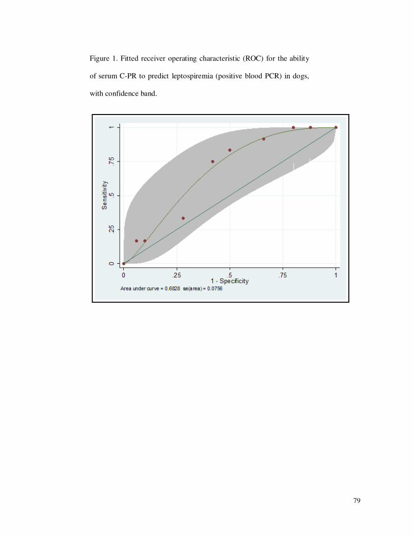
79
Figure 1. Fitted receiver operating characteristic (ROC) for the ability
of serum C-PR to predict leptospiremia (positive blood PCR) in dogs,
with confidence band.

80
5. CONSIDERAÇÕES FINAIS As informações obtidas neste trabalho mostram a importância do diagnóstico,
tratamento e prevenção da leptospirose canina. Foram observadas as diferentes formas da
apresentação da infecção, assim como a eficácia do tratamento na eliminação do estado de
portador renal e possíveis alterações laboratoriais que poderiam auxiliar na triagem dos
animais contaminados.
As populações de cães aqui estudadas foram selecionadas por apresentar algumas
particularidades, com diferenças entre elas que pudessem ser relevantes no comportamento da
doença. O grupo Centro de Controle de Zoonoses (CCZ) foi escolhido por representar a
população de cães errantes de Porto Alegre, recolhidos em várias áreas. Apesar de não se
possuir histórico prévio destes animais, assume-se que não tenham recebido vacina nos
últimos meses, e isto somado ao fato do contato com outros animais e fatores de risco,
aumentaria sua chance de contaminação. O grupo Arquipélago representa os cães oriundos
principalmente da Ilha dos Marinheiros e da Ilha do Pavão (região de Porto Alegre), que são
comunidades pobres cuja principal atividade é a coleta de lixo para reciclagem. Um estudo
realizado por Henkes (2008) mostrou que as pessoas residentes no bairro Arquipélago
apresentam cinco vezes mais chance de terem leptospirose, em relação à população residente
em bairros do estrato socioeconômico alto. Além do baixo poder socioeconômico das pessoas
do bairro Arquipélago e o acúmulo de entulho no local, com uma grande população de ratos,
o fato de ser uma região alagadiça também contribui para as condições ideais para a
manutenção da doença. A maioria dos cães das ilhas, apesar de terem donos, vivem em
condições precárias, soltos nas ruas, e convivem com outros animais. O terceiro grupo de
cães avaliados, referente aos cães atendidos no Hospital Veterinário, apresenta como
particularidade o fato de serem suspeitos da doença por apresentar sinais clínicos compatíveis
com leptospirose, ou coabitarem com outro animal com diagnóstico positivo. Este grupo
corresponde a uma amostra propositalmente “viciada”, na qual se esperava maior número de
cães infectados, com a intenção de compará-la aos outros grupos, tanto em relação à presença
de leptospiremia e leptospirúria, quanto em relação a outras alterações clinicopatológicas.
A maioria dos estudos encontrados na literatura se referem a cães sintomáticos,
atendidos em hospitais veterinários, e isto justifica os sinais clínicos esperados nestes animais
e descritos como “clássicos”, como as alterações renais, hepáticas e hematológicas, incluindo

81
leucocitose e anemia. Estas alterações também foram verificadas no presente trabalho em
relação aos animais que apresentavam sinais clínicos, porém foi verificado que tais alterações
nao correspondiam necessariamente aos cães infectados, mostrando a importância do
diagnóstico específico e definitivo da doença, para que tais animais sejam encaminhados ao
tratamento, independente de seu estado aparente de saúde. O fato de vários cães
aparentemente saudáveis apresentarem positividade a Leptospira no sangue ou na urina,
mediante a técnica de PCR, mostra o potencial destes animais de se tornarem carreadores
crônicos da leptospirose, contaminando desta forma o ambiente, outros animais e seres
humanos.
A técnica de PCR, apesar de recomendada para a detecção das leptospiras nas
amostras de sangue, urina, líquor ou tecidos do paciente, ainda não é amplamente utilizada
como diagnóstico de rotina. Esta técnica também apresenta algumas limitações, pois pode
apresentar resultados falsos-negativos caso a concentração de leptospiras na amostra esteja
abaixo do nível mínimo detectável (sensibilidade), e neste caso, a PCR em tempo real poderia
ser mais adequada, porém seu custo é mais elevado. No primeiro artigo, a comparação entre
resultados obtidos através de sorologia e PCR convencional mostra a discrepância dos
resultados entre os testes, já observados e explicados na literatura, visto que a produção de
anticorpos e a presença da bactéria no indivíduo não ocorrem necessariamente ao mesmo
tempo. Os resultados obtidos chamam a atenção para o fato de animais assintomáticos e com
exames clinicopatológicos normais apresentarem a infecção.
Em relação ao tratamento, é importante o acompanhamento dos animais durante e
após este período, pois os resultados de negatividade em relação à presença das leptospiras no
paciente são úteis, tanto individualmente quanto epidemiologicamente. Como já observado na
literatura, alguns antibióticos melhoram o estado clínico do paciente sem no entanto eliminar
a bactéria dos tecidos. A avaliação de um grande número de animais naturalmente infectados,
submetidos a tratamentos específicos, auxiliaria nesta avaliação da eficácia de cada
tratamento, uma vez que possam ocorrer variações quanto ao ambiente, animais e cepas
infectantes, dificilmente avaliados artificialmente. Da mesma forma, estudos para a utilização
de testes de triagem alternativos, como foi proposto com a proteína C-reativa, são relevantes
para a detecção precoce da doença e acompanhamento. O desafio consiste na identificação de
marcadores que sejam úteis mesmo nos casos de cães assintomáticos e sem alterações no
hemograma, urinálise e exames bioquímicos de rotina. Estes testes não substituem os exames
de PCR ou isolamento das leptospiras para o diagnóstico definitivo, porém auxiliam na

82
conduta dos clínicos. Infelizmente, apesar da proteína C-reativa ser um exame de baixo custo
e poder ser mensurada de forma ambulatorial no caso da aglutinação em látex, o teste não se
mostrou útil isoladamente para triagem da leptospirose em cães.

83
6. REFERÊNCIAS ADICIONAIS
ADIN, C. A.; COWGILL, L. D. Treatment and outcome of dogs with leptospirosis: 36 cases
(1990-1998). Journal of the American Veterinary Medical Association, v. 216, n. 3, p.
371-375, 2000.
AHMAD, S. N.; SHAH, S. H.; AHMAD, F. M. Laboratory diagnosis of leptospirosis.
Journal of Postgraduate Medicine, v. 51, n. 3, p. 195-200, 2005.
ALT, D. P.; BOLIN, C. A. Preliminary evaluation of antimicrobial agents for treatment of
Leptospira interrogans serovar pomona infection in hamsters and swine. American Journal
of Veterinary Research, v. 57, n. 1, p. 59-62, 1986.
BARCELLOS, C. et al. Distribuição especial da leptospirose no Rio Grande do Sul, Brasil:
recuperando a ecologia dos estudos ecológicos. Cadernos de Saúde Pública, v. 19, p. 1283-
1292, 2003.
BARTGES, J. W. Distúrbios dos túbulos renais. In: ETTINGER, S.J., FELDMAN, E.C.
(Eds.) Tratado de medicina interna veterinária. Doenças do cão e do gato. Rio de Janeiro:
Guanabara Koogan, 2004. 5 ed. v. 2, cap. 144, p. 1795-1802.
BATISTA, C. S. A. et al. Soroprevalência e fatores de risco par a leptospirose em cães de
Campina Grande, Paraíba. Arquivo Brasileiro de Medicina Veterinária e Zootecnia, v. 57,
supl. 2, p. 179-185, 2005.
BIRNBAUM, N; CENTER, S. A.; RANDOLPH, J. F. Naturally acquired leptospirosis in 36
dogs: serological clinicopathological features. Journal of Small Animal Practice, v. 39, p.
231-236, 1998.
BURR, P.; LUNN, K.; YAM, P. Current perspectives on canine leptospirosis. In Practice, v.
3, p. 98-102. 2009.
CASPI, D. et al. C-reactive protein in dogs. American Journal of Veterinary Research, v.
48, p. 919–921, 1987.
CERÓN, J. J.; ECKERSALL, P. D.; MARTÍNEZ-SUBIELA, S. Acute phase proteins in dogs
and cats: current knowledge and future perspectives. Veterinary Clinical Pathology, v. 34,
p. 85-99, 2005.
CHARELLO, T. et al. PCR no diagnóstico da leptospirose canina em presença de sorologia
negativa. In: 14 EVINCI- Evento de iniciação científica, 2006, Curitiba. Anais... Curitiba,
UFPR, 2006. p.139.

84
COWGILL, L. D.; ELLIOTT, D. A. Insuficiência renal aguda. In: ETTINGER, S.J.,
FELDMAN, E.C. Tratado de medicina interna veterinária. Doenças do cão e do gato. 5.
ed. Rio de Janeiro: Guanabara Koogan, 2004. v. 2, cap. 168, p. 1701-1721.
DALMOLIN, M. L.; GONZÁLEZ, F. H. D. Sinais clínicos, alterações hematológicas e
bioquímicas, fatores de risco e sorovares prevalentes em cães suspeitos de leptospirose no
HCV UFRGS - janeiro de 2005 a julho de 2007. In: XIX Salão de Iniciação Científica,
2007, Porto Alegre. Anais...Porto Alegre, UFRGS, 2007. 1 CD-ROM.
FURTADO, L. R. I. et al. Prevalência e avaliação de fatores de risco à leptospirose canina, no
Município de Pelotas, RS. Arquivos do Instituto Biológico, v. 64, p. 57-61, 1997.
GERRITSEN, M. J. et al. Effective treatment with dihydroestreptomycin of naturally infected
cows shedding Leptospira interrogans serovar hardjo subtype hardjobovis. American
Journal of Veterinary Research, v. 55, p. 339-343, 1994.
GHNEIM, G. S. et al. Use of a case-control study and geographic information systems to
determine environmental and demographic risk factors for canine leptospirosis. Veterinary
Research, v. 38, p. 37-50, 2007.
GIRIO, T. M. S. et al. Uso de estreptomicina na eliminação da leptospirúria em touros (bos
taurus indicus) naturalmente infectados pelo sorovar hardjo. Arquivos do Instituto
Biológico, v. 72, p. 161-170, 2005.
GONZÁLEZ, F. H. D.; SILVA, S. C. Introdução à bioquímica clínica veterinária. 2. ed.
Porto Alegre: UFRGS, 2006. 364 p.
GRAVEKAMP, C. et al. Detection of seven species of pathogenic leptospires by PCR using
two sets of primers. Journal of General Microbiology, 139, 1691-1700. 1993.
GREENE, C. E. et al. Leptospirosis. In: GREENE, C.E. (Ed.), Infectious diseases of the dog
and cat. 3rd ed. Philadelphia: W. B. Saunders, 2006. p. 402-415.
GREENLEE, J. J. et al. Experimental canine leptospirosis caused by Leptospira interrogans
serovars pomona and bratislava. American Journal of Veterinary Research, v. 66, p. 1816-
1822, 2005.
GUIMARÃES, M. A. et al. Epidemiologia e controle da leptospirose em bovinos. Papel do
portador e seu controle terapêutico. Revista da Faculdade de Medicina Veterinária e
Zootecnia da Universidade de São Paulo, v.19, p.202, 1982.
HARKIN, K. R.; ROSHTO, Y. M.; SULLIVAN, J. T. Clinical application of a polymerase
chain reaction assay for diagnosis of leptospirosis in dogs. Journal of the American
Veteterinary Medical Association, v. 222, p. 1224-1229, 2003.

85
HEATH, S. E.; JOHNSON, R. Leptospirosis. Journal of the American Veteterinary
Medical Association, v. 205, p. 1518-1523, 1994.
HENKES, W. E. Ecologia de paisagem da leptospirose em Porto Alegre entre 2001-2006.
2008. 36f. Trabalho de conclusão de curso (Especialização). Faculdade de Medicina,
Universidade Federal do Rio Grande do Sul, Porto Alegre. 2008.
HERRMANN, G. P. et al. Soroprevalência de aglutininas anti-Leptospira spp. em ovinos nas
mesorregiões sudeste e sudoeste do Estado do Rio Grande do Sul, Brasil. Ciência Rural, v.
34, p. 443-448, 2004.
JORGE, S. et al. Prevalência de leptospirose em cães domicilados sugere implantação de
programa de educação em saúde. In: IXV Congresso de Iniciação Científica, 2005, Pelotas.
Anais... Pelotas, UFPel, 2005. Disponível em
<http://www.ufpel.edu.br/xivcic/arquivos/CB_01124.rtf>. Acesso em: 15 dez 2006.
JOUGLARD, S. D. D.; BROD, C. S. Leptospirose em cães: prevalência e fatores de risco no
meio rural do município de Pelotas, RS. Arquivos do Instituto Biológico, v. 67, p. 181-185,
2000.
LANGSTON, C. E.; HEUTER, K. J. Leptospirosis: a re-emerging zoonotic disease.
Veterinary Clinics of North America: Small Animal Practice, v. 33, p. 791-807, 2003.
LAPPIN, M. R. Polysystemic bacterial disease. In: NELSON, R. W., COUTO, C. G. (Eds.).
Small animal internal medicine. 3 ed. St Louis: Mosby, 2003. cap. 100, p. 1259-1264.
LEVETT, P. N. Leptospirosis. Clinical Microbiology Reviews, v. 14, p. 296-326, 2001.
LOBO, E. A. et al. Estudo comparativo do padrão sorológico de animais domésticos
potencialmente transmissores de leptospirose no município de Santa Cruz do Sul, RS, Brasil,
entre os anos 2002 e 2003. Caderno de Pesquisa Série Biologia, v. 16, p. 47-64, 2004.
MACHADO, R. R. et al. Leptospirose canina na região sul do Rio Grande do Sul, no ano de
1998. In: 14 Congresso Estadual de Medicina Veterinária, & 3 Congresso de Medicina
Veterinária do Cone Sul, 1999, Gramado. Anais...Gramado, Sovergs, 1999. p. 103.
MAJED, Z. et al. Identification of variable-number tandem-repeat loci in Leptospira
interrogans sensu stricto. Journal of Clinical Microbiology, v. 43, n. 2, p. 539-545, 2005.
MEEYAM, T et al. Seroprevalence and risk factors associated with leptospirosis in dogs. The
Southeast Asian Journal of Tropical Medicine and Public Health, v. 37, p. 148-153,
2006.
MÉRIEN, F. et al. Polymerase chain reaction for detection of Leptospira spp. in clinical
samples. Journal of Clinical Microbiology, v. 30, p. 2219-2224, 1992.
86
MICHEL, V.; BRANGER, C.; ANDRÉ-FONTAINE, G. Epidemiology of leptospirosis. Rev
ista Cubana de Medicina Tropical, v.54, p. 7-10, 2002.
MOON, J. E. et al. Effect of timing and duration of azithromycin therapy of leptospirosis in a
hamster model. Journal of Antimicrobial Chemotherapy, v. 59, p. 148-151, 2007.
OLIVEIRA, S. J.; PIRES NETO, J. A. S. Aspectos etiológicos e de diagnóstico nas
leptospiroses. Revista do Conselho Federal de Medicina Veterinária, v. 10, p. 36-46,
2004.
SALAÜN, L. et al. Application of multilocus variable-number tandem-repeat analysis for
molecular typing of the agent of leptospirosis. Journal of Clinical Microbiology, v. 44, p.
3954–3962, 2006.
SANTIN, K. et al. Pesquisa de aglutininas antileptospira em cães clinicamente sadios e em
cães com suspeita clínica de leptospirose. Clínica Veterinária, v. 11, p. 48-52, 2006.
SCHMIDT, V.; AROSI, A.; SANTOS, A. R. Levantamento sorológico da leptospirose em
caprinos leiteiros no Rio Grande do Sul, Brasil. Ciência Rural, v. 32, p. 609-612, 2002.
SESSIONS, J. K.; GREENE, C. E. Canine leptospirosis: treatment, prevention, and zoonosis.
Compendium on Continuing Education for the Practising Veterinarian, v. 26, p. 700-
711, 2004.
SHERDING, R. G. Leptospirose, brucelose e outras doenças infecciosas bacterianas. In:
BIRCHARD, S. J.; SHERDING, R. G. Manual Saunders: clínica de pequenos animais.
São Paulo: Roca, 1998. cap. 11, p. 143-147.
THIESEN, S. V. et al. Aspectos relacionados à ocorrência de leptospirose em Porto Alegre
no ano de 2007. Boletim Epidemiológico (Porto Alegre), v. 10, p. 1-4, 2008.
TRUCCOLO, J. et al. Quantitative PCR assay to evaluate ampicillin, ofloxacin, and
doxycycline for treatment of experimental leptospirosis. Antimicrobial Agents and
Chemotherapy, v. 46, n, 3, p. 848-853, 2002.
VAN DE MAELE, I. et al. Leptospirosis in dogs: a review with emphasis on clinical aspects.
Veterinary Record, v. 163, p. 409-413, 2008.
VASCONCELLOS, S.A. Leptospirose animal. In: 3 Encontro Nacional em Leptospirose,
1993, Rio de Janeiro. Anais...Rio de Janeiro: Ministério da Saúde, 1993. p. 62-66.
VISITH, S.; KEARKIAT, P. Nephropaty in leptospirosis. Journal of Postgraduate
Medicine, v. 51, p. 184-188, 2005.
WARD, M. P.; GUPTILL, L.F.; WU, C.C. Evaluation of environmental risk factors for
leptospirosis in dogs: 36 cases (1997–2002). J. Am. Vet. Med. Assoc. 225, 72-77. , 2004.
87
WOLFFENBÜTTEL, S. et al. Achados clínico-laboratoriais em sete cães com resposta
sorológica à leptospirose. MedveP, v. 2, p. 44-50, 2004.
World Health Organization. Human Leptospirosis: guidance for diagnosis, surveillance and
control. 2003. 109p.
YAMAMOTO, S. et al. Changes in serum C-reactive protein levels in dogs with various
disorders and surgical traumas. Veterinary Research Communications, v. 17, p. 85-93,
1993.
YANG, C. W.; WU, M. S.; PAN, M. J. Leptospirosis renal disease. Nephrology Dialysis
Transplantation, v. 16, suppl. 5, p. 73-77, 2001.

88
ANEXO 1

89
ANEXO 2