


Línguas
Páginas
Legal


Mestrado Integrado em Medicina Dentária da Universidade do Porto
Master’s degree in dental medicine, Oporto University
Artigo de Revisão Bibliográfica
Bibliographic revision
“Probióticos em Medicina Dentária e Oral: tendências recentes”
“Probiotics in dentistry and oral medicine: recent trends”
Cláudia Sofia da Silva Campos
Porto, 2020

ii
“Probióticos em Medicina dentária e oral: tendências recentes”
“Probiotics in dentistry and oral medicine: recent trends”
Unidade Curricular “Monografia de Investigação / Relatório de Atividade Clínica”
Artigo de Revisão Bibliográfica
Cláudia Sofia da Silva Campos
Aluna do 5º Ano do Mestrado Integrado em Medicina Dentária da Faculdade de Medicina Dentária da Universidade
do Porto
5th year student of the master’s degree in Dental Medicine, Oporto’s University
Orientadora/ Advisor
Prof. Doutora Otília Adelina Pereira Lopes
Professora Auxiliar da Faculdade de Medicina Dentária da Universidade do Porto
Assistant Professor at the Faculty of Dental Medicine, Oporto’s University
Co-Orientadora/ Co-Advisor
Prof. Doutora Maria Benedita Almeida Garrett de Sampaio Maia Marques
Assistant Professor at the Faculty of Dental Medicine, Oporto’s University
Porto, 2020

iii

iv
Once more, with feeling
“Even if you see them coming, you’re not ready for the big moments. No one
asks for their life to change, not really. But it does. So what – are we helpless
puppets? No. The big moments are ‘gonna come. You can’t help that. It’s what
you do afterwards that counts. That’s when you find out who you are”
Buffy the Vampire Slayer

v

vi
Acknowledgments
First, I’d like to thank my beloved parents for their unwavering support. I wouldn’t
be able to accomplish anything without them.
Then, I would also like to thank both by advisor and co-advisor for their
supervision and constructive criticism.
vii
Resumo
Probióticos são microrganismos benéficos que auxiliam na modulação de agentes
patogénicos, contribuindo para a prevenção ou tratamento de doenças. Acredita-se que
podem ser capazes de substituir tratamentos associados a um maior número de efeitos
adversos, ou ser complemento de outros, melhorando a sua eficiência. Por isso o objetivo
deste trabalho é a pesquisa e compilação de provas concretas da eficiência e aplicabilidade
de probióticos no tratamento de doenças no âmbito da medicina dentária.
Para isso foram avaliados 61 ensaios clínicos produzidos na última década (2009-
2019), incidindo na intervenção sobre cárie dentária, doença periodontal, infeções
fúngicas por Candida albicans, líquen plano e mucosite.
A prevenção da doença (cárie dentária e doença periodontal) foi o objetivo em
52,4% dos estudos (n=33), enquanto que o tratamento de cárie dentária e periodontite
ativas foi o foco na restante amostra. Os estudos relacionados com Candida albicans
focaram-se essencialmente em populações idosas, que já tinham maiores taxas de
colonização pelo fungo, e os seus objetivos eram a redução da carga microbiana e da
sintomatologia associada (n=6). Por outro lado, a maioria dos ensaios clínicos focados no
tratamento e prevenção de cáries recorreram a populações jovens (crianças em idade
escolar) e os focados na periodontite, recorreram a adultos. As estirpes probióticas mais
utilizadas foram Lactobacillus reuteri (27%, n=17), Lactobacillus rhamnosus (11,1%,
n=7), Lactobacillus casei (9,5%, n=6), Lactobacillus paracasei (6,3%, n=4).
Globalmente, em 28 ensaios, as estirpes escolhidas foram capazes de melhorar um
sintoma associado a uma das doenças supracitadas, em 30 ensaios foram capazes de
modular o microbioma oral e em 8 ensaios provou-se terem sido capazes de estimular o
sistema imunitário do hospedeiro. Em geral, a ação probiótica foi apenas parcialmente
bem-sucedida, pois não foi efetiva em todos os parâmetros que os ensaios se propuseram
melhorar, o que indica que a utilização de probióticos poderá ser mais eficiente quando
administrada em conjunto com outros tratamentos e protocolos já utilizados,
especialmente no que toca à prevenção e tratamento de cárie dentária em crianças e como
coadjuvantes no tratamento das causas e sintomas da doença periodontal.
Palavras chave
Probióticos, Lactobacillus reuteri, Lactobacillus rhamnosus, Llactobacillus casei, medicina
dentária, medicina oral, cárie dentária, doença periodontal, Candida albicans, mucosite, líquen plano

viii
Abstract
Probiotics are beneficial microbes that can help to modulate the proliferation of
pathogens and prevent or treat disease. Probiotics are believed to be able to substitute
treatments with a heavy load of side effects or aid others, improving their effectiveness.
Hence, this study’s objective is the research and complication of concrete evidence
proving that probiotics can effectively be applied in dentistry and oral medicine.
In order to do so 61 clinical trials performed during the last decade (2009-2019)
were evaluated regarding caries, periodontal disease, Candida albicans infections, lichen
planus and mucositis were assessed in this matter.
Disease prevention (caries and periodontitis) was the objective in 52,4% (n=33)
of trials, while the treatment of active caries and chronic periodontitis was the goal in the
remaining sample. The studies regarding C. albicans usually relied on an older
population, which already had higher counts of the fungi, and their objective was reducing
symptoms and microbial load (n=6). On the other hand, most caries trials were based on
school aged children and periodontitis in adults. The most used probiotic strains were
Lactobacillus reuteri (27%, n=17), Lactobacillus rhamnosus (11,1%, n=7), Lactobacillus
casei (9,5%, n=6) and Lactobacillus paracasei and Lactobacillus crispatus (both with
6,3%, n=4). Globally, in 28 trials, the probiotic strain was successful in improving a
clinical symptom, in 30 they were able to modulate the surrounding microbiome and in 8
they were able to stimulate the host’s immune response. Probiotics were often only
partially successful, indicating that their most effective administration is in conjunction
with already established protocols, especially when it comes to caries disease progression
in children as well as in supporting the treatment of causes and symptoms of periodontal
disease.
Key Words
Probiotics, Lactobacillus reuteri, Lactobacillus rhamnosus, Lactobacillus casei,, dentistry, oral
medicine, caries, periodontal disease, Candida albicans, mucositis, lichen planus
ix
Contents
Acknowledgments .......................................................................................................................vi
Resumo ........................................................................................................................................ vii
Abstract ...................................................................................................................................... viii
Table Index .................................................................................................................................. 1
Abbreviations list ........................................................................................................................ 2
1. Introduction ..................................................................................................................... 3
1.1. Probiotics – an overview ............................................................................................. 3
1.2. Caries ............................................................................................................................ 5
1.3. Periodontal disease ...................................................................................................... 5
1.4. Mucositis ...................................................................................................................... 7
1.5. Candida albicans infection .......................................................................................... 7
1.6. Lichen Planus .............................................................................................................. 8
2. Materials and methods ........................................................................................................ 8
3. Results ................................................................................................................................ 12
4. Discussion ........................................................................................................................... 26
4.1. Currently available commercial probiotic formulations ....................................... 33
5. Conclusions ........................................................................................................................ 35
6. Future research developments ......................................................................................... 35
7. Bibliography ...................................................................................................................... 36

1
Table Index
page
Table 1 - Fermentation processes of the main oral probiotics
(Lactobacillus) 3
Table 2: Virulence factors of the most common periodontal pathogens 6
Table 3: Search terms 8
Table 4: Tested variables 10
Table 5: Main significant (p>0.05) probiotic effects on oral illnesses (by
strain) 13
Table 6: Probiotics grouped by type fermentation process and their
statistically significant (p<0.05) outcomes on the trials’ variables 15
Table 7: Clinical Trials regarding probiotics and oral health care (2009 –
2019) 18
Table 8: Commonly used probiotic products 25
2
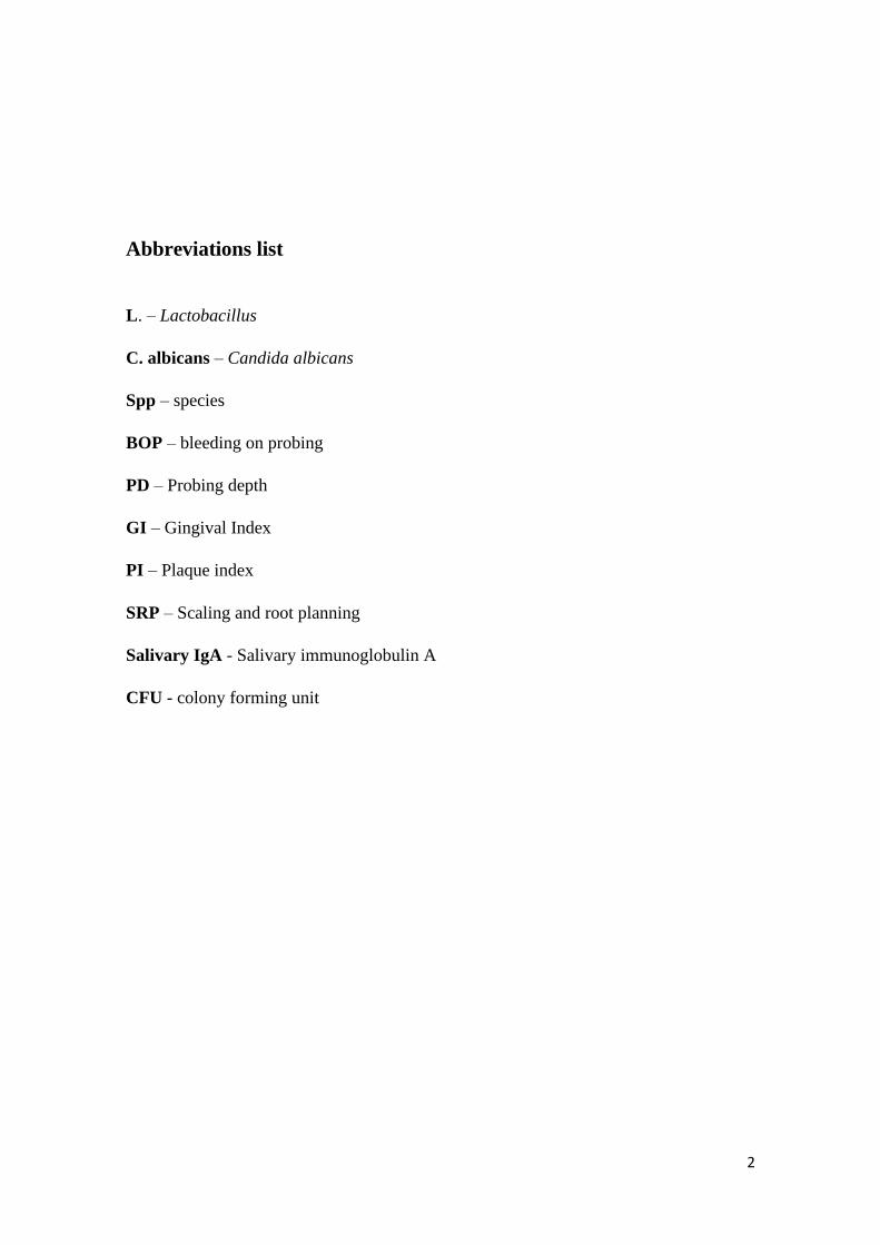
Abbreviations list
L. – Lactobacillus
C. albicans – Candida albicans
Spp – species
BOP – bleeding on probing
PD – Probing depth
GI – Gingival Index
PI – Plaque index
SRP – Scaling and root planning
Salivary IgA - Salivary immunoglobulin A
CFU - colony forming unit

3
1. Introduction
1.1. Probiotics – an overview
According to the World Health Organization (WHO), probiotics are living
microorganisms that “when administered in adequate amounts, confer a health benefit on
the host”. These microorganisms are generally lactic acid bacteria (LAB), meaning that
metabolize sugars into lactic acid trough fermentation. Probiotic LAB mainly belongs to
the Firmicutes (Lactobacillus, Lactococcus, Staphylococcus, Streptococcus) and
Actinobacteria phylum (Bifidobacteria). This study aims to summarize the most recent
clinical trials applying probiotics to oral health and possibly offer a therapeutic alternative
or addition to already existing treatments.
Lactobacillus are gram positive, non-spore forming, catalase negative bacteria.
They generally have low cytosine plus guanine (CG) content and are facultative
anaerobes. Taking fermentation processes as a taxonomic criterion, the Lactobacillus
group can be divided in the homofermentative, facultative heterofermentative and
heterofermentative groups. The organisms in the homofermentative group exclusively
transform hexoses into lactic acid trough glycolysis. On the other hand,
heterofermentative bacteria can use a wider variety of sugars (pentoses) to produce other
byproducts (CO2, acetic acid, ethanol), using O2 as a growth stimulator and electron
acceptor, which results in greater ATP formation (Charalampopoulos and Rastall 2009,
Lahtinen, Salminen et al. 2012). In table 1 the main probiotic strains used in oral health
are presented:
Table 1: Fermentation processes of the main oral probiotics (lactobacillus)
Homofermentative Facultative heterofermentative Heterofermentative
Lactobacillus acidophilus
Lactobacillus Jonhsonii
Lactobacillus crispatus
Lactobacillus gasseri
Lactobacillus casei
Lactobacillus paracasei
Lactobacillus rhamnosus
Lactobacillus curvatus
Lactobacillus plantarum
Lactobacillus salivarius
Lactobacillus brevis
Lactobacillus fermentum
Lactobacillus reuteri
Adapted from S. Lahtinen, A.C. Owehand et al “Lactic Acid Bacteria. Microbiological and functional aspects”

4
The probiotic’s influence on extracellular pH is their major form of action. Lactic
acid production has an inhibitory effect on many pathogenic organisms by causing the
dissociation of small fatty acids. These penetrate the cellular membrane and disrupt
microbial metabolism. The acids produced by heterofermentative lactobacilli aren’t as
strong (Charalampopoulos and Rastall 2009, Lahtinen, Salminen et al. 2012).
Bifidobacterium differ from lactobacilli because they use a specific enzyme
(fructose-6-phosphoketolase) to degrade hexoses into lactic acid. They are also
heterofermentative, non-spore forming anaerobes. They have strong adhesion capabilities
and are safe for consumption (Charalampopoulos and Rastall 2009, Lahtinen, Salminen
et al. 2012).
Lactobacillus fermentum, Lactobacillus rhamnosus, Lactobacillus salivarius,
Lactobacillus casei, Lactobacillus acidophilus and Lactobacillus plantarum can be
normally found in human saliva or dental plaque, even though only accounting for 1% of
cultivable microbes. It is believed that their positive effects, when administered in higher
numbers that usual, are pH reduction, inhibition of pathogens in dental biofilm,
antimicrobial substance production, nutrient and adhesion sites competition with oral
pathogens, immunomodulation of the host’s response and improvement in mucosal
permeability. The reduction in oral pathogens can be achieved both by pH decrease and
the probiotic’s production of antimicrobial products – bacteriocins; for example, reuterin
6, produced by Lactobacillus reuteri (Charalampopoulos and Rastall 2009, Lahtinen,
Salminen et al. 2012).
In addition, probiotics can improve immunity functions by adhering to epithelial
cells in the mucosa. Cell structures such as fimbriae and surface proteins bind to mucine,
glycoproteins and human fibronectin. L. acidophilus has “Mub proteins” that adhere to
fibronectine, while L. rhamnosus has “Spac pilin” (pili) that connects with mucus and
aids its persistence in the gastrointestinal tract when ingested. This adds to acid and bile
resistance of L. rhamnosus. Some oral benefits can be attained with probiotic’s presence
in the gut, but their persistence in the oral cavity is an objective whenever local lesions
are to be treated – such as caries. Hydrophobic nature probiotics have better adhesion
properties and can connect with salivary mucin. Lactobacillus paracasei are the most
hydrophobic potentially beneficial microbes isolated from tooth surfaces. On the gingiva,
lactobacilli congregate in the presence of ammonia and can either positively regulate
5
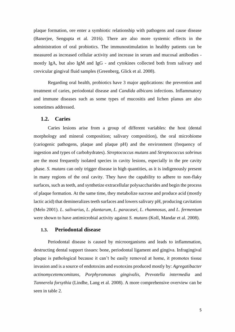
plaque formation, ore enter a symbiotic relationship with pathogens and cause disease
(Banerjee, Sengupta et al. 2016). There are also more systemic effects in the
administration of oral probiotics. The immunostimulation in healthy patients can be
measured as increased cellular activity and increase in serum and mucosal antibodies -
mostly IgA, but also IgM and IgG - and cytokines collected both from salivary and
crevicular gingival fluid samples (Greenberg, Glick et al. 2008).
Regarding oral health, probiotics have 3 major applications: the prevention and
treatment of caries, periodontal disease and Candida albicans infections. Inflammatory
and immune diseases such as some types of mucositis and lichen planus are also
sometimes addressed.
1.2. Caries
Caries lesions arise from a group of different variables: the host (dental
morphology and mineral composition; salivary composition), the oral microbiome
(cariogenic pathogens, plaque and plaque pH) and the environment (frequency of
ingestion and types of carbohydrates). Streptococcus mutans and Streptococcus sobrinus
are the most frequently isolated species in cavity lesions, especially in the pre cavity
phase. S. mutans can only trigger disease in high quantities, as it is indigenously present
in many regions of the oral cavity. They have the capability to adhere to non-flaky
surfaces, such as teeth, and synthetize extracellular polysaccharides and begin the process
of plaque formation. At the same time, they metabolize sucrose and produce acid (mostly
lactic acid) that demineralizes teeth surfaces and lowers salivary pH, producing cavitation
(Melo 2001). L. salivarius, L. plantarum, L. paracasei, L. rhamnosus, and L. fermentum
were shown to have antimicrobial activity against S. mutans (Koll, Mandar et al. 2008).
1.3. Periodontal disease
Periodontal disease is caused by microorganisms and leads to inflammation,
destructing dental support tissues: bone, periodontal ligament and gingiva. Infragingival
plaque is pathological because it can’t be easily removed at home, it promotes tissue
invasion and is a source of endotoxins and exotoxins produced mostly by: Agregatibacter
actinomycetemcomitans, Porphyromonas gingivalis, Prevotella intermedia and
Tannerela forsythia (Lindhe, Lang et al. 2008). A more comprehensive overview can be
seen in table 2.

6
Table 2: Virulence factors of the most common periodontal pathogens
Pathogen Virulence factors Detection sites
Aggregatibacter
actinomycetemcomitans
Leukotoxin, catalase and superoxide dismutase
production
Endotoxins
Invasion of epithelial and endothelial cells
Detected in high counts in some
chronic periodontitis lesions
Porphyromonas
gingivalis
Superoxide dismutase production
LPS and adhesins
Proteolytic enzymes that destroy connective
tissue
Fimbriae
Invasive capabilities: alkaline phosphatase (bone
invasion)
Bacteriocins
Highly related with periodontal
disease – not present in regular
oral microbiota
Tannerella forsythia Invasive capabilities
Shares antigens with P. gingivalis
Detected in high counts in some
refractory chronic periodontitis,
as well as in abscesses and active
lesions
Prevotella intermedia
LPS and adhesins
Proteolytic enzymes
Fimbriae
Detected in high counts in
ulcerative gingivitis and
refractory periodontitis
Treponema denticola
Endotoxins and proteolytic enzymes
Mobility
Diminishes lymphocyte response
The main pathogen of ulcerative
gingivitis and active periodontitis
lesions
Fusobacterium
nucleatum
Endotoxins and leukotoxins
Inhibits leucocyte quimiotaxis
Detected in high counts in
chronic periodontitis and
abscesses
Adapted from J. Lindhe, N.P Lang et all “Clinical Periodontology and Implant Dentistry
There is some data that implies that probiotic organisms have the capability to
disrupt plaque formation, by interfering with its pathogens. As it has been referred, LAB
produce many antimicrobial substances; for example, L. reuteri produces hydrogen
peroxide (Szkaradkiewicz, Stopa et al. 2014, Tobita, Watanabe et al. 2018). Furthermore,
L. rhamnosus have a strong inhibitory effect against cariogenic species and gram-negative
periodontal pathogens (Morales, Carvajal et al. 2017). And L. brevis has the capability to
prevent nitric oxide production, and hence inhibit gingival inflammation (Lee, Kim et al.
2015). Streptoccocus spp. is able to proliferate in periodontal pockets after root scaling,
avoiding the recolonization of such sites by unwanted species (Laleman, Yilmaz et al.
2015).
Other than the epithelial barrier itself, the organism has innate defenses – saliva
and the inflammatory process, and specific responses – cellular and humoral immunity.
For example L. plantarum L-137 is capable of inducing IL-12, which leads to a Th1

7
immune response and the production of type I IFN in humans (Iwasaki, Maeda et al.
2016). And Bifidobacterium animalis decreased the levels of IL-1 β in gingival crevicular
fluid (GCF) in simulated plaque formation after a 5-day no brush period (Kuru, Laleman
et al. 2017).
1.4. Mucositis
Oral mucositis is an inflammatory condition on the mucosa. Its pathogenesis is
mainly correlated with an external aggression and an increase in cytokine production that
affects connective tissue. There is increased growth of S. mutans, lactobacilli, C. albicans
and gram-negative bacilli, that may result in oral infections. Some probiotic strains are
expected to be able to control these microbial populations by direct competition or the
production of bacteriocins (Neville, Damn et al. , Greenberg, Glick et al. 2008).
1.5. Candida albicans infection
The pathological proliferation of C. albicans is called candidiasis, and it is the
most common form of fungal oral infection in humans. Prosthetic stomatitis tends to be
grouped with erythematous candidiasis because both have a characteristic mucosal
erythema. Nevertheless, prosthetic stomatitis is mostly related with older patients and
some level of neglect in their denture’s hygiene, while the erythematous type is more
correlated with systemic conditions, such as cancer treatment (Neville, Damn et al.).
The environment provided by the combination of oral mucosa and denture surface
is ideal for the growth of this species: nutrient rich, with a decreased flow of oxygen and
saliva and with a nonrenewable (acrylic) surface on which the fungus can attach itself and
proliferate. C. albicans is associated with the development of denture stomatitis but other
pathogens such as S. mutans can aid its adhesion to the tissue/dentures. S. mutans
produces an extra cellular matrix polysaccharide that facilitates the attachment of other
microorganisms. Mucosal infection begins when the fungus adheres to epithelial cells –
for example, when an ill-fitting denture causes friction and disrupts the epithelium – or
due to systemic diseases such as poorly controlled diabetes.
The infection may also arise due to the immunocompromised state of the host,
triggered by radiotherapy and chemotherapy. Patients receiving cytotoxic drugs are
highly susceptible to fungal infections, that not only cause pain and discomfort, but can
also extent to the esophagus leading to disseminated candidiasis (Lashof, Bock et al.
2004). As for radiation therapy, the decrease in saliva production is a well-known

8
predisposing factor for candidiasis. Radiotherapy to a dose of 50-60 Gy generally tends
to cause lifelong damage to the salivary glands, and hence, permanent xerostomia
(Rautemaa, Rusanen et al. 2006).
1.6. Lichen Planus
Lichen planus is a mucocutaneous disease with immunological mediation: auto
reactive T cells that cannot distinguish between host cells and foreign antigens are
activated triggering the agents of the inflammatory process (Neville, Damn et al. ,
Greenberg, Glick et al. 2008). It’s erosive form is usually treated with corticosteroids that
can lead to C. albicans infection (Neville, Damn et al.). And, as recent study discusses,
probiotics are able to diminish microbial infection and suppress T cell activation and
proliferation, as well as diminishing keratinocyte apoptosis and modulating the
production of inflammatory cytokines, MMP-9 expression and mast cell degranulation
(Han, Zhang et al. 2017).
2. Materials and methods
This study aimed to examine recent clinical trials regarding probiotics and oral
health care. The search was performed on PubMed’s database, with the following criteria:
Clinical trials published between 2009 and 2019, in human subjects. Table 3 shows the
results of the search, by target disease:
Table 3: Search terms
Target disease keywords Number of trials
Caries “Probiotics” AND “caries n=28
Periodontal disease “Probiotics” AND “periodontal
disease1” OR “Periodontitis” n=26
Yeast infections2 “Probiotics” and “oral yeasts” n=1
“Probiotics” and “Candida” n=20
Mucositis
“Probiotics” and “Mucositis”
n=6 “Probiotics” and “Mucosistis” and
“Neoplasms”
Lichen planus “Probiotics” and “lichen planus” n=1
1 Periodontitis as a broader term that includes gingivitis 2 The use of the term “fungi” yielded no results regarding exclusively the oral cavity

9
As for exclusion criteria, trials that evaluated the performance of probiotics or the
treatment of diseases outside the oral cavity weren’t addressed. In the case of mucositis,
most trials regarded mucositis in the context of implantology, and not as result of other
etiologies – cancer treatment, for example. This meant that most studies (n=5) in this
category were also found in the context of periodontology and probiotics. The same for
lichen planus, whose only trial also discussed C. albicans infection. Then the search for
C. albicans infections and probiotics yielded 20 results of which 6 concerned the oral
cavity. In the end, 61 trials met the criteria to be included in this study.
Descriptive statistical evaluation was performed in order to convey the major
trends seen in probiotics applied to oral health in the last decade. So, the trials were
summarized in a series of variables: intervention period, sample size, probiotic strain
used, form of probiotic administration, target disease and the existence of positive
statistically significant outcomes in terms of microbiological modulation, improvement
of clinical signs and/or the host’s immune response.
Study variables varied across trials. Clinical variables for caries were cavitated
lesions, remineralization of white spots and plaque index (PI). Some studies also
addressed gingival health, though it wasn’t the focus. Microbiological variables were
evaluated by assessing the reduction of cariogenic microorganisms. Whenever the long-
term permanence of a Lactobacillus strain was assessed it referred to the probiotic strain
itself and not the possible pathogen. As for periodontal diseases (chronic periodontitis,
gingivitis and peri-implant mucositis), clinical success was evaluated mainly as a
reduction in probing depth (PD), bleeding on probing (BoP), clinical attachment loss
(CAL), gingival index (GI) and plaque index (PI). Then the effects on the microbiome
were based on the reduction of periodontal pathogens. Immunological variables were also
addressed in some clinical trials regarding periodontitis, mostly the presence of
inflammatory cytokines in GCF and saliva. Further explanation in table 4.

10
Table 4: Tested variables
Target
disease Clinical variables Microbiological variables Imunological variables
Caries
Caries increment
(Stecksen-Blicks, Sjostrom et al. 2009, Stensson, Koch et al. 2013,
Hedayati-Hajikand, Lundberg et al. 2015, Wattanarat, Makeudom et
al. 2015, Rodriguez, Ruiz et al. 2016, Villavicencio, Villegas et al.
2017)
White spot leseions (WSL)
(Gizani, Petsi et al. 2015)
Early caries lesions (changes in enamel fluorescense)
(Keller, Nohr Larsen et al. 2014)
Salivary flow
(Nishihara, Suzuki et al. 2014)
Primary root caries lesions (PRCL)
(Petersson, Magnusson et al. 2011)
Salivary S. mutans and Lactobacillus counts
(Chuang, Huang et al. 2010, Aminabadi, Erfanparast et
al. 2011, Jindal, Pandey et al. 2011, Singh, Damle et al.
2011, Cildir, Sandalli et al. 2012, Glavina, Gorseta et al.
2012, Mortazavi and Akhlaghi 2012, Stensson, Koch et
al. 2013, Gizani, Petsi et al. 2015, Villavicencio, Villegas
et al. 2017, Alamoudi, Almabadi et al. 2018, Tobita,
Watanabe et al. 2018)
Salivary S. mutans counts
(Juneja and Kakade 2012, Romani Vestman, Hasslof et
al. 2013, Taipale, Pienihakkinen et al. 2013, Teanpaisan
and Piwat 2013, Ghasemi, Mazaheri et al. 2017,
Pahumunto, Piwat et al. 2018)
Salivary buffer capacity
(Chuang, Huang et al. 2010,
Glavina, Gorseta et al. 2012,
Nishihara, Suzuki et al. 2014,
Villavicencio, Villegas et al.
2017)
Salivary IgA
(Stensson, Koch et al. 2013)
Salivary HNP1-3 levels
(Wattanarat, Makeudom et
al. 2015)
Periodontal
ilnessess
Gingival index (GI) and Bleeding on probing (BOP)
Plaque index (PI)
Probing depth (PD)
Clinical Attachment loss (CAL)
(Shimauchi, Mayanagi et al. 2008, Mayanagi, Kimura et al. 2009,
Harini and Anegundi 2010, Iwamoto, Suzuki et al. 2010, Teughels,
Durukan et al. 2013, Szkaradkiewicz, Stopa et al. 2014, Toiviainen,
Jalasvuori et al. 2014, Flichy-Fernandez, Ata-Ali et al. 2015,
Hallstrom, Lindgren et al. 2015, Kraft-Bodi, Jorgensen et al. 2015,
Laleman, Yilmaz et al. 2015, Lee, Kim et al. 2015, Alkaya, Laleman et
Aggregatibacter actinomycetemcomitans, Tannerella
forsythia, Treponema denticola, Prevotella intermedia,
Fusobacterium nucleatum gingival counts
(Mayanagi, Kimura et al. 2009, Teughels, Durukan et al.
2013, Ince, Gursoy et al. 2015, Alkaya, Laleman et al.
2016, Alanzi, Honkala et al. 2017, Galofre, Palao et al.
2017, Montero, Iniesta et al. 2017, Morales, Gandolfo
et al. 2017, Sajedinejad, Paknejad et al. 2017, Tobita,
Watanabe et al. 2018, Tartaglia, Tadakamadla et al.
2019)
Peri implant crevicular fluid
(Flichy-Fernandez, Ata-Ali et
al. 2015)
Peri implant concentrations of
inflamatory citokines
(Flichy-Fernandez, Ata-Ali et
al. 2015)
GCF cytokines
(Szkaradkiewicz, Stopa et al.
2014, Hallstrom, Lindgren et
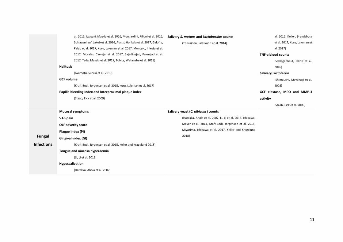
11
al. 2016, Iwasaki, Maeda et al. 2016, Mongardini, Pilloni et al. 2016,
Schlagenhauf, Jakob et al. 2016, Alanzi, Honkala et al. 2017, Galofre,
Palao et al. 2017, Kuru, Laleman et al. 2017, Montero, Iniesta et al.
2017, Morales, Carvajal et al. 2017, Sajedinejad, Paknejad et al.
2017, Tada, Masaki et al. 2017, Tobita, Watanabe et al. 2018)
Halitosis
(Iwamoto, Suzuki et al. 2010)
GCF volume
(Kraft-Bodi, Jorgensen et al. 2015, Kuru, Laleman et al. 2017)
Papilla bleeding Index and Interproximal plaque index
(Staab, Eick et al. 2009)
Salivary S. mutans and Lactobacillus counts
(Toiviainen, Jalasvuori et al. 2014)
al. 2015, Keller, Brandsborg
et al. 2017, Kuru, Laleman et
al. 2017)
TNF-a blood counts
(Schlagenhauf, Jakob et al.
2016)
Salivary Lactoferrin
(Shimauchi, Mayanagi et al.
2008)
GCF elastase, MPO and MMP-3
activity
(Staab, Eick et al. 2009)
Fungal
Infections
Mucosal symptoms
VAS-pain
OLP severity score
Plaque index (PI)
Gingival index (GI)
(Kraft-Bodi, Jorgensen et al. 2015, Keller and Kragelund 2018)
Tongue and mucosa hyperaemia
(Li, Li et al. 2013)
Hypossalivation
(Hatakka, Ahola et al. 2007)
Salivary yeast (C. albicans) counts
(Hatakka, Ahola et al. 2007, Li, Li et al. 2013, Ishikawa,
Mayer et al. 2014, Kraft-Bodi, Jorgensen et al. 2015,
Miyazima, Ishikawa et al. 2017, Keller and Kragelund
2018)

12
3. Results
In general, disease prevention (caries and periodontitis) was the objective in
52,4% (n=33) trials, while the treatment of active caries and chronic periodontitis was the
goal in 7,9% (n=8) and 12,7% (n=5), respectively. Most caries trials were based on school
aged children and periodontitis in adults. The studies regarding C. albicans usually relied
on an older population, which already had higher counts of the fungi, and their objective
was reducing symptoms and microbial load (n=6).
In the 28 trials that addressed caries treatment and prevention, the intervention
period lasted a mean of 125 days (SD = 154), with a sample size of around 101
participants (SD=77,218), generally preschool children. Much of the sample was healthy
(82%), in the sense of no active caries to treat. And so, most trials had the purpose of
addressing means to prevent oral disease (82%), while only 17,9% were about treating a
present caries lesion. Food products were the primary form of administration (50%),
followed by tablets (21%) and lozenges (17,9%).
As for preferred strains, L. reuteri and L. rhamnosus were the choice in 21,4%
(n=6) of cases, each. L. paracasei was employed in 14,3% (n=4) of studies. Then,
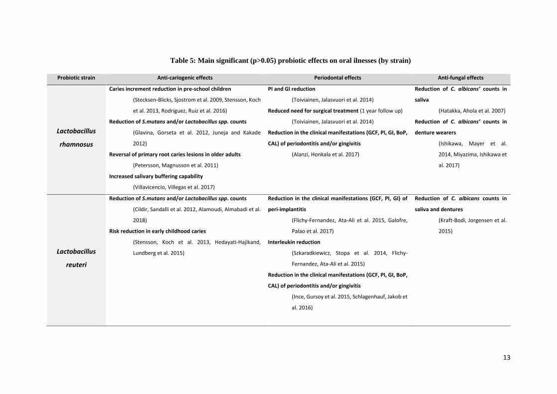
regarding the probiotic’s effect on oral diseases, the main results can be seen on table 5.
13
Table 5: Main significant (p>0.05) probiotic effects on oral ilnesses (by strain)
Probiotic strain Anti-cariogenic effects Periodontal effects Anti-fungal effects
Lactobacillus
rhamnosus
Caries increment reduction in pre-school children
(Stecksen-Blicks, Sjostrom et al. 2009, Stensson, Koch
et al. 2013, Rodriguez, Ruiz et al. 2016)
Reduction of S.mutans and/or Lactobacillus spp. counts
(Glavina, Gorseta et al. 2012, Juneja and Kakade
2012)
Reversal of primary root caries lesions in older adults
(Petersson, Magnusson et al. 2011)
Increased salivary buffering capability
(Villavicencio, Villegas et al. 2017)
PI and GI reduction
(Toiviainen, Jalasvuori et al. 2014)
Reduced need for surgical treatment (1 year follow up)
(Toiviainen, Jalasvuori et al. 2014)
Reduction in the clinical manifestations (GCF, PI, GI, BoP,
CAL) of periodontitis and/or gingivitis
(Alanzi, Honkala et al. 2017)
Reduction of C. albicans’ counts in
saliva
(Hatakka, Ahola et al. 2007)
Reduction of C. albicans’ counts in
denture wearers
(Ishikawa, Mayer et al.
2014, Miyazima, Ishikawa et
al. 2017)
Lactobacillus
reuteri
Reduction of S.mutans and/or Lactobacillus spp. counts
(Cildir, Sandalli et al. 2012, Alamoudi, Almabadi et al.
2018)
Risk reduction in early childhood caries
(Stensson, Koch et al. 2013, Hedayati-Hajikand,
Lundberg et al. 2015)
Reduction in the clinical manifestations (GCF, PI, GI) of
peri-implantitis
(Flichy-Fernandez, Ata-Ali et al. 2015, Galofre,
Palao et al. 2017)
Interleukin reduction
(Szkaradkiewicz, Stopa et al. 2014, Flichy-
Fernandez, Ata-Ali et al. 2015)
Reduction in the clinical manifestations (GCF, PI, GI, BoP,
CAL) of periodontitis and/or gingivitis
(Ince, Gursoy et al. 2015, Schlagenhauf, Jakob et
al. 2016)
Reduction of C. albicans counts in
saliva and dentures
(Kraft-Bodi, Jorgensen et al.
2015)

14
Lactobacillus
paracasei
Reduction of S.mutans and Lactobacillus spp. counts
(Chuang, Huang et al. 2010, Teanpaisan and Piwat
2013, Wattanarat, Makeudom et al. 2015,
Pahumunto, Piwat et al. 2018)
NE
Lactobacillus
casei
Reduction of S.mutans and Lactobacillus spp. counts
(Mortazavi and Akhlaghi 2012)
Reduction of papilary bleeding and interproximal PI.
Decreased MMP-3 and elastase activity and increased
MPO
(Staab, Eick et al. 2009)
NE
Bacillus
coagulans
Reduction of S. mutans and/or Lactobacillus spp. counts
(Jindal, Pandey et al. 2011)
NE NE
Lactobacillus
salivarius
Reduction of S. mutans and/or Lactobacillus spp. counts
(Nishihara, Suzuki et al. 2014)
Increased salivary buffering capacity
(Nishihara, Suzuki et al. 2014)
Reduction in periodontal pathogens (table 3)
(Mayanagi, Kimura et al. 2009, Sajedinejad,
Paknejad et al. 2017)
Reduction in the clinical manifestations (GCF, PI, GI, BoP,
CAL) of periodontitis and/or gingivitis
(Sajedinejad, Paknejad et al. 2017)
NE
NE: No effect

15
Furthermore, 50% (n=14) of studies focused on the impacts of probiotic usage on
clinical symptoms of caries progression and gingival health. Out of those, 71% showed a
statistically significant (p<0.05) influence of the probiotic strain in use. The probiotics’
capabilities to modulate oral microbiota were studied in 85% (n=24) of the trials and
yielded significant results (p<0.005) in 60% (n=17) of the cases. Only one study looked
up the influence of probiotics on immunological biomarkers.
Then, we can access the grouping of probiotic bacteria across fermentation types
and its effects on the trials, as is seen on table 6.
Table 6: Probiotics grouped by type fermentation process and their statistically
significant (p<0.05) outcomes on the trials’ variables
Significant results (%)
No
effect Clinical Microbiological Immunological
More
than one
effect
Total
Ferm
en
tati
on
pro
cess
Homofermentative 10.0%
(n=1) - - -
15.4%
(n=2)
5.6%
(n=3)
Facultative
heterofermentative
30.0%
(n=3)
61.5%
(n=8)
60.0%
(n=9)
33.3%
(n=1)
61.5%
(n=8)
53.7%
(n=29)
Heterofermentative 60.0%
(n=6)
38.5%
(n=5)
40.0%
(n=6)
66.7%
(n=2)
23.1%
(n=3)
40.7%
(n=22)
Total
100.0%
(n=10)
100.0%
(n=13)
100.0%
(n=15)
100.0%
(n=3)
100.0%
(n=13)
100.0%
(n=54)
Lactobacilli were grouped in accordance to Table 1. Bifidobacteria are facultative
heterofermentatives, non-lactic, acid-producing bacteria, and preparations with more than
one bacterial strain with different fermentation processes were excluded to simplify the
analysis. Whenever a preparation has more than one microbe, it can be unclear which
one had the most (if not all) impact on the trial’s outcomes. No correlation was found
between fermentation process and the existence of significant results in probiotic
administration. Roughly 60% of trials employing facultative heterofermentative bacteria
had positive effects on both clinical and microbiological parameters (refer to table 3), and
16
around 40% of heterofermentative bacteria had the same results. Homofermentative
bacteria were by far, the least used strains.
For the periodontitis trials the protocols lasted in average 51 days (SD= 42,15)
with a sample size of 45 (SD= 18,77) volunteers. Some studies calculated sample size
based on other studies with the same design (Kuru, Laleman et al. 2017, Morales, Carvajal
et al. 2017), while others used convenience samples (Keller and Kragelund 2018).
In the trials addressing periodontitis, probiotics were mostly administered by oral
medical appliances such as lozenges (29,6%), tablets (31%) and capsules (29%), that
account for 74,1% of the analyzed trials. Food products such as cheese, yogurt and milk
(8,6%) and oral hygiene appliances like toothbrushes and toothpastes (8,6%) were less
used.
Most trials focused preventing periodontitis on healthy patients (37%). Most of
them collected samples and performed a clinical analysis at baseline, during the usage of
the probiotic, and at the end of the treatment. Some even followed the probiotic usage by
a no-brush period to assess if the formulations could affect the formation of plaque and/or
change the host’s microbiomes.
The treatment of chronic periodontitis, characterized differently in the various
studies, was the focus of 26,9% of the trials, and implant mucositis of 14,8% - the same
as gingivitis (14,8%). Only one study was directed towards the study of halitosis.
Twenty-five trials studied the implications of probiotics on clinical parameters
and 60% of them had at least one statistically significant (p <0.05) outcome. As for the
influence of beneficial microorganisms in controlling possible oral pathogens, it was
addressed by 13 trials, of which 61% (8 trials) had a statistically significant (p <0.05)
result. Only 10 studies were based around the immunomodulation capabilities of probiotic
organisms, but out of those, seven had significant (p<0.05) results. This indicated that
probiotics such as L. reuteri have some capability to reduce inflammatory mediators.
As for probiotic species, the Lactobacillus spp. was clearly used in most studies.
L. reuteri accounted for 30,8%, L. rhamnosus for 19,2% and L. salivarius for 15,4% of
the trials. L. reuteri, for example, was the exclusive strain used in the clinical trials
regarding implant mucositis, it also was chosen in 37,5% in periodontitis treatment trials
17
and 25% in gingivitis ones. L. rhamnosus and L. salivarius were both used in 22,2% of
the trials regarding preventative oral health care studies.
Table 7 compiles all the major findings in this research, and table 8 regards products
based on probiotic bacteria that can be purchased nowadays.

18
Table 7: Clinical Trials regarding probiotics and oral health care (2009 – 2019)
Reference Intervention3 Sample4 Probiotic strain form of
administration target disease
Outcomes (p<0.05)
Clinical Microbiological Immunological
(Stecksen-Blicks, Sjostrom et
al. 2009) 105 248 Lactobacillus rhamnosus Milk Caries prevention Yes NT NT
(Glavina, Gorseta et al.
2012) 14 25 Lactobacillus rhamnosus Yogurt Caries prevention NT Yes NT
(Alamoudi, Almabadi et al.
2018) 28 178 Lactobacilli reuteri Lozenges Caries prevention No Yes NT
(Aminabadi, Erfanparast et
al. 2011) 21ϯ 105 Lactobacillus rhamnosus Yogurt Caries prevention NT No NT
(Burton, Drummond et al.
2013) 90 100 Streptococcus salivarius lozenges Caries prevention Yes Yes NT
(Chuang, Huang et al. 2010) 14 ϯ 78 Lactobacillus paracasei Tablet Caries prevention NT Yes NT
(Cildir, Sandalli et al. 2012) 100 19 Lactobacillus reuteri Drops Caries prevention NT No NT
(Kavitha, Prathima et al.
2019) 30 ϯ ϯ 60
Streptococcus fecalis
Clostridium butyricum
Bacillus mesentricus
Lactobacillus sporogenes
lozenge Active caries NT Yes No
3 Total days of probiotic administration 4 Sample at the beginning of the study

19
(Ghasemi, Mazaheri et al.
2017) 90 ϯ 50
Lactobacillus acidophilus
Bifidobacterium bifidum Yogurt Caries prevention NT No NT
(Gizani, Petsi et al. 2015) 510 85 Lactobacillus reuteri Lozenge Caries prevention No Yes NT
(Hedayati-Hajikand,
Lundberg et al. 2015) 364 138
Streptococcus uberis,
Streptococcus oralis ,
Streptococcus ratti
Chewing
tablet Caries prevention Yes NT NT
(Jindal, Pandey et al. 2011)
14 ϯ 150
Lactobacillus rhamnosus
Bifidobacterium spp.
Bacillus coagulans
Sachets Caries prevention NT Yes NT
(Juneja and Kakade 2012) 21 ϯ 40 Lactobacillus rhamnosus Milk Caries prevention NT Yes NT
(Ghasempour, Sefdgar et al.
2014) 14 ϯ 22
Lactobacillus casei
Saccharomyces cerevisiae Kefir drink Caries prevention NT Yes NT
(Keller, Nohr Larsen et al.
2014) 90 ϯ 36 Lactobacillus reuteri Tablets Active caries No NT NT
(Nishihara, Suzuki et al.
2014) 14 ϯ 64 Lactobacillus salivarius Tablets Caries prevention Yes Yes NT
(Pahumunto, Piwat et al.
2018) 90 ϯ 124 Lactobacillus paracasei Milk (powder) Caries prevention Yes Yes NT
(Petersson, Magnusson et
al. 2011) 450 160 Lactobacillus rhamnosus Milk Active caries Yes No NT
(Rodriguez, Ruiz et al. 2016) 300 261 Lactobacillus rhamnosus Milk Caries prevention Yes NT NT

20
(Romani Vestman, Hasslof
et al. 2013) 42 ϯ ϯ 62 Lactobacillus reuteri lozenges Caries prevention NT Yes NT
(Mortazavi and Akhlaghi
2012) 14 60 Lactobacillus casei Cheese Caries prevention NT Yes NT
(Singh, Damle et al. 2011) 10 ϯ 40
Bifidobacterium lactis
Lactobacillus acidophilus Ice cream Caries prevention NT Yes NT
(Stensson, Koch et al. 2013) 364 ϯ ϯ 113 Lactobacillus reuteri
Oil drops
(both) Caries prevention Yes No NT
(Taipale, Pienihakkinen et al.
2012) 30 ϯ ϯ 106 Bifidobacterium animalis
Tablets (on
spoon/pacifie) Caries prevention NT Yes NT
(Teanpaisan and Piwat
2013) 28 ϯ 40 Lactobacillus paracasei Milk powder Caries prevention NT Yes NT
(Villavicencio, Villegas et al.
2017) 270 363
Lactobacillus rhamnosus
Bifidobacteruim longum Milk
Preventive oral
care Yes No NT
(Wattanarat, Makeudom et
al. 2015) 364 60 Lactobacillus paracasei Milk
Preventive oral
care Yes Yes Yes
(Flichy-Fernandez, Ata-Ali et
al. 2015)
30 ϯ ϯ 77 Lactobacillus reuteri Tablets
Peri-implant
mucositis Yes NT Yes
(Galofre, Palao et al. 2017) 30 ϯ 44 Lactobacillus reuteri lozenge
Peri-implant
mucositis Yes No NT

21
(Ince, Gursoy et al. 2015,
Meenakshi, Gupta et al.
2016)
21 ϯ ϯ 55
Lactobacillus reuteri lozenge Chronic
periodontitis Yes NT NT
(Hallstrom, Lindgren et al.
2015)
90 ϯ 49 Lactobacillus reuteri lozenge
Peri-implant
mucositis No No No
(Iwasaki, Maeda et al. 2016) 12 ϯ 39 Lactobacillus plantarum Capsule
Chronic
periodontitis Yes NT NT
(Morales, Gandolfo et al.
2017)
90 ϯ ϯ 47 Lactobacillus rhamnosus Tablets
Chronic
periodontitis No No No
(Alkaya, Laleman et al.
2016)
56 40 Bacillus subtilis
Bacillus megaterium-
Bacillus pumulus
Toothpaste,
mouth rinse
and tooth
brush
Generalized
gingivitis No NT NT
(Alanzi, Honkala et al. 2017) 28 101 Lactobacillus rhamnosus
Bifidobacterium lactis Iozenge
Periodontitis
prevention Yes yes NT
(Tobita, Watanabe et al.
2018)
28 16 Lactobacillus crispatus Food tablet
Periodontitis
prevention Yes yes NT
(Harini and Anegundi 2010) 14 45 No info Mouth rinse
Periodontitis
prevention Yes NT NT
(Kuru, Laleman et al. 2017) 28 51 Bifidobacterium animalis Yogurt
Periodontitis
prevention Yes NT Yes

22
(Iwamoto, Suzuki et al.
2010)
28 20 Lactobacillus salivarius Tablets Halitosis Yes yes NT
(Keller, Brandsborg et al.
2017)
28 47 Lactobacillus rhamnosus
Lactobacillus curvatus Tablets Gingivitis No NT No
(Laleman, Yilmaz et al. 2015) 168 48 Streptococcus oralis KJ3,
Streptococcus
uberis KJ2, Streptococcus
ratti JH145
Tablets Chronic
periodontitis No No NT
(Lee, Kim et al. 2015) 14 34 Lactobacillus brevis lozenge
Periodontitis
prevention No NT Yes
(Mayanagi, Kimura et al.
2009, Macura-Karbownik,
Chladek et al. 2016)
56 66
Lactobacillus salivarius Tablets
(dissolving)
Periodontitis
prevention NT Yes NT
(Mongardini, Pilloni et al.
2016)
14 20 Lactobacillus plantarum
Lactobacillus brevis Tablets
Periodontitis
prevention
(implants)
Yes NT NT
(Montero, Iniesta et al.
2017)
42 59 Lactobacillus plantarum
Lactobacillus brevis
Pediococcus acidilactici
Tablets Gingivitis No Yes NT
(Morales, Carvajal et al.
2017)
90 ϯ ϯ 28 Lactobacillus Rhamnosus Sachet
Chronic
periodontitis No NT NT

23
(Sajedinejad, Paknejad et al.
2017)
28 45 Lactobacillus salivarius Mouth rinse
Chronic
periodontitis Yes Yes NT
(Schlagenhauf, Jakob et al.
2016)
49 45 Lactobacillus reuteri lozenge
pregnancy
gingivitis Yes NT Yes
(Shimauchi, Mayanagi et al.
2008)
56.0 66
Lactobacillus salivarius Tablets Periodontitis
prevention
Yes
(smoke
rs)
NT Yes (smokers)
(Staab, Eick et al. 2009) 56.0 50 Lactobacillus casei Milk
Periodontitis
prevention No NT Yes
(Szkaradkiewicz, Stopa et al.
2014)
- 24 Lactobacillus reuteri
Tablets
(suction)
Chronic
periodontitis Yes NT Yes
(Tada, Masaki et al. 2017) 168 30 Lactobacillus reuteri Tablets
Peri implant
mucositis Yes Yes NT
(Teughels, Durukan et al.
2013)
84 30 Lactobacillus reuteri lozenge
Chronic
periodontitis No Yes NT
(Toiviainen, Jalasvuori et al.
2014)
28 62 Lactobacillus rhamnosus
Bifidobacterium animalis
lozenge
(chewing
gum)
Periodontitis
prevention Yes No NT
(Hatakka, Ahola et al. 2007) 112 294
Lactobacillus rhamnosus
Propionibacterium Cheese
Candida albicans
infection Yes Yes NT
(Ishikawa, Mayer et al.
2014) 35 59
Lactobacillus rhamnosus
Lactobacillus acidophilus, Capsule
Candida albicans
infection NT Yes NT

24
Bifidobacterium bifidum
(Keller and Kragelund 2018)
112 Ϯ 22 Lactobacillus reuteri lozenges
Candida albicans
infection and
lichen planus
Yes No NT
(Li, Li et al. 2013)
28 65
Bifidobacterium
Longum
Lactobacillus bulgaricus
Streptococcus
thermophilus
lozenges
Candida
associated
stomatitis
No Yes NT
(Miyazima, Ishikawa et al.
2017) 56 60
Lactobacillus acidophilus
Lactobacillus rhamnosus Cheese
Candida albicans
infection NT Yes NT
(Kraft-Bodi, Jorgensen et al.
2015) 84 219 Lactobacillus reuteri lozenge
Candida albicans
infection No Yes NT
(Sanctis, Belgoia et al. 2019)
Variable* 75 Lactobacillus brevis CD2 lozenges
Oral mucositis
(cancer therapy
side effect)
No No NT
Ϯ Follow up: less than 6 months after intervention period
Ϯϯ Follow up: 6 months or more after intervention period
NT – parameter not tested in the trial
Candida albicans infection – high C. albicans counts
(*)probiotic administration was concomitant with radiotherapy treatment – RT - (and a week after RT) and variable for each patient

25
Table 8: Commonly used probiotic products
Brand/ Product Strain Posology Significant (p<0.05) results
BioGaia
Prodentis
lozenges /
(Gum)
Periobalanceϯ
Lactobacillus reuteri
Prodentis (L. reuteri
DSM 17938 and L.
reuteri ATCC PTA
5289) 1x10⁸ CFU
30 Probiotic lozenges
(24 g)
1 – 2 lozenges a
day
(let the
lozenges melt
in the mouth,
after brushing)
PD and CAL reduction, as well as in pro
inflammatory cytokines
(Szkaradkiewicz, Stopa et al. 2014)
Improvement of PD and CAL when
used in junction with professional
prophylaxis (Teughels, Durukan et al.
2013)
Improvements on clinical parameters
of peri-implantits (Flichy-Fernandez,
Ata-Ali et al. 2015, Galofre, Palao et al.
2017)
Reduction of GI and PI in pregnancy
gingivitis (Schlagenhauf, Jakob et al.
2016)
Reduction in S. mutans counts in
children (Alamoudi, Almabadi et al.
2018)
Wakamoto
Pharmaceutical
Co. Minna
No zendamakin
W21 tablets
Lactobacillus
salivarius 6.7x10⁸ CFU
+ Xilitol (280 mg)
1 – 2 lozenges a
day
(let the tablets
melt in the
mouth)
Improvement of physiological halitosis
(Iwamoto, Suzuki et al. 2010)
Reduction in periodontal pathogens
(Mayanagi, Kimura et al. 2009)
Improvement of periodontal health in
smokers (Shimauchi, Mayanagi et al.
2008)
Reduction in S. mutans in children
(Nishihara, Suzuki et al. 2014)
Honsha Co, Ltd
Yakult
Lactobacillus casei
shirota 1x10⁶ CFU
Fermented milk
product (one
daily bottle)
Reduction in induced plaque formation
(Slawik, Staufenbiel et al. 2011)
MMP-3 reduction (Staab, Eick et al.
2009)
Ϯ Commercially available in Portuguese pharmacies
26
4. Discussion
This bibliographic revision has shown that probiotics have proven clinical benefits
in many areas within the scope of action dentistry and oral medicine. The most prevalent
findings regarded the efficiency of certain probiotic strains in avoiding cavity lesions in
children, as well as reducing periodontal disease symptoms. This was due mostly to the
reduction in the proliferation of cariogenic and periodontal pathogens. Nevertheless, there
are various nuances in these processes that need to be addressed.
Lactobacilli can be both a risk marker, isolated in healthy mouths, and a caries
prevention method, used in probiotic preparations. While some species tend to appear in
deep caries, corelated with the lesion’s progression, other species have been shown to be
able to help modulate the microbial environment around them. For example, a study
points out that L. fermentum and S. mutans with S. sobrinus were positively associated
with caries, while the probiotic L. acidophilus was negatively associated with caries in
preschool aged children (Kanasi, Johansson et al. 2010). Even so, the production of lactic
acid from beneficial species can be considered as a side effect of their usage. Lactobacilli
can potentially be cariogenic, but account for a very small percentage of the oral
microbiome and have a low impact in the development of caries – even though they have
a more significant role in its evolution, across the cavitated phase (Lahtinen, Salminen et
al. 2012). Both xylitol and fluoride have also been used to successfully prevent caries
lesions in children, but their administration can also result in the development of fluoride
resistant bacteria (Marinho, Worthington et al. 2013, Banerjee, Sengupta et al. 2016, Lin,
Fang et al. 2016).
Whenever Lactobacillus counts are evaluated in these trials, the strain type is
important since the increase in probiotic lactobacilli may be beneficial (testing the
persistence of the probiotic after the intervention period) while other species within the
genus can be detrimental (cavitated lesions). For example, L. plantarum can quickly
transform sugars in to lactic acid, while L. paracasei and L. rhamnosus have a slower
metabolism, being less cariogenic (Lahtinen, Salminen et al. 2012). None of the trials
evaluated in this study have employed L. plantarum to treat or prevent oral cavities.
Another study showed that L. reuteri had the capability to reduce the growth of cariogenic
S. mutans but it wasn´t always detected in the mouth after the intervention period (Romani
Vestman, Hasslof et al. 2013). Even the administration of probiotics, as early as at birth
or infancy, could effectively reduce S. mutans counts throughout childhood, with positive

27
effects on primary dentition (Stensson, Koch et al. , Taipale, Pienihakkinen et al. 2013).
These probiotics are intentionally administered and can be more effective if they are given
the chance to colonize the oral biofilm earlier (Lahtinen, Salminen et al. 2012). In these
cases, while pathogens are being effectively reduced for years, no traces of the probiotic
strain are found in recent saliva samples. Hence, the effects of early usage of probiotics
in children are long lasting, but the colonization itself isn’t – meaning, the microbes do
not definitely colonize the mouth. Maybe by colonizing plaque in its formation,
pathological microbes aren’t allowed to adhere.
As most studies regarded caries prevention and progression on children, the
preferred method of probiotic administration tended to be food products. Food products
have high oral clearance and so, measures need to be taken in order to keep them longer
in the mouth. Some studies refer giving specific recommendations to the patients taking
probiotic milk: to drink it slowly, in portions, without heating it up and avoiding brushing
their teeth for up to 1 hour (Juneja and Kakade 2012). Others also point out the need to
wait 1 hour before brushing, after taking a kefir drink (Ghasempour, Sefdgar et al. 2014).
As for the effects on microbiome modulation, the administration of probiotic
bacteria tends to have different effects on streptococci and on lactobacilli. A trial found
that a combination of Bifidobacterium lactis and L. acidophilus successfully decreased S.
mutans colonization but had no effect on other Lactobacillus strains (Singh, Damle et al.
2011). L. casei showed a similar behavior (Mortazavi and Akhlaghi 2012). On the other
hand, L. reuteri showed to have the capability to reduce other Lactobacillus strains on
more than one study (Gizani, Petsi et al. 2015, Alamoudi, Almabadi et al. 2018). And L.
paracasei was able not only to suppress the growth of MS and other lactobacilli, but did
so while producing less lactic acid than other strains – more cariogenic strains, such as L.
salivarius (Wattanarat, Makeudom et al. 2015). In fact, L. salivarius was never used on
its own as a probiotic strain to address caries in any of the presented trials.
Different stages of caries progression are related with different pathogens – S.
mutans in early lesions and Lactobacillus in advanced ones. And different strains of
lactobacilli showed to have capability to reduce the pathogenic microbes of both phases.
It is also important to note that lactobacilli, as lactic acid producers are potentially
cariogenic, being widely present in carious dentine (Byun, Nadkarni et al. 2004). That
may be the reasoning behind the usage of these species in prevention of carious lesions

28
instead of in its treatment. Remineralization attempts with probiotics were generally
unsuccessful. Most trials in this study revolved around preventing caries in children.
Hence the usage of acid producing bacteria that can be added to amenable food products
such as milk, cheese and ice cream.
In the periodontitis trials, health and disease are measured in different manners. A
study defines moderate to severe periodontitis as PD > 4 mm, CAL > 3 mm and bone loss
> 3 mm, while another describes periodontitis as patients with detected horizontal bone
loss, the presence of at least 2 teeth with an approximal site each with a PD of 5-7 mm
and a GI of ≥2 in each quadrant (Ince, Gursoy et al. 2015, Sajedinejad, Paknejad et al.
2017). Furthermore, some trials specify periodontitis as moderate or severe, according to
probing depths and other clinical parameters. As recently as 2011, the American
Academy of Periodontology and the European Federation of periodontology came up
with a new Classification for Periodontal and Per-Implant diseases and Conditions,
rendering the concepts of chronic and aggressive periodontitis obsolete. The trials in this
study do not comply by a standardized definition of periodontal illness, and so their results
are not directly comparable.
Different strains of L. salivarius can be more or less effective according to their
probiotic features (Ruiz, Margolles et al. 2013, Sajedinejad, Paknejad et al. 2017).
Sajedinejad et all in their 2017 clinical trial found that L. salivarius NK02 had the highest
microbial activity against A. actinomycetemcomintans in addition to all the other
parameters listed before. While these are beneficial it is important to note that due to the
high oral clearance, the local application of probiotics would be of little effect.
Nevertheless, the immunomodulation caused by these species in the GI tract may
positively impact the oral cavity. Other probiotic products such as lozenges, chewing gum
and straws may prove to be more effective than mouthwashes and food items for these
reasons (Charalampopoulos and Rastall 2009). And 74,1% of the trials analyzed
administered the probiotics as lozenges, tablets or capsules. Some studies even went as
far as explaining if these devices were to be left to dissolve in the mouth (Hallstrom,
Lindgren et al. 2015, Galofre, Palao et al. 2017, Tobita, Watanabe et al. 2018) or simply
consumed (Iwasaki, Maeda et al. 2016).
According to J. H. Meurman (Charalampopoulos and Rastall 2009) Lactobacillus
spp. have varying antimicrobial activity across its different strains. Different pathogens
29
may need the action of a different probiotic strain. L. reuteri inhibits the growth of P.
gingivalis and P. intermedia in 82 and 55%, respectively, with that diminishing gingival
bleeding (Charalampopoulos and Rastall 2009). And, in the present clinical trial review,
L. reuteri was also proven to be effective against P. gingivalis. L. rhamnosus has shown
evidence to be efficient at reducing the levels of A. actinomycetemcomitans and F.
nucleatum in saliva and plaque, and P. gingivalis in plaque (Alanzi, Honkala et al. 2017).
L. salivarius decreased the counts of A. actinomycetemcomitans and T. forsythia.
Homofermentative lactobacilli were more frequent in healthy mouths, in comparison with
chronic periodontitis patients. Nevertheless, both homofermentative and
heterofermentative probiotics have positive effects on biofilm modulations, even though
the complete mechanisms behind this dynamic are still unknown (Lahtinen, Salminen et
al. 2012). A study found that the strongest anti-microbial activity was seen in facultative
heterofermentative bacteria and strict homofermentatives. While L. gasseri and L.
crispatus (homofermentatives) showed to highly inhibit P. gingivalis, L. plantarum
(heterofermentative) had no impact on periodontal pathogens. In low glucose
environments microbial activity decreased due to the reduction of fermentation substrate
and lower lactic acid production (Koll-Klais, Mandar et al. 2005). It is important to note
that most of the studies that were performed on patients with periodontitis, the usage of
probiotics was concomitant with mechanic professional prophylaxis. No studies were
performed where a control group had no prophylaxis done, for obvious ethical reasons.
Probiotics were evaluated as coadjutant to planning and root scaling, the gold standard of
non-surgical periodontal treatment. Whenever the effect of probiotics on their own was
tested, healthy patients (after a period of probiotic products intake), were asked to stop
oral health hygiene for a small period. This provoked intentional inflammation and the
first stages of plaque formation. In this matter, 3 studies were able to prove that the regular
usage of probiotic supplements could diminish the counts of oral periodontal pathogens
(Mayanagi, Kimura et al. 2009, Alanzi, Honkala et al. 2017, Tobita, Watanabe et al.
2018), and one showed that they didn’t (Toiviainen, Jalasvuori et al. 2014). Other than
controlling bacterial populations, probiotics can also stimulate and regulate the immune
system. Gill, Grover et al. (Charalampopoulos and Rastall 2009) refer that, among other
functions, probiotics can increase cellular immunity (NK cell activity, phagocytosis and
oxidative bursts), humoral activity (increase in immunoglobulin levels – IgA, IgG, IgM)
and interfere with the production of inflammatory cytokines (Charalampopoulos and
Rastall 2009). L. reuteri was pointed as capable of reducing inflammatory cytokine levels
30
in three trials (Szkaradkiewicz, Stopa et al. 2014, Flichy-Fernandez, Ata-Ali et al. 2015,
Schlagenhauf, Jakob et al. 2016). Finally, the most studied variables were the clinical
parameters – GI, PD, BoP and PI – in 24 trials. L. reuteri (n=7) and L. rhamnosus (n=5)
were the most used probiotic strains. 60% of all the studies considering these variables
had a positive outcome.
Taking together the above-described information, the best probable usage of
probiotics in the treatment of periodontal illnesses is as an aid to home oral hygiene and
professional prophylaxis.
Mucositis has been mostly approached in these recent trials as an implant related
disease. In this manner it is highly correlated with the maintenance of periodontal health,
and hence generally circumscribed localized issue.
Some studies refer the importance of non-surgical, mechanic periodontal
treatment, before initiating probiotics treatment, in order to reduce the bacterial load
pretrial and ensure the best results (Hallstrom, Lindgren et al. 2015, Mongardini, Pilloni
et al. 2016, Galofre, Palao et al. 2017). These trials aimed at preventing the development
of peri implant mucositis.
Other trials have the objective of treating active implant mucositis. Hence, they
don’t include healthy individuals or patients who used antibiotics 3 months prior to the
study (Hallstrom, Lindgren et al. 2015). While others specifically select patients with
<15% full mouth plaque score and <15% full mouth bleeding score. After a phase of
intentional plaque induction at the implant site (14 days, using an acrylic stent during self-
performed oral hygiene), the probiotic test protocol was put to the test (Mongardini,
Pilloni et al. 2016). These recent trials have shown that probiotics seem to have little to
no influence pathological periodontal microbiomes in crevicular gingival fluid. Only one
study found that L. reuteri had a significant on the bacterial load of P. gingivalis in peri-
implant mucositis, while it had no other impacts on the remaining bacteria. A.
actinomycetemcomitans, P. gingivalis, T. forsythia, T. denticola and P. intermedia, major
periodontal pathogens from the red and yellow group (gram negative, facultative
anaerobic or complete anaerobes) were unaffected (Galofre, Palao et al. 2017). Even older
studies have found no connections between mucositis and probiotics usage (Flichy-
Fernandez, Ata-Ali et al. 2015). However, there seems to be a positive effect on the usage
of L. reuteri: reduced levels of inflammatory mediators in crevicular gingival fluid.

31
Nevertheless, it is shown that the best results in managing peri implant health can be
achieved with proper oral hygiene and professional mechanical removal of dental plaque.
In these instances, the usage of probiotics may not be strictly recommended solely on a
cost effectiveness basis.
As for mucositis as sequelae of oropahringeal cancer treatment, it is generally
accepted that it is associated with the intensity and toxicity of both radio and
chemotherapy. The cytotoxicity of these treatments has direct effects on connective tissue
and epithelial cells, resulting of thinning of the epithelium and, as time progresses, it’s
loss. On such studies measurements other than crevicular fluid are used, such as the oral
mucositis grade (OM). The OM is a clinical observation measure that ranges between 0
and IV, from the least amount of oral discomfort and mucosal compromise (0) to the
greatest (IV). These studies have, due to these variables, more difficulties in drawing
definitive conclusions.
In neutropenic patients with mucositis, there is an increased risk for systemic
infections originating from opportunistic elements of the oral microbiome due to mucosal
ulceration. In that sense there is an increased importance in avoiding the proliferation of
oral pathogens in these immunocompromised individuals (Greenberg, Glick et al. 2008).
A recent trial attempted to modulate the microbial composition of the saliva of patients
with neck and head tumors, by adding a strain of L. brevis into their diet. No differences
were observed between the placebo control group (sodium bicarbonate mouthwash) and
the group receiving the probiotic (Sanctis, Belgoia et al. 2019). On the other hand, it was
found that the usage lozenges containing L. brevis reduced the development of grade III
and IV mucositis (28% of patients treated with L. brevis did not develop mucositis, while
only 7% of those on the placebo had the same outcome) (Sharma, Rath et al. 2012). One
must note a difference in metrics between these studies: while one assessed a clinical
parameter (mucositis grade), the other discussed the effects on the microbiome. Probiotics
seem to have a positive influence on the patient’s quality of life, but the underlying
biological mechanisms need further research. For example, the positive results in
reducing the production of cytokines cited in other trials (Staab, Eick et al. 2009,
Szkaradkiewicz, Stopa et al. 2014, Flichy-Fernandez, Ata-Ali et al. 2015, Kuru, Laleman
et al. 2017), has been proven beneficial. Even though, there might not be a direct effect
in pathogen control, probiotics may help strengthen the mucosal barrier by reducing
inflammatory molecules that negatively impact epithelial cell proliferation and worsens
32
tissue damage (Greenberg, Glick et al. 2008). Furthermore, there is a difference between
trying to modulate the microbiome of a healthy individual - cases of peri-implantitis -
versus the one existing on a patient during cancer treatment - mucositis due to cancer
treatment toxicity.
As for the efficiency of probiotics in the treatment of yeast infections, it is
measured in comparison with the one already achieved by anti-fungal medications.
Probiotics have the added benefit on not causing microbial resistance and being generally
less aggressive to the host’s organism. Li et all (2013) prove that adding a probiotic to
nystatin increases the reduction in C. albicans colonization, versus the nystatin
monotherapy. A study that compared the two separately, would be of interest. Another
study directly compared the effects of L. reuteri and nystatin as prophylaxis in skin and
stool Candida colonization in very low birth weight infants. In this study the L. reuteri
was as effective as nystatin. The skin samples were collected from the axilla,
interinginous and moist mucosa region, which points the fact that the application of this
protocol to the oral cavity might be a viable research option (Oncel, Arayici et al.).
Probiotic effects are strain specific, therefore there is a need to test which strains
are more suited to treat a specific condition. An investigation tested L. acidophulus and
L. rhamnosus in their capabilities to reduce Candida spp. infections, and both were
effective (Ishikawa, Mayer et al. 2014, Miyazima, Ishikawa et al. 2017). It is suggested
that to assess the varying impacts of both strains, a larger sample and longer evaluation
period would be necessary. Another study tested the anti-fungal capabilities of L.
rhamnosus and L. casei on resin surface dentures. Both strains were effective at reducing
yeast proliferation and did not affect the roughness of the resin, an added benefit for
patients that use removable oral prosthetics (Song and Lee 2017).
Probiotic delivery vehicles also need to be addressed. Food products such as
cheese and milk have a shorter activity clearance due to salivary flow. Direct application
on oral prosthetics or a more viscous adherent vehicle could be beneficial (Ishikawa,
Mayer et al. 2014).
Medical co-mobilities such as diabetes and medication intake should also be
considered, especially in studies regarding elderly populations. Diabetes, generally
regarded as a Candida spp. colonization facilitator (due to reduced salivary flow), had no
impact in the probiotic’s effect (Ishikawa, Mayer et al. 2014).

33
Regarding the treatment of head and neck tumors, sequelae such as xerostomia
and, therefore, oral mucositis and candidiasis may arise. While no specific trials on the
direct usage of probiotics on this population, it is safe to infer that maintaining and
adequate salivary flow and controlling the proliferation of potentially pathogenic fungi
would be of great advantage. So, besides the standard preventative measures (diet control,
fluoride supplementation, treatment of infectious sites and regular oral prosthetic’s
maintenance), the cancer patient can also benefit from the usage of probiotic preparations
in order to avoid a range of oral diseases: caries, periodontal disease, xerostomia and
mucositis.
As it has been discussed before, there seems to be an association between lichen
planus and C. albicans infections (Neville, Damn et al.). Hence the attempt to tackle both
conditions with the same probiotic microorganism is justifiable. The usage of L. reuteri
has only had significant effects in the decrease of the gingival index (GI), but no effects
in C. albican’s counts (Keller and Kragelund 2018). It is believed that oral microbes may
also be implied in the progression of lichen planus. A study found that patients with
current Lichen planus had relatively higher counts of Porphyromonas and Solobacterium,
in comparison with healthy controls (Wang, Lu et al. 2016). Porphyromonas is especially
prone to generate inflammatory response and cytokine production. Therefore, the
improvement of gingival index measures may prove beneficial to control the proliferation
of Porphyromonas and help reduce inflammation and pain.
4.1. Currently available commercial probiotic formulations
The Lactobacillus prodentis® (L. reuteri DSM 17938 and Lactobacillus reuteri
ATCC PTA 5289 - 1x10⁸ CFU) formulation is commonly used across studies. While it
shows positive results in reducing periodontal disease symptoms, it is less effective in
reducing its pathogens. However, when applied to the treatment of caries in children, it
has shown the ability to suppress the growth of S. mutans in the study by Alamoudy,
Almabady et all (2018). Nevertheless, this product has also produced some non-
significant results: no microbiome alterations (reduction of S. mutans) (Gizani, Petsi et
al. 2015), as well as no effect on the surgency of white spot lesions (Keller, Nohr Larsen
et al. 2014, Gizani, Petsi et al. 2015). BioGaia also produces oil drops, mostly aimed at
the regulation of gut microbiota (Lactobacillus protectis ® - L. reuteri DSM 17938).
34
These products have originated from a L. reuteri strain isolated from breast milk in the
1950’s - ATCC 55730. This strain was used in the oil drops formula applied by Stensson,
Koch et al (2013) on their clinical trial. The test group, 60 (out of 113) mothers were
given daily probiotic drops during the 4 weeks before the expected date of delivery, and
their children for 365 days (their first year of life). Nine years after the intervention,
children in the test group had reduced caries prevalence and gingivitis score in primary
dentition (Stensson, Koch et al. 2013). Hence, this product seems particularly suitable to
treat periodontitis symptoms and to prevent the surgency of caries in primary dentition,
if given to children early on in life. Periobalance ® is available in Portuguese pharmacies.
A possible clinical application of L. reuteri to periodontal disease treatment can be the
daily intake of probiotic lozenges after scaling and root planning. The most common
approach is the usage of chlorohexidine mouth rinses during a controlled period after
SRP. Chlorohexidine is still the gold standard when it comes to periodontal disease
treatment because it performs three different tasks simultaneously: it is both a bactericide,
a bacteriostatic and has substantivity in the oral cavity. This cannot be said about
probiotics, whose presence in the oral cavity is short lived. Nevertheless, there is no
evidence pointing that probiotics have the same side effects as chlorohexidine, such as
extrinsic teeth staining (Moshrefi 2002), and less frequently, mucosal desquamation and
subjective feelings of dryness, soreness or burning sensation (Flotra 1973). Teeth
staining, was more prevalent as usage period of chlorohexidine increased (Tartaglia,
Tadakamadla et al. 2019). Furthermore, chlorohexidine is considered as a pollutant, being
found in hospital sewage waters (Lasek, Karpel et al. 2018) and is suggested to be
cytotoxic towards osteoblastic, endothelial and fibroblastic cell lines in “in vitro” studies
(Giannellia, F.Chellinib et al. 2008, Reddersen, Wiegand et al. 2019). In this sense,
probiotics can be an option when long term management of periodontitis is concerned.
The W21 tablets produced by Wakamoto Pharmaceutical Co were effective at
controlling periodontal (Mayanagi, Kimura et al. 2009) and cariogenic pathogens
(Nishihara, Suzuki et al. 2014). They were also capable of improving periodontal health
in smokers and reducing physiological halitosis (Shimauchi, Mayanagi et al. 2008,
Iwamoto, Suzuki et al. 2010). Despite the positive results, these products aren’t, at the
moment, available in Portugal.
The L. casei Shirota found in Yakult ® yogurts has shown to be effective at
reducing plaque formation and gingival inflammatory markers (Staab, Eick et al. 2009,
35
Slawik, Staufenbiel et al. 2011). This product is mostly associated with gastrointestinal
benefits but L. casei and L. paracasei strains have also been proved to have positive
effects on oral health, especially on caries prevention. An example is the novel L.
paracasei SD1 (Teanpaisan and Piwat 2013, Wattanarat, Makeudom et al. 2015).
However, these microorganisms haven’t been added to commercially available
formulations yet.
5. Conclusions
Probiotics have proven to be beneficial in preventing the development of cavities
in school aged children, reducing inflammation markers and clinical symptoms of
periodontitis in adults and fungal counts in the mucosa of the elderly.
While most studies show that there is some benefit in the usage of probiotics to
ameliorate the most prevalent conditions seen in the dentist’s daily practice, their effects
aren’t completely predictable and hence they shouldn’t be used in a monotherapy regime.
6. Future research developments
There is still room for further research, mainly in realm of the possible benefits
that probiotic usage can have on some populations, namely patients receiving treatment
for head and neck cancer, as well as some immunologically mediated illnesses with oral
manifestations like lichen planus, pemphigus, and aphthous stomatitis. In the end,
research proves that probiotics are a clinically verified treatment option and can safely
and effectively be used in many oral aliments and in all age groups.

36
7. Bibliography
Alamoudi, N. M., E. S. Almabadi, E. A. El Ashiry and D. A. El Derwi (2018).
"Effect of Probiotic Lactobacillus reuteri on Salivary Cariogenic Bacterial Counts among
Groups of Preschool Children in Jeddah, Saudi Arabia: A Randomized Clinical Trial." J
Clin Pediatr Dent 42(5): 331-338.
Alanzi, A., S. Honkala, E. Honkala, A. Varghese, M. Tolvanen and E. Soderling
(2017). "Effect of Lactobacillus rhamnosus and Bifidobacterium lactis on gingival health,
dental plaque, and periodontopathogens in adolescents: a randomised placebo-controlled
clinical trial." Benef Microbes 9(4): 593-602.
Alkaya, B., I. Laleman, S. Keceli, O. Ozcelik, M. Cenk Haytac and W. Teughels
(2016). "Clinical effects of probiotics containing Bacillus species on gingivitis: a pilot
randomized controlled trial." J Periodontal Res 52(3): 497-504.
Aminabadi, N. A., L. Erfanparast, A. Ebrahimi and S. G. Oskouei (2011). "Effect
of chlorhexidine pretreatment on the stability of salivary lactobacilli probiotic in six- to
twelve-year-old children: a randomized controlled trial." Caries Res 45(2): 148-154.
Banerjee, G., A. Sengupta, T. Roy, Prajna Paramita Banerjee, A. Chattopadhyay
and A. K. Raya (2016). "Isolation and characterization of fluoride resistant bacterial
strains from fluoride endemic area." Fluoride 49(1): 429-440.
Burton, J. P., B. K. Drummond, C. N. Chilcott, J. R. Tagg, W. M. Thomson, J. D.
Hale and P. A. Wescombe (2013). "Influence of the probiotic Streptococcus salivarius
strain M18 on indices of dental health in children: a randomized double-blind, placebo-
controlled trial." J Med Microbiol 62(Pt 6): 875-884.
Byun, R., M. A. Nadkarni, K. L. Chhour, F. E. Martin, N. A. Jacques and N.
Hunter (2004). "Quantitative analysis of diverse Lactobacillus species present in
advanced dental caries." J Clin Microbiol 42(7): 3128-3136.
Charalampopoulos, D. and R. A. Rastall (2009). Prebiotics and Probiotics Science
and Technology, Springer Science+Business Media.
37
Chuang, L. C., C. S. Huang, L. W. Ou-Yang and S. Y. Lin (2010). "Probiotic
Lactobacillus paracasei effect on cariogenic bacterial flora." Clin Oral Investig 15(4):
471-476.
Cildir, S. K., N. Sandalli, S. Nazli, F. Alp and E. Caglar (2012). "A novel delivery
system of probiotic drop and its effect on dental caries risk factors in cleft lip/palate
children." Cleft Palate Craniofac J 49(3): 369-372.
Flichy-Fernandez, A. J., J. Ata-Ali, T. Alegre-Domingo, E. Candel-Marti, F. Ata-
Ali, J. R. Palacio and M. Penarrocha-Diago (2015). "The effect of orally administered
probiotic Lactobacillus reuteri-containing tablets in peri-implant mucositis: a double-
blind randomized controlled trial." J Periodontal Res 50(6): 775-785.
Flotra, L. (1973). "Different modes of chlorhexidine application and related local
side effects., 8(s12), 41–44." Journal of Periodontal Research 8.
Galofre, M., D. Palao, M. Vicario, J. Nart and D. Violant (2017). "Clinical and
microbiological evaluation of the effect of Lactobacillus reuteri in the treatment of
mucositis and peri-implantitis: A triple-blind randomized clinical trial." J Periodontal Res
53(3): 378-390.
Ghasemi, E., R. Mazaheri and A. Tahmourespour (2017). "Effect of Probiotic
Yogurt and Xylitol-Containing Chewing Gums on Salivary S Mutans Count." J Clin
Pediatr Dent 41(4): 257-263.
Ghasempour, M., S. A. Sefdgar, A. A. Moghadamnia, R. Ghadimi, S. Gharekhani
and L. Shirkhani (2014). "Comparative study of Kefir yogurt-drink and sodium fluoride
mouth rinse on salivary mutans streptococci." J Contemp Dent Pract 15(2): 214-217.
Giannellia, M., F.Chellinib, M.Margherib, P.Tonellia and A.Tanib (2008). "Effect
of chlorhexidine digluconate on different cell types: A molecular and ultrastructural
investigation." Toxicology in Vitro 22.
Gizani, S., G. Petsi, S. Twetman, C. Caroni, M. Makou and L. Papagianoulis
(2015). "Effect of the probiotic bacterium Lactobacillus reuteri on white spot lesion
development in orthodontic patients." Eur J Orthod 38(1): 85-89.
38
Glavina, D., K. Gorseta, I. Skrinjaric, D. N. Vranic, K. Mehulic and K. Kozul
(2012). "Effect of LGG yoghurt on Streptococcus mutans and Lactobacillus spp. salivary
counts in children." Coll Antropol 36(1): 129-132.
Greenberg, M. S., M. Glick and J. A. Ship (2008). Burkett's Oral Medicine.
Hamilton, Ontario, BC Decker Inc.
Hallstrom, H., S. Lindgren, C. Widen, S. Renvert and S. Twetman (2015).
"Probiotic supplements and debridement of peri-implant mucositis: a randomized
controlled trial." Acta Odontol Scand 74(1): 60-66.
Han, X., J. Zhang, Y. Tan and G. Zhou (2017). "Probiotics: A non-conventional
therapy for oral lichen planus." Arch Oral Biol 81: 90-96.
Harini, P. M. and R. T. Anegundi (2010). "Efficacy of a probiotic and
chlorhexidine mouth rinses: a short-term clinical study." J Indian Soc Pedod Prev Dent
28(3): 179-182.
Hatakka, K., A. J. Ahola, H. Yli-Knuuttila, M. Richardson, T. Poussa, J. H.
Meurman and R. Korpela (2007). "Probiotics Reduce the Prevalence of Oral Candida in
the Elderly--a Randomized Controlled Trial." J DENT RES 86(125).
Hedayati-Hajikand, T., U. Lundberg, C. Eldh and S. Twetman (2015). "Effect of
probiotic chewing tablets on early childhood caries--a randomized controlled trial." BMC
Oral Health 15(1): 112.
Ince, G., H. Gursoy, S. D. Ipci, G. Cakar, E. Emekli-Alturfan and S. Yilmaz
(2015). "Clinical and Biochemical Evaluation of Lozenges Containing Lactobacillus
reuteri as an Adjunct to Non-Surgical Periodontal Therapy in Chronic Periodontitis." J
Periodontol 86(6): 746-754.
Ishikawa, K. H., M. P. Mayer, T. Y. Miyazima, V. H. Matsubara, E. G. Silva, C.
R. Paula, T. T. Campos and A. E. Nakamae (2014). "A multispecies probiotic reduces
oral Candida colonization in denture wearers." J Prosthodont 24(3): 194-199.
Iwamoto, T., N. Suzuki, K. Tanabe, T. Takeshita and T. Hirofuji (2010). "Effects
of probiotic Lactobacillus salivarius WB21 on halitosis and oral health: an open-label
pilot trial." Oral Surg Oral Med Oral Pathol Oral Radiol Endod 110(2): 201-208.

39
Iwasaki, K., K. Maeda, K. Hidaka, K. Nemoto, Y. Hirose and S. Deguchi (2016).
"Daily Intake of Heat-killed Lactobacillus plantarum L-137 Decreases the Probing Depth
in Patients Undergoing Supportive Periodontal Therapy." Oral Health Prev Dent 14(3):
207-214.
Jindal, G., R. K. Pandey, J. Agarwal and M. Singh (2011). "A comparative
evaluation of probiotics on salivary mutans streptococci counts in Indian children." Eur
Arch Paediatr Dent 12(4): 211-215.
Juneja, A. and A. Kakade (2012). "Evaluating the effect of probiotic containing
milk on salivary mutans streptococci levels." J Clin Pediatr Dent 37(1): 9-14.
Kanasi, E., I. Johansson, S. C. Lu, N. R. Kressin, M. E. Nunn, J. R. Kent and A.
C. R. Tanner (2010). "Microbial Risk Markers for Childhood Caries in Pediatricians’
Offices." J Dent Res 84(4).
Kavitha, M., G. Prathima, G. Kayalvizhi, A. Sanguida, G. Ezhumalai and V.
Ramesh (2019). "Evaluation of Streptococcus mutans serotypes e, f, and k in saliva
samples of 6–12- year-old school children before and after a short-term daily intake of
the probiotic lozenge." Official journal of the Indian Society of Pedodontics and
Preventive 37(1): 67-74.
Keller, M. K., E. Brandsborg, K. Holmstrom and S. Twetman (2017). "Effect of
tablets containing probiotic candidate strains on gingival inflammation and composition
of the salivary microbiome: a randomised controlled trial." Benef Microbes 9(3): 487-
494.
Keller, M. K. and C. Kragelund (2018). "Randomized pilot study on probiotic
effects on recurrent candidiasis in oral lichen planus patients." Oral Dis 24(6): 1107-1114.
Keller, M. K., I. Nohr Larsen, I. Karlsson and S. Twetman (2014). "Effect of
tablets containing probiotic bacteria (Lactobacillus reuteri) on early caries lesions in
adolescents: a pilot study." Benef Microbes 5(4): 403-407.
Koll-Klais, P., R. Mandar, E. Leibur, H. Marcotte, L. Hammarstrom and M.
Mikelsaar (2005). "Oral lactobacilli in chronic periodontitis and periodontal health:
species composition and antimicrobial activity." Oral Microbiol Immunol 20(6): 354-361.

40
Koll, P., R. Mandar, H. Marcotte, E. Leibur, M. Mikelsaar and L. Hammarstrom
(2008). "Characterization of oral lactobacilli as potential probiotics for oral health." Oral
Microbiol Immunol 23(2): 139-147.
Kraft-Bodi, E., M. R. Jorgensen, M. K. Keller, C. Kragelund and S. Twetman
(2015). "Effect of Probiotic Bacteria on Oral Candida in Frail Elderly." J Dent Res 94(9
Suppl): 181S-186S.
Kuru, B. E., I. Laleman, T. Yalnizoglu, L. Kuru and W. Teughels (2017). "The
Influence of a Bifidobacterium animalis Probiotic on Gingival Health: A Randomized
Controlled Clinical Trial." J Periodontol 88(11): 1115-1123.
Lahtinen, S., S. Salminen, A. Ouwehand and A. v. Wright (2012). Lactic Acid
Bacteria. Microbiological and functional aspects. Boca Raton, FL 33487-2742, Taylor &
Francis Group, LLC.
Laleman, I., E. Yilmaz, O. Ozcelik, C. Haytac, M. Pauwels, E. R. Herrero, V.
Slomka, M. Quirynen, B. Alkaya and W. Teughels (2015). "The effect of a streptococci
containing probiotic in periodontal therapy: a randomized controlled trial." J Clin
Periodontol 42(11): 1032-1041.
Lasek, F., N. Karpel, V. Leitner, G. Rauwel, L. Blanchier, O. Castel and S.
Ayraud-Thevenot (2018). "Discharge of biocidal products from healthcare activities into
a sewage system—a case study at a French university hospital." Environmental Science
and Pollution Research 26: 4938–4951.
Lashof, A. M. L. O., R. D. Bock, Herbrecht, B. E. d. Pauw, V. Krcmery, M. Aoun,
M. Akova, Cohen, H. Siffnerov, M. Egyed, M. Ellis, A. Marinus, R. Sylvester and B. J.
Kullberg (2004). "An open multicentre comparative study of the efficacy, safety and
tolerance of fluconazole and itraconazole in the treatment of cancer patients with
oropharyngeal candidiasis." European Journal of Cancer 40: 1314–1319.
Lee, J. K., S. J. Kim, S. H. Ko, A. C. Ouwehand and D. S. Ma (2015). "Modulation
of the host response by probiotic Lactobacillus brevis CD2 in experimental gingivitis."
Oral Dis 21(6): 705-712.
Li, D., Q. Li, C. Liu, M. Lin, X. Li, X. Xiao, Z. Zhu, Q. Gong and H. Zhou (2013).
"Efficacy and safety of probiotics in the treatment of Candida-associated stomatitis."
Mycoses 57(3): 141-146.

41
Lin, H. K., C. E. Fang, M. S. Huang, H. C. Cheng, T. W. Huang, H. T. Chang and
K. W. Tam (2016). "Effect of maternal use of chewing gums containing xylitol on
transmission of mutans streptococci in children: a meta-analysis of randomized controlled
trials." Int J Paediatr Dent 26(1): 35-44.
Lindhe, J., N. P. Lang and T. Karring (2008). Clinical Periodontology and Implant
Dentistry, Blackwell Munksgaard.
Macura-Karbownik, A., G. Chladek, J. Żmudzki and J. Kasperski (2016).
"Chewing efficiency and occlusal forces in PMMA, acetal and polyamide removable
partial denture wearers." Acta of Bioengineering and Biomechanics 18(1): 127-134.
Marinho, V. C., H. V. Worthington, T. Walsh and J. E. Clarkson (2013). "Fluoride
varnishes for preventing dental caries in children and adolescents." Cochrane Database
Syst Rev(7): Cd002279.
Mayanagi, G., M. Kimura, S. Nakaya, H. Hirata, M. Sakamoto, Y. Benno and H.
Shimauchi (2009). "Probiotic effects of orally administered Lactobacillus salivarius
WB21-containing tablets on periodontopathic bacteria: a double-blinded,
placebocontrolled, randomized clinical trial." J Clin Periodontol 36: 506–513.
Meenakshi, A., R. Gupta, V. Bharti, G. Sriramaprabu and R. Prabhakar (2016).
"An Evaluation of Retentive Ability and Deformation of Acetal Resin and Cobalt-
Chromium Clasps." J Clin Diagn Res 10(1): Zc37-41.
Melo, P. R. G. R. d. (2001). Influência de diferentes métodos de administração de
fluoretos nas variações de incidência de cárie. Doctorate, Faculdade de Medicina da
Universidade do Porto.
Miyazima, T. Y., K. H. Ishikawa, M. Mayer, S. Saad and A. Nakamae (2017).
"Cheese supplemented with probiotics reduced the Candida levels in denture wearers-
RCT." Oral Dis 23(7): 919-925.
Mongardini, C., A. Pilloni, R. Farina, G. Di Tanna and B. Zeza (2016).
"Adjunctive efficacy of probiotics in the treatment of experimental peri-implant mucositis
with mechanical and photodynamic therapy: a randomized, cross-over clinical trial." J
Clin Periodontol 44(4): 410-417.

42
Montero, E., M. Iniesta, M. Rodrigo, M. J. Marin, E. Figuero, D. Herrera and M.
Sanz (2017). "Clinical and microbiological effects of the adjunctive use of probiotics in
the treatment of gingivitis: A randomized controlled clinical trial." J Clin Periodontol
44(7): 708-716.
Morales, A., P. Carvajal, N. Silva, M. Hernandez, C. Godoy, G. Rodriguez, R.
Cabello, J. Garcia-Sesnich, A. Hoare, P. I. Diaz and J. Gamonal (2017). "Clinical Effects
of Lactobacillus rhamnosus in Non-Surgical Treatment of Chronic Periodontitis: A
Randomized Placebo-Controlled Trial With 1-Year Follow-Up." J Periodontol 87(8):
944-952.
Morales, A., A. Gandolfo, J. Bravo, P. Carvajal, N. Silva, C. Godoy, J. Garcia-
Sesnich, A. Hoare, P. Diaz and J. Gamonal (2017). "Microbiological and clinical effects
of probiotics and antibiotics on nonsurgical treatment of chronic periodontitis: a
randomized placebo- controlled trial with 9-month follow-up." J Appl Oral Sci 26:
e20170075.
Mortazavi, S. and N. Akhlaghi (2012). "Salivary Streptococcus mutans and
Lactobacilli levels following probiotic cheese consumption in adults: A double blind
randomized clinical trial." Journal of research in medical sciences 17(1).
Moshrefi, A. (2002). "Clorohexidine." The Journal of the Western Society of
Periodontology/Periodontal Abstracts 50.
Neville, B. W., D. D. Damn, C. M. Allen and J. E. Bouquot Patologia oral e
Maxilo facial, Elsevier Editora Ldta.
Nishihara, T., N. Suzuki, M. Yoneda and T. Hirofuji (2014). "Effects of
Lactobacillus salivarius-containing tablets on caries risk factors: a randomized open-label
clinical trial." BMC Oral Health 14: 110.
Oncel, M. Y., S. Arayici, F. N. Sari, G. K. Simsek, S. Yurttutan, O. Erdeve, S.
Saygan, N. Uras, S. S. Oguz and U. Dilmen "Comparison of Lactobacillus reuteri and
nystatin prophylaxis on Candida colonization and infection in very low birth weight
infants." J Matern Fetal Neonatal Med 28(15): 1790-1794.
Pahumunto, N., S. Piwat, O. Chankanka, N. Akkarachaneeyakorn, K.
Rangsitsathian and R. Teanpaisan (2018). "Reducing mutans streptococci and caries

43
development by Lactobacillus paracasei SD1 in preschool children: a randomized
placebo-controlled trial." Acta Odontol Scand 76(5): 331-337.
Petersson, L. G., K. Magnusson, U. Hakestam, A. Baigi and S. Twetman (2011).
"Reversal of primary root caries lesions after daily intake of milk supplemented with
fluoride and probiotic lactobacilli in older adults." Acta Odontol Scand 69(6): 321-327.
Rautemaa, R., P. Rusanen, M. Richardson and J. H. Meurman (2006). "Optimal
sampling site for mucosal candidosis in~ oral cancer patients is the labial sulcus." Journal
of Medical Microbiology 55: 1447–1451.
Reddersen, K., C. Wiegand, P. Elsner and Uta-ChristinaHipler (2019). "Three-
dimensional human skin model infected with Staphylococcus aureus as a tool for
evaluation of bioactivity and biocompatibility of antiseptics." International Journal of
Antimicrobial Agents 54: 283-291.
Rodriguez, G., B. Ruiz, S. Faleiros, A. Vistoso, M. L. Marro, J. Sanchez, I. Urzua
and R. Cabello (2016). "Probiotic Compared with Standard Milk for High-caries
Children: A Cluster Randomized Trial." J Dent Res 95(4): 402-407.
Romani Vestman, N., P. Hasslof, M. K. Keller, E. Granstrom, S. Roos, S.
Twetman and C. Stecksen-Blicks (2013). "Lactobacillus reuteri influences regrowth of
mutans streptococci after full-mouth disinfection: a double-blind, randomised controlled
trial." Caries Res 47(4): 338-345.
Ruiz, L., A. Margolles and B. Sánchez (2013). "Bile resistance mechanisms in
Lactobacillus and Bifidobacterium." Front Microbiol 4: 396.
Sajedinejad, N., M. Paknejad, B. Houshmand, H. Sharafi, R. Jelodar, H. Shahbani
Zahiri and K. A. Noghabi (2017). "Lactobacillus salivarius NK02: a Potent Probiotic for
Clinical Application in Mouthwash." Probiotics Antimicrob Proteins 10(3): 485-495.
Sanctis, V. d., L. Belgoia, D. Cante, M. R. L. Porta, O. Caspiani, R. Guarnaccia
and A. Argenone (2019). "Lactobacillus brevis CD2 for Prevention of Oral Mucositis in
Patients With Head and Neck Tumors: A Multicentric Randomized Study." Anticancer
Res 39(4): 1935-1942.

44
Schlagenhauf, U., L. Jakob, M. Eigenthaler, S. Segerer, Y. Jockel-Schneider and
M. Rehn (2016). "Regular consumption of Lactobacillus reuteri-containing lozenges
reduces pregnancy gingivitis: an RCT." J Clin Periodontol 43(11): 948-954.
Sharma, A., G. K. Rath, S. P. Chaudhary, A. Thakar, B. K. Mohanti and S.
Bahadur (2012). "Lactobacillus brevis CD2 lozenges reduce radiation- and
chemotherapy-induced mucositis in patients with head and neck cancer: a randomized
double-blind placebo-controlled study." Eur J Cancer 48(6): 875-881.
Shimauchi, H., G. Mayanagi, S. Nakaya, M. Minamibuchi, Y. Ito, K. Yamaki and
H. Hirata (2008). "Improvement of periodontal condition by probiotics with Lactobacillus
salivarius WB21: a randomized, double-blind, placebo-controlled study." J Clin
Periodontol 35: 897–905.
Singh, R. P., S. G. Damle and A. Chawla (2011). "Salivary mutans streptococci
and lactobacilli modulations in young children on consumption of probiotic ice-cream
containing Bifidobacterium lactis Bb12 and Lactobacillus acidophilus La5." Acta
Odontol Scand 69(6): 389-394.
Slawik, S., I. Staufenbiel, R. Schilke, S. Nicksch, K. Weinspach, M. Stiesch and
J. Eberhard (2011). "Probiotics affect the clinical inflammatory parameters of
experimental gingivitis in humans." Eur J Clin Nutr 65(7): 857-863.
Song, Y. G. and S. H. Lee (2017). "Inhibitory effects of Lactobacillus rhamnosus
and Lactobacillus casei on Candida biofilm of denture surface." Arch Oral Biol 76: 1-6.
Staab, B., S. Eick, G. Knofler and H. Jentsch (2009). "The influence of a probiotic
milk drink on the development of gingivitis: a pilot study." J Clin Periodontol 36(10):
850-856.
Stecksen-Blicks, C., I. Sjostrom and S. Twetman (2009). "Effect of long-term
consumption of milk supplemented with probiotic lactobacilli and fluoride on dental
caries and general health in preschool children: a cluster-randomized study." Caries Res
43(5): 374-381.
Stensson, M., G. Koch, S. Coric, T. R. Abrahamsson, M. C. Jenmalm, D. Birkhed
and L. K. Wendt "Oral administration of Lactobacillus reuteri during the first year of life
reduces caries prevalence in the primary dentition at 9 years of age." Caries Res 48(2):
111-117.
45
Stensson, M., G. Koch, S. Coric, T. R. Abrahamsson, M. C. Jenmalm, D. Birkhed
and L. K. Wendt (2013). "Oral administration of Lactobacillus reuteri during the first year
of life reduces caries prevalence in the primary dentition at 9 years of age." Caries Res
48(2): 111-117.
Szkaradkiewicz, A. K., J. Stopa and T. M. Karpinski (2014). "Effect of oral
administration involving a probiotic strain of Lactobacillus reuteri on pro-inflammatory
cytokine response in patients with chronic periodontitis." Arch Immunol Ther Exp
(Warsz) 62(6): 495-500.
Tada, H., C. Masaki, S. Tsuka, T. Mukaibo, Y. Kondo and R. Hosokawa (2017).
"The effects of Lactobacillus reuteri probiotics combined with azithromycin on peri-
implantitis: A randomized placebo-controlled study." J Prosthodont Res 62(1): 89-96.
Taipale, T., K. Pienihakkinen, P. Alanen, J. Jokela and E. Soderling (2013).
"Administration of Bifidobacterium animalis subsp. lactis BB-12 in early childhood: a
post-trial effect on caries occurrence at four years of age." Caries Res 47(5): 364-372.
Taipale, T., K. Pienihakkinen, S. Salminen, J. Jokela and E. Soderling (2012).
"Bifidobacterium animalis subsp. lactis BB-12 administration in early childhood: a
randomized clinical trial of effects on oral colonization by mutans streptococci and the
probiotic." Caries Res 46(1): 69-77.
Tartaglia, G. M., S. K. Tadakamadla, S. T. Connelly, C. Sforza and C. Martin
(2019). "Adverse events associated with home use of mouthrinses: a systematic review."
Ther Adv Drug Saf 10: 2042098619854881.
Teanpaisan, R. and S. Piwat (2013). "Lactobacillus paracasei SD1, a novel
probiotic, reduces mutans streptococci in human volunteers: a randomized placebo-
controlled trial." Clin Oral Investig 18(3): 857-862.
Teughels, W., A. Durukan, O. Ozcelik, M. Pauwels, M. Quirynen and M. C.
Haytac (2013). "Clinical and microbiological effects of Lactobacillus reuteri probiotics
in the treatment of chronic periodontitis: a randomized placebo-controlled study." J Clin
Periodontol 40(11): 1025-1035.
Tobita, K., I. Watanabe, M. Tomokiyo and M. Saito (2018). "Effects of heat-
treated Lactobacillus crispatus KT-11 strain consumption on improvement of oral cavity
environment: a randomised double-blind clinical trial." Benef Microbes 9(4): 585-592.

46
Toiviainen, A., H. Jalasvuori, E. Lahti, U. Gursoy, S. Salminen, M. Fontana, S.
Flannagan, G. Eckert, A. Kokaras, B. Paster and E. Soderling (2014). "Impact of orally
administered lozenges with Lactobacillus rhamnosus GG and Bifidobacterium animalis
subsp. lactis BB-12 on the number of salivary mutans streptococci, amount of plaque,
gingival inflammation and the oral microbiome in healthy adults." Clin Oral Investig
19(1): 77-83.
Villavicencio, J., L. M. Villegas, M. C. Arango, S. Arias and F. Triana (2017).
"Effects of a food enriched with probiotics on Streptococcus mutans and Lactobacillus
spp. salivary counts in preschool children: a cluster randomized trial." J Appl Oral Sci 26:
e20170318.
Wang, K., W. Lu, Q. Tu, Y. Ge, J. He, Y. Zhou, Y. Gou, J. D. Van Nostrand, Y.
Qin, J. Li, J. Zhou, Y. Li, L. Xiao and X. Zhou (2016). "Preliminary analysis of salivary
microbiome and their potential roles in oral lichen planus." Sci Rep 6: 22943.
Wattanarat, O., A. Makeudom, T. Sastraruji, S. Piwat, S. Tianviwat, R.
Teanpaisan and S. Krisanaprakornkit (2015). "Enhancement of salivary human neutrophil
peptide 1-3 levels by probiotic supplementation." BMC Oral Health 15: 19.
47

48
Top Related