


Línguas
Páginas
Legal

6
Brazilian Journal of Microbiology (2010) 41: 6-14 ISSN 1517-8382
VAGINAL LACTOBACILLI AS POTENTIAL PROBIOTICS AGAINST Candida spp.
Natalia F. Gil1, Rafael C.R. Martinez1, Bruna C. Gomes1, Auro Nomizo1, Elaine C. P. De Martinis1*
1Faculdade de Ciências Farmacêuticas de Ribeirão Preto, Universidade de São Paulo, Ribeirão Preto, SP, Brasil.
Submitted: July 16, 2008; Returned to authors for corrections: April 09, 2009; Approved: September 28, 2009.
ABSTRACT
Urogenital infections affect millions of people every year worldwide. The treatment of these diseases usually
requires the use of antimicrobial agents, and more recently, the use of probiotic lactic acid bacteria (LAB)
cultures for the management of vaginal infections has been extensively studied. In this work, 11 vaginal
lactobacilli isolates, previously obtained from healthy patients, were studied to screen microorganisms with
probiotic properties against Candida spp. The LAB were tested for their ability of auto-aggregation, co-
aggregation with C. albicans, C. glabrata, C. krusei, and C. tropicalis, adhesion to Caco-2 epithelial cells and
production of lactic acid and hydrogen peroxide (H2O2). All lactobacilli isolates tested were able to auto-
aggregate (ranging from 25.3% to 75.4% assessed at 4 hours of incubation) and to co-aggregate with the four
Candida species into different degrees; among them L. crispatus showed the highest scores of co-
aggregation. The highest amount of lactic acid was produced by L. salivarius (13.9 g/l), followed by L.
johnsonii (6.5 g/l), L. acidophilus (5.5 g/l), and L. jensenii (5.4 g/l). All isolates produced H2O2 , but the
highest levels (3 - 10 mg/l) were observed for L. acidophilus, L. crispatus, L. gasseri, L. johnsonii, and L.
vaginalis. Only L. agilis, L. jensenii, L. johnsonii and L. ruminus were able to adhere to epithelial Caco-2
cells. Among the isolates evaluated, L agilis, L. jensenii, L. johnsonii, and L. ruminus exhibited
simultaneously several desirable properties as potential probiotic strains justifying future studies to evaluate
their technological properties in different pharmaceutical preparations for human use.
Key words: Lactobacillus spp., probiotic, Candida spp.
INTRODUCTION
In women of childbearing age, the vaginal ecosystem is
dominated by Lactobacillus spp. (41). These microorganisms
can prevent the colonization of the urogenital tract by several
pathogens and they are important for women’s reproductive
and general healthy (17, 24, 40, 42).
Lactobacilli modulate the vaginal microbiota by different
mechanisms such as: (i) auto-aggregation, (ii) production of
lactic acid, hydrogen peroxide, bacteriocins, and biosurfactants,
(iii) co-aggregation with pathogenic microorganisms, and (iv)
adhesion to epithelial cells (17, 29, 37).
*Corresponding Author. Mailing address: Departamento de Análises Clínicas, Toxicológicas e Bromatológicas, Faculdade de Ciências Farmacêuticas de Ribeirão Preto – Universidade de São Paulo, Av. do Café, s/n, 14040-903 - Ribeirão Preto – SP – Brazil.; Tel. +55 16 36024267 Fax +55 16 36024725.; E-mail: [email protected]
7
Probiotics against Candida spp.
Bacterial vaginosis (BV) and vulvovaginal candidiasis
(VVC) are the most prevalent vaginal infections worldwide
(30). BV is responsible for up to 50% of all the cases of vaginal
infections and it is characterized by a significant reduction in
lactobacilli population, and increase in facultative aerobic and
anaerobic pathogens (10, 16).
VVC affects up to 75% of women at least once in their
lives and despite pruritus and vaginal discharge are usual
complaints associated with this disease neither is specific to the
infection (34). The majority of cases of VVC (ca. 90%) caused
by Candida albicans are treated with oral or topical antifungal
agents, with increasing reports on episodes of VVC due to non-
albicans species (27, 28). There is an overgrowing concern
about the spread use of over-the-counter preparations (such as
topical azole agents) which may contribute for the selection of
non-albicans resistant strains that are normally more difficult
to be eradicated (23, 35).
Probiotics are defined as live microorganisms which when
administered in adequate quantity confer health benefits to the
host and lactobacilli of human origin are potential probiotics
against urogenital tract infections (11, 25). Some clinical
studies showed positive results for the use of L. fermentum RC-
14 and L. rhamnosus GR-1 to treat patients with BV by oral
intake and intravaginal administration (1, 2). Also, a recent
clinical trial showed that oral administration of capsules
containing L. fermentum RC-14 and L. rhamnosus GR-1 was
effective as adjuvant in the treatment of patients diagnosed
with VVC (20).
Probiotics do not show collateral effects usually seen for
traditional antibacterial and antifungal agents because they act
by several mechanisms, which minimize punctual mutations
involved in the emergence of antimicrobial resistant pathogens.
The technology necessary to produce probiotic agents does not
appear to be complex, and this can stimulate their production at
reasonable costs. This scenario certainly encourages more
researches to be undertaken to select and test new strains with
probiotic properties.
The aim of the present work was to evaluate the ability of
Lactobacillus spp., previously isolated from the vaginal
microbiota of healthy Brazilian patients, as potential probiotics
against Candida species.
MATERIALS AND METHODS
Strains
A total of 11 vaginal Lactobacillus spp. were previously
isolated from a group of 64 healthy Brazilian women (21) and
the use of the strains for this study was approved by local Ethic
Review Board (250/CEP-CSE-FMRP-USP). The isolates
studied were L. acidophilus, L. agilis, L. coleohominis, L.
crispatus, L. fermentum, L. gasseri, L. jensenii, L. johnsonii, L.
salivarius, L. ruminus and L. vaginalis. Additionally, for the
study of adhesion to epithelial cells, L. bulgaricus and L.
rhamnosus GG were employed as negative and positive
controls, respectively. The bacterial strains were kept at - 70°C
in MRS broth (de Man, Rogosa and Sharpe – Oxoid, UK)
added of 20 % (v/v) of glycerol.
A total of four Candida spp. strains were used in this
study, to know: C. albicans ATCC 18804, C. tropicalis ATCC
750, C. krusei ATCC 20298 and C. glabrata ATCC 2001. The
yeast strains were kept in SDA (Sabouraud-dextrose agar –
Oxoid, UK) at room temperature.
Auto-aggregation studies
Lactobacillus spp. was grown overnight at 37ºC in MRS
broth (1.0%, v/v), centrifuged at 6,000g for 15min (Fanem,
mod. 208 N, Brazil) and cell pellets were resuspended in
phosphate buffered saline (PBS) to obtain an optical density
(O.D.) of 0.6 at 600nm (UVmini-1240, Shimadzu, Japan).
Auto-aggregation inversely correlated with O.D. and it was
monitored every 1h for up to 4h of incubation (13, 25). Gram
staining was used to visualize the aggregates under oil
immersion microscopy with 100 times magnification (CX-31 –
Olympus, Japan).
Co-aggregation studies
Culture plates of 24 wells containing round glass slides
were added of: i) 500µL of an overnight culture of
8
Gil, N.F. et al.
Lactobacillus spp. grown at 37oC in MRS broth and ii)
500µL of an overnight culture of Candida spp. grown at 37oC
in BHI broth (Brain-Heart Infusion – Oxoid, UK). Plates were
incubated at 37oC for 4h in an orbital shaker at 100 rpm (CT-
712, Cientec, Brazil) and co-aggregation was determined by
Gram staining of the round glass slides and observation under
oil immersion microscopy (CX-31 – Olympus, Japan). Scoring
was done according to Reid et al. (31).
Production of lactic acid
Homofermentative metabolism was verified by absence of
production of gas from glucose (33) and lactic acid production
was quantified in grams per liter, by acid-base titration,
according to Edema and Sanni (9).
Production of hydrogen peroxide
Determination of hydrogen peroxide (H2O2) production by
Lactobacillus isolates was performed according to Wilks et al.
(41) with modifications. Briefly, lactobacilli were grown in
MRS broth (Oxoid, UK) for 24h at 37ºC and 100µl-aliquots of
the broths were seeded on MRS agar plates (Oxoid, UK) and
incubated for 48h at 37ºC, under anaerobic atmosphere.
Selected colonies were put in contact with strips containing
peroxidase (Merckoquant Peroxide Test - Merck, Germany).
Different tones of blue products were visually compared, with
a scale provided by the manufacturer. Results were expressed
in ranges of H2O2 production according to Wilks et al. (41).
Adhesion to the epithelial cells
Adhesion to intestinal epithelial Caco-2 cells (ATCC
7348406) was evaluated according to Duprè et al. (8). Briefly,
Caco-2 cells were cultivated at 37°C under 5% CO2 in RPMI
medium (Gibco, USA) supplemented with 10% of fetal bovine
serum and 100U/ml of streptomycin and penicillin (Sigma,
USA). When confluent growth was achieved, adhered cells
were trypsinized, transferred to 24-well plates containing round
glass slides and re-incubated. After 24 hours, the RPMI
medium with antibiotics was removed and replaced by RPMI
supplemented with 2% of fetal bovine serum. Bacterial cultures
were previously grown overnight at 37°C in MRS broth,
diluted in RPMI containing 2% of fetal bovine serum and
added to each well (ca. 106 bacteria) containing the Caco-2
cells and incubation was done at 37°C for 3 hours. After that,
slides were washed, fixed, stained with May–Grunwald–
Giemsa and analyzed under oil immersion microscopy with
100 times magnification (CX-31 – Olympus, Japan).
The number of bacteria adhered to Caco-2 cells was
obtained by scoring adhesion to 100 random eukaryotic cells,
using criteria proposed by Del Re et al. (7) as non-adhesive (<5
bacteria/100 cells), adhesive (6-40 bacteria/100 cells) and
strongly adhesive (>40 bacteria/100 cells). L. bulgaricus and L.
rhamnosus GG were used as negative and positive controls,
respectively.
RESULTS AND DISCUSSION
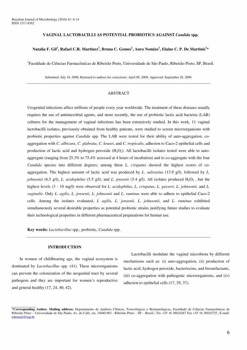
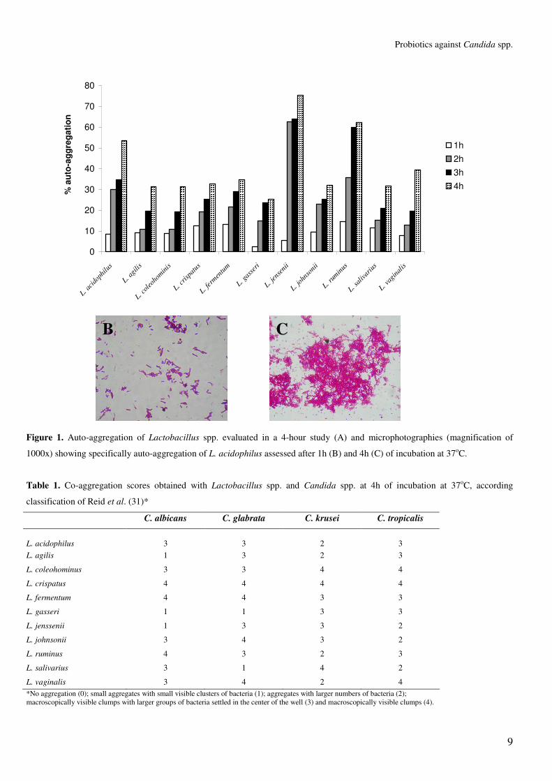
All lactobacilli isolates tested in the present study
exhibited some degree of auto-aggregation at all time-points
tested, but the highest degree of auto-aggregation was observed
after 4h of incubation at 37°C for L. jensenii, followed by L.
ruminus and L. acidophilus (Figure 1). Figure 1 also illustrates
auto-aggregation observed for L. acidophilus in two time-
points. The ability of auto-aggregation of vaginal lactobacilli is
an intrinsic characteristic and may substantially increase the
colonization of environments with short residence times (25).
According to Juarez-Tomás et al. (13) the ability of auto-
aggregation is higher in acid environments where probiotic
lactobacilli are more adapted to survive and represents the first
step towards the formation of biofilms by lactobacilli strains,
which helps to inhibit the overgrowth and proliferation of
pathogenic microorganisms (14, 37).
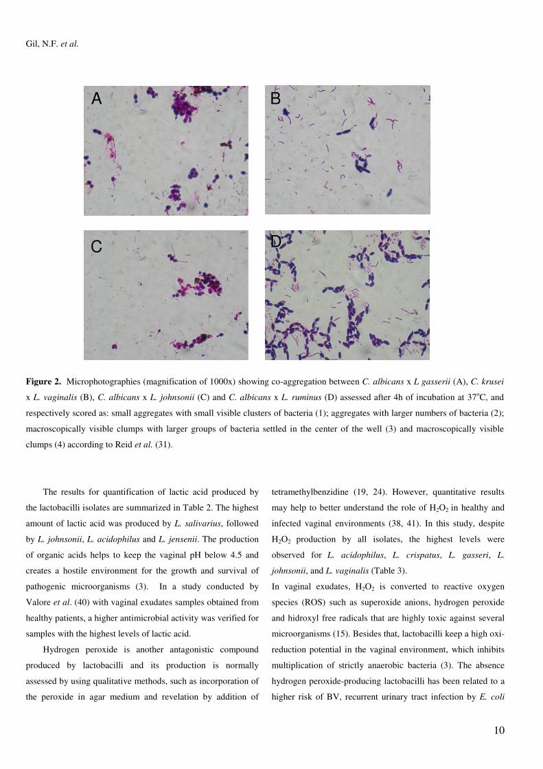
The co-aggregation scores obtained after 4h of incubation
at 37°C for Lactobacillus spp. and Candida spp. are shown in
Table 1 and illustrated in Figure 2. L. crispatus showed
macroscopically visible clumps when evaluated with all four
Candida strains. The co-aggregation can create a
microenvironment around the pathogen with a higher
concentration of inhibitory substances and it can also block the
dissemination of pathogens to tissue receptors (22, 29).
9
Probiotics against Candida spp.
0
10
20
30
40
50
60
70
80
L. acid
ophil
us
L. agil
is
L. cole
ohom
inis
L. cris
patus
L. ferm
entum
L. gas
seri
L. jens
enii
L. john
sonii
L. rum
inus
L. sali
variu
s
L. vag
inalis
% a
uto-
aggr
egat
ion
1h2h3h4h
Figure 1. Auto-aggregation of Lactobacillus spp. evaluated in a 4-hour study (A) and microphotographies (magnification of
1000x) showing specifically auto-aggregation of L. acidophilus assessed after 1h (B) and 4h (C) of incubation at 37oC.
Table 1. Co-aggregation scores obtained with Lactobacillus spp. and Candida spp. at 4h of incubation at 37oC, according
classification of Reid et al. (31)*
C. albicans C. glabrata C. krusei C. tropicalis
L. acidophilus
3
3
2
3
L. agilis 1 3 2 3
L. coleohominus 3 3 4 4
L. crispatus 4 4 4 4
L. fermentum 4 4 3 3
L. gasseri 1 1 3 3
L. jenssenii 1 3 3 2
L. johnsonii 3 4 3 2
L. ruminus 4 3 2 3
L. salivarius 3 1 4 2
L. vaginalis 3 4 2 4 *No aggregation (0); small aggregates with small visible clusters of bacteria (1); aggregates with larger numbers of bacteria (2); macroscopically visible clumps with larger groups of bacteria settled in the center of the well (3) and macroscopically visible clumps (4).
BB CC
10
Gil, N.F. et al.
Figure 2. Microphotographies (magnification of 1000x) showing co-aggregation between C. albicans x L gasserii (A), C. krusei
x L. vaginalis (B), C. albicans x L. johnsonii (C) and C. albicans x L. ruminus (D) assessed after 4h of incubation at 37oC, and
respectively scored as: small aggregates with small visible clusters of bacteria (1); aggregates with larger numbers of bacteria (2);
macroscopically visible clumps with larger groups of bacteria settled in the center of the well (3) and macroscopically visible
clumps (4) according to Reid et al. (31).
The results for quantification of lactic acid produced by
the lactobacilli isolates are summarized in Table 2. The highest
amount of lactic acid was produced by L. salivarius, followed
by L. johnsonii, L. acidophilus and L. jensenii. The production
of organic acids helps to keep the vaginal pH below 4.5 and
creates a hostile environment for the growth and survival of
pathogenic microorganisms (3). In a study conducted by
Valore et al. (40) with vaginal exudates samples obtained from
healthy patients, a higher antimicrobial activity was verified for
samples with the highest levels of lactic acid.
Hydrogen peroxide is another antagonistic compound
produced by lactobacilli and its production is normally
assessed by using qualitative methods, such as incorporation of
the peroxide in agar medium and revelation by addition of
tetramethylbenzidine (19, 24). However, quantitative results
may help to better understand the role of H2O2 in healthy and
infected vaginal environments (38, 41). In this study, despite
H2O2 production by all isolates, the highest levels were
observed for L. acidophilus, L. crispatus, L. gasseri, L.
johnsonii, and L. vaginalis (Table 3).
In vaginal exudates, H2O2 is converted to reactive oxygen
species (ROS) such as superoxide anions, hydrogen peroxide
and hidroxyl free radicals that are highly toxic against several
microorganisms (15). Besides that, lactobacilli keep a high oxi-
reduction potential in the vaginal environment, which inhibits
multiplication of strictly anaerobic bacteria (3). The absence
hydrogen peroxide-producing lactobacilli has been related to a
higher risk of BV, recurrent urinary tract infection by E. coli
DD
AA BB
CC
11
Probiotics against Candida spp.
and increased susceptibility to the infection by Human
Immunodeficiency Virus (HIV-1) (31, 38).
Table 2. Lactic acid production by vaginal Lactobacillus spp.
isolates, expressed in g/l
Species
Lactic acid (g/l)*
L. acidophilus 5.52
L. agilis 1.72
L. coleohominis 0.77
L. crispatus 1.32
L. fermentum 1.22
L. gasseri 1.35
L. jensenii 5.42
L. johsonii 6.50
L. ruminus 1.22
L. salivarius 13.95
L. vaginalis 1.72
*According to Edema and Sanni (9) Table 3. Semi-quantification of H2O2 production by vaginal
Lactobacillus spp. isolates obtained from healthy patients
Production of H2O2 (mg/l)*
1-3
3-10
L. agilis
L. colehominis
L. fermentum
L. jensenii
L. ruminus
L. salivarius
L. acidophilus
L. crispatus
L. gasseri
L. johnsonii
L. vaginalis
*Production of H2O2 was scored according to Wilks et al. (41) as: negative, 1-3, 3-10, 10-30, 30-100 mg/l of H2O2.
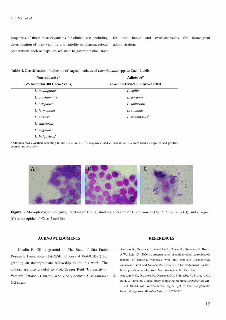
Atassi et al. (4) affirmed that adhesion to epithelial tissue
is the first step towards the formation of a barrier by
lactobacilli that will prevent colonization by pathogenic
microorganisms. In our study lactobacilli isolates were tested
for adhesion to epithelial tissue, using Caco-2 cell, which
presents a confluent growth when cultivated in vitro and shows
characteristics of mature enterocytes, making it an excellent
model for studying adherence of microorganisms (6, 12).
Among all lactobacilli isolates tested, only L. agilis, L. jensenii,
L. johnsonii and L. ruminus were able to adhere to epithelial
Caco-2 cells (Table 4 and Figure 3) indicating adhesion is
specific for each bacterial strain, as verified also by Chauvière
et al. (6).
Some vaginal Lactobacillus species are capable of
synthesizing antimicrobial peptides known as bacteriocins (5).
In our study, we have also evaluated bacteriocin production of
all lactobacilli isolates against Candida strains by agar
antagonism method, but no inhibitory activity was detected
(data not shown). Osset et al. (26) studied the production of
bacteriocin by several LAB isolates against C. albicans and C.
glabrata and they did not observe inhibition zones when agar
plate method was used. However, in broth cultures those
authors verified that some lactobacilli were inhibitory against
C. albicans to some extent. In our experiments, highest
inhibitory activity against C. albicans assessed in BHI broth
was obtained for both L. jensenii and L. johnsonii isolates (data
not shown).
Ström et al. (36) observed that several antifungal
compounds, such as cyclic dipeptides, pyroglutamic acid and
lactones were produced by Lactobacillus isolates and played an
important role against Candida spp. Thus further investigation
is required to clarify the nature of the inhibitory substance
produced by lactobacilli isolates against the yeast in our study.
In conclusion, our results indicated that among the 11
vaginal lactobacilli isolates tested, L agilis, L. jensenii, L.
johnsonii, and L. ruminus exhibited desirable properties as
potential probiotic strains including the ability to adhere to
epithelial mucosa, to auto-aggregate, co-aggregate with
Candida species, and to produce both lactic acid and H2O2.
Also, future studies are encouraged to assess technological
12
Gil, N.F. et al.
properties of those microorganisms for clinical use, including
determination of their viability and stability in pharmaceutical
preparations such as capsules resistant to gastrointestinal tract
for oral intake and ovules/capsules for intravaginal
administration.
Table 4. Classification of adhesion of vaginal isolates of Lactobacillus spp. to Caco-2 cells
Non-adhesive*
(<5 bacteria/100 Caco-2 cells)
Adhesive*
(6-40 bacteria/100 Caco-2 cells)
L. acidophilus
L. colehominis
L. crispatus
L. fermentum
L. gasseri
L. salivarius
L. vaginalis
L. bulgaricus¥
L. agilis
L. jensenii
L. johnsonii
L. ruminus
L. rhamnosus¥
*Adhesion was classified according to Del Re et al. (7). ¥L. bulgaricus and L. rhamnosus GG were used as negative and positive controls, respectively.
Figure 3. Microphotographies (magnification of 1000x) showing adhesion of L. rhamnosus (A), L. bulgaricus (B), and L. agilis
(C) to the epithelial Caco-2 cell line.
ACKNOWLEDGMENTS
Natalia F. Gil is grateful to The State of São Paulo
Research Foundation (FAPESP, Process # 06/60105-7) for
granting an undergraduate fellowship to do this work. The
authors are also grateful to Prof. Gregor Reid (University of
Western Ontario - Canada), who kindly donated L. rhamnosus
GG strain.
REFERENCES
1. Anukam, K.; Osazuwa, E.; Ahonkhai, I.; Ngwu, M.; Osemene, G.; Bruce,
A.W.; Reid, G. (2006 a). Augmentation of antimicrobial metronidazole
therapy of bacterial vaginosis with oral probiotic Lactobacillus
rhamnosus GR-1 and Lactobacillus reuteri RC-14: randomized, double-
blind, placebo controlled trial. Microbes Infect., 8, 1450-1454.
2. Anukam, K.C.; Osazuwa, E.; Osemene, G.I.; Ehigiagbe, F.; Bruce, A.W.;
Reid, G. (2006 b). Clinical study comparing probiotic Lactobacillus GR-
1 and RC-14 with metronidazole vaginal gel to treat symptomatic
bacterial vaginosis. Microbes Infect., 8, 2772-2776.
�� �� ��
13
Probiotics against Candida spp.
3. Aroutcheva, A.; Gariti, D.; Simon, M.; Shott, S.; Faro, J.; Simoes, JA;
Gurguis, A; Faro, S. (2001). Defense factors of vaginal lactobacilli. Am.
J. Obstet. Gynecol., 185, 375-379.
4. Atassi, F.; Brassart, D.; Grob, P.; Graf, F.; Servin, A.L. (2006).
Lactobacillus strains isolated from the vaginal microbiota of healthy
women inhibit Prevotella bivia and Gardnerella vaginalis in coculture
and cell culture. FEMS Immunol. Med. Microbiol., 48, 424-432.
5. Boris, S.; Barbès, C. (2000). Role played by Lactobacillus in controlling
the population of vaginal pathogens. Microbes Infect., 2, 543-546.
6. Chauvière, G.; Coconnier, M.H.; Kernéis, S.; Fourniat, J.; Servin, A.L.
(1992). Adhesion of human Lactobacillus acidophilus strain LB to
human enterocyte-like Caco-2 cells. J. Gen. Microbiol., 138, 1689-1696.
7. Del Re, B.; Sgorbati, B.; Miglioli, M.; Palenzona, D. (2000). Adhesion,
autoaggregation and hydrophobicity of 13 strains of Bifidobacterium
longum. Lett. Appl. Microbiol. 31, 438-442.
8. Duprè, I.; Zanetti, S.; Schito, A.M.; Fadda, G.; Sechi, L.A. (2003).
Incidence of virulence determinants in clinical Enterococcus faecium and
Enterococcus faecalis isolates collected in Sardinia (Italy). J. Med.
Microbiol., 52, 491-498.
9. Edema, M.O.; Sanni, A.I. (2008). Functional properties of selected starter
cultures for sour maize bread. Food Microbiol., 25, 616-625.
10. Eschenbach, D.A.; Hillier, S.; Critchlow, C.; Stevens, C.; DeRouen, T.;
Holmes, K.K. (1988). Diagnosis and clinical manifestations of bacterial
vaginosis. Am. J. Obstet. Gynecol., 158, 819-828.
11. FAO-WHO (2001). Food and agriculture organization of the United Nations,
World Health Organization. Report of a joint FAO-WHO expert consultation on
evaluation of health and nutritional properties of probiotics in food including
powder milk with live lactic acid bacteria. Cordoba, Argentina, 2001. Available
in: ftp://ftp.fao.org./docrep/fao/009/a0512e/a0512e00.pdf
12. Hauri, H.P.; Sander, B.; Naim, H. (1994). Induction of lactase
biosynthesis in the human intestinal epithelial cell line Caco-2. Eur. J.
Biochem., 219, 539-546.
13. Juárez Tomás., M.S.; Wiese, B.; Nader-Macías, M.E. (2005). Effects of
culture conditions on the growth and auto-aggregation ability of vaginal
Lactobacillus johnsonii CRL 1294. J. Appl. Microbiol., 99, 1383-1391.
14. Kos, B.; Suskovi�, J.; Vukovi�, S.; Simpraga, M.; Frece, J.; Matosi�, S.
(2003). Adhesion and aggregation ability of probiotic strain
Lactobacillus acidophilus M92. J. Appl. Microbiol., 94, 981-987.
15. Kulisaar, T.; Zilmer, M.; Mikelsaar, M.; Vihalemm, T.; Annuk, H.;
Kairane, C.; Kilk, A. (2002). Two antioxidative lactobacilli strains as
promising probiotics. Int. J. Food Microbiol., 72, 215-224.
16. Larsson, P.G.; Forsum, U. (2005). Bacterial vaginosis – a disturbed
bacterial flora and treatment enigma. APMIS, 113, 305-316.
17. Lepargneur, J.P.; Rousseau, V. (2002). Protective role of the Doderlein
flora. J. Gynecol. Obstet. Biol. Reprod., 31, 485-494.
18. Lewus, C.B.; Montville, T.J. (1991). Detection of bacteriocins produced
by lactic acid bacteria. J. Microbiol. Methods, 13, 145-150.
19. Martin, H.L.; Richardson, B.A.; Nyange, P.M.; Lavreys, L.; Hillier, S.L.;
Chohan, B.; Mandaliya, K.; Ndinya-Achola, J.O.; Bwayo, J.; Kreiss, J.
(1999). Vaginal lactobacilli, microbial flora, and risk of human
immunodeficiency virus type 1 and sexually transmitted disease
acquisition. J. Infect. Dis., 180, 1863-1868.
20. Martinez, R.C.R.; Franceschini, S.A.; Patta, M.C.; Quintana, S.M.;
Candido, R.C.; Ferreira, J.C.; De Martinis, E.C.P.; Reid, G. (2009).
Improved treatment of vulvovaginal candidiasis with fluconazole plus
probiotic Lactobacillus rhamnosus GR-1 and Lactobacillus reuteri RC-
14. Lett. Appl. Microbiol., 48, 269-274.
21. Martinez, R.C.R.; Franceschini, A.S.; Patta, M.C.; Quintana, S.M.;
Nunes, A.C.; Moreira, J.L.S.; Anukam, K.C.; Reid, G.; De Martinis,
E.C.P. (2008). Analysis of vaginal lactobacilli from healthy and infected
Brazilian women. Appl. Environ. Microbiol., 74, 4539-4542.
22. Mastromarino, P.; Brigidi, P.; Macchia, S.; Maggi, L.; Pirovano, F.;
Trinchieri, V.; Conte, U.; Matteuzzi, D. (2002). Characterization and
selection of vaginal Lactobacillus strains for the preparation of vaginal
tablets. J. Appl. Microbiol., 93, 884-893.
23. Mathema, B.; Cross, E.; Dun, E.; Park, S.; Bedell, J.; Slade, B.; Willians,
M.; Riley, L.; Chaturvedi, V.; Perlin, D.S. (2001). Prevalence of vaginal
colonization by drug-resistant Candida species in college-age women
with previous exposure to over-the-counter azole antifungals. Clin.
Infect. Dis., 33, 23-27.
24. Mija�, V.D.; Duki�, S.V.; Opavski, N.Z.; Duki�, M.K.; Ranin, L.T.
(2006). Hydrogen peroxide producing lactobacilli in women with vaginal
infections. Eur. J. Obst. Gynecol. Reprod. Biol., 129, 69-76.
25. Ocaña, V.S.; Nader-Macías, M.E. (2002). Vaginal lactobacilli: self- and
co-aggregation ability. Br. J. Biomed. Sci., 59, 183-190.
26. Osset, J.; García, E.; Bartolomé, R.M.; Andreu, A. (2001). Role of
Lactobacillus as protector against vaginal candidiasis. Med. Clin., 22,
285-288.
27. Owen, M.K.; Clenney, T.L. (2004). Management of vaginits. Am. Fam.
Physician., 70, 2125-2132.
28. Paulitsch, A.; Weger, W.; Ginter-Hanselmayer, G.; Marth, E.; Buzina,
W. (2006). A 5-year (2000-2004) epidemiological survey of Candida and
non-Candida yeast species causing vulvovaginal candidiasis in Graz,
Austria. Mycoses 49, 471-475.
29. Reid, G. (2001). Probiotic agents to protect the urogenital tract against
infection. Am. J. Clin. Nutr., 73, 437-443.
30. Reid, G.; Bruce, A.W.; Fraser, N.; Heinemann, C.; Owen, J.; Henning, B.
(2001). Oral probiotics can resolve urogenital infections. FEMS
Immunol. Med. Microbiol., 30, 49-52.
31. Reid, G.; MacGroarty, J.A.; Domingue, P.A.G.; Chow, A.W.; Bruce,
A.W.; Eisen, A.; Costerton, J.W. (1990). Coaggregation of urogenital
bacteria in vitro and in vivo. Current Microbiol., 20, 47-52.
32. Reid, G.; Kim, S.O.; Köhler, G.A. (2006). Selecting, testing and
understanding probiotic microorganisms. FEMS Immunol. Med.
Microbiol., 46, 149-157.
33. Schillinger, U.; Lücke, F.K. (1987). Identification of lactobacilli from
meat and meat products. Food Microbiol., 4, 199-208.
34. Sobel, J.D. (2007). Vulvovaginal candidiasis. Lancet 369, 1961-1971.
14
Gil, N.F. et al.
35. Sobel, J.D. (1998). Vulvovaginitis due to Candida glabrata. An emergin
problem. Mycoses 41, 18-22.
36. Ström, K.; Sjögren, J.; Broberg, A.; Schnürer, J. (2002). Lactobacillus
plantarum MiLAB 393 produces the antifungal cyclic dipeptides cyclo
(L-Phe-L-Pro) and cyclo (L-Phe-trans-4-OH-L-Pro) and 3-phenyllactic
acid. Appl. Environ. Microbiol., 68, 4322-4327.
37. Strus, M.; Kucharska, A.; Kukla, G.; Brzychczy-Włoch, M.; Maresz, K.;
Heczko, P.B. (2005). The in vitro activity of vaginal Lactobacillus with
probiotic properties against Candida. Infect. Dis. Obstet. Gynecol., 13,
69-75.
38. Tomas, M.S.; Bru, E.; Nader-Macías, M.E. (2003). Comparison of the
growth and hydrogen peroxide production by vaginal lactobacilli under
different culture conditions. Am. J. Obstet. Gynecol., 188, 35-44.
39. Vallor, A.C.; Antonio, M.A.; Hawes, S.E.; Hillier, S.L. (2001). Factors
associated with acquisition of, or persistent colonization by, vaginal
lactobacilli: role of hydrogen peroxide production. J. Infect. Dis., 184,
1431-1436.
40. Valore, E.V.; Park, C.H.; Igreti, S.L.; Ganz, T. (2002). Antimicrobial
components of vaginal fluid. Am. J. Obstet. Gynecol., 187, 561-568.
41. Wilks, M.; Wiggins, R.; Whiley, A.; Hennessy, E.; Warwick, S.; Porter,
H.; Corfield, A.; Millar, M. (2004). Identification and H2O2 production of
vaginal lactobacilli from pregnant womem at high risk o preterm birth
and relation with outcome. J. Clin. Microbiol., 42, 713-717.
42. Zárate, G.; Nader-Macías, M.E. (2006). Influence of probiotic vaginal
lactobacilli on in vitro adhesion of urogenital pathogens to vaginal
epithelial cells. Lett. Appl. Microbiol., 43, 174-180.
Top Related