ANÁLISE DE PARÂMETROS IMUNOLÓGICOS EM …. Com o intuito de manter o equilíbrio homeostático do...
48
PONTIFÍCIA UNIVERSIDADE CATÓLICA DO RIO GRANDE DO SUL FACULDADE DE BIOCIÊNCIAS PROGRAMA DE PÓS-GRADUAÇÃO EM BIOLOGIA CELULAR E MOLECULAR CARINE HARTMANN DO PRADO ANÁLISE DE PARÂMETROS IMUNOLÓGICOS EM MULHERES COM TRANSTORNO BIPOLAR TIPO I EUTÍMICAS PORTO ALEGRE, 2012
Transcript of ANÁLISE DE PARÂMETROS IMUNOLÓGICOS EM …. Com o intuito de manter o equilíbrio homeostático do...
PONTIFÍCIA UNIVERSIDADE CATÓLICA DO RIO GRANDE DO SUL
FACULDADE DE BIOCIÊNCIAS
PROGRAMA DE PÓS-GRADUAÇÃO EM BIOLOGIA CELULAR E MOLECULAR
CARINE HARTMANN DO PRADO
ANÁLISE DE PARÂMETROS IMUNOLÓGICOS EM
MULHERES COM TRANSTORNO BIPOLAR TIPO I
EUTÍMICAS
PORTO ALEGRE, 2012
ii
PONTIFÍCIA UNIVERSIDADE CATÓLICA DO RIO GRANDE DO SUL
FACULDADE DE BIOCIÊNCIAS
PROGRAMA DE PÓS-GRADUAÇÃO EM BIOLOGIA CELULAR E MOLECULAR
DISSERTAÇÃO DE MESTRADO
ANÁLISE DE PARÂMETROS IMUNOLÓGICOS EM
MULHERES COM TRANSTORNO BIPOLAR TIPO I,
EUTÍMICAS
Dissertação de mestrado apresentada ao Pro-
grama de Pós-Graduação em Biologia Celu-
lar e Molecular da Pontifícia Universidade
Católica do Rio Grande do Sul como requisi-
to para obtenção do grau de Mestre.
Autor
Carine Hartmann do Prado
Orientador
Prof. Dr. Moisés Evandro Bauer
PORTO ALEGRE, 2012

iii
Dedico esta dissertação aos meus pais,
pelo constante apoio, incentivo e compreensão.

iv
AGRADECIMENTOS
Aos meus amigos que, de uma forma ou de outra, contribuíram com sua ami-
zade e com sugestões efetivas para a realização deste trabalho, gostaria de expressar minha
profunda gratidão.
Um agradecimento especial aos meus pais, Rose e Tarciso, a quem dedico esta
dissertação. O apoio, dedicação e carinho foram essenciais ao longo desta trajetória de dois
anos, bem como durante todo o meu percurso acadêmico. Sem o seu apoio emocional e sacri-
fício dificilmente teria sido possível chegar até aqui.
Gostaria de agradecer intensamente ao professor Dr. Moisés Evandro Bauer
pela competência com que orientou a minha dissertação e o tempo que me dedicou transmi-
tindo-me os mais úteis ensinamentos. Agradeço pela oportunidade, confiança, ensinamentos e
por me permitir percorrer meus próprios caminhos no decorrer dessa trajetória, que foram
fundamentais para o sucesso deste trabalho.
Ao meu colega e amigo Lucas Rizzo, por esta parceria que deu muito certo.
Agradeço muito a ele, principalmente pela amizade, paciência, carinho e ajuda em todos os
experimentos realizados durante a pesquisa. Sua ajuda foi essencial para o desenvolvimento e
conclusão deste trabalho.
À Andréa Wieck, minha colega de mestrado e, sobretudo uma grande amiga,
pelo zelo, conforto, incentivo, sugestões e principalmente por tornarem melhores os momen-
tos de trabalho. Outro agradecimento também para o amigo Rafael Czepielewski, pela dispo-
sição com que me ajudou inúmeras vezes com as análises de citometria e realização da quanti-
ficação de citocinas.
Aos demais colegas da pesquisa: Talita Siara, Priscila Salvato, Taiane Garcia,
Bruna Luz e Thiago Borges. Obrigada pelo carinho, paciência e apoio durante todo o mestra-
do.
v
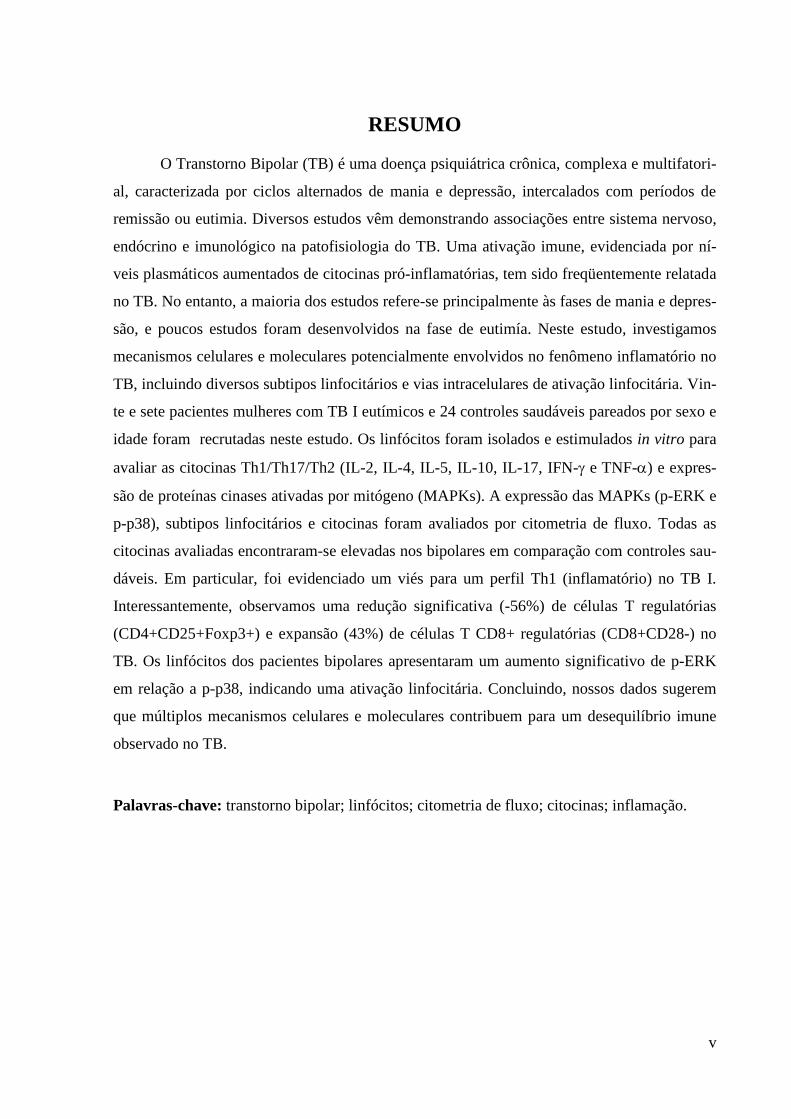
RESUMO
O Transtorno Bipolar (TB) é uma doença psiquiátrica crônica, complexa e multifatori-
al, caracterizada por ciclos alternados de mania e depressão, intercalados com períodos de
remissão ou eutimia. Diversos estudos vêm demonstrando associações entre sistema nervoso,
endócrino e imunológico na patofisiologia do TB. Uma ativação imune, evidenciada por ní-
veis plasmáticos aumentados de citocinas pró-inflamatórias, tem sido freqüentemente relatada
no TB. No entanto, a maioria dos estudos refere-se principalmente às fases de mania e depres-
são, e poucos estudos foram desenvolvidos na fase de eutimía. Neste estudo, investigamos
mecanismos celulares e moleculares potencialmente envolvidos no fenômeno inflamatório no
TB, incluindo diversos subtipos linfocitários e vias intracelulares de ativação linfocitária. Vin-
te e sete pacientes mulheres com TB I eutímicos e 24 controles saudáveis pareados por sexo e
idade foram recrutadas neste estudo. Os linfócitos foram isolados e estimulados in vitro para
avaliar as citocinas Th1/Th17/Th2 (IL-2, IL-4, IL-5, IL-10, IL-17, IFN- e TNF-) e expres-
são de proteínas cinases ativadas por mitógeno (MAPKs). A expressão das MAPKs (p-ERK e
p-p38), subtipos linfocitários e citocinas foram avaliados por citometria de fluxo. Todas as
citocinas avaliadas encontraram-se elevadas nos bipolares em comparação com controles sau-
dáveis. Em particular, foi evidenciado um viés para um perfil Th1 (inflamatório) no TB I.
Interessantemente, observamos uma redução significativa (-56%) de células T regulatórias
(CD4+CD25+Foxp3+) e expansão (43%) de células T CD8+ regulatórias (CD8+CD28-) no
TB. Os linfócitos dos pacientes bipolares apresentaram um aumento significativo de p-ERK
em relação a p-p38, indicando uma ativação linfocitária. Concluindo, nossos dados sugerem
que múltiplos mecanismos celulares e moleculares contribuem para um desequilíbrio imune
observado no TB.
Palavras-chave: transtorno bipolar; linfócitos; citometria de fluxo; citocinas; inflamação.
vi
ABSTRACT
Bipolar Disorder (BD) is a complex, multifactorial and chronic psychiatic illness. It is
characterized by alternating cycles of mania and depression, with periods of remission or
euthymia. Several studies have suggested a direct interaction between nervous, endocrine and
immune systems in the pathophysiology of BD. An immune activation, as evidenced by in-
creased plasma levels of proinflammatory cytokines, has been frequently reported in BD.
However, the majority of the studies have mainly investigated mania and depression phases,
and very few studies have been developed with euthymic patients. In this study, we investi-
gated cellular and molecular mechanisms potentially involved in the inflammatory process in
BD, including various lymphocytes subtypes and intracelular pathways of lymphocyte activa-
tion. Twenty-seven euthymic female subjects with BD type I and 24 age- and sex-matched
controls were recruited in this study. Lymphocytes were isolated and stimulated in vitro to
assess Th1/Th17/Th2 cytokines (IL-2, IL-4, IL-5, IL-10, IL-17, IFN- and TNF-) and ex-
pression of mitogen-activated protein kinases (MAPKs). The expression of MAPKs (p-oERK
and p38), lymphocyte subtypes and cytokines were assessed by flow cytometry. All cytokines
assessed were found elevated in bipolar disorder compared with healthy controls. In particu-
lar, it was evidenced a bias to a Th1 inflammatory profile in BD. Interestingly, we observed a
significant reduction (-56%) of regulatory T cells (CD4+CD25+Foxp3+) and expansion
(43%) of CD8+ regulatory T cells (CD8+CD28-) in BD. The lymphocytes of BD patients
showed an significant increase in p-ERK in relation to p-p38, indicating lymphocyte activa-
tion. Our data suggest that multiple molecular and cellular mechanisms contribute to the im-
munological imbalance observed in BD.
Keywords: bipolar disorder; lymphocytes; flow cytometry; cytokines; inflammation

vii
SUMÁRIO
1. CAPÍTULO 1 ............................................................................................................................................. 1
1.1. INTRODUÇÃO ....................................................................................................................................... 2
1.1.1. Descrição Clínica do Transtorno Bipolar .......................................................................................... 2
1.1.2. Fisiopatologia .................................................................................................................................. 3
1.1.3. Alterações imunológicas no Transtorno Bipolar .............................................................................. 5
1.1.3.1. Alterações nas citocinas ..................................................................................................................6
1.1.3.2. Alterações celulares ...............................................................................................................................7
1.1.3.3. Alterações na sinalização celular - MAPKs ............................................................................................8
1.2. OBJETIVOS .......................................................................................................................................... 10
1.2.1. Objetivo Geral ................................................................................................................................ 10
1.2.2. Objetivos Específicos ...................................................................................................................... 10
2. CAPÍTULO 2 ............................................................................................................................................11
2.1. ARTIGO CIENTÍFICO ............................................................................................................................ 12
3. CAPÍTULO 3 ............................................................................................................................................35
3.1. CONSIDERAÇÕES FINAIS ............................................................................................................................ 36
4. REFERÊNCIAS BIBLIOGRÁFICAS ..............................................................................................................38

1
1. CAPÍTULO 1
2
1.1. INTRODUÇÃO
1.1.1. Descrição Clínica do Transtorno Bipolar
O Transtorno Bipolar é um transtorno mental que se caracteriza por ciclos alternados
de mania (extrema elevação do humor) e depressão, intercalados ou não por períodos de re-
missão ou eutimia (Kim, Rapoport et al., 2010; Kupferschmidt e Zakzanis, 2011). A eutimia é
definida como o indivíduo que não está em episódio atual de humor e que não apresenta sin-
tomas significativos de mania ou depressão. Usualmente, a eutimia é do ponto de vista opera-
cional, definida como não preencher critérios para episódio maníaco, hipomaníaco ou depres-
sivo atual. Já a remissão implica no indivíduo manter a eutimia, a ponto de a doença ser con-
siderada sob controle. O TB é classificado de acordo com o Manual Diagnóstico e Estatístico
de Transtornos Mentais, 4ª edição (DSM-IV). Existem duas formas distintas de TB: o tipo I e
tipo II. O TB tipo I (transtorno bipolar clássico) é caracterizado pela ocorrência de um ou
mais episódios maníacos ou episódios mistos. O TB tipo II inclui ao menos um episódio de
hipomania (uma forma menos grave de mania) e para o diagnóstico correto do TB II é exigida
a ausência de episódios maníacos ou mistos. Ambos os pacientes com TB tipo I e tipo II po-
dem apresentar episódios depressivos ao longo do curso do transtorno (Provencher, Hawke et
al., 2011). Os episódios são de diferente gravidade, freqüência e duração, e o episódio misto
ocorre quando o paciente preenche simultaneamente os critérios diagnósticos para episódio
maníaco e episódio depressivo (Benazzi, 2007). A incidência do TB é de cerca de 1 a 5 % na
população mundial (Vermani, Marcus et al., 2011). A prevalência do TB tipo I é estimada na
faixa de 1%, e não há diferenças entre o sexo masculino e feminino. Já o TB tipo II é mais
prevalente no sexo feminino, afetando entre 0,5- 3% (Bauer e Pfennig, 2005; Kessler,
Merikangas et al., 2007).
Os indivíduos com TB possuem um risco estimado de suicídio em 15%, e este risco
aumenta consideravelmente durante a fase depressiva da doença (Baldassano, Hosey et al.,
2011). Tal transtorno, crônico e em boa parte incapacitante, é altamente relevante devido ao
prejuízo associado a esta condição, não somente aos pacientes, mas também aos familiares e
cuidadores. Além dos sintomas de depressão, mania (ou hipomania) e o risco de suicídio, os
pacientes com TB enfrentam múltiplas comorbidades psiquiátricas e físicas, como o transtor-
no de ansiedade, altas taxas de suicídio, abuso de substâncias, obesidade, diabetes tipo II, do-
3
enças cardiovasculares, entre outras (Degenhardt, Gatz et al., 2011; Provencher, Hawke et al.,
2011).
O diagnóstico do TB inclui alterações de humor, com uma sintomatologia complexa e
de etiologia multifatorial, na qual fatores biológicos e psicossociais interagem em diversos
níveis. Mesmo tendo sido descrito há longo tempo, ainda existem casos em que o TB não é
diagnosticado em tempo hábil para um tratamento adequado ou mesmo de forma correta, sen-
do comum um diagnóstico inicial de depressão unipolar ou esquizofrenia (Gonzalez-Pinto,
Gutierrez et al., 1998). O diagnóstico exato e avaliação do TB são essenciais para a tomada de
decisões clínicas e a determinação de prognóstico e tratamentos. Para muitos pacientes, um
episódio inicial de mania ou depressão evolui para uma doença ao longo da vida. Portanto
para evitar a recorrência, o comportamento suicida e a cronicidade, o tratamento farmacológi-
co em longo prazo é indicado no início do curso da doença.
O tratamento de indivíduos com TB envolve estratégias distintas nas diferentes fases
da doença: mania, depressão e eutimia, e objetiva a manutenção das mudanças de humor e a
redução da freqüência das passagens entre as crises de mania e os estados depressivos. Uma
variedade de medicamento são atualmente aprovadas pelo Food and Drug Administration
(FDA), a agência americana responsável pelo controle de medicamentos e alimentos, incluin-
do o lítio, carbamazepina e ácido valpróico, além de anticonvulsivantes e antipsicóticos atípi-
cos (Jamrozinski, Gruber et al., 2009; Offord, 2011). Para o tratamento de mania, é utilizado o
lítio, carbamazepina e clorpromazina, juntamente com os antipsicóticos atípicos. Para a fase
depressiva da doença, foram aprovados apenas a combinação olanzapina-fluoxetina e a queti-
apina (Sanches e Soares, 2011). Existe considerável controvérsia sobre o uso de antidepressi-
vos em pacientes bipolares, não apenas pelo aumento no risco de indução de mania, mas tam-
bém devido à sua eficácia questionável. Os estabilizadores de humor são compostos utilizados
muitas vezes com antidepressivos para manter a eutimia em pacientes e evitar um episódio de
mania ou depressão.
1.1.2. Fisiopatologia
O TB é uma doença complexa, multifatorial, e diversos modelos etiológicos tentam
explicar o surgimento e a manifestação dos sintomas característicos desta doença. Embora o
conhecimento da patofisiologia do TB seja ainda bastante limitado, existem evidências de que

4
múltiplos sistemas biológicos estão envolvidos, especialmente o sistema nervoso central, sis-
tema endócrino e sistema imunológico (Kupka, Breunis et al., 2002; Knijff, Breunis et al.,
2006). Com o intuito de manter o equilíbrio homeostático do organismo, componentes do
sistema imune interagem com o sistema neuroendócrino atuando em vias específicas envolvi-
dos diretamente com o humor, energia e atividade motora, podendo resultar em alterações
comportamentais. Independente de sua etiologia, as reações inflamatórias crônicas observadas
em indivíduos com transtornos de humor (depressão maior e transtorno bipolar) podem resul-
tar em prejuízos biológicos e contribuir o para o agravamento da doença e manifestação de
morbidades médicas associadas ao TB.
Por mais de três décadas, as bases biológicas dos transtornos de humor têm sido expli-
cadas por meio da hipótese monoaminérgica. Essa teoria propõe que os sintomas psiquiátricos
sejam conseqüência de uma menor disponibilidade de aminas biogênicas cerebrais, em parti-
cular de serotonina, noradrenalina e/ou dopamina (Capuron, Lucile et al., 2011). Tal preposi-
ção é reforçada pelo conhecimento do mecanismo de ação dos antidepressivos, que se baseia,
principalmente, no aumento da disponibilidade desses neurotransmissores na fenda sináptica,
seja pela inibição (seletiva ou não) de suas recaptações, seja pela inibição da enzima respon-
sável por suas degradações (inibidores da monoaminoxidase).
Neurotransmissores, hormônios e citocinas atuam através do eixo hipotálamo-
pituitária-adrenal (HPA), formando um circuito de regulação que mantém a homeostase do
organismo. Em pacientes com TB, o funcionamento do eixo HPA e do sistema imunológico
parece estar comprometido (Watson, Gallagher et al., 2004; Daban, Vieta et al., 2005). Entre
os marcadores neurobiológicos que estão relacionados à patofisiologia do TB, as citocinas
inflamatórias e as neurotrofinas como o BDNF (Brain-Derived Neurotrophic Factor) desta-
cam-se como moléculas chaves envolvidas em potenciais mecanismos de neurodegeneração e
comprometimento cognitivo (Kauer-Sant'anna, Kapczinski et al., 2009).
Estudos relatam que fatores que influenciam negativamente o curso do TB, como por
exemplo, estresse e o trauma, estão associados com uma diminuição nos níveis séricos de
BDNF (Kauer-Sant'anna, Kapczinski et al., 2009; Shalev, Lerer et al., 2009). Além disso,
fatores genéticos tais como o polimorfismo no gene Val66Met, que altera os níveis de expres-
são de BDNF (Fan e Sklar, 2008; Vinberg, Trajkovska et al., 2009) e genes relacionados ao
sistema de neurotransmissores, como a serotonina (SLC6A4 e TPH2), dopamina (DRD4 e
SLC6A3) e glutamato (DAOA e DTNBP1) tem sido relacionados com a patofisiologia do TB

5
(Serretti, Lattuada et al., 2000). Por ser uma doença multifatorial, diversos estudos em áreas
distintas têm sido conduzidos, com o objetivo de compreender quais mecanismos biológicos
estão envolvidos na patogênese deste transtorno.
1.1.3. Alterações imunológicas no Transtorno Bipolar
O TB é o menos estudado imunologicamente dentre os principais tipos de transtornos
de humor. A dificuldade de estudá-lo reside no fato da bipolaridade estar associada com alte-
rações imunes diferenciais ao longo dos ciclos de mania e depressão (Abeer, El-Sayed et al.,
2006). Existem algumas evidências sugerindo de que o sistema imune, em interação direta
com o sistema nervoso central, possui papel importante na patofisiologia do TB (Padmos,
Bekris et al., 2004; Ortiz-Dominguez, Hernandez et al., 2007). Citocinas podem exercer pro-
fundos efeitos no SNC e no sistema endócrino alterando o metabolismo central das monoami-
nas, que são moléculas sintetizadas no cérebro a partir de seus aminoácidos precursores tripto-
fano e tirosina (Raison, Capuron et al., 2006; Brietzke, Stertz et al., 2009). As modificações
comportamentais induzidas pelas citocinas estão associadas a alterações no metabolismo da
serotonina, norepinefrina e dopamina em regiões do cérebro essenciais para regulação das
emoções e da função psicomotora. Marcadores inflamatórios como IL-1, IL-6 e INF-alfa po-
dem alterar vias enzimáticas associadas com o metabolismo das monoaminas, como a I-
DO (indolamina-2,3-dioxigenase). Quando ativada, a IDO é capaz de metabolizar o triptofano
em quinurenina, resultando na diminuição da síntese de serotonina. Curiosamente, pacientes
infectados pelo vírus da hepatite C submetidos a terapia com INF-alfa apresentaram uma di-
minuição nos níveis de triptofano e aumento de quinurenina, concomitantes com o desenvol-
vimento de sintomas depressivos (Raison, Capuron et al., 2006). Citocinas podem ainda indu-
zir a liberação de glutamato por astrócitos e reduzir a expressão de transportadores de gluta-
mato, reduzindo a recaptação glutamatérgica. O glutamato liberado por astrócitos tem acesso
preferencialmente a receptores extrasinápticos NMDA (N-metil D-Aspartato) que irão reduzir
a expressão de BDNF e suporte neurotrófico, e consequentemente aumentar a susceptibilidade
neuronal ao estresse oxidativo (Miller, Maletic et al., 2009).
O TB é acompanhado de múltiplos sinais de ativação e alterações do sistema imunoló-
gico (Kupka, Breunis et al., 2002), variando de acordo com a fase em que o paciente se en-
contra (mania, depressão ou eutimia) (Ortiz-Dominguez, Hernandez et al., 2007). No entanto,

6
a grande maioria dos estudos foca em alterações imunes presentes nas fases de mania e de-
pressão, e poucos estudos associam estas alterações a pacientes eutímicos.
1.1.3.1. Alterações nas citocinas
Nos transtornos de humor de uma forma geral é observado um aumento de citocinas
pró-inflamatórias plasmáticas e hiperatividade de linfócitos do tipo Th1 (Brietzke e
Kapczinski, 2008; Kim, Rapoport et al.). Em indivíduos bipolares, alguns estudos sugerem
que tanto os episódios maníacos quanto depressivos podem estar associados a um perfil pró-
inflamatório. Porém, alterações de caráter imunológico em pacientes eutímicos também são
encontradas (Rapaport, 1994).
Estudos recentes têm sugerido aumento da resposta inflamatória na patogênese do TB.
As células do sistema imune secretam uma variedade de citocinas responsáveis por promover
reações inflamatórias e tendem a estimular ou ativar células imunocompetentes. Com base na
produção de citocinas, as células T CD4+ naive podem diferenciar tradicionalmente em Th1,
Th2 e Th17 (Akdis et al, 2011; Bettelli et al, 2008). Sabe-se que a resposta linfocitária do tipo
Th1 (pró-inflamatória) é responsável pela produção de interleucina-2 (IL-2), IL-6, fator de
necrose tumoral- (TNF-) e interferon-γ (IFN- γ), enquanto que as células Th2 (anti-
inflamatória) secretam as citocinas IL-4, IL-5, IL-10 e IL-13 (Raison, Capuron et al., 2006).
As células Th17 protegem contra bactérias e fungos através da ativação de macrófagos via
citocina pró-inflamatória IL-17. Em um estudo que avaliou os níveis de IL-17 em indivíduos
bipolares e controles saudáveis, não foi observada diferença estatística nos níveis de IL-17
entre os grupos.
O aumento dos níveis de citocinas pró-inflamatórias tais como IL-6, IFN-γ (Kim,
Myint et al., 2004), TNF-α (O'brien, Scully et al., 2006; Kim, Rapoport et al.), IL-2 (Brietzke,
Stertz et al., 2009) e receptor solúvel IL-6 (sIL-6R) (Guloksuz, Aktas Cetin et al.) foram rela-
tados em indivíduos durante episódios maníacos. Da mesma forma, os níveis plasmáticos do
receptor solúvel TNF-α (sTNF-RI) estão elevados em pacientes bipolares em comparação
com controles saudáveis (Hope, Melle et al., 2009). Em contrapartida, alguns estudos têm
proposto uma diminuição dos níveis de IL-1 e IL-2 em pacientes na fase maníaca da doença
(Ortiz-Dominguez, Hernandez et al., 2007). Além disso, estudos indicam um aumento signifi-
cativo dos níveis de IL-4 de pacientes em episódio de mania (Kim, Myint et al., 2004; Ortiz-
7
Dominguez, Hernandez et al., 2007), indicando possivelmente uma resposta homeostática na
inibição da resposta inflamatória crônica nesta doença.
Os episódios depressivos têm sido associados com um aumento nos níveis de IL-6
(O'brien, Scully et al., 2006; Ortiz-Dominguez, Hernandez et al., 2007) e TNF-α (O'brien,
Scully et al., 2006; Ortiz-Dominguez, Hernandez et al., 2007). Além disso, os níveis séricos
do sIL-2R foram encontrados elevados em pacientes bipolares quando comparados com indi-
víduos sem transtorno de humor, e o mesmo ocorreu quando comparando indivíduos manía-
cos com depressivos (Breunis, Kupka et al., 2003).
O equilíbrio entre citocinas pró- e anti-inflamatórias é essencial para manter a homeos-
tase no sistema. Dados existentes na literatura afirmam que o desequilíbrio entre Th1/Th2
pode estar associado com a patofisiologia do TB, mais precisamente com a fase maníaca da
doença (Kim, Myint et al., 2004). Ainda, este desequilíbrio poderia ser consequente de altera-
ções na proporção dos principais subtipos celulares que secretam estas citocinas. Deve ser
ressaltado, no entanto, que os estudos anteriores focaram suas pesquisas com amostras de
plasma/soro que impossibilitam a identificação dos tipos celulares produtores das citocinas.
1.1.3.2. Alterações celulares
Poucos estudos investigaram as alterações celulares no TB. Em estudos realizados por
Breunis e colaboradores (2003), a porcentagem de células T ativadas (CD3+HLADR+,
CD3+CD25+ e CD3+CD71+) e células B (CD19+CD20+) em pacientes bipolares foram mai-
ores em comparação com indivíduos controles (Breunis, Kupka et al., 2003), apoiando o esta-
do inflamatório observado nesta doença. Foi especulado que o estado de ativação celular e
perfil inflamatório observados no TB poderiam ser consequentes de uma falta de regulação
imunológica adequada. Dentre as várias vias de regulação, destacam-se as células T regulató-
rias naturais (Tregs). As Tregs são produzidas no timo, apresentam um fenótipo
CD4+CD25+Foxp3+ e constituem um subgrupo especializado de células T com função im-
portante na supressão de respostas autoimunes, alergias e indução de câncer (Sakaguchi,
Yamaguchi et al., 2008). Dessa forma, as células Tregs são indispensáveis para manter o e-
quilíbrio homeostático do organismo, e disfunções neste subtipo podem acarretar em desor-
dens imunológicas, como as doenças crônico-inflamatórias. Contudo, um estudo recente não
verificou alteração nestas células em pacientes com TB I quando comparados com controles

8
saudáveis (Drexhage, Hoogenboezem et al., 2011). Estudos recentes demonstram que a popu-
lação de células Tregs em indivíduos com depressão maior está significativamente diminuída
quando comparados com controles saudáveis (Li, Xiao et al., 2010; Chen, Jiang et al., 2011).
Como acontece com qualquer sistema do organismo, o sistema imunológico exibe
mudanças características com o avanço da idade. A reestruturação do sistema imune durante o
envelhecimento é referida como imunossenescência, tornando o indivíduo mais susceptível às
inflamações sistêmicas persistentes e patologias associadas com o envelhecimento, como o
diabetes tipo II, doenças cardiovasculares, depressão maior, entre outras. Em comparação com
adultos mais jovens, o sistema imune dos idosos é marcado por uma série de características,
tais como: a redução do número e função de células-tronco hematopoiéticas, involução tímica,
redução de células T naive circulantes, expansão de células T CD8+CD28- (também regulató-
rias), aumento nos níveis de citocinas inflamatórias, incluindo interleucina-6 (IL-6) e TNF-α,
e redução das taxas CD4/CD8 (Deeks, 2011). O envelhecimento tem sido associado com um
aumento nos níveis de citocinas pró-inflamatórias, ativação do eixo HPA (aumento de cortisol
e redução de DHEA) e redução da sensibilidade linfocitária aos glicocorticóides (Bauer,
2008).
Estudos com depressão maior vêm sugerindo uma relação entre transtornos de humor
e um envelhecimento precoce do sistema imune (Heuser, 2002; Brown, Varghese et al., 2004;
Evans, Charney et al., 2005). Por exemplo, foi demonstrado um encurtamento da região telo-
mérica de células mononucleares (linfócitos e monócitos) neste transtorno (Wolkowitz,
Mellon et al., 2011), uma alteração comum em células senescentes ou de idosos. Recentemen-
te um estudo também observou o encurtamento telomérico no TB (Elvsashagen, Vera et al.,
2011). No entanto, os estudos direcionados até o momento à imunossenescência não avalia-
ram características imunes, como por exemplo, subtipos linfocitários associados ao processo
de envelhecimento.
1.1.3.3. Alterações na sinalização celular - MAPKs
As células reconhecem e respondem ao estímulo extracelular através de vias intracelu-
lares específicas, tais como a cascata de sinalização que leva à ativação de MAPKs (mitogen-
activated protein kinases). As MAPKs são uma família de serina-treonina cinases, ativas por

9
fosforilação, responsáveis por coordenar diversas atividades celulares, como proliferação ce-
lular, diferenciação, sobrevivência e apoptose (Raman, Chen et al., 2007; Kim e Choi, 2010).
As três principais MAPKS identificadas são as cinases reguladas por sinal extracelular (ERK
1/2), quinase c-Jun N-terminal (JNK 1/2) e a p38. Todas as vias operam em forma de cascata:
uma MAP cinase cinase cinase (MAP3K) ativa, por fosforilação, uma MAP cinase cinase
(MAP2K) que, por sua vez, fosforila e ativa a MAP cinase (MAPK) (Raman, Chen et al.,
2007; Krishna e Narang, 2008; Furler e Uittenbogaart, 2010; Sosa, Avivar-Valderas et al.,
2011).
As MAPKs podem ser ativadas por uma variedade de diferentes estímulos. A ERK1/2
é ativada preferencialmente em resposta a fatores de crescimento, citocinas, soro, estresse,
entre outros (Krishna e Narang, 2008; Sosa, Avivar-Valderas et al., 2011). A ativação da
ERK1/2 tem sido apontada como um regulador chave em processos de proliferação celular,
diferenciação e sobrevida (Roux e Blenis, 2004; Raman, Chen et al., 2007). Assim, inibidores
da via ERK são considerados fortes candidatos para o desenvolvimento de agentes anti-
câncer. A via de sinalização p38 é ativada por uma ampla variedade de eventos estressores,
tais como citocinas pró-inflamatórias (ex. TNF-α), estresse oxidativo, lipopolissacarídeo
(LPS), radiação UV, entre outros. A ativação de p38 está associada principalmente com pro-
cessos de inflamação, anergia e apoptose (Raman, Chen et al., 2007; Krishna e Narang,
2008). Tendo em vista as respostas celulares de cada MAPK, pode-se considerar que estas
duas vias de ativação, ERK1/2 e p38 possuem atividades opostas, como de proliferação e a-
nergia, respectivamente.

10
1.2. OBJETIVOS
1.2.1. Objetivo Geral
Avaliar marcadores imunológicos celulares e moleculares envolvidos no processo in-
flamatório no TB I.
1.2.2. Objetivos Específicos
Identificar por citometria de fluxo os principais subtipos linfocitários no sangue periférico.
Avaliar a produção linfocitária de citocinas Th1/Th2/Th17 in vitro.
Avaliar a expressão linfocitária intracelular das enzimas MAPK fosforiladas.
Comparar todas as variáveis imunológicas com um grupo controle.

11
2. CAPÍTULO 2

12
2.1. ARTIGO CIENTÍFICO
13
Submetido ao Biological Psychiatry
REDUCED REGULATORY T CELLS ARE ASSOCIATED WITH
HIGHER LEVELS OF TH1/TH17 CYTOKINES AND ACTIVATED
MAPK IN TYPE 1 BIPOLAR DISORDER
Carine Hartmann do Prado1, Lucas Bortolotto Rizzo, Andréa Wieck, Rodrigo Pestana
Lopes, Antonio L. Teixeira, Rodrigo Grassi-Oliveira and Moisés Evandro Bauer
Laboratory of Immunosenescence, Institute of Biomedical Research (CHP, LBR, AW,
MEB), Pontifical Catholic University of the Rio Grande do Sul (PUCRS), Porto Alegre, Bra-
zil; Faculty of Biosciences (MEB), PUCRS, Porto Alegre, Brazil; Faculty of Psychology
(RG-O), PUCRS, Porto Alegre, Brazil; BD Biosciences (RPL), São Paulo, Brazil; and De-
partment of Internal Medicine (ALT), School of Medicine, UFMG, Belo Horizonte, Brazil.
Correspondent author: Moisés E. Bauer, PhD, Instituto de Pesquisas Biomédicas,
Hospital São Lucas da PUCRS, Av. Ipiranga 6690, 2º andar. P.O. Box 1429. Porto Alegre, RS
90.610-000, Brazil. Email: [email protected]
1 Carine Hartmann do Prado and Lucas Bortolotto Rizzo contributed equally to this work.

14
ABSTRACT
Background: Bipolar Disorder (BD) has been associated with an immunologic imbal-
ance shown by increased peripheral inflammatory markers. The underlying mechanisms of
this phenomenon may include changes in circulating cells and differential activation of mito-
gen-activated protein kinases (MAPKs). Methods: Twenty-seven euthymic female subjects
with BD type I and 24 age- and sex-matched controls were recruited in this study. Lympho-
cytes were isolated and stimulated in vitro to assess Th1/Th17/Th2 cytokines (IL-2, IL-4, IL-
5, IL-10, IL-17, IFN- and TNF-) and MAPK phosphorylation. The expression of phospho-
MAPKs, a large panel of lymphocyte subsets and cytokines were assessed by multi-color flow
cytometry. Results: BD patients had reduced proportions of natural T regulatory cells
(CD4+CD25+FoxP3+) (p<0.01) in parallel to higher cytokine production (all p<0.01) than
healthy controls. In particular, BD was associated with a strong bias to Th1 rather than Th2
profile. There was an expansion of senescence-associated cells (CD8+CD28-) in BD
(p<0.0001). T cells of BD patients showed an increased p-ERK signaling in relation to p-p38
(p<0.0001), indicating lymphocyte activation. Conclusions: Our data suggest that multiple
molecular and cellular mechanisms may contribute to the immunologic imbalance observed in
BD. In addition, our data concur to an early senescence process in these patients.
15
Introduction
There is increasing evidence suggesting that the immune and inflammatory systems
play important roles in the pathogenesis of Bipolar Disorder (BD) (1). Several studies have
investigated the potential role of cytokines in psychiatric disorders, based on their important
actions in modulating metabolism of central neurotransmitters, hypothalamic-pituitary-
adrenal (HPA) axis and neurotrophic support (2). It has been observed increased plasma lev-
els of pro-inflammatory cytokines during BD manic episodes, including higher levels of IL-6,
IFN-, TNF-, IL-2 and serum soluble IL-6 receptor (sIL-6R) (3-7). We have recently ob-
served that BD patients in mania had higher sTNFR1 levels than euthymic BD patients and
controls (4). In addition, some studies also reported elevated IL-4 levels (a Th2 cytokine) in
patients with manic episodes (6, 8). Similar to mania, increased levels of IL-6 and TNF-
were also observed during depressive episodes (6-8). It should be noted that previous studies
assessed inflammatory markers in plasma/serum samples. The analysis of biomarkers in cellu-
lar supernatants has advantage to serum/plasma sampling as it can precise the cellular source
of cytokines. The underlying mechanisms of this immunologic imbalance in BD are largely
unknown and may include changes in circulating lymphocytes and the differential expression
of intracellular signaling cascades.
Changes in circulating leukocytes may contribute to the immunologic imbalance ob-
served in BD. The analyses of lymphocyte subsets, particularly T, B and NK cells are very
scarce in BD though. It has been observed increased percentage of activated T cells (i.e.
CD3+MHCII+; CD3+CD25+ and CD3+CD71+) and B cells (CD19+CD20+) in BD com-
pared to healthy controls (9). This activation state could be theoretically due to a lack of pe-
ripheral regulatory cells. A recent study did not observe changes in regulatory T cells (Tregs)
in BD patients as compared to controls (10). Tregs (CD4+CD25+Foxp3+) play key roles in
suppressing excessive or misguided immune responses that can be harmful to the host. In par-
ticular, they are responsible for turning off immune responses against self-antigens in auto-
immune diseases, allergies or commensal microbes in certain inflammatory diseases (11). To
date, the roles of CD8+ regulatory T cells (CD8+CD28- and CD8+CD103+) in BD are largely
unknown.
The immunologic imbalance observed in BD could be also explained by the differen-
tial expression of intracellular signaling cascades. Mitogen-activated protein kinases
16
(MAPKs) are important intracellular signal transduction systems and participate in a series of
physiological and pathological processes, including cell growth, differentiation and apopto-
sis(12, 13). Three major MAPK cascades are known, including the extracellular signal-
regulated protein kinase (ERK), c-jun amino-terminal protein kinase/stress-activated protein
kinase (JNK/SAPK) and p-38. Of note, ERK is associated with cell proliferation, differentia-
tion and survival, while p38 is often linked to inflammation, anergy, apoptosis and
immunoregulatory actions (14, 15). Interestingly, these two enzymes have reciprocal antago-
nistic actions. There is no information regarding the expression of intracellular signaling
transduction systems in BD.
Here, we assessed the cellular and molecular mechanisms that may influence the in-
flammatory state observed in BD. Specifically, we determined (a) Th1/Th2/Th17 cytokines in
supernatants and addressed the role of (b) regulatory T cells and various lymphocyte subsets,
as well as (c) the intracellular expression of different activated MAPKs (p38 and ERK) in
euthymic type 1 BD patients and healthy controls.
Materials and methods
Subjects
Twenty-seven euthymic female subjects with BD type I were recruited at the Psychiat-
ric Clinic of the Presidente Vargas Hospital, Porto Alegre, Brazil. Age- and sex-matched
healthy controls also took part in this study. All subjects provided their written informed con-
sent before inclusion in the study approved by the Ethical Committee of the institution. The
BD type 1 diagnosis was based on clinical interview and confirmed with the Structured Clini-
cal Interview for DMS-IV-Axis I Disorder (SCID-I) administered by an expert and well-
trained psychiatrist. Severity of depressive and maniac symptoms were assessed by the Ham-
ilton Depression Rating Scale (HDRS) and the Young Mania Rating Scale (YMRS), respec-
tively. All patients were euthymic at the time of blood collection, euthymia was defined by
YMRS and HDRS scores < 8 (16). Exclusion criteria to both patients and controls included:
a) Presence of major axis I psychiatric disorder such as psychotic disorder, mood disorder (for
control group), anxiety disorder or substance related disorder according to SCID-I; b) history
of a severe medical illness; c) history of brain injury; d) presence of systemic diseases (includ-
ing hypertension, inflammatory diseases, such as rheumatoid arthritis or infection) or neuro-
17
logical disorder, e) use of any substance that may induce immunological or endocrinological
changes (exception of psychopharmacotherapy for BD patients), or f) do not agree to partici-
pate in the study.
Blood collection and cell isolation
Twenty milliliters of peripheral blood were collected by venipuncture between 12:00
and 14:00 PM and stored in EDTA tubes prior to analyses. Peripheral blood mononuclear
cells (PBMCs) were isolated density gradient under centrifugation for 30 min at 900 g. Cells
were counted by means of microscopy (100 x) and viability always exceeded 95%, as judged
from their ability to exclude Trypan Blue (Sigma). PBMCs were resuspended complete cul-
ture medium (RPMI-1640, supplemented with 0.5% gentamicine, 1% glutamine, 1% hepes,
0.1% fungizone, and 10% fetal calf serum, FCS; all from Sigma) and adjusted to yield a final
concentration of 2x10⁵
cells/well.
Immunophenotyping
A large panel of lymphocyte subpopulations was identified by multi-color flow
cytometry. Briefly, PBMCs were washed in flow cytometry buffer (PBS containing 1% FCS
and 0.01% sodium azide) and treated with Fc Block solution for 20 min. In order to evaluate
specific lymphocyte subsets, cells were stained for 30 min with combinations of the following
monoclonal antibodies: anti-CD3 FITC, anti-CD3PECy5, anti-CD4 PE, anti-CD4 FITC, anti-
CD8 PE, anti-CD19 PE, anti-CD56 FITC, anti-CD28 FITC, anti-CD45RO FITC, anti-CD69
FITC, anti-FOXP3 PECy5, anti-CD103 FITC, anti-CCR7 Cy7, anti-CD45RA FITC (all from
BD Biosciences, San José, CA, USA). Immediately after staining, cells were washed, resus-
pended and analyzed by flow cytometry. A minimum of 20,000 lymphocytes were identified
by size (FSC) and granularity (SSC) and acquired with a FACS Canto II flow cytometer (BD
Biosciences). The instrument has been checked for sensitivity and overall acquisition. Data
were analyzed using the Flowjo 7.2.5 software (Tree Star Inc., Ashland, Or, USA).
Quantification of cytokines
To determine cytokine production, PBMCs were cultured (1.5x10⁵
cells) in RPMI me-
dium with 10% FCS (Sigma-Aldrich) and 1% phytohemagglutinin (PHA, from Invitrogen,
Carlsbad, CA, USA), for 72h at 37°C and in a 5% CO2 atmosphere. The supernatants were

18
collected and stored at -80°C for later analysis. The reduction variation, samples were thawed
in the same day and processed together. Multiple soluble cytokines (IL-2, IL-10, IL-4, IL-5,
IFN-, TNF- and IL-17) were simultaneously measured by flow cytometry using the
Cytometric Bead Array (CBA) Human Th1/Th2/Th17 Kit (BD Biosciences). Acquisition was
performed with a FACSCanto II flow cytometer (BD Biosciences). The instrument has been
checked for sensitivity and overall performance with Cytometer Setup & Tracking beads (BD
Biosciences) prior to data acquisition. Quantitative results were generated using FCAP Array
v1.0.1 software (Soft Flow Inc., Pecs, Hungary).
Analysis of intracellular activated MAPKs in lymphocytes
Activated MAPKs in lymphocytes were assessed by flow cytometric evaluation of in-
tracellular phospho-p38 and phospho-ERK expression in CD3+CD4+ and CD3+CD8+ cells.
Isolated PBMCs were cultured in RPMI medium with 10% FCS and stimulated with 40nM
phorbol 12-myristate 13-acetate (PMA) and 1M ionomycin (IONO, all from Sigma-Aldrich)
for 15 min at 37°C and in a 5% CO2 atmosphere. Cells were harvested and immediately fixed
and stored frozen (-80 °C) in Cytofix solution (BD Biosciences) for later analysis. All sam-
ples were thawed in the same day and processed together to reduce variation. Cells were
permeabilised on ice for 30 min with Phosflow Perm Buffer III (BD Biosciences). Cells were
washed (600g, 6 min), stained for 60 min at room temperature, washed (600g, 6 min) and
resuspended in final concentration of 4.5 x 10⁵ cells / 200 L), all in Pharmingen staining
buffer (BD Biosciences). The 4-color immunofluorescent staining procedure was performed
combining the following monoclonal antibodies: anti-CD3 PerCP, anti-CD4 FITC and anti-
CD8 PE with anti-ERK1/2 Alexa Fluor 647 (pT202/pY204) or anti-p38 Alexa Fluor 647
(pT180/pY182, all from BD Biosciences). Lyophilized human control cells (BD Biosciences)
were used as positive and negative controls due to the known presence of mitogen-activated
(upregulation of phosphorylated MAPKs) or non-activated (basal levels of phospho-MAPKs)
T cells in each control, respectively. A minimum of 20,000 lymphocytes were identified by
size (FSC) and granularity (SSC) and acquired with a FACS Canto II flow cytometer (BD
Biosciences). The instrument has been checked for sensitivity and overall performance with
Cytometer Setup & Tracking beads (BD Biosciences) prior to data acquisition. Data were
analyzed using the Flowjo 7.2.5 software (Tree Star Inc., Ashland, Or, USA).
19
Statistical analysis
All variables were tested for homogeneity of variances and normality of distribution
by means of the Levene and Kolmogorov-Smirnov tests, respectively. Continuous variables
differences between groups were analyzed by Student t-test or Mann-Whitney U test when
appropriate. Statistical interactions between categorical variables and group were compared
by means of the chi-square (2) test. Statistical analyses were performed using the Statistical
Package for Social Sciences, SPSS Statistics 17.0 software (SPSS Inc., Chicago, IL, USA).
The significance level was set at = 0.05 (two-tailed).
Results
Characteristics of the studied populations
Demographic and clinical characteristics of the samples are summarized in Table 1.
Both groups were homogenous regarding age, gender, ethnicity, BMI and smoking habits.
Lymphocyte subsets
We screened a large panel of circulating lymphocyte subpopulations by multicolor
flow cytometry, including activated, regulatory and immunosenescence markers (Table 2).
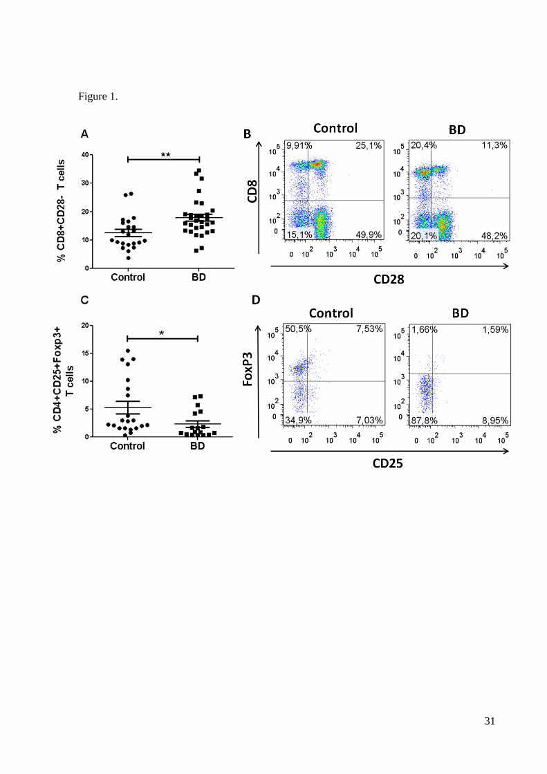
The studied groups were homogenous regarding most lymphocyte markers. However, BD
patients had altered proportions of regulatory T cells (Figure 1). In particular, lower percent-
ages of natural Treg cells (CD4+CD25+FoxP3+) were significantly decreased in BD patients,
as shown in Figures 1C and D (p<0.01). In contrast, BD patients had higher frequencies of
CD8+CD28- T cells as compared to controls (p<0.0001). With respect to possible effects of
pharmacotherapy, no significant associations were found with the immunological measures
(all p=N.S.).
Cytokine production
Multiple Th1/Th2/Th17 cytokines (IL-2, IL-4, IL-5, IL-10, IL-17, IFN- and TNF-)
were assessed in culture supernatants by CBAs. Table 3 shows the cytokine profiles following
20
polyclonal T-cell stimulation. All cytokines were found significantly increased in BD when
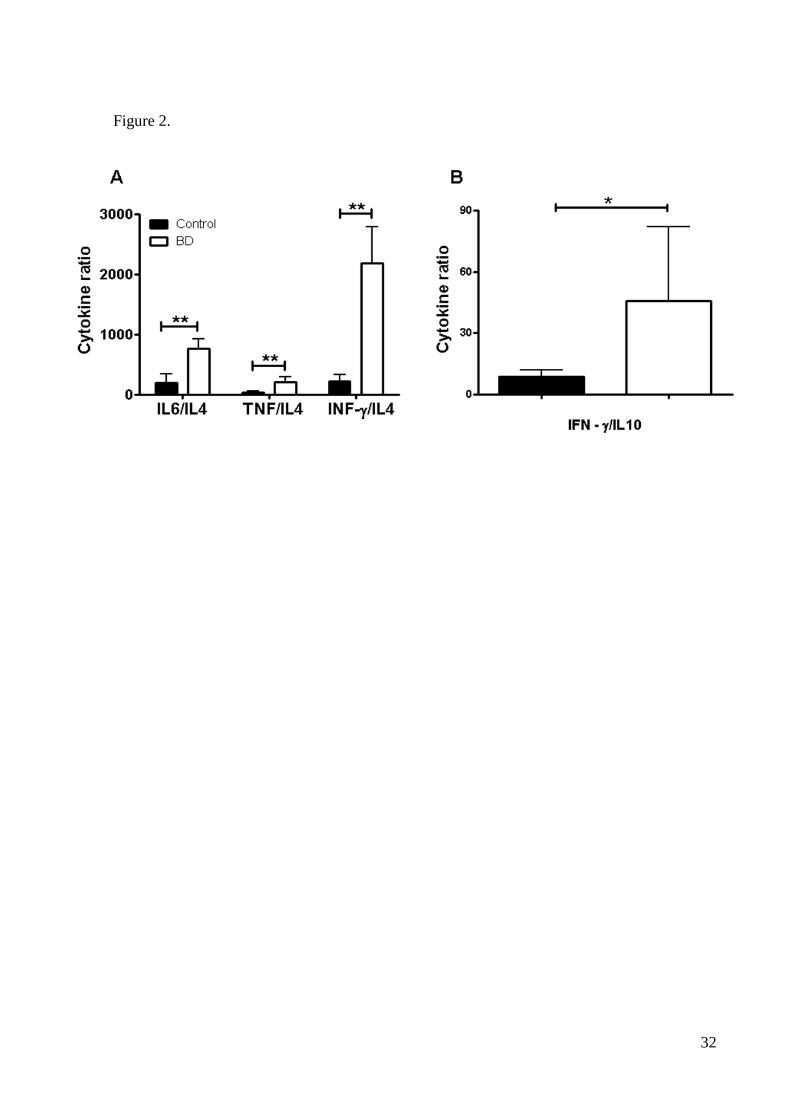
compared with healthy controls (all p<0.01). To further investigate the cytokine profiles we
compared the cytokine ratios between the two groups. BD patients showed higher IL-6/IL-4,
TNF-/IL-4, IFN-/IL-4 and IFN-/IL-10 ratios compared to controls (Figure 2), suggesting a
strong bias to Th1 rather than Th2 profile. There were no statistical differences regarding the
remaining cytokine ratios (all p=N.S.).
Analysis of intracellular phospho-MAPKs in peripheral lymphocytes
We have also analyzed the expression of phosphorylated p-38 and p-ERK MAPKs in
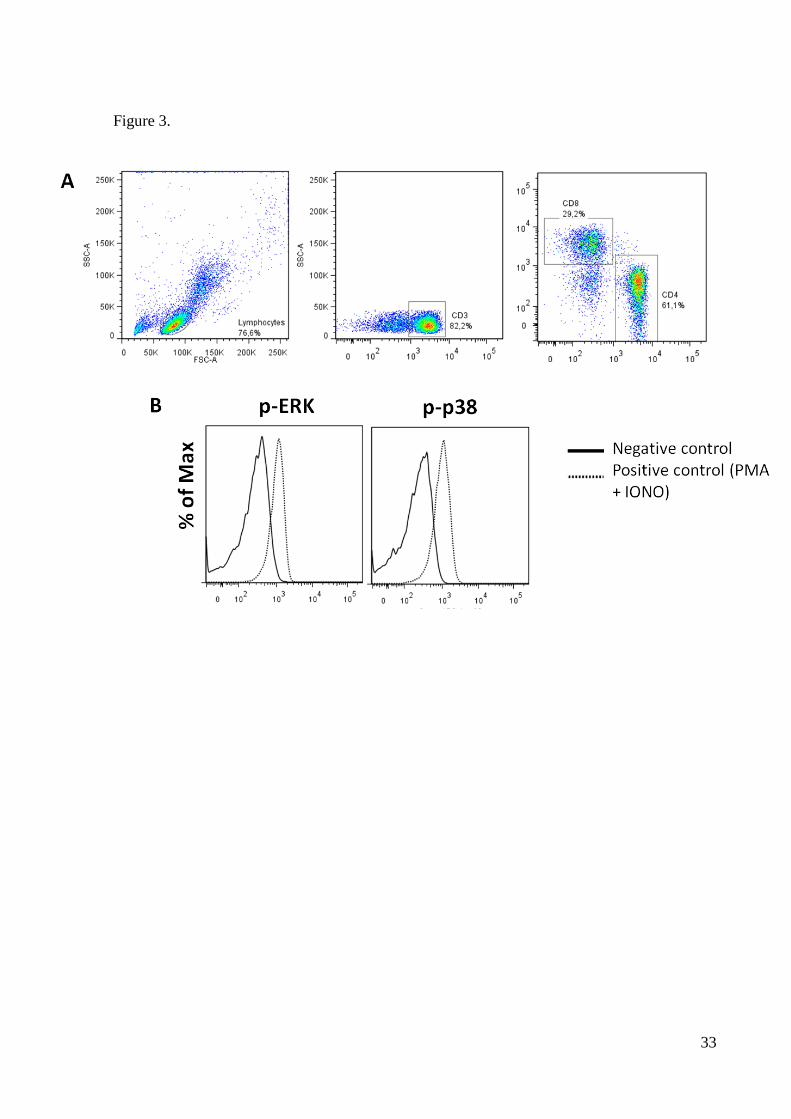
lymphocytes following stimulation with PMA and IONO. Figure 3 shows the unstimulated
(solid line) and activated (dotted line) profiles of a representative sample. The expression of
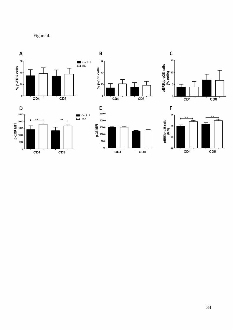
p-ERK MAPK in T CD8+ and CD4+ cells, as estimated by the mean fluorescence intensity
(MFI) was found increased in patients in comparison with controls (Figure 4D). This was not
observed for the p-p38 expression (Figure 4E). Since p38 and ERK have opposite cellular
effects, we also analyzed the p-ERK/p-p38 ratios in T cells (Figures 4C,F). As shown in Fig-
ure 4F, the p-ERK/p-p38 ratio was found increased in patients compared to controls. There
were no significant differences in the percentages of T cells expressing p-ERK or p-p38 (Fig-
ures 5A,B and C).
Discussion
To our knowledge, this is the first study addressing multiple cellular and molecular
mechanisms that may influence the inflammatory state observed in BD. Briefly, patients had
lower proportions of natural regulatory T cells (CD4+CD25+FoxP3+) in parallel to higher
cytokine production than healthy controls (with strong bias to Th1). We also observed an in-
creased p-ERK signaling in relation to p-p38 in peripheral T-cell subsets of BD patients, indi-
cating lymphocyte activation.
Data presented here are in accordance to previous studies suggesting an im-
mune/inflammatory imbalance in BD. PBMCs of BD patients produced significantly higher
amounts of IL-2, IL-4, IL-5, IL-10, IL-17, IFN- and TNF- than controls in vitro. This
method has advantages to serum/plasma sampling because it minimizes the effects of medica-
tions in serum, as cells are repeatedly washed prior to culture, and it can precise the cell

21
source of cytokines. In order to have a better understanding of what could represent these high
rates of cytokines, we analyzed the pro-inflammatory/anti-inflammatory cytokine ratios. BD
was associated with a strong bias to Th1 (pro-inflammatory) rather than Th2 profile. There is
scarce immune data regarding euthymic BD patients (3, 17, 18). Previous studies have ob-
served increased plasma levels of pro-inflammatory cytokines during manic (3-7) or depres-
sive episodes (6-8), suggesting this immune/inflammatory imbalance could be a trait phenom-
enon in BD and may contribute to its pathophysiology. Indeed, we have recently shown that
plasma TNF-α and sTNFR2 levels were negatively correlated to cognitive (executive) func-
tions in BD type I euthymic patients (19).
Specific changes in circulating lymphocytes may contribute to the im-
mune/inflammatory imbalance observed in BD. This study investigated for the first time in
BD a large panel of lymphocyte subpopulations, including activated and regulatory cells as
well as immunosenescence markers. BD patients had significantly lower proportions of natu-
ral Tregs than controls (-55.91%), potentially implicated with the pro-inflammatory status.
Indeed, the lack of regulatory T cells has been observed in several chronic inflammatory con-
ditions (11). The study of the Tregs cells in mood disorders is extremely scarce. Our study is
in accordance with recent studies reporting low proportions of Tregs in major depression (20,
21). However, a recent study did not observe changes in Tregs in BD patients as compared to
controls (10). Future studies are necessary to address the functional activity of these cells and
to give further support to the data presented here.
BD patients had also significantly higher frequencies (42.9%) of CD8+CD28- regula-
tory T cells, which are commonly associated with immunosenescence (22-25). The co-
stimulatory molecule CD28 is necessary to initiate T-cell mediated immune responses. Aging
has been associated with progressive loss of CD28 marker and increased CD8+CD28- T cell
populations (22-25). The CD8+CD28- T cells have regulatory functions and are defined as
highly expanded, terminally differentiated, or senescent memory T lymphocytes. These cells
have undergone many rounds of cell divisions presumably as a result of lifetime exposure to
common persistent antigens. Indeed, CD8+CD28- cells accumulate during persistent viral
infections (e.g. HIV, CMV, EBV and HTLV) (26-30) and autoimmune diseases (e.g. lupus,
rheumatoid arthritis and Crohn’s disease) (31-33). It has been shown a dramatic loss of te-
lomerase activity in CD8+CD28- cells that limits their proliferative capacity (23). Recently,
Elvsashagen and colleagues (2011) found a reduction in telomere length in BD type 2 pa-

22
tients, representing 13 years of accelerated aging and correlated with lifetime number of de-
pressive episodes (34). Taken together these data point to a possible role of early aging of
immune system in the pathophysiology of mood disorders, notably BD. These results also
concur to the hypothesis of increased allostatic load in BD (35), suggesting that cumulative
stress- or episode-induced changes in brain regions involved with the emotional circuitry
would render BD patients more vulnerable to subsequent environmental stressors.
The immune/inflammatory imbalance observed in BD could be also explained by the
differential expression of intracellular signaling cascades. From many intracellular signaling
cascades potentially involved with a pro-inflammatory status, we have studied the MAPKs
that are crucially involved with lymphocyte activation and proliferation (13). Of note, we
evaluated MAPKs ERK and p38 that are implicated with lymphocyte activation/proliferation
(36) and cellular anergy (37), respectively. We specifically targeted the expression of phos-
phorylated (activated) ERK and p38 in major T-cell subsets, CD3+CD4+ and CD3+CD8+
cells. We report for the first time an increased p-ERK signaling in relation to p-p38 in BD as
compared to controls, potentially underlying activation of T lymphocytes and contributing to
immune/inflammatory imbalance observed in BD. Research is under progress in our laborato-
ries to explore different intracellular signaling cascades involved with cell activation in type I
euthymic BD patients.
There are some limitations in this study to be discussed. One of the major limitations
of our study is that all BD patients were receiving psychotropic drugs (i.e. lithium) that may
modulate immune functions. It should be noted, however, that previous studies indicate that
lithium exhibits potent anti-inflammatory effects (38, 39). These findings do not mitigate our
data since all cytokines assessed in supernatants were found significantly increased in BD as
compared to healthy controls. Other limitation is the criteria for euthymic state. We adopt a
recognized criterion with one point assessment, but some authors have been suggested to con-
sider at least 4 months without symptoms as a better criterion (40). The point is that our pa-
tients have around 10 years of illness duration, turning very difficult to find those type 1 BD
euthymic patients. This contributed to our relatively small sample, despite the stringed exclu-
sion criteria.
In conclusion, our data suggest that BD patients exhibit an immune/inflammatory im-
balance, associated with reduced proportions of circulating Tregs and expansion of terminally
differentiated or senescent T cells (CD8+CD28-). These data concur to an early

23
immunosenescence process in these patients. MAPKs and other intracellular signaling cas-
cades should be further explored to assess their role to the pathophysiology of BD.
Acknowledgments
We are very grateful to the patients and staff at the Hospital Presidente Vargas. We
would like also to thank Ledo Daruy Filho, MD for helping with psychiatric assessments.
This work was supported by grants from CNPq (MEB, LBR, ALT and RG-O) and CAPES
(CHP, AW).
Financial disclosures
The authors have no conflict of interests to declare.
24
References
1. Brietzke E, Stabellini R, Grassi-Oliveira R, Lafer B (2011): Cytokines in Bipolar Dis-
order: Recent Findings, Deleterious Effects But Promise for Future Therapeutics. CNS Spectr.
2. Miller AH, Maletic V, Raison CL (2009): Inflammation and its discontents: the role of
cytokines in the pathophysiology of major depression. Biol Psychiatry. 65:732-741.
3. Brietzke E, Stertz L, Fernandes BS, Kauer-Sant'anna M, Mascarenhas M, Escosteguy
Vargas A, et al. (2009): Comparison of cytokine levels in depressed, manic and euthymic pa-
tients with bipolar disorder. J Affect Disord. 116:214-217.
4. Barbosa IG, Huguet RB, Mendonca VA, Sousa LP, Neves FS, Bauer ME, et al.
(2011): Increased plasma levels of soluble TNF receptor I in patients with bipolar disorder.
Eur Arch Psychiatry Clin Neurosci. 261:139-143.
5. Kim HW, Rapoport SI, Rao JS (2010): Altered expression of apoptotic factors and
synaptic markers in postmortem brain from bipolar disorder patients. Neurobiol Dis. 37:596-
603.
6. Kim YK, Myint AM, Lee BH, Han CS, Lee SW, Leonard BE, et al. (2004): T-helper
types 1, 2, and 3 cytokine interactions in symptomatic manic patients. Psychiatry Res.
129:267-272.
7. O'Brien SM, Scully P, Scott LV, Dinan TG (2006): Cytokine profiles in bipolar affec-
tive disorder: focus on acutely ill patients. J Affect Disord. 90:263-267.
8. Ortiz-Dominguez A, Hernandez ME, Berlanga C, Gutierrez-Mora D, Moreno J,
Heinze G, et al. (2007): Immune variations in bipolar disorder: phasic differences. Bipolar
Disord. 9:596-602.
9. Breunis MN, Kupka RW, Nolen WA, Suppes T, Denicoff KD, Leverich GS, et al.
(2003): High numbers of circulating activated T cells and raised levels of serum IL-2 receptor
in bipolar disorder. Biol Psychiatry. 53:157-165.
10. Drexhage RC, Hoogenboezem TH, Versnel MA, Berghout A, Nolen WA, Drexhage
HA (2011): The activation of monocyte and T cell networks in patients with bipolar disorder.
Brain Behav Immun. 25:1206-1213.
11. Sakaguchi S, Yamaguchi T, Nomura T, Ono M (2008): Regulatory T cells and im-
mune tolerance. Cell. 133:775-787.
12. Sosa MS, Avivar-Valderas A, Bragado P, Wen HC, Aguirre-Ghiso JA (2011):
ERK1/2 and p38alpha/beta signaling in tumor cell quiescence: opportunities to control
dormant residual disease. Clin Cancer Res. 17:5850-5857.
13. Strniskova M, Barancik M, Ravingerova T (2002): Mitogen-activated protein kinases
and their role in regulation of cellular processes. Gen Physiol Biophys. 21:231-255.
14. Furler RL, Uittenbogaart CH (2010): Signaling through the P38 and ERK pathways: a
common link between HIV replication and the immune response. Immunol Res. 48:99-109.
15. Raman M, Chen W, Cobb MH (2007): Differential regulation and properties of
MAPKs. Oncogene. 26:3100-3112.
16. Clark L, Iversen SD, Goodwin GM (2002): Sustained attention deficit in bipolar dis-
order. Br J Psychiatry. 180:313-319.
25
17. Guloksuz S, Aktas Cetin E, Cetin T, Deniz G, Oral ET, Nutt DJ (2010): Cytokine lev-
els in euthymic bipolar patients. J Affect Disord.
18. Kunz M, Cereser KM, Goi PD, Fries GR, Teixeira AL, Fernandes BS, et al. (2011):
Serum levels of IL-6, IL-10 and TNF-alpha in patients with bipolar disorder and schizophre-
nia: differences in pro- and anti-inflammatory balance. Rev Bras Psiquiatr. 33:268-274.
19. Barbosa IG, Rocha NP, Huguet RB, Ferreira RA, Salgado JV, Carvalho LA, et al.
(2012): Executive dysfunction in euthymic bipolar disorder patients and its association with
plasma biomarkers. J Affect Disord.
20. Li Y, Xiao B, Qiu W, Yang L, Hu B, Tian X, et al. (2010): Altered expression of
CD4(+)CD25(+) regulatory T cells and its 5-HT(1a) receptor in patients with major depres-
sion disorder. J Affect Disord. 124:68-75.
21. Chen Y, Jiang T, Chen P, Ouyang J, Xu G, Zeng Z, et al. (2011): Emerging tendency
towards autoimmune process in major depressive patients: a novel insight from Th17 cells.
Psychiatry Res. 188:224-230.
22. Pawelec G, Derhovanessian E, Larbi A, Strindhall J, Wikby A (2009): Cytomegalovi-
rus and human immunosenescence. Rev Med Virol. 19:47-56.
23. Strioga M, Pasukoniene V, Characiejus D (2011): CD8+ CD28- and CD8+ CD57+ T
cells and their role in health and disease. Immunology. 134:17-32.
24. Weng NP, Akbar AN, Goronzy J (2009): CD28(-) T cells: their role in the age-
associated decline of immune function. Trends Immunol. 30:306-312.
25. Faria AM, de Moraes SM, de Freitas LH, Speziali E, Soares TF, Figueiredo-Neves SP,
et al. (2008): Variation rhythms of lymphocyte subsets during healthy aging.
Neuroimmunomodulation. 15:365-379.
26. Appay V, Rowland-Jones SL (2002): Premature ageing of the immune system: the
cause of AIDS? Trends Immunol. 23:580-585.
27. Pillat MM, Correa BL, da Rocha CF, Muller GC, Lopes RP, Lampert SS, et al. (2009):
Changes in T cell phenotype and activated MAPKs are correlated to impaired cellular re-
sponses to antigens and glucocorticoids during HTLV-I infection. J Neuroimmunol. 216:76-
84.
28. Fiorentino S, Dalod M, Olive D, Guillet JG, Gomard E (1996): Predominant involve-
ment of CD8+CD28- lymphocytes in human immunodeficiency virus-specific cytotoxic ac-
tivity. J Virol. 70:2022-2026.
29. Ouyang Q, Wagner WM, Wikby A, Walter S, Aubert G, Dodi AI, et al. (2003): Large
numbers of dysfunctional CD8+ T lymphocytes bearing receptors for a single dominant CMV
epitope in the very old. J Clin Immunol. 23:247-257.
30. Effros RB, Allsopp R, Chiu CP, Hausner MA, Hirji K, Wang L, et al. (1996): Short-
ened telomeres in the expanded CD28-CD8+ cell subset in HIV disease implicate replicative
senescence in HIV pathogenesis. AIDS. 10:F17-22.
31. Neil GA, Summers RW, Cheyne BA, Carpenter C, Huang WL, Waldschmidt TJ
(1994): Analysis of T-lymphocyte subpopulations in inflammatory bowel diseases by three-
color flow cytometry. Dig Dis Sci. 39:1900-1908.
26
32. Imberti L, Sottini A, Signorini S, Gorla R, Primi D (1997): Oligoclonal CD4+ CD57+
T-cell expansions contribute to the imbalanced T-cell receptor repertoire of rheumatoid arthri-
tis patients. Blood. 89:2822-2832.
33. Kaneko H, Saito K, Hashimoto H, Yagita H, Okumura K, Azuma M (1996): Preferen-
tial elimination of CD28+ T cells in systemic lupus erythematosus (SLE) and the relation with
activation-induced apoptosis. Clin Exp Immunol. 106:218-229.
34. Elvsashagen T, Vera E, Boen E, Bratlie J, Andreassen OA, Josefsen D, et al. (2011):
The load of short telomeres is increased and associated with lifetime number of depressive
episodes in bipolar II disorder. J Affect Disord. 135:43-50.
35. Kapczinski F, Vieta E, Andreazza AC, Frey BN, Gomes FA, Tramontina J, et al.
(2008): Allostatic load in bipolar disorder: implications for pathophysiology and treatment.
Neurosci Biobehav Rev. 32:675-692.
36. Li YQ, Hii CS, Der CJ, Ferrante A (1999): Direct evidence that ERK regulates the
production/secretion of interleukin-2 in PHA/PMA-stimulated T lymphocytes. Immunology.
96:524-528.
37. Ohkusu-Tsukada K, Tominaga N, Udono H, Yui K (2004): Regulation of the mainte-
nance of peripheral T-cell anergy by TAB1-mediated p38 alpha activation. Mol Cell Biol.
24:6957-6966.
38. Goldstein BI, Kemp DE, Soczynska JK, McIntyre RS (2009): Inflammation and the
phenomenology, pathophysiology, comorbidity, and treatment of bipolar disorder: a systemat-
ic review of the literature. J Clin Psychiatry. 70:1078-1090.
39. Knijff EM, Breunis MN, Kupka RW, de Wit HJ, Ruwhof C, Akkerhuis GW, et al.
(2007): An imbalance in the production of IL-1beta and IL-6 by monocytes of bipolar pa-
tients: restoration by lithium treatment. Bipolar Disord. 9:743-753.
40. Olley A, Malhi GS, Mitchell PB, Batchelor J, Lagopoulos J, Austin MP (2005): When
euthymia is just not good enough: the neuropsychology of bipolar disorder. J Nerv Ment Dis.
193:323-330.
27
LEGENDS FOR TABLES AND FIGURES
Table 1. Characteristics of the studied populations.
Table 2. Immunophenotyping of lymphocyte subsets.
Table 3. Th1 and Th2 cytokines in bipolar disorder (n=27) compared with healthy
controls (n=24).
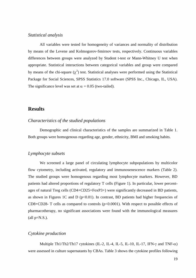
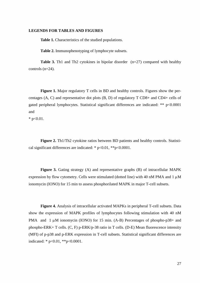
Figure 1. Major regulatory T cells in BD and healthy controls. Figures show the per-
centages (A, C) and representative dot plots (B, D) of regulatory T CD8+ and CD4+ cells of
gated peripheral lymphocytes. Statistical significant differences are indicated: ** p<0.0001
and
* p<0.01.
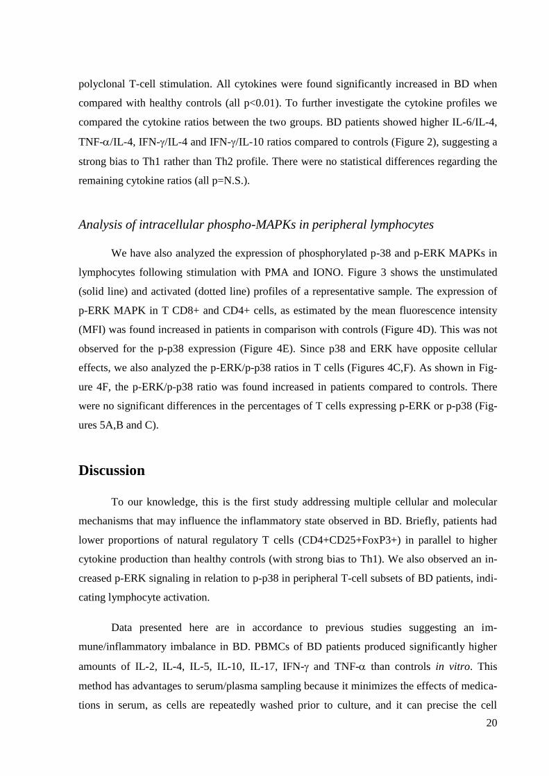
Figure 2. Th1/Th2 cytokine ratios between BD patients and healthy controls. Statisti-
cal significant differences are indicated: * p<0.01, **p<0.0001.
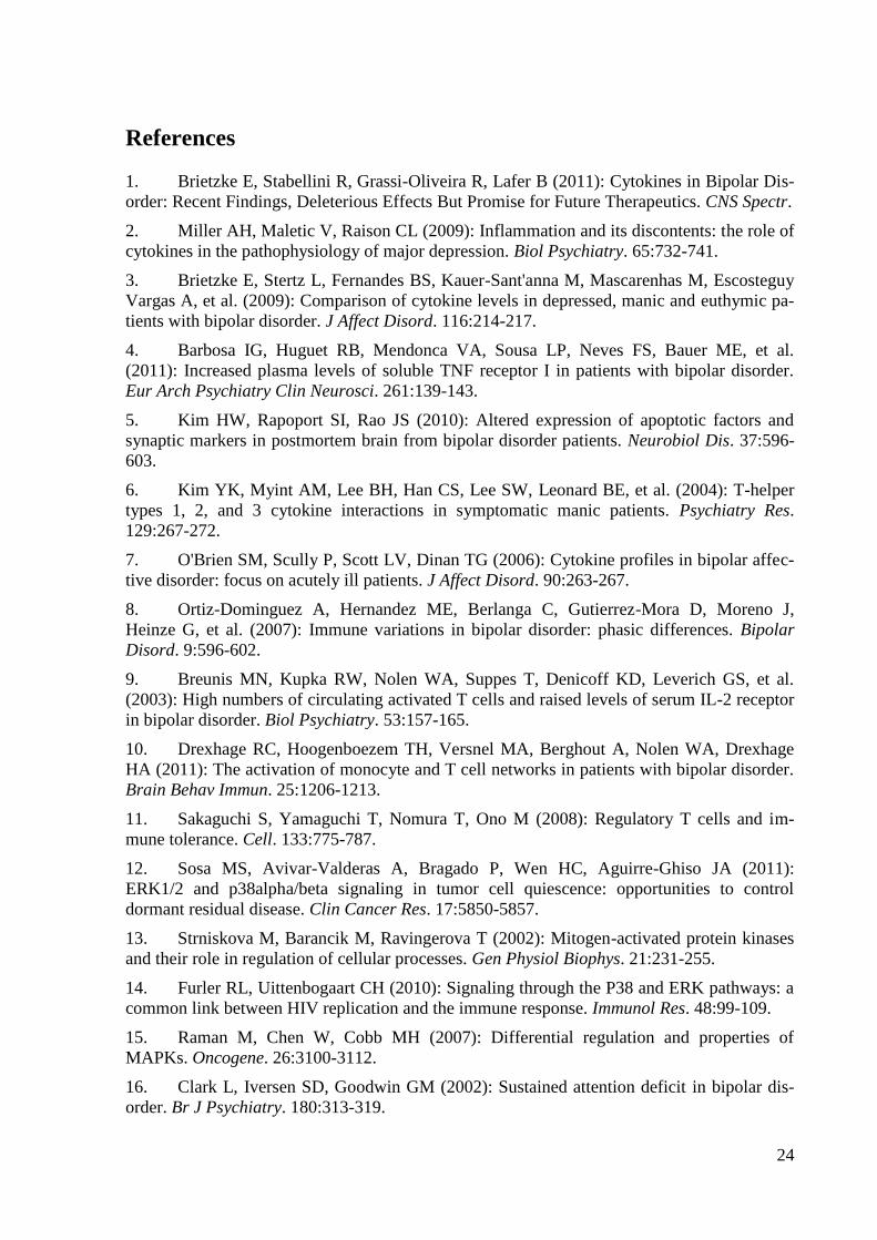
Figure 3. Gating strategy (A) and representative graphs (B) of intracellular MAPK
expression by flow cytometry. Cells were stimulated (dotted line) with 40 nM PMA and 1 M
ionomycin (IONO) for 15 min to assess phosphorilated MAPK in major T-cell subsets.
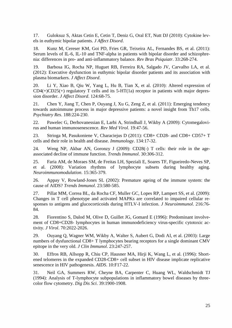
Figure 4. Analysis of intracellular activated MAPKs in peripheral T-cell subsets. Data
show the expression of MAPK profiles of lymphocytes following stimulation with 40 nM
PMA and 1 M ionomycin (IONO) for 15 min. (A-B) Percentages of phospho-p38+ and
phospho-ERK+ T cells. (C, F) p-ERK/p-38 ratio in T cells. (D-E) Mean fluorescence intensity
(MFI) of p-p38 and p-ERK expression in T-cell subsets. Statistical significant differences are
indicated: * p<0.01, **p<0.0001.

28
Table 1.
BD (M±DP) Controls P-value
N 27 24 -
Age (M ± SD) 45.72 ± 9.22 40.48 ± 13.24 NS
BMI (M ± SD) 29.21 ± 5.55 23.5 ±2.18 NS
Years of illness (mean and interval) 10.44 (1-46) - -
HDRS (M ± SD) 4.96 ± 2.21 - -
YMRS (M ± SD) 1.70 ± 2.09 - -
Ethnicity (white/non-white) 21/6 23/1 NS
Smoking 5 2 NS
Lithium 18 - -
Antidepressants 11 - -
Antipsychotics 11 - -
Anticonvulsants 3 - -
Data shown as mean (M) ± standard deviation (SD). Abbreviations: BMI, body mass index;
Bipolar disorder (BD); Hamilton Depression Rating Scale (HDRS); the Young Mania Rating
Scale (YMRS); NS, not significant.

29
Table 2.
Markers Cell Type BD (%) Control (%) P-value
CD3+CD4+ Th 48.51 ± 6.83 48.19 ± 6.98 NS
CD3+CD8+ Tc 24.96 ± 6.92 25.09 ± 6.11 NS
CD3-CD19+ B 8.59 ± 4.61 7.40 ± 2.30 NS
CD3-CD56+ NK 11.18 ± 11.47 13.02 ± 8.51 NS
CD3+CD56+ NK T 6.55 ± 6.08 8.46 ± 20.13 NS
CD4+CD45RO+ Memory (Th) 27.86 ± 9.73 27.40 ± 5.10 NS
CD8+CD45RO+ Memory (Tc) 10.52 ± 6.60 9.99 ± 3.01 NS
CD4+CD25+ Activated T cell 1.34 ± 0.72 1.97 ± 1.66 NS
CD3+CD69+ Activated T cell 2.32 ± 1.33 2.27 ± 1.23 NS
CD8+CD28+ Activated T cell 14.24 ± 7.96 15.68 ± 5.08 NS
CD8+CD28- Regulatory T Cell 17.92 ± 6.76 12.55 ± 5.08 0.002**
CD4+CD25+FOXP3+ Regulatory T Cell 2.33 ± 2.46 7.18 ± 10.14 0.014*
CD8+CD103+ Regulatory T Cell 0.695 ± 0.31 1.05 ± 0.68 NS
CD4+CCR7+CD45RA- Central Memory (Th) 44.51 ± 11.22 45.40 ± 9.97 NS
CD4+CCR7-CD45RA- Effector Memory (Th) 16.42 ± 5.68 14.42 ± 5.21 NS
CD4+CCR7+CD45RA+ Naïve T cell (Th) 34.47 ± 13.75 34.86 ± 11.56 NS
CD8+CCR7+CD45RA- Central Memory (Tc) 24.81 ± 9.74 23.11 ± 6.95 NS
CD8+CCR7+CD45RA+ Naïve T cell (Tc) 40.85 ± 12.15 44.68 ± 9.65 NS
CD8+CCR7-CD45RA+ EMRA (Tc) 18.39 ± 6.67 16.33 ± 6.01 NS
CD8+CCR7-CD45RA- Effector Memory (Tc) 15.88 ± 6.24 15.19 ± 5.26 NS
Abbreviations: CM = central memory. EM = effector memory. Th = T helper cell; Tc = T cytotoxic cell.
Statistical significant differences are indicated: * p < 0.05, **p < 0.01.

30
Table 3.
BD (pg/ml) Controls(pg/ml) P-value
IL-2 978.86 ± 1862.96 92.70 ± 230.38 0.005**
IL-4 60.04 ± 123.43 36.65 ± 81.32 0.015*
IL-6 9548.51 ± 3442.92 3325.25 ± 5214.56 0.001**
IL-10 3359.06 ± 2524.33 1293.92 ± 2116.79 0.002 **
TNF- 2678.99 ± 2913.88 566.51 ± 1076.69 0.001**
IFN- 26572.70 ± 16014.60 9168.61 ± 17420.08 0.001**
IL-17 275.90 ± 251.24 167.08 ± 217.54 0.028*
PBMCs were isolated and stimulated with phytohemagglutinin for 72h. Supernatants were
collected and cytokines measured by CBA (Cytometric Bead Array, BD). Data are shown as
mean (pg/ml) ± S.D. Statistical significant differences are indicated: *p<0.05; **p<0.01.
31
Figure 1.
32
Figure 2.
33
Figure 3.
34
Figure 4.

35
3. CAPÍTULO 3
36
3.1. CONSIDERAÇÕES FINAIS
Nesse estudo, analisamos vias celulares e moleculares que poderiam explicar o dese-
quilíbrio imunológico observado no TB. Atualmente, o interesse em investigar o papel dos
sistemas imune e inflamatório na patofisiologia do TB aumentou consideravelmente, devido
às ações das citocinas no sistema nervoso central.
Inicialmente, verificamos a produção de citocinas no sobrenadante de linfócitos esti-
mulados in vitro. Essa técnica tem vantagens em relação às medidas no plasma/soro, que não
permitem a identificação das fontes produtoras das citocinas. Observamos níveis muito eleva-
dos de citocinas pró-inflamatórias (Th1/Th17) e antiinflamatórias (Th2) em indivíduos bipola-
res em comparação com controles saudáveis. Além disso, para examinar de que forma as cito-
cinas estão contribuindo para a desregulação imune observada no TB, decidimos investigar a
razão Th1/Th2. Nossos resultados indicaram um aumento da razão Th1/Th2 em indivíduos
com TB, sugerindo que este transtorno está associado com respostas inflamatórias, mais pre-
cisamente com aumento de citocinas do tipo Th1.
As proporções celulares periféricas podem contribuir para o processo inflamatório ob-
servado no TB. O presente estudo foi o primeiro a investigar no TB uma ampla variedade de
subtipos linfocitários, associados principalmente com ativação, regulação e imunossenescên-
cia. Deve ser salientado que existem poucos estudos que analisaram as populações celulares
no TB. Estes subtipos celulares foram aqui avaliados com o intuito de averiguar a contribui-
ção destas células no desequilíbrio imune observado no TB. Interessantemente, verificamos
que os pacientes bipolares apresentaram uma diminuição de células T regulatórias naturais
(CD4+CD25+FoxP3+) quando comparados com controles saudáveis, e um aumento na por-
centagem de células T regulatórias-senescentes (CD8+CD28-). Nossos resultados estão de
acordo com estudos anteriores que observaram diminuição de células T regulatórias na de-
pressão (Li, Xiao et al., 2010; Chen, Jiang et al., 2011). Portanto, um número reduzido de
células Tregs poderia contribuir para o desequilíbrio imunológico observado no TB. Além
disso, a expansão de células T regulatórias-senescentes (CD8+CD28-) poderia estar envolvida
no processo de imunossenescência precoce no TB.
Avaliamos vias intracelulares envolvidas com o processo de ativação linfocitária. Es-
pecificamente, verificamos se as MAPK estão envolvidas neste desequilíbrio imunológico do
TB. Isso foi possível através do uso de uma técnica inovadora de phosflow (BD Biosciences)

37
que permite a identificação e quantificação de células que expressam proteínas fosforiladas
intracelulares. As vias de sinalização p-ERK e p-p38 são responsáveis por atividades de proli-
feração/diferenciação e inflamação/anergia, respectivamente. Verificamos um aumento signi-
ficativo nos níveis de p-ERK em relação à p-38 em indivíduos com TB, quando comparados
com controles saudáveis. Além disso, a razão p-ERK/p-38 está elevada em bipolares em com-
paração com controles saudáveis. O aumento de p-ERK no TB indica ativação linfocitária,
confirmando novamente que esse transtorno está relacionado com processos de ativação imu-
ne/inflamatória.
Este estudo apresenta algumas limitações. Primeiramente, todos os sujeitos do estudo
apresentavam-se medicados, a fim de manter seu humor estável (fase eutímica). O tratamento
dos pacientes foi realizado em grande parte com o lítio, uma droga sabidamente capaz intera-
gir com o sistema imune. Embora o lítio tenha uma ação anti-inflamatória, este aparentemente
não influenciou o perfil pró-inflamatório encontrado nos pacientes. A segunda limitação do
estudo refere-se à dificuldade de incluir no estudo apenas pacientes bipolares eutímicos, res-
tringindo o tamanho amostral deste estudo. Como perspectivas futuras, podemos aprofundar
as pesquisas em relação às MAPKs e outras vias de sinalização intracelular, e explorar outros
marcadores de envelhecimento, como o comprimento da região telomérica.
Concluindo, levando em consideração o exposto, os achados deste trabalho corrobo-
ram estudos anteriores sugerindo um desequilíbrio imune no TB. Além disso, nosso estudo
fornece pela primeira vez informações relevante sobre as células T regulatórias e vias de sina-
lização MAPKs no TB, que podem futuramente servir de alvos terapêuticos para este trans-
torno. O uso de anti-inflamatórios deveria ser melhor investigado na terapêutica deste impor-
tante transtorno do humor.
38
4. REFERÊNCIAS BIBLIOGRÁFICAS
ABEER et al. Immunological changes in patients with mania: changes in cell mediated immunity in a sample from Egyptian patients. Egypt J Immunol [S.I.], v. 13, n. 1, p. 79-85, 2006. BALDASSANO, C. F. et al. Bipolar depression: an evidence-based approach. Curr Psychiatry Rep [S.I.], v. 13, n. 6, p. 483-7, Dec 2011. BAUER, M.; PFENNIG, A. Epidemiology of bipolar disorders. Epilepsia [S.I.], v. 46 Suppl 4, p. 8-13, 2005. BAUER, M. E. Chronic stress and immunosenescence: a review. Neuroimmunomodulation [S.I.], v. 15, n. 4-6, p. 241-50, 2008. BENAZZI, F. Bipolar II disorder : epidemiology, diagnosis and management. CNS Drugs [S.I.], v. 21, n. 9, p. 727-40, 2007. BREUNIS, M. N. et al. High numbers of circulating activated T cells and raised levels of serum IL-2 receptor in bipolar disorder. Biol Psychiatry [S.I.], v. 53, n. 2, p. 157-65, Jan 15 2003. BRIETZKE, E.; KAPCZINSKI, F. TNF-alpha as a molecular target in bipolar disorder. Prog Neuropsychopharmacol Biol Psychiatry [S.I.], v. 32, n. 6, p. 1355-61, Aug 1 2008. BRIETZKE, E. et al. Comparison of cytokine levels in depressed, manic and euthymic patients with bipolar disorder. J Affect Disord [S.I.], v. 116, n. 3, p. 214-7, Aug 2009. BROWN, E. S. et al. Association of depression with medical illness: does cortisol play a role? Biol Psychiatry [S.I.], v. 55, n. 1, p. 1-9, Jan 1 2004. CHEN, Y. et al. Emerging tendency towards autoimmune process in major depressive patients: a novel insight from Th17 cells. Psychiatry Res [S.I.], v. 188, n. 2, p. 224-30, Jul 30 2011. DABAN, C. et al. Hypothalamic-pituitary-adrenal axis and bipolar disorder. Psychiatr Clin North Am [S.I.], v. 28, n. 2, p. 469-80, Jun 2005. DEEKS, S. G. HIV infection, inflammation, immunosenescence, and aging. Annu Rev Med [S.I.], v. 62, p. 141-55, Feb 18 2011. DEGENHARDT, E. K. et al. Predictors of relapse or recurrence in bipolar I disorder. J Affect Disord [S.I.], Oct 28 2011. DREXHAGE, R. C. et al. The activation of monocyte and T cell networks in patients with bipolar disorder. Brain Behav Immun [S.I.], v. 25, n. 6, p. 1206-13, Aug 2011.

39
ELVSASHAGEN, T. et al. The load of short telomeres is increased and associated with lifetime number of depressive episodes in bipolar II disorder. J Affect Disord [S.I.], v. 135, n. 1-3, p. 43-50, Dec 2011. EVANS, D. L. et al. Mood disorders in the medically ill: scientific review and recommendations. Biol Psychiatry [S.I.], v. 58, n. 3, p. 175-89, Aug 1 2005. FAN, J.; SKLAR, P. Genetics of bipolar disorder: focus on BDNF Val66Met polymorphism. Novartis Found Symp [S.I.], v. 289, p. 60-72; discussion 72-3, 87-93, 2008. FURLER, R. L.; UITTENBOGAART, C. H. Signaling through the P38 and ERK pathways: a common link between HIV replication and the immune response. Immunol Res [S.I.], v. 48, n. 1-3, p. 99-109, Dec 2010. GONZALEZ-PINTO, A. et al. First episode in bipolar disorder: misdiagnosis and psychotic symptoms. J Affect Disord [S.I.], v. 50, n. 1, p. 41-4, Jul 1998. GULOKSUZ, S. et al. Cytokine levels in euthymic bipolar patients. J Affect Disord [S.I.], May 27. HEUSER, I. Depression, endocrinologically a syndrome of premature aging? Maturitas [S.I.], v. 41 Suppl 1, p. S19-23, Apr 15 2002. HOPE, S. et al. Similar immune profile in bipolar disorder and schizophrenia: selective increase in soluble tumor necrosis factor receptor I and von Willebrand factor. Bipolar Disord [S.I.], v. 11, n. 7, p. 726-34, Nov 2009. JAMROZINSKI, K. et al. Neurocognitive functions in euthymic bipolar patients. Acta Psychiatr Scand [S.I.], v. 119, n. 5, p. 365-74, May 2009. KAUER-SANT'ANNA, M. et al. Brain-derived neurotrophic factor and inflammatory markers in patients with early- vs. late-stage bipolar disorder. Int J Neuropsychopharmacol [S.I.], v. 12, n. 4, p. 447-58, May 2009. KESSLER, R. C. et al. Prevalence, comorbidity, and service utilization for mood disorders in the United States at the beginning of the twenty-first century. Annual Review of Clinical Psychology [S.I.], v. 3, p. 137-158, 2007. KIM, E. K.; CHOI, E. J. Pathological roles of MAPK signaling pathways in human diseases. Biochim Biophys Acta [S.I.], v. 1802, n. 4, p. 396-405, Apr 2010. KIM, H. W. et al. Altered expression of apoptotic factors and synaptic markers in postmortem brain from bipolar disorder patients. Neurobiol Dis [S.I.], v. 37, n. 3, p. 596-603, Mar 2010. KIM, Y. K. et al. T-helper types 1, 2, and 3 cytokine interactions in symptomatic manic patients. Psychiatry Res [S.I.], v. 129, n. 3, p. 267-72, Dec 30 2004.

40
KNIJFF, E. M. et al. A relative resistance of T cells to dexamethasone in bipolar disorder. Bipolar Disord [S.I.], v. 8, n. 6, p. 740-50, Dec 2006. KRISHNA, M.; NARANG, H. The complexity of mitogen-activated protein kinases (MAPKs) made simple. Cell Mol Life Sci [S.I.], v. 65, n. 22, p. 3525-44, Nov 2008. KUPFERSCHMIDT, D. A.; ZAKZANIS, K. K. Toward a functional neuroanatomical signature of bipolar disorder: quantitative evidence from the neuroimaging literature. Psychiatry Res [S.I.], v. 193, n. 2, p. 71-9, Aug 30 2011. KUPKA, R. W. et al. Immune activation, steroid resistancy and bipolar disorder. Bipolar Disord [S.I.], v. 4 Suppl 1, p. 73-4, 2002. LI, Y. et al. Altered expression of CD4(+)CD25(+) regulatory T cells and its 5-HT(1a) receptor in patients with major depression disorder. J Affect Disord [S.I.], v. 124, n. 1-2, p. 68-75, Jul 2010. O'BRIEN, S. M. et al. Cytokine profiles in bipolar affective disorder: focus on acutely ill patients. J Affect Disord [S.I.], v. 90, n. 2-3, p. 263-7, Feb 2006. OFFORD, J. Genetic approaches to a better understanding of bipolar disorder. Pharmacol Ther [S.I.], Oct 8 2011. ORTIZ-DOMINGUEZ, A. et al. Immune variations in bipolar disorder: phasic differences. Bipolar Disord [S.I.], v. 9, n. 6, p. 596-602, Sep 2007. PADMOS, R. C. et al. A high prevalence of organ-specific autoimmunity in patients with bipolar disorder. Biol Psychiatry [S.I.], v. 56, n. 7, p. 476-82, Oct 1 2004. PROVENCHER, M. D. et al. Psychotherapies for comorbid anxiety in bipolar spectrum disorders. J Affect Disord [S.I.], v. 133, n. 3, p. 371-80, Oct 2011. RAISON, C. L. et al. Cytokines sing the blues: inflammation and the pathogenesis of depression. Trends Immunol [S.I.], v. 27, n. 1, p. 24-31, Jan 2006. RAMAN, M. et al. Differential regulation and properties of MAPKs. Oncogene [S.I.], v. 26, n. 22, p. 3100-12, May 14 2007. RAPAPORT, M. H. Immune parameters in euthymic bipolar patients and normal volunteers. J Affect Disord [S.I.], v. 32, n. 3, p. 149-56, Nov 1994. ROUX, P. P.; BLENIS, J. ERK and p38 MAPK-activated protein kinases: a family of protein kinases with diverse biological functions. Microbiol Mol Biol Rev [S.I.], v. 68, n. 2, p. 320-44, Jun 2004.

41
SAKAGUCHI, S. et al. Regulatory T cells and immune tolerance. Cell [S.I.], v. 133, n. 5, p. 775-87, May 30 2008. SANCHES, M.; SOARES, J. C. New drugs for bipolar disorder. Curr Psychiatry Rep [S.I.], v. 13, n. 6, p. 513-21, Dec 2011. SERRETTI, A. et al. Dopamine receptor D2 Ser/Cys 311 variant is associated with delusion and disorganization symptomatology in major psychoses. Mol Psychiatry [S.I.], v. 5, n. 3, p. 270-4, May 2000. SHALEV, I. et al. BDNF Val66Met polymorphism is associated with HPA axis reactivity to psychological stress characterized by genotype and gender interactions. Psychoneuroendocrinology [S.I.], v. 34, n. 3, p. 382-8, Apr 2009. SOSA, M. S. et al. ERK1/2 and p38alpha/beta signaling in tumor cell quiescence: opportunities to control dormant residual disease. Clin Cancer Res [S.I.], v. 17, n. 18, p. 5850-7, Sep 15 2011. VERMANI, M. et al. Rates of detection of mood and anxiety disorders in primary care: a descriptive, cross-sectional study. Prim Care Companion CNS Disord [S.I.], v. 13, n. 2, 2011. VINBERG, M. et al. The BDNF Val66Met polymorphism: relation to familiar risk of affective disorder, BDNF levels and salivary cortisol. Psychoneuroendocrinology [S.I.], v. 34, n. 9, p. 1380-9, Oct 2009. WATSON, S. et al. Hypothalamic-pituitary-adrenal axis function in patients with bipolar disorder. Br J Psychiatry [S.I.], v. 184, p. 496-502, Jun 2004. WOLKOWITZ, O. M. et al. Leukocyte telomere length in major depression: correlations with chronicity, inflammation and oxidative stress--preliminary findings. PLoS One [S.I.], v. 6, n. 3, p. e17837, 2011.