ARYANE FLAUZINO MACHADO EFEITOS IMEDIATOS E … · Ao meu amado companheiro Rafael Rodrigues por...
110
ARYANE FLAUZINO MACHADO EFEITOS IMEDIATOS E TARDIOS DA IMERSÃO EM ÁGUA FRIA PÓS- EXERCÍCIO: UMA REVISÃO SISTEMÁTICA E UM ENSAIO CLÍNICO RANDOMIZADO Presidente Prudente 2015
Transcript of ARYANE FLAUZINO MACHADO EFEITOS IMEDIATOS E … · Ao meu amado companheiro Rafael Rodrigues por...
ARYANE FLAUZINO MACHADO
EFEITOS IMEDIATOS E TARDIOS DA IMERSÃO EM ÁGUA FRIA PÓS-
EXERCÍCIO: UMA REVISÃO SISTEMÁTICA E UM ENSAIO CLÍNICO
RANDOMIZADO
Presidente Prudente
2015
ARYANE FLAUZINO MACHADO
EFEITOS IMEDIATOS E TARDIOS DA IMERSÃO EM ÁGUA FRIA PÓS-
EXERCÍCIO: UMA REVISÃO SISTEMÁTICA E UM ENSAIO CLÍNICO
RANDOMIZADO
Dissertação apresentada à Faculdade de Ciências e
Tecnologia da Universidade Estadual Paulista “Júlio
de Mesquita Filho” (FCT/UNESP) – Presidente
Prudente, para obtenção do título de mestre no
Programa de Pós-Graduação Stricto Sensu em
Fisioterapia.
Orientador: Prof. Dr. Carlos Marcelo Pastre
Coorientador: Prof. Dr. Paulo Henrique Ferreira
Presidente Prudente
2015
FICHA CATALOGRÁFICA
Machado, Aryane Flauzino
M129i
Efeitos imediatos e tardios da imersão em água pós-exercício: uma revisão
sistemática e um ensaio clínico / Aryane Flauzino Machado - Presidente Prudente
: [s.n.], 2015
110 f.
Orientador: Carlos Marcelo Pastre
Coorientador: Paulo Henrique Ferreira
Dissertação (mestrado) - Universidade Estadual Paulista, Faculdade de
Ciências e Tecnologia
Inclui bibliografia
1. Recuperação da função fisiológica. 2. Crioterapia. 3. Imersão. I. Pastre,
Carlos Marcelo. II. Ferreira, Paulo Henrique. III. Universidade Estadual Paulista.
Faculdade de Ciências e Tecnologia. IV. Efeitos imediatos e tardios da imersão
em água pós-exercício: uma revisão sistemática e um ensaio clínico
Ficha catalográfica elaborada pela Seção Técnica de Aquisição e Tratamento da
Informação Serviço Técnico de Biblioteca e Documentação – UNESP, Campus de Presidente
Prudente.

Dedicatória
À minha amada família por todo
companheirismo, apoio e dedicação. Meus pais
Jurandir e Vânia e meu irmão Gabriel. Nossa
vitória, nossa conquista!

Agradecimentos
Há muito o que e a quem agradecer.
À Deus minha infinita gratidão, pela paz interior e sabedoria. Obrigada por
permitir a realização desse sonho. A Nossa Senhora Aparecida por me iluminar em todos
os momentos da minha vida.
A minha família. Meus pais, amigos e parceiros, Vânia e Jurandir. Agradeço
todo amparo e suporte em todo esse processo, e principalmente por tornarem esse sonho,
um sonho nosso! Ao meu pequeno-grande homem, meu irmão Gabriel. À eles, que mesmo
distantes, seja há 200 ou 15000 km, se fizeram presente, dedico e agradeço todo essa
conquista. Serei eternamente grata a vocês. Minha inspiração de caráter, dedicação e
amor. Amo vocês incondicionalmente!
Ao meu orientador, Prof. Carlos Marcelo Pastre. Agradeço pela oportunidade,
pelos ensinamentos e pela amizade. Por me mostrar o caminho e me encorajar a ser e fazer
sempre o melhor. Grandes desafios foram superados e oportunidades aproveitadas graças
ao seu apoio. Obrigada por toda atenção e incentivo. Espero que essa jornada não acabe
aqui!
Ao meu coorientador, Prof. Paulo H. Ferreira que me recebeu de portas e
braços abertos em Sydney. Responsável por grande parte do crescimento profissional e
pessoal que o estágio no exterior me proporcionou. Obrigada por toda atenção destinada a
mim e a nossa pesquisa.
Ao querido Prof. Jayme Netto pelos abraços matinais e principalmente por
todos os ensinamentos, sejam eles profissionais ou pessoais. Muito obrigada!
Ao amigo Prof. Fábio A. N. Martini que sempre me incentivou a seguir essa
trajetória. Um grande amigo que não mediu esforços e que depositou uma grande
confiança em mim. Serei eternamente grata!

Ao meu amado companheiro Rafael Rodrigues por todos os momentos. Pela
paciência, atenção, dedicação e carinho em todo esse processo. Muito obrigada!
Aos meus companheiros de todos os dias Aline Castilho, Ítalo Lemes e Jéssica
Micheletti. Obrigada por todo atenção, amizade e dedicação. Somos uma equipe incrível e
assim, estamos aos poucos conquistando o que almejamos. Muito obrigada. Desejo a vocês
sucesso. Muito sucesso!
Aos integrantes do Laboratório de Fisioterapia Desportiva (LAFIDE –
FCT/UNESP), por toda dedicação destinada a essa pesquisa. Obrigada por terem me
acolhido. Espero que eu tenha conseguido retribuir toda confiança e amizade. Orgulho-me
sempre em fazer parte dessa equipe!
Aos integrantes do Arthritis and Musculoskeletal Research Group (AMRG –
The University of Sydney) pela atenção, paciência, ensinamentos e recepção incrível!
Espero reencontrá-los em breve. Agradeço especialmente ao Prof. Paulo, Prof. Evangelos
Pappas, seus alunos de doutorado Marina Pinheiro, Amabile Dario e Matt Fernandez e os
companheiros de viagem Ítalo R Lemes e Nathalie F Souto.
Aos meus amigos. A todos meus amigos que participaram dessa conquista.
Aqueles que estiveram presente durante as conquistas e as dificuldades e que, de maneira
singular, tornaram esse processo mais fácil. Em especial, as minhas amigas Aline,
Jaqueline e Jéssica, que alegraram meu dia-a-dia, e que, compartilhando das mesmas
alegrias e angústias, tornaram-se essenciais.
Aos membros da banca examinadora, Prof. Dr. Fabio Nakamura e Prof. Dr.
Rafael Zambelli, pela disponibilidade em participar e pelas contribuições.
Aos funcionários da FCT/UNESP por toda atenção e dedicação, principalmente
ao André T Meira que sempre esteve pronto a ajudar, com o máximo de educação,
profissionalismo e simpatia.

Aos participantes dessa pesquisa que não hesitaram em colaborar momento
algum. Que proporcionaram não só esse trabalho, mas também divertidas noites de coleta
de dados.
A Coordenação de Aperfeiçoamento de Pessoal de Nível Superior (CAPES) e
Fundação de Amparo à Pesquisa do Estado de São Paulo – FAPESP (processos nº
2013/12474-7 e 2014/03778-5) pelo apoio financeiro destinado a essa pesquisa.
E por fim, agradeço a todos que contribuíram direta ou indiretamente para que
mais essa etapa pudesse ser concluída. Muito obrigada!

Epígrafe

“Reconheça o que está ao alcance dos
seus olhos e o que está oculto tornar-se-á claro
para você.”
(Autor desconhecido)
SUMÁRIO
Apresentação .......................................................................................................... 14
Introdução .............................................................................................................. 16
Artigo I: Can the water temperature and immersion time influence the effects of
cold water immersion on pain? A systematic review and meta-analysis …………..
18
Artigo II: Immediate and delayed effects of cold water immersion after eccentric
exercise-induced muscle damage: randomized controlled trial ……………………
48
Conclusões ..……….……………………………………………………………... 77
Referências ………………………………………………………………………... 78
Anexos ……………………………………………………………………………... 80
14
Apresentação
Essa dissertação está apresentada em consonância com as normas do modelo
alternativo de dissertação do Programa de Pós-Graduação Stricto Sensu em Fisioterapia da
Faculdade de Ciências e Tecnologia da Universidade Estadual Paulista “Júlio de Mesquita
Filho”. O conteúdo desse trabalho contempla o material originado a partir da pesquisa
intitulada “Efeitos imediatos e tardios da imersão em água fria pós-exercício: uma revisão
sistemática e um ensaio clínico randomizado” que foi realizada em duas etapas:
1-) Revisão Sistemática e Meta-análise, realizada na The University of Sydney,
Faculty of Health Sciences, Sydney – NSW, Austrália, financiada pela Fundação de Amparo à
Pesquisa do Estado de São Paulo – FAPESP (Linha de Fomento: Bolsa de Pesquisa de
Estágio no Exterior, processo: 2014/03778-5);
2-) Ensaio clínico randomizado, realizado na Univ. Estadual Paulista “Júlio de
Mesquita Filho”, Faculdade de Ciências e Tecnologia, Presidente Prudente – SP, Brasil,
financiada pela Coordenação de Aperfeiçoamento de Pessoal de Ensino Superior – CAPES e
posteriormente financiada pela Fundação de Amparo à Pesquisa do Estado de São Paulo –
FAPESP (Linha de Fomento: Bolsa de Mestrado no País, processo 2013/12474-7).
Assim, o presente material está dividido nas seguintes sessões:
• Introdução ao tema, para contextualização do tema pesquisado;
• Artigo I: Machado AF, Ferreira PH, Micheletti JK, Almeida AC, Lemes IR,
Vanderlei FM, Netto Junior J, Pastre CM. Can the water temperature and immersion time
influence the effects of cold water immersion on pain? A systematic review and meta-analysis;
em revisão pelo periódico Sports Medicine;
• Artigo II: Machado AF, Almeida AC, Micheletti JK, Netto Junior J, Vanderlei
FM, Netto Junior J, Pastre CM. Immediate and delayed effects of cold water immersion after
15
eccentric exercise-induced muscle damage: randomized controlled trial; em revisão pelo
periódico PLoS ONE;
• Conclusões, a partir de ambas as pesquisas realizadas;
• Referências, em formato recomendado pelo International Committee of
Medical Journals Editours (ICMJE), para as referências citadas na introdução.
Ressalta-se que cada artigo está apresentado de acordo com as normas dos seus
respectivos periódicos, apresentadas em anexo ao final (com exceção das figuras que estão
apresentadas no texto principal).
16
Introdução
O processo de recuperação pós-exercício é fundamental para a preservação de
estruturas e tecidos, bem como para a manutenção das funções motoras, visando à prevenção
de agravos ou melhora da performance e rendimento do atleta. Seu objetivo principal é a
restauração dos diferentes sistemas do corpo a condições basais, ou seja, a níveis pré-
exercício1,2
.
Dentre as diversas técnicas que aceleram a recuperação pós-exercício, a
crioterapia é um procedimento comumente utilizado na prática esportiva e é proposta para
redução da resposta inflamatória em caso de lesão tecidual e dor muscular resultante do
esforço físico, apresentando evidências particulares na aplicação após o dano muscular
induzido por exercício (DMIE)3–5
.
Uma das modalidades da crioterapia é a imersão em água fria (IAF), que consiste
na imersão de parte do corpo em água com temperatura igual ou inferior à 15°C6. Essa
estratégia de recuperação aparece no cenário atual como uma técnica eficaz e de baixo custo,
facilmente reproduzida em diferentes situações. Os potenciais efeitos dessa estratégia de
recuperação têm sido avaliados a partir de diferentes marcadores de dano muscular, tais como
concentração sanguínea de creatina quinase (CK) 5,7–9
, dor3,4,7–10
, alteração de sensibilidade10
,
percepção de recuperação11
e força3–5,7–9,11
.
Sabe-se que a IAF é capaz de reduzir a temperatura do tecido muscular3, a
permeabilidade celular de vasos sanguíneos, linfáticos e capilares devido à vasoconstrição,
com consequente diminuição da difusão dos fluídos nos espaços intersticiais4,12,13
além de
reduzir a velocidade da condução nervosa, a atividade do fuso muscular, a espasticidade e a
dor3. Entretanto, autores afirmam que apesar dos efeitos relatados não há evidência fisiológica
17
clara para confirmar essas teorias e que os reais efeitos ainda não estão plenamente
elucidados9.
Apesar dos resultados, os indivíduos ainda respondem de maneira diferente aos
sinais e sintomas. Esse fato pode ser explicado pela utilização de diferentes metodologias
adotadas e influenciado pela complexidade e especificidade de cada indivíduo14
. Pastre et al.15
atribuíram parte do cenário aos diferentes métodos de controle utilizados pelos pesquisadores
tanto na maneira de indução de estresse quanto à forma de aplicação da técnica. Glasgow et
al.9 concordam ao afirmar que na prática clínica existe uma grande variação de protocolos de
IAF, que se diferem principalmente quanto à temperatura da água e ao tempo de imersão.
Essa variação corresponde à dose no qual o indivíduo é exposto, influenciando nos futuros
desfechos.
Deve-se refletir à luz desta situação problema que requer uma melhor
fundamentação científica a fim de eliminar possíveis vieses de rotina e de interpretação de
resultados e objetivando identificar a melhor estratégia baseada na relação da dose-resposta na
aplicação da IAF pós-exercício.
Assim, o objetivo da presente pesquisa foi determinar os efeitos da imersão em
água fria pós-exercício, por meio de diferentes tipos de estudo, como a revisão sistemática e
meta-análise e o ensaio clínico randomizado, além de identificar a melhor dose de aplicação
dessa técnica, por meio da relação da dose-resposta e os possíveis efeitos deletérios.

18
Artigo 1
Can water temperature and immersion time influence the effect of cold water immersion
on pain? A systematic review and meta-analysis
Different protocols of CWI on pain: a meta-analysis
Aryane Flauzino Machado1, Paulo Henrique Ferreira
2, Jéssica Kirsch Micheletti
1, Aline
Castilho de Almeida3, Ítalo Ribeiro Lemes
1, Franciele Marques Vanderlei
1, Jayme Netto
Junior1, Carlos Marcelo Pastre
1
1Univ. Estadual Paulista,
Faculdade de Ciências e Tecnologia, Departamento de Fisioterapia,
Presidente Prudente, Brazil
2The University of Sydney, Faculty of Health Science, Discipline of Physiotherapy, Sydney,
Australia
3Univ. Federal de São Carlos, Centro de Ciências Biológicas e da Saúde, Departamento de
Fisioterapia, São Carlos, Brazil
Corresponding author
Carlos Marcelo Pastre
305 Roberto Simonsen – Presidente Prudente / São Paulo, Brazil. Postcode: 19060-900
E-mail: [email protected]
Telephone number: +55 18 3229 5528
19
ABSTRACT
Background: Cold water immersion (CWI) is a technique commonly used in post-exercise
recovery. However, the procedures involved in the technique may vary, particularly in terms
of water temperature and immersion time, and the most effective approach remains unclear.
Purpose: To determine the efficacy of CWI in pain management compared with passive
recovery. We also aimed to identify which water temperature and immersion time provides
the best results.
Methods: MEDLINE, EMBASE, SPORTDiscus, PEDro and The Cochrane Library
databases were searched up to January 2015. Only randomized controlled trials that compared
CWI to passive recovery were included in this review.
Results: Nine studies were included in the review and meta-analysis. The results of meta-
analysis revealed that CWI has a more positive effect than passive recovery in terms of
immediate (WMD=0.290, 95% CI [0.037, 0.543]; p=0.025) and delayed effects
(WMD=0.315, 95% CI [0.048, 0.581], p=0.021). The pooled of studies that used water
temperature of between 10-15°C demonstrated the best results for immediate (WMD=0.273,
95% CI [0.107, 0.440], p=0.001) and delayed effects (WMD=0.317, 95% CI [0.102, 0.532],
p=0.004). In terms of immersion time, immersion of between 10-15 minutes had the best
results for immediate (WMD=0.227, 95% [0.139, 0.314], p=0.000) and delayed effects
(WMD=0.317, 95% [0.102, 0.532], p=0.004).
Conclusions: CWI is more effective than passive recovery in management of pain. The
results also demonstrated the presence of a dose-response relationship, indicating that CWI
with a water temperature of between 11 and 15ºC and an immersion time of 11 to 15 minutes
provided the best results.
Keywords: recovery of function; cryotherapy; immersion; muscle soreness.
20
1. BACKGROUND
Several post-exercise recovery techniques are currently employed in an attempt to
return the body to its pre-exercise state [1,2]. Cold water immersion (CWI) has become
popular in sports [3,4] as it is a low-cost technique that is easily performed in different
situations [5] and has been found to minimize the immediate and delayed negative effects of
exercise [6]. Compared to controlled interventions and other traditional recovery techniques,
CWI achieves positive pain reduction results following a range of exercise types [7,8]. Yet the
specific mechanisms associated with CWI response are unknown [9,10].
Despite its widespread use, significant procedural variations in CWI exist [11,12].
Investigations have suggested that water temperature contributes to the beneficial effect of
CWI [6]. However, other factors may influence recovery. Pastre et al. [13] attribute response
variation to differences in the application of CWI, such as water temperature, immersion time
and type of CWI.
In recent years, the number of studies focusing on CWI has increased, and major
systematic reviews have been performed to compare the effects of CWI and other pain
recovery strategies [8,12]. However, the dose-response relationship of this technique has not
yet been investigated. Bleakley et al. [12] found no clinical trials comparing different
procedures while authors [10] showed that studies focusing on different CWI application
strategies can contribute to determining the risks and benefits for athletes.
A systematic review involving the dose-response relationship will clarify the most
effective method of application of CWI for post-exercise pain. Therefore, the purpose of this
systematic review was to determine the efficacy of cold water immersion on management of
pain compared with control intervention (passive recovery). An analysis of which dosage of
application provides the best results, focusing on water temperature and immersion time, was
also undertaken.
21
2. METHODS
This systematic review was registered in an international database of systematic
reviews in health and social care. (Available: registration number CRD42015016573;
http://www.crd.york.ac.uk/PROSPERO/).
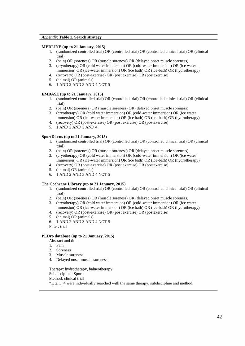
2.1 Search strategies
Studies were selected after searching five databases (MEDLINE, EMBASE,
SPORTDiscus, PEDro and The Cochrane Library) from the earliest record of each database to
January 21, 2015. The terms and keywords used to search optimization were related to
randomized controlled trials; cold water immersion and post-exercise recovery (see details in
Appendix 1). The reference list of eligible clinical trials was searched by hand to complement
the electronic searches.
2.2 Study selection
The studies selected involved CWI treatment of human participants and assessed
the effect on immediate and/or delayed pain or muscle soreness. CWI was defined as
immersion in water with a temperature less than or equal to 15°C [5,6,11]. To be eligible,
studies had to 1) be randomized controlled trials, comparing cold water immersion and
control conditions (passive recovery) post-exercise; 2) be studies that assessed muscle
soreness; 3) be studies that used a single session of exercises; 4) apply CWI within 1 hour of
the end of the exercise; 5) include only one immersion on the first day. Studies using
intermittent immersions or more than one immersion on subsequent days were excluded. No
restrictions were applied to the sample conditions (age, gender, exercise level) or language of
the studies.
22
The study selection process was conducted by title, followed by abstract and full
text (Figure 1). These steps were performed independently by two authors (ACA and JKM)
and consensus was used to solve disagreements.
2.3 Data extraction
Outcome data, including mean scores, SDs (final values) and sample size was
extracted by two reviewers (AFM, JKM). The data extraction process was performed using a
standardized form that included details such as characteristics of participants, exercise
procedures, cold water immersion procedures, outcome measures and methodological
characteristics. Disagreements between authors regarding data extraction were resolved by
consensus.
Some studies included multiple observations. In such cases, data was extracted at
a clinically relevant time point in order to analyze: immediate effects (up to 24 hours post-
exercise) and delayed effects (after 24 hours post-exercise). For the delayed effects, the peak
pain of the control group was considered, in order to minimize interference caused by the
intervention. Pain scores were converted to a common 0-10 scale.
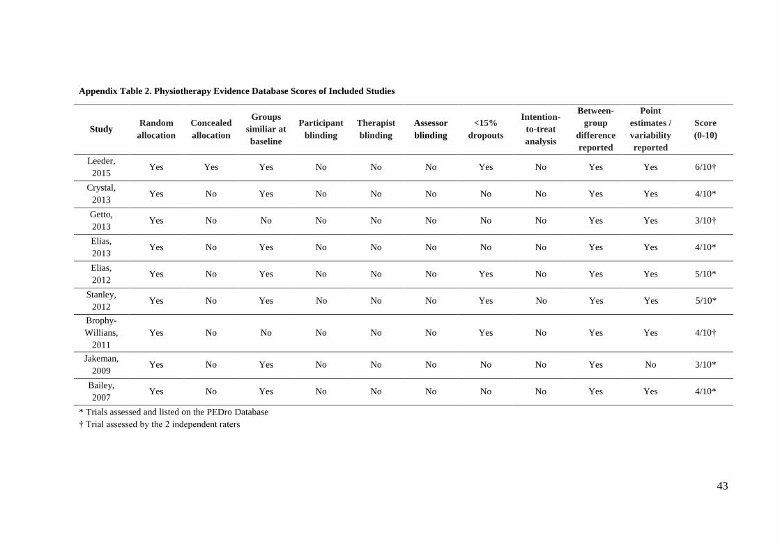
2.4 Quality Assessment
All studies included were assessed for methodological quality. This process was
performed by two independent reviewers (AFM and JKM) using the PEDro Scale [14,15].
Each study was assessed for random allocation, concealed allocation, baseline comparability,
blinding participants, therapists and assessors, adequate follow-up, intention-to-treat,
between-group comparison, point estimates and variability. If trials had already been assessed
and listed on the PEDro database, such scores were adopted. Methodological quality was not
an inclusion criteria.
23
2.5 Data synthesis and analysis
Analysis of the temperature and immersion time of each study was performed. It
was necessary to establish cutoff points for each of these covariates. For water temperature
analysis two categories were created: severe cold, with water temperature between 5-10°C;
and moderate cold, where temperature was between 11-15°C. Three categories were used for
immersion time: short, immersions of 10 minutes or less; medium, immersions of 11-15
minutes; and longer, with immersions between 16-20 minutes.
Comprehensive Meta-Analysis software, version 2.2.04 (Biostat, USA) was used
for all analysis and pooled estimates were calculated using a random-effect model, due to the
heterogeneity of the studies (represented by I²). Data was pooled in meta-analyses and
described as weighted mean differences (WMD) with 95% confidence intervals (CI). The
immediate and delayed effects were calculated in order to analyze the effect of cold water
immersion, independent of water temperature and duration of immersion. In case of more than
one intervention group per study, the group that represented the lowest effect size was used.
3. RESULTS
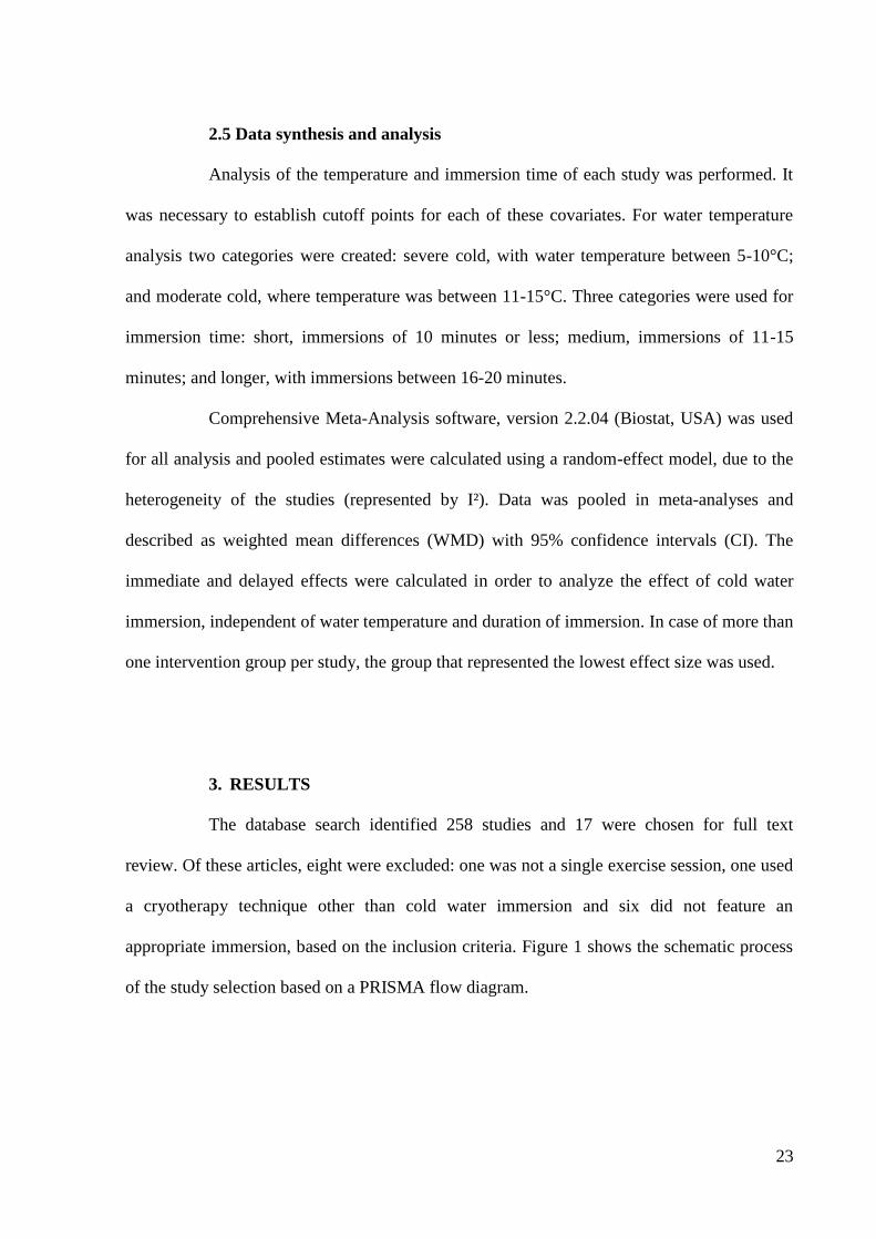
The database search identified 258 studies and 17 were chosen for full text
review. Of these articles, eight were excluded: one was not a single exercise session, one used
a cryotherapy technique other than cold water immersion and six did not feature an
appropriate immersion, based on the inclusion criteria. Figure 1 shows the schematic process
of the study selection based on a PRISMA flow diagram.
24
Fig.1 Flow chart for selection of studies
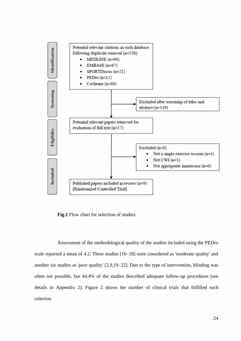
Assessment of the methodological quality of the studies included using the PEDro
scale reported a mean of 4.2. Three studies [16–18] were considered as 'moderate quality' and
another six studies as 'poor quality' [2,9,19–22]. Due to the type of intervention, blinding was
often not possible, but 44.4% of the studies described adequate follow-up procedures (see
details in Appendix 2). Figure 2 shows the number of clinical trials that fulfilled each
criterion.

25
Fig.2 Number of trials for each PEDro criteria
The nine eligible studies were published between 2007 [9] and 2015 [16]. These
studies comprised a total of 169 participants (male, n=141; female=28). The health conditions
of the participants, the level of exercise, fluctuated between physically active and athletes.
The studies were from Australia [2,17,18,21], the United Kingdom [9,16,22] and
USA[19,20]. All were randomized controlled trial type studies, while six were parallel group
trials [9,16,19–22], and three used a cross-over design [2,17,18]. Exercise protocols consisted
primarily of exercises that required high physical ability with possible subsequent onset of
26
pain, such as shuttle running [9,16], downhill running [19], Australian football match and
training [17,21], high intensity intervals [2,18] and counter-movement jumps [22].
Interventions were varied. Water temperature ranged from 5 [19] to 15°C [2] and
immersion time varied between 5 [18] and 20 [19] minutes. All studies used passive recovery
in which participants had to remain seated with minimal movement. Immersion depth ranged
from immersion of the lower limbs [9,19,22] to immersion of the whole body, excluding only
the head and neck [18]. It was observed that six [2,9,16,17,19–21] of eight studies that
evaluated delayed effect on pain found peak pain at 24 hours post-exercise, and only one [22]
found peak pain at 48 hours post-exercise.
The characteristics of the included studies are summarized in Table 1.

27
Table 1. Characteristics of the included studies
Study,
year
Study
design
Characteristics
of participants
Exercise
protocol CWI group Control group
Pain
assessment
Time of
assessment
Time of
analysis
PEDro
Score
Leeder,
2015[16]
Parallel
groups
N=24
male; well
trained
21±3 years
Intermittent
shuttle
running
14°C; 14 minutes; n=8
TI: Immediately post-
exercise
WL: DNR
Remained seated
14 minutes; n=8
VAS=0-200mm
Squat at 90° knee
flexion
24, 48, 72 hours
post-exercise
24 hours post-
exercise 6
Crystal,
2013[19]
Parallel
groups
N=20
male
21.2±2.3 years
Downhill
run
5±2°C; 20 minutes;
n=10
TI: DNR
WL: Up to top of the
thigh
Position: DNR;
n=10
VAS= 0-100mm
Leg soreness
while walking
down the stairs
Immediately, 1,
6, 24, 48 e 72
hours post-
exercise
1 and 24 hours
post-exercise 4
Getto,
2013[20]
Parallel
groups
N=23
13 male; 10
female
Age: DNR
Exhaustive
exercise
session
10°C; 10 minutes; n=7
TI: Immediately post-
exercise
WL: Up to level of
chest
Remained seated
10 minutes; n=8
VAS= 0-60
Calves,
quadriceps,
hamstrings, hip
adductors, hip
abductors and low
back
Immediately
post-exercise
and immediately
and 24 post-
intervention
Immediately
post-intervention
and 24 hours
post-exercise
3
Elias,
2013[21]
Parallel
groups
N=24
male; Australian
football players
19.9±2.8 years
Australian
football
match
12°C; 14 minutes; n=7
TI: Within 12 minutes
post-exercise
WL: Up to xiphoid
process
Remained seated
14 minutes; n=8
VAS= 0-100mm
DNR
Immediately, 1,
24, 48 hours
post-exercise
1 and 24 hours
post-exercise 4
Elias,
2012[17]
Cross-
over
N=14
male; Australian
football players
20.9±3.3 years
Australian
football
training
12°C; 14 minutes;
n=14
TI: Within 12 minutes
post-exercise
WL: Up to xiphoid
process
Remained seated;
14 minutes; n=14
VAS=0-100mm
DNR
Immediately, 1,
24, 48 hours
post-exercise
1 and 24 hours
post-exercise 5
Stanley,
2012[23]
Cross-
over
N= 18
male; cyclist
27±7 years
High
intensity
interval
session
14,2±0,6°C; 5 minutes;
n=18
TI: 20 minutes post-
exercise
WL: Body excluding
head and neck
Remained seated
10 minutes; n=18
VAS=1-10
Leg soreness
Immediately
post-intervention
Immediately
post-
intervention
5

28
Brophy-
Williams,
2011[2]
Cross-
over
N= 8
male; well
trained
20.9±1.2 years
High
intensity
interval
session
15±1°C; 15 minutes;
n=8
TI: Immediately post-
exercise
WL: Up to mid-
sternum
Remained seated
15 minutes; n=8
VAS=0-7
DNR
24 hours post-
exercise
24 hours post-
exercise 4
Jakeman,
2009[22]
Parallel
groups
N=18
female; athletes
19.9±0.97 years
Counter-
movement
jumps
10±1°C; 10 minutes;
n=9
TI: Within 10 minutes
post-exercise
WL: Up to level of the
superior iliac crest
Remained seated
10 minutes; n=9
VAS=0-10
Unweighted squat
at 90° knee
flexion (2 s)
1, 24, 48, 72, 96
hours post-
exercise
1 and 48 hours
post-exercise 3
Bailey,
2007[9]
Parallel
groups
N=20
male; healthy
22.3±3.3 years
Intermittent
shuttle
running
10±0,5°C; 10 minutes;
n=10
TI: Immediately post-
exercise
WL: Up to level of
iliac crest
Remained seated
10 minutes; n=10
VAS=1-10
General whole-
body soreness;
palpation of
major muscle
group
Immediately, 1,
24, 48, 168
hours post-
exercise
1 and 24 hours 4
TI: time of immersion; WL: water level; DNR: Data not reported; VAS: Visual Analog Scale; °C: degrees Celsius
29
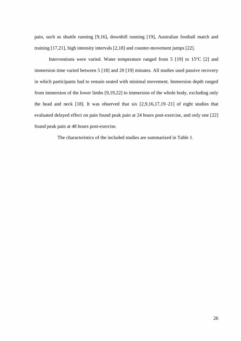
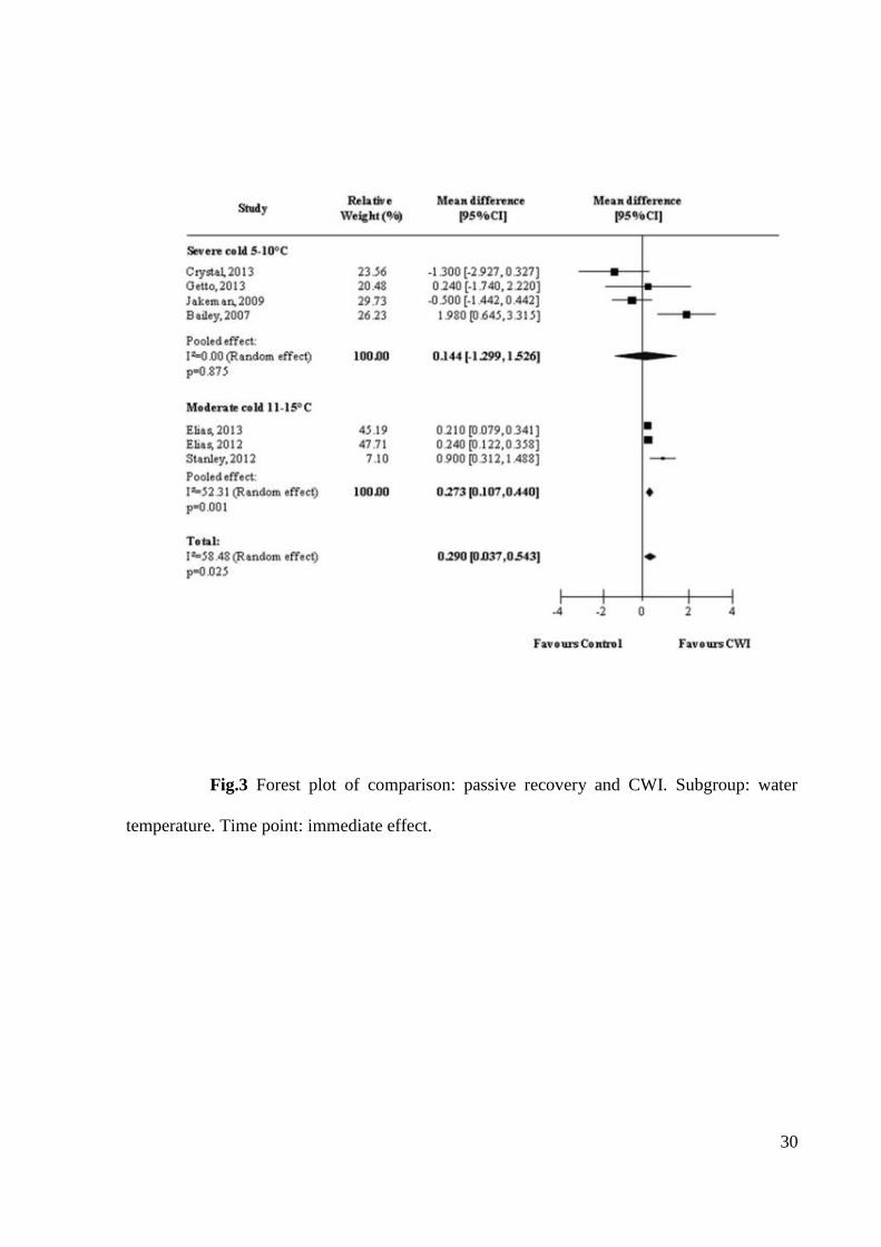
3.1 Analysis of water temperature
Seven studies [9,17–22] provided data related to the immediate effects of cold
water immersion. The subgroup analysis of the pooled results is shown in Figure 3. A general
analysis of the immediate effects shows a significant pooled effect for cold water immersion
(WMD=0.290, 95% CI [0.037, 0.543]; p=0.025). When subgroups were analyzed, it was
observed that studies using a water temperature of between 11-15°C (moderate cold)
produced better results than those using water between 5-10°C (severe cold). Therefore,
temperatures higher than 10°C present the best results for immediate effect on pain (Severe
cold: WMD=0.144, 95% CI [-1.299, 1.526], p=0.875]; Moderate cold: WMD=0.273, 95% CI
[0.107, 0.440], p=0.001).
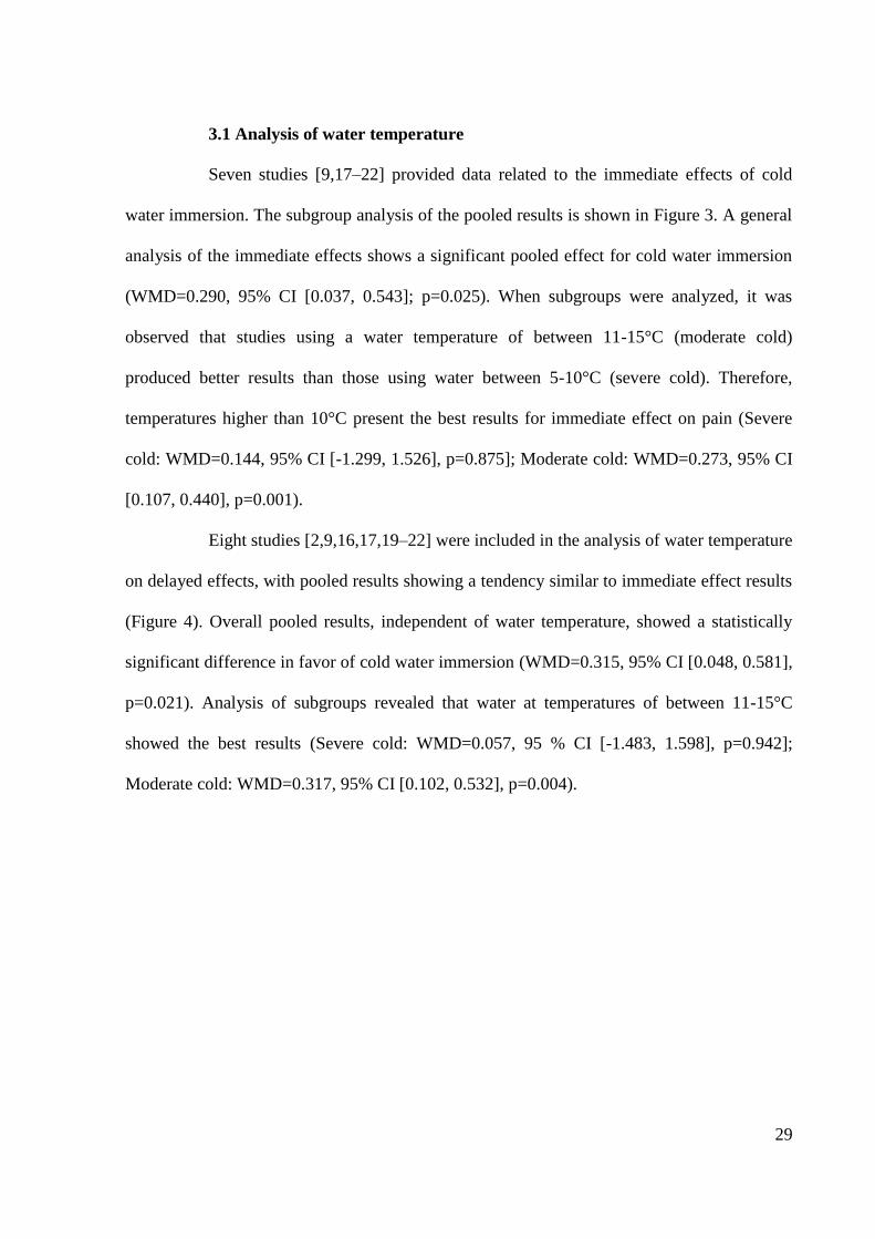
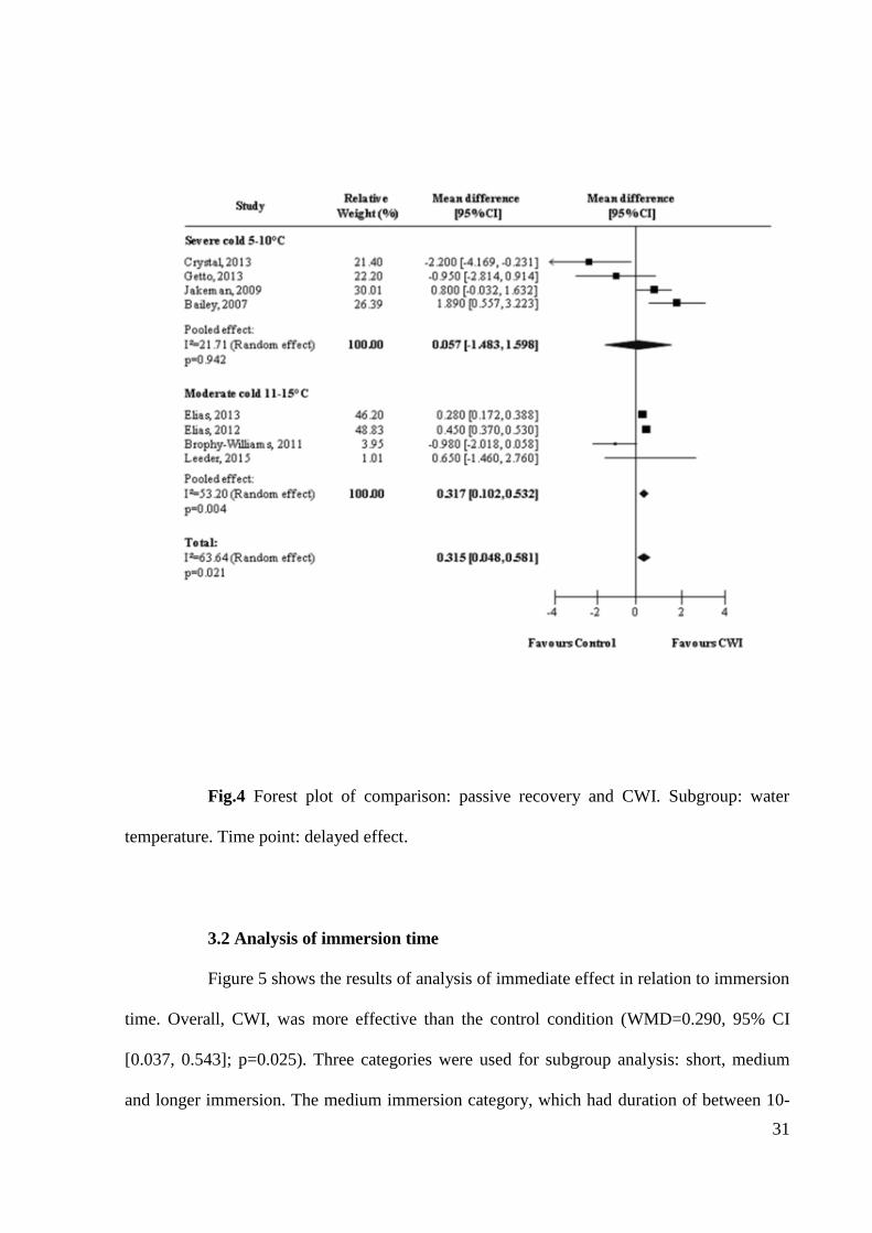
Eight studies [2,9,16,17,19–22] were included in the analysis of water temperature
on delayed effects, with pooled results showing a tendency similar to immediate effect results
(Figure 4). Overall pooled results, independent of water temperature, showed a statistically
significant difference in favor of cold water immersion (WMD=0.315, 95% CI [0.048, 0.581],
p=0.021). Analysis of subgroups revealed that water at temperatures of between 11-15°C
showed the best results (Severe cold: WMD=0.057, 95 % CI [-1.483, 1.598], p=0.942];
Moderate cold: WMD=0.317, 95% CI [0.102, 0.532], p=0.004).
30
Fig.3 Forest plot of comparison: passive recovery and CWI. Subgroup: water
temperature. Time point: immediate effect.
31
Fig.4 Forest plot of comparison: passive recovery and CWI. Subgroup: water
temperature. Time point: delayed effect.
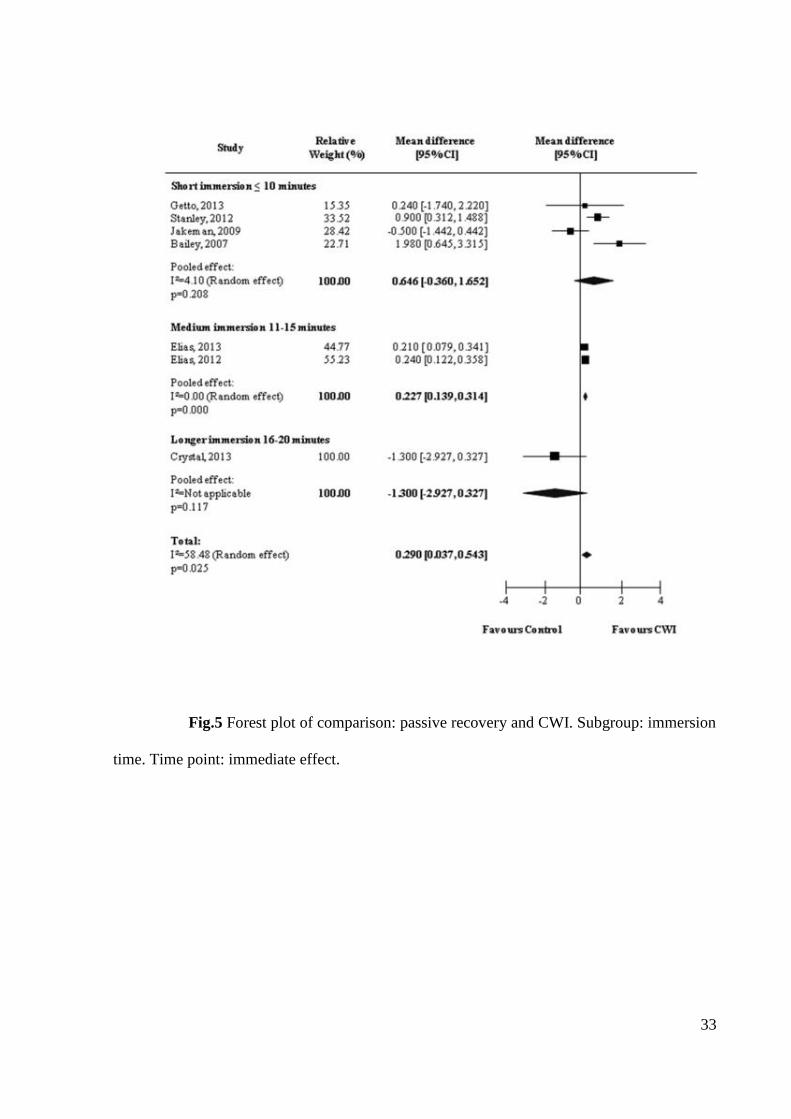
3.2 Analysis of immersion time
Figure 5 shows the results of analysis of immediate effect in relation to immersion
time. Overall, CWI, was more effective than the control condition (WMD=0.290, 95% CI
[0.037, 0.543]; p=0.025). Three categories were used for subgroup analysis: short, medium
and longer immersion. The medium immersion category, which had duration of between 10-
32
15 minutes, was responsible for the best results in terms of immediate effects. Although there
is only one study featuring 'longer immersion' [19] it was observed no effect for this category
(Short immersion: WMD=0.646, 95% [-0.360, 1.652], p=0.208; Medium immersion:
WMD=0.227, 95% [0.139, 0.314], p=0.000; Longer immersion: WMD=-1.300 [-2.927,
0.327], p=0.117).
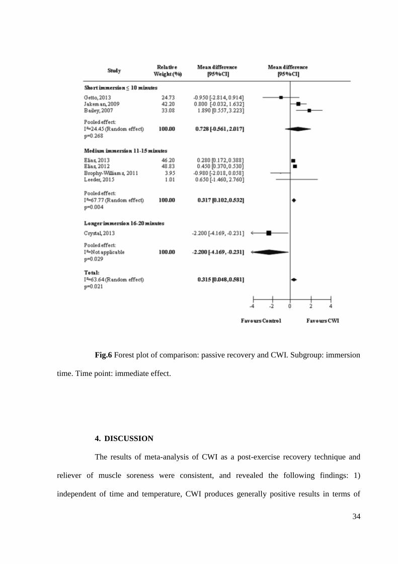
In terms of delayed effects, the overall pooled effects of CWI described in the
eight studies analyzed were positive (WMD = 0.315, 95% CI [0048, 0581], p = 0.021) (Figure
6). As with the immediate effects, an immersion time of between 11-15 minutes produced the
best results (Short immersion: WMD=0.728, 95% [-0.561, 2.017], p=0.268; Medium
immersion: WMD=0.317, 95% [0.102, 0.532], p=0.004; Longer immersion: WMD=-2.200 [-
4.169, -0.231], p=0.029).
33
Fig.5 Forest plot of comparison: passive recovery and CWI. Subgroup: immersion
time. Time point: immediate effect.
34
Fig.6 Forest plot of comparison: passive recovery and CWI. Subgroup: immersion
time. Time point: immediate effect.
4. DISCUSSION
The results of meta-analysis of CWI as a post-exercise recovery technique and
reliever of muscle soreness were consistent, and revealed the following findings: 1)
independent of time and temperature, CWI produces generally positive results in terms of
35
both immediate and delayed effects; 2) immersion in water at temperatures between 11-15°C
appeared to produce a greater reduction of muscle soreness after exercise; 3) 11-15 minutes
appeared to be the optimal immersion time for the relief of muscle soreness caused by
exercise.
The findings regarding CWI, independent of immersion temperature and time, are
in accordance with previous reviews, such as the studies by Leeder et al. [8] and Bleakley et
al. [12]. The authors claim that the technique is capable of altering blood flow, thereby
causing vasoconstriction and redirection of the blood. The real effects of CWI have not been
fully elucidated [4], but it has been speculated that this technique is able to reduce lymphatic
and capillary cell permeability [24], resulting in vasoconstriction and consequent reduction of
the inflammatory process caused by exercise [6]. The technique also can reduce nerve
transmission and muscle spasm. The reduction of pain can therefore be explained by these
factors, together with analgesia, which occurs in response to the reduction of the pain-spasm
cycle [25].
While the effects of CWI have been widely investigated, opinions vary with
regard to method of application, immersion time and water temperature [13]. One hypothesis
is that outcomes may differ depending on the recovery strategy used. The variance in effects
caused by temperature change observed in this review revealed that CWI was more effective
in terms of both immediate and delayed effects when temperatures were in the 'moderate cold'
category. The mean temperature of this category in studies was 12.7° C for immediate effect
and 13.2° C for delayed effect.
The benefits of ‘moderate cold’ temperatures have not been discussed in clinical
studies. However, it has been shown that immersion in very low temperatures can cause
adverse effects, interpreted by the body as noxious stimuli, and that peak pain appears around

36
3°C [26]. This may explain the generally negative effects described in the study by Crystal et
al. [19], which featured CWI at 5°C.
A study by Getto et al. [20] claims that short immersions are less efficient at
lessening muscle pain caused by exercise, due to limited muscle temperature reduction [5].
Such statement confirms the findings of this study, which indicate that medium immersions of
between 11 and 15 minutes produce better results than short immersions (≤10 minutes).
Additionally, during the immediate effect, there is the presence of the category 'longer
immersion', responsible for the worst results. Although pooled results were not available for
this category due to an insufficient number of studies (n=1), it compared unfavorably with
passive recovery. The study in question [19] considered the use of a very low temperature
(5°C) for 20 minutes. Davis et al. [27] claimed that for CWI to produce harmful stimulation
and pain, an application of ten seconds at low temperatures was required. Such effects can be
exacerbated during long immersion conditions [28,29].
Accordingly, it is important to analyze studies by subdivisions of water
temperature and immersion time. The limited number of studies, however, does not allow the
implementation of closed protocol comparisons. The relationship between the two variables
could allow objective inferences about the most effective recovery model to be used.
As Crystal et al. [19], other studies produced results that compared unfavorably
with passive recovery. Getto et al. [20] used a scale that involved six different pain points,
including the low back, to analyze pain. This type of evaluation considers a larger number of
pain points than other studies, and can result in participant confusion in relation to the effects
of CWI. Jakeman et al. [22] found that results of immediate effects of CWI compared
unfavorably with control following countermovement jumps. Goodall et al. [30] and
Howatson et al. [31] used similar exercise procedures and observed a reduction of pain in the

37
CWI group only after 24 hours post-exercise. These adverse results can be explained by the
type of muscle stress and pain magnitude [32].
Overall, the studies selected for this review show similar models of inducing
stress, represented by physical activities featuring high intensity of effort. This factor is
relevant to data interpretation, as different types of stress provide different outcomes, as
previously explained. For example, the characteristics of injuries induced in localized
eccentric exercise can differ from those sustained during sporting activities, and respond
differently to the application of CWI [7,32].
To the authors’ knowledge this is the first systematic review and meta-analysis to
investigate the effects of different CWI procedures, namely variations in water temperature
and immersion time. The strengths of this systematic review relate mainly to the rationale of
the study, which aims to analyze the dose-response relationship, which is still poorly
investigated in studies of this nature. One of the limitations of the study is the poor
methodological quality of the studies included, as assessed by PEDro Scale. Future trials
should be attentive to the criteria for the development of a high quality study, which would
result in surveys with greater scientific evidence. Another limitation is that the research
focused only on pain. Although it is a key outcome in recovery of an athlete, further studies
should consider the dose-response effect of CWI on other markers of muscle damage, in order
to identify the best CWI recovery strategy based on different factors.
5. CONCLUSION
The findings of the present study suggest that CWI is more effective than passive
recovery in management of pain. The results also demonstrate the presence of a dose-response
relationship, indicating that CWI provides the most effective results at temperatures between
38
11 and 15ºC, for 11 to 15 min. The low quality of the included studies should be considered.
Higher quality studies are needed to investigate whether the dose-response relationship of the
results can be reliably reproduced.
The findings of the study allow athletes using CWI to have a better understanding
of the technique, resulting in a better dynamic in training and competition, leading to less
aggressive and painful immersion. For those applying CWI, it will allow the use of improved
logistics and therefore result in lower costs, due to the most effective use of immersion time
and water temperature.
ACKNOWLEDGMENTS
The authors would like to thank the Univ. Estadual Paulista, The University of
Sydney and the Sao Paulo Research Foundation (FAPESP).
39
6. REFERENCES
1. Bastos FN, Vanderlei LCM, Nakamura FY, et al. Effects of cold water
immersion and active recovery on post-exercise heart rate variability. Int J Sports Med.
2012;33(11):873–9.
2. Brophy-Williams N, Landers G, Wallman K. Effect of immediate and delayed
cold water immersion after a high intensity exercise session on subsequent run performance. J
Sports Sci Med. 2011:665–670.
3. Pournot H, Bieuzen F, Duffield R, et al. Short term effects of various water
immersions on recovery from exhaustive intermittent exercise. Eur J Appl Physiol.
2011;111(7):1287–95.
4. Broatch JR, Petersen A, Bishop DJ. Postexercise cold water immersion
benefits are not greater than the placebo effect. Med Sci Sports Exerc. 2014;46(11):2139–
2147.
5. Bleakley CM, Davison GW. What is the biochemical and physiological
rationale for using cold-water immersion in sports recovery? A systematic review. Br J Sports
Med. 2010;44(3):179–87.
6. Wilcock IM, Cronin JB, Hing WA. Physiological response to water immersion:
a method for sport recovery ? Sports Med. 2006;36(9):1–18.
7. Bieuzen F, Bleakley CM, Costello JT. Contrast water therapy and exercise
induced muscle damage: a systematic review and meta-analysis. PLoS One.
2013;8(4):e62356.
8. Leeder J, Gissane C, Someren K Van, et al. Cold water immersion and
recovery from strenuous exercise: a meta-analysis. Br J Sports Med. 2012 Mar;46(4):233-40.
9. Bailey DM, Erith SJ, Griffin PJ, et al. Influence of cold-water immersion on
indices of muscle damage following prolonged intermittent shuttle running. J Sports Sci.
2007;25(11):1163–70.
10. Glasgow PD, Ferris R, Bleakley CM. Cold water immersion in the
management of delayed-onset muscle soreness: Is dose important? A randomised controlled
trial. Phys Ther Sport. 2014.
11. Leal Junior EC, de Godoi V, Mancalossi JL, et al. Comparison between cold
water immersion therapy (CWIT) and light emitting diode therapy (LEDT) in short-term
skeletal muscle recovery after high-intensity exercise in athletes--preliminary results. Lasers
Med Sci. 2011;26(4):493–501.
12. Bleakley C, Mcdonough S, Gardner E, et al. Cold-water immersion
(cryotherapy) for preventing and treating muscle soreness after exercise. Cochrane Database
Syst Rev. 2012 Feb 15;2:CD008262.
40
13. Pastre CM, Bastos FN, Netto Jr J, et al. Métodos de recuperação pós-
exercício: uma revisão sistemática. Rev Bras Med Esporte. 2009;15:138–144.
14. Maher CG, Sherrington C, Robert D, et al. Research report reliability of the
PEDro Scale for rating quality of randomized. Phys Ther. 2003:713–721.
15. Macedo LG, Elkins MR, Maher CG, et al. There was evidence of convergent
and construct validity of Physiotherapy Evidence Database quality scale for physiotherapy
trials. J Clin Epidemiol. 2010;63(8):920–5.
16. Leeder JDC, Van Someren K a., Bell PG, et al. Effects of seated and standing
cold water immersion on recovery from repeated sprinting. J Sports Sci. 2015;(January):1–9.
17. Elias GP, Varley MC, Wyckelsma VL, et al. Effects of water immersion on
posttraining recovery in Australian footballers. Int J Sports Physiol Perform. 2012;7(4):357–
66.
18. Stanley J, Buchheit M, Peake JM. The effect of post-exercise hydrotherapy on
subsequent exercise performance and heart rate variability. Eur J Appl Physiol.
2012;112(3):951–61.
19. Crystal NJ, Townson DH, Cook SB, et al. Effect of cryotherapy on muscle
recovery and inflammation following a bout of damaging exercise. Eur J Appl Physiol.
2013;113(10):2577–86.
20. Getto CN, Golden G. Comparison of active recovery in water and cold-water
immersion after exhaustive exercise. Athl Train Sport Heal Care. 2013;5(4):169–176.
21. Elias GP, Wyckelsma VL, Varley MC, et al. Effectiveness of Water
Immersion on Postmatch Recovery in Elite Professional Footballers. Int J Sports Physiol
Perform. 2013:243–254.
22. Jakeman JR, Macrae R, Eston R. A single 10-min bout of cold-water
immersion therapy after strenuous plyometric exercise has no beneficial effect on recovery
from the symptoms of exercise-induced muscle damage. Ergonomics. 2009;52(4):456–60.
23. Stanley J, Buchheit M, Peake JM. The effect of post-exercise hydrotherapy on
subsequent exercise performance and heart rate variability. Eur J Appl Physiol.
2012;112(3):951–61.
24. Ascensão A, Leite M, Rebelo AN, et al. Effects of cold water immersion on
the recovery of physical performance and muscle damage following a one-off soccer match. J
Sports Sci. 2011;29(3):217–25.
25. Halson SL, Quod MJ, Martin DT, et al. Physiological responses to cold water
immersion following cycling in the heat. Int J Sport Physiol Perform. 2008;3:331–346.
26. Sellwood KL, Brukner P, Williams D, et al. Ice-water immersion and delayed-
onset muscle soreness: a randomised controlled trial. Br J Sports Med. 2007;41(6):392–7.
41
27. Davis KD, Pope GE. Noxious cold evokes multiple sensations with distinct
time courses. Pain. 2002;98(1-2):179–185.
28. Yeargin SW, Casa DJ, McClung JM, et al. Body cooling between two bouts of
exercise in the heat enhances subsequent performance. J strength Cond Res / Natl Strength
Cond Assoc. 2006;20(2):383–389.
29. Versey NG, Halson SL, Dawson BT. Water immersion recovery for athletes:
effect on exercise performance and practical recommendations. Sports Med.
2013;43(11):1101–30.
30. Goodall S, Howatson G. The effects of multiple cold water immersions on
indices of muscle damage. J Sports Sci Med. 2008 Jun; 7(2): 235–241.
31. Howatson G, Goodall S, van Someren K a. The influence of cold water
immersions on adaptation following a single bout of damaging exercise. Eur J Appl Physiol.
2009;105(4):615–21.
32. Pointon M, Duffield R, Cannon J, et al. Cold application for neuromuscular
recovery following intense lower-body exercise. Eur J Appl Physiol. 2011;111(12):2977–86.
42
Appendix Table 1. Search strategy
MEDLINE (up to 21 January, 2015)
1. (randomized controlled trial) OR (controlled trial) OR (controlled clinical trial) OR (clinical
trial)
2. (pain) OR (soreness) OR (muscle soreness) OR (delayed onset muscle soreness)
3. (cryotherapy) OR (cold water immersion) OR (cold-water immersion) OR (ice water
immersion) OR (ice-water immersion) OR (ice bath) OR (ice-bath) OR (hydrotherapy)
4. (recovery) OR (post-exercise) OR (post exercise) OR (postexercise)
5. (animal) OR (animals)
6. 1 AND 2 AND 3 AND 4 NOT 5
EMBASE (up to 21 January, 2015)
1. (randomized controlled trial) OR (controlled trial) OR (controlled clinical trial) OR (clinical
trial)
2. (pain) OR (soreness) OR (muscle soreness) OR (delayed onset muscle soreness)
3. (cryotherapy) OR (cold water immersion) OR (cold-water immersion) OR (ice water
immersion) OR (ice-water immersion) OR (ice bath) OR (ice-bath) OR (hydrotherapy)
4. (recovery) OR (post-exercise) OR (post exercise) OR (postexercise)
5. 1 AND 2 AND 3 AND 4
SportDiscus (up to 21 January, 2015)
1. (randomized controlled trial) OR (controlled trial) OR (controlled clinical trial) OR (clinical
trial)
2. (pain) OR (soreness) OR (muscle soreness) OR (delayed onset muscle soreness)
3. (cryotherapy) OR (cold water immersion) OR (cold-water immersion) OR (ice water
immersion) OR (ice-water immersion) OR (ice bath) OR (ice-bath) OR (hydrotherapy)
4. (recovery) OR (post-exercise) OR (post exercise) OR (postexercise)
5. (animal) OR (animals)
6. 1 AND 2 AND 3 AND 4 NOT 5
The Cochrane Library (up to 21 January, 2015)
1. (randomized controlled trial) OR (controlled trial) OR (controlled clinical trial) OR (clinical
trial)
2. (pain) OR (soreness) OR (muscle soreness) OR (delayed onset muscle soreness)
3. (cryotherapy) OR (cold water immersion) OR (cold-water immersion) OR (ice water
immersion) OR (ice-water immersion) OR (ice bath) OR (ice-bath) OR (hydrotherapy)
4. (recovery) OR (post-exercise) OR (post exercise) OR (postexercise)
5. (animal) OR (animals)
6. 1 AND 2 AND 3 AND 4 NOT 5
Filter: trial
PEDro database (up to 21 January, 2015)
Abstract and title:
1. Pain
2. Soreness
3. Muscle soreness
4. Delayed onset muscle soreness
Therapy: hydrotherapy, balneotherapy
Subdiscipline: Sports
Method: clinical trial
*1, 2, 3, 4 were individually searched with the same therapy, subdiscipline and method.
43
Appendix Table 2. Physiotherapy Evidence Database Scores of Included Studies
Study Random
allocation
Concealed
allocation
Groups
similiar at
baseline
Participant
blinding
Therapist
blinding
Assessor
blinding
<15%
dropouts
Intention-
to-treat
analysis
Between-
group
difference
reported
Point
estimates /
variability
reported
Score
(0-10)
Leeder,
2015 Yes Yes Yes No No No Yes No Yes Yes 6/10†
Crystal,
2013 Yes No Yes No No No No No Yes Yes 4/10*
Getto,
2013 Yes No No No No No No No Yes Yes 3/10†
Elias,
2013 Yes No Yes No No No No No Yes Yes 4/10*
Elias,
2012 Yes No Yes No No No Yes No Yes Yes 5/10*
Stanley,
2012 Yes No Yes No No No Yes No Yes Yes 5/10*
Brophy-
Willians,
2011
Yes No No No No No Yes No Yes Yes 4/10†
Jakeman,
2009 Yes No Yes No No No No No Yes No 3/10*
Bailey,
2007 Yes No Yes No No No No No Yes Yes 4/10*
* Trials assessed and listed on the PEDro Database
† Trial assessed by the 2 independent raters
44
Appendix: Registration PROSPERO. Available:
http://www.crd.york.ac.uk/PROSPERO/display_record.asp?ID=CRD42015016573#.VO99aP
nF9bc

45
46
47
48
Artigo 2
Immediate and delayed effects of cold water immersion with different dosages after
eccentric exercise-induced muscle damage: a randomized controlled trial
Cold water immersion with different dosages after eccentric exercise
Aryane Flauzino Machado1†
; Aline Castilho de Almeida2†
; Jéssica Kirsch Micheletti1†
;
Franciele Marques Vanderlei3†
; Marcelo Fernandes Tribst3; Jayme Netto Junior
3†;
Carlos Marcelo Pastre3†
*
1Programa de Pós-Graduação em Fisioterapia. Univ Estadual Paulista, Presidente
Prudente/SP, Brazil
2Programa de Pós-Graduação em Fisioterapia. Univ Federal de São Carlos, São Carlos/SP,
Brazil
3Departamento de Fisioterapia. Univ Estadual Paulista, Presidente Prudente/SP, Brazil
†Laboratório de Fisioterapia Desportiva. Univ Estadual Paulista, Presidente Prudente/SP,
Brazil
* Corresponding author:
E-mail: [email protected] (CMP)
49
ABSTRACT
Purpose: To compare the effects of two strategies of cold water immersion (CWI), using
different water temperatures, with passive recovery post-exercise, in the management of
markers of muscle damage and to observe whether any of the techniques used caused
deleterious effects on performance.
Materials and Methods: 60 healthy male participants performed an eccentric exercise
protocol to induce muscle damage and were then randomized to one of three groups (CWI1:
15 min at 9°C; CWI2: 15 min at 14°C; CG: control group). Levels of creatine kinase, muscle
soreness, pain threshold, perception of recovery and maximal voluntary isometric contraction
were monitored up to 96 hours post-exercise.
Results: Regarding the immediate effect, cold water immersion, independent of water
temperature, presented an earlier recovery for soreness and the CWI2 group presented an
early recovery for performance (P<0.05). It was observed that there were no deleterious
effects and no evidence was found to suggest a dose-response relationship.
Conclusions: The use of CWI is recommended as a post-exercise recovery technique and the
application for 15 min at 14°C is considered the best dosage when the aim is early
performance recovery; however the application should be appropriate to the specific intended
outcome.
Keywords: recovery of function; cryotherapy; immersion; muscle soreness; muscle strength.
50
1. BACKGROUND
Intense, eccentric or unaccustomed exercise has been commonly documented as
exercise able to produce delayed onset muscle soreness (DOMS) and alter various markers of
muscle damage [1–5]. Responses such as a decrease in muscle function can also be related to
muscle damage [6], demonstrating that these types of exercises can influence the performance
and recovery of athletes [2]. Different scenarios of recovery strategies are currently being
investigated in order to minimize performance decrements, such as massage, active recovery,
contrast water therapy and cryotherapy [6, 7, 8].
Cold water immersion (CWI) is one recovery technique which is commonly used
by athletes post-exercise to promote the restoration of body systems to baseline conditions
and establish the physiological system to a pre-exercise state [9, 10]. The effects of CWI such
as cooling the body tissues, reducing lymphatic, capillary and cellular permeability and
decreasing nerve conduction velocity, muscular spindle activity and spasticity, have been
discussed in several studies [2, 11, 12]. Despite these responses, the specific mechanism of
CWI is unknown [12] and can change according to the type of exercise performed prior to the
CWI and the methodology adopted for immersion [13], presenting specific evidence after
exercise-induced muscle damage (EIMD) [2,11,12].
Several clinical trials and systematic reviews have compared the effects of CWI
with other post-exercise recovery strategies [1-3; 5-10; 14-17]. Recent reviews found that,
compared to control interventions and other traditional techniques of recovery, CWI is a more
effective technique to reduce pain after a range of exercises [8, 18]. Nevertheless, studies that
use different methodologies for the application of CWI, such as water temperature, duration
of immersion and type of CWI, for example continuous or intermittent immersion [14] and
the dose-response relationship of this technique have not yet been fully investigated.
51
Bleakley et al., 2012 [4] claimed that there are still insufficient studies available
to determine the best method of CWI. From this statement, it can be is understood that
conclusions about the effects of CWI are not well elucidated and require further research,
including studies which approach the dose-response relationship. The purpose of this study
was to analyze the immediate and delayed effects of CWI as a mode of post-exercise recovery
on the management of some markers of muscle damage. The primary objective was to
compare the effects of two strategies of CWI, using different water temperatures, with passive
recovery post-exercise. Furthermore, from these findings, to observe whether any of the
techniques used caused deleterious effects on performance on subsequent days post-exercise.
Considering the positive results of previously cited studies, the hypothesis of this study was
that CWI would present better results when compared with passive recovery. Moreover,
considering that CWI is more efficient than other modalities for reducing neural conduction
velocity [19], together with the knowledge that reduction in performance is proportional to
muscle cooling [20, 21], another hypothesis was that CWI may have a negative influence on
performance, related to the deleterious effects, especially when applied at 9°C.
2. METHODS
2.1 Participants
Sixty young, healthy male participants (aged 18-25 years, height 1.74±0.06 m,
body mass 74.4±11.1 kg and body mass index 24.4±4.1 kg . m -2) participated in this study, as
show in the flowchart (Fig. 1). To be included, the participants were required to report the
absence of anemia, inflammation, diabetes, cardiovascular diseases and muscle injuries in the
lower limbs and/or spine in the previous six months. Participants were required to abstain
from anti-inflammatory and analgesic drugs and not perform any exercise during the study.

52
Prior to data collection, a medical evaluation was performed to ensure that the participants
were fully able to participate in the study.
Fig 1. Flowchart of participants
To define the sample size a priori knowledge was used, based on the findings of
Bailey et al., 2007 [2] for concentration of creatine kinase (SD=200 U/L). A sample size of
twenty participants per group was stipulated by a test of hypothesis (two-tail), with a 5% level
of significance and 80% power. Thus, a randomization sequence was created using software
(Microsoft Office Excel 2007) and a computer-generated random list was used for allocation.

53
The participants were allocated into three groups: control group (CG: passive recovery for 15
minutes) and two intervention groups (CWI1 – 15 minutes at 9±1°C and CWI2 – 15 minutes
at 14±1°C). During the study, the participants received no information about which
intervention was considered therapeutic.
2.2 Ethics statement
The study had been previously approved by the Ethics Committee in Research of
the Univ. Estadual Paulista (Faculty of Science and Technology – UNESP; protocol number:
2013/396.666). The participants received oral and written instructions and signed a Consent
Form agreeing to the research procedures.
2.3 Study design
The study procedure was carried out between January and April 2014 at the
Centre for Studies and Treatment in Physical Therapy and Rehabilitation – UNESP. All
procedures were performed in standardized conditions (temperature: 21-23°C; relative
humidity: 40-60%) [22].
Each participant attended the laboratory on five consecutive days. Prior to the
procedures, the participants were assessed for anthropometric characteristics using a scale
(Tanita BC 554, Iron Man/Inner - Illinois, USA) and a stadiometer (Sany – American Medical
do Brasil, São Paulo, Brazil) (Table 1). The baseline assessment, eccentric exercise-induced
muscle damage (EIMD), intervention and immediate assessment were performed on the first
day. Subsequent visits were performed 24, 48, 72 and 96 hours after the EIMD, relating to the
delayed effect. An overview of the study is presented in Fig 2.
54
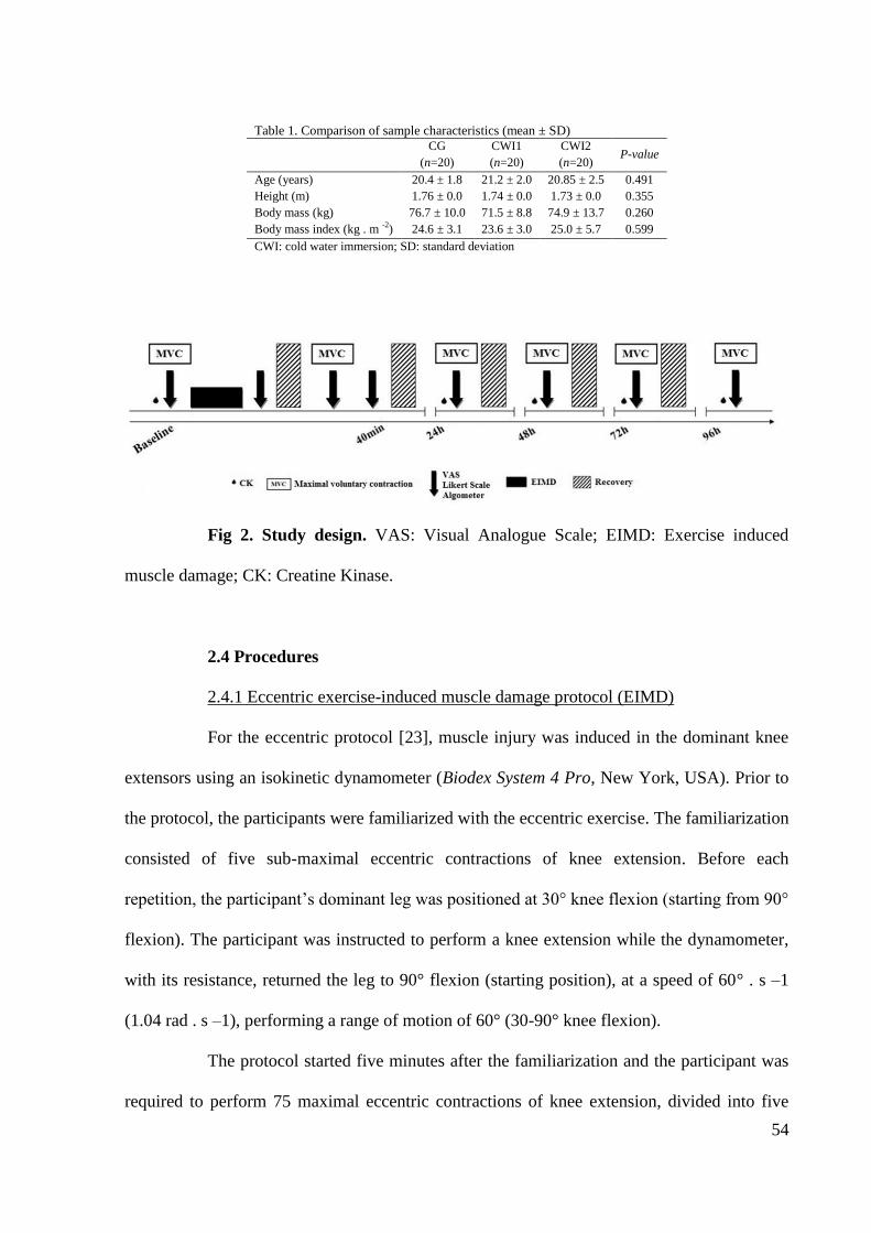
Table 1. Comparison of sample characteristics (mean ± SD)
CG
(n=20)
CWI1
(n=20)
CWI2
(n=20) P-value
Age (years) 20.4 ± 1.8 21.2 ± 2.0 20.85 ± 2.5 0.491
Height (m) 1.76 ± 0.0 1.74 ± 0.0 1.73 ± 0.0 0.355
Body mass (kg) 76.7 ± 10.0 71.5 ± 8.8 74.9 ± 13.7 0.260
Body mass index (kg . m -2
) 24.6 ± 3.1 23.6 ± 3.0 25.0 ± 5.7 0.599
CWI: cold water immersion; SD: standard deviation
Fig 2. Study design. VAS: Visual Analogue Scale; EIMD: Exercise induced
muscle damage; CK: Creatine Kinase.
2.4 Procedures
2.4.1 Eccentric exercise-induced muscle damage protocol (EIMD)
For the eccentric protocol [23], muscle injury was induced in the dominant knee
extensors using an isokinetic dynamometer (Biodex System 4 Pro, New York, USA). Prior to
the protocol, the participants were familiarized with the eccentric exercise. The familiarization
consisted of five sub-maximal eccentric contractions of knee extension. Before each
repetition, the participant’s dominant leg was positioned at 30° knee flexion (starting from 90°
flexion). The participant was instructed to perform a knee extension while the dynamometer,
with its resistance, returned the leg to 90° flexion (starting position), at a speed of 60° . s –1
(1.04 rad . s –1), performing a range of motion of 60° (30-90° knee flexion).
The protocol started five minutes after the familiarization and the participant was
required to perform 75 maximal eccentric contractions of knee extension, divided into five

55
sets of 15 repetitions, separated by a rest period of 30s. The velocity and range of motion were
similar to the familiarization.
2.4.2 Creatine kinase (CK)
Creatine kinase concentration was obtained from capillary blood, collected by
finger-prick (32 µL). The blood sample (total blood) was analyzed using a Reflotron Plus
reader (Roche Diagnostics, Mannheim, Germany) in 37°C.
2.4.3 Muscle soreness (VAS) and pain threshold (algometer)
The participants were instructed to assess the soreness in their leg (induced by the
damage), using a Visual Analogue Scale (VAS) ranging from 0 “no soreness” to 10 “extreme
soreness” [2]. During the assessment, the participant performed a maximal isometric
contraction on an isokinetic dynamometer with the knee flexed at 60° during the assessment.
Pain threshold was assessed using a pressure algometer (FPX 50/220, Wagner
Instruments, Greenwich, USA). The participant indicated specific painful points during the
isometric contraction (60º knee flexion). The algometer was then applied to the indicated
point until the participant reported discomfort. The pain threshold was defined in Kgf and did
not exceed 2.55 Kgf, as suggested by Jönhagen et al., 2009 [24]. The assessment was
performed with the leg at 60º knee flexion, with muscles relaxed.
2.4.4 Perception of recovery (Likert Scale)
Perception of recovery was obtained using a 10-point Likert Scale, with a rating
of 1 indicating the feeling “not recovered” and a rating of 10 the feeling “fully recovered”
[25]. The participant was asked the following question to assess muscle function: “If you had
to perform the MVIC now, how recovered do you feel?
56
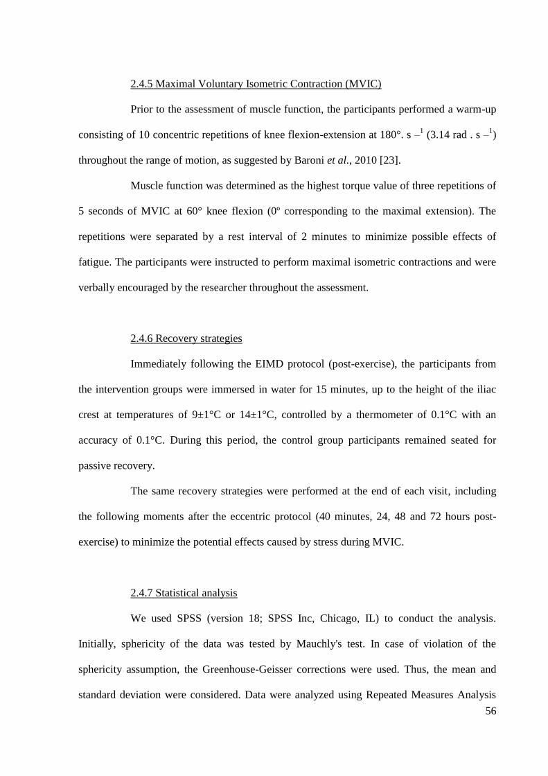
2.4.5 Maximal Voluntary Isometric Contraction (MVIC)
Prior to the assessment of muscle function, the participants performed a warm-up
consisting of 10 concentric repetitions of knee flexion-extension at 180°. s –1 (3.14 rad . s –
1)
throughout the range of motion, as suggested by Baroni et al., 2010 [23].
Muscle function was determined as the highest torque value of three repetitions of
5 seconds of MVIC at 60° knee flexion (0º corresponding to the maximal extension). The
repetitions were separated by a rest interval of 2 minutes to minimize possible effects of
fatigue. The participants were instructed to perform maximal isometric contractions and were
verbally encouraged by the researcher throughout the assessment.
2.4.6 Recovery strategies
Immediately following the EIMD protocol (post-exercise), the participants from
the intervention groups were immersed in water for 15 minutes, up to the height of the iliac
crest at temperatures of 9±1°C or 14±1°C, controlled by a thermometer of 0.1°C with an
accuracy of 0.1°C. During this period, the control group participants remained seated for
passive recovery.
The same recovery strategies were performed at the end of each visit, including
the following moments after the eccentric protocol (40 minutes, 24, 48 and 72 hours post-
exercise) to minimize the potential effects caused by stress during MVIC.
2.4.7 Statistical analysis
We used SPSS (version 18; SPSS Inc, Chicago, IL) to conduct the analysis.
Initially, sphericity of the data was tested by Mauchly's test. In case of violation of the
sphericity assumption, the Greenhouse-Geisser corrections were used. Thus, the mean and
standard deviation were considered. Data were analyzed using Repeated Measures Analysis

57
of Variance (Bonferroni’s test was performed when required), which provide information of
time, group and interaction effects. All statistical analysis assumed a significance level of 5%.
3. RESULTS
Values of Table 2 are presented as mean and standard deviation values and shows
a significant effect for time for all outcomes (P<0.001). There were no significant group and
interaction (Group*time) effects.
Creatine kinase activity had increased significantly at 24 hours and continued
increasing until 96 hours post-exercise (P<0.05). The CK peak occurred between 72 and 96
hours post-exercise.
The exercise protocol resulted in severe muscle soreness that peaked immediately
after the protocol and 48 hours post-exercise. Both intervention groups demonstrated reduced
ratings of muscle soreness immediately post-recovery and 40 minutes post-exercise. All
groups reported a reduced rating of soreness at 72 hours post-exercise (P>0.05). Ratings of
perception of recovery decreased significantly post-exercise (P<0.05) and began to increase at
72 hours post-exercise.
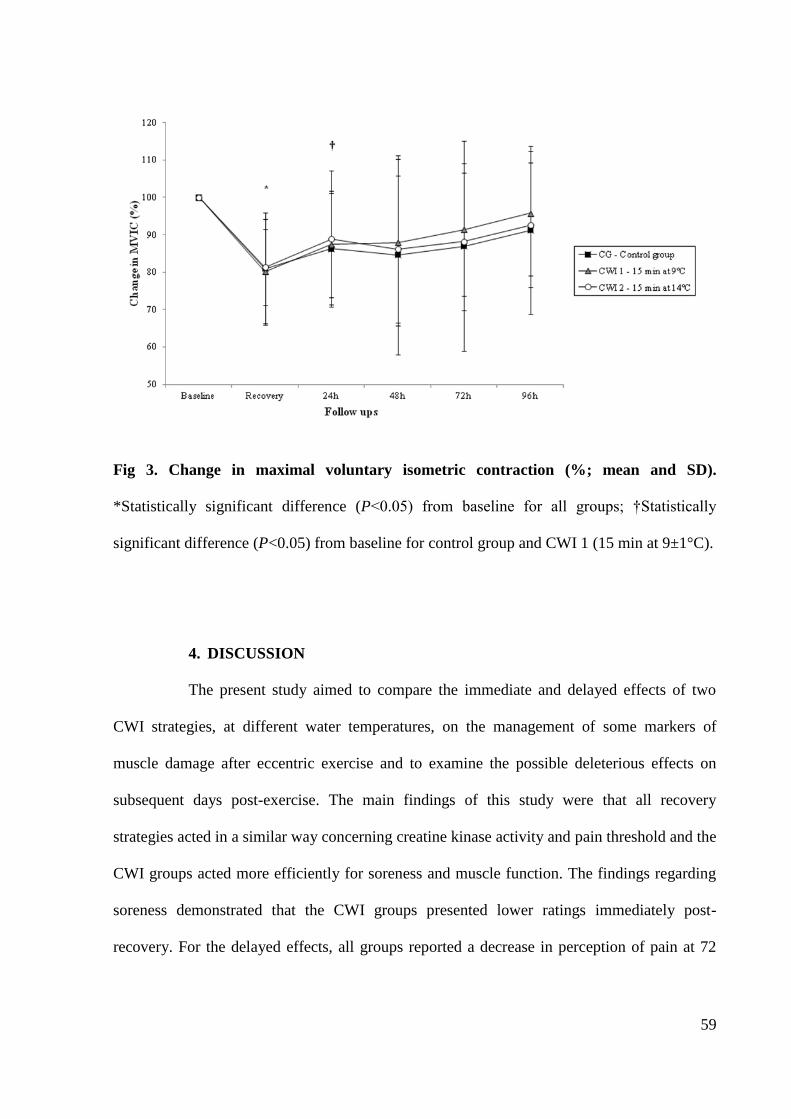
All groups presented reduced MVIC post-recovery (P<0.05). However,
significant muscle function losses were recorded 24 hours post-exercise for the control and
CWI 1 groups, while the CWI 2 demonstrated recovered values and presented no difference to
baseline after the first day (Fig 3).
58
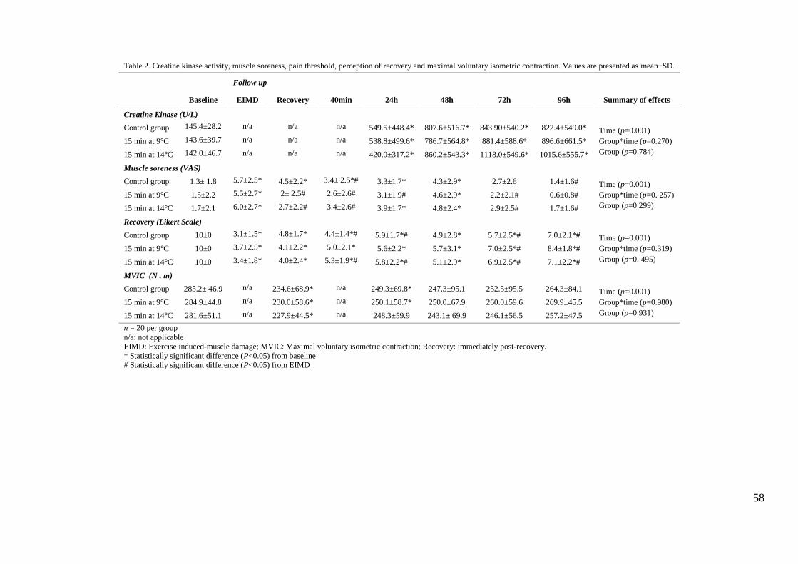
Table 2. Creatine kinase activity, muscle soreness, pain threshold, perception of recovery and maximal voluntary isometric contraction. Values are presented as mean±SD.
Follow up
Baseline EIMD Recovery 40min 24h 48h 72h 96h Summary of effects
Creatine Kinase (U/L)
Control group 145.4±28.2 n/a n/a n/a 549.5±448.4* 807.6±516.7* 843.90±540.2* 822.4±549.0* Time (p=0.001)
Group*time (p=0.270)
Group (p=0.784)
15 min at 9°C 143.6±39.7 n/a n/a n/a 538.8±499.6* 786.7±564.8* 881.4±588.6* 896.6±661.5*
15 min at 14°C 142.0±46.7 n/a n/a n/a 420.0±317.2* 860.2±543.3* 1118.0±549.6* 1015.6±555.7*
Muscle soreness (VAS)
Control group 1.3± 1.8 5.7±2.5* 4.5±2.2* 3.4± 2.5*# 3.3±1.7* 4.3±2.9* 2.7±2.6 1.4±1.6# Time (p=0.001)
Group*time (p=0. 257)
Group (p=0.299)
15 min at 9°C 1.5±2.2 5.5±2.7* 2± 2.5# 2.6±2.6# 3.1±1.9# 4.6±2.9* 2.2±2.1# 0.6±0.8#
15 min at 14°C 1.7±2.1 6.0±2.7* 2.7±2.2# 3.4±2.6# 3.9±1.7* 4.8±2.4* 2.9±2.5# 1.7±1.6#
Recovery (Likert Scale)
Control group 10±0 3.1±1.5* 4.8±1.7* 4.4±1.4*# 5.9±1.7*# 4.9±2.8* 5.7±2.5*# 7.0±2.1*# Time (p=0.001)
Group*time (p=0.319)
Group (p=0. 495)
15 min at 9°C 10±0 3.7±2.5* 4.1±2.2* 5.0±2.1* 5.6±2.2* 5.7±3.1* 7.0±2.5*# 8.4±1.8*#
15 min at 14°C 10±0 3.4±1.8* 4.0±2.4* 5.3±1.9*# 5.8±2.2*# 5.1±2.9* 6.9±2.5*# 7.1±2.2*#
MVIC (N . m)
Control group 285.2± 46.9 n/a 234.6±68.9* n/a 249.3±69.8* 247.3±95.1 252.5±95.5 264.3±84.1 Time (p=0.001)
Group*time (p=0.980)
Group (p=0.931)
15 min at 9°C 284.9±44.8 n/a 230.0±58.6* n/a 250.1±58.7* 250.0±67.9 260.0±59.6 269.9±45.5
15 min at 14°C 281.6±51.1 n/a 227.9±44.5* n/a 248.3±59.9 243.1± 69.9 246.1±56.5 257.2±47.5
n = 20 per group
n/a: not applicable
EIMD: Exercise induced-muscle damage; MVIC: Maximal voluntary isometric contraction; Recovery: immediately post-recovery.
* Statistically significant difference (P<0.05) from baseline
# Statistically significant difference (P<0.05) from EIMD
59
Fig 3. Change in maximal voluntary isometric contraction (%; mean and SD).
*Statistically significant difference (P<0.05) from baseline for all groups; †Statistically
significant difference (P<0.05) from baseline for control group and CWI 1 (15 min at 9±1°C).
4. DISCUSSION
The present study aimed to compare the immediate and delayed effects of two
CWI strategies, at different water temperatures, on the management of some markers of
muscle damage after eccentric exercise and to examine the possible deleterious effects on
subsequent days post-exercise. The main findings of this study were that all recovery
strategies acted in a similar way concerning creatine kinase activity and pain threshold and the
CWI groups acted more efficiently for soreness and muscle function. The findings regarding
soreness demonstrated that the CWI groups presented lower ratings immediately post-
recovery. For the delayed effects, all groups reported a decrease in perception of pain at 72
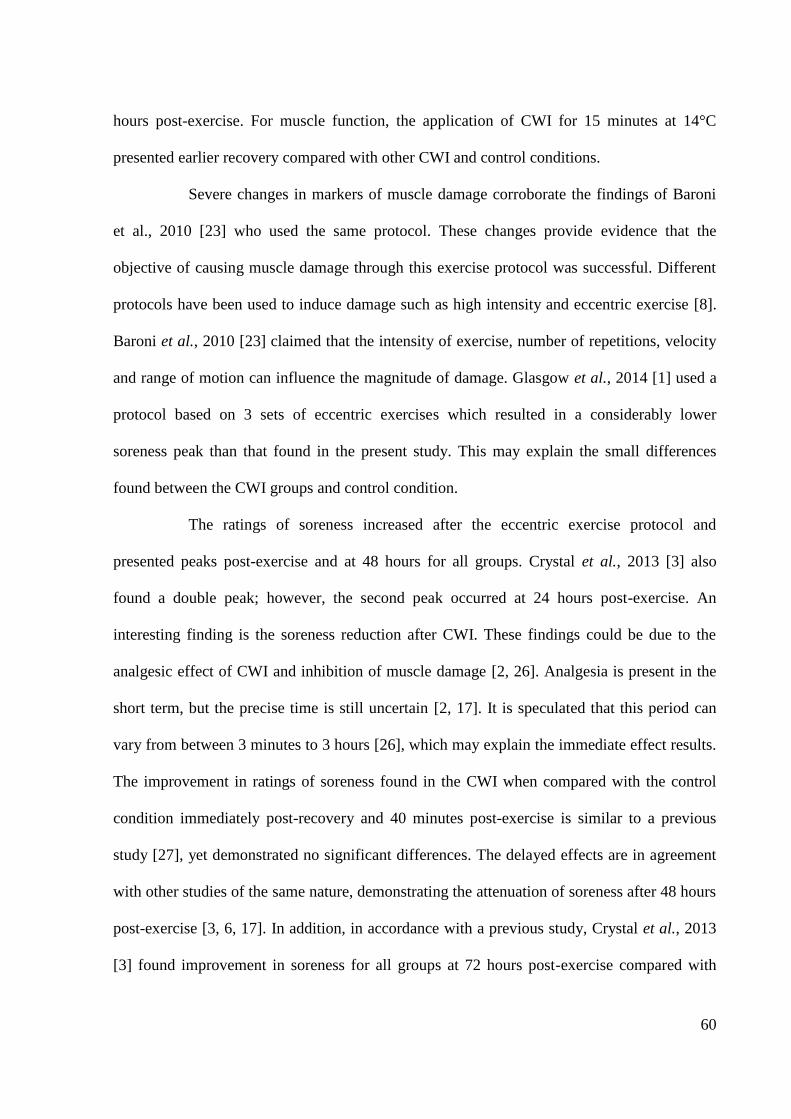
60
hours post-exercise. For muscle function, the application of CWI for 15 minutes at 14°C
presented earlier recovery compared with other CWI and control conditions.
Severe changes in markers of muscle damage corroborate the findings of Baroni
et al., 2010 [23] who used the same protocol. These changes provide evidence that the
objective of causing muscle damage through this exercise protocol was successful. Different
protocols have been used to induce damage such as high intensity and eccentric exercise [8].
Baroni et al., 2010 [23] claimed that the intensity of exercise, number of repetitions, velocity
and range of motion can influence the magnitude of damage. Glasgow et al., 2014 [1] used a
protocol based on 3 sets of eccentric exercises which resulted in a considerably lower
soreness peak than that found in the present study. This may explain the small differences
found between the CWI groups and control condition.
The ratings of soreness increased after the eccentric exercise protocol and
presented peaks post-exercise and at 48 hours for all groups. Crystal et al., 2013 [3] also
found a double peak; however, the second peak occurred at 24 hours post-exercise. An
interesting finding is the soreness reduction after CWI. These findings could be due to the
analgesic effect of CWI and inhibition of muscle damage [2, 26]. Analgesia is present in the
short term, but the precise time is still uncertain [2, 17]. It is speculated that this period can
vary from between 3 minutes to 3 hours [26], which may explain the immediate effect results.
The improvement in ratings of soreness found in the CWI when compared with the control
condition immediately post-recovery and 40 minutes post-exercise is similar to a previous
study [27], yet demonstrated no significant differences. The delayed effects are in agreement
with other studies of the same nature, demonstrating the attenuation of soreness after 48 hours
post-exercise [3, 6, 17]. In addition, in accordance with a previous study, Crystal et al., 2013
[3] found improvement in soreness for all groups at 72 hours post-exercise compared with
61
pre-exercise. No effects were observed for pain threshold, which corroborates with
Sellwood’s study [11].
Perception of recovery has been presented as an important tool for evaluating the
effectiveness of different techniques and plays a crucial role in the adaptive process [28, 29].
The improvement in perception of recovery has a direct relationship with the benefits of CWI
[13, 25, 28, 30]. No evidence of an improvement in the perception of recovery in the
immediate effects, unlike the studies of Parouty et al. [30] and Buchheit et al. [25]. Some
authors [28, 31] have pointed out that there is a contribution from the psychological
mechanism and athletes often present better performances when they believe they have
received a beneficial treatment, which was not observed in this study.
Another commonly employed marker of muscle damage is the blood
concentration of creatine kinase. It was observed that all groups similar responses in this
respect and that the period of 96 hours post-exercise was not sufficient for recovery to
baseline concentration levels, demonstrating a time effect as in the studies of Glasgow et al.
[1] and Ingram et al. [32]. Eston and Peters, 1999 [33] claimed that the actual mechanisms
involved in the alterations of CK are still unclear and some researchers have questioned
whether CK levels accurately assess the severity of muscle damage [32-34]. Warren and
Lowe and Armstrong, 1999 [34] further claimed that CK levels are dissociated from
histological signs of damage and suggest, as do Morton et al., 2005 [35], that the assessment
of maximal voluntary contraction is a more relevant marker.
The possible effects of the techniques on muscle function were also verified.
Studies claim that the electrical activity of the muscle is considerably lower after cooling
techniques, although the relationship between muscle cooling and subsequent performance
remains unclear [20]. Crowe et al., 2007 [36] claim that decreases in blood flow after the
application of CWI can be detrimental to performance when the athlete needs to compete
62
again and a decrease in muscle temperature has been related to a decrease in muscle power
and strength [30, 36-39]. However, the hypothesis that CWI can worsen performance was not
upheld in this study and, in fact, contradictorily, the findings reflected performance
improvements in the time function for CWI applied for 15 minutes at 14°C, indicating
recovery at 24 hours, while the other groups recovered from 48 hours post-exercise. As
previously cited, performance reduction after CWI is proportionally dependent on the
temperature at which the muscle is cooled [21], supporting the findings and demonstrating
that lower temperatures (CWI applied at 9°C), which further reduced the muscle temperature,
were not able to more efficiently restore the isometric contraction values.
The current study presents high methodological quality due to being a randomized
controlled trial with parallel groups and allocation concealment. As stated previously, we
ensured that the participants were not informed about the benefits of each technique [1].
However, it is not possible to fully blind the participants due to the control condition. Despite
its widespread use and the large body of research involving CWI, there are few studies
reporting the effects of different methodologies of CWI on immediate and delayed responses,
the dose-response and deleterious effects of CWI. From the findings of the present study, no
dose-response relationship was observed for any outcome based on the application of the
temperatures used. Nevertheless, it was observed that CWI for 15 minutes at 14°C was the
most appropriate dosage of application, represented by the early recovery of MVIC, which
contradicts many studies. A potential limitation of the present study was the characteristics of
the participants. Although the study sample consisted of young, healthy participants, it was
not possible to ensure the exercise specificity of each participant, such as level or type of
exercise, which could have influenced the responses. Therefore, we suggest further
investigations which consider dose-response and a wider range of temperatures and durations
of immersion, in addition to investigating the responses of high performance athletes.
63
5. CONCLUSIONS
From the findings of the present study, the application of CWI, independent of
water temperature, presents an earlier recovery for soreness at immediate effect. It was noted
that CWI for 15 minutes at 14°C represented the best dosage when the aim was performance
recovery, presenting an anticipated recovery for maximal voluntary isometric contraction.
However, no evidence was found to suggest a dose-response relationship for any outcome. It
was also observed that there were no deleterious effects on performance after application of
CWI.
Thus, the use of CWI is recommended as a post-exercise recovery technique;
however the application should be appropriate to the specific intended outcome.
ACKNOWLEDGMENTS
The authors would like to thank the volunteers for their participation in the study.
64
6. REFERENCE
1. Glasgow PD, Ferris R, Bleakley CM. Cold water immersion in the management
of delayed-onset muscle soreness: Is dose important? A randomised controlled trial. Phys
Ther Sport. 2014.
2. Bailey DM, Erith SJ, Griffin PJ, et al. Influence of cold-water immersion on
indices of muscle damage following prolonged intermittent shuttle running. J Sports
Sci. 2007;25(11):1163-70.
3. Crystal NJ, Towson DH, Cook SB, LaRoche DP. Effect of cryotherapy on
muscle recovery and inflammation following a bout of damaging exercise. Eur J Appl
Physiol. 2013;113(10):2577-86.
4. Bleakley C, McDonough S, Gardner E, et al. Cold-water immersion
(cryotherapy) for preventing and treating muscle soreness after exercise (Review). Cochrane
Database Syst Rev. 2012;(2).
5. Vaile J, Halson S, Gill N, et al. Effect of hydrotherapy on the signs and
symptoms of delayed onset muscle soreness. Eur J Appl Physiol. 2008;102(4):447-55.
6. Howatson G, Goodall S, van Someren KA. The influence of cold water
immersions on adaptation following a single bout of damaging exercise. Eur J Appl
Physiol. 2009;105(4):615-21.
7. Pastre CM, Bastos FN, Netto Junior, et al. Métodos de recuperação pós-
exercício: uma revisão sistemática. Rev Bras Med Esporte. 2009;15(2):138-44.
8. Leeder J, Gissane C, van Someren K, et al. Cold water immersion and recovery
from strenuous exercise: a meta-analysis. Br J Sports Med. 2012;46(4):233-40.
9. Bleakley CM, Davison GW. What is the biochemical and physiological
rationale for using cold-water immersion in sports recovery? A systematic review. Br J Sports
Med. 2010;44(3):179-87.
65
10. Brophy-Williams N, Landers G, Wallman K. Effect of Immediate and
Delayed Cold Water Immersion After a High Intensity Exercise Session on Subsequent Run
Performance. J Sports Sci Med. 2011;10(4):665-70.
11. Sellwood KL, Brukner P, Williams D, et al. Ice-water immersion and delayed-
onset muscle soreness: a randomised controlled trial. Br J Sports Med. 2007;41(6):392-97.
12. Ascensão A, Leite M, Rebelo AN, et al. Effects of cold water immersion on
the recovery of physical performance and muscle damage following a one-off soccer match. J
Sports Sci. 2011;29(3):217-25.
13. Versey NG, Halson SL, Dawson BT. Water immersion recovery for athletes:
effect on exercise performance and practical recommendations. Sports
Med. 2013;43(11):1101-30.
14. Elias GP, Wyckelsma VL, Varley MC, et al. Effectiveness of water immersion
on postmatch recovery in elite professional footballers. Int J Sports Physiol
Perform. 2013;8(3):243-53.
15. Elias GP, Varley MC, Wyckelsma VL, et al. Effects of water immersion on
posttraining recovery in Australian footballers. Int J Sports Physiol Perform. 2012;7(4):357-
66.
16. Pournot H, Bieuzen F, Duffield R, et al. Short term effects of various water
immersions on recovery from exhaustive intermittent exercise. Eur J Appl
Physiol. 2011;111(7):1287-95.
17. Jakeman JR, Macrae R, Eston R. A single 10-min bout of cold-water
immersion therapy after strenuous plyometric exercise has no beneficial effect on recovery
from the symptoms of exercise-induced muscle damage. Ergonomics. 2009;52(4):456-60.
18. Bieuzen F, Bleakley CM, Costello JT. Contrast water therapy and exercise
induced muscle damage: a systematic review and meta-analysis. PLoS One. 2013;8(4).
http://www.ncbi.nlm.nih.gov/pubmed?term=Wyckelsma%20VL%5BAuthor%5D&cauthor=true&cauthor_uid=22954483
66
19. Herrera E, Sandoval MC, Camargo DM, et al. Effect of walking and resting
after three cryotherapy modalities on the recovery of sensory and motor nerve conduction
velocity in healthy subjects. Rev Bras Fisioter. 2011;15(3):233-40.
20. Vieira A, Oliveira AB, Costa JR, et al. Cold modalities with different
thermodynamic properties have similar effects on muscular performance and activation. Int J
Sports Med. 2013;34(10):873-80.
21. Ranatunga KW, Sharpe B, Turnbull B. Contractions of a human skeletal
muscle at different temperatures. J Physiol. 1987;390:383-95.
22. Bastos FN, Vanderlei LC, Nakamura FY, et al. Effects of cold water
immersion and active recovery on post-exercise heart rate variability. Int J Sports
Med. 2012;33(11):873-79.
23. Baroni BM, Leal Junior EC, De Marchi T, et al. Low level laser therapy
before eccentric exercise reduces muscle damage markers in humans. Eur J Appl
Physiol. 2010;110(4):789-96.
24. Jönhagen S, Ackermann P, Saartok T. Forward lunge: a training study of
eccentric exercises of the lower limbs. J Strength Cond Res. 2009;23(3):972-78.
25. Buchheit M, Peiffer JJ, Abbiss CR, et al. Effect of cold water immersion on
postexercise parasympathetic reactivation. Am J Physiol Heart Circ
Physiol. 2009;296(2):H421-27.
26. Meeusen R, Lievens P. The use of cryotherapy in sports injuries. Sports
Med. 1986;3(6):398-414.
27. Getto CN, Golden G. Comparison of active recovery in water and cold-water
immersion after exhaustive exercise. Athletic Training & Sports Health Care. 2013;5(4):169-
76.
67
28. Stanley J, Buchheit M, Peake JM. The effect of post-exercise hydrotherapy on
subsequent exercise performance and heart rate variability. Eur J Appl
Physiol. 2012;112(3):951-61.
29. Cook CJ, Beaven CM. Individual perception of recovery is related to
subsequent sprint performance. Br J Sports Med 2013;47:705-09.
30. Parouty J, Al Haddad H, Quod M, et al. Effect of cold water immersion on
100-m sprint performance in well-trained swimmers. Eur J Appl Physiol. 2010;109(3):483-90.
31. Higgins TR, Heazlewood IT, Climstein M. A random control trial of contrast
baths and ice baths for recovery during competition in U/20 rugby union. J Strength Cond
Res. 2011;25(4):1046-51.
32. Ingram J, Dawson B, Goodman C, et al. Effect of water immersion methods
on post-exercise recovery from simulated team sport exercise. J Sci Med
Sport. 2009;12(3):417-21
33. Eston R, Peters D. Effects of cold water immersion on the symptoms of
exercise-induced muscle damage. J Sports Sci. 1999;17(3):231-38.
34. Warren GL, Lowe DA, Armstrong RB. Measurement tools used in the study
of eccentric contraction-induced injury. Sports Med 1999;27(1):43-59.
35. Morton JP, Atkinson G, MacLaren DP, et al. Reliability of maximal muscle
force and voluntary activation as markers of exercise-induced muscle damage. Eur J Appl
Physiol. 2005;94(5-6):541-48.
36. Crowe MJ, O’Connor D, Rudd D. Cold water immersion reduces anaerobic
performance. Int J Sports Med. 2007;28:994–8.
37. Yamane M, Teruya H, Nakano M, et al. Post-exercise leg and forearm flexor
muscle cooling in humans attenuates endurance and resistance training effects on muscle
performance and on circulatory adaptation. Eur J Appl Physiol. 2006;96(5):572–80.
68
38. Schniepp J, Campbell TS, Powell KI, et al. The effects of cold water
immersion on power output and heart rate in elite cyclists. J Strength Cond Res.
2002;16(4):561–6.
39. Peiffer JJ, Abbiss CR, Nosaka K, et al. Effect of cold water immersion after
exercise in the heat on muscle function, body temperatures, and vessel diameter. J Sci Med
Sport. 2009;12(1):91–6.
69
Appendix: Registration REBEC (Registro Brasileiro de Ensaios Clínicos). Available:
http://www.ensaiosclinicos.gov.br/rg/RBR-2bz8rj/
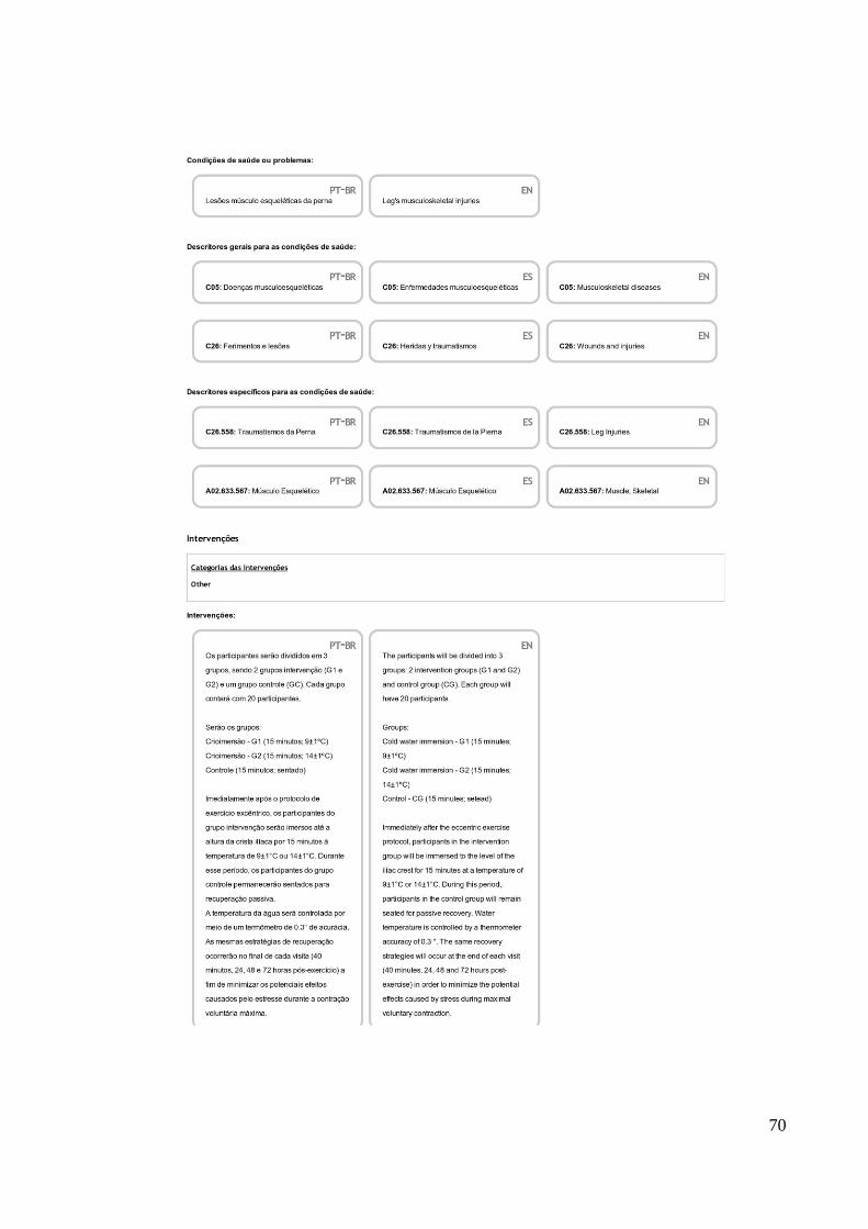
70

71

72
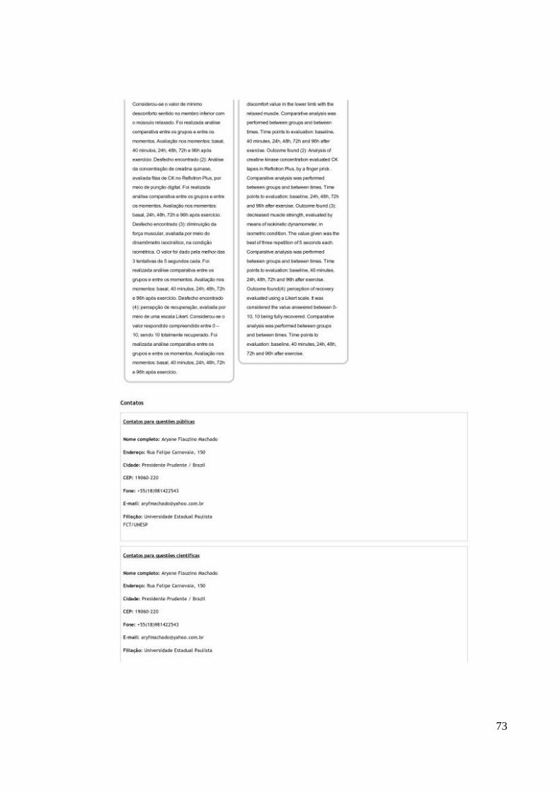
73

74
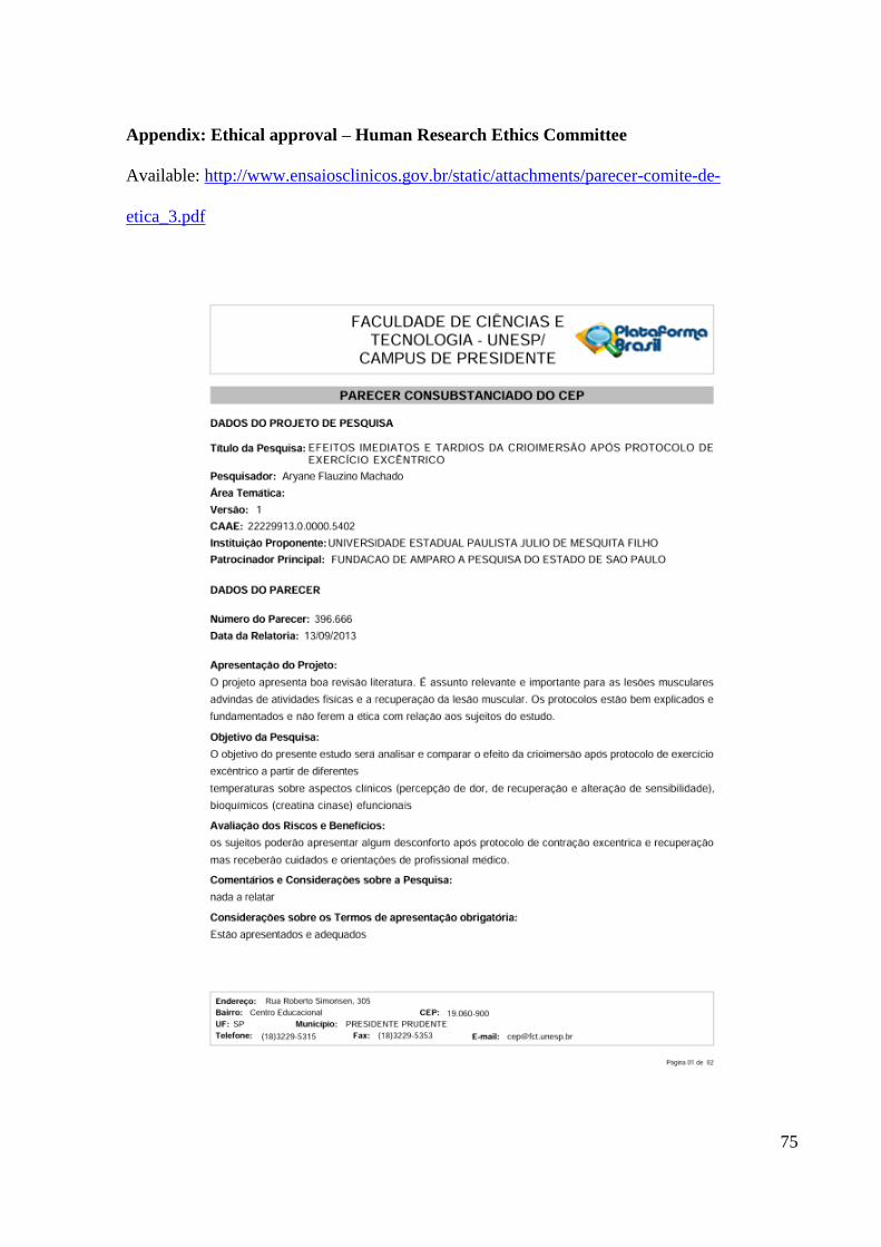
75
Appendix: Ethical approval – Human Research Ethics Committee
Available: http://www.ensaiosclinicos.gov.br/static/attachments/parecer-comite-de-
etica_3.pdf

76
77
Conclusões
A partir da pesquisa realizada pode-se concluir que os resultados da aplicação da
imersão em água fria sobre os marcadores de dano muscular ainda não estão plenamente
elucidados, variando quanto aos desfechos analisados. Entretanto, ambos os estudos apontam
uma dose de aplicação similar.
Ainda que a revisão sistemática seja composta por artigos de baixa qualidade
metodológica, a meta-análise realizada sugere uma aplicação com água à temperatura em
torno de 11 a 15°C com o tempo de imersão variando entre 11 e 15 minutos. Corroborando os
achados, o ensaio clínico randomizado, apresenta uma melhor dose quando aplicada a 14°C
por 15 minutos, ainda que seja para recuperação antecipada da performance. Sob outro
enfoque, conclui-se também que ambos os estudos apontam que a imersão em água fria,
quando não produz efeitos que minimizam os marcadores de dano muscular, também não
provoca efeitos deletérios.
78
Referências
1. Bleakley CM, Davison GW. What is the biochemical and physiological
rationale for using cold-water immersion in sports recovery? A systematic review. Br J Sports
Med. 2010 Feb;44(3):179–87.
2. Brophy-Williams N, Landers G, Wallman K. Effect of immediate and delayed
cold water immersion after a high intensity exercise session on subsequent run performance. J
Sports Sci Med. 2011 Jan;10(4):665–70.
3. Bailey DM, Erith SJ, Griffin PJ, Dowson A, Brewer DS, Gant N, et al.
Influence of cold-water immersion on indices of muscle damage following prolonged
intermittent shuttle running. J Sports Sci. 2007 Sep;25(11):1163–70.
4. Sellwood KL, Brukner P, Williams D, Nicol A, Hinman R. Ice-water
immersion and delayed-onset muscle soreness: a randomised controlled trial. Br J Sports
Med. 2007 Jun;41(6):392–7.
5. Ascensão A, Leite M, Rebelo AN, Magalhäes S, Magalhäes J. Effects of cold
water immersion on the recovery of physical performance and muscle damage following a
one-off soccer match. J Sports Sci. 2011 Feb;29(3):217–25.
6. Bleakley C, McDonough S, Gardner E, Baxter GD, Hopkins JT, Davison GW.
Cold-water immersion (cryotherapy) for preventing and treating muscle soreness after
exercise. Cochrane Database Syst Rev. 2012 Jan;2:CD008262.
7. Pournot H, Bieuzen F, Duffield R, Lepretre P-M, Cozzolino C, Hausswirth C.
Short term effects of various water immersions on recovery from exhaustive intermittent
exercise. Eur J Appl Physiol. 2011 Jul;111(7):1287–95.
79
8. Ingram J, Dawson B, Goodman C, Wallman K, Beilby J. Effect of water
immersion methods on post-exercise recovery from simulated team sport exercise. J Sci Med
Sport. 2009 May;12(3):417–21.
9. Glasgow PD, Ferris R, Bleakley CM. Cold water immersion in the
management of delayed-onset muscle soreness: Is dose important? A randomised controlled
trial. Phys Ther Sport. 2014 Jan 29.
10. Getto CN, Golden G. Comparison of Active Recovery in Water and Cold-
Water Immersion After Exhaustive Exercise. Athl Train Sport Heal Care. 2013 Jul
2;5(4):169–76.
11. Jakeman JR, Macrae R, Eston R. A single 10-min bout of cold-water
immersion therapy after strenuous plyometric exercise has no beneficial effect on recovery
from the symptoms of exercise-induced muscle damage. Ergonomics. 2009 Apr;52(4):456–
60.
12. Wilcock IM, Cronin JB, Hing WA. Physiological Response to Water
Immersion A Method for Sport Recovery? Sports Med. 2006;36(9):1–18.
13. Eston R, Peters D. Effects of cold water immersion on the symptoms of
exercise-induced muscle damage. J Sports Sci. 1999 Mar;17(3):231–8.
14. Andrews JR, Harrelson GL, Wilk KE. Reabilitação física nas lesões
desportivas. 2.ed. Guanabara Koogan, 2000.
15. Pastre CM, Bastos FN, Netto Junior, J, Vanderlei LCM, Hoshi RA. Métodos
de Recuperação Pós-exercício: uma Revisão Sistemática. Rev Bras Med Esporte.
2009;15:138–44.

80
Anexos
Instructions for Author – Sports Medicine
Types of papers
The word counts given below do not include the abstract, references, figure
legends or table captions.
Systematic Review. Word count up to 10,000. Collates all empirical evidence
that fits pre- specified eligibility criteria to answer a specific research question. It uses
explicit, systematic methods that are selected with a view to minimizing bias, thus providing
reliable findings from which conclusions can be drawn and decisions made. Please refer to
Integrity of Research and Reporting below for more information on reporting requirements.
Manuscript Submission
Manuscript Submission
Submission of a manuscript implies: that the work described has not been
published before; that it is not under consideration for publication anywhere else; that its
publication has been approved by all coauthors, if any, as well as by the responsible
authorities – tacitly or explicitly – at the institute where the work has been carried out. The
publisher will not be held legally responsible should there be any claims for compensation.
Permissions
Authors wishing to include figures, tables, or text passages that have already been
published elsewhere are required to obtain permission from the copyright owner(s) for both
81
the print and online format and to include evidence that such permission has been granted
when submitting their papers. Any material received without such evidence will be assumed
to originate from the authors.
Online Submission
Authors should submit their manuscripts online. Electronic submission
substantially reduces the editorial processing and reviewing times and shortens overall
publication times. Please follow the hyperlink “Submit online” on the right and upload all of
your manuscript files following the instructions given on the screen.
Internal Review by Editorial Staff
The journal editor will perform an initial appraisal of each manuscript. If your
paper has been peer reviewed by another journal as part of a prior submission, the journal
editor will also assess any previous editorial/referee comments and how these have been dealt
with as part of the appraisal process. If your manuscript is considered unsuitable for the
journal to which it has been submitted, it may be assessed for suitability for publication in
other Adis journals by appropriate editors. However, it will not be progressed to external peer
review for an alternative journal without your permission.
Title page
The title page should include:
The name(s) of the author(s)
A concise and informative title
The affiliation(s) and address(es) of the author(s)
82
The email address, telephone and fax numbers of the corresponding author
Abstract
Please provide an abstract of 150 to 250 words. The abstract should not contain
any undefined abbreviations or unspecified references. For manuscripts reporting the results
of a systematic review with or without a meta-analysis, the abstract should be structured as
described in the PRISMA statement. For these types of manuscripts the abstract length can be
increased from the 250 word limit to allow full compliance with PRISMA / CONSORT.
Text Formatting
Manuscripts should be submitted in Word.
Use a normal, plain font (e.g., 10point Times Roman) for text.
Use italics for emphasis.
Use the automatic page numbering function to number the pages.
Do not use field functions.
Use tab stops or other commands for indents, not the space bar.
Use the table function, not spreadsheets, to make tables.
Save your file in docx format (Word 2007 or higher) or doc format (older
Word versions).
Headings
Please use the decimal system of headings with no more than three levels.

83
Abbreviations
Abbreviations should be defined at first mention and used consistently thereafter.
Footnotes
Footnotes can be used to give additional information, which may include the
citation of a reference included in the reference list. They should not consist solely of a
reference citation, and they should never include the bibliographic details of a reference. They
should also not contain any figures or tables.
Footnotes to the text are numbered consecutively; those to tables should be
indicated by superscript lowercase letters (or asterisks for significance values and other
statistical data).
Footnotes to the title or the authors of the article are not given reference symbols.
Always use footnotes instead of endnotes.
Acknowledgments
Acknowledgments of people, grants, funds, etc. should be placed in a separate
section before the reference list. The names of funding organizations should be written in full.
References
Citation
Reference citations in the text should be identified by numbers in square brackets.
Some examples:
1. Negotiation research spans many disciplines [3].
2. This result was later contradicted by Becker and Seligman [5].
3. This effect has been widely studied [13,7].
84
Reference list
The list of references should only include works that are cited in the text and that
have been published or accepted for publication. Personal communications and unpublished
works should only be mentioned in the text. Do not use footnotes or endnotes as a substitute
for a reference list.
The entries in the list should be numbered consecutively.
Journal article: Smith JJ. The world of science. Am J Sci. 1999;36:234–5.
Article by DOI: Slifka MK, Whitton JL. Clinical implications of dysregulated
cytokine production. J Mol Med. 2000; doi:10.1007/s001090000086
Book: Blenkinsopp A, Paxton P. Symptoms in the pharmacy: a guide to the
management of common illness. 3rd ed. Oxford: Blackwell Science; 1998.
Book chapter: Wyllie AH, Kerr JFR, Currie AR. Cell death: the significance of
apoptosis. In: Bourne GH, Danielli JF, Jeon KW, editors. International review of cytology.
London: Academic; 1980. pp. 251–306.
Online document: Doe J. Title of subordinate document. In: The dictionary of
substances and their effects. Royal Society of Chemistry. 1999. http://www.rsc.org/dose/title
of subordinate document. Accessed 15 Jan 1999.
Always use the standard abbreviation of a journal’s name according to the ISSN
List of Title Word Abbreviations, see ISSN.org LTWA
Tables
All tables are to be numbered using Arabic numerals.
Tables should always be cited in text in consecutive numerical order.
85
For each table, please supply a table caption (title) explaining the components
of the table.
Identify any previously published material by giving the original source in the
form of a reference at the end of the table caption.
Footnotes to tables should be indicated by superscript lowercase letters (or
asterisks for significance values and other statistical data) and included beneath the table
body.
Artwork and illustrations guidelines
Electronic Figure Submission
Supply all figures electronically.
Indicate what graphics program was used to create the artwork.
For vector graphics, the preferred format is EPS; for halftones, please use TIFF
format. MSOffice files are also acceptable.
Vector graphics containing fonts must have the fonts embedded in the files.
Name your figure files with "Fig" and the figure number, e.g., Fig1.eps.
Line Art
Definition: Black and white graphic with no shading.
Do not use faint lines and/or lettering and check that all lines and lettering
within the figures are legible at final size.
All lines should be at least 0.1 mm (0.3 pt) wide.
Scanned line drawings and line drawings in bitmap format should have a
minimum resolution of 1200 dpi.
Vector graphics containing fonts must have the fonts embedded in the files.
86
Halftone Art
Definition: Photographs, drawings, or paintings with fine shading, etc.
If any magnification is used in the photographs, indicate this by using scale
bars within the figures themselves.
Halftones should have a minimum resolution of 300 dpi.
Combination Art
Definition: a combination of halftone and line art, e.g., halftones containing
line drawing, extensive lettering, color diagrams, etc.
Combination artwork should have a minimum resolution of 600 dpi.
Color Art
Color art is free of charge for online publication.
If black and white will be shown in the print version, make sure that the main
information will still be visible. Many colors are not distinguishable from one another when
converted to black and white. A simple way to check this is to make a xerographic copy to see
if the necessary distinctions between the different colors are still apparent.
If the figures will be printed in black and white, do not refer to color in the
captions.
Color illustrations should be submitted as RGB (8 bits per channel).
Figure Lettering
To add lettering, it is best to use Helvetica or Arial (sans serif fonts).
87
Keep lettering consistently sized throughout your final-sized artwork, usually
about 2–3 mm (8–12 pt).
Variance of type size within an illustration should be minimal, e.g., do not use
8pt type on an axis and 20pt type for the axis label.
Avoid effects such as shading, outline letters, etc.
Do not include titles or captions within your illustrations.
Figure Numbering
All figures are to be numbered using Arabic numerals.
Figures should always be cited in text in consecutive numerical order.
Figure parts should be denoted by lowercase letters (a, b, c, etc.).
If an appendix appears in your article and it contains one or more figures,
continue the consecutive numbering of the main text. Do not number the appendix figures,
"A1, A2, A3, etc." Figures in online appendices (Electronic Supplementary Material) should,
however, be numbered separately.
Figure Captions
Each figure should have a concise caption describing accurately what the
figure depicts. Include the captions in the text file of the manuscript, not in the figure file.
Figure captions begin with the term Fig. in bold type, followed by the figure
number, also in bold type.
No punctuation is to be included after the number, nor is any punctuation to be
placed at the end of the caption.
88
Identify all elements found in the figure in the figure caption; and use boxes,
circles, etc., as coordinate points in graphs.
Identify previously published material by giving the original source in the form
of a reference citation at the end of the figure caption.
Figure Placement and Size
When preparing your figures, size figures to fit in the column width.
For most journals the figures should be 39 mm, 84 mm, 129 mm, or 174 mm wide
and not higher than 234 mm.
For books and book-sized journals, the figures should be 80 mm or 122 mm wide
and not higher than 198 mm.
Permissions
If you include figures that have already been published elsewhere, you must
obtain permission from the copyright owner(s) for both the print and online format. Please be
aware that some publishers do not grant electronic rights for free and that Springer will not be
able to refund any costs that may have occurred to receive these permissions. In such cases,
material from other sources should be used.
Accessibility
In order to give people of all abilities and disabilities access to the content of your
figures, please make sure that. All figures have descriptive captions (blind users could then
use a text-to-speech software or a text-to-Braille hardware)
Patterns are used instead of or in addition to colors for conveying information
(colorblind users would then be able to distinguish the visual elements)
89
Any figure lettering has a contrast ratio of at least 4.5:1
Submission
Supply all supplementary material in standard file formats.
Please include in each file the following information: article title, journal name,
author names; affiliation and email address of the corresponding author.
To accommodate user downloads, please keep in mind that larger-sized files may
require very long download times and that some users may experience other problems during
downloading.
Text and Presentations
Submit your material in PDF format; .doc or .ppt files are not suitable for long-
term viability. A collection of figures may also be combined in a PDF file.
Spreadsheets
Spreadsheets should be converted to PDF if no interaction with the data is
intended. If the readers should be encouraged to make their own calculations, spreadsheets
should be submitted as .xls files (MS Excel).
Specialized Formats
Specialized format such as .pdb (chemical), .wrl (VRML), .nb (Mathematica
notebook), and .tex can also be supplied.
Collecting Multiple Files
It is possible to collect multiple files in a .zip or .gz file.
90
Numbering
If supplying any supplementary material, the text must make specific mention of
the material as a citation, similar to that of figures and tables. Refer to the supplementary files
as “Online Resource”, e.g., "... as shown in the animation (Online Resource 3)", “... additional
data are given in Online Resource 4”. Name the files consecutively, e.g. “ESM_3.mpg”,
“ESM_4.pdf”.
Captions
For each supplementary material, please supply a concise caption describing the
content of the file.
Processing of supplementary files
Electronic supplementary material will be published as received from the author
without any conversion, editing, or reformatting.
After acceptance
Upon acceptance of your article you will receive a link to the special Author
Query Application at Springer’s web page where you can sign the Copyright Transfer
Statement online and indicate whether you wish to order OpenChoice and offprints.
Once the Author Query Application has been completed, your article will be
processed and you will receive the proofs.
Open Choice
In addition to the normal publication process (whereby an article is submitted to
the journal and access to that article is granted to customers who have purchased a

91
subscription), Springer now provides an alternative publishing option: Springer Open Choice.
A Springer Open Choice article receives all the benefits of a regular subscription-based
article, but in addition is made available publicly through Springer’s online platform
SpringerLink.
Springer Open Choice
Copyright transfer: Authors will be asked to transfer copyright of the article to the
Publisher (or grant the Publisher exclusive publication and dissemination rights). This will
ensure the widest possible protection and dissemination of information under copyright laws.
Open Choice articles do not require transfer of copyright as the copyright remains with the
author. In opting for open access, the author(s) agree to publish the article under the Creative
Commons Attribution License.
Color illustrations: Publication of color illustrations is free of charge.
Proof reading: The purpose of the proof is to check for typesetting or conversion
errors and the completeness and accuracy of the text, tables and figures. Substantial changes
in content, e.g., new results, corrected values, title and authorship, are not allowed without the
approval of the Editor. After online publication, further changes can only be made in the form
of an Erratum, which will be hyperlinked to the article.
Online First: The article will be published online after receipt of the corrected
proofs. This is the official first publication citable with the DOI. After release of the printed
version, the paper can also be cited by issue and page numbers.
92
Integrity of research and reporting
Ethical standards
Manuscripts submitted for publication must contain a statement to the effect that
all human and animal studies have been approved by the appropriate ethics committee and
have therefore been performed in accordance with the ethical standards laid down in the 1964
Declaration of Helsinki and its later amendments. It should also be stated clearly in the text
that all persons gave their informed consent prior to their inclusion in the study. Details that
might disclose the identity of the subjects under study should be omitted.
These statements should be added in a separate section before the reference list. If
these statements are not applicable, authors should state: The manuscript does not contain
clinical studies or patient data.
The editors reserve the right to reject manuscripts that do not comply with the
above-mentioned requirements. The author will be held responsible for false statements or
failure to fulfill the above-mentioned requirements.
Conflict of interest
Authors must indicate whether or not they have a financial relationship with the
organization that sponsored the research. They should also state that they have full control of
all primary data and that they agree to allow the journal to review their data if requested.
Therefore the manuscript must be accompanied by the “Conflict of Interest Disclosure Form”.
General
Adis journals endorse the ‘Uniform Requirements for Manuscripts Submitted to
Biomedical Journals,’ issued by the International Committee for Medical Journal Editors and
are members of the Committee on Publication Ethics.

93
Ethics/Institutional Review Board Approval of Research
Authors should be able to submit, upon request, a statement from the research
ethics committee or institutional review board indicating approval of the research. If the study
was not submitted to a research ethics committee or institutional review board, authors may
need to provide documentation to prove that not seeking review for the study was in
accordance with the policy of their institution. Details of the ethical approval status of the
research must be described in the methods section of the paper.
Health Research Reporting Guidelines
Systematic reviews (with or without a meta-analysis) should follow the reporting
guidelines of PRISMA. Authors must provide a completed PRISMA flowchart and checklist.
Meta-analysis of observational studies in epidemiology should following the reporting
guidelines of MOOSE. Authors must provide a completed MOOSE checklist.
Use of Personal Communications and Unpublished Data
Authors must include a signed statement of permission from each individual
identified as a source of information in a personal communication or as a source for
unpublished data (this includes papers that have been submitted, but not yet accepted for
publication), and specify the date of communication and whether the communication was
written or oral.
Duplicate Publication and Duplicate Submission
Manuscripts are considered with the understanding that they have not been
published previously and are not under consideration by another publication. Copies of
possibly duplicative materials (i.e. those containing substantially similar content or using the
94
same or similar data) that have been previously published or are being considered elsewhere
must be provided at the time of manuscript submission. Submitted or published manuscripts
that are found to be duplicated in any substantive way will be addressed in accordance with
the guidelines of the Committee on Publication Ethics.
Committee on Publication Ethics
The journal will, however, consider republication of a paper previously published
in a language other than English, or simultaneous publication of a paper in multiple journals
with different audiences, if the specific circumstances warrant this action. This will be done
with full and prominent disclosure of the original source and with any necessary permission.
The journal does not consider posting of protocols and results in clinical trial registries to be
prior publication. Press releases of studies presented at scientific meetings are also not
considered prior publication and will not compromise an author’s ability to write up a full
study provided the release does not disclose results beyond those presented in the meeting
abstract or poster.
Plagiarism
Plagiarism is the use of others' published and unpublished ideas or words (or other
intellectual property) without attribution or permission, and presenting them as new and
original rather than derived from an existing source. Plagiarism is scientific misconduct and
will be addressed as such following the Committee on Publication Ethics guidelines.

95
Instructions for Author: PLoS ONE
1. Format Requirements
PLOS ONE does not consider pre-submission inquiries. All submissions should
be prepared with the following files:
Cover letter
Manuscript, including tables and figure legends
Figures
Prior to submission, authors who believe their manuscripts would benefit from
professional editing are encouraged to use language-editing and copyediting services.
Obtaining this service is the responsibility of the author, and should be done before initial
submission. These services can be found on the web using search terms like "scientific editing
service" or "manuscript editing service." Submissions are not copyedited before publication.
Submissions that do not meet the PLOS ONE Publication Criterion for language
standards may be rejected.
Cover Letter
You should supply an approximately one page cover letter that:
Concisely summarizes why your paper is a valuable addition to the scientific
literature
Briefly relates your study to previously published work
Specifies the type of article you are submitting (for example, research article,
systematic review, meta-analysis, clinical trial)
Describes any prior interactions with PLOS regarding the submitted
manuscript
96
Suggests appropriate PLOS ONE Academic Editors to handle your manuscript
Lists any opposed reviewers
Your cover letter should not include requests to reduce or waive publication fees.
Should your manuscript be accepted, you will have the opportunity to include your requests at
that time. See PLOS ONE Editorial Policy for more information regarding publication fees.
Manuscript Organization
PLOS ONE considers manuscripts of any length. There are no explicit restrictions
for the number of words, figures, or the length of the supporting information, although we
encourage a concise and accessible writing style. We will not consider monographs.
All manuscripts should be double-spaced and include line numbers and page
numbers.
Manuscripts should begin with the ordered sections:
Title
Authors
Affiliations
Abstract
Introduction
And end with the sections of:
Acknowledgments
References
Supporting Information Captions
97
Figures should be cited in ascending numeric order upon first appearance. Each
figure caption should then be inserted immediately after the first paragraph in which it is cited
in the article file.
Figures should not be included in the main manuscript file. Each figure must be
prepared and submitted as an individual file. Tables should be cited in ascending numeric
order upon first appearance. Each table should then be inserted immediately after the first
paragraph in which it is cited in the article file.
The title, authors, and affiliations should all be included on a title page as the first
page of the manuscript file.
There are no explicit requirements for section organization between these
beginning and ending sections. Articles may be organized in different ways and with different
section titles, according to the authors' preference. In most cases, internal sections include:
Materials and Methods
Results
Discussion
Conclusions (optional)
PLOS ONE has no specific requirements for the order of these sections, and in
some cases it may be appropriate to combine sections.
Abbreviations should be kept to a minimum and defined upon first use in the text.
Non-standard abbreviations should not be used unless they appear at least three times in the
text.
Standardized nomenclature should be used as appropriate, including appropriate
usage of species names and SI units.
98
PLOS articles do not support text footnotes. If your accepted submission contains
footnotes, you will be asked to move that material into either the main text or the reference
list, depending on the content.
Manuscript File Requirements
Authors may submit their manuscript files in Word (as .doc or .docx), LaTeX (as
.pdf), or RTF format. Word files must not be protected.
2. Guidelines for Standard Sections
Title
Manuscripts must be submitted with both a full title and a short title, which will
appear at the top of the PDF upon publication if accepted. Only the full title should be
included in the manuscript file; the short title will be entered during the online submission
process.
The full title must be 250 characters or fewer. It should be specific, descriptive,
concise, and comprehensible to readers outside the subject field. Avoid abbreviations if
possible. Where appropriate, authors should include the species or model system used (for
biological papers) or type of study design (for clinical papers).
The short title must be 50 characters or fewer and should state the topic of the
paper.
Authors and Affiliations
All author names should be listed in the following order:
First names (or initials, if used),
99
Middle names (or initials, if used), and
Last names (surname, family name)
Each author should list an associated department, university, or organizational
affiliation and its location, including city, state/province (if applicable), and country. If the
article has been submitted on behalf of a consortium, all author names and affiliations should
be listed at the end of the article.
This information cannot be changed after initial submission, so please ensure that
it is correct.
To qualify for authorship, one should contribute to all of the following:
1. Conception and design of the work, acquisition of data, or analysis and
interpretation of data
2. Drafting the article or revising it critically for important intellectual content
3. Final approval of the version to be published
4. Agreement to be accountable for all aspects of the work
All persons designated as authors should qualify for authorship, and all those who
qualify should be listed. Each author must have participated sufficiently in the work to take
public responsibility for appropriate portions of the content. Those who contributed to the
work but do not qualify for authorship should be listed in the acknowledgments.
When a large group or center has conducted the work, the author list should
include the individuals whose contributions meet the criteria defined above, as well as the
group name.
All authors must approve the final manuscript before submission. PLOS ONE will
contact all authors by email at submission to ensure that they are aware of the submission of
the manuscript.
100
One author should be designated as the corresponding author, and his or her email
address or other contact information should be included on the manuscript cover page. This
information will be published with the article if accepted.
Abstract
The abstract should:
Describe the main objective(s) of the study
Explain how the study was done, including any model organisms used, without
methodological detail
Summarize the most important results and their significance
Not exceed 300 words
Abstracts should not include:
Citations
Abbreviations, if possible
Introduction
The introduction should:
Provide background that puts the manuscript into context and allows readers
outside the field to understand the purpose and significance of the study
Define the problem addressed and why it is important
Include a brief review of the key literature
Note any relevant controversies or disagreements in the field
Conclude with a brief statement of the overall aim of the work and a comment
about whether that aim was achieved
101
Materials and Methods
This section should provide enough detail to allow suitably skilled investigators to
fully replicate your study. Specific information and/or protocols for new methods should be
included in detail. If materials, methods, and protocols are well established, authors may cite
articles where those protocols are described in detail, but the submission should include
sufficient information to be understood independent of these references.
We encourage authors to submit detailed protocols for newer or less well-
established methods as Supporting Information.
Methods sections of papers on research using human or animal subjects and/or
tissue or field sampling must include required ethics statements.
Methods sections of papers with data that should be deposited in a publicly
available database should specify where the data have been deposited and provide the relevant
accession numbers and version numbers, if appropriate. Accession numbers should be
provided in parentheses after the entity on first use. If the accession numbers have not yet
been obtained at the time of submission, please state that they will be provided during review.
They must be provided prior to publication.
Results, Discussion, and Conclusions
These sections may all be separate, or may be combined to create a mixed
Results/Discussion section or a mixed Discussion/Conclusions section. These sections may be
further divided into subsections, each with a concise subheading, as appropriate. These
sections have no word limit, but the language should be clear and concise.
Together, these sections should describe the results of the experiments, the
interpretation of these results, and the conclusions that can be drawn. Authors should explain
how the results relate to the hypothesis presented as the basis of the study and provide a
102
succinct explanation of the implications of the findings, particularly in relation to previous
related studies and potential future directions for research.
PLOS ONE editorial decisions do not rely on perceived significance or impact, so
authors should avoid overstating their conclusions.
Acknowledgments
People who contributed to the work but do not fit the PLOS ONE authorship
criteria should be listed in the acknowledgments, along with their contributions. You must
ensure that anyone named in the acknowledgments agrees to being so named.
Funding sources should not be included in the acknowledgments, or anywhere in
the manuscript file. You will provide this information during the manuscript submission
process.
References
General guidelines
PLOS uses the reference style as outlined in the ICMJE sample references, also
referred to as the “Vancouver” style.
References must be listed at the end of the manuscript and numbered in the
order that they appear in the text.
In the text, citations should be indicated by the reference number in brackets.
Authors may cite any and all available works in the reference list.
Authors may not cite unavailable and unpublished work, including manuscripts
that have been submitted but not yet accepted (e.g., “unpublished work,” “data not shown”).
If an article is submitted to a journal and also publicly available as a pre-print,
the pre-print may be cited.
103
If related work has been submitted to PLOS ONE or elsewhere, authors should
include a copy with the submitted article as confidential supplementary information, for
review purposes only.
Authors should not state 'unpublished work' or 'data not shown,' but instead
include those data as supplementary material or deposit the data in a publicly available
database.
Journal name abbreviations should be those found in the NCBI databases.
Reference formatting
Because all references will be linked electronically as much as possible to the
papers they cite, proper formatting of the references is crucial. References should be
formatted as follows:
Published papers: Hou WR, Hou YL, Wu GF, Song Y, Su XL, Sun, B, et al.
cDNA, genomic sequence cloning and overexpression of ribosomal protein gene L9 (rpL9) of
the giant panda (Ailuropoda melanoleuca). Genet Mol Res. 2011;10: 1576-1588.
Accepted, unpublished papers: Same as above, but “In press” appears instead
of the page numbers or DOI.
Websites or online articles: Huynen MMTE, Martens P, Hilderlink HBM. The
health impacts of globalisation: a conceptual framework. Global Health. 2005;1: 14.
Available: http://www.globalizationandhealth.com/content/1/1/14.
Books: Bates B. Bargaining for life: A social history of tuberculosis. 1st ed.
Philadelphia: University of Pennsylvania Press; 1992.

104
Book chapters: Hansen B. New York City epidemics and history for the public.
In: Harden VA, Risse GB, editors. AIDS and the historian. Bethesda: National Institutes of
Health; 1991. pp. 21-28.
Deposited articles (preprints, e-prints, or arXiv): Krick T, Shub DA, Verstraete
N, Ferreiro DU, Alonso LG, Shub M, et al. Amino acid metabolism conflicts with protein
diversity; 1991. Preprint. Available: arXiv:1403.3301v1. Accessed 17 March 2014.
Published media (print or online newspapers and magazine articles): Fountain
H. For Already Vulnerable Penguins, Study Finds Climate Change Is Another Danger. The
New York Times. 29 Jan 2014. Available:
http://www.nytimes.com/2014/01/30/science/earth/climate-change-taking-toll-on-penguins-
study-finds.html. Accessed 17 March 2014.
New media (blogs, websites, or other written works): Allen L. Announcing
PLOS Blogs. 2010 Sep 1 [cited 17 March 2014]. In: PLOS Blogs [Internet]. San Francisco:
PLOS 2006 - . [about 2 screens]. Available: http://blogs.plos.org/plos/2010/09/announcing-
plos-blogs/.
Masters' theses or doctoral dissertations: Wells A. Exploring the development
of the independent, electronic, scholarly journal. M.Sc. Thesis, The University of Sheffield.
1999. Available: http://cumincad.scix.net/cgi-bin/works/Show?2e09.
Databases and repositories (Figshare, arXiv): Roberts SB. QPX Genome
Browser Feature Tracks; 2013. Database: figshare [Internet]. Accessed:
http://figshare.com/articles/QPX_Genome_Browser_Feature_Tracks/701214.
Multimedia (videos, movies, or TV shows): Hitchcock A, producer and
director. Rear Window [Film]; 1954. Los Angeles: MGM.
105
Figure Legends
Figures should not be included in the manuscript file, but figure legends should
be. Figure legends should describe the key messages of a figure. Legends should have a short
title of 15 words or less. The full legend should have a description of the figure and allow
readers to understand the figure without referring to the text. The legend itself should be
succinct, avoid lengthy descriptions of methods, and define all non-standard symbols and
abbreviations.
Figures should be cited in ascending numeric order upon first appearance. Each
figure caption should be inserted immediately after the first paragraph in which they are cited
in the article file.
Supporting Information Captions
Because Supporting Information is accessed via a hyperlink attached to its
captions, captions must be listed in the article file. Do not submit a separate caption file. It is
acceptable to have them in the file itself in addition, but they must be in the article file for
access to be possible in the published version.
The file category name and number is required, and a one-line title is highly
recommended. A legend can also be included but is not required. Supporting Information
captions should be formatted as follows.
Data Reporting Guidelines
All data and related metadata underlying the findings reported in a submitted
manuscript should be deposited in an appropriate public repository, unless already provided as
part of the submitted article. Repositories may be either subject-specific (where these exist)
and accept specific types of structured data, or generalist repositories that accept multiple data

106
types. We recommend that authors select repositories appropriate to their field. Repositories
may be subject-specific, general, or institutional, as long as DOIs or accession numbers are
provided and the data are at least as open as CCBY. Authors are encouraged to select
repositories that meet accepted criteria as trustworthy digital repositories, such as criteria of
the Centre for Research Libraries or Data Seal of Approval. Large, international databases are
more likely to persist than small, local ones.
To support data sharing and author compliance of the PLOS data policy, we have
integrated our submission process with a select set of data repositories. The list is neither
representative nor exhaustive of the suitable repositories available to authors. Current
repository integration partners include: Dryad and figshare. Please contact [email protected] to
make recommendations for further partnerships.
Accession Numbers
All appropriate datasets, images, and information should be deposited in public
resources. Please provide the relevant accession numbers (and version numbers, if
appropriate). In addition, as much as possible, please provide accession numbers or
identifiers for all entities such as genes, proteins, mutants, diseases, etc., for which there is an
entry in a public database. Providing accession numbers allows linking to and from
established databases and integrates your article with a broader collection of scientific
information.
Striking Images
Authors are encouraged to upload a "striking image" that may be used to represent
their paper online in places like the journal homepage or in search results. The striking image
must be derived from a figure or supporting information file from the paper, ie. a cropped
107
portion of an image or the entire image. Striking images should ideally be high resolution,
eye-catching, single panel images, and should ideally avoid containing added details such as
text, scale bars, and arrows. If no striking image is uploaded, a figure from the paper will be
designated as the striking image.
Please keep in mind that PLOS's Creative Commons Attribution License applies
to striking images. As such, do not submit any figures or photos that have been previously
copyrighted unless you have express written permission from the copyright holder to publish
under the CCAL license. Note that all published materials in PLOS ONE are freely available
online, and any third party is permitted to read, download, copy, distribute, and use these
materials in any way, even commercially, with proper attribution.
Care should be taken with the following image types in particular:
1. PLOS ONE is unable to publish any images generated by Google software
(Google Maps, Street View, and Earth)
2. Maps in general are usually copyrighted, especially satellite maps
3. Photographs
4. Commercial or government images, slogans, or logos
5. Images from Facebook or Twitter
Authors must also take special care when submitting manuscripts that contain
potentially identifying images of people. Identifying information should not be included in the
manuscript unless the information is crucial and the individual has provided written consent
by completing the Consent Form for Publication in a PLOS Journal (PDF).
Tables
Tables should be cited in ascending numeric order upon first appearance. Each
table should be inserted immediately after the first paragraph in which it is cited in the article
108
file. All tables should have a concise title. Footnotes can be used to explain abbreviations.
Citations should be indicated using the same style as outlined above. Tables occupying more
than one printed page should be avoided, if possible. Larger tables can be published
as Supporting Information.
3. Specific Reporting Guidelines
Human Subject Research
Methods sections of papers on research using human subject or samples must
include ethics statements that specify:
The name of the approving institutional review board or equivalent
committee(s). If approval was not obtained, the authors must provide a detailed statement
explaining why it was not needed
Whether informed consent was written or oral. If informed consent was oral, it
must be stated in the manuscript:
- Why written consent could not be obtained
- That the Institutional Review Board (IRB) approved use of oral consent
- How oral consent was documented
For studies involving humans categorized by race/ethnicity, age,
disease/disabilities, religion, sex/gender, sexual orientation, or other socially constructed
groupings, authors should:
Explicitly describe their methods of categorizing human populations
Define categories in as much detail as the study protocol allows
Justify their choices of definitions and categories, including for example
whether any rules of human categorization were required by their funding agency

109
Explain whether (and if so, how) they controlled for confounding variables
such as socioeconomic status, nutrition, environmental exposures, or similar factors in their
analysis.
In addition, outmoded terms and potentially stigmatizing labels should be changed
to more current, acceptable terminology. Examples: "Caucasian" should be changed to
"white" or "of [Western] European descent" (as appropriate); "cancer victims" should be
changed to "patients with cancer."
For papers that include identifying, or potentially identifying, information, authors
must download the Consent Form for Publication in a PLOS Journal (PDF), which the
individual, parent, or guardian must sign once they have read the paper and been informed
about the terms of PLOS open-access license. The signed consent form should not be
submitted with the manuscript, but authors should securely file it in the individual's case notes
and the methods section of the manuscript should explicitly state that consent authorization
for publication is on file, using wording like:
The individual in this manuscript has given written informed consent (as outlined
in PLOS consent form) to publish these case details.
Clinical Trials
Authors of manuscripts describing the results of clinical trials must adhere to
the CONSORT reporting guidelines appropriate to their trial design, available on
the CONSORT Statement website. Before the paper can enter peer review, authors must:
1. Provide the registry name and number in the methods section of the manuscript
2. Provide a copy of the trial protocol as approved by the ethics committee and a
completed CONSORT checklist as Supporting Information (which will be published
alongside the paper, if accepted). This should be named S1 CONSORT Checklist.
110
3. Include the CONSORT flow diagram as the manuscript's "Fig. 1"
Any deviation from the trial protocol must be explained in the paper. Authors
must explicitly discuss informed consent in their paper, and we reserve the right to ask for a
copy of the patient consent form.
The methods section must include the name of the registry, the registry number,
and the URL of your trial in the registry database for each location in which the trial is
registered.