Bruno Martins - repositorio.ufsm.br
47
UNIVERSIDADE FEDERAL DE SANTA MARIA CENTRO DE CIÊNCIAS RURAIS PROGRAMA DE PÓS-GRADUAÇÃO EM MEDICINA VETERINÁRIA RELAÇÃO ANTIGÊNICA ENTRE ALFAHERPESVÍRUS CAPRINO 1 (CpHV-1) E ALFAHERPESVÍRUS BOVINO 1 (BoHV-1) E INFECÇÃO EXPERIMENTAL DE CABRITOS E BEZERROS COM O CpHV-1 DISSERTAÇÃO DE MESTRADO Bruno Martins Santa Maria, RS, Brasil 2019
Transcript of Bruno Martins - repositorio.ufsm.br
1
UNIVERSIDADE FEDERAL DE SANTA MARIA
CENTRO DE CIÊNCIAS RURAIS
PROGRAMA DE PÓS-GRADUAÇÃO EM MEDICINA VETERINÁRIA
RELAÇÃO ANTIGÊNICA ENTRE ALFAHERPESVÍRUS CAPRINO 1
(CpHV-1) E ALFAHERPESVÍRUS BOVINO 1 (BoHV-1) E INFECÇÃO
EXPERIMENTAL DE CABRITOS E BEZERROS COM O CpHV-1
DISSERTAÇÃO DE MESTRADO
Bruno Martins
Santa Maria, RS, Brasil
2019
2
RELAÇÃO ANTIGÊNICA ENTRE ALFAHERPESVÍRUS CAPRINO 1
(CpHV-1) E ALFAHERPESVÍRUS BOVINO 1 (BoHV-1) E INFECÇÃO
EXPERIMENTAL DE CABRITOS E BEZERROS COM O CpHV-1
Bruno Martins
Dissertação apresentada ao Curso de Mestrado do
Programa de Pós-Graduação em Medicina
Veterinária, Área de Concentração em Sanidade e
Reprodução Animal, da Universidade Federal de
Santa Maria (UFSM, RS), como requisito parcial
para obtenção do grau de Mestre em Medicina
Veterinária
Orientador: Prof. Eduardo Furtado Flores
Santa Maria, RS, Brasil
2019
Sistema de geração automática de ficha catalográfica da UFSM. Dados fornecidos pelo autor(a). Sob supervisão da Direção da Divisão de Processos Técnicos da Biblioteca Central. Bibliotecária responsável Paula Schoenfeldt Patta CRB 10/1728.
Martins, Bruno RELAÇÃO ANTIGÊNICA ENTRE ALFAHERPESVÍRUS CAPRINO 1(CpHV-1) E ALFAHERPESVÍRUS BOVINO 1 (BoHV-1) E INFECÇÃOEXPERIMENTAL DE CABRITOS E BEZERROS COM O CpHV-1 / Bruno Martins.- 2019. 46 p.; 30 cm
Orientador: Eduardo Furtado Flores Dissertação (mestrado) - Universidade Federal de SantaMaria, Centro de Ciências Rurais, Programa de PósGraduação em Medicina Veterinária, RS, 2019
1. Herpesvírus caprino 2. Herpesvírus bovino 3.Patogênese 4. Relação antigênica 5. Infecção latente I.Furtado Flores, Eduardo II. Título.
4
AGRADECIMENTOS
Primeiramente, agradeço a Deus por guiar o meu caminho e por todas as oportunidades, que
me proporcionaram a realização deste objetivo.
A toda minha família, em especial a minha mãe Solange de Fátima Martins, que sempre foi
minha base, minha referência e maior apoiadora em todas as minhas decisões, sem ela nada
disso seria possível.
A Fernanda Polanski, pelo amor incondicional e por toda paciência e incentivo, que não me
permitiram desanimar diante dos obstáculos.
Aos meus orientadores, Rudi Weiblen e Eduardo Furtado Flores, pela oportunidade, confiança
e amizade, por todos os ensinamentos durante os anos de convívio e as diversas histórias
compartilhadas, vocês foram indispensáveis para o meu crescimento pessoal e profissional.
A todos os colegas e ex-colegas do Setor de Virologia (SV/UFSM), pelo companheirismo,
amizade e por toda a colaboração durante a execução dos experimentos, sentirei muito a falta
das boas conversas e risadas. Em especial, ao Rafael Costa Ebling, José Conrado dos Santos
Jardim, Mathias Martins, José Valter Joaquim Silva Junior, Juliana Felipetto Cargnelutti e
Franciele Liz Monteiro, pela disponibilidade, apoio e auxílio que sempre me deram.
Agradeço em especial a todos meus professores que durante a graduação e pós-graduação
compartilharam seu conhecimento, estes foram fundamentais na minha formação.
A Universidade Federal de Santa Maria (UFSM) e ao Programa de Pós-Graduação em Medicina
Veterinária pela oportunidade de realizar o mestrado em um programa de excelência da CAPES,
em especial a técnica administrativa Maria Moro da Rosa, por toda dedicação e auxílio prestado.
A todos que contribuíram direta ou indiretamente para a concretização deste objetivo, ou que
simplesmente torceram pelo meu sucesso.
Muito obrigado!
5
RESUMO
Dissertação de Mestrado
Programa de Pós-Graduação em Medicina Veterinária
Universidade Federal de Santa Maria
RELAÇÃO ANTIGÊNICA ENTRE ALFAHERPESVÍRUS CAPRINO 1 (CpHV-1) E
ALFAHERPESVÍRUS BOVINO 1 (BoHV-1) E INFECÇÃO EXPERIMENTAL DE
CABRITOS E BEZERROS COM O CpHV-1
AUTOR: BRUNO MARTINS
ORIENTADOR: EDUARDO FURTADO FLORES
Santa Maria, 19 de dezembro de 2019
O alfaherpesvírus caprino 1 (Caprine alphaherpesvirus 1, CpHV-1) pertence à família
Herpesviridae, gênero Varicellovirus e está relacionado genética e antigenicamente com o
alfaherpesvírus bovino 1 (Bovine alphaherpesvirus 1, BoHV-1). Em caprinos jovens, o CpHV-1
tem sido associado com doença gastroentérica e respiratória, enquanto em animais adultos a
infecção é geralmente subclínica ou associada com abortos. Neste estudo, investigou-se as relações
antigênicas entre o CpHV-1 e BoHV-1 e a patogênese do CpHV-1 em cabritos e bezerros. A relação
antigênica foi analisada pela reatividade com anticorpos monoclonais (AcMs) contra o
alfaherpesvírus bovino e por soro-neutralização (SN). A reatividade dos AcMs revelou que o
CpHV-1 e BoHV-1 compartilham epítopos nas principais glicoproteínas do envelope: gB, gC e gD.
Adicionalmente, a relação antigênica foi demonstrada por ensaios de SN, quando se observou
neutralização cruzada entre os vírus. Nesses testes, verificou-se que anti-soro contra o BoHV-1
neutralizou o CpHV-1 com maior eficiência do que o anti-soro do CpHV-1 neutralizou o BoHV-1.
A relação antigênica entre a CpHV-1 e BoHV-1 pode ser associada à possível transmissão desses
vírus em hospedeiros heterólogos, com um potencial impacto em sua epidemiologia, diagnóstico
sorológico e controle. A patogênese do CpHV-1 (isolado WI 13-46) foi estudada em caprinos jovens
e bezerros. Para tal, sete caprinos, com idade entre quatro e seis meses, e doze bovinos, com idade
de seis a oito meses, foram inoculados pela via intranasal (IN) com dose de 5x10⁷ doses infectantes
para 50% dos cultivos celulares (DICC50) do isolado WI 13-46. Após a inoculação, os animais
foram monitorados nos aspectos clínicos, virológicos e sorológicos. Os sete caprinos inoculados
apresentaram secreção nasal entre os dias 3 e 14 pós-infecção (pi) e dificuldade respiratória entre
os dias 5 e 8 pi. O vírus foi isolado de secreções nasais dos caprinos inoculados, entre os dias 1 e 9
pi. Com objetivo de investigar a capacidade do CpHV-1 reativar a infecção latente, a partir do dia
36 (pi) os cabritos foram tratados com dexametasona (Dex, 0,4mg/kg/dia) por 5 dias e monitorados
nos 15 dias subsequentes. A administração de Dex não resultou em excreção de vírus nas secreções
nasais ou em aumento nos títulos dos anticorpos neutralizantes. No entanto, a infecção latente foi
estabelecida, como evidenciado pela detecção do DNA do CpHV-1 no gânglio trigêmeo (GT) e
bulbos olfatórios (BO) dos caprinos eutanasiados no dia 67 pi. A inoculação de CpHV-1 em
bezerros não resultou em infecção produtiva, não havendo replicação ou excreção viral. Em
conjunto, esses resultados demonstram que: i) CpHV-1 e BoHV-1 são antigenicamente
relacionados; ii) O CpHV-1 (WI 13-46) replica com eficiência em caprinos jovens e pode produzir
doença respiratória de leve à moderada após inoculação IN; iii) O CpHV-1 estabelece infecção
latente no GT e BO de caprinos, mas não é reativado por protocolos-padrão de reativação de
alfaherpesvírus; e iv) Bezerros não foram susceptíveis à infecção por CpHV-1 após inoculação IN.
Por fim, esses resultados auxiliam no conhecimento das relações antigênicas entre herpesvírus
animais e da patogenia da infecção pelo CpHV-1 em caprinos jovens e bezerros.
Palavras-chave: herpesvírus caprino; herpesvírus bovino; patogênese; relação antigênica; infecção
latente.
6
ABSTRACT
Master’s Dissertation
Programa de Pós-Graduação em Medicina Veterinária
Universidade Federal de Santa Maria
ANTIGENIC RELATIONSHIP BETWEEN CAPRINE ALPHAHERPESVIRUS 1
(CpHV-1) AND BOVINE ALPHAHERPESVIRUS 1 (BoHV-1) AND PATHOGENESIS
OF CpHV-1 IN KIDS AND CALVES
AUTHOR: BRUNO MARTINS
ADVISER: EDUARDO FURTADO FLORES
Santa Maria, December 19th, 2019
Caprine alphaherpesvirus 1 (CpHV-1), family Herpesviridae, genus Varicellovirus is genetically
and antigenically related to Bovine alphaherpesvirus 1 (BoHV-1). In kids, CpHV-1 infection has
been associated with gastroenteric and respiratory diseases, whereas in adult the infection is usually
subclinical or associated with abortions. Here, we investigated further the antigenic relationships of
CpHV-1 with BoHV-1 and the pathogenesis of CpHV-1 in goats and calves. The antigenic
relationship between CpHV-1 and BoHV-1 was analyzed by reactivity with monoclonal antibodies
(MAbs) and by virus-neutralizing assays (VN). Reactivity of MAbs revealed that CpHV-1 and
BoHV-1 share epitopes on the major envelope glycoproteins, e.g. gB, gC and gD. Additionally, the
antigenic relationship was demonstrated by VN assays, when cross neutralization was observed
between the viruses. On these tests, BoHV-1 antisera neutralized CpHV-1 more efficiently than
CpHV-1 antisera neutralized BoHV-1. The antigenic relationship between CpHV-1 and BoHV-1
may have impact in serological diagnosis and control. The pathogenesis of CpHV-1 (Isolate WI 13-
46) was studied in experimentally infected kids and calves. For this, seven four to six-months-old
kids, and twelve calves (six to eight-months-old), were inoculated intranasally (IN) with WI 13-46
isolate (5x107,6 infective doses, TCID50). After inoculation, clinical, serological and virological
monitoring were performed. The seven kids inoculated with CpHV-1 presented nasal secretion
between day 3 and 14 post-infection (pi) and respiratory distress between days 5 and 8 pi. The virus
was isolated from the nasal swabs of all kids between day 1 and 9 pi. In order to verify the
reactivation of the latent infection, on day 36 pi, the animals were treated with dexamethasone (Dex,
0.4mg/Kg/day) for 5 days and monitored for 15 subsequent days. Administration of Dex did not
result in virus excretion in nasal secretions nor in increase in neutralizing antibodies titers. However,
latent infection had been established, as evidenced by detection of CpHV-1 DNA in the trigeminal
ganglia (TG) and olfactory bulbs (OB) of kids euthanized on day 67 pi. On the other hand,
inoculation of CpHV-1 in calves did not result in viral replication/excretion, clinical signs or
seroconversion. Overall, these results demonstrate that: i) CpHV-1 and BoHV-1 are antigenically
related; ii) CpHV-1 (WI 13-46) replicates efficiently in kids and may produce mild to moderate
respiratory disease after IN inoculation; iii) CpHV-1 establishes latent infection in TG and OB of
kids but was not reactivated following standard herpesviruses reactivation protocols; and iv) Calves
were not susceptible to CpHV-1 infection after IN inoculation. Finally, these results helped in
understanding the antigenic relationships among animal herpesviruses and pathogenesis of CpHV-
1 infection in kids and calves.
Keywords: caprine alphaherpesvirus; bovine alphaherpesvirus; pathogenesis; antigenicenic
relationship; latent infection.
7
LISTA DE FIGURAS
CAPÍTULO 1
FIGURA 1 (Fig. 1) - Replication kinetics of Caprine alphaherpesvirus (CpHV-1) in three cell
lines. (A) Multi-step growth curves of CpHV-1 in bovine turbinate (BT), Madin-Darby bovine
kidney (MDBK) and CRIB cells (Madin-Darby bovine kidney BVDV-infection-resistant) at
multiplicity of infections (MOI) of 0.1. (B) Single-step growth curve of CpHV-1 in BT, MDBK
and CRIB cells at MOI of 10. Virus titers were determined on each time point using end-point
dilutions and the Spearman and Karber's method and expressed as mean tissue culture infectious
doses per milliliter (TCID50.mL-1). Virus titers were calculated based on two independent
experiments and the bars represent the standard
deviation…………………………………………………………........................................... 23
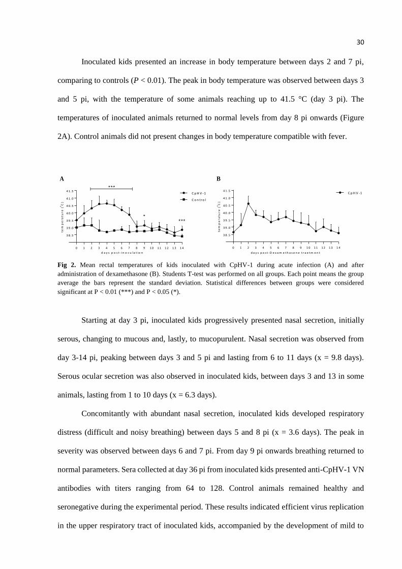
FIGURA 2 (Fig. 2) - Mean rectal temperatures of kids inoculated with CpHV-1 during acute
infection (A) and after administration of dexamethasone (B). Students T-test was performed on
all groups. Each point means the group average the bars represent the standard deviation.
Statistical differences between groups were considered significant at P < 0.01 (***) and P <
0.05 (*) ……………….………………………………………...……………………….…....30
8
LISTA DE TABELAS
CAPÍTULO 1
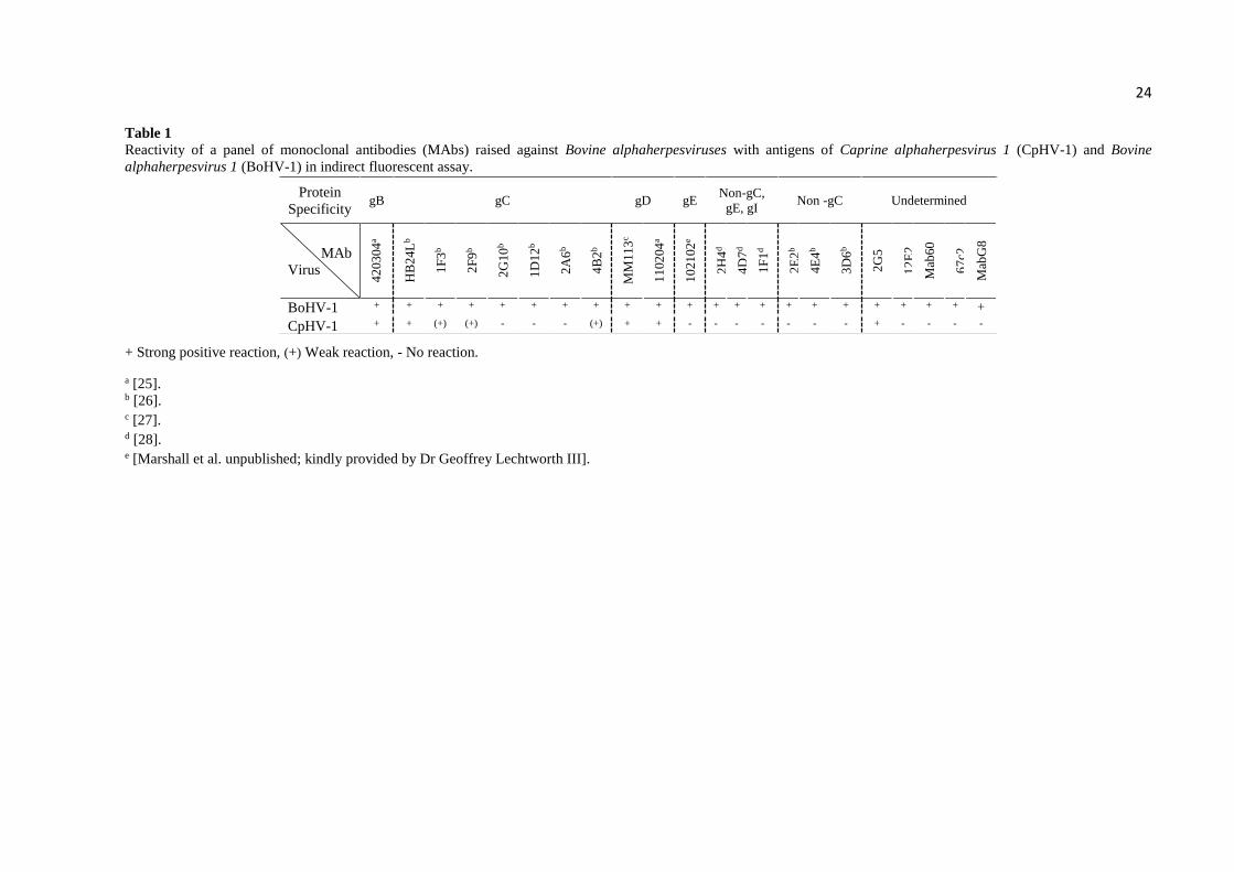
TABELA 1 (Table 1) - Reactivity of a panel of monoclonal antibodies (MAbs) raised against
Bovine alphaherpesviruses with antigens of Caprine alphaherpesvirus 1 (CpHV-1) and Bovine
alphaherpesvirus 1 (BoHV-1) in indirect fluorescent
assay.……………………...……………………………………………...…………………...24
TABELA 2 (Table 2) - Virus neutralizing activity of sera of cattle and goats
immunized/inoculated with Caprine alphaherpesvirus 1 (CpHV-1) and Bovine
alphaherpesvirus 1 (BoHV-1) against the homologous and heterologous
virus…………………………………………………………………………………………...26
TABELA 3 (Table 3) - Virus neutralizing (VN) activity of bovine sera submitted to routine
serology to Bovine alphaherpesvirus 1 (BoHV-1)
……………………………………………………..………………………………………….28
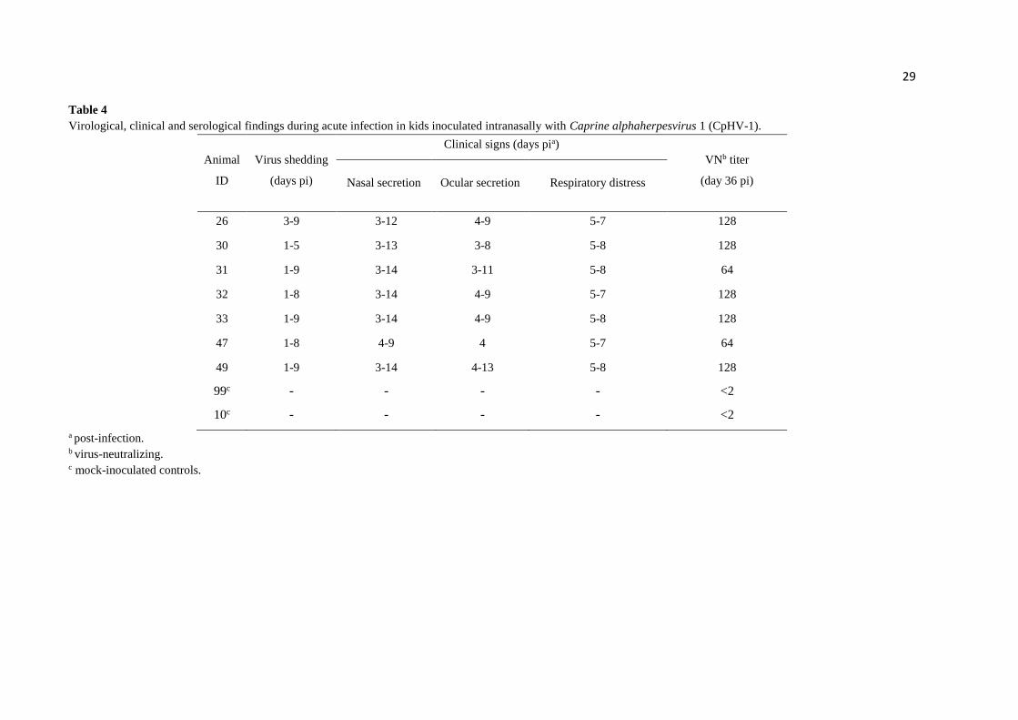
TABELA 4 (Table 4) - Virological, clinical and serological findings during acute infection in
kids inoculated intranasally with Caprine alphaherpesvirus 1 (CpHV-1)
…………………………………………………………………………………………..…….29
TABELA 5 (Table 5) - Virological, clinical and serological findigs after dexamethasone
administration (days 36-40 pi) in kids inoculated intranasally with Caprine alphaherpesvirus 1
(CpHV-1) ……………………………………………………………..………………...……32
9
SUMÁRIO
1. INTRODUÇÃO .......................................................................................................... 10
2. CAPÍTULO 1 – Antigenic relationship between Caprine alphaherpesvirus 1
(CpHV-1) and Bovine alphaherpesvirus 1 (BoHV-1) and experimental CpHV-1
infection of kids and calves............................................................................................ 13
Highlights........................................................................................................................ 15
Abstract........................................................................................................................... 16
Introduction ................................................................................................................... 17
Material and Methods ................................................................................................... 19
Results ............................................................................................................................. 22
Discussion ....................................................................................................................... 33
Acknowledgements ........................................................................................................ 37
References ....................................................................................................................... 37
3. REFERÊNCIAS ......................................................................................................... 43
10
1. INTRODUÇÃO
O alfaherpesvírus caprino 1 (Caprine alphaherpesvirus 1, CpHV-1) pertence à ordem
Herpesvirales, família Herpesviridae, subfamília Alphaherpesvirinae e gênero Varicellovirus
(ICTV, 2019). Os membros da família Herpesviridae são vírus envelopados, possuem cerca de
120 a 300 nm de diâmetro e apresentam como genoma uma fita dupla de DNA, com extensão
entre 125 a 235 kb (MOCARSKI & ROIZMAN, 1982; BOEHMER & NIMONKAR, 2003).
Uma das principais características dos herpesvírus é a capacidade de estabelecer latência e
persistir indefinidamente em seus hospedeiros. Essa latência, por sua vez, pode ser reativada
por diferentes fatores, resultando na retomada da replicação e excreção viral, com ou sem a
recorrência de doença (BOEHMER & NIMONKAR, 2003).
O CpHV-1 foi inicialmente descrito na década de 1970 nos Estados Unidos e na Suíça,
em infecções graves e generalizadas de cabritos (WALDVOGEL et al., 1981; UZAL et al.,
2004; MCCOY et al., 2007). Em seguida, infecções pelo CpHV-1 foram relatadas em vários
países, como Nova Zelândia, Austrália, Suécia, Espanha, Itália e França (METTLER et al.,
1979; HORNER; HUNTER; DAY, 1982; TISDALL et al., 1984; ROPERTO et al., 2000;
KEUSER et al., 2004; THIRY et al., 2008). A infecção por CpHV-1 é prevalente em países
europeus e mediterrâneos, onde a soroprevalência chega a 50% dos rebanhos (SUAVET et al.,
2016). Um estudo sobre os fatores de risco envolvendo CpHV-1 demonstrou que a presença do
CpHV-1 está associada à criação extensiva dos caprinos (BERTOLINI et al., 2018).
Em infecções naturais, o CpHV-1 geralmente penetra no hospedeiro pela mucosa oral
ou respiratória (TEMPESTA et al., 2004). Após a replicação primária nesses locais, os
componentes do capsídeo viral migram para o núcleo de células nervosas, onde o genoma
permanece inativo (sem expressão gênica e/ou replicação) em neurônios dos gânglios trigêmeos
ou sacrais por tempo indefinido, o que caracteriza infecção latente (VOGEL et al., 2003). Os
locais de infecção latente dependem da via de infecção e da disseminação do vírus
(TEMPESTA et al., 2004). Por outro lado, a latência do CpHV-1 pode ser de difícil reativação.
Em condições naturais, a reativação foi observada apenas em animais no cio e com baixos
títulos de anticorpos neutralizantes (TEMPESTA et al., 1998; CAMERO et al., 2010).
O CpHV-1 está associado com uma variedade de manifestações clínicas em caprinos
jovens, decorrentes de infecções gastroentéricas e respiratórias. Já em animais adultos, embora
as infecções por CpHV-1 sejam frequentemente subclínicas, o vírus pode ser responsável por
diferentes sinais clínicos, como febre, quadros de doenças respiratórias, vulvovaginite e
11
balanopostite, podendo também levar ao aborto ou à morte de neonatos (TEMPESTA et al.,
1999; SUAVET et al., 2016). Os casos de abortos, natimortos e outras falhas reprodutivas
resultam em importantes perdas econômicas, especialmente em países com alta concentração
de rebanhos caprinos (ROPERTO et al., 2000). Os animais neonatos podem também apresentar
doença grave, caracterizada por febre, conjuntivite, aumento de secreção ocular e nasal,
dispneia, lesões ulcerativas e necróticas em todo o trato entérico, as quais podem cursar com
alta morbidade e mortalidade (TEMPESTA et al., 2000; CAMERO et al., 2015).
O CpHV-1 é genética e antigenicamente relacionado com outros membros da subfamília
Alphaherpesvirinae de ruminantes, incluindo o alphaherpesvírus de cervídeo (Cervid
alphaherpesvirus, CvHV-1), o alphaherpesvírus de búfalo (Buffalo alphaherpesvirus, BuHV-
1) (DE CARLO et al., 2004) e os alphaherpesvírus 1 e 5 de bovinos (Bovine alphaherpesvirus
1 e 5, BoHV-1 e BoHV-5) (THIRY et al., 2006). Especificamente em relação ao BoHV-1,
agente da rinotraqueíte infecciosa bovina (Infectious bovine rhinotracheitis, IBR), a relação
antigênica com o CpHV-1 foi evidenciada por reação cruzada de anticorpos, especialmente com
anticorpos direcionados à glicoproteína B (gB), mas também contra a gC e gD (ENGELS et
al., 1992; BERTOLOTTI et al., 2013). Essa proximidade antigênica resulta na dificuldade de
um diagnóstico específico entre os alphaherpesvírus (BERTOLOTTI et al., 2013). De fato,
ainda não foi desenvolvido um teste sorológico específico que seja capaz de detectar o CpHV-
1 e que apresente baixa ou limitada reatividade cruzada com outros vírus de ruminantes
pertencentes à família dos alphaherpesvírus (BERTOLOTTI et al., 2013). Apesar disso, o
diagnóstico sorológico da infecção pelo CpHV-1 pode ser realizado utilizando o teste de
soroneutralização (SN) e o teste de ensaio imunoenzimático (Enzyme-linked immunosorbent
assay, ELISA). O teste SN é considerado o padrão-ouro e exibe alta sensibilidade. A
especificidade da SN, por sua vez, pode ser avaliada pelo teste de soroneutralização cruzada,
no qual a média dos títulos de anticorpos contra o vírus homólogo deve ser maior que os títulos
contra o vírus heterólogo (THIRY et al., 2008). O ELISA indireto provou ser tão sensível
quanto o teste de SN, embora tenha sido detectada reação cruzada com caprinos infectados
experimentalmente com BoHV-1 (MARINARO et al., 2010).
Experimentos realizados com CpHV-1 e BoHV-1 sugerem que tanto bovinos são
susceptíveis ao CpHV-1, quanto caprinos são susceptíveis ao BoHV-1, quando esses são
inoculados pela via intranasal. Em espécie heteróloga, o CpHV-1 e o BoHV-1 seriam ainda
capazes de serem excretados durante a infecção aguda e de estabelecer infecção latente. O
BoHV-1 é capaz de provocar sinais clínicos leves quando inoculado em cabras (SIX, et al.,
2001; THIRY et al., 2006). Entretanto, sinais clínicos provocados pelo CpHV-1 parecem ser

12
restritos ao seu hospedeiro natural, sendo a patogênese da infecção do CpHV-1 em bovinos
ainda não totalmente compreendida (TEMPESTA et al., 2001).
Assim, os objetivos do presente estudo foram aprofundar a investigação sobre as
relações antigênicas entre CpHV-1 e BoHV-1 e estudar a patogênese do CpHV-1 (isolado WI
13-46) em caprinos e bovinos.
13
2. CAPÍTULO 1
Antigenic relationship between Caprine alphaherpesvirus 1 (CpHV-1) and Bovine
alphaherpesvirus 1 (BoHV-1) and experimental CpHV-1 infection of kids and calves
Bruno Martins, Rafael Costa Ebling, Mathias Martins, Diego Gustavo Diel, Rudi Weiblen,
Eduardo Furtado Flores
(Artigo publicado no periódico Microbial Pathogenesis, v.136, 9 de agosto de 2019)
14
Antigenic relationship between Caprine alphaherpesvirus 1 (CpHV-1) and Bovine
alphaherpesvirus 1 (BoHV-1) and experimental CpHV-1 infection of kids and calves
Bruno Martinsa; Rafael C. Eblinga; Mathias Martinsb; Diego G. Dielc,d; Rudi Weiblena; Eduardo
F. Floresa*
aSetor de Virologia (SV), Departamento de Medicina Veterinária Preventiva (DMVP),
Universidade Federal de Santa Maria (UFSM). Santa Maria, RS, 97105-900, Brazil.
bLaboratório de Virologia, Universidade do Oeste de Santa Catarina (UNOESC). Xanxere, SC,
89820-000, Brazil.
cAnimal Disease Research and Diagnostic Laboratory, Department of Veterinary and
Biomedical Sciences, South Dakota State University (SDSU). Brookings, SD, 57007, USA.
dSouth Dakota Center for Biologics Research and Commercialization (SD-CBRC), South
Dakota State University (SDSU). Brookings, SD, 57007, USA.
*Corresponding author: Av. Roraima 1000, Centro de Eventos, Building 63A. Universidade
Federal de Santa Maria. Santa Maria, RS, Brazil. 97105-900. Phone/Fax +55 55 32208055. E-
mail: [email protected].
15
Highlights
- CpHV-1 and BoHV-1 are antigenically closely related;
- Monoclonal antibodies to BoHV-1 glycoproteins B, C and D recognize CpHV-1 antigens;
- BoHV-1 antisera neutralized CpHV-1 with higher efficiency than CpHV-1 antisera
neutralized BoHV-1;
- CpHV-1 is able to produce respiratory disease in kids, but does not replicate to detectable in
calves.
16
Abstract
Caprine alphaherpesvirus 1 (CpHV-1) is a worldwide pathogen of goats and is closely related
to Bovine alphaherpevirus 1 (BoHV-1). We herein studied the antigenic relationships of CpHV-
1 with BoHV-1 and investigated the pathogenesis of CpHV-1 in kids and calves. Monoclonal
antibody reactivity revealed that CpHV-1 and BoHV-1 share immunogenic epitopes in the
major envelope glycoproteins, gB, gC and gD. The antigenic relationship was further
demonstrated by virus-neutralizing assays, in which CpHV-1 and BoHV-1 antisera presented
varied degrees of cross-neutralization against the respective heterologous viruses. Although
cross-neutralization was observed between both viruses and the heterologous antisera, BoHV-
1 antisera neutralized CpHV-1 with higher efficiency than CpHV-1 antisera neutralized BoHV-
1. Hence, the antigenic cross-reactivity between CpHV-1 and BoHV-1 should be considered
upon serologic testing of goats and cattle in regions where the two viruses co-circulate.
Intranasal (IN) inoculation of CpHV-1 (WI13-46 isolate) in seven seronegative kids resulted in
efficient viral replication in the respiratory tract. Additionally, mild to moderate systemic and
respiratory signs were observed, including apathy, hyperthermia, nasal discharge and
respiratory distress. Dexamethasone administration to the inoculated kids between days 36 and
40 pi did not result in virus shedding in nasal secretions. However, latent infection had been
established, as evidenced by the detection of CpHV-1 DNA in trigeminal ganglia and olfactory
bulbs of kids euthanized at day 67 pi. Contrasting with the outcome of infection in kids, IN
inoculation of CpHV-1 in calves did not result in productive infection as no virus replication or
shedding were detected, and the animals did not develop clinical signs nor seroconverted. The
animal experiments demonstrated that CpHV-1 was able to produce respiratory disease in kids,
but did not replicate to detectable levels in calves.
Keywords: caprine herpesvirus, bovine herpesvirus, pathogenesis, antigenic relationship,
latent infection.
17
1. Introduction
Caprine alphaherpesvirus 1 (CpHV-1) is a member of the family Herpesviridae,
subfamily Alphaherpesvirinae and genus Varicellovirus [1]. Varicelloviruses are viruses
ranging from 120 to 300 nm in diameter, enveloped, containing a double-stranded linear DNA
genome arranged in a long unique region (UL) and a short unique region (US) flanked by
inverted internal- (IR) and terminal repeats (TR) [2]. CpHV-1 is widely distributed in goat
populations and is associated with severe systemic disease in young kids and reproductive
disease and abortion in adult goats [3–6]. CpHV-1 is genetically and antigenically related to
Bovine alphaherpesvirus 1 (BoHV-1), an important pathogen of cattle involved in respiratory,
reproductive disease and abortions [2].
The genetic and antigenic relationship between CpHV-1 and BoHV-1 has potential
implications in their epidemiology, diagnostic and control [2]. Data from natural and
experimental infections have shown the ability of these viruses to cross-infect the respective
heterologous hosts. Upon experimental inoculation, BoHV-1 is able to replicate acutely, to
establish and to reactivate from latent infection in goats [7]. In addition, natural cases of BoHV-
1 latent infection and positive serology have been reported in goats [8,9]. Similarly, CpHV-1
replicated efficiently and was shed by experimentally infected calves during acute infection and
established latent infection, yet did not reactivate from latency upon corticosteroid
administration [7,10]. These observations suggest that cross-infections between BoHV-1 and
CpHV-1 in their respective hosts may potentially occur.
The similarity in viral encoded structural proteins, including envelope glycoproteins,
gB, gC and gD, results in serological cross-reactivity between BoHV-1 and CpHV-1 [11]. In
addition, antibodies reacting with BoHV-1 have been detected in other ruminants, including
goats, suggesting a potential role of this species as BoHV-1 reservoir [2,9,12]. The serological
cross-reactivity between BoHV-1 and CpHV-1 may affect the specificity of serological tests
18
and, thus, compromise diagnostic and control efforts [2]. A better understanding of the antigenic
relationship between BoHV-1 and CpHV-1 and the potential impact of cross-species
transmission is critical for the design and implementation of effective and specific diagnostic
tools and control measures.
The pathogenesis of acute CpHV-1 infection in goats and of BoHV-1 in calves is similar
[2,13]. Natural CpHV-1 infections of adult and immunocompetent goats are frequently
subclinical [6,14,15], but have been associated with systemic disease and high mortality in
young kids [4,16], reproductive disorders and abortions in adult goats [17–19]. After acute
infection, CpHV-1 establishes latent infection in trigeminal or sacral nerve ganglia, depending
on the route of entry [20–22]. Latent infection may be naturally reactivated under stressful
conditions [20–22], but it seems rather infrequent [23]. Likewise, experimental reactivation is
difficult to achieve and usually requires the administration of high doses of corticosteroids
[10,20].
CpHV-1 and BoHV-1 are important pathogens in their natural hosts and cross-infection
of their respective heterologous hosts might have clinical, epidemiological and diagnosis
implications. Due to their clinical relevance, both agents have been targets for vaccine
development in the last decades. Importantly, BoHV-1 eradication programs have been initiated
in Europe. We are currently prospecting viral agents to serve as new vaccine delivery vectors
for cattle. In this sense, CpHV-1 appeared to be an attractive alternative. Here, we investigated
the antigenic relationship between CpHV-1 and BoHV-1 and assessed the pathogenesis of
CpHV-1 in kids and calves.
19
2. Materials and methods
2.1 Cells and viruses
Cell lines MDBK (Madin-Darby bovine kidney – ATCC-CL22), CRIB (Madin-Darby
bovine kidney BVDV-infection-resistant) and bovine turbinate (BT) (ATCC – CRL - 1390)
were used for virus amplification, titrations and growth curves. Cells were cultured in minimum
essential medium (MEM) (Vitrocell®, Nova Campinas, Sao Paulo - Brazil), supplemented with
10% fetal bovine serum (FBS) (Vitrocell®), penicillin (100 IU.mL-1) and streptomycin (100
μg.mL-1) (Sigma – Aldrich®, Darmstadt, Hessen, Germany) at 37 °C with 5% CO2. The viruses
used herein were: CpHV-1 WI 13-46 isolated and BoHV-1 SV56/90 isolate. CpHV-1 WI 13-
46 was isolated in BT cells from the lungs of sick kids from herd in Wisconsin (2013) with
history of weak kids, diarrhea, coughing, eye infections and abortions. The virus was amplified
in MDBK cells and used for animal inoculation at passage # 7. SV56/90 strain is a BoHV-1
isolated from preputial swabs and semen of a bull with balanoposthitis in Southern Brazil [24].
This virus was amplified in CRIB cells and used to immunize calves to produce BoHV-1
antisera (passage # 8) and in MAB-binding assay an VN assays.
2.2 Growth curves
The replication properties of CpHV-1 were assessed in vitro by single- and multi-step
growth curves in MDBK, CRIB and BT cells. Cells cultured in 12-well plates were inoculated
with CpHV-1 at multiplicity of infections (MOI) of 0.1 or 10 (multi-step and single-step growth
curves, respectively) and harvested at various time points post-infection (6, 12, 24, 48 and 72 h
pi). Cells were freeze-thawed three times, centrifuged at 5.000 x g and the virus titers were
determined by limiting dilution. Virus titers were determined for each time point using end-
point dilutions and the Spearman and Karber's method and expressed as mean tissue culture
infectious doses per mL (TCID50.mL-1).
20
2.3. Antigenic analysis
Antigenic analysis of CpHV-1 and BoHV-1 was performed by monoclonal antibody
(MAb) reactivity and virus-neutralizing (VN) assays. For MAb-reactivity assays, CRIB cells
were inoculated with CpHV-1 or BoHV-1 at an MOI of 0.1 and 18 – 24 h later were fixed with
cold acetone (5 min), washed in phosphate-buffered saline and distilled water and incubated
with individual MAbs produced against BoHV-1 (and BoHV-5) antigen (Table 1), followed by
washing and incubation with a FITC-conjugated anti-mouse IgG antibody (1:100) (Sigma,
Inc.). Slides were observed under UV light in an epifluorescence microscope (Axiolab
ZEISS®).
The serological cross-reactivity between CpHV-1 and BoHV-1 was investigated by VN
assays using the following sera: i. Sera of calves inoculated intranasally (IN) with BoHV-1
(SV56/90 isolate) or subcutaneously (SC) with CpHV-1 (WI 13-46 strain); ii. Sera of goats
inoculated IN with CpHV-1; iii. Field bovine sera submitted to routine BoHV-1 serology. These
sera were tested against CpHV-1 WI 13-46 and BoHV-1 SV56/90 in a standard VN assay.
Briefly, VN assays were performed in 96-well plates, by incubating two-fold dilutions of serum
against 100-200 TCID50 of the respective virus for 2 h. Then, a suspension of CRIB cells was
added and the plates were incubated at 37 °C with 5% CO2. End-point titers were determined
by the presence or not of cytopathic effect on infected cell cultures that were read after 96 h of
incubation. The neutralizing titers were considered the reciprocal of the highest serum dilution
capable of preventing virus replication.
2.4. Animal experiments
This study was approved by the institutional Ethics Committee on Animal Use (CEUA
– UFSM - protocol nº 7209040618).
21
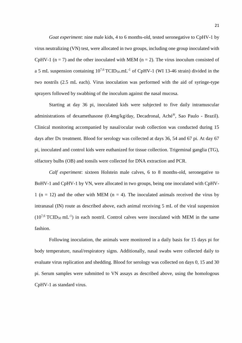
Goat experiment: nine male kids, 4 to 6 months-old, tested seronegative to CpHV-1 by
virus neutralizing (VN) test, were allocated in two groups, including one group inoculated with
CpHV-1 (n = 7) and the other inoculated with MEM (n = 2). The virus inoculum consisted of
a 5 mL suspension containing 107,6 TCID50.mL-1 of CpHV-1 (WI 13-46 strain) divided in the
two nostrils (2.5 mL each). Virus inoculation was performed with the aid of syringe-type
sprayers followed by swabbing of the inoculum against the nasal mucosa.
Starting at day 36 pi, inoculated kids were subjected to five daily intramuscular
administrations of dexamethasone (0.4mg/kg/day, Decadronal, Aché®, Sao Paulo - Brazil).
Clinical monitoring accompanied by nasal/ocular swab collection was conducted during 15
days after Dx treatment. Blood for serology was collected at days 36, 54 and 67 pi. At day 67
pi, inoculated and control kids were euthanized for tissue collection. Trigeminal ganglia (TG),
olfactory bulbs (OB) and tonsils were collected for DNA extraction and PCR.
Calf experiment: sixteen Holstein male calves, 6 to 8 months-old, seronegative to
BoHV-1 and CpHV-1 by VN, were allocated in two groups, being one inoculated with CpHV-
1 (n = 12) and the other with MEM (n = 4). The inoculated animals received the virus by
intranasal (IN) route as described above, each animal receiving 5 mL of the viral suspension
(107,6 TCID50 mL-1) in each nostril. Control calves were inoculated with MEM in the same
fashion.
Following inoculation, the animals were monitored in a daily basis for 15 days pi for
body temperature, nasal/respiratory signs. Additionally, nasal swabs were collected daily to
evaluate virus replication and shedding. Blood for serology was collected on days 0, 15 and 30
pi. Serum samples were submitted to VN assays as described above, using the homologous
CpHV-1 as standard virus.
22
2.5. DNA extraction and PCR
Total DNA extraction and PCR for CpHV-1 DNA were performed in tissues collected
at necropsy (day 67 pi). DNA was extracted from TGs, OB and tonsils (~100 mg) using the
phenol/chloroform method, according to standard protocols. Approximately 200 ng of each
DNA sample were subjected to PCR to amplify an internal region of CpHV-1 thymidine kinase
(TK) gene. For this, primers forward 5 '- CTC GTC GTC TGC ACC CTT C-3' and reverse 5 '-
CGA CAT GTC CAG CGT GAA TA-3' were used, resulting in the amplification of a 399 bp
product. PCR reactions were performed in a volume of 25 μL using 2 μL of template DNA,
12.5 mM of each primer, 2.5 mM MgCl2, 10 mM dNTPs, 1x reaction buffer and 1 unit of Taq
DNA polymerase (Fischer™). PCR conditions were: initial denaturation (94 °C for 10 min),
followed by 30 cycles of 94 °C for 1 min; 54 °C for 1 min for primer annealing and 72 °C for
1 min for primer extension, and a final extension of 7 min at 72 °C. The products were
visualized on 1.5% agarose gel, stained with Gel Red (Biotium®) and visualized under UV light.
In all reactions, the DNA extracted from cells infected with CpHV-1 was used as positive
control and ultrapure water as negative control.
2.6. Statistical analysis
Statistical analysis was performed using the Prism software (GraphPad; 6th version).
Students T-test was performed on all groups. Statistical differences between groups were
considered significant at P<0.01 (***) or P<0.05 (*).
3. Results
3.1. Growth curves
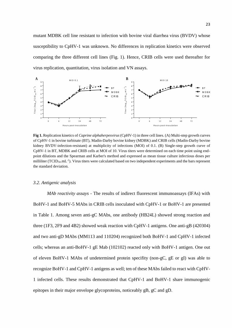
The replication kinetics of CpHV-1 was assessed in BT, MDBK and CRIB cells (MOI
= 0.1 [multi-step growth curve], and MOI = 10 [single-step growth curve]). CRIB cells are a
23
mutant MDBK cell line resistant to infection with bovine viral diarrhea virus (BVDV) whose
susceptibility to CpHV-1 was unknown. No differences in replication kinetics were observed
comparing the three different cell lines (Fig. 1). Hence, CRIB cells were used thereafter for
virus replication, quantitation, virus isolation and VN assays.
Fig 1. Replication kinetics of Caprine alphaherpesvirus (CpHV-1) in three cell lines. (A) Multi-step growth curves
of CpHV-1 in bovine turbinate (BT), Madin-Darby bovine kidney (MDBK) and CRIB cells (Madin-Darby bovine
kidney BVDV-infection-resistant) at multiplicity of infections (MOI) of 0.1. (B) Single-step growth curve of
CpHV-1 in BT, MDBK and CRIB cells at MOI of 10. Virus titers were determined on each time point using end-
point dilutions and the Spearman and Karber's method and expressed as mean tissue culture infectious doses per
milliliter (TCID50.mL-1). Virus titers were calculated based on two independent experiments and the bars represent
the standard deviation.
3.2. Antigenic analysis
MAb reactivity assays - The results of indirect fluorescent immunoassays (IFAs) with
BoHV-1 and BoHV-5 MAbs in CRIB cells inoculated with CpHV-1 or BoHV-1 are presented
in Table 1. Among seven anti-gC MAbs, one antibody (HB24L) showed strong reaction and
three (1F3, 2F9 and 4B2) showed weak reaction with CpHV-1 antigens. One anti-gB (420304)
and two anti-gD MAbs (MM113 and 110204) recognized both BoHV-1 and CpHV-1 infected
cells; whereas an anti-BoHV-1 gE Mab (102102) reacted only with BoHV-1 antigen. One out
of eleven BoHV-1 MAbs of undetermined protein specifity (non-gC, gE or gI) was able to
recognize BoHV-1 and CpHV-1 antigens as well; ten of these MAbs failed to react with CpHV-
1 infected cells. These results demonstrated that CpHV-1 and BoHV-1 share immunogenic
epitopes in their major envelope glycoproteins, noticeably gB, gC and gD.
Tit
er
(lo
g1
0T
CID
50
.mL
-1)
0 6 1 2 2 4 4 8 7 2
0
1
2
3
4
5
6
7
8
9
H o u rs p o s t - in o c u la t io n
M D B K
B T
A
C R IB
M O I 0 .1
Tit
er
(lo
g1
0T
CID
50
.mL
-1)
0 6 1 2 2 4 4 8 7 2
0
1
2
3
4
5
6
7
8
9
H o u rs p o s t - in o c u la t io n
M D B K
B T
B
C R IB
M O I 1 0
24
Table 1
Reactivity of a panel of monoclonal antibodies (MAbs) raised against Bovine alphaherpesviruses with antigens of Caprine alphaherpesvirus 1 (CpHV-1) and Bovine
alphaherpesvirus 1 (BoHV-1) in indirect fluorescent assay.
+ Strong positive reaction, (+) Weak reaction, - No reaction.
a [25]. b [26]. c [27]. d [28]. e [Marshall et al. unpublished; kindly provided by Dr Geoffrey Lechtworth III].
Protein
Specificity gB gC gD gE
Non-gC,
gE, gI Non -gC Undetermined
MAb
Virus
42
03
04
a
HB
24
Lb
1F
3b
2F
9b
2G
10
b
1D
12
b
2A
6b
4B
2b
MM
11
3c
11
02
04
a
10
21
02
e
2H
4d
4D
7d
1F
1d
2E
2b
4E
4b
3D
6b
2G
5
12
E2
Mab
60
67
c2
Mab
G8
BoHV-1 + + + + + + + + + + + + + + + + + + + + + +
CpHV-1 + + (+) (+) - - - (+) + + - - - - - - - + - - - -
25
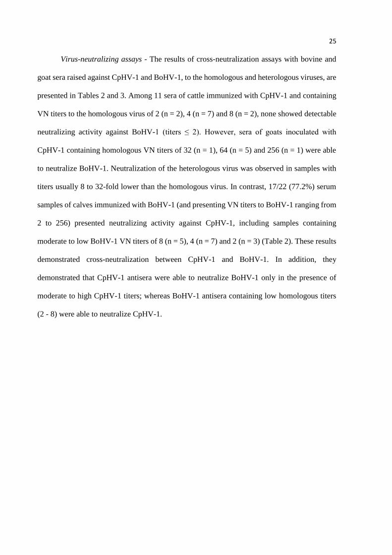
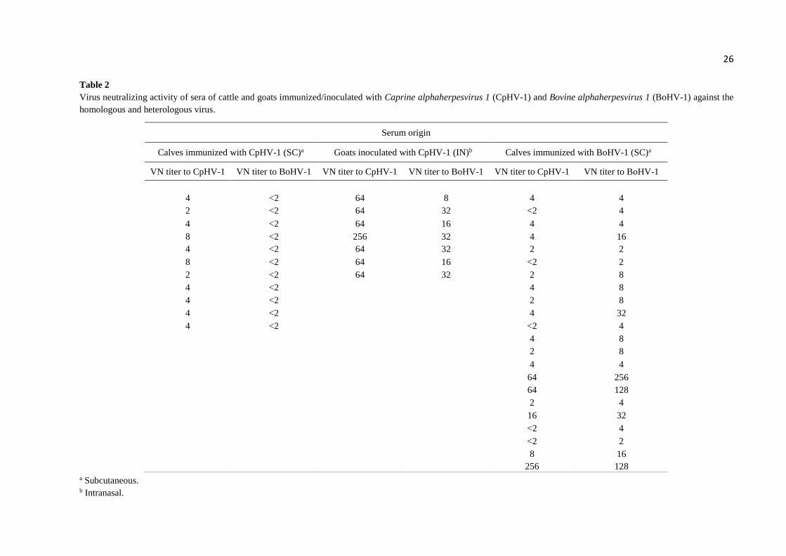
Virus-neutralizing assays - The results of cross-neutralization assays with bovine and
goat sera raised against CpHV-1 and BoHV-1, to the homologous and heterologous viruses, are
presented in Tables 2 and 3. Among 11 sera of cattle immunized with CpHV-1 and containing
VN titers to the homologous virus of 2 (n = 2), 4 (n = 7) and 8 (n = 2), none showed detectable
neutralizing activity against BoHV-1 (titers ≤ 2). However, sera of goats inoculated with
CpHV-1 containing homologous VN titers of 32 (n = 1), 64 (n = 5) and 256 (n = 1) were able
to neutralize BoHV-1. Neutralization of the heterologous virus was observed in samples with
titers usually 8 to 32-fold lower than the homologous virus. In contrast, 17/22 (77.2%) serum
samples of calves immunized with BoHV-1 (and presenting VN titers to BoHV-1 ranging from
2 to 256) presented neutralizing activity against CpHV-1, including samples containing
moderate to low BoHV-1 VN titers of 8 (n = 5), 4 (n = 7) and 2 (n = 3) (Table 2). These results
demonstrated cross-neutralization between CpHV-1 and BoHV-1. In addition, they
demonstrated that CpHV-1 antisera were able to neutralize BoHV-1 only in the presence of
moderate to high CpHV-1 titers; whereas BoHV-1 antisera containing low homologous titers
(2 - 8) were able to neutralize CpHV-1.
26
Table 2
Virus neutralizing activity of sera of cattle and goats immunized/inoculated with Caprine alphaherpesvirus 1 (CpHV-1) and Bovine alphaherpesvirus 1 (BoHV-1) against the
homologous and heterologous virus.
a Subcutaneous. b Intranasal.
Serum origin
Calves immunized with CpHV-1 (SC)a Goats inoculated with CpHV-1 (IN)b Calves immunized with BoHV-1 (SC)a
VN titer to CpHV-1 VN titer to BoHV-1 VN titer to CpHV-1 VN titer to BoHV-1 VN titer to CpHV-1 VN titer to BoHV-1
4 <2 64 8 4 4
2 <2 64 32 <2 4
4 <2 64 16 4 4
8 <2 256 32 4 16
4 <2 64 32 2 2
8 <2 64 16 <2 2
2 <2 64 32 2 8
4 <2 4 8
4 <2 2 8
4 <2 4 32
4 <2 <2 4
4 8
2 8
4 4
64 256
64 128
2 4
16 32
<2 4
<2 2
8 16 256 128
27
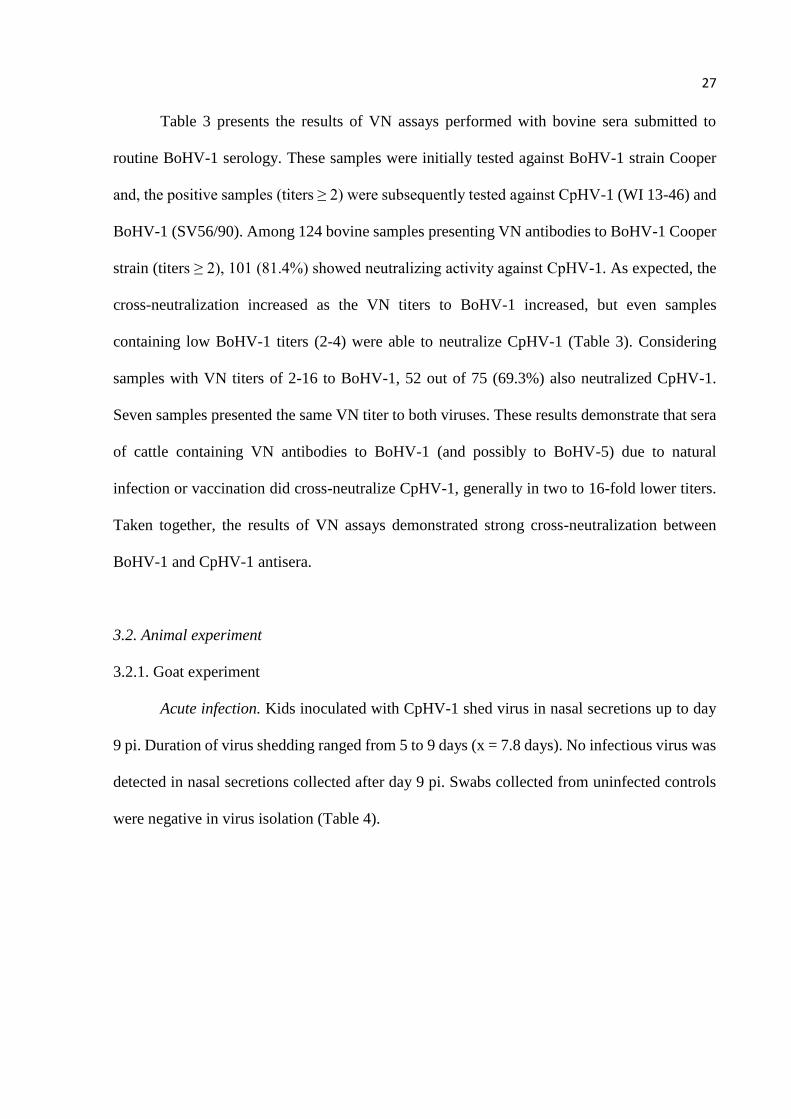
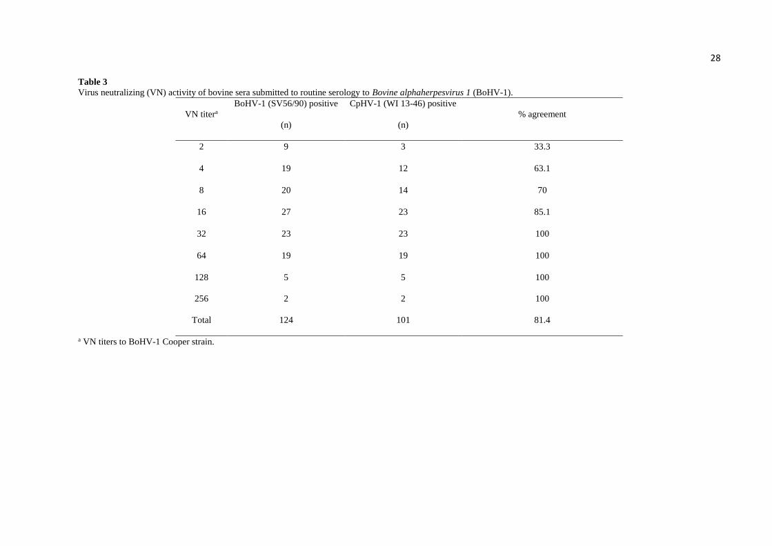
Table 3 presents the results of VN assays performed with bovine sera submitted to
routine BoHV-1 serology. These samples were initially tested against BoHV-1 strain Cooper
and, the positive samples (titers ≥ 2) were subsequently tested against CpHV-1 (WI 13-46) and
BoHV-1 (SV56/90). Among 124 bovine samples presenting VN antibodies to BoHV-1 Cooper
strain (titers ≥ 2), 101 (81.4%) showed neutralizing activity against CpHV-1. As expected, the
cross-neutralization increased as the VN titers to BoHV-1 increased, but even samples
containing low BoHV-1 titers (2-4) were able to neutralize CpHV-1 (Table 3). Considering
samples with VN titers of 2-16 to BoHV-1, 52 out of 75 (69.3%) also neutralized CpHV-1.
Seven samples presented the same VN titer to both viruses. These results demonstrate that sera
of cattle containing VN antibodies to BoHV-1 (and possibly to BoHV-5) due to natural
infection or vaccination did cross-neutralize CpHV-1, generally in two to 16-fold lower titers.
Taken together, the results of VN assays demonstrated strong cross-neutralization between
BoHV-1 and CpHV-1 antisera.
3.2. Animal experiment
3.2.1. Goat experiment
Acute infection. Kids inoculated with CpHV-1 shed virus in nasal secretions up to day
9 pi. Duration of virus shedding ranged from 5 to 9 days (x = 7.8 days). No infectious virus was
detected in nasal secretions collected after day 9 pi. Swabs collected from uninfected controls
were negative in virus isolation (Table 4).
28 Table 3
Virus neutralizing (VN) activity of bovine sera submitted to routine serology to Bovine alphaherpesvirus 1 (BoHV-1).
VN titera
BoHV-1 (SV56/90) positive
(n)
CpHV-1 (WI 13-46) positive
(n)
% agreement
2 9 3 33.3
4 19 12 63.1
8 20 14 70
16 27 23 85.1
32 23 23 100
64 19 19 100
128 5 5 100
256 2 2 100
Total 124 101 81.4
a VN titers to BoHV-1 Cooper strain.
29
Table 4
Virological, clinical and serological findings during acute infection in kids inoculated intranasally with Caprine alphaherpesvirus 1 (CpHV-1).
Animal
ID
Virus shedding
(days pi)
Clinical signs (days pia)
VNb titer
(day 36 pi) Nasal secretion Ocular secretion Respiratory distress
26 3-9 3-12 4-9 5-7 128
30 1-5 3-13 3-8 5-8 128
31 1-9 3-14 3-11 5-8 64
32 1-8 3-14 4-9 5-7 128
33 1-9 3-14 4-9 5-8 128
47 1-8 4-9 4 5-7 64
49 1-9 3-14 4-13 5-8 128
99c - - - - <2
10c - - - - <2
a post-infection. b virus-neutralizing. c mock-inoculated controls.
30
Inoculated kids presented an increase in body temperature between days 2 and 7 pi,
comparing to controls (P < 0.01). The peak in body temperature was observed between days 3
and 5 pi, with the temperature of some animals reaching up to 41.5 °C (day 3 pi). The
temperatures of inoculated animals returned to normal levels from day 8 pi onwards (Figure
2A). Control animals did not present changes in body temperature compatible with fever.
Fig 2. Mean rectal temperatures of kids inoculated with CpHV-1 during acute infection (A) and after
administration of dexamethasone (B). Students T-test was performed on all groups. Each point means the group
average the bars represent the standard deviation. Statistical differences between groups were considered
significant at P < 0.01 (***) and P < 0.05 (*).
Starting at day 3 pi, inoculated kids progressively presented nasal secretion, initially
serous, changing to mucous and, lastly, to mucopurulent. Nasal secretion was observed from
day 3-14 pi, peaking between days 3 and 5 pi and lasting from 6 to 11 days (x = 9.8 days).
Serous ocular secretion was also observed in inoculated kids, between days 3 and 13 in some
animals, lasting from 1 to 10 days (x = 6.3 days).
Concomitantly with abundant nasal secretion, inoculated kids developed respiratory
distress (difficult and noisy breathing) between days 5 and 8 pi (x = 3.6 days). The peak in
severity was observed between days 6 and 7 pi. From day 9 pi onwards breathing returned to
normal parameters. Sera collected at day 36 pi from inoculated kids presented anti-CpHV-1 VN
antibodies with titers ranging from 64 to 128. Control animals remained healthy and
seronegative during the experimental period. These results indicated efficient virus replication
in the upper respiratory tract of inoculated kids, accompanied by the development of mild to
tem
pe
ratu
re (
oC
)
0 1 2 3 4 5 6 7 8 9 1 0 1 1 1 2 1 3 1 4
3 8 .5
3 9 .0
3 9 .5
4 0 .0
4 0 .5
4 1 .0
4 1 .5
d a y s p o s t -D e x a m e t h a s o n e t r a e t m e n t
B
C p H V - 1
tem
pe
ratu
re (
oC
)
0 1 2 3 4 5 6 7 8 9 1 0 1 1 1 2 1 3 1 4
3 8 .5
3 9 .0
3 9 .5
4 0 .0
4 0 .5
4 1 .0
4 1 .5
d a y s p o s t - in o c u l a t i o n
C o n tro l
C p H V -1
***
****
A
31
moderate respiratory signs. The summary of virological, clinical and serological findings
observed in kids inoculated with CpHV-1 is presented in Table 4.
Latent infection. No infectious virus was detected in nasal secretions collected from
inoculated kids during days 36-40 pi or after Dx treatment (days 41-54 pi). The animals
subjected to Dx treatment presented a transient increase in body temperature, reaching a peak
at day 2 after the drug administration (Fig. 2B). No other clinical signs were observed in these
animals following Dx treatment. VN titers remained without significant changes after Dx
administration, as demonstrated by VN assays performed with sera collected at days 36 and 54
pi. PCR performed in total DNA extracted from TGs and OBs of kids euthanized at day 67 pi
revealed the presence of CpHV-1 DNA in both TGs and OB of inoculated animals. Inoculation
of TG and OB homogenates followed by three passages in CRIB cells did not result in isolation
of CpHV-1, confirming latent infection. Total DNA extracted from tonsils were negative for
viral DNA. These results indicated that CpHV-1 did establish latent infection in TGs and OBs
after acute infection but was not reactivated upon Dx treatment (Table 5).
3.2.2. Calf experiment
Inoculation of 12 calves with CpHV-1 isolate WI 13-46 did not result in detectable virus
replication in the nasal mucosa. No infectious CpHV-1 was isolated from nasal secretions
collected after virus inoculation after three passages in CRIB cells. Likewise, CpHV-1
inoculation in calves did not result in evident nasal or systemic signs of infection. Additionally,
no neutralizing antibodies were detected in serum collected on day 30 pi. Taken together, these
results demonstrated that CpHV-1 did not replicate to detectable levels in calves upon IN
inoculation.
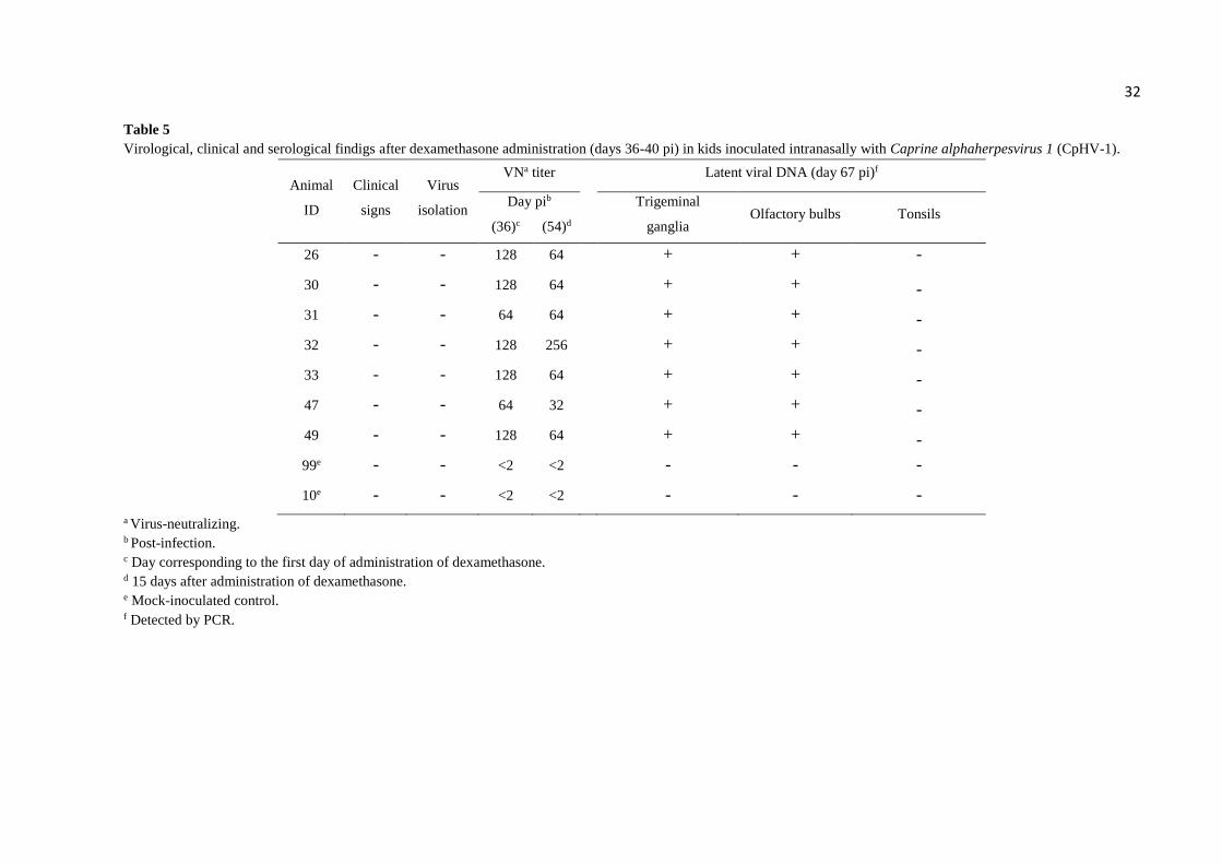
32 Table 5
Virological, clinical and serological findigs after dexamethasone administration (days 36-40 pi) in kids inoculated intranasally with Caprine alphaherpesvirus 1 (CpHV-1).
Animal
ID
Clinical
signs
Virus
isolation
VNa titer Latent viral DNA (day 67 pi)f
Day pib
Trigeminal
ganglia Olfactory bulbs Tonsils
(36)c (54)d
26 - - 128 64 + + -
30 - - 128 64 + + -
31 - - 64 64 + + -
32 - - 128 256 + + -
33 - - 128 64 + + -
47 - - 64 32 + + -
49 - - 128 64 + + -
99e - - <2 <2 - - -
10e - - <2 <2 - - -
a Virus-neutralizing. b Post-infection. c Day corresponding to the first day of administration of dexamethasone. d 15 days after administration of dexamethasone. e Mock-inoculated control. f Detected by PCR.
33
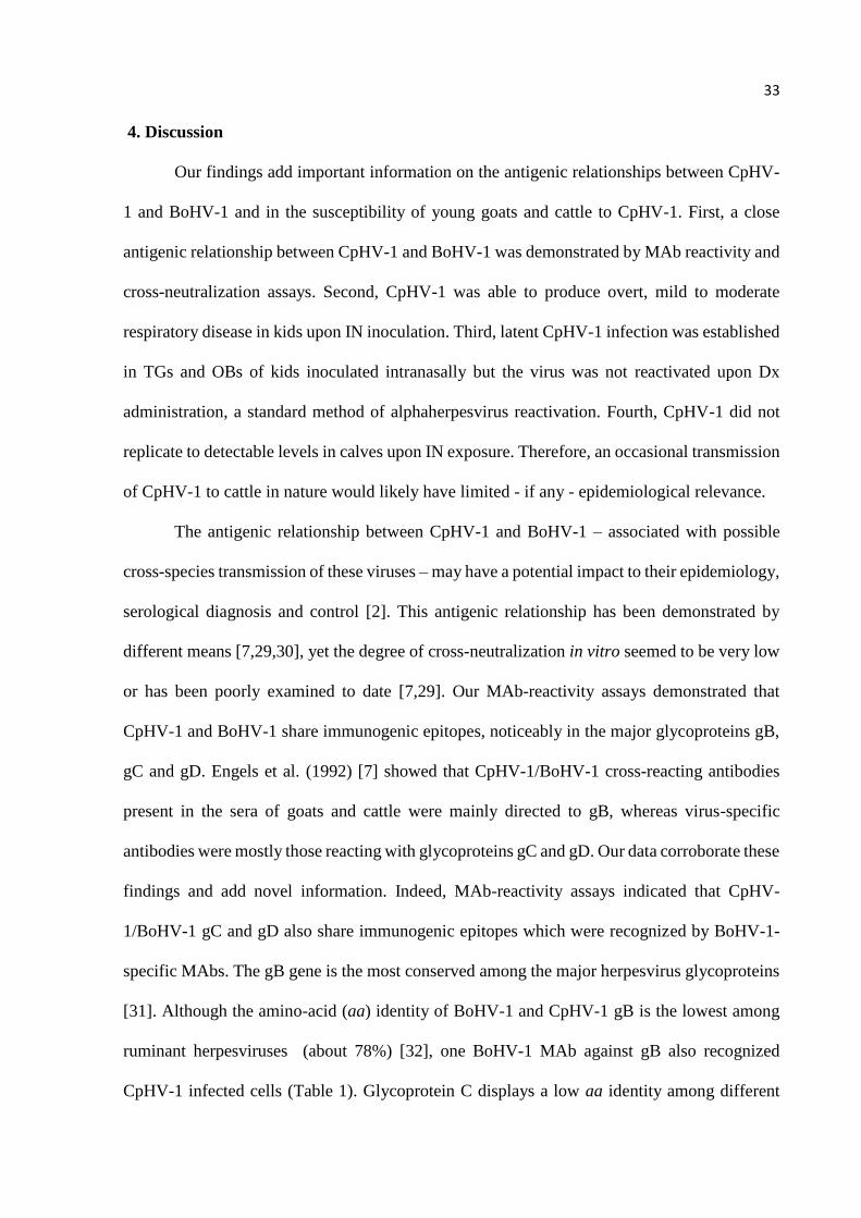
4. Discussion
Our findings add important information on the antigenic relationships between CpHV-
1 and BoHV-1 and in the susceptibility of young goats and cattle to CpHV-1. First, a close
antigenic relationship between CpHV-1 and BoHV-1 was demonstrated by MAb reactivity and
cross-neutralization assays. Second, CpHV-1 was able to produce overt, mild to moderate
respiratory disease in kids upon IN inoculation. Third, latent CpHV-1 infection was established
in TGs and OBs of kids inoculated intranasally but the virus was not reactivated upon Dx
administration, a standard method of alphaherpesvirus reactivation. Fourth, CpHV-1 did not
replicate to detectable levels in calves upon IN exposure. Therefore, an occasional transmission
of CpHV-1 to cattle in nature would likely have limited - if any - epidemiological relevance.
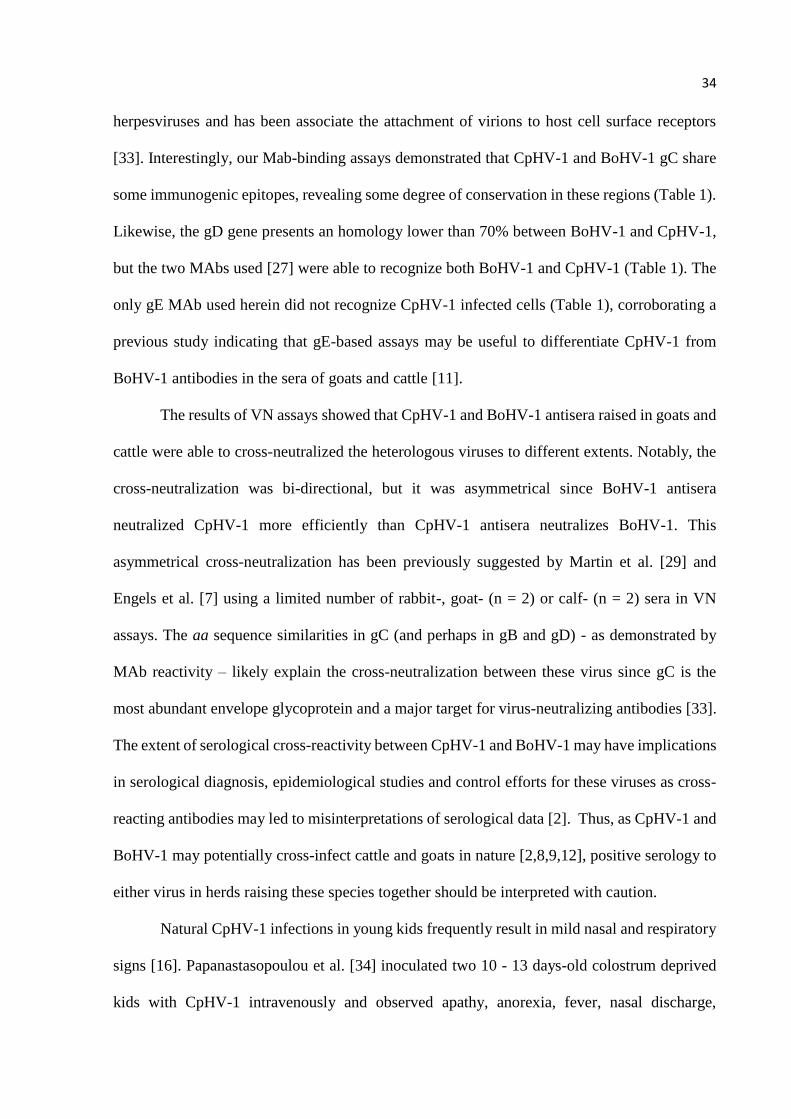
The antigenic relationship between CpHV-1 and BoHV-1 – associated with possible
cross-species transmission of these viruses – may have a potential impact to their epidemiology,
serological diagnosis and control [2]. This antigenic relationship has been demonstrated by
different means [7,29,30], yet the degree of cross-neutralization in vitro seemed to be very low
or has been poorly examined to date [7,29]. Our MAb-reactivity assays demonstrated that
CpHV-1 and BoHV-1 share immunogenic epitopes, noticeably in the major glycoproteins gB,
gC and gD. Engels et al. (1992) [7] showed that CpHV-1/BoHV-1 cross-reacting antibodies
present in the sera of goats and cattle were mainly directed to gB, whereas virus-specific
antibodies were mostly those reacting with glycoproteins gC and gD. Our data corroborate these
findings and add novel information. Indeed, MAb-reactivity assays indicated that CpHV-
1/BoHV-1 gC and gD also share immunogenic epitopes which were recognized by BoHV-1-
specific MAbs. The gB gene is the most conserved among the major herpesvirus glycoproteins
[31]. Although the amino-acid (aa) identity of BoHV-1 and CpHV-1 gB is the lowest among
ruminant herpesviruses (about 78%) [32], one BoHV-1 MAb against gB also recognized
CpHV-1 infected cells (Table 1). Glycoprotein C displays a low aa identity among different
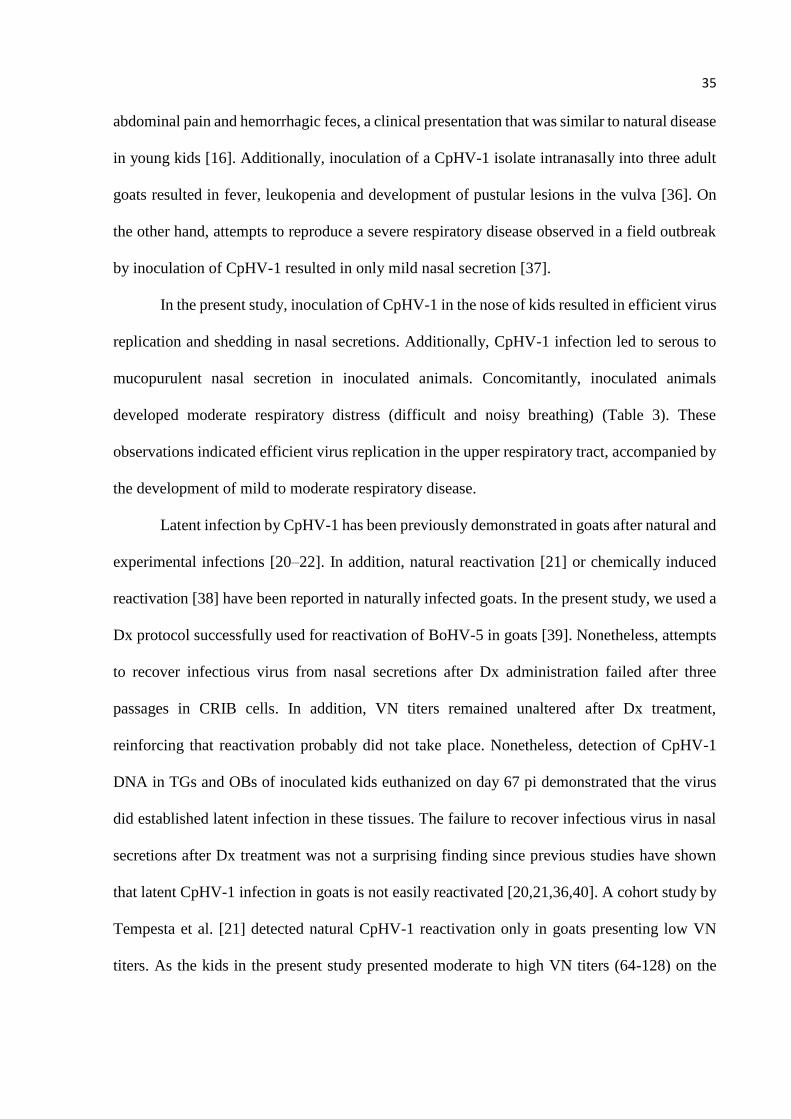
34
herpesviruses and has been associate the attachment of virions to host cell surface receptors
[33]. Interestingly, our Mab-binding assays demonstrated that CpHV-1 and BoHV-1 gC share
some immunogenic epitopes, revealing some degree of conservation in these regions (Table 1).
Likewise, the gD gene presents an homology lower than 70% between BoHV-1 and CpHV-1,
but the two MAbs used [27] were able to recognize both BoHV-1 and CpHV-1 (Table 1). The
only gE MAb used herein did not recognize CpHV-1 infected cells (Table 1), corroborating a
previous study indicating that gE-based assays may be useful to differentiate CpHV-1 from
BoHV-1 antibodies in the sera of goats and cattle [11].
The results of VN assays showed that CpHV-1 and BoHV-1 antisera raised in goats and
cattle were able to cross-neutralized the heterologous viruses to different extents. Notably, the
cross-neutralization was bi-directional, but it was asymmetrical since BoHV-1 antisera
neutralized CpHV-1 more efficiently than CpHV-1 antisera neutralizes BoHV-1. This
asymmetrical cross-neutralization has been previously suggested by Martin et al. [29] and
Engels et al. [7] using a limited number of rabbit-, goat- (n = 2) or calf- (n = 2) sera in VN
assays. The aa sequence similarities in gC (and perhaps in gB and gD) - as demonstrated by
MAb reactivity – likely explain the cross-neutralization between these virus since gC is the
most abundant envelope glycoprotein and a major target for virus-neutralizing antibodies [33].
The extent of serological cross-reactivity between CpHV-1 and BoHV-1 may have implications
in serological diagnosis, epidemiological studies and control efforts for these viruses as cross-
reacting antibodies may led to misinterpretations of serological data [2]. Thus, as CpHV-1 and
BoHV-1 may potentially cross-infect cattle and goats in nature [2,8,9,12], positive serology to
either virus in herds raising these species together should be interpreted with caution.
Natural CpHV-1 infections in young kids frequently result in mild nasal and respiratory
signs [16]. Papanastasopoulou et al. [34] inoculated two 10 - 13 days-old colostrum deprived
kids with CpHV-1 intravenously and observed apathy, anorexia, fever, nasal discharge,
35
abdominal pain and hemorrhagic feces, a clinical presentation that was similar to natural disease
in young kids [16]. Additionally, inoculation of a CpHV-1 isolate intranasally into three adult
goats resulted in fever, leukopenia and development of pustular lesions in the vulva [36]. On
the other hand, attempts to reproduce a severe respiratory disease observed in a field outbreak
by inoculation of CpHV-1 resulted in only mild nasal secretion [37].
In the present study, inoculation of CpHV-1 in the nose of kids resulted in efficient virus
replication and shedding in nasal secretions. Additionally, CpHV-1 infection led to serous to
mucopurulent nasal secretion in inoculated animals. Concomitantly, inoculated animals
developed moderate respiratory distress (difficult and noisy breathing) (Table 3). These
observations indicated efficient virus replication in the upper respiratory tract, accompanied by
the development of mild to moderate respiratory disease.
Latent infection by CpHV-1 has been previously demonstrated in goats after natural and
experimental infections [20–22]. In addition, natural reactivation [21] or chemically induced
reactivation [38] have been reported in naturally infected goats. In the present study, we used a
Dx protocol successfully used for reactivation of BoHV-5 in goats [39]. Nonetheless, attempts
to recover infectious virus from nasal secretions after Dx administration failed after three
passages in CRIB cells. In addition, VN titers remained unaltered after Dx treatment,
reinforcing that reactivation probably did not take place. Nonetheless, detection of CpHV-1
DNA in TGs and OBs of inoculated kids euthanized on day 67 pi demonstrated that the virus
did established latent infection in these tissues. The failure to recover infectious virus in nasal
secretions after Dx treatment was not a surprising finding since previous studies have shown
that latent CpHV-1 infection in goats is not easily reactivated [20,21,36,40]. A cohort study by
Tempesta et al. [21] detected natural CpHV-1 reactivation only in goats presenting low VN
titers. As the kids in the present study presented moderate to high VN titers (64-128) on the
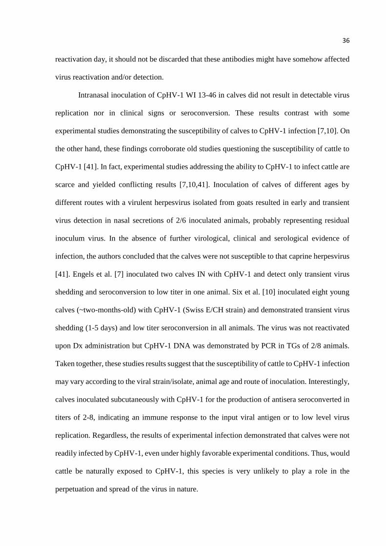
36
reactivation day, it should not be discarded that these antibodies might have somehow affected
virus reactivation and/or detection.
Intranasal inoculation of CpHV-1 WI 13-46 in calves did not result in detectable virus
replication nor in clinical signs or seroconversion. These results contrast with some
experimental studies demonstrating the susceptibility of calves to CpHV-1 infection [7,10]. On
the other hand, these findings corroborate old studies questioning the susceptibility of cattle to
CpHV-1 [41]. In fact, experimental studies addressing the ability to CpHV-1 to infect cattle are
scarce and yielded conflicting results [7,10,41]. Inoculation of calves of different ages by
different routes with a virulent herpesvirus isolated from goats resulted in early and transient
virus detection in nasal secretions of 2/6 inoculated animals, probably representing residual
inoculum virus. In the absence of further virological, clinical and serological evidence of
infection, the authors concluded that the calves were not susceptible to that caprine herpesvirus
[41]. Engels et al. [7] inoculated two calves IN with CpHV-1 and detect only transient virus
shedding and seroconversion to low titer in one animal. Six et al. [10] inoculated eight young
calves (~two-months-old) with CpHV-1 (Swiss E/CH strain) and demonstrated transient virus
shedding (1-5 days) and low titer seroconversion in all animals. The virus was not reactivated
upon Dx administration but CpHV-1 DNA was demonstrated by PCR in TGs of 2/8 animals.
Taken together, these studies results suggest that the susceptibility of cattle to CpHV-1 infection
may vary according to the viral strain/isolate, animal age and route of inoculation. Interestingly,
calves inoculated subcutaneously with CpHV-1 for the production of antisera seroconverted in
titers of 2-8, indicating an immune response to the input viral antigen or to low level virus
replication. Regardless, the results of experimental infection demonstrated that calves were not
readily infected by CpHV-1, even under highly favorable experimental conditions. Thus, would
cattle be naturally exposed to CpHV-1, this species is very unlikely to play a role in the
perpetuation and spread of the virus in nature.
37
Acknowledgements
The authors thank the Conselho Nacional de Desenvolvimento Cientifico e Tecnologico
for the scholarships. Bruno Martins received Master’s Scholarship, Eduardo F. Flores (process
301414/2010-6) and Rudi Weiblen (process 304153/2014-1) were supported by CNPq research
fellowships. This study was financed in part by the Coordenação de Aperfeiçoamento de
Pessoal de Nível Superior (CAPES), Brasil - Finance code 001. BM and RCE are graduate
students of the Programa de Pós-graduação em Medicina Veterinária – PGMV/UFSM.
Appendix A. Supplementary data
Supplementary data to this article can be found online at
https://doi.org/10.1016/j.micpath.2019.103663.
Conflict of interest statement
The authors declare no conflict of interest.
References
[1] ICTV 2019. International Committee on taxonomy of viruses. Available in
http://www.ictvonline.org/taxonomy, (2019) accessed April 6 , 2019.
[2] J. Thiry, V. Keuser, B. Muylkens, F. Meurens, S. Gogev, A. Vanderplasschen, E. Thiry,
Ruminant alphaherpesviruses related to bovine herpesvirus 1, Vet. Res. 37 (2006) 169–
90, https://doi.org/10.1051/vetres:2005052.
[3] A.S. Grewal, R. Wells, Vulvovaginitis of goats due to a herpesvirus, Aust. Vet. J. 63
(1986) 79–82, https://doi.org/10.1111/j.1751-0813.1986.tb02935.x.
[4] F. Mettler, M. Engels, P. Wild, A. Bivetti, Herpesvirus infection in kids in Switzerland,
38
Schweiz. Arch. Tierheilkd. 121 (1979) 655–662.
[5] J. Thiry, C. Saegerman, C. Chartier, P. Mercier, V. Keuser, E. Thiry, Serological
evidence of caprine herpesvirus 1 infection in Mediterranean France, Vet. Microbiol.
128 (2008) 261–268, https://doi.org/10.1016/J.VETMIC.2007.10.009.
[6] F. Suavet, J.-L. Champion, L. Bartolini, M. Bernou, J.-P. Alzieu, R. Brugidou, S.
Darnatigues, G. Reynaud, C. Perrin, G. Adam, R. Thiéry, V. Duquesne., First description
of infection of caprine herpesvirus 1 (CpHV-1) in goats in mainland France, Pathogens.
5 (2016) 17, https://doi.org/10.3390/pathogens5010017.
[7] M. Engels, M. Palatini, A.E. Metzler, U. Probst, U. Kihm, M. Ackermann, Interactions
of bovine and caprine herpesviruses with the natural and the foreign hosts, Vet.
Microbiol. 33 (1992) 69–78, https://doi.org/10.1016/0378-1135(92)90036-S.
[8] F. Tolari, H. White, P. Nixon, Isolation and reactivation of bovid herpesvirus 1 in goats,
Microbiologica. 13 (1990) 67–71.
[9] S. Gür, N. Erol, O. Yapıcı, M. Kale, M.T. Tan, T. Turan, M.A. Çakmak, C. Tosun, S.
Yılmaz, A. Acar, I. Özenli, C. Gür, The role of goats as reservoir hosts for bovine herpes
virus 1 under field conditions, Trop. Anim. Health Prod. 51 (2019) 753–758,
https://doi.org/10.1007/s11250-018-1746-9.
[10] A. Six, M. Banks, M. Engels, C.R. Bascuñana, M. Ackermann, Latency and reactivation
of bovine herpesvirus 1 (BHV-1) in goats and of caprine herpesvirus 1 (CapHV-1) in
calves, Arch. Virol. 146 (2001) 1325–1335, https://doi.org/10.1007/s007050170094.
[11] L. Bertolotti, A. Rosamilia, M. Profiti, E. Brocchi, L. Masoero, V. Franceschi, M.
Tempesta, G. Donofrio, S. Rosati, Characterization of caprine herpesvirus 1 (CpHV1)
glycoprotein E and glycoprotein I ectodomains expressed in mammalian cells, Vet.
39
Microbiol. 164 (2013) 222–228, https://doi.org/10.1016/j.vetmic.2013.02.008.
[12] G. Meyer, M. Lemaire, C. Ros, K. Belak, A. Gabriel, D. Cassart, F. Coignoul, S. Belak,
E. Thiry, Comparative pathogenesis of acute and latent infections of calves with bovine
herpesvirus types 1 and 5., Arch. Virol. 146 (2001) 633–652.
[13] M. Engels, M. Ackermann, Pathogenesis of ruminant herpesvirus infections, Vet.
Microbiol. 53 (1996) 3–15, https://doi.org/10.1016/S0378-1135(96)01230-8.
[14] M. Tempesta, G. Greco, M. Camero, G. Bozzo, F. Guarda, C. Buonavoglia, Virological
and histological findings in goats infected by caprine herpesvirus 1., New Microbiol. 25
(2002) 281–284.
[15] M. Tempesta, G. Greco, E. Tarsitano, J. Thiry, M. Camero, N. Decaro, V. Martella, E.
Thiry, C. Buonavoglia, Analysis of antibody response in goats to caprine herpesvirus 1,
Biologicals 33 (2005) 283-287, https://doi.org/10.1016/j.biologicals.2005.07.003
[16] F. Roperto, A. Pratelli, G. Guarino, V. Ambrosio, M. Tempesta, P. Galati, G. Iovane, C.
Buonavoglia, Natural Caprine Herpesvirus 1 (CpHV-1) Infection in Kids, J. Comp.
Pathol. 122 (2000) 298–302, https://doi.org/10.1053/JCPA.1999.0375.
[17] N.M. Williams, M.L. Vickers, R.R. Tramontin, M.B. Petrites-Murphy, G.P. Allen,
Multiple abortions associated with caprine herpesvirus infection in a goat herd, J. Am.
Vet. Med. Assoc. 211 (1997) 89–91.
[18] S. Chénier, C. Montpetit, P. Hélie, Caprine herpesvirus- 1 abortion storm in a goat herd
in Quebec, Can. Vet. J. La Rev. Vet. Can. 45 (2004) 241–243.
[19] K. Piper, C. Fitzgerald, N. Ficorilli, M. Studdert, Isolation of caprine herpesvirus 1 from
a major outbreak of infectious pustular vulvovaginitis in goats, Aust, Vet. J. 86 (2008)
40
136–138, https://doi.org/10.1111/j.1751-0813.2008.00273.x.
[20] C. Buonavoglia, M. Tempesta, A. Cavalli, V. Voigt, D. Buonavoglia, A. Conserva, M.
Corrente, Reactivation of caprine herpesvirus 1 in latently infected goats, Comp.
Immunol. Microbiol. Infect. Dis. 19 (1996) 275–281, https://doi.org/10.1016/0147-
9571(96)00014-8.
[21] M. Tempesta, D. Buonavoglia, P. Sagazio, A. Pratelli, C. Buonavoglia, Natural
reactivation of caprine herpesvirus 1 in latently infected goats, Vet. Rec. 143 (1998) 200-
201, https://doi.org/10.1136/vr.143.7.200.
[22] M. Tempesta, A. Pratelli, G. Greco, V. Martella, C. Buonavoglia, Detection of caprine
herpesvirus 1 in sacral ganglia of latently infected goats by PCR, J. Clin. Microbiol. 37
(1999) 1598–1599.
[23] M. Tempesta, M. Camero, R.L. Sciorsci, G. Greco, R. Minoia, V. Martella, A. Pratelli,
C. Buonavoglia, Experimental infection of goats at different stages of pregnancy with
caprine herpesvirus 1, Comp. Immunol. Microbiol. Infect. Dis. 27 (2004) 25–32,
https://doi.org/10.1016/S0147-9571(03)00012-2.
[24] R.Weiblen, L.C. Kreutz, T.F. Canabarro, L.F. Schuch M.C. Rebelatto. Isolation of
bovine herpesvirus 1 from preputial swabs and semen of bulls with balanoposthitis, J.
Vet. Diagn. Invest. 4 (1992) 341-343.
[25] R.L. Marshall, B.A. Israel, G.J. Letchworth. Monoclonal antibody analysis of bovine
herpesvirus-1 glycoprotein antigenic areas relevant to natural infection, Virology. 165
(1988) 338–347.
[26] I. Oldoni, R. Weiblen, M.A. Inkelmann, E.F. Flores, Production and characterization of
monoclonal antibodies to a Brazilian bovine herpesvirus type 5, Braz. J. Med. Biol. Res.
41
37 (2004) 213–221, https://doi.org/10.1590/S0100-879X2004000200008.
[27] S. Srikumaran, D. V Onisk, M. V Borca, C. Nataraj, T.J. Zamb, Anti-idiotypic antibodies
induce neutralizing antibodies to bovine herpesvirus 1, Immunology 70 (1990) 284–289.
[28] E.R. Winkelmann, L.F. da Silva, S.V. Mayer, K.C. Mazzutti, R. Weiblen, E.F. Flores,
Produção e caracterização de anticorpos monoclonais contra uma cepa do herpesvírus
bovino tipo 1 defectiva na glicoproteína C (gC), Ciência Rural. 37 (2007) 1066–1072,
https://doi.org/10.1590/S0103-84782007000400024.
[29] W.B. Martin, G. Castrucci, F. Frigeri, M. Ferrari, A serological comparison of some
animal herpesvirus, Comp. Immunol. Microbiol. Infect. Dis. 13 (1990) 75–84,
https://doi.org/10.1016/0147-9571(90)90519-Y.
[30] P. Nixon, S. Edwards, H. White, Serological comparisons of antigenically related
herpesviruses in cattle, red deer and goats, Vet. Res. Commun. 12 (1988) 355–362.
[31] A.M. Griffin, The nucleotide sequence of the glycoprotein gB gene of infectious
laryngotracheitis virus: analysis and evolutionary relationship to the homologous gene
from other herpesviruses, J. Gen. Virol. 72 (1991) 393–398.
[32] C. Ros, S. Belák, Characterization of the glycoprotein B gene from ruminant
alphaherpesviruses, Virus Genes 24 (2002) 99–105,
https://doi.org/10.1023/A:1014504730475.
[33] X. Liang, L.A. Babiuk, S. van Drunen Littel-van Hurk, D.R. Fitzpatrick, T.J, Bovine
Herpesvirus 1 attachment to permissive cells is mediated by its major glycoproteins gI,
glll, and gIV, 65 (1991) 1124–1132.
[34] M. Papanastasopoulou, G. Koptopoulos, S. Lekkas, O. Papadopoulos, H. Ludwig, An
42
experimental study on the pathogenicity of the caprine herpesvirus type 1 (CHV-1),
Comp. Immunol. Microbiol. Infect. Dis. 14 (1991) 47–53, https://doi.org/10.1016/0147-
9571(91)90040-K.
[35] C. Ros, Studies of genetic relationships between bovine, caprine, cervine, and rangiferine
alphaherpesviruses and improved molecular methods for virus detection and
identification, J. Clin. Microbiol. 37(5) (1999) 1247–1253.
[36] M. Tempesta, A. Pratelli, M. Corrente, C. Buonavoglia, A preliminary study on the
pathogenicity of a strain of caprine herpesvirus-1, Comp. Immunol. Microbiol. Infect.
Dis. 22 (1999) 137–143, https://doi.org/10.1016/S0147-9571(98)00029-0.
[37] B.M. Buddle, A. Pfeffer, D.J. Cole, H.D. Pulford, M.J. Ralston, A caprine pneumonia
outbreak associated with caprine herpesvirus and Pasteurella haemolytica respiratory
infections, N. Z. Vet. J. 38 (1990) 28–31, https://doi.org/10.1080/00480169.1990.35610.
[38] V. Keuser, J. Espejo-Serrano, F. Schynts, J.P. Georgin, E. Thiry, Isolation of caprine
herpesvirus type 1 in Spain, Vet. Rec. 154 (2004) 395–399.
[39] D.G. Diel, S.R. Almeida, M.C.S. Brum, R. Dezengrini, R. Weiblen, E.F. Flores, Acute
and latent infection by bovine herpesvirus type 5 in experimentally infected goats, Vet.
Microbiol. 121 (2007) 257–267, https://doi.org/10.1016/j.vetmic.2006.12.019.
[40] G. Koptopoulos, M. Papanastasopoulou, S. Lekkas, G. Skaragas, O. Papadopoulos,
Immunosuppression in goats by dexamethasone and cyclophosphamide, Comp.
Immunol. Microbiol. Infect. Dis. 15 (1992) 235–242, https://doi.org/10.1016/0147-
9571(92)90002-9.
[41] P.E. Berrios, D.G. Mckercher, H.D. knight, Pathogenicity of a caprine herpesvirus., Am.
J. Vet. Res. 36 (1975):1763-1769.
43
3.REFERÊNCIAS
BERTOLINI, S.; ROSAMILIA, A.; CARUSO, C.; MAURELLA, C.; INGRAVALLE, F.;
QUASSO, A.; ACUTIS, P. L.; PITTI, M.; MASOERO, L.; RU, R. A cross-sectional study to
identify a set of risk factors for caprine herpesvirus 1 infection. Veterinary Research, v. 14,
n. 94, mar. 2018, https://doi.org/10.1186/s12917-018-1401-8.
BERTOLOTTI, L.; ROSAMILIA, A.; PROFITI, M.; BROCCHI, E.; MASOERO, L.;
FRANCESCHI, V.; TEMPESTA, M.; DONOFRIO, G.; ROSATI, S. Characterization of
caprine herpesvirus 1 (CpHV1) glycoprotein E and glycoprotein I ectodomains expressed in
mammalian cells, Veterinary Microbiology, v. 164, n. 3-4, p. 222-228, jun. 2013.
https://doi.org/10.1016/j.vetmic.2013.02.008.
BOEHMER, P. & NIMONKAR, A. Herpes Virus Replication. IUBMB Life (International
Union of Biochemistry and Molecular Biology: Life), v. 55, n. 1, p. 13–22, jan. 2003,
https://doi.org/10.1080/1521654031000070645.
CAMERO, M.; CRESCENZO, G.; MARINARO, M.; TARSITANO, E.; BELLACICCO, A.
L.; ARMENISE, C.; BUONAVOGLIA, C.; TEMPESTA, M. Cidofovir does not prevent
caprine herpesvirus type-1 neural latency in goats. Antiviral therapy, v. 15, n. 5, p 785-788,
aug. 2010. https://doi.org/10.3851/IMP1611.
CAMERO, M.; LAROCCA, V.; LOVERO, A.; LOSURDO, M.; CIRONE, F.; MARINARO,
M.; BUONAVOGLIA, C.; TEMPESTA, M. Caprine herpesvirus type 1 infection in goat: Not
just a problem for females. Small Ruminant Research, v. 128, p. 59-62, jul. 2015.
https://doi.org/10.1016/j.smallrumres.2015.04.015.
DE CARLO, E.; RE, G. N.; LETTERIELLO, R.; DEL VECCHIO, V.; GIORDANELLI, M.
P.; MAGNINO, S.; FABBI, M.; BAZZOCCHI, C.; BANDI, C.; GALIERO, G. Molecular
characterisation of a field strain of bubaline herpesvirus isolated from buffaloes (Bubalus
bubalis) after pharmacological reactivation. The Veterinary Record. v. 154, p. 171-174, feb.
2004. http://dx.doi.org/10.1136/vr.154.6.171.
ENGELS, M.; PALATINI, M.; METZLER, A. E.; PROBST, U.; KIHM, U.; ACKERMANN,
M. Interactions of bovine and caprine herpesviruses with the natural and the foreign hosts.
Veterinary Microbiology. v. 33, n 3-4, p. 69-78, nov. 1992. https://doi.org/10.1016/0378-
1135(92)90036-S.
HORNER, G. W.; HUNTER, R.; DAY, A. M. An outbreak of vulvovaginitis in goats caused
by a caprine herpesvirus. New Zealand Veterinary Journal, v. 30, n. 10, p. 150–152, out.
1982. https://doi.org/10.1080/00480169.1982.34919.
44
KEUSER, V.; ESPEJO-SERRANO, J.; SCHYNTS, F.; GEORGIN, J. P.; THIRY, E. Isolation
of caprine herpesvirus type 1 in Spain, The Veterinary record, v. 154, n. 13, p. 395–399,
mar. 2004. http://dx.doi.org/10.1136/vr.154.13.395.
ICTV 2019. International Committee on taxonomy of viruses. Available in:
http://www.ictvonline.org/taxonomy (accessed september 6, 2019).
MARINARO, M.; BELLACICCO, A. L.; TARSITANO, E.; CAMERO, M.; COLAO, V.;
TEMPESTA, M.; BUONAVOGLIA, C. Detection of caprine herpesvirus 1-specific
antibodies in goat sera using an enzyme-linked immunosorbent assay and serum
neutralization test. Journal of Veterinary Diagnostic Investigation. v. 22, n. 2, p. 245-248.
mar. 2010. https://doi.org/10.1177/104063871002200213.
MCCOY, M.H.; MONTGOMERY, D.L.; BRATANICH, A.C.; CAVENDER, J.;
SCHARKO, P.B.; VICKERS, M.L. Serologic and reproductive findings after a herpesvirus-1
abortion storm in goats. Journal of the American Veterinary Medical Association. v. 231,
n. 8, p.1236-1239, oct. 2007, https://doi.org/10.2460/javma.231.8.1236.
METTLER, F.; ENGELS, M.; WILD, P.; BIVETTI, A. Herpesvirus infection in kids in
Switzerland. Schweizer Archiv fur Tierheilkunde, v. 121, n. 12, p. 655–62, 1979.
MOCARSKI, E. S. & ROIZMAN, B. Structure and role of the herpes simplex virus DNA
termini in inversion, circularization and generation of virion DNA. Cell, v. 31, n. 1, p. 89–97,
nov. 1982, https://doi.org/10.1016/0092-8674(82)90408-1.
ROPERTO, F.; PRATELLI A.; GUARINO, G.; AMBROSIO, V.; TEMPESTA M.;
GALATI, P.; IOVANE, G.; BUONAVOGLIA, C. Natural Caprine Herpesvirus 1 (CpHV-1)
infection in kids, Journal of Comparative Pathology, v. 122, n. 4, p. 298–302, abr. 2000,
https://doi.org/10.1053/JCPA.1999.0375.
SIX, A.; BANKS, M.; ENGELS, M.; BASCUÑANA, C. R.; ACKERMANN, M. Latency and
reactivation of bovine herpesvirus 1 (BHV-1) in goats and of caprine herpesvirus 1 (CapHV-
1) in calves, Archives of Virology. V. 146, n. 7, p. 1325–1335, jan. 2001,
https://doi.org/10.1007/s007050170094.
SUAVET, F.; CHAMPION, J. L.; BARTOLINI, L.; BERNOU, M.; ALZIEU, J. P.;
BRUGIDOU, R.; DARNATIGUES, S.; REYNAUD, G.; PERRIN, C.; ADAM, G.; THIÉRY,
R.; DUQUESNE, V. First description of infection of caprine herpesvirus 1 (CpHV-1) in goats
in mainland France, Pathogens, v. 5, n. 1, p. 17, feb. 2016.
https://doi.org/10.3390/pathogens5010017.
45
TEMPESTA, M.; BUONAVOGLIA, D.; SAGAZIO, P.; PRATELLI, A.; BUONAVOGLIA,
C. Natural reactivation of caprine herpesvirus 1 in latently infected goats, Veterinary
Research, v. 143 n. 7, p. 200-201, aug. 1998. https://doi.org/10.1136/vr.143.7.200.
TEMPESTA, M.; CAMERO, M.; GRECO, G.; PRATELLI, A.; MARTELLA, V.;
BUONAVOGLIA, C. A classical inactivated vaccine induces protection against caprine
herpesvirus 1 infection in goats. Vaccine. v. 19, n. 28-29, p. 3860-3864, jul. 2001.
https://doi.org/10.1016/S0264-410X(01)00136-0.
TEMPESTA, M.; CAMERO, M.; SCIORSCI, R. L.; GRECO, G.; MINOIA, R.;
MARTELLA, V.; PRATELLI, A.; BUONAVOGLIA, C. Experimental infection of goats at
diferente stages of pregnancy with caprine herpesvirus 1, Comparative Immunology,
Microbiology and Infectious Diseases, v. 27, n. 1, p 25-32, jan. 2004.
https://doi.org/10.1016/S0147-9571(03)00012-2.
TEMPESTA, M.; PRATELLI, A.; CORRENTE, M.; BUONAVOGLIA, C. A preliminary
study on the pathogenicity of a strain of caprine herpesvirus-1, Comparative Immunology,
Microbiology and Infectious Diseases. v. 22, n. 2, p. 137-143, abr. 1999.
https://doi.org/10.1016/S0147-9571(98)00029-0.
TEMPESTA, M.; PRATELLI, A.; NORMANNO, G.; CAMERO, M.; BUONAVOGLIA, D.;
GRECO, G.; BUONAVOGLIA, C. Experimental intravaginal infection of goats with caprine
herpesvirus 1. Journal of Veterinary Medicine, Series B, v. 47, n. 3, p. 197-201, abr. 2000.
https://doi.org/10.1046/j.1439-0450.2000.00334.x
THIRY, J.; KEUSER, V.; MUYLKENS, B.; MEURENS, F.; GOGEV, S.;
VANDERPLASSCHEN, A.; THIRY, E. Ruminant alphaherpesviruses related to bovine
herpesvirus 1. Veterinary Research. v. 37, n. 2, p. 169-190. feb. 2006.
https://doi.org/10.1051/vetres:2005052.
THIRY, J.; SAEGERMAN, C.; CHARTIER, C.; MERCIER, P.; KEUSER, V.; THIRY, E.
Serological evidence of caprine herpesvirus 1 infection in Mediterranean France. Veterinary
microbiology. v. 128, n. 3-4, p. 261–268, apr. 2008.
https://doi.org/10.1016/J.VETMIC.2007.10.009.
TISDALL, D. J.; BENTLEY, C. B.; COLLINS, D. M.; HORNER, G. W. New Zealand
caprine herpes virus: Comparison with an Australian isolate and with bovine herpes virus type
1 by restriction endonuclease analysis. New Zealand Veterinary Journal, v. 32, n. 6, p. 99–
100, apr. 1984, https://doi.org/10.1080/00480169.1984.35081.
UZAL, F.A.; WOODS, L.; STILLIAN, M.; NORDHAUSEN, R.; READ, D.H.; VAN KAM-
PEN, H.; ODANI, J.; HIETALA, S.; HURLEY, E.J.; VICKERS, M.L.; GARD, S.M.
46
Abortion and ulcerative posthitis associated with caprine herpesvirus 1 infection in goats in
California. Journal of Veterinary Diagnostic Investigation. v. 16, n. 5, p. 478–484, sep.
2004, https://doi.org/10.1177/104063870401600523.
VOGEL, F. S. F.; CARON, L.; FLORES, E. F.; WEIBLEN, R.; WINKELMANN, E. R.;
MAYER, S. V.; R.G. BASTOS, R.G. Distribution of bovine herpesvirus type 5 DNA in the
central nervous systems of latently, experimentally infected calves. Journal of Clinical
Microbiology. v. 41, n. 10, p. 4512–4520, oct. 2003, http://doi.org/10.1128 /
JCM.41.10.4512-4520.2003.
WALDVOGEL, A.; ENGELS, M.; WILD, P.; STÜNZI, H.; WYLER, R. Caprine
Herpesvirus Infection in Switzerland: Some Aspects of its Pathogenicity. Zoonoses and
Public Health. v. 28, n. 8, p. 612-623, Oct. 1981, https://doi.org/10.1111/j.1439-
0450.1981.tb01782.x.