CAPACIDADE CARDIORRESPIRATÓRIA EM CARDIOPATAS...
93
UNIVERSIDADE DE BRASÍLIA FACULDADE DE CEILÂNDIA PROGRAMA DE PÓS-GRADUAÇÃO EM CIÊNCIAS E TECNOLOGIAS DA SAÚDE CAPACIDADE CARDIORRESPIRATÓRIA EM CARDIOPATAS: INSTRUMENTOS DIRETOS E INDIRETOS DE AVALIAÇÃO E OS EFEITOS DA MIOESTIMULACAO ELÉTRICA LAURA MARIA TOMAZI NEVES ORIENTADOR: GERSON CIPRIANO JUNIOR CO-ORIENTADOR: RODRIGO LUIZ CARREGARO DEFESA DE TESE BRASÍLIA - DF: 04 DE ABRIL DE2014.
Transcript of CAPACIDADE CARDIORRESPIRATÓRIA EM CARDIOPATAS...
UNIVERSIDADE DE BRASÍLIA
FACULDADE DE CEILÂNDIA
PROGRAMA DE PÓS-GRADUAÇÃO EM CIÊNCIAS E TECNOLOGIAS DA SAÚDE
CAPACIDADE CARDIORRESPIRATÓRIA EM CARDIOPATAS:
INSTRUMENTOS DIRETOS E INDIRETOS DE AVALIAÇÃO E OS
EFEITOS DA MIOESTIMULACAO ELÉTRICA
LAURA MARIA TOMAZI NEVES
ORIENTADOR: GERSON CIPRIANO JUNIOR
CO-ORIENTADOR: RODRIGO LUIZ CARREGARO
DEFESA DE TESE
BRASÍLIA - DF: 04 DE ABRIL DE2014.

i
UNIVERSIDADE DE BRASÍLIA
FACULDADE DE CEILÂNDIA
PROGRAMA DE PÓS-GRADUAÇÃO EM CIÊNCIAS E TECNOLOGIAS DA SAÚDE
CAPACIDADE CARDIORRESPIRATÓRIA EM CARDIOPATAS:
INTRUMENTOS DIRETOS E INDIRETOS DE AVALIAÇÃO E OS
EFEITOS DA MIOESTIMULACAO ELÉTRICA
LAURA MARIA TOMAZI NEVES
Área de concentração: promoção, prevenção e intervenção em saúde.
Linha de pesquisa: saúde, funcionalidade, ocupação e cuidado.
Tese de Doutorado submetida ao programa de Pós-Graduação em Ciências e Tecnologias da Saúde da
Universidade de Brasília como parte dos requisitos necessários para a obtenção do Grau de Doutor.
APROVADA POR:
______________________________________________________
GERSON CIPRIANO JUNIOR (UnB)(ORIENTADOR)
______________________________________________________
JOÃO LUIZ QUAGLIOTTI DURIGAN – EXAMINADOR INTERNO 1
______________________________________________________
GRAZIELLA FRANÇA BERNARDELLI CIPRIANO – EXAMINADORAEXTERNA 1
______________________________________________________
VERA REGINA FERNANDES DA SILVA MARÃES – EXAMINADOR EXTERNO2
______________________________________________________
KARLA HELENA COELHO VILAÇA – EXAMINADORA EXTERNA 3
BRASÍLIA - DF: 04 DE ABRIL DE 2014

ii
DEDICATÓRIA
Dedico estes trabalho aos meus pais, como primeiros professores, por me ensinarem o valor das palavras confiança, comprometimento e gratidão.

iii
AGRADECIMENTOS E HOMENAGENS
À Deus, por sempre mostrar-me o melhor caminho, me guiando e protegendo por toda essa
trajetória pessoal, profissional e acadêmica;
Aos meus pais, Jomar e Marfiza, que sempre estiveram ao meu lado, me dando todo o suporte
para que eu pudesse desenvolver minha vida acadêmica, além de me guiarem para o melhor
caminho;
As famílias Veiga Neves (Lúcia, Neila, Luíza, Manuel, Renatinho, Neto, Felina, ...) e Tomazi
Galvão (Beni, Camilla, Victor, Tayana, Betina, Helena, Inês, Lou, ...) que acompanham meus
passos desde pequena e são responsáveis pela minha formação enquanto pessoa;
Ao meu orientador, Professor Gerson Cipriano Junior, por ter apostado em mim desde sempre
e acreditado que eu poderia fazer mais do que nunca imaginei. Muito mais que um orientador
tive um amigo durante esta trajetória o que me permitiu seguir com tranquilidade e segurança
por estes longos anos;
Aos companheiros do Grupo de Pesquisa em Reabilitação Cardiorrespiratória e Tecnologias
Assistivas em Fisioterapia (Alexandra, Mariane, Filippe, Vinicius, Fernanda, Priscila,
Luciana, Claudio, Robson, Amatuzzi, Gaspar,...) pela ajuda nas coletas de dados, nas
discussões clínicas, na escrita dos manuscritos e pelos bons momentos compartilhados nesse
período;
To my advisor at University of Miami Larry Cahalin an adorable person and exemplar
professor to support me during all my stay, adopt me as his second daughter and encourage
my future dreams.
To professor Ross Arena an distinguished professional to dedicate time to review a number of
papers already published and in writing process.
Aos amigos de perto e os de longe (São Paulo, São Carlos, Belém, Miami,...) pelo
companheirismo, por entenderem minha ausência em muitos momentos importantes em suas
vidas e por me fazerem sentir em casa onde que eu estivesse;
Aos colegas da Reabilitação Cardíaca do Hospital das Forças Armadas (Liana, Filippe, Pedro,
Joana, Tati e Barbara) e da Secretaria de Saúde Distrito Federal, por serem flexíveis e
compreensíveis às minhas necessidades de horário e folgas;

iv
A todos os voluntários participantes da pesquisa e aos pacientes que tive o privilégio em
poder oferecer o conhecimento adquirido. Vocês são a maior inspiração para a busca pelo
saber e sem a colaboração de todos isso não seria possível!!
MUITO OBRIGADA!!!!

v
RESUMO
Introdução: O Estudo 1 teve como objetivo desenvolver uma versão brasileira do Duke
Activity Status Index (DASI) e adaptar culturalmente para a avaliação indireta da CCR em
pacientes com doença cardiovascular no Brasil. Métodos: O processo de tradução envolveu
quatro etapas: tradução inicial, retrotradução, revisão multidisciplinar por um comitê e pré-
teste com 16 indivíduos (8 cardiopatas e 8 indivíduos saudáveis). Os testes qui-quadrado e
Mann -Whitney foram empregados para determinar as diferenças entre os controles e os
indivíduos com doença cardíaca. Resultados: A amostra foi caracterizada por indivíduos com
idade ≥ 50 anos, não-fumantes (56,2%), fisicamente inativos (57,25%). O nível de não-
compreensão foi de menos de 10 % da amostra. As dúvidas foram prevalentes em indivíduos
com doença cardíaca (75%) em relação ao nível de fadiga causada ao executar uma tarefa.
Conclusão: O DASI foi traduzido e adaptado para o Português Brasileiro com sucesso.
Introdução: O Estudo 2 teve como objetivo a realização de uma revisão sistemática e
metanálise dos efeitos EENM sobre as medidas ergoespirométricas: consumo de oxigênio no
pico do exercício (VO2pico), consumo de oxigênio no limiar ventilatório (VO2LV), frequência
cardíaca pico (FCpico) e potência em pacientes com IC. Métodos: Foi realizada uma busca
sistemática, sem restrição de data ou idioma nas bases de dados Medline, Embase.com,
Cochrane Central Register of Controlled Trials, CINAHL, Amedeo e Pedro. Foram incluídos
ensaios clínicos randomizados, com ou sem estratégia de cruzamento, de intervenções
baseadas na EENM utilizando eletrodos de superfície para produzir uma contração muscular
comparativamente aos cuidados médicos habituais ou exercício em indivíduos com IC. Os
estudos foram classificados de forma independente para a qualidade (Jadad, Pedro e Escala de
Qualidade da Pesquisa). Para avaliação do efeito da intervenção foi utilizado forest plot, mean
difference, teste Z e p-valor (≤0,05). A heterogeneidade foi avaliada através do teste qui-
quadrado e o viés de publicação foi avaliado de forma qualitativa (Begg’s funnel plots) e
quantitativamente (Egger’s regression). Resultados: Primeira revisão sistemática com
metanálise que analisou o impacto da EENM no VO2pico, VO2LV, FCpico e potência em
pacientes com IC, sendo que esta proporciona ganhos semelhantes na potência em
comparação com o exercício ou tratamento habitual, e produz efeitos benéficos sobre o
VO2pico, VO2LV, FCpico, mas não maiores do que o exercício. Conclusão: A EENM melhora
no desempenho do TECP e, portanto, pode ser uma intervenção terapêutica valiosa, alterando
positivamente a trajetória clínica dos pacientes com IC.

vi
Palavras-chaves: Tradução (processo); Teste de esforço; Insuficiência Cardíaca; Metanálise;
Terapia por Estimulação Elétrica.

vii
ABSTRACT
Introduction: Study 1 aimed to develop a Brazilian version of the Duke Activity Status
Index (DASI) and culturally adapt this instrument for indirect assessment of CRC in
patients with cardiovascular disease in Brazil. Methods: The translation process
involved four stages: initial translation, back translation, multidisciplinary committee
review and previous testing with 16 individuals (8 cardiac patients and 8 healthy
subjects). The chi- square and Mann-Whitney tests were used to determine differences
between controls and individuals with heart disease. Results: The sample was
characterized by individuals aged ≥ 50 years, non-smokers (56.2 %), physically inactive
(57.25 %). The level of non-understanding was less than 10% of the sample. Doubts
were prevalent in individuals with heart disease (75 %) compared to the level of fatigue
caused to perform a task. Conclusion: The DASI was successfully translated and
adapted to Brazilian Portuguese. Introduction: Study 2 aimed to conduct a systematic
review and meta-analysis of the effects of NMES on ergospirometric measures: oxygen
consumption at peak exercise (VO2peak), oxygen consumption at the ventilatory
threshold (VO2LA), peak heart rate (HRpeak) and peak workload (PW) in HF patients.
Methods: A systematic search without language or date restriction was performed in
data bases: Medline, Embase.com, Cochrane Central Register of Controlled Trials,
CINAHL, Amedeo and Pedro. Were included randomized clinical trials, with or without
crossover strategy based on interventions with NMES using surface electrodes to
produce a muscle contraction compared to usual medical care or aerobic exercise in HF
patients. Studies were classified independently for quality (Jadad, Pedro and Quality
Scale Research). To evaluate the effect of intervention were used forest plot, mean
difference, Z test and p-value (≤ 0.05). Heterogeneity was assessed using the chi-
squared test and publication bias was assessed qualitatively (Begg's funnel plots) and
quantitatively (Egger's regression). Results: First systematic review with meta-analysis
that examined the impact of NMES in VO2peak , VO2AT , HRpeak and PW in HF patients,
demonstrating that NMES provides similar improvement in PW compared with exercise
or usual care, and produces beneficial effects on VO2peak , VO2AT , HRpeak, but not
greater than exercise. Conclusion: NMES improved performance CPX and therefore

viii
may be a valuable therapeutic intervention, positively altering the clinical course of HF
patients.
Key-words: Translating; Exercise test; Heart failure; Meta-Analysis; Electric
Stimulation Therapy.

ix
LISTA DE FIGURAS
CAPÍTULO 1
Figura 1 – Microfotografia da distribuição dos tipos de fibra muscular (escuras tipo II e
claras tipo I) em indivíduos normais (A) e com insuficiência cardíaca (B).........................
17
CAPÍTULO 3
Figura 2 – Formato de onda da corrente FES..................................................................... 28
Figura 3 – Esquema da miopatia em indivíduos com insuficiência cardíaca..................... 29
x
LISTA DE ABREVIAÇÕES, NOMENCLATURAS E SÍMBOLOS
IC Insuficiência Cardíaca
CCR Capacidade Cardiorrespiratória
EENM Estimulação Elétrica Neuromuscular
DASI Duke Activity Status Index
VO2 Consumo de Oxigênio
METS Equivalente Metabólico
NYHA New York Heart Association
TECP Teste de Esforço Cardiopulmonar
VE Ventilação Pulmonar
VCO2 Produção de Dióxido de Carbono
PetO2 Pressão Expirada Final de Oxigênio
PetCO2 Pressão Expirada Final de Dióxido de Carbono
LA Limiar de Anaerobiose
FES Functional Electrical Stimulation
VO2 pico Consumo de Oxigênio no Pico do Exercício
VO2 LV Consumo de Oxigênio no Limiar Ventilatório

xi
SUMÁRIO
1. INTRODUÇÃO ............................................................................................................................... 13
1.1 CONTEXTUALIZAÇÃO GERAL ................................................................................. 13
1.2 OBJETIVO DO TRABALHO ......................................................................................... 16
1.3 ORGANIZAÇÃO DO TRABALHO .............................................................................. 16
2. AVALIAÇÃO DA CAPACIDADE CARDIORRESPIRATÓRIA DE CARDIOPATAS
UTILIZANDO INSTRUMENTOS DIRETOS E INDIRETOS ...................................................... 18
2.1. BREVE CONTEXTO ..................................................................................................... 18
2.2. REFERENCIAL TEÓRICO .......................................................................................... 18
2.2.1. Avaliação da capacidade cardiorrespiratória de cardiopatas ......................................... 18
2.2.1.1. Definição e Visão Geral ............................................................................................ 18
2.2.1.2. Instrumentos Diretos ................................................................................................ 18
2.2.1.3. Intrumentos Indiretos ............................................................................................... 18
2.2.1.4. Duke Activity Status Index ...................................................................................... 19
3. OS EFEITOS DA ESTIMULAÇÃO ELÉTRICA NEUROMUSCULAR NA CAPACIDADE
CARDIORRESPIRATÓRIA DE CARDIOPATAS ......................................................................... 21
3.1. BREVE CONTEXTO ..................................................................................................... 21
3.2. REFERENCIAL TEÓRICO .......................................................................................... 21
3.2.1. Insuficiência Cardíaca .................................................................................................... 21
3.2.1.1. Definição ....................................................................................................................21
3.2.1.2. Epidemiologia ............................................................................................................21
3.2.1.3. Classificação .............................................................................................................22
3.2.1.4. Insuficiência Cardíaca e Exercício Físico ............................................................... 22

xii
3.2.2. Estimulação Elétrica Neuromuscular ............................................................................. 24
3.2.2.1. Definição e Visão Geral ............................................................................................ 24
3.2.2.2. Efeitos na Capacidade Cardiorrespiratória ........................................................... 25
4. CONCLUSÕES ............................................................................................................................... 28
4.1. CONCLUSÕES (ESTUDO 1) ......................................................................................... 28
4.2. CONCLUSÕES (ESTUDO 2) ......................................................................................... 28
4.3. TRABALHOS FUTUROS .............................................................................................. 28
REFERÊNCIA BIBLIOGRÁFICA ...................................................................................... 29
ANEXOS .............................................................................................................................................. 38
ANEXO 1 NORMAS DA REVISTA FISIOTERAPIA EM MOVIMENTO ............................................ 38
ANEXO 2 NORMAS DA REVISTA HEART FAILURE REVIEWS ................................................... 42
ANEXO 3 PARECER CONSUBSTANCIADO DO COMITÊ DE ÉTICA EM PESQUISA ..................... 54
APÊNDICE .......................................................................................................................................... 56
CONTRIBUIÇÕES CIENTÍFICAS ..................................................................................... 56
Apêndice A. Artigo publicado .................................................................................................. 56
Apêndice B. Manuscrito..................... ...................................................................................... 69
Apêndice C. Outros Artigos publicados ................................................................................... 90

13
1. INTRODUÇÃO Este capítulo apresenta a contextualização do trabalho elaborado, bem como os objetivos e a organização desta dissertação. Entende-se que esta parte introdutória é fundamental para justificar os objetivos e hipóteses propostos nesta tese.
1.1 CONTEXTUALIZAÇÃO GERAL
Apesar de, nos últimos anos, observar-se no Brasil o declínio da tendência de
mortalidade por doenças cardiovasculares estas representaram a terceira principal causa
de internações (875.003 casos) em indivíduos adultos e idosos, sendo a insuficiência
cardíaca (IC) a causa mais frequente de hospitalização por doença cardiovascular, com
um gasto anual aproximado de 25 milhões de reais no ano de 2013 1. Dentre as
principais causas de morte por doenças cardiovasculares, destacam-se o infarto agudo
do miocárdio, a IC e a hipertensão arterial, totalizando juntas mais da metade das causas
de morte por doenças do aparelho circulatório 1.
Além do elevado índice de mortalidade, as doenças cardiovasculares são
caracterizadas, entre outros fatores, pela redução da capacidade funcional e intolerância
ao exercício físico. Em cardiopatas, a baixa tolerância à prática de atividade física se dá
pelo desequilíbrio da complexa interação entre os sistemas cardiovascular, respiratório,
metabólico e muscular 2-5. Estudos prévios demonstram que indivíduos com doenças
cardiovasculares, em especial na IC, apresentam importante redução da massa muscular
e desenvolvimento da miopatia oriunda da doença, que se relaciona com o seu grau de
gravidade 6, performance ao exercício 7 e mortalidade nessa população8. O desequilíbrio
entre os processos anabólicos e catabólicos têm sido propostos como os determinantes
da fisiopatologia da miopatia da musculatura esquelética nessa população 9. O aumento
da degradação de proteínas, da circulação de citocinas pró-inflamatórias e do stress
oxidativo são características comuns da atrofia muscular induzida por doenças
sistêmicas 7. A diminuição na densidade mitocondrial, redução na superfície das cristas
mitocondriais, aumento na proporção de fibras do tipo II (fadigabilidade mais rápida)
são características bem marcantes da evolução da doença, refletindo numa intensa
redução da capacidade oxidativa dos músculos, aumentando assim a tendência a fadiga
precoce ao esforço físico nesses indivíduos 10. (Figura 1)

14
Figura 1. Microfotografia da distribuição dos tipos de fibra muscula (escuras tipo II e claras
tipo I) em indivíduos normais (A) e com insuficiência cardíaca (B). Fonte: Hambrecht R, Fiehn
E, Yu J et al. Effects of Endurance Training on Mitochondrial Ultrastructure and Fiber Type
Distribution in Skeletal Muscle of Patients With Stable Chronic Heart Failure. J Am Coll
Cardiol. 1997;29(5):1067-1073.
A tolerância ao exercício depende da integração e da capacidade de resposta
principalmente dos sistemas respiratório, muscular e cardiovascular 11. De maneira
geral, a limitação ao exercício está associada a uma inadequada oferta de oxigênio para
o músculo em atividade contudo, as causas e a magnitude de importância de cada
sistema na limitação ao exercício em cardiopatas ainda não estão totalmente
esclarecidas 12. Os estudos revelam que, independente do sistema predominante, a
limitação ao exercício nessa população está relacionada ao surgimento da dispnéia e/ou
fadiga nos membros inferiores, sendo a gravidade desses sintomas associada a
intensidade do exercício 13-16.
Em consequência da marcante intolerância ao exercício, a partir da constante
avaliação da capacidade cardiorrespiratória (CCR) nestes indivíduos são propostas e
acompanhadas as estratégias terapêuticas com intuito de reduzir a morbi-mortalidade na
população 17-19. O teste ergoespirométrico é o método de maior acurácia nesta avaliação
CCR. As variáveis geradas são largamente utilizadas para a prescrição adequada de
exercício físico, estadiamento da doença, avaliação prognóstica e mudança do estado
funcional após intervenções 11,18. Contudo, os testes ergoespirométricos tem
15
aplicabilidade reduzida em virtude de seu elevados custos e tempo necessário para
avaliação e dificuldades técnico-operacionais. Além disto, este tipo de avaliação
apresenta uma grande dificuldade de acesso ao exame na rede pública de saúde, em
especial para avaliação rotineira e de rastreamento, normalmente presentes em estudos
epidemiológicos 17-19. Tais dificuldades motivaram o desenvolvimento de ferramentas
simplificadas para estimativa da aptidão cardiorrespiratória (teste de campo, equações
de regressão múltipla, questionários) baseadas em informações clínicas e funcionais 20-
22. Ferramentas estas que quando devidamente interpretadas, tem grande importância,
considerando sua utilidade e relação custo/benefício. A criação de ferramentas desse
tipo, mais simples, menos onerosas e de rápida aplicação favorece a avaliação e
utilização da aptidão cardiorrespiratória como variável de exposição em estudos
epidemiológicos, e principalmente em localidades com pouca infraestrutura 17,20-22. Para
que esses instrumentos sejam utilizados em escala global é necessário que sejam
submetidos ao processo de tradução e adaptação cultural ao idioma e cultura do país.
A partir da avaliação da CCR são propostas estratégias que contribuam para a
melhora particularmente da intolerância ao exercício. A prática de exercício físico é
reconhecidamente uma importante estratégia de tratamento e altamente indicada para
esta população. A literatura científica tem demonstrado, a partir da década de 60, os
efeitos positivos da atividade física no processo de saúde e na reabilitação do paciente
cardiopata 23-25. Por sua vez, a exploração de outras opções de tratamento que podem
promover uma série de efeitos fisiológicos positivos semelhantes aos induzidos pelo
treinamento físico aeróbio, com um menor nível de supervisão ao paciente tem sido
objeto de estudo sendo a estimulação elétrica neuromuscular (EENM) a mais
intensamente explorada.
A EENM consiste em estimulação rítmica do músculo esquelético utilizando
eletrodos de superficie sobre a pele em uma intensidade que evoca contrações
musculares visíveis. Esta produz adaptações positivas no músculo esquelético em
pacientes incapazes de participar de treinamento físico aeróbio tradicional e/ou
programas de treinamento de resistência 26. Diversos estudos tem avaliado os efeitos da
EENM em pacientes com doencas cardiovasculares em especial na IC, demonstrando
efeitos benéficos em diferentes domínios 27-37.
Na presente tese, buscou-se agrupar os elementos: doenças cardiovasculares,
avaliação da capacidade cardiorrespiratória e estimulação elétrica neuromuscular com o
intuito de aprofundar o conhecimento nestas lacunas do conhecimento em duas

16
hipóteses: (1) O questionário Duke Activity Status Index – DASI está traduzido e
adaptado culturalmente para o português? (2) A estimulação elétrica neuromuscular é
capaz de produzir efeitos positivos nas variáveis máximas e submáximas do teste de
esforço cardiopulmonar em indivíduos com insuficiência cardíaca? Para isso, foram
realizados dois trabalhos distintos: (1) Desenvolvimento de uma versão brasileira do
questionário Duke Activity Status Index – DASI adaptada culturalmente para ser
utilizada na avaliação da CCR de cardiopatas no Brasil e (2) uma revisão sistemática e
metanálise com o objetivo de comparar, em pesquisas independentes, os efeitos da
intervenção estimulação elétrica neuromuscular na CCR de indivíduos com IC
comparativamente ao exercício e/ou controle.
1.2 OBJETIVOS DO TRABALHO
A presente tese possui três objetivos: (1) desenvolver uma versão brasileira do
questionário Duke Activity Status Index – DASI (2) adapta-la culturalmente para ser
utilizada na avaliação da CCR de cardiopatas no Brasil; e (3) levantar, de forma
metodológica, o estado da arte dos efeitos da intervenção estimulação elétrica
neuromuscular na CCR (revisão sistemática) de cardiopatas e comparar os resultados
dos trabalhos encontrados com esta temática (metanálise);
1.3 ORGANIZAÇÃO DO TRABALHO
A presente tese, intitulada “Capacidade cardiorrespiratória em cardiopatas:
instrumentos diretos e indiretos de avaliação e os efeitos da estimulação elétrica
neuromuscular”, foi organizada da seguinte maneira:
Neste Capítulo 1 foi apresentada a contextualização geral, os objetivos e a
organização da presente tese.
O Capítulo 2 refere-se a Avaliação da capacidade cardiorrespiratória de
cardiopatas utilizando instrumentos diretos e indiretos. Posteriormente nos
apêndices (Apêndice A) será apresentado o manuscrito publicado (Fisioter. Mov.,
Curitiba, v. 26, n. 3, p. 631-638, jul./set. 2013): “Translation and cross-cultural
adaptation of the Duke Activity Status Index to Brazilian Portuguese”.
O Capítulo 3 refere-se ao tema Os efeitos da estimulação elétrica
neuromuscular na capacidade cardiorrespiratória de cardiopatas. Posteriormente nos
apêndices (Apêndice B) será apresentado um manuscrito no formato de artigo completo
intitulado “Effect of Chronic Neuromuscular Electrical Stimulation on Primary

17
Cardiopulmonary Exercise Test Variables in Heart Failure Patients: A Systematic
Review and Meta-Analysis”, escrito e submetido nas normas da revista JACC Heart
Failure (normas da revista apresentadas no Anexo 1).
Finalmente, o Capítulo 4 apresenta as conclusões acerca do que foi desenvolvido
e explorado.
18
2. AVALIAÇÃO DA CAPACIDADE CARDIORRESPIRATÓRIA DE CARDIOPATAS
UTILIZANDO INSTRUMENTOS DIRETOS E INDIRETOS
2.1. BREVE CONTEXTO
2.2. REFERENCIAL TEÓRICO
2.2.1. Avaliação da capacidade cardiorrespiratória de cardiopatas
2.2.1.1. Definição e Visão Geral
A avaliação da capacidade cardiorrespiratória em nível populacional é uma
forma importante de análise e acompanhamento de estratégias preventivas dos fatores
de risco cardiovascular, favorecendo desta forma a redução da morbi-mortalidade na
população 17-19.
2.2.1.2. Instrumentos Diretos
Como descrito no Capítulo 1, o teste ergoespirométrico é o método de maior
acurácia na avaliação da CCR. A ergoespirometria é um teste de esforço realizado em
esteira, bicicleta ou cicloergômetro, onde se analisa o sistema cardiopulmonar através
das variações fisiológicas ou não, ocorridas nas variáveis cardiovasculares e
respiratórias por meio da monitorização eletrocardiográfica, pressórica, quantificação de
esforço e análise das trocas gasosas 11,18. A partir desse teste são obtidos diversos
parâmetros que refletem a integridade e capacidade de resposta dos sistemas
cardiovascular, pulmonar, muscular e metabólico. Dentre estes um índice de medida
total do condicionamento físico, denominado consumo de oxigênio (VO2 máximo) 19,
sendo largamente utilizada para a prescrição de exercício, estadiamento da doença,
avaliação prognóstica e de possíveis mudanças do estado funcional após intervenções 18.
Contudo, os testes ergoespirométricos tem aplicabilidade reduzida em virtude
de seus elevados custos e tempo necessário para avaliação, das dificuldades técnico-
operacionais e da capacitação de pessoal para realização do exame, em especial para
avaliação rotineira e de rastreamento, normalmente presentes em estudos
epidemiológicos 17-19.
2.2.1.3. Intrumentos Indiretos
19
Tais dificuldades motivaram o desenvolvimento de ferramentas simplificadas
para estimativa da aptidão cardiorrespiratória (testes de campo, equações de regressão
múltipla, questionários, etc) baseados em informações clínicas e funcionais.
Ferramentas esta que quando devidamente interpretadas, tem grande importância,
considerando sua utilidade e relação custo/benefício. Dentre estas ferramentas, o
questionário pela sua simplicidade, menor custo e rápida aplicação favoreceria a
avaliação e utilização da aptidão cardiorrespiratória como variável de exposição em
estudos epidemiológicos, principalmente em localidades com pouca infra-estrutura 17,20-
22.
Alguns exemplos de questionários de avaliação do status e da capacidade
funcional com ou sem comprometimento cardiovascular mais utilizados na literatura
são: Kansas City Cardiomyopathy Questionnaire, Veterans Specific Activity
Questionnaire e o Duke Activity Status Index – DASI 20,21. Dentre estes questionários, o
DASI por ser altamente utilizado na literatura, ser auto-aplicável, apresentar
didaticamente avaliação baseada em níveis crescentes de esforço e utilizar de atividades
relacionadas ao dia a dia do indivíduo da população cardiopata como padrão de
comparação parece preencher uma lacuna na avaliação.
2.2.1.4. Duke Activity Status Index
Hlatkyet al.21 criaram nos Estados Unidos um questionário auto-aplicável
preditor da aptidão cardiorespiratória – denominado Duke Activity Status Index – DASI,
sendo esse validado para a aplicação em pacientes com doença cardiovascular e
largamente utilizado desde sua concepção em 1989 até o momento atual 38-45. O DASI é
composto de 12 itens, englobando atividades de cuidado pessoal, atividades no lar,
atividades sexual e recreativa, ponderadas por seu gasto metabólico individual, em
unidades de equivalente metabólico (METs). Cada item recebe uma pontuação
ponderal, que varia de 1,75 a 8,00 pontos, em ordem crescente de METs, e sendo a
resposta “NÃO”, nula na somatória. A pontuação total pode variar entre 0 a 58,2
pontos, sendo que quanto mais elevado, menor é o grau de limitação funcional. A
somatória dos itens ao serem adicionadas a uma equação de regressão simples proposta
pelo questionário, estimam o VO2máx do paciente com forte correlação com o teste
cardiopulmonar (padrão-ouro) com valor de R = 0,80 21. A inexistência de instrumentos
com a finalidade de predição da CCR em cardiopatas em âmbito nacional e a
necessidade eminente de avaliar as diferentes populações quanto ao nível de CCR nos

20
motivaram a busca por ferramentas validadas e amplamente descritas na literatura
científica.
O DASI nos pareceu uma das opções mais adequadas para a nossa população,
contudo, esse instrumento não poderia ser utilizado adequadamente no Brasil visto que,
até o momento, não havia sido submetido ao processo de tradução e adaptação cultural 38-45. Sendo assim, este estudo teve como finalidade traduzir o questionário DASI e
adaptá-lo culturalmente para ser utilizado na avaliação da CCR de cardiopatas no Brasil
sendo apresentado nos apêndices (Apêndice A) o manuscrito publicado (Fisioter. Mov.,
Curitiba, v. 26, n. 3, p. 631-638, jul./set. 2013): “Translation and cross-cultural adaptation of
the Duke Activity Status Index to Brazilian Portuguese”.

21
3. OS EFEITOS DA ESTIMULAÇÃO ELÉTRICA NEUROMUSCULAR NA
CAPACIDADE CARDIORRESPIRATÓRIA DE CARDIOPATAS
3.1. BREVE CONTEXTO
3.2. REFERENCIAL TEÓRICO
3.2.1. Insuficiência Cardíaca
A insuficiência cardíaca (IC) é uma condição prevalente, de alto custo e
progressiva, iniciando-se a partir da presença de fatores de risco (hipertensão arterial,
diabetes e dislipidemia), seguida de mudanças assintomáticas na função com posterior
aparecimento dos sinais e sintomas e findando com a incapacidade e morte 46. No Brasil,
a insuficiência cardíaca (IC) é causa primaria de, aproximadamente, 4% das internações
gerais e 31% das internações por doenças cardiovasculares, sendo período médio de
permanência hospitalar maior de cinco dias e com taxas de mortalidade de até 6 % 1.
3.2.1.1. Definição
A IC é uma complexa síndrome clínica que resulta de qualquer
comprometimento estrutural ou funcional de enchimento ventricular ou de ejeção de
sangue pelo coração 47. As alterações hemodinâmicas associadas à doença se
caracterizam por resposta inadequada ao débito cardíaco e elevação das pressões
pulmonar e venosa sistêmica 48. Consequentemente, o coração torna-se incapaz de
bombear quantidade suficiente de sangue para manutenção das demandas metabólicas
tissulares47,49-50. Entre outras coisas, o processo combinado da doença e um estilo de
vida sedentário leva a fraqueza muscular esquelética/atrofia e pior desempenho no teste
de esforço cardiopulmonar (TECP) 11,18. Assim, a disfunção do músculo esquelético
parece ter a capacidade de piorar as principais medidas TECP, com importância
prognóstica, em pacientes com IC 51-59.
3.2.1.2. Epidemiologia
A IC representa um ônus significativo de cuidados de saúde em muitos
países. Há mais de 5 milhões de pessoas diagnosticadas com IC, com aproximadamente
550.000 casos novos, notificados a cada ano 60. Pacientes com IC, normalmente, não

22
mantêm a função cardíaca estável para o resto de sua vida e, consequentemente,
requerem tratamento médico contínuo e internações hospitalares
intermitentes. Hospitalizações anuais com diagnóstico primário ou secundário de IC
atingem um número de aproximadamente 900.000 e 2 milhões, respectivamente 60. O
total de despesas de saúde de cuidados para o tratamento da IC foi estimado em
27,9 bilhões de dólares em 2005. Atualmente, a IC é a causa primária em cerca de
300.000 mortes a cada ano 60. No Brasil, as doenças cardiovasculares representaram a
terceira principal causa de internações e a IC é a causa mais frequente de hospitalização
por doença cardiovascular 1,49. Além disso, com o envelhecimento da população, a
incidência de IC deverá aumentar nas próximas décadas. Dada a magnitude deste
problema, é importante identificar estratégias de tratamento clinicamente eficazes para a
população com IC 46.
3.2.1.3. Classificação
A classificação da IC é baseada em dois princípios: O primeiro relaciona-se
à progressão da doença e é dividida em quatro estágios. O estágio A inclui indivíduos
sob o risco de desenvolver a IC, mas ainda sem alterações estruturais cardíacas e
sintomatologia. O estágio B refere-se àqueles que possuem lesão estrutural cardíaca sem
sintomatologia; o estágio C engloba os indivíduos com lesão estrutural e sintomatologia
e o estágio D inclui os indivíduos refratários ao tratamento clínico 46.
O segundo princípio considera a classificação da IC baseada na
sintomatologia, propondo quatro classes funcionais de acordo com a New York Heart
Association (NYHA), assim descritas: classe I, sem sintomatologia; classe II, apresenta
leve limitação durante a atividade física, mas assintomático em repouso; classe III,
marcada por limitação por dispnéia e fadiga durante a atividade física; e classe IV, os
indivíduos que apresentam sintomatologia em repouso 61.
3.2.1.6. Insuficiência Cardíaca e Exercício Físico
A capacidade cardiorrespiratória e o prognóstico da IC podem ser determinados
pelos resultados das variáveis do TECP 11,18. Este teste pode ser realizado em esteira
rolante, bicicleta ou ergômetro adotando-se protocolos de exercício com incrementos
progressivos de velocidade e/ou inclinação. Durante o teste é feita a captação de
variáveis ventilatórias e metabólicas respiração a respiração e continuamente a

23
avaliação do ritmo cardíaco através do eletrocardiograma. A cada dois minutos, é
aferida a pressão arterial e a escala de percepção de esforço (escala de Borg modificada)
para dispnéia e para fadiga nos membros inferiores. O teste é interrompido quando o
indivíduos apresentaram sinais ou sintomas de fadiga, devendo atingir o limite inferior
de 85% da frequência cardíaca máxima prevista em relação a idade ([220-idade]x0,85)
ou corrigida para o uso de β-bloqueadores. Após completar o teste, protocolo pode ser
estabelecido para recuperação ativa ou passiva. O laudo ergoespirométrico permite a
identificação dos limiares ventilatórios, com base nas variações da ventilação pulmonar
(VE) com relação ao consumo de oxigênio (VO2) e a produção de dióxido de carbono
(VCO2), a pressão expirada final de oxigênio (PetO2) e a pressão expirada final de
dióxido de carbono (PetCO2), o equivalente ventilatório de oxigênio (VE/VO2) e a
razão de troca respiratória (VCO2/VO2). Através do método ventilatório, pode ser
identificado o limiar de anaerobiose (VO2LA), por três avaliadores independentes no
momento que ocorre mais de uma variação em determinados índices ventilatórios: perda
da linearidade entre a produção de VCO2 e o consumo de VO2 (VCO2/VO2), perda da
linearidade entre VE e o VO2 (VE/VO2) e menor valor de PetO2, sendo representativo da
superposição do metabolismo aeróbio e do nível de intensidade moderada de exercício.
O ponto de compensação respiratória ou segundo limiar é determinado com a perda da
linearidade da relação de VE e VCO2 e/ou o maior valor de PetCO2 antes de uma queda
abrupta, sendo representativo da superposição da compensação metabólica da produção
de gás carbônico e do nível intenso de exercício. O maior valor de consumo de oxigênio
nos últimos 30 s do exercício é caracterizado como o VO2pico, sendo representativo da
capacidade máxima de esforço 11,18.
Dentre as metodologias que objetivam a melhoria da tolerância ao exercício
físico e consequentemente das variáveis do teste ergométrico estão a otimização da
medicação, suplementação de oxigênio, exercício físico aeróbio, exercício resistido e
mais recentemente a eletroestimulação neuromuscular. A importância do exercício
aeróbio regular para manter a saúde cardiovascular é clara. Existe um considerável
corpo de evidências indicando que a participação em programas de exercício físico
produz uma riqueza de adaptações positivas da saúde em pacientes com IC, dentre estes
o aumento da capacidade máxima de exercício, aumento da força e resistência à fadiga
muscular, melhora da disfunção autonômica cardíaca, redução do tônus simpático,
aumento do tônus vagal, reversão de disfunção endotelial, interferindo positivamente no
prognóstico, na sobrevida e na qualidade de vida desses pacientes 61-68. A exploração de

24
outras opções de tratamento que podem imitar uma série de efeitos fisiológicos
positivos induzidos pelo treinamento físico aeróbio, com um menor nível de supervisão
ao paciente, é, no entanto, justificada.
3.2.2. Estimulação Elétrica Neuromuscular
3.2.2.1. Definição e Visão Geral
A estimulação elétrica neuromuscular (EENM) tem se mostrado
consistentemente capaz de provocar adaptações positivas do músculo esquelético em
pacientes incapazes de participar de aeróbia e/ou programas de treinamento resistido 69.
Geralmente, EENM consiste em estimulação rítmica e repetida do músculo esquelético
em um estado estático, utilizando eletrodos de superfície sobre a pele, em uma
intensidade que evoca contrações musculares visíveis 69.
A eletroestimulação neuromuscular simula a passagem do impulso nervoso
levando o músculo a contrair-se de visualmente semelhante a contração muscular
voluntaria porem diferente em relação a frequências de ativação, despolarização e na
ordem de recrutamento das unidades motoras. Em virtude da indução de contrações de
alta velocidade, da superficialidade e do menor limiar de excitabilidade das unidades
motoras de diâmetros largos, a EENM ativa os axônios colaterais com maiores
diâmetros e consequentemente as fibras nervosas largas primeiramente em relação as
fibras nervosas mais finas 70.
A modulação são os ajustes empregados nos equipamentos para obter a máxima
eficácia quanto aos resultados pretendidos. As modulações mais comuns são: ajustes de
intensidade de corrente (amplitude), rampas de subida (rise) e descida (decay), duração
(largura) de pulso, frequência portadora e modulada, ciclo (duty cicle), sustentação e
repouso (tempo ON e tempo OFF) e forma de onda. Nos equipamentos de eletro
estimulação neuromuscular as modulações normalmente encontradas são os ajustes de
intensidade, que se constitui na escolha da saída de corrente (quantidade de fluxo de
elétrons) em mA; as modulações de rampa de subida e de descida, que são aumentos ou
diminuições cíclicos e sequenciais que podem ocorrer na largura do pulso, mas que são
característicos mesmos da intensidade (amplitude); a frequência modulada, que é
utilizada para diferenciar as unidades motoras que se objetiva priorizar na estimulação;
a frequência portadora, que se caracteriza pela frequência da corrente introdutória do
25
estímulo excitomotor; a sustentação e o repouso, caracterizados pelo tempo que a
corrente é transmitida para os tecidos, assim como deixa de fazê-lo 70,71.
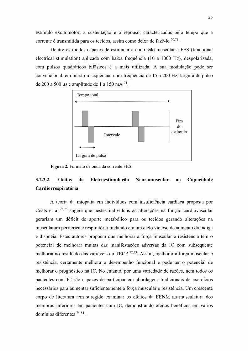
Dentre os modos capazes de estimular a contração muscular a FES (functional
electrical stimulation) aplicada com baixa frequência (10 a 1000 Hz), despolarizada,
com pulsos quadráticos bifásicos é a mais utilizada. A sua modulação pode ser
convencional, em burst ou sequencial com frequência de 15 a 200 Hz, largura de pulso
de 200 a 500 µs e amplitude de 1 a 150 mA 71.
Figura 2. Formato de onda da corrente FES.
3.2.2.2. Efeitos da Eletroestimulação Neuromuscular na Capacidade
Cardiorrespiratória
A teoria da miopatia em indivíduos com insuficiência cardíaca proposta por
Coats et al.72,73 sugere que nestes indivíduos as alterações na função cardiovascular
gerariam um déficit de aporte metabólico para os tecidos gerando alterações na
musculatura periférica e respiratória findando em um ciclo vicioso de aumento da fadiga
e dispnéia. Estes autores propoem que melhorar a força muscular e resistência tem o
potencial de melhorar muitas das manifestações adversas da IC com subsequente
melhoria no resultado das variáveis do TECP 72,73. Assim, melhorar a força muscular e
resistência, certamente melhora o desempenho funcional e pode ter o potencial de
melhorar o prognóstico na IC. No entanto, por uma variedade de razões, nem todos os
pacientes com IC são capazes de participar em abordagens tradicionais de exercícios
necessários para aumentar suficientemente a força muscular e resistência. Um crescente
corpo de literatura tem suregido examinar os efeitos da EENM na musculatura dos
membros inferiores em pacientes com IC, demonstrando efeitos benéficos em vários
domínios diferentes 74-84 .

26
Figura 3. Esquema da miopatia em indivíduos com insuficiência cardíaca. Adaptado de Coats
AJ. The "muscle hypothesis" of chronic heart failure. J Mol Cell Cardiol. 1996;28(11):2255-62.
Maillefert et al.85 e Deley et al.77 relataram uma melhora significativa no
consumo de oxigênio no pico do exercício (VO2 pico) e no limiar ventilatório (VO2 LV)
em um grupo de indivíduos com IC submetidos a um programa de cinco semanas de
EENM. Além disso, várias revisões sistemáticas têm sugerido que a EENM pode ser um
complemento importante na reabilitação de pacientes com IC 38,39. As duas principais
revisões sistemáticas e metanálises sobre o tema foram conduzidas por Smart et al.86 e
Sbruzzi et al87. Ambas procuram ver os efeitos da EENM no VO2pico e na distância do
teste de caminhada de seis minutos comparada ao exercício aeróbio e ao controle. No
estudo de Smart et al.86 foram analisados apenas estudos que utilizaram bicicleta e
também sendo avaliada as mudanças na qualidade de vida. O tratamento com FES gerou
um menor ganho no VO2pico em comparação com o treinamento em bicicleta -0,32
mL.kg-1.min-1 (95% IC-0.63 a 0,02 mL.kg-1.min-1, p=0,04), no entanto FES provocou
melhorias superiores no VO2pico 2,30 mL.kg-1.min-1 (IC 95% 1,98-2,62 mL.kg-1.min-1,
p<0,00001) e na distância do teste de caminhada de 6 minutos em relacao ao grupo
controle 46,9 m (IC 95% 22,5 a 71,3 m, p=0,0002). Não houve diferença
em mudança na qualidade de vida entre treinamento em bicicleta e FES. Além disso, o
total de horas de intervenção FES foram fortemente correlacionada com a mudança no
VO2pico (r = 0,80, p = 0,02). No estudo de Sbruzzi et al.87 foram analisados estudos que
utilizaram esteira ou bicicleta com indivíduos com IC (NYHA II-IV) por pelo menos 5
27
semanas sendo também avaliada como desfecho as mudanças na força muscular (Nm).
A busca analisou 794 artigos, dos quais foram incluídos sete estudos na analise final. O
tratamento com FES gerou um menor ganho no VO2pico em comparação com o exercicio
aerobio {- 0,74 ml / kg por min [95%IC:-1,38 a -0,10]}.Não houve diferença na força
muscular [- 0,33Nm (IC 95%: - 4,56-3,90)] e na distância do teste de caminhada de 6
minutos [2,73m (IC 95%: - 15,39-20,85)], comparando FES com exercicio aerobio. Um
aumento no VO2pico de 2,78 mL/kg.min (IC 95%: 1,44-4,13) foi observada no FES
versus o grupo controle. Ambos concluíram que a EENM produz efeitos benéficos
superiores ao grupo controle, porém em relação ao grupo exercício os efeitos são
menores com exceção da distância do teste de caminhada de seis minutos no qual os
resultados são semelhantes e a qualidade de vida no qual não apresenta diferença em
relação ao controle.
Apesar da abrangência destas revisões, nenhuma para o nosso conhecimento,
aborda os efeitos da EENM em outros parâmetros cardiovasculares e metabólicos com
os marcadores prognósticos máximos e sub-máximos do TECP que tem impacto na
sobrevida, estado funcional e qualidade de vida destes pacientes. Sendo assim nos
apêndices (Apêndice B) foi apresentado um manuscrito no formato de artigo completo
intitulado “Effect of Chronic Neuromuscular Electrical Stimulation on Primary
Cardiopulmonary Exercise Test Variables in Heart Failure Patients: A Systematic
Review and Meta-Analysis”, escrito e submetido nas normas da revista JACC Heart
Failure (normas da revista apresentadas no Anexo 1).

28
4. CONCLUSÕES
CAPACIDADE CARDIORRESPIRATÓRIA EM CARDIOPATAS:
INTRUMENTOS DIRETOS E INDIRETOS DE AVALIAÇÃO E OS EFEITOS
DA MIOESTIMULACAO ELÉTRICA
4.1 CONCLUSÕES (ESTUDO 1)
Considerando-se a avaliação da capacidade cardiorrespiratória em cardiopatas
utilizando instrumentos indiretos, nesta tese foi proposta e realizada a tradução e
adaptação cultural para o português do Duke Activity Status Index – DASI. Este
instrumento parece estar adequado para avaliação da capacidade cardiorrespiratória em
cardiopatas no país podendo ser aplicado para avaliação prognóstica e de intervenções
de forma indireta.
4.2 CONCLUSÕES (ESTUDO 2)
Considerando os efeitos da estimulação elétrica neuromuscular na capacidade
cardiorrespiratória, os resultados da revisão sistemática sugerem que a EENM pode ser
um instrumento importante para os pacientes que são incapazes de realizar o
treinamento aeróbio ou de força para melhorar a tolerância ao exercício e a manutenção
a longo prazo da capacidade funcional.
4.3 TRABALHOS FUTUROS
Acreditamos que deve ser feita a validação do questionário DASI com o teste
ergoespirométrico para adaptação dos pesos dos escores do DASI envolvendo outros
grupos de pacientes com doença cardiovascular. Em relação a eletroestimulação
neuromuscular acreditamos que mais detalhes são necessários para esclarecer se o uso
associado da EENM e intervenções baseadas em exercícios físicos em programas de
reabilitação são capaz de produzir melhores resultados clínicos, em comparação com a
intervenção isolada.
29
REFERÊNCIA BIBLIOGRÁFICA
1. Ministério da Saúde. Indicadores. DATASUS 2013.
2. Haykowsky MJ, Brubaker PH, John JM, Stewart KP, Morgan TM, Kitzman
DW. Determinants of exercise intolerance in elderly heart failure patients with
preserved ejection fraction. J Am Coll Cardiol. 2011;58(3):265–274.
3. Kitzman DW, Higginbotham MB, Cobb FR, Sheikh KH, Sullivan MJ. Exercise
intolerance in patients with heart failure and preserved left ventricular systolic
function: failure of the Frank-Starling mechanism. J Am CollCardiol.
1991;17(5):1065–1072.
4. Roger VL, Go AS, Lloyd-Jones DM et al. Heart disease and stroke statistics--
2012 update: a report from the American Heart Association. Circulation
2012;125(1):e2–e220.
5. Clark A, Coats A. Mechanisms of exercise intolerance in cardiac failure:
abnormalities of skeletal muscle and pulmonary function. Curr OpinCardiol.
1994;9(3):305–314.
6. Volterrani M, Clark AL, Ludman PF et al. Predictors of exercise capacity in
chronic heart failure. Eur Heart J. 1994;15(60:801-9.
7. Mancini DM, Walter G, Reichek N et al. Contribution of skeletal muscle
atrophy to exercise intolerance and altered muscle metabolism in heart failure.
Circulation. 1992;85(4):1364-73.
8. Hülsmann M, Quittan M, Berge R et al. Muscle strength as a predictor of long-
term survival in severe congestive heart failure. Eur J Heart Fail. 2004;6(1):101-
7.
9. Cunha TF, Bacurau AVN, Moreira JBN et al. Exercise training prevents
oxidative stress and ubiquitin-proteasome system overactivity and reverse
skeletal muscle atrophy in heart failure. PLoS One. 2012;7(8):e41701.
10. Piepoli MF CA e Crisafulli A. Pathophysiology of Human Heart Failure:
Importance of Skeletal Muscle Myopathy and. ExpPhysiol. 2013; Ahead of
print.
11. Neder JA e Nery LE. Fisiologia Clínica do Exercício: Teoria e prática. São
Paulo: Artes Médicas, 2003.

30
12. Jones NL, Killian KJ. Mechanisms of disease: Exercise limitation in health and
disease. N Engl J Med. 2000;342(9):632-41.
13. Scano G, Stendardi J, Grazzini M. Understanding dyspnoea by its language. Eur
Respir J. 2005;25:380–5.
14. Wastford ML, Murphy AJ, Pine MJ. The effects of ageing on respiratory muscle
function and performance in older adults. J Sci Med Sport. 2007;10:36-44.
15. Ambrosino N, Serradori M. Determining the cause of dyspnoea: linguistic and
biological descriptors. Chron Respir Dis. 2006;3:117-22.
16. Caroci AS, Lareau SC; Linda l. Descriptors of dyspnea by patients with chronic
obstructive pulmonary disease versus congestive heart failure. Heart & Lung.
2004;33:102-10.
17. Guimarães GV, Bellotti G, Bacal F, Mocelin A, Bocchi EA. Pode o teste
ergoespirométrico de caminhada de seis minutos ser representativo das
atividades habituais de pacientes com insuficiência cardíaca? Arq BrasCardiol.
2002;78(6):553-6.
18. Myers J, Zaheer M, Quaglietti S, Madhavan R, Froelicher V, Heidenreich P.
Association of functional and health status measures in heart failure. J CardFail.
2006;12(6):439-45.
19. Taylor HL, Buskirk E, Henschel A. Maximal oxygen intake as objective
measure of cardiorespiratory performance. J ApplPhysiol. 1955;8:73.
20. Green CP, Porter CB, Bresnahan DR, Spertus JA. Development and evaluation
of the Kansas city cardiomyopathy questionnaire: a new health status measure
for heart failure. J Am Coll Cardiol. 2000;35(5):1245-55.
21. Hlatky MA, Boineau RE, Higginbotham MB, Lee KL, Mark DB, Califf RM, et
al. A brief self-administered questionnaire to determine functional capacity (the
Duke Activity Status Index). Am J Cardiol. 1989;64(10):651-4.
22. Olsson LG, Swedberg K, Clark AL, Witte KK, Cleland JGF. Six minute corridor
walk test as an outcome measure for the assessment of treatment in randomized,
blinded intervention trials of chronic heart failure: a systematic review. Eur
Heart J. 2005;26(8):778-93.
23. Giannuzzi P, Tavazzi L, Meyer K. Recommendations for exercise training in
chronic heart failure patients. Eur Hear J 2001;22(2):125–135.
31
24. Martins-Pinge MC. Cardiovascular and autonomic modulation by the central
nervous system after aerobic exercise training. Brazilian J MedBiol Res
2011;44(9):848–854.
25. Brum PC, Bacurau VN, Medeiros A, Ferreira JCB, Vanzelli S, Negrão CE.
Aerobic exercise training in heart failure: impact on sympathetic hyperactivity
and cardiac and skeletal muscle function. Brazilian J Med Biol Res
2011;44(9):827–835.
26. Maddocks M, Gao W, Higginson IJ , Wilcock A. Neuromuscular electrical
stimulation for muscle weakness in adults with advanced disease. Cochrane
Database of Systematic Reviews, 2013(1).
27. Banerjee P, Caulfield B , Crowe G, Clark AL. Prolonged electrical muscle
stimulation exercise improves strength, peak VO2, and exercise capacity in
patients with stable chronic heart failure. J CardFail. 2009;15(4):319-26.
28. Deftereos S, Giannopoulos L, Raisakis K et al. Comparison of muscle functional
electrical stimulation to conventional bicycle exercise on endothelium and
functional status indices in patients with heart failure. Am J Cardiol.
2010;106(11):1621-5.
29. Deley G, G Kervio, Verges B et al. Neuromuscular adaptations to low-frequency
stimulation training in a patient with chronic heart failure. Am J Phys Med
Rehabil. 2008;87(6): 502-9.
30. Deley G, G Kervio , Verges B et al. Comparison of low-frequency electrical
myostimulation and conventional aerobic exercise training in patients with
chronic heart failure. Eur J Cardiovasc Prev Rehabil. 2005;12(3):226-33.
31. Dobsák P, Nováková M, Fiser B et al. Electrical stimulation of skeletal muscles.
An alternative to aerobic exercise training in patients with chronic heart failure?
Int Heart J. 2006;47(3):441-53.
32. Dobšák P, Tomandl J, Spinarova L et al. Effects of neuromuscular electrical
stimulation and aerobic exercise training on arterial stiffness and autonomic
functions in patients with chronic heart failure. Artif Organs. 2012;36(10):920-
30.
33. Eicher JC, Dobšák P, Berteau O et al., Rehabilitation in Chronic Congestive
Heart Failure: Comparison of Bycicle Training and Muscle Electrical
Stimulation. ScriptaMedica (BRNO). 2004;77(5-6):261-270.

32
34. Harris S, LeMaitre JP, Mackenzie G et al., A randomised study of home-based
electrical stimulation of the legs and conventional bicycle exercise training for
patients with chronic heart failure. Eur Heart J. 2003;24(9):871-8.
35. Karavidas AI, Raisakis KG, Parissis JT et al. Functional electrical stimulation
improves endothelial function and reduces peripheral immune responses in
patients with chronic heart failure. Eur J Cardiovasc Prev Rehabil.
2006;13(4):592-7.
36. LeMaitre JP, Harris S, Hannan J , Fox KA, Denvir MA. Maximum oxygen
uptake corrected for skeletal muscle mass accurately predicts functional
improvements following exercise training in chronic heart failure. Eur J Heart
Fail. 2006;8(3):243-8.
37. Nuhr MJ, Pette D, Berger R et al. Beneficial effects of chronic low-frequency
stimulation of thigh muscles in patients with advanced chronic heart failure. Eur
Heart J.2004;25(2):136-43.
38. Nelson CL, Herdon JE, Mark DB, Pryor DB, Callif RM, Hlatky MA. Relation of
clinical and angiographic factors to functional capacity as measured by the Duke
Activity Status Index. Am J Cardiol. 1991;68:973-5.
39. Alonso J, Permanyer-Miralda G, Cascant P, Brotons C, Prieto L, Soler-Soler J.
Measuring functional status of chronic coronary patients. Reliability, validity
and responsiveness to clinical change of the reduced version of the Duke
Activity Status Index (DASI). Eur Heart J. 1997;18(3):414-9.
40. Arena R, Humphrey R, Peberdy MA. Using the duke activity status index in
heart failure. J CardiopulmRehabil. 2002;22(2):93-5.
41. Carter R, Holiday DB, Grothues C, Nwasuruba C, Stocks J, Tiep B. Criterion
validity of the Duke Activity Status Index for assessing functional capacity in
patients with chronic obstructive pulmonary disease. J CardiopulmRehabil.
2002;22(4):298-308.
42. Koch CG, Li L, Lauer M, Sabik J, Starr NJ, Blackstone EH. Effect of functional
health-related quality of life on long-term survival after cardiac surgery.
Circulation. 2007 Feb;115(6):692-9.
43. Koch CG, Li L, Shishehbor M, Nissen S, Sabik J, Starr NJ, et al. Socioeconomic
status and comorbidity as predictors of preoperative quality of life in cardiac
surgery. J ThoracCardiovascSurg. 2008;136(3):665-U67.

33
44. Parissis JT, Nikolaou M, Birmpa D, Farmakis D, Paraskevaidis IA, Bistola V et
al. Clinical and prognostic value of Duke’s Activity Status Index along with
plasma B-Type natriuretic peptide levels in chronic heart failure secondary to
ischemic or idiopathic dilated cardiomyopathy. Am J Cardio. 2009; 103:73–75.
45. Mark DB, Lam LC, Lee KL et al. Identification of patients with coronary
disease at high risk for loss of employment: A prospective validation Study.
Circulation. 1994;86:1485-1494.
46. Yancy CW, Jessup M, Bozkurt B et al. 2013ACCF/AHA guideline for the
management of heart failure: a report of the American College of Cardiology
Foundation/American Heart Association Task Force on practice guidelines.
Circulation 2013;128(16):e240–319.
47. McMurray JJ, Adamopoulos S, Anker SD et al. ESC Guidelines for the diagnosis
and treatment of acute and chronic heart failure 2012: The Task Force for the
Diagnosis and Treatment of Acute and Chronic Heart Failure 2012 of the
European Society of Cardiology. Developed in collaboration with the Heart
Failure Association (HFA) of the ESC. Eur Heart J. 2012;33(14):1787-847.
48. Bocchi EA, Marcondes-Braga FG, Ayub-Ferreira SM et al. Sociedade Brasileira
de Cardiologia. III Diretriz Brasileira de Insuficiência Cardíaca Crônica. Arq
Bras Cardiol. 2009;93(1):1-71.
49. Packer M. How should physicians view heart failure? The philosophical and
physiological evolution of three conceptual models of the disease. Am J Cardiol.
1993;71:3C-11C.
50. Arena R, Myers J , J Abella et al. The prognostic value of the heart rate
response during exercise and recovery in patients with heart failure: influence of
beta-blockade. Int J Cardiol, 2010;138(2):166-73.
51. Cahalin, LP, Chase P, Arena R et al., A meta-analysis of the prognostic
significance of cardiopulmonary exercise testing in patients with heart failure.
Heart Fail Rev.2013; 18(1):79-94.
52. Huelsmann M, Stefenelli T, Berger R, Frey B, R Pacher, Prognostic impact of
workload in patients with congestive heart failure. Am Heart J,
2002;143(2):308-12.
53. Lang CC, Agostoni P, Mancini DM. Prognostic significance and measurement
of exercise-derived hemodynamic variables in patients with heart failure. J Card
Fail. 2007;13(8):672-9.

34
54. Lauer MS, Okin PM, Larson MG, Evans JC, Levy D . Impaired heart rate
response to graded exercise. Prognostic implications of chronotropic
incompetence in the Framingham Heart Study. Circulation. 1996;93(8):1520-6.
55. Leeper NJ, Dewey FE , Ashley EA et al., Prognostic value of heart rate increase
at onset of exercise testing. Circulation. 2007;115(4):468-74.
56. Manetos C, Dimopoulos S, Tzanis L et al. Skeletal muscle microcirculatory
abnormalities are associated with exercise intolerance, ventilatory inefficiency,
and impaired autonomic control in heart failure. J Heart Lung Transplant.
2011;30(12):1403-8.
57. Myers J, Gullestad L, Vagelos R et al., Clinical, hemodynamic, and
cardiopulmonary exercise test determinants of survival in patients referred for
evaluation of heart failure. Ann Intern Med. 1998;129(4):286-93.
58. Pardaens S, Calders P, Derom E, De Sutter J. Exercise intolerance in heart
failure: update on exercise parameters for diagnosis, prognosis and therapeutic
interventions. Acta Cardiol. 2013;68(5):495-504.
59. Hunt SA, Baker DW, Chin MH et al. ACC/AHA Guidelines for the Evaluation
and Management of Chronic Heart Failure in the Adult: Executive Summary A
Report of the American College of Cardiology/American Heart Association
Task Force on Practice Guidelines (Committee to Revise the 1995 Guidelines
for the Evaluation and Management of Heart Failure). Circulation.
2001;104(24):2996-3007.
60. New York Heart Associantion. Nomeclature and Criteria for Diagnosis of
Disease of Heart and Great Vessels. Estados Unidos: Little, Brown and Co,
1994.
61. Meka N, Katragadda S, Cherian B, Arora RR. Endurance exercise and resistance
training in cardiovascular disease. Ther Adv Cardiovasc Dis. 2008;2(2):115–21.
62. Beckers PJ, Denollet J, Possemiers NM, Wuyts FL, Vrints CJ, Conraads VM.
Combined endurance-resistance training vs. endurance training in patients with
chronic heartfailure: a prospective randomized study. Eur Heart J.
2008;29(15):1858–66.
63. Beniaminovitz A, Lang CC, LaManca J, Mancini DM. Selective low-level leg
muscle training alleviates dyspnea in patients with heart failure. J Am Coll
Cardiol.2002;40(9):1602–8.

35
64. Brum PC, Bacurau a. VN, Medeiros a., Ferreira JCB, Vanzelli a. S, Negrão CE.
Aerobicexercise training in heart failure: impact on sympathetic hyperactivity
and cardiac and skeletal muscle function. Brazilian J Med Biol Res.
2011;44(9):827–35.
65. Palevo G, Keteyian SJ, Kang M, Caputo JL. Resistance exercise training
improves heart function and physical fitness in stable patients with heart failure.
J Cardiopulm Rehabil Prev. 2009;29(5):294–8.
66. Downing J, Balady GJ. The role of exercise training in heart failure. J Am Coll
Cardiol. 2011;58(6):561–9.
67. Pina IL, Apstein CS, Balady GJ et al. Exercise and heart failure: a statement
from the American Heart Association Committee on exercise, rehabilitation,
and prevention. Circulation. 2003;107(8):1210-1225.
68. Smart N, Marwick TH. Exercise training for patients with heart failure: a
systematic review of factors that improve mortality and morbidity. The
American Journal of Medicine. 2004;116(10):693-706.
69. Maddocks M, Gao W, Higginson IJ , Wilcock A. Neuromuscular electrical
stimulation for muscle weakness in adults with advanced disease. Cochrane
Database of Systematic Reviews, 2013(1).
70. Marquete T, Hug F, Decherchi P, Jammes Y. Changes in neuromuscular
function after training by functional electrical stimulation. Muscle Nerve.
2003;28(2):181-188.
71. Doucet BM, Lam A, Griffin L. Neuromuscular electrical stimulation for skeletal
muscle function. Yale J Biol Med. 2012;85(2):201-15.
72. Coats A.J. The "muscle hypothesis" of chronic heart failure. J Mol Cell Cardiol.
1996;28(11):2255-62.
73. Coats AJ, Clark AL, Piepoli M, Volterrani M, Poole-Wilson PA et al.
Symptoms and quality of life in heart failure: the muscle hypothesis. Br Heart J.
1994;72(2):S36-9.
74. Banerjee P, Caulfield B , Crowe G, Clark AL. Prolonged electrical muscle
stimulation exercise improves strength, peak VO2, and exercise capacity in
patients with stable chronic heart failure. J CardFail. 2009;15(4):319-26.
75. Deftereos S, Giannopoulos L, Raisakis K et al. Comparison of muscle functional
electrical stimulation to conventional bicycle exercise on endothelium and

36
functional status indices in patients with heart failure. Am J Cardiol.
2010;106(11):1621-5.
76. Deley G, G Kervio, Verges B et al. Neuromuscular adaptations to low-frequency
stimulation training in a patient with chronic heart failure. Am J Phys Med
Rehabil. 2008;87(6): 502-9.
77. Deley G, G Kervio , Verges B et al. Comparison of low-frequency electrical
myostimulation and conventional aerobic exercise training in patients with
chronic heart failure. Eur J Cardiovasc Prev Rehabil. 2005;12(3):226-33.
78. Dobsák P, Nováková M, Fiser B et al. Electrical stimulation of skeletal muscles.
An alternative to aerobic exercise training in patients with chronic heart failure?
Int Heart J. 2006;47(3):441-53.\
79. Dobšák P, Tomandl J, Spinarova L et al. Effects of neuromuscular electrical
stimulation and aerobic exercise training on arterial stiffness and autonomic
functions in patients with chronic heart failure. Artif Organs. 2012;36(10):920-
30.
80. Eicher JC, Dobšák P, Berteau O et al., Rehabilitation in Chronic Congestive
Heart Failure: Comparison of Bycicle Training and Muscle Electrical
Stimulation. ScriptaMedica (BRNO). 2004;77(5-6):261-270.
81. Harris S, LeMaitre JP, Mackenzie G et al., A randomised study of home-based
electrical stimulation of the legs and conventional bicycle exercise training for
patients with chronic heart failure. Eur Heart J. 2003;24(9):871-8.
82. Karavidas AI, Raisakis KG, Parissis JT et al. Functional electrical stimulation
improves endothelial function and reduces peripheral immune responses in
patients with chronic heart failure. Eur J Cardiovasc Prev Rehabil.
2006;13(4):592-7.
83. LeMaitre JP, Harris S, Hannan J , Fox KA, Denvir MA. Maximum oxygen
uptake corrected for skeletal muscle mass accurately predicts functional
improvements following exercise training in chronic heart failure. Eur J Heart
Fail. 2006;8(3):243-8.
84. Nuhr MJ, Pette D, Berger R et al. Beneficial effects of chronic low-frequency
stimulation of thigh muscles in patients with advanced chronic heart failure. Eur
Heart J.2004;25(2):136-43.

37
85. Maillefert JF, Eicher JC, Walker P et al. Effects of low-frequency electrical
stimulation of quadriceps and calf muscles in patients with chronic heart failure.
J Cardiopulm Rehabil. 1998;18(4):277-282.
86. Sbruzzi G, Ribeiro RA , Schaan BD et al., Functional electrical stimulation in
the treatment of patients with chronic heart failure: a meta-analysis of
randomized controlled trials. Eur J Cardiovasc Prev Rehabil. 2010;17(3):254-60.
87. Smart NA, Dieberg G and Giallauria F. Functional electrical stimulation for
chronic heart failure: a meta-analysis. Int J Cardiol. 2013;167(1):80-6.

38
ANEXOS
ANEXO 1 NORMAS DA REVISTA FISIOTERAPIA EM MOVIMENTO
Normas Editoriais A Revista Fisioterapia em Movimento publica trimestralmente artigos científicos na área de Fisioterapia, na forma de trabalhos de pesquisa original e de trabalhos de revisão. Os artigos submetidos à Revista Fisioterapia em Movimento devem preferencialmente enquadrar-se na categoria de Artigos Científicos. Os estudos são apresentados na forma de Artigos Originais (oriundos de pesquisas inéditas com informações de materiais e métodos, discussão e resultados relatados de maneira sistemática), Artigos de Revisão (oriundos de estudos com delineamento definido e baseado em pesquisa bibliográfica consistente com análise crítica e considerações que possam contribuir com o estado da arte) e cartas ao Editor. A Revista aceita submissão de manuscritos nas áreas de Fisioterapia e saúde humana, tais como: Análise do Movimento Funcional, Cinesiologia e Biomecânica, Cinesioterapia, Ensino em Fisioterapia, Ergonomia, Fisioterapia Cardiorrespiratória, Fisioterapia Dermato-Funcional, Fisioterapia em Geriatria e Gerontologia, Fisioterapia Músculo-Esquelética, Fisioterapia Neurofuncional, Fisioterapia Preventiva, Fisioterapia Uroginecológica, Fundamentos da Fisioterapia e Recursos Terapêuticos Físicos Naturais, e Saúde Coletiva. Os artigos recebidos são encaminhados a dois revisores (pareceristas) para avaliação pelos pares (peerreview). Os editores coordenam as informações entre os autores e revisores, cabendo-lhes a decisão final sobre quais artigos serão publicados com base nas recomendações feitas pelos revisores. Quando recusados, os artigos serão devolvidos com a justificativa do editor. A Revista Fisioterapia em Movimento está alinhada com as normas de qualificação de manuscritos estabelecidas pela OMS e do InternationalCommitteeof Medical JournalEditors (ICMJE), disponíveis em e . Somente serão aceitos os artigos de ensaios clínicos cadastrados em um dos Registros de Ensaios Clínicos recomendados pela OMS e ICMJE. Instruções aos autores
Os manuscritos deverão ser submetidos à Revista Fisioterapia em Movimento por meio do site na seção “submissão de artigos”. Todos os artigos devem ser inéditos e não podem ter sido submetidos para avaliação simultânea em outros periódicos. As revisões para este periódico são aceitas apenas na modalidade Revisão Sistemática nos moldes da COCHRANE. Para tanto acessar o site http://www.virtual.epm.br/cursos/metanalise/. É obrigatório anexar uma declaração assinada por todos os autores quanto à exclusividade do artigo, na qual constará endereço completo, telefone, fax e e-mail. Na carta de pedido de publicação, é obrigatório transferir os direitos autorais para a Revista Fisioterapia em Movimento. Afirmações, opiniões e conceitos expressados nos artigos são de responsabilidade exclusiva dos autores. Trabalhos que contenham resultados de estudos humanos e/ou animais somente serão aceitos para publicação se estiver claro que todos os princípios de ética foram utilizados na investigação (enviar cópia do parecer do comitê de ética). Esses trabalhos devem obrigatoriamente incluir uma afirmação de que o protocolo de pesquisa foi aprovado por um comitê de ética institucional. (Reporte-se à Resolução 196/96, do Conselho Nacional de Saúde, que trata do Código de Ética da Pesquisa envolvendo Seres Humanos). Para experimentos com animais, considere as diretrizes internacionais Pain, publicada em: PAIN, 16: 109-110, 1983. Quando utilizados estudos/atividades envolvendo pessoas, deverá ser encaminhada uma autorização assinada e datada pelo envolvido no estudo, ou seu responsável legal, autorizando a publicação da imagem. Os pacientes têm o direito à privacidade, o qual não pode ser infringido sem um consentimento esclarecido. Em caso de utilização de fotografias de pessoas/pacientes, estas não podem ser identificáveis ou as fotografias devem estar acompanhadas de permissão específica escrita para uso e divulgação das imagens. O uso de máscaras oculares não é considerado proteção adequada para o anonimato. É imprescindível o envio da declaração de responsabilidade de conflitos de interesse manifestando a não existência de eventuais conflitos de interesse que possam interferir no resultado da pesquisa. Contato Revista Fisioterapia em Movimento Clínica de Fisioterapia Pontifícia Universidade Católica do Paraná Rua Imaculada Conceição, 1155, Prado Velho CEP 80215-901, Curitiba, PR, Brasil e-mail: [email protected] telefone: +55(41) 3271-1608

39
Forma e preparação dos manuscritos
A Revista Fisioterapia em Movimento recebe artigos das seguintes categorias: Artigos Originais: oriundos de resultado de pesquisa de natureza empírica, experimental ou conceitual, sua estrutura deve conter: Introdução, Materiais e Métodos, Resultados, Discussão, Conclusão, Referências. O texto deve ser elaborado com, no máximo, 6.000 palavras e conter até 5 ilustrações. Artigos de Revisão: oriundos de estudos com delineamento definido e baseado em pesquisa bibliográfica consistente com análise crítica e considerações que possam contribuir com o estado da arte (máximo de 8.000 palavras e 5 ilustrações). Os manuscritos devem ser submetidos pelo site na seção “submissão de artigos”. Os trabalhos devem ser digitados em Word for Windows, fonte Times New Roman, tamanho 12, espaçamento entre linhas de 1,5 respeitando o número de palavras de cada manuscrito, incluindo referências, ilustrações, quadros, tabelas e gráficos. O número máximo permitido de autores por artigo é seis. As ilustrações (figuras, gráficos, quadros e tabelas) devem ser limitadas ao número máximo de cinco (5), inseridas no corpo do texto, identificadas e numeradas consecutivamente em algarismos arábicos. A arte final, figuras e gráficos devem estar em formato .tiff. Envio de ilustrações com baixa resolução (menos de 300 DPIs) pode acarretar atraso na aceitação e publicação do artigo. Os trabalhos podem ser encaminhados em português ou inglês. Abreviações oficiais poderão ser empregadas somente após uma primeira menção completa. Deve ser priorizada a linguagem científica. Deverão constar, no final dos trabalhos, o endereço completo de todos os autores, afiliação, telefone, fax e e-mail (atualizar sempre que necessário) para encaminhamento de correspondência pela comissão editorial.
Outras considerações:
• sugere-se acessar um artigo já publicado para verificar a formatação dos artigos publicados pela revista;
• todos os artigos devem ser inéditos e não podem ter sido submetidos para avaliação simultânea em outros periódicos (anexar carta, assinada por todos os autores, na qual será declarado tratar-se de artigo inédito, transferindo os direitos autorais e assumindo a responsabilidade sobre aprovação em comitê de ética, quando for o caso.);
• afirmações, opiniões e conceitos expressados nos artigos são de responsabilidade dos autores;
• todos os artigos serão submetidos ao Comitê Editorial da revista e, caso pertinente, à área da Fisioterapia para avaliação dos pares;
• não serão publicadas fotos coloridas, a não ser em caso de absoluta necessidade e a critério do Comitê Editorial. No preparo do original, deverá ser observada a seguinte estrutura:
Cabeçalho
Título do artigo em português (LETRAS MAIÚSCULAS em negrito, fonte Times New Roman, tamanho 14, parágrafo centralizado), subtítulo em letras minúsculas (exceção para nomes próprios) e em inglês (somente a primeira letra do título em maiúscula, as demais palavras em letras minúsculas – exceção para nomes próprios), em itálico, fonte Times New Roman, tamanho 12, parágrafo centralizado. O título deve conter no máximo 12 palavras, sendo suficientemente específico e descritivo.
Apresentação dos autores do trabalho
Nome completo, titulação, afiliação institucional (nome da instituição para a qual trabalha), vínculo (se é docente, professor ou está vinculado a alguma linha de pesquisa), cidade, estado, país e e-mail.
Resumo estruturado / Structured Abstract
O resumo estruturado deve contemplar os tópicos apresentados na publicação. Exemplo: Introdução, Desenvolvimento, Materiais e métodos, Discussão, Resultados, Considerações finais. Deve conter no mínimo 150 e máximo 250 palavras, em português/inglês, fonte Times New Roman, tamanho 11, espaçamento simples e parágrafo justificado. Na última linha, deverão ser indicados os descritores (palavras-chave/keywords). Para padronizar os descritores, solicitamos utilizar os Thesaurus da área de saúde (DeCS) (). O número de descritores desejado é de no mínimo 3 e no máximo 5, sendo representativos do conteúdo do trabalho.

40
Corpo do Texto
• Introdução: Deve apontar o propósito do estudo, de maneira concisa, e descrever quais os avanços que foram alcançados com a pesquisa. A introdução não deve incluir dados ou conclusões do trabalho em questão.
• Materiais e métodos: Deve ofertar, de forma resumida e objetiva, informações que permitam que o estudo seja replicado por outros pesquisadores. Referenciar as técnicas padronizadas.
• Resultados: Devem oferecer uma descrição sintética das novas descobertas, com pouco parecer pessoal.
• Discussão: Interpretar os resultados e relacioná-los aos conhecimentos existentes, principalmente os que foram indicados anteriormente na introdução. Esta parte deve ser apresentada separadamente dos resultados.
• Conclusão ou Considerações finais: Devem limitar-se ao propósito das novas descobertas, relacionando-as ao conhecimento já existente. Utilizar apenas citações indispensáveis para embasar o estudo.
• Agradecimentos: Sintéticos e concisos, quando houver.
• Referências: Devem ser numeradas consecutivamente na ordem em que são primeiramente mencionadas no texto.
• Citações: Devem ser apresentadas no texto, tabelas e legendas por números arábicos entre parênteses. Exemplos: “o caso apresentado é exceção quando comparado a relatos da prevalência das lesões hemangiomatosas no sexo feminino (6, 7)” ou “Segundo Levy (3), há mitos a respeito dos idosos que precisam ser recuperados”.
Referências
Todas as instruções estão de acordo com o Comitê Internacional de Editores de Revistas Médicas (Vancouver), incluindo as referências. As informações encontram-se disponíveis em: (). Recomenda-se fortemente o número mínimo de referências de 30 para artigos originais e de 40 para artigos de revisão. As referências deverão originar-se de periódicos que tenham no mínimo o Qualis desta revista ou equivalente.
Artigos emRevistas
Atéseisautores Naylor CD, Williams JI, Guyatt G. Structured abstracts of proposal for clinical and epidemiological studies. J ClinEpidemiol. 1991;44:731-37. - Mais de seis autores Listar os seis primeiros autores seguidos de et al. Parkin DM, Clayton D, Black RJ, Masuyer E, Friedl HP, Ivanov E, et al Childhood leukaemia in Europe after Chernobyl: 5 year follow-up. Br J Cancer. 1996;73:1006-12. - Suplemento de volume Shen HM, Zhang QF. Risk assessment of nickel carcinogenicity and occupational lung cancer. Environ Health Perspect. 1994; 102 Suppl 1:275-82. - Suplemento de número Payne DK, Sullivan MD, Massie MJ. Women´s psychological reactions to breast cancer. SeminOncol. 1996;23(1 Suppl 2):89-97. - Artigosemformatoeletrônico Al-Balkhi K. Orthodontic treatment planning: do orthodontists treat to cephalometric norms. J Contemp Dent Pract. [serial on the internet] 2003 [cited 2003 Nov. 4]. Available from: URL:www.thejcdp.com.
Livros e monografias
Livro Berkovitz BKB, Holland GR, Moxham BJ. Color atlas & textbook of oral anatomy. Chicago:Year Book Medical Publishers; 1978. - Capítulo de livro Israel HA. Synovial fluid analysis. In: Merril RG, editor. Disorders of the temporomandibular joint I: diagnosis and arthroscopy. Philadelphia: Saunders; 1989. p. 85-92. - Editor, CompiladorcomoAutor Norman IJ, Redfern SJ, editors. Mental health care for elderly people. New York: Churchill Livingstone; 1996. - Livros/Monografiasem CD-ROM CDI, clinical dermatology illustrated [monograph on CD-ROM], Reeves JRT, Maibach H. CMEA Multimedia Group, producers. 2 nd ed. Version 2.0. San Diego: CMEA; 1995. - Anais de congressos, conferências congêneres Damante JH, Lara VS, Ferreira Jr O, Giglio FPM. Valor das informações clínicas e radiográficas no diagnóstico final. Anais X Congresso Brasileiro de Estomatologia; 1-5 de julho 2002; Curitiba, Brasil. Curitiba, SOBE; 2002. Bengtsson S, Solheim BG. Enforcement of data protection, privacy and security in medical informatics. In: Lun KC, Degoulet P, Piemme TE, Rienhoff O, editors. MEDINFO 92. Proceedings of the 7th World Congress of Medical Informatics;1992 Sept 6-10; Geneva, Switzerland. Amsterdam:North-Holland; 1992. p. 1561-5. Trabalhos acadêmicos (Teses e Dissertações) Kaplan SJ. Post-hospital home health care: the elderly´s access and utilization [dissertation]. St. Louis: Washington Univ.; 1995.

41
• É importante que, durante a execução do trabalho, o autor consulte a página da revista online (http://www.pucpr.br/pesquisa_cientifica/revistas_cientificas.php) e verifique a apresentação dos artigos publicados, adotando o mesmo formato. Além de revisar cuidadosamente o trabalho com relação às normas solicitadas: tamanho da fonte em cada item do trabalho, numeração de página, notas em número arábico, a legenda de tabelas e quadros, formatação da página e dos parágrafos, citação no corpo do texto e referências conforme solicitado. O português e/ou inglês do trabalho. E, por fim, se todos os autores citados constam nas Referências e no final do trabalho. NOTA: Fica a critério da revista a seleção dos artigos que deverão compor os fascículos, sem nenhuma obrigatoriedade de publicá-los, salvo os selecionados pelos pares

42
ANEXO 2 NORMAS DA REVISTA HEART FAILURE REVIEWS

43

44
45

46

47

48

49

50

51

52

53

54
ANEXO 3 PARECER CONSUBSTANCIADO DO COMITÊ DE ÉTICA EM PESQUISA

55

56
APÊNDICES
CONTRIBUIÇÕES CIENTÍFICAS
Apêndice A. Artigo publicado
ISSN 0103-5150
Fisioter. Mov., Curitiba, v. 26, n. 3, p. 631-638, jul./set. 2013
TRANSLATION AND CROSS-CULTURAL ADAPTATION OF THE DUKE
ACTIVITY STATUS INDEX TO BRAZILIAN PORTUGUESE
Laura Maria Tomazi Neves[a], Alberto Klohn Neto[b], Fernanda Pasquale Arenas[b], Luis Vicente
Franco de Oliveira[c], Ross Arena[d], Gerson Cipriano Jr.[e]
[a]Master’s degree, Doctoral student, Postgraduate Program in Health Sciences and
Technology, Universidade de Brasília, Brasília, DF, Brazil. [b]Specialist, Sector of Cardiovascular Surgery, Universidade Federal de São Paulo -
UNIFESP, São Paulo, SP, Brazil. [c]Doctoral degree, Teaching staff member, Postgraduate Program in Rehabilitation Sciences,
Universidade Nove de Julho –UNINOVE, São Paulo, SP, Brazil.
[d] Doctoral degree, Teaching staff member, Department of Physical Therapy, College of
Applied Health Sciences, University of Illinois Chicago, Chicago, IL, USA. [e]Doctoral degree, Postgraduate Program in Physical Education; Postgraduate Program
in Health Sciences and Technology, Universidade de Brasília – UnB, Brasília, DF,
Brazil; Teaching staff member in Physical Therapy Course, Universidade de Brasília,
Brasília, DF, Brazil.
Study conducted in Cardiovascular Surgery Sector, Department of Surgery,
Universidade Federal de São Paulo – Unifesp
Address: Rua Napoleão de Barros, 715 - 3º andar Vila Clementino
CEP: 04024-002 - São Paulo-SP, Brazil
Telefone: (11) 55764055 Fax: (11) 55712719
Number of words: 3729
57
Introduction
The assessment of cardiorespiratory fitness on a population level is important to
the establishment of preventive strategies aimed at addressing risk factors, favoring a
reduction in cardiovascular disease.1-3 However, specific tests for the evaluation of
cardiorespiratory fitness, such as cardiopulmonary exercise testing,have a lower degree
of broad applicability due to the high cost, the time required for the evaluation and
technical-operational logistics.
The development of fast, simple tools for the determination of cardiorespiratory
fitness, such as questionnaires and multiple regression equations, is of considerable
importance due the favorable cost/benefit ratio and usefulness in epidemiological
studies, especially in locations with inadequate infrastructure for advanced exercise
testing.1,4-6 Based on these principles, Hlatky et al.5 created a self-administrated
questionnaire denominated the Duke Activity Status Index(DASI) in the United States.
This index is employed to predict cardiorespiratory fitness and has been widely used for
patients with cardiovascular disease.7-14
In Brazil, however, there is a lack of tools for this purpose and an eminent need
to assess the degree of cardiorespiratory capacity, especially in patients with
cardiovascular disease; given it is the third leading cause of death in the country. There
is also a need to find assessment tools that are widely employed in the scientific
literature and validate such measures for use on patients in Brazil. Thus, the DASI
seems to be one of the most adequate options for the Brazilian population. However,
this index has not yet been submitted to translation and cross-cultural adaptation to
Brazilian Portuguese.15-19
The aim of the present study was to develop the Brazilian version of the DASI
and cross-culturally adapt it for the evaluation of cardiorespiratory fitness among
patients with cardiovascular disease in Brazil.
Methods
This study received approval from the Human Research Ethics Committee of the
Universidade Federal de São Paulo (Brazil) under process number nº 006/2009 in
58
compliance with Resolution 196/96 of the Brazilian National Board of Health. All
participants received information on the objectives of the study and agreed to participate
by signing a statement of informed consent. The study was performed in two steps –
translation and cross-cultural adaptation.
Participants
Two qualified Brazilian translators (laypersons in the field of knowledge in
question) performed the translation of the DASI into Brazilian Portuguese. A review
committee made up of three Brazilian physical therapists preformed the content analysis
of all steps of the translation process. All three members of the committee were
university professors (experts with a Master’s or Doctoral degree), with clinical
experience in cardiorespiratory fitness and advanced knowledge of the English
language. Two native-English speaking translators performed the back-translation of the
DASI, both of whom were fluent in Portuguese and laypersons in the field of
knowledge in question. The translated questionnaire was administered to a sample
composed of middle-aged (44 to 65 years) male and female individuals (8 patients with
a diagnosis of heart failure and 8 healthy individuals) for the semantic analysis of the
items on the index. Patients were screened from outpatient Cardiac Surgery, Hospital
São Paulo. A cardiologist made the heart failure diagnosis, based on clinical evaluation
and ejection fraction, evaluated by echocardiogram. Patients were clinically stable at
the time of study inclusion. Exclusion criteria were: having undergone surgical
procedures in the previous six months, cognitive impairment, illiteracy and any
orthopedic or rheumatologic condition.
Questionnaire
The DASI is composed of 12 items addressing personal care, household
activities, sexual activity and recreational activities. The item scores are weighted based
on metabolic cost measured in metabolic equivalents (METs). Each item receives a
score ranging from 1.75 to 8.00 points in increasing order of METs. Responses of “no”
are scored 0. The total score ranges from 0 to 58.2 points, with higher scores denoting
greater functional limitation. The sum of the scores of the 12 items is used in the simple
regression equation provided by the questionnaire to estimate peak oxygen uptake
(VO2peak) during exercise. In the original study, a strong correlation (r = 0.80) was found

59
between the DASI and VO2peakobtained from the cardiopulmonary exercise test (gold
standard).5
Translation and cross-cultural adaptation
The translation and cross-cultural adaptation of the DASI followed the
procedures proposed for measures developed in other linguistic and socio-cultural
contexts based on studies carried out by Guillemin et al,15,16Reichenheim and
Moraes,17Maher et al18 and Beaton et al.19 Figure 1 displays the flowchart of the
translation and adaptation process.
a) Translation: Two duly qualified Brazilian translators fluent in English
performed the translation of the DASI into Portuguese, focusing on a conceptual rather
than literal translation. The translators received information on the objectives of the
study and were instructed to be precise in their use of language. A review committee
composed of three Brazilian health professionals with ample knowledge of English,
blinded to the objectives of the study and with experience in the translation of
questionnaires, evaluated the two translated versions of the DASI for the correction of
possible errors that could lead to different results than those originally proposed. This
committee then created a synthesis of the index in Portuguese based on the versions of
the two translators.
b) Back-translation: The synthesis version was back-translated into English by
two duly qualified native English-speaking translators in an independent fashion. The
translators were not informed of the objectives of the study and had no access to the
original version. The back-translated versions were then compared to the original
version of the index by the review committee. Version 2 of the DASI in Portuguese was
established following consensus among the committee members on each item of the
index.
c) Cross-cultural adaptation: Version 2 was submitted to semantic analysis to
determine whether the terms used in the items and paragraphs were clear. Equivalence
was determined through an evaluation of the degree of comprehension or possible
doubts regarding each item. Since the DASI was designed as a self-administered
questionnaire, the target population participated in this stage. For such, Version 2 of the
questionnaire was administered to eight patients with heart failure and eight healthy
individuals.
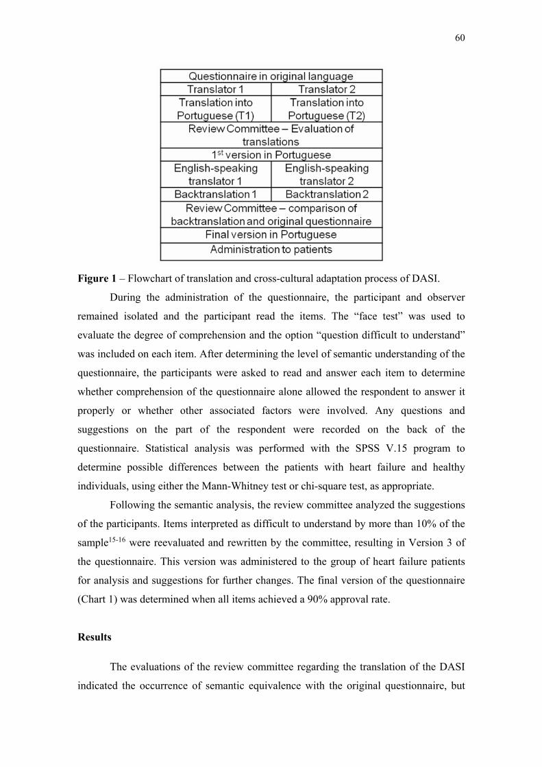
60
Figure 1 – Flowchart of translation and cross-cultural adaptation process of DASI.
During the administration of the questionnaire, the participant and observer
remained isolated and the participant read the items. The “face test” was used to
evaluate the degree of comprehension and the option “question difficult to understand”
was included on each item. After determining the level of semantic understanding of the
questionnaire, the participants were asked to read and answer each item to determine
whether comprehension of the questionnaire alone allowed the respondent to answer it
properly or whether other associated factors were involved. Any questions and
suggestions on the part of the respondent were recorded on the back of the
questionnaire. Statistical analysis was performed with the SPSS V.15 program to
determine possible differences between the patients with heart failure and healthy
individuals, using either the Mann-Whitney test or chi-square test, as appropriate.
Following the semantic analysis, the review committee analyzed the suggestions
of the participants. Items interpreted as difficult to understand by more than 10% of the
sample15-16 were reevaluated and rewritten by the committee, resulting in Version 3 of
the questionnaire. This version was administered to the group of heart failure patients
for analysis and suggestions for further changes. The final version of the questionnaire
(Chart 1) was determined when all items achieved a 90% approval rate.
Results
The evaluations of the review committee regarding the translation of the DASI
indicated the occurrence of semantic equivalence with the original questionnaire, but
61
with difficulties on some items. Some activities were replaced with others of similar
energy cost that were more suitable to Brazilian culture. The evaluation of the
equivalence of the back-translated version to the original questionnaire revealed
changes in the grammatical structure of some items. These changes were performed to
achieve semantic, idiomatic, experimental and conceptual equivalence of the items
(Table 1).
The sample was characterized by individuals aged ≥ 50 years who were mainly
from the southeastern region of Brazil (62.5%) and were non-smokers (56.2%). Less
than half of the cohort was considerd physically active (43.75%). Regarding the
understanding of the questionnaire, doubts were predominant among individuals with
heart failure (75.0%) in comparison to the healthy individuals (12.5%). Items 9 and 11
generated the most doubt. On Item 9, the individuals questioned the equivalence
between “picking up fallen leaves” and “doing yard work”. On Item 11, the participants
questioned the equivalence between “kicking a ball” and “throwing a baseball or
football”. The patients with heart failure were also unclear as to whether they should
take into account the level of fatigue caused when performing a task.
Table 1 – Summary of translation process of DASI
Item Comparative analysis Joint evaluation
Introduction T1 T2 T1
1 T1 T2 T1 or T2
2 T1 T2 T2
3 T1 T2 T2
4 T1 T2 T1
5 T1 T2 T1 or T2
6 T1 T2 RV MV
7 T1 T2 T2
8 T1 T2 T2
9 T1 T2 RV MV
10 T1 T2 T1 or T2
11 T1 T2 RV NV
12 T1 T2 RV NV
Interpretation of scores T1 T2 T1
Legend: T1= translation 1; T2= translation 2; T1 = T2= versions identical; T1 T2= versions similar; T1 T2= versions different; RV MV= versions (T1 and T2) rejected by evaluation committee, with modified version based on elements from T1
62
and T2; VR NV= rejected version (T1 and T2), with new version established by evaluation committee
The results of the administration of the DASI demonstrated better functional
capacity in the healthy individuals. The median time required to fill out the
questionnaire was similar between groups (120 seconds among the healthy individuals
and 173.5 seconds among the heart failure patients; p = 0.24) (Table 2).
Table 2 – Distribution of DASI scores, VO2peak estimated by DASI and time required to fill out questionnaire among healthy individuals and heart patients
Variable Healthy individuals (n = 8) µ (1st to 3rd quartile)
Heart patients (n = 8) µ (1st to 3rd quartile)
p-value
DASI 58.2 (55.4 - 58.2) 15.0 (10.3 - 30.3) 0.01†
VO2peak (mL/min) 34.6 (33.4 - 34.6) 16.0 (14.0 – 22.6) 0.01†
DASI time (s) 120.0 (102.5 - 150.0) 173.5 (111.0 – 245.0) 0.24†
Legend: DASI – Duke Activity Status Index; † Mann-Whitney test
Discussion
The DASI was adequately translated and adapted in accordance with discerning
standards for cross-cultural adaptation to Brazilian Portuguese. A number of difficulties
were encountered regarding the suitability of activities due to cultural differences. To
resolve these issues, repeated changes to some items were required.
Scales and questionnaires designed to assess either general health or a specific
disease have been widely employed.20,21 However, many of these assessment tools are
not available in Portuguese. The translation and standardization of foreign assessment
tools has garnered the interest of a number of Brazilian research groups. This structured
process follows a particular sequence of actions.16,19 Indeed, a lack of rigor in the
process can lead to biased results and problems, such as the inadequate administration
of the measure or incongruence between the original and translated versions, thereby
invalidating the use of the measure in other cultures. Moreover, cultural differences
should be taken into account to ensure the suitable adaptation of the measure.20,21 In the
present study, the choice of cross-culturally adapting the DASI rather than developing a
new questionnaire was based on the possibility of comparing the findings with studies
carried out in different countries. Moreover, the development of an original
questionnaire is both time consuming and costly.22

63
The translation and cross-cultural adaptation of a validated questionnaire is a
complex process that goes beyond mere translation due to the need to preserve the
semantic and conceptual structure of each item on the original questionnaire and the
need to adapt the measure to another culture. The administration of the measure to a
sample of the target population assists in the determination of possible errors and
ensures the understanding of the measure by individuals with different levels of
schooling.20 During the translation of the Brazilian version of the DASI back into
English, a number of grammatical changes were necessary. These changes made the
items more suitable to Brazilian culture in order to achieve semantic equivalence
(between words), idiomatic equivalence (between different idiomatic expressions used
in the different cultures), experimental equivalence (words adequate to the cultural
context) and conceptual equivalence (validity of the concept explored and events
experienced by laypersons).23,24
Studies have demonstrated that the DASI is correlated with the distance travelled
on the Six-Minute Walk Test (r = 0.53),10 B-type natriuretic peptide levels (r = 0.67),13
the Canadian Cardiovascular Society Classification (r = 0.58)7 and patient quality of life
and survival (r = 0.64).11 Moreover, the questionnaire is sensitive to changes in
functional status between the preoperative and postoperative periods of cardiovascular
surgery8,12 and a predictor of the ability to return to work.14 The close-ended questions
allow the comparison of results with less variability and facilitate the process of filling
out the questionnaire as well as analyzing, coding and transferring the responses to a
computer system. However, the use of dichotomous scales on questionnaires addressing
functional capacity can lead to imprecise results due to the inherent nuances in this type
of evaluation. Thus, the data could be interpreted better using a cumulative scale with a
number of response options in an increasing order of intensity for a given characteristic
or activity.25 Based on the present findings, the following instructions should be given to
respondents prior to filling out the questionnaire:
Answer YES for any activity that you have been able to perform most days in
the previous month without becoming very tired;
Answer NO for any activity that you have not performed in the previous month
or if you became very tired when performing the activity.
Limitations

64
One limitation of the present study resides in the fact that there was no
adaptation of the weights of the scores, since such changes would hinder the
comparison of the results of Brazilian studies with those carried out in English-speaking
countries.25 Moreover, the sample size was small and illiterate individuals were
excluded. Thus, further studies involving other groups of patients with cardiovascular
disease are needed to determine the validity and reliability of the Brazilian version of
the DASI.
Conclusion
The DASI was satisfactorily translated and cross-culturally adapted to Brazilian
Portuguese. This index demonstrates adequate psychometric properties for the indirect
evaluation of cardiorespiratory fitness in patients with heart failure. The administration
of this measure can be accomplished in a short period of time and appears both reliable
and valid. Moreover, the use of the DASI in the Brazilian cardiovascular patient
population allows for comparison of data across different regions and cultures.

65
Figure 2 – Duke Activity Status Index translated into Brazilian Portuguese
References
1. Guimarães GV, Bellotti G, Bacal F, Mocelin A, Bocchi EA. Pode o Teste
Ergoespirométrico de Caminhada de Seis Minutos ser Representativo das

66
Atividades Habituais de Pacientes com Insuficiência Cardíaca?. Arq Bras
Cardiol. 2002;78(6): 553-6.
2. Myers J, Zaheer M, Quaglietti S, Madhavan R, Froelicher V, Heidenreich P.
Association of Functional and Health Status Measures in Heart Failure. J Card
Fail. 2006;12(6):439-45.
3. Taylor HL, Buskirk E, Henschel A. Maximal oxygen intake as objective
measure of cardiorespiratory performance. J Appl Physiol 1955;8:73.
4. Green CP, Porter CB, Bresnahan DR, Spertus JA. Development and evaluation
of the Kansas City Cardiomyopathy Questionnaire: a new health status measure
for heart failure. J Am Coll Cardiol. 2000;35:1245-55.
5. Hlatky MA, Boineau RE, Higginbotham MB, Lee KL, Mark DB, Califf RM et
al. A brief self-administered questionnaire to determine functional capacity (The
Duke Activity Status Index). Am J Cardiol. 1989;64(10):651-4.
6. Olsson LG, Swedberg K, Clark AL, Witte KK, Cleland JGF. Six minute corridor
walk test as an outcome measure for the assessment of treatment in randomized,
blinded intervention trials of chronic heart failure: a systematic review. Eur
Heart J.2005;26(8):778-93.
7. Nelson CL, Herdon JE, Mark DB, Pryor DB, Callif RM, Hlatky MA. Relation of
clinical and angiographic factors to functional capacity as measured by the Duke
Activity Status Index. Am J Cardiol. 1991;68:973-5.
8. Alonso J, Permanyer-Miralda G, Cascant P, Brotons C, Prieto L, Soler-Soler J.
Measuring functional status of chronic coronary patients. Reliability, validity
and responsiveness to clinical change of the reduced version of the Duke
Activity Status Index (DASI). Eur Heart J.1997;18(3):414-9.
9. Arena R, Humphrey R, Peberdy MA. Using the duke activity status index in
heart failure. J Cardiopulm Rehabil. 2002;22(2):93-5.
10. Carter R, Holiday DB, Grothues C, Nwasuruba C, Stocks J, Tiep B. Criterion
validity of the Duke Activity Status Index for assessing functional capacity in
patients with chronic obstructive pulmonary disease. J Cardiopulm Rehabil.
2002;22(4):298-308.
11. Koch CG, Li L, Lauer M, Sabik J, Starr NJ, Blackstone EH. Effect of functional
health-related quality of life on long-term survival after cardiac surgery.
Circulation. 2007;115(6):692-9.

67
12. Koch CG, Li L, Shishehbor M, Nissen S, Sabik J, Starr NJ, et al. Socioeconomic
status and comorbidity as predictors of preoperative quality of life in cardiac
surgery. J Thorac Cardiovasc Surg. 2008;136(3):665-U67.
13. Parissis JT, Nikolaou M, Birmpa D, Farmakis D, Paraskevaidis IA, Bistola V et
al. Clinical and prognostic value of Duke’s Activity Status Index along with
plasma B-Type natriuretic peptide levels in chronic heart failure secondary to
ischemic or idiopathic dilated cardiomyopathy. Am J Cardio.2009; 103:73–75.
14. Mark DB, Lam LC, Lee KL, Clap-Channing NE, Williams RB, Pryor DB et al.
Identification of patients with coronary disease at high risk for loss of
employment: A prospective validation Study. Circulation. 1994;86:1485-1494.
15. Guillemin F, Bombardier C, Beaton D. Cross-cultural adaptation of health-
related quality of life measures: Literature review and proposed guidelines. J
Clin Epidemiol. 1993;46:1417-1432.
16. Guillemin F. Cross-cultural adaptation and validation of health status measures:
Editorial. Scand J Rheumatol. 1995; 24: 61-3.
17. Reichenheim M, Moraes C. Operacionalização de adaptação transcultural de
instrumentos de aferição usados em epidemiologia. Rev. Saúde Pública. 2007;
41(4): 665-73.
18. Maher C, Latmer J, Costa L. The relevance of cross-cultural adaptation and
clinimetrics for physical therapy instruments. Rev. Bras. Fisioter. 2007; 11(4):
245-52.
19. Beaton D, Bombardier C, Guillemin F, Ferraz M. Guidelines for process of
cross-cultural adaptation of self-report measures. Spine. 2000; 24: 3186-91.
20. Paxton EW, Fithian DC, Stone ML, Silva P. The reliability and validity of knee-
specific and general health instruments in assessing acute patellar dislocation
outcomes. Am J Sports Med. 2003;31(4):487-92.
21. Salaffi F, Carotti M, Grassi W. Health-related quality of life in patients with hip
or knee osteoarthritis: comparison of generic and disease-specific instruments.
Clin Rheumatol. 2005;24(1):29-37.
22. Tavares MGS, Pizzichini MMM, Steidle LJM, Nazário NO, Rocha CC, Perraro
MC et al. Tradução e adaptação cultural do Asthma Control Scoring System
(Sistema de Escore para Controle Abrangente da Asma) para uso no Brasil. J
Bras Pneumol. 2010;36(6):683-692.
68
23. Victor JF, Ximenes LB, Almeida PC. Cross-cultural adaptation of the Exercise
Benefits/Barriers Scale (EBBS) for application in elderly Brazilians: preliminary
version. Cad Saude Publica. 2008; 24(12):2852-60.
24. Paixão Jr. CM, Reichenheim ME, Moraes CL, Coutinho ESF, Veras RP.
Adaptação transcultural para o Brasil do instrumento Caregiver Abuse Screen
(CASE) para detecção de violência de cuidadores contra idosos. Cad Saude
Publica. 2007; 23:2013-22.
25. Sardinha A, Nardi AE, Eifert GH. Tradução e adaptação transcultural da versão
brasileira do Questionário de Ansiedade Cardíaca. Rev Psiquiatr RS.
2008;30(2): 139-49.

69
Apêndice B. Manuscrito
Effect of Chronic Neuromuscular Electrical Stimulation on Primary
Cardiopulmonary Exercise Test Variables in Heart Failure Patients: A
Systematic Review and Meta-Analysis
Laura Maria Tomazi Neves, PT, MS; Lawrence Patrick Cahalian, PT, PhD;
Vinícius Zacarias Maldaner Silva, PT, PhD; Marianne Lucena Silva, PT, MS;
Ross Arena, PT, PhD; Neil Irwin Spielholz, PT, PhD; Gerson Cipriano Junior,
PT, PhD.
- Brief title: Systematic Review of NMES and CPEX variables.
- Financial support: CAPES/Brazil – (BEX-6285/13-9).
- There are no relationships with industry.
- Address for correspondence: Flamboyant Avenue, PLOT 28, Real Panoramic, APT
906-A, Postal Code: 71917000, Aguas Claras Norte - DF, Brazil. Phone: +55-61-
81431769; Fax: +55-61-33760252; e-mail: [email protected]

70
Introduction
Prognosis in heart failure (HF) is commonly determined using cardiopulmonary
exercise test (CPX) results with a greater peak oxygen consumption, oxygen
consumption at the anaerobic threshold, peak workload, and peak heart rate (VO2peak,
VO2AT, PW, HRpeak, respectively) being associated with greater survival. Among other
things, the combined HF disease process and a sedentary lifestyle leads to skeletal
muscle weakness/atrophy and poorer CPX performance. Thus, skeletal muscle
dysfunction appears to have the capacity to worsen key CPX measures, with prognostic
importance, in patients with HF [1-9].
In fact, the Muscle Hypothesis of Chronic Heart Failure proposed by Coats et al
highlights the viscous cycle of HF in which skeletal muscle weakness and myopathy
contribute from a proximal position to the dyspnea and fatigue as well as ventilatory,
neurohumeral, and cardiovascular abnormalities associated with this condition [10, 11].
Thus, improving skeletal muscle strength and endurance certainly improves functional
performance and may have the potential to improve prognosis in HF. However, for a
variety of reasons, not all patients with HF are able to participate in traditional exercise
approaches needed to sufficiently increase skeletal muscle strength and endurance.
Neuromuscular electrical stimulation (NMES) has been consistently shown to
elicit positive skeletal muscle adaptations in patients unable to participate in traditional
aerobic and/or resistance training programs at an appropriate stimulus.[12]. Generally,
NMES consists of repeated, rhythmic stimulation of skeletal muscle in a static state,
using skin electrodes, at an intensity that evokes visible muscle contractions. A growing
body of literature has emerged examining the effects of NMES in patients with HF,
demonstrating beneficial effects in several different domains, including improvements
in muscle strength, exercise capacity, endothelial and autonomic function. [13-23].
Moreover, several systematic reviews have suggested that NMES may be an important
adjunct in the rehabilitation of patients with HF [24, 25]. However, to our knowledge,
no previous systematic review has examined the effects of NMES on both maximal and
sub-maximal CPX prognostic markers, which can impact on survival, functional status
and quality of life [9-11]. Specifically, this systemic review reports on the effects of
NMES compared to standard treatment (moderate aerobic exercise or no-exercise
control) on key CPX variables (VO2peak, VO2AT, PW, HRpeak) in HF patients.
71
Methods
This meta-analysis was conducted in accordance with the recommendations and
criteria as outlined in the Preferred Reporting Items for Systematic Reviews and Meta-
analysis (PRISMA) statement [26] and registered at PROSPERO (CRD42014009329).
Criteria for considering studies for this review
Types of studies
Randomized controlled trials, with or without crossover strategy, of NMES-
based interventions, according to Cochrane Review concept [12], with a comparison
group submitted to usual medical care or exercise training.
Types of participants
The study population comprised adults aged between 50 and 65. Only those
studies with a diagnosis of systolic HF [based on clinical findings and objective indices
such as assessment of ejection fraction, LVEF<45%, and NYHA (I-IV)] [27].
Types of interventions
An ambulatory or home-based NMES interventions, with application of any
therapeutic electrical stimulation using surface electrodes to produce a muscle
contraction in both quadriceps muscles, were included. The comparison group was an
ambulatory or home-based moderate aerobic exercise group or a no-exercise control
group as defined by the study. The exercise group interventions included an aerobic
exercise program with the following parameters: 1) At least 30 minutes, 3 times per
week of moderate aerobic exercise training (60-80% of HRpeak, HR at VO2AT or RPE 13-
15). Were excluded patients with pacemaker or automated external defibrillator and
musculoskeletal disorders that could limit exercise tolerance. Studies that included
resistance exercise alone or combined with aerobic exercise were also excluded. Lastly,
studies providing NMES and exercise simultaneously were also excluded.
Types of outcome measures
Outcome measures assessed included one or more of the following: 1) VO2peak
(mL.kg-1.min-1), 2) VO2AT (mL.kg-1.min-1), 3) Peak Heart Rate (bpm), and/or 4) Peak
Workload (watts)
Search methods for identification of studies

72
Potential studies were identified by a systematic review librarian. A systematic
search was conducted of Medline (Ovid) (1950–March 2014), Embase.com (1974–
March 2014), Cochrane Central Register of Controlled Trials and CINAHL (1981–
March 2014) Amedeo (1997- March 2014) and PEDro (1929- March 2014), all they
without date restriction. The search strategy included a mix of keywords selected
according to the Medical Subject Headings (MeSH) of the United States National
Library of Medicine (NLM) and free text terms for the key concepts (Intervention +
Population) described above with filters to limit to Clinical Trial’s (Phase I-IV), RCT’s
and RS’s search. No language or other limitations were imposed. Reference lists of
papers found were scrutinized for new references. All identified papers and its
methodological quality were assessed independently by two reviewers (LMTN and LC).
Searches of published papers were conducted up until March 2014 2013.
Search Terms strategy for interventions
“Electric Stimulation Therapy”[Mesh] OR “Neuromuscular Electrical Stimulation”
OR “Neuromuscular Stimulation” OR “Functional Electrical” OR
“Functional Electrical Stimulation” OR “Neuromuscular Electrical Stimulation” OR
“Electrical Muscle Stimulation” OR “Electrical stimulation Muscle”
Search Terms strategy for population
“Heart Failure”[Mesh] OR “Left-Sided Heart Failure” OR “Left Sided Heart Failure”
OR “Right-Sided Heart Failure” OR “Right Sided Heart Failure”OR “Congestive Heart
Failure” OR “Heart Failure, Congestive” OR “Heart Decompensation” OR
Cardiomegaly[Mesh] OR Cardiomyopathies[Mesh] OR “Heart Enlargement” OR
“Enlarged Heart” OR “Cardiac Hypertrophy” OR “Heart Hypertrophy” OR
Cardiomyopathy OR “Myocardial Diseases” OR “Myocardial Disease” OR
Myocardiopathies OR Myocardiopathy OR “Secondary Cardiomyopathies” OR
“Secondary Cardiomyopathy” OR “Secondary Myocardial Diseases” OR “Secondary
Myocardial Disease” OR “Primary Cardiomyopathies” OR “Primary Cardiomyopathy”
OR “Primary Myocardial Diseases” OR “Primary Myocardial Disease”
Data collection and analyses
Study selection

73
The references identified by the search strategy were screened by title and
abstract, and clearly, irrelevant studies were discarded. For selection, abstracts had to
clearly identify the study design, an appropriate population, and relevant components of
the intervention as described above. The main outcomes extracted were VO2peak (mL.kg-
1.min-1), VO2AT (mL.kg-1.min-1), Peak Workload (watts), Peak Heart Rate (bpm). The
full-text reports of all potentially relevant trials were obtained and assessed
independently by two review authors (LMTN and LC) for eligibility based on the
defined inclusion criteria. Any disagreements were resolved by discussion (Figure 1).
Data extraction
The data from the papers included in the review were extracted and input
directly into a single data collection form consisting of the primary source of
information (journal article) and included relevant data regarding inclusion criteria
(study design; participants; interventions including type of NMES/exercise, frequency,
duration, intensity, and modality; comparisons; and outcomes), risk of bias
(randomization, blinding, attrition, and control), and results. The data extraction process
was conducted independently by two persons from the same discipline area (LMTN and
MLS). Inter-reviewer disagreements were resolved by consensus. The agreement ratio
prior to amending any discrepancies was assessed using the kappa statistic and was
found to be greater than 0.90. Study authors were contacted to seek clarification on the
issues of reporting or to obtain further outcome details.
Quality assessment
The risk of bias and study quality of eligible trials was assessed independently
by 2 reviewers (LMTN and LC). Study quality was performed using three different
scales [28]: The Jadad Scale, PEDro Scale [6,7] and The Quality of Research Score
Sheet (QRSS) [8]. The Jadad scale assesses the quality of published clinical trials based
methods relevant to random assignment, double blinding, and the flow of patients
considering 7 items. Items were marked as either present (yes/1) or absent (no/0).The
last 2 items are assigned a negative score, achieving the range of possible scores
between 0 (bad) to 5 (good) [29]. The Pedro scale is a checklist used to measure the
quality of reports based on the Delphi list, developed by Verhagen et al. [30]. The
PEDro scale included eligibility criteria (not used to calculate score), random allocation,
concealment of allocation, similarity at baseline, subject blinding, therapist blinding,
assessor blinding, adequacy of follow-up, intention-to-treat analysis, between-group

74
statistical analysis, and reports of both point estimates and measures of variability.
Items were marked as either present (yes/1) or absent (no/0), and a score out of 10 was
obtained [31]. The QRSS was first used by Smith et al.[32] and included 16 items:
concealed allocation, random sequence generation, patients were matched according to
relevant patient characteristics, blinding of observer(s), blinding of those performing
statistical, blinding of patients, drop-outs numbers description, intercurrent drop-outs
description, intention-to-treat analysis, intra/inter-observer reliability, the relevant
measurement instruments were compared statistically with other instruments measuring,
other co-interventions leading to systematic differences between groups were avoided,
adjunctive (medical) interventions were reported, comparability of patients,
predetermined rehabilitation time and/or number and/or dosage of exercises, actual
rehabilitation time and/or number and/or dosage of exercises. Items were marked as
either present (yes/1) or absent (no/0), and a score out of 16 was obtained. Also, to
assess for evidence of publication bias, Begg’s funnel plots and Egger’s regression test
were examined [29] and were considered adequate when p>0.05 (Figures 2 and 3).
Data analysis
Data were processed in accordance with the Cochrane Handbook for Systematic
Reviews of Interventions [30]. The outcomes are presented as continuous data using the
data extracted from eligible studies and included the mean value of the outcome
measurements in each intervention and control group (ME and MC), the standard
deviation of the outcome measurements in each intervention and control group
(SDE and SDC) and the number of participants on whom the outcome was measured in
each intervention and control group (NE and NC).Net changes were compared (that is
NMES group minus exercise/control group to give differences) by weighted mean
difference (WMD) and 95% confidence interval (CI). The standard deviation was
calculated for each study based on the change score method. Heterogeneity among
included studies was explored qualitatively (by comparing the characteristics of
included studies) and quantitatively (using the chi-square test of heterogeneity and the I2
statistic). The funnel plot of standard difference of means was used as the qualitative
method to examine heterogeneity when more than two studies were analyzed. Where
appropriate, the results from included studies were combined for each outcome to give
an overall estimate of treatment effect. For all variables NMES intervention was
compared exclusively to moderate aerobic exercise intervention or usual treatment
75
(moderate aerobic and no-exercise control). For VO2peak, NMES intervention was
compared exclusively to no-exercise control. A fixed-effect model meta-analysis was
used based on qualitative evaluation of the heterogeneity and the low risk of bias. All
analyses were conducted using Review Manager Version 5.2 and comprehensive meta-
analysis (Biostat Inc, Englewood, USA, 2013).
Results
The initial search led to the identification of 2176 studies for NMES and HF
patients from which 23 were considered as potentially relevant and were retrieved for
detailed analysis. Only 9[14-18, 20-23] were included for all outcomes and comparison.
The comparison of NMES versus moderate aerobic exercise comparison included 7
[14-18, 20, 22] studies for VO2peak, 4 studies [15-18] for VO2AT, 2 studies [16, 17] for
PW and 4 studies [15-18] for HRpeak. The comparison of NMES versus no-exercise
control comparison included 2 studies [21, 23] for VO2peak. and NMES versus usual
treatment included 9 studies [14-18, 20-23] for VO2peak, 5 studies [15-18, 23] for
VO2AT, 3 studies [16, 17, 23] for PW and 5 studies [15-18, 23] for HRpeak. Figure 1
shows the flow diagram of studies in this review. The level of concordance between the
two reviewers examined by Kappa statistic was 0.95 [IC 95 % (0.88; 1.0)].
Insert Figure 1
For the included studies (Table 1) the date of publications ranged from 2003 to
2012. The 9 studies included 316 patients, 259 (81.96 %) male, with mean age ranged
from 53 to 65 years. The NMES-intervention group number of hours of treatment
ranged from 12.5 to 280 (5-7 times per week, for 6-12 weeks), with the stimulation
frequency from 10 to 25 Hz, the pulse from 200 to 500 s, the time on from 2 to 20 s,
the time off from 4 to 20 s, the number of contraction from 90 to 2400 and the total
energy delivered from 900 to 4800 J. Five of the moderate aerobic exercise-
intervention studies used bicycle training, one study used bicycle or treadmill training,
and one study used bicycle, treadmill or cycling training. The moderate aerobic
exercise-intervention number of hours of treatment ranged from 15 to 36 (3-5 times per
week, for 6 to 12 weeks).
Insert Table 1
The characteristics of studies excluded are presented in Table 2. Four studies
had data in different units. The VO2AT expressed in metabolic equivalents (METs) was
76
converted to mL.kg-1.min-1 (1 MET= 3.5 mL.kg-1.min-1) in one study [16]. The standard
deviations were not given in two studies [16, 19], but in one study the standard
deviations were extracted by visual analyses while the second study was excluded
because of the absence of information on variability. In another study [18] the data of
peak workload were presented in W/kg and in another study [13] the data of VO2peak
were presented in L/min. Requests for the above data were made via e-mail to the
authors of the above studies, but none of the authors responded which led the exclusion
of the both studies. All of the included studies were classified as RCTs [14-18, 20-23].
Insert Table 2
Heterogeneity of the included studies was low with non-significant chi-squared
test (p<0.05) and the I2 value 52%. Comparing the use of NMES versus moderate
aerobic exercise the results were favorable to exercise for VO2peak, VO2AT and HRpeak,
but PW was similar for NMES and Exercise. (Figure 2) Comparing the use of NMES
versus Usual Treatment, the results were favorable to Usual Treatment for only HRpeak.
(Figure 3). Comparing the results of use of NMES versus no-exercise control for
VO2peak, the results were not favorable to any of interventions. (Figure 4)
Insert Figure 2
Insert Figure 3
Insert Figure 4
Discussion
To our knowledge, this is the first systematic review with meta-analysis which
has assessed key CPX variables, with both prognostic and functional significance. We
found that for HF patients NMES apparently provides similar improvement in PW
compared to moderate aerobic exercise or usual treatment, and produces beneficial
effects on VO2peak, VO2AT and HRpeak but not greater than moderate aerobic exercise.
Two previous systematic reviews in HF patients examined the effect of
functional electrical stimulation (FES) compared to exercise training or no-exercise
control on the magnitude of change in VO2peak in mL.kg-1.min-1[24, 25]. Sbruzzi et al.
[24] compared the effects of FES versus conventional aerobic exercise training for at
least 5 weeks in NYHA I-IV HF patients and observed that FES produced a beneficial
effect, but not greater than exercise (-0.74 mL.kg-1.min-1 [95% C.I. -1.38 to -0.10
p<0.02]). Similarly, Smart et al. [25] conducted a systematic review with meta-analysis

77
in NYHA III-IV HF patients examined the effects of FES versus conventional aerobic
cycle exercise training for at least 2 weeks on VO2peak and also observed that FES
produced beneficial effects, but not greater than exercise (−0.32 mL.kg-1.min-1 [95%
C.I. −0.63 to −0.02, p=0.04]). The results of the present study are in agreement, but are
slightly lower than our findings (-0.84 mL.kg-1.min-1 [95% C.I. -1.25 to -0.43
p<0.0001]).
Our findings for VO2AT suggest that the NMES can produce effects equal to
usual treatment [0.3 mL.kg-1.min-1 (95% C.I. −0.90 to 1.5, p=0.62)], but not greater than
moderate aerobic exercise (−0.46 mL.kg-1.min-1 [95% C.I. −0.83 to 0.10, p=0.01]).
NMES also appears to produce beneficial effects in HRpeak, but also no greater than with
exercise [−5.26 bpm (95% C.I. −8.55 to −1.96, p=0.002)] or usual treatment [−4.96 bpm
(95% C.I. −8.22 to −1.7, p=0.003)]. The favorable changes in VO2peak, VO2AT and
HRpeak from both NMES and moderate aerobic exercise were expected since the
improvement in these variables is secondary to peripheral adaptations. Over half of
studies examining the effect of NMES and moderate aerobic exercise on VO2peak were
observed to have a clinical significant improvement (> 1L/kg.min) [31]. Despite the fact
that there does not appear to be a clinical significant value for increase in HRpeak and
VO2AT any increase in both is important for exercise capacity in patients with HF since
lower values are related to poor outcomes [32]. The maintenance of the results for
HRpeak even with usual treatment was unexpected and may be related to the small
sample size of the no-exercise study [21, 23].
Our findings suggest that the NMES can produce effects equal to moderate
aerobic exercise [−9.91 W (95% C.I. −21.02 to 1.2, p=0.08)] or usual treatment [−3.12
W (95% C.I. −19.51 to 13.27, p=0.71)]. Greater levels of PW have been associated with
improved prognosis in patients with HF [2,4,9], thus making it a clinically relevant
measurement. The production of repeated skeletal muscles contractions at a dose
sufficient to evoke visible muscle contractions induces muscle hypertrophy [33].
Considering that the improvement in PW may be the primary mechanism driving the
improvement in the other CPX variables, the benefits on muscle strength that NMES
produces appears to positively impact global aerobic capacity. Thus, the findings of our
systematic review with meta-analysis are important since improved prognosis in all
patients with HF, specially for who are unable to exercise, may benefit from NMES.
In fact, although the results of our study found traditional exercise led to a
greater improvement in VO2peak, VO2AT, and HRpeak, NMES was still found improve all
78
of the above CPX variables, but not to the extent produced by exercise. Furthermore,
our finding of similar effects of both NMES and exercise on PW is an important finding
that has not been previously reported and thus provides a potentially greater rationale
for either intervention to improve prognosis in HF.
The selection data of population was embracing including all severity class
(NYHA I-IV) in consequence of the lack of studies that intent to compare individuals
with different severities and the selection data of intervention was in accordance of the
current guidelines for aerobic exercise prescription for HF patients [10]. Even the
analysis presented a low heterogeneity, clinically the NMES protocols demonstrate a
high variance of parameters inducing different muscle fiber type stimulation. Because of
the lack of studies evaluating these prognostic variables comparing NMES with no-
exercise control group, we also chose a combined analysis of both exercise and control
which was defined as “usual treatment”. Despite the fact that this methodology was
previously used in a published meta-analysis[12], these results should be cautiously
interpreted because of the heterogeneity of the interventions which can increase the
occurrence of null results. The studies included in this meta-analysis were of a moderate
to high quality, with principal weaknesses of small sample size, concealment of
allocation and adequacy of follow-up.
The findings of this systematic review with meta-analysis suggests that NMES
may be an important vehicle for all HF patients specially for who are unable to perform
aerobic or strength training to improve skeletal muscle strength and endurance in order
to avoid some of the adverse manifestations of HF as well as improve exercise capacity,
prognosis and survival [10, 11, 34-37]. Systematic reviews and guidelines [27, 38]
strongly recommend traditional exercise-based interventions for patients with HF and
also identify the importance of the improvement and long-term maintenance of
functional capacity. Also, considering the fact that not all patients adhere to exercise or
are able to perform aerobic exercise at a sufficient dose to elicit training adaptations, the
addition of new technologies such as NMES to produce positive effects on exercise
capacity and CPX results is highly important. We also believe that further details are
necessary to clarify if the synergistic use of NMES and traditional exercise-based
interventions is able to produce better clinical outcomes in comparison to either [39].
Reference

79
1. Arena, R., et al., The prognostic value of the heart rate response during exercise and recovery in patients with heart failure: influence of beta-blockade. Int J Cardiol, 2010. 138(2): p. 166-73.
2. Cahalin, L.P., et al., A meta-analysis of the prognostic significance of cardiopulmonary exercise testing in patients with heart failure. Heart Fail Rev, 2013. 18(1): p. 79-94.
3. Huelsmann, M., et al., Prognostic impact of workload in patients with congestive heart failure. Am Heart J, 2002. 143(2): p. 308-12.
4. Lang, C.C., P. Agostoni, and D.M. Mancini, Prognostic significance and measurement of exercise-derived hemodynamic variables in patients with heart failure. J Card Fail, 2007. 13(8): p. 672-9.
5. Lauer, M.S., et al., Impaired heart rate response to graded exercise. Prognostic implications of chronotropic incompetence in the Framingham Heart Study. Circulation, 1996. 93(8): p. 1520-6.
6. Leeper, N.J., et al., Prognostic value of heart rate increase at onset of exercise testing. Circulation, 2007. 115(4): p. 468-74.
7. Manetos, C., et al., Skeletal muscle microcirculatory abnormalities are associated with exercise intolerance, ventilatory inefficiency, and impaired autonomic control in heart failure. J Heart Lung Transplant, 2011. 30(12): p. 1403-8.
8. Myers, J., et al., Clinical, hemodynamic, and cardiopulmonary exercise test determinants of survival in patients referred for evaluation of heart failure. Ann Intern Med, 1998. 129(4): p. 286-93.
9. Pardaens, S., et al., Exercise intolerance in heart failure: update on exercise parameters for diagnosis, prognosis and therapeutic interventions. Acta Cardiol, 2013. 68(5): p. 495-504.
10. Coats, A.J., The "muscle hypothesis" of chronic heart failure. J Mol Cell Cardiol, 1996. 28(11): p. 2255-62.
11. Coats, A.J., et al., Symptoms and quality of life in heart failure: the muscle hypothesis. Br Heart J, 1994. 72(2 Suppl): p. S36-9.
12. M., M., et al., Neuromuscular electrical stimulation for muscle weakness in adults with advanced disease. Cochrane Database of Systematic Reviews, 2013(1).
13. Banerjee, P., et al., Prolonged electrical muscle stimulation exercise improves strength, peak VO2, and exercise capacity in patients with stable chronic heart failure. J Card Fail, 2009. 15(4): p. 319-26.
14. Deftereos, S., et al., Comparison of muscle functional electrical stimulation to conventional bicycle exercise on endothelium and functional status indices in patients with heart failure. Am J Cardiol, 2010. 106(11): p. 1621-5.
15. Deley, G., et al., Neuromuscular adaptations to low-frequency stimulation training in a patient with chronic heart failure. Am J Phys Med Rehabil, 2008. 87(6): p. 502-9.
16. Deley, G., et al., Comparison of low-frequency electrical myostimulation and conventional aerobic exercise training in patients with chronic heart failure. Eur J Cardiovasc Prev Rehabil, 2005. 12(3): p. 226-33.
17. Dobsak, P., et al., Electrical stimulation of skeletal muscles. An alternative to aerobic exercise training in patients with chronic heart failure? Int Heart J, 2006. 47(3): p. 441-53.

80
18. Dobsak, P., et al., Effects of neuromuscular electrical stimulation and aerobic exercise training on arterial stiffness and autonomic functions in patients with chronic heart failure. Artif Organs, 2012. 36(10): p. 920-30.
19. Eicher, J.C., et al., Rehabilitation in Chronic Congestive Heart Failure: Comparison of Bycicle Training and Muscle Electrical Stimulation. Scripta Medica (BRNO), 2004. 77(5-6): p. 261-270.
20. Harris, S., et al., A randomised study of home-based electrical stimulation of the legs and conventional bicycle exercise training for patients with chronic heart failure. Eur Heart J, 2003. 24(9): p. 871-8.
21. Karavidas, A.I., et al., Functional electrical stimulation improves endothelial function and reduces peripheral immune responses in patients with chronic heart failure. Eur J Cardiovasc Prev Rehabil, 2006. 13(4): p. 592-7.
22. LeMaitre, J.P., et al., Maximum oxygen uptake corrected for skeletal muscle mass accurately predicts functional improvements following exercise training in chronic heart failure. Eur J Heart Fail, 2006. 8(3): p. 243-8.
23. Nuhr, M.J., et al., Beneficial effects of chronic low-frequency stimulation of thigh muscles in patients with advanced chronic heart failure. Eur Heart J, 2004. 25(2): p. 136-43.
24. Sbruzzi, G., et al., Functional electrical stimulation in the treatment of patients with chronic heart failure: a meta-analysis of randomized controlled trials. Eur J Cardiovasc Prev Rehabil, 2010. 17(3): p. 254-60.
25. Smart, N.A., G. Dieberg, and F. Giallauria, Functional electrical stimulation for chronic heart failure: a meta-analysis. Int J Cardiol, 2013. 167(1): p. 80-6.
26. Moher, D., et al., Preferred reporting items for systematic reviews and meta-analyses: the PRISMA statement. J Clin Epidemiol, 2009. 62(10): p. 1006-12.
27. Yancy, C.W., et al., 2013 ACCF/AHA guideline for the management of heart failure: executive summary: a report of the American College of Cardiology Foundation/American Heart Association Task Force on practice guidelines. Circulation, 2013. 128(16): p. 1810-52.
28. Olivo, S.A., et al., Scales to assess the quality of randomized controlled trials: a systematic review. Phys Ther, 2008. 88(2): p. 156-75.
29. Egger, M., G.D. Smith, and A.N. Phillips, Meta-analysis: principles and procedures. BMJ, 1997. 315(7121): p. 1533-7.
30. Lundh, A. and P.C. Gotzsche, Recommendations by Cochrane Review Groups for assessment of the risk of bias in studies. BMC Med Res Methodol, 2008. 8: p. 22.
31. Keteyian, S.J., et al., Peak aerobic capacity predicts prognosis in patients with coronary heart disease. Am Heart J, 2008. 156(2): p. 292-300.
32. Lauer, M.S. and C.E. Snader, Using exercise testing to prognosticate patients with heart failure. Which parameter should we measure? Cardiol Clin, 2001. 19(4): p. 573-81.
33. Maffiuletti, N.A., et al., Electrical stimulation for neuromuscular testing and training: state-of-the art and unresolved issues. Eur J Appl Physiol, 2011. 111(10): p. 2391-7.
34. Artero, E.G., et al., Effects of muscular strength on cardiovascular risk factors and prognosis. J Cardiopulm Rehabil Prev, 2012. 32(6): p. 351-8.
35. Hulsmann, M., et al., Muscle strength as a predictor of long-term survival in severe congestive heart failure. Eur J Heart Fail, 2004. 6(1): p. 101-7.

81
36. Clark, A., D. Rafferty, and K. Arbuthnott, Relationship between isokinetic muscle strength and exercise capacity in chronic heart failure. Int J Cardiol, 1997. 59(2): p. 145-8.
37. Fulster, S., et al., Muscle wasting in patients with chronic heart failure: results from the studies investigating co-morbidities aggravating heart failure (SICA-HF). Eur Heart J, 2013. 34(7): p. 512-9.
38. Rees, K., et al., Exercise based rehabilitation for heart failure. Cochrane Database Syst Rev, 2004(3): p. CD003331.
39. Karavidas, A., et al., Functional electrical stimulation is more effective in severe symptomatic heart failure patients and improves their adherence to rehabilitation programs. J Card Fail, 2010. 16(3): p. 244-9.

82
Table 1. Characteristics of included studies – clinical, demographics and intervention description
Study Quality
assessment Outcome
HF Population
N Age
(years)
Intervention description Type
(Frequency/Pulse/Time On-off and Modality/Intensity)
Frequency (days/week
)
Duration
(min/day)
Protocol (weeks)
Harris et al., 2003 [20]
J-2/P-5/Q-9 VO2peak NYHA I-III LVEF <40%
NMES (24) Exercise (22)
63±10 62±10.8
25Hz/ND/5-5s Bicycle/70% HRmax
5 5
30 30
6 6
Nuhr et al., 2004 [23]
J-3/P-6/Q-10 All NYHA II-IV LVEF <35%
NMES (15) Control (17)
53±7 53±13
15Hz/500s/2-4s Control
5 ND
60 ND
5 ND
Deley et al., 2005 [16]
J-2/P-4/Q-11 All NYHA II-III LVEF <40%
NMES (12) Exercise (12)
56±8 57±6
10Hz/200s/12-8s Bicycle, treadmill or cycling/60-70%
HRmax
5 5
60 60
5 5
Dobsak et al., 2006 [17]
J-1/P-4/Q-5 All NYHA II-III LVEF <40%
NMES (15) Exercise (15)
ND ND
10Hz/200s/20-20s Bicycle/VO2AT
7 3
60 60
8 8
Karavidas et al., 2006 [21]
J-2/P-6/Q-10 VO2peak NYHA II-III LVEF <40%
NMES (16) Control (8)
57±15.364±8.1
25Hz/ND/5-5s Control
5 ND
30 ND
6 ND
Lemaitre et al., 2006 [22]
J-2/P-5/Q-7 Only VO2peakNYHA II-III LVEF <35%
NMES (17) Exercise (19)
64±4.7 61±2.6
25Hz/ND/5-5s Bicycle/70% Hrmax
5 5
30 30
5 6
Deley et al., 2008 [15]
J-2/P-5/Q-8 Not PW NYHA II-IV LVEF <40%
NMES (22) Exercise (22)
55±10 56±7
10Hz/200s/12-8s Bicycle or treadmill/Borg 13-15
5 5
60 60
5 5
Defteros et al., 2010 [14]
J-2/P-4/Q-7 VO2peak NYHA II-III LVEF <35%
NMES (31) Exercise (31)
61±2.1 61±2.1
25 Hz/ND/5-5s Bicycle/70% HRmax
5 5
30 30
5 6
Dobsak et al., 2012 [18]
J-3/P-4/Q-11 Not PW NYHA II-III LVEF <40%
NMES (23) Exercise (26)
59±1.4 59±2.2
10Hz/200s/20-20s Bicycle/VO2AT
7 3
120 60
12 12
Legend: P= PEDro Scale; J= Jadad Scoring; Q=Quality of Research Score Sheet; VO2peak= peak consumption of oxygen; VO2AT= consumption of oxygen at anaerobic threshold; PW= peak workload; HRpeak= peak hear rate; NYHA= New York Heart Association; LVEF= Left-ventricular ejection fraction; NMES= Neuromuscular electrical stimulation; ND= Not described.

83
Table 2. Characteristics of excluded studies.
Study Reasons for Exclusion
Malek and Mark, 1989 No application of any NMES Crevenna et al., 2004 Without control group Dobsak et al., 2006b Without control group Fritzsche et al, 2010 Without control group Maillefert et al., 1998 Without control group Quittan et al., 1999 Without control group Wiesinger et al., 2001 Without control group Karavidas et al., 2010 Review study Vaquero et al., 98 Non-HF population Araújo et al., 2012 None of selected outcomeKaravidas et al., 2008 None of selected outcomeQuittan et al., 2001 None of selected outcomeBittencourt et al., 2001 None of selected outcomeEicher et al. 2004 No measure of variability
Legend: NMES= neuromuscular electrical stimulations; HF= Heart Failure.

84
Figures legends
Figure 1: Flowchart summary of study selection process.
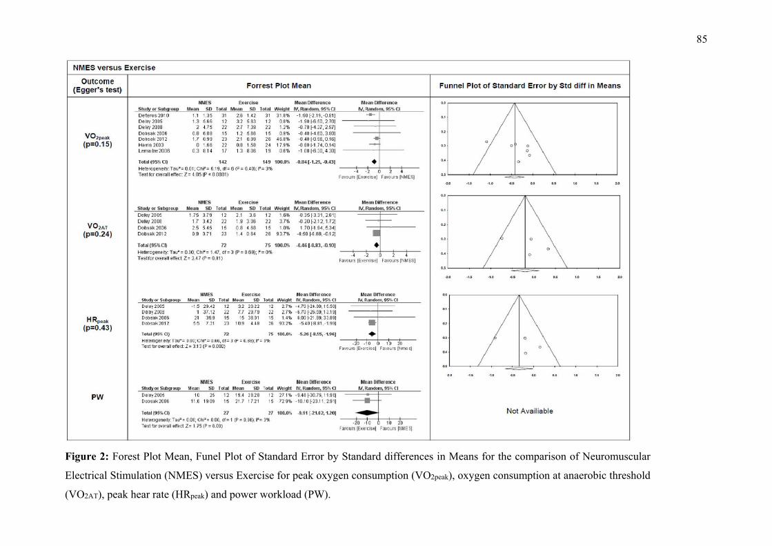
85
Figure 2: Forest Plot Mean, Funel Plot of Standard Error by Standard differences in Means for the comparison of Neuromuscular
Electrical Stimulation (NMES) versus Exercise for peak oxygen consumption (VO2peak), oxygen consumption at anaerobic threshold
(VO2AT), peak hear rate (HRpeak) and power workload (PW).

86
Figure 3: Forest Plot Mean, Funel Plot of Standard Error by Standard differences in Means for the comparison of Neuromuscular
Electrical Stimulation (NMES) versus Usual Treatment (Exercise + Control) for peak oxygen consumption (VO2peak), oxygen
consumption at anaerobic threshold (VO2AT), peak hear rate (HRpeak) and power workload (PW).

87
Figure 4: Forest Plot Mean of Standard Error by Standard differences in Means for the comparison of Neuromuscular Electrical
Stimulation (NMES) versus no-exercise control for peak oxygen consumption (VO2peak)
.
88
89
Apêndice C. Outros Artigos Publicados

90
91