INCONTINÊNCIA URINÁRIA Fisioterapia Ginecológica e Obstétrica Prof a Cristiane Magaldi.

i
SANJA DRAGOSAVAC
PET/CT COM FDG-18F EM PACIENTES COM SUSPEITA DE RECIDIVA DE CARCINOMA DE OVÁRIO
Dissertação de Mestrado
ORIENTADOR: Prof. Dr. GUSTAVO ANTONIO DE SOUZA CO-ORIENTADORA: Profª. Drª. SOPHIE FRANÇOISE MAURICETTE DERCHAIN
Unicamp 2011
UNIVERSIDADE ESTADUAL DE CAMPINAS
Faculdade de Ciências Médicas
PET/CT COM FDGDE RECIDIVA DE CARCINOMA DE OVÁRIO
UNIVERSIDADE ESTADUAL DE CAMPINAS Faculdade de Ciências Médicas
PET/CT COM FDG-18F EM PACIENTES COM SUSPEITA DE RECIDIVA DE CARCINOMA DE OVÁRIO
SANJA DRAGOSAVAC Dissertação de Mestrado apresentada ao Programa de Pós-Graduação Tocoginecologia da Faculdade de Ciências Médicas da Universidade de Campinas - UNICAMP para obtenção de título de Mestre em Ciências Saúde, área de concentração em Oncologia Ginecológica e Mamária,sob orientação do Prof. Dr. Gustavo Antonio de Souza e co-orientação da Profa. Dra. Sophie Mauricette Derchain
Campinas, 2011
ii
PACIENTES COM SUSPEITA DE RECIDIVA DE CARCINOMA DE OVÁRIO
SANJA DRAGOSAVAC
Dissertação de Mestrado apresentada Graduação em Faculdade de
Ciências Médicas da Universidade de UNICAMP para obtenção
de título de Mestre em Ciências da , área de concentração em
Oncologia Ginecológica e Mamária, ob orientação do Prof. Dr. Gustavo
orientação da rofa. Dra. Sophie Françoise
FICHA CATALOGRÁFICA ELABORADA POR
ROSANA EVANGELISTA PODEROSO – CRB8/6652 BIBLIOTECA DA FACULDADE DE CIÊNCIAS MÉDICAS
UNICAMP
Informações para Biblioteca Digital Título em inglês: FDG PET/CT in patients with suspected ovarian câncer recurrence Palavras-chave em inglês:
Fluorodeoxyglucose F18 PET Scan Ovarian Neoplasms
Área de concentração: Oncologia Ginecológica e Mamária Titulação: Mestre em Ciências da Saúde Banca examinadora:
Gustavo Antonio de Souza Celso Darío Ramos Carlos Alberto Buchpiguel
Data da defesa: 26-08-2011 Programa de Pós-Graduação: Faculdade de Ciências Médicas
Diagramação e arte final: Assessoria Técnica do CAISM (ASTEC)
Dragosavac, Sanja,1977 - D787p PET/CT com FDG-18 F em pacientes com suspeita de
recidiva de carcinoma de ovário. / Sanja Dragosavac. -- Campinas, SP : [s.n.], 2011.
Orientador: Gustavo Antonio de Souza Coorientador: Sophie Françoise Mauricette Derchain Dissertação (Mestrado) - Universidade Estadual de
Campinas, Faculdade de Ciências Médicas. 1. Fluordesoxiglucose F18. 2. Tomografia por Emissão
de Pósitrons. 3. Neoplasias ovarianas. I. Souza, Gustavo Antonio de. II. Derchain, Sophie Françoise Mauricette. III. Universidade Estadual de Campinas. Faculdade de Ciências Médicas. IV. Título.


iv
Dedico este trabalho...
...aos meus pais, que tiveram a coragem e a pitada de loucura necessárias para sair dos Bálcãs em busca de um
futuro melhor para seus filhos. Meu MUITO OBRIGADA!
AMO VOCÊS.

v
Agradecimentos
Ao meu orientador Prof. Dr. Gustavo de Souza, pela oportunidade de fazer um
mestrado e por aceitar mais este desafio.
A minha co-orientadora Profa. Dra. Sophie Derchain pelo apoio, por me ensinar
que aprendemos melhor com os acertos do que com os erros, por ajudar a
desenvolver meu potencial intelectual e mais importante, por acreditar que
é possível.
Aos membros da banca da qualificação Prof. Dr. Celso Darío Ramos e Prof. Dr.
Allan dos Santos pela contribuição intelectual e correção desta dissertação.
Ao Prof. Dr. Nelson Caserta pelas correções do manuscrito e pelas tardes no
PET/CT Campinas, aprendizados sobre tomografia, psitacídios e
importância dos chinelos virados e por me incentivar a me lançar nesta
aventura acadêmica.
Ao Dr.Roberval de Campos, por acreditar em meu potencial.
Aos meus amigos e colegas da DIMEN, pelo trabalho em equipe, apoio, coberturas
e incentivo na minha busca de uma melhoria contínua profissional e pessoal.
Ao meu marido, Marcus Vinicius Stradiotto Farbiarz, por dividir comigo esta
caminhada e por estar lá sempre que preciso.
Aos funcionários do CAISM e da FCM UNICAMP que me ajudaram durante a
realização deste trabalho.
vi
Aos colaboradores do PET/CT Campinas que contribuíram direta ou
indiretamente para a realização deste trabalho.
Aos professores do curso da pós-graduação da Tocoginecologia pelo aprendizado
e pela preocupação genuína com a excelência do programa.
Aos amigos e colegas de mestrado pelo aprendizado conjunto, pelas
sugestões, críticas e contribuições com o trabalho e por dividirem comigo
as alegrias e as angústias do processo de se fazer um mestrado.
vii
Sumário
Símbolos, Siglas e Abreviaturas .................................................................................................. viii
Resumo .......................................................................................................................................... x
Summary ....................................................................................................................................... xii
1. Introdução ............................................................................................................................... 14
2. Objetivos ................................................................................................................................. 19
2.1. Objetivo geral .................................................................................................................. 19
2.2. Objetivos específicos....................................................................................................... 19
3. Publicação ............................................................................................................................... 20
4. Conclusões.............................................................................................................................. 44
5. Referências Bibliográficas ....................................................................................................... 45
6. Anexos .................................................................................................................................... 49
6.1. Anexo 1 – Carta de pedido de dispensa do termo de consentimento ............................ 49
6.2. Anexo 2 – Ficha para coleta de dados clínicos ............................................................... 50
6.3. Anexo 3 – Dados de todas as pacientes ......................................................................... 51
6.4. Anexo 4 – Parecer do CEP ............................................................................................. 52
Símbolos, Siglas e Abreviaturas viii
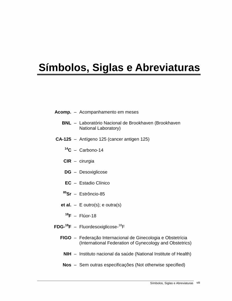
Símbolos, Siglas e Abreviaturas
Acomp. – Acompanhamento em meses
BNL – Laboratório Nacional de Brookhaven (Brookhaven National Laboratory)
CA-125 – Antígeno 125 (cancer antigen 125)
14C – Carbono-14
CIR – cirurgia
DG – Desoxiglicose
EC – Estadio Clínico
85Sr – Estrôncio-85
et al. – E outro(s); e outra(s)
18F – Flúor-18
FDG-18F – Fluordesoxiglicose-18F
FIGO – Federação Internacional de Ginecologia e Obstetrícia (International Federation of Gynecology and Obstetrics)
NIH – Instituto nacional da saúde (National Institute of Health)
Nos – Sem outras especificações (Not otherwise specified)
Símbolos, Siglas e Abreviaturas ix
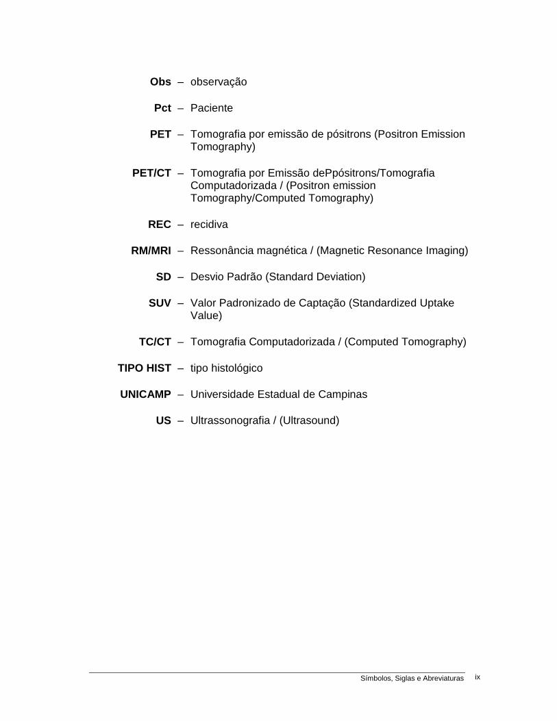
Obs – observação
Pct – Paciente
PET – Tomografia por emissão de pósitrons (Positron Emission Tomography)
PET/CT – Tomografia por Emissão dePpósitrons/Tomografia Computadorizada / (Positron emission Tomography/Computed Tomography)
REC – recidiva
RM/MRI – Ressonância magnética / (Magnetic Resonance Imaging)
SD – Desvio Padrão (Standard Deviation)
SUV – Valor Padronizado de Captação (Standardized Uptake Value)
TC/CT – Tomografia Computadorizada / (Computed Tomography)
TIPO HIST – tipo histológico
UNICAMP – Universidade Estadual de Campinas
US – Ultrassonografia / (Ultrasound)
Resumo x
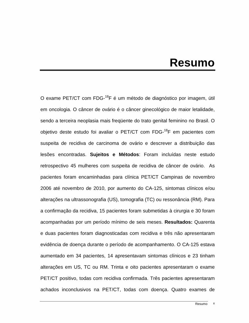
Resumo
O exame PET/CT com FDG-18F é um método de diagnóstico por imagem, útil
em oncologia. O câncer de ovário é o câncer ginecológico de maior letalidade,
sendo a terceira neoplasia mais freqüente do trato genital feminino no Brasil. O
objetivo deste estudo foi avaliar o PET/CT com FDG-18F em pacientes com
suspeita de recidiva de carcinoma de ovário e descrever a distribuição das
lesões encontradas. Sujeitos e Métodos : Foram incluídas neste estudo
retrospectivo 45 mulheres com suspeita de recidiva de câncer de ovário. As
pacientes foram encaminhadas para clínica PET/CT Campinas de novembro
2006 até novembro de 2010, por aumento do CA-125, sintomas clínicos e/ou
alterações na ultrassonografia (US), tomografia (TC) ou ressonância (RM). Para
a confirmação da recidiva, 15 pacientes foram submetidas à cirurgia e 30 foram
acompanhadas por um período mínimo de seis meses. Resultados: Quarenta
e duas pacientes foram diagnosticadas com recidiva e três não apresentaram
evidência de doença durante o período de acompanhamento. O CA-125 estava
aumentado em 34 pacientes, 14 apresentavam sintomas clínicos e 23 tinham
alterações em US, TC ou RM. Trinta e oito pacientes apresentaram o exame
PET/CT positivo, todas com recidiva confirmada. Três pacientes apresentaram
achados inconclusivos na PET/CT, todas com doença. Quatro exames de
Resumo xi
PET/CT eram negativos, sendo que uma paciente teve recidiva confirmada e as
demais permaneceram sem evidências de doença durante o acompanhamento.
Onze pacientes com CA-125 elevado apresentavam resultados de US, TC ou
RM normais. Todas tiveram doença confirmada, sendo que a PET/CT detectou
recidiva em nove e foi inconclusiva em duas. Entre as onze pacientes com CA-
125 normal, foram detectadas metástases na PET/CT em oito. As metástases
mais freqüentes foram diagnosticadas em linfonodos, sendo localizados na
região pélvica e abdominal em 30 pacientes, na região torácica em 16 e em
sete pacientes, na região cervical. Implantes pélvicos e abdominais foram
detectados em 27 pacientes. Outros locais de metástases foram fígado (n=7),
baço (n=2), pleura (n=2), pulmão (n=2) e osso (n=2). O exame PET/CT
detectou lesões não suspeitas em 20 das 45 pacientes (44,4%). A PET/CT
detectou um novo tumor primário de tireoide numa paciente sem recidiva de
carcinoma de ovário. Conclusão: O exame PET/CT foi útil para avaliação da
extensão da recidiva de carcinoma de ovário. A recidiva acometeu mais
freqüentemente os linfonodos, sendo a maioria localizada na região pélvica e
abdominal. Metástases em linfonodos torácicos foram um achado freqüente
nesta população estudada.
Summary xii
Summary
18F-FDG PET/CT is a diagnostic method useful in oncology. Ovarian cancer is
the third most frequent cancer of the female genital tract in Brazil, however, it
has the highest mortality of all gynecological cancers. The aim of this study was
to evaluate the use of 18F-FDG PET/CT in patients with suspected ovarian
cancer recurrence and describe the distribution of metastasis. Methods: Forty-
five female patients with suspicion of ovarian cancer recurrence were included in
this retrospective study. They were referred to PET/CT Campinas clinic from
November 2006 to November 2010, because of elevated CA-125, clinical
suspicion of ovarian cancer recurrence, or alterations detected on ultrasound
(US), computed tomography (CT) or magnetic resonance imaging (MRI).
PET/CT results were compared with histologic findings (n=15) or clinical follow-
up for at least six months (n=30). Results: Forty-two patients were confirmed
with ovarian cancer recurrence. Three patients remained free of disease during
clinical follow-up. CA-125 was elevated in a total of 34 patients, 14 patients had
clinical symptoms of disease and 23 presented with alterations on US, CT and
MRI. Thirty eight patients had positive PET/CT scan, all with confirmed disease.
Three patients had equivocal PET/CT findings and in all three, recurrence was
confirmed. Four patients had negative PET/CT scan: one with confirmed
recurrence and three free from disease during follow-up. Nine out of 11 patients
Summary xiii
with elevated CA-125 and normal conventional imaging had positive PET/CT
scan and two had equivocal findings. There were eleven patients with normal
CA-125 levels, eight presented with positive PET/CT scan. Lymph nodes were
the most frequent site of relapse of disease, most being in the pelvic/abdominal
region (n=30) and others in thoracic (n=16) or cervical region (n=7). Peritoneal
implants were found in 27 patients. Distant sites of metastasis included liver
(n=6), spleen (n=2), pleura (n=2), lung (n=2) and bone (n=2). PET/CT detected
unsuspected lesions in 20/45 patients (44.4%). One patient with PET/CT
negative for ovarian cancer recurrence was diagnosed with primary papillary
carcinoma of the thyroid. Conclusion: 18F-FDG PET/CT was a useful tool for
evaluation of the extension of ovarian cancer recurrence. In the current series,
lymph nodes were the most frequent site of relapse of disease, with supra-
diaphragmatic lymph node metastasis in large number of cases.

Introdução 14
1. Introdução
O câncer de ovário é o câncer ginecológico de maior letalidade, sendo a
terceira neoplasia mais freqüente do trato genital feminino no Brasil, depois do
carcinoma de colo do útero e do endométrio (1). Até 75% das pacientes
apresentam recidiva da doença nos primeiros dois anos após o diagnóstico,
mesmo quando há uma boa resposta ao tratamento inicial (1,2,3).
Os exames de imagem, tais como ultrassonografia (US), tomografia
computadorizada (TC) e ressonância magnética (RM) são usados no
acompanhamento das pacientes com carcinoma de ovário quando a paciente
apresenta sinais clínicos de recidiva da doença ou quando há elevação de
marcador tumoral CA-125. Em uma metanálise, Gu et al. 2008 (4) avaliaram
CA-125, tomografia por emissão de pósitrons (PET) sozinho, PET/CT, TC e RM
para diagnóstico de recidiva do carcinoma de ovário. O CA-125 apresentou
melhor especificidade, de 93%, enquanto que a PET/CT apresentou melhor
sensibilidade, de 91%. A TC e a RM tiveram desempenho diagnóstico
semelhante com sensibilidade de 79% e 75% e especificidade de 84% e 78%,
respectivamente.
Introdução 15
Outra metanálise comparou TC, RM, PET e PET/CT para avaliação de
metástases linfonodais em pacientes com carcinoma de ovário (5). Os exames
PET e PET/CT apresentaram melhor acurácia diagnóstica com sensibilidade de
73,2% e especificidade de 96,7%. O método diagnosticou corretamente
aproximadamente 70% dos linfonodos comprometidos pela doença e 97% dos
linfonodos normais. Não houve diferença significativa entre o desempenho da
TC (sensibilidade: 42,6%; especificidade: 95,5%) e da RM (sensibilidade:
54,7%; especificidade: 88,3%).
Entretanto, a TC possui uma sensibilidade baixa para detecção da
recidiva, sendo limitada principalmente pela doença microscópica e lesões
pequenas (4,5,6). A RM da pelve pode ajudar na avaliação da recidiva local,
porém sua especificidade é baixa devido às alterações anatômicas decorrentes
de manipulação cirúrgica e/ou radioterapia (5,6,7).
A elevação do marcador tumoral CA-125 é um método sensível para
detecção da recorrência do carcinoma de ovário, com acurácia diagnóstica
variando de 79% a 95%, sendo que pode ocorrer até 3 a 6 meses antes de
sinais clínicos da doença (8,9). Entretanto, o CA-125 não é um marcador
específico para carcinoma de ovário e pode apresentar elevação em várias
condições benignas e malignas, além de não permitir localização anatômica da
recorrência da doença (8-11).
PET com fluordesoxiglicose-18F (FDG-18F) é um método de imagem que
permite visualizar o metabolismo de glicose das células neoplásicas antes das

Introdução 16
alterações anatômicas detectáveis e é cada vez mais usado em oncologia
(12,13). O Conceito de tomografia por emissão e transmissão foi introduzido por
David Kuhl e Roy Edwars na década de 1950, o que levou a construção de
vários equipamentos para imagem tomográfica na Universidade de Pensilvânia,
EUA (14). Os equipamentos inicialmente foram desenhados somente para
estudos cerebrais permitindo avaliação da quebra da barreira hemato-
encefálica e visualização de tumores e infartos cerebrais.
Na década de 1970, Louis Sokoloff do Instituto nacional da saúde dos
EUA (NIH) e Martin Reivich da Universidade de Pensilvânia mostraram que a
desoxiglicose (DG) marcada com carbono 14 (14C), um beta-emissor, pode ser
usada para avaliar metabolismo cerebral regional. Eles demonstraram que a
DG atravessa a barreira hemato-encefálica e é fosforilada pelas hexoquinases
para formar a DG-6-fosfato dentro das células, semelhante à glicose comum.
Entretanto, ela não continua nas próximas etapas da glicólise e permanece
retida dentro das células, de modo que se acumula mais em células com alto
metabolismo, tais como células tumorais, do que em células com metabolismo
normal (14,15,16).
A marcação de DG pelo flúor-18 (18F) deu-se na década de 1970, através
da colaboração de Martin Reivich, David Kuhl, Abass Alavi, médicos da
Universidade de Pensilvânia, e Alfred Wolf, químico orgânico do Laboratório
Nacional de Brookhaven (BNL). O 18F foi escolhido para marcação
principalmente devido à sua meia-vida de 110 minutos o que permitia transporte
para locais mais distantes. As primeiras imagens cerebrais com FDG-18F foram

Introdução 17
adquiridas em dois voluntários sadios em agosto de 1976, assim como primeira
imagem do corpo inteiro, utilizando uma gamma câmera de duas cabeças
equipada com colimadores de alta energia, normalmente usada para estudos
ósseos com estrôncio-85 (85Sr).
Durante a década de 1980, muitos estudos de PET com FDG-18F de
corpo inteiro foram conduzidos, validando seu uso, principalmente na área de
oncologia. Assim, além de detectar atividade metabólica anômala
precocemente graças ao seu comportamento semelhante à glicose, outra
vantagem do PET com FDG-18F em relação aos outros exames de imagem, é
que avalia o corpo todo.
A introdução dos aparelhos híbridos PET/CT na década de 1990,
permitiu fusão das imagens metabólicas da PET com as imagens anatômicas
da TC e proporcionou localização mais precisa das lesões. Dados da literatura
mostram acréscimo na sensibilidade e a acurácia da PET/CT quando
comparada com PET ou TC isoladas (17). A tomografia também é utilizada para
correção da atenuação da PET e isso permite uma maior velocidade na
execução do exame comparado com a PET sozinha, melhorando o conforto dos
pacientes.
Os exames PET e PET/CT já foram estudados para diagnóstico de
recidiva de câncer de ovário (4,5,18-27). A sensibilidade varia de 88,2% a 100%; a
especificidade de 71,4% a 93,3%; valor preditivo positivo 92,0% a 97,7%; valor
preditivo negativo 87,5% a 100%; acurácia diagnóstica de 85,4% a 97,0%.

Introdução 18
A PET/CT com FDG-18F tem levado a mudanças significativas na
conduta das pacientes com câncer de ovário (18-10,25). Em um trabalho
prospectivo, Simcock et al. (2006) observaram que houve mudança de
tratamento em 57% das pacientes quando avaliadas com PET/CT, baseada na
mudança da distribuição da detecção da doença observada em 61% dos casos
(18). Um trabalho prospectivo com 90 mulheres Australianas com suspeita de
recorrência de câncer de ovário, corroborou estes achados (25). Neste estudo,
foram detectados 168 locais adicionais e não conhecidos da doença em 61
pacientes (67.8%). Estes achados alteraram a conduta em 53 pacientes
(58.9%). PET/CT foi superior à TC de abdômen e pelve para detecção de
doença em linfonodos, peritôneo e metástases sub-capsulares hepáticas.
As pacientes com recidiva de câncer de ovário apresentam prognóstico
ruim. O diagnóstico da recidiva é necessário no esforço para escolher o
tratamento mais apropriado, na tentativa de melhorar a sobrevida e a qualidade
da vida. O exame PET/CT com FDG-18F pode ter um papel importante na
avaliação da recidiva de carcinoma de ovário, uma vez que o traçador
metabólico pode melhorar detecção das lesões e a fusão das imagens
metabólicas com as imagens antômicas pode melhorar a precisão da
localização da doença, além de avaliar o corpo todo.
Objetivos 19
2. Objetivos
2.1. Objetivo geral
O objetivo do estudo foi avaliar desempenho do exame PET/CT com
FDG-18F na avaliação da extensão da doença em pacientes com suspeita de
recidiva de carcinoma de ovário.
2.2. Objetivos específicos
− Avaliar a relação entre os achados do exame PET/CT e os valores de
CA-125, os sintomas clínicos e exames radiológicos convencionais.
− Descrever a distribuição das metástases encontradas pelo exame
PET/CT.
Publicação 20
3. Publicação
--------------------------- Mensagem Original ---------------------------- Assunto: JNUMED/2011/096081 Manuscript Submission De: [email protected] Data: Seg, Julho 25, 2011 12:04 pm Para: "Sophie Derchain" <[email protected]> -------------------------------------------------------------------------- MS ID#: JNUMED/2011/096081 MS TITLE: Staging recurrent ovarian cancer with 18F-FDG PET/contrast enhanced CT in patients with normal or raised CA-125 Dear Dr., This is an automatic message acknowledging receipt of your online submission to The Journal of Nuclear Medicine. You can check the status of your paper at any time by visiting the journal site at http://submit-jnm.snmjournals.org. Thank you for your submission. Best regards, Susan Alexander Associate Director of Publications The Journal of Nuclear Medicine

Publicação 21
STAGING RECURRENT OVARIAN CANCER WITH 18F-FDG PET/CONTRAST
ENHANCED CT IN PATIENTS WITH NORMAL OR RAISED CA-12 5
Sanja Dragosavac 1; Sophie Derchain1*; Nelson Márcio Gomes Caserta2;
Gustavo de Souza1
1Department of Obstetrics and Gynecology, Faculty of Medical Sciences, State
University of Campinas – Unicamp, Campinas, São Paulo, Brazil
2Department of Radiology, Faculty of Medical Sciences, State University of
Campinas – Unicamp, Campinas, São Paulo, Brazil
*Address for correspondence:
Sophie F. M. Derchain, Department of Obstetrics and Gynecology Faculty of
Medical Sciences, PO Box 6111 State University of Campinas – UNICAMP, Zip
Code 13083-970, Campinas, SP, Brazil.
e-mail: [email protected]
Publicação 22
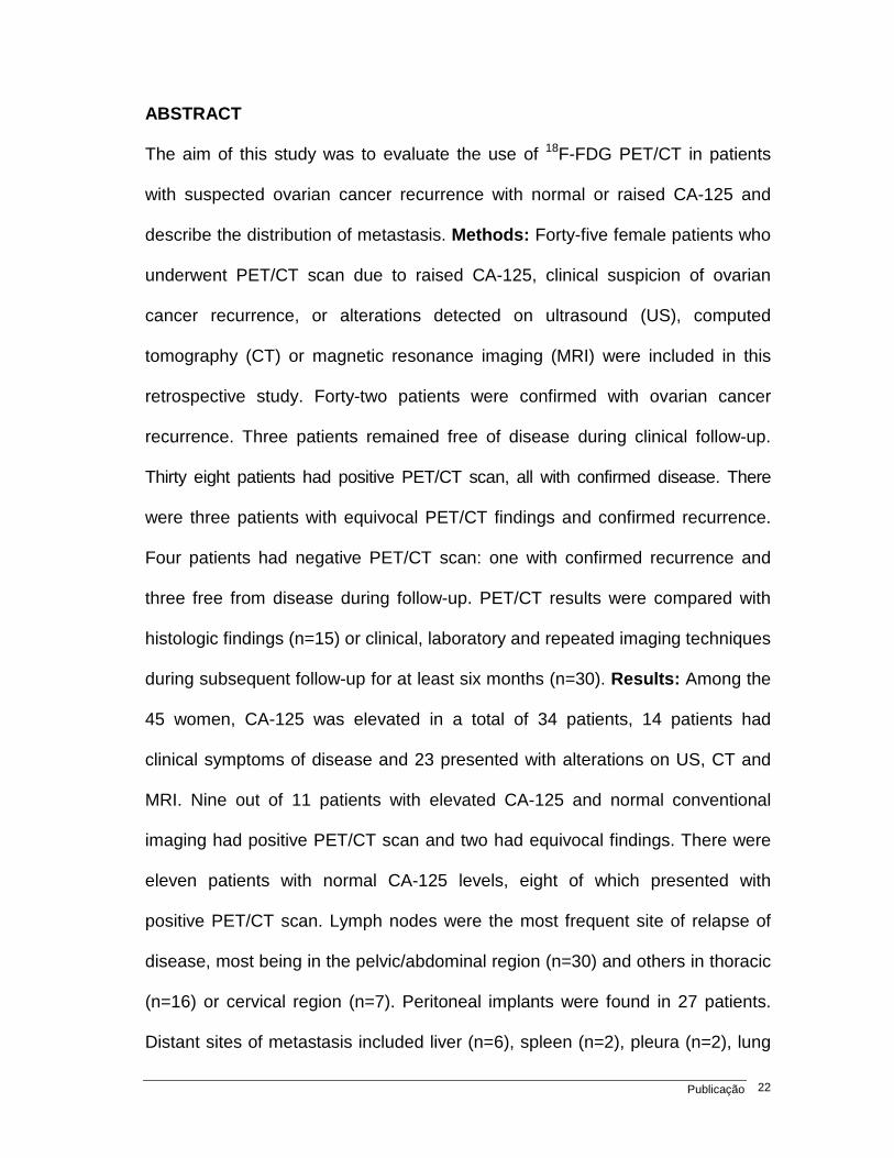
ABSTRACT
The aim of this study was to evaluate the use of 18F-FDG PET/CT in patients
with suspected ovarian cancer recurrence with normal or raised CA-125 and
describe the distribution of metastasis. Methods: Forty-five female patients who
underwent PET/CT scan due to raised CA-125, clinical suspicion of ovarian
cancer recurrence, or alterations detected on ultrasound (US), computed
tomography (CT) or magnetic resonance imaging (MRI) were included in this
retrospective study. Forty-two patients were confirmed with ovarian cancer
recurrence. Three patients remained free of disease during clinical follow-up.
Thirty eight patients had positive PET/CT scan, all with confirmed disease. There
were three patients with equivocal PET/CT findings and confirmed recurrence.
Four patients had negative PET/CT scan: one with confirmed recurrence and
three free from disease during follow-up. PET/CT results were compared with
histologic findings (n=15) or clinical, laboratory and repeated imaging techniques
during subsequent follow-up for at least six months (n=30). Results: Among the
45 women, CA-125 was elevated in a total of 34 patients, 14 patients had
clinical symptoms of disease and 23 presented with alterations on US, CT and
MRI. Nine out of 11 patients with elevated CA-125 and normal conventional
imaging had positive PET/CT scan and two had equivocal findings. There were
eleven patients with normal CA-125 levels, eight of which presented with
positive PET/CT scan. Lymph nodes were the most frequent site of relapse of
disease, most being in the pelvic/abdominal region (n=30) and others in thoracic
(n=16) or cervical region (n=7). Peritoneal implants were found in 27 patients.
Distant sites of metastasis included liver (n=6), spleen (n=2), pleura (n=2), lung
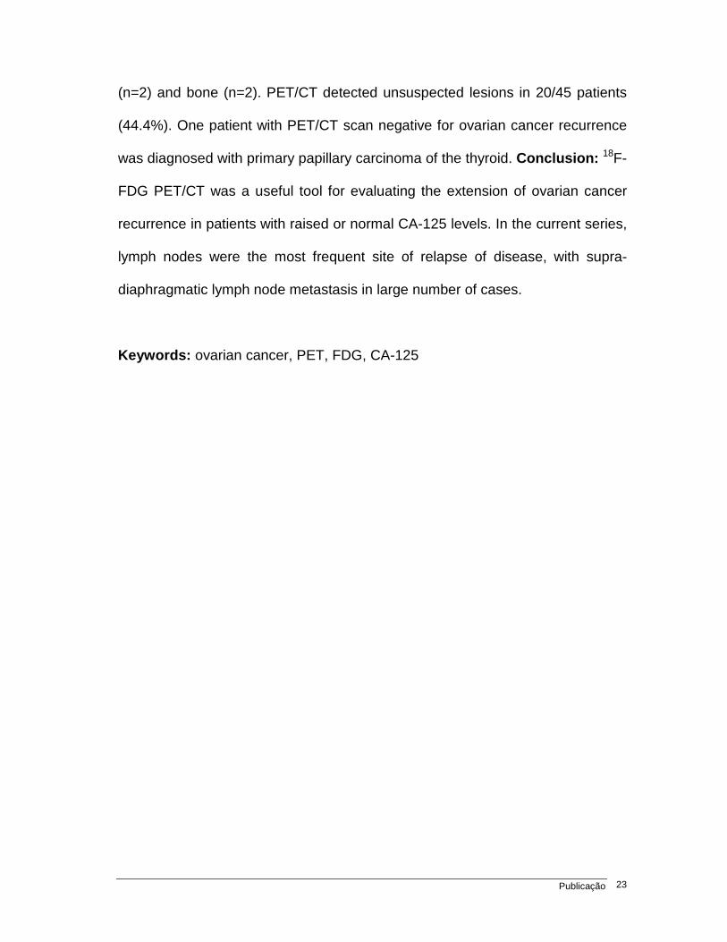
Publicação 23
(n=2) and bone (n=2). PET/CT detected unsuspected lesions in 20/45 patients
(44.4%). One patient with PET/CT scan negative for ovarian cancer recurrence
was diagnosed with primary papillary carcinoma of the thyroid. Conclusion: 18F-
FDG PET/CT was a useful tool for evaluating the extension of ovarian cancer
recurrence in patients with raised or normal CA-125 levels. In the current series,
lymph nodes were the most frequent site of relapse of disease, with supra-
diaphragmatic lymph node metastasis in large number of cases.
Keywords: ovarian cancer, PET, FDG, CA-125
Publicação 24
INTRODUCTION
Ovarian cancer represents 3% of all cancers in women; however, it has
the highest mortality of all gynecological cancers (1). Most patients have
advanced stage disease at presentation due to a paucity and incidious onset of
symptoms. Initial treatment consists of cytoreductive surgery and adjuvant
platinum-based cytotoxic chemotherapy (1,2). Despite high response after initial
treatment, approximately 20-30% of patients with early-stage disease (stage IA-
IIA) and up to 75% of patients with advanced disease (IIB-IV) present with
recurrence within two years (2,3).
The imaging exams, such as ultrasound (US), computed tomography
(CT) and magnetic resonance imaging (MRI) are performed during patient
follow-up when there is clinical suspicion of ovarian cancer recurrence or CA-
125 elevation. A systematic review and meta-analyses by Gu et al. 2008, (4)
evaluated CA-125, positron emission tomography (PET) alone, PET/CT, CT and
MRI in diagnosing recurrent ovarian carcinoma. CA-125 had the highest pooled
specificity, 93%, while PET/CT had highest pooled sensitivity, 91%. CT
(sensitivity 79%, specificity 84%) and MRI (sensitivity 75%, specificity 78%)
showed similar diagnostic performance.
While CA-125 has been shown to be a sensitive marker for tumor
recurrence and may rise 3 to 6 months before there is clinically apparent
disease, it does not provide information concerning the size and distribution of
the lesions. It can also rise in a number of benign conditions, not being a specific
marker for ovarian cancer, and many patients with relapse of disease present
with normal CA-125 levels (4, 5). CT has low sensitivity for detecting disease
Publicação 25
recurrence probably due to its limitation to detect small peritoneal implants and
normal-sized lymph node metastasis (4,6). Pelvic MRI can be useful for
evaluation of local recurrence of disease, however, it has low specificity due to
post-surgical anatomical alterations (4,7).
18F-Fluorodeoxyglucose (18F-FDG) PET/CT may play an important role in
ovarian cancer recurrence because metabolic tracer can increase lesion
detection, the fusion of metabolic and anatomic imaging can help determine the
exact location of disease and it can survey the whole body. Several studies
examined the performance of PET/CT scan in patients with recurrent ovarian
cancer (4, 8-14).
An Australian prospective, multi-centre, cohort study with 90 women (14)
assessed the impact of 18F-FDG PET/CT in the management of patients with
suspected recurrent ovarian cancer and evaluated incremental information provided
by 18F-FDG PET/CT in this clinical context. PET/CT detected 168 additional and
unsuspected sites of disease in 61 patients (67.8%). The management was
changed in 53 patients (58.9%) based on PET/CT scan findings. PET/CT was
superior to abdominal and pelvic CT in the detection of nodal, peritoneal and
subcapsular liver disease and it also allowed the identification of patients whose
disease was likely to progress within 12 months. The authors suggested that
PET-CT should be preferred imaging modality in patients with suspected ovarian
carcinoma recurrence.
The aim of this study was to evaluate 18F-FDG PET/contrast enhanced
CT in patients with suspicion of ovarian cancer recurrence with or without
elevated CA-125 levels and describe the distribution of metastasis.
Publicação 26
MATERIAL AND METHODS
Patients
Forty-five female patients (age range, 39 - 84 y; mean age + SD, 59.5 +
10.0 y) with suspicion of ovarian cancer recurrence were included in this
retrospective study. The patients performed PET/CT scan at PET/CT Campinas
private clinic from November 2006 to November 2010. Indications for PET/CT
were clinical suspicion of relapse of disease, elevated CA-125 or abnormal or
equivocal findings on abdominal/pelvic US, CT or MRI. All patients had been
submitted to surgery and all but one, adjuvant chemotherapy at the time of
diagnosis. Eighteen patients had already had relapse of disease during their
previous follow-up and PET/CT was performed for suspicion of new progression
of disease. The study was approved by local ethics committee. For detailed
patient and tumor characteristics, see table 1.
18F-FDG PET/CT Imaging
All patients fasted for at least 6 hours, maintaining their blood glucose
levels below 150 mg/dl, before the injection of approximately 444 MBq (mean +
SD; 481 + 85.1 MBq) of 18F-FDG. The patients rested in a supine position during
40 to 60 minutes after the injection and then were positioned for PET/CT
imaging. All PET/CT scans were obtained on a combined 16-slice CT/BGO PET
scanner (Discovery STE, GE Medical Systems Inc, Milwaukee, WI). The
patients received oral contrast (Urografina® 291, Schering; 25ml diluted in 1l of
water), two glasses before the 18F-FDG injection and two glasses immediately
Publicação 27
before the imaging. After the initial scout, a contrast-enhanced CT was acquired
from top of the head to mid-thighs, without any specific breath-holding
instructions. Intra-venous contrast, 100ml, Optiray, Mallinckrodt, was injected,
unless patient was allergic to iodine. The parameters of the CT scan were 140
kV, 150-250 mAs, slice thickness of 3.75 mm. The CT was followed by PET
scanning, covering the same transverse field of view during normal breathing.
The imaging was acquired with 6 to 8 bed positions on a 2D mode for 5 minutes
per bed position, or on a 3D mode for 3 minutes for bed position. PET images
were reconstructed iteratively using the contrast-enhanced CT data for
attenuation correction. Coregistered images were displayed on a workstation,
using dedicated software which allowed the viewing of PET, CT and fusion
images on transaxial, sagittal and coronal displays.
18F-FDG PET/CT Analysis
All 18F-FDG PET/CT scans were interpreted by experienced radiologist in
conjunction with experienced nuclear medicine physician, who were both aware of
suspicion of ovarian carcinoma recurrence and patients’ laboratory and imaging
findings. The 18F-FDG PET portion and the CT portion of PET/CT were jointly
interpreted using a dedicated image fusion workstation. All areas of increased
18F-FDG uptake that corresponded to a CT abnormality were interpreted as
positive for recurrent disease. Semi-quantitative analysis was also performed to
derive a standardized uptake value (SUV). All PET/CT reports and images were
reviewed by experienced nuclear physician for consistency of the data.
Publicação 28
The results of 18F-FDG PET/CT were correlated with patient follow-up
information for at least 6 months after the exam (mean + SD; 21.0 + 12.0).
The diagnosis of recurrence was confirmed with surgery (n=15) or clinically
(n=30), by persistent elevation of CA-125 with abnormal findings on further
imaging and treatment response following chemotherapy.
RESULTS
Forty-two patients had diagnosis of recurrence of ovarian cancer after
surgery or during clinical follow-up. Three patients remained free of disease
during clinical follow-up. CA-125 was raised in a total of 34 patients, 14 patients
had clinical suspicion of recurrence and 23 presented with alterations on US, CT
or MRI. There were 11 patients with high CA-125 levels and normal imaging
exams. The characteristics of the patients according to the PET/CT findings can
be seen on table 2.
18F-FDG PET/CT scan was positive in 38 patients (84%), all confirmed
with recurrence of disease.
There were 3 patients with high CA-125 levels with equivocal findings on
PET/CT scan (9%). All three were confirmed with ovarian cancer recurrence.
One of the patients had negative abdominal ultrasound, CT and MRI imaging
and presented with abdominal pain. The PET/CT showed mild increase of
metabolic activity in soft tissue near abdominal wall and in left inguinal normal-
sized lymph nodes with SUV respectively of 3.3 and 2.3. There was also small
lytic lesion in the left femoral trochanter with SUV of 2.2. The patient was
submitted to chemotherapy because of clinical evidence of relapse of disease
Publicação 29
and one year later her CA-125 levels lowered to 50 U/ml. However, PET/CT at
that time showed similar metabolic pattern. No new hypermetabolic lesions were
identified. The patient died of disease progression 20 months after the initial
PET/CT imaging. Other two patients with equivocal findings on PET/CT showed
progression of disease on conventional imaging during follow-up and further
elevation of CA-125.
18F-FDG PET/CT scan was negative in 4 patients (9%): one with confirmed
recurrence and three free from disease during follow-up, with normal CA-125
levels and no evidence of disease on imaging exams. One of the patients
without ovarian cancer recurrence presented with focal abnormal uptake in the
right thyroid lobe (SUV=17.0) and new primary tumor was diagnosed after
surgery (figure 1).
Nine out of 11 patients with raised CA-125 and normal conventional
imaging had positive PET/CT scan and two had equivocal findings. There were
eleven patients with normal CA-125 levels, eight of which presented with
positive PET/CT scan (figure 2). Four of those patients had localized pelvic or
abdominal disease (two of which were amenable to surgical resection) and 4
also presented with supra-diaphragmatic lymph node metastasis.
Lymph nodes were the most frequent site of relapse of disease (table 3),
being localized in the pelvic/abdominal region in 30 patients (66%), in thoracic
region in 16 (35%) and cervical region in 7 patients (15%). Peritoneal implants
were found in 27 patients (60%). Distant sites of metastasis included liver (n=6),
spleen (n=2), pleura (n=2), lung (n=2) and bone (n=2).
Publicação 30
PET/CT found unsuspected lesions in twenty out of forty-five patients
(44.4%), most being supra-diaphragmatic lymph node metastases or normal
sized abdominal lymph nodes with abnormal 18F-FDG uptake (figures 2 and 3).
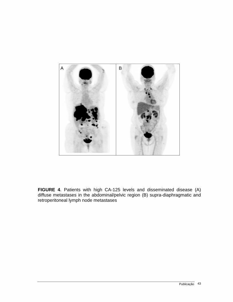
Patients with elevated CA-125 tended to have more disseminated
disease (figure 4).
Twelve patients (26%) died during follow-up (mean + SD; 15.3 + 7.9 months
after the exam, range 6-35 months). Five of these patients had disseminated
abdominal and supra-diaphragmatic disease, while 7 had pelvic/abdominal
limited disease.
DISCUSSION
PET/CT correctly diagnosed patients with suspected ovarian cancer
recurrence. Most of the patients with raised CA-125 and normal conventional
imaging had positive PET/CT scan. On the other hand, most of the patients with
normal CA-125 levels in this series also presented with positive PET/CT scan.
Lymph nodes were the most frequent site of relapse of disease, most being in
the pelvic/abdominal region and others in thoracic or cervical region. Peritoneal
implants were found in more than half of the patients. Distant sites of metastasis
included liver, spleen, pleura, lung and bone. PET/CT detected unsuspected
lesions in almost half of the patients, most being supra-diaphragmatic lymph
node metastasis.
Our population had high pre-test probability of disseminated disease, once
that 18/45 patients (40%) had previously had relapse of ovarian cancer and the
referral to PET/CT was to evaluate progression of disease and to restage. The
Publicação 31
advantage of PET/CT in this clinical setting was the whole body evaluation that
could help correctly select patients amenable to surgical resection.
Most of the findings are in accordance with previously described in
literature, except for the high prevalence of supra-diaphragmatic lymph node
metastases (7-14). Iagaru et al, 2008 (9), evaluated retrospectively 43 patients with
ovarian carcinoma and they described the distribution of extra-pelvic metastases in
19 patients, 5 of which (11%) had supra-diaphragmatic lymph node involvement.
A prospective, multi-centre Australian study (14) showed that 14 out of 90
patients (15%) presented with disease above the diaphragm.
In the Australian study (14), PET/CT findings changed management plan
with medium to high impact in 58% of patients, because of its ability to detect
unsuspected lesions and the advantage of the whole-body evaluation.
The change in management based on PET/CT was also previously
described by Simcock et al. 2006 (8). They prospectively evaluated 56 women
with ovarian cancer who underwent PET/CT scan for suspicion of recurrence or
surveillance with no evidence of disease. PET/CT altered the apparent disease
distribution in 40 scans (61%), with high impact on management plan in 32
patients (57%).
Many clinicians routinely measure CA-125 once it is often the first
evidence of ovarian cancer recurrence and can rise 3 to 6 months before the
clinical evidence of disease (5). However, without localized disease, there is no
rationale to initiate treatment based on a laboratory test alone and this can
cause considerable patient anxiety. Therefore, it is important to have an
Publicação 32
accurate method that can both diagnose and localize recurrence of disease and
help select patients amenable to surgical resection.
Recent meta-analyses evaluated CT, MRI, PET and PET/CT for detection
of metastatic lymph nodes in patients with ovarian cancer (15). PET and
PET/CT were a more accurate modality for lymph node metastasis detection
with global pooled sensitivity of 73.2%, specificity of 96.7%. However, the
greater specificity of PET or PET/CT compared to those of CT or MRI was
statistically insignificant. CT and MRI showed similar diagnostic performance,
with pooled sensitivity, respectively, of 42.6% and 54.7% and pooled specificity
of, respectively, 95.0% and 88.3%.
PET/contrast enhanced CT in the same study (15) showed sensitivity of
84.4% and specificity of 97.4%, which was better than non-contrast enhanced
PET/CT. Some authors believe that the CT portion of the study should be
performed without contrast. Those who defend the use of contrast usually
perform a non-contrast enhanced CT before the diagnostic contrast-enhanced
CT, for the attenuation correction purposes of PET images. Yau et al (2005) (18)
showed that application of intravenous contrast does not interfere with
diagnostic value of PET/CT when contrast-enhanced CT is used for the
attenuation correction purpose.
The PET/CT evaluation of pelvic and abdominal regions can be
challenging due to urinary excretion and bladder concentration of 18F-FDG.
Contrast material can help distinguish vessels and urethers from small nodal
disease, which can result in better sensitivity of the PET/CT scan. This can be of
particular importance in patients with ovarian cancer once that most metastasis
Publicação 33
involve pelvic and abdominal lymph nodes or implants. Some authors (19)
suggest use of diuretics and dual time imaging as another way of improving the
sensitivity of the exam for detection of pelvic lesions.
A limitation of our study is that there was no pathological confirmation of
all the sites of abnormal FDG uptake. However, the confirmation of all the sites
would not have been ethical just for the purpose of validation of PET/CT
findings. On the other hand, accurate surgical assessment of pelvic and
retroperitoneal lymph nodes is difficult, and surgery seems to be unreliable gold
standard, with disease recurrence in a third of women with negative surgical
findings (16). We agree with Simcock et al. 2006 (8) and believe that the course
of disease and clinical outcomes may more accurately validate PET/CT data.
Another limitation was that we didn’t have data concerning treatment plan
before the PET/CT, therefore, it was not possible to evaluate the change in
management in our study. However, PET/CT showed unsuspected lesions in
44.4% of our patients, which is in accordance with previously published data.
There is no evidence that PET/CT can improve overall survival of patients
with diagnosis of ovarian cancer recurrence. However, it is whole body exam
that can show disease extension in patients with high CA-125 levels, as well as
in those whose CA-125 levels are within normal range. This can help to correctly
restage patients considered for further treatment.
CONCLUSION
18F-FDG PET/CT was an accurate and useful tool for diagnosis and
evaluation of the extension of disease in patients with ovarian cancer recurrence
Publicação 34
with raised or normal CA-125 levels. In the current series, lymph nodes were the
most frequent site of relapse of disease, with supra-diaphragmatic lymph node
metastasis in large number of cases.
REFERENCES
1. Bertone-Johnson ER. Epidemiology of ovarian cancer: a status report.
Lancet. 2005;365(9454):101-102.
2. Cannistra SA. Cancer of the ovary. N England J Med. 2004;351(24):2519-
2529.
3. Gadducci A, Cosio S, Zola P, et al. Surveillance procedures for patients
treated for epithelial ovarian cancer: a review of the literature. Int J Gynecol
Cancer. 2007;17(1):21-31.
4. Gu P, Pan LL, Wu SQ, et al. CA 125, PET alone, PET-CT and MRI in
diagnosing recurrent ovarian carcinoma. A systematic review and meta-
analysis. Eur J Radiol. 2009;71(1):164-174.
5. Goonewardene TI, Hall MR, Rustin GJS. Management of asymptomatic
patients on follow-up for ovarian cancer with rising CA-125 concentrations.
Lancet Oncol. 2007;8(9):813-821.
6. Togashi K. Ovarian cancer: the clinical role of US, CT and MRI. Eur Radiol.
2003;13(Suppl 4):L87-L104.
7. Low RN, Duggan B, Barone RM, Saleh F, Song SYT. Treated ovarian
cancer: MR imaging, laparotomy assessment and serum CA-125 values
compared with clinical outcome at 1year. Radiology. 2005;235(3):918-926.
Publicação 35
8. Simcock B, Neesham D, Quinn M, et al. The impact of PET/CT in the
management of recurrent ovarian cancer. Gynecol Oncol. 2006;103(1):271-
276. Epub 2006 Apr 19.
9. Iagaru A, Mittra ES, McDougall R, Quon A, Gambhir SS. 18F FDG PET/CT
evaluation in patients with ovarian carcinoma. Nucl Med Commun.
2008;29(12):1046-1051.
10. Bilici A, Ustaalioglu BBO, Seker M, et al. Clinical value of FDG PET/CT in
the diagnosis of suspected recurrent ovarian cancer: is there an impact of
FDG PET/CT on patient management? Eu J Nucl Med Mol Imaging.
2010;37(7):1259-1269. Epub 2010 Mar 23.
11. Soussan M, Wartski M, Cherel P, et al. Impact of FDG PET/CT imaging on
the decision making in the biologic suspicion of ovarian carcinoma
recurrence. Gynecol Oncol. 2008;108(1):160-165. Epub 2007 Oct 24.
12. Nanni C, Rubello D, Farsad M, et al. 18F-FDG PET/CT in the evaluation of
recurrent ovarian cancer: a prospective study on forty-one patients. Eur J
Surg Oncol. 2005;31(7):792-797.
13. Pan HS, Lww SL, Huang LW, Chen YK. Combined positron emission
tomography-computed tomography and tumor markers for detecting
recurrent ovarian cancer. Arch Gynecol Obstet. 2011;283(2):335-341. Epub
2010 Mar 11.
14. Fulham MJ, Carter J, Baldey A, Hicks RJ, Ramshaw JE, Gibson M. The
impact of PET-CT in suspected recurrent ovarian cancer: A prospective
multi-centre study as part of the Australian PET Data Collection Project.
Gynecol Oncol. 2009;112(3):462-468. Epub 2009 Jan 15.

Publicação 36
15. Yuan Y, Gu ZX, Tao XF Liu SY. Computer tomography, magnetic resonance
imaging, and positron emission tomography or positron emission
tomography/computer tomography for detection of metastatic lymph nodes
in patients with ovarian cancer: A meta-analysis. Eur J Radiol. Feb 22, 2011
[Epub ahead of print].
16. Friedman JB, Weiss NS. Second thoughts about second-look laparotomy in
advanced ovarian cancer. N Engl J Med. 1990;322(15):1079-82.
17. Choi JY, Lee KS, Kim HJ, et al. Focal thyroid lesions incidentally identified
by integrated 18F-FDG PET/CT: Clinical significance and improved
characterization. J Nucl Med. 2006;47(4):609-615.
18. Yau YY, Chan WS, Tam YM, et al. Application of intravenous contrast in
PET/CT: does it really introduce significant attenuation correction error? J
Nucl Med. 2005;46(2):283-291.
19. Anjos DA, Etchebehere EC, Ramos CD, et al. 18F-FDG PET/CT delayed
images after diuretic for restaging invasive bladder cancer. J Nucl Med.
2007;48(5):764-70.

Publicação 37
TABLE 1 Patient and tumor characteristics (n=45)
Characteristic Value
Age (y) Average + SD* 59.5 + 10.0 Range 39 - 84
Histologic type (n) Serous adenocarcinoma 29 Endometrioid adenocarcinoma 9 Mixed type adenocarcinoma 1 Clear cell adenocarcinoma 1 Adenocarcinoma nos† 4
Sertoli cells tumor 1
FIGO‡ stage at diagnosis
I 2 II 3 III 34 IV 6
SD = standard deviation; † nos = not otherwise specified; ‡ FIGO = International Federation of Gynecology and Obstetrics
Publicação 38
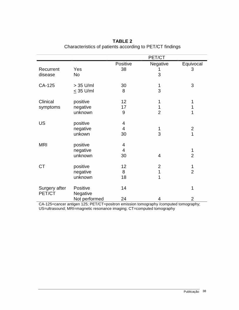
TABLE 2 Characteristics of patients according to PET/CT findings
PET/CT Positive Negative Equivocal Recurrent disease
Yes No
38 1 3
3
CA-125 > 35 U/ml
< 35 U/ml 30 8
1 3
3
Clinical symptoms
positive negative unknown
12 17 9
1 1 2
1 1 1
US positive
negative unkown
4 4 30
1 3
2 1
MRI positive
negative unknown
4 4 30
4
1 2
CT positive
negative unknown
12 8 18
2 1 1
1 2
Surgery after PET/CT
Positive Negative Not performed
14
24
4
1
2 CA-125=cancer antigen 125; PET/CT=positron emission tomography /computed tomography; US=ultrasound; MRI=magnetic resonance imaging; CT=computed tomography
Publicação 39
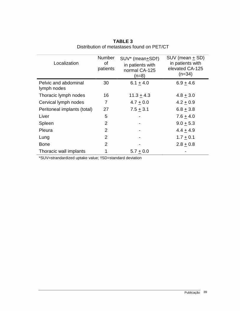
TABLE 3 Distribution of metastases found on PET/CT
Localization Number
of patients
SUV* (mean+SD†) in patients with normal CA-125
(n=8)
SUV (mean + SD) in patients with
elevated CA-125 (n=34)
Pelvic and abdominal lymph nodes
30 6.1 + 4.0 6.9 + 4.6
Thoracic lymph nodes 16 11.3 + 4.3 4.8 + 3.0
Cervical lymph nodes 7 4.7 + 0.0 4.2 + 0.9
Peritoneal implants (total) 27 7.5 + 3.1 6.8 + 3.8
Liver 5 - 7.6 + 4.0
Spleen 2 - 9.0 + 5.3
Pleura 2 - 4.4 + 4.9
Lung 2 - 1.7 + 0.1
Bone 2 - 2.8 + 0.8
Thoracic wall implants 1 5.7 + 0.0 - *SUV=strandardized uptake value; †SD=standard deviation
Publicação 40
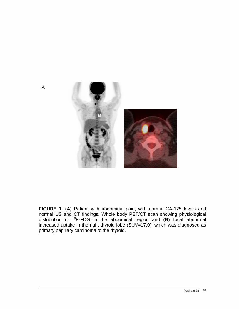
FIGURE 1. (A) Patient with abdominal pain, with normal CA-125 levels and normal US and CT findings. Whole body PET/CT scan showing physiological distribution of 18F-FDG in the abdominal region and (B) focal abnormal increased uptake in the right thyroid lobe (SUV=17.0), which was diagnosed as primary papillary carcinoma of the thyroid.
A
B

Publicação 41
FIGURE 2. Patients with CA-125 levels within normal range with positive PET/CT scan showing supra-diaphragmatic lymph node and pelvic /abdominal metastases (A) CA-125=13.8 U/ml (B) CA-125=17.4 U/ml (C) CA-125=20.8 U/ml (D) CA-125=32.3 U/ml
A B C D
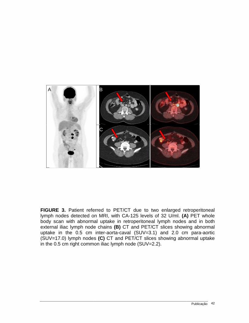
FIGURE 3. Patient referred to PET/CT due to two enlarged retroperitoneal lymph nodes detected on MRI, with CAbody scan with abnormal uptake in retroperitoneal lymph nodes and in both external iliac lymph node chains uptake in the 0.5 cm inter(SUV=17.0) lymph nodes in the 0.5 cm right common iliac lymph node (SUV=2.2).
A
Patient referred to PET/CT due to two enlarged retroperitoneal lymph nodes detected on MRI, with CA-125 levels of 32 U/ml. (A)body scan with abnormal uptake in retroperitoneal lymph nodes and in both external iliac lymph node chains (B) CT and PET/CT slices showing abnormal uptake in the 0.5 cm inter-aorta-caval (SUV=3.1) and 2.0 cm para(SUV=17.0) lymph nodes (C) CT and PET/CT slices showing abnormal uptake in the 0.5 cm right common iliac lymph node (SUV=2.2).
B
C
Publicação 42
Patient referred to PET/CT due to two enlarged retroperitoneal (A) PET whole
body scan with abnormal uptake in retroperitoneal lymph nodes and in both CT and PET/CT slices showing abnormal
caval (SUV=3.1) and 2.0 cm para-aortic CT and PET/CT slices showing abnormal uptake
Publicação 43
FIGURE 4. Patients with high CA-125 levels and disseminated disease (A) diffuse metastases in the abdominal/pelvic region (B) supra-diaphragmatic and retroperitoneal lymph node metastases
A B
Conclusões 44
4. Conclusões
– PET/CT foi positivo em todas as pacientes com níveis elevados de CA-125 e
também em grande maioria dos pacientes com CA-125 normal. As pacientes
com dor abdominal e PET/CT negativo se mantiveram sem evidência de
recidiva durante o seguimento. Em todos os casos das pacientes com dor
abdominal e o exame PET/CT positivo, houve confirmação da recidiva.
Quanto aos exames de imagem, as pacientes encaminhadas devido às
alterações detectadas em tomografia computadorizada ou ultrassonografia
abdominal que tiveram o exame PET/CT negativo mantiveram-se sem
evidência de doença no período de acompanhamento.
– O local mais freqüente de recidiva foi em linfonodos, sendo a maioria
localizada na região pélvica/abdominal. Houve grande prevalência de pacientes
com metástases em linfonodos supradiafragmáticos. Implantes peritoneais foram
encontrados em mais da metade dos pacientes. As metástases à distância foram
mais freqüentemente encontradas em fígado, baço, pleura, pulmão e osso.
Referências Bibliográficas 45
5. Referências Bibliográficas
1. Ristow CM, Yamamoto CT, Fávaro M. Fatores de risco e patogênese das
neoplasias epiteliais de ovário: revisão de literatura. Rev Bras Cancerol.
2006;52(2):185-195.
2. Bertone-Johnson ER. Epidemiology of ovarian cancer: a status report.
Lancet. 2005;365(9454):101-2.
3. Cannistra SA. Cancer of the ovary. N England J Med. 2004;351:2519-29.
4. Gu P, Pan LL, Wu SQ, Sun L, Huang G. CA 125, PET alone, PET-CT, CT
and MRI in diagnosing recurrent ovarian carcinoma. A systematic review
and meta-analysis. Eur J Radiol. 2009;71(1):164-74.
5. Yuan Y, Gu ZX, Tao XF Liu SY. Computer tomography, magnetic resonance
imaging, and positron emission tomography or positron emission
tomography/computer tomography for detection of metastatic lymph nodes
in patients with ovarian cancer: A meta-analysis. Eur J Radiol. Feb 22, 2011
[Epub ahead of print].
6. Togashi K. Ovarian cancer: the clinical role of US, CT and MRI. Eur Radiol.
2003;13(Suppl 4):L87-L104.
Referências Bibliográficas 46
7. Low RN, Duggan B, Barone RM, Saleh F, Song SYT. Treated ovarian
cancer: MR imaging, laparotomy assessment and serum CA-125 values
compared with clinical outcome at 1year. Radiology. 2005;235(3):918-926.
8. Rustin G. Tuxen M. Use of CA125 in folow-up of ovarian cancer. Lancet.
1996;348(9021):191-2.
9. Niloff JM, Bast RC Jr, Schaetzl EM, Knapp RC. Predictive value of CA 125
antigen levels in second-look procedures for ovarian cancer. Am J Obstet
Gynecol. 1985;151(7):981-6.
10. Meden H, Fattahi-Meibodi A. Ca 125 in benign gynecological conditions. Int
J Biol Markers. 1998;13(4):231-7.
11. Goonewardene TI, Hall MR, Rustin GJS. Management of asymptomatic
patients on follow-up for ovarian cancer with rising CA-125 concentrations.
Lancet Oncol. 2007;8(9):813-21
12. Kluetz PG, Meltzer CC, Villemagne VL, Kinahan PE, Chander S, Martinelli
MA et al. Combined PET/CT imaging in oncology: impact on patient
management. Clin Positron Imaging. 2000;3(6):223-30.
13. Hustinx R, Benard F, Alavi A. Whole-body FDG-PET imaging in the
management of patients with cancer. Semin Nucl Med. 2002;32(1):35-46.
14. Alavi A, Reivich M. Guest Editorial: The conception of FDG-PET imaging.
Semin Nucl Med. 2002;32(1):2-5.
15. Wahl RL. Targeting glucose transporters for tumor imaging:”sweet” idea,
“sour” result. J Nucl Med. 1996;37(6):1038-41.

Referências Bibliográficas 47
16. Pauwels EK, Ribeiro MJ, Stoot JH, McCready VR, Bourguignon M, Mazière
B. FDG accumulation and tumor biology. Nucl Med Biol. 1998;25(4):317-22.
17. Antoch G, Saoudi N, Kuehl H, Dahmen G, Mueller SP, Beyer T et al.
Accuracy of whole-body dual-modality fluorine-18-2-fluoro-2-deoxy-D-
glucose positron emission tomography and computed tomography (FDG-
PET/CT) for tumor staging in solid tumors: comparison with CT and PET. J
Clin Oncol. 2004;22(21):4357-68.
18. Simcock B, Neesham D, Quinn M, Drummond E, Milner A, et al. The impact
of PET/CT in the management of recurrent ovarian cancer. Gynecol Oncol.
2006;103(1):271-6.
19. Bilici A, Ustaalioglu BBO, Seker M, Canpolat N, Tekinsoy B et al. Clinical
value of FDG PET/CT in the diagnosis of suspected recurrent ovarian
cancer: is there an impact of FDG PET/CT on patient management? Eu J
Nucl Med Mol Imaging. 2010;37(7):1259-69.
20. Soussan M, Wartski M, Cherel P, Fourme E, Goupil A et al. Impact of FDG
PET/CT imaging on the decision making in the biologic suspicion of ovarian
carcinoma recurrence. Gynecol Oncol. 2008 Jan;108(1):160-5. Epub 2007
Oct 24.
21. Takekuma M, Maeda M, Ozawa, Yasumi K, Torizuka T. Positron emission
tomography with 18F-fluoro-2-deoxyglucose for the detection of recurrent
ovarian cancer. Int J Oncol. 2005;10(3):177-81.
22. Thrall MM, DeLoia JA, Gallion H, Avril N. Clinical use of combined pósitron
emission tomography and computed tomography (FDG-PET/CT) in
recurrent ovarian cancer. Gynecol Oncol. 2007;105(1):17-22.

Referências Bibliográficas 48
23. Nanni C, Rubello D, Farsad M, De Iaco P, Sansovini M. 18F-FDG PET/CT
in the evaluation of recurrent ovarian cancer: a prospective study on forty-
one patients. Eur J Surg Oncol. 2005;31(7):792-7.
24. Pan HS, Lww SL, Huang LW, Chen YK. Combined positron emission
tomography-computed tomography and tumor markers for detecting
recurrent ovarian cancer. Arch Gynecol Obstet. 2010 Mar 11. [Epub ahead
of print].
25. Iagaru A, Mittra ES, McDougall R, Quon A, Gambhir SS. 18F FDG PET/CT
evaluation in patients with ovarian carcinoma. Nucl Med Commun.
2008;29(12):1046-51.
26. Fulham MJ, Carter J, Baldey A, Hicks RJ, Ramshaw JE, Gibson M. The
impact of PET-CT in suspected recurrent ovarian cancer: A prospective
multi-centre study as part of the Australian PET Data Collection Project.
Gynecol Oncol. 2009(3);112:462-8.
27. Palomar A, Nanni C, Castelluci et al. Value of FDG PET/CT in patients with
treated ovarian cancer and raised CA125 serum levels. Mol Imaging Biol.
2011; Jan 15 [Epub ahead of print].
Anexos 49
6. Anexos
6.1. Anexo 1 – Carta de pedido de dispensa do termo de consentimento
Campinas, 10 de setembro de 2010
Ao Comitê de Ética e Pesquisa
Venho por meio desta solicitar dispensa do Termo de consentimento livre e
esclarecido para meu projeto de pesquisa intitulado “PET/CT na avaliação de recidiva
de câncer de ovário”.
Tal pedido se justifica uma vez que a pesquisa utilizará dados de prontuário e
análise retrospectiva de exames de imagem (PET/CT) já realizados. Estes
procedimentos não irão alterar o acompanhamento das pacientes. Será respeitado o
sigilo das informações, bem como os princípios enunciados na declaração de Helsinki
(HELSINKI, 1986) e cumpridos os termos da resolução 196 (10/10/97) do Conselho
Nacional de Saúde (Inf. Epidem. do SUS - Brasil, ano V, nº 02, 1996).
A análise dos exames de PET/CT, por se tratar de uma análise retrospectiva,
não modificará o padrão de tratamento já instituído, além do fato de que muitas
pacientes podem ter falecido ou abandonado o serviço, dificultando o uso de
consentimento livre e esclarecido.
Atenciosamente,
Sanja Dragosavac
Anexos 50
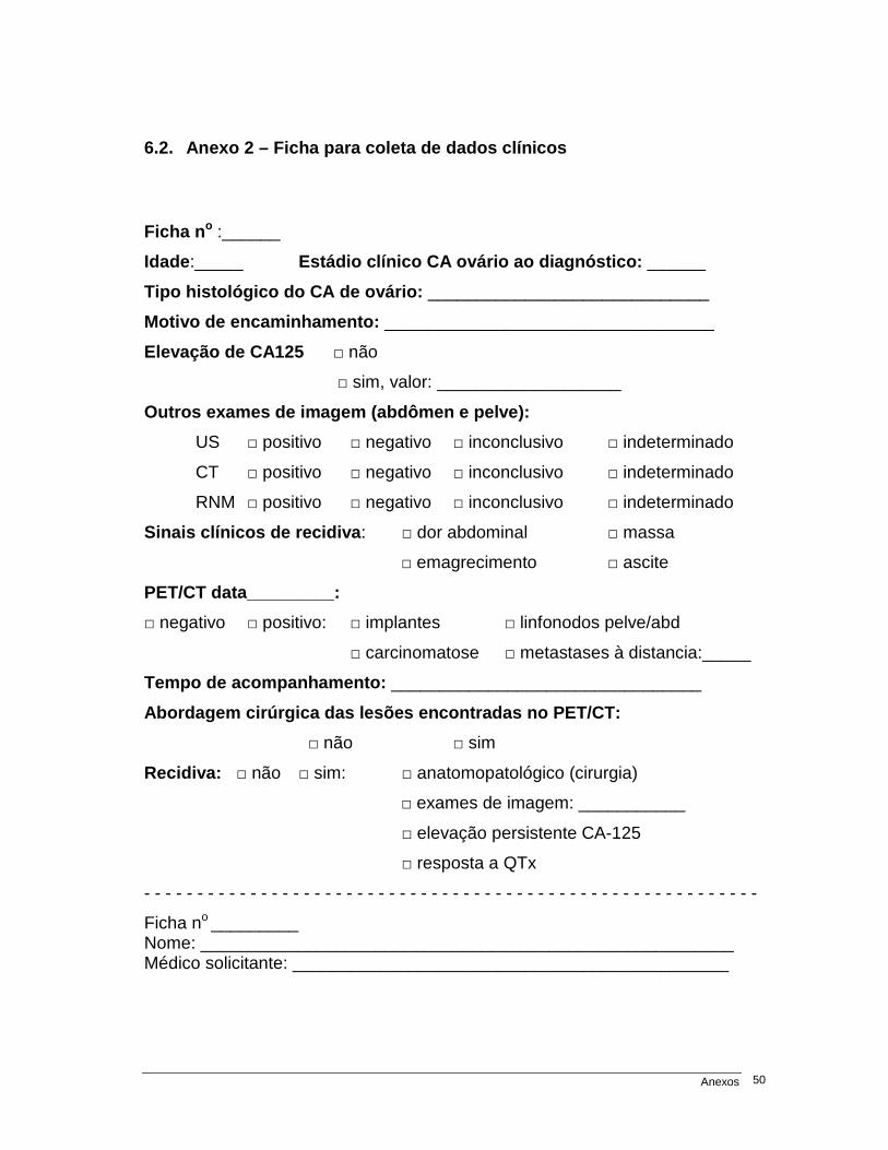
6.2. Anexo 2 – Ficha para coleta de dados clínicos
Ficha n o :______
Idade :_____ Estádio clínico CA ovário ao diagnóstico: ______
Tipo histológico do CA de ovário: _____________________________
Motivo de encaminhamento: __________________________________
Elevação de CA125 □ não
□ sim, valor: ___________________
Outros exames de imagem (abdômen e pelve):
US □ positivo □ negativo □ inconclusivo □ indeterminado
CT □ positivo □ negativo □ inconclusivo □ indeterminado
RNM □ positivo □ negativo □ inconclusivo □ indeterminado
Sinais clínicos de recidiva : □ dor abdominal □ massa
□ emagrecimento □ ascite
PET/CT data_________:
□ negativo □ positivo: □ implantes □ linfonodos pelve/abd
□ carcinomatose □ metastases à distancia:_____
Tempo de acompanhamento: ________________________________
Abordagem cirúrgica das lesões encontradas no PET/C T:
□ não □ sim
Recidiva: □ não □ sim: □ anatomopatológico (cirurgia)
□ exames de imagem: ___________
□ elevação persistente CA-125
□ resposta a QTx
- - - - - - - - - - - - - - - - - - - - - - - - - - - - - - - - - - - - - - - - - - - - - - - - - - - - - - - - - -
Ficha no _________ Nome: _______________________________________________________ Médico solicitante: _____________________________________________
Anexos 51
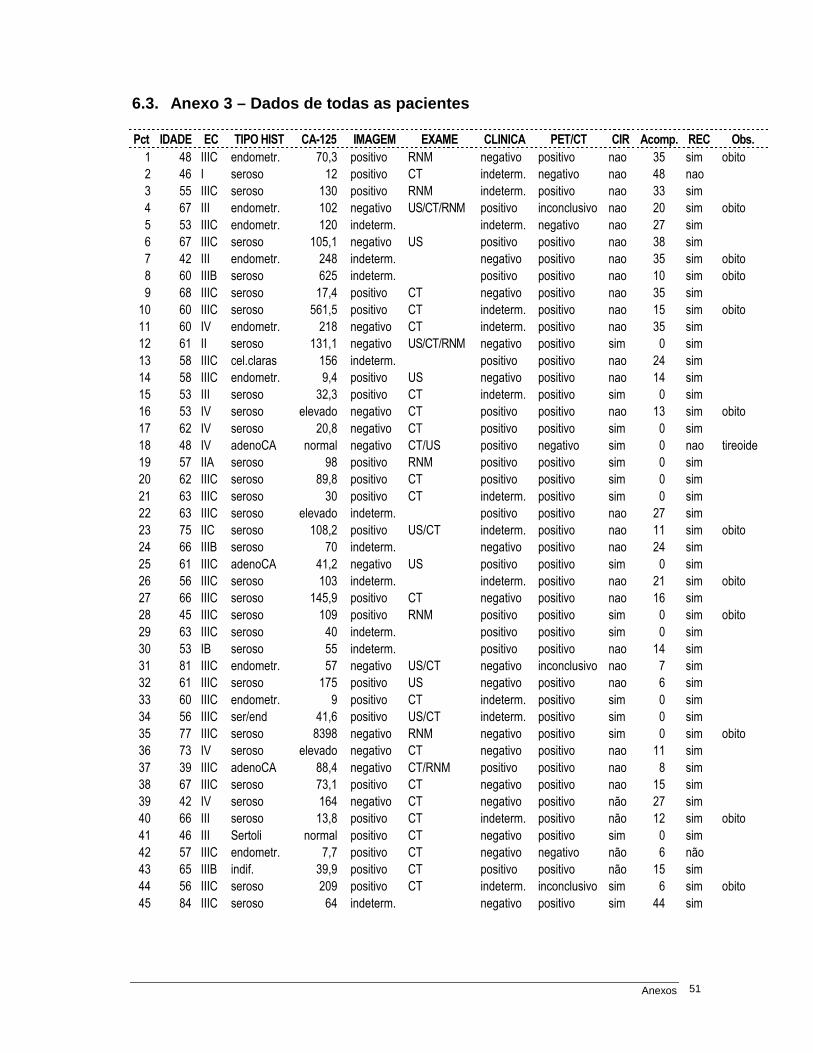
6.3. Anexo 3 – Dados de todas as pacientes
Pct IDADE EC TIPO HIST CA-125 IMAGEM EXAME CLINICA PET/CT CIR Acomp. REC Obs.
1 48 IIIC endometr. 70,3 positivo RNM negativo positivo nao 35 sim obito
2 46 I seroso 12 positivo CT indeterm. negativo nao 48 nao
3 55 IIIC seroso 130 positivo RNM indeterm. positivo nao 33 sim
4 67 III endometr. 102 negativo US/CT/RNM positivo inconclusivo nao 20 sim obito
5 53 IIIC endometr. 120 indeterm. indeterm. negativo nao 27 sim
6 67 IIIC seroso 105,1 negativo US positivo positivo nao 38 sim
7 42 III endometr. 248 indeterm. negativo positivo nao 35 sim obito
8 60 IIIB seroso 625 indeterm. positivo positivo nao 10 sim obito
9 68 IIIC seroso 17,4 positivo CT negativo positivo nao 35 sim
10 60 IIIC seroso 561,5 positivo CT indeterm. positivo nao 15 sim obito
11 60 IV endometr. 218 negativo CT indeterm. positivo nao 35 sim
12 61 II seroso 131,1 negativo US/CT/RNM negativo positivo sim 0 sim
13 58 IIIC cel.claras 156 indeterm. positivo positivo nao 24 sim
14 58 IIIC endometr. 9,4 positivo US negativo positivo nao 14 sim
15 53 III seroso 32,3 positivo CT indeterm. positivo sim 0 sim
16 53 IV seroso elevado negativo CT positivo positivo nao 13 sim obito
17 62 IV seroso 20,8 negativo CT positivo positivo sim 0 sim
18 48 IV adenoCA normal negativo CT/US positivo negativo sim 0 nao tireoide
19 57 IIA seroso 98 positivo RNM positivo positivo sim 0 sim
20 62 IIIC seroso 89,8 positivo CT positivo positivo sim 0 sim
21 63 IIIC seroso 30 positivo CT indeterm. positivo sim 0 sim
22 63 IIIC seroso elevado indeterm. positivo positivo nao 27 sim
23 75 IIC seroso 108,2 positivo US/CT indeterm. positivo nao 11 sim obito
24 66 IIIB seroso 70 indeterm. negativo positivo nao 24 sim
25 61 IIIC adenoCA 41,2 negativo US positivo positivo sim 0 sim
26 56 IIIC seroso 103 indeterm. indeterm. positivo nao 21 sim obito
27 66 IIIC seroso 145,9 positivo CT negativo positivo nao 16 sim
28 45 IIIC seroso 109 positivo RNM positivo positivo sim 0 sim obito
29 63 IIIC seroso 40 indeterm. positivo positivo sim 0 sim
30 53 IB seroso 55 indeterm. positivo positivo nao 14 sim
31 81 IIIC endometr. 57 negativo US/CT negativo inconclusivo nao 7 sim
32 61 IIIC seroso 175 positivo US negativo positivo nao 6 sim
33 60 IIIC endometr. 9 positivo CT indeterm. positivo sim 0 sim
34 56 IIIC ser/end 41,6 positivo US/CT indeterm. positivo sim 0 sim
35 77 IIIC seroso 8398 negativo RNM negativo positivo sim 0 sim obito
36 73 IV seroso elevado negativo CT negativo positivo nao 11 sim
37 39 IIIC adenoCA 88,4 negativo CT/RNM positivo positivo nao 8 sim
38 67 IIIC seroso 73,1 positivo CT negativo positivo nao 15 sim
39 42 IV seroso 164 negativo CT negativo positivo não 27 sim
40 66 III seroso 13,8 positivo CT indeterm. positivo não 12 sim obito
41 46 III Sertoli normal positivo CT negativo positivo sim 0 sim
42 57 IIIC endometr. 7,7 positivo CT negativo negativo não 6 não
43 65 IIIB indif. 39,9 positivo CT positivo positivo não 15 sim
44 56 IIIC seroso 209 positivo CT indeterm. inconclusivo sim 6 sim obito
45 84 IIIC seroso 64 indeterm. negativo positivo sim 44 sim
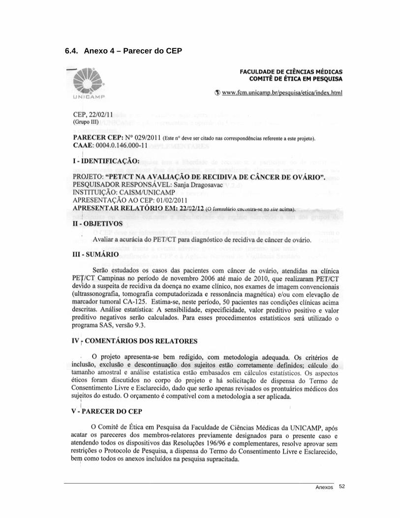
6.4. Anexo 4 – Parecer do CEPParecer do CEP
Anexos 52
Anexos 53