Mariana Roriz Abrantes - Universidade do...
95
outubro de 2013 Universidade do Minho Escola de Engenharia Mariana Roriz Abrantes Influence of antibiotics on Staphylococcus epidermidis biofilms UMinho|2013 Mariana Roriz Abrantes Influence of antibiotics on Staphylococcus epidermidis biofilms
Transcript of Mariana Roriz Abrantes - Universidade do...
outubro de 2013
Universidade do MinhoEscola de Engenharia
Mariana Roriz Abrantes
Influence of antibiotics on Staphylococcus epidermidis biofilms
UM
inho
|20
13M
aria
na R
oriz
Abr
ante
sIn
flu
en
ce o
f a
nti
bio
tics
on
Sta
phyl
oco
ccus
epid
erm
idis
bio
film
s
Dissertação de Mestrado Mestrado Integrado em Engenharia BiomédicaRamo de Engenharia Clínica
Trabalho efetuado sob a orientação daProfessora Doutora Mariana Contente Rangel HenriquesUniversidade do Minhoe doProfessor Doutor Johan Van EldereKatholieke Universiteit of Leuven
outubro de 2013
Universidade do MinhoEscola de Engenharia
Mariana Roriz Abrantes
Influence of antibiotics on Staphylococcus epidermidis biofilms

DECLARAÇÃO
Nome: Mariana Roriz Abrantes
Endereço electrónico: [email protected] Telefone: +351 916838465
Número do Bilhete de Identidade: 13768774
Título dissertação:
Influence of antibiotics on Staphylococcus epidermidis biofilms
Orientador(es):
Professora Doutora Mariana Contente Rangel Henriques
Professor Doutor Johan Van Eldere
Ano de conclusão: 2013
Designação do Mestrado:
Ciclo de Estudos Integrados Conducentes ao Grau de Mestre em Engenharia Biomédica
Área de Especialização: Engenharia Clínica
Escola: de Engenharia
Departamento: de Engenharia Biológica
DE ACORDO COM A LEGISLAÇÃO EM VIGOR, NÃO É PERMITIDA A REPRODUÇÃO DE QUALQUER PARTE DESTA TESE/TRABALHO
Universidade do Minho, ___/___/______
Assinatura: ______________________________________________
iii
Acknowledgments
Apart from the efforts of myself, the success of any project depends largely on the
encouragement and guidelines of many others. I take this opportunity to express my gratitude to
the people who have been instrumental in the successful completion of this project.
First of all, I would like to express my gratitude to my supervisors Dr. Mariana Henriques
and Dr. Johan Van Eldere, whose expertise, attention and understanding contributed considerably
to my dissertation work.
Above all, I am especially grateful to Dr. Mariana Henriques for all the support, dedication,
understanding and time put on my dissertation. All the enthusiasm and all the advices that she
gave me were very important in the project and essential to get to my dissertation-writing period. I
can’t thank her enough and this work would not have been possible without her support.
To Rita Merckx and Jolien Claessens for the time they dedicated to me, for the experience
and knowledge that shared with me, for the sympathy and availability demonstrated which proved
to be essential for my self-confidence, autonomy and organization in the experimental work.
I also have to thank to all my Erasmus friends that spend an amazing time with me. They
always provided me a stimulating and fun environment in which I learned and grew.
Last, but by no means least, I thank to my parents and to Paulo, as my solid anchors and
to my friends, as my “borrowed” family. I thank them with all my heart. They were all extremely
important to me during this dissertation period. For all the emotional support, understanding,
affection, and constant encouragement, I am deeply grateful. The knowledge that they will always
be there to pick me up, helping me and making smile at life is what allowed me to keep on moving
even in difficult times.

v
Influence of antibiotics on Staphylococcus epidermidis biofilms
Abstract
Staphylococcus epidermidis is a member of the coagulase-negative staphylococci and has
gained substantial interest in recent years because it has become the most frequently cause of
infections related to indwelling medical devices. Since S. epidermidis is resistant to some antibiotic
treatments, it is of major importance to understand the mechanisms and the bacterial components
involved in those mechanisms.
Therefore, the present dissertation aimed to understand the mechanisms and the bacterial
components involved in the antibacterial resistance mechanisms. The first goal of this work was
the evaluation of the activity of different groups of antibiotics, such as cell-wall and protein synthesis
inhibitors, on S. epidermidis biofilms. Changes on S. epidermidis biofilms were evaluated regarding
the total biofilms biomass, bacteria viability and biofilm’s matrix changes after treatment. The
second goal of this work was the evaluation of the influence of antibiotics on the matrix composition
considering the contribution of PIA and extracellular DNA to the changes referred.
Antibiotics treatment with the glycopeptides, vancomycin and teicoplanin, had an effect on
significantly increasing the total biofilm biomass on all strains. Even though these antibiotics were
effective against biofilm associated bacteria assessed by XTT, by DMMB staining method it become
obvious that they had an effect on increasing the amount of biofilms matrix. Thus imply that they
promoted the membrane rupture, enhancing PIA’s production and the formation of a rougher
biofilm. Results found on the combinated treatment of DNase I plus antibiotics indicated that
destruction of eDNA by DNase I leads to a decrease in the matrix, and as a result, antibacterial
agents protein inhibitors, such as rifampicin and gentamicin, act more effectively to reduce the
biofilm biomass.
In conclusion, it was possible to evaluate that antibiotic agents promoted alterations on S.
epidermidis biofilms. It was also possible to conclude that S. epidermidis biofilms from different
strains showed an enhanced resistance to the application of treatment with glycopeptide
antibiotics. Those antibiotics are normally reserved for use against multi-resistant staphylococci,
however from this study data, it is of major importance to have a better understanding of the
resistance mechanisms and to find different alternatives.

vi
vii
Table of Contents
Acknowledgments ................................................................................................................ iii
Abstract .................................................................................................................................. v
Table of Contents ................................................................................................................ vii
Abbreviations ........................................................................................................................ ix
List of Figures ....................................................................................................................... xi
List of Tables ........................................................................................................................ xv
Chapter 1 Introduction ........................................................................................................ 1
1.1 Genus Staphylococcus ............................................................................................... 3
1.2 Biofilm formation process .......................................................................................... 5
1.2.1 Adhesion ........................................................................................................... 6
1.2.2 Accumulation..................................................................................................... 7
1.2.3 Maturation and detachment ............................................................................. 10
1.3 Importance of S. epidermidis biofilms in clinical practice .......................................... 12
1.4 Treatment of biofilm related infections ..................................................................... 15
1.4.1 Antibiotic treatment.......................................................................................... 15
1.4.2 Antimicrobial resistance ................................................................................... 17
1.5 Aims and Objectives ................................................................................................ 21
Chapter 2 Materials and Methods ................................................................................... 23
2.1 Bacterial strains and growth conditions .................................................................... 25
2.2 Minimum inhibitory concentration determination ...................................................... 25
2.3 Biofilm formation ..................................................................................................... 26
2.4 Treatment of biofilms with antibiotics ....................................................................... 26
2.4.1 Treatment........................................................................................................ 26
2.4.2 Biofilm analysis................................................................................................ 26
2.5 Combined treatment of biofilms with DNase I and antibiotics ................................... 28
2.6 Extraction of eDNA .................................................................................................. 28
2.7 PIA dot-blot ............................................................................................................. 29
2.7.1 gDNA extraction for detection of icaA operon .................................................... 30
2.8 Scanning electron microscopy ................................................................................. 31
2.9 Statistical analysis ................................................................................................... 31
Chapter 3 Results ............................................................................................................... 33
3.1 MIC determination ................................................................................................... 35
3.2 Evaluation of activity of antibiotics on biofilms .......................................................... 36
viii
3.3 Influence of antibiotic treatment on biofilm matrix DNA and PIA ................................ 41
3.3.1 Extracellular DNA ............................................................................................. 41
3.3.2 PIA (PIA dot-blot).............................................................................................. 44
Chapter 4 Discussion ......................................................................................................... 47
4.1 Antibiotic susceptibility ............................................................................................ 49
4.2 Evaluation of antibiotics activity on biofilms .............................................................. 51
4.3 Influence of antibiotic treatment on biofilm matrix DNA and PIA ................................ 55
Chapter 5 Conclusion and future perspectives .............................................................. 59
Chapter 6 References ........................................................................................................ 63
Appendix .............................................................................................................................. 75
Appendix 1: Clinical Breakpoints defined by EUCAST ........................................................... 75
ix
Abbreviations
agr – Accessory gene regulator
ANOVA – Analysis of Variance
AtlE – Autolysin E (E indicates its origin in S. epidermidis)
BSA - Bovine serum albumin
CoNS – Coagulase-negative Staphylococcus spp.
CV – Crystal violet
DMMB - 1,9-Dimethyl-Methylene Blue zinc chloride double salt
DNA – Deoxyribonucleic acid
eDNA – Extracellular Deoxyribonucleic acid
EDTA – Etgylenediaminetetraacetic acid
EPS – Extracellular polymeric substance
gDNA – Genomic Deoxyribonucleic acid
Genta – Gentamicin
h - Hours
HMDS – Hexamethyldisilazane
ica – Intercellular adhesion
MIC - Minimum inhibitory concentration
min – Minutes
OD – Optical density
Oxa – Oxacillin
P – Significance value
PBS - Phosphate buffered saline
PCR – Polymerase chain reaction
PIA - Polysaccharide intercellular adhesion
Rif – Rifampicin
s – Seconds
SD – Standard deviation
SDS - Sodium dodecyl sulphate
x
SEM - Scanning electron microscopy
TBS – Tris buffer saline
Teico – Teicoplanin
TSB – Tryptic soy broth
TTBS – Tris buffer saline with 0.4% Tween
Vanco – Vancomycin
Vs – Versus
WGA – Wheat germ agglutinin
XTT - 2,3-Bis-(2-Methoxy-4-Nitro-5-Sulfophenyl)-2H-Tetrazolium-5-Carboxanilide
Influence of antibiotics on Staphylococcus epidermidis biofilms
xi
List of Figures
Figure 1 Staphylococcus (a) Gram-stained (adapted) [5]. (b) Staphylococci organized like a grape
cluster (adapted) [6]. ................................................................................................................ 3
Figure 2 Scanning electron micrograph of a Staphylococcus biofilm on the inner surface of a
needleless connector[75]. ......................................................................................................... 5
Figure 3 Schematic overview of the phases involved in Staphylococcus epidermidis biofilm
formation on a surface [76]. ..................................................................................................... 6
Figure 4 Structure of PIA [12]. .................................................................................................. 8
Figure 5 The exopolysaccharide poly-N-acetylglucosamine. a) The exopolysaccharide poly-N-
acetylglucosamine (PIA), a partially de-acetylated β1-6-linked N-acetylglucosamine (GlcNAc)
homopolymer involved in immune evasion and biofilm aggregation, is synthesized by the
membrane-located GlcNAc transferase IcaA, which needs the accessory IcaD membrane protein
for activity (step 1). The growing PNAG chain is probably exported by the IcaC membrane protein
(step 2). After export, IcaB de-acetylase, located on the cell surface, removes some of the N-acetyl
groups, giving the polymer a cationic character that is essential for surface attachment (step 3). b)
The Ica proteins are encoded by the ica gene locus containing the icaADBC operon and the icaR
gene, which encodes a regulatory protein. Expression of the icaADBC operon is regulated either
directly at the icaA promoter or through expression of IcaR (adapted) [17]. ................................ 9
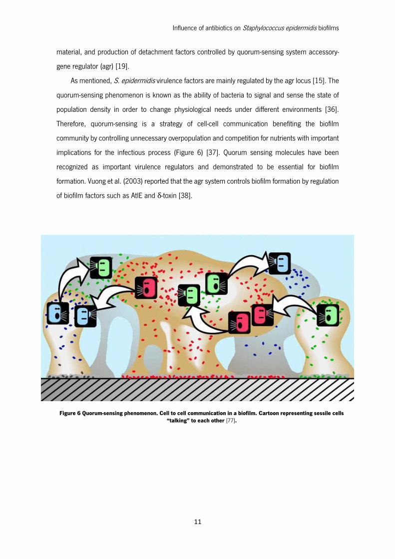
Figure 6 Quorum-sensing phenomenon. Cell to cell communication in a biofilm. Cartoon
representing sessile cells “talking” to each other [77].............................................................. 11
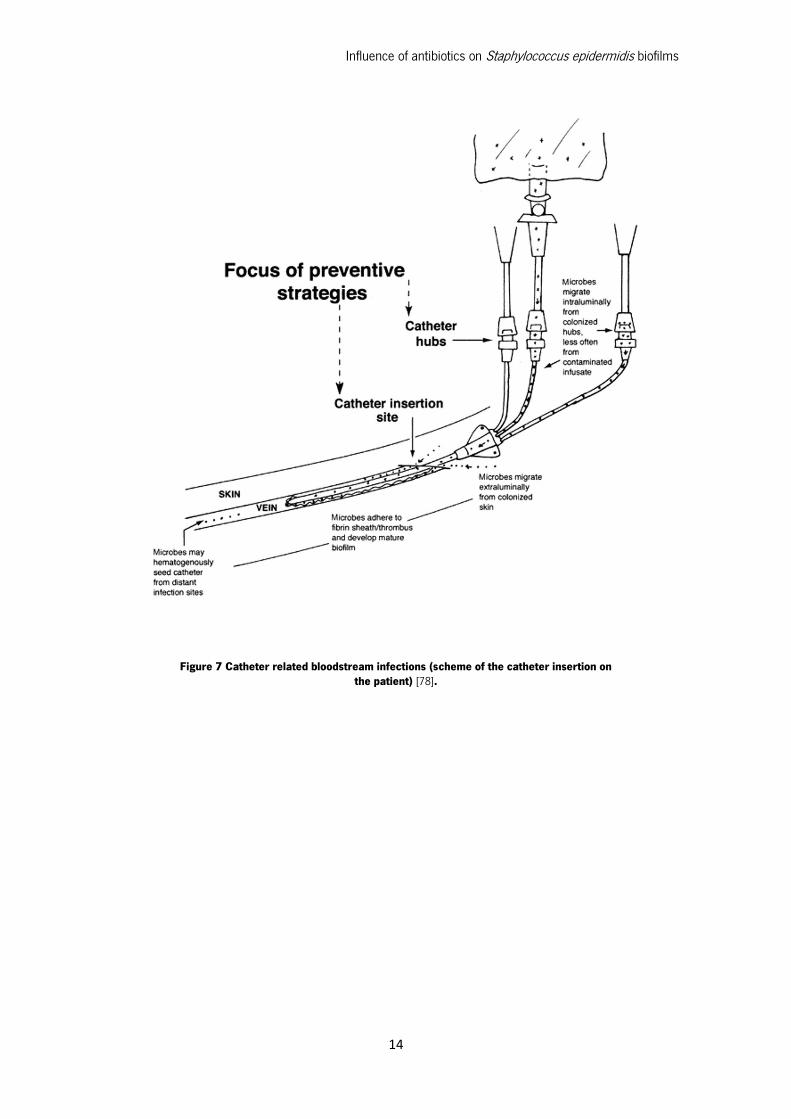
Figure 7 Catheter related bloodstream infections (scheme of the catheter insertion on the patient)
[78]. ....................................................................................................................................... 14
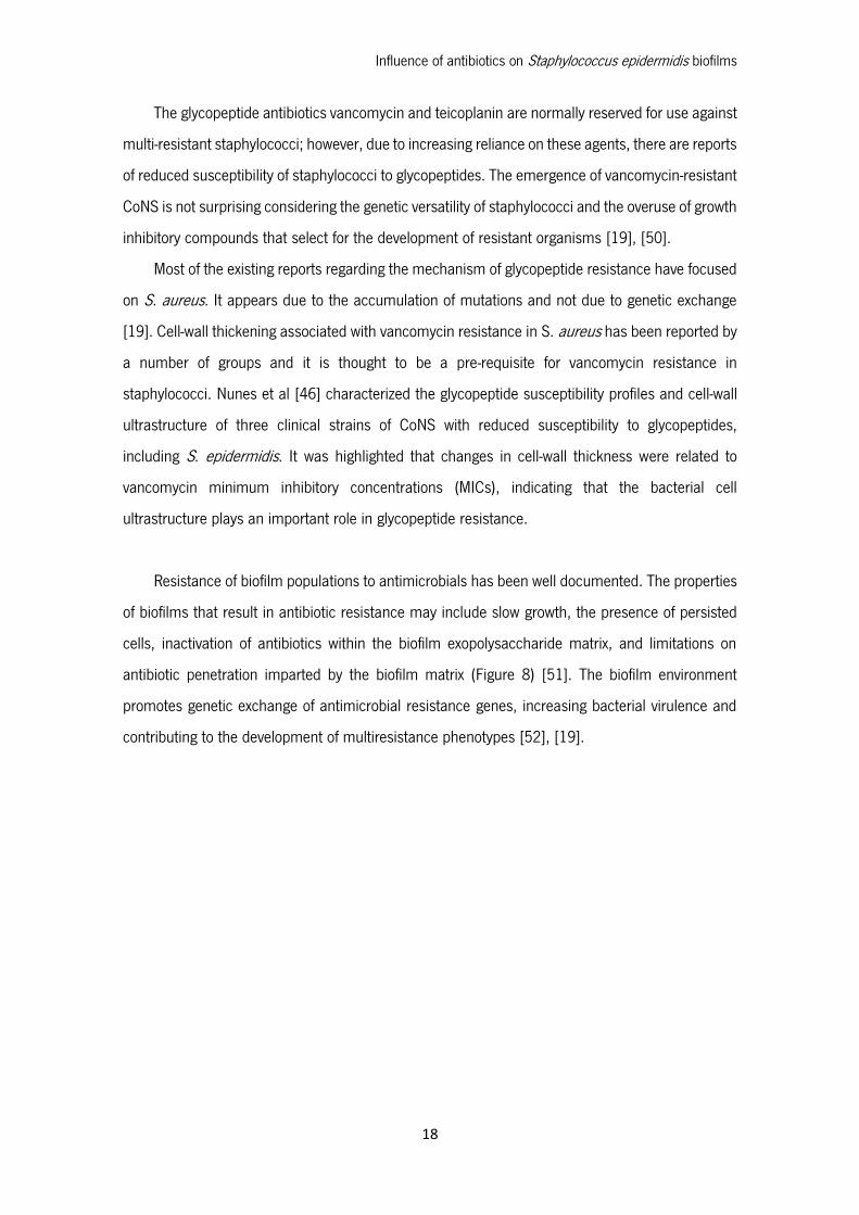
Figure 8 Antimicrobial agents resistance- schematic comparison between planktonic cell and
biofilms (adapted). A- Fee-floating cells utilize nutrients but do not have sufficient metabolic activity
to deplete substrates from the neighborhood of the cells. In contrast, the collective metabolic
activity of groups of cells in the biofilm leads to substrate concentration gradients and localized
chemical environments. Reduced metabolic activity may result in less susceptibility to
antimicrobials. B- Free-floating cells carry the genetic code for numerous protective stress
Influence of antibiotics on Staphylococcus epidermidis biofilms
xii
responses. Planktonic cells, however, are readily overwhelmed by a strong antimicrobial challenge.
These cells die before stress responses can be activated. In contrast, stress responses are
effectively implemented in some of the cells in a biofilm at the extense of other cells which are
sacrificed. C- Free-floating cells neutralize the antimicrobial agent. The capacity of a lone cell,
however, is insufficient to draw down the antimicrobial concentration in the neighborhood of the
cell. In contrast, the collective neutralizing power of groups of cells leads to slow or incomplete
penetration of the antimicrobial in the biofilm. D- Free-floating cells spawn protected persister cells.
But under permissive growth conditions in a planktonic culture, persisters rapidily revert to a
susceptible state. In contrast, persister cells accumulate in biofilms because they revert less and
readily and are physically retained by the biofilm matrix [79]. ................................................. 19
Figure 9 Absorbance values of S. epidermidis (Se) 1457, Se 1457atlE mutant, Se 10b, Se 567
and Se 567-1 biofilms stained with crystal violet after treatment with vancomycin (Vanco),
teicoplanin (Teico), oxacillin (Oxa), rifampicin (Rif) and gentamycin (Genta). Absorbance was
measured at 590nm. Error bars represent 95% confidence interval. Statistical differences
compared to control (TSB 1/100) are marked with an asterisk (one-way ANOVA, p<0.05). ...... 36
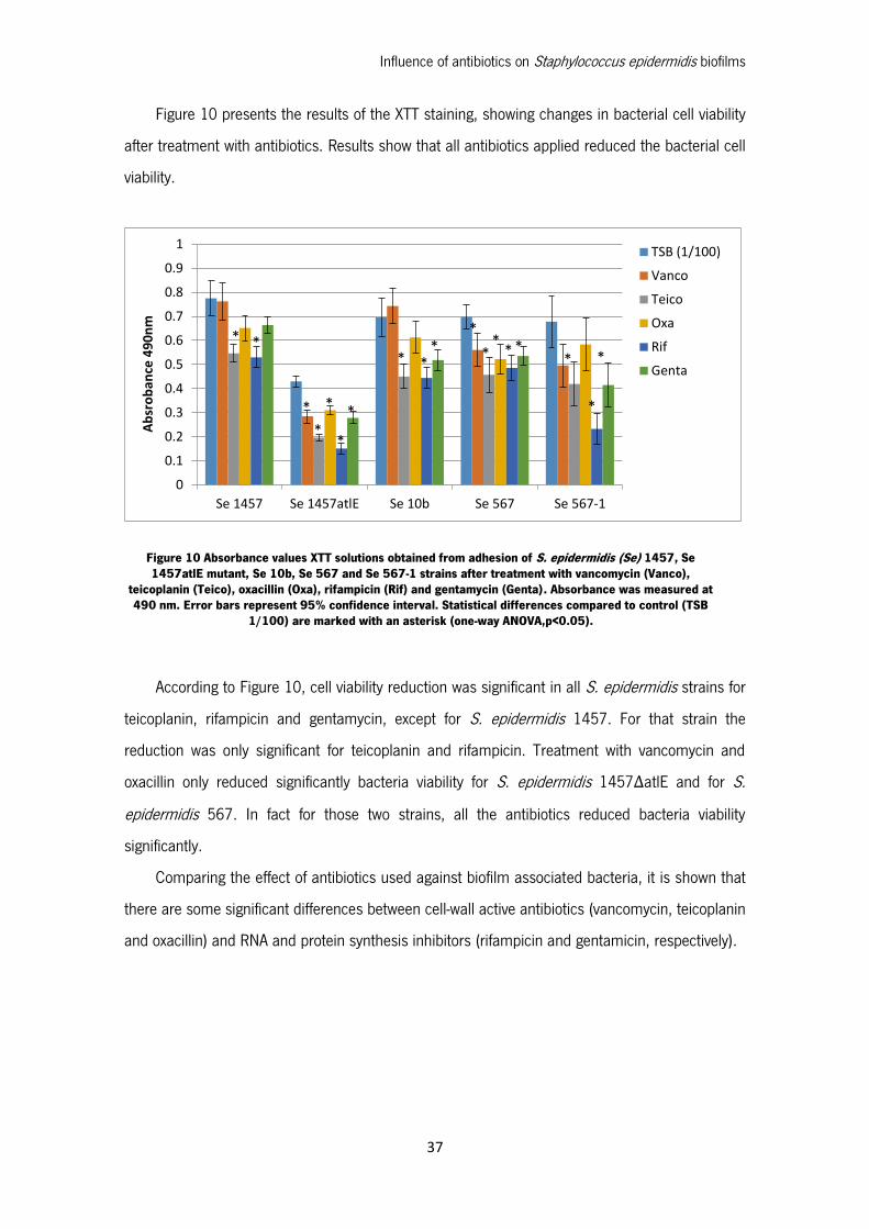
Figure 10 Absorbance values XTT solutions obtained from adhesion of S. epidermidis (Se) 1457,
Se 1457atlE mutant, Se 10b, Se 567 and Se 567-1 strains after treatment with vancomycin
(Vanco), teicoplanin (Teico), oxacillin (Oxa), rifampicin (Rif) and gentamycin (Genta). Absorbance
was measured at 490 nm. Error bars represent 95% confidence interval. Statistical differences
compared to control (TSB 1/100) are marked with an asterisk (one-way ANOVA,p<0.05). ....... 37
Figure 11 Absorbance values DMMB solutions obtained from adhesion of S. epidermidis (Se) 1457,
Se 1457atlE mutant, Se 10b, Se 567 and Se 567-1 strains after treatment with vancomycin
(Vanco), teicoplanin (Teico), oxacillin (Oxa), rifampicin (Rif) and gentamycin (Genta). Absorbance
was measured at 630 nm. Error bars represent 95% confidence interval. Statistically different
compared to control (TSB 1/100) (one-way ANOVA, p<0.05). ................................................. 38
Figure 12 Representative scanning electronic microscopy photographs of S. epidermidis 1457
biofilms after treatment with: lane I- control (TSB 1/100); II- Vancomycin; III- Teicoplanin; IV-
Oxacillin; V- Rifampicin and VI- Gentamicin. Experiments were performed in triplicate and biofilms
were viewed at 1000x magnification (column a), 4000x magnification (column b) and 10000x
magnification (column c)......................................................................................................... 39
Influence of antibiotics on Staphylococcus epidermidis biofilms
xiii
Figure 13 Percentual reduction of biofilm biomass assayed with CV staining. Absorbance reduction
was compared between antibiotic treated (AB) and DNaseI combined with antibiotic treated biofilms
(DNaseI+AB). Antibiotics (AB) used were Vanco – vancomycin; Teico- teicoplanin; Rif- rifampicin;
Oxa- oxacillin and Genta- gentamicin. ...................................................................................... 41
Figure 14 Gel electrophoresis of extracellular DNA extracted from all S. epidermidis strains used
grown only in culture medium (Se – S. epidermidis). ............................................................... 42
Figure 15 Gel electrophoresis of extracellular DNA extracted from different strains (Se – S.
epidermidis). Lanes represent the treatments used: 1- TSB (1/100); 2- Vancomicin; 3- Teicoplanin;
4- Oxacillin; 5- Rifampicin; 6-Gentamicin.................................................................................. 43
Figure 16 Relative amounts of PIA on biofilms were determined using a PIA dot blot assay after
treatment with antibiotics: Vanco- Vancomycin; Teico- Teicoplanin; Oxa- Oxacillin; Rif- Rifampicin
and Genta- Gentamicin. To determine the relative amounts of PIA, it was defined that the amount
of PIA produced by each strain individually when treated with TSB (1/100) as 100% and expressed
all other amounts as relative to the amount on that TSB (1/100) treatment. ............................ 44
Figure 17 Representative S. epidermidis 1457 dot blot for determining spot volumes on each
treatment: lane TSB (1/100)- control; Vanco- Vancomycin; Teico- Teicoplanin; Oxa- Oxacillin; Rif-
Rifampicin and Genta- Gentamicin. Columns represent the different dilutions used: 1- 1/10; 2-
1/50; 3- 1/100. ..................................................................................................................... 45
Figure 18 Gel electrophoresis result of S. epidermidis 1457 and S. epidermidis 1457 ΔatlE PCR
on the icaA gene to check for its presence. ............................................................................. 46

Influence of antibiotics on Staphylococcus epidermidis biofilms
xiv
Influence of antibiotics on Staphylococcus epidermidis biofilms
xv
List of Tables
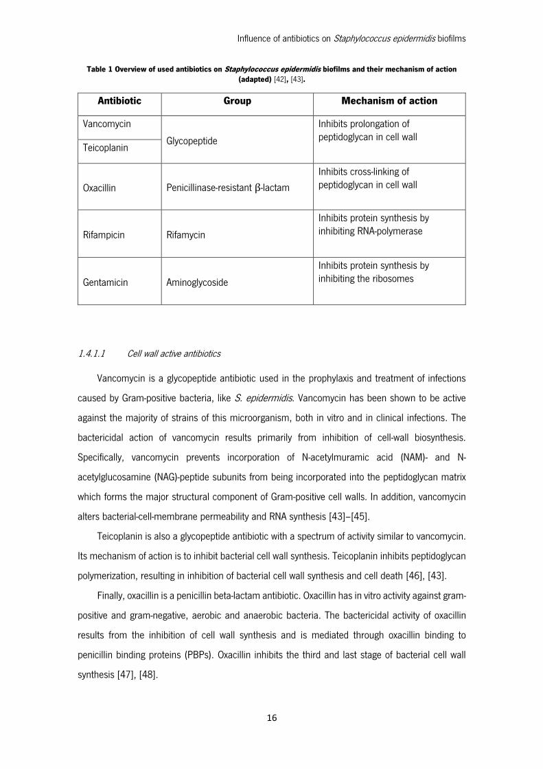
Table 1 Overview of used antibiotics on Staphylococcus epidermidis biofilms and their mechanism
of action (adapted) [42], [43]. ................................................................................................. 16
Table 2 Primers used in this study for icaA. ............................................................................. 30
Table 3 Determination of antibiotic susceptibilities by Staphylococcus epidermidis used strains in
planktonic cultures (MIC) ........................................................................................................ 35

Influence of antibiotics on Staphylococcus epidermidis biofilms
xvi
Influence of antibiotics on Staphylococcus epidermidis biofilms
1
Chapter 1 Introduction
Influence of antibiotics on Staphylococcus epidermidis biofilms
2
Influence of antibiotics on Staphylococcus epidermidis biofilms
3
1.1 Genus Staphylococcus
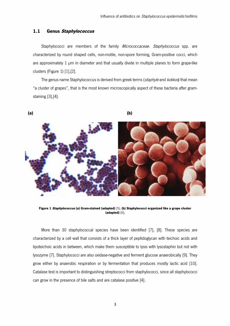
Staphylococci are members of the family Micrococcaceae. Staphylococcus spp. are
characterized by round shaped cells, non-motile, non-spore forming, Gram-positive cocci, which
are approximately 1 µm in diameter and that usually divide in multiple planes to form grape-like
clusters (Figure 1) [1],[2].
The genus name Staphylococcus is derived from greek terms (staphyle and kokkos) that mean
“a cluster of grapes”, that is the most known microscopically aspect of these bacteria after gram-
staining [3],[4].
(a) (b)
Figure 1 Staphylococcus (a) Gram-stained (adapted) [5]. (b) Staphylococci organized like a grape cluster (adapted) [6].
More than 30 staphylococcal species have been identified [7], [8]. These species are
characterized by a cell wall that consists of a thick layer of peptidoglycan with teichoic acids and
lipoteichoic acids in between, which make them susceptible to lysis with lysostaphin but not with
lysozyme [7]. Staphylococci are also oxidase-negative and ferment glucose anaerobically [9]. They
grow either by anaerobic respiration or by fermentation that produces mostly lactic acid [10].
Catalase test is important to distinguishing streptococci from staphylococci, since all staphylococci
can grow in the presence of bile salts and are catalase positive [4].
Influence of antibiotics on Staphylococcus epidermidis biofilms
4
Staphylococci can be divided into two large groups based on the ability to produce the enzyme
coagulase that coagulates plasma. The first group, known as coagulase-positive staphylococci, is
mainly represented by S. aureus and S. intermedius, which can cause a variety of infections ranging
from cutaneous to systemic infections. The second group is known as coagulase-negative
staphylococci (CoNS) [11], [12]. They are normal inhabitants of the upper respiratory tract, skin,
vagina and intestine, being among the most important bacteria that causes various infections in
humans (e.g., folliculitis, scalded-skin syndrome and boils) [9].
CoNS are among the most commonly isolated organisms in the clinical microbiology
laboratory. Owing to their ubiquitous nature and relative low virulence, CoNS have for a long time
been considered to be clinically insignificant pathogens [1], [13]. Due to the widespread use of
implanted medical devices, in recent decades, these bacteria are now considered as important
causative agents of nosocomial bacteremias, infections of indwelling devices [14].
Staphylococcus epidermidis is currently the most significant member of the CoNS group. S.
epidermidis is a Gram-positive bacteria, found on skin and mucous membranes of humans and
other organisms, representing an important part of its normal microflora, and includes 65 to 90%
of all staphylococci isolated from these environments [4], [15], [16].
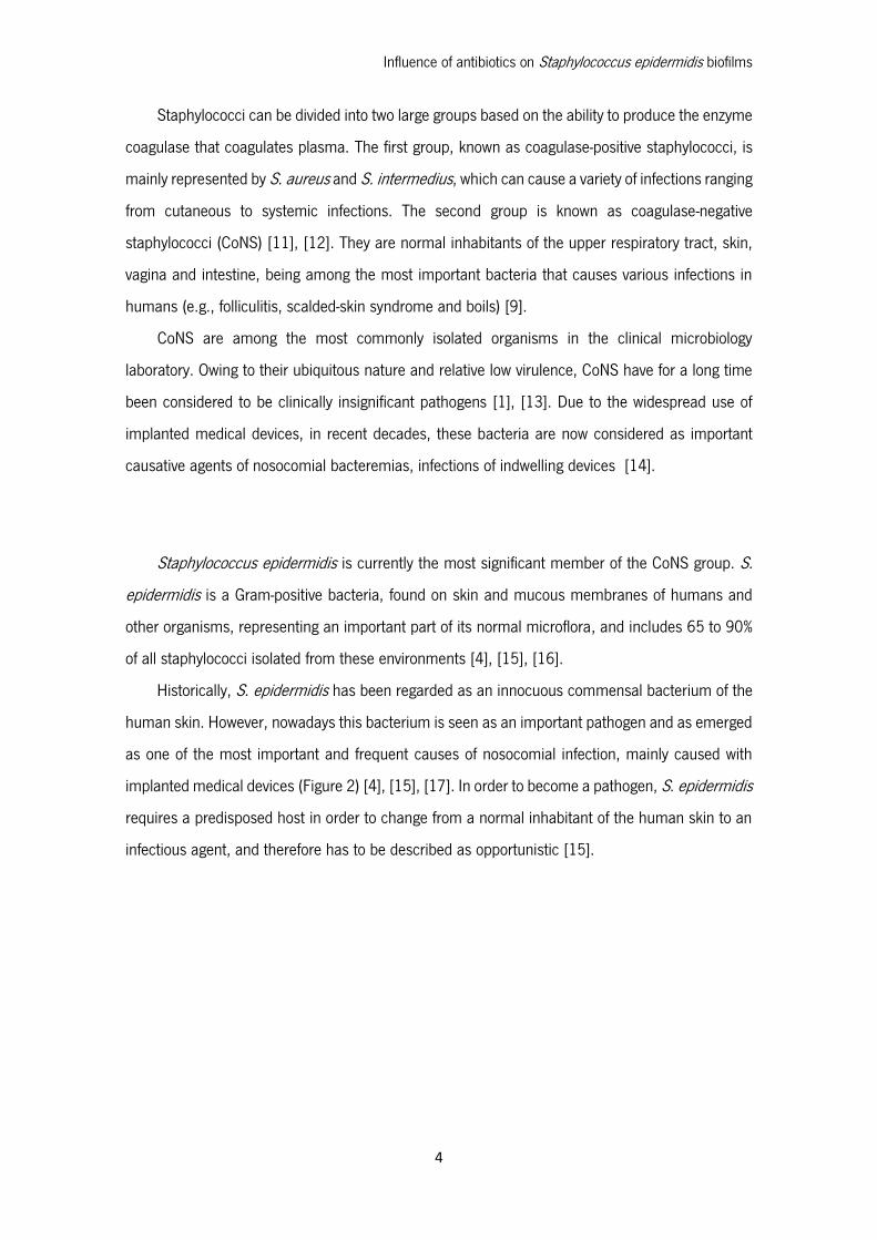
Historically, S. epidermidis has been regarded as an innocuous commensal bacterium of the
human skin. However, nowadays this bacterium is seen as an important pathogen and as emerged
as one of the most important and frequent causes of nosocomial infection, mainly caused with
implanted medical devices (Figure 2) [4], [15], [17]. In order to become a pathogen, S. epidermidis
requires a predisposed host in order to change from a normal inhabitant of the human skin to an
infectious agent, and therefore has to be described as opportunistic [15].
Influence of antibiotics on Staphylococcus epidermidis biofilms
5
1.2 Biofilm formation process
In 1987, Costerton et al. defined a biofilm as the accumulation of microorganisms and their
extracellular products to form a highly structured bacterial community on a surface [18]. Biofilm
formation as a multi-step process is considered the most important virulence factor of S.
epidermidis [14]. Biofilm formation occurs in four distinct phases – attachment (adhesion),
accumulation, maturation and detachment [19]. The process starts with the initial adhesion of cells
to a surface and their subsequent maturation phase. Detachment of cells or cell clusters from an
existing biofilm can lead to the dissemination of the infection (Figure 3).
Figure 2 Scanning electron micrograph of a Staphylococcus biofilm on the inner surface of a needleless connector[75].
Influence of antibiotics on Staphylococcus epidermidis biofilms
6
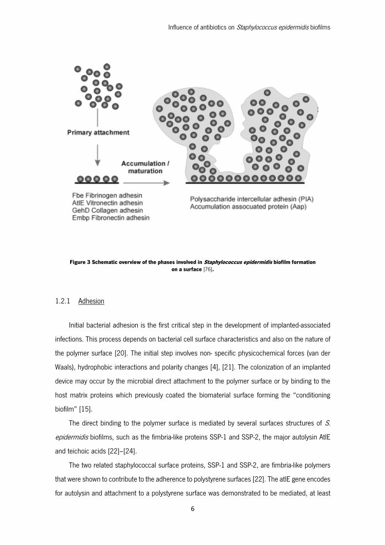
1.2.1 Adhesion
Initial bacterial adhesion is the first critical step in the development of implanted-associated
infections. This process depends on bacterial cell surface characteristics and also on the nature of
the polymer surface [20]. The initial step involves non- specific physicochemical forces (van der
Waals), hydrophobic interactions and polarity changes [4], [21]. The colonization of an implanted
device may occur by the microbial direct attachment to the polymer surface or by binding to the
host matrix proteins which previously coated the biomaterial surface forming the “conditioning
biofilm” [15].
The direct binding to the polymer surface is mediated by several surfaces structures of S.
epidermidis biofilms, such as the fimbria-like proteins SSP-1 and SSP-2, the major autolysin AtlE
and teichoic acids [22]–[24].
The two related staphylococcal surface proteins, SSP-1 and SSP-2, are fimbria-like polymers
that were shown to contribute to the adherence to polystyrene surfaces [22]. The atlE gene encodes
for autolysin and attachment to a polystyrene surface was demonstrated to be mediated, at least
Figure 3 Schematic overview of the phases involved in Staphylococcus epidermidis biofilm formation on a surface [76].
Influence of antibiotics on Staphylococcus epidermidis biofilms
7
in part, by the surface-associated autolysin AtlE. AtlE is active during the attachment to both
conditioned and unconditioned polymer surfaces [25].
Aside from proteins, polysaccharide intercellular adhesin molecule (PIA), has been also
associated with the initial adherence and slime production [25]. In fact, for a long time studies
were prevalently centered on PIA of S. epidermidis biofilms, considered the major cell-to-cell
connecting substance. However, recently the attention has been progressively focused also on
extracellular DNA, another conspicuous component of the biofilm matrix [26]. The atlE gene,
mentioned above, is necessary for both attachment and biofilm development. Since there is no
evidence that AtlE protrudes from the bacterial cell surface, rather than a direct role in primary
attachment, this autolysin may have an indirect role in S. epidermidis biofilm formation by releasing
of eDNA. In concordance with this hypothesis is the finding that in cultures of atlE-defective S.
epidermidis mutants the amount of eDNA is dramatically decreased when compared with its level
in the wild-type strains [27].
Finally, also the host contributes to adhesion in device-related infections particularly with
staphylococci, as mentioned above. Multiple specific receptors on the cell surface, called adhesins,
bind to the host molecules, such as glycoprotein components in plasma or components of the host
extracellular matrix. Many of these proteins belong to a family of microbial surface components
that recognize adhesive matrix molecules (MSCRAMMs). MSCRAMMs mediate adhesion to various
host cell types as well as to polymer surfaces coated with host plasma proteins [12].
1.2.2 Accumulation
Following microbial adherence to the implanted medical device, bacteria proliferate and
accumulate as multilayered cell clusters, resulting in an extensive network of accumulated bacteria
[19]. The biofilm accumulation involves intercellular aggregation that is mediated by intercellular
adhesins. These include surface macromolecules such as exopolysaccharide, surface proteins,
teichoic acids and extracellular DNA originating from lysed cells which are involved in formation of
extracellular biofilm matrix [14].
In Staphylococcus spp., production of PIA by the genes in the intercellular adhesion (ica)
operon, is nowadays the best-understood mechanism (ica or PIA-dependent mechanism) of biofilm
formation [14], [28]. PIA is a major component of the extracellular staphylococcal carbohydrate
Influence of antibiotics on Staphylococcus epidermidis biofilms
8
matrix of S. epidermidis [19]. Mack et al. described PIA as a major functional component involved
in the intercellular adhesion, essential for the accumulation of multilayered S. epidermidis biofilms
[29].
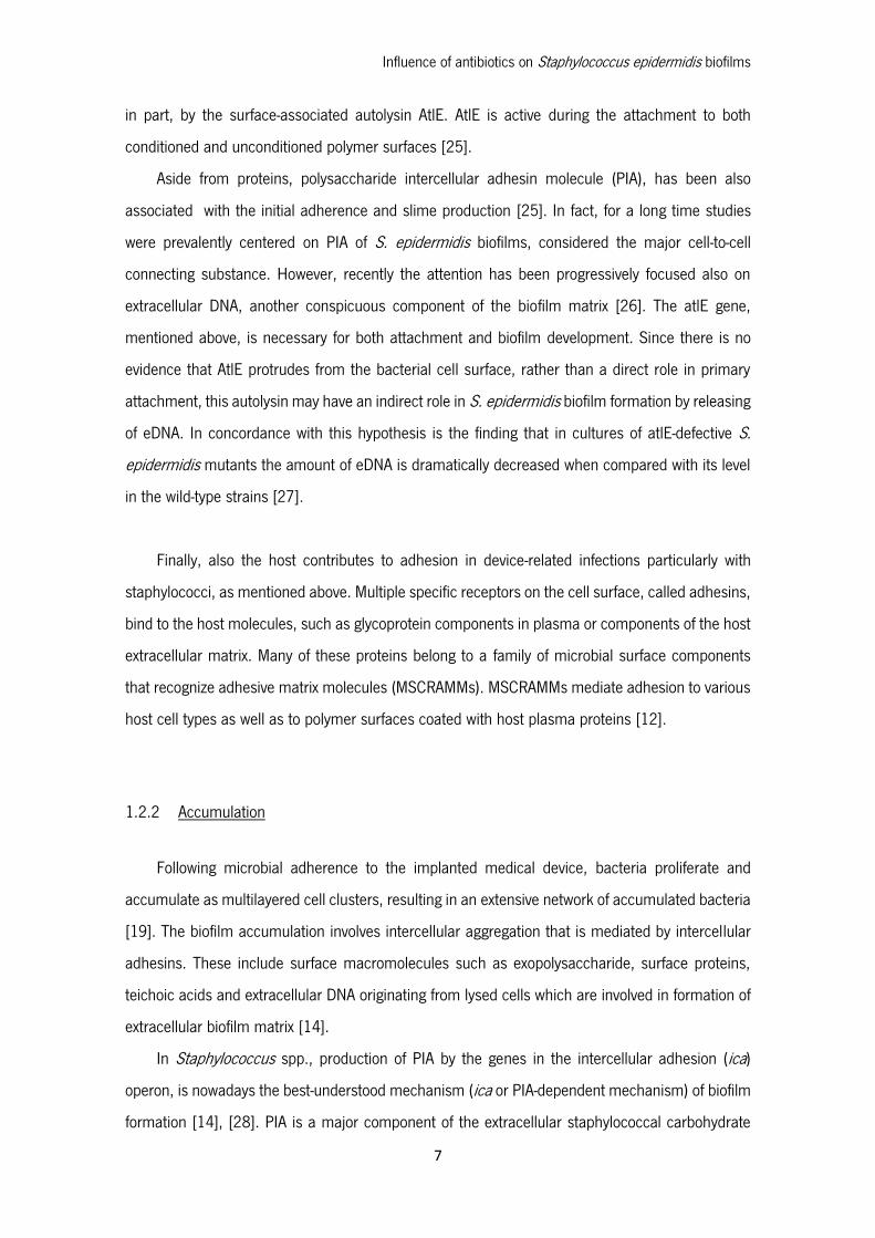
PIA is a linear β-1,6-linked glucosaminoglycan, composed of β-1,6-linked N-acetylglucosamine
residues containing up to 15% de-N-acetylated amino groups and positively charged (Figure 4).
These positive charges have been shown to have a major biological importance in biofilm formation,
virulence and immune evasion [19], [30].
PIA is synthesized by enzymes encoded by the intercellular adhesion (ica) operon, which is
more prevalent in colonizing S. epidermidis isolates [19]. The ica locus is composed of an operon,
icaADBC, which encodes the structural genes required for PIA synthesis. The ica operon comprises
4 genes: icaA, icaD, icaB and icaC. A fifth gene, the divergently transcribed icaR gene, responsible
for the transcription of icaADBC is located upstream of the icaA start codon. IcaR gene is a member
of the TetR family of transcriptional regulators and negatively regulates icaADBC expression [14].
The icaA gene product is responsible for N-acetylglucosaminyltransferase activity during PIA
synthesis. IcaA adds N-acetylglucosamine from UPD-N-acetylglucosamine to the growing PIA chain,
requiring the icaD gene product for optimal transferase activity. N-acetylglucosamine oligomers
produced by icaAD reach a maximal length of 20 residues. Only when icaAD is coexpressed with
icaC, longer oligomer chains are synthesized. The transmembrane protein IcaC plays a putative
role in externalization, elongation and translocation of the growing polysaccharide to the cell surface
and is responsible for the production of full-length PIA molecule [19]. The icaB gene product is a
Figure 4 Structure of PIA [12].
Influence of antibiotics on Staphylococcus epidermidis biofilms
9
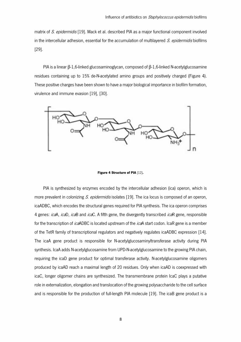
surface-attached protein responsible for decacetylation of the poly-N-acetylglucosamine molecule
(Figure 5).
The ica locus and biofilm formation are important parameters for staphylococcal colonization
and survival on implanted medical devices. However, recent publications have revealed the
emergence of biofilm-positive and ica-negative staphylococcal clinical isolates [31]. In cases of PIA-
negative biofilm formation, adhesive proteins have been suggested to be involved in the
accumulation phase. In some strains (ica-positive or negative), biofilm formation is mediated by
specific surface proteins such as accumulation-associated protein (Aap), biofilm-associated protein
Figure 5 The exopolysaccharide poly-N-acetylglucosamine. a) The exopolysaccharide poly-N-acetylglucosamine (PIA), a partially de-acetylated β1-6-linked N-acetylglucosamine (GlcNAc) homopolymer
involved in immune evasion and biofilm aggregation, is synthesized by the membrane-located GlcNAc transferase IcaA, which needs the accessory IcaD membrane protein for activity (step 1). The growing PNAG chain is probably exported by the IcaC membrane protein (step 2). After export, IcaB de-acetylase, located on
the cell surface, removes some of the N-acetyl groups, giving the polymer a cationic character that is essential for surface attachment (step 3). b) The Ica proteins are encoded by the ica gene locus containing
the icaADBC operon and the icaR gene, which encodes a regulatory protein. Expression of the icaADBC operon is regulated either directly at the icaA promoter or through expression of IcaR (adapted) [17].
Influence of antibiotics on Staphylococcus epidermidis biofilms
10
(Bap), or Bap homologue (Bhp) [19]. Biochemical and functional properties clearly differentiate
Aap from other factors that have been implicated in biofilm formation. It was proposed that Aap
has a role in the anchoring of PIA to cell surface [32].
1.2.3 Maturation and detachment
Maturation of the S. epidermidis biofilm is characterized by the generation of a slime
glycocalix, which encases surface-bound organisms in a gelatinous matrix. The slime
exopolysaccharide is not essential to the overall process of surface colonization, it is thought to
increase the stability of the biofilm and therefore contribute to a more robust structure and making
implanted medical devices colonized with slime-positive strains more difficult to treat.
A mature biofilm is seen as a very heterogeneous arrangement, with a basic community
structure which comprises several layers, including the main bulk of the biofilm, a linking film, a
conditioning film and the substratum to which the biofilm is attached [19], [33]. The mature
structure reveals groups of micro colonies of bacterial cells encased in extracellular polymeric
substance matrix [34]. The matrix is one of the most distinctive features of microbial biofilm where,
in addition to PIA and protein, extracellular DNA has also been shown to be important in stabilizing
the biofilm [35].
Mature biofilms form a three-dimensional, gel-like, highly hydrated and locally charged
environment where microorganisms are largely immobilized. Matrix-enclosed micro colonies,
sometimes described as mushroom-like forms are separated by fluid-filled channels [34]. Liquid
flow in these channels, allowing diffusion of nutrients and oxygen to all cells in the biofilm [19].
At last, individual bacterial cells, capable of actively leaving a biofilm, can arise and spread
from the surface film on the outer side of the mature biofilm to colonies in distant sites. The
dispersion of virulent staphylococci has important implications from S. epidermidis biofilm
infections. Even if cells actively detaching from the biofilm may colonize alternative sites, it can
also contribute to the toxicology associated with acute infections [19].
In contrast to primary attachment and accumulation, detachment is not well understood.
However, several factors have been proposed to be involved in biofilm detachment including:
mechanical forces, changes in nutrient concentrations, cessation of production of biofilm building
Influence of antibiotics on Staphylococcus epidermidis biofilms
11
material, and production of detachment factors controlled by quorum-sensing system accessory-
gene regulator (agr) [19].
As mentioned, S. epidermidis virulence factors are mainly regulated by the agr locus [15]. The
quorum-sensing phenomenon is known as the ability of bacteria to signal and sense the state of
population density in order to change physiological needs under different environments [36].
Therefore, quorum-sensing is a strategy of cell-cell communication benefiting the biofilm
community by controlling unnecessary overpopulation and competition for nutrients with important
implications for the infectious process (Figure 6) [37]. Quorum sensing molecules have been
recognized as important virulence regulators and demonstrated to be essential for biofilm
formation. Vuong et al. (2003) reported that the agr system controls biofilm formation by regulation
of biofilm factors such as AtlE and δ-toxin [38].
Figure 6 Quorum-sensing phenomenon. Cell to cell communication in a biofilm. Cartoon representing sessile cells “talking” to each other [77].
Influence of antibiotics on Staphylococcus epidermidis biofilms
12
1.3 Importance of S. epidermidis biofilms in clinical practice
In recent years, S. epidermidis has emerged as one of the most important and frequently
causes of nosocomial infections, mainly associated with implanted medical devices [4], [15], [21].
Therefore and as referred above S. epidermidis species subsist both as commensal and as
pathogenic, creating strategies for the purpose to transform the hospital environment in a new
ecological niche. In fact, nearly 80 % of the cells involved in biomaterial associated infections are
S. epidermidis related [12].
Infections caused by S. epidermidis are usually related to immunocompromised,
immunosuppressed, long-term hospitalized and critically ill patients [19]. These individuals
represent a very susceptible hosts with a less powerful immune system and that is why it is
considered an opportunistic pathogen [39]. Physiological changes in S. epidermidis biofilms protect
the bacteria from the host immune defense system by lowering the sensitivity towards harmful
molecules. Such immune-evasion tactics enable the bacteria to persist during infection. Yao et al.
(2005) described changes in S. epidermidis biofilm gene expression, including low metabolism,
decreased transcription and translation and a change from aerobic production of energy to
fermentation, resulting in non-aggressive and protected mode of growth that implies that S.
epidermidis is less sensitive to antibiotics and host immune system and optimally suitable to long-
term survival in the organism [19], [40]. So, early and acute infections are usually associated with
Staphylococcus aureus biofilms, but S. epidermidis are typically responsible for chronic and
profound infections, which occur months to years after the medical device implantation [4].
Furthermore, S. epidermidis infections are often persistent and relapsing and result from a rupture
in the cutaneous surface, caused by trauma or insertion of a medical device [16]. That can be the
channel in which bacteria enters the organism and becomes pathogenic [4].
The success of S. epidermidis as a pathogen can be explained by its highly adaptive nature,
inherent genetic variability and intrinsic genetic flexibility, all of which enable it to resist to different
external environments [40]. In addition to this factor, the increasing number of
immunocompromised patients, the growing need of medical devices and the extensive use of
antibiotics and disinfectants on hospital environments leading to antimicrobial resistance, provides
conditions for a successful bacterial colonization of the implanted medical device [39].
Influence of antibiotics on Staphylococcus epidermidis biofilms
13
In addition to implanted medical devices in immunocompromised patients, S. epidermidis is
also responsible for native valve endocarditis in immunocompetent individuals, that results from
the interaction between the vascular endothelium and bacteria circulating in the bloodstream [4],
[21]. Furthermore, cases of wound infection, urinary tract infection, meningitis and pneumonia,
associated to S. epidermidis have also been reported [4].
The indwelling medical devices mostly affected by S. epidermidis persistent infections include,
prosthetic heart valves, urinary catheters and central venous catheters. Currently and in respect to
S. epidermidis infections, catheter related infections are a major cause of patient morbidity and
mortality, justifying most of the time the premature device removal and the increase costs and use
of resources [4], [41].
In catheter related infections, in particular, the skin insertion site and the catheter hub are the
two most important infection sources. In the case of short-term catheters, skin contamination is
the most likely mechanism of pathogenesis, but for long-term catheters, hub contamination is more
frequent [4]. After skin contamination, bacteria immediately migrate through the insertion site along
the external surface of the catheter, colonizing the distal intravascular tip of the catheter, and
making contact with the bloodstream. This ultimately leads to bloodstream infection [4], [41].
In long-term catheters the hub contamination is normal, due to the fact that such catheters
are continuously and regularly manipulated and intercepted and so bacteria are usually more easily
introduced into the hub by the hands of medical personnel. From the contaminated hub,
microorganisms migrate through the internal surface of the catheter, where they can cause a
bloodstream infection [4]. These devices are in direct contact with bloodstream, platelets, plasma
and host tissue proteins are rapidly absorbed on the surface of intravenous catheters forming a
conditioning film that enhances bacterial adherence to the medical device (Figure 7).
The virulence mechanisms referred for catheter related infections by S. epidermidis are in sort
a way similar to the other implanted medical devices, such as urinary tract catheters, endotracheal
tubes, contact lenses and voice prostheses.
Influence of antibiotics on Staphylococcus epidermidis biofilms
14
Figure 7 Catheter related bloodstream infections (scheme of the catheter insertion on the patient) [78].
Influence of antibiotics on Staphylococcus epidermidis biofilms
15
1.4 Treatment of biofilm related infections
Traditional treatment of S. epidermidis infections, including those caused by biofilms on
implanted medical devices, involves the use of conventional antibiotic therapy directed against the
known or the likely causative strain, being the final choice dependent on the microbiological,
pharmacological and toxicological properties of the antibacterial agents. Accepted clinical practice
often includes therapy of combination which of two or more antimicrobials. This approach comes
with the clinical standard that a broader spectrum of activity of antibiotics is achieved and lower
concentrations are required, and results in a more effective therapy and less resistance [19].
Administration of prophylactic antibiotic therapy to prevent colonization is also common
practice during surgical insertion of most biomaterials [16]. However, infective complications often
arise and it has been shown that even in the presence of antibiotics, adherence, colonization and
the establishment of infection can occur on the surface of implanted medical devices.
Unfortunately, implant-associated infections are recalcitrant to typical antimicrobial therapy and
host defenses. These bacterial infections tend to be very difficult to eradicate and relapses occur
frequently [17].
1.4.1 Antibiotic treatment
A number of factors conspire to render medical device-related infections resistant to standard
antimicrobial treatment, including the distinct mode of growth displayed by biofilm populations,
multi-drug bacterial resistance and the increasing prevalence of S. epidermidis as a nosocomial
pathogen [17]. In Table 1 are shown some of the most used antibiotics on the clinical practice in
order to eradicate S. epidermidis biofilm associated infections.
Influence of antibiotics on Staphylococcus epidermidis biofilms
16
Table 1 Overview of used antibiotics on Staphylococcus epidermidis biofilms and their mechanism of action (adapted) [42], [43].
Antibiotic Group Mechanism of action
Vancomycin
Glycopeptide
Inhibits prolongation of peptidoglycan in cell wall
Teicoplanin
Oxacillin Penicillinase-resistant β-lactam
Inhibits cross-linking of peptidoglycan in cell wall
Rifampicin Rifamycin
Inhibits protein synthesis by inhibiting RNA-polymerase
Gentamicin Aminoglycoside
Inhibits protein synthesis by inhibiting the ribosomes
1.4.1.1 Cell wall active antibiotics
Vancomycin is a glycopeptide antibiotic used in the prophylaxis and treatment of infections
caused by Gram-positive bacteria, like S. epidermidis. Vancomycin has been shown to be active
against the majority of strains of this microorganism, both in vitro and in clinical infections. The
bactericidal action of vancomycin results primarily from inhibition of cell-wall biosynthesis.
Specifically, vancomycin prevents incorporation of N-acetylmuramic acid (NAM)- and N-
acetylglucosamine (NAG)-peptide subunits from being incorporated into the peptidoglycan matrix
which forms the major structural component of Gram-positive cell walls. In addition, vancomycin
alters bacterial-cell-membrane permeability and RNA synthesis [43]–[45].
Teicoplanin is also a glycopeptide antibiotic with a spectrum of activity similar to vancomycin.
Its mechanism of action is to inhibit bacterial cell wall synthesis. Teicoplanin inhibits peptidoglycan
polymerization, resulting in inhibition of bacterial cell wall synthesis and cell death [46], [43].
Finally, oxacillin is a penicillin beta-lactam antibiotic. Oxacillin has in vitro activity against gram-
positive and gram-negative, aerobic and anaerobic bacteria. The bactericidal activity of oxacillin
results from the inhibition of cell wall synthesis and is mediated through oxacillin binding to
penicillin binding proteins (PBPs). Oxacillin inhibits the third and last stage of bacterial cell wall
synthesis [47], [48].
Influence of antibiotics on Staphylococcus epidermidis biofilms
17
1.4.1.2 Protein synthesis inhibitors
Rifampicin is a bactericidal antibiotic drug of the rifamycin group. Rifampicin is an antibiotic
that inhibits DNA-dependent RNA polymerase activity in susceptible cells. It is bactericidal and has
a very broad spectrum of activity against most gram-positive and gram-negative organisms [43],
[45].
Gentamycin is an aminoglycoside antibiotic. Aminoglycosides are highly potent, broad-
spectrum antibiotics with many desirable properties for the treatment of life threatening infections.
Aminoglycosides like gentamicin "irreversibly" bind to specific 30S-subunit proteins and 16S rRNA.
Specifically gentamicin binds to four nucleotides of 16S rRNA and a single amino acid of protein
S12. This interferes with decoding site in the vicinity of nucleotide 1400 in 16S rRNA of 30S
subunit. This region interacts with the wobble base in the anticodon of tRNA. This leads to
interference with the initiation complex, misreading of mRNA so incorrect amino acids are inserted
into the polypeptide leading to nonfunctional or toxic peptides and the breakup of polysomes into
nonfunctional monosomes [43], [45].
1.4.2 Antimicrobial resistance
The treatment of S. epidermidis infection is very difficult especially due to the increasing
resistance to antibacterial agents. The frequent use of antibiotics, incorrect diagnosis, inappropriate
prescribing and the preferential management of patients with broad-range antibiotics promoted the
rapid spread of resistance even for modern antibacterial agents [49].
Antimicrobial resistance has a significant impact on patient outcome by enhancing virulence,
delaying the administration of appropriate therapy, limiting available therapy and increasing
hospitalization time and subsequent recovery, leading to increased morbidity and mortality [19]. A
study carried out by Arciola et al. [48] on antibiotic resistance in exopolysaccharide-forming S.
epidermidis strains from implant infections found that only 10% of the 342 clinical isolates tested
were sensitive to all screened antibiotics. In that study, up to 80% of the isolates were ß-lactam
resistant, 37% were methicillin resistant (MRSE) and 38% were resistant to imipenem.
Aminoglycoside resistance was also observed in the clinical isolates with a frequency of 31–32%.
Influence of antibiotics on Staphylococcus epidermidis biofilms
18
The glycopeptide antibiotics vancomycin and teicoplanin are normally reserved for use against
multi-resistant staphylococci; however, due to increasing reliance on these agents, there are reports
of reduced susceptibility of staphylococci to glycopeptides. The emergence of vancomycin-resistant
CoNS is not surprising considering the genetic versatility of staphylococci and the overuse of growth
inhibitory compounds that select for the development of resistant organisms [19], [50].
Most of the existing reports regarding the mechanism of glycopeptide resistance have focused
on S. aureus. It appears due to the accumulation of mutations and not due to genetic exchange
[19]. Cell-wall thickening associated with vancomycin resistance in S. aureus has been reported by
a number of groups and it is thought to be a pre-requisite for vancomycin resistance in
staphylococci. Nunes et al [46] characterized the glycopeptide susceptibility profiles and cell-wall
ultrastructure of three clinical strains of CoNS with reduced susceptibility to glycopeptides,
including S. epidermidis. It was highlighted that changes in cell-wall thickness were related to
vancomycin minimum inhibitory concentrations (MICs), indicating that the bacterial cell
ultrastructure plays an important role in glycopeptide resistance.
Resistance of biofilm populations to antimicrobials has been well documented. The properties
of biofilms that result in antibiotic resistance may include slow growth, the presence of persisted
cells, inactivation of antibiotics within the biofilm exopolysaccharide matrix, and limitations on
antibiotic penetration imparted by the biofilm matrix (Figure 8) [51]. The biofilm environment
promotes genetic exchange of antimicrobial resistance genes, increasing bacterial virulence and
contributing to the development of multiresistance phenotypes [52], [19].
Influence of antibiotics on Staphylococcus epidermidis biofilms
19
Treatment with antibiotics may kill planktonic bacteria shed from the biofilm surface; however,
they fail to eradicate those embedded within the biofilm, which can then subsequently act as a
local for a relapsing infection [34]. Following standard antibiotic treatment, a minority of drug-
resistant bacteria exist that repopulate the biofilm. Subsequent retreatment of the repopulated
biofilm results only in a reduction in bacterial number, indicating that the repopulated biofilm is
much more resistant to treatment [19].
Figure 8 Antimicrobial agents resistance- schematic comparison between planktonic cell and biofilms (adapted). A- Fee-floating cells utilize nutrients but do not have sufficient metabolic activity to deplete substrates from the
neighborhood of the cells. In contrast, the collective metabolic activity of groups of cells in the biofilm leads to substrate concentration gradients and localized chemical environments. Reduced metabolic activity may result in less susceptibility to
antimicrobials. B- Free-floating cells carry the genetic code for numerous protective stress responses. Planktonic cells, however, are readily overwhelmed by a strong antimicrobial challenge. These cells die before stress responses can be
activated. In contrast, stress responses are effectively implemented in some of the cells in a biofilm at the extense of other cells which are sacrificed. C- Free-floating cells neutralize the antimicrobial agent. The capacity of a lone cell, however, is
insufficient to draw down the antimicrobial concentration in the neighborhood of the cell. In contrast, the collective neutralizing power of groups of cells leads to slow or incomplete penetration of the antimicrobial in the biofilm. D- Free-floating cells spawn protected persister cells. But under permissive growth conditions in a planktonic culture, persisters
rapidily revert to a susceptible state. In contrast, persister cells accumulate in biofilms because they revert less and readily and are physically retained by the biofilm matrix [79].
Influence of antibiotics on Staphylococcus epidermidis biofilms
20
At present, conventional systemic therapies, using standard antimicrobial agents, represent
the main strategy for the treatment and prevention of medical device associated infection. However,
as detailed above, the available antibiotic therapies are usually ineffective because of the
phenomenon of multidrug resistance and the resilient nature of adherent biofilm bacteria. As a
result, effective eradication of the infection often necessitates the removal of the implant and its
substitution. Importantly, major advances have been made, leading to a greater understanding of
the complexities of biofilm formation of S. epidermidis and resulting in significant developments in
the treatment and prevention of infections related to this member of the CoNS group [19], [51].
Influence of antibiotics on Staphylococcus epidermidis biofilms
21
1.5 Aims and Objectives
S. epidermidis is currently the most significant member of the coagulase-negative
staphylococci and constitutes the most widespread and persistent species found on skin and
mucous membranes of the human body, representing an important part of its normal microflora.
This pathogen has gained substantial interest in recent years because it has become the most
frequently cause of infections related to indwelling medical devices, mainly due to its capability to
adhere to surfaces and form multilayered, highly structured biofilms. In fact, the formation of
biofilms has been considered the main virulence mechanism of S. epidermidis [15], [19].
Biofilms are very difficult to eradicate and are a source of many recalcitrant infections, with
increased costs and used sources. Device-related biofilm infections with sessile populations are up
to 1000-fold more resistant to antimicrobial agents than their planktonic counterparts [15].
Since S. epidermidis is resistant to some antibiotic treatments, it is of major importance to
understand the mechanisms and the bacterial components involved in those mechanisms.
The first goal of this work is the evaluation of the influence of antibiotic treatment on S.
epidermidis biofilms. Moreover, changes in matrix composition after treatment with antibiotics were
considered. The contribution of PIA and extracellular DNA to those changes were analyzed.
Henceforth, the specific aims of this work are:
- Study of S. epidermidis biofilms treated with antibiotics (biofilm biomass, PIA’s matrix and
viable bacteria);
- Determination of PIA’s contribution on biofilm formation by PIA dot blot;
- Determination of excretion of extracellular DNA after treatment with antibiotics by
extracellular DNA extraction;
- Visualization of S. epidermidis biofilms by scanning electron microscopy.

Influence of antibiotics on Staphylococcus epidermidis biofilms
22
Influence of antibiotics on Staphylococcus epidermidis biofilms
23
Chapter 2 Materials and Methods

Influence of antibiotics on Staphylococcus epidermidis biofilms
24
Influence of antibiotics on Staphylococcus epidermidis biofilms
25
2.1 Bacterial strains and growth conditions
In this work, five Staphylococcus epidermidis strains were used: S. epidermidis 1457, S.
epidermidis 1457ΔatlE, S. epidermidis 10b, S. epidermidis 567 and S. epidermidis 567-1. S.
epidermidis 1457 was isolated from a catheter related bloodstream infection [53]. S. epidermidis
1457ΔatlE is derived from the strain 1457, and contains an erythromycin insertion mutation in the
atlE gene [27]. S. epidermidis 10b is also a clinical isolate, from a central venous catheter infection
[54]. S. epidermidis 567 is a clinical isolate from a urinary tract catheter infection. Strain 567-1 is
the agr-mutated derivative from S. epidermidis 567 [55], which contains an erythromycin insertion
mutation in the accessory gene regulator (Agr) quorum-sensing system. Bacteria were cultivated in
tryptic soy broth (TSB, Oxoid, England), with erythromycin (10µg/ml; Sigma-Aldrich) added for S.
epidermidis 1457ΔatlE and S. epidermidis 567-1.
2.2 Minimum inhibitory concentration determination
The minimum inhibitory concentration (MIC) values of vancomycin (Sigma-Aldrich),
teicoplanin (Sigma-Aldrich), oxacillin (Sigma-Aldrich), rifampicin (Sigma-Aldrich) and gentamicin
(Sigma-Aldrich) were determined with the broth dilution methods, as recommended by European
Committee on Antimicrobial Susceptibility Testing (EUCAST). Shortly, bacteria were grown
overnight on blood agar plates. Several colonies were resuspended in physiologic saline (0.9%
NaCl) to reach a density of 0.5 McFarland (Cobas Inocheck, Roche, Switzerland). The bacteria
were diluted 1/100 in Mueller Hinton Broth II Cation Adjusted (CAMH, Oxoid England) and
combined with an equal volume of the antibiotic solution, to reach a final concentration of 5 ×
105 CFU/mL. After 18 h incubation at 37°C, bacterial growth was evaluated by measuring
absorbance at 590 nm (Multilabel counter Victor3, PerkinElmer, USA). The MIC was determined
as the lowest antibiotic concentration at which no bacterial growth was observed.
Influence of antibiotics on Staphylococcus epidermidis biofilms
26
2.3 Biofilm formation
Biofilms were inoculated from overnight cultures on blood agar plates. Several colonies from
the culture were resuspended in 0.9% NaCl to reach an optical density equal to 0.5 McFarland
(Cobas Inocheck, Roche, Switzerland). Of these suspensions, 100 µl was added to 10 ml of the
appropriate medium. S. epidermidis 1457 and S. epidermidis 10b were grown in 10 times diluted
TSB (1/10 TSB). S. epidermidis 567 biofilms were grown in TSB with 4% NaCl (Sigma-Aldrich,
Steinheim, Germany) and S. epidermidis 567-1 and S. epidermidis 1457ΔatlE biofilms were grown
in TSB supplemented with 10 µg/mL erythromycin (Sigma-Aldrich). Then, 200 µl of the bacterial
suspension was added in each well of a 96-well microtiter plate (Cellstar, Greiner Bio-One,
Belgium). The plates were incubated statically at 37°C for 24 h.
2.4 Treatment of biofilms with antibiotics
2.4.1 Treatment
After biofilm formation for 24 h, the supernatants were discarded and the biofilms were
washed once with phosphate-buffered saline (PBS, pH 7.4). The biofilms were treated with 200µl
of antibiotic solutions, made in 100 times diluted TSB (1/100 TSB). A negative control was
performed with a biofilm not exposed to the antibiotics. The tested antibiotics and concentrations
were: vancomycin (40µg/mL, Sigma-Aldrich,), teicoplanin (50µg/mL, Sigma-Aldrich), oxacillin
(10µg/mL, Sigma-Aldrich), rifampicin (10µg/mL, Sigma-Aldrich) and gentamicin (10µg/mL,
Sigma-Aldrich). After adding the antibiotics, the plates were incubated at 37°C for 24 h.
2.4.2 Biofilm analysis
To assess the influence of the antibiotics on the preformed biofilms, they were stained with:
crystal violet (CV, Sigma-Aldrich), XTT ([2,3 bis(2-Methoxy-4-nitro-5-sulfophenyl)-5-
[(phenyalamino)carbonyl]-2H-tetrazolium hydroxide], Sigma-Aldrich,) or dymethylmethylene
(DMMB, Sigma-Aldrich,).Crystal violet was used to determine the total biofilm biomass, XTT was
used to assess the viability of the biofilm-associated bacteria and DMMB was used to stain biofilm’s
matrix.
Influence of antibiotics on Staphylococcus epidermidis biofilms
27
2.4.2.1 Crystal violet assay
Supernatants were discarded and biofilms were gently washed twice with 250 µl PBS (pH
7.4). After fixation with 400 µl ethanol (95º) for 20 min, the plates were dried. The biofilms were
stained with 230 µl of Hucker’s CV during 15 min. The excess CV stain was removed by washing
the plate under running tap water and the plates were dried. The bound CV was eluted by adding
150 µl acetic acid (5% v/v in ddH2O) [56]. To measure the amount of CV that was bound, 100 µl
of elute was transferred to a new 96-well microtitre plate and absorbance was read at 590nm using
a spectrophotometer (Multilabel counter Victor3, PerkinElmer, USA). All samples were conducted
in quadruplicate, and each experiment was repeated three times.
2.4.2.2 XTT assay
Biofilms were gently washed twice with 250 µl of PBS and then 250 µl of the XTT staining
solution (containing 200 µg/mL XTT and 20 µg/ml phenazine methasulphate in 1/100 TSB) was
added to each well. The microtiter plates were incubated in the dark for 2 h at 37 °C [55], [57].
The plates were then centrifuged for 5 min at 3000 rpm (Allegra X-22R Centrifuge, Belckman
Coulter) and 100 µl from each well was transferred to a new microtiter plate and the absorbance
read at 490nm.
2.4.2.3 DMMB assay
For the DMMB staining, two solutions were used: a staining solution and a decomplexation
solution. Both solutions were prepared as described by Toté et al (2008) [58]. After discarding the
supernatants, the plates were gently washed twice with 250 µl PBS. A volume of 250 µl of the
DMMB staining solution was added to all wells and the plates were incubated at room temperature
during 30 min, protected from light. Plates were then centrifuged at 2800 rpm for 20 min (Allegra
X-22R Centrifuge, Belckman Coulter) and unbound DMMB was removed by rinsing with ddH2O. To
each well 100 µl decomplexation solution was added and plates were incubated for 30 min at
room temperature and protected from light [58]. After which 100 µl of the decomplexation solution
Influence of antibiotics on Staphylococcus epidermidis biofilms
28
was transferred to a new microtitre plate and absorbance was measured at 630nm (Multiskan
Ascent, Thermo Electron Corporation, Finland).
2.5 Combined treatment of biofilms with DNase I and antibiotics
Biofilms were grown for 24 h at 37 °C, the supernatants were discarded and biofilms were
washed once. Each well was filled with 200 µl of antibiotic in 1/100 TSB and DNaseI (Sigma-
Aldrich) was added to a final concentration of 50 µg/mL [59]. Control wells were filled with
antibiotic solutions in 1/100 TSB, without DNase I. Plates were incubated at 37°C for 24 h.
Biofilms were washed twice with PBS and stained with CV as previously described.
2.6 Extraction of eDNA
Biofilm formation was carried out in a 6 well plate and 30 µl of a bacterial suspension of 0.5
McFarland was added to 3 ml of TSB. After 24 h of incubation at 37°C, biofilms were washed once
with PBS and the supernatant from each well was gently aspirated. Each well was treated with
antibiotics diluted in 1/100 TSB. Control wells were filled only with 1/100 TSB. The plates were
incubated for 24 h at 37°C. A volume of 2 ml of the supernatant was aspirated. Biofilms were
gently washed three times with PBS, without disturbing the adherent biofilm and then scraped from
the wells in the presence of 1 ml of a two times concentrated proteinase k buffer (Qiagen, Belgium).
Biofilm samples of the same condition from different wells were combined together to extract
extracellular DNA from [60], [61].
The pooled biofilm samples were homogenized by vortexing during 10 min. After
homogenization, samples were mixed with 10 µg/mL proteinase K (Qiagen, Belgium) and the
mixtures were incubated at 37°C for 1 h. Afterwards biofilm samples were filtered through 0.2 µm
polyethersulfone membranes (PALL Life Sciences, USA) [61]. The extracellular DNA was
precipitated using cetyltrimethylammonium bromide (CTAB)-DNA precipitation method [62]. In
brief, samples were incubated with 1 volume of CTAB solution (1% CTAB in 50 mM Tris-10mM
EDTA, pH 8.0) at 65°C for 30 min and then centrifuged at 5000 × g for 10 min at 4°C. The pellets
were resuspended in 500 µl of NAES buffer (50mM sodium acetate pH 5.1, 10mM EDTA pH 8.0
and 1% sodium dodecyl sulphate (SDS)). An equal volume of acidic phenol chloroform (pH 4.5, IAA
(125:24:1), Ambion) was added to each preparation and then centrifuged for 5 min at 10.000 ×
Influence of antibiotics on Staphylococcus epidermidis biofilms
29
g. The upper layer of the preparation was precipitated with 520 µl of isopropylalcohol and 35 µl of
sodium acetate (NaOAc 3 M, Sigma). Samples were centrifuged for 15 min at 10.000 × g. Lastly,
the pellet was washed two times with ethanol, dried under vacuum for 25 min and resuspended
in MiliQ water [62]. The DNA concentration was measured by determining the absorbance at
260nm (𝐴260) and the purity of DNA was checked by determining the ratio of 𝐴260/𝐴280 using
a spectrophotometer (NanoDrop 2000C, Thermo Fisher Scientific, Inc. Waltham, MA). The size of
extracellular DNA was measured by gel electrophoresis.
2.7 PIA dot-blot
For this assay biofilms were grown on polyurethane catheter fragments of 0,7 cm in length
(Multi-Lumen Central Venous Catheterization Set, Arrow International). The catheter fragments
were suspended in 3 ml of the appropriated medium and 30 µl of 0.5 McFarland bacterial
suspensions was added. Biofilms were grown at 37°C for 24 h, washed once and treated with
antibiotics, again for 24 h at 37°C.
The PIA dot-blot was conducted as follows. Catheter fragments were washed twice with PBS
and transferred to an eppendorf tube containing 500 µl 0.5 M EDTA (pH 8.0). Samples were
incubated at 100°C for 5 min and then centrifuged at 13.200 rpm for 10 min. A volume of 250
µl of supernatant was treated with proteinase K (Qiagen, Belgium )at a final concentration of 2
mg/mL for 1 h at 60°C and afterwards for 30 min at 80°C to inactivate the proteinase K [63]. A
series of twofold dilutions of PIA extract was prepared in Tris-buffered saline [TBS: 20 mM Tris/HCl
(pH 7.4), 0.9% (w/v) NaCl]).
An Immobilon-P nitrocellulose membrane (Milipore Corporation, Ireland) was pretreated with
methanol for 15 s, washed with ddH2O for 2 min and then with TBS for 5 min. The membrane was
put on a Whatson filter paper, wetted with TBS and put in the vacuum blotter machine (DotBlot
System/Acryl, Schleichor Schuell, Germany). A volume of 200 µl was transferred to the membrane
using a vacuum blotter. The membrane was dried at 55°C for 2 h and then blocked in 50 ml 5%
(w/v) BSA (bovine serum albumin), in TTBS (TBS plus 0.05% Tween 20) for 30 min [64]. The
membrane was washed three times for 10 min each in TTBS and incubated overnight with 50 ml
of Wheat germ agglutinin-horsradish peroxidase conjugate (WGA-HRP, 130 ng/mL Sigma) in 1%
(w/v) BSA- TTBS at 4 °C. The membrane was washed three times with TTBS for 10 min each and
Influence of antibiotics on Staphylococcus epidermidis biofilms
30
ECL Western blotting detection agent Plus (GE Healthcare, Italy) was added for 1 min. PIA was
detected using the Bio-Rad ChemiDoc™ XRS (Bio-Rad, USA).
2.7.1 gDNA extraction for detection of icaA operon
S. epidermidis 1457 and S. epidermidis 1457ΔatlE were cultured overnight in 5 ml TSB.
Cultures were harvested by centrifugation for 2 min at 13.200 rpm and further DNA isolation was
performed with InstaGene Matrix (Bio-Rad, USA) according to the instructions of the manufacturer.
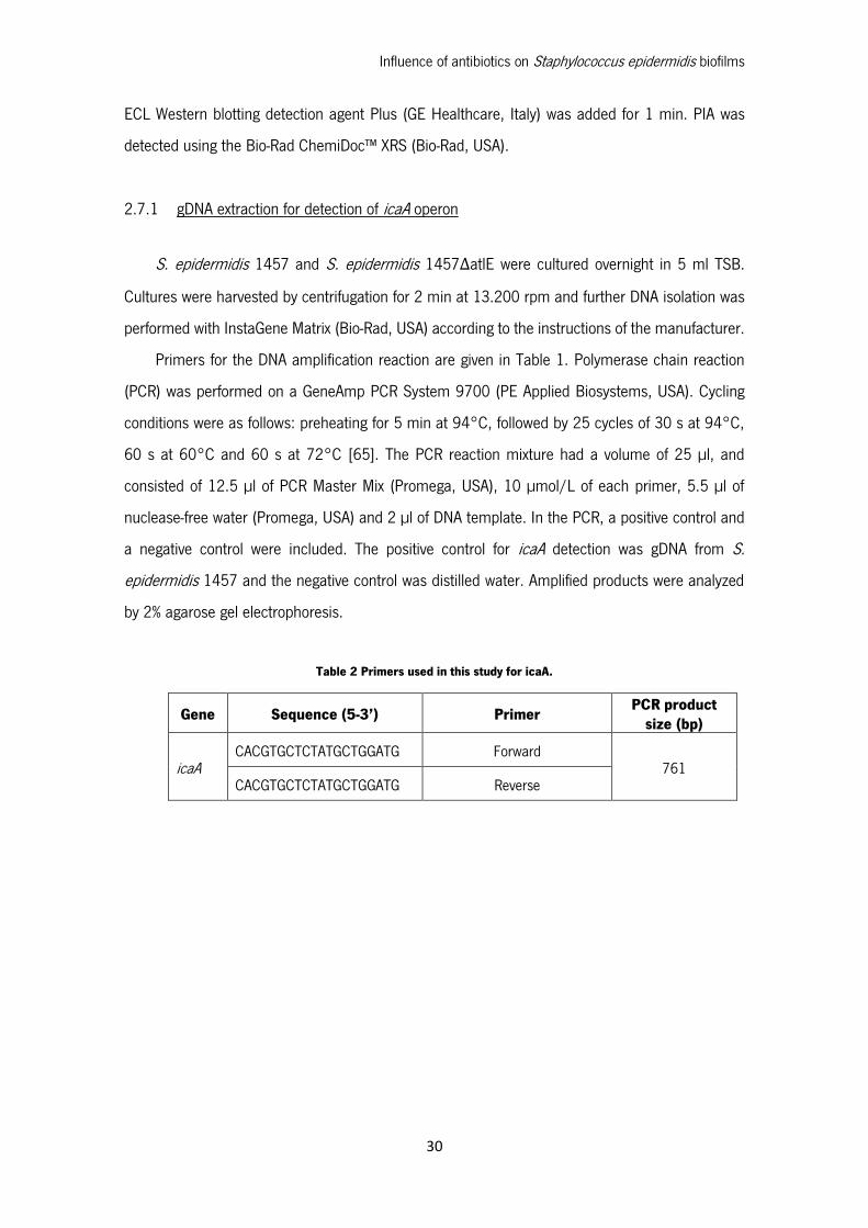
Primers for the DNA amplification reaction are given in Table 1. Polymerase chain reaction
(PCR) was performed on a GeneAmp PCR System 9700 (PE Applied Biosystems, USA). Cycling
conditions were as follows: preheating for 5 min at 94°C, followed by 25 cycles of 30 s at 94°C,
60 s at 60°C and 60 s at 72°C [65]. The PCR reaction mixture had a volume of 25 µl, and
consisted of 12.5 µl of PCR Master Mix (Promega, USA), 10 µmol/L of each primer, 5.5 µl of
nuclease-free water (Promega, USA) and 2 µl of DNA template. In the PCR, a positive control and
a negative control were included. The positive control for icaA detection was gDNA from S.
epidermidis 1457 and the negative control was distilled water. Amplified products were analyzed
by 2% agarose gel electrophoresis.
Table 2 Primers used in this study for icaA.
Gene Sequence (5-3’) Primer PCR product
size (bp)
icaA CACGTGCTCTATGCTGGATG Forward
761 CACGTGCTCTATGCTGGATG Reverse
Influence of antibiotics on Staphylococcus epidermidis biofilms
31
2.8 Scanning electron microscopy
Scanning electron microscopy (SEM) was performed in order to check changes in biofilm
structure and bacterial viability, after treatment with antibiotics. This experiment was only
conducted for S. epidermidis 1457.
Biofilms were grown on a cover slip disc in a 12 well-plate, with TSB as culture medium. The
inoculums was prepared as described in 2.3. After 24 h of growth, biofilms were rinsed with PBS
and treated with antibiotics for 24 h at 37°C, in 1/100 TSB. After 24 h, samples were rinsed with
1 ml of PBS and fixed with 2.5% glutaraldehyde in 0.1 M sodium cacodylatebuffer pH 7.4 during
2 h at room temperature. After primary fixation, samples were washed with 0.1 M cacodylatebuffer
and postfixed for 2 h in 1% osmiumtetroxide. Biofilms were dehydrated by immersion in an
ascending ethanol series 30%, 50%, 70% and 90% (v/v) for 5 min and 100% (v/v) ethanol for 3 x
5 min. Additionally, the samples were dried with hexamethyldisilazane (HMDS, Sigma-Aldrich) for
30 min with HMDS. [66]. The samples were then transferred to a vacuum desiccators for overnight
drying. Samples were mounted on aluminum stubs with carbon tape, sputter-coated with platinum
(Auto sputter coater, Agar Scientific) and observed with in a scanning electron microscope (Jeol,
JSM 7401F).
In order to assess biofilm morphology in each sample, three fields were used for image
analysis. Images were recorded at magnifications of 1.000 x, 4.000 x, 10.000 x and 25.000 x.
2.9 Statistical analysis
Results were compared with XLSTAT software (Addinsoft, France). Significant differences were
determined via one-way analysis of variance (one-way ANOVA). All tests were performed with a
confidence level of 95%.

Influence of antibiotics on Staphylococcus epidermidis biofilms
32
Influence of antibiotics on Staphylococcus epidermidis biofilms
33
Chapter 3 Results
Influence of antibiotics on Staphylococcus epidermidis biofilms
34
Influence of antibiotics on Staphylococcus epidermidis biofilms
35
3.1 MIC determination
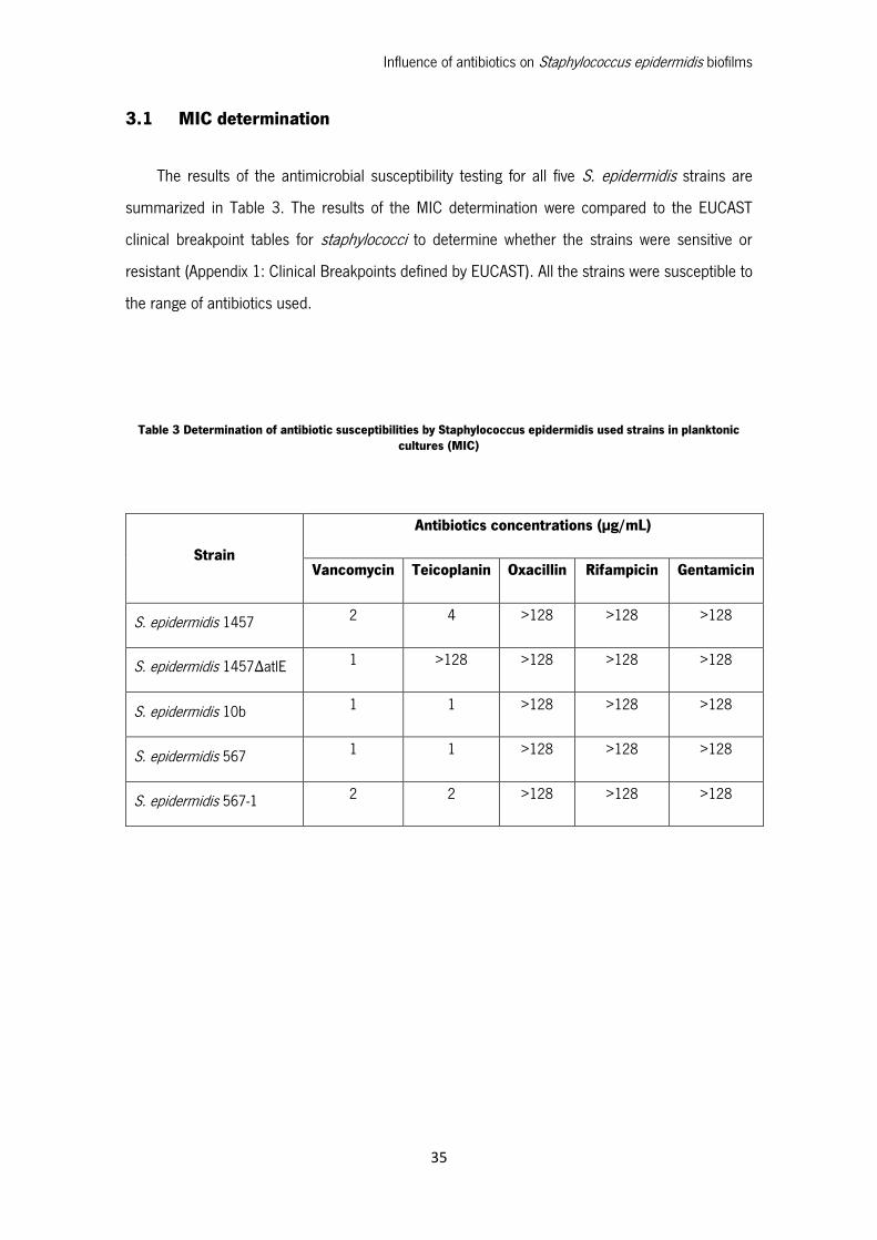
The results of the antimicrobial susceptibility testing for all five S. epidermidis strains are
summarized in Table 3. The results of the MIC determination were compared to the EUCAST
clinical breakpoint tables for staphylococci to determine whether the strains were sensitive or
resistant (Appendix 1: Clinical Breakpoints defined by EUCAST). All the strains were susceptible to
the range of antibiotics used.
Table 3 Determination of antibiotic susceptibilities by Staphylococcus epidermidis used strains in planktonic cultures (MIC)
Strain
Antibiotics concentrations (µg/mL)
Vancomycin Teicoplanin Oxacillin Rifampicin Gentamicin
S. epidermidis 1457 2 4 >128 >128 >128
S. epidermidis 1457ΔatlE 1 >128 >128 >128 >128
S. epidermidis 10b 1 1 >128 >128 >128
S. epidermidis 567 1 1 >128 >128 >128
S. epidermidis 567-1 2 2 >128 >128 >128
Influence of antibiotics on Staphylococcus epidermidis biofilms
36
3.2 Evaluation of activity of antibiotics on biofilms
Different methods were applied to evaluate the effects of the antibiotics treatment on the
biofilm biomass, bacterial cell viability, changes on biofilms matrix and changes on biofilms
structure. Antibiotics concentrations used corresponded to the serum peak concentrations.
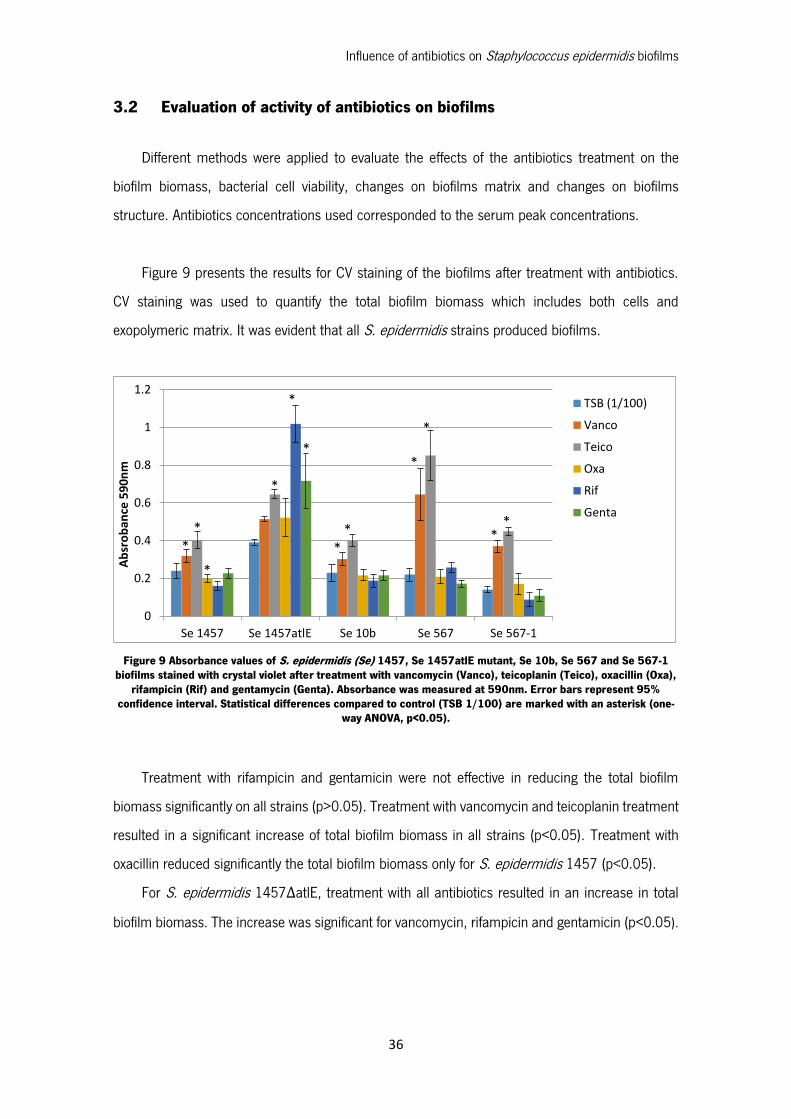
Figure 9 presents the results for CV staining of the biofilms after treatment with antibiotics.
CV staining was used to quantify the total biofilm biomass which includes both cells and
exopolymeric matrix. It was evident that all S. epidermidis strains produced biofilms.
Figure 9 Absorbance values of S. epidermidis (Se) 1457, Se 1457atlE mutant, Se 10b, Se 567 and Se 567-1 biofilms stained with crystal violet after treatment with vancomycin (Vanco), teicoplanin (Teico), oxacillin (Oxa),
rifampicin (Rif) and gentamycin (Genta). Absorbance was measured at 590nm. Error bars represent 95% confidence interval. Statistical differences compared to control (TSB 1/100) are marked with an asterisk (one-
way ANOVA, p<0.05).
Treatment with rifampicin and gentamicin were not effective in reducing the total biofilm
biomass significantly on all strains (p>0.05). Treatment with vancomycin and teicoplanin treatment
resulted in a significant increase of total biofilm biomass in all strains (p<0.05). Treatment with
oxacillin reduced significantly the total biofilm biomass only for S. epidermidis 1457 (p<0.05).
For S. epidermidis 1457ΔatlE, treatment with all antibiotics resulted in an increase in total
biofilm biomass. The increase was significant for vancomycin, rifampicin and gentamicin (p<0.05).
0
0.2
0.4
0.6
0.8
1
1.2
Se 1457 Se 1457atlE Se 10b Se 567 Se 567-1
Ab
sro
ban
ce 5
90
nm
TSB (1/100)
Vanco
Teico
Oxa
Rif
Genta
**
*
*
*
*
**
*
*
**
Influence of antibiotics on Staphylococcus epidermidis biofilms
37
Figure 10 presents the results of the XTT staining, showing changes in bacterial cell viability
after treatment with antibiotics. Results show that all antibiotics applied reduced the bacterial cell
viability.
According to Figure 10, cell viability reduction was significant in all S. epidermidis strains for
teicoplanin, rifampicin and gentamycin, except for S. epidermidis 1457. For that strain the
reduction was only significant for teicoplanin and rifampicin. Treatment with vancomycin and
oxacillin only reduced significantly bacteria viability for S. epidermidis 1457ΔatlE and for S.
epidermidis 567. In fact for those two strains, all the antibiotics reduced bacteria viability
significantly.
Comparing the effect of antibiotics used against biofilm associated bacteria, it is shown that
there are some significant differences between cell-wall active antibiotics (vancomycin, teicoplanin
and oxacillin) and RNA and protein synthesis inhibitors (rifampicin and gentamicin, respectively).
0
0.1
0.2
0.3
0.4
0.5
0.6
0.7
0.8
0.9
1
Se 1457 Se 1457atlE Se 10b Se 567 Se 567-1
Ab
sro
ban
ce 4
90
nm
TSB (1/100)
Vanco
Teico
Oxa
Rif
Genta
* *
*
*
*
*
*
* **
*
**
* **
*
*
Figure 10 Absorbance values XTT solutions obtained from adhesion of S. epidermidis (Se) 1457, Se 1457atlE mutant, Se 10b, Se 567 and Se 567-1 strains after treatment with vancomycin (Vanco),
teicoplanin (Teico), oxacillin (Oxa), rifampicin (Rif) and gentamycin (Genta). Absorbance was measured at 490 nm. Error bars represent 95% confidence interval. Statistical differences compared to control (TSB
1/100) are marked with an asterisk (one-way ANOVA,p<0.05).
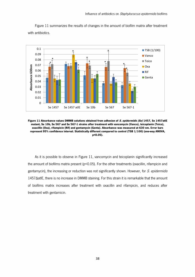
Influence of antibiotics on Staphylococcus epidermidis biofilms
38
Figure 11 summarizes the results of changes in the amount of biofilm matrix after treatment
with antibiotics.
Figure 11 Absorbance values DMMB solutions obtained from adhesion of S. epidermidis (Se) 1457, Se 1457atlE mutant, Se 10b, Se 567 and Se 567-1 strains after treatment with vancomycin (Vanco), teicoplanin (Teico), oxacillin (Oxa), rifampicin (Rif) and gentamycin (Genta). Absorbance was measured at 630 nm. Error bars
represent 95% confidence interval. Statistically different compared to control (TSB 1/100) (one-way ANOVA, p<0.05).
As it is possible to observe in Figure 11, vancomycin and teicoplanin significantly increased
the amount of biofilms matrix present (p<0.05). For the other treatments (oxacillin, rifampicin and
gentamycin), the increasing or reduction was not significantly shown. However, for S. epidermidis
1457ΔatlE, there is no increase in DMMB staining. For this strain it is remarkable that the amount
of biofilms matrix increases after treatment with oxacillin and rifampicin, and reduces after
treatment with gentamicin.
*
0
0.01
0.02
0.03
0.04
0.05
0.06
0.07
0.08
0.09
0.1
Se 1457 Se 1457 atlE Se 10b Se 567 Se 567-1
Ab
sorb
ance
63
0n
m
TSB (1/100)
Vanco
Teico
Oxa
Rif
Genta
**
*
*
** *
*
**
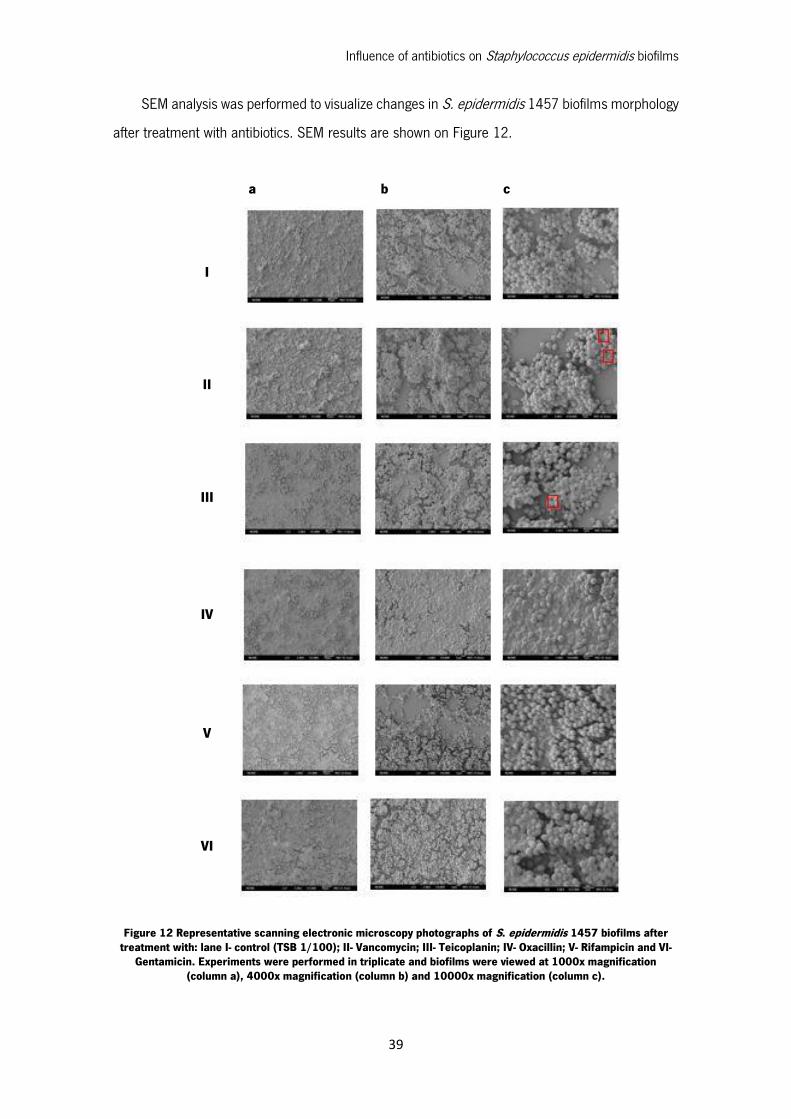
Influence of antibiotics on Staphylococcus epidermidis biofilms
39
SEM analysis was performed to visualize changes in S. epidermidis 1457 biofilms morphology
after treatment with antibiotics. SEM results are shown on Figure 12.
Figure 12 Representative scanning electronic microscopy photographs of S. epidermidis 1457 biofilms after treatment with: lane I- control (TSB 1/100); II- Vancomycin; III- Teicoplanin; IV- Oxacillin; V- Rifampicin and VI-
Gentamicin. Experiments were performed in triplicate and biofilms were viewed at 1000x magnification (column a), 4000x magnification (column b) and 10000x magnification (column c).
I
II
III
IV
V
VI
a b c
Influence of antibiotics on Staphylococcus epidermidis biofilms
40
Vancomycin treatment resulted in a rougher biofilm (Figure 12 IIa vs. Ia), with likely more
bacteria attached (Figure 12 IIb vs Ib). Damaged bacteria can be identified from the creases
(marked with red squares). Also treatment with teicoplanin resulted in damaged bacteria. The
biofilms treated with teicoplanin look very similar to treatment with TSB 1/100 (Figure 12 IIIb vs
Ib). Treatment with oxacillin (Figure 12 IV) made bacteria detach from the biofilm but the matrix
was left behind. Also rifampicin treatment (Figure 12 V) resulted in bacterial detachment with matrix
remaining, though to a lesser extent then oxacillin treatment. Gentamicin treatment finally (Figure
12 VI) gave a smooth biofilm containing the same amount of bacteria and matrix (Figure 12 VIb),
or more (Figure 12 VIc) then the TSB 1/100 treated biofilms.
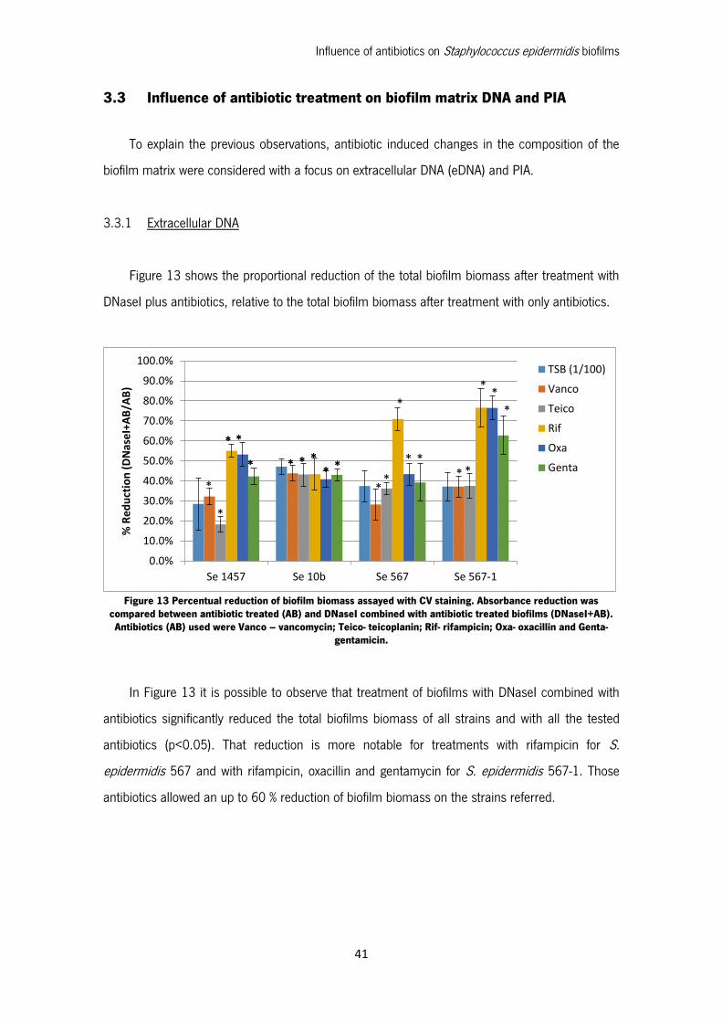
Influence of antibiotics on Staphylococcus epidermidis biofilms
41
3.3 Influence of antibiotic treatment on biofilm matrix DNA and PIA
To explain the previous observations, antibiotic induced changes in the composition of the
biofilm matrix were considered with a focus on extracellular DNA (eDNA) and PIA.
3.3.1 Extracellular DNA
Figure 13 shows the proportional reduction of the total biofilm biomass after treatment with
DNaseI plus antibiotics, relative to the total biofilm biomass after treatment with only antibiotics.
Figure 13 Percentual reduction of biofilm biomass assayed with CV staining. Absorbance reduction was compared between antibiotic treated (AB) and DNaseI combined with antibiotic treated biofilms (DNaseI+AB). Antibiotics (AB) used were Vanco – vancomycin; Teico- teicoplanin; Rif- rifampicin; Oxa- oxacillin and Genta-
gentamicin.
In Figure 13 it is possible to observe that treatment of biofilms with DNaseI combined with
antibiotics significantly reduced the total biofilms biomass of all strains and with all the tested
antibiotics (p<0.05). That reduction is more notable for treatments with rifampicin for S.
epidermidis 567 and with rifampicin, oxacillin and gentamycin for S. epidermidis 567-1. Those
antibiotics allowed an up to 60 % reduction of biofilm biomass on the strains referred.
0.0%
10.0%
20.0%
30.0%
40.0%
50.0%
60.0%
70.0%
80.0%
90.0%
100.0%
Se 1457 Se 10b Se 567 Se 567-1
% R
ed
uct
ion
(D
Nas
eI+
AB
/AB
)
TSB (1/100)
Vanco
Teico
Rif
Oxa
Genta
*
* *
* * * ** *
**
*
* *
* *
* * * ** *
* *
**
*
*
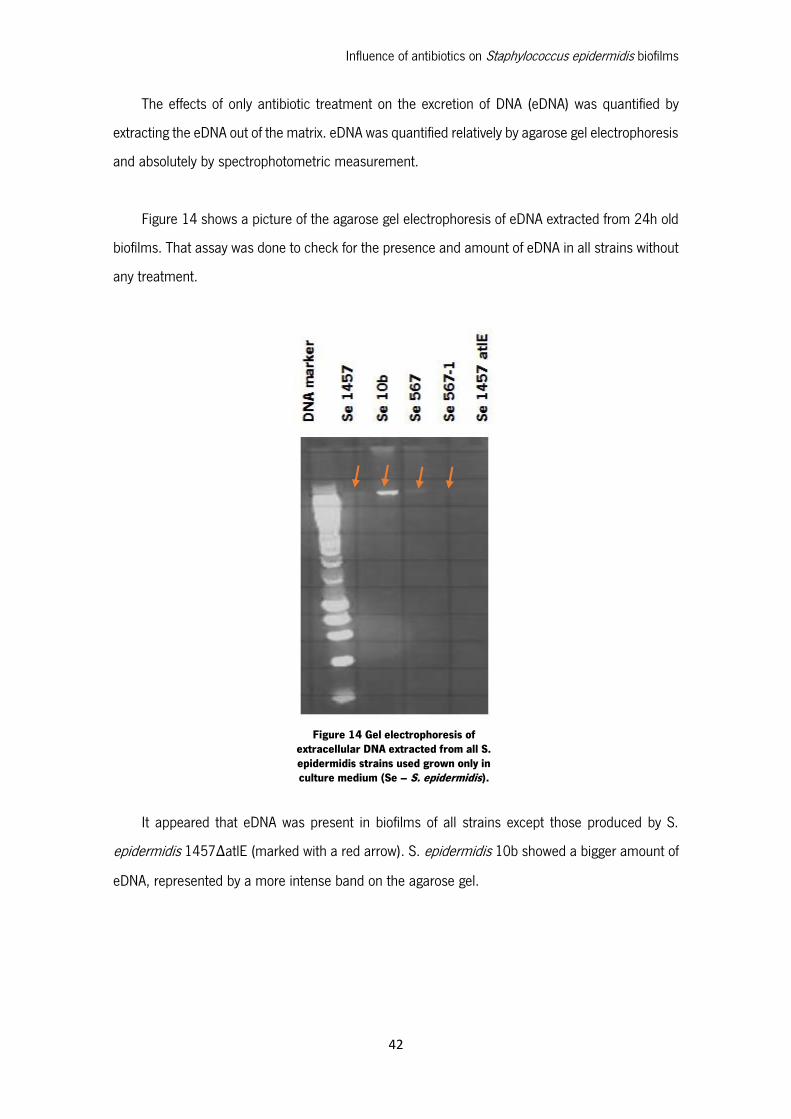
Influence of antibiotics on Staphylococcus epidermidis biofilms
42
The effects of only antibiotic treatment on the excretion of DNA (eDNA) was quantified by
extracting the eDNA out of the matrix. eDNA was quantified relatively by agarose gel electrophoresis
and absolutely by spectrophotometric measurement.
Figure 14 shows a picture of the agarose gel electrophoresis of eDNA extracted from 24h old
biofilms. That assay was done to check for the presence and amount of eDNA in all strains without
any treatment.
It appeared that eDNA was present in biofilms of all strains except those produced by S.
epidermidis 1457ΔatlE (marked with a red arrow). S. epidermidis 10b showed a bigger amount of
eDNA, represented by a more intense band on the agarose gel.
Figure 14 Gel electrophoresis of extracellular DNA extracted from all S. epidermidis strains used grown only in culture medium (Se – S. epidermidis).
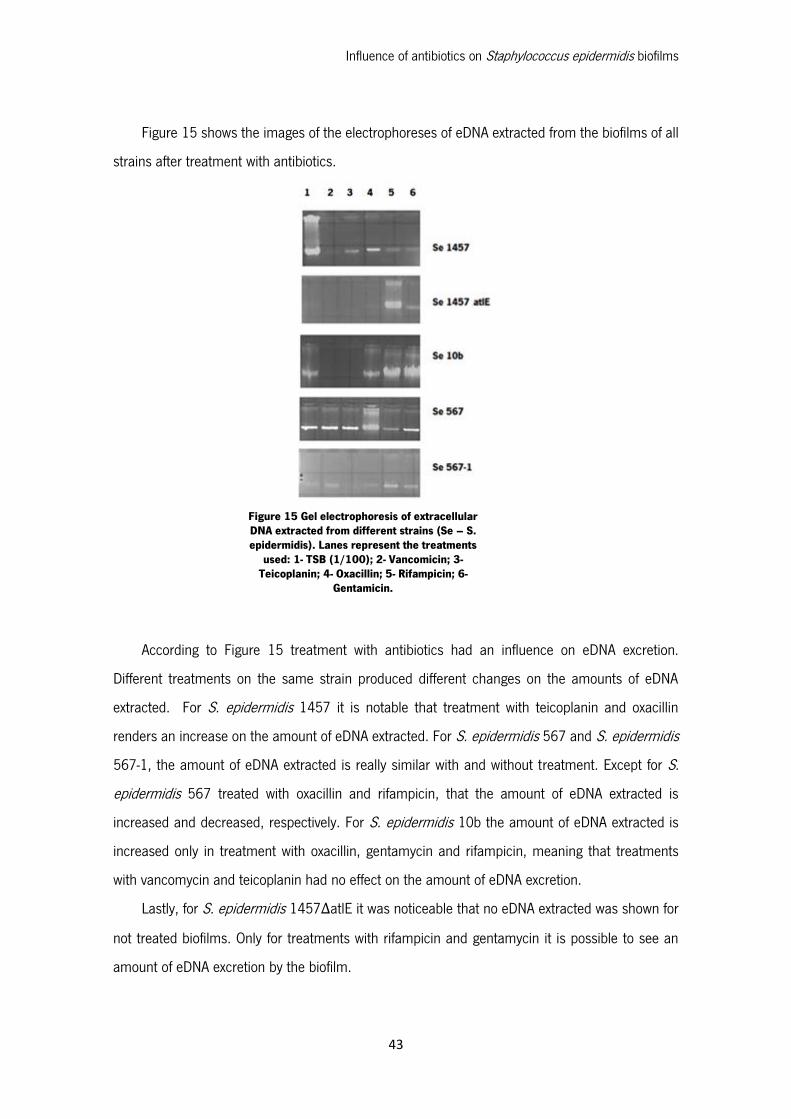
Influence of antibiotics on Staphylococcus epidermidis biofilms
43
Figure 15 shows the images of the electrophoreses of eDNA extracted from the biofilms of all
strains after treatment with antibiotics.
According to Figure 15 treatment with antibiotics had an influence on eDNA excretion.
Different treatments on the same strain produced different changes on the amounts of eDNA
extracted. For S. epidermidis 1457 it is notable that treatment with teicoplanin and oxacillin
renders an increase on the amount of eDNA extracted. For S. epidermidis 567 and S. epidermidis
567-1, the amount of eDNA extracted is really similar with and without treatment. Except for S.
epidermidis 567 treated with oxacillin and rifampicin, that the amount of eDNA extracted is
increased and decreased, respectively. For S. epidermidis 10b the amount of eDNA extracted is
increased only in treatment with oxacillin, gentamycin and rifampicin, meaning that treatments
with vancomycin and teicoplanin had no effect on the amount of eDNA excretion.
Lastly, for S. epidermidis 1457ΔatlE it was noticeable that no eDNA extracted was shown for
not treated biofilms. Only for treatments with rifampicin and gentamycin it is possible to see an
amount of eDNA excretion by the biofilm.
Figure 15 Gel electrophoresis of extracellular DNA extracted from different strains (Se – S. epidermidis). Lanes represent the treatments
used: 1- TSB (1/100); 2- Vancomicin; 3- Teicoplanin; 4- Oxacillin; 5- Rifampicin; 6-
Gentamicin.

Influence of antibiotics on Staphylococcus epidermidis biofilms
44
3.3.2 PIA (PIA dot-blot)
The effects of antibiotic treatments on the biofilm matrix with regard to the PIA content were
assessed by PIA dot-blot.
Figure 16 shows the relative amounts of PIA in the biofilm matrix after treatment with
antibiotics compared to the control (biofilms formed only in TSB (1/100)).
Figure 16 Relative amounts of PIA on biofilms were determined using a PIA dot blot assay after treatment with antibiotics: Vanco- Vancomycin; Teico- Teicoplanin; Oxa- Oxacillin; Rif- Rifampicin and Genta- Gentamicin. To
determine the relative amounts of PIA, it was defined that the amount of PIA produced by each strain individually when treated with TSB (1/100) as 100% and expressed all other amounts as relative to the amount
on that TSB (1/100) treatment.
Interestingly, for S. epidermidis 1457ΔatlE it was not possible to determine the relative
amount of PIA because that strain did not show any PIA on the dot blot assay performed.
For S. epidermidis 567-1 treatment with vancomycin, oxacillin, rifampicin and gentamicin
(Figure 16) there was an enhancement in the production of PIA up to more than 20% comparing
with the absence of antibiotics treatment. For S. epidermidis 1457 and S. epidermidis 10b,
vancomycin and teicoplanin treatment showed a higher amount of PIA when compared with
treatment with the other antibiotics, although the difference is small. For S. epidemidis 567 the
amount of PIA on vancomycin treatment is shown to be really reduced compared with the other
treatments.
0%
20%
40%
60%
80%
100%
120%
140%
Se 1457 Se 10b Se 567 Se 567-1
Re
lati
ve P
IA's
am
ou
nt
(%)
Vanco
Teico
Oxa
Rif
Genta
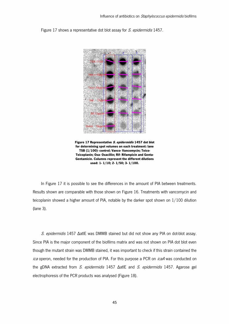
Influence of antibiotics on Staphylococcus epidermidis biofilms
45
Figure 17 shows a representative dot blot assay for S. epidermidis 1457.
In Figure 17 it is possible to see the differences in the amount of PIA between treatments.
Results shown are comparable with those shown on Figure 16. Treatments with vancomycin and
teicoplanin showed a higher amount of PIA, notable by the darker spot shown on 1/100 dilution
(lane 3).
S. epidermidis 1457 ΔatlE was DMMB stained but did not show any PIA on dot-blot assay.
Since PIA is the major component of the biofilms matrix and was not shown on PIA dot blot even
though the mutant strain was DMMB stained, it was important to check if this strain contained the
ica operon, needed for the production of PIA. For this purpose a PCR on icaA was conducted on
the gDNA extracted from S. epidermidis 1457 ΔatlE and S. epidermidis 1457. Agarose gel
electrophoresis of the PCR products was analysed (Figure 18).
Figure 17 Representative S. epidermidis 1457 dot blot for determining spot volumes on each treatment: lane
TSB (1/100)- control; Vanco- Vancomycin; Teico- Teicoplanin; Oxa- Oxacillin; Rif- Rifampicin and Genta- Gentamicin. Columns represent the different dilutions
used: 1- 1/10; 2- 1/50; 3- 1/100.
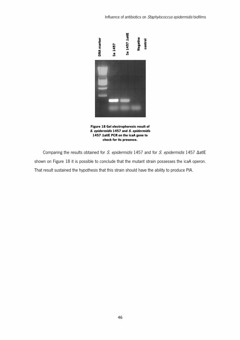
Influence of antibiotics on Staphylococcus epidermidis biofilms
46
Comparing the results obtained for S. epidermidis 1457 and for S. epidermidis 1457 ΔatlE
shown on Figure 18 it is possible to conclude that the mutant strain possesses the icaA operon.
That result sustained the hypothesis that this strain should have the ability to produce PIA.
Figure 18 Gel electrophoresis result of S. epidermidis 1457 and S. epidermidis
1457 ΔatlE PCR on the icaA gene to check for its presence.
Influence of antibiotics on Staphylococcus epidermidis biofilms
47
Chapter 4 Discussion

Influence of antibiotics on Staphylococcus epidermidis biofilms
48
Influence of antibiotics on Staphylococcus epidermidis biofilms
49
Staphylococcus epidermidis is the most frequently isolated species of the CoNS group. S.
epidermidis is found on skin and mucous membranes of humans and other organisms,
representing an important part of its normal microflora [16]. Previously regarded as relatively
innocuous, has gained significant interest in recent years and has become one of the leading
causes of infections related to implanted medical devices. Those infections have especial interest
among immunocompromised, immunosuppressed and critically ill patients [15], [19], [39].
Treatment of S. epidermidis infections has become very difficult due to the increasing
resistance to antibacterial agents. Antimicrobial resistance has a significant impact on patient
outcome by enhancing virulence, delaying the administration of therapeutic medicines leading to
increased morbidity and mortality [19]. It is a serious problem particularly notable in S. epidermidis,
since many clinical isolates of this organism are resistant to up to eight different antibiotics. In
addition to this problem is the fact that the major virulence factor of S. epidermidis is biofilm
formation and that cells in biofilms are normally more resistant to antibiotics than planktonic cells,
making drug resistance in a CoNS infection an even more serious problem [15], [16]. Therefore,
it is crucial to understand the mechanisms of action and the changes promoted by antibiotics on
biofilms, in order to better address this problem of antibiotic resistance.
4.1 Antibiotic susceptibility
Firstly, it became important to determine if the chosen antibiotics had an effect on the S.
epidermidis strains used. Therefore, an assay to determine the MIC concentration for planktonic
cells was carried out. Although standard antimicrobial susceptibility testing, which challenges
planktonic (suspended) cells with an antimicrobial agent, will not accurately predict the efficacy of
an agent against biofilm associated organisms, it is important in order to understand if the strain
is naturally susceptible to the antibiotic or not.
To evaluate the efficacy of the antibiotics used in this study, a concentration range was chosen.
These values were well defined in the standard [67] endpoints for antimicrobial susceptibility
testing and thus enable evaluation of the efficacy of antibiotics against planktonic microorganisms.
Results shown on Table 3 make it is possible to conclude that all the S. epidermidis strains used
were susceptible to the antibiotics. It is important to note that the results are consistent with clinical
breakpoints defined by EUCAST for S. epidermidis (Appendix 1: Clinical Breakpoints defined by
EUCAST) [67].
Influence of antibiotics on Staphylococcus epidermidis biofilms
50
Regarding the results for each strain separately, for S. epidermidis 1457 and S. epidermidis
10b the results are according to the expect comparing with the literature [68], [45]. MIC results
for S. epidermidis 1457ΔatlE treated with vancomycin show to be different from the MIC for the
wild-type strain. According to Hello et al. [69] that was not an expected result. In their study it was
found that the altE mutant susceptibility to vancomycin was similar to the wild-type strain
susceptibility. That difference found can maybe be explained by the fact that the mutant strain
Influence of antibiotics on Staphylococcus epidermidis biofilms
51
4.2 Evaluation of antibiotics activity on biofilms
The pathogenesis of medical device infections associated with S. epidermidis is characterized
by the microorganism’s ability to colonize the surface of implanted medical devices by the
formation of highly resistant biofilms [19]. Biofilm formation is, as mentioned, the major virulence
factor in S. epidermidis infections and therefore it is of especial interest to address the mechanisms
of action of the antimicrobial treatment applied. In this study, to evaluate the effect of antibiotics,
it was necessary to discriminate between the biofilm matrix and the bacteria living within it. That is
from a major importance because it is documented that when an antibiotic only acts on the slime
layer, biofilm bacteria will start forming a new matrix almost immediately and if only biofilm bacteria
are killed, the remaining matrix is predestined to become re-colonized [70].
The CV, XTT and DMMB assays were used in this study in order to determine the influence of
antibiotic treatments on the biofilms, whether on the biofilm biomass, bacteria viability and biofilms
matrix, respectively. The different antibiotics used and its different mechanisms of action are shown
in Table 1 and explained in Chapter 1.5.1.
In particular, CV staining was used to evaluate the effect of antibiotics treatment on biofilms,
regarding the changes on total biofilm biomass. This assay was important in order to conclude if
the antibiotics had an effect on increasing or decreasing the total biofilm biomass formed compared
to the control (cells growing only in the presence of TSB 1/100). Results obtained in the CV assay
allowed to conclude that all S. epidermidis strains produced biofilms (Figure 9), as it was expected
[15], [17].
Evaluating the strains capacity to form biofilms without any antibiotic treatment, it was possible
to detect some changes in relation to the expectable results. For instances, S. epidermidis 1457
ΔatlE formed more biofilm biomass than S. epidermidis 1457 (its wild-type). Since the extracellular
DNA is found as a major component required for initial bacterial attachment to surfaces, as well
as for the subsequent early phase of biofilm development by S. epidermidis, it was expectable that
the mutant strain formed less biofilm. That was the expectable result because the release of
extracellular DNA is known to be mainly caused by the activity of the autolysin AtlE. Since S.
epidermidis 1457 ΔatlE lacks on the atlE gene it was supposed to have less bacteria attached to
the surface [27]. These results can maybe be explained by the media growth dilution used for the
wild-type strain and not on the mutant strain, giving it more nutrients to colonize the surface of the
Influence of antibiotics on Staphylococcus epidermidis biofilms
52
plate and reproduce. That difference in the growth media is related to the protocol for S.
epidermidis 1457 ΔatlE biofilms cultivation referred by Qin et al. [27]
Results presented in Figure 9 also showed that biofilms of S. epidermidis 567 and S.
epidermidis 567-1 have a very similar biomass. That result was not expectable, according to the
literature, since the agr mutant, S. epidermidis 567-1, in previous studies exhibited a stronger
biofilm formation [55]. Since the aim of this study was to evaluate the influence of antibiotics
treatment on S. epidermidis biofilms, those results are of less importance and are only related to
the control biofilms.
In fact, in the same strain the differences noted on the biofilm biomass were caused by the
antibiotics treatments applied. Different antibiotics had different effects on each strain. For
instances, by CV staining on Figure 9, it is noticeable that vancomycin and teicoplanin treatments
had an effect, significantly increasing the total biofilm biomass on all strains. That result has already
been documented by Hsu [44]. This vancomycin and teicoplanin tolerance of S. epidermidis was
attributed to the poor activity of glycopeptides related to a diminished antimicrobial effect on S.
epidermidis cells in biofilm already documented by Farber et al. [71].
The biofilm biomass enhancement caused by vancomycin and teicoplanin, in this study, can
be explained by an increased excretion of extracellular DNA or and increased production of PIA
caused by the action of those antibiotics on the cell wall. Increased excretion of eDNA enhance
biofilm formation and adhesion cell-to-cell. The DNA in the extracellular space, released by the
action of the antibiotics, can aggregate, therefore enhancing biofilms formation. The suggested
increasing on PIA’s production can be related to the rupture of the cell wall due to the antibiotics
action. That can enhance PIA’s production and creation of a linked connection between bacterial
cells [44].
Only for S. epidermidis 1457 oxacillin treatment decreased significantly the total biofilm
biomass formed. That can imply that this antibiotic has a bigger antimicrobial effect on reducing
the total biofilm biomass formed compared to the other two cell wall active antibiotics, for this
strain in particular.
S. epidermidis 1457 ΔatlE showed also more biofilm biomass comparing with all the other
strains used in this work, except for S. epidermidis 567 treated with vancomycin and teicoplanin.
Also in S. epidermidis 1457ΔatlE strain, treatment with teicoplanin, rifampicin and gentamycin had
a significant increase on biofilm biomass. As this strain lacks on atlE gene, the biofilm formed
Influence of antibiotics on Staphylococcus epidermidis biofilms
53
cannot be explained by the extracellular DNA release [27]. So, that may imply that the large amount
of biofilm formed is due to the increased production of PIA.
The crystal violet assay has been extensively used, but likely because the estimated biomass
does not distinguish between dead or living bacteria, it is not very informative when not
complemented with a viability assay. Even with the increasing biofilms biomass detected it was
important to determine if the antibiotics had an antimicrobial effect on the biofilm associated
bacteria. That is why it was important to add the XTT assay. This method allows to stain only active
bacteria cells in the formed biofilm, which means that it quantifies the bacteria in biofilms that have
the capacity to continue to grow and proliferate the infection. According to Figure 10, it was
expected all antibiotic treatments reduced bacteria viability for all strains since they were proven to
be susceptible to the antibiotics in use by the MIC determination. Those results make it possible
to conclude that all the antibiotics were successful on killing biofilm associated bacteria, suggesting
that the increasing CV staining found on the biofilms, and therefore a large amount of biofilm
biomass, had to be related to the increased amount of dead cells and biofilms matrix.
Comparing the effects on killing biofilm associated bacteria by different antibiotics, it is
possible to conclude that for S. epidermidis 567 and S. epidermidis 10b, teicoplanin, rifampicin
and gentamicin had a similar effect.
For S. epidermidis ΔatlE comparing to the wild-type, it is possible to see that the mutant strain
was more susceptible to killing by antibiotics. In fact, it is possible to conclude that cell-wall active
antibiotics had an increased effect on bacteria killing on that mutant strain. That result is supported
by the literature that reported an implication of AtlE loss in the tolerance of S. epidermidis to cell
wall active antibiotics [68].
To the CV and XTT staining methods, it was added the DMMB staining method. The aim of
this staining method was the evaluation of the changes on the biofilms matrix in addition with the
other two assays performed. DMMB is a cationic dye used for quantification of sulphated
polysaccharides, more specifically glycosaminoglycans (GAGs) in biological samples. As there is
structural similarity between PIA and GAGs, it was hypothesized by Toté et al. [58] that DMMB
could be used for the specific detection of S. aureus biofilm matrix. Since PIA is also the major
component of S. epidermidis biofilms matrix, it was also used in this study to detect PIA’s content
on the biofilms [72]. As seen before treatment, cell-wall active antibiotics, as vancomycin and
Influence of antibiotics on Staphylococcus epidermidis biofilms
54
teicoplanin, had an effect on increasing biofilms’ biomass. With the DMMB, Figure 11, it is possible
to see that treatments with those antibiotics had an obvious effect on increasing the amount of
biofilms matrix. Those cell-wall active antibiotics act on the bacteria cell-wall causing the membrane
rupture and enhancing matrix production [73].
Besides the effects on biofilms biomass, bacteria viability and biofilms matrix, the changes
promoted by antibiotics treatments in biofilms were visualized by scanning electron microscopy
(SEM). The changes on S. epidermidis morphology were evaluated using 1457 S. epidermidis
strain. This strain is known as a biofilm forming strain that produces a thick biofilm [53] and thus
it was chosen to better illustrate the effects of the antibiotics on the biofilm structure, cell viability
and the biofilm matrix through SEM analysis (Figure 12). According to the results, vancomycin
treatment resulted in a rougher biofilm (Figure 12 IIa vs. Ia), with likely more bacteria attached
(Figure 12 IIb vs Ib). It is possible to visualize some damaged bacteria but the main conclusion is
that treatment with this antibiotic promoted a cell wall thickening. That result has been already
documented by Hsu [44], that proved that treatment with vancomycin enhanced the biofilm
thickness. That result for vancomycin treatment is also supported by the results obtained after CV,
XTT and DMMB staining. This strain showed an higher biofilm biomass formed after CV staining,
with a small effect on bacteria killing assayed by XTT staining. Even with DMMB staining, this strain
showed a significant increase on the biofilm matrix after treatment with vancomycin.
Treatment with teicoplanin had an effect on increasing biofilms thickness even if in a less
extent as vancomycin treatment. It also resulted in damaged bacteria. The biofilms treated with
teicoplanin look very similar to treatment with TSB (1/100) (Figure 12 IIIb vs Ib). These SEM results
are in agreement with the previous results obtained in CV, XTT and DMMB staining. It is noticeable
the increased amount of biofilms biomass and biofilms matrix, as assayed by CV and DMMB
staining methods.
Treatment with oxacillin (Figure 12 IV) and rifampicin (Figure 12 V) resulted in bacterial
detachment with matrix remaining. Gentamicin treatment finally (Figure 12 VI) gave a smooth
biofilm containing the same amount of bacteria and matrix or more then the TSB (1/100) treated
biofilms. These results are in accordance with the ones obtained after the staining methods applied.
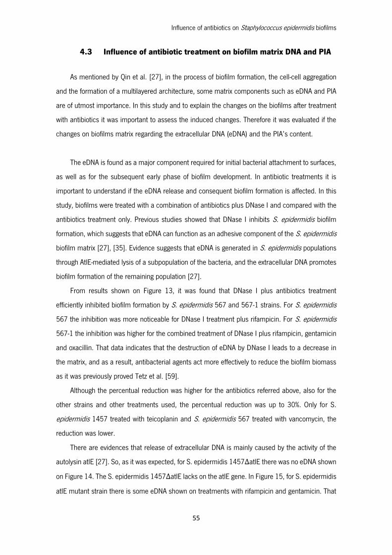
Influence of antibiotics on Staphylococcus epidermidis biofilms
55
4.3 Influence of antibiotic treatment on biofilm matrix DNA and PIA
As mentioned by Qin et al. [27], in the process of biofilm formation, the cell-cell aggregation
and the formation of a multilayered architecture, some matrix components such as eDNA and PIA
are of utmost importance. In this study and to explain the changes on the biofilms after treatment
with antibiotics it was important to assess the induced changes. Therefore it was evaluated if the
changes on biofilms matrix regarding the extracellular DNA (eDNA) and the PIA’s content.
The eDNA is found as a major component required for initial bacterial attachment to surfaces,
as well as for the subsequent early phase of biofilm development. In antibiotic treatments it is
important to understand if the eDNA release and consequent biofilm formation is affected. In this
study, biofilms were treated with a combination of antibiotics plus DNase I and compared with the
antibiotics treatment only. Previous studies showed that DNase I inhibits S. epidermidis biofilm
formation, which suggests that eDNA can function as an adhesive component of the S. epidermidis
biofilm matrix [27], [35]. Evidence suggests that eDNA is generated in S. epidermidis populations
through AtlE-mediated lysis of a subpopulation of the bacteria, and the extracellular DNA promotes
biofilm formation of the remaining population [27].
From results shown on Figure 13, it was found that DNase I plus antibiotics treatment
efficiently inhibited biofilm formation by S. epidermidis 567 and 567-1 strains. For S. epidermidis
567 the inhibition was more noticeable for DNase I treatment plus rifampicin. For S. epidermidis
567-1 the inhibition was higher for the combined treatment of DNase I plus rifampicin, gentamicin
and oxacillin. That data indicates that the destruction of eDNA by DNase I leads to a decrease in
the matrix, and as a result, antibacterial agents act more effectively to reduce the biofilm biomass
as it was previously proved Tetz et al. [59].
Although the percentual reduction was higher for the antibiotics referred above, also for the
other strains and other treatments used, the percentual reduction was up to 30%. Only for S.
epidermidis 1457 treated with teicoplanin and S. epidermidis 567 treated with vancomycin, the
reduction was lower.
There are evidences that release of extracellular DNA is mainly caused by the activity of the
autolysin atlE [27]. So, as it was expected, for S. epidermidis 1457ΔatlE there was no eDNA shown
on Figure 14. The S. epidermidis 1457ΔatlE lacks on the atlE gene. In Figure 15, for S. epidermidis
atlE mutant strain there is some eDNA shown on treatments with rifampicin and gentamicin. That
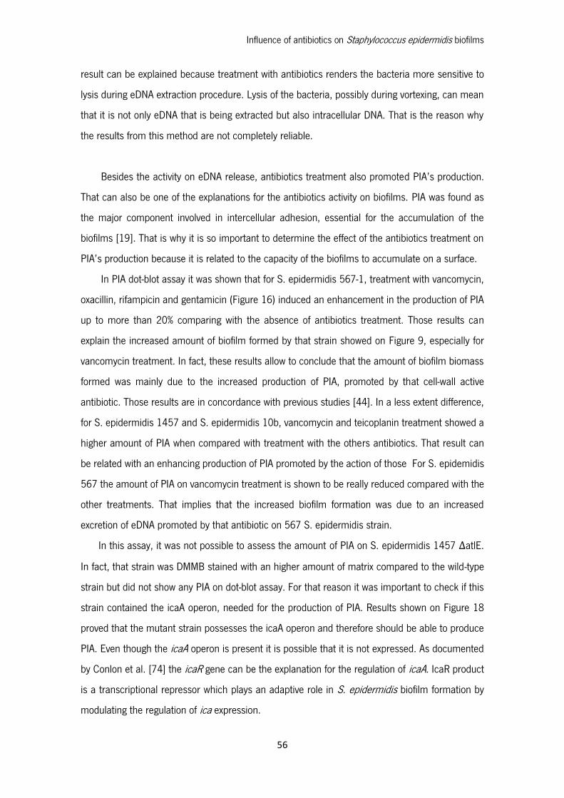
Influence of antibiotics on Staphylococcus epidermidis biofilms
56
result can be explained because treatment with antibiotics renders the bacteria more sensitive to
lysis during eDNA extraction procedure. Lysis of the bacteria, possibly during vortexing, can mean
that it is not only eDNA that is being extracted but also intracellular DNA. That is the reason why
the results from this method are not completely reliable.
Besides the activity on eDNA release, antibiotics treatment also promoted PIA’s production.
That can also be one of the explanations for the antibiotics activity on biofilms. PIA was found as
the major component involved in intercellular adhesion, essential for the accumulation of the
biofilms [19]. That is why it is so important to determine the effect of the antibiotics treatment on
PIA’s production because it is related to the capacity of the biofilms to accumulate on a surface.
In PIA dot-blot assay it was shown that for S. epidermidis 567-1, treatment with vancomycin,
oxacillin, rifampicin and gentamicin (Figure 16) induced an enhancement in the production of PIA
up to more than 20% comparing with the absence of antibiotics treatment. Those results can
explain the increased amount of biofilm formed by that strain showed on Figure 9, especially for
vancomycin treatment. In fact, these results allow to conclude that the amount of biofilm biomass
formed was mainly due to the increased production of PIA, promoted by that cell-wall active
antibiotic. Those results are in concordance with previous studies [44]. In a less extent difference,
for S. epidermidis 1457 and S. epidermidis 10b, vancomycin and teicoplanin treatment showed a
higher amount of PIA when compared with treatment with the others antibiotics. That result can
be related with an enhancing production of PIA promoted by the action of those For S. epidemidis
567 the amount of PIA on vancomycin treatment is shown to be really reduced compared with the
other treatments. That implies that the increased biofilm formation was due to an increased
excretion of eDNA promoted by that antibiotic on 567 S. epidermidis strain.
In this assay, it was not possible to assess the amount of PIA on S. epidermidis 1457 ΔatlE.
In fact, that strain was DMMB stained with an higher amount of matrix compared to the wild-type
strain but did not show any PIA on dot-blot assay. For that reason it was important to check if this
strain contained the icaA operon, needed for the production of PIA. Results shown on Figure 18
proved that the mutant strain possesses the icaA operon and therefore should be able to produce
PIA. Even though the icaA operon is present it is possible that it is not expressed. As documented
by Conlon et al. [74] the icaR gene can be the explanation for the regulation of icaA. IcaR product
is a transcriptional repressor which plays an adaptive role in S. epidermidis biofilm formation by
modulating the regulation of ica expression.
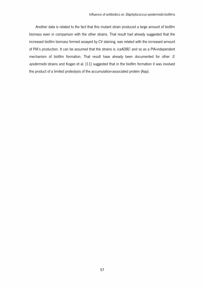
Influence of antibiotics on Staphylococcus epidermidis biofilms
57
Another data is related to the fact that this mutant strain produced a large amount of biofilm
biomass even in comparison with the other strains. That result had already suggested that the
increased biofilm biomass formed assayed by CV staining, was related with the increased amount
of PIA’s production. It can be assumed that the strains is icaADBC- and so as a PIA-independent
mechanism of biofilm formation. That result have already been documented for other S.
epidermidis strains and Kogan et al. [11] suggested that in the biofilm formation it was involved
the product of a limited proteolysis of the accumulation-associated protein (Aap).

Influence of antibiotics on Staphylococcus epidermidis biofilms
58
Influence of antibiotics on Staphylococcus epidermidis biofilms
59
Chapter 5 Conclusion and future perspectives

Influence of antibiotics on Staphylococcus epidermidis biofilms
60
Influence of antibiotics on Staphylococcus epidermidis biofilms
61
Staphylococcus epidermidis is the most frequently isolated species of the CoNS group.
Previously regarded as relatively innocuous, has gained significant interest in recent years and has
become one of the leading causes of infections related to implanted medical devices. Treatment
of S. epidermidis infections has become very difficult due to the increasing resistance to
antibacterial agents. It is a serious problem particularly notable in S. epidermidis, since many
clinical isolates of this organism are resistant to up to eight different antibiotics. Therefore, it is
crucial to understand the mechanisms of action and the changes promoted by different antibiotics
on biofilms, in order to better address this problem of antibiotic resistance. So, this work aims to
evaluate the influence of antibiotics on S. epidermidis biofilms in terms of changes in matrix
composition after treatment, regarding the eDNA and the PIA content.
Regarding the MIC results, it was proven that all the S. epidermidis strains used were
susceptible to all the antibiotics chosen for this study (vancomycin, teicoplanin, oxacillin, rifampicin
and gentamicin). Those results for planktonic cells were also supported by the results obtained
after XTT staining assay since all the antibiotics were successful on killing biofilm associated
bacteria.
In relation to the biofilm biomass content and changes, results of crystal violet assay made it
possible to conclude that all S. epidermidis strains produced biofilms and that the changes on
biofilm biomass were promoted by antibiotics treatment. It was noticeable that vancomycin and
teicoplanin treatments had an effect, significantly increasing the total biofilm biomass on all strains.
Those glycopeptide had an effect on increasing biofilms biomass and by DMMB staining methods
it become obvious that they had an effect on increasing the amount of biofilms matrix because
they promote the membrane rupture and enhanced matrix production.
The changes promoted by antibiotics treatments in biofilms were visualized by scanning
electron microscopy (SEM) on 1457 S. epidermidis strain. This strain was chosen because it forms
a thicker biofilm and thus better illustrates the effects of the antibiotics on the biofilm structure,
cell viability and the biofilm matrix through SEM analysis. According to the results, vancomycin and
teicoplanin treatments resulted in a rougher biofilm, proving that treatment with vancomycin
enhanced the biofilm thickness.
Concerning the differences promoted by antibiotics, the influence on matrix components such
as eDNA and PIA were evaluated. From results it was found that DNase I plus rifampicin and
gentamicin efficiently inhibited biofilm formation. That data indicated that the destruction of eDNA
Influence of antibiotics on Staphylococcus epidermidis biofilms
62
by DNase I leads to a decrease in the matrix, and as a result, antibacterial agents act more
effectively to reduce the biofilm biomass on protein synthesis inhibitor antibiotics.
Besides the activity on eDNA release, antibiotics treated biofilms were also tested by its PIA’s
production pattern changes after antibiotic treatment. The results obtained allowed the conclusion
that the increased biofilm biomass formed was mainly due to the increased production of PIA,
promoted by glycopeptide antibiotics, as vancomycin and teicoplanin.
In general, it was possible to conclude that S. epidermidis biofilms from different strains
showed an enhanced resistance to the application of treatment with glycopeptide antibiotics. Those
antibiotics are normally reserved for use against multi-resistant staphylococci, however from this
study data and from previous studies, it is at major importance to have a better understanding of
the resistance mechanisms and the look for different alternatives.
As future work, it would be interesting to perform an analysis with further precision to the S.
epidermidis 1457 ΔatlE that lacks on the atlE gene, seen as an important component of biofilm
formation. It would be as of major interest to better understand the mechanisms of biofilm
formation regarding that strain and perform different and more precise analysis to determine what
are the mechanisms that prevent PIA’s production by that strain.
Influence of antibiotics on Staphylococcus epidermidis biofilms
63
Chapter 6 References

Influence of antibiotics on Staphylococcus epidermidis biofilms
64
Influence of antibiotics on Staphylococcus epidermidis biofilms
65
[1] V. Pintens, “Role of virulence regulator genes in Staphylococcus epidermidis biofilm
formation,” KULeuven, 2009.
[2] P. Singleton and D. Sainsbury, Dictionary of microbiology and molecular biology. 2001.
[3] H. G. Schlegel, General Microbiology. 1993.
[4] C. Sousa, “Staphylococcus epidermidis adhesion and biofilm formation onto biomaterials,”
University of Minho, 2008.
[5] A. C. Smith, “Gram positive, cocci, Staphylococcus epdermidis,” 2005. [Online]. Available:
http://www.microbelibrary.org/library/gram-stain/2859-gram-stain-gram-positive-cocci.
[Accessed: 20-Aug-2013].
[6] T. Kenneth, “Staphylococcus and staphylococcal Disease,” 2009. [Online]. Available:
http://textbookofbacteriology.net/themicrobialworld/staph.html. [Accessed: 20-Aug-
2013].
[7] F. Gotz, T. L. Bannerman, and K. H. Schleifer, “The Genera Staphylococcus and
Macrococcus,” in in The Prokaryotes, M. Dworkin, S. Falkow, E. Rosenberg, K.-H. Schleifer,
and E. Stackebrandt, Eds. New York, NY: Springer US, 2006, pp. 5–75.
[8] K. Trülzsch, B. Grabein, P. Schumann, A. Mellmann, U. Antonenka, J. Heesemann, and K.
Becker, “Staphylococcus pettenkoferi sp. nov., a novel coagulase-negative staphylococcal
species isolated from human clinical specimens.,” Int. J. Syst. Evol. Microbiol., vol. 57, no.
Pt 7, pp. 1543–8, Jul. 2007.
[9] J. Willey, L. Sherwood, and C. Woolverton, Prescott’s Microbiology: Ninth Edition. McGraw-
Hill Higher Education, 2013.
[10] Pelczar, Microbiology, 5th ed. Singapore: McGraw-Hill Education, 1998.
[11] G. Kogan, I. Sadovskaya, P. Chaignon, A. Chokr, and S. Jabbouri, “Biofilms of clinical
strains of Staphylococcus that do not contain polysaccharide intercellular adhesin.,” FEMS
Microbiol. Lett., vol. 255, no. 1, pp. 11–6, Feb. 2006.
Influence of antibiotics on Staphylococcus epidermidis biofilms
66
[12] F. Gotz, “Staphylococcus and biofilms,” Mol. Microbiol., vol. 43, no. 6, pp. 1367–1378,
Mar. 2002.
[13] A. Pinna, S. Zanetti, M. Sotgiu, L. A. Sechi, G. Fadda, and F. Carta, “Identification and
antibiotic susceptibility of coagulase negative staphylococci isolated in corneal/external
infections,” Br. J. Ophthalmol., vol. 83, no. 7, pp. 771–773, Jul. 1999.
[14] M. Shahrooei, “Identification of potencial targets for vaccination against Staphylococcus
epidermidis biofilms,” KULeuven, 2010.
[15] C. Vuong and M. Otto, “Staphylococcus epidermidis infections.,” Microbes Infect., vol. 4,
no. 4, pp. 481–9, Apr. 2002.
[16] J. P. O’Gara and H. Humphreys, “Staphylococcus epidermidis biofilms: importance and
implications.,” J. Med. Microbiol., vol. 50, no. 7, pp. 582–7, Jul. 2001.
[17] M. Otto, “Staphylococcus epidermidis--the ‘accidental’ pathogen.,” Nat. Rev. Microbiol., vol.
7, no. 8, pp. 555–67, Aug. 2009.
[18] J. W. Costerton, K. J. Cheng, G. G. Geesey, T. I. Ladd, J. C. Nickel, M. Dasgupta, and T. J.
Marrie, “Bacterial biofilms in nature and disease.,” Annu. Rev. Microbiol., vol. 41, pp. 435–
64, Jan. 1987.
[19] M. T. McCann, B. F. Gilmore, and S. P. Gorman, “Staphylococcus epidermidis device-
related infections: pathogenesis and clinical management.,” J. Pharm. Pharmacol., vol. 60,
no. 12, pp. 1551–71, Dec. 2008.
[20] R. M. Donlan, “Biofilm formation: a clinically relevant microbiological process.,” Clin. Infect.
Dis., vol. 33, no. 8, pp. 1387–92, Oct. 2001.
[21] C. von Eiff, G. Peters, and C. Heilmann, “Pathogenesis of infections due to
coagulasenegative staphylococci,” Lancet Infect. Dis., vol. 2, no. 11, pp. 677–685, 2002.
[22] G. Veenstra, F. Cremers, H. van Dijk, and A. Fleer, “Ultrastructural organization and
regulation of a biomaterial adhesin of Staphylococcus epidermidis,” J. Bacteriol., vol. 178,
no. 2, pp. 537–541, Jan. 1996.
Influence of antibiotics on Staphylococcus epidermidis biofilms
67
[23] C. Heilmann, M. Hussain, G. Peters, and F. Götz, “Evidence for autolysin-mediated primary
attachment of Staphylococcus epidermidis to a polystyrene surface.,” Mol. Microbiol., vol.
24, no. 5, pp. 1013–24, Jun. 1997.
[24] M. Gross, S. E. Cramton, F. Götz, and A. Peschel, “Key role of teichoic acid net charge in
Staphylococcus aureus colonization of artificial surfaces.,” Infect. Immun., vol. 69, no. 5,
pp. 3423–6, May 2001.
[25] M. Rupp, J. Ulphani, and P. Fey, “Characterization of the importance of polysaccharide
intercellular adhesin/hemagglutinin of Staphylococcus epidermidis in the pathogenesis of
biomaterial-based,” Infect. …, 1999.
[26] C. R. Arciola, D. Campoccia, P. Speziale, L. Montanaro, and J. W. Costerton, “Biofilm
formation in Staphylococcus implant infections. A review of molecular mechanisms and
implications for biofilm-resistant materials,” Biomaterials, vol. 33, no. 26, pp. 5967–5982,
2012.
[27] Z. Qin, Y. Ou, L. Yang, Y. Zhu, T. Tolker-Nielsen, S. Molin, and D. Qu, “Role of autolysin-
mediated DNA release in biofilm formation of Staphylococcus epidermidis.,” Microbiology,
vol. 153, no. Pt 7, pp. 2083–92, Jul. 2007.
[28] S. E. Cramton, C. Gerke, N. F. Schnell, W. W. Nichols, and F. Gotz, “The Intercellular
Adhesion (ica) Locus Is Present in Staphylococcus aureus and Is Required for Biofilm
Formation,” Infect. Immun., vol. 67, no. 10, pp. 5427–5433, Oct. 1999.
[29] D. Mack, M. Nedelmann, A. Krokotsch, A. Schwarzkopf, J. Heesemann, and R. Laufs,
“Characterization of transposon mutants of biofilm-producing Staphylococcus epidermidis
impaired in the accumulative phase of biofilm production: genetic identification of a
hexosamine-containing polysaccharide intercellular adhesin.,” Infect. Immun., vol. 62, no.
8, pp. 3244–3253, Aug. 1994.
[30] C. Vuong, J. M. Voyich, E. R. Fischer, K. R. Braughton, A. R. Whitney, F. R. Deleo, and M.
Otto, “Polysaccharide intercellular adhesin ( PIA ) protects Staphylococcus epidermidis
against major components of the human innate immune system,” vol. 6, pp. 269–275,
2004.
Influence of antibiotics on Staphylococcus epidermidis biofilms
68
[31] Z. Qin, X. Yang, L. Yang, J. Jiang, Y. Ou, S. Molin, and D. Qu, “Formation and properties of
in vitro biofilms of ica-negative Staphylococcus epidermidis clinical isolates.,” J. Med.
Microbiol., vol. 56, no. Pt 1, pp. 83–93, Jan. 2007.
[32] F. Fitzpatrick, H. Humphreys, and J. P. O’Gara, “Evidence for icaADBC-independent biofilm
development mechanism in methicillin-resistant Staphylococcus aureus clinical isolates.,”
J. Clin. Microbiol., vol. 43, no. 4, pp. 1973–6, Apr. 2005.
[33] W. M. Dunne, “Bacterial Adhesion: Seen Any Good Biofilms Lately?,” Clin. Microbiol. Rev.,
vol. 15, no. 2, pp. 155–166, Apr. 2002.
[34] R. M. Donlan and J. W. Costerton, “Biofilms: survival mechanisms of clinically relevant
microorganisms.,” Clin. Microbiol. Rev., vol. 15, no. 2, pp. 167–193, 2002.
[35] E. a Izano, M. a Amarante, W. B. Kher, and J. B. Kaplan, “Differential roles of poly-N-
acetylglucosamine surface polysaccharide and extracellular DNA in Staphylococcus aureus
and Staphylococcus epidermidis biofilms.,” Appl. Environ. Microbiol., vol. 74, no. 2, pp.
470–6, Jan. 2008.
[36] S. Swift, J. P. Throup, P. Williams, G. P. C. Salmond, and G. S. A. B. Stewart, “Quorum
sensing: a population-density component in the determination of bacterial phenotype,”
Trends Biochem. Sci., vol. 21, no. 6, pp. 214–219, 1996.
[37] M. E. Davey and G. a O’toole, “Microbial biofilms: from ecology to molecular genetics.,”
Microbiol. Mol. Biol. Rev., vol. 64, no. 4, pp. 847–67, Dec. 2000.
[38] C. Vuong, C. Gerke, G. a Somerville, E. R. Fischer, and M. Otto, “Quorum-sensing control
of biofilm factors in Staphylococcus epidermidis.,” J. Infect. Dis., vol. 188, no. 5, pp. 706–
18, Sep. 2003.
[39] W. Ziebuhr, S. Hennig, M. Eckart, H. Kränzler, C. Batzilla, and S. Kozitskaya, “Nosocomial
infections by Staphylococcus epidermidis: how a commensal bacterium turns into a
pathogen.,” Int. J. Antimicrob. Agents, vol. 28 Suppl 1, pp. S14–20, Aug. 2006.
Influence of antibiotics on Staphylococcus epidermidis biofilms
69
[40] Y. Yao, D. E. Sturdevant, A. Villaruz, L. Xu, Q. Gao, and M. Otto, “Factors characterizing
Staphylococcus epidermidis invasiveness determined by comparative genomics.,” Infect.
Immun., vol. 73, no. 3, pp. 1856–60, Mar. 2005.
[41] J. B. Dimick, “Increased Resource Use Associated With Catheter-Related Bloodstream
Infection in the Surgical Intensive Care Unit,” Arch. Surg., vol. 136, no. 2, p. 229, Feb.
2001.
[42] I. Uçkay, D. Pittet, P. Vaudaux, H. Sax, D. Lew, and F. Waldvogel, “Foreign body infections
due to Staphylococcus epidermidis.,” Ann. Med., vol. 41, no. 2, pp. 109–19, Jan. 2009.
[43] I. GenomeQuest, “DrugBank,” Antibiotics mode of action, 2011. [Online]. Available:
http://www.drugbank.ca/. [Accessed: 22-Aug-2013].
[44] C.-Y. Hsu, M.-H. Lin, C.-C. Chen, S.-C. Chien, Y.-H. Cheng, I.-N. Su, and J.-C. Shu,
“Vancomycin promotes the bacterial autolysis, release of extracellular DNA, and biofilm
formation in vancomycin-non-susceptible Staphylococcus aureus.,” FEMS Immunol. Med.
Microbiol., vol. 63, no. 2, pp. 236–47, Nov. 2011.
[45] S. Deresinski, “Vancomycin in combination with other antibiotics for the treatment of
serious methicillin-resistant Staphylococcus aureus infections.,” Clin. Infect. Dis., vol. 49,
no. 7, pp. 1072–9, Oct. 2009.
[46] A. P. F. Nunes, L. M. Teixeira, N. L. P. Iorio, C. C. R. Bastos, L. de S. Fonseca, T. Souto-
Padrón, and K. R. N. dos Santos, “Heterogeneous resistance to vancomycin in
Staphylococcus epidermidis, Staphylococcus haemolyticus and Staphylococcus warneri
clinical strains: characterisation of glycopeptide susceptibility profiles and cell wall
thickening,” Int. J. Antimicrob. Agents, vol. 27, no. 4, pp. 307–315, 2006.
[47] G. Best, N. Best, and A. Koval, “Evidence for participation of autolysins in bactericidal action
of oxacillin on Staphylococcus aureus,” Antimicrob. agents …, 1974.
[48] C. R. Arciola, D. Campoccia, S. Gamberini, M. E. Donati, V. Pirini, L. Visai, P. Speziale, and
L. Montanaro, “Antibiotic resistance in exopolysaccharide-forming Staphylococcus
Influence of antibiotics on Staphylococcus epidermidis biofilms
70
epidermidis clinical isolates from orthopaedic implant infections,” Biomaterials, vol. 26, no.
33, pp. 6530–6535, 2005.
[49] M. Otto, “Quorum-sensing control in Staphylococci – a target for antimicrobial drug
therapy?,” FEMS Microbiol. Lett., vol. 241, no. 2, pp. 135–141, Dec. 2004.
[50] M. Sritharan and V. Sritharan, “Emerging problems in the management of infectious
diseases: the biofilms.,” Indian J. Med. Microbiol., vol. 22, no. 3, pp. 140–2, Jan. 2004.
[51] F. I. Gomes, “New therapeutic strategies against Staphylococcus epidermidis biofilms.” 30-
Aug-2010.
[52] G. L. de Araujo, L. R. Coelho, C. B. de Carvalho, R. M. Maciel, A. Z. Coronado, R.
Rozenbaum, B. T. Ferreira-Carvalho, A. M. S. Figueiredo, and L. A. Teixeira, “Commensal
isolates of methicillin-resistant Staphylococcus epidermidis are also well equipped to
produce biofilm on polystyrene surfaces.,” J. Antimicrob. Chemother., vol. 57, no. 5, pp.
855–64, May 2006.
[53] D. Mack, N. Siemssen, and R. Laufs, “Parallel induction by glucose of adherence and a
polysaccharide antigen specific for plastic-adherent Staphylococcus epidermidis: evidence
for functional relation to intercellular adhesion.,” Infect. Immun., vol. 60, no. 5, pp. 2048–
57, May 1992.
[54] E. Van Wijngaerden, W. E. Peetermans, J. Vandersmissen, S. Van Lierde, H. Bobbaers, and
J. Van Eldere, “Foreign body infection: a new rat model for prophylaxis and treatment.,” J.
Antimicrob. Chemother., vol. 44, no. 5, pp. 669–74, Nov. 1999.
[55] C. F. Batzilla, S. Rachid, S. Engelmann, M. Hecker, J. Hacker, and W. Ziebuhr, “Impact of
the accessory gene regulatory system (Agr) on extracellular proteins, codY expression and
amino acid metabolism in Staphylococcus epidermidis.,” Proteomics, vol. 6, no. 12, pp.
3602–13, Jun. 2006.
[56] S. Stepanović, D. Vuković, V. Hola, G. Di Bonaventura, S. Djukić, I. Cirković, and F. Ruzicka,
“Quantification of biofilm in microtiter plates: overview of testing conditions and practical
Influence of antibiotics on Staphylococcus epidermidis biofilms
71
recommendations for assessment of biofilm production by staphylococci.,” APMIS, vol.
115, no. 8, pp. 891–9, Aug. 2007.
[57] N. Cerca, S. Martins, F. Cerca, K. K. Jefferson, G. B. Pier, R. Oliveira, and J. Azeredo,
“Comparative assessment of antibiotic susceptibility of coagulase-negative staphylococci in
biofilm versus planktonic culture as assessed by bacterial enumeration or rapid XTT
colorimetry.,” J. Antimicrob. Chemother., vol. 56, no. 2, pp. 331–6, Aug. 2005.
[58] K. Toté, D. Vanden Berghe, L. Maes, and P. Cos, “A new colorimetric microtitre model for
the detection of Staphylococcus aureus biofilms.,” Lett. Appl. Microbiol., vol. 46, no. 2, pp.
249–54, Mar. 2008.
[59] G. V Tetz, N. K. Artemenko, and V. V Tetz, “Effect of DNase and antibiotics on biofilm
characteristics.,” Antimicrob. Agents Chemother., vol. 53, no. 3, pp. 1204–9, Mar. 2009.
[60] V. V Tetz and G. V Tetz, “Effect of extracellular DNA destruction by DNase I on
characteristics of forming biofilms.,” DNA Cell Biol., vol. 29, no. 8, pp. 399–405, Aug.
2010.
[61] J. Wu and C. Xi, “Evaluation of different methods for extracting extracellular DNA from the
biofilm matrix.,” Appl. Environ. Microbiol., vol. 75, no. 16, pp. 5390–5, Aug. 2009.
[62] C. Corinaldesi, “Simultaneous recovery of extracellular and intracellular DNA suitable for
molecular studies from marine sediments,” Appl. …, vol. 71, no. 1, pp. 46–50, Jan. 2005.
[63] R. Singh, P. Ray, A. Das, and M. Sharma, “Enhanced production of exopolysaccharide
matrix and biofilm by a menadione-auxotrophic Staphylococcus aureus small-colony
variant.,” J. Med. Microbiol., vol. 59, no. Pt 5, pp. 521–7, May 2010.
[64] N. T. Stevens, C. M. Greene, J. P. O’Gara, and H. Humphreys, “Biofilm characteristics of
Staphylococcus epidermidis isolates associated with device-related meningitis.,” J. Med.
Microbiol., vol. 58, no. Pt 7, pp. 855–62, Jul. 2009.
[65] S. J. Vandecasteele, W. E. Peetermans, R. R Merckx, B. J. a Rijnders, and J. Van Eldere,
“Reliability of the ica, aap and atlE genes in the discrimination between invasive, colonizing
Influence of antibiotics on Staphylococcus epidermidis biofilms
72
and contaminant Staphylococcus epidermidis isolates in the diagnosis of catheter-related
infections.,” Clin. Microbiol. Infect., vol. 9, no. 2, pp. 114–9, Feb. 2003.
[66] S. L. Erlandsen, C. J. Kristich, G. M. Dunny, and C. L. Wells, “High-resolution visualization
of the microbial glycocalyx with low-voltage scanning electron microscopy: dependence on
cationic dyes.,” J. Histochem. Cytochem., vol. 52, no. 11, pp. 1427–35, Nov. 2004.
[67] European Committee on Antimicrobial Susceptibility Testing, “EUCAST: Clinical
breakpoints,” 2013. [Online]. Available: http://www.eucast.org/clinical_breakpoints/.
[Accessed: 24-Aug-2013].
[68] H. M., C. N., J. C., C. J., P. G., and B. E., “Impact of loss of major autolysin AtlE on activity
of cloxacillin and vancomycin against Staphylococcus epidermidis,” in 19th European
Congress of Clinical Microbiology and Infectious Diseases, 2009.
[69] M. Hello, N. Caroff, C. Jacqueline, J. Caillon, G. Potel, and E. Batard, “Influence of the AtlE
autolysin on the activity of cell wall-active agents against Staphylococcus epidermidis.,” Int.
J. Antimicrob. Agents, vol. 35, no. 2, pp. 204–6, Mar. 2010.
[70] K. Toté, D. Vanden Berghe, M. Deschacht, K. de Wit, L. Maes, and P. Cos, “Inhibitory
efficacy of various antibiotics on matrix and viable mass of Staphylococcus aureus and
Pseudomonas aeruginosa biofilms.,” Int. J. Antimicrob. Agents, vol. 33, no. 6, pp. 525–31,
Jun. 2009.
[71] B. F. Farber, M. H. Kaplan, and A. G. Clogston, “Staphylococcus epidermidis Extracted
Slime Inhibits the Antimicrobial Action of Glycopeptide Antibiotics,” J. Infect. Dis., vol. 161,
no. 1, pp. 37–40, Jan. 1990.
[72] H. Rohde, S. Frankenberger, U. Zähringer, and D. Mack, “Structure, function and
contribution of polysaccharide intercellular adhesin (PIA) to Staphylococcus epidermidis
biofilm formation and pathogenesis of biomaterial-associated infections.,” Eur. J. Cell Biol.,
vol. 89, no. 1, pp. 103–11, Jan. 2010.
[73] L. Cui, X. Ma, K. Sato, K. Okuma, F. C. Tenover, E. M. Mamizuka, C. G. Gemmell, M. Kim,
M. Ploy, N. El Solh, V. Ferraz, and K. Hiramatsu, “Cell Wall Thickening Is a Common Feature
Influence of antibiotics on Staphylococcus epidermidis biofilms
73
of Vancomycin Resistance in Staphylococcus aureus Cell Wall Thickening Is a Common
Feature of Vancomycin Resistance in Staphylococcus aureus,” 2003.
[74] K. M. Conlon, H. Humphreys, and J. P. O’Gara, “icaR Encodes a Transcriptional Repressor
Involved in Environmental Regulation of ica Operon Expression and Biofilm Formation in
Staphylococcus epidermidis,” J. Bacteriol., vol. 184, no. 16, pp. 4400–4408, Aug. 2002.
[75] University of Iowa, “Staphylococcus Biofilm Picture (Hardin MD / CDC),” 2010. [Online].
Available: http://hardinmd.lib.uiowa.edu/cdc/staph/sem3.html. [Accessed: 22-Aug-
2013].
[76] L. Änderung and M. Christner, “UKE - Institut für Medizinische Mikrobiologie, Virologie und
Hygiene - Rohde Staphylococcus epidermidis biofilm foreign body infection crbsi PIA Aap,”
2010. [Online]. Available:
http://www.uke.de/institute/infektionsmedizin/index_34896.php. [Accessed: 22-Aug-
2013].
[77] A. Levchenko, “Biofilm bacteria,” Johns Hopkins University, 2012. [Online]. Available:
http://mpkb.org/home/pathogenesis/microbiota/biofilm. [Accessed: 20-Aug-2013].
[78] L. A. Mermel, “Prevention of Intravascular Catheter–Related Infections,” Ann. Intern. Med.,
vol. 132, no. 5, p. 391, Mar. 2000.
[79] A. B. Cunningham, J. E. Lennox, and J. R. Rockford, “Introduction to biofilms,” 2008.
[Online]. Available:
http://biofilmbook.hypertextbookshop.com/public_version/contents/chapters/chapter00
1/section004/green/page001.html. [Accessed: 22-Aug-2013].

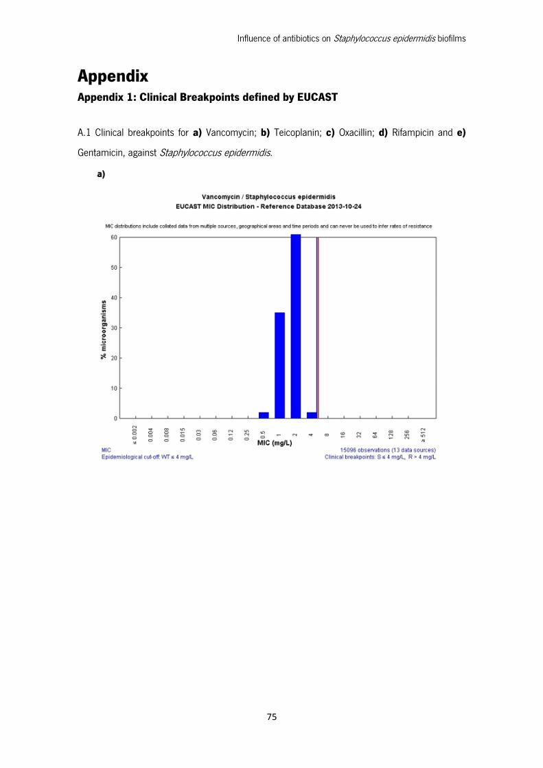
Influence of antibiotics on Staphylococcus epidermidis biofilms
75
Appendix Appendix 1: Clinical Breakpoints defined by EUCAST
A.1 Clinical breakpoints for a) Vancomycin; b) Teicoplanin; c) Oxacillin; d) Rifampicin and e)
Gentamicin, against Staphylococcus epidermidis.
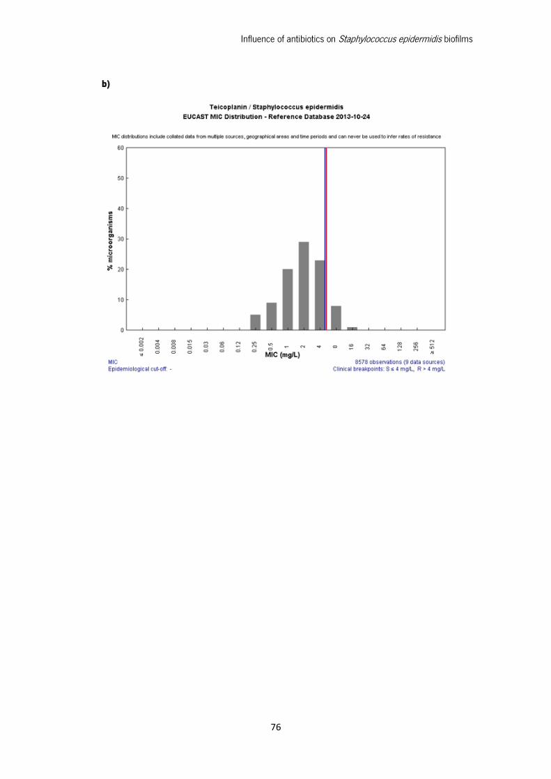
Influence of antibiotics on Staphylococcus epidermidis biofilms
76
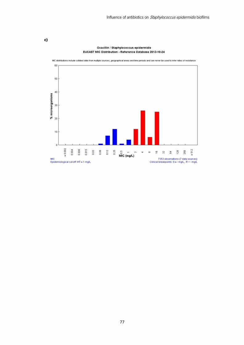
Influence of antibiotics on Staphylococcus epidermidis biofilms
77
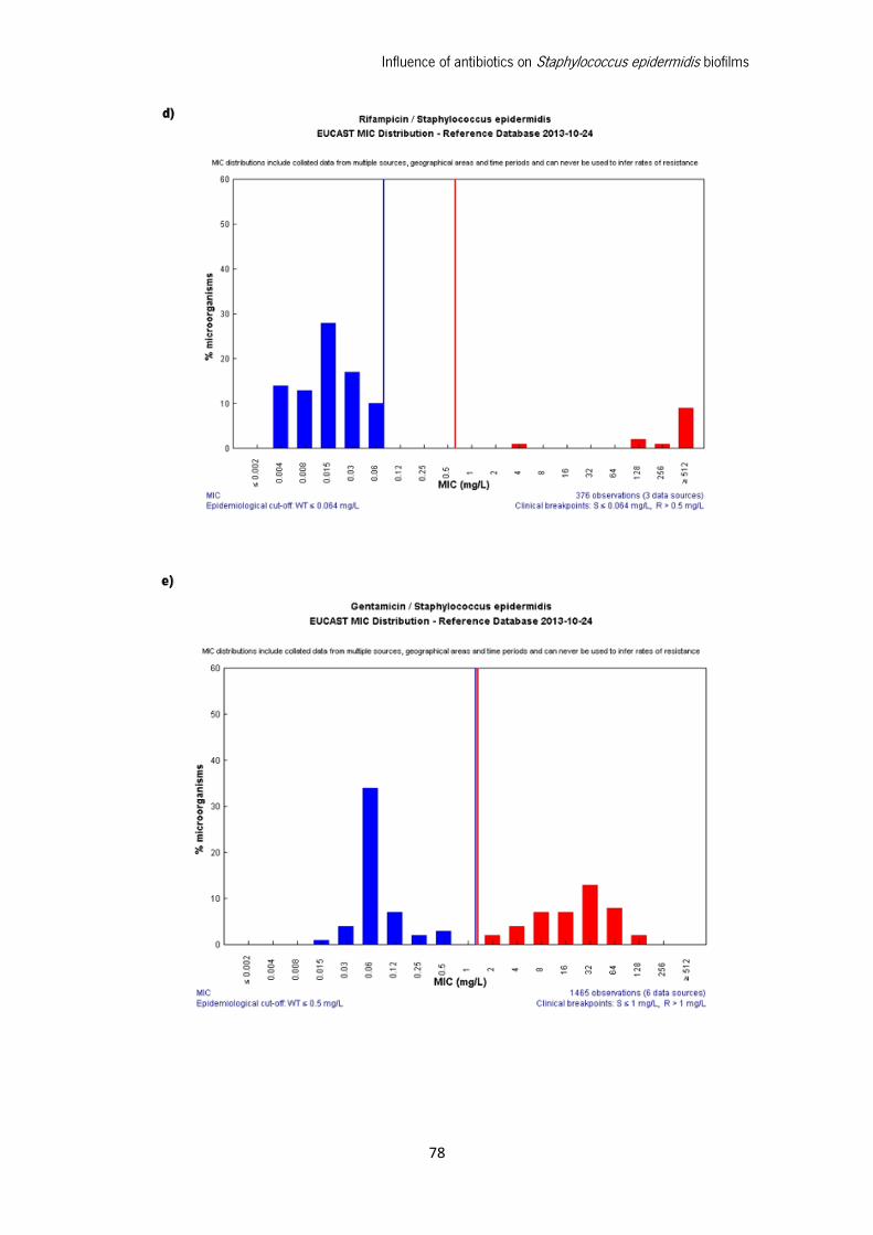
Influence of antibiotics on Staphylococcus epidermidis biofilms
78