ORIGINAL ARTICLE ASSOCIATION BETWEEN CLINICAL …
12
63 Rev Patol Trop Vol. 46 (1): 63-74. jan.-mar. 2017 doi: 10.5216/rpt.v46i1.46295 1. Programa de Pós-Graduação em Saúde, Medicina Laboratorial e Tecnologia Forense, Universidade do Estado do Rio de Janeiro (UERJ), Rio de Janeiro, Brazil. 2. Faculdade de Ciências Médicas, UERJ, Rio de Janeiro, Brazil. 3. Laboratório de Parasitologia do Hospital Universitário Pedro Ernesto, Rio de Janeiro, Brazil. 4. Laboratório de Avaliação e Promoção da Saúde Ambiental (LAPSA), Instituto Oswaldo Cruz, FIOCRUZ, Rio de Janeiro, Brazil. Corresponding author: Dr. Maurício Carvalho de Vasconcellos. Laboratório de Avaliação e Promoção da Saúde Ambiental (LAPSA), Av. Brasil 4365, Pavilhão Lauro Travassos, Instituto Oswaldo Cruz, FIOCRUZ. 21045-900 Rio de Janeiro, RJ, Brazil. E-mail: mau@ioc.fiocruz.br Received for publication: 24/6/2016. Reviewed: 29/8/2016. Accepted: 15/3/2017. ORIGINAL ARTICLE ASSOCIATION BETWEEN CLINICAL INDICATIONS AND COPROPARASITOLOGICAL TESTS CARRIED OUT AT THE PEDRO ERNESTO UNIVERSITY HOSPITAL (HUPE/UERJ) IN RIO DE JANEIRO, BRAZIL Thiely Rodrigues Ott 1 , Raphael Javert 2 , Nívia Oliveira 3 , Alexandre Ribeiro Bello 1,2 and Maurício Carvalho de Vasconcellos 4 ABSTRACT Enteroparasitosis is a public health problem in Brazil. Clinical indications and the appropriate stool examination are essential to obtain an adequate result. This study aims to evaluate whether the clinical indications and the choice of coproparasitological tests requested by the medical services may influence the diagnosis of enteroparasitosis. The data was obtained from the records in the Laboratory of Parasitology at the Pedro Ernesto University Hospital (HUPE/ UERJ) of the State University of Rio de Janeiro (UERJ) from 2009 to 2014. The qualitative variables were grouped in medical services (medical surgery, infectious and parasitic diseases, gastroenterology, pediatrics and rheumatology); types of tests requested (parasitological stool examination (PSE), merthiolate-iodine-formaldehyde (MIF), and sodium-acetate acetic acid- formaldehyde (SAF)) and clinical indications (anemia, diarrhea, abdominal pain, eosinophilia, routine tests, HTLV patients, HIV patients, parasitosis and transplantation research). The chi square (X²) and the Spearman coefficient correlation tests were performed to calculate the association between the clinical indications and the coproparasitological tests. A significant association was evident in the clinical indication: parasitosis found among the MIF tests and Trichrome Wheatley (ρ = 0.980). In other clinical indications such as anemia, surgery/ transplant, diarrhea, patients with HIV, HTLV and eosinophilia (despite the PSE tests and MIF having presented a strong link (ρ = 0.802), there was no significant association among the tests. Clinical indications are essential and they have a great influence on the parasitological diagnosis, requiring a combination of diagnostic methods for the detection of protozoa and helminths of medical interest. KEY WORDS: Enteroparasitosis; clinical indications; coproparasitological.
Transcript of ORIGINAL ARTICLE ASSOCIATION BETWEEN CLINICAL …

63Rev Patol Trop Vol. 46 (1): 63-74. jan.-mar. 2017
doi: 10.5216/rpt.v46i1.46295
1. Programa de Pós-Graduação em Saúde, Medicina Laboratorial e Tecnologia Forense, Universidade do Estado do Rio de Janeiro (UERJ), Rio de Janeiro, Brazil.2. Faculdade de Ciências Médicas, UERJ, Rio de Janeiro, Brazil.3. Laboratório de Parasitologia do Hospital Universitário Pedro Ernesto, Rio de Janeiro, Brazil.4. Laboratório de Avaliação e Promoção da Saúde Ambiental (LAPSA), Instituto Oswaldo Cruz, FIOCRUZ, Rio de Janeiro, Brazil.
Corresponding author: Dr. Maurício Carvalho de Vasconcellos. Laboratório de Avaliação e Promoção da Saúde Ambiental (LAPSA), Av. Brasil 4365, Pavilhão Lauro Travassos, Instituto Oswaldo Cruz, FIOCRUZ. 21045-900 Rio de Janeiro, RJ, Brazil. E-mail: [email protected]
Received for publication: 24/6/2016. Reviewed: 29/8/2016. Accepted: 15/3/2017.
ORIGINAL ARTICLE
ASSOCIATION BETWEEN CLINICAL INDICATIONS AND COPROPARASITOLOGICAL TESTS CARRIED
OUT AT THE PEDRO ERNESTO UNIVERSITY HOSPITAL (HUPE/UERJ) IN
RIO DE JANEIRO, BRAZIL
Thiely Rodrigues Ott1, Raphael Javert2, Nívia Oliveira3, Alexandre Ribeiro Bello1,2 and Maurício Carvalho de Vasconcellos4
ABSTRACT
Enteroparasitosis is a public health problem in Brazil. Clinical indications and the appropriate stool examination are essential to obtain an adequate result. This study aims to evaluate whether the clinical indications and the choice of coproparasitological tests requested by the medical services may influence the diagnosis of enteroparasitosis. The data was obtained from the records in the Laboratory of Parasitology at the Pedro Ernesto University Hospital (HUPE/UERJ) of the State University of Rio de Janeiro (UERJ) from 2009 to 2014. The qualitative variables were grouped in medical services (medical surgery, infectious and parasitic diseases, gastroenterology, pediatrics and rheumatology); types of tests requested (parasitological stool examination (PSE), merthiolate-iodine-formaldehyde (MIF), and sodium-acetate acetic acid-formaldehyde (SAF)) and clinical indications (anemia, diarrhea, abdominal pain, eosinophilia, routine tests, HTLV patients, HIV patients, parasitosis and transplantation research). The chi square (X²) and the Spearman coefficient correlation tests were performed to calculate the association between the clinical indications and the coproparasitological tests. A significant association was evident in the clinical indication: parasitosis found among the MIF tests and Trichrome Wheatley (ρ = 0.980). In other clinical indications such as anemia, surgery/transplant, diarrhea, patients with HIV, HTLV and eosinophilia (despite the PSE tests and MIF having presented a strong link (ρ = 0.802), there was no significant association among the tests. Clinical indications are essential and they have a great influence on the parasitological diagnosis, requiring a combination of diagnostic methods for the detection of protozoa and helminths of medical interest.
KEY WORDS: Enteroparasitosis; clinical indications; coproparasitological.

64 Rev Patol Trop Vol. 46 (1): 63-74. jan.-mar. 2017
INTRODUCTION
Enteroparasitic infections are among the most common infectious diseases found in all geographic areas of the planet, mainly in tropical regions. It is estimated that the number of infected people in the world is approximately 3.5 billion (Hotez et al., 2008). In Brazil, parasitoses are widespread and highly prevalent; 130 million people are affected by some kind of intestinal parasite (Eyre et al., 2013). Parasitic diagnosis is based on the microscopic examination of fecal samples. In laboratory routines it is important to conduct more than one laboratory method to detect the parasitic forms of protozoa and helminths, especially when there is a low parasite load (Cox, 2002). In order to provide appropriate care for patients; evaluate the effectiveness of drugs; monitor the effect of public health programs and increase understanding of intestinal parasites, the evaluation of the performance of diagnostic tests is vital (Basso et al., 2008). The association of several diagnostic techniques applying different criteria is a means of identifying a greater number of parasitic structures, thus increasing diagnostic effectiveness. The parasitological diagnosis should rely on highly sensitive techniques and specificity that allow a visualization of parasitic intestinal structures, according to the specific treatment required for each patient (Carvalho et al., 2012). In spite of these diseases being prevalent and resulting in significant morbidity, they are often underestimated by health professionals. In addition to the sensitivity and specificity problems of techniques applied to diagnosis, and pre-analytical difficulties, the indication of which test to apply is extremely valuable in obtaining adequate result. It is essential that health professionals understand the parasite elimination routes in order not to request inappropriate scans and thus generate erroneous results (Coura, 2013). Mastering the foundation of parasitological methods allows for proper guidance in the form of collection and conservation of the fecal material, and enables the application of appropriate methods to assist in the diagnosis and confirmation of the clinical suspicion. The types of fecal analysis techniques requested by clinicians for parasitic infection can affect the detection of different evolutionary forms of the parasites, and thus the positivity of the test. The purpose of this study is to analyze the results of the general protocols utilized in the Laboratory of Parasitology at the Pedro Ernesto University Hospital (PEUH) in the State University of Rio de Janeiro (SURJ), with a view to verifying possible associations between the clinical indications provided by the hospital medical services and the tests requested by the physicians, and the results of the co-analytical tests for detection of intestinal parasites.

65Rev Patol Trop Vol. 46 (1): 63-74. jan.-mar. 2017
METHODS
After approval of the study by the Ethics Committee of HUPE/UERJ (protocol CAAE 41907115.5.0000.5259), the data were obtained from the records of the Laboratory of Parasitology at HUPE/UERJ from 2009 to 2014. The tests were performed with three fecal samples for one PSE assay, and one fecal assay for coccidia and trophozoite screening techniques. For the PSE (parasitological stool examination), without preservative, the following techniques were performed: Hofffman, Pons & Janer (1934), Kato–Katz (Kato & Miura, 1954; Katz et al., 1970), Baerman & Moraes (1917) and Faust et al. (1939) in order to identify the parasite at any stage of development. Stool preserved with MIF (Merthiolate-Iodine-formaldehyde) was analyzed through an adaptation of the Hoffmann-Lutz centrifugation technique (1919). The search for trophozoites was performed with a Wheatley Trichrome staining (Garcia et al.,1997) and the coccidia investigation was performed with a Safranin-Methylene Blue staining (Fayer et al., 1999), both preserved with SAF (Acetate Sodium, Acetic Acid and Formaldehyde) (Yang et al., 1977).
The qualitative variables were grouped and tabulated as follows: medical services (medical surgery, infectious and parasitic diseases, gastroenterology, pediatrics and rheumatology); clinical indications (anemia, diarrhea, abdominal pain, eosinophilia, routine tests, HTLV patients, HIV patients, parasitosis and transplantation investigation); types of tests requested: PSE, MIF, SAF, the latter included as subcategories, Wheatley Trichrome subcategories for the search for trophozoites, and Safranin-blue- Methylene for the coccidian investigation. A descriptive analysis based on the relative frequencies was performed to evaluate the percentage of fecal requests by medical services, as well as assessing the percentage of each test within these services. Statistical analysis was performed using the Stata/SE version 2.1 program and chi square x2 test to verify any association between the clinical indications and the positivity of parasitological tests. In addition Spearman’s rank correlation coefficient, identified by the Greek letter ρ (rho), was evaluated to estimate, within each clinical indication, the association between coproparasitological results from 2009 to 2014.
RESULTS
In the temporal analysis, a sharp drop in the requests for PSE was noted. In 2009, there were 3,885 requests for PSE without preservative, declining continuously over the years. The remaining tests ordered such as MIF, SAF with its subgroups TW (Trichrome Wheatley) and SAM (Safranin-blue methylene), remained constant throughout the period, as shown in Figure 1.

66 Rev Patol Trop Vol. 46 (1): 63-74. jan.-mar. 2017
Figure 1. Quantitative analysis of fecal tests requested from 2009 to 2014, at the Pedro Ernesto University Hospital, in Rio de Janeiro, Brazil. Vertical axis represents the number of tests requested for the period 2009 - 2014, the horizontal axis referring to the time line in years. Parasitological stool examination (PSE); coproparasitological examination with merthiolate-iodine-formaldehyde preservative (MIF); coproparasitological exam with sodium-acetate-acetic acid preservative and formaldehyde safranin-methylene blue (SAM); coproparasitological examination with sodium-acetate-acetic acid preservative and formaldehyde Trichrome Wheatley (TW).
The most frequently requested test in all clinics is the PSE without preservative ranging from 44.2% to 69.9%, followed by MIF (43.9%-21.2%), Trichrome Wheatley (26.7% - 0%) and Safranin-blue-Methylene (27.1% - 0%), as in Figure 2.
Figure 2. Percentage of types of coproparasitological examinations requested in the parasitology service according to each clinic from 2009 to 2014 at the Pedro Ernesto University Hospital, in Rio de Janeiro, Brazil. The vertical axis corresponds to the requests for parasitological examinations in percentages and the horizontal axis corresponds to the medical clinics. Parasitological stool examination (PSE); coproparasitological test with merthiolate-iodine-formaldehyde preservative (MIF); coproparasitological test with sodium-acetate-acetic acid and formaldehyde safranin-methylene blue preservative; coproparasitological test with sodium-acetate-acetic acid preservative and formaldehyde Trichrome Wheatley.

67Rev Patol Trop Vol. 46 (1): 63-74. jan.-mar. 2017
Regarding the frequency of intestinal parasites observed in each health clinic in Figure 3, in all clinics the detection of protozoa proved to be high, IPD (89%), gastroenterology (87%), surgical clinic (83%), pediatrics (79%) and rheumatology (73%). However, considering the helminths, this detection was lower: rheumatology (27%), pediatrics (21%), surgical clinic (17%), gastroenterology (13%) and IPD (11%).
Figure 3. Frequency of enteroparasitoses found according to each clinic from 2009 to 2014 at the Pedro Ernesto University Hospital - HUPE, in Rio de Janeiro, Brazil. The vertical axis corresponds to the percentage of intestinal parasites. The horizontal axis is divided into two large groups of helminths and protozoa, according to each clinic.
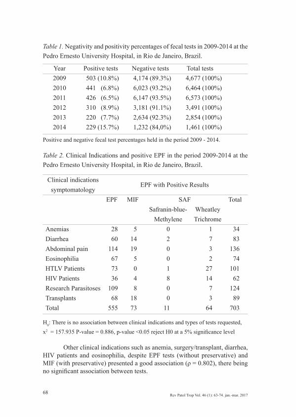
Considering the period from 2009 to 2014, the percentage of negative results in fecal tests is noticeable, ranging from 84.3% in 2014 to 93.5% in 2011. However, the percentage of positive fecal tests varies from 15.7% in 2014 to 6.5% in 2011, as described in Table 1.
Correlating clinical indications and positive coproparasitological tests, there is an association between these attributes (x2 = 157.935, p-value = 0.886) (Table 2).
The Spearman Correlation Test for each clinical indication during the study period enabled the positivity evaluation among coproparasitological tests. According to the results in Table 2, there was a strong positive and statistically significant association in clinical research indicating parasitosis among MIF tests with preservative and Trichrome Wheatley (ρ = 0.980). The MIF with preservative had a good association with EPF without preservative (ρ = 0.828), but they were not statistically significant. For the indication, abdominal pain and the EPF with no preservatives had a strong positive association with MIF with preservative (ρ = 0.897). Both tests have the same influence on the positivity of this condition.
68 Rev Patol Trop Vol. 46 (1): 63-74. jan.-mar. 2017
Table 1. Negativity and positivity percentages of fecal tests in 2009-2014 at the Pedro Ernesto University Hospital, in Rio de Janeiro, Brazil.
Year Positive tests Negative tests Total tests2009 503 (10.8%) 4,174 (89.3%) 4,677 (100%) 2010 441 (6.8%) 6,023 (93.2%) 6,464 (100%) 2011 426 (6.5%) 6,147 (93.5%) 6,573 (100%) 2012 310 (8.9%) 3,181 (91.1%) 3,491 (100%) 2013 220 (7.7%) 2,634 (92.3%) 2,854 (100%) 2014 229 (15.7%) 1,232 (84,0%) 1,461 (100%)
Positive and negative fecal test percentages held in the period 2009 - 2014.
Table 2. Clinical Indications and positive EPF in the period 2009-2014 at the Pedro Ernesto University Hospital, in Rio de Janeiro, Brazil.
Clinical indications symptomatology
EPF with Positive Results
EPF MIF SAF TotalSafranin-blue-
MethyleneWheatley Trichrome
Anemias 28 5 0 1 34Diarrhea 60 14 2 7 83Abdominal pain 114 19 0 3 136Eosinophilia 67 5 0 2 74HTLV Patients 73 0 1 27 101HIV Patients 36 4 8 14 62Research Parasitoses 109 8 0 7 124Transplants 68 18 0 3 89Total 555 73 11 64 703
H0: There is no association between clinical indications and types of tests requested, x2 = 157.935 P-value = 0.886, p-value <0.05 reject H0 at a 5% significance level
Other clinical indications such as anemia, surgery/transplant, diarrhea, HIV patients and eosinophilia, despite EPF tests (without preservative) and MIF (with preservative) presented a good association (ρ = 0.802), there being no significant association between tests.

69Rev Patol Trop Vol. 46 (1): 63-74. jan.-mar. 2017
DISCUSSION
Based on the results noted in the quantitative analysis of application for fecal tests requested in the period 2009-2014, a decrease was noted in requests for fecal tests over the 6 years. Basso et al. (2008) demonstrated through statistical studies of regression and temporal analysis that there was a decrease in the frequency of intestinal parasites over a period of 35 years in the South and Southeast regions. It can be assumed that this reduction in the frequency of intestinal parasites caused the decline in the requests for fecal tests presented in this study (Uecker etal., 2007; Neves et al., 2011; Siqueira et al., 2011; Knopp et al., 2014).
Regarding the coproparasitological analysis, the test most frequently requested was the EPF without preservative; it was indicated by the pediatric clinics (26%) and IPD (49%) as the first choice coproparasitological test corroborating the literature data ( Kaminsky et al., 2004; Bachur et al., 2008; Hotez et al., 2008; Neves et al., 2011; Becker et al., 2013; Braz et al., 2014).
The high negativity reported in this study raises some hypotheses to be considered, as for example, the fact that during the medical appointment, the clinicians request coproparasitological tests and also prescribe medication; this may cause false negative results. The literature indicates that drug therapies may interfere in the EPF results because some drugs, such as laxatives, hinder the recognition of the protozoa and lead to an underestimated prevalence of enteroparasites (Polman et al., 2015).
The enteroparasitosis prevalence is related to poor sanitation, poor hygiene and poor quality in health care. Another hypothesis for the high negativity observed in our study, considering only the location under study, would be the high/average socioeconomic status of the population assisted by the hospital. Some studies mention an oscillation between positivity and negativity according to the region. Carrillo et al. (2005) observed a positivity of 53% of enteroparasitosis in Morro de Santana, in the city of Ouro Preto/Minas Gerais. Epidemiological studies on intestinal parasitosis are carried out in several Brazilian states. However, few studies compare the environmental risk factors versus the intestinal parasitosis in the spread of the disease. The most consistently studied factors are socio-economic conditions, housing, basic sanitation, and drinking water; which were not analyzed in this study (Sousa et al., 2011).
Hurtado et al. (2005) found a rate of 72.8% positivity in the Amazon region. Researchers from Rio Grande do Sul, including Ely & Engroff (2006), obtained only 12.9% positivity. Engroff (2014) noted 10.8% positive samples and Larré (2014) only 4.0%. Our study identified 6.8% positive samples in 2013, indicating an oscillation of positivity and negativity among the various Brazilian regions.

70 Rev Patol Trop Vol. 46 (1): 63-74. jan.-mar. 2017
We should take into account that the high negativity observed in this study may be justified by potential problems in the pre-analytical variables, such as appropriate sample preparation, storage and preservation of specimens submitted for diagnosis, permanent quality control of reagents, equipment monitoring, supervision and suitable training of technical staff, and use of procedure manuals.
In the parasitological examination of feces, test repetition with a different sample is recommended in the case of a negative result (Neves, 2011). The World Health Organization (WHO) determines the reading of three slides for each parasitological sample. This decreases the possibility of negative results, especially when there is concentration of the analyzed samples. It is emphasized that each technique is specific to detect certain parameters. Thus, an association between the techniques increases the reliability of the results, and at least three methods are recommended in the EPF (Coura, 2013).
Detection and identification of intestinal parasites are in direct relation to the quality of the sample delivered to the laboratory (Souza & Amor, 2010). Based on the results, there is some unreliability in the type of parasitological technique, due to the fact that the most frequently used technique is the EPF without preservative, which may lead to an increase in the false-negative results. As highlighted by Coura (2013) and Neves et al. (2011), this method is mainly suitable for the investigation for Schistosoma mansoni eggs, and eggs and larvae of other helminths; however it presents limited or no relevance to the search of Enterobius vermicularis eggs, and protozoan cysts and oocysts.
There is no method able to diagnose all parasitic forms at the same time. Some methods are more general, easy to perform, inexpensive, and routinely used, allowing diagnosis of multiple intestinal parasites. However, the use of specific techniques in the laboratory routine is critical and contributes to the reduction of false-negative results, since some species of parasites are only evidenced by special techniques. An isolated test in which the result is negative should not be conclusive.
Explanation by the professionals to the population is necessary in order to have quality samples in the laboratory (Neves et al., 2011).
Considering the results obtained in this study, the high negativity may be influenced by the fact that the medical requests are not suitable for the research. Some studies have shown that the clinical requisition is an important tool and serves as a means of communication between clinicians and laboratory (Bates & Gawande, 2003; Laposata et al., 2004; Fernandez et al., 2015).
A survey conducted on the medical expertise in the parasitology area showed that, even when there is proper diagnostic suspicion of the disease, failures might occur in the process. This usually happens due to the lack of laboratory tests for the disease. Most interviewees (60.2%) think that laboratories are able to detect the parasite on a routine parasitological feces test, without a doctor’s request for a specific investigation. In fact, no investigation for any parasite that has not been previously requested by the doctor is performed (Mezzari et al., 2002).

71Rev Patol Trop Vol. 46 (1): 63-74. jan.-mar. 2017
Laboratory identification is important, not only to establish the diagnosis, but also to adjust epidemiological and public health programs, especially considering that parasites are a public health problem both in developed and developing countries (Hotez et al., 2008).
Enteroparasitosis have a spectrum of clinical manifestations, ranging from asymptomatic cases to mild and severe forms. In the most common presentation, there are unspecific symptoms such as anorexia, anemia, irritability, sleep disorders, nausea, vomiting, abdominal pain and diarrhea. The severe cases occur in increased parasite load, immune-compromised and malnourished patients. The onset or exarcebation of malnutrition occurs through damage to mucous (Giardia intestinalis, Necator americanus, Strongyloides stercoralis, coccidia), altered bile salt metabolism (Giardia intestinalis), food competition (Ascaris lumbricoides), intestinal exudation (Giardia intestinalis, Strongyloides stercoralis, Necator americanus, Trichuris trichiura), favoring bacterial proliferation (Entamoeba histolytica) and bleeding (Necator americanus, Trichuris trichiura), thereby causing anemia (Cox, 2002; Bachur et al., 2008; Hotez et al., 2008; Coura, 2013).
Among the associations between the clinical indications and coproparasitological examinations requested by the medical services, significant association were detected between parasite investigation and MIF and Trichrome Wheatley tests, both with similar positivity. A significant association was found in abdominal pain: EPF without preservative and MIF, both also with comparable positivity.
Knowledge of parasitic infections becomes an essential tool for the diagnosis of intestinal parasites in order to provide appropriate patient care. This study evidenced a decrease in the requests for coproparasitological exams along the years, due to the advance of techniques in molecular biology and immunology. Coproparasitological analyses are becoming second choice tests usually being replaced by a more advanced technique.
Among the coproparasitological analyses performed by the Laboratory of Parasitology at HUPE/UERJ, the EPF without preservative is the most requested by clinicians, because the test is practical, fast and inexpensive. However, efficiency and increased sensitivity and specificity require the combination of this exam with other specific diagnostic methods for the detection of protozoa and helminths.
The data reported here could be useful to clinicians, in order to increase their attention during anamnesis and clinical indications before ordering the tests for patients with suspected intestinal parasites. The information provided by clinicians is extremely important for a reliable diagnosis. The pre-analytical variables are important in preventing false negative results. In addition, regarding the high negativity, we believe that the region where these patients come from is not endemic, presenting adequate sanitation and reasonable economic conditions. We can also attribute the high negativity to the initiation of therapy, before carrying out any coproparasitological tests, and thus increasing false negative results.

72 Rev Patol Trop Vol. 46 (1): 63-74. jan.-mar. 2017
The symptoms are relatively mild or non-specific, which influences the particular nature of the clinical indication. The long incubation periods and the lack of adequate laboratory methods contribute to underestimate the prevalence of enteroparasitosis. This study might also be useful for parasitologists in order to obtain adequate information and plan the implementation of appropriate tools to provide an accurate laboratory diagnosis of parasitic infections.
ACKNOWLEDGEMENTS
The authors acknowledge the contribution of the biologists Denise Costa Lisboa and Marcos Santana Tobias Miglionico from the Laboratory of Parasitology of the Pedro Ernesto University Hospital (HUPE/UERJ) in Rio de Janeiro, Brazil. In addition, we are grateful to the doctoral student Simone de Souza Cardoso from the Institute of Social Medicine of the Rio de Janeiro State University for her contribution to the statistical review and suggestions about the text. Financial support. Rio de Janeiro State University.
REFERENCES
1.Bachur TPR, Vale JM, Coelho ICB, Queiroz TRBS de, Chaves C de S. Enteric parasitic infections in HIV/AIDS patients before and after the highly active antiretroviral therapy. Braz J Infect Dis 12: 115-122, 2008.
2. Baermann, G. Eine Einfache Methode zur Auffindung von Ankylostomun (Nematoden) Larven in Erdpnoben. Mededeel. mit h. Geneesk. Lab. Weltreve Feestbundel Batavia, 1917. (p.41-47).
3.Basso RMC, Silva-Ribeiro RT, Soligo DS, Ribacki SI, Callegari-Jacques SM, Zoppas BCDA. Evolução da prevalência de parasitoses intestinais em escolares em Caxias do Sul, RS. Rev Soc Bras Med Trop 41: 263-268, 2008.
4.Bates D, Gawande A. Improving safety with information technology. N Engl J Med 348: 2526-2534, 2003.
5.Braz AS, Andrade CAF De, Mota LMH Da, Lima CMBL. Recomendações da Sociedade Brasileira de Reumatologia sobre diagnóstico e tratamento das parasitoses intestinais em pacientes com doenças reumáticas autoimunes. Rev Bras Reumatol 11: 1-13, 2014.
6. Becker SL, Vogt J, Knopp S, Panning M, Warhurst DC, Polman K, Marti H, von Müller L, Yansouni CP, Jacobs J, Bottieau E, Sacko M, Rijal S, Meyanti F, Miles MA, Boelaert M, Lutumba P, van Lieshout L, N’Goran EK, Chappuis F, Utzinger J. Persistent digestive disorders in the tropics: causative infectious pathogens and reference diagnostic tests. BMC Infect Dis 13: 37, 2013.
7.Carrilo. Prevalência de enteroparasitoses em escolares do bairro Morro de Santana no município de Ouro Preto, MG. Rev Bras Análises Clin 38: 191-193, 2005.
8.Carvalho GLX, Moreira LE, Pena JL, Marinho CC, Bahia MT, Coelho GLLM. A comparative study of the TF-Test®, Kato-Katz, Hoffman-Pons-Janer, Willis and Baermann-Moraes coprologic methods for the detection of human parasitosis. Mem Inst Oswaldo Cruz 107: 80-84, 2012.
9.Coura JR. Dinâmica das doenças infecciosas e parasitárias. 2 ed. Rio de Janeiro: Guanabara Koogan, 2013.
10.Cox FEG. History of Human Parasitology. Clin Microbiol Rev 4: 595-612, 2002.

73Rev Patol Trop Vol. 46 (1): 63-74. jan.-mar. 2017
11.Ely LS, Engroff P, Lopes GT. Prevalência de Enteroparasitos em Idosos. Pathogens and Global Health 8: 637-646, 2006.
12.Engroff P, Ely LS, Guiselli RS, Goularte HF, Gomes I, Viegas K, De Carli GA. Prevalência de Infecções Enteroparasitárias e Soroprevalência de Toxoplasma gondii em idosos atendidos pela Estratégia Saúde da Família de Porto Alegre. Ciência Saúde Coletiva 19: 3385-3393, 2014.
13. Eyre D, Vieira A, Linda M, Novaes F De. Fatores ambientais e socioeconômicos associados a ocorrência de enteroparasitoses em usuários atendidos na rede pública de saúde em Manaus , AM, Brasil. Biosci J 29:487-498, 2013.
14.Faust EC, Sawitz W, Tobie J, Odom V, Peres C, Lincicome DR. Comparative efficiency of various technics for the diagnosis of protozoa and helminthes in feces. Jornal Brasileiro de Parasitologia 25: 241-262, 1939.
15.Fayer R, Lewis EJ, Trout JM. Cryptosporidium parvum in Systers from Commercial Harvesting Sites in the Chesapeake Bay. Emerg Infect Dis 5: 706-716, 1999.
16.Fernandez-Millan R, Medina-Merodio J-A, Plata R, Martinez-Herraiz J-J, Gutierrez-Martinez J-M. A Laboratory Test Expert System for Clinical Diagnosis Support in Primary Health Care. Appl Sci 5: 222-240, 2015.
17.Garcia LS, Bruckner DA. Diagnostic Medical Parasitology. 3ed. Washington: ASM Press, 1997. (p.411-433).
18.Hoffman WA, Pons JA, Janer JL. The sedimentation-concentration method in schistosomiasis mansoni. J Public Health 9: 281-298, 1934.
19. Hotez PJ, Brindley PJ, Bethony JM, King CH, Pearce EJ, Jacobson J. Review series Helminth infections : The great neglected tropical diseases. J Clin Invest 118: 1311–1321, 2008.
20.Hurtado-Guerrero AF, Alencar FH, Hurtado-Guerrero JC. Ocorrência de enteroparasitas na população geronte de Nova Olinda do Norte – Amazonas, Brasil. Acta Amaz 35: 487-490, 2005.
21.Kaminsky RG, Soto RJ, Campa A, Baum MK. Intestinal parasitic infections and eosinophilia in an human immunodeficiency virus positive population in Honduras. Mem Inst Oswaldo Cruz 99: 773-778, 2004.
22.Kato K, Miura M. Comparative examinations. Jap J Parasitol 3: 35, 1954.23.Katz N, Coelho PMZ, Pellegrino J. Evaluation of Kato’s quantitative method through the
recovery of Schistosoma mansoni eggs added to human feces. J Parasitol 56: 1030-1033, 1970.
24.Knopp S, Salim N, Schindler T, Karagiannis Voules D, Rothen J, Lweno O. Diagnostic accuracy of Kato-Katz, FLOTAC, Baermann, and PCR methods for the detection of light-intensity hookworm and Strongyloides stercoralis infections in Tanzania. Am J Trop Med Hyg 90: 535-545, 2014.
25.Laposata ME, Laposata M, Van Cott EM, Buchner DS, Kashalo MS, Dighe AS. Physician survey of a laboratory medicine interpretive service and evaluation of the influence of interpretations on laboratory test ordering. Arch Pathol Lab Med 12: 1424-1427, 2004.
26. Larré AB, Bürgie CD, Engroff P, De Carli GA. Prevalência de infecções por Enteroparasitos na população de idosos residentes em Instituições de longa permanência na região metropolitana de Porto Alegre e na Serra do Rio Grande do Sul. Porto Alegre. Rev Bras Geriatria Gerontologia 14: 637-646, 2014.
27.Lutz AO. Schistosoma mansoni e a schistosomose, segundo observações feitas no Brasil. Mem Inst Oswaldo Cruz 11: 121-155, 1919.
28.Mezzari A, Schenato LK, Richter VT, Bohme ES. Avaliação do conhecimento sobre criptosporidiose em uma amostra de médicos de Porto Alegre, Rio Grande do Sul, Brasil. Med Educ 38: 119-123, 2002.

74 Rev Patol Trop Vol. 46 (1): 63-74. jan.-mar. 2017
29.Neves DP, Wagner R, Vitor DA. Parasitologia Humana. 12 ed. Atheneu, Rio de Janeiro, 2011.30. Polman K, Becker SL, Alirol E, Bhatta NK, Bhattarai NR, Bottieau E, Martin W, Burza BS
, Coulibaly JT, Doumbia MN, Horié NS, Jacobs J , Basudha K ,Landouré A, Mahendradhata Y, Meheus F, Mertens P, Meyanti F, EH, Murhandarwati, EK , Ravinetto R, Sacko RM, Saye R, Schneeberger PHH, Schurmans C ,Silué KD, Mamadou JAT, Traoré S, Lieshout LV, Loeo VH ,Verdonck K, Müller LV , Yansouni CP, Yao JA,Yao PK,Yap P, Boelaert M, Chappuis F, Utzinger J.Diagnosis of neglected tropical diseases among patients with persistent digestive disorders (diarrhoea and/or abdominal pain ≥14 days): Pierrea multi-country, prospective, non-experimental case–control study. BMC Infectious Diseases 15: 338, 2015.
31.Siqueira LMV, Coelho PMZ, de Oliveira ÁA, Massara CL, Carneiro NF de F, Lima ACL. Evaluation of two coproscopic techniques for the diagnosis of schistosomiasis in a low-transmission area in the state of Minas Gerais, Brazil. Mem Inst Oswaldo Cruz 106: 844-850, 2011.
32. Souza RF, Amor ALM. Controle de qualidade de técnicas realizadas nos laboratórios de parasitologia da Secretaria Municipal de Saúde do Município de Salvador, Bahia. Rev Bras Anál Clín 42: 101-106, 2010.
33. Silva FM, Sousa MI, Carneiro EV, Silva EM, Diniz B, Pinho J, Borges LG, Cabral R, Figueiredo AC, Melo TA,Teixeira O, Azevedo A.R,Garcia G. Aspectos epidemiológicos e prevalência de enteroparasitoses em crianças do bairro Jambeiro,São Luís,MA. Rev Ciênc Saúde 13:123-130, 2011.
34.Uecker M, Copetti CE, Poleze L, Flores V. Infecções parasitárias: diagnóstico imunológico de enteroparasitoses. Revista Brasileira de Análises Clínicas 39: 15-19, 2007.
35.Yang J, Scholten T. A fixative for intestinal parasites permitting the use of concentration and permanent staining procedures. Am J Clin Pathol 67: 300-304, 1977.