POLIMORFISMOS DOS GENES ABO, LEWIS E SECRETORlivros01.livrosgratis.com.br/cp023447.pdf ·...
148
UNIVERSIDADE ESTADUAL PAULISTA “JÚLIO DE MESQUITA FILHO” FACULDADE DE CIÊNCIAS FARMACÊUTICAS CAMPUS DE ARARAQUARA POLIMORFISMOS DOS GENES ABO, LEWIS E SECRETOR CORRELACIONADOS COM O CÂNCER DE MAMA DÉBORA BARRETO TERESA GRADELLA ARARAQUARA-SP 2007
Transcript of POLIMORFISMOS DOS GENES ABO, LEWIS E SECRETORlivros01.livrosgratis.com.br/cp023447.pdf ·...

UNIVERSIDADE ESTADUAL PAULISTA
“JÚLIO DE MESQUITA FILHO”
FACULDADE DE CIÊNCIAS FARMACÊUTICAS
CAMPUS DE ARARAQUARA
POLIMORFISMOS DOS GENES ABO, LEWIS E SECRETOR
CORRELACIONADOS COM O CÂNCER DE MAMA
DÉBORA BARRETO TERESA GRADELLA
ARARAQUARA-SP
2007

Livros Grátis
http://www.livrosgratis.com.br
Milhares de livros grátis para download.

i
UNIVERSIDADE ESTADUAL PAULISTA
“JÚLIO DE MESQUITA FILHO”
FACULDADE DE CIÊNCIAS FARMACÊUTICAS
CAMPUS DE ARARAQUARA
DÉBORA BARRETO TERESA
POLIMORFISMOS DOS GENES ABO, LEWIS E SECRETOR
CORRELACIONADOS COM O CÂNCER DE MAMA
Tese apresentada ao curso de Pós-Graduação
em Análises Clínicas da Faculdade de
Ciências Farmacêuticas – UNESP –
Araraquara como pré-requisito para obtenção
do título de doutor
ORIENTADORA: Dra. CHRISTIANE PIENNA SOARES
ARARAQUARA-SP
2007

ii
Ficha Catalográfica
Elaborada Pelo Serviço Técnico de Biblioteca e Documentação Faculdade de Ciências Farmacêuticas
UNESP – Campus de Araraquara
Gradella, Débora Barreto Teresa
G732p Polimorfismos dos genes ABO, Lewis e Secretor correlacionados com o câncer de mama. / Débora Barreto Teresa Gradella . – Araraquara, 2007.
127 f. Tese (Doutorado) – Universidade Estadual Paulista. “Júlio de Mesquita
Filho”. Faculdade de Ciências Farmacêuticas. Programa de Pós Graduação em Análises Clínicas
Orientador: Christiane Pienna Soares . 1.Gene ABO. 2.Gene Lewis. 3.Gene Secretor. 4.Câncer de mama.
5.Polimorfismo. I. Soares, Christiane Pienna, orient. II. Título. CDD: 612.11825
CAPES:40300005

iii
COMISSÃO EXAMINADORA
Profa. Dra. Christiane Pienna Soares – Orientadora e Presidente
Profa. Dra. Sílvia Regina Rogatto – Membro Titular
Profa. Dra. Lílian Maria de Castilho – Membro Titular
Prof. Dr. Luiz Carlos de Mattos – Membro Titular
Prof. Dr. Haroldo Wilson Moreira – Membro Titular

iv
Tenha coragemcoragemcoragemcoragem para mudar o que
pode ser mudado, paciênciapaciênciapaciênciapaciência para
aceitar o que não pode ser mudado e
sabedoriasabedoriasabedoriasabedoria para diferenciar uma coisa
da outra.
Ditado Chinês

v
Dedicatória
Dedico este trabalho à minha
amada filha Heloísa, a maior
razão da minha vida.

vi
Agradecimentos Especiais
Ao meu querido marido
Alexandre, por toda sua compreensão, amor, paciência, apoio,
pelo exemplo de pai e marido e pela nossa maravilhosa filha.
À minha mãe amada
Maria Luiza, exemplo de mãe, por tudo fez e faz pelos seus filhos,
não medindo esforços para que nossa felicidade venha sempre em
primeiro lugar. Obrigada pelo amor, dedicação, apoio, paciência,
por cuidar da nossa nenezinha, enfim, por tudo...
Ao meu amado pai
José, pelo amor, paciência, apoio e por me oferecer todas as
condições para que eu pudesse chegar até aqui.
Aos meus queridos irmãos
Amanda e Fabrício, pelos bons momentos que passamos juntos,
pelos conselhos, pelo amor. Vocês me enchem de orgulho e alegria.
Aos meus padrinhos queridos
Maria do Carmo e Neto pela participação sempre constante na
minha vida, pela amizade, carinho e apoio.

vii
Agradecimentos
À Profa. Dra. Christiane Pienna Soares, pela oportunidade,
confiança, amizade e formação científica.
Ao Prof. Dr. Haroldo Wilson Moreira pela amizade, confiança,
paciência, apoio e por permitir o uso de seu laboratório.
Ao Prof. Dr. Luiz Carlos de Mattos, pelo auxílio e colaboração em
todas as etapas deste projeto.
Aos membros da banca Dra. Sílvia Rogatto, Dra. Lílian Maria de
Castilho, Dr. Luiz Carlos de Mattos e Dr. Haroldo Wilson Moreira
pelas críticas e sugestões, contribuindo para a melhoria do
trabalho.
Aos médicos Dr. Leonardo da Cunha, Dr. Hélio Humberto Angotti
Carrara e Dr. Nicolino Lia Neto por terem contribuído no
recrutamento das pacientes e no diagnóstico histopatológico.
À Profa. Dra. Catarina Satie Takahashi pela colaboração na
realização deste trabalho.
À Profa. Dra. Maria José Mendes Gianini por permitir o uso de
equipamentos do seu laboratório.
Ao Prof. Dr. Amauri Antiquera Leite pelos cartões utilizados na
fenotipagem do sistema Lewis.

viii
Às queridas amigas Luciane, Paula, Sami e Roberta, pela
amizade, paciência, auxílio e pelos bons momentos que passamos
juntas.
Aos meus familiares de Pereira Barreto, pelos momentos de
descontração e felicidade.
À Juliana Cintra e Raquel Alves Santos pela disposição, paciência
e auxílio na realização deste trabalho.
A Lúcia e Patrícia, pela paciência e coleta de sangue das
pacientes.
À Gisela, Maísa, Pâmela e Aline pela disposição em auxiliar nas
etapas finais deste trabalho.
A Elza Regina, pela realização da fenotipagem ABO das mulheres
doadoras de sangue.
Às funcionárias do Hemonúcleo Regional de Araraquara,
Cristiane e Marcy pela colaboração durante a coleta de sangue
das doadoras de sangue.
Ao Agenor por viabilizar o empréstimo da centrífuga.
Ao Max, Rosemira e Nancy, pelos momentos agradáveis de
muitas risadas.
Às funcionários da Seção de Pós-Gradução, Sônia, Laura e Cláudia
pela paciência e atenção.

ix
À Diamed pelo fornecimento dos cartões para fenotipagem Lewis
Aos voluntários participantes deste trabalho, pelas amostras
cedidas.
A todos que contribuíram direta ou indiretamente com este
trabalho.
Trabalho financiado pela FUNDUNESP

x
SUMÁRIO
Lista de Abreviaturas ....................................................................................... xii
Lista de Tabelas ................................................................................................ xiv
Lista de Figuras ................................................................................................ xv
Resumo ............................................................................................................. xvi
Abstract ............................................................................................................ xvii
1. Introdução ................................................................................................... 01
2. Justificativa ................................................................................................. 18
3. Objetivos ..................................................................................................... 20
4. Materiais e Métodos
4.1. Casuística
4.1.1. Grupo de Estudo ..................................................................
4.1.2. Grupo Controle ....................................................................
4.2. Metodologia
4.2.1. Coleta dos materiais.............................................................
4.2.2. Fenotipagem sorológica dos grupos ABO e Lewis .............
4.2.3. Extração e quantificação do DNA .......................................
4.2.4, Amplificação gênica para verificação dos alelos do locus
ABO ...............................................................................................
4.2.5. Identificação dos alelos do locus ABO por restrição
enzimática ......................................................................................
4.2.6. Amplificação gênica alelo específico para verificação de
mutações do gene FUT3 ................................................................
4.2.7. Definição e uso dos genótipos Lewis ..................................
4.2.8. Amplificação gênica alelo específico para verificação da
mutação G428A do gene FUT2 .....................................................
4.3. Análise estatística ............................................................................
23
24
25
25
25
26
28
31
33
34
35
5. Resultados ................................................................................................... 36
6. Discussão ..................................................................................................... 52
7. Conclusões ................................................................................................... 60
8. Referências Bibliográficas ......................................................................... 63

xi
9. Anexos
Artigo 1 ..........................................................................................
Artigo 2 ..........................................................................................
83
101

xii
LISTA DE ABREVIATURAS
as - antisense
Ca – Carcinoma
CADI – Carcinoma Ductal Invasivo
CADIS – Carcinoma Ductal in situ
CALIS – Carcinoma Lobular in situ
CCND1 – Ciclina D1
COMT – Catecol o-metiltransferase
CYP17 – Citocromo P450 família 17
DNA – Ácido desoxirribonucléico
FUT – Fucosiltransferase
Gal – Galactose
GalNAc – N-Acetilgalactosamina
GlcNAc – N-Acetilglicosamina
GSTP-1 – Glutationa S-tranferase 1
H - Heterozigoto
hGH – Hormônio do crescimento
Le – Lewis
M – Mutante homozigoto
MTHFR – metilenotetrahidrofolato redutase
pb – pares de base
PCR – Reação de Polimerase em Cadeia
PCR-AE - Reação de Polimerase em Cadeia – Alelo Específico
S - Selvagem

xiii
SDS – Dodecil Sulfato de sódio
Se – Secretor
TE – Tampão Tris-EDTA
XPD – Xeroderma Pigmentosum D

xiv
LISTA DE TABELAS
Tabela 1 - Primers utilizados para caracterização dos genótipos do grupo
sanguíneo ABO.....................................................................................................
27
Tabela 2 - Condições de reação utilizadas na amplificação dos fragmentos por
PCR.......................................................................................................................
27
Tabela 3 - Seqüência de primers sense e anti-sense e os alelos correspondentes
à ligação de cada primer. ......................................................................................
32
Tabela 4 - Tamanho dos produtos obtidos por PCR-AE para determinação das
mutações T59G, T1067A, T202C e C314T do gene Lewis (FUT3).....................
33
Tabela 5 - Seqüência de primers sense e anti-sense utilizados para
determinação da mutação G428A no gene FUT2.................................................
34
Tabela 6 – Freqüência de fenótipos e genótipos do grupo sanguíneo ABO nas
pacientes portadoras de câncer de mama e no grupo controle .............................
40
Tabela 7 – Características clinicopatológicas das pacientes portadoras de
câncer de mama associadas com o grupo sanguíneo ABO ..................................
42
Tabela 8 – Freqüência das mutações e dos alelos do gene FUT3 em pacientes
portadoras de câncer de mama e doadoras de sangue ..........................................
44
Tabela 9 – Genótipos obtidos a partir da associação de quatro polimorfismos
do gene FUT3 (T59G, T1067A, T202C e C314T) em pacientes portadoras de
câncer de mama (grupo em estudo) e doadoras de sangue (grupo controle)........
47
Tabela 10 - Mutações do gene FUT3 (T59G, T1067A, T202C e C314T)
encontradas nos indivíduos fenotipados como Lewis negativo: Le(a-b-).............
48
Tabela 11 – Características clinicopatológicas das pacientes portadoras de
câncer de mama associadas com os genótipos dos grupos Lewis e Secretor........
51

xv
LISTA DE FIGURAS
Figura 1 – Síntese dos antígenos ABH e Lewis................................................... 04
Figura 2 – Modelo Genético para o desenvolvimento do câncer de mama......... 14
Figura 3 - Representação dos exons 6 e 7 do gene ABO e as mutações
selecionadas para diferenciar os alelos A, B, e O...............................................
26
Figura 4 - Estratégia de análise e interpretação para caracterização dos alelos
do locus ABO. ......................................................................................................
29
Figura 5 – Padrão de bandas obtido com a digestão do fragmento de 249pb
com a enzima KpnI. Genótipos do grupo A (AA, AO1, AO2), genótipos do
grupo B (BO1, BO
2, BB) e genótipos do grupo O (O1
O1, O
1O
2 e O2O
2)..............
30
Figura 6 - Padrão de bandas de diferentes genótipos obtidos após digestão do
fragmento de 249pb com a enzima KpnI. ............................................................
37
Figura 7 - Padrão de bandas de diferentes genótipos obtidos após digestão do
fragmento de 467pb com enzima MspI. ...............................................................
38
Figura 8 - Padrão de bandas obtido pela digestão com NarI do fragmento de
467pb amplificado do exon 7. ..............................................................................
39
Figura 9 - Padrão de bandas obtido pela reação de amplificação alelo
específico (PCR-AE) para as mutações T59G, T1067A, T202C e C314T do
gene FUT3. ...........................................................................................................
45
Figura 10 - Padrão de bandas obtido pela PCR-AE para a mutação G428A do
gene FUT2 que amplifica um fragmento de 519/520pb. .....................................
49

xvi
RESUMO
A expressão de antígenos ABH e Lewis tem sido associada com o
desenvolvimento e prognóstico do câncer, diferenciação tumoral e metástase.
Considerando que o carcinoma ductal invasivo (CADI) de mama apresenta múltiplas
alterações, o objetivo deste estudo foi avaliar se o polimorfismo dos genes ABO, Lewis e
Secretor, bem como a fenotipagem ABO podem estar associados ao câncer de mama e
com parâmetros anatomoclínicos do tumor. Foram avaliadas 76 mulheres portadoras de
CADI e 78 mulheres doadoras de sangue para a fenotipagem/genotipagem ABO e
genotipagem Lewis e Secretor. Fenotipagem foi realizada por método de
hemaglutinação, a genotipagem ABO por PCR seguida de restrição enzimática e a
genotipagem dos grupos Lewis e Secretor por PCR alelo específico (PCR-AE). A
genotipagem foi realizada considerando o polimorfismo dos genes em um único
nucleotídeo, sendo que para o gene Lewis nas posições 59, 1067, 202 e 314, para o gene
Secretor na posição 428 e para o gene ABO nas posições 261 (alelo O1), 526 (alelos O2
e B) e 703 (alelo B). Os genótipos Lewis foram classificados em Lewis positivo (tipo
selvagem ou com duas/três mutações) e Lewis negativo (duas a quatro mutações). O
genótipo secretor foi classificado em Secretor (selvagem ou com a mutação em
heterozigose) e Não-secretor (homozigoto mutante). Nenhuma associação foi
encontrada entre as pacientes com câncer de mama e o fenótipo (P=0,9323) e genótipo
(P=0,9356) ABO. Mulheres com genótipo Lewis negativo foram associadas com CADI
(P=0,0126), mas não foram associadas com os parâmetros anatomoclínicos (tamanho
tumor, estadiamento TNM e metástase em linfonodos axilares). Genótipo Não-secretor
foi associado com metástase em linfonodos axilares (P=0,0149). Em conclusão, os
genótipos Lewis e Secretor podem ser úteis para predizer, respectivamente
suscetibilidade e metástase em linfonodos axilares.

xvii
ABSTRACT
ABH and Lewis antigens expression have been associated with cancer
development, prognosis, tumor differentiation and metastasis. Considering that
invasive ductal breast carcinoma (IDC) present multiple molecular alterations, the aim
of the present study was evaluated if the polymorphism of ABO, Lewis and secretor
genes, as well as ABO phenotyping could be associated to breast cancer and
anatomoclinical parameters of the tumor. In 76 women with IDC and 78 health women
blood donors, ABO phenotyping/genotyping, Lewis and Secretor genotyping were
evaluated. Phenotyping was performed by hemmaglutination method and genotyping by
Polymerase Chain Reaction with Sequence-Specific Primers (PCR-SSP). ABO, Lewis
and Secretor were classified by individual single nucleotide polymorphism (SNP) at
sites 59, 1067, 202 and 314 of the Lewis gene, 428 of the Secretor gene and 261 (O1
allele), 526 (O2 and B allele) and 703 (B allele). Lewis genotype was classified in
Lewis positive (wild-type or two/three SNPs) or Lewis negative (two to four different
SNPs). The Secretor genotype was classified in Secretor (wild type or SNP in
heterozygoses) and Non-secretor (SNP in homozygoses). No assocaition was found
between patients with breast cancer and ABO antigen expression (P=0.9323) and
genotypes (P=0.9356). Lewis negative was associated with IDC (P=0.0126), but were
not associated with anatomoclinical parameters (tumor size, TNM staging and axillary
lymph nodes metastasis). Non-secretor genotype was associated with axilary lymph
nodes metastasis (P=0.0149). In conclusion, the Lewis and Secretor genotyping could
be useful to predict, respectively, of breast cancer susceptibility and axillary lymph
nodes metastasis.

1
1. Introdução

2
O sistema de grupo sanguíneo ABO foi descoberto por Karl Landsteiner no
início do século XX. Os antígenos ABH foram inicialmente descobertos em eritrócitos,
entretanto sua distribuição tecidual em humanos é muito grande, sendo encontrados em
muitos tipos celulares, principalmente nas células epiteliais, mas não no músculo e
tecido conjuntivo (RAVN & DABELSTEEN, 2000; LE PENDU et al., 2001). Esses
antígenos são moléculas de carboidratos de glicoproteínas e glicolipídios presentes na
superfície de eritrócitos, células epiteliais e endoteliais, que podem estar presentes
também nos fluídos biológicos (YAMAMOTO et al., 1990a; YAMAMOTO &
HAKOMORI, 1990). Inicialmente, três fenótipos, A, B e O, foram caracterizados e
definidos por reações de aglutinação com eritrócitos e um quarto fenótipo AB foi
descoberto por De Castello e Sturli em 1902. Em 1924, ao analisar dados familiares,
Bernstein propôs um modelo de herança genética do sistema sanguíneo ABO contendo
três alelos, A,B e O, os quais são formas alternativas do mesmo locus gênico, herdados
de forma mendeliana. Através desse modelo permitiu-se a descrição de um
polimorfismo genotípico, compreendendo inicialmente seis genótipos (AA, AO, BB, BO,
AB, OO) (SALMON et al., 1984).
Os antígenos ABH são sintetizados em várias etapas, pela adição de
monossacarídeos pela ação de glicosiltranferases, a partir de cinco tipos de precursores
dissacarídeos: tipo1 , Galβ1-3GlcNAcβ1-R; tipo 2, Galβ1-4GlcNAcβ1-R; tipo 3,
Galβ1-3GalNAcα1-R; tipo 4, Galβ1-3GalNAcβ1-R e tipo 5, Galβ1-4Glcβ1-R Os
precursores tipo 1 e 2 podem fazer parte tanto de O ou N-glicoproteínas como de
glicolipídeos. Tipo 3 é encontrado exclusivamente em O-glicanas, tipo 4 em
glicolipídeos das séries globo e ganglio e tipo 5 corresponde ao glicolipídeo de
lactoceramida ou ceramida (MARIONNEAU et al., 2001). A adição nos precursores de
uma unidade de fucose em ligação α1,2 origina o antígeno H. Em humanos, o antígeno

3
H é determinado por dois tipos de α1,2-fucosiltransferases (enzimas H e Se) codificadas
pelos genes FUT1 (ou H) e FUT2 (ou Se) respectivamente. A enzima H regula a
expressão do antígeno H principalmente nas membranas de eritrócitos e em células do
endotélio vascular, agindo no precursor tipo 2. Já a enzima Se regula a expressão do
antígeno H principalmente em células do epitélio gastrointestinal e nos fluidos como
saliva, agindo tanto no precursor tipo 1 como no tipo 2, entretanto com preferenência
pelo tipo1 (LIU et al., 1998a ; LIU et al., 1999).
A perda funcional do alelo FUT1 e FUT2 leva ao fenótipo Bombay,
caracterizado pela ausência de antígenos ABH nos eritrócitos, enquanto a perda
funcional do alelo FUT2 é responsável pelo fenótipo não secretor, caracterizado pela
ausência dos antígenos ABH na saliva e em vários tipos de células epiteliais
(WATKINS, 2001). Uma vez o antígeno H formado, a biossíntese pode continuar pela
adição de uma N-acetilgalactosamina ou uma galactose, formando os antígenos A ou B
respectivamente. Esta etapa é catalisada pelas glicosiltransferases A ou B que são
codificadas por diferentes alelos no locus ABO do cromossomo 9. Uma série de alelos
deste locus foram descritos, alguns codificando variantes das enzimas A e B, outros
sendo inativados por várias mutações correspondendo ao alelo O (HAKOMORI, 1999).
A estrutura dos antígenos ABH e sua relação com outros antígenos, como, por exemplo,
antígenos Lewis presentes nos tecidos e adsorvidos aos eritrócitos, já é bem
caracterizada (WATKINS, 1995). A síntese dos antígenos ABH e Lewis está
demonstrada na figura 1.

4
Figura 1 – Síntese dos antígenos ABH e Lewis. α3GT – α1,3galactosiltransferase;
α3/4FT - α1,3 ou α1,4fucosiltransferase; α2FT - α1,2fucosiltransferase; α3ST -
α2,3sialiltransferase; enzimas A ou B são α1,3N-acetilgalactosaminil e
α1,3galactosiltransferases respectivamente. Extraído de MARIONNEAU et al., 2001.
Os antígenos Lewis fazem parte do esqueleto de proteínas (glicoproteínas) ou
lipídeos (glicolipídeos). As glicoproteínas Lewis podem ser encontradas nas secreções
tais como, leite, saliva ou plasma. Glicolipídeos, ao contrário, são encontrados ou em
complexos com lipoproteínas circulantes no plasma, ou inseridos em membranas
celulares com a porção glicídica voltada para o ambiente extracelular. Os antígenos
Lewis presentes nos eritrócitos, plaquetas e macrófagos são adquiridos secundariamente
do plasma (HENRY, 1996). A expressão dos antígenos Lewis requer duas diferentes
fucosiltrasferases (enzimas Se e Lewis), produtos de dois diferentes genes (FUT2 e
FUT3). A enzima Lewis pode usar como substrato tanto o precursor tipo 1 como o
precursor tipo 2. A enzima Lewis adiciona uma fucose, em ligação α(1,4), no precursor

5
tipo 1 produzindo o antígeno Lea que não pode ser posteriormente glicosilado. O
antígeno Leb é sintetizado quando o antígeno H, gerado pela enzima Se a partir do
precursor tipo 1, é usado como substrato pela enzima Lewis (SOEJIMA; KODA, 2005).
O termo ABH secretor, como é usado nos bancos de sangue, refere-se à secreção
dos antígenos de indivíduos do sistema ABO em fluidos como, saliva, suor, lágrima,
sêmem e soro, de acordo com o seu grupo sangüíneo. Por exemplo, um indivíduo do
grupo O secreta, nesses fluídos, o antígeno H, enquanto os indivíduos do grupo A
secretam antígenos A e H e assim sucessivamente (D'ADAMO; KELLY, 2001). Assim,
indivíduos Lewis-positivo e ABH não-secretores são fenotipados como Le (a+b-) e
indivíduos Lewis-positivo e ABH secretores como Le (a-b+). O fenótipo Le (a+b+)
pode ocorrer em indivíduos que expressam o antígeno H de maneira diminuída e
indivíduos que perderam o alelo funcional FUT3 são chamados Lewis negativo e são
caracterizados por eritrócitos com fenótipo Le(a-b-) (LE PENDU et al., 2001;
MARIONNEAU et al., 2001). Os antígenos sialil-Lea e sialil-Lex são obtidos pela
adição de um ácido siálico seguido pela adição de uma fucose nos precursores tipo 1 e 2
respectivamente (MARIONNEAU et al., 2001).
O locus ABO estende-se por uma região de 18/20kb, no cromossomo 9,
consistindo de 7 exons e 6 introns (YAMAMOTO et al., 1995; OLSSON; CHESTER,
2001). A análise da seqüência de nucleotídeos do cDNA da glicosiltransferase A1
revelou uma região codificadora de 1062pb, produzindo um polipeptídeo de 354
aminoácidos (YAMAMOTO et al., 1990b; OLSSON; CHESTER, 2001;). Esta enzima
possui três domínios: região N-terminal curta, região transmembrana hidrofóbica e um
longo domínio C-terminal responsável pela atividade catalítica. Como os exons 6 e 7 do
gene ABO são responsáveis pela tradução do domínio C-terminal das
glicosiltransferases, pesquisadores focaram nas diferenças destes exons para discriminar

6
os alelos ABO (LEE & REID, 2000) A seqüência do alelo A1 (A101) é tomada como
base de comparação em relação a todos os outros alelos do gene ABO diferenciando-se
do B em sete mutações: A297G, C526G, C657T, G703A, C796A, G803C e G930A, das
quais apenas 4 são responsáveis pela substituição de aminoácidos (YAMAMOTO, et
al., 1990a; LEE; REID, 2000). O alelo A2 (A201) é caracterizado pela mutação C467T
(uma substituição que não altera a atividade da transferase) e uma deleção em um dos
três resíduos consecutivos de citosina, no nucleotídeo 1059 – 1061, próximo à carboxila
terminal. Em conseqüência disso, são adicionados à transferase A mais 21 aminoácidos,
diminuindo entre 30 a 50 vezes a atividade da enzima e levando a um espectro limitado
de substrato aceptor (LEE; REID, 2000; YIP, 2000). Outros alelos A foram descobertos,
entre eles: A1v, A
3, A
x, A
el, variando na seqüência de nucleotídeos em diferentes posições
(YAMAMOTO et al., 1993; HANSEN et al., 1998; BARJAS-CASTRO, et al., 2000;
LEE; REID, 2000; OLSON; CHESTER, 2001;).
Foi nomeado como O1 (O01), o primeiro alelo O descrito, o qual difere do alelo
A1 em apenas uma posição: deleção de uma guanina no nucleotídeo 261 do exon 6. Essa
deleção é responsável pela criação de um códon de parada, ou seja, uma mutação sem
sentido, ocasionando a parada prematura na leitura do RNAm e a produção de uma
transferase de apenas 117 aminoácidos enzimaticamente inativa (YAMAMOTO, et al.,
1990a; YIP, 2000). Uma variação do alelo de O1, denominado O1v (O02) foi descoberto
posteriormente e, além de apresentar a deleção de uma única base no nucleotídeo 261,
possui também outras nove substituições de nucleotídeos (G106T, G188A, C189T,
C220T, A297G, T646A, G681A, C771T e G829A). Posteriormente foi determinado um
alelo O mutante, no qual a deleção na posição 261 estava ausente. Este alelo foi
denominado de O2 (O03) e se diferencia do alelo A
1 em quatro substituições de
nucleotídeos: A297G, C526G, G802A e G1096A. A mutação responsável pela perda da

7
atividade enzimática é a G802A, uma vez que a alteração do aminoácido glicina pela
arginina, na posição 268, está localizada na região da glicosiltransferase envolvida com
a atividade enzimática (YAMAMOTO, et al., 1990a; LEE; REID, 2000). Ainda, outros
alelos O foram descobertos: O3, O
4 e O
5 (OLSSON; CHESTER, 1996; OLSSON;
CHESTER, 2001).
O fragmento de DNA genômico que contém a região codificadora do gene
FUT2 (Se) responsável pela codificação da enzima secretora (Se) foi isolado em 1995
(KELLY et al., 1995; ROUQUIER et al., 1995). O DNA genômico completo e o cDNA
foram identificados em 1997 demonstrando que o cDNA corresponde a um simples
transcrito de 3,35kb e codifica uma proteína de 343 aminoácidos (KODA et al., 1997).
O gene FUT2 é formado por dois exons separados por um íntron (7kb). O primeiro exon
é um exon não-codificante, enquanto que o segundo exon contem a região codificadora
completa (KODA et al., 2000). A análise molecular deste gene demonstrou uma grande
variedade de alelos que levam à incapacidade funcional da enzima responsável pela
secreção dos antígenos ABH nas secreções (alelo se). A mutação sem sentido G428A
causa um alelo não secretor em Caucasianos sendo também freqüente em Africanos
(KELLY et al., 1995), enquanto que a substituição A385T produz um alelo não
funcional responsável pela produção de uma enzima deficiente na população oriental
(YU et al., 1995; HENRY, et al., 1996; KODA et al., 1996). Outras três mutações sem
sentido que tornam a enzima inativa (C571T, C628T e G849A) foram encontradas
também na região asiática (YU et al., 1995; HENRY, et al., 1996; KODA et al., 1996).
Muitas mutações inativadoras ou não tem sido descobertas, mas estudos em diferentes
populações têm demonstrado que a variedade de alelos produzida por estas mutações
está relacionada com a etnia (YU et al., 1995; HENRY, et al., 1996; KODA et al., 1996;
LIU et al., 1998; PANG et al., 1998b; PENG et al., 1999; YU et al., 1999).

8
O gene FUT3 (Le) está localizado no cromossomo 19 e o isolamento de seu
cDNA demonstrou uma região codificadora de 1083pb que codifica uma proteína de
361 aminoácidos (KUKOWSKA-LATALLO et al., 1990). O alelo funcional Le codifica
uma α(1,3/1,4)fucosiltransferase funcional e o alelo le codifica uma enzima não-
funcional. Indivíduos homozigotos para le não expressam tanto o antígeno Lea quanto o
Leb, expressando o fenótipo Le(a-b-) nos eritrócitos (KODA, et al., 2001). Várias
mutações têm sido encontradas em indivíduos Le(a-b-): T59G, G508A, T1067A,
T202C, C314T, C445T, G484A, G667A, G808A, G760A, G16A, G1022T e G47C
(ELMGREN et al., 1993; KODA et al., 1993; LIU, et al., 1996; ELMGREN et al.,
1997; PANG, et al., 1998a; PANG, et al., 1998b; LIU et al., 1999; COOLING et al.,
2003; SOEJIMA et al., 2004). O tipo e a freqüência dos diferentes alelos do gene FUT3
mostram diferenças em diferentes populações e grupos étnicos e, a correspondência
genótipo/fenótipo nem sempre é obtida (ELMGREEN et al., 1996; PANG et al., 1998b;
SALOMAA et al., 2000). Em estudos de populações com caucasianos, foi verificado
que 90 a 95% dos indivíduos Lewis negativo podem ser identificados pelo
polimorfismo em T59G, T1067A, T202C e C314T, e essas quatro mutações parecem
ser comuns na população em geral (SALOMAA et al., 2000). Estudos in vitro indicam
que as mutações T202C, G508A, C445T, G484A, G667A, G808A e T1067A são
mutações responsáveis pelo fenótipo Lewis negativo, enquanto que a mutação T59G, a
qual reside no domínio transmembrana, deve impedir a localização correta da enzima
alterando a especificidade do substrato (NISHIHARA et al., 1999; SOEJIMA; KODA,
2005).
A existência de associação entre os antígenos dos grupos sanguíneos e doenças
tem sido muito pesquisada e, significantes associações foram reportadas como infecções

9
microbianas, doenças cardiovasculares, doenças psiquiátricas e câncer
(MARIONNEAU et al., 2001).
Durante a transformação neoplásica, a expressão de antígenos dos grupos
sanguíneos se alteram profundamente, sendo que a expressão alterada de antígenos
ABH e Lewis tem sido encontrada em muitos tipos de carcinomas e está frequentemente
associada com o prognóstico (HAKAMORI, 1985; MARTENSSON et al., 1995;
HAKOMORI, 1996; SKOVLUND, 1997; NAKAGOE et al., 1998; XIE et al., 1999;
MARIANO et al., 2000; MOLDVAY et al., 2000). A presença de sialil-Lea e sialil-Lex
está associada com mau prognóstico em vários tipos de tumores, além disso, foi
demonstrado que esses antígenos permitem adesão de células cancerosas ao endotélio
vascular via reconhecimento por seletinas, podendo favorecer o processo metastático
(MARIONNEAU et al., 2001).
Estudos relatam que indivíduos dos grupos A e B podem ter probabilidade um
pouco maior de desenvolver um câncer do sistema digestivo que indivíduos do grupo O
(MOURANT et al., 1978). Isto pode ser explicado pelo aumento da resistência a
apoptose de células epiteliais que apresentam os antígenos A e B (LE PENDU et al.,
2001). Em contraste, indivíduos com grupo sanguíneo O que desenvolveram carcinoma
pulmonar possuem menor probabilidade de sobrevivência quando comparado aos do
grupo sangüíneo A e B (MOLDVAY et al., 2000). Este fato pode ser explicado
considerando que as células cancerosas de pacientes do grupo sanguíneo O, desprovidas
dos antígenos A e B, teriam uma maior motilidade, sendo mais facilmente propensas à
invasão aos vasos sanguíneos e linfáticos. Ainda, uma maior resistência basal a morte
celular aumenta a probabilidade de sobrevivência de células que acumularam alterações
genéticas e que deveriam ser eliminadas. Entretanto, num estágio mais avançado,
quando o fenótipo de "resistência a apoptose", torna-se estabelecido pela aquisição de

10
mecanismos mais potentes (mutações de p53, alterações da expressão de membros da
família Bcl-2 ou de proteínas de stress como hsp70), a perda da expressão dos antígenos
A, B e possivelmente H, facilitariam o processo metastático pelo aumento da motilidade
celular. Passado este estágio, as células que expressam antígenos sialil-Lea ou sialil-Lex
podem ser selecionadas, uma vez que possuem uma capacidade aumentada de se manter
em microvasos. Assim, a importância dos vários antígenos ABH e Lewis depende do
estágio de progressão do tumor (LE PENDU et al., 2001).
O câncer de mama é uma neoplasia extremamente incidente e associada a
morbidade e mortalidade entre as mulheres (KOIFMAN et al., 1998; BERSTEIN et al.,
1999; FERRINI et al., 2001; SILVA et al., 2002). Este tipo de doença aparece com
maior freqüência em certos países Europeus, na América do Norte e alguns países Sul
Americanos (SILVA et al., 2002). Os estados mais desenvolvidos e mais
industrializados como São Paulo, Rio de Janeiro e Rio Grande do Sul, apresentam maior
incidência desta doença (PAIVA et al., 2002; SILVA et al., 2002). De acordo com o
Instituto Nacional do câncer (INCA), a estimativa de incidência para 2006 será de
48.930 novos casos (BRASIL, 2007). A alta incidência está relacionada a diferentes
fatores como, por exemplo, o diagnóstico tardio entre as mulheres pertencentes às
classes de menor poder aquisitivo (ABREU e KOIFMAN, 2002).
Vários são os fatores de risco para o desenvolvimento do câncer de mama e
embora heterogeneidade da história natural do câncer de mama seja razoavelmente
conhecida, seus determinantes ainda permanecem desconhecidos (BERSTEIN et al.,
1999; ABREU; KOIFMAN, 2002). A etiologia envolve a interação de diversos fatores
de risco, e, além dos fatores endógenos (idade, história familiar, fatores reprodutivos,
obesidade, doença benigna da mama, lactação, abortos, paridade), os fatores exógenos
(dieta, fumo, exposição à radiação ionizantes, inseticidas organoclorados e atividade

11
física) também têm sido associados ao câncer de mama (KOIFMAN et al., 1998;
BERSTEIN et al. 1999).
Dentre os fatores endógenos, a história familiar está relacionada, em
aproximadamente 5 a 10% das pacientes com câncer de mama. Parece ser consenso que
o desenvolvimento do câncer de mama é mais comum nos países desenvolvidos, em
pacientes brancas e de classe social mais elevada. Entretanto, essa relação não constitui,
uma regra, podendo não se observar, algumas vezes, essas associações na dependência
da população amostrada (PAIVA et al., 2002).
A condição sócio-econômica pode refletir o prognóstico do câncer de mama
como conseqüência das dificuldades de acesso aos programas de prevenção e aos
cuidados médicos que as classes menos favorecidas encontram mesmo nos países
desenvolvidos, sendo o diagnóstico estabelecido numa fase já avançada da doença
(ABREU; KOIFFMAN, 2002).
Outro fator de risco para o desenvolvimento do câncer de mama bastante
controverso na literatura é o uso de contraceptivo oral ou reposição hormonal.
Resultados de estudos epidemiológicos têm demonstrado que o uso de contraceptivos
orais ou a terapia de reposição hormonal tem sido relacionado com o aumento do risco
de câncer de mama, inclusive nas pacientes que fazem uso do contraceptivo entre 5 a 10
anos (HANKINSON; STAMPER, 1997; TESSARO et al, 2001; LUKKAINEN, 2003).
Desta forma, a exposição de estrógeno pode promover um ambiente seletivo para o
crescimento clonal de células que já contenham mutações somáticas. Ainda, um longo
período de vida reprodutiva, bem como, fatores endógenos como a menarca precoce,
menopausa tardia e idade avançada para o primeiro filho, podem contribuir para o
aumento do risco de desenvolvimento de neoplasias na mama (KOIFMAN et al., 1998;
BERNSTEIN et al., 1999). A intensa atividade proliferativa ocasionada pela ação do

12
estrógeno pode resultar na replicação de células que contenham danos de DNA e cujos
supressores de tumor não consigam realizar reparo ou a indução de apoptose. Embora o
estrógeno possua ação de agente carcinogênico fraco, segundo The International
Agency for Research on Câncer (IARC), a terapia estrogênica pode ocasionar danos de
DNA por formação de radicais livres nas células epiteliais mamárias (KENEMANS e
BOSMAN, 2003).
Com relação à idade, o câncer de mama parece estar relacionado à faixa etária
entre 45 a 50 anos, com dois picos incidência: aos 50 e aos 70 anos (TESSARO et al.,
2001). Assim, o risco do desenvolvimento da doença aumenta gradualmente com a
idade, tendo um rápido aumento nos anos que proximamente antecedem a menopausa,
seguido de lenta elevação na menopausa, possivelmente em função do declínio na
liberação estrógeno do ovariano. Embora o desenvolvimento de câncer de mama em
pacientes com idade inferior a 35 anos seja considerado incomum, em média, 5% das
pacientes portadoras de câncer de mama apresentam idade inferior a 30 anos
(MAGGARD et al, 2003), estudos epidemiológicos têm documentado uma pior
evolução e um prognóstico desfavorável para essas pacientes (MAGGARD et al., 2003;
SIDONI et al., 2003). A influência da idade no momento do diagnóstico e para o
prognóstico de sobrevida do câncer de mama permanece ainda controversa. Estes
conflitos são resultantes da diversidade observada entre os diferentes estudos: pequeno
número de pacientes, diferença na estratificação das idades e da correção dos óbitos
ocorridos por outras causas (ABREU; KOIFMAN, 2002). Apesar das discordâncias
apresentadas, vários estudos destacam que pacientes com câncer de mama a partir
quinta década de vida, apresentam melhor prognóstico (JARVIS et al., 1998; SIDONI et
al., 2003).

13
Dentre as neoplasias malignas de mama, o carcinoma ductal invasivo (CADI) é
o mais comumente observado na rotina histopatológica, não pertencente a nenhuma das
outras categorias ou subtipos especiais de carcinoma invasivo. Sua prevalência varia de
45-75% de todos os carcinomas invasivos da mama e a classificação morfológica
baseia-se na histogênese e aspectos histopatológico (HANBY, 2005). O carcinoma
ductal invasivo é designado como um tumor infiltrativo, que mostra formação de um ou
mais túbulos ou lumens, compostos por células atípicas, organizados em agrupamentos
celulares sólidos pequenos ou grandes, geralmente pouco coesos, de forma irregular ou
angulada (BRASIL, 2002). No entanto, podem apresentar vários padrões morfológicos
diferentes e combinações de mais de um subtipo histológico pode estar presente em um
mesmo tumor. A adequada classificação ou tipagem histológica tumoral tem
implicações clínicas, pois um prognóstico especial é conferido apenas as formas puras
dos tipos especiais e os tumores mistos tendem a se comportar de forma intermediária
entre o carcinoma ductal que têm pior prognóstico e os tipos especiais de excelente
prognóstico (BRASIL, 2002; HANBY, 2005). O CADI é mais freqüente em mulheres
de meia idade ou depois dos 50 anos. O tumor apresenta-se, como uma massa detectada
na palpação ou mamografia, geralmente de 2-3 cm ou ocupando uma porção
significativa da mama. A pele pode estar comprometida com edema, “peau d`orange”
(como casca de laranja), retração do mamilo e/ou descarga mamilar, doença de Paget e
ulceração, significando doença avançada (TAVASOLI, 1992; HANBY, 2005).
Um modelo de carcinogênese de mama sugere que o carcinoma invasivo origina
de uma série de lesões hiperplásicas intermediárias através de vários graus de atipia que
evoluem ao carcinoma ductal in situ (CADIS) e deste até o carcinoma ductal invasivo
(KURBEL, 2005; PLATET et al., 2004). Um modelo genético faz algumas proposições
para o desenvolvimento do câncer de mama: 1) o carcinoma lobular (CAL) desenvolve-

14
se por um caminho genético que envolve inativação de caderina E; 2) mutações nos
genes p53 e HER2 estão associadas com câncer pobremente diferenciado; 3) mutações
nos genes BRCA1/2 em células germinativas levam ao desenvolvimento do carcinoma
ductal invasivo (VAN DE VIJVER, 2000).
Figura 2 – Modelo Genético para o desenvolvimento do câncer de mama.
Extraído de VAN DE VIJVER.Cur.Diagn.Pathol. v.6: p.271-281, 2000.
Recentes estudos têm procurado elucidar quais os mecanismos de carcinogênese
do tecido mamário, buscando marcadores de suceptibilidade ao câncer, permitindo o
diagnóstico precoce e o estabelecimento de conduta terapêutica apropriada. Diversos
estudos têm demonstrado associação entre polimorfismos de diversos genes com risco
Mutação p53, amplificação de HER2
Inativação do alelo selvagem BRCA1/2
Mutação germline BRCA1/2
LOH 16q
Precursor Comum?
CALIS
CADIS Bem
diferenciado
CADIS Pobremente diferenciado
Ca lobular invasivo
Ca ductal invasivo grau
I
Ca ductal invasivo grau
II
Ca ductal invasivo grau
III
?
Inativação de E-caderina
Ca ductal invasivo grau
III
CADIS moderadamente diferenciado

15
de suscetibilidade ao câncer de mama. Um estudo realizado em Nova Iorque
demonstrou que indivíduos expostos a hidrocarbonetos aromáticos e indivíduos
fumantes podem ter um risco maior ao desenvolvimento do câncer de mama se
apresentar um alelo variante do gene XPD, um gene envolvido no reparo do DNA por
excisão de nucleotídeos (TERRY et al., 2004). Também houve maior risco de
desenvolvimento de câncer de mama em indivíduos que apresentam um alelo variante
do gene COMT, responsável pelo metabolismo do metabólito do estrógeno, o estrógeno
catecol (CHENG et al., 2005).
Polimorfismos de outros genes também foram associados ao risco de câncer de
mama, tais como: 1) GSTP-I, que codifica a enzima glutationa S-transferase P1 que
possui importante função na detoxificação de carcinógenos (EGAN et al., 2004); 2) o
gene Aurora A que atua na regulação da duplicação do centrossomo (LO et al., 2005);
3) MTHFR responsável pela codificação da enzima metilenotetrahidrofolato redutase
envolvida no metabolismo do ácido fólico (CAMPBEL et al., 2002; CHOU et al.,
2006); 4) CCND1 que codifica a proteína ciclina D1, importante na regulação do ciclo
celular (CESCHI et al., 2005, SHU et al., 2005); 5) p53, que codifica a proteína p53,
uma proteína supressora de tumor envolvida no controle do ciclo celular e apoptose
(FRANEKOVA et al., 2007); 6) o gene CYP17 que codifica uma enzima envolvida na
biosíntese do estrógeno (MITRUNEN ; HIRVONEN, 2003).
Recentes avanços nos métodos em biologia molecular identificaram vários
fatores prognósticos que revelam mudanças inesperadas na progressão do câncer de
mama (SILVA et al., 2002, TAKASHIMA et al., 2002). Alguns biomarcadores são
reconhecidos como fatores prognósticos característicos como avaliação da expressão de
produtos protéicos de alguns genes supressores de tumor, oncogenes, marcadores de

16
proliferação celular e antígenos dos grupos sangüíneos (VAN DE VIJVER, 2000;
MOURÃO-NETO et al., 2001; NAKAGOE et al., 2002).
Trabalhos sobre a associação dos antígenos ABH e Lewis com o câncer de
mama são escassos na literatura, entretanto, há estudos que observaram a perda da
expressão dos antígenos A e B (VOWDEN et al., 1986; NAKAGOE et al., 1991; INAI
et al., 1992; URA et al., 1992) diminuição da expressão dos antígenos Lea e Leb (INAI
et al., 1992; URA et al., 1992), expressão de H e Ley (VOWDEN et al., 1986) e
aumento dos antígenos sialil-Lex e sialil-Lea nesses tumores (JESCHKE et al., 2005).
Além disso, foi observado que a perda da expressão dos antígenos A e B, correlaciona-
se com o aumento na expressão do antígeno H (VOWDEN et al., 1986). Ainda, o
comportamento metastático do tumor possivelmente se relaciona com a expressão de
antígenos do grupo sanguíneo (NAKAGOE et al., 1998), a expressão do antígeno sialil-
Lex em células tumorais está associada com mau prognóstico (NAKAGOE et al., 2002)
e a alta expressão de Ley/b relaciona-se com diminuição de sobrevida de pacientes com
nódulos linfáticos negativos (MADJD et al., 2005).
Os métodos sorológicos, amplamente empregados na investigação das
especificidades ABO e Lewis, permitem apenas a identificação dos fenótipos
eritrocitário, secretor e não secretor. A definição dos respectivos genótipos necessita de
estudos familiares, que muitas vezes são de difícil realização, bem como podem ser
inconclusivos em função da recente descrição dos diversos alelos silenciosos
(JOHNSON & HOPKINSON, 1992; GRUNNETT et al., 1994; YU et al., 1997). O uso
da técnica de PCR seguido pela verificação da existência ou não de sítios de restrição e
a PCR alelo específico tem mostrado ser de grande utilidade na definição dos genótipos
dos Sistemas ABO, Lewis e Secretor, principalmente aqueles que não podem ser
detectados pelos métodos sorológicos (CHANG et al., 1992; LEE; CHANG, 1992;

17
GRUNNET et al., 1994;). Portanto, estas metodologias podem ser amplamente
empregadas quando se pretende realizar estudos moleculares em sistemas polimórficos
associados a doenças, à medicina forense, ao monitoramento da evolução dos
transplantes de medula, à investigação dos subgrupos sanguíneos (YAMAMOTO et al.,
1993; CHANG et al., 1995; CROUSE & VINCEK, 1995; LIECHTI-GALLATI;
NEESER, 1996).

18
2. Justificativa

19
O câncer de mama é o tumor de maior incidência e mortalidade no mundo,
apresentando alta freqüência nos países europeus, América do Norte e alguns países Sul
Americanos. Recentes estudos têm procurado elucidar quais os mecanismos de
carcinogênese do tecido mamário, buscando marcadores de suceptibilidade ao câncer
que permitiriam o diagnóstico precoce e o estabelecimento de conduta terapêutica
apropriada, minimizando a morbi-mortalidade por esta neoplasia. Trabalhos sobre a
associação dos antígenos ABH e Lewis com o câncer de mama são escassos na
literatura, entretanto, alguns estudos observaram a perda da expressão dos antígenos A e
B, diminuição da expressão dos antígenos Lea e Leb e aumento dos antígenos Lex, sialil-
Lex e sialil-Lea nesses tumores. O comportamento metastático do tumor possivelmente
relaciona-se com a expressão de antígenos do grupo sanguíneo, e, em especial, a
expressão do antígeno sialil-Lex está relacionado a lesões mamárias de pior prognóstico.
Até o momento, segundo nosso conhecimento, não existem estudos de genotipagem de
grupos sanguíneos (ABO, Lewis e secretor) e sua possível associação com câncer de
mama e suas características clinicopatológicas, tais como tamanho tumoral, metástase
em linfonodo. Com o presente estudo espera-se encontrar associação positiva entre essa
neoplasia e grupo (s) sanguíneo(s) específico(s) e seu potencial como marcador da
carcinogênese em mama.

20
3. Objetivos

21
OBJETIVO GERAL
Verificar associação entre o carcinoma ductal invasivo de mama e os sistemas
ABO, Lewis e secretor e ainda, associação destes grupos sanguíneos com características
clinicopatológicas dos tumores
OBJETIVOS ESPECÍFICOS
1) Caracterizar o fenótipo do sistema ABO, bem como os polimorfismos dos genes
ABO, Lewis e Secretor em sangue periférico de pacientes portadoras de carcinoma
ductal invasivo e de mulheres doadoras de sangue, consideradas controle.
2) Verificar as correlações existentes dos resultados observados no grupo em estudo
frente ao grupo controle.
3) Verificar associação entre os polimorfismos dos genes ABO, Lewis e Secretor e
características clinicopatológicas dos tumores.

22
4. Materiais e Métodos

23
4.1. CASUÍSTICA
4.1.1. Grupo de Estudo
Após consentimento informado e de conformidade com o Comitê de Ética em
Pesquisa, foram avaliadas 76 pacientes, do sexo feminino, portadoras de carcinoma
ductal invasivo de mama (Grupo de Estudo). Destas, 49 pacientes foram recrutadas pelo
Dr. Hélio Humberto Angotti Carrara do Ambulatório de Ginecologia e Obstetrícia do
Hospital das Clínicas da Escola de Medicina de Ribeirão Preto – Universidade de São
Paulo (HCFMRP-USP) e 27 pacientes pelo Dr. Leonardo Cunha no Centro de
Oncologia da Cidade de Araraquara (CORA). A idade média destas pacientes foi 54,1
anos (mínino de 30 e máximo de 84 anos) e quando a cor ou raça, 81,6% foram da cor
branca, 11,8% preta, 6,6% parda e 0% amarela.
Ainda, para avaliação de associação entre os polimorfismos dos grupos ABO,
Lewis e Secretor e as características clinicopatológicas dos tumores, algumas mulheres
foram excluídas da avaliação, pois não apresentavam todos os dados necessários.
Assim, para este parâmetro, a casuística do grupo em estudo variou entre 71 e 75
pacientes.
A classificação dos tumores foi realizada de acordo com a classificação TNM
(Greene et al., 2002), sendo que T representa a extensão do tumor primário, N
representa a ausência ou presença e a extensão de metátase em linfonodos regionais e M
a ausência ou presença de metástase à distância. Assim seguindo esta classificação, as
seguintes definições são utilizadas: para T: TX quando o tumor primário não pode ser
avaliado; T0 quando não há evidência de tumor primário; Tis Carcinoma in situ e T1,
T2, T3, T4 refere-se ao tamanho crescente e/ou extensão local do tumor primário. Com
relação ao N, NX quando os linfonodos regionais não podem ser avaliados; N0 na
ausência de metástase em linfonodos regionais e N1, N2, N3 quando há

24
comprometimento crescente dos linfonodos regionais. Além disso, a extensão direta do
tumor primário para o linfonodo é classificada como metástase linfonodal. Metástase
em qualquer linfonodo que não seja regional é classificada como metástase à distância.
Para M, MX representa que a presença de metástase à distância não pode ser avaliada.
M0 indica ausência de metástase à distância e M1 metástase à distância.
Na avaliação dos dados clinicopatológicos utilizando a classificação TNM, foi
utilizado como parâmetros de comparação a dimensão do tumor primário (T),
comprometimento em linfonodos regionais (N), sendo considerado como negativo
quando N0 e positivo os classificados como N1 e N2. O parâmetro de metástase à
distância não foi avaliado, uma vez que o número de tumores com essa característica foi
muito pequeno (n=4). Outro parâmetro utilizado foi classificação por estadios, sendo:
estadio I: T1N0M0; estadio IIA: T1N1M0 ou T2N0M0; estadio IIB: T2N1M0 ou T3N0M0;
estadio IIIA: T1N2M0, T2N2M0, T3N1M0 ou T3N2M0; estadio IIIB: T4 com qualquer NM0
ou qualquer T N3M0; estadio IV qualquer TNM1.
4.1.2. Grupo Controle
O grupo controle foi composto por 78 mulheres, doadoras de sangue, sem
história prévia de câncer de mama na família, recrutadas no Hemonúcleo da Regional de
Araraquara Profa. Dra Clara Pechmann Mendonça, da Faculdade de Ciências
Farmacêuticas de Araraquara (UNESP). A média de idade foi 35,8 (mínimo de 18 e
máximo de 65 anos) e quanto a raça ou cor, 80,8% branca, 3,8% preta, 9,0% parda e
6,4% amarela.

25
4.2. METODOLOGIA
4.2.1. Coleta dos materiais
De ambos os grupos foram coletados por punção venosa 5 ml de sangue total
incluídos em anticoagulante EDTA, para as fenotipagens dos sistemas ABO e Lewis.
4.2.2. Fenotipagem sorológica dos sistemas ABO e Lewis
Os grupos sangüíneos ABO foram definidos por métodos de hemaglutinação
direta e reversa em tubos e os fenótipos Lewis determinados em gel (Diamed),
utilizando-se para estas metodologias os eritrócitos do sangue periférico.
4.2.3. Extração e quantificação do DNA
O DNA genômico, das pacientes do Grupo de Estudo (n=76) e do Grupo
Controle (n=78), foi extraído a partir do sangue total colhido com EDTA segundo a
metodologia proposta por ABDEL-RAHMAN, 1994 com algumas modificações. Os
eritrócitos foram submetidos à lise com solução saturada de glicose, seguida de
centrifugação a 13 000 rpm, 1 minuto e lavagem do sedimento com água Milli-Q e nova
centrifugação (13 000 rpm, 1 minuto). O sobrenadante foi descartado e o sedimento
obtido foi submetido à digestão com proteinase K e SDS 20%, à 37° C, overnight. Após
a digestão, as proteínas foram precipitadas com solução de NaCl 6M, submetidas à
centrifugação (13 000 rpm, 6min), o sobrenadante foi recolhido em outro tubo e o
sedimento, contendo as proteínas precipitadas, foi descartado. Na seqüência, o
sobrenadante foi submetido à precipitação com etanol absoluto gelado, seguido de
centrifugação a 13 000 rpm, por 3 minutos. Após a centrifugação, o sobrenadante foi
desprezado e o sedimento, contendo o DNA precipitado, foi lavado com etanol 70%
gelado e ressuspendido em tampão TE.

26
O material genético foi diluído na proporção 1:100 em TE e em seguida, foi
determinada a densidade ótica em 260 e em 280 nm, usando TE para zerar o
espectrofotômetro.
4.2.4. Amplificação gênica para verificação dos alelos do locus ABO
No DNA obtido do grupo controle e do grupo em estudo foi realizado a
amplificação gênica por reação da polimerase em cadeia (PCR) com o objetivo de
estabelecer os genótipos do locus ABO. A estratégia para essa genotipagem foi baseada
em 2 reações, sendo que para diferenciar o alelo O1 utilizamos os primers Fy-46 e Fy-57
(PCR 1), os quais amplificam o exon 6 do gene ABO. Os demais alelos foram
diferenciados na PCR 2 (figura 3 e tabela 1), utilizando os primers Fy-43 e Fy-31 que
delimitam uma região do exon 7. Assim, foram utilizados 2 pares de primers de acordo
com o proposto por YAMAMOTO et al (1990a) e OLSSON e CHESTER (1995).
Figura 3 - Representação dos exons 6 e 7 do gene ABO e as mutações selecionadas
para diferenciar os alelos A, B, e O.
1 2O O B 261 526 703
Fy 57 Fy 46
Fy 43 Fy 31
PCR 1
PCR 2

27
Tabela 1- Primers utilizados para caracterização dos genótipos do grupo sanguíneo
ABO.
Primers Seqüência de nucleotídeos
Fy-46 5'-GAATTCACTCGCCACTGCCTGGGTCTC-3'
Fy-57 5'-GAATTCATGTGGGTGGCACCCTGCCA-3'
Fy-43 5’-CCAGGGGTGCACGGCCGGCGGC-3’
Fy-31 5’-GAAATCGCCCTCGTCCTT-3’
As reações de PCR foram realizadas em um volume final de 25 µl, e as
condições de reação foram estabelecidas segundo OLSSON e CHESTER (1995) com
algumas modificações (Tabela 2).
Tabela 2- Condições de reação utilizadas na amplificação dos fragmentos por PCR
Componentes Volume Concentração final
Água Milli-Q 16,25uL
5X Green GoTaqTM reaction buffer (Promega) 5uL 1X (1,5mM MgCl2)
Dntp 0,5uL 0,2mM
Primer sense 0,5uL 0,5uM
Primer antisense 0,5uL 0,5uM
GoTaqTM DNA polymerase (Promega) 0,25uL 1,25U
DNA 2uL 200 - 400ng

28
A amplificação do exon 6 produz um fragmento de 248/249pb (primers Fy46 e
Fy57), utilizando-se 35 ciclos com desnaturação a 940 C por dois minutos, hibridização
a 570C por um minuto e extensão a 720 C por um minuto. Para a PCR2 as condições de
amplificação foram: 35 ciclos com desnaturação a 940C por um minuto, hibridização a
530 C por um minuto e extensão a 720C por um minuto, amplificando um fragmento do
exon 7 de 467pb (primers Fy43 e Fy31). Os produtos de amplificação foram separados
por corrida eletroforética em gel de agarose a 2%, corado com brometo de etídio e
observado em transiluminador .
4.2.5. Identificação dos alelos do locus ABO por restrição enzimática
Os produtos de amplificação foram submetidos à digestão com as seguintes
enzimas: PCR 1 com Kpn I (Promega), PCR 2 com Msp I (Promega) e Nar I (Promega)
e analisados em gel de agarose a 2%. As mutações características de cada alelo, os pares
de primers para cada reação, as enzimas de restrição utilizadas, os locais de
reconhecimento enzimático, os alelos identificados e os correspondentes fragmentos
resultantes da digestão estão demonstrados na figura 4. A PCR1 produz um fragmento
de 248pb para o alelo O1 e 249pb para os demais alelos, enquanto que a PCR2 produz
um fragmento de 467pb. O padrão de bandas obtido após a digestão enzimática por
restrição do fragmento de 467pb, utilizando a enzima Msp I gerou múltiplos fragmentos,
de acordo com o alelo presente: a banda de 129pb dos alelos A1, O e B, a de 209pb dos
alelos A e O, enquanto que a de 223pb é comum do alelo B. A banda de 160pb é
produzida em função da presença da mutação C467T, comum ao alelo A2, embora não
seja específica deste alelo e por isso não a utilizamos para classificar este alelo. (figuras
4 e 5).

29
PCR Exon Mutação Primers Enzimas
[Sítio Reconhecido]
Alelos Fragmentos da
digestão
1
VI
G261-
Fy-57
Fy-46
Kpn I
GGTAC / C
GGTGACC
O1
O2, A, B
84 + 164
249
2
VII
C467T
G703A
Fy-43
Fy-31
Msp I
CTGG
CCAG
C / CGG
A2*
B
A, O
160 + 204
129 + 31 + 223
129 + 204
2
VII
C526G
Fy-43
Fy-31
Nar I
GG / CGCC
CGCGCC
O2, B
O1, A
205 + 262
467
Figura 4- Estratégia de análise e interpretação para caracterização dos alelos do locus
ABO. * Não é exclusiva deste alelo
Para diferenciar o alelo O1, o fragmento de 248/249pb, produzido pela PCR1 e
correspondente ao exon 6, foi digerido com a enzima de restrição KpnI. A presença da
deleção no nucleotídeo 261 do exon 6 pode ser demonstrada pelo corte enzimático do
fragmento de 248pb produzindo duas bandas, 164 e 84 pb, indicando o alelo O1 (Figuras
3 e 4).

30
Fenótipos A B O
AA
AO2
AO1 BB
BO2
BO1 O
1O
1 O
1O
2 O
2O
2
Genótipos
Figura 5 – Padrão de bandas obtido com a digestão do fragmento de 249pb com a
enzima KpnI. Genótipos do grupo A (AA, AO1, AO2), genótipos do grupo B (BO
1, BO
2,
BB) e genótipos do grupo O (O1O
1, O
1O
2 e O2O
2)
Todas as amostras foram testadas para a mutação G261del, a fim de avaliar a
presença do alelo O1 e todas as amostras, com exceção das caracterizadas como O1O
1
foram submetidas à amplificação pela PCR2 e corte enzimático com MspI para
verificação das mutações C467T e G703A (alelo B). Para demonstração do alelo O2, o
fragmento de 467pb do exon 7, amplificado pela PCR2 foi submetido à restrição
enzimática com Nar I. A presença dos fragmentos de 262 e 205pb demonstra a mutação
C526G, característica dos alelos O2 e B.
249 164 84

31
4.2.6. Amplificação gênica alelo específica para verificação de alelos do gene FUT3
A genotipagem foi baseada na amplificação alelo específico (PCR-AE) para a
determinação de quatro mutações: T59G, T1067A, T202C e C314T e um fragmento do
gene do hormônio de crescimento foi usado como controle interno em cada
amplificação produzindo um fragmento de 428pb (GRAHN, et al., 2001). Para cada
mutação foram realizadas duas reações de amplificação sendo que no primeiro tubo
coloca-se o primer sense (Tabela 3), para o alelo selvagem, no segundo tubo o primer
sense para o alelo mutante e em ambos os tubos o primer anti-sense e o par de primers
do controle interno (hGH-1 e hGH-2).
As reações de PCR foram realizadas num volume final de 12,5 µl nas seguintes
condições: 10 mM de Tris-HCl; 1,5 mM de MgCl2; 50 mM de KCl; 200 µM de cada
dNTP (dATP, dTTP, dCTP, dGTP), 25 pmol/µL de cada primer; 0,25 U de Taq
polimerase e 100 - 200ng do DNA genômico. Os ciclos utilizados foram: para as
mutações T59G, T202C e C314T – 5 ciclos a 96o C 20s, 70o C 45s, 72o C 25s; 21 ciclos
a 96o C 25s, 65o C 50s, 72o C 30s; 4 ciclos a 96o C 30s, 55o C 60s, 72o C 90s – para a
mutação T1067A - 35 ciclos a 94o C, 60o C e 72o C, todos por 1 minuto. Os produtos
das amplificações (Tabela 4) foram analisados em gel de agarose a 1%.

32
Tabela 3: Seqüência de primers sense e anti-sense e os alelos correspondentes à ligação
de cada primer.
Primers Alelo Sequência*
III-48s Lewis selvagem – Le 5’-CGCTGTCTGGCCGCACT-3’
III-47s Lewis mutante – Le59 5’-GCTGTCTGGCCGCACgG-3’
III-54s Lewis selvagem – Le 5’-CCAGACGGTGCGCAGCAT-3’
III-53s Lewis mutante – le1067 5’-CCAGACGGTGCGCAGCAa-3’
III-50s Lewis selvagem – Le 5’- CCCTCCTGATCCTGCTATG -3’
III-49s Lewis mutante – le202 5’- ACCCTCCTGATCCTGCTAc -3’
III-52s Lewis selvagem – Le 5’- GTACCCACAGGCAGACACG -3’
III-51s Lewis mutante – Le314 5’- TGTACCCACAGGCAGACAt -3’
III-55as Lewis – anti-sense 5’-TTCTGGAGGGGAGAGGCT-3’
hGH-1 Hormônio de crescimento 5’- GCCTTCCCAACCATTCCCTT-3’
hGH-2 Hormônio de crescimento 5’- TCACGGATTTCTGTTGTGTTTC-3’
* as letras minúsculas indicam os nucleotídeos mutados. hGH1 e hGH2 são controles de
reação e amplificam um fragmento de 428pb.

33
Tabela 4: Tamanho dos produtos obtidos por PCR-AE para determinação das mutações
T59G, T1067A, T202C e C314T do gene Lewis (FUT3)
Mix Mutação Primers sense Primers anti-sense Tamanho do produto (pb)
1 59 selvagem III-48s III-55as 1186
2 59T>G III-47s III-55as 1185
3 1067 selvagem III-54s III-55as 180
4 1067T>A III-53s III-55as 180
5 202 selvagem III-50s III-55as 1045
6 202T>C III-49s III-55as 1044
7 314 selvagem III-52s III-55as 932
8 314C>T III-51s III-55as 933
4.2.7. Definição dos genótipos Lewis
A distribuição das mutações no gene FUT3 foi realizada separadamente, de
acordo com cada mutação em: homozigoto selvagem, heterozigoto e homozigoto
mutante. Para T59G foram respectivamente TT, TG e GG; para T1067A, TT, TA e AA;
para T202C, TT, TC e CC e para C314T, CC, CT e TT. Além disso, as mutações foram
estudadas em combinações, formando genótipos que compunham os quatro
polimorfismos estudados (CAKIR et al., 2002). Assim, foram formados vários
genótipos com a seguinte ordem das mutações: T59G, T1067A, T202C e C314T.
Quando o indivíduo não apresentava a mutação, o alelo foi considerado como selvagem
(S), quando a mutação estava presente em apenas um alelo, considerou-se heterozigoto
(H) e quando a mutação estava presente nos dois alelos, homozigoto mutante (M).
Dessa forma, segue-se alguns exemplos: SSSS, quando não foi encontrado nenhuma

34
mutação nas quatro posições estudas do gene FUT3; SSMM quando apresentou apenas
as mutações T202C e C314T em estado homozigoto e nas posições 59 e 1067 não foram
encontradas mutações; MHSS quando a mutação T59G está presente em dois alelos
(homozigoto mutante), a T1067A em apenas um alelo (heterozigoto) e as mutações nas
posições 202C e 314 não foram encontradas.
Como as mutações T59G e C314T não inativam a enzima Lewis, os genótipos
considerados como responsáveis pela ausência de atividade da enzima Lewis e, portanto
de indivíduos genotipados como Lewis-negativo foram: SSMM, MMSS, HHHH,
HSMH, MMHS, SSMH e HHMH.
4.2.8. Amplificação gênica alelo específico para verificação da mutação G428A do
gene FUT2
A determinação da mutação G428A do gene FUT2 foi realizada por
amplificação alelo específico (PCR-AE) utilizando também a amplificação de um
fragmento do gene do hormônio de crescimento como controle interno de reação
(428pb). Os primers específicos (Tabela 5) amplificam fragmentos de 519/520pb de
acordo com o alelo presente (PROCTER et al., 1997).
Tabela 5: Seqüência de primers sense e anti-sense utilizados para determinação da
mutação G428A no gene FUT2.
Primers Alelo Sequência*
Se Secretor selvagem – Se 5’-GCTACCCCTGCTCCTGG-3’
Se Secretor mutante – se428 5’-CGGCTACCCCTGCTCCTa-3’
Se-as Secretor – anti-sense 5’-GGCTGCCTCTGGCTTAAAG-3’
* as letras minúsculas indicam os nucleotídeos mutados

35
As reações de PCR foram realizadas num volume final de 12,5 µl nas seguintes
condições: 10 mM de Tris-HCl; 1,5 mM de MgCl2; 50 mM de KCl; 200 µM de cada
dNTP (dATP, dTTP, dCTP, dGTP), 25 pmol/µL de cada primer; 0,25 U de Taq
polimerase e 100 - 200ng do DNA genômico. O ciclo de amplificação utilizado foi: 5
ciclos a 96o C 25s, 70o C 45s, 72o C 45s; 21 ciclos a 96o C 25s, 65o C 50s, 72o C 45s; 4
ciclos a 96o C 25s, 55o C 60s, 72o C 120s. Os produtos amplificados foram verificados
em gel de agarose 2%.
4.3. ANÁLISE ESTATÍSTICA
O teste de Qui-Quadrado (χ2) foi realizado na comparação dos polimorfismos e
da fenotipagem ABO entre os grupos estudo e controle e para a associação dos dados
clinicopatológicos. Para os sistemas Lewis e secretor, na avaliação dos polimorfismos
entre grupo estudo e controle e para avaliação dos dados clinicopatológicos de tamanho
tumoral e classificação TNM. Já o teste exato de Fisher foi utilizado para o sistema
Lewis na comparação dos polimorfismos entre os grupos estudo e controle e para os
dados clinicopatológicos de metástase em linfonodos. Para o Secretor, este teste foi
utilizado na associação com os dados clinicopatológicos de metástase em linfonodos. Os
valores de p<0,05 foram considerados estatisticamente significantes e o software
utilizado para as análises foi BioEstat 3.0.

36
5. Resultados

37
1 2 3 4 5 6 M
249 164
84
A genotipagem do sistema sanguíneo ABO foi realizada pelo polimorfismo por
tamanhos dos fragmentos de restrição com a realização de duas reações de PCR e a
utilização de 3 enzimas de restrição: MspI, KpnI e NarI. Todas as amostras foram
submetidas a PCR 1 para amplificação de um fragmento de 248/249pb do exon 6,
seguidas da digestão enzimática com KpnI. A presença das bandas de 164pb e 84pb é
característica da deleção no nucleotídeo 261 e, portanto do alelo O1 (Figura 6).
Figura 6- Padrão de bandas de diferentes genótipos obtidos após digestão do fragmento
de 249pb com a enzima KpnI. Canaletas 1= fenótipo B, genótipo BO1; 2= fenótipo A,
genótipo AA; 3= fenótipo O, genótipo O1O
1; 4= fenótipo B, genótipo BB; 5= fenótipo A,
genótipo AA e 6= fenótipo O, genótipo O1O
1; M=massa molecular de 100pb.

38
Todas as amostras, com exceção das genotipadas como O1O
1 foram submetidas a
PCR2 seguidas por digestão enzimática com MspI. A presença da banda de 223pb é
característica do alelo B (mutação G703A) e a banda de 160pb (mutação C467T) pode
ser encontrada no alelo A2, embora esteja presente também em outros alelos, como por
exemplo no alelo A1v (Figura 7).
Figura 7 - Padrão de bandas de diferentes genótipos obtidos após digestão do fragmento
de 467pb com enzima MspI. 1=fenótipo B, genótipo BO1; 2= fenótipo B, genótipo BO
1;
3=presença da mutação C467T; 4=fenótipo A, genótipo A O1; M=massa molecular de
50pb
1 2 M 3 4 5
6
223 204
160 129

39
Entretanto, a análise da digestão dos fragmentos de 467pb com MspI e 249pb
com KpnI não permitiram a identificação do alelo O2. Assim, outra digestão foi
realizada com a enzima NarI no fragmento de 467pb, produzindo os fragmentos de 262
e 205pb e identificando a mutação C526G, comum aos alelos B e O2. Como essa
digestão não foi realizada em nenhuma amostra de fenótipo B, a presença dos
fragmentos digeridos foi considerada apenas como conclusiva para o alelo O2. Portanto,
um padrão de bandas composto pelos fragmentos de 262 e 205pb seria conclusiva da
presença do alelo O2 (Figura 8).
Figura 8- Padrão de bandas obtido pela digestão com NarI do fragmento de 467pb
amplificado do exon 7. Canaletas 1= fenótipo A, genótipo AA; 2= fenótipo O, genótipo
O1O
2; 3= fenótipo A, genótipo AA; 4= fenótipo A, genótipo AA e M= massa molecular
de 100pb.
1 2 3 4 M
467
262
205

40
De acordo com a fenotipagem e com o padrão de bandas produzido na digestão
enzimática, os genótipos mais comuns no grupo em estudo (pacientes com câncer de
mama) foram O1O
1 (n=34, 44,7%), AO1 (n=20, 26,3%), BO
1 (n=7, 9,2%) e AA (n=6,
7,9%). No grupo controle composto por mulheres doadoras de sangue, os genótipos
mais comuns foram O1O
1 (n=32, 41,0%), AO1 (n=20, 25,6%) e AA (n=10, 12,8%) e
BO1 (n=9, 11,5%). A comparação destes resultados não demonstrou nenhuma diferença
estatisticamente significativa entre os grupos (Tabela 6).
Tabela 6 – Freqüência de fenótipos e genótipos do grupo sanguíneo ABO nas pacientes
portadoras de câncer de mama e no grupo controle
Fenótipo Genótipo Pacientes (n=76) Controle (n=78)
N % N %
O O1O
1 34 44,7 32 41,0
O1O
2 2 2,6 1 1,3
A AA 6 7,9 10 12,8
AO1 20 26,3 20 25,6
B BB 3 3,9 2 2,6
BO1 7 9,2 9 11,5
AB AB 4 5,3 4 5,1
Total 76 100 78 100
Qui quadrado (χ2), comparação entre os grupos teste e controle para a fenotipagem
(P=0,9323) e genotipagem do grupo ABO (P=0,9356).

41
As características clinicopatológicas das pacientes portadoras de câncer de
mama separadas por grupo sanguíneo ABO estão apresentadas na tabela 7. Os
resultados não demonstraram diferença estatisticamente significativa tanto para
extensão do tumor primário (T) como para metástase em linfonodos regionais. O
estadiamento do tumor seguido pela classificação TNM também não mostrou diferença
estatisticamente significativa quando comparado com os diferentes grupos sanguíneos
ABO, tanto no fenótipo (tabela 7) como no genótipo.

42
Tabela 7 – Características clinicopatológicas das pacientes portadoras de câncer de
mama associadas com o grupo sanguíneo ABO
Característica O A B AB Valor P
Tumor N=35 (%) N=26 (%) N=10 (%) N=4 (%) 0,6128
T1 12 (34,3) 4 (15,4) 3 (30,0) 1 (25,0)
T2 14 (40,0) 12 (46,1) 5 (50,0) 2 (50,0)
T3 6 (17,1) 4 (15,4) 2 (20,0) 1 (25,0)
T4 3 (8,6) 6 (23,1) 0 (0,0) 0 (0,0)
Metástase em
linfonodos
N=34 (%) N=25 (%) N=10 (%) N=4 (%) 0,1985
Positivo 9 (26,5) 11 (44,0) 4 (40,0) 3 (75,0)
Negativo 25 (73,5) 14 (56,0) 6 (60,0) 1 (25,0)
Estágio TNM N=34 (%) N=24 (%) N=9 (%) N=4 (%) 0,3625
I 11 (32,3) 4 (16,7) 3 (33,3) 0 (0,0)
IIA 10 (29,4) 7 (29,2) 1 (11,1) 1 (25,0)
IIB 7 (20,6) 5 (20,8) 4 (44,4) 3 (75,0)
IIIA 3 (8,8) 1 (4,2) 1 (22,2) 0 (0,0)
IIIB 2 (5,9) 4 (16,7) 0 (0,0) 0 (0,0)
IV 1 (2,9) 3 (12,5) 0 (0,0) 0 (0,0)

43
Sobre o polimorfismo no gene FUT3, quatro mutações foram avaliadas pelo
método de PCR-AE: T59G, T1067A, T202C e C314T. A banda de 1185/6pb
corresponde à amplificação de parte do gene FUT3 onde se encontra a mutação T59G,
já a banda de 180pb corresponde à amplificação para verificação da mutação T1067A.
As reações de amplificação para caracterização das mutações T202C e C314T
produzem fragmentos de 1045/1044pb e 932/933pb respectivamente. Um fragmento do
gene do hormônio de crescimento (hGH) foi usado como controle de reação
amplificando um fragmento de 428bp (Figuras 9).
A mutação no gene FUT3 mais freqüentemente encontrada foi T202C em ambos
os grupos, com uma freqüência do alelo C de 25% (n=38) para o grupo em estudo e de
17,3% (n=27) para o grupo controle. Em seguida encontramos a mutação C314T com
uma freqüência do alelo T de 20,4% (n=31) no grupo de pacientes com câncer de mama
e de 16,0% (n=25) para as doadoras de sangue. A mutação menos encontrada em ambos
os grupos foi T1067A, com uma freqüência do alelo A de 9,9% (n=15) no grupo em
estudo e de 5,8% (n=9) no grupo controle. Não foi encontrada nenhuma diferença
estatisticamente significativa entre as pacientes portadoras de carcinoma ductal invasivo
e as mulheres doadoras de sangue quando essas quatro mutações foram avaliadas
separadamente (Tabela 8)

44
Tabela 8 – Freqüência das mutações e dos alelos do gene FUT3 em pacientes portadoras
de câncer de mama e doadoras de sangue
Mutação Alelo Grupo Estudo Grupo Controle Valor P*
N % N %
T59G TT 56 73,7 63 80,8 0,5769
TG 16 21,0 12 15,4
GG 4 5,3 3 3,8
Alelo T 128 84,2 138 88,5
alelo G 24 15,8 18 11,5
T1067A TT 63 82,9 70 89,7 0,4573
TA 11 14,5 7 9,0
AA 2 2,6 1 1,3
Alelo T 137 90,1 147 94,2
Alelo A 15 9,9 9 5,8
T202C TT 44 57,9 54 69,3 0,2827
TC 26 34,2 21 26,9
CC 6 7,9 3 3,8
Alelo T 114 75,0 129 82,7
Alelo C 38 25,0 27 17,3
C314T CC 48 63,2 56 71,8 0,4946
CT 25 32,9 19 24,4
TT 3 3,9 3 3,8
Alelo C 121 79,6 131 84,0
Alelo T 31 20,4 25 16,0
* Valor P obtido a partir do teste de Qui-quadrado

45
•
Figura 9 - Padrão de bandas obtido pela reação de amplificação alelo específico (PCR-
AE) para as mutações T59G, T1067A, T202C e C314T do gene FUT3. Canaletas 1 e 2
representam a amplificação utilizando o primer III-48s (para o alelo selvagem - T na
posição 59) e o primer III-47s (alelo mutante – G na posição 59), respectivamente –
indivíduo homozigoto mutante (GG); Canaletas 3 e 4 representam a amplificação
usando o primer III-54s (alelo selvagem – T na posição 1067) e o primer III-53s (alelo
mutante – A na posição 1067), respectivamente – indivíduo heterozigoto (TA);
Canaletas 5 e 6 representam a amplificação utilizando o primer III-50s (para o alelo
selvagem - T na posição 202) e o primer III-49s (alelo mutante – C na posição 202),
respectivamente – indivíduo homozigoto mutante (CC); Canaletas 7 e 8 representam a
amplificação usando o primer III-52s (alelo selvagem – C na posição 314) e o primer
III-51s (alelo mutante – C na posição 314), respectivamente – indivíduo homozigoto
mutante (TT); Canaleta M = massa molecular de 100pb. Banda de 428pb corresponde
ao controle interno de reação (hGH).
1 2 3 4 M 5 6 7 8
1185/1186pb 1045/1044pb 932/933pb 428pb 180pb

46
Com relação aos genótipos realizados em combinação dos polimorfismos em
quatro posições do gene FUT3 (T59G, T1067A, T202C e C314T), o genótipo mais
comum em ambos os grupos foi o tipo-selvagem, SSSS, 44,7% (n=34) para o grupo em
estudo e 52,6% (n=41) no grupo controle (Tabela 9). Para comparação da associação do
sistema Lewis com o câncer de mama, realizou-se a análise estatística considerando os
genótipos responsáveis pela inativação da enzima Lewis (SSMM, MMSS, HHHH,
HSMH, MMHS, SSMH, HHMH) e os genótipos os quais não inativam a enzima.
Assim, 63 pacientes com câncer de mama (82,9%) apresentaram genótipos em que a
enzima Lewis é funcional (Lewis positivo) e 13 pacientes (17,1%) com genótipos
responsáveis pela inativação da enzima Lewis (Lewis negativo). No grupo controle, 74
(94,9%) mulheres apresentaram genótipo para Lewis positivo e 4 (5,1%) mulheres para
Lewis negativo. Dessa forma, foi observada diferença estatisticamente significativa
(teste exato de Fisher) entre o grupo de pacientes portadoras de câncer de mama e o
grupo controle (P=0,0126).
Indivíduos fenotipados como Lewis-negativos, Le(a-b-), refletem a ausência de
atividade da enzima Lewis e deveriam, portanto, possuir alguma mutação inativadora no
gene FUT3. Entretanto, dos 18 indivíduos da nossa casuística fenotipados como Lewis
negativo - Le(a-b), 16 (88,9%) são portadores de duas ou mais, das quatro mutações no
gene FUT3, pesquisadas neste estudo, 1 (5,6%) é portador de apenas uma mutação e 1
(5,6%) não apresenta nenhuma das quatro mutações. Além disso, somente 10 (55,6%)
dos 18 indivíduos possuem genótipos que realmente determinam a incapacidade
funcional da enzima (Tabela 10).

47
Tabela 9 – Genótipos obtidos a partir da associação de quatro polimorfismos do gene
FUT3 (T59G, T1067A, T202C e C314T) em pacientes portadoras de câncer de mama
(grupo em estudo) e doadoras de sangue (grupo controle).
Grupo Estudo Grupo Controle Genótipos
N = 76 % N = 78 %
SSSS 34 44,7 41 52,6
HHSS 3 3,9 6 7,7
SSHH 17 22,4 18 23,1
HSSS 4 5,3 4 5,1
HSHH 0 0,0 1 1,3
MSSS 1 1,3 1 1,3
MHSS 1 1,3 1 1,3
SSHS 2 2,6 1 1,3
HHHS 1 1,3 0 0,0
HSHS 0 0,0 1 1,3
SSMM* 1 1,3 3 3,8
MMSS* 1 1,3 1 1,3
HHHH* 5 6,6 0 0,0
HSMH* 2 2,6 0 0,0
MMHS* 1 1,3 0 0,0
SSMH* 2 2,6 0 0,0
HHMH* 1 1,3 0 0,0
* Genótipos Lewis negativo; Teste exato de Fisher, Lewis positivo versus Lewis Negativo, P = 0,0126. S – Selvagem, H – Heterozigoto, M – Mutante homozigoto

48
Tabela 10- Mutações do gene FUT3 (T59G, T1067A, T202C e C314T) encontradas nos
indivíduos fenotipados como Lewis negativo: Le(a-b-).
Indivíduos* Mutação
05P 06P 08P 11P 28P 32P 10 13 19
T59G TG TG TG TT TT TT TG TT TG
T1067A TA TA TA TT TT TT TA TT TT
T202C TC TC TC CC CC CC TT CC TC
C314A CT CT CT CT CT TT CC TT CC
Genótipo HHHH HHHH HHHH SSMH SSMH SSMM HHSS SSMM HSHS
Indivíduos* Mutação
24 34 37 47 51 53 63 69 75
T59G TT GG TG TT TT TG GG TT GG
T1067A TT TT TA TT TT TT AA TT TA
T202C CC TT TT TC TT TC TT CC TT
C314A TT CC CC CT CC CT CC TT CC
Genótipo SSMM MSSS HHSS SSHH SSSS HSHH MMSS SSMM MHSS
* 5P a 32P (indivíduos do grupo de pacientes com câncer de mama), 10 a 75 (indivíduos do grupo controle). Em negrito estão representados os genótipos responsáveis pela produção de enzima Lewis não funcional
O polimorfismo do gene FUT2 para caracterização do estado secretor foi
verificado pela técnica de PCR-AE para identificação da mutação G428A. A
amplificação do gene do hormônio de crescimento (hGH) foi usada como controle
interno de reação produzindo um fragmento de 428pb. A banda de 519/520pb
corresponde à amplificação de parte do gene FUT2 onde se encontra a mutação G428A
de acordo com o primer utilizado (Figura 10).

49
Figura 10- Padrão de bandas obtido pela PCR-AE para a mutação G428A do gene
FUT2 que amplifica um fragmento de 519/520pb. Canaletas 1,3 e 5 representam a
amplificação para o alelo selvagem (G na posição 428) utilizando o primer Se;
Canaletas 2,4 e 6 representam a amplificação do alelo mutante (A na posição 428)
utilizando primer se. Canaletas 1 e 2 representam um indivíduo homozigoto para o alelo
selvagem (GG), Canaletas 3 e 4 representam indivíduo heterozigoto (GA) e Canaletas 5
e 6 representam um indivíduo homozigoto para a mutação (AA). Canaleta M = massa
molecular de 100pb. Banda de 428pb corresponde ao controle interno de reação (hGH).
1 2 3 4 5 6 M
519/520pb
428pb

50
A freqüência da mutação G428A do gene FUT2 encontrada em ambos os grupos
foi elevada, sendo que 73,7% (n=56) das mulheres portadoras de câncer de mama
apresentaram a mutação e destas, 21,1% (n=16) em estado homozigoto e 52,6% (n=40)
em heterozigoto. No que se refere ao grupo controle, 56 (71,8%) mulheres apresentaram
a mutação, sendo que 18 (23,1%) em homozigose e 38 (48,7%) em estado heterozigoto.
Portanto, não houve diferença estatisticamente significativa entre os dois grupos
(P=0,8877).
O fenótipo secretor e não-secretor pode ser determinado pela fenotipagem
Lewis, quando esta é positiva, pois indivíduos Le(a-b+) são secretores, Le(a+b-) não-
secretores e Le(a+b+) secretores parciais. Entre os indivíduos não-secretores (n=22)
fenotipados como Le(a+b-), 95,5% (n=21) foram homozigotos para a mutação G428A e
4,5% (n=1) homozigoto para o alelo normal (GG), demonstrando que a mutação G428A
é a principal causa da incapacidade funcional da enzima codificada pelo gene FUT2 na
casuística deste estudo.
Sobre a associação das características clinicopatológica dos tumores e os grupos
Lewis e Secretor, não foram encontradas diferenças estatisticamente significativas para
os genótipos do grupo Lewis com nenhum parâmetro clinicopatológico. Entretanto, foi
observada diferença estatisticamente significante na comparação dos genótipos secretor
e não-secretor, sendo que tumores de indivíduos com genótipo não-secretor, ou seja,
com a mutação G428A em homozigose, estariam mais associados com a presença de
metástase em linfonodos axilares (P=0,0149). Para os outros parâmetros
clinicopatológicos, não foram encontradas diferenças significativas (Tabela 11).

51
Tabela 11 – Características clinicopatológicas das pacientes portadoras de câncer de
mama associadas com os genótipos dos grupos Lewis e Secretor
Característica Genótipos
Lewis +
Genótipos
Lewis -a
Valor P Genótipo
Secretor b
Genótipo
não-secretor c
Valor P
Tumor N=62 (%) N=13 (%) N=59 N=16 (%)
T1 17 (27,4) 3 (23,1) 0,6089 d 15 (25,4) 5 (29,4) 0,7051 d
T2 28 (45,2) 5 (38,5) 27 (45,8) 6 (35,3)
T3 11 (17,7) 2 (15,4) 11 (18,4) 2 (11,7)
T4 6 (9,7) 3 (23,1) 6 (10,2) 3 (17,6)
Metástase em
linfonodos
N=61 (%) N=12 (%) N=57 (%) N=16 (%)
Positivo 21 (34,4) 4 (33,3) 1,000 e 15 (26,3) 10 (62,5) 0,0149 e
Negativo 40 (65,6) 8 (66,7) 42 (73,7) 6 (37,5)
Estágio TNM N=59 (%) N=12 (%) N=55 N=16 (%)
I 15 (25,4) 3 (25,0) 0,0893 d 14 (25,5) 4 (24,8) 0,1187 d
IIA 18 (30,5) 1 (8,3) 16 (29,1) 3 (18,8)
IIB 14 (23,7) 5 (41,7) 16 (29,1) 3 (18,8)
IIIA 5 (8,5) 0 (0,0) 2 (3,6) 3 (18,8)
IIIB 3 (5,1) 3 (25,0) 3 (5,4) 3 (18,8)
IV 4 (6,8) 0 (0,0) 4 (7,3) 0 (0,0)
a Lewis – (Lewis negativo) são os genótipos responsáveis pela produção de enzima Lewis não-funcional (SSMM, MMSS, HHHH, HSMH, MMHS, SSMH, HHMH). b Genótipos obtidos pela pesquisa da mutação G428A. Genótipos considerados secretores: GG (homozigoto selvagem) e GA (heterozigoto). c Genótipos obtidos pela pesquisa da mutação G428A. Genótipos considerados não-secretores: AA (homozigoto mutante). d P obtido pelo Teste de Qui-quadrado. e P obtido pelo Teste exato de Fisher

52
6. Discussão

53
Os grupos sanguíneos ABO são definidos tradicionalmente por testes de
hemaglutinação com soros específicos, mas a identificação dos genótipos tornou-se
possível, de maneira mais específica, com a introdução de métodos moleculares. Neste
estudo, utilizou-se a técnica de PCR seguida por restrição enzimática para determinação
dos genótipos, identificando os alelos A, B, O1 e O
2 em indivíduos portadores de câncer
de mama e em mulheres doadoras de sangue pertencentes à região central do Estado de
São Paulo.
Os genes ABO, FUT2 e FUT3 são claramente polimórficos, pelo menos
nos humanos. Seus produtos, presentes principalmente nas células epiteliais em contato
com o ambiente, sugerem alguma função em nível de interação com microrganismos. A
existência de associação entre os marcadores de grupos sanguíneos e doenças tem sido
investigada e significantes associações foram reportadas em patologias diversas
(MARIONNEAU et al., 2001). Maior risco de desenvolvimento de doença coronariana
tem sido encontrada em indivíduos não-O (GARRISON et al., 1976). Uma associação
entre genótipos ABO e o nível do fator de von Willebrand tem sido reportado, sendo
que indivíduos OO apresentam os menores índices (SOUTO, et al., 2000).
Estudos com câncer verificaram uma associação entre o grupo sanguíneo A e
mulheres com câncer de mama (VOGEL, 1970). Nenhuma associação foi encontrada
entre os fenótipos do grupo sanguíneo ABO e o câncer de pele em estudo realizado por
TURSEN et al (2005). Em um estudo na Armênia, foi encontrado uma associação entre
mulheres do grupo AB e Rh D negativo com o carcinoma endometrial (ADAMIAN,
2005). Não houve associação entre o grupo sangüíneo ABO e carcinoma de esôfago
quando considerado o tecido em geral, entretanto, houve uma associação entre os
indivíduos do grupo B apresentando maior risco de desenvolvimento de carcinoma de
esôfago na terceira porção do órgão (SU et al., 2001).

54
Estudos de culturas celulares de carcinoma de colon de rato indicam que células
que expressam o antígeno A são resistentes a apoptose e contribuem para a formação de
tumores (MARIONNEAU et al., 2002). Entretanto, estudos usando ratos ou
camundongos como modelos experimentais têm mostrado que no epitélio estratificado a
expressão de antígenos do grupo sanguíneo ABO é oposta ao que ocorre em humanos,
já que células basais expressam os antígenos A ou B, enquanto que as células
espinhosas expressam o antígeno precursor H. Assim, o modelo em ratos não deve ser
comparado com a carcinogênese humana (MARIONNEAU et al., , 2002;
MACKENZIE et al., 1995). Ainda, estudos têm demonstrado que a expressão de
antígenos A e B inibem a migração celular e que pacientes que mantém a expressão
destes antígenos nas células tumorais possuem um melhor prognóstico do que pacientes
do grupo O (DABELSTEEN & GAO 2005).
No presente estudo, comparando-se os grupos sanguíneos ABO, Lewis e
Secretor, não foi encontrada diferenças estatisticamente significativas entre pacientes
com câncer de mama e um grupo controle composto por mulheres doadoras de sangue
para os fenótipos ABO e genótipos dos grupos sanguíneo ABO e Secretor. Entre as
pacientes portadoras de câncer de mama, o fenótipo mais comum foi O com 47,3%,
seguido pelo A com 34,2%, B com 13,1% e AB 5,3%. Os resultados são semelhantes
para o grupo controle com 42,3% de O, 38,4% de A, 14,1% de B e 5,1% de AB. Não foi
encontrado, portanto associação estatisticamente significativa entre os dois grupos. Para
os genótipos, a freqüência também foi semelhante nos dois grupos, sendo o O1O
1 o mais
encontrado (44,7% para o grupo em estudo e 41% para o grupo controle), seguido pelo
AO1 (26,3% para o grupo em estudo e 25,6 no grupo controle), BO
1 no grupo em estudo
(9,2%) e AA no grupo controle (12,8%), não havendo diferença significativa entre os
grupos. Esses resultados de freqüência do grupo sanguíneo ABO são semelhantes aos

55
encontrados na literatura em outras regiões do Estado de São Paulo (MATTOS, et al.,
2001; NOVARETTI, et al., 2000).
Para os dados clinicopatológicos dos tumores, não houve associação
estatisticamente significativa entre o grupo sanguíneo ABO e Lewis. Entretanto, houve
uma associação estatisticamente significativa para o grupo secretor, sendo que a
presença de linfonodos axilares comprometidos pelo tumor estaria mais relacionada
com indivíduos não-secretores. Em estudos com células tumorais de pancreas, foi
verificado que a expressão de alfa(1,2)fucolsiltransferase está diminuída, o que pode
estar associado com as propriedades metastáticas de tumores pancreáticos, pois a
redução da atividade desta enzima se relacionou com o aumento da síntese de antígenos
sialil-Lex (AUBERT et al., 2000). Assim, nossos resultados sugerem que a ausência de
atividade da enzima secretora poderia se relacionar com a possibilidade de metástase em
linfonodos regionais.
Estudos comparando os dados clinipatológicos de tumores e os grupos
sanguíneos são raros na literatura e ainda, os mais comuns são com grupo sanguíneo
ABO. Em um estudo realizado com carcinoma de esôfago, foi observada uma
associação entre o grau de diferenciação do tumor, tendo uma proporção menor de
tumores pobremente diferenciados entre os indivíduos do grupo sanguíneo O (NOZOE
et al., 2004). Ainda neste mesmo estudo, o maior tamanho dos tumores e um estágio
TNM mais alto foram significativamente associados aos indivíduos do grupo sanguíneo
AB.
Outros estudos relacionando características clinicopatológicas de tumores e o
grupo sanguíneo ABO, não foram encontrados resultados estatisticamente significativos
(MOLDVAY et al., 2000; DRESLER et al., 1995; BRYNE et al., 1991). Em estudo
realizado no Sri Lanka com câncer oral e fenótipo secretor, não foi observado nenhuma

56
diferença estatisticamente significativa (LAMEY, et al, 1994) Em um estudo na China,
também não foi encontrada associação entre indivíduos com câncer de estômago e
indivíduos sem câncer para o polimorfismo do gene FUT2 (JIANG et al., 2003b). Em
um estudo de 1999, foi observado uma associação entre a ocorrência de displasia
epitelial da mucosa oral, que ocorreu exclusivamente em pacientes fenotipados como
não-secretores (VIDAS et al., 1999).
A expressão alterada de antígenos ABH e Lewis tem sido encontrada em muitos
tipos de carcinomas e estão freqüentemente associados com o prognóstico (LE PENDU
et al., 2001; HAKAMORI, 1996). A presença do antígeno H em células de carcinoma
de cólon contribuem para o aumento da resistência a apoptose e, ainda, a presença de
antígenos α1,2fucosilados em carcinomas de esôfago, cólon, mama, cabeça e pescoço e
pulmão está associada com mau prognóstico (MARIONNEAU et al., 2001).
Os antígenos Lewis não são formados intrinsecamente nos eritrócitos, mas
adsorvidos do plasma pela membrana do eritrócito (WATKINS 1995). Processos
patológicos e outras condições, tais como gestação (YAZAWA et al., 1996; HAMMER
et al., 1981), cirrose (STIGENDAL et al., 1984), câncer (ORNTOFT et al., 1991;
ERNEST, et al., 1984) podem também alterar a expressão dos antígenos Lewis nos
eritrócitos. Assim, torna-se necessário a utilização de outras metodologias para
avaliação do sistema Lewis, principalmente em indivíduos com patologias. Análises
moleculares do gene FUT3 têm sido utilizadas para verificação das mutações que levam
a inativação da enzima Lewis em diversas patologias e em diferentes populações
(SERPA et al., 2003; CAKIR et al., 2002; MATTOS et al., 2002; LIU et al., 2000;
YAZAWA et al., 1996). No presente trabalho não foi realizado uma correlação de
fenótipos do sistema Lewis com o câncer de mama, pois não foi possível obter o
fenótipo de todas as pacientes portadoras de câncer de mama. Ainda, a genotipagem do
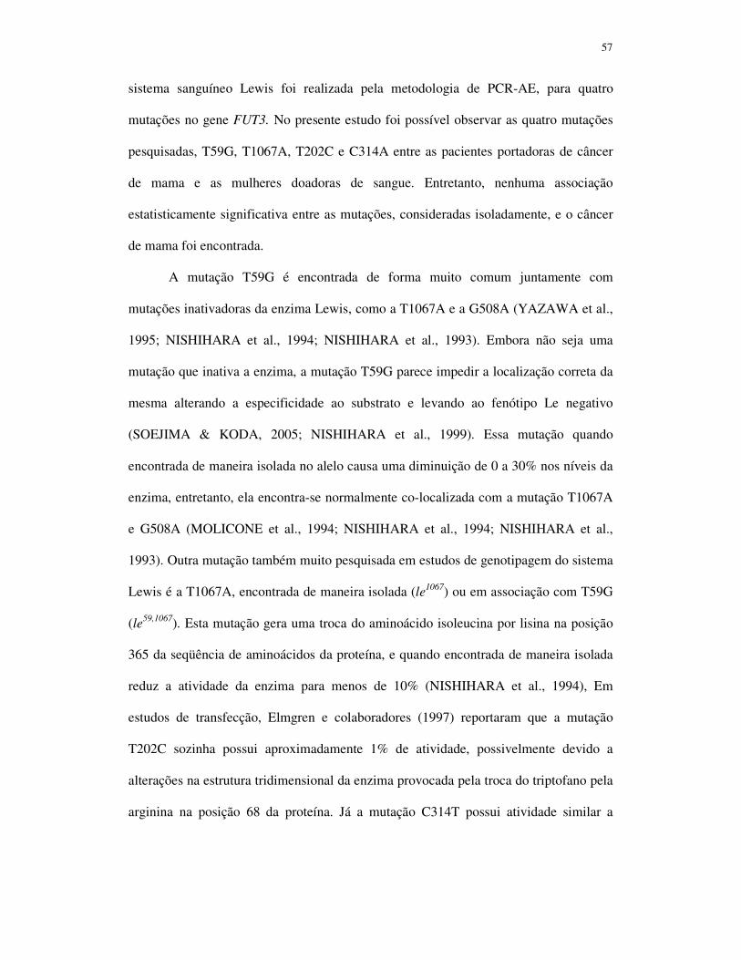
57
sistema sanguíneo Lewis foi realizada pela metodologia de PCR-AE, para quatro
mutações no gene FUT3. No presente estudo foi possível observar as quatro mutações
pesquisadas, T59G, T1067A, T202C e C314A entre as pacientes portadoras de câncer
de mama e as mulheres doadoras de sangue. Entretanto, nenhuma associação
estatisticamente significativa entre as mutações, consideradas isoladamente, e o câncer
de mama foi encontrada.
A mutação T59G é encontrada de forma muito comum juntamente com
mutações inativadoras da enzima Lewis, como a T1067A e a G508A (YAZAWA et al.,
1995; NISHIHARA et al., 1994; NISHIHARA et al., 1993). Embora não seja uma
mutação que inativa a enzima, a mutação T59G parece impedir a localização correta da
mesma alterando a especificidade ao substrato e levando ao fenótipo Le negativo
(SOEJIMA & KODA, 2005; NISHIHARA et al., 1999). Essa mutação quando
encontrada de maneira isolada no alelo causa uma diminuição de 0 a 30% nos níveis da
enzima, entretanto, ela encontra-se normalmente co-localizada com a mutação T1067A
e G508A (MOLICONE et al., 1994; NISHIHARA et al., 1994; NISHIHARA et al.,
1993). Outra mutação também muito pesquisada em estudos de genotipagem do sistema
Lewis é a T1067A, encontrada de maneira isolada (le1067) ou em associação com T59G
(le59,1067). Esta mutação gera uma troca do aminoácido isoleucina por lisina na posição
365 da seqüência de aminoácidos da proteína, e quando encontrada de maneira isolada
reduz a atividade da enzima para menos de 10% (NISHIHARA et al., 1994), Em
estudos de transfecção, Elmgren e colaboradores (1997) reportaram que a mutação
T202C sozinha possui aproximadamente 1% de atividade, possivelmente devido a
alterações na estrutura tridimensional da enzima provocada pela troca do triptofano pela
arginina na posição 68 da proteína. Já a mutação C314T possui atividade similar a

58
enzima produzida pelo alelo selvagem, entretanto, quando as mutaçãoes T202C e
C314T estão presentes no mesmo alelo, a enzima produzida não tem atividade.
Em nossos resultados, a mutação mais encontrada em ambos os grupos foi a
T202C com uma freqüência de 25,0% do alelo mutante C para as pacientes com câncer
de mama e de 17,3% para o grupo controle. Em um estudo realizado nos Estados
Unidos, a freqüência deste alelo foi de 21,3% nos Caucasianos e de 10,8% nos Afro-
Americanos (CAKIR et al., 2002). Entre os 18 indivíduos deste estudo fenotipados
como Lewis negativo, Le(a-b-), somente 10 (55,6%) indivíduos possuem genótipos que
realmente determinam a incapacidade funcional da enzima, 16 (88,9%) são portadores
de duas ou mais, das quatro mutações no gene FUT3, pesquisadas neste estudo, 1
(5,6%) é portador de apenas uma mutação e 1 (5,6%) não apresenta nenhuma das quatro
mutações. Esses resultados demonstram a necessidade da análise genotípica e não
apenas fenotípica do sistema Lewis.
Os genótipos obtidos a partir dos quatro pontos de polimorfismos do gene FUT3
foram agrupados e, dessa forma, houve uma associação estatisticamente significativa
entre os genótipos relacionados à perda funcional da enzima Lewis (Lewis negativo) e
as pacientes com câncer de mama. Os estudos de correlação com polimorfismos de
genes dos grupos sanguíneos e tumores são escassos e em um estudo realizado na
China, não foi possível observar diferença estatisticamente significativa na incidência
de câncer de estômago com a mutação T59G do gene FUT3 (JIANG et al., 2003a).
A enzima secretora codificada pelo gene FUT2 tem a função de fucosilar em
ligação α(1,2) o precursor tipo 1 e formar o antígeno H tipo 1, o qual pode ser utilizado
pelas glicosiltransferases A, B e Lewis, se presente. O sistema secretor controla a
formação de substância H (a qual pode ser modificada em substâncias A e/ou B)
encontrada nas secreções corporais, tais como saliva, fluido seminal (HENRY, 1996). A

59
mutação sem sentido G428A é a principal mutação causadora da inativação da enzima
secretora, produzindo um alelo não-secretor em indivíduos caucasianos (europeus e
iranianos) e africanos, enquanto a mutação de sentido trocado A385T está incluída
como a mutação mais comum causadora da produção de uma enzima deficiente na
população asiática (LIU et al., 1998b; KODA et al., 1996; KELLY et al., 1995). A
frequência da mutação G428A neste estudo foi de 73,7% para as pacientes portadoras
de câncer de mama e de 71,8% entre as mulheres do grupo controle. Os valores são
maiores do que os encontrados por VESTERGAARD et al (1999) com 63,3% na
população Dinamarquesa e por LIU et al (1998b) numa população africana e semelhante
a encontrada por PROCTER et al (1997). Neste estudo, em quase todos os indivíduos
fenotipados como não-secretores - Le(a+b-) – exceto por 1 indivíduo, a mutação G428A
foi encontrada em homozigose. Como ainda não há outros estudos publicados sobre o
polimorfismo do gene FUT2 na população brasileira, com esses resultados pode-se
demonstrar que esta mutação é a principal responsável pelo fenótipo não-secretor na
população central do Estado de São Paulo.
Estudos de associação de doenças com os grupos sanguíneos envolvem uma área
complexa, com muitas controvérsias. Muitos dos problemas são causados pela
inadequada confiabilidade da fenotipagem do grupo sanguíneo Lewis em portadores de
patologias. Os estudos de associação devem também além de considerar os parâmetros
fenotípicos dos três grupos - ABO, Lewis e secretor - incluir os estudos genotípicos
(HENRY, 1996).

60
7. Conclusões

61
• A verificação de genótipos do grupo sanguíneo ABO demonstrou que os genótipos
mais comuns nas pacientes portadoras de carcinoma ductal invasivo foram O1O
1,
AO1 e BO
1 e nas mulheres doadoras de sangue (grupo controle) foram O1O
1, AO
1 e
A., Entretanto, não houve diferença estatisticamente significativa entre esses dois
grupos e também não houve diferença significativa quando este grupo sanguíneo
foi comparado com as características clinicopatológicas dos tumores;
• A verificação das mutações T59G, T1067A, T202C e C314T no gene FUT3
demonstrou que a maioria dos indivíduos com fenótipo Le(a-b-) apresentaram uma
ou mais destas mutações. Entretanto nem todos os indivíduos fenotipados como
Lewis negativo apresetaram estas mutações, sendo necessária a pesquisa do
genótipo e não apenas do fenótipo para a avaliação do sistema Lewis;
• Das quatro mutações estudadas no gene FUT3, as mais freqüentes tanto nas
mulheres portadoras de CADI como nas mulheres doadoras de sangue foram
T202C e C314T, não havendo diferença estatisticamente significativa entre os
grupos quando as mutações foram avaliadas separadamente. Entretanto, houve
diferença estatisticamente significativa entre estes dois grupos quando as quatro
mutações foram avaliadas em conjunto, com uma proporção maior de genótipos
Lewis negativo para as pacientes portadoras de CADI. Além disso, não houve
diferença significativa na comparação entre este grupo sanguíneo e as
características clinicopatológicas dos tumores

62
• A mutação G428A do gene FUT2 foi muito freqüente na população estudada e
determinada em homozigose na maioria dos indivíduos fenotipados como não-
secretores. Entretanto, não houve diferença estatisticamente significativa entre as
mulheres portadoras de CADI e as doadoras de sangue para essa mutação.
Entretanto, houve diferença significativa para esta mutação quando relacionada a
presença de metástase em linfonodos, sendo que tumores com comprometimento
em linfonodos axilares estariam mais associados ao genótipo não secreotor.

63
8. Referências Bibliográficas

64
ABDEL-RAHMAN, S.Z. Isolation of DNA using salting-out procedure. J. Biochem.
Toxicol., v.9, p.191-198, 1994.
ABREU, E.; KOIFMAN, S. Fatores prognósticos no cancer da mama feminine. Rev.
Bras. Cancerol., v.48, n.1, p. 113-131, 2002.
ADAMIAN R.T. Blood-type and rhesus distribution in Armenian women with
endometrial carcinoma. Vopr. Onkol., v.51, p.575-576, 2005.
AUBERT M., PANICOT, L.; CROTTE, C.; GIBIER, P.; LOMBARDO, D.;
SADOULET, M.O.; MAS, E. Restoration of alpha(1,2) focosyltransferase activity
decreases adhesive and metastatic properties of human pancreatic cancer cells. Cancer
Res, v.60, p.1449-1456, 2000.
BARJAS-CASTRO, M.L.; CARVALHO, M.H. LOCATELLI, M.F.; BORDIN, S.;
SAAD, S.T.O. Molecular heterogeneity of the A3 subgroup. Clin. Lab. Haem. v.22, p.
73-78, 2000.
BERSTEIN, J.L.; LÓPEZ-CARRILLO, L.; WANG, L. The epidemiology of Her-2/neu
and p53 in breast cancer. Rev. Salud Pública México, v.41, suppl.2, p.s114-s123,
Gwenavaca. Nov.1999.
BRASIL. Ministério da Saúde. Instituto Nacional do Câncer (INCA). Diagnóstico
histopatológico e citopatológico das lesões da mama. Rio de Janeiro: Conprev, 2002,
80 p.
BRASIL. Ministério da Saúde. Instituto Nacional do Câncer (INCA). Câncer de
mama. Rio de Janeiro, 2004. Disponível em: http://www.inca.gov.br/cancer/mama/.
Acessado em 01/03/07.
BRASIL. Ministério da Saúde. Instituto Nacional do Câncer (INCA). Câncer de mama.
Rio de Janeiro, 2005. Disponível em: http://www.inca.gov.br/cancer/mama/. Acessado
em 10/06/05.

65
BRYNE, M.; EIDE, G.E.; LILLENG, R.; LANGMARK, F.; THRANE, P.S.;
DABELSTEEN, E. A multivariate study of the prognosis of oral squamous cell
carcinoma. Are blood group and hemoglobin new peognostic factors? Cancer, v.68,
p.1994-1998, 1991.
CAKIR, B.; PANKOW, J.S.; SALOMAA, V.; COUPER, D.; MORRIS, T.L.;
BRANTLEY, K.R.; HILLER, K.M.; HEISS, G.; WESTON, B.W. Distribution of Lewis
(FUT3) genotype and allele: frequencies in a biethnic United States population. Ann.
Hematol., v.81, p.558-565, 2002.
CAMPBELL, I.G.; BAXTER, S.W.; ECCLES, D.M.; CHOONG, D.Y.
Methylenetetrahydrofolate reductase polymorphism and susceptibility to breast cancer.
Breast Cancer Res., v.4, p.R14, 2002.
CESCHI, M.; SUN, C.L.; VAN DEN BERG, D.; KOH, W.P.; YU, M.C.; PROBST-
HENSCH, N. The effect of cyclin D1 (CCND1) G870A-polymorphism on breast cancer
risk is modified by oxidative stress among Chinese women in Singapore.
Carcinogenesis, v.26, p.1457-1464, 2005.
CHANG, C.; LIN, S.; LEE, J.; CHANG, J.; LIU, T.; HUANG, S.; CHEN, T.; LIU, H.
Application of ABO genotyping using polimerase chain reaction method to assess
engraftment in bone marrow transplantation - a case report. Kaohsiung J. Med. Sci.,
v.11, p.574-577, 1995.
CHANG, J.; LEE, L.; CHEN, P. Rapid genotyping of the ABO blood group. Blood,
v.79, p.3176-7, 1992.
CHENG, T.C.; CHEN, S.T.; HUANG, C.S.; FU, Y.P.; YU, J.C.; CHENG, C.W.; WU,
P.E.; SHEN, C.Y. Breast cancer risk associated with genotype polymorphism of the
catechol estrogen-metabolizing genes: a multigenic study on cancer susceptibility. Int.
J. Cancer, v.113, p.345-353, 2005.
CHOU, Y.C.; WU, M.H.; YU, J.C.; LEE, M.S.; YANG, T.; SHIH, H.L.; WU, T.Y.;
SUN, C.A. Genetic polymorphisms of the methylenetetrahydrofolate reductase gene,

66
plasma folate levels and breast cancer susceptibility: a case-control study in Taiwan.
Carcinogenesis, v.27, p.2295-2300, 2006.
COOLING, L.; GU, Y. Identification of two new single-nucleotide polymorphisms in
FUT3 associated with the Lewis-null phenotype. Transfusion, v.43, p.1760-1761,
2003.
CROUSE, C.; VINCEK, V. Identification of ABO alleles on forensic type specimens
using rapid ABO genotyping. Biotechnics, v.18, p.478-82, 1995.
DABELSTEEN, E.; GAO, S. ABO blood-group antigens in oral cancer. J. Dent. Res.,
v.84, p.21-28, 2005.
D'ADAMO, P.J.; KELLY, G.S. Metabolic and immunologic consequences of ABH
secretor and Lewis subtype status. Altern. Med. Rev., v.6, p.390-405, 2001.
DRESLER, C.M.; RITTER, J.H.; WICK, M.R. ROPER, C.L.; PATTERSON, G.A.;
COOPER, J.D. Immunostains for blood group antiges lack prognostic significance in
T1 lubg carcinoma. Ann. Thorac. Surg., v.59, p.1069-1073, 1995.
EGAN, K.M.; CAI, Q.; SHU, X.O.; JIN, F.; ZHU, T.L.; DAI, Q.; GAO, Y.T.; ZHENG,
W. polymorphisms in GSTM1, GSTP1, and GSTT1 and the risk for breast cancer:
results from the Shanghai Breast Cancer Study and meta-analysis. Cancer Epidemiol.
Biomarkers Prev., v.13, p.197-204, 2004.
ELMGREN, A.; BÖRJESON, C.; SVERSSON, L.; RYDBERG, L.; LARSON, G. DNA
sequencing and screening for point mutations in the human Lewis (FUT3) gene enables
the molecular genotyping of the human Lewis blood group system. Vox Sang., v.70,
p.90-103, 1996.
ELMGREN, A.; MOLLICONE, R.; COSTACHE, M.; BORJESON, C.; ORIOL, R.;
HARINGTON, J.; LARSON, G. Significance of individual point mutations, T202C and
C314T, in the human Lewis (FUT3) gene for expression of Lewis antigens by the
human α(1,3/1,4)-fucosyltransferase, Fuc-TIII. J. Biol. Chem., v.272, p.21994-21998,
1997.

67
ELMGREN, A.; RYDBERG, L.; LARSON, G. Genotypic heterogeneity among Lewis
negative individuals. Biochem. Biophys. Res. Commun, v.196, p.515-520, 1993.
ERNEST, C.; ATKINSON, B.; WYSOCKA, M.; BLASZCZYK, M.; HERLYN, M.;
SEARS, H.; STEPLEWSKY, Z.; KOPROWSKY, H. Monoclonal antibody localization
of Lewis antigens in fixed tissue. Lab. Invest., v.50, p.394-400, 1984.
FERRINI, F.S., ROSSI, M.A.; MOURÃO NETO, M.; SOARES,F.A. Schirrous
invasive ductal carcinoma of the breat overexpress p53 oncoprotein. São Paulo Med.
J., v.119, n.1, p. 04-06, São Paulo. Jan.2001.
FRANEKOVA, M.; ZUBOR, P.; STANCLOVA, A.; DUSSA, C.A.; BOHUSOVA, T.;
GALO, S.; DOBROTA, D.; KAJO, K.; PEC, M.; RACAY, P. Association of p53
polymorphisms with breast cancer: a case-control study in Slovak population.
Neoplasma, v.54, p.155-161, 2007.
GARRISON, R.J.; HAVLIK, R.J.; HARRIS, R.B.; FEINLEIB, M.; KANNEL, W.B.;
PADGETT, S.J. ABO blood group and cardiovascular disease: the Framingham study.
Atherosclerosis, v.25, p.311-318, 1976.
GRAHN, A.; ELMGREN, A.; ABERG, L.; SVENSSON, L.; JANSSON, P-A.;
LÖNNROTH, P.; LARSON, G. Determination of Lewis FUT3 gene mutations by PCR
using sequence-specific primers enables efficient genotyping if clinical samples. Hum.
Mutat., v.18, p.358-359, 2001.
GRENE, F.L.; PAGE, D.; MORROW, M.; BALCH, C.; HALLER, D.; FRITZ, A.;
FLEMING, I.; eds. AJCC Cancer Staging Manual 6th ed. New York: Springer; 2002.
GRUNNET, N.; STEFFELSON, R.; BENNETT, E. P.; CLAUSEN, H. Evaluation of
histo-blood group ABO genotyping in a danish population: frequency of a novel O
allele defined as O2. Vox Sang., v. 67, p.210-215, 1994.
HAKOMORI, S.I. Aberrant glycosylation in cancer cell membranes as focused on
glycolipids: overview and perspectives. Cancer Res., v. 45, p.2405-2414, 1985.

68
HAKOMORI, S.I. Antigen and structure and genetic bases of histo-blood groups A, B
and O: their changes associated with human cancer. Biochim. Bioph. Acta, v.1473,
p.247-266, 1999.
HAKOMORI, S.I. Tumor malignancy defined by aberrsnt glycosylation and
sphingo(glyco)lipid metabolism. Cancer Res., v.56, p.5309-5318, 1996.
HAMMER, L.; MANSSON, S.; ROHR, T.; CHESTER, M.A.; GINSBURG, V.;
LUNDBLAD, A.; ZOPF, D. Lewis-phenotype of erytrocytes and Leb active glycolipid
in serum of pregnant women. Vox sang., v.40, p.27-33, 1981.
HANBY, A.M The pathology of breast cancer and the role of the histopathology
laboratory. Clinical Oncology, 2005, In Press.
HANKINSON, S.E.; STAMPFER. M.J. Estrogens and breast cancer. Rev. Salud
Pública Méx., v. 39, n. 4, p.370-378, 1997.
HANSEN, T.; NAMORK, E.; OLSSON, M.L.; CHESSER, M.A. HEIER, H.I. Different
genotypes causing indiscernible patterns of A expression Ael red blood cells as
visualized by scanning immunogold electron microscopi. Vox. Sang., v., 1998.
HENRY, S.; MOLLICONE, R.; FERNANDEZ, P.; SAMUELSSON, B.; ORIOL, R.;
LARSON, G. Homozygous expression of a missense mutation at nucleotide 685 in the
FUT2 gene associates with the Le(a+b+) partial-secretor phenotype in an Indonesian
family. Biochem. Biophys. Res. Commun, v.219, p.675-678, 1996.
INAI, K., YAMAMOTO, A.; ARIHIRO, K.; KHATUN, N.; KURIHARA, K.;
TAKEDA, S. Epithelial hyperplasia of the breast. Comparison of incidence between
cases of breast carcinoma and control autopsy specimens with immunohistochemical
observation of blood group antigens. Acta Pathol. Jpn., v.42, p.193-200, 1992.
JARVIS, E. M.; KIRK, J. A.; CLARKE C, L, Loss of nuclear BRCA1 expression in
breast cancers is associated with a highly proliferative tumor phenotype. Cancer Genet
Cytogenet, v. 101, p.109-115, 1998

69
JESCHKE, U.; MYLONAS, I.; SHABANI, N.; KUNERT-KEIL, C.; SCHINDLBECK,
C.; GERBER, B.; FRIESE, K. Expression of sialyl Lewis X, sialyl Lewis A, E-cadherin
and cathepsin-D in human breast cancer: immunohistochemical analysis in mammary
carcinoma in situ, invasive carcinomas and their lymph node metastasis. Anticancer
Res., v.25, p.1615-22, 2005.
JIANG, J.M.; PAN, K.F.; MA, J.L.; NING, T.; LU, G.R.; YOU, W.C.; KE, Y. Analysis
of Lewis gene polymorphism in high and low incidence area of gastric cancer in
Shandong province. Yi Chuan. V.25, p.258-260, 2003a.
JIANG, J.M.; PAN, K.F.; MA, J.L.; NING, T.; LU, G.R.; YOU, W.C.; KE, Y. Analysis
of polymorphisms on Lewis blood group antigen related Se gene in the populations in
Shandong with high-risk of gastric cancer and Beijing. Yi Chuan, v.25, p.261-266,
2003b.
JOHNSON, P. H.; HOPKINSON, D. A. Detection of ABO blood group polymorphism
by denaturing gradient gel electrophoresis. Hum. Mol. Genet., v.1, p.341-344, 1992.
KELLY, R.J.; ROUQYUER, S.; GIORGI, D.; LENNON, G.G.; LOWE, J.B. Sequence
and expression of a candidate for the human secretor blood group α(1,2)-
fucosyltransferase gene (FUT2). Homozygosity for an enzyme-inactivating nonsense
mutation commonly correlates with the nonsecretor phenotype. J. Biol., Chem., v.270,
p.4640-4649, 1995.
KENEMANS, P.; BOSMAN, A. Breast cancer and post-menopausal hormone therapy.
Best Pract. Res. Clin. Endocrinol. Met., v. 17, n.1, p.123-137, 2003.
KODA, Y.; KIMURA, H; MEKADA, E. Analysis of Lewis fucosyltransferase genes
from the human gastric mucosa of Lewis-positive and –negative individuals. Blood,
v.82, p.2915-2919, 1993.
KODA, Y.; SOEJIMA, M.; JOHNSON, P.H.; SMART, E.; KIMURA, H. An Alu-
mediated large deletion of the FUT2 gene in individuals with the ABO-Bombay
phenotype. Hum. Genet., v.106, p.80-85, 2000.

70
KODA, Y.; SOEJIMA, M.; KIMURA, H. The polymorphisms of fucosyltransferases.
Leg. Med., v.3, p.02-14, 2001.
KODA, Y.; SOEJIMA, M.; LIU, Y-H.; KIMURA, H. Molecular basis for secretor type
α(1,2)-fucosyltransferase gene deficiency in a Japanese population: a fusion gene
generated by unequal crossover responsible for the enzyme deficiency. Am. J. Hum.
Genet., v.59, p.343-350, 1996.
KODA, Y.; SOEJIMA, M.; WANG, B-J; KIMURA, H. Structure and expression of the
gene encoding secretor-type galactoside 2-α-L-fucosyltransferase (FUT2). Eur. J.
Biochem., v.246, p.750-755, 1997.
KOIFMAN, R.J; KOIFMAN, S; VIEIRA, R.J.S. Familial aggregation of breast/ovarian
cancer: age of onset along subsequent gerations in Brazil. Cad. Saude Publica, v.14, p.
181-185, 1998.
KUKOWSKA-LATALLO, J.F.; LARSEN, R.D.; NAIR, R.P.; LOWE, J.B. A cloned
human cDNA determines expression od a mouse stage-specific embryonic antigen and
the Lewis blood group α(1,3/1,4)-fucosyltransferase. Genes Dev., v.4, p.1288-1303,
1990.
KURBEL, S. Selective reduction of estrogen receptor (ER) positive breast occurrence
by estrogen receptor modulators supports etiological distinction between ER positive
and ER negative breast cancers. Med. Hypotheses, v.64, p. 1182-1187, 2005.
LAMEY, P-J.; DOUGLAS, P.S.; NAPIER, S.S. Secretor status and oral cancer. Br. J.
Oral Maxil. Surg., v.32, p.214-217, 1994.
LE PENDU, J.; MARIONNEAU, S.; CAILLEAU-THOMAS, A.; ROCHER, J.; LE
MOULLAC-VAIDYE, B.; CLÉMENT, M. ABH and Lewis histo-blood group antigens
in cancer. APMIS, v.109, p.9-31, 2001.

71
LEE, A.H.; REID, M.E. ABO blood group system: a review of molecular aspects.
Immunohematol., v.16, p.01-06, 2000.
LEE, J. C.; CHANG, J. G. ABO genotyping by polymerase chain reaction, J. Foren.
Sci., v.37, p.1269-1275, 1992.
LIECHTI-GALLATI, S.; NEESER, D. Efficient and reliable PCR based detection of
the ABO blood group alleles: genotyping on stamps and others biological evidence
samples. J. Forens. Sci., v.41, p.653-57, 1996.
LIU, T.C.; CHANG, J.G.; LIN, S.F.; CHANG, W.C.; YANG, T.Y.; LIN, C.L.; WANG,
N.M.; TSAI, C.H. Lewis (FUT3) genotypes in Taiwanese, Thai and Filipino
populations. Ann. Hematol., v.79, p.599-603, 2000.
LIU, Y.; KODA, Y.; SOEJIMA, M.; PANG, H.; SCHLAPHOFF, T.; DU TOIT, E.;
KIMURA, H. Extensive polymorphism of the FUT2 gene in an African (Xhosa)
population of South Africa. Hum. Genet., v.103, p.204-210, 1998b.
LIU, Y-H.; FUJITANI, N.; KODA, Y.; KIMURA, H. Distribution of H type 1 and of H
type 2 antigens of ABO blood group in different cells on human submandibular gland.
J. Histochem. Cytochem., v.46, p.69-76, 1998a.
LIU, Y-H.; KODA, Y.; SOEJIMA, M.; PANG, H.; WANG, B.J.; KIMURA, H. Lewis
(FUT3) genotypes in two different Chinese populations. J. Forensic Sci., v.44, p.82-86,
1999.
LIU, Y-H.; KODA, Y.; SOEJIMA, M.; UCHIDA, N.; KIMURA, H. PCR analysis of
Lewis-negative gene mutations and the distribution of Lewis alleles in a Japanese
population. J. Forensic Sci., v.41, p.1018-1021, 1996.
LO, Y.L.; YU, J.C.; CHEN, S.T.; YANG, H.C.; FANN, C.S.; MAU, Y.C.; SHEN, C.Y.
Breast cancer risk associated with genotypic polymorphism of the mitosis-regulating
gene Aurora-A/STK15/BTAK. Int. J. Cancer, v.115, p.276-283, 2005.

72
LUKKAINEN, T. Issues to debate oon the Women´s Health Initiative study failure of
estrogen plus progestin therapy for prevention of breast cancer risk. European Society
of Human Reproduction and Eembryology, v.18, n.8, p.1559-1563, 2003.
MACKENZIE, I.C.; DABELSTEEN, E.; RITTMAN, G.; JUNGGREN, L.; TOH, H.
Expression of blood group-related glycoconjugates in the junctional and other oral
epithelia of rodents. Anat. Rec., v.241, p.310-318, 1995.
MADJD, Z.; PARSONS, T.; WATSON, N.F.; SPENDLOVE, I.; ELLIS, I.;
DURRANT, L.G. High expression of Lewis y/b antigens associated with decrease
survival in lymph node negative breast carcinomas. Breast Cancer Res., v.7, p.780-7,
2005.
MAGGARD, M.A.; OÇONNELL, J.B.; LANE, K.E.; LIU, J.H.; ETZIONI, D.A.; KO,
C.Y. Do young breast cancer patients have worse outcomes? J. Surg. Res., v.113,
p.109-113, 2003.
MARIANO, A.; DI CARLO, A.; SANTONASTASO, C.; OLIVA, A.; D’ARMIENTO,
M.; MACCHIA, V. Expression of Lewis carbohydrate antigens and chromogranin A in
human prostatic cancer. Int. J. Oncol., v.17, p.167-171, 2000.
MARIONNEAU, S.; CAILLEAU-THOMAS, A.; ROCHER, J.; MOULLAC-VAIDYE,
B.; RUVOËN, N.; CLÉMENT, M.; LE PENDU, J. ABH and Lewis histo-blood group
antigens, a model for the meaning of oligosaccharide diversity in the face of a changing
world. Biochimie, v.83, p.565-573, 2001.
MARIONNEAU, S.; MOULLAC-VAIDYE, B.; LE PENDU, J. Expression of histo-
blood group A antigen increases resistance to apoptosis and facilitates escape from
immune control of rat colon carcinoma cells. Glycobiol., v.12, p.851-856, 2002.
MATERSSON, S.; BIGLER, S.A.; BROWN, M.; LANGE, P.H.; BRAWER, M.K.;
HAKOMORI, S. Sialyl-Lewis(x) and related carbohydrate antigens in the prostate.
Hum. Pathol., v.26, p.735-739, 1995.

73
MATTOS, L.C.; CINTRA, J.R.; SANCHES, F.E.; SILVA, R.C.M.A.; RUIZ, M.A.;
MOREIRA, H.W. ABO, Lewis, secretor and non-secretor phenotypes in patients
infected or uninfected by the Helicobacter pylori bacillus. São Paulo Med. J., v.120, p.
55-58, 2002.
MATTOS, L.C.; SANCHEZ, F.E.; CINTRA, J.R.; SALLES, A.C.F.; BONINI-
DOMINGOS, C.R.; MOREIRA, H.W. Genotipagem do lócus ABO (9q34.1) em
doadores de sangue da região noroeste do Estado de São Paulo. Rev. Bras. Hematol.
Hemoter., v.23, p.15-22, 2001.
MILLER, S.A.; DYKES, D.D.; POLESKY, H.F. A simple salting out procedure for
extracting DNA from human nucleated cells. Nucleic Acids Research, v.16, p.1215,
1988.
MITRUNEN, K.; HIRVONEN, A. Molecular epidemiology of sporadic breast cancer.
The role of polymorphic genes involved in oestrogen biosynthesis and metabolism.
Mutat. Res., v.544, p.9-41, 2003.
MOLDVAY, J.; SCHEID, P.; WILD, P.; NABIL, K.; SIAT, J.; BORRELLY, J.;
MARIE, B.; FARRE, G.; LABIB, T.; POTTIER, G.; SESBOUE, R.; BRONNER, C.;
VIGNAUD, J.M.; MARTINET, Y.; MARTINET, N. Predictive survival markers in
patients with surgically resected non-small cell lung carcinoma. Clin. Cancer Res., v.6,
p.1125-1234, 2000.
MOLLICONE, R.; REGUIGNE, I.; KELLY, R.J. et al. Molecular basis for Lewis
α(1,3/1,4)fucosiltransferase gene deficiency (FUT3) found in Lewis-negative
Indonesian pedigrees. J. Biol. Chem., v.269, p.20987-20994, 1994.
MOURANT, A.E.; KOPEC, A.C.; DOMANIEWSKA-SOBCZAK, K. Blood groups
and diseases: A study of associations of diseases with blood groups and other
polymorphisms. Oxford University Press, Oxford, 1978.
MOURÃO NETO, M.; LOGUELO, A F.; NONOGAKI, S.; BOENTAMI, R.R.;
BRENTANI, M.M Expression of cerbB-2, p53 and c-myc proteins in male breast

74
carcinoma. Comparison with traditional prognóstico factores and survival. Braz. J.
Med. Biol. Res., v.34, p. 887-894, 2001.
NAKAGOE, T.; FUKUSHIMA, K.; HIROTA, M.; KUSANO, H.; KAWAHARA, K.;
AYABE, H. et al. Immunohistochemical expresión of blood group substances and
related carbohydrate antigens in breast carcinoma. Jpn. J. Cancer Res., v.82, p.559-
568, 1991.
NAKAGOE, T.; FUKUSHIMA, K.; ITOYANAGI, N.; IKUTA, Y.; OKA, T.;
NAGAYASU, T.; AYABE, H.; HARA, S.; ISHIKAWA, H.; MINAMI, H. Expression
of ABH/Lewis-related antigens as prognostic factors in patients with breast cancer. J.
Cancer Res. Clin. Oncol., v.128, p.257-264, 2002.
NAKAGOE, T.; FUKUSHIMA, K.; TUJI, T.; SAWAI, T.; NANASHIMA, A.,
YAMAGUCHI, H. YASUTAKE, T.; HARA, S.; AYABE, H.; MATUO, T.;
KAMIHIRA, S. Immunohistochemical expression of ABH/Lewis-related antigens in
primary breast carcinomas and metastatic lymph node lesions. Cancer Detect. Prev.,
v.22, p.499-505, 1998.
NISHIHARA, S.; HIRAGA, T.; IKEHARA, Y.; IWASAKI, H.; KUDO, T.; YAZAWA,
S.; MOROZUMI, K.; SUDA, Y.; NARIMATSU, H. Molecular behavior of mutant
Lewis enzymes in vivo. Glycobiology, v.9, p.373-382, 1999.
NISHIHARA, S.; NARIMATSU, H.; IWASAKI, H.; YAZAWA, S.; AKAMATSU, S.;
ANDO, T.; SENO, T.; NARIMATSU, I. Molecular genetic analysis of the human
Lewis histo-blood group system. J. Biol. Chem., v.269, p.29271-29278, 1994.
NISHIHARA, S.; YAZAWA, S.; IWASAKI, H.; NAKAZATO, N.; KUDO, T.; ANDO,
T.; NARIMATSU, H. α(1,3/1,4)Fucosyltransferase (FucT-III) gene is inactivated by a
single amino acid substitution in Lewis histo-blood type negative individuals. Biochem.
Biophys. Res. Commun, v.196, p.624-631, 1993.

75
NOVARETTI, M.C.Z.; DORLHIAC-LLACER, P.E.; CHAMONE, D.A.F. Estudo de
grupos sanguíneos em doadores de sangue caucasóides e negróides na cidade de São
Paulo. Rev. Bras. Hematol. Hemoter., v.22, p.23-32, 2000.
NOZOE, T.; EZAKI, T.; BABA, H.; KAKEJI, Y.; MAEHARA, Y. Correlation of ABO
blood group with clinicopathologic characteristics of patients with esophageal
squamous cell carcinoma. Diseases of the Esophagus, v.17, p.146-149, 2004.
OLSSON, M.L.; CHESSER, M.A. Evidence for a new type of O allele at the ABO
locus, due to a combination of the A2 nucleotide deletion and the Ael nucleotide
insertion. Vox Sang, v.71, p.113-117, 1996.
OLSSON, M.L.; CHESTER, M.A. A rapid and simple ABO genotype screening
method using a novel B/O2 versus A/O2 discriminating nucleotide substitution at the
ABO locus. Vox Sang., v.69, p.242-247, 1995.
OLSSON, M.L.; CHESTER, M.A. Polymorphism and recombination events at the
ABO locus: a major challenge for genomic ABO blood grouping strategies. Transf.
Med., v.11, p.295-313, 2001.
ORNTOFT, T.F.; HOLMES, H.; JOHNSON, J.; HAKOMORI, S.; CLAUSEN, H.
Differential tissue expression of the Lewis blood antigens: enzymatic,
immunohistologic and immunochemical evidence for Lewis a and b antigen expression
in Le(a-b-) individuals. Blood, v.77, p.1389-1396, 1991.
PAIVA, C.E.; RIBEIRO, B.S.;GODINHO, A.A.; MEIRELLES, R.S. P; SILVA,
E.V.G.; MARQUES, G.D’A.; ROSSINI JÚNIOR,O. Fatores de risco para câncer de
mama em Juiz de Fora (MG): um estudo caso-controle .Rev. Brasileira de
Cancerologia, v.48, n.2, 2002, p.231-237.
PANG, H.; KODA, Y.; SOEJIMA, M.; KIMURA, H. Significance of each of three
missense mutation, G484A, G667A and G808A, present in an inactive allele of the
human Lewis gene (FUT3) α(1,3/1,4)-fucosyltransferase inactivation. Glycoconjugate
J., v.15, p.961-967, 1998a.
76
PANG, H.; LIU, Y-H.; KODA, Y.; SOEJIMA, M.; JIA, J.T.; SCHLAPHOFF, T.; du
TOIT, E.D.; KIMURA, H. Five novel mutations of Lewis gene (FUT3) in African
(Xhosa) and Caucasian populations in South Africa. Hum. Genet., v.102, p.675-680,
1998b.
PENG, C-T; TSAI, C-H; LIN, T-P; PENG, L-I; KAO, M-C; YANG, T-Y; WANG, N-
M; LIU T-C; LIN, S-F; CHANG, J-G. Molecular characterization of secretor type
α(1,2)-fucosyltransferase gene deficiency in the Philippine population. Ann Hematol.,
v.78, p.463-467, 1999.
PLATET, N.; CATHIARD, A. M.; GLEIZES, M.; GARCIA, M. Estrogens and their
recepetors in breast câncer progression: a dual role in câncer proliferation and ivasion.
Crit. Rev. Oncol./ Hematol., v. 51, p. 55-67, 2004.
PROCTER, J.; CRAWFORD, J.; BUNCE, M.; WELSH, K.I. A rapid molecular method
(polymerase chain reaction with sequence-specific primers) to genotype for ABO blood
group and secretor status and its potential for organ transplants. Tissue Antigens, v.50,
p.475-483, 1997.
RAVN, V.; DABELSTEEN, E. Tissue distribution of histo-blood group antigens.
APMIS, v.108, p.108-128, 2000.
ROUQUIER, S.; LOWE, J.B.; KELLY, R.J.; FERTITTA, A.L.; LENNON, G.G.;
GIORGI, D. Molecular cloning of a human genomic region containing the H blood
group α(1,2)-fucosyltransferase gene and two H locus-related DNA restriction
fragments. Isolation of a candidate for the human secretor blood group locus. J. Biol.
Chem., v.270, p.4632-4639, 1995.
SALMON, C.; CARTRON, J. P.; ROUGER, P. The human blood groups. Chicago:
Year Book Medical Publishers. INC, 1984, 460p.
SALOMAA, V.; PANKOW, J.; HEISS, G.; CAKIR, B.; ECKFELDT, J.H.; ELLISON,
R.C.; MYERS, R.H.; HILLER, K.M.; BRANTLEY, K.R.; MORRIS, T.L.; WESTON,
B.W. Genetic background of Lewis-negative blood group phenotype and its association

77
with atherosclerotic disease in the NHLBI Famoly Heart Study. J. Intern Med., v.247,
p.689-698, 2000.
SERPA, J.; ALMEIDA, R.; OLIVEIRA, C.; SILVA, F.S.; SILVA, E.; REIS, C.; LE
PENDU, J.; OLIVEIRA, G.; RIBEIRO, L.M.C.; DAVID, L. Lewis enzyme (α1-3/4
fucosyltransferase) polymorphisms do not explain the Lewis phenotype in the gastric
mucosa of a Portuguese population. J. Hum. Genet., v.48, p.183-189, 2003.
SHU, X.O.; MOORE, D.B.; CAI, Q.; CHENG, J.; WEN, W.; PIERCE, L.; CAI, H.;
GAO, Y.T.; ZHENG, W. Association of cyclin D1 genotype with breast cancer risk and
survival. Cancer Epidemiol Biomarkers Prev., v.14, p.91-97, 2005.
SIDONI, A.; CAVALIERE, A.; BELLEZA, G.; SCHEIBEL, M.; BUCCIARELLI, E.
Breast cancer in young women: clinicopathological features and biological specificity.
The Breast, v.12, p.247-250, 2003.
SILVA, D. M.; SADDI, V.A MOMOTUK, E.G. Marcadores moleculares associados ao
câncer de mama não metastático. Rev. Brasileira de Cancerologia, v. 48, n.1, p.39-48,
2002.
SKOVLUND, V.R. ABH and related histo-blood group antigens in normal & malignant
human endometrium in relation to genetic and hormonal factors. APMIS, v.69(Suppl),
p.1-33, 1997.
SOEJIMA, M.; KIMURA, H.; KODA, Y. Two novel FUT3 alleles responsible for
Lewis-null phenotypes in SriLanka. Transfusion, v.44, p.1534-1535, 2004.
SOEJIMA, M.; KODA, Y. Molecular mechanisms of Lewis antigen expresión. Legal
Med., v.7, p.266-269, 2005.
SOUTO, J.C.; ALMASY, L.; MUNIZ-DIAZ, E.; SORIA, J.M.; BORELL, M.;
BAYEN, L.; MATEO, J.; MADOZ, P.; STONE, W.; BLANGERO, J.
FONTEUBERTA, J. Functional effects of the ABO locus polymorphism on plasma

78
levels of von Willebrand factor, factor VIII and activated partial thromboplastin time.
Arterioscler. Thromb. Vasc. Biol, v.20, p.2024-2028, 2000.
STIGENDAL, L.; OLSSON, R.; RYDBERG, L.; SAMUELSSON, B.E. Blood group
Lewis phenotype on erytrocytes and in saliva in alcoholic pancreatitis and chronic liver
disease. J. Clin. Pathol., v.37, p. 778-786, 1984.
SU, M.; LU, S-M.; TIAN, D.-P.; ZHAO, H.; XIAO-YUNLI; LI, D.-R.; ZHENG, Z.-C.
Relationship between ABO blood groups and carcinoma of esophagus and cardia in
Chaoshan onhabitants of China. World J. Gastroenterol., v.7, p.657-661, 2001.
TAKASHIMA, T.; ONODO, N.; ISHIKAWA, T; AGAWA, Y.; KATO, Y.;
FUJIMOTO, Y; SAWA, M.; HIRAKAWA. Prognostic value of combined analysis of
estrogen receptor status and cellular proliferative activity in breast cancer patients with
extensive lymph node metastases. Oncology Reports, v.9, p. 589-594, 2002.
TAVASSOLI, F. A. Pathology of the breast. Norwalk: Appleton & Lange. 1992, p.
25-53, 293-307.
TERRY, M.B.; GAMMON, M.D.; ZHANG, F.F.; ENG, S.M.; SAGIV, S.K.; PAYKIN,
A.B.; WANG Q, HAYES, S.; TEITELBAUM, S.L.; NEUGUT, A.I.; SANTELLA,
R.M. Polymorphism in the DNA repair gene XPD, polycyclic aromatic hydrocarbon-
DNA adducts, cigarette smoking, and breast cancer risk. Cancer Epidemiol.
Biomarkers Prev., v.13, p.2053-2058, 2004.
TESSARO, S.; BÉRIA, J.U.; TOMASI, E.; BARROS, A.A.J.D. Contraceptivos orais e
câncer de mama: estudo de casos e controles. Rev. Saúde Pública, v. 35, n.1, p. 32-38,
2001.
TURSEN, U.; TIFTIK, E.N.; UNAL, S.; GUNDUZ, O.; KAYA, T.I.; CAMDEVIREN,
H.; IKIZOGLU, G. Relationship between ABO blood groups and skin cancers.
Dermatol. Online J., v.11, p.44, 2005.

79
URA, Y.; DION, A., WILLIAMS, C.; OLSEN, B.; REDFIELD, E.; ISHIDA, M.;
HERLYN, M.; MAJOR, P.P. Quantitative dot blot analyses of blood-group-related
antigens in paired normal and malignant human breast tissues. Int. J. Cancer, v.50,
p.57-63, 1992.
VAN DE VIJVER, M. Genetic alterations in breast cancer. Curr. Diagn. Pathol., v. 6,
p.271-281, 2000.
VESTERGAARD, E.M.; HEIN, H.O.; MEYER, H.; GRUNNET, N.; JOTGENSEN, J.;
WOLF, H.; ORNTOFT, T.F. Reference values and biological variation for tumor
marker CA 19-9 in serum for different Lewis and secretor genotypes and evaluation of
secretor and Lewis genotyping in a Caucasian population. Clin. Chem., v.45, p.54-61,
1999.
VIDAS, I.; DELAJLIJA, M.; TEMMER-VUKSAN, B.; STIPETIC-MRAVAK, M.;
CINDRIC, N.; MARICIC, D. Examining the secretor status in the saliva of patients
with oral pre-cancerous lesions. J. Oral Rehab., v.26, p.177-182, 1999.
VOGEL, F. ABO blood groups and disease. Am. J. Hum. Genet., v.22, p.465-475,
1970.
VOWDEN, P.; LOWE, A.D.; LENNOX, E.S.; BLEEHEN, N.M. The expression of
ABH and Y blood group antigens in benign and malignant brast tissue: the preservation
of the H and Y antigens in malignant epithelium. Br. J. Cancer, v.53, p.313-319, 1986.
WATKINS, W.M. The ABO blood group system: historical background. Transf. Med.,
v. 11, p.243-265, 2001.
WATKINS W.M. Molecular basis of antigenic specificity in the ABO, H and Lewis
blood group systems. In: Montreuil J. Vliegenthart JFG, Schachter H, editors.
Glycoproteins. Amsterdam: Elsevier Science BV, 1995, p 313-390.
XIE, X.; BOYSEN, M.; CLAUSEN, O.P.; BRYNE, M.A. Prognostic value of Le(y)
and H antigens in oral tongue carcinomas. Laringoscope, v.109, p.1474-1480, 1999.

80
YAMAMOTO, F.; CLAUSEN, H.; WHITE, T.; MARKEN, J.; HAKOMORI, S.
Molecular genetic basis of the histo-blood group ABO system. Nature, v.345, p.229-
233, 1990a.
YAMAMOTO, F.; MARKEN, J.; TSUJI, T.; WHITE, T.; CLAUSEN, H.;
HAKOMORI, S. Cloning and characterization of DNA complementary to human UDP-
GalNAz:fuc-alpha 1-2Gal alpha 1-3GalNAc transferase (histo-blood group a
transferase) mRNA. J. Biol., Chem., v.265, p.1146-1151, 1990b.
YAMAMOTO, F.; HAKOMORI, S. Sugar-nucleotide donor specificity of histo-blood
group A and B transferases is based on amino acid substitutions. J. Biol. Chem., v. 265,
p.19257-19262, 1990.
YAMAMOTO, F.; McNEILL, P. D.; HAKOMORI, S. genomic organization of human
histo-blood group ABO genes. Glicobiology, v.5, 1995.
YAMAMOTO, F.; McNEILL, P. D.; YAMAMOTO, M.; HAKOMORI, S.; HARRIS,
T. Molecular genetic analyses of ABO blood group system: 3. Ax and B(A) alleles. Vox
Sang. v.64, p.171-174, 1993.
YAZAWA, S.; NISHIHARA, S.; IWASAKI, H.; ASAO, T.; NAGAMACHI, Y.;
NARIMATSU, H. Gentic and enzymatic evidence for Lewis enzyme expression in
Lewis-negative cancer patients. Cancer Res., v.55, p.1473-1478, 1995.
YAZAWA, S.; OH-KAWARA, H.; NAKAJIMA, T.; HOSOMI, O.; AKAMATSU, A.;
KISHI, K. Histo-blood group Lewis genotyping from human hairs and blood. Jpn. J.
Human Genet., v.41, p.177-188, 1996.
YIP, S.P. Single-tube multiplex PCR-SSCP analysis distinguishes 7 common ABO
alleles and readily identifies new alleles. Blood, v.95, p.1487-1492, 2000.

81
YU, L; LEE-H-L; CHU, C.C.; BROADBERRY, R.E.; LIN, M. A newly identified
nonsecretor allele of the human histo-blood group α(1,2)-fucosyltransferase gene
(FUT2). Vox Sang., v.76, p.115-119, 1999
YU, L-C.; YANG, Y-H.; BROADBERRY, R.E.; CHEN, Y-H.; CHAN, Y-S.; LIN, M.
Heterogeneity of the human blood group α(1,2)fucosyltransferase gene among para-
Bombay individuals. Vox Sang., v.72, p.36-40, 1997.
YU, L-C.; YANG, Y-H; BROADBERRY, R.E.; CHEN, Y-H.; CHAN, Y-S.; LIN, M.
Correlation of a missense mutation in the human secretor α(1,2)-fucosyltransferase gene
with the Lewis(a+b+) phenotype: a potential molecular basis for the weak secretor allele
(Sew). Biochem. J., v.312, p.329-332, 1995.

82
9. Anexos

83
DISTRIBUTION OF LEWIS (FUT3) PHENOTYPE AND GENOTYPE IN A
BRASILIAN POPULATION
D.B. Teresa (a), C.P. Soares * (a), L.C. de Mattos (b), M. Giocondo (a), A.A. Leite (a); E.
Leite (a); H.W. Moreira (a)
(a) Department of Clinical Analysis, School Pharmaceutical Sciences, University of
São Paulo State (UNESP), Rua Expedicionários do Brasil, 1621, Zip code 14 801
902, Araraquara, Sao Paulo, Brazil.
(b) Faculty of Medicine of São José do Rio Preto (FAMERP), Avenida Brigadeiro Faria
Lima, 5416, São Pedro, Zip code 15090-000, São Jose do Rio Preto, São Paulo,
Brazil
Author to whom correspondence must be sent:
C.P.Soares. Address: Rua Expedicionários do Brasil, 1621, Zip code 14 801 902,
Araraquara, São Paulo, Brazil. Phone 55 16 201 6554; fax 55 16 201 6547.
* e-mail address:[email protected], [email protected]

84
Abstract
The Lewis histo-blood group system comprises two major antigens, Lea and Leb, whose
determinants are composed of type 1 carbohidrate chains. The Lea antigen is synthesized
from a type 1 precursor substrate by a FUT3-encoded α(1,3/1,4) fucosyltransferase (Le
enzyme), whereas the Leb antigen is synthesized from an H type 1 substrate by the
enzyme. The cDNA encoding Lewis fucosyltransferase has been isolated, and 13 single
bases substitutions have been identified in a number of different Le (a-b-) individuals –
T59G, G508A, T1067A, T202C, C314T, C445T, G484A, G667A, G808A, G760A,
G13A, G1022T and G47C. In this study, we have investigated the frequency of Lewis
phenotype and the four mutations of the FUT3 gene, T59G, T1067A, T2020C e C314T,
in blood donors at the city of Araraquara, Sao Paulo State by using a polymerase chain
reaction/sequence-specific primers (PCR/SSP) method. One hundred and eighty-one
healthy blood donors arriving to the blood bank were subjects of the study. The
genotypes comprising the four common mutations (T59G, T1067A, T202C and C314T)
and the most common genotype was the “all-wild” genotype (i.e., WWWW), which was
found to be present in 45.9% (n=83) of participants. The others genotypes were WWHH
(n=45, 24.9%), HHWW (n=20, 11.0%), HWWW (n=14, 7.7%), WWMM (n=7, 3.9%),
MMWW (n=5, 2.8%), HWHH (n=2, 1.1%) and MWWW, MHWW, WWHW, HHHW and
HWHW with 0.6% each (n=1). With regard to the molecular basis of phenotypes of Le
(a-b+), 66 cases (54.6%) were “all-wild” genotype (WWWW), 44 (36.4%) were double
heterozygotes (HHWW or WWHH), 1 (0.8%) was WWHW and 1 (0.8%) was HHHW.
Concerning the phenotypes of Le (a+b-), the genotype most common was WWWW
(n=12, 40.0%). Concerning the phenotypes of Le (a-b-), 14 cases (48.3%) were
homozygous or double homozigotes for a mutant allele (WWMM, MMWW, MWWW,
MHWW), 11 cases (37.9%) had at least a mutant heterozygote allele. The most

85
common mutation was at nucleotide position 202 with frequency of the 17.7% for C
allele. The second most common mutant allele was T allele at nucleotide position 314
with 16.9%. In conclusion, our study indicated that at the Brazilian population of the
Araraquara city the T202C and C314T mutations were the most frequents. Theses
mutations were presented in 86.2% of the Le (a-b-) participants, though only 41.34% of
the Lewis (a-b-) individuals produced genotypes Lewis-negative.
Keywords: Lewis gene, FUT3 gene, PCR-SSP, polymorphism

86
Introduction
The Lewis histo-blood group system comprises two major antigens, Lea and Leb, whose
determinants are composed of type 1 carbohidrate chains. In contrast to the other blood
group antigens, the synthesis of Lewis antigens do not occur in erytroid tissues; rather
they are secondarily acquired by the erythrocyte membranes from other glycolipids
expressing endoderm-derived tissue(s) through the circulating soluble forms bound to
lipoproteins (Watkins, 1995). The Lea antigen is synthesized from a type 1 precursor
substrate by a FUT3-encoded α(1,3/1,4) fucosyltransferase (Le enzyme), whereas the
Leb antigen is synthesized from an H type 1 substrate by the enzyme. The gene FUT2
encoded a α(1,2) fucosyltransferase (Se enzyme) that synthesize H antigen mainly on
the gastrointestinal epithelial cells and in body fluids such as saliva (Clausen &
Hakomori 1989). Therefore, the Lewis system phenotypes are determined by both the
Lewis (FUT3) and secretor (FUT2) loci and individuals lacking a functional FUT3
allele are called ‘Lewis negative’ and are characterized by the red cell phenotype Le(a-
b-). People with a ‘Lewis positive’ phenotype, either Le(a-b+) or Le(a+b-), possess at
least one functional FUT3 allele and are respectively secretors or non-secretors
(Marionneau, 2001). A secretor has the ABH antigen in saliva and other body fluids and
at least one functional allele FUT2 while a non-secretor is homozygous for a allele
nonfunctional and is characterized by the absence of ABH antigens in saliva and on
various epithelial cell types (Watkins, 1995).
The Lewis antigens are not intrinsic to red blood cells, the typing of Lewis
phenotypes is difficult and is sometimes misjudged because if weak hemagglutination
as a result of the small numbers of the antigens on red cells, and low titers (Good 1992;
Wang, 1994). Therefore, it is important and useful to determine the genotypes of the
Lewis system.

87
The FUT3 gene is polymorphic, located on chromosome 19 and codes for an
α(1,3/1,4) fucosyltransferase that is expressed in exocrine secretions (Kukowska-
Latallo et al., 1990; Elmgren et al., 1993). The cDNA encoding Lewis
fucosyltransferase has been isolated (Kukowska-Latallo et al., 1990), and 13 single
bases substitutions have been identified in a number of different Le (a-b-) individuals –
T59G, G508A, T1067A, T202C, C314T, C445T, G484A, G667A, G808A, G760A,
G13A, G1022T and G47C (Elmgren et al., 1993; Koda et al., 1993; Liu et al., 1996; Liu
et al., 1999; Pang et al., 1998; Elmgren et al., 1997; Cooling & Gu, 2003; Soejima et al.,
2004). In addition, two silent base substitutions, C304A and T370G, were also observed
in an African population (Pang et a., 1998). While the le202,314 allele is common in
Caucasian Le(a-b-) individuals, these mutations are quite rare in Asian populations
(Koda et al., 1993; Liu et al., 1996; Liu et al., 1999; Pang et al., 1998; Elmgren et al.,
1997). On the hand, the G508A mutation (the le59,508), which is common in Asian
populations, is rare in Caucasian populations (Liu et al., 1996; Elmgren et al., 1997).
The G484A, G667A and G808A mutations are observed only in a African population
(Pang et al., 1998). The mutations T59G and T1067A seem to be wide-spread, all over
the world. They frequently occur as single mutations in Indonesia, whereas in Japan and
Europe, they are almost always co-localized (Orntoft et al., 2996). In addition, the G47C
mutation is likely to be specific for Sinhalese (Soejima et al., 2004).
Although several mutations of FUT3 were found in different ethnic populations,
there were no reports based on the genetic analysis of the FUT3 gene in Brazilians. In
this study, we have investigated the frequency of Lewis phenotype and the four
mutations of the FUT3 gene, T59G, T1067A, T2020C e C314T, in blood donors at the
city of Araraquara, Sao Paulo State by using a polymerase chain reaction/sequence-
specific primers (PCR/SSP) method.

88
Materials and Methods
Subjects and determination of Lewis blood phenotypes
One hundred and eighty-one healthy blood donors arriving to the blood bank were
subjects of the study. The study was approved by the Ethical Committee at the. Lewis
hemagglutination assay was performed with micro typing system (DiaMed,
Switzerland).
DNA extraction
Genomic DNA was isolated from peripheral blood leukocytes as described previously
and diluted in TE buffer (10mM Tris-HCl, 1mM Na2-EDTA, pH 7,5) (Miller et al.,
1988).
PCR-SSP genotyping of FUT3 mutations
The amplifications of the DNA were performed in 25uL of solution containing 1X
Green Buffer (Promega-USA), 0,2mM each dNTP, 0.5µM each primer, 1,25U
GoTaqTM DNA polymerase (Promega – USA) and 500ng genomic DNA. The cycling
parameters were 96oC 60s; 5 cycles at 96oC 20s, 70oC 45s, 72oC 25s; 21 cycles at 96oC
25s, 65oC 50s, 72oC 30s; 4 cycles at 96oC 30s, 55oC 60s, 72oC 90s. The mutated FUT3
oligonucleotide primers had genotypically specific mutations on the penultimate or last
nucleotide at the 3’-end, and a Tm between 58oC-60oC (Granh et al., 2001). The human
growth hormone (hGH) gene was used, as an internal control in each PCR
amplification, with 5’-GCCTTCCCAACCATTCCCTT-3’ as sense primer and 5’-
TCACGGATTTCTGTTGTGTTTG-3’ as anti-sense primer (Procter et al, 1997). All
primer-mixes included 0.5µM of each hGH-primer, which gave raise to a 428bp
amplification product and an anti-sense primer (III-55as) with 5’-

89
TTCTGGAGGGGAGAGGCT-3’ specific for the wild allele FUT3 in the position
1212-1229. All PCR-amplification were run on 1% agarose gel (Promega – Madison,
USA) in 0.5 mg/ml ethidium bromide for 30-50 minutes at 100V and visualized using
UV illumination.
Table 1- The primer sequences and corresponding PCR product sizes. UP upstream; DP
downstream.
Mix
Allele Primer sequencea Product size (bp)
1 59wt UP: 5’- CGCTGTCTGGCCGCACT – 3’
DP: III-55as
1186
2 59T>G UP: 5’- GCTGTCTGGCCGCACgG – 3’
DP: III-55as
1185
3 202wt UP: 5’- CCCTCCTGATCCTGCTATG – 3’
DP: III-55as
1045
4 202T>C UP: 5’- ACCCTCCTGATCCTGCTAc – 3’
DP: III-55as
1044
5 314wt UP: 5’- GTACCCACAGGCAGACACG – 3’
DP: III-55as
932
6 314C>T UP: 5’- TGTACCCACAGGCAGACAt – 3’
DP: III-55as
933
7 1067wt UP: 5’- CCAGACGGTGCGCAGCAT – 3’
DP: III-55as
180
8 1067T>A UP: 5’- CCAGACGGTGCGCAGCAa – 3’
DP: III-55as
180
a Deliberately mutated nucleotides are indicated by small letters in bold. Wt= wild type

90
Definition of Lewis genotype
Distributions of genotype and allele frequencies for each individual single nucleotide
polymorphism (i.e., T59G, T1067A, T202C and C314T) were studied separately, and as
combinations of four FUT3 gene SNPs, i.e., Lewis genotype groups. The table 2
summarizes some groups of FUT3 gene polymorphisms into the corresponding Lewis
genotype (Cakir et al., 2002).
Table 2- Identifications of Lewis genotype groups by combining information on FUT3
gene mutations. W wild, H heterozygous, M homozygous mutant
Lewis genotype at site
59 1067 202 314
W W W W
M M W W
W W H H
H H H W
Results
Genotyping was completed for 181 blood donors. There were 37% (n=67) male
and 63.0% (n=114) female subjects with a mean age of 34.85 years (ranging from 18
years to 61 years). Concern ethnic the majority was Caucasians (84.5%, n=153), 12.2%
(n=22) Africans Americans and 3.3% (n=6) Mongols. The genotypes comprising the
four common mutations (T59G, T1067A, T202C and C314T) and the most common
genotype was the “all-wild” genotype (i.e., WWWW), which was found to be present in
45.9% (n=83) of participants. The others genotypes were WWHH (n=45, 24.9%),
HHWW (n=20, 11.0%), HWWW (n=14, 7.7%), WWMM (n=7, 3.9%), MMWW (n=5,

91
2.8%), HWHH (n=2, 1.1%) and MWWW, MHWW, WWHW, HHHW and HWHW with
0.6% each (n=1).
With regard to the molecular basis of phenotypes of Le (a-b+), 66 cases (54.6%)
were “all-wild” genotype (WWWW), 44 (36.4%) were double heterozygotes (HHWW or
WWHH), 1 (0.8%) was WWHW and 1 (0.8%) was HHHW. Concerning the phenotypes
of Le (a+b-), the genotype most common was WWWW (n=12, 40.0%). Concerning the
phenotypes of Le (a-b-), 14 cases (48.3%) were homozygous or double homozigotes for
a mutant allele (WWMM, MMWW, MWWW, MHWW), 11 cases (37.9%) had at least a
mutant heterozygote allele (Table 3).
Table 4 presents the distribution of genotype and allele frequencies for each
individual single nucleotide polymorphism. The most common mutation was at
nucleotide position 202 with frequency of the 17.7% for C allele. The second most
common mutant allele was T allele at nucleotide position 314 with 16.9% (Table 4).

92
Table 3- Distribution of combinations of SNPs of the FUT3 gene and Lewis blood
phenotypes. W wild, H heterozygous, M homozygous mutant
Lewis phenotypes
Le (a-b+) Le (a+b-) Le (a-b-) Le (a+b+)
Lewis genotypes (at sites
59, 1067, 202, 314)
n % n % N % n %
WWWW 66 54.6 12 40.0 4 13.8 1 100
HHWW 9 7.4 9 30.0 2 6.9 -
WWHH 35 29.0 5 16.7 5 17.2 -
HWWW 9 7.4 4 13.3 1 3.4 -
WWMM - - 7 24.1 -
MMWW - - 5 17.2 -
MWWW - - 1 3.4 -
MHWW - - 1 3.4 -
HWHH - - 2 6.9 -
WWHW 1 0.8 - - -
HHHW 1 0.8 - - -
HWHW - - 1 3.4 -
121 100 30 100 29 100 1 100

93
Table 4- Distribution of Lewis genotype and allele frequencies for individual SNPs.
Mutation Genotype Participants
N %
T59G TT 136 75.1
TG 38 21.0
GG 7 3.9
T allele 310 85.6
G allele 52 14.4
T1067A TT 154 85.1
TA 22 12.2
AA 5 2.8
T allele 330 91.2
A allele 32 8.8
T202C TT 124 68.5
TC 50 27.6
CC 7 3.9
T allele 298 82.3
C allele 64 17.7
C314T CC 127 70.2
CT 47 26.0
TT 7 3.9
C allele 301 83.1
T allele 61 16.9

94
Figure 1- PCR-SSP patterns from the FUT3 genotyping. Lanes 1 and 2 show the results
using the 59wt and 59T>G primers (genotype - GG), lanes 3 and 4 show the results
using the 1067wt and 1067T>A primers (genotype - TA), lanes 6 and 7 show the results
using the 202wt and 202T>C primers (genotype - CC), lanes 8 and 9 show the results
using the 314wt and 314C>T primers (genotype - TT). The lane 5 indicate molecular
weight marker of the 100bp. Internal control (hGH) is included in lines 1-4 and 6-9.
1186/1185 bp 1045/1044 bp 932/933 bp 428 bp (hGH) 180 bp
1 2 3 4 5 6 7 8 9

95
Discussion
There are at least two alleles at the Lewis site: Le, which encoding a functional
α(1,3/1,4) fucosyltransferase, and le, which encode nonfunctional α(1,3/1,4)
fucosyltransferase. These silent variants contain single nucleotide polymorphisms
(SNPs) in coding region of the FUT3 gene and are consistent with a decrease in enzyme
levels and/or Lewis-negative phenotype in individuals (Henry et al., 1995). Until now,
four missense mutations in the FUT3 gene, viz., T59G, T1067A, T202C and C314T,
associated with a non-functional Lewis allele (le) which are known to explain the 90-
95% of the Lewis-negative phenotype in Caucasians (Elmgren et al. 1997; Mollicone et
al. 1994; Pang et al. 1998; Koda et al. 2001; Cakir et al. 2002). These four mutations
produces at least five alleles, Le59
, le59,1067
, le1067
, le202
and le202,314, which have been
demonstrated to show a race-specific distribution. Lewis genotyping in this study
included the identification of four major single nucleotide polymorphisms of FUT3
gene at nucleotide positions 59, 1067, 202 and 314. The most common genotype was
the “wild type” genotype at all four nucleotide positions and was found to be present in
45.9% of participants. Previous studies reported the frequency of the “wild” genotype
(LeLe) as 23%, 47%, 46.7%, 57.2%, 45.3%, 38% and 41.6% in Xhosa (n=100), South
Africa Caucasian (n=100) (Pang et al., 1998), Chinese in Guangzhou (n=154), Chinese
in Shenyang (n=138) (Liu et al., 1999), Taiwanese (n=137), Thai (n=71) and Filipino
(n=125) populations (Liu et al., 2000), respectively.
These study the most common mutant allele was C allele at nucleotide position
202 of the FUT3 gene with 17.7%, compared to 15.5% in South Africa Caucasians
(Pang et al., 1998). The frequency of the mutant T allele at site 314 (16.9%) was almost
the same as for the mutant C allele at site 202 (17.7%), and this finding was consistent
with the literature in that SNPs at nucleotide positions 202 and 314 usually occur

96
together (Elmgren et al., 1996). The T59G mutation (14.4% G allele) was most
frequent that T1067A (8.8% A allele), though often occur together. The G508A
mutation was not examined in the study and the le59,508 and Le
59 alleles were
indistinguishable. Hence the higher frequency of the T59G mutation may be
representative of the le59,508 allele, rather than le59 allele. Salomaa et al. (2000) studied
Lewis genotype-phenotype correspondence in a Caucasian population and have
indicated that the point mutations at nucleotide positions 59, 1067, 202 and 314 of the
FUT3 gene and/or combinations of these mutations are explanatory for more than 90%
of individuals with Lewis-negative phenotype.
The phenotype Le (a+b+) was found our study in only 1 case (0.6%) confirming
results of the literature in that these phenotype is virtually absent in Caucasians (Henry
1996). The phenotype Le (a-b-) was found in 16.0% (n=29), however the genotype
Lewis-negative was found in 6.6% (n=12) from participants and 41.34% from Le (a-b-)
individuals, whereas 13.8% (n=4) of the Lewis (a-b-) were wild type genotype
(WWWW). Studies report a frequency of approximately 10% of Le (a-b-) phenotypes in
Caucasians populations (Ellison et al., 1999; Watkins 1995b). The SNPs at nucleotide
positions 59, 1067, 202 and 314 were presented in 86.2% of the Lewis (a-b-)
participants, but the genotypes Lewis-negative were WWMM (n=7, 3.9%) and MMWW
(n=5, 2.7%). The genotypes MWWW, MHWW and HWHH though founds only in Le (a-
b-) individuals not annuls totally the enzyme activity. Elmgreen et al. (1997) have
reported, in their transfection study of a chimeric FUT3 gene construct containing
T202C or C314T, that the T202C mutation alone has about 10% activity compared with
a construct containing the wild type allele. The C314T construct had an activity similar
to the wild type allele, whereas the construct containing both T202C and C314T
mutations in combination had no activity. This study suggested a synergistic negative

97
effect on the protein activities of T202C and C314T mutations (Elmgren et al., 1996;
Elmgren et al 1997). The T59G mutation alone may cause a 0- 30% decrease in the
Lewis enzyme levels, although the T59G mutation is usually co-localized with the
T1067A mutation (Molicone et al., 1994; Nishihara et al., 1993; Nishihara et al., 1994).
A single at site 1067 was found to reduce the fucosyltransferase enzyme activity to less
than 10% (Nishihara et al., 1994).
In conclusion, our study indicated that at the Brazilian population of the
Araraquara city, was found the four SNPs of FUT3 gene at nucleotide positions 59,
1067, 202 and 314 and that the T202C and C314T mutations were the most frequents.
Theses mutations were presented in 86.2% of the Le (a-b-) participants, though only
41.34% of the Lewis (a-b-) individuals produced genotypes Lewis-negative. Moreover,
studies of the association between Lewis blood group system and different disease
might has using genotyping method, for Lewis phenotyping is an unreliable method for
correct classification of the individuals Lewis-negative.
Acknowledgements
The present study was supported by FUNDUNESP, a Brazilian research funding
organization.

98
References
Cakir B, Pankow JS, Samomaa V et al. 2002. Distribution of Lewis (FUT3) genotype
and allele: frequencies in a biethnic United State population. Ann Hematol 81:558-65.
Clausen H, Hakomori S. 1989. ABH and related histo-blood group antigens:
immunochemical differences in carrier isotypes and their distribution. Vox Sang 56:1-
20.
Cooling L, Gu Y. 2003. Identification of two new single-nucleotide polymorphisms in
FUT3 associated with the Lewis-null phenotype. Transfision 43:1760-1.
Ellison RC, Zhang Y, Myers RH et al.1999. Lewis blood group phenotype as an
independent risk factor for coronary heart disease (The NHLBI Faamily Heart Study).
Am J Cardiol 83:345-8.
Elmgren A, Börjeson C, Svnsson L et al. 1996. DNA sequencing and screening for
point mutations in the human Lewis (FUT3) gene enables the molecular genotyping of
the human Lewis blood group system. Vox Sang 70:90-103.
Elmgren A, Mollicone R, Costaghe M, Borjeson C, Oriol R, Harrington J, Larson G.
1997. Significance of individual point mutations, T202C and C314T, in the human
Lewis (FUT3) gene for expression of Lewis antigens by the human
α(1,3/1,4)fucosyltransferase, Tuc-TIII. J Biol Chem 272:21994-8.
Elmgren A, Rydberg L, Larson G. 1993. Genotypic heterogeneity among Lewis
negative individuals. Biochem Biophys Res Commun 196:515-20.
Good AH, Yau D, Lamontagne LR, Oriol R. 1992. Serological and chemical
specificities of twelve monoclonal anti-Lea and anti-Leb antibodies. Vox Sang 62:180-9.
Grahn A, Elmgren A, Aberg L et al. 2001. Determination of Lewis FUT3 gene
mutations by PCR using sequence-specific primers enables efficient genotyping of
clinical simples. Hum Mutat 18:358-9.

99
Henry SM. 1996. Review: phenotyping for Lewis and secretor histo-blood group
antigens. Immunohematol 12:51-61.
Koda Y, Kimura H, Mekada E. 1993. Analisis of lewis fucosyltransferase gene180s
from the human gastric mucosa of Lewis-positive and –negative individuals. Blood
82:2915-9.
Kukowska-Latallo JF, Larsen RD, Nair RP, Lowe JB. 1990. A cloned human cDNA
determines expression of a mouse stage-specific embryonic antigen and the Lewis blood
group α(1,3/1,4)fucosyltransferase. Genes Dev 4:1288-1303.
Liu TC, Chang JG, Lin SF et al. 2000. Lewis (FUT3) genotypes in Taiwanese, Thai,
and Filipino populations. Ann Hematol 79:599-603.
Liu Y-H, Koda Y, Soejima M, Pang H, Wang BJ, Kimura H. 1999. Lewis (FUT3)
genotypes in two different Chinese populations. J Forensic Sci 44:82-6.
Liu Y-H, Koda Y, Soejima M, Uchida N, Kimura H. 1996. PCR análisis of Lewis-
negative gene mutations and the distribution of Lewis alleles in a Japanese population. J
Forensic Sci 41:1018-21.
Marionneau S, Cailleau-Thomas A, Rocher J, Le Moullac-Vaidye B, Ruvoën N,
Clément M, Le Pendu J. 2001. ABH and Lewis histo-blood group antigens, a model for
the meaning of oligosaccharide diversity in the face of a changing world. Biochimie
83:565-73.
Miller SA, Dykes DD, Polesky HF. 1988. A simple salting out procedure for extracting
DNA from human erytrocytes. Nucleic Acids Res 16:1215.
Nishihara S, Narimatsu H, Iwasaki H et al1994. Molecular genetic analysis of the
human Lewis histo-blood group system. J Biol Chem 269:292718.

100
Nishihara S, Yazawa S, Iwasaki H et al. 1993 α(1,3/1,4)Fucosyltransferase (FucT-III)
gene is inactivated by a single amino acid substitution in Lewis histo-blood type
negative individuals. Biochem Biophys Res Commun 196:62431.
Orntoft TF, Vestergaard EM, Homes E et al. 1996. Influence of Lewis α(1,3/1,4)
fucosyltransferase (FUT3) gene mutations on enzyme activity, erythrocyte phenotyping,
and circulating tumor marker sialyl-Lewis a levels. J Biol Chem 271:32260-8.
Pang H, Liu Y-H, Koda Y, Soejima M, Jia JT, Schlaphoff T, Du Toit ED, Kimura H.
1998. Five novel mutations of Lewis gene (FUT3) in African (Xhosa) and Caucasian
populations in South Africa. Hum Genet 102:675-80.
Salomaa V, Pankow J, Heiss G et al. 2000. Genetic background of Lewis-negative
blood group phenotype and its association with atherosclerotic disease in the NHLBI
Family Heart Study. J Intern Med 247:689-98.
Soejima M, Kimura H, Koda Y. 2004. Two novel FUT3 responsable for Lewis-null
phenotypes in Sri Lanka. Transfusion 44:1534-5.
Wang B-J, Akiyama K, Kimura H. 1994. Quantitative analysis of Lea and Leb antigens
in human saliva. Vox Sang 66:280-6.
Watkins WM. 1995a. Molecular basis of antigenic specificity in the ABO, H and Lewis
blood group systems. In: Montreuil J. Vliegenthart JFG, Schachter H, editors.
Glycoproteins. Amsterdam: Elsevier Science BV, p 313-390.
Watkins WM. 1995b. Biochemistry and genetics of the ABO, Lewis, and P blood group
systems. Adv Hum Genet 10:82-117.

101
POLYMORPHISMS OF LEWIS AND SECRETOR GENES ARE RELATED TO THE
BREAST CANCER AND METASTATISIS IN AXILLARY LYMPH NODES .
D.B.Teresa(a), R.A.Santos(b), C.S.Takahashi(c), H.H.Carrara(b), H.W.Moreira(a),
L.C.Mattos(d), N. Lia-Neto(e), L.A.Cunha(f), C.L.Bassi(b) ,E.G. Soares(b), C.P. Soares* (a)
a. School of Pharmaceutical Sciences, UNESP, University of São Paulo State, Rua
Expedicionários do Brasil, 1621, Zip code 14 801 902, Araraquara, Sao Paulo,
Brazil.
b. Faculty of Medicine of Ribeirão, University of São Paulo (USP), Av dos
Bandeirantes, 3900, Monte Alegre, Zip Code 14041-900 - Ribeirão Preto, São Paulo
– Brazil
c. Faculty of Philosophy, Sciences and Letter of Ribeirão Preto, University of São
Paulo (USP), Av dos Bandeirantes, 3900, Monte Alegre, Zip Code 14041-900 -
Ribeirão Preto, São Paulo – Brazil
d. Faculty of Medicine of São José do Rio Preto (FAMERP), Avenida Brigadeiro
Faria Lima, 5416, São Pedro, Zip code 15090-000, São Jose do Rio Preto, São
Paulo, Brazil
e. Institute of Surgical Pathology and Cytopathology (IPC), Rua Voluntários da Pátria,
2020 – Centro, Zip Code 14801-320, Araraquara, São Paulo, Brazil.
f. Center of Oncology of Araraquara City (CORA),
Author to whom correspondence must be sent:
C.P.Soares. Address: Rua Expedicionários do Brasil, 1621, Zip code 14 801 902,
Araraquara, São Paulo, Brazil. Phone 55 16 201 6554; fax 55 16 201 6547.
* e-mail address:[email protected], [email protected]

102
SUMMARY
ABH and Lewis antigens expression have been associated with cancer
development, prognosis, tumor differentiation and metastasis. Considering that invasive
ductal breast carcinoma (IDC) present multiple molecular alterations, the aim of the
present study was evaluated if the polymorphism of ABO, Lewis and secretor genes, as
well as ABO phenotyping could be associated to tumor differentiation and lymph nodes
metastasis. In 76 women with IDC and 78 health women blood donors, ABO
phenotyping/genotyping, Lewis and Secretor genotyping were evaluated. Phenotyping
was performed by hemmaglutination method and genotyping by Polymerase Chain
Reaction with Sequence-Specific Primers (PCR-SSP). ABO, Lewis and Secretor were
classified by individual single nucleotide polymorphism (SNP) at sites 59, 1067, 202
and 314 of the Lewis gene, 428 of the Secretor gene and 261 (O1 allele), 526 (O2 and B
allele) and 703 (B allele). Lewis genotype was classified in Lewis positive (wild-type or
two/three SNPs) or Lewis negative (two to four different SNPs). The Secretor genotype
was classified in Secretor (wild type or SNP in heterozygoses) and Non-secretor (SNP
in homozygoses). No assocaition was found between patients with breast cancer and
ABO antigen expression (P=0.9323) and genotypes (P=0.9356). Lewis negative was
associated with IDC (P=0.0126), but were not associated with anatomoclinical
parameters (tumor size, TNM staging and axillary lymph nodes metastasis). Non-
secretor genotype was associated with axilary lymph nodes metastasis (P=0.0149). In
conclusion, the Lewis and Secretor genotyping could be useful to predict, respectively,
of breast cancer susceptibility and axillary lymph nodes metastasis.
Key Words: ABO, Lewis, Secretor, Genotyping, Phenotyping and human breast cancer

103
Introduction
Breast cancer is an extremely diverse group of disease, presenting histological
subtypes, such as tubular, cribform and colloid cancers which are known to have a good
prognosis, while ductal carcinomas have a relative poor prognosis (Henry and Hayes,
2006). Moreover, breast cancer is a complex genetic disease characterized by the
accumulation of multiple molecular alterations. Invasive ductal carcinoma of breast
(IDC) is the more commom malignant lesion, whose behavior is evaluated through
clinical (age of the patients), histological paramethers, lymph node status and biological
markers (Viale, 2003; Gago et al., 1998). Among biological markers, the ABH, Lewis
and secretor antigens expression, have been associated with increased susceptibility to
infections and cancer, promoting higher sites of adhesion between, respectively, to
microrganism or cancer cells and target cells (Hurd and Domino, 2004; Ikehara et al,
2001). The Lewis blood group determinants are structurally related to the antigens of
the ABO blood group system and they are made by sequential addition of specific
monosaccharides onto terminal saccharide precursor chains on glycolipids or
glycoproteins. The glycopilids on which they reside on the erythrocyte surface are not
synthesized in erytroid tissues, but are acquired by erythrocyte membranes from other
tissue through circulating soluble forms that are bound to lipoproteins (Watkins, 1995).
The synthesis of the epitopes is dependent on the interaction of two different
fucosyltransfearses: α(1,2)fucosyltransferase encoded by the FUT2 or Secretor (Se)
gene, and α (1,3/1,4) fucosyltransferase encoded by the FUT3 or Lewis (Le) gene
(Clausen and Hakomori 1989). Absence of histoblood group antigens expression is
usually related to gene mutations and they are associated with loss of
glycosyltransferase activity in bladder tumors (Orlow et al., 1998). During neoplastic

104
transformation, histoblood group antigen expression changes profoundly and the
presence of ABO, Lewis and Secretor genes polymorphisms could represent a genetic
susceptibility to the tumor invasion and metastatisis (Dabelsteen and Gao, 2005; Madjd
et al., 2005). Previous studies demonstrated that deregulation of adhesion molecules
could be an important mechanism to invasion and metastasis of malignant tumors
(Regidor et al., 1998; Madhavan et al., 2002, Gong et al., 2005). Considering that
malignant neoplasm needs to adhere in extracellular matrix and blood vessels (Zhang et
al., 2006; Jeschke et al., 2005; Rakha et al., 2005) to invade and metastatisis, the aim of
the present study was evaluated if the polymorphism of ABO, Lewis and Secretor genes
and as well as ABO phenotyping are related to anatomoclinical parameters and lymph
nodes metastasis in IDC.
Materials and Methods
Specimens
Seventy-six patients (test group) with invasive ductal carcinoma (IDC) were
attended in Gynaecology and Obstetric Ambulatory, of Clinical Hospital of School of
Medicine of Ribeirão Preto, University of São Paulo (HCFMRP-USP) and Center of
Oncology of Araraquara City (CORA), São Paulo Brazil. Tumor specimens were
graded by th WHO classification and staged according to the TNM staging system
(AJCC, 1992). In control group, 78 healthy women blood donors were evaluated in the
Blood Bank of School of Pharmaceutical Sciences, UNESP – University of São Paulo
State, Araraquara, São Paulo, Brazil. The women with breast cancer and health donors
were pared for age, when it was possible. The present study was approved by the ethical
commitee in human experimentation (process HCFMRP nº 15671/2005 and FCFAr
process nº 24/2003)

105
ABO antigens phenotyping
The ABO blood group type (A, B, O or AB) antigens expression were evaluated
by standard hemmaglutination method (Vengelen-Tyler, 1996), using monoclonal
antibodies (DiaMed, Switzerland).
ABO, Lewis and Secretor Genotyping
DNA extraction
Blood samples (5,0mL) are collected with vacutainer tube and genomic DNA
was isolated from peripheral blood leukocytes as described previously (Miller et al.,
1988) and diluted in TE buffer (10mM Tris-HCl, 1mM Na2-EDTA, pH 7,5).
ABO Genotyping
Two PCR reactions were performed with the following oligodeoxynucleotide
primers : PCR1 (Fy 46 - 5' GAATTCACTCGCCACTGCCTGGGTCTC 3' and Fy57 -
5'GAATTCATGTGGGTGGCACCCTGCCA3') and PCR2 (Fy43 - 5’-
CCAGGGGTGCACGGCCGGCGGC-3’ and Fy31-5’GAAATCGCCCTCGTCCTT3’).
For each reaction 0.5uM fo each primer, 0.2mM 0,2mM each dNTP was mixed with
0.2-0.4ug genomic DNA and 1.25U of Taq polymerase (Promega).The PCR1 was
performed with an initial denaturation at 94oC for 5 min and the 35 cycles of
denaturation at 94oC for 2 min, anneling at 57oC for 1 min and extension at 72oC for 1
min. The PCR2 was performed with an initial denaturation at 94oC for 5 min and the 35
cycles of denaturation at 94oC for 1 min, anneling at 53oC for 1 min and extension at
72oC for 1 min. PCR1 product with 248/249 pb was digested with KpnI restriction
enzyme to indentify O1 allele. PCR2 product with 467bp was submitted to restriction
digestion with MspI and NarI to identify, respectively, B allele and O2 allele.

106
Lewis and Secretor Genotyping
Polymerase Chain Reaction with Sequence-Specific Primers (PCR-SSP) was
performed to evaluate FUT3 and FUT2 gene mutation, identifying, respectively, the
polymorfism of Lewis and Secretor genes. The amplifications of the DNA were
performed in 25uL of solution containing 1X Green Buffer (Promega-USA), 0.2mM
each dNTP, 0.5�M each primer, 1,25U GoTaqTM DNA polymerase (Promega – USA)
and 500ng genomic DNA. The cycling parameters for FUT3 mutations were 96oC 60s;
5 cycles at 96oC 20s, 70oC 45s, 72oC 25s; 21 cycles at 96oC 25s, 65oC 50s, 72oC 30s; 4
cycles at 96oC 30s, 55oC 60s, 72oC 90s and for FUT2 mutation was. 96oC 60s; 5 cycles
at 96oC 25s, 70oC 45s, 72oC 45s; 21 cycles at 96oC 25s, 65oC 50s, 72oC 45s; 4 cycles at
96oC 25s, 55oC 60s, 72oC 120s The human growth hormone (hGH) gene was used, as
an internal control in each PCR amplification, with 5’-GCCTTCCCAACCATTCCCTT-
3’ as sense primer and 5’-TCACGGATTTCTGTTGTGTTTG-3’ as anti-sense primer
(Procter et al, 1997). All primer-mixes (table 1) included 0.5�M of each hGH-primer,
gave raise to an 428bp amplification product and an anti-sense primer (III-55as) with
5’-TTCTGGAGGGGAGAGGCT-3’ specific for the wild allele FUT3 in the position
1212-1229 were used (Grah et al, 2001)

107
Table 1- The primer sequences and corresponding PCR product sizes. UP upstream; DP
downstream.
Mix Allele Primer sequencea Product size (bp) 1 59wt
FUT3 UP: 5’- CGCTGTCTGGCCGCACT – 3’ DP: III-55as
1186
2 59T>G FUT3
UP: 5’- GCTGTCTGGCCGCACgG – 3’ DP: III-55as
1185
3 202wt FUT3
UP: 5’- CCCTCCTGATCCTGCTATG – 3’ DP: III-55as
1045
4 202T>C FUT3
UP: 5’- ACCCTCCTGATCCTGCTAc – 3’ DP: III-55as
1044
5 314wt FUT3
UP: 5’- GTACCCACAGGCAGACACG – 3’ DP: III-55as
932
6 314C>T FUT3
UP: 5’- TGTACCCACAGGCAGACAt – 3’ DP: III-55as
933
7 1067wt FUT3
UP: 5’- CCAGACGGTGCGCAGCAT – 3’ DP: III-55as
180
8 1067T>A FUT3
UP: 5’- CCAGACGGTGCGCAGCAa – 3’ DP: III-55as
180
9 428wt FUT2
UP: 5’-GCTACCCCTGCTCCTGG-3’ DP: 5’-GGCTGCCTCTGGCTTAAAG-3’
519
10 428G>A FUT2
UP: 5’-CGGCTACCCCTGCTCCTa-3’ DP: 5’-GGCTGCCTCTGGCTTAAAG-3’
520
a Deliberately mutated nucleotides are indicated by small letters in bold. Wt= wild type
Definition of ABO, Lewis and Secretor genotype
ABO genotype were evaluated separately and individual single nucleotide
polymorphism (SNP) defined O genotype in O1(G261del) and O2(C526G); B
genotyped were defined to SNP(G703A). A type blood antigen genotype were classified
for exclusion, when the genotyping did not presented O and B polymorphisms. AB
genotype were characterized to SNP (G703A) and allele A.
Distributions of Lewis genotype and allele frequencies for each, SNP (T59G,
T1067A, T202C and C314T) were studied separately. Moreover, these four SNPs of
FUT3 gene were evaluated in different permutations, named Wild-type (W),

108
Heterozygous (H) and Homozygous mutant (M), they are classified in three groups
SNPs permutations, which are related to FUT3 enzyme activity (Cakir et al., 2002): 1)
active enzyme (WWWW) = absence of SNPs; 2) enzyme less active (HHWW, WWHH,
HWWW, HWHH, MWWW, MHWW, WWHW, HHHW, HWHW) = presence of
permutations of two or three SNPs and 3) enzyme inactive (WWMM, MMWW,
HHHH, HWMH, MMHW, WWMH, HHMH = Presence of permutations of two to four
SNPs.
Distribution of Secretor genotype and allele frequency for an unique SNP
(G428A) were classified in two groups: 1) Secretor genotype = wild-type or mutation
G428A in heterozygoses and 2) Non-secretor genotype = G428A gene mutation in
homozygoses.
Statistical Analysis
The statistical analysis of phenotypic (ABO) and genotypic (ABO, Lewis and
Secretor genes) characterization between women with invasive ductal breast carcinoma
and health donors women were performed using chi-squared test (χ2) and Fisher exact
test.
Results
Seventy-six patients with invasive ductal carcinoma of the breast and 78 women
health donors were evaluated to histoblood group antigen expression and ABO
genotypes (Table 2). Thirty-four (44.7%) out of 76 patients with breast cancer presented
O1O
1 allele, 20(26.3%) AO1 allele, 7 (9.2%) BO
1 allele and 6 (7.9%) AA allele. Among
health women donors, 32 (41,0%) out of 78 women were O1O
1 allele, 20 (25.6%) AO1

109
allele, 10 (12.8%) AA allele and 9 (11.5%) BO1 allele. No statistical diference was
found between patients with breast cancer and health women to ABO antigen
expression (P=0.9323) and genotypes (P=0.9356).
Table 2 - Phenotyping and genotyping frequency of ABO histoblood group in health
donors women (control group) and invasive ductal breast patients
Phenotype (a) Genotype (b) Patients (n=76) Control (n=78)
N % N %
O O1O1 34 44.7 32 41.0
O1O2 2 2.6 1 1.3
A AA 6 7.9 10 12.8
AO1 20 26.3 20 25.6
B BB 3 3.9 2 2.6
BO1 7 9.2 9 11.5
AB AB 4 5.3 4 5.1
76 100 78 100
Chi-squared test (χ2) – (a) ABO phenotyping (P=0.9323) and (b) genotyping (P=0.9356).
ABO phenotyping, in 76 patients with invasive ductal carcinoma, were stratified
according TNM classification, lymph nodes metastasis and tumor size (Table 3).No
statistic difference was verified between anatomoclinical parametersand ABO
histoblood group antigen expression (p>0,05). The association between ABO antigen
expression and distant metastasis was not possible to evaluate since only four (5.3%)
patients presented metastasis to distant anatomical sites. Moreover, no association was
found between ABO genotyping and anatomoclinical parameters.

110
Table 3 – ABO antigen expression in 76 women with invasive ductal carcinoma of
breast stratified according anatomoclinical parameters.
Variables O A B AB P Value (a)
Tumor n = 35 n = 26 n = 10 n = 4
n (%) n (%) n (%) n (%) 0.6128
T1 12 (34.3) 4 (15.4) 3 (30.0) 1 (25.0)
T2 14 (40.0) 12 (46.1) 5 (50.0) 2 (50.0)
T3 6 (17.1) 4 (15.4) 2 (20.0) 1 (25.0)
T4 3 (8.6) 6 (23.1) 0 (0.0) 0 (0.0)
Metastasis (Lymph
nodes)
n = 34 n = 25 n = 10 n = 4
n (%) n (%) n (%) n (%) 0.1985
Positive 9 (26.5) 11 (44.0) 4 (40.0) 3 (75.0)
Negative 25 (73.5) 14 (56.0) 6 (60.0) 1 (25.0)
TNM stage n = 34 n = 24 n = 9 n = 4
n (%) n (%) n (%) n (%) 0.3625
I 11 (32.3) 4 (16.7) 3 (33.3) 0 (0.0)
IIA 10 (29.4) 7 (29.2) 1 (11.1) 1 (25.0)
IIB 7 (20.6) 5 (20.8) 4 (44.4) 3 (75.0)
IIIA 3 (8.8) 1 (4.2) 1 (22.2) 0 (25.0)
IIIB 2 (5.9) 4 (16.7) 0 (0.0) 0 (0.0)
IV 1 (2.9) 3 (12.5) 0 (0.0) 0 (0.0) (a) Chi-squared test (χ2)
Four FUT3 gene mutation were analyzed in women with breast cancer and
health (Table 4). T202C mutation of FUT3 gene was frequently found in the patients
with breast cancer and health women donors, which C allele frequency was,
respectively 25% (n=38) to breast cancer group and 17.3% (n=27) control group. To
C314T mutation, T allele frequency was found in 31 (20.4%) out of 76 breast cancer
patients and 25 (16.0%) out of 78 health women donors. In T1067A mutation, A allele
was less frequently observed in 15 (9.9%) out of 76 breast cancer patients and 9
(5.8%)out of 78 health women. However, association was not observed between breast

111
cancer patients and health women donors to those FUT3 gene mutations, compared
separately.
Table 4 – Frequency of FUT3 allele mutations in 76 women with breast cancer and 78
health women donors.
Mutations Allele Patients Control P Value(a)
n % n %
T59G TT 56 73.7 63 80.8 0.5769
TG 16 21.0 12 15.4
GG 4 5.3 3 3.8
Allele T 128 84.2 138 88.5
Allele G 24 15.8 18 11.5
T1067A TT 63 82.9 70 89.7 0.4573
TA 11 14.5 7 9.0
AA 2 2.6 1 1.3
Allele T 137 90.1 147 94.2
Allele A 15 9.9 9 5.8
T202C TT 44 57.9 54 69.3 0.2827
TC 26 34.2 21 26.9
CC 6 7.9 3 3.8
Allele T 114 75.0 129 82.7
Allele C 38 25.0 27 17.3
C314T CC 48 63.2 56 71.8 0.4946
CT 25 32.9 19 24.4
TT 3 3.9 3 3.8
Allele C 121 79.6 131 84.0
Allele T 31 20.4 25 16.0 (a) Chi-squared test (χ2)

112
Different Lewis gene polymorphism (FUT3) permutation, related to the activity
of the enzyme were found in breast cancer patients and health women (Table 5).
WWWW FUT3 gene polymorphism (active enzyme) was the one more frequent found
in 34 (44.7%) out of 76 women with breast cancer and in 41 (52.6%) out of 78 health
women donors. The permutations analysis, which represent the less active enzyme
(HHWW, WWHH, HWWW, HWHH, MWWW, MHWW, WWHW, HHHW, HWHW)
or inactive (WWMM, MMSS, HHHH, HSMH, MMHS, SSMH, HHMH), were
evaluated in patients with breast cancer and health women. Sixty-three (82.9%) out of
76 patients with breast cancer showed genotype of active and less active enzyme (Lewis
positive) and 14(17.1%) with genotype of inactive enzyme (Lewis Negative). In
contrast, 74 (94.9%) out of 78 health women donors showed Lewis positive genotype
and 4 (5.1%) Lewis negative. Close association was found between the patients with
breast cancer and the Lewis negative genotype (P=0.0126).

113
Table 5 – Lewis genotypes, derived from permutations of four FUT3 gene
polymorphisms (T59G, T1067A, T202C e C314T), in breast cancer patiens and health
women donors (Control group).
Patients Control Genotypes
N = 76 % N = 78 %
WWWW (a) 34 44.7 41 52.6
HHWW (b) 3 3.9 6 7.7
WWHH (b) 17 22.4 18 23.1
HWWW (b) 4 5.3 4 5.1
HWHH (b) 0 0.0 1 1.3
MWWW (b) 1 1.3 1 1.3
MHWW (b) 1 1.3 1 1.3
WWHW (b) 2 2.6 1 1.3
HHHW (b) 1 1.3 0 0.0
HWHW (b) 0 0.0 1 1.3
WWMM (c) 1 1.3 3 3.8
MMWW (c) 1 1.3 1 1.3
HHHH (c) 5 6.6 0 0.0
HWMH (c) 2 2.6 0 0.0
MMHW (c) 1 1.3 0 0.0
WWMH (c) 2 2.6 0 0.0
HHMH (c) 1 1.3 0 0.0
(a) Lewis positive (active enzyme); (b) Lewis positive (less active enzyme); (c) Lewis negative
(inactive enzyme). Fisher’s Exact Test, Lewis positve versus Lewis negative, P= 0.0126.

114
Lewis and Secretor genotyping were associated with anatomoclinical
parameters, evaluated by tumor size, TNM staging and lymph nodes metastasis (Table
6). High frequency of Secretor gene mutation (FUT2) was found in women with breast
cancer and health donors. Secretor gene mutation was found in 56(73.7%) out of 76
women with breast cancer, which 16 (21.1%) out of 56 women with breast cancer was
found in homozygosis and 40(52.6%) in heterozygosis. G428A mutation of gene FUT2
was observed in 56 (71.8%) out of 78 health women donors. Among 56 health women
donors, Secretor gene mutation was found in homozygosis in 18 (23.1%) and 38
(48,7%) was found in heterozygosis. No association was verified to Secretor gene
mutation between women with breast cancer and health donors ( p = 0.8877). Close
association was demonstrated to non-secretor genotyping in women with breast cancer
with axilary lymph nodes metastasis (P = 0.0149).

115
Tabela 6 – Anatomoclinical parameters of invasive ductal carcinoma of breast
associated with Lewis e Secretor genotyping.
Variables Lewis (+)
Genotype
Lewis
(-)
Genotype(a)
P Value Secretor
Genotype(b)
Non-secretor
Genotype(c)
P Value
Tumor n (%) n (%) 0.6089(d) n (%) n (%) 0.7051(d)
T1 17 (27.4) 3 (23.1) 15 (25.4) 5 (29.4)
T2 28 (45.2) 5 (38.5) 27 (45.8) 6 (35.3)
T3 11 (17.7) 2 (15.4) 11 (18.4) 2 (11.7)
T4 6 (9.7) 3 (23.1) 6 (10.2) 3 (17.6)
Total (n = 62) (n = 13) (n = 59) (n = 16)
Metastasis in
lymph nodes
n (%) n (%) 1.000 e n (%) n (%) 0.0149 e
Positive 21 (34.4) 4 (33.3) 15 (26.3) 10 (62.5)
Negative 40 (65.6) 8 (66.7) 42 (73.7) 6 (37.5)
Total (n = 61) (n = 12) (n = 57) (n = 16)
TNM stage n (%) n (%) 0.0893 d n (%) n (%) 0.1187 d
I 15 (25.4) 3 (25.0) 14 (25.5) 4 (24.8)
IIA 18 (30.5) 1 (8.3) 16 (29.1) 3 (18,.8)
IIB 14 (23.7) 5 (41.7) 16 (29.1) 3 (18.8)
IIIA 5 (8.5) 0 (0.0) 2 (3.6) 3 (18.8)
IIIB 3 (5.1) 3 (25.0) 3 (5.4) 3 (18.8)
IV 4 (6.8) 0 (0.0) 4 (7.3) 0 (0.0)
Total (n = 59) (n = 12) (n = 55) (n = 16)
(a) Lewis negative = inactive enzyme; (b) G428A mutation in Secretor gene – Secretor genotypes GG (wild-type homozygote) and GA (heterozygote); (c) G428A mutation in Secretor gene – Non-secretor genotype AA (mutant homozygote); (d) Chi-squared test (χ2), Lewis positive versus Lewis negative genotypes and/or Secretor versus Non-secretor genotype and anatomoclinical parameters (Tumor size and TNM staging); (e) Fisher’s Exact Test, Lewis positive versus Lewis negative genotypes and/or Secretor versus Non-secretor genotype and metastasis in axillary lymph nodes.

116
Discussion
ABO, FUT2 and FUT3 are polymorphic genes, at least in human being. The
histoblood antigens are also expressed in epithelial cells, suggesting that ABO, Lewis
and Secretor antigens could be important to cellular adhesion (Karlsson, 1998). The role
of histoblood antigen expression and diseases have been investigated and it seems to be
associated with infections, cancer prognosis (Dabelsteen and Gao, 2005, Marionneau et
al., 2001, Henry, 2001). Previous studies demonstrated that ABH or Lewis antigens are
associated with high risk to coronary disease (Garrison et al., 1976), better von
Willebrand levels (Souto, et al., 2000), with susceptibility to Candida sp (Johansson et
al., 2000) and Helicobacter pylori (Ilver et al., 1998). Although several studies have
found association between histoblood antigen expression and cancer, the association
between ABO phenotyping and genotyping and carcinogenesis is still poorly
understanding. In the present study no association was found between ABO
phenotyping and genotyping in women with breast cancer and anatomoclinical
parameters of the breast tumor. The absent association between anatomoclinical
parameters, ABO antigen expression and cancer were also verified in previous studies
such as observed in the present study (Moldvay et al., 2000; Dresler et al., 1995; Bryne
et al., 1991). Few studies have been addressed to evaluate cancer and ABO genotyping,
and in some tumors, alteration of ABO/Lewis-related antigens is associated with
malignant transformations (Nakagoe et al., 2001, You et al., 2000, Pyd et al., 1995,
Pinkston and Cole, 1996). Blood group type O has been associated well-differentiated
tumors and AB blood type presented higher tumor sizes and TNM staging in patients
with esophageal carcinoma (Nozoe et al., 2004). Women having AB histoblood type
group and Rh negative factor presented higher frequency of endometrial carcinoma
(Adamian, 2005). A pioneer study in breast cancer demonstrated that breast cancer

117
susceptibility were observed in women having type A group (Vogel, 1970). However,
ABO antigens expression were not associated with the skin and esophageal cancer
susceptibility (Tursen et al.,2005; Su et al., 2001). Although ABO antigen expression
seems to be different in human and mouse, experimental studies with implant of colon
carcinoma in mouse demonstrated that the tumor cells expressing A antigen were more
resistant to apoptosis, permiting tumor’s cells survival (Marionneau et al., 2002;
Mackenzie et al., 1995). A and B antigen expression in tumor cells inhibit cellular
migration and the patients demonstrated better prognosis that patients which tumor cells
expressed O antigen (Dabelsteen and Gao 2005)
Physiological and pathological processes such as pregnancy (Yazawa et al.,
1996; Hammer et al., 1981), cirrosis (Stigendal et al., 1984), and cancer (Orntoft et al.,
1991; Ernest, et al., 1984) could modify Lewis antigen expression in erythrocytes.
Genetic polymorphism of FUT3 gene could be useful to evaluate gene mutations
associated to Lewis enzyme inactivation and related to several diseases and in different
populations (Serpa et al., 2003; Cakir et al., 2002; Mattos et al., 2002; Liu et al., 2000;
Yazawa et al., 1996). Four different polymorphisms was evaluated in the present study
(T59G, T1067A, T202C e C314A) and the association between Lewis negative
genotype and breast cancer was observed in the present study. These polymorphism
could result in decreasing of the enzyme activity and the lack of Lewis antigen in
erythrocytes and epithelial cells (Nishihara et al., 1994). Moreover, Lewis negative
genotype are devoid of the Lewis enzyme in the Golgi apparatus, resulting in an
inability to synthesize Lewis antigens with the type-1 chain (Narimatsu et al., 1996).
Although Lewis genotyping studies are scarce, T59G FUT3 gene mutation not was
associated with gastric cancer (JIANG et al., 2003a). Adhesion molecules have been
considered important to cancer metastatic processes, in which metastatic tumor cells

118
acquire specific cell adhesion or recognition systems to migrate to distant anatomical
sites. Increased expression of sialyl Lewisx is correlated with poor prognosis of certain
types of carcinoma while the low expression of Lex , sialyl-Lewisx , Lea and sialyl-Lea
has been found in oral carcinomas (Dabelsteen and Gao, 2005). Tumor tissue expression
of sialyl Lewisx in human breast, pulmonary, colorectal, gastric and other cancers were
useful for predicting metastasis, the prognosis of the patients and also understanding the
route of metastasis spread (Kawaguchi, 2005). The lack of adhesion molecules, such as
integrins, in lung carcinoma cells result in lower cellular adhesiviness to fibronectin and
laminin, resulting in the loss of tumorigenicity (Kabayama et al., 2001). Our findings
might indicate that the higher frequency of Lewis negative genotype could result in the
lack of Lewis antigen in epithleial tissues and/or tumor cells, resulting in decrease of
adhesiviness of breast tumor cells which seems to be essential for the development of
the malignant tumors in vivo. Patients with non-small cell lung cancer that sialyl-Lewisx
antigen was absented were TNM stage I and relative risk was 6.10 lower than patients
with the Lewis antigen (Mizuguchi et al., 2007). However, tumor cells with absent or
low expression of Lewis antigen could be escape to immunosurveillance mediated by
Natural Killer (NK) cells, since previous studies demonstrated that NK cells attack
melanoma tumor cells expressing high levels of sialyl-Lewisx (Ohyama et al., 2002).
Thus, patients with breast cancer who present Lewis negative genotyping could have a
deficient immune response mediated by NK cells, contributing to tumor cells survive
and tumor development. However, we could not find an association between
anatomoclinical parameters of invasive ductal carcinoma of breast and Lewis
genotyping.
Alpha(1,2) fucosyltransferase encoded by FUT2 gene participate of H antigen
production which could be modified and secreted to the body fluids (Henry, 1996).

119
Secretor status is defined by the expression of H type 1 antigen on external secretions
and secretor individuals seems to be more susceptible to some bacterial and viral
infections of the genito-urinary and gastric lymphoma (Oberhuber et al., 1997). In the
present study we found a positive association between non-secretor genotype and breast
cancer patients with axillary lymph nodes metastasis. G428A mutation of FUT2 gene is
main cause of the secretor enzyme (alpha 1,2 fucosyltransferase) inactivation, resulting
in a non-secretor allele in caucasians and africans, while in asiatic population A385T
mutation results in an enzyme with deficient activity (Liu et al., 1998b; Koda et al.,
1996; Kelly et al., 1995). The expression of alpha(1,2) fucosyltransferase is decreased in
human metastatic pancreatic cancer cells and it could play an important role in
metastatic properties of human pancreatic cancer cells (Aubert et al., 2000). Reduction
of metastatic properties of BL6 melanoma cells was observed in tumor cells expressing
terminal fucose(alpha)1-2-galactose after alpha1,2-fucosyltransferase cDNA
transfection (Gorelik et al., 1997). Thus, our findings suggest that the absence of H
antigen in non-secretor genotype could increase metastatic process in invasive ductal
carcinoma of the breast.
In the present study, se428 allele was higher in women with breast cancer
(73.7%) and in health women donors (71.8%) as previously found (Vestergaard et
al.,1999; Liu et al.,1998, Procter et al 1997). However, we could not found association
between FUT2 gene mutation and breast cancer. While previous study could not
verified association to secretor status and genotyping in oral cancer (Lamey, et al, 1994)
and gastric cancer (Jiang et al., 2003b), other study demontrated that higher intensity of
oral disease occurred in the individuals with non-secretor genotype, and the occurrence
of epithelial dysplasia was found exclusively in the non-secretor individuals (Vidas et
al., 1999).

120
We can conclude that Lewis negative genotyping could be related with invasive
ductal carcinoma of the breast, but not associated with tumor size, TNM staging and
metastasis in axillary lymph nodes. In addtion, non-secretor genotype may associated
with higher potential to metastasis to axillary lymph nodes in women with breast cancer.
Apparently, ABO histoblood antigens were not associated with the susceptibility to
develop breast cancer.
Acknowledgements
The present study was supported by FUNDUNESP and FAEPA, a Brazilian research
funding organization.
References
Adamian R.T. 2005. Blood-type and rhesus distribution in Armenian women with
endometrial carcinoma. Vopr Onkol 51:575-576.
Aubert M, Panicot L, Crotte C, Gibier P, Lombardo D, Sadoulet MO, Mas E. 2000.
Restoration of alpha(1,2) focosyltransferase activity decreases adhesive and metastatic
properties of human pancreatic cancer cells. Cancer Res 60:1449-56.
Bryne, M.; Eide, G.E.; Lilleng, R.; Langmark, F.; Thrane, P.S.; Dabelsteen, E. 1991. A
multivariate study of the prognosis of oral squamous cell carcinoma. Are blood group
and hemoglobin new peognostic factors? Cancer 68:1994-1998.
Cakir B, Pankow JS, Samomaa V et al. 2002. Distribution of Lewis (FUT3) genotype
and allele: frequencies in a biethnic United State population. Ann Hematol 81:558-65.
Clausen H, Hakomori S. 1989. ABH and related histo-blood group antigens:
immunochemical differences in carrier isotypes and their distribution. Vox Sang 56:1-
20.

121
Dabelsteen E, Gao S. 2005. ABO blood-group antigens in oral cancer. J Dent Res
84:21-8.
Dabelsteen E, Gao S. 2005. ABO blood-group antigens in oral cancer. J Dent Res
84:21-8.
Dresler, C.M.; Ritter, J.H.; Wick, M.R. Roper, C.L.; Patterson, G.A.; Cooper, J.D. 1995.
Immunostains for blood group antiges lack prognostic significance in T1 lubg
carcinoma. Ann Thorac Surg 59:1069-1073.
Ernest C; Atkinson B; Wysocka M; Blaszczyk M; Herlyn M; Sears H; Steplewsky Z;
Koprowsky H. 1984. Monoclonal antibody localization of Lewis antigens in fixed
tissue. Lab Invest 50:394-400.
Gago, FE, Tello, OM, Diblasi, AM, Ciocca DR. Integration of estrogen and
progesterona receptors with pathological and molecular prognostic factors in breast
cancer patients. J Steroid Molec Biol 1998; 5-6:431-437.
Garrison RJ; Havlik RJ; Harris RB; Feinleib M; Kannel WB; Padgett SJ. 1976. ABO
blood group and cardiovascular disease: the Framingham study. Atherosclerosis 25:311-
318.
Gong Y, Sun X, Huo L, Wiley EL, Rao MS. 2005. Expresión of cell adhesión
molecules, CD44s and E-cadherin, and microvessel density in invasive micropapillary
carcinoma of the breast. Histopathol 46:24-30.
Grahn A, Elmgren A, Aberg L et al. 2001. Determination of Lewis FUT3 gene
mutations by PCR using sequence-specific primers enables efficient genotyping of
clinical simples. Hum Mutat, 18: 358-9.
Hammer, L.; Mansson, S.; Rohr, T.; Chester, M.A.; Ginsburg, V.; Lundblad, A.; Zopf,
D. 1981. Lewis-phenotype of erytrocytes and Leb active glycolipid in serum of pregnant
women. Vox Sang 40:27-33.

122
Henry NL, Hayes DF. 2006. Uses abuses of tumor markers in the diagnosis, monitoring,
and treatment of primary and metastatic breast cancer. Oncologist 11:541-52.
Henry SM. 1996. Review:phenotyping for Lewis and secretor histo-blood group
antigens. Immunohematol 12:51-61.
Henry SM. 2001. Molecular diversity in the biosynthesis of GI tract glycoconjugates. A
blood group related chart of microorganism receptors. Transfus Clin Biol 8:226-30.
Hurd EA, Domino SE. 2004. Increased susceptibility of secretor factor gene Fut2-null
mice to experimental vaginal candidiasis. Infect Immun 72:4279-81.
Ikehara Y, Nishihara S, Yasutomi H, Kitamura T, Matsuo K, Shimizu N, Inada K,
Kodera Y, Yamamura Y, Narimatsu H, Hamajima N, Tatematsu M. 2001.
Polymorphisms of two fucosyltransferase genes (Lewis and Secretor genes) involving
type I Lewis antigens are associated with the presence of anti-Helicobacter pylori IgG
antibody. Cancer Epidemiol Biomarkers Prev 10:971-7.
Ilver D, Arnqvist A, Ogren J, Frick IM, Kersulyte D, Incecik ET. 1998. Helicobacter
pylori adhesion binding fucosylated histo-blood group antigens revealed by retagging.
Science 279:373-377.
Jeschke U, Mylonas I, Shabani N, Kunert-Keil C, Schindlbeck C, Gerber B, Friese K.
2005. Expression of sialyl Lewis X, sialyl Lewis A, E-cadherin and cathepsin-D in
human breast cancer: immunohistochemical analysis in mammary carcinoma in situ,
invasive carcinomas and their lymph node metastasis. Anticancer Res 25:1615-22.
Johansson I, Bratt P, Hay DI, Schluckebier S, Stromberg N. 2000. Adhesion of Candida
albicans, but not Candida krusei, to salivary statherin and mimicking host molecules.
Oral Microbiol Immunol 15:112-8.
Kabayama K, Ito N, Honke K, Igarashi Y, Inokuchi J. 2001. Suppression of integrin
expression and tumorigenicity by sulfation of lactosylceramide in 3LL Lewis lung
carcinoma cells. J Biol Chem 276:26777-83.

123
Karlsson KA. 1998. Meaning and therapeutic potential of microbial recognition of host
glycoconjugates, Mol. Microbiol. 29:1-11.
Kawaguchi T. Cancer metastasis: characterization and identification of the behavior of
metastatic tumor cells and the cell adhesion molecules, including carbohydrates. Curr
Drug Targets Cardiovasc Haematol Disord 5:39-64.
Kelly RJ, Rouqyuer S, Giorgi D, Lennon GG, Lowe JB. 1995. Sequence and expression
of a candidate for the human secretor blood group alfa(1,2)-fucosyltransferase gene
(FUT2). Homozygosity for an enzyme-inactivating nonsense mutation commonly
correlates with the nonsecretor phenotype. J Biol Chem 270:4640-4649.
Koda Y, Soejima M, Liu Y-H, Kimura H. 1996. Molecular basis for secretor type
alfa(1,2)-fucosyltransferase gene deficiency in a Japanese population: a fusion gene
generated by unequal crossover responsible for the enzyme deficiency. Am J Hum
Genet 59:343-350.
Liu TC, Chang JG, Lin SF et al. 2000. Lewis (FUT3) genotypes in Taiwanese, Thai,
and Filipino populations. Ann Hematol 79:599-603.
Liu Y, Koda Y, Soejima M, Pang H, Schlaphoff T, Du Toit E, Kimura H. 1998.
Extensive polymorphism of the FUT2 gene in an African (Xhosa) population of South
Africa. Hum. Genet 103:204-210
Mackenzie, I.C.; Dabelsteen, E.; Rittman, G.; Junggren, L.; TOH, H. 1995. Expression
of blood group-related glycoconjugates in the junctional and other oral epithelia of
rodents. Anat Rec 241:310-318.
Madhavan M, Srinivas P, Abraham E, Ahmed I, Vijayalekshmi NR, Balaram P. 2002.
Down regulation of endothelial adhesion molecules in node positive breast câncer:
possible failure of host defence mechanism. Pathol Oncol Res 8:125-8.

124
Madjd Z, Parsons T, Watson NF, Spendlove I, Ellis I, Durrant LG. 2005. High
expression of Lewis y/b antigens associated with decrease survival in lymph node
negative breast carcinomas. Breast Cancer Res 7:780-7.
Marionneau S, Cailleau-Thomas A, Rocher J, Le Moullac-Vaidye B, Ruvoën N,
Clément M, Le Pendu J. 2001. ABH and Lewis histo-blood group antigens, a model for
the meaning of oligosaccharide diversity in the face of a changing world. Biochimie
83:565-73.
Mattos LC; Cintra JR; Sanches FE; Silva RCMA; Ruiz MA; Moreira HW. 2002. ABO,
Lewis, secretor and non-secretor phenotypes in patients infected or uninfected by the
Helicobacter pylori bacillus. São Paulo Med J 120:55-58.
Miller SA, Dykes DD, Polesky HF. 1988. A simple salting out procedure for extracting
DNA from human erytrocytes. Nucleic Acids Res 16:1215.
Mizuguchi S, Nishiyama N, Iwata T, Nishida T, Izumi N, Tsukiota T, Inoue K,
Kameyama M, Suehiro S. 2007. Clinical value of serum cytokeration 19 fragment and
sialyl-Lewis x in non-smal cell lung cancer. Ann Thorac Surg 83:216-21.
Moldvay, J.; Scheid, P.; Wild, P.; Nabil, K.; Siat, J.; Borrelly, J.; Marie, B.; Farre, G.;
Labib, T.; Pottier, G.; Sesboue, R.; Bronner, C.; Vignaud, J.M.; Martinet, Y.; Martinet,
N. 2000. Predictive survival markers in patients with surgically resected non-small cell
lung carcinoma. Clin Cancer Res 6:1125-1234.
Nakagoe T, Fukushima K, Nanashima A, Sawai T, Tsuji T, Jibiki MA, et al. 2001.
Comparison of the expression of ABH/Lewis-related antigens in polypoid and non-
polypoid growth types of colorectal carcinoma. J Gastroenterol Hepatol, 16:176-183.
Narimatsu H, Iwasaki H, Nishihara S et al. 1996. Genetic evidence for the Lewis
enzyme, which synthesizes type-1 Lewis antigens in colon tissue, and intracellular
localization of the enzyme. Cancer Res 56:330-8.

125
Nishihara S, Narimatsu H, Iwasaki H et al 1994. Molecular genetic analysis of the
human Lewis histo-blood group system. J Biol Chem 269:292718.
Nozoe, T.; Ezaki, T.; Baba, H.; Kakeji, Y.; Maehara, Y. 2004. Correlation of ABO
blood group with clinicopathologic characteristics of patients with esophageal
squamous cell carcinoma. Dis Esophagus, 17:146-149.
Oberhuber G, Kranz A, Dejaco C, Dragosics B, Mosberger I, Mayr W, Radaszkiewicz
T. 1997. Blood groups Lewis (b) and ABH expression in gastric mucosa:lack of inter-
relation with Helicobacter pylori colonization and occurrence of gastric MALT
lymphoma. Gut 41:37-42.
Ohyama C, Kanto S, Kato K, Nakano O, Arai Y, Kato T, Chen S, Fukuda MN, Fukuda
M. 2003. Natural killer cells attack tumor cells expressing high levels of sialyl Lewis x
oligosaccharides. Proc Natl Acad Sci USA 99:13789-94.
Olsson, M.L.; Chester, M.A. 1995. A rapid and simple ABO genotype screening
method using a novel B/O2 versus A/O2 discriminating nucleotide substitution at the
ABO locus. Vox Sang 69:242-247.
Orlow I, Lacombe L, Pellicer I, Rabbani F, Delgado R, Zhang ZF, Szijan I, Cordon-
Cardo C. 1998. Genotypic and phenopypic characterization of the histoblood group
ABO(H) in primary bladder tumors. Int J Cancer 75:819-24.
Orntoft TF; Holmes H; Johnson J; Hakomori S; Clausen H. 1991. Differential tissue
expression of the Lewis blood antigens: enzymatic, immunohistologic and
immunochemical evidence for Lewis a and b antigen expression in Le(a-b-) individuals.
Blood 77:1389-1396.
Pinkston JA, Cole P. 1996. ABO blood groups and salivary gland tumors (Alabama,
United States). Cancer Causes Control, 7:572-4.
Procter J, Crawford J, Bunce M, Welsh KI. 1997. A rapid molecular method
(polymerase chain reaction with sequence-specific primers) to genotype for ABO blood

126
group and secretor status and its potential for organ transplants. Tissue Antigens.
50:475-483.
Pyd M, Rzewnicki I, Suwayach U. 1995. ABO blood groups in patients with laryngeal
and hypopharyngeal cancer. Otolaryngol Pol, 49:396-8.
Rakha EA, Abd El Rehim D, Pinder SE, Lewis SA, Ellis IO. 2005. E-cadherin
expresión in invasive non-lobular carcinoma of the breast and its prognostic
significance. Histopathol 46:685-93.
Regidor PA, Callies R, Regidor M, Schindler AE. 1998. Expression of the cell adhesion
molecules ICAM-1 and VCAM-1 in the cytosol of breast cancer tissue, benign breast
tissue and corresponding sera. Eur J Gynaecol Oncol 19:377-83.
Serpa J; Almeida R; Oliveira C; Silva FS; Silva E, Reis C; Le Pendu J; Oliveira, G.;
Ribeiro, L.M.C.; David, L. 2003. Lewis enzyme (�1-3/4 fucosyltransferase)
polymorphisms do not explain the Lewis phenotype in the gastric mucosa of a
Portuguese population. J Hum Genet 48:183-189.
Stigendal, L.; Olsson, R.; Rydberg, L.; Samuelsson, B.E. 1984. Blood group Lewis
phenotype on erytrocytes and in saliva in alcoholic pancreatitis and chronic liver
disease. J Clin Pathol 37:778-786.
Su M, Lu SM, Tian DP, Zhao H, Li XY, Li DR, Zheng ZC. 2001. Relationship between
ABO blood groups and carcinoma of esophagus and cardia in Chaosan inhabitants of
China. World J Gastroenterol 2001, 7:657-661.
Tursen, U.; Tiftik, E.N.; Unal, S.; Gunduz, O.; Kaya, T.I.; Camdeviren, H.; Ikizoglu, G.
2005. Relationship between ABO blood groups and skin cancers. Dermatol Online J
11:44.
Vengelen-Tyler V. 1996. Technical manual, 12th ed. American Association of Blood
Banks.

127
Vestergaard EM, Hein HO, Meyer H, Grunnet N, Jotgensen J, Wolf H, Orntoft TF.
1999. Reference values and biological variation for tumor marker CA 19-9 in serum for
different Lewis and secretor genotypes and evaluation of secretor and Lewis genotyping
in a Caucasian population. Clin. Chem 45:54-61.
Viale, G. Histopathology of primary breast cancer 2003. The Breast. 2003; 12: 391-396.
Vidas I, Delajlija M, Temmer-Vuksan B, Stipetic-Mravak M, Cindric N, Maricic D.
1999. Examining the secretor status in the saliva of patients with oral pre-cancerous
lesions. J Oral Rehab 26:177-182.
Vogel, F. 1970. ABO blood groups and disease. Am. J. Hum. Genet., 22(465-475).
Watkins WM. 1995. Molecular basis of antigenic specificity in the ABO, H and Lewis
blood group systems. In: Montreuil J. Vliegenthart JFG, Schachter H, editors.
Glycoproteins. Amsterdam: Elsevier Science BV, p 313-390.
Yamamoto, F.; Clausen, H.; White, T.; Marken, J.; Hakomori, S. 1990. Molecular
genetic basis of the histo-blood group ABO system. Nature 345:229-233.
Yazawa, S.; OH-Kawara, H.; Nakajima, T.; Hosomi, O.; Akamatsu, A.; Kishi, K. 1996
Histo-blood group Lewis genotyping from human hairs and blood. Jpn J Human Genet
41:177-188.
You WC, Ma JL, Liu WD, Gail MH, Chang YS, Zhang L, et al. 2000. Blood type and
family cancer history in relation to precancerous gastic lesions. Int J Epidemiol, 29:405-
407.
Zhang W, Alt-Holland A, Margulis A, Shamis Y, Fusenig NE, Rodeck U, Garlick JÁ.
2006. E-cadherin loss promotes the initiation os squamous cell carcinoma invasion
through modulation of integrion-mediated adhesion. J Cell Sci 15:283-91.

Livros Grátis( http://www.livrosgratis.com.br )
Milhares de Livros para Download: Baixar livros de AdministraçãoBaixar livros de AgronomiaBaixar livros de ArquiteturaBaixar livros de ArtesBaixar livros de AstronomiaBaixar livros de Biologia GeralBaixar livros de Ciência da ComputaçãoBaixar livros de Ciência da InformaçãoBaixar livros de Ciência PolíticaBaixar livros de Ciências da SaúdeBaixar livros de ComunicaçãoBaixar livros do Conselho Nacional de Educação - CNEBaixar livros de Defesa civilBaixar livros de DireitoBaixar livros de Direitos humanosBaixar livros de EconomiaBaixar livros de Economia DomésticaBaixar livros de EducaçãoBaixar livros de Educação - TrânsitoBaixar livros de Educação FísicaBaixar livros de Engenharia AeroespacialBaixar livros de FarmáciaBaixar livros de FilosofiaBaixar livros de FísicaBaixar livros de GeociênciasBaixar livros de GeografiaBaixar livros de HistóriaBaixar livros de Línguas

Baixar livros de LiteraturaBaixar livros de Literatura de CordelBaixar livros de Literatura InfantilBaixar livros de MatemáticaBaixar livros de MedicinaBaixar livros de Medicina VeterináriaBaixar livros de Meio AmbienteBaixar livros de MeteorologiaBaixar Monografias e TCCBaixar livros MultidisciplinarBaixar livros de MúsicaBaixar livros de PsicologiaBaixar livros de QuímicaBaixar livros de Saúde ColetivaBaixar livros de Serviço SocialBaixar livros de SociologiaBaixar livros de TeologiaBaixar livros de TrabalhoBaixar livros de Turismo