PONTIFÍCIA UNIVERSIDADE CATÓLICA DO RIO GRANDE DO...
110
PONTIFÍCIA UNIVERSIDADE CATÓLICA DO RIO GRANDE DO SUL FACULDADE DE ODONTOLOGIA PROGRAMA DE PÓS-GRADUAÇÃO EM ODONTOLOGIA DOUTORADO ÁREA DE CONCENTRAÇÃO EM CIRURGIA E TRAUMATOLOGIA BUCOMAXILOFACIAL WÂNEZA DIAS BORGES HIRSCH ANÁLISE DA BIOCOMPATIBILIDADE, CITOTOXICIDADE E OSTEOCONDUÇÃO DO POLICAPROLACTONA – ESTUDO EM RATOS Prof. Dr. Claiton Heitz Orientador Porto Alegre 2014
Transcript of PONTIFÍCIA UNIVERSIDADE CATÓLICA DO RIO GRANDE DO...

PONTIFÍCIA UNIVERSIDADE CATÓLICA DO RIO GRANDE DO SUL FACULDADE DE ODONTOLOGIA
PROGRAMA DE PÓS-GRADUAÇÃO EM ODONTOLOGIA DOUTORADO
ÁREA DE CONCENTRAÇÃO EM CIRURGIA E TRAUMATOLOGIA BUCOMAXILOFACIAL
WÂNEZA DIAS BORGES HIRSCH
ANÁLISE DA BIOCOMPATIBILIDADE, CITOTOXICIDADE E OSTEOCONDUÇÃO
DO POLICAPROLACTONA – ESTUDO EM RATOS
Prof. Dr. Claiton Heitz Orientador
Porto Alegre
2014

WÂNEZA DIAS BORGES HIRSCH
ANÁLISE DA BIOCOMPATIBILIDADE, CITOTOXICIDADE E OSTEOCONDUÇÃO DO POLICAPROLACTONA – ESTUDO EM RATOS
Tese apresentada como parte dos requisitos exigidos para a obtenção do título de Doutor em Odontologia pelo Programa de Pós-Graduação da Faculdade de Odontologia da Pontifícia Universidade Católica do Rio Grande do Sul, com área de concentração em Cirurgia e Traumatologia Bucomaxilofacial.
Prof. Dr. Claiton Heitz Orientador
Porto Alegre
2014

Dados Internacionais de Catalogação (CIP)
H669a Hirsch, Wâneza Dias Borges
Análise da biocompatibilidade, citotoxicidade e osteocondução do
policaprolactona: estudo em ratos / Wâneza Dias Borges Hirsch. – Porto Alegre: 2014.
111 f. ; tab. : fig.
Tese (Doutorado em Odontologia) - Faculdade de Odontologia, PUCRS. Área de Concentração: Cirurgia e Traumatologia Bucomaxilofacial. Orientação: Prof. Dr. Claiton Heitz
1. Engenharia de tecido ósseo. 2. Policaprolactona. 3. Biocompatibilidade. 4. Osteocondução. 5. Scaffold. I. Heitz, Clainton II. Título.
CDD 617.522059
Ficha catalográfica elaborada pelo bibliotecário: Fabiano Domingues Malheiro - CRB 10/1955

Dedicatória

À minha amada família, Glaicon,
Waner e Valdereza, Viviane e Vinícius.

Agradecimentos

AGRADECIMENTOS ESPECIAIS
A Deus, por tudo de bom que me tem concedido. A minha amada família. Mãe e Pai, Obrigada pelo amor incondicional, pelo exemplo e por me ensinar que o conhecimento é o bem mais valioso que temos e, que para conquistá-lo, o caminho nem sempre é o mais fácil, mas o mais gratificante, certamente! Glaicon, obrigada pelo teu amor, cuidado e ajuda constantes! Sejas sempre essa pessoa iluminada e de grande coração. Vivi e Vinícius, obrigada pelo amor, apoio e incentivo! Agradeço também à Simone, ao Diego e ao Nilton por estarem conosco em todos os momentos! Ao Dr. Claiton Heitz, meu querido orientador, por me fazer amadurecer profissionalmente durante todo esse convívio que tivemos na PUCRS. Pelo exemplo de dedicação, competência e responsabilidade acadêmica. Mas, acima de tudo, pela amizade e generosidade em compartilhar seus conhecimentos cirúrgicos e acadêmicos comigo! Aos meus queridos professores de cirurgia, pelo exemplo de mestres competentes e profissionais e por todos os ensinamentos durante minha formação acadêmica: Dra. Cristina Xavier, Dra. Elaini Hosni, Dr. Marcos Torriani, Dr. Mário Pires, Dra. Daniela Nascimento, Dr. Manoel Sant’ana Filho, Dra.Marília Oliveira, Dr. Rogério Belle e Dr. Rogério Pagnoncelli. Aos professores de anatomia da UFPel, Dr. Carlos Alberto Tavares, Dra. Caroline Crespo, Dr. Márcio Guerrreiro, Dr. Mateus Casanova, Dr. Ademar Fonseca, Dr. Alisson Fonseca, Dr. Antônio Leites e Dr. Renato Azevedo pela confiança, acolhida, ensinamento generoso e amizade durante todo o período que convivemos. A colega e amiga Milene Campagnaro, por ter compartilhado todos os momentos dessa pesquisa, com sua presença incansável e comprometida nos laboratórios e vivário!

AGRADECIMENTOS
A Pontifícia Universidade Católica do Rio Grande do Sul - PUCRS, representada pelo seu Magnífico Reitor, Prof. Dr. Joaquim Clotet, ao qual expresso minha admiração e respeito. A Faculdade de Odontologia da PUCRS, representada pelo seu Diretor, Prof. Dr. Alexandre Bahlis, por capacitar a realização do Curso de Pós-Graduação em Cirurgia e Traumatologia Bucomaxilofacial – CTBMF. Ao Programa de Pós-Graduação em Odontologia, na pessoa de sua coordenadora, Profa. Dra. Ana Maria Spohr, por oferecer um curso de qualidade aos seus alunos. A CAPES (Coordenação de Aperfeiçoamento de Pessoal de Nível Superior) por viabilizar recursos para a realização deste Curso de Doutorado em Odontologia, na área de concentração em CTBMF. A todos os meus Professores, pela importância do incentivo desde a infância, o que nos impulsiona a seguir em frente em busca dos nossos objetivos. Aos Professores do Curso de Pós-Graduação em CTBMF, Maria Martha Campos, Dra. Fernanda Salum, Karen Cherubini, Dra. Maria Antônia Figueiredo, Dra. Rosemary Shinkay, Dr. Márcio Grossi, Dr. Eraldo Batista, Dr. José Antônio Figueiredo, pelo empenho na formação de profissionais, investindo no ensino, na prática clínica e na pesquisa. Aos colegas do Programa de Pós-Graduação, em especial, Janaine Ferri, Ana Carolina Vasconcelos, Maria Noel, Rosana Kalaoun, Marcello Vannucci, Márcia Payeras, Miguel Silva, Juliana Goelzer, Luciano Mayer e Marcus Woltmann, pelos agradáveis momentos compartilhados nesses anos de PUCRS. A colega de Pós-Graduação, de consultório e grande amiga Karine Squeff, pelos momentos agradáveis de convivência. A Professora Dra. Adriana Etges, do Departamento de Patologia da UFPel, por permitir a realização da parte histológica deste trabalho no laboratório da FO- UFPel, pela oportunidade de aprendizagem e pela amizade. Ao Dr. Heitor, do Lapacit, pela importante contribuição na avaliação histológica deste trabalho. Aos Professores Ana Paula Nunes e Luiz Fernando Minello, do Departamento de Morfologia da UFPel, pela importante contribuição na aquisição das imagens histológicas deste trabalho. A Professora Dra. Helena Oliveira e à Geisa Medeiros, da Faciem 3D, por permitir e auxiliar valiosamente na parte tomográfica desta pesquisa e pela amizade. Ao Professor João Feliz, pela importante contribuição na análise estatística desta pesquisa.

Aos Engenheiros do Centro de Tecnologia da Informação Renato Archer, pela valiosa contribuição na prototipagem do biomaterial utilizado neste trabalho. Aos funcionários do Hospital São Lucas da PUCRS e aos funcionários da Faculdade de Odontologia da PUCRS que fazem com que tudo funcione perfeitamente. A Profª. Dra. Fernanda Morrone, por permitir a execução do experimento no laboratório de farmacologia. Para as funcionárias do laboratório de Patologia da Faculdade de Odontologia da UFPel, Silvana e Ivana, pelo apoio técnico na parte laboratorial. Aos funcionários da Biblioteca Central da PUCRS. Aos funcionários da Secretaria de Pós–Graduação em Odontologia, Ana Prestes, Davenir Brusch, Marcos Correia, Carlos Minossi, Kleber Silva, Vanessa Alves e Gabriel Silva que sempre nos ajudam com eficiência e simpatia. Aos colegas e amigos do Proasa/FAU-UFPel, pelos momentos agradáveis de convivência. Aos meus treinadores da equipe master de natação, João Paulo e Nico e meu instrutor de Yoga, Pablo, por fazer esse período intenso do doutorado ser mais leve e prazeroso. Para as minhas queridas amigas e amigos, por entenderem quando precisei estar ausente, em função dos estudos. A todos que direta ou indiretamente, contribuíram para a conclusão de mais uma etapa em minha vida, meu sincero agradecimento.

Resumo

RESUMO
Polímeros biorreabsorvíveis vêm sendo utilizados como scaffolds na engenharia
tecidual, destacando-se como alternativa para reconstrução de lesões e perdas
teciduais. Neste estudo, avaliou-se o desempenho in vivo de scaffolds
tridimensionais de polímero policaprolactona (PCL), através do implante do PCL nos
tecidos subcutâneos do dorso e na calvária, bem como da reação dos órgãos rins,
pulmões e fígado de ratos. A análise histológica qualitativa do processo de reparo
ósseo nas calvárias mostrou neoformação óssea e que o osso neoformado cresceu
em direção ao centro de defeitos. Nos tecidos adjacentes ao scaffold implantado no
dorso, percebeu-se que em todos os animais houve formação de cápsula fibrosa
fina, com fibras colágenas organizadas envolvendo o implante. Com relação aos
eventos ocorridos nos rins, fígado e pulmões dos animais, não houve alterações
teciduais danosas aos órgãos, tampouco a presença de processo inflamatório,
hiperplasia, metaplasia, displasia ou hemorragia. A análise quantitativa do processo
de reparo ósseo foi realizada através de histomorfometria e tomografia
computadorizada de feixe cônico (TCFC). Após análise estatística, a área total de
neoformação óssea em mm2 foi maior nos defeitos experimentais aos 21, 60 e 120
dias, com diferença estatisticamente significativa. Na análise tomográfica, percebeu-
se uma tendência de maior neoformação óssea nos defeitos experimentais, mas
sem diferença estatisticamente significativa. Considerando-se a análise tomográfica
como uma nova metodologia para avaliação de neoformação óssea, os dados
obtidos através dessa avaliação não puderam ser correlacionados com aqueles
obtidos na análise histomorfométrica. Portanto, conclui-se que os scaffolds de PCL
produzidos na plataforma experimental de manufatura aditiva são biocompatíveis,
não citotóxicos, biorreabsorvíveis e promovem osteocondução. O PCL apresentou
grande potencial de aplicação clínica nos defeitos onde se espera aumentar a área
óssea e parece adequado como um biomaterial de escolha para outros estudos que
elucidem as questões pertinentes. A TCFC não parece ser uma ferramenta útil na
avaliação da neoformação óssea em calvária de ratos, de modo que a análise
histomorfométrica permanece como método mais adequado.
Palavras-chave: Engenharia de tecido ósseo. Policaprolactona. Biocompatibilidade.
Osteocondução. scaffold.

Abstract

ABSTRACT
Bioresorbable polymers have been used as scaffolds in tissue engineering, thus
representing an important alternative for reconstruction of lesions and tissue losses.
This study aimed to evaluate the in vivo performance of three-dimensional scaffolds
made of polycaprolactone (PCL), by means of through a PCL implant on the
subcutaneous tissues of rats’ back and calvaria, as well as the reaction of their
kidneys, lungs and liver. The histological analysis of the bone repair process in
calvaria showed the presence of newly formed bone growing towards the center of
the defects. The formation of a thin fibrous capsule was observed in the tissues
adjacent to the scaffold implanted on the back of all animals, with collagenous fibers
involving the implant. As for events occurring in animals' kidneys, lungs and liver,
there were no harmful tissue alterations in these organs nor the presence of
inflammatory process, hyperplasia, metaplasia, dysplasia or hemorrhage. A
quantitative analysis of the bone repair process was performed using
histomorphometry and cone beam computed tomography (CBCT). Results showed
that the newly formed bone grew towards the center of the defects. Statistical
analysis revealed that the total area of new bone formation was greater in
experimental defects at 21, 60 and 120 days, showing a statistically significant
difference. In tomographic analysis found that new bone formation is more likely to
occur in experimental defects, but with no statistically significant difference.
Considering tomographic analysis as a new method for the assessment of new bone
formation, the data obtained from this assessment could not be correlated with those
obtained from histomorphometric analysis. Therefore, it can be concluded that PCL
scaffolds produced on an additive manufacturing machine are biocompatible, non-
cytotoxic and bioresorbable products that promote osteoconduction. PCL showed
great potential for clinical use in the treatment of bone defects by increasing bone
área and seems to be an appropriate biomaterial to be used in other studies aiming
to elucidate issues related to this topic. Additionally, CBCT does not seem to be a
useful tool in the evaluation of new bone formation of rat calvaria, which means that
histomorphometric analysis is still the most appropriate method.
Keywords: Bone tissue engineering. Polycaprolactone. Biocompatibility.
osteoconduction. scaffold.

Lista de Ilustrações

LISTA DE ILUSTRAÇÕES Artigo 1 – Figure 1. A- Incision in rat’s calvarium. B- Bone defects prepared with bone trephine. C- Experimental bone defect filled with polycaprolactone disc and empty control defect.................................................................................................
48 Artigo 1 – Figure 2. Incisions at midline on rat’s back. B- Insertion of a polycaprolactone disc into surgical cavity. C- Suture of dorsal tissues....................
48 Artigo 1 – Figure 3. Histologic images of new formed bone in defects containing biomaterial at 7 days (A), 21 days (B), 60 days (C), 90 days (D), and 120 days, showing the formation of a bone bridge (E). Areas of new bone formation (arrow).
49 Artigo 1 – Figure 4. Histologic images of animals’ organs. Kidney with mild glomerular hypercellularity (A), kidney with vascular congestion and foci of capillary aggregates (B), liver with vascular and sinusoidal congestion (C), liver with cells presenting with macrovesicular steatosis (arrow) (D), lung with peribronchial lymphoid aggregates (E), and lung with mild alveolar septal thickening and vascular congestion (F)....................................................................
49 Artigo 1 – Figure 5. Histologic images of tissues adjacent to the disc implanted on animals’ back at 60 days. Formation of a thin fibrous capsule involving the implant (A), detail of the fibrous capsule, with organized collagen fibers involving the implant (B and C)................................................................................................
49 Artigo 1 – Figure 6. Histologic images of animals’ organs. Kidney with mild glomerular hypercellularity and vascular congestion (A), liver with vascular and sinusoidal congestion and cell presenting with macrovesicular steatosis (arrow) (B), and lung with peribronchial lymphoid agglomerates, mild alveolar septal thickening, and vascular congestion (C)...................................................................
50 Artigo 2 – Figure 1. Schematic representation of the computed tomography scan of a rat calvarium. Bone defects (experimental and control cavities).....................................................................................................................
73 Artigo 2 – Figure 2. A- Defect preparation in a rat calvarium using bone trephine. B-Bone defects (experimental and control cavities). C- Experimental bone defect filled with PCL...........................................................................................................
73 Artigo 2 – Figure 3. Schematic representation of analysis using the Image Pro Plus software, version 6.2® (Media Cybernetics, Bethesda, USA). The total area of control (A) and experimental (B) defects and the area of new bone formation were measured, as well as the amount of remaining biomaterial within the experimental defect..................................................................................................
74
Artigo 2 – Figure 4. Schematic representation of analysis using the Image J software (National Institute of Health, Bethesda, USA). Three-dimensional original image (A). Application of a mask to eliminate regions external to the regions of interest. (B) Result of the application of the mask (C). Mask to determine the total area of the defect without the biomaterial (D) and with the biomaterial (E)..........................................................................................................
74 Artigo 2 – Figure 5. Analysis of an experiment using a randomized block design with repetitions.* Significant at 1% probability level (p < 0.01). ** Significant at 5% probability level (0.01 ≤ p < 0.05). *** Different letters indicate statistically significant differences. SV = source of variation, DF = degrees of freedom, SSQ = sum of squares, MSQ = mean of squares, msd = minimum significant difference...
75 Artigo 2 – Figure 6. Analysis of the interaction between treatment and times blocks.Different letters indicate statistically significant differences.Tukey’s test was performed. B1 = 7 days, B2 = 21 days, B3 = 60 days, B4 = 90 days, B5 = 120 days, T1 = control group, T2 = experimental group, msd = minimum significant difference, OM = overall mean, %CV = percentage of coefficient of variation....................................................................................................................
75 Artigo 2 – Figure 7. Comparison of the area of new bone formation in the different time blocks..................................................................................................
76 Artigo 2 – Figure 8. Analysis of the area of new bone formation using the Image Pro Plus software, version 6.2® (Media Cybernetics, Bethesda, USA). Defect with biomaterial at 7 days (A), 21 days (B), 60 days (C), 90 days (D), and 120 days (E). Defect without biomaterial at 7 days (F), 21 days (G), 60 days (H), 90 days (I), and 120 days (J). Areas of new bone formation (arrow).....................................
76 Artigo 2 – Figure 9. Analysis of the area of new bone formation using the Image J software (National Institute of Health, Bethesda, USA). Defect with biomaterial at 7 days (A), 21 days (B), 60 days (C), 90 days (D), and 120 days (E). Defect without biomaterial at 7 days (F), 21 days (G), 60 days (H), 90 days (I), and 120 days (J). Areas of new bone formation (arrow)........................................................
76

Lista de Tabelas

LISTA DE TABELAS
Artigo 2 - Table 1. Descriptive statistics of the association between the use of biomaterial and new bone formation in the different time blocks..............................
77 Artigo 2 - Table 2. Mean area of new bone formation (mm) in the different time blocks.......................................................................................................................
78 Artigo 2 - Table 3. Tests of between-subjects effects.........................................
79

Lista de Abreviaturas, Siglas e Símbolos

LISTA DE ABREVIATURAS, SIGLAS E SÍMBOLOS
ANOVA- Análise de Variância BIOFABRIS - Instituto Nacional de Biofabricação CAPES - Coordenação de Aperfeiçoamento de Pessoal de Nível Superior CBCT - tomografia cone beam cm – centímetro DF - degrees of freedom g – grama GPa - giga pascal h – hora HE - Hematoxilina e Eosina Kg – quilograma Km – quilômetro Ltda. – limitada msd - minimum significant difference mg – miligrama mL– mililitro mm– milímetro MSQ - mean of squares no.- número OM - overall mean PCL – policaprolactona PGA - poli(ácido glicólico) PLA - poli(ácido láctico) PLGA - poli(ácido láctico-co-ácido-glicólico) PUCRS – Pontifícia Universidade Católica do Rio Grande do Sul Sig. - Significance SP – São Paulo SPSS - Statistic Packet of Social Science SSQ - sum of squares SV - source of variation TIC - Terminal Intermodal de Cargas 3D – tridimensionais µm - micrômetro ® – marca registrada % – por cento %CV - percentage of coefficient of variation °C – graus Celsius x – vezes

Sumário

SUMÁRIO
1.INTRODUÇÃO ................................................................................................... 23 2.ARTIGO 1............................................................................................................ 27 2.1. Introduction...................................................................................................... 30 2.2. Materials and methods.................................................................................... 31 2.3. Results............................................................................................................. 38 2.4. Discussion....................................................................................................... 40 2.5. References...................................................................................................... 43 3. ARTIGO 2........................................................................................................... 52 3.1. Introduction...................................................................................................... 55 3.2. Materials and methods.................................................................................... 56 3.3. Results............................................................................................................. 63 3.4. Discussion....................................................................................................... 64 3.5. Conclusions..................................................................................................... 67 3.6. References...................................................................................................... 68 4. DISCUSSÃO GERAL......................................................................................... 81 REFERÊNCIAS .....................................................................................................
85
ANEXOS ................................................................................................................ 89 ANEXO A – Normas para publicação - periódico International Journal of Oral & Maxillofacial Surgery..............................................................................................
90
ANEXO B – Normas para publicação - periódico Biomaterials.............................. 99 ANEXO C – Aprovação da Comissão Científica e de Ética da Faculdade de Odontologia da PUCRS..........................................................................................
107
ANEXO D – Aprovação do Comitê de Ética para o uso de animais...................... 108

Introdução

1 INTRODUÇÃO
Perdas de tecido ósseo em decorrência de anormalidades congênitas (fendas
palatinas) ou adquiridas dos ossos faciais (traumatismo facial, patologias, infecções,
sequelas de tratamentos cirúrgicos) podem resultar em grandes defeitos ósseos na
face dos pacientes (PETERSON et al., 2005; EAP et al., 2012; LOHFELD et al.,
2012).
A capacidade de influenciar ou estimular o crescimento ósseo no local onde
ocorreram perdas ósseas tornou-se mais previsível nos últimos anos. Os materiais
para aumento do volume ósseo podem ser incorporados com o intuito de estimular o
crescimento em áreas onde houve perda desse tecido (GRANDI et al., 2011;
LOHFELD et al., 2012).
Os biomateriais para substituição do tecido ósseo podem ser classificados de
acordo com seu modo de ação em osteocondutores ou osteoindutores. Uma grande
vantagem dos substitutos ósseos é não produzir um trauma adicional ao paciente, o
que ocorre na obtenção do enxerto autógeno - o único com propriedades
osteogênicas, isto é, o crescimento ósseo derivado das células viáveis transferidas
dentro do enxerto (MISCH, 2006; MARZOUK, 2007).
O material osteocondutor é aquele que promove o crescimento ósseo por meio
da aposição do osso circunjacente, ocorrendo, portanto, na presença de osso ou
células mesenquimais diferenciadas. Sua estrutura serve de arcabouço estrutural
favorável para a migração celular e deposição óssea. (URIST, 2002). São
biocompatíveis e não possuem capacidade de induzir a citodiferenciação de
osteoblastos, embora preencham a falha orientando as novas células originadas por
proliferação de células osteoprogenitoras das bordas do defeito a promoverem a
neoformação de tecido ósseo (COOK; RUEGER, 1996; MISCH, 2006).
Os materiais osteoindutores promovem a formação de osso novo a partir de
células osteoprogenitoras derivadas das células mesenquimais primitivas, sob a
influência de um ou mais agentes indutores que emanam da matriz óssea. Eles
contribuem mais para a formação óssea durante o processo de remodelagem
(COOK; RUEGER, 1996; MISCH, 2006).
A osteogênese refere-se ao crescimento ósseo das células viáveis e sua forma
mais eficaz é o osso esponjoso, que fornece a maior concentração de células
ósseas. O osso neoformado é regenerado pelos osteoblastos e pelas células que se
originam na medula, transferidas com o enxerto. O enxerto autógeno, o único com
tais propriedades, possui um crescimento ósseo de três fases. A fase um refere-se à
proliferação e formação de um produto osteóide, está associado ao número de
células transplantadas e determina a quantidade de osso novo que se formará, além
da dimensão original. A fase dois reabsorverá e substituirá o osso da fase um, na
proporção de um para um. A fase três se dá quando o osso novo se forma por meio
da substituição por deformação (MISCH, 2006).
Os substitutos ósseos devem apresentar características como
biocompatibilidade, atoxicidade e resistência à deformação, para que sejam
utilizados no organismo. A resistência ou não à reabsorção depende da aplicação
desejada e caso sejam reabsorvíveis, devem ser metabolizados pelo organismo ou
excretados por uma via normal fisiológica. Além disso, eles não devem ser
alergênicos nem carcinogênicos (SANTOS, 2002; VALERIO et al., 2004).
Biomateriais como as biocerâmicas (hidroxiapatita ou corais), os polímeros
naturais (colágeno, quitosana) ou os sintéticos (PGA poli(ácido glicólico), PLA
poli(ácido láctico), PLGA poli(ácido láctico-co-ácido-glicólico) e PCL poli(ε-
caprolactona) vêm sendo considerados de excelência para a remodelação e
reconstrução de defeitos ósseos (FONTES, 2010). Dentre os polímeros
bioabsorvíveis utilizados como Scaffolds (suporte, arcabouço) para a cultura de
células na engenharia tecidual, o polímero PCL apresenta grande potencial de uso,
pois apresenta características mecânicas semelhantes aos dos materiais biológicos
(PIETRZAC; SARVER; VERSTYNEN, 1997; BARBANTI, 2005; BÁRTOLO et al.,
2008; BARBANTI et al., 2011; SENEDESE, 2011).
O PCL é um termoplástico sintético, denso e poroso, preparado com
características precisas, que permitem o crescimento, a proliferação celular e a
formação de um novo tecido. É descrito como um material biodegradável e
biorreabsorvível (SENEDESE, 2011; EAP et al., 2012; GANESH et al., 2012).
Biodegradável é a denominação utilizada para polímeros e dispositivos
sólidos que, devido à degradação macromolecular, sofrem dispersão in vivo, mas
sem a eliminação dos produtos e subprodutos pelo organismo. Biorreabsorvível
significa um material polimérico e dispositivo sólido que apresenta degradação
através da diminuição de tamanho, e é reabsorvido in vivo, isto é, é eliminado
totalmente sem efeitos colaterais residuais (PIETRZAC; SARVER; VERSTYNEN,
1997; BARBANTI, 2005; BARBANTI et al., 2011).
Originalmente, o PCL foi utilizado para a confecção de fios de sutura
reabsorvíveis, mas, atualmente, pode ser utilizado em reconstituição nervosa
periférica, sistemas de liberação controlada de drogas ou, como substituto ósseo
temporário, sendo esta a aplicação mais recente e em fase de pesquisas (CHOONG
et al., 2006; CHEN et al., 2011; GANESH et al., 2012; LOHFELD et al., 2012).
O PCL possui temperatura de fusão entre 58 e 63 graus Celsius (°C), módulo
de elasticidade de 0,4 giga pascal (GPa) e seu tempo de reabsorção varia de 24 a
36 meses22. Destaca-se, ainda, que é biocompatível em vários ensaios e surge
como alternativa ao autoenxerto, demonstrando, assim, sua eficiência, melhorando
qualitativa e quantitativamente a regeneração periférica (MIDDLETON; TIPTON,
2000; WOODRUFF; HUTMACHER, 2010; SENEDESE, 2011).
A presente tese é composta por dois trabalhos apresentados sob a forma de
artigos científicos. O primeiro teve por objetivo apresentar a biocompatibilidade, a
citotoxicidade e a osteocondução de scaffolds tridimensionais (3D) de PCL
estruturados por meio da plataforma experimental de manufatura aditiva Fab@CTI,
através de um estudo in vivo. O segundo descreve outro experimento in vivo, cujo
objetivo foi realizar uma análise tomográfica, através de TCFC, e histomorfométrica
de scaffolds de PCL no reparo ósseo em calvárias de ratos.

Artigo 1

2 ARTIGO 1
O artigo a seguir intitula-se Analysis of biocompatibility, cytotixicity and bone
conductivity of polycaprolactone: an in vivo study e foi formatado e submetido de
acordo com as normas do periódico International Journal of Oral and Maxillofacial
Surgery (Anexo A).

ANALYSIS OF BIOCOMPATIBILITY, CYTOTIXICITY AND BONE CONDUCTIVITY
OF POLYCAPROLACTONE: AN IN VIVO STUDY
Wâneza Dias Borges Hirsch,1 Janaine Ferri,1 Adriana Etges,2 Paulo Inforçatti Neto,3
Frederico David Alencar de Sena Pereira,3 Cláiton Heitz1
1 School of Dentistry, Pontifícia Universidade Católica do Rio Grande do Sul
(PUCRS), Porto Alegre, Brazil.
2 School of Dentistry, Universidade Federal de Pelotas, Pelotas, Brazil.
3 Centro de Tecnologia da Informação Renato Archer, Campinas, Brazil.
Institution: School of Dentistry, Pontifícia Universidade Católica do Rio Grande do
Sul (PUCRS), Av. Ipiranga, 6681 Prédio 06, Partenon, CEP: 90619-900, Porto
Alegre, Brazil.
Corresponding author
Wâneza Dias Borges Hirsch
School of Dentistry, Pontifícia Universidade Católica do Rio Grande do Sul (PUCRS)
Av. Ipiranga, 6681 Prédio 06, Partenon, CEP: 90619-900, Porto Alegre, Brazil.
Telephone: +55 (53) 9130.6088
Fax: +55 (51) 3320.3500
E-mail: [email protected]
Sources of support: None
Keywords: bone tissue engineering; biocompatibility; biomaterials; polycaprolactone,
animal model.
Running head: PCL biocompatibility and conductivity
ABSTRACT
Bioresorbable polymers have been used as scaffolds in tissue engineering, thus
representing an important alternative for the treatment of lesions and tissue losses.
This study aimed to evaluate the in vivo performance of three-dimensional scaffolds
made of polycaprolactone (PCL), by means of through a PCL implant on the
subcutaneous tissues of rats’ back and calvaria, as well as the reaction of their
kidneys, lungs and liver. The histological analysis of the bone repair process in
calvaria showed the presence of newly formed bone growing towards the center of
the defects. The formation of a thin fibrous capsule was observed in the tissues
adjacent to the scaffold implanted on the back of all animals, with collagenous fibers
involving the implant. As for events occurring in animals' kidneys, lungs and liver,
there were no harmful tissue alterations in these organs nor the presence of
inflammatory process, hyperplasia, metaplasia, dysplasia or hemorrhage. Therefore,
in view of the results obtained, it can be concluded that PCL scaffolds produced on
an additive manufacturing machine are biocompatible, non-cytotoxic and
bioresorbable products that promote osteoconduction. Thus, PCL seems to be an
appropriate biomaterial to be used in other studies aiming to elucidate issues related
to this topic.
INTRODUCTION
Bioresorbable polymers have been used as scaffolds (support) for cell cultures
in tissue engineering, thus representing an important alternative for the treatment of
lesions and tissue losses1. The polymer named polycaprolactone (PCL), a dense and
porous type of support, is prepared with specific characteristics that allow for cell
growth and proliferation, as well as the formation of new tissue. It is described as a
biodegradable and bioresorbable material with very well established indications2-4,
having a melting point between 58 and 63 degrees Celsius (°C) and elastic modulus
of 0.4 gigapascal (GPa). Additionally, its time of degradation ranges from 24 to 36
months2,5,6.
Furthermore, biomaterials like PCL have properties that are of great interest
for tissue engineering, such as time of degradation, porosity, biocompatibility, and
mechanical resistance. Scaffolds from these materials may be made with a variety of
shapes and sizes4,7,8.
The processes of biodegradation and bioresorption have a complex
mechanism of cellular and biochemical events. With the implantation of a synthetic
material, the organism promotes an inflammatory reaction to the foreign body. The
influence of bioresorbable polymers on the degradation due to the presence of
peroxides, enzymes, and phagocytic cells represents an important focus of research
on bioresorbable polymers2,9.
This study used PCL to structure three-dimensional scaffolds by means of an
experimental platform made on the Fab@CTI additive manufacturing machine, which
has an interchangeable extrusion head designed to allow the material to be inserted

as a filament. From then on, scaffolds may be prototyped in different shapes and
sizes10.
Bioabsorbable polymers, such as PCL, are alternative materials for the
treatment of lesions and tissue losses. They have great potential of use, in addition to
presenting mechanical characteristics similar to those of biologic materials. These
polymers allow for cell growth and proliferation, as well as for the formation of new
tissue3,8,11,12.
In order to contribute to the study on bone substitutes, this paper aimed to
observe their biocompatibility by analyzing the reactions between prototyped PCL
scaffolds and subcutaneous tissues of rats’ back. It also aimed to assess systemic
toxicity by analyzing animals' liver, lungs and kidneys 60 days after surgery by
microscopic analysis, as well as 7, 21, 60, 90 and 120 days after surgery in animals
that received a calvarial implant.
MATERIALS AND METHODS
The present study was approved by the institution where it was conducted
(protocol no. 10/00204), and animal care was in accordance with institution
guidelines. Thirteen 120 days-old male Wistar rats weighting between 250 and 300g
were used.
During the entire experiment, all animals were given water and Nuvital®
(Nuvital Nutrientes S/A, Curitiba, Brazil) chow ad libitum and were housed in a
vivarium in ventilated shelves equipped with input and output air filters (Alesco Ltda.,
Monte Mor, Brazil), at a controlled temperature (22 + 1ºC) and a dark-bright cycle of
12h (lights are turned on at 7 a.m. and turned out at 7 p.m.). Rats were kept in

standard cages filled with pine wood chips, which were changed three times a week,
and properly identified according to the group animals belonged to, and containing at
most six animals per cage.
Rats were randomly distributed into two groups, one with five animals (group
1) and another with six animals (groups 2). In group 1, systemic toxicity was
evaluated by analyzing their organs according to the time when animals were
euthanized: 7, 21, 60, 90 and 120 days after surgery, with PCL being inserted into
the bone defect of each animal's calvarium.
In group 2, biocompatibility and systemic toxicity were assessed 60 days after
surgery for PCL scaffold implantation on rats’ back by observing animals’ tissue
responses to the implanted biomaterial and by analyzing their organs. PCL implants
were subcutaneously inserted into animals' back with the preparation of surgical
cavities in the subcutaneous connective tissue. The left (experimental) cavity was
filled with PCL, while the right (control) cavity did not receive any material, because it
acted as a control cavity for wound repair.
In the control group, which included two animals, PCL was not implanted, so
their organs were used for the sake of comparison to evaluate tissue alterations in
the organs of animals that received the implants.
After being weighed on a precision scale, animals were anesthetized by an
intraperitoneal injection of a mixture of ketamine hydrochloride (ketamin®, Cristália
Produtos Químicos Farmacêuticos Ltda., Itapira, Brazil) (100mg/kg) and xylazine
hydrochloride (calmiun®, Agener União, São Paulo, Brazil) (10mg/kg). Once
anesthesia was induced, hairs were removed from the upper region of the head
located between external ears, in animals of group 1, and from the back, in animals
of group 2, using an electric hair trimmer (Panasonic® ER389K mustache and beard

trimmer, Osaka, Japan) Subsequently, the hairless region and the surrounding coat
underwent antisepsis with 2% chlorhexidine digluconate. Next, animals received local
anesthesia by subcutaneous anesthetic infiltration with 2% lidocaine chlorhydrate
and 1:50.000 norepinephrine (Lidostesim 2%, Probem®, Catanduva, Brazil), in order
to achieve hemostasis and additional analgesia during surgery, besides controlling
pain at the immediate postoperative period.
After anesthetic infiltration, animals from group 1 received a coronal linear
incision between the two ears, which was made with a scalpel blade no. 15 (Solidor,
São Paulo, Brazil) mounted on a Bard Parker scalpel handle no.3 (Schobell Industrial
Ltda., Rio Claro, Brazil) and measuring around 1.5 cm in size, always supported by a
bone base. After this procedure, soft tissues of the head were retracted using two
Farabeuf retractors (Schobell Industrial Ltda. Rio Claro, Brazil), providing good
visualization of the periosteum, which was incised, divulsed by a Molt retractor and
retracted along with the remaining tissues, thus exposing the external surface of the
calvarium. Subsequently, the region was irrigated with 0.9% saline using a 20-ml
disposable syringe and then dried with sterile gauze.
Two bone defects were prepared using an electric motor rotating at low speed
and bone trephine measuring 5 mm in diameter, which corresponded to the size of
the bone defects created during surgery (Figure 1). After being prepared, cavities
were abundantly irrigated with saline to remove the residues produced in the process
of defect preparation and dried with sterile gauze. PCL was inserted into the cavities
located on the left side of calvaria using Adson Brown forceps (Schobell Industrial
Ltda., Rio Claro, Brazil). Control cavities were prepared on the right side of calvaria
and filled with blood cloth (Figure 1).
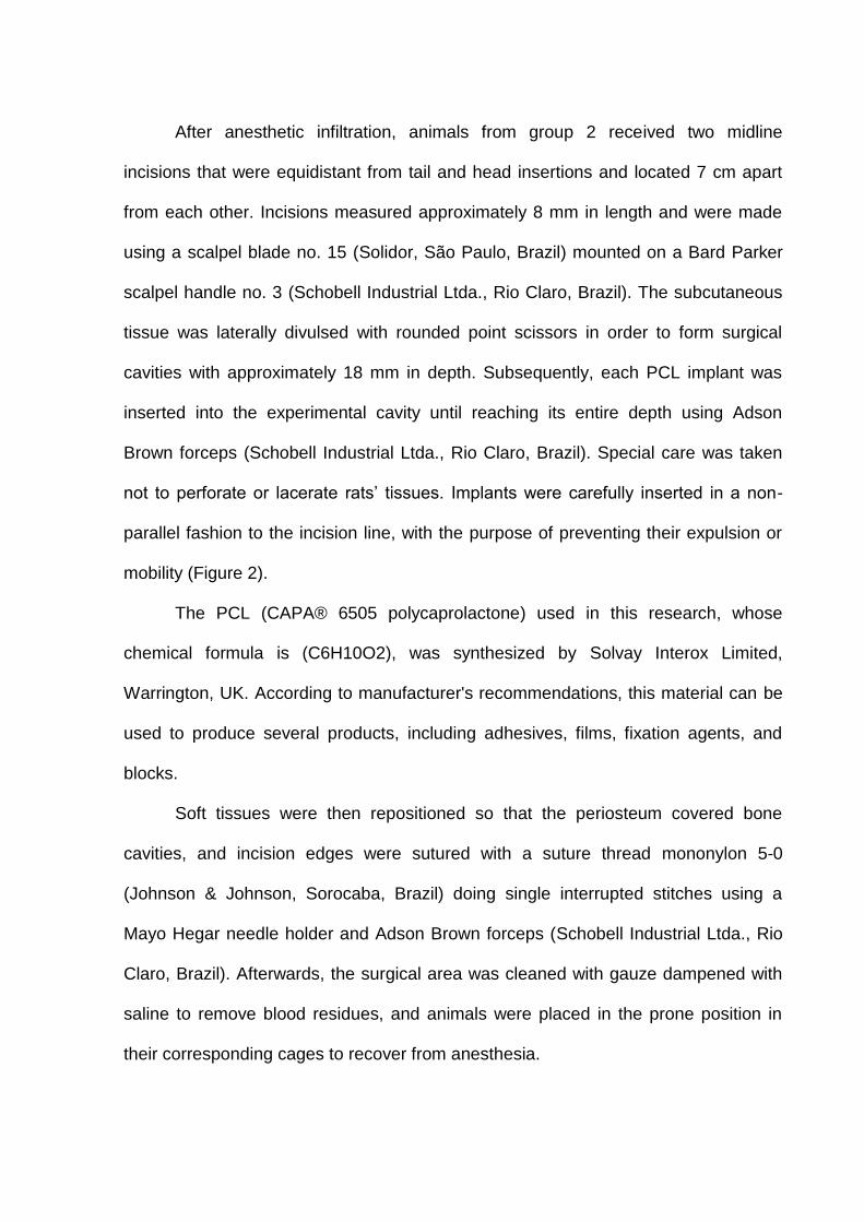
After anesthetic infiltration, animals from group 2 received two midline
incisions that were equidistant from tail and head insertions and located 7 cm apart
from each other. Incisions measured approximately 8 mm in length and were made
using a scalpel blade no. 15 (Solidor, São Paulo, Brazil) mounted on a Bard Parker
scalpel handle no. 3 (Schobell Industrial Ltda., Rio Claro, Brazil). The subcutaneous
tissue was laterally divulsed with rounded point scissors in order to form surgical
cavities with approximately 18 mm in depth. Subsequently, each PCL implant was
inserted into the experimental cavity until reaching its entire depth using Adson
Brown forceps (Schobell Industrial Ltda., Rio Claro, Brazil). Special care was taken
not to perforate or lacerate rats’ tissues. Implants were carefully inserted in a non-
parallel fashion to the incision line, with the purpose of preventing their expulsion or
mobility (Figure 2).
The PCL (CAPA® 6505 polycaprolactone) used in this research, whose
chemical formula is (C6H10O2), was synthesized by Solvay Interox Limited,
Warrington, UK. According to manufacturer's recommendations, this material can be
used to produce several products, including adhesives, films, fixation agents, and
blocks.
Soft tissues were then repositioned so that the periosteum covered bone
cavities, and incision edges were sutured with a suture thread mononylon 5-0
(Johnson & Johnson, Sorocaba, Brazil) doing single interrupted stitches using a
Mayo Hegar needle holder and Adson Brown forceps (Schobell Industrial Ltda., Rio
Claro, Brazil). Afterwards, the surgical area was cleaned with gauze dampened with
saline to remove blood residues, and animals were placed in the prone position in
their corresponding cages to recover from anesthesia.

Postoperative pain was controlled with paracetamol (Tylenol® JANSSEN-
CILAG Farmacêutica, São Paulo, Brazil) (80 mg/kg) given orally immediately after
the procedure and after 12 hours. All animals were given a single intramuscular dose
of penicillin G benzathine (Benzetacil, Eurofarma Laboratórios Ltda., São Paulo,
Brazil) (20000 units/kg) immediately after the end of the procedure.
After the end of the postoperative observation period proposed for each group,
animals were euthanized by isoflurane inhalation. Hairs from the regions of interest
were removed using an electric hair trimmer (Panasonic® ER389K mustache and
beard trimmer, Osaka, Japan) and then these areas underwent antisepsis with
0.12% chlorhexidine digluconate.
Specimens from animals of group 1 were obtained through an incision in the
most posterior region of soft tissues of the head using a scalpel blade no. 15
mounted in a Bard-Parker scalpel handle no. 3 (Schobell Industrial Ltda., Rio Claro,
Brazil). The soft tissue overlying the calvarium was removed using Metzenbaum
scissors and Adson Brown (Schobell Industrial Ltda., Rio Claro, Brazil), which made
it possible to achieve a great visualization of the calvarium, including parietal bones.
Subsequently, the calvarium was removed by osteotomy using a conical stem
multilaminated drill no. 701 rotating at low speed and under constant irrigation with
0.9% saline. Four osteotomy lines were drawn around bone defects and the
calvarium was removed using a straight chisel and Adson Brown forceps (Schobell
Industrial Ltda., Rio Claro, Brazil). In order to evaluate systemic toxicity, animals’
liver, lung and kidneys were removed through an abdominal incision for histological
analysis.
Specimens from animals in group 2 were obtained through excision biopsy of
the implant area, after the implant was located by palpation. This biopsy was

performed with a safety margin of 1 cm and began with an incision using a scalpel
blade no. 15 mounted in a Bard-Parker scalpel handle no. 3 (Schobell Industrial
Ltda., Rio Claro, Brazil). The dorsal subcutaneous tissue was divulsed using
Metzenbaum scissors and Adson Brown forceps (Schobell Industrial Ltda., Rio Claro,
Brazil), which made it possible to achieve a great visualization of the calvarium,
including the PCL implant and an enough amount of normal adjacent tissue. In order
to evaluate systemic toxicity, animals’ liver, lungs and kidneys were removed for
histological analysis. After local macroscopic examination, specimens were
immediately stored in identified plastic containers and immersed in 10% neutral
buffered formalin for tissue fixation and conservation, in order to prevent post-mortem
alterations in the tissues.
After specimens were fixed in formaldehyde for more than 24 hours and less
than 72 hours, another stage of the research started: the preparation and analysis of
histological slides. Specimens from group 1 were decalcified in 5% nitric acid solution
(10 ml) for approximately 72 hours and defects were separated between themselves
and divided in half. Specimens from the back, belonging to group 2, and from organs
used to evaluate systemic toxicity did not require decalcification. Subsequently,
standard procedures for staining with hematoxylin and eosin (HE) were performed,
as well as the routine histological processing for the preparation of slides, which
included paraffin embedding, the performance of four semi-serial sections of
approximately 6 µm in thickness in each block – with a distance of 15 µm between
each section, measured on a microtome (Jung RM 2055 microtome, Leica
Biosystems, Wetzlar, Germany) –, HE staining, and examination of the slides on a
light optical microscope (BX 50 microscope, Olympus, Melville, NY, USA). Slides

were codified in such a way that the observer was unaware of which group they
belonged to.
Evaluation was performed by the same previously calibrated examiner.
Histological analyses were carried out using a light microscope at 40, 100 and 400x
magnifications, distributed into fields scanning all the area containing PCL.
Analysis and description of the slides were based on the criteria established
next. Calvaria containing PCL were assessed for new bone formation originating
from the margins of the bone defect or from the center of the bone defect, or located
on the edges of the biomaterial, as well as for the presence of absence of material
resorption.
Back containing PCL were microscopically evaluated for cellular and tissue
reactions, the presence of fibrous capsule adjacent to the material that had been
implanted and its thickness, the presence of inflammatory infiltrate and of
inflammatory multinucleated giant cells, vascular alterations, and the formation of
granulation tissue. The fibrous capsule was defined as thin or thick; the granulation
tissue as young or mature; fibrosis as organized or disorganized; finally,
vasodilatation, hyperemia and edema were defined as mild, moderate and severe.
Moreover, the inflammatory infiltrate located close to the material under analysis was
defined as absent when the percentage of inflammatory cells was up to 10%;
moderately present if the presence of inflammatory cells was observed, but they did
not dominate the histological field in analysis, with a percentage ranging from 10 to
50%; and severely present when cells form an infiltrate around the bone portion to be
observed, with a percentage higher than 50%13.
According to Souza et al.14, experimental materials are considered
biocompatible if the intensity of the inflammatory reaction in the connective tissue
A
decreases over time. Therefore, after microscopic evaluation of specimens for 60
days, the material under investigation was considered biocompatible when the
sample has a thin layer of fibrous capsule around the implant and there was no
evidence of inflammatory reaction, macrophages or inflammatory multinucleated
giant cells. On the other hand, it was considered non-biocompatible when there was
a persistent inflammatory reaction related to macrophages and giant cells, as well as
the development of a thick fibrous capsule.
Additionally, each animal was assessed for systemic toxicity by investigating
liver, kidney and lung changes, such as the presence of cellular or inflammatory
infiltration and tissue alterations like hyperplasia, metaplasia and/or dysplasia. No
statistical tests were applied, since it was a qualitative study.
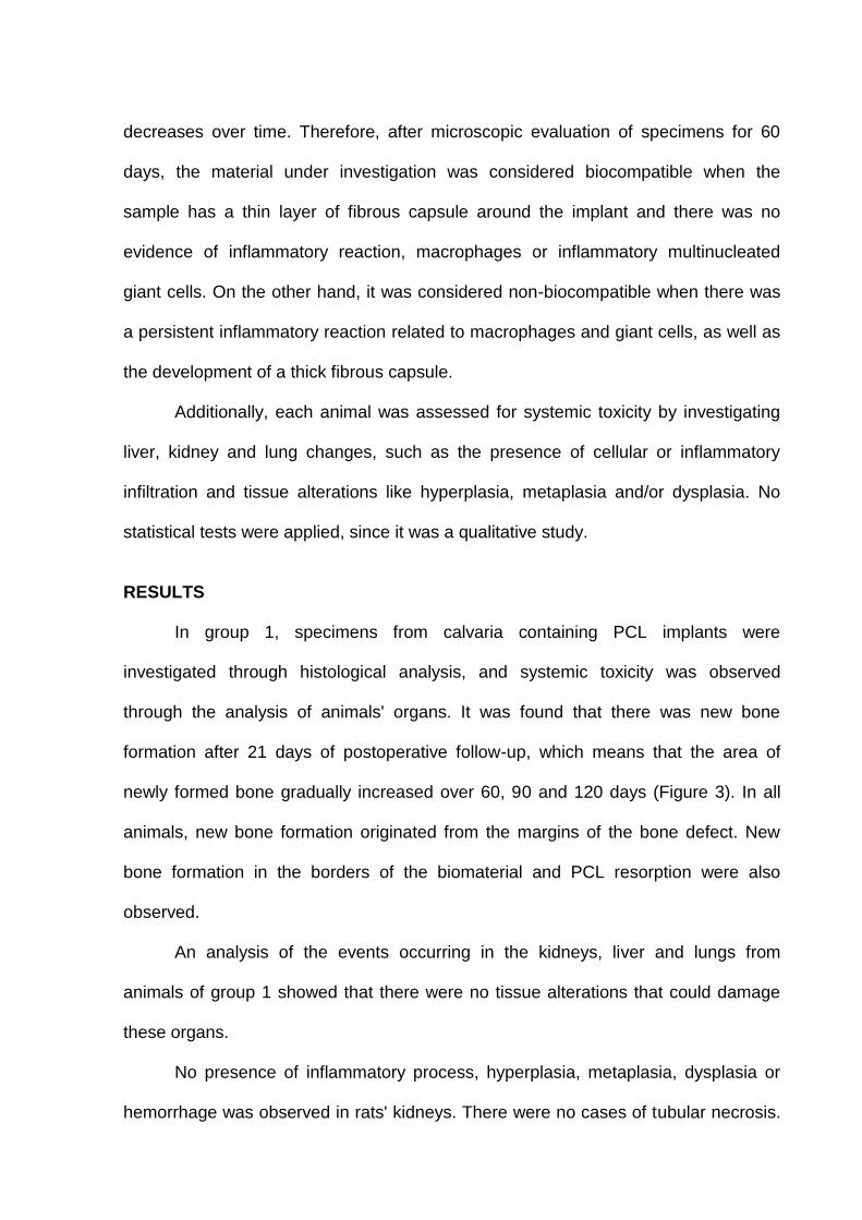
RESULTS
In group 1, specimens from calvaria containing PCL implants were
investigated through histological analysis, and systemic toxicity was observed
through the analysis of animals' organs. It was found that there was new bone
formation after 21 days of postoperative follow-up, which means that the area of
newly formed bone gradually increased over 60, 90 and 120 days (Figure 3). In all
animals, new bone formation originated from the margins of the bone defect. New
bone formation in the borders of the biomaterial and PCL resorption were also
observed.
An analysis of the events occurring in the kidneys, liver and lungs from
animals of group 1 showed that there were no tissue alterations that could damage
these organs.
No presence of inflammatory process, hyperplasia, metaplasia, dysplasia or
hemorrhage was observed in rats' kidneys. There were no cases of tubular necrosis.

The only alterations found in these animals were mild glomerular hypercellularity,
vascular congestions, and foci of capillary aggregates, which also appeared in
control animals.
There were no signs of inflammatory process, hyperplasia, metaplasia,
dysplasia or hemorrhage in animals' liver as well. In addition, no microvesicular
steatosis, necrosis or apoptosis were observed. There were only very few cells with
macrovesicular steatosis or vascular and sinusoidal congestions, events that were
also observed in control animals.
No presence of inflammatory process, hyperplasia, metaplasia, dysplasia or
hemorrhage was found in animals' lungs. The only significant finding was the
presence of peribronchial lymphoid aggregates, alveolar septal thickening, and
vascular congestion, events that were also observed in control animals (Figure 4).
In animals from group 2, specimens from rats' back containing a PCL implant
were investigated through histological analysis, and systemic toxicity was observed
through the analysis of animals' organs.
When tissues adjacent to the disc implanted on animals’ back were observed
after 60 days, the formation of a thin fibrous capsule was found in all animals, with
organized collagenous fibers involving the implant (Figure 5). There were no signs of
inflammatory infiltrate, granulation tissue, vasodilation, hyperemia, edema or abscess
60 days after discs were implanted.
When it comes to events occurring in the kidneys, lungs or liver of animals
from group 2, no harmful tissues alterations were reported. No inflammatory process,
hyperplasia, metaplasia, dysplasia or hemorrhage were observed in animals'
kidneys, lungs and liver. Their kidneys did not present with tubular necrosis, and only
cases of mild glomerular hypercellularity, vascular congestion, and foci of capillary

aggregates were found. Their liver did not develop microvesicular steatosis, necrosis
or apoptosis. There were only a few isolated cells with macrovesicular steatosis and
vascular and sinusoidal congestion. Rats' lungs showed peribronchial lymphoid
aggregates, mild punctual alveolar septal thickening, and vascular congestion,
events that were also observed in the two control animals (Figure 6).
DISCUSSION
The use of materials to improve or repair the body dates back to antiquity,
when natural materials such as wood were used in an attempt to structurally replace
tissues lost to trauma or disease15. Since the 20th century, these natural materials
began to be replaced with polymers, which provided better performance, functionality
and reproducibility15.
Currently, biomaterials are an increasingly important alternative source in
bone regeneration. They should ideally be biocompatible and biodegradable, as well
as having the appropriate porosity that allows for vascularization and ensures
mechanical resistance. Additionally, its degradation products should be non-toxic16-18.
PCL is a type of bioabsorbable polymer that has a great potential for use in
bone repair, because it presents mechanical characteristics similar to that of biologic
materials, allowing for cell growth and proliferation, as well as the formation of new
tissue3,8,11,12.
The preparation of an appropriate three-dimensional scaffold is essential to
determine whether the material can be used as a bone substitute. An ideal scaffold
should have pores able to provide enough space for a uniform cell distribution and an
appropriate oxygen and nutrient reception, in addition to having good biocompatibility
and osteoconductivity19,20.

In the present study, PCL scaffolds were prototyped in an experimental
platform of the Fab@CTI additive manufacturing machine, in order for the material to
be initially transformed into filaments to be used on the machine. Therefore, it
became necessary to observe the in vivo characteristics of PCL after all this
process10. This study evaluated PCL biocompatibility through the histological analysis
of tissue reaction to PCL scaffolds implanted on rats’ back and calvarium, as well as
their systemic toxicity through the analysis of animals' kidneys, lungs and liver.
The main advantages of producing scaffolds by additive manufacturing are
precision in material deposition and process reproducibility, making it possible to
obtain three-dimensional complex structures and to control internal morphology.
Additionally, this process takes a short time and has a relatively low cost10,21.
The methodology used in this study also allowed evaluating tissue reactions in
animal models, which is an essential stage to complete the evaluation of this type of
material. In areas where PCL scaffolds were implanted, this material became directly
in contact with tissue, including bone tissue, similarly to what would occur if the
biomaterial was clinically applied22.
Our histological analysis made it possible to assess the presence of newly
formed bone on calvaria, showing that new bone formation occurred towards the
center of the defects, as well as to qualitatively assess the presence of remaining
portions of the PCL disc19,23,24. The results obtained from this analysis showed that
new bone formation occurred after 21 days post-implantation, with the formation of a
bone bridge from one margin of the defect to another (Figure 3 E) but not the total
replacement of the biomaterial with bone tissue25. Thus, this evaluation made it
possible to investigate the beginning of the osteoconduction process, as well as the
slow biomaterial resorption and the replacement of PCL with bone.24 The histological

analysis of tissues from animals' back at 60 days allowed observing the formation of
a thin fibrous capsule in all animals, with organized collagenous fibers involving the
PCL implant, which confirmed findings from other studies26,27.
With regard to the events occurring in animals' organs, histological analysis
did not reveal tissue alterations that could damage their organs, since no signs of
inflammatory process, hyperplasia, metaplasia, dysplasia or hemorrhage were
observed in rats' kidneys, lungs and liver.
Some punctual isolated alterations were found, such as mild glomerular
hypercellularity and vascular congestion in the kidneys; isolated cells with
macrovesicular steatosis and vascular and sinusoidal congestion in the liver; and
mild alveolar septal thickening and vascular congestion in the lungs. However, these
events were also observed in control animals, which did not receive any type of
treatment.
Thus, the characteristics observed in the PCL used in the present study
corroborate those conceptually necessary for the material to be appropriate for use in
tissue repair, since it did not produce an exacerbated inflammatory reaction, was not
rejected by the body, and allowed for osteoconduction19,28-30.
Therefore, in view of the results obtained, it is possible to conclude that PCL
scaffolds produced on the Fab@CTI additive manufacturing machine are
biocompatible, non-cytotoxic and bioresorbable products that promote
osteoconduction. Hence, PCL seems to be an appropriate biomaterial to be used in
other studies aiming to elucidate issues related to this topic and in future clinical
trials.

ACKNOWLEDGMENTS
The authors would like to thank the Coordination for the Improvement of Higher
Education Personnel (Coordenação de Aperfeiçoamento de Pessoal de Nível
Superior, CAPES), because WH is a dental PhD student supported by CAPES.
We would also like to thank Dr. Fernanda Morrone, head of the Laboratory of Applied
Pharmacology, School of Pharmacy, PUCRS, for allowing the use of laboratory
facilities.
Conflict of interest
None to declare
Role of the funding source
None
Statement of authorship
All authors have read and approved the manuscript as submitted, are qualified for
authorship, believe the submission represents honest work and take full
responsibility for the reported findings.
REFERENCES
1. Langer R, Vacanti JP. Tissue engineering. Science 1993;260:920-926.
2. Barbanti SH, Santos AR, Jr., Zavaglia CA, Duek EA. Poly(epsilon-
caprolactone) and poly(D,L-lactic acid-co-glycolic acid) scaffolds used in bone tissue

engineering prepared by melt compression-particulate leaching method. J Mater Sci
Mater Med 2011;22:2377-2385.
3. Ganesh N, Jayakumar R, Koyakutty M, Mony U, Nair SV. Embedded silica
nanoparticles in poly(caprolactone) nanofibrous scaffolds enhanced osteogenic
potential for bone tissue engineering. Tissue Eng Part A 2012;18:1867-1881.
4. Liu C, Xia Z, Czernuszka JT. Design and development of three-dimensional
scaffolds for tissue engineering. Chem Eng Res Des. 2007;85:1051-1064.
5. Middleton JC, Tipton AJ. Synthetic biodegradable polymers as orthopedic
devices. Biomaterials 2000;21:2335-2346.
6. Woodruff MA, Hutmacher DW. The return of a forgotten polymer—
Polycaprolactone in the 21st century. Prog Polym Sci 2010;35:1217-1256.
7. Domingos M, Dinucci D, Cometa S, Alderighi M, Bartolo PJ, Chiellini F.
Polycaprolactone scaffolds fabricated via bioextrusion for tissue engineering
applications. Int J Biomater 2009;2009:239643.
8. Chen M, Le DQ, Baatrup A, Nygaard JV, Hein S, Bjerre L, Kassem M, Zou X,
Bunger C. Self-assembled composite matrix in a hierarchical 3-D scaffold for bone
tissue engineering. Acta Biomater 2011;7:2244-2255.
9. Pietrzak WS, Sarver DR, Verstynen ML. Bioabsorbable polymer science for
the practicing surgeon. J Craniofac Surg 1997;8:87-91.
10. Senedese AL. Estruturação tridimensional de scaffolds de policaprolactona via
manufatura aditiva [Thesis]. Campinas: Universidade Estadual de Campnas;
Faculdade de Engenharia Química; 2011.
11. Lohfeld S, Cahill S, Barron V, McHugh P, Durselen L, Kreja L, Bausewein C,
Ignatius A. Fabrication, mechanical and in vivo performance of

polycaprolactone/tricalcium phosphate composite scaffolds. Acta Biomater
2012;8:3446-3456.
12. Choong CS, Hutmacher DW, Triffitt JT. Co-culture of bone marrow fibroblasts
and endothelial cells on modified polycaprolactone substrates for enhanced
potentials in bone tissue engineering. Tissue Eng 2006;12:2521-2531.
13. Figueiredo JA, Pesce HF, Gioso MA, Figueiredo MA. The histological effects
of four endodontic sealers implanted in the oral mucosa: submucous injection versus
implant in polyethylene tubes. Int Endod J 2001;34:377-385.
14. Souza PP, Aranha AM, Hebling J, Giro EM, Costa CA. In vitro cytotoxicity and
in vivo biocompatibility of contemporary resin-modified glass-ionomer cements. Dent
Mater 2006;22:838-844.
15. Huebsch N, Mooney DJ. Inspiration and application in the evolution of
biomaterials. Nature 2009;462:426-432.
16. Ousterhout DK, Stelnicki EJ. Plastic surgery's plastics. Clin Plast Surg
1996;23:183-190.
17. Valerio P, Pereira MM, Goes AM, Leite MF. The effect of ionic products from
bioactive glass dissolution on osteoblast proliferation and collagen production.
Biomaterials 2004;25:2941-2948.
18. Knabe C, Stiller M, Berger G, Reif D, Gildenhaar R, Howlett CR, Zreiqat H.
The effect of bioactive glass ceramics on the expression of bone-related genes and
proteins in vitro. Clin Oral Implants Res 2005;16:119-127.
19. Fu S, Ni P, Wang B, Chu B, Peng J, Zheng L, Zhao X, Luo F, Wei Y, Qian Z.
In vivo biocompatibility and osteogenesis of electrospun poly(epsilon-caprolactone)-
poly(ethylene glycol)-poly(epsilon-caprolactone)/nano-hydroxyapatite composite
scaffold. Biomaterials 2012;33:8363-8371.

20. Roosa SM, Kemppainen JM, Moffitt EN, Krebsbach PH, Hollister SJ. The pore
size of polycaprolactone scaffolds has limited influence on bone regeneration in an in
vivo model. J Biomed Mater Res A 2010;92:359-368.
21. Raymond BJ. Indirect tissue scaffold fabrication via additive manufacturing
and biomimetic mineralization. Blacksburg: Virginia Polytechnic Institute, 2010.
22. Scarparo RK, Haddad D, Acasigua GA, Fossati AC, Fachin EV, Grecca FS.
Mineral trioxide aggregate-based sealer: analysis of tissue reactions to a new
endodontic material. J Endod 2010;36:1174-1178.
23. Marzouk KM, Gamal AY, Al-Awady AA, Sharawy MM. Osteoconductive effects
of vinyl styrene microbeads in rat calvarial defects. J Oral Maxillofac Surg
2007;65:1508-1516.
24. Eski M, Ilgan S, Cil Y, Sengezer M, Ozcan A, Yapici K. Assessment of
distraction regenerate using quantitative bone scintigraphy. Ann Plast Surg
2007;58:328-334.
25. Grandi G, Heitz C, Santos LA, Silva ML, Sant'Ana Filho M, Pagnocelli RM,
Silva DN. Comparative histomorphometric analysis between ±-Tcp cement and ²-
Tcp/Ha granules in the bone repair of rat calvaria. Mat Res 2011;14:11-16.
26. Giavaresi G, Tschon M, Daly JH, Liggat JJ, Sutherland DS, Agheli H, Fini M,
Torricelli P, Giardino R. In vitro and in vivo response to nanotopographically-modified
surfaces of poly(3-hydroxybutyrate-co-3-hydroxyvalerate) and polycaprolactone. J
Biomater Sci Polym Ed 2006;17:1405-1423.
27. Follmann CS. Análise local e sistêmica das reações tissulares a diferentes
materiais utilizados em pulpotomias: estudo em ratos [Thesis]. Porto Alegre:
Pontifícia Universidade Católica do Rio Grande do Sul; Faculdade de Odontologia;
2011.

28. Kurashina K, Kurita H, Hirano M, Kotani A, Klein CP, de Groot K. In vivo study
of calcium phosphate cements: implantation of an alpha-tricalcium
phosphate/dicalcium phosphate dibasic/tetracalcium phosphate monoxide cement
paste. Biomaterials 1997;18:539-543.
29. Arafat MT, Lam CX, Ekaputra AK, Wong SY, Li X, Gibson I. Biomimetic
composite coating on rapid prototyped scaffolds for bone tissue engineering. Acta
Biomater 2011;7:809-820.
30. Sharaf B, Faris CB, Abukawa H, Susarla SM, Vacanti JP, Kaban LB, Troulis
MJ. Three-dimensionally printed polycaprolactone and beta-tricalcium phosphate
scaffolds for bone tissue engineering: an in vitro study. J Oral Maxillofac Surg
2012;70:647-656.
CAPTIONS TO ILUSTRATIONS
Figure 1. A- Incision in rat’s calvarium. B- Bone defects prepared with bone trephine.
C- Experimental bone defect filled with polycaprolactone disc and empty control
defect.
Figure 2. Incisions at midline on rat’s back. B- Insertion of a polycaprolactone disc
into surgical cavity. C- Suture of dorsal tissues.
Figure 3. Histologic images of new formed bone in defects containing biomaterial at
7 days (A), 21 days (B), 60 days (C), 90 days (D), and 120 days, showing the
formation of a bone bridge (E). Areas of new bone formation (arrow).
Figure 4. Histologic images of animals’ organs. Kidney with mild glomerular
hypercellularity (A), kidney with vascular congestion and foci of capillary aggregates

(B), liver with vascular and sinusoidal congestion (C), liver with cells presenting with
macrovesicular steatosis (arrow) (D), lung with peribronchial lymphoid aggregates
(E), and lung with mild alveolar septal thickening and vascular congestion (F).
Figure 5. Histologic images of tissues adjacent to the disc implanted on animals’
back at 60 days. Formation of a thin fibrous capsule involving the implant (A), detail
of the fibrous capsule, with organized collagen fibers involving the implant (B and C).
Figure 6. Histologic images of animals’ organs. Kidney with mild glomerular
hypercellularity and vascular congestion (A), liver with vascular and sinusoidal
congestion and cell presenting with macrovesicular steatosis (arrow) (B), and lung
with peribronchial lymphoid agglomerates, mild alveolar septal thickening, and
vascular congestion (C).
FIGURES
Figure 1
Figure 2

Figure 3
Figure 4

Figure 5
Figure 6

Artigo 2

3 ARTIGO 2
O artigo a seguir intitula-se Tomographic and histomorphometric analysis of
polycaprolactone scaffolds in bone repair – an in vivo study e foi formatado e
submetido de acordo com as normas do periódico Biomaterials (Anexo B).

TOMOGRAPHIC AND HISTOMORPHOMETRIC ANALYSIS OF
POLYCAPROLACTONE SCAFFOLDS IN BONE REPAIR – AN IN VIVO STUDY
Abbreviated title: Bone repair vs. polycaprolactone – a study in rats
Wâneza D. B. Hirsch*,a Milene B. Campagnaro,a Ana Lívia C. Senedese,b,c,d Prof. Dr.
Helena W. de Oliveira,a Prof. Dr. Daniela N. Silva,e Prof. Dr. Cláiton Heitza
a School of Dentistry, Pontifícia Universidade Católica do Rio Grande do Sul
(PUCRS), Av. Ipiranga, 6681 Prédio 06, Partenon, CEP: 90619-900, Porto Alegre,
Brazil.
b School of Chemical Engineering, Universidade Estadual de Campinas. Cidade
Universitária Zeferino Vaz. Av. Albert Einstein, 500, CEP 13083-852, Campinas,
Brazil.
c Instituto Nacional de Biofabricação (BIOFABRIS). Av. Albert Einstein, 500,
CEP 13083-852, Campinas, Brazil.
d Centro de Tecnologia da Informação Renato Archer, Rodovia D. Pedro I (SP-65)
Km 143,6, Terminal Intermodal de Cargas (TIC), CEP 13069-901, Campinas, Brazil.
e School of Dentistry, Universidade Federal do Espírito Santo, Av. Marechal Campos,
1468, Maruípe, CEP 29.040-090, Vitória, Brazil.
* Corresponding author. Address: School of Dentistry, Pontifícia Universidade
Católica do Rio Grande do Sul (PUCRS), Av. Ipiranga, 6681 Prédio 06, Partenon,

CEP: 90619-900, Porto Alegre, Brazil. Phone: +55 (53) 9130.6088. Fax: +55 (51)
3320.3500. E-mail: [email protected].
Abstract
Tissue engineering has been studying several biomaterials for bone tissue
replacement. The present study evaluated the in vivo performance of
polycaprolactone (PCL) scaffolds in bone repair of rat calvarial defects. A quantitative
analysis of the bone repair process was performed using histomorphometry and cone
beam computed tomography (CBCT). Results showed that the newly formed bone
grew towards the center of the defects. Statistical analysis revealed that the total
area of new bone formation was greater in experimental defects at 21, 60 and 120
days, showing a statistically significant difference. However, a tomographic analysis
found that new bone formation is more likely to occur in experimental defects, but
with no statistically significant difference. Thus, considering tomographic analysis as
a new method for the assessment of new bone formation, the data obtained from this
assessment could not be correlated with those obtained from histomorphometric
analysis. Therefore, PCL showed great potential for clinical use in the treatment of
bone defects by increasing bone area, due to the fact that it promoted
osteoconduction. Additionally, CBCT does not seem to be a useful tool in the
evaluation of new bone formation of rat calvaria, which means that
histomorphometric analysis is still the most appropriate method.
Keywords: bone tissue engineering; histomorphometry; scaffold; polycaprolactone
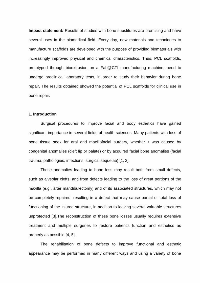
Impact statement: Results of studies with bone substitutes are promising and have
several uses in the biomedical field. Every day, new materials and techniques to
manufacture scaffolds are developed with the purpose of providing biomaterials with
increasingly improved physical and chemical characteristics. Thus, PCL scaffolds,
prototyped through bioextrusion on a Fab@CTI manufacturing machine, need to
undergo preclinical laboratory tests, in order to study their behavior during bone
repair. The results obtained showed the potential of PCL scaffolds for clinical use in
bone repair.
1. Introduction
Surgical procedures to improve facial and body esthetics have gained
significant importance in several fields of health sciences. Many patients with loss of
bone tissue seek for oral and maxillofacial surgery, whether it was caused by
congenital anomalies (cleft lip or palate) or by acquired facial bone anomalies (facial
trauma, pathologies, infections, surgical sequelae) [1, 2].
These anomalies leading to bone loss may result both from small defects,
such as alveolar clefts, and from defects leading to the loss of great portions of the
maxilla (e.g., after mandibulectomy) and of its associated structures, which may not
be completely repaired, resulting in a defect that may cause partial or total loss of
functioning of the injured structure, in addition to leaving several valuable structures
unprotected [3].The reconstruction of these bone losses usually requires extensive
treatment and multiple surgeries to restore patient's function and esthetics as
properly as possible [4, 5].
The rehabilitation of bone defects to improve functional and esthetic
appearance may be performed in many different ways and using a variety of bone

substitutes, such as autogenous graft (which is the gold standard), allogeneic graft,
xenogeneic graft, the combination of these grafts, and alloplastic or synthetic grafts
[5].
Bioabsorbable polymers, such as polycaprolactone (PCL), are alternative
materials for the treatment of lesions and tissue losses. They show great potential to
be used as support for cell culture in tissue engineering, in addition to presenting
mechanical characteristics similar to those of biological materials. These polymers
allow for cell growth and proliferation, as well as the formation of new tissue [2, 6-8].
PCL is a biodegradable and bioresorbable material that provides a dense and
porous support for the newly formed bone [1, 8]. It has a melting point between 58
and 63 degrees Celsius (°C) and elastic modulus of 0.4 gigapascal (GPa), and its
time of degradation ranges from 24 to 36 months [9, 10].
In view of the foregoing, the aim of this study was to observe the in vivo
performance of PCL at 7, 21, 60, 90 and 120 days after graft implant surgery by
undertaking a tomographic and histomorphometric analysis of bone repair in rats with
critical calvarial defects.
2. Materials and Methods
2.1. Study design
The present investigation was developed following a traditional quantitative
paradigm and was characterized as a true experimental study. The tree-dimensional
PCL scaffolds used in this study were prototyped by means of bioextrusion using on
the platform of an additive Fab@Home manufacturing machine at the Information
Technology Center of Centro de Tecnologia da Informação Renato Archer
(Campinas, Brazil), contained 0.5 mm micropores, and measured 5 mm in diameter
and 1 mm in thickness. Afterwards, they were inserted into critical bone defects of rat
calvaria, with the purpose of evaluating new bone formation.
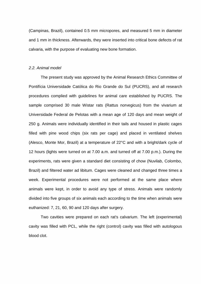
2.2. Animal model
The present study was approved by the Animal Research Ethics Committee of
Pontifícia Universidade Católica do Rio Grande do Sul (PUCRS), and all research
procedures complied with guidelines for animal care established by PUCRS. The
sample comprised 30 male Wistar rats (Rattus norvegicus) from the vivarium at
Universidade Federal de Pelotas with a mean age of 120 days and mean weight of
250 g. Animals were individually identified in their tails and housed in plastic cages
filled with pine wood chips (six rats per cage) and placed in ventilated shelves
(Alesco, Monte Mor, Brazil) at a temperature of 22°C and with a bright/dark cycle of
12 hours (lights were turned on at 7.00 a.m. and turned off at 7.00 p.m.). During the
experiments, rats were given a standard diet consisting of chow (Nuvilab, Colombo,
Brazil) and filtered water ad libitum. Cages were cleaned and changed three times a
week. Experimental procedures were not performed at the same place where
animals were kept, in order to avoid any type of stress. Animals were randomly
divided into five groups of six animals each according to the time when animals were
euthanized: 7, 21, 60, 90 and 120 days after surgery.
Two cavities were prepared on each rat's calvarium. The left (experimental)
cavity was filled with PCL, while the right (control) cavity was filled with autologous
blood clot.

Sample size (N=6 per group, total N=30) was defined using literature data [3,
11]; thus, we decided to work with the minimum number that would not compromise
the results.
2.3. Surgical procedures
Surgical procedures were carried out at the Laboratory of Applied
Pharmacology, room 148, block C, of the School of Pharmacy of PUCRS and
complied with all principles of biosecurity and infection control.
After being weighed, animals were anesthetized by an intraperitoneal injection
of a mixture of ketamine hydrochloride (ketamin®) (100mg/kg) (Cristália Produtos
Químicos Farmacêuticos Ltda., Itapira, Brazil) and xylazine hydrochloride (calmiun®)
(10mg/kg) (Agener União, São Paulo, Brazil). Subsequently, hair was removed and
antisepsis was performed with 2% chlorhexidine digluconate (Clorhexidina s,
Digluconato de clorexidina 2%, FGM Produtos Odontológicos Ltda., Joinvile, Brazil).
Next, surgery was performed using a sterile fenestrated surgical drape.
Animals received local anesthesia by subcutaneous anesthetic infiltration with 2%
lidocaine hydrochloride and 1:50.000 norepinephrine (Probem® – Lidostesim 2%,
Catanduva, Brazil), in order to achieve hemostasis and additional analgesia during
surgery. A coronal linear incision of nearly 1.5 cm in length was done between the
two ears using a scalp and a blade no.15 (Solidor, São Paulo, Brazil). The soft
tissues of the head were separated, providing good visualization of the periosteum,
which was subsequently incised, divulsed and moved away along with the other
tissues to expose the external surface of the calvarium. The region was irrigated with
0.9% saline and then dried with sterile gauze.

The sites where cavities should be prepared were delimited using an
exploratory probe that preserved the median sagittal suture. Right and left cavities
were distributed laterally to the median sagittal suture, at the parietal bones, with a
distance of 2 mm between each other, as measured by a Quinelato® analogue
surgical caliper (www.quinelato.com.br/odonto/imagens/compasso1.gif).
Bone defects were prepared using an electric motor rotating at low speed and
bone trephine measuring 5 mm in diameter, which corresponded to the size of the
bone defects created during surgery (Figure 1). Trephine was slightly pressed with
intermittent movements in the superoinferior direction, making it possible to prepare
the bone defect by disrupting external and internal cortical bones of the calvarium
without damaging the meninges. Cavities were abundantly irrigated with saline to
remove the residues produced in the process of defect preparation and dried with
sterile gauze.
PCL was inserted into left (experimental) cavities, which were the using Adson
Brown forceps (Figure 2). Control cavities were prepared on the right side of the
calvarium, but they did not receive any material and were filled with clot. The PCL
(CAPA® 6505 polycaprolactone) used in this research was synthesized by Solvay
Interox Limited, Warrington, UK. Its chemical formula is (C6H10O2). According to
manufacturer's recommendations, this material can be used to produce several
products, including adhesives, films, fixation agents, and blocks.
Subsequently, soft tissues were repositioned so that the periosteum recovered
bone cavities, and incision edges were sutured with a suture thread mononylon 5-0
(Johnson & Johnson, Sorocaba, Brazil) doing simple interrupted stitches. The
surgical area was cleaned with gauze dampened with saline to remove blood

residues, and animals were placed in the prone position in their corresponding cages
to recover from anesthesia.
Postoperative pain was controlled with paracetamol (80 mg/kg) (Tylenol®,
JANSSEN-CILAG Farmacêutica, São Paulo, Brazil) given orally immediately after the
procedure and after 12 hours. All animals were given a single intramuscular dose of
penicillin G benzathine (20000 units/kg) (Benzetacil, Eurofarma Laboratórios Ltda.,
São Paulo, Brazil) immediately after the end of the procedure.
After the end of the postoperative observation period proposed for each group,
animals were euthanized by isoflurane inhalation. Hairs were removed and the area
underwent antisepsis with 2% chlorhexidine digluconate (Clorhexidina s, Digluconato
de clorexidina 2%, FGM Produtos Odontológicos Ltda., Joinvile, Brazil).
Specimens were obtained by an incision in the most posterior region of the
soft tissues of the head with a scalp and a blade no. 15, and the soft tissue overlying
the calvarium was removed using Metzenbaum scissors and Adson Brown forceps,
which made it possible to achieve a great visualization of the calvarium, including
parietal bones. Subsequently, the calvarium was removed by osteotomy using a
conical stem multilaminated drill no. 701 rotating at low speed and under constant
irrigation with 0.9% saline. The osteotomy line kept a distance of 4 to 5 mm of
defective areas. Four osteotomy lines were drawn around bone defects and the
calvarium was removed using a straight chisel and Adson Brown forceps. After local
macroscopic examination, specimens were stored in identified containers with 10%
neutral buffered formalin.

2.4. Histological process
After being fixed in formaldehyde, specimens were decalcified in 5% nitric acid
solution and defects were separated between themselves and divided in half.
Subsequently, standard procedures for staining with hematoxylin and eosin (HE)
were performed, as well as the routine histological processing for the preparation of
slides, which included paraffin embedding, the performance of four semi-serial
sections of approximately 6µm in diameter in each block – with a distance of 15 µm
between each section, measured on a microtome (Jung RM 2055 microtome, Leica
Biosystems, Wetzlar, Germany) and based on the greatest diameter of the defect –,
HE staining, and examination on a light optical microscope (BX 50 microscope,
Olympus, Melville, NY, USA).
Histological assessment was performed by the same previously calibrated
examiner. Fifteen slides were used for examiner's calibration and examined both by
the examiner (evaluator A) and by an experienced pathologist (evaluator B). Errors
were analyzed by comparing the results of evaluators A and B through the kappa test
for interobserver agreement. The level of agreement for the results of the kappa test
was quantified according to the percentage recommended by Landis and Koch [12],
considering the following values: 0.61 to 0.80, representing significant agreement,
and 0.81 to 1.0, representing almost complete agreement.
2.5. Histomorphometry
The histological images were captured from the microscope by the computer
at 40X magnification. The slides were analyzed using the Image Pro Plus software,
version 6.2® (Media Cybernetics, Bethesda, USA) (Figure 3), which allowed A

measuring, in millimeters, the total area of each bone defect and the area of newly
formed bone inside the defect.
Histological analyses included fields scanning all the area of the defect.
Analysis and description of the slides were based on the area of newly formed bone
from the edge of the bone defect and on the amount of material resorption. The
percentage of newly formed bone and of remaining material was quantified. Values
were recorded on a table specifically designed for data collection.
2. 6. Cone beam computed tomography (CBCT)
Tomographic images were obtained using a volumetric CBCT scanner (Kodak
Cone Beam 3D System, Carestream Health Inc., Rochester, USA) and then analyzed
using the Image J software (National Institute of Health, Bethesda, USA) (Figure 4),
which allowed measuring the total area of each defect, the area of newly formed
bone inside the defect, and the amount of material that remained inside the
experimental defect, all of them measured in pixels and converted into millimeters.
Analyzes were performed by segmenting the images and applying masks to
eliminate regions outside our regions of interest and to determine the region affected
by bone defects (with and without biomaterial).
The percentage of newly formed bone and of remaining material were
quantified in millimeters, and values were recorded on a data collection table.
2.7. Statistical analysis
Experimental vs. control cavities were compared with regard to the percentage
of newly formed bone using descriptive statistical analysis (mean and standard

deviation) in the form of tables and graphs. The results obtained were assessed by
means of analysis of variance (ANOVA) at 5% probability.
Data processing and analysis were performed using the Statistical Package
for Social Sciences (SPSS)® software, version 17 (SPSS Inc., Chicago, USA), as well
as Assistat software, version 7.6 (Departamento de Engenharia Agrícola do Centro
de Ciências e Tecnologia da Universidade Federal da Paraíba, Campina Grande,
Brazil), on the Microsoft Windows operating system.
3. Results
3.1. Histomorphometric analysis
Figure 5 shows the results for the comparison of the area of new bone
formation between the two groups (with and without PCL [treatment]) at the
respective times (7, 21, 60, 90 and 120 days [time blocks]), which revealed an
interaction between treatments and time blocks.
Figure 6 describes the analysis of the interaction between the use of the
biomaterial or not (treatment) and blocks of time simultaneously. Results show that
there was a statistically significant difference of 5% in the groups analyzed at 21, 60
and 120 days after surgery (squares), in which there was a greater area of new bone
formation in the defects filled with biomaterial. In the groups analyzed 7 and 90 days
after surgery, although the area of new bone formation was statistically greater on
the defect containing biomaterial, these values were not statistically significant.

3.2. Tomographic analysis
Table 1 shows that means for new bone formation were higher along the
experimental period in the groups in which the defect was filled with biomaterial
compared to those in which the defect was not filled with biomaterial.
Table 2 shows the analysis of mean areas of new bone formation for time
blocks (7, 21, 60, 90 and 120 days), with new bone formation increasing over the
study period.
Table 3 shows that, when time blocks were compared between themselves,
there was a statistically significant difference, i.e., there was higher new bone
formation over time. However, when the use of PCL or not to fill the cavities was
compared over time, it did not show statistically significant difference.
3.3. Comparison between histomorphometric and tomographic analyses
A comparison of the total area of new bone formation in mm2 obtained from
histomorphometric and tomographic analyses revealed that histomorphometric
analysis showed greater new bone formation in the groups in which the defects were
filled with PCL, with a statistically significant difference in the groups analyzed 21, 60
and 120 days after surgery. In the tomographic analysis, it can be observed that
there was a trend towards higher new bone formation in the groups that used PCL to
fill the defects, although this difference was not statistically significant (Figures 7, 8
and 9).
4. Discussion
Tissue engineering has been studying several biomaterials to replace bone
tissue that underwent total or partial losses, regardless if caused by pathological,

traumatic or congenital reasons. Autologous or allogeneic grafts are the most
commonly used procedures to replace bone tissue. However, autologous grafts have
the disadvantage of requiring an additional surgery on the donor area, in addition to
the possibility of the amount of graft not being enough to fill the receptor area [3, 4].
Allogeneic grafts may lead to intense immune response, require the use of
immunosuppressants, or transmit diseases [3, 4]. Thus, synthetic biomaterials are
viable alternatives to replace bone tissue.
The ideal biomaterial should be biocompatible and biodegradable, as well as
having the appropriate porosity that allows for vascularization and ensures
mechanical resistance. Additionally, its degradation products should be non-toxic [13-
15].
PCL was one of the first polymers to be synthesized and made commercially
available in the 1930s by the Carothers group due to its ability of being degraded by
microorganisms. However, although being initially the focus of many investigations, it
was set aside with the emergence of other polymers that were more rapidly resorbed
[16]. Thus, developments in tissue engineering have revived the interest in using
PCL, because it has appropriate properties to be used as biomaterial for tissue
reconstruction [10, 17-19]. The characteristics observed in the PCL used in the
present study corroborate those conceptually necessary for the material to be
appropriate for use in tissue repair, since it did not produce an exacerbated
inflammatory reaction, was not rejected by the body, and allowed for osteoconduction
[20-22].
Additionally, the analysis of the defects induced in this study showed that the
volume of the defect containing PCL was maintained, which did not occur in the
control defect filled with blood clot. This finding suggests that the portion of the PCL

disc which was not resorbed can serve as a scaffold for bone formation,
corroborating the findings of Grandi et al. [3].
The histomorphometric analysis performed in this study allowed assessing the
presence of PCL discs and of newly formed bone, as well as quantifying the area of
new bone formation in mm2. This type of assessment is reliable and important tool to
quantitavely prove the effectiveness of biomaterials in promoting new bone formation
[5, 23].
The results from this analysis showed that there was a statistically greater
area of new bone formation in the defects that were filled with PCL at 21, 60 and 120
days. No new bone formation was observed at 7 days in any of the defects. There
was a larger area of new bone formation in the defects filled with biomaterial at 90
days, although this difference was not statistically significant, possibly due to the fact
that the number of animals per group was limited to six.
This assessment of new bone formation over time is important, because it
allows establishing when the process of osteoconduction started and investigating
the slow biomaterial resorption and the replacement of biomaterial with bone.
Additionally, a close relationship was found between PCL and newly formed bone, as
reported by several authors assessing bioceramics [24].
Tomographic analysis made it possible to evaluate the presence of newly
formed bone, but the results obtained only showed that there was a trend of higher
rates of new bone formation in the groups whose defects were filled with PCL.
However, the difference between groups was not statistically significant, i.e., whether
using the biomaterial or not, the defect presented a larger area of new bone
formation over time. Moreover, PCL did not appear on tomographic images, which
only showed the area of newly formed bone interspersed with the PCL disc.
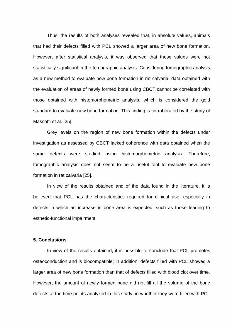
Thus, the results of both analyses revealed that, in absolute values, animals
that had their defects filled with PCL showed a larger area of new bone formation.
However, after statistical analysis, it was observed that these values were not
statistically significant in the tomographic analysis. Considering tomographic analysis
as a new method to evaluate new bone formation in rat calvaria, data obtained with
the evaluation of areas of newly formed bone using CBCT cannot be correlated with
those obtained with histomorphometric analysis, which is considered the gold
standard to evaluate new bone formation. This finding is corroborated by the study of
Massotti et al. [25].
Grey levels on the region of new bone formation within the defects under
investigation as assessed by CBCT lacked coherence with data obtained when the
same defects were studied using histomorphometric analysis. Therefore,
tomographic analysis does not seem to be a useful tool to evaluate new bone
formation in rat calvaria [25].
In view of the results obtained and of the data found in the literature, it is
believed that PCL has the characteristics required for clinical use, especially in
defects in which an increase in bone area is expected, such as those leading to
esthetic-functional impairment.
5. Conclusions
In view of the results obtained, it is possible to conclude that PCL promotes
osteoconduction and is biocompatible; in addition, defects filled with PCL showed a
larger area of new bone formation than that of defects filled with blood clot over time.
However, the amount of newly formed bone did not fill all the volume of the bone
defects at the time points analyzed in this study, in whether they were filled with PCL

or not. Furthermore, histomorphometry is still considered the most appropriate
method to evaluate new bone formation in rat calvaria, since tomographic analysis
using CBCT has been shown to be unsuitable for this type of assessment.
Acknowledgments
The authors would like to thank the Coordination for the Improvement of Higher
Education Personnel (Coordenação de Aperfeiçoamento de Pessoal de Nível
Superior, CAPES), because WH is a dental PhD student supported by CAPES.
We would also like to thank Dr. Fernanda Morrone, head of the Laboratory of Applied
Pharmacology, School of Pharmacy, PUCRS, for allowing the use of laboratory
facilities.
Role of funding source: None
References
[1] Eap S, Ferrand A, Palomares CM, Hebraud A, Stoltz JF, Mainard D, et al.
Electrospun nanofibrous 3D scaffold for bone tissue engineering. Biomed Mater Eng.
2012;22:137-141.
[2] Lohfeld S, Cahill S, Barron V, McHugh P, Durselen L, Kreja L, et al. Fabrication,
mechanical and in vivo performance of polycaprolactone/tricalcium phosphate
composite scaffolds. Acta Biomater. 2012;8:3446-3456.
[3] Grandi G, Heitz C, Santos LAd, Silva ML, Sant'Ana Filho M, Pagnocelli RM, et al.
Comparative histomorphometric analysis between ±-Tcp cement and ²-Tcp/Ha
granules in the bone repair of rat calvaria. Materials Research. 2011;14:11-16.

[4] Cardoso AK, Barbosa Ade A, Jr., Miguel FB, Marcantonio E, Jr., Farina M, Soares
GD, et al. Histomorphometric analysis of tissue responses to bioactive glass implants
in critical defects in rat calvaria. Cells Tissues Organs. 2006;184:128-137.
[5] Marzouk KM, Gamal AY, Al-Awady AA, Sharawy MM. Osteoconductive effects of
vinyl styrene microbeads in rat calvarial defects. J Oral Maxillofac Surg.
2007;65:1508-1516.
[6] Chen M, Le DQ, Baatrup A, Nygaard JV, Hein S, Bjerre L, et al. Self-assembled
composite matrix in a hierarchical 3-D scaffold for bone tissue engineering. Acta
Biomater. 2011;7:2244-2255.
[7] Choong CS, Hutmacher DW, Triffitt JT. Co-culture of bone marrow fibroblasts and
endothelial cells on modified polycaprolactone substrates for enhanced potentials in
bone tissue engineering. Tissue Eng. 2006;12:2521-2531.
[8] Ganesh N, Jayakumar R, Koyakutty M, Mony U, Nair SV. Embedded silica
nanoparticles in poly(caprolactone) nanofibrous scaffolds enhanced osteogenic
potential for bone tissue engineering. Tissue Eng Part A. 2012;18:1867-1881.
[9] Middleton JC, Tipton AJ. Synthetic biodegradable polymers as orthopedic
devices. Biomaterials. 2000;21:2335-2346.
[10] Woodruff MA, Hutmacher DW. The return of a forgotten polymer—
Polycaprolactone in the 21st century. Progress in Polymer Science. 2010;35:1217–
1256.
[11] Zanetta-Barbosa D, de Carvalho AC. Effect of brief storage in ATP solution on
periodontal healing after replantation of teeth in rats. Endod Dent Traumatol.
1990;6:193-199.
[12] Landis JR, Koch GG. The measurement of observer agreement for categorical
data. Biometrics. 1977;33:159-174.

[13] Ousterhout DK, Stelnicki EJ. Plastic surgery's plastics. Clin Plast Surg.
1996;23:183-190.
[14] Valerio P, Pereira MM, Goes AM, Leite MF. The effect of ionic products from
bioactive glass dissolution on osteoblast proliferation and collagen production.
Biomaterials. 2004;25:2941-2948.
[15] Knabe C, Stiller M, Berger G, Reif D, Gildenhaar R, Howlett CR, et al. The effect
of bioactive glass ceramics on the expression of bone-related genes and proteins in
vitro. Clin Oral Implants Res. 2005;16:119-127.
[16] Van Natta FJ, Hill JW, Carothers WH. Studies of polymerization and ring
formation, e-caprolactone and its polymers. J Am Chem Soc. 1934;56:455-459.
[17] Allo BA, Rizkalla AS, Mequanint K. Synthesis and electrospinning of epsilon-
polycaprolactone-bioactive glass hybrid biomaterials via a sol-gel process. Langmuir.
2010;26:18340-18348.
[18] Liu C, Xia Z, Czernuszka JT. Design and development of three-dimensional
scaffolds for tissue engineering. Chemical Engineering Research and Design.
2007;85:1051–1064.
[19] Butscher A, Bohner M, Hofmann S, Gauckler L, Muller R. Structural and material
approaches to bone tissue engineering in powder-based three-dimensional printing.
Acta Biomater. 2011;7:907-920.
[20] Kurashina K, Kurita H, Hirano M, Kotani A, Klein CP, de Groot K. In vivo study of
calcium phosphate cements: implantation of an alpha-tricalcium phosphate/dicalcium
phosphate dibasic/tetracalcium phosphate monoxide cement paste. Biomaterials.
1997;18:539-543.

[21] Arafat MT, Lam CX, Ekaputra AK, Wong SY, Li X, Gibson I. Biomimetic
composite coating on rapid prototyped scaffolds for bone tissue engineering. Acta
Biomater. 2011;7:809-820.
[22] Fu S, Ni P, Wang B, Chu B, Peng J, Zheng L, et al. In vivo biocompatibility and
osteogenesis of electrospun poly(epsilon-caprolactone)-poly(ethylene glycol)-
poly(epsilon-caprolactone)/nano-hydroxyapatite composite scaffold. Biomaterials.
2012;33:8363-8371.
[23] Eski M, Ilgan S, Cil Y, Sengezer M, Ozcan A, Yapici K. Assessment of
Distraction Regenerate Using Quantitative Bone Scintigraphy. Annals of Plastic
Surgery. 2007;58:328-334.
[24] Kitsugi T, Yamamuro T, Nakamura T, Oka M. Transmission electron microscopy
observations at the interface of bone and four types of calcium phosphate ceramics
with different calcium/phosphorus molar ratios. Biomaterials. 1995;16:1101-1107.
[25] Massotti FP. [Tomographic and histomorphometric evaluation of the influence of
low level laser therapy on the peri-implant tissue repair in jaws of rabbits]. Porto
Alegre: Universidade Federal do Rio Grande do Sul. Faculdade de Odontologia.
Programa de Pós-Graduação em Odontologia; 2013.
Figure Captions Fig. 1. Schematic representation of the computed tomography scan of a rat
calvarium. Bone defects (experimental and control cavities).
Fig. 2. A- Defect preparation in a rat calvarium using bone trephine. B-Bone defects
(experimental and control cavities). C- Experimental bone defect filled with PCL.
Fig. 3. Schematic representation of analysis using the Image Pro Plus software,
version 6.2® (Media Cybernetics, Bethesda, USA). The total area of control (A) and

experimental (B) defects and the area of new bone formation were measured, as well
as the amount of remaining biomaterial within the experimental defect.
Fig. 4. Schematic representation of analysis using the Image J software (National
Institute of Health, Bethesda, USA). Three-dimensional original image (A).
Application of a mask to eliminate regions external to the regions of interest. (B)
Result of the application of the mask (C). Mask to determine the total area of the
defect without the biomaterial (D) and with the biomaterial (E).
Fig. 5. Analysis of an experiment using a randomized block design with repetitions.
* Significant at 1% probability level (p < 0.01).
** Significant at 5% probability level (0.01 ≤ p < 0.05).
*** Different letters indicate statistically significant differences.
SV = source of variation, DF = degrees of freedom, SSQ = sum of squares, MSQ =
mean of squares, msd = minimum significant difference.
Fig. 6. Analysis of the interaction between treatment and times blocks
Different letters indicate statistically significant differences.
Tukey’s test was performed
B1 = 7 days, B2 = 21 days, B3 = 60 days, B4 = 90 days, B5 = 120 days, T1 = control
group, T2 = experimental group, msd = minimum significant difference, OM = overall
mean, %CV = percentage of coefficient of variation.
Fig. 7. Comparison of the area of new bone formation in the different time blocks.
Fig. 8. Analysis of the area of new bone formation using the Image Pro Plus
software, version 6.2® (Media Cybernetics, Bethesda, USA). Defect with biomaterial
at 7 days (A), 21 days (B), 60 days (C), 90 days (D), and 120 days (E). Defect
without biomaterial at 7 days (F), 21 days (G), 60 days (H), 90 days (I), and 120 days
(J). Areas of new bone formation (arrow).

Fig. 9. Analysis of the area of new bone formation using the Image J software
(National Institute of Health, Bethesda, USA). Defect with biomaterial at 7 days (A),
21 days (B), 60 days (C), 90 days (D), and 120 days (E). Defect without biomaterial
at 7 days (F), 21 days (G), 60 days (H), 90 days (I), and 120 days (J). Areas of new
bone formation (arrow).
FIGURES
Figure 1
Figure 2

Figure 3
Figure 4

Figure 5
Figure 6
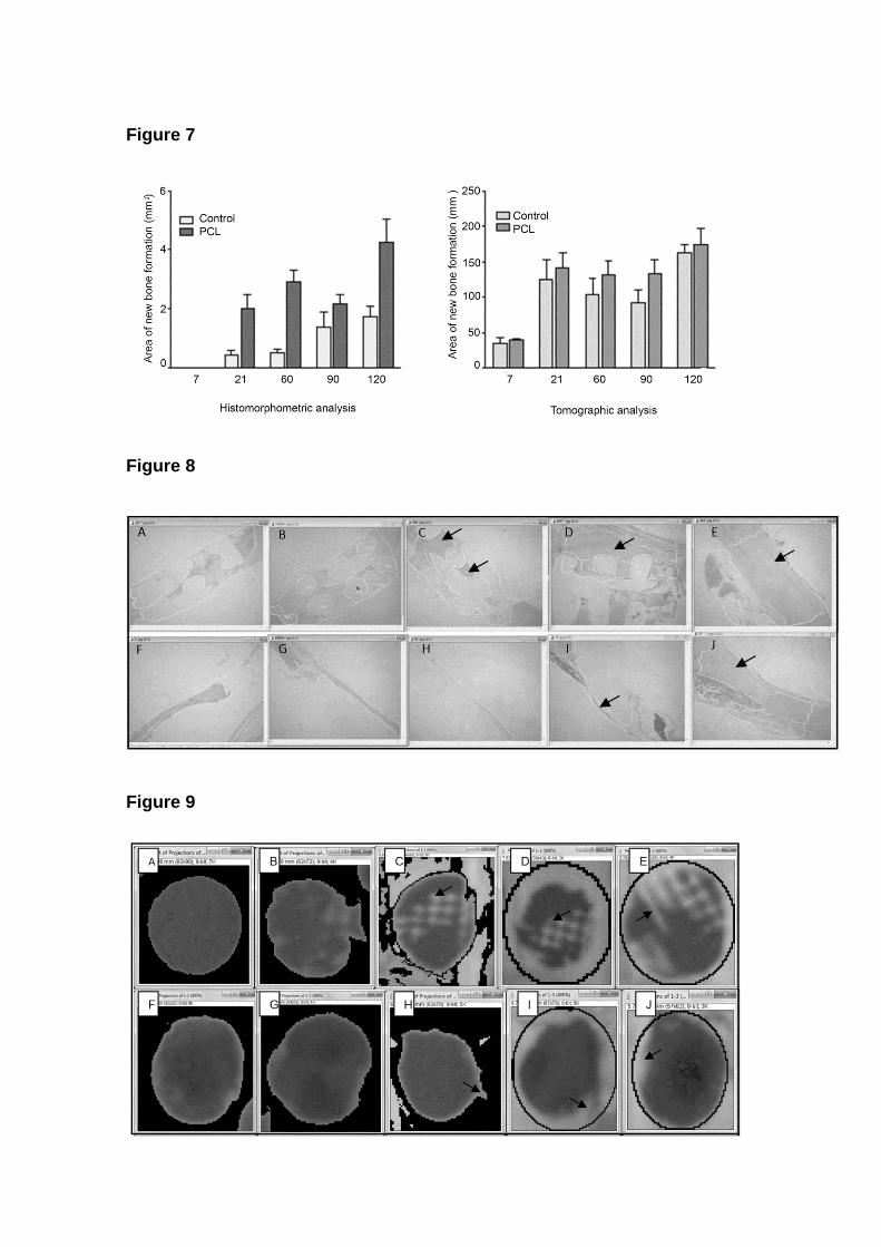
Figure 7
Figure 8
Figure 9

TABLES
Table 1. Descriptive statistics of the association between the use of biomaterial and
new bone formation in the different time blocks.
Time (days)
Mean area of new bone
formation (mm) Standard deviation N
7
Without biomaterial 33.775 17.8248 4
With biomaterial 38.800 4.9538 4
Total 36.287 12.4056 8
21
Without biomaterial 137.820 56.8020 5
With biomaterial 131.320 42.4382 5
Total 134.570 47.3937 10
60
Without biomaterial 103.833 55.6606 6
With biomaterial 131.517 46.5824 6
Total 117.675 51.0251 12
90
Without biomaterial 91.368 46.0256 6
With biomaterial 133.867 45.9040 6
Total 112.617 49.1251 12
120

Without biomaterial 162.800 28.2921 6
With biomaterial 174.000 55.7309 6
Total 168.400 42.5422 12
Overall sample
Without biomaterial 110.082 58.8650 27
With biomaterial 127.707 58.5231 27
Total 118.895 58.8145 54
Table 2. Mean area of new bone formation (mm) in the different time blocks
Time (days) N
Subset
1* 2** 3***
7 8 36.287
90 12 112.617
60 12 117.675 117.675
21 10 134.570 134.570
120 12 168.400
Sig. 1.000 0.792 0.088
Tukey's honestly significant difference (HSD) test
Sig. = Significance
* Little or no bone formation.
** Greater bone formation.
*** Better bone formation.

Table 3. Tests of between-subjects effects
Source
Type III sum of
squares df Mean square F Sig.
Time (days) 86948.734 4 21737.183 10.852 0.000*
BIOMATERIAL1no2yes 3360.566 1 3360.566 1.678 0.202
Time (days) *
BIOMATERIAL1no2yes
4055.959 4 1013.990 0.506 0.731
Error 88136.297 44 2003.098
Total 946675.282 54
Corrected total 183334.903 53
* R squared = 0.519 (adjusted R squared = 0.421).
df = degrees of freedom, Sig. = Significance.

Discussão Geral

4 DISCUSSÃO GERAL
A eficácia do uso clínico dos biomateriais para reparar, reconstruir, substituir
ou regenerar áreas lesadas por perda óssea foi um dos fatores responsáveis pela
ampla difusão do uso de substitutos ósseos nas últimas décadas. O uso de
biomateriais sintéticos é uma alternativa frente a algumas das limitações do uso de
enxerto autógeno para reparar estruturas ósseas perdidas, como a necessidade de
uma cirurgia adicional em área doadora diferente da área receptora, bem como a
questão da quantidade de enxerto disponível, que pode não ser suficiente para o
preenchimento da área receptora (MACEDO et al., 2004; VALERIO et al.,2004;
CARDOSO et al., 2006; GRANDI et al., 2011;).
A literatura comporta uma série de pesquisas que investigam o desempenho
dos biomateriais, o que é imprescindível antes do uso clínico. Um biomaterial ideal
deve ser biocompatível, biodegradável com produtos de degradação atóxicos, além
de possuir porosidade adequada que permita vascularização e garanta resistência
mecânica (OUSTERHOUT; STELNICKI, 1996; VALERIO et al., 2004; KNABE et al.,
2005).
O PCL é descrito na literatura como um biomaterial que possui propriedades
adequadas para ser utilizado na reconstrução tecidual, dentre elas, a lenta
degradação, a biocompatibilidade e a resistência mecânica (VALERIO et al., 2004;
KNABE et al., 2005; SENEDESE, 2011; FU et al., 2012). Além disso, pode ser
utilizado para construir scaffolds com as mais variadas formas e dimensões (LIU et
al., 2007; DOMINGOS et al., 2009; CHEN et al., 2011).
O processo de produzir scaffolds por manufatura aditiva apresenta vantagens
como a precisão na deposição do material e a reprodutibilidade do processo,
permitindo a obtenção de estruturas complexas 3D, bem como o controle da
morfologia interna, o tempo e o custo relativamente baixos (RAYMOND, 2010;
SENEDESE, 2011).
O presente estudo permitiu verificar, através de análise histológica, que os
scaffolds de PCL utilizados nas calvárias de ratos permitem neoformação óssea, a
qual ocorreu em direção ao centro dos defeitos (ESKI et al., 2007; MARZOUK et al.,
2007; FU et al., 2012) a partir do 21.º dia, de modo que, aos 120 dias, houve a
formação de uma ponte óssea de margem a margem do defeito. Não ocorreu a

substituição total do biomaterial por tecido ósseo, o que evidenciou a lenta
degradação do PCL (ESKI et al., 2007).
A análise histológica dos tecidos subcutâneos do dorso dos animais, aos 60
dias, permitiu visualizar a formação de cápsula fibrosa fina em todos os espécimes,
com fibras colágenas organizadas envolvendo o implante de PCL, o que indica
biocompatibilidade do biomaterial (GIAVARESI et al., 2006; SOUZA et al., 2006;
FOLLMANN, 2011).
Quanto à citotoxicidade sistêmica, avaliada através da análise dos eventos
ocorridos nos rins, pulmões e fígado dos animais, mostrou que a implantação do
PCL no dorso dos ratos não promove alterações teciduais danosas nestes órgãos.
Não sendo observada a presença de processo inflamatório, hiperplasia, metaplasia,
displasia ou hemorragia. Algumas alterações isoladas foram encontradas,
pontualmente, nos órgãos, como uma hipercelularidade glomerular leve e congestão
vascular nos rins; células isoladas com esteatose macrovesicular e congestão
vascular e sinusoidal no fígado; assim como leve espessamento dos septos
alveolares e congestão vascular. Entretanto, esses eventos também foram
observados nos animais-controle, que não haviam recebido qualquer tipo de
tratamento.
O reparo ósseo foi analisado quantitativamente, através de análise
tomográfica, por TCFC, e histomorfométrica dos defeitos preenchidos com PCL ou
coágulo nas calvárias dos animais.
Os resultados obtidos através da análise histomorfométrica mostram que
houve uma área maior de neoformação óssea nos defeitos que foram preenchidos
com PCL aos 21, 60 e 120 dias, sendo estatisticamente significantes, quando
comparados aos defeitos que continham coágulo sanguíneo. Aos 7 dias, não
ocorreu neoformação óssea nos defeitos; aos 90 dias, apesar de ter havido uma
maior área de neoformação óssea no defeito preenchido com biomaterial, esse dado
não foi estatisticamente significativo.
Observou-se que, na análise dos defeitos confeccionados, o volume do
defeito contendo PCL foi mantido, o que não ocorreu no defeito controle, preenchido
com coágulo, sugerindo que a porção do disco que não foi reabsorvida serve como
arcabouço para a formação óssea.
Na análise por TCFC avaliou-se a presença de osso neoformado, entretanto
os resultados obtidos mostram que houve uma tendência de maior neoformação
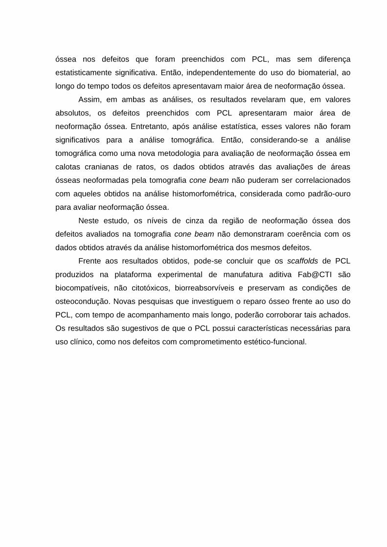
óssea nos defeitos que foram preenchidos com PCL, mas sem diferença
estatisticamente significativa. Então, independentemente do uso do biomaterial, ao
longo do tempo todos os defeitos apresentavam maior área de neoformação óssea.
Assim, em ambas as análises, os resultados revelaram que, em valores
absolutos, os defeitos preenchidos com PCL apresentaram maior área de
neoformação óssea. Entretanto, após análise estatística, esses valores não foram
significativos para a análise tomográfica. Então, considerando-se a análise
tomográfica como uma nova metodologia para avaliação de neoformação óssea em
calotas cranianas de ratos, os dados obtidos através das avaliações de áreas
ósseas neoformadas pela tomografia cone beam não puderam ser correlacionados
com aqueles obtidos na análise histomorfométrica, considerada como padrão-ouro
para avaliar neoformação óssea.
Neste estudo, os níveis de cinza da região de neoformação óssea dos
defeitos avaliados na tomografia cone beam não demonstraram coerência com os
dados obtidos através da análise histomorfométrica dos mesmos defeitos.
Frente aos resultados obtidos, pode-se concluir que os scaffolds de PCL
produzidos na plataforma experimental de manufatura aditiva Fab@CTI são
biocompatíveis, não citotóxicos, biorreabsorvíveis e preservam as condições de
osteocondução. Novas pesquisas que investiguem o reparo ósseo frente ao uso do
PCL, com tempo de acompanhamento mais longo, poderão corroborar tais achados.
Os resultados são sugestivos de que o PCL possui características necessárias para
uso clínico, como nos defeitos com comprometimento estético-funcional.

Referências
REFERÊNCIAS
ALLO, B. A. et al. Synthesis and electrospinning of epsilon-polycaprolactone-bioactive glass hybrid biomaterials via a sol-gel process. Langmuir, n. 26, p. 18340-18348, 2010. ARAFAT, M. T. et al. Biomimetic composite coating on rapid prototyped scaffolds for bone tissue engineering. Acta Biomaterialia, n. 7, p. 809-820, 2011. BARBANTI, Samuel Hilsdorf, Polímeros bioreabsorvíveis como suportes na engenharia de tecidos. 2005. 138f. Tese. Faculdade de Engenharia Mecânica, Universidade Estadual de Campinas, Campinas, 2005. BARBANTI, S. H. et al. Poly(e-caprolactone) and poly(D,L-lactic acid-co-glycolic acid) scaffolds used in bone tissue engineering prepared by melt compression–particulate leaching method. Journal of Materials Science: Materials in Medicine, n. 22, p. 2377–2385, 2011. BÁRTOLO P. J. Virtual and rapid manufacturing. Taylor & Francis Group: London, 2008. 849 p. BUTSCHER, A. et al. Structural and material approaches to bone tissue engineering in powder-based three-dimensional printing. Acta Biomaterialia, n. 7, p. 907-920, 2011. CARDOSO, A.K. et al. Histomorphometric analysis of tissue responses to bioactive glass implants in critical defects in rat calvaria. Cells Tissues Organs, n. 184, p. 128-137, 2006. CHEN, M. et al. Self-assembled composite matrix in a hierarchical 3-D scaffold for bone tissue engineering. Acta Biomaterialia, n. 7, p. 2244-2255, 2011. CHOONG, C. S. et al. Co-culture of bone marrow fibroblasts and endothelial cells on modified polycaprolactone substrates for enhanced potentials in bone tissue engineering. Tissue Engineering, n. 12, p. 2521-2531, 2006. COOK, S. D; RUEGER D.C. Osteogenic protein-1: biology and applications. Clinical Orthopaedics and Related Research, n. 324, p. 29-38, 1996. DOMINGOS, M. et al. Polycaprolactone scaffolds fabricated via bioextrusion for tissue engineering applications. International Journal of Biomaterials, n. 9, p. 1-9, 2009. EAP, S. et al. Electrospun nanofibrous 3D scaffold for bone tissue engineering. Biomedical Materials and Engineering, n. 22, p. 137-141, 2012. ESKI, M. et al. Assessment of distraction regenerate using quantitative bone scintigraphy. Annals of Plastic Surgery, n. 58, p. 328-334, 2007.
FIGUEIREDO, J. A. Et al. The histological effects of four endodontic sealers implanted in the oral mucosa: submucous injection versus implant in polyethylene tubes. International Endodontic Journal, n. 34, p. 377-385, 2001. FOLLMANN, Carina Staudt. Análise local e sistêmica das reações tissulares a diferentes materiais utilizados em pulpotomias: estudo em ratos. 2011. 60f. Dissertação. Faculdade de Odontologia, Pontifícia Universidade Católica do Rio Grande do Sul, Porto Alegre, 2011. FONTES, Ruben Aurélio Madeira. Fabrico e Caracterização de Scaffolds à Base de Fosfatos de Cálcio. 2010. 98f. Dissertação. Instituto Superior Técnico, Universidade Técnica de Lisboa, Lisboa, 2010. FU, S. et al. In vivo biocompatibility and osteogenesis of electrospun poly(epsilon-caprolactone)-poly(ethylene glycol)-poly(epsilon-caprolactone)/nano-hydroxyapatite composite scaffold. Biomaterials, n. 33, p. 8363-8371, 2012. GANESH, N. et al. Embedded silica nanoparticles in poly(caprolactone) nanofibrous scaffolds enhanced osteogenic potential for bone tissue engineering. Tissue Engineering, n. 18, p. 1867-1881, 2012. Part A. GIAVARESI, G. et al. In vitro and in vivo response to nanotopographically-modified surfaces of poly(3-hydroxybutyrate-co-3-hydroxyvalerate) and polycaprolactone. Journal of Materials Science, Polimer Edition, n. 17, p. 1405-1423, 2006. GRANDI, G. et al. Comparative histomorphometric analysis between ±-Tcp cement and ²-Tcp/Ha granules in the bone repair of rat calvaria. Materials Research, n. 14, p. 11-16, 2011. HUEBSCH, N.; MOONEY, D. J. Inspiration and application in the evolution of biomaterials. Nature, n. 462, p. 426-432, 2009. KITSUGI, T. et al. Transmission electron microscopy observations at the interface of bone and four types of calcium phosphate ceramics with different calcium/phosphorus molar ratios. Biomaterials, n. 16, p. 1101-1107, 1995. KNABE, C. et al. The effect of bioactive glass ceramics on the expression of bone-related genes and proteins in vitro. Clinical Oral Implants Research, n. 16, p. 119-127, 2005. KURASHINA, K. et al. In vivo study of calcium phosphate cements: implantation of an alpha-tricalcium phosphate/dicalcium phosphate dibasic/tetracalcium phosphate monoxide cement paste. Biomaterials, n. 18, p. 539-543, 1997. LANDIS, J. R.; KOCH, G. G. The measurement of observer agreement for categorical data. Biometrics, n. 33, p. 159-174, 1977. LANGER, R.; VACANTI, J. P. Tissue engineering. Science, n. 260, p. 920-926, 1993.

LIU, C. et al. Design and development of three-dimensional scaffolds for tissue engineering. Chemical Engineering Research and Design, n. 85, p. 1051-1064, 2007. LOHFELD, S. et al. Fabrication, mechanical and in vivo performance of polycaprolactone/tricalcium phosphate composite scaffolds. Acta Biomaterialia, n. 8, p. 3446-3456, 2012. MACEDO, N. L. et al. Bone defect regeneration with bioactive glass implantation in rats. Journal of Applied Oral Science, n. 12, p. 137-43, 2004. MARZOUK, K. M. et al. Osteoconductive effects of vinyl styrene microbeads in rat calvarial defects. Journal of Oral and Maxillofacial Surgery, n. 65, p. 1508-1516, 2007. MASSOTTI, Fabrício Poletto. Avaliação tomográfica e histomorfométrica da influência da terapia a laserde baixa intensidade sobre o reparo tecidual perimplantar em mandíbulas de coelhos. 2013. 130f. Dissertação. Faculdade de Odontologia, Universidade Federal do Rio Grande do Sul, Porto Alegre, 2013. MISCH, C. E. Implantes dentários contemporâneos. 2. ed. Tradução de Maria de Lourdes Giannini. São Paulo: Santos, 2006. 685 p. MIDDLETON, J. C.; TIPTON, A. J. Synthetic biodegradable polymers as orthopedic devices. Biomaterials, n. 21, p. 2335-2346, 2000. OUSTERHOUT, D. K.; STELNICKI, E. J. Plastic surgery's plastics. Clinics in Plastic Surgery, n. 23, p. 183-190, 1996. PETERSON, L. J. et al. Cirurgia oral e maxilofacial contemporânea. Tradução de Ramon Loureiro e Débora Fonseca. Rio de Janeiro: Elsevier; 2005. 794 p. PIETRZAK, W. S.; SARVER, D. R.; VERSTYNEN, M. L. Bioabsorbable polymer science for the practicing surgeon. Journal of Craniofacial Surgery, n. 8, p. 87-91, 1997. RAYMOND, B. J. Indirect tissue scaffold fabrication via additive manufacturing and biomimetic mineralization. Blacksburg: Virginia Polytechnic Institute, 2010. 176 p.
ROOSA, S. M. et al. The pore size of polycaprolactone scaffolds has limited influence on bone regeneration in an in vivo model. Journal of Biomedical Materials Research A, n. 92, p. 359-368, 2010. SANTOS, Luís Alberto. Desenvolvimento de Cimento de Fosfato de Cálcio Reforçado por Fibras para Uso na Área Médico-Odontológica. 2002. 247f. Tese. Faculdade de Engenharia Mecânica, Universidade Estadual de Campinas, Campinas, 2002.

SCARPARO, R. K. et al. Mineral trioxide aggregate-based sealer: analysis of tissue reactions to a new endodontic material. Journal of Endodontics, n. 36, p. 1174-1178, 2010. SENEDESE, Ana Lívia Chemeli. Estruturação tridimensional de scaffolds de policaprolactona via manufatura aditiva. 2011. 114f. Dissertação. Faculdade de Engenharia Química, Universidade Estadual de Campinas, Campinas, 2011. SHARAF, B. et al. Three-dimensionally printed polycaprolactone and beta-tricalcium phosphate scaffolds for bone tissue engineering: an in vitro study. Journal of Oral and Maxillofacial Surgery, n. 70, p. 647-656, 2012. SOUZA, P. P. et al. In vitro cytotoxicity and in vivo biocompatibility of contemporary resin-modified glass-ionomer cements. Dental Materials, n. 22, p. 838-844, 2006. URIST, MR. Bone: Formation by Autoinduction. Clin Orthop Relat Res, n. 395, p. 4-10, 2002. VALERIO, P. et al. The effect of ionic products from bioactive glass dissolution on osteoblast proliferation and collagen production. Biomaterials, n. 25, p. 2941-2948, 2004. VAN NATTA, F. J.; HILL, J. W.; CAROTHERS, W. H. Studies of polymerization and ring formation, e-caprolactone and its polymers. Journal of the American Chemical Society, n. 56, p. 455-459, 1934. WOODRUFF, M. A.; HUTMACHER, D. W. The return of a forgotten polymer—Polycaprolactone in the 21st century. Progress in Polymer Science, n. 35, p. 1217-1256, 2010. ZANETTA-BARBOSA, D.; DE CARVALHO, A. C. Effect of brief storage in ATP solution on periodontal healing after replantation of teeth in rats. Endodontics Dental Traumatology, n. 6, p. 193-199, 1990.

Anexos

ANEXO A - Normas para publicação - periódico International Journal of Oral & Maxillofacial Surgery
Guide for Authors Would authors please note that the reference style for the journal has now changed. Please pay special attention to the guidelines under the heading "References" below Authors wishing to submit their work to the journal are urged to read this detailed guide for authors and comply with all the requirements, particularly those relating to manuscript length and format. This will speed up the reviewing process and reduce the time taken to publish a paper following acceptance. Online Submission Submission and peer-review of all papers is now conducted entirely online, increasing efficiency for editors, authors, and reviewers, and enhancing publication speed. Authors requiring further information on online submission are strongly encouraged to view the system, including a tutorial, at External link http://ees.elsevier.com/ijoms A comprehensive Author Support service is available to answer additional enquiries at [email protected]. Once a paper has been submitted, all subsequent correspondence between the Editorial Office ([email protected]) and the corresponding author will be by e-mail. Editorial Policy A paper is accepted for publication on the understanding that it has not been submitted simultaneously to another journal, has been read and approved by all authors, and that the work has not been published before. The Editors reserve the right to make editorial and literary corrections. Any opinions expressed or policies advocated do not necessarily reflect the opinions and policies of the Editors. Declarations Upon submission you will be required to complete and upload the declarations page (pdf version or word version) to declare funding, conflict of interest and to indicate whether ethical approval was sought. This information must also be inserted into your manuscript under the acknowledgements section with the headings below. Upon submission you will be required to complete and upload this form (pdf version or word version) to declare funding, conflict of interest, and to indicate whether ethical approval and patient consent were sought. Lastly you must confirm that all authors have agreed to the submission. PLEASE NOTE that all funding must be declared at first submission, as the addition of funding at acceptance stage may invalidate the acceptance of your manuscript.

Authorship All authors should have made substantial contributions to all of the following: (1) the conception and design of the study, or acquisition of data, or analysis and interpretation of data (2) drafting the article or revising it critically for important intellectual content (3) final approval of the version to be submitted. Normally one or two, and no more than three, authors should appear on a short communication, technical note or interesting case/lesson learnt. Full length articles may contain as many authors as appropriate. Minor contributors and non-contributory clinicians who have allowed their patients to be used in the paper should be acknowledged at the end of the text and before the references. The corresponding author is responsible for ensuring that all authors are aware of their obligations. Before a paper is accepted all the authors of the paper must sign the Confirmation of Authorship form. This form confirms that all the named authors agree to publication if the paper is accepted and that each has had significant input into the paper. Please download the form and send it to the Editorial Office. (pdf version or word version) It is advisable that to prevent delay this form is submitted early in the editorial process. Acknowledgements All contributors who do not meet the criteria for authorship as defined above should be listed in an acknowledgements section. Examples of those who might be acknowledged include a person who provided purely technical help, writing assistance, or a department chair who provided only general support. Authors should disclose whether they had any writing assistance and identify the entity that paid for this assistance. Conflict of interest At the end of the main text, all authors must disclose any financial and personal relationships with other people or organisations that could inappropriately influence (bias) their work. Examples of potential conflicts of interest include employment, consultancies, stock ownership, honoraria, paid expert testimony, patent applications/registrations, and grants or other funding. If an author has no conflict of interest to declare, this should be stated. Role of the funding source All sources of funding should be declared as an acknowledgement at the end of the text. Authors should declare the role of study sponsors, if any, in the study design, in the collection, analysis and interpretation of data; in the writing of the manuscript; and in the decision to submit the manuscript for publication. If the study sponsors had no such involvement, the authors should so state. Open access This journal offers you the option of making your article freely available to all via the ScienceDirect platform. To prevent any conflict of interest, you can only make this choice after receiving notification that your article has been accepted for publication.

The fee of $3,000 excludes taxes and other potential author fees such as color charges. In some cases, institutions and funding bodies have entered into agreement with Elsevier to meet these fees on behalf of their authors. Details of these agreements are available at External link http://www.elsevier.com/fundingbodies. Authors of accepted articles, who wish to take advantage of this option, should complete and submit the order form (available at External link http://www.elsevier.com/locate/openaccessform.pdf). Whatever access option you choose, you retain many rights as an author, including the right to post a revised personal version of your article on your own website. More information can be found here: External link http://www.elsevier.com/authorsrights. Ethics Any manuscript concerned with human subjects, medical records, or human tissue that is submitted to the International Journal of Oral and Maxillofacial Surgery should comply with the principles stated in the Declaration of Helsinki Ethical Principles for Medical Research Involving Human Subjects, adopted by the 18th World Medical Assembly, Helsinki, Finland, June 1964, and as amended most recently by the 64th World Medical Assembly, Fontaleza, Brazil, October 2013. The manuscript should contain a statement that the work has been approved by the appropriate Ethical Committee related to the institution(s) in which the work was performed, and that subjects gave informed consent to the work. The International Journal of Oral and Maxillofacial Surgery requires institutional Ethics Committee approval for all human studies. For retrospective studies of records either a statement of approval or a statement of exemption from the Committee is appropriate. This statement should be provided upon submission of the manuscript. Studies involving experiments with animals must state that their care was in accordance with institution guidelines. Patient confidentiality Patients have a right to privacy. Therefore identifying information, including patients' images, names, initials, or hospital numbers, should not be included in videos, recordings, written descriptions, photographs, and pedigrees unless the information is essential for scientific purposes and you have obtained written informed consent for publication in print and electronic form from the patient (or parent, guardian or next of kin where applicable). If such consent is made subject to any conditions, The Editor and Publisher must be made aware of all such conditions. Written consents must be provided to the Editorial Office on request. Even where consent has been given, identifying details should be omitted if they are not essential. If identifying characteristics are altered to protect anonymity, such as in genetic pedigrees, authors should provide assurance that alterations do not distort scientific meaning and editors should so note. If consent for publication has not been obtained, personal details of patients included in any part of the paper and in any supplementary materials (including all illustrations and videos) must be removed before submission. Language Editing Services Papers will only be accepted when they are written in an acceptable standard of English. Authors, particularly those whose first language is not English, who require information about language editing and copyediting services pre- and post-

submission should visit External link http://www.elsevier.com/wps/find/authorshome.authors/languagepolishing or contact [email protected] for more information. Please note, Elsevier neither endorses nor takes responsibility for any products, goods or services offered by outside vendors through our services or in any advertising. For more information please refer to our Terms and Conditions External link http://www.elsevier.com/wps/find/termsconditions.cws_home/termsconditions. Article Types The following contributions will be accepted for publication. Please take careful note of the maximum length where applicable. Overlength articles will be returned to the authors without peer review: • editorials (commissioned by the editor) • clinical papers: no more than 5000 words and 30 references • research papers: no more than 6000 words and 40 references • review papers - no limit on length or number of references • technical notes (surgical techniques, new instruments, technical innovations) - no more than 2000 words, 10 references and 4 figures • case reports - no more than 2000 words, 10 references and 2 figures • book reviews • letters to the editor - please see detailed guidelines provided at the end of the main guide for authors • IAOMS announcements • general announcements. Please note: Case reports will be considered for publication only if they add new information to the existing body of knowledge or present new points of view on known diseases. All authors must have contributed to the paper, not necessarily the patient treatment. Technical notes and case reports are limited to a maximum of 4 authors, in exceptional circumstances, 5. Criteria for Publication Papers that will be considered for publication should be: • focused • based on a sound hypothesis and an adequate investigation method analysing a statistically relevant series, leading to relevant results that back the conclusion • well written in simple, scientific English grammar and style • presented with a clear message and containing new information that is relevant for the readership of the journal • Note the comment above relating to case reports. Following peer-review, authors are required to resubmit their revised paper within 3 months; in exceptional circumstances, this timeline may be extended at the editor's discretion. Presentation of Manuscripts General points Papers should be submitted in journal style. Failure to do so will result in the paper being immediately returned to the author and may lead to significant delays in
publication. Spelling may follow British or American usage, but not a mixture of the two. Papers should be double-spaced with a margin of at least 3 cm all round. Each line must be numbered. Format Observational or Case Cohort Studies, as well as Case Series must be presented in conformance with STROBE guidelines: External link www.strobe-statement.org Randomized Controlled Trials must be presented in conformance with CONSORT guidelines: External link www.consort-statement.org Systematic Reviews and Meta-Analyses must be presented according to PRISMA guidelines: External link www.prisma-statement.org Papers should be set out as follows, with each section beginning on a separate page: • title page • abstract • text • acknowledgements • references • tables • captions to illustrations. Please note that the qualifications of the authors will not be included in the published paper and should not be listed anywhere on the manuscript. Title page The title page should give the following information: • title of the article • full name of each author • name and address of the department or institution to which the work should be attributed • name, address, telephone and fax numbers, and e-mail address of the author responsible for correspondence and to whom requests for offprints should be sent • sources of support in the form of grants • key words. If the title is longer than 40 characters (including spaces), a short title should be supplied for use in the running heads. Abstract 200 words maximum. Do not use subheadings or abbreviations; write as a continuous paragraph. Must contain all relevant information, including results and conclusion. Text Please ensure that the text of your paper conforms to the following structure: Introduction, Materials and Methods, Results, Discussion. There is no separate Conclusion section. There should be no mention of the institution where the work was carried out, especially in the Materials and Methods section. Introduction • Present first the nature and scope of the problem investigated

• Review briefly the pertinent literature • State the rationale for the study • Explain the purpose in writing the paper • State the method of investigation and the reasons for the choice of a particular method •; Should be written in the present tense Materials and Methods • Give the full details, limit references • Should be written in the past tense • Include exact technical specifications, quantities and generic names • Limit the number of subheadings, and use the same in the results section • Mention statistical method • Do not include results in this section Results • Do not describe methods • Present results in the past tense • Present representations rather than endlessly repetitive data • Use tables where appropriate, and do not repeat information in the text Discussion • Discuss - do not recapitulate results • Point out exceptions and lack of correlations. Do not try to cover up or 'fudge' data • Show how results agree/contrast with previous work • Discuss the implications of your findings • State your conclusions very clearly Headings: Headings enhance readability but should be appropriate to the nature of the paper. They should be kept to a minimum and may be removed by the Editors. Normally only two categories of headings should be used: major ones should be typed in capital letters; minor ones should be typed in lower case (with an initial capital letter) at the left hand margin. Quantitative analysis: If any statistical methods are used, the text should state the test or other analytical method applied, basic descriptive statistics, critical value obtained, degrees of freedom, and significance level, e.g. (ANOVA, F=2.34; df=3,46; P<0.001). If a computer data analysis was involved, the software package should be mentioned. Descriptive statistics may be presented in the form of a table, or included in the text. Abbreviations, symbols, and nomenclature: Only standardized terms, which have been generally accepted, should be used. Unfamiliar abbreviations must be defined when first used. For further details concerning abbreviations, see Baron DN, ed. Units, symbols, and abbreviations. A guide for biological and medical editors and authors, London, Royal Society of Medicine, 1988 (available from The Royal Society of Medicine Services, 1 Wimpole Street, London W1M 8AE, UK). The minus sign should be -. If a special designation for teeth is used, a note should explain the symbols. Scientific names of organisms should be binomials, the generic name only with a capital, and should be italicised in the typescript. Microorganisms should be named according to the latest edition of the Manual of Clinical Microbiology, American Society of Microbiology.
Drugs: use only generic (non-proprietary) names in the text. Suppliers of drugs used may be named in the Acknowledgments section. Do not use 'he', 'his' etc where the sex of the person is unknown; say 'the patient' etc. Avoid inelegant alternatives such as 'he/she'. Patients should not be automatically designated as 'she', and doctors as 'he'. References The journal's reference style has changed. References should be numbered consecutively throughout the article, beginning with 1 for the first-cited reference. References should be listed at the end of the paper in the order in which they appear in the text (not listed alphabetically by author and numbered as previously). The accuracy of references is the responsibility of the author. References in the text should be numbered with superscript numerals inside punctuation: for example "Kenneth and Cohen14 showed..."; "each technique has advantages and disadvantages5-13." Citations in the text to papers with more than two authors should give the name of the first author followed by "et al."; for example: "Wang et al37 identified..." All references cited in the text must be included in the list of references at the end of the paper. Each reference listed must include the names of all authors. Please see section "Article Types" for guidance on the maximum number of reference for each type of article. Titles of journals should be abbreviated according to Index Medicus (see www.nlm.nih.gov.uk) . When citing papers from monographs and books, give the author, title of chapter, editor of book, title of book, publisher, place and year of publication, first and last page numbers. Internet pages and online resources may be included within the text and should state as a minimum the author(s), title and full URL. The date of access should be supplied and all URLs should be checked again at proof stage. Examples: Journal article: Halsband ER, Hirshberg YA, Berg LI. Ketamine hydrochloride in outpatient oral surgery. J Oral Surg 1971: 29: 472-476. When citing a paper which has a Digital Object Identifier (DOI), use the following style: Toschka H, Feifel H. Aesthetic and functional results of harvesting radial forearm flap. Int J Oral Maxillofac Surg 2001: 30: 45-51. doi: 10.1054/ijom.2000.0005 Book/monograph: Costich ER, White RP. Fundamentals of oral surgery. Philadelphia: WB Saunders, 1971: 201-220. Book chapter: Hodge HC, Smith FA. Biological properties of inorganic fluorides. In: Simons JH, ed.: Fluorine chemistry. New York: Academic Press, 1965: 135. Internet resource: International Committee of Medical Journal Editors. Uniform requirements for manuscripts submitted to biomedical journals. External link http://www.icmje.org [Accessibility verified March 21, 2008] Tables Tables should be used only to clarify important points. Double documentation in the form of tables and figures is not acceptable. Tables should be numbered consecutively with Arabic numerals. They should be double spaced on separate

pages and contain only horizontal rules. Do not submit tables as photographs. A short descriptive title should appear above each table, with any footnotes suitably identified below. Care must be taken to ensure that all units are included. Ensure that each table is cited in the text. Figures All illustrations (e.g. graphs, drawings or photographs) are considered to be figures, and should be numbered in sequence with Arabic numerals. Each figure should have a caption, typed double-spaced on a separate page and numbered correspondingly. The minimum resolution for electronically generated figures is 300 dpi. Line illustrations: All line illustrations should present a crisp black image on an even white background (127 x 178 mm (5 x 7 in), or no larger than 203 x 254 mm (8 x 10 in). The size of the lettering should be appropriate, taking into account the necessary size reduction. Photographs and radiographs: Photomicrographs should show magnification and details of any staining techniques used. The area(s) of interest must be clearly indicated with arrows or other symbols. Colour images are encouraged, but the decision whether an illustration is accepted for reproduction in colour in the printed journal lies with the editor-in-chief. Figures supplied in colour will appear in colour in the online version of the journal. Size of photographs: The final size of photographs will be: (a) single column width (53 mm), (b) double column width (110 mm), (c) full page width (170 mm). Photographs should ideally be submitted at the final reproduction size based on the above figures. Funding body agreements and policies Elsevier has established agreements and developed policies to allow authors who publish in Elsevier journals to comply with potential manuscript archiving requirements as specified as conditions of their grant awards. To learn more about existing agreements and policies please visit External link http://www.elsevier.com/fundingbodies Proofs One set of page proofs in PDF format will be sent by e-mail to the corresponding author, which they are requested to correct and return within 48 hours. Elsevier now sends PDF proofs which can be annotated; for this you will need to download Adobe Reader version 7 available free from External link http://www.adobe.com/products/acrobat/readstep2.html. Instructions on how to annotate PDF files will accompany the proofs. The exact system requirements are given at the Adobe site: External link http://www.adobe.com/products/acrobat/acrrsystemreqs.html#70win. If you do not wish to use the PDF annotations function, you may list the corrections (including replies to the Query Form) and return to Elsevier in an e-mail. Please list your corrections quoting line number. If, for any reason, this is not possible, then mark the corrections and any other comments (including replies to the Query Form) on a printout of your proof and return by fax, or scan the pages and e-mail, or by post.

Please use this proof only for checking the typesetting, editing, completeness and correctness of the text, tables and figures. Significant changes to the article as accepted for publication will only be considered at this stage with permission from the Editor. We will do everything possible to get your article published quickly and accurately. Therefore, it is important to ensure that all of your corrections are sent back to us in one communication: please check carefully before replying, as inclusion of any subsequent corrections cannot be guaranteed. Proofreading is solely your responsibility. Note that Elsevier may proceed with the publication of your article if no response is received. Offprints The corresponding authorwill be provided , at no cost, with a PDF file of the article via e-mail. The PDF file is a watermarked version of the published article and includes a cover sheet with the journal cover image and a disclaimer outlining the terms and conditions of use. Additional paper offprints can be ordered by the authors. An order form with prices will be sent to the corresponding author. Accepted Articles For the facility to track accepted articles and set email alerts to inform you of when an article's status has changed, visit: External link http://authors.elsevier.com/TrackPaper.html There are also detailed artwork guidelines, copyright information, frequently asked questions and more. Contact details for questions arising after acceptance of an article, especially those related to proofs, are provided after registration of an article for publication. Instructions for Letters to the Editor The IJOMS welcomes Letters to the Editor. To facilitate submission of the highest quality of Letters to the Editor, the following guidelines should be followed: 1. Letters are meant to be focus pieces and, therefore, are limited to no more than 600 words, 6 references and a maximum of 2 figures. One reference should include a reference to the IJOMS article being addressed. 2. It is recommended that you limit your letter to one or two important and critical points to which you wish to provide a clear and precise discussion regarding the previously published article. 3. One should support all assertion by peer review literature which should be a primary research or large clinical studies rather than a case report. 4. Please include any financial disclosures at the end of the letter. This would include the potential conflicts of interest not just related to the specific content of your letter but also the content of the IJOMS article and other related areas. 5. Please recognize that letters that are essentially in agreement with the author's findings and offer no additional insights provide little new information for publication. Likewise, letters that highlight the writer's own research or are otherwise self promotional will receive a low publication priority. 6. There may be a need for additional editing. Should editing be required the letter will be sent back to the author for final approval of the edited version. 7. It is important to use civil and professional discourse. It is not advisable that one adopt a tone that may be misconstrued to be in anyway insulting. 8. Finally, it is not advisable to provide a letter that is anecdotal. While personal experiences can have great value in patient care, it is generally not strong evidence to be placed in a letter to the editor.

ANEXO B- Normas para publicação - periódico Biomaterials
Guide for Authors The peer review submission system for Biomaterials is located here: http://ees.elsevier.com/biomat/ The Elsevier Editorial System (EES) is a web-based submission and review system. Authors may submit manuscripts and track their progress through the system to publication. Reviewers can download manuscripts and submit their opinions to the editor. Editors can manage the whole submission/review/revise/publish process. Please register at: http://ees.elsevier.com/biomat/ Referees, whose names are not normally disclosed to the authors, will study all contributions which the Editor-in-Chief deems to be of sufficient significance and interest to be sent for peer review. The criteria by which this initial assessment is made include relevance to the scope of the journal, the originality of the work and its significance to the broad development of the field of biomaterials. Before You Begin All authors, especially those submitting to the journal for the first time, are encouraged to read the following document authored by the Editor in Chief, which gives an overview of the journal as well as the writing and selection process of academic publishing as it relates to Biomaterials: Writing Papers for Biomaterials. Ethics in Publishing: For information on Ethics in Publishing and Ethical guidelines for journal publication see http://www.elsevier.com/authorethics and http://www.elsevier.com/ethicalguidelines. Changes to authorship This policy concerns the addition, deletion, or rearrangement of author names in the authorship of accepted manuscripts: Before the accepted manuscript is published in an online issue: Requests to add or remove an author, or to rearrange the author names, must be sent to the Journal Manager from the corresponding author of the accepted manuscript and must include: (a) the reason the name should be added or removed, or the author names rearranged and (b) written confirmation (e-mail, fax, letter) from all authors that they agree with the addition, removal or rearrangement. In the case of addition or removal of authors, this includes confirmation from the author being added or removed.

Requests that are not sent by the corresponding author will be forwarded by the Journal Manager to the corresponding author, who must follow the procedure as described above. Note that: (1) Journal Managers will inform the Journal Editors of any such requests and (2) publication of the accepted manuscript in an online issue is suspended until authorship has been agreed. After the accepted manuscript is published in an online issue: Any requests to add, delete, or rearrange author names in an article published in an online issue will follow the same policies as noted above and result in a corrigendum. Mandatory Author Declaration: In addition to uploading manuscripts and figures, it is required that the corresponding author of each manuscript uploads a separate Author Declaration. All authors must sign this declaration; the corresponding author may not sign on behalf of other authors. This Declaration covers a number of logistic and ethical issues. Please use the template given above for this Declaration. Authors may save this template, obtain the required signatures and then upload it as an integral part of their submission. The editorial process will not begin until and unless all authors have signed the declaration. N.B.: The Author Declaration is independent of, and in addition to, the "Journal Publishing Agreement" agreement which is issued on acceptance. Amendments to Manuscripts In order to maintain the integrity of the scientific record, the version that is published in an issue on ScienceDirect and in print MUST be identical. One set of page proofs will be sent to the corresponding author. Please note that authors are urged to check their proofs carefully before return, but corrections are restricted to typesetting errors only. Proofs are NOT to be considered as drafts. No changes in, or additions to, the accepted (and subsequently edited) manuscript will be allowed at this stage. Proofreading is solely the responsibility of the corresponding author. Copyright All authors must sign the "Journal Publishing Agreement" before the article can be published. An e-mail (or letter) will be sent to the corresponding author confirming receipt of the manuscript together with an agreement form or a link to the online version of this agreement. This transfer agreement enables Elsevier Ltd to protect the copyrighted material for the authors, but does not relinquish the author's proprietary rights. As an author you (or your employer or institution) retain certain rights; for details you are referred to: http://www.elsevier.com/authorsrights. The copyright transfer covers the exclusive rights to reproduce and distribute the article. Subscribers may reproduce tables of contents or prepare lists of articles including abstracts for internal circulation within their institutions. Permission of the Publisher is required for resale or distribution outside the institution and for all other derivative works, including compilations and translations (please consult http://www.elsevier.com/permissions). If excerpts from other copyrighted works are included, the author(s) must obtain written permission from the copyright owners and credit the source(s) in the article. Elsevier has preprinted forms for use by authors in these cases: please consult http://www.elsevier.com/permissions.

Role of the Funding Source You are requested to identify who provided financial support for the conduct of the research and/or preparation of the article and to briefly describe the role of the sponsor(s), if any, in study design; in the collection, analysis and interpretation of data; in the writing of the report; and in the decision to submit the paper for publication. If the funding source(s) had no such involvement then this should be stated. Please see http://www.elsevier.com/funding. Funding Bodies Agreements and Policies Elsevier has established agreements and developed policies to allow authors whose articles appear in journals published by Elsevier, to comply with potential manuscript archiving requirements as specified as conditions of their grant awards. To learn more about existing agreements and policies please visit http://www.elsevier.com/fundingbodies. Open Access This journal offers authors two choices to publish their research; 1. Open Access • Articles are freely available to both subscribers and the wider public with permitted reuse • An Open Access publication fee is payable by authors or their research funder 2. Subscription • Articles are made available to subscribers as well as developing countries and patient groups through our access programs (http://www.elsevier.com/access) • No Open Access publication fee All articles published Open Access will be immediately and permanently free for everyone to read and download. Permitted reuse is defined by your choice of one of the following Creative Commons user licenses: Creative Commons Attribution (CC-BY): lets others distribute and copy the article, to create extracts, abstracts, and other revised versions, adaptations or derivative works of or from an article (such as a translation), to include in a collective work (such as an anthology), to text or data mine the article, even for commercial purposes, as long as they credit the author(s), do not represent the author as endorsing their adaptation of the article, and do not modify the article in such a way as to damage the author's honor or reputation. Creative Commons Attribution-Non Commercial-ShareAlike (CC BY-NC-SA): for non-commercial purposes, lets others distribute and copy the article, to create extracts, abstracts and other revised versions, adaptations or derivative works of or from an article (such as a translation), to include in a collective work (such as an anthology), to text and data mine the article, as long as they credit the author(s), do not represent the author as endorsing their adaptation of the article, do not modify the article in such a way as to damage the author's honor or reputation, and license their new adaptations or creations under identical terms (CC BY NC SA).

Creative Commons Attribution-NonCommercial-NoDerivs (CC-BY-NC-ND): for non-commercial purposes, lets others distribute and copy the article, and to include in a collective work (such as an anthology), as long as they credit the author(s) and provided they do not alter or modify the article. To provide Open Access, this journal has a publication fee which needs to be met by the authors or their research funders for each article published Open Access. The Open Access publication fee for this journal is $USD 3300, excluding taxes. Learn more about Elsevier's pricing policy: http://www.elsevier.com/openaccesspricing Manuscript Preparation General: Manuscripts must be word processed (preferably in Word format), double-spaced with wide margins and a font size of 12 or 10 pt. Files prepared in LaTEX are not supported. Figure captions should be in Arial font 9pt. Please check the current style of the journal, particularly the reference style (Vancouver), and avoid excessive layout styling as most formatting codes will be removed or replaced during the processing of your article. In addition, do not use options such as automatic word breaking, justified layout, double columns or automatic paragraph numbering (especially for numbered references). However do use bold face, italic, subscripts, superscripts etc. The corresponding author should be identified (include a Fax number and E-mail address). Full postal addresses must be given for all co-authors. The Editors reserve the right to adjust style to certain standards of uniformity. The preferred style is: Surname, Initials, Department, Institution, City/State, Postal Code, Country. Authors should retain copies of all versions of their manuscript submitted to the journal. Authors are especially requested to be vigilant over the submission of the correct version of the manuscript at the various stages of the editorial process. English Language: Manuscripts should be proof-read and have English language errors corrected before submission as we may have to return papers due to poor language usage. Authors who require information about language editing and copyediting services pre- and post-submission please visit http://www.elsevier.com/languagepolishing for more information. Paper Length: Authors are urged to write as concisely as possible. Abstracts: All manuscripts are to be supplied with an Abstract of about 100-200 words in length. Keywords: Authors must provide 4-6 keywords for indexing purposes. A keyword list can be found at the end of these instructions. click here). Text: Follow this order when typing manuscripts:Title, Authors, Affiliations, Abstract, Keywords, Introduction, Materials and Methods, Results, Discussion, Conclusions, Acknowledgements, References, Appendix (where necessary), Figures, Figure Captions and Tables, Supplementary Data. The corresponding author should be identified with an asterisk and footnote. All other footnotes (except for table
footnotes) should be identified with superscript Arabic numbers. An abbreviated title for use as a running headline should also be supplied. Authors are requested to acknowledge funding sources for the work. Units: The SI system should be used for all scientific and laboratory data; if, in certain instances, it is necessary to quote other units, these should be added in parentheses. Temperatures should be given in degrees Celsius. The unit 'billion' (109 in America, 1012 in Europe) is ambiguous and must not be used. If a large number of symbols are used it is helpful if authors submit a list of these symbols and their meanings. References: All publications cited in the text should be presented in a list of references following the text of the manuscript. In the text refer to references by a number in square brackets on the line (e.g. Since Peterson[1]), and the full reference should be formatted according to the permutation of the Vancouver reference system with numbers in the text. It is suggested that authors refer to the following link for a comprehensive overview of the Vancouver reference styles: Vancouver Reference Styles Examples of formatting follow: 1. Driessens FCM, Boltong MG, Bermudez O, Planell JA. Formulation and setting times of some calcium orthophosphate cements: a pilot study. J Mater Sci: Mater Med 1993;4:503-508. 2. Nancollas H. In vitro studies of calcium phosphate crystallisation. In: Mann S, Webb J, Williams RJP, editors. Biomineralization. Chemical and biochemical perspectives. New York: VCH, 1989. p. 157-182. 3. Brown W, Chow LC. Combinations of sparingly soluble calcium phosphates in slurries and paste as mineralizers and cements. US Patent No. 4612053, 1986.
N.B.: “Et al” must be used after the first 6 authors have been named. Biomaterials
does not use the publication month or day. Online Sources: References to online sources, including articles in press, should contain at a minimum the full URL and year the source was accessed. Furthermore, if known, the following information should be given: author names, dates, reference to a source publication. Examples of formatting follow: 1. UK House of Commons Science and Technology Committee. Scientific Publications: Free for All? Tenth Report of Session 2003-4 Volume 1. London: The Stationary Office Ltd. Online. 2004 July. Available from URL: http://www.publications.parliament.uk/pa/cm200304/cmselect/cmsctech/399/39902.html 2. Wellcome Trust. Economic Analysis of Scientific Research Publishing. Histon, UK: Wellcome Trust. Online. 2003. Available from URL: http://www.wellcome.ac.uk/doc_wtd003181.html 3. Keeney M, Lai JH, Yang F. Recent progress in cartilage tissue engineering. Curr Opin Biotechnol. 2011. Available from URL: http://www.ncbi.nlm.nih.gov/pubmed/21531126 (DOI: 10.1016/j.copbio.2011.04.003).

Artwork Electronic artwork General points • Make sure you use uniform lettering and sizing of your original artwork. • Save text in illustrations as "graphics" or enclose the font. • Only use the following fonts in your illustrations: Arial, Courier, Times, Symbol. • Number the illustrations according to their sequence in the text. • Use a logical naming convention for your artwork files. • Provide captions to illustrations separately. • Produce images near to the desired size of the printed version. • Submit each figure as a separate file. A detailed guide on electronic artwork is available on our website: http://www.elsevier.com/artworkinstructions You are urged to visit this site; some excerpts from the detailed information are given here. Formats Regardless of the application used, when your electronic artwork is finalised, please "save as" or convert the images to one of the following formats (note the resolution requirements for line drawings, halftones, and line/halftone combinations given below): EPS: Vector drawings. Embed the font or save the text as "graphics". TIFF: color or grayscale photographs (halftones): always use a minimum of 300 dpi. TIFF: Bitmapped line drawings: use a minimum of 1000 dpi. TIFF: Combinations bitmapped line/half-tone (color or grayscale): a minimum of 500 dpi is required. DOC, XLS or PPT: If your electronic artwork is created in any of these Microsoft Office applications please supply "as is". Please do not: • Supply embedded graphics in your wordprocessor (spreadsheet, presentation) document; • Supply files that are optimised for screen use (like GIF, BMP, PICT, WPG); the resolution is too low; • Supply files that are too low in resolution; • Submit graphics that are disproportionately large for the content. Color artwork Please make sure that artwork files are in an acceptable format (TIFF, EPS or MS Office files) and with the correct resolution. If, together with your accepted article, you submit usable color figures then Elsevier will ensure, at no additional charge, that these figures will appear in color on the Web (e.g., ScienceDirect and other sites) regardless of whether or not these illustrations are reproduced in color in the printed version. For color reproduction in print, you will receive information regarding the costs from Elsevier after receipt of your accepted article. Please indicate your preference for color in print or on the Web only. For further information on the preparation of electronic artwork, please see http://www.elsevier.com/artworkinstructions. Please note: Because of technical complications which can arise by converting color figures to "gray scale" (for the

printed version should you not opt for color in print) please submit in addition usable black and white versions of all the color illustrations. Video Data Elsevier accepts video material and animation sequences to support and enhance your scientific research. Authors who have video or animation files that they wish to submit with their article are strongly encouraged to include these within the body of the article. This can be done in the same way as a figure or table by referring to the video or animation content and noting in the body text where it should be placed. All submitted files should be properly labeled so that they directly relate to the video file's content. In order to ensure that your video or animation material is directly usable, please provide the files in one of our recommended file formats with a maximum size of 30 MB and running time of 5 minutes. Video and animation files supplied will be published online in the electronic version of your article in Elsevier Web products, including ScienceDirect: http://www.sciencedirect.com. Please supply 'stills' with your files: you can choose any frame from the video or animation or make a separate image. These will be used instead of standard icons and will personalize the link to your video data. For more detailed instructions please visit our video instruction pages at http://www.elsevier.com/artworkinstructions. Note: since video and animation cannot be embedded in the print version of the journal, please provide text for both the electronic and the print version for the portions of the article that refer to this content. Files can be stored on diskette, ZIP-disk or CD (either MS-DOS or Macintosh).Audio Slides: The journal encourages authors to create an AudioSlides presentation with their published article. AudioSlides are brief, webinar-style presentations that are shown next to the online article on ScienceDirect. This gives authors the opportunity to summarize their research in their own words and to help readers understand what the paper is about. More information and examples are available at http://www.elsevier.com/audioslides. Authors of this journal will automatically receive an invitation e-mail to create an AudioSlides presentation after acceptance of their paper. After Acceptance Proofs: One set of page proofs in PDF format will be sent by e-mail to the corresponding author and should be returned within 48 hours of receipt. The average amount of time between acceptance and receipt of typeset proof is 6 working days. Papers are published in print within another 8 weeks upon receipt of author corrections. Corrections should be restricted to typesetting errors. Any queries should be answered in full. Please note that authors are urged to check their proofs carefully before return. Elsevier now sends PDF proofs which can be annotated; for this you will need to download Adobe Reader© version 7 (or higher) available free from http://www.adobe.com/products/acrobat/readstep2.html. Instructions on how to annotate PDF files will accompany the proofs. The exact system requirements are given at the Adobe site: http://www.adobe.com/products/acrobat/acrrsystemreqs.html#70win. If you do not wish to use the PDF annotations function, you may list the corrections (including replies to the Query Form) and return to Elsevier in an e-mail. Please list your corrections quoting line number. If, for any reason, this is not possible, then mark the

corrections and any other comments (including replies to the Query Form) on a printout of your proof and return by fax, or scan the pages and e-mail, or by post. Please use this proof only for checking the typesetting, editing, completeness and correctness of the text, tables and figures. Significant changes to the article as accepted for publication will only be considered at this stage with permission from the Editor. We will do everything possible to get your article published quickly and accurately. Therefore, it is important to ensure that all of your corrections are sent back to us in one communication: please check carefully before replying, as inclusion of any subsequent corrections cannot be guaranteed. Proofreading is solely your responsibility. Note that Elsevier may proceed with the publication of your article if no response is received. Track a Paper: Authors can track their paper status online after the paper has been accepted and forwarded to the Publisher. Enter your Elsevier reference number (JBMT xxx) and the Corresponding author's family name at the following web page: http://www.elsevier.com/trackarticle. Corresponding authors will receive an acknowledgement email from Elsevier with the reference number and the family name on it. Authors can also go to the 'track a paper' page by clicking onto the 'track a paper' button on the left hand side of the journal home page. Offprints: The corresponding author will be provided with a PDF of the article via email. The PDF is a watermarked version of the published article and includes a cover sheet with the journal cover image and a disclaimer outlining the terms of use. Additional paper offprints can be ordered by the authors. An order form with prices will be sent to the corresponding author. Author Enquiries: For enquiries relating to the submission of articles (including
electronic submission where available) please visit this journal’s homepage. You can
track accepted articles at http://www.elsevier.com/trackarticle and set up e-mail alerts
to inform you of when an article’s status has changed. Also accessible from here is
information on copyright, frequently asked questions and more. Contact details for questions arising after acceptance of an article, especially those relating to proofs, will be provided by the publisher. Keywords">Keyword List

ANEXO C - Aprovação da Comissão Científica e de Ética da Faculdade de
Odontologia da PUCRS

ANEXO D – Aprovação do Comitê de Ética para o uso de animais