Universidade de Lisboa Faculdade de Farmáciarepositorio.ul.pt/bitstream/10451/24995/1/Tese...
79
Universidade de Lisboa Faculdade de Farmácia Research Institute for Medicines and Pharmaceutical Sciences (iMed.UL) Neuron Glia Biology in Health and Disease Group MODULATION OF MICROGLIA REACTIVITY BY S100B IN MULTIPLE SCLEROSIS Carla Isabel Silveira Ferreira Dissertação de Mestrado MESTRADO EM CIÊNCIAS BIOFARMACÊUTICAS 2015
Transcript of Universidade de Lisboa Faculdade de Farmáciarepositorio.ul.pt/bitstream/10451/24995/1/Tese...
Universidade de Lisboa
Faculdade de Farmácia
Research Institute for Medicines and Pharmaceutical Sciences
(iMed.UL)
Neuron Glia Biology in Health and Disease Group
MODULATION OF MICROGLIA REACTIVITY BY S100B IN
MULTIPLE SCLEROSIS
Carla Isabel Silveira Ferreira
Dissertação de Mestrado
MESTRADO EM CIÊNCIAS BIOFARMACÊUTICAS
2015


Universidade de Lisboa
Faculdade de Farmácia
Research Institute for Medicines and Pharmaceutical Sciences
(iMed.UL)
Neuron Glia Biology in Health and Disease Group
MODULATION OF MICROGLIA REACTIVITY BY S100B IN
MULTIPLE SCLEROSIS
Carla Isabel Silveira Ferreira
Dissertação de Mestrado orientada pela Prof.ª Doutora Adelaide Maria Afonso
Fernandes Borralho e pela Doutora Andreia Pereira Barateiro
MESTRADO EM CIÊNCIAS BIOFARMACÊUTICAS
2015


À Margarida.


Agradecimentos
Quero começar por agradecer à Professora Doutora Dora Brites, investigadora principal
do grupo Neuron Glia Biology in Health and Disease, pela forma como me acolheu e por me ter
dado a oportunidade de desenvolver a minha tese de mestrado neste grupo. Além disso quero
agradecer pelo sentido crítico que me incutiu e que me irá certamente ajudar no futuro.
À Professora Doutora Adelaide Fernandes, um enorme obrigada é pouco! Não há
palavras que mostrem a gratidão que sinto por ti. Porque trabalhar nisto era especial para mim
e deste-me a oportunidade de o fazer. Obrigada por toda a ajuda, dedicação e por todo o
conhecimento que me transmitiste. Foste incansável. Um dia gostava de ser como tu!
À Doutora Andreia Barateiro, muito obrigada por tudo. Por toda a tua paciência. Por
toda a ajuda que me deste ao longo deste ano e pela tua disponibilidade (até para falar de
assuntos de noivas ;) ). Muito obrigada! :)
Carolina, Cátia, Cláudia e Gisela, obrigada por estarem sempre prontas a ajudar ou
simplesmente para rir um bocadinho. Muito obrigada por toda a dedicação, ajuda e diversão
que me proporcionaram ao longo deste ano!
Dora, companheira de almoço (desde as idas à cantina até à esplanada do F)! Obrigada
por este ano. Aturaste os meus devaneios, as minhas queixas e loucuras, as minhas “coisas de
noiva”… obrigada por me fazeres rir de cada vez que alguém te assustava ao ir falar contigo ou
dizias “laranjas” :p, por o teu tablet ganhar personalidade e dizer-me coisas hilariantes xD e por
me deixares eufórica com a notícia mais fixe deste ano :D simplesmente obrigada! :)
Maria, és um exemplo para mim! Exemplo de esforço e dedicação! Obrigada pelos
momentos de diversão (tendo em conta que estava à tua frente era relativamente fácil começar
a rir contigo :p) e pelos ensinamentos sobre Western Blot. Mas olha, mesmo assim o record
ainda é meu! :D Torço por ti e sei que tens um futuro brilhante à tua frente!
Mafalda, partilhámos a orientadora, o fluxo durante algum tempo e até os anticorpos
para WB!!! O que há a dizer?! Obrigada pela ajuda que me foste dando ao longo deste tempo
ou por me aturares quando eu cá ficava até mais tarde. Sei que vais chegar longe :)
Ritinha :D lembro-me de logo nos primeiros módulos me sentar perto de ti. Depois
rapidamente éramos as poderosas! Obrigada por seres assim, sempre disponível mesmo
estando mais longe. Sabes que estarei sempre aqui, onde quer que estejas :)
Para ti Rui, vai um agradecimento especial! Pelas boleias todas que me deste, pelas
massagens enquanto eu contava beads, por teres sempre uma palavra amiga. Não deixes de ser
assim como és e tenho a certeza que vai haver muita sogra a tentar “roubar-te” :p xD

Ao Chico, por todas as conversas e cafés. Obrigada por toda a ajuda e claro, pelas
correções! “A verdadeira amizade não é ser inseparáveis, mas sim estar separados sem que nada
mude.” Nós somos assim! :)
À Filipa e à Diana, obrigada por aturarem a madrinha. Tenho muito orgulho em vocês!
À Andreia Freitas, porque mesmo longe, estiveste sempre pronta para me ajudar e
apoiar. És a melhor madrinha de curso que alguém pode ter. Desde o grupo de Genética que
não nos separámos. Viste-me sorrir, viste-me chorar, viste-me vacilar… o nosso último ano uniu-
nos e sei que nunca mais nos iremos separar! És especial, muito especial! Obrigada por tudo!
À Teresa e ao Nuno, ao Ricardo e à Joana, por estarem lá sempre que foi necessário :)
À minha Shikha. Porque como já te disse, foi amor à primeira vista. Porque acabámos a
licenciatura juntas, concorremos a mestrado juntas e vamos continuar juntas, disso tenho a
certeza :) obrigada por me ouvires, por nadares comigo, por seres uma irmã, por seres como és,
especial e única!
À São Piçarra, pelas correções, pelas conversas sobre Esclerose Múltipla e microglias,
pelos salames e pelos tão deliciosos “S”, muito obrigada!
À Margarida, por fazer parte da minha vida! Porque para além de ser mana, melhor
amiga e madrinha, me faz lutar a cada dia para deixar uma pequena marca neste mundo da
Esclerose Múltipla. Esta tese é para ti, é por ti!
António, Guida, Luísa, Luís, avó Margarida e tio Teófilo, obrigada pelo apoio e
disponibilidade incansáveis nestes dois anos!
Aos meus irmãos e cunhadas, obrigada por serem exemplo! Aos meus amores
pequeninos, Miguel, Rui, Pedro, Ana, Sara e Rafael, obrigada por me fazerem sorrir até nas
alturas de maior cansaço.
Aos meus avós, que lá do céu olham por mim.
Não pode faltar um agradecimento muito especial aos meus pais por me permitirem
chegar até aqui. Por todo o vosso apoio e amor incondicional, obrigada!
Também ao Dogus, à Miaucas, ao Branco, ao Soneca, à Cinha, à Lili, à Kuka e ao Miki,
pelos miminhos que foram dando :)
Por último quero agradecer a ti, Luís. Não há palavras que demonstrem o quanto te
agradeço. Apoiaste-me quando as coisas não corriam bem. Festejaste comigo (e com o
Olafinho!) quando as coisas correram melhor. Lutaste por este mestrado tanto quanto eu…
Lutaste comigo desde o início, quando ainda nem sabia se entrava. Lutaste comigo diariamente
para que chegasse até aqui. Esta vitória também é tua, é nossa! Obrigada! <3
Modulation of microglia reactivity by S100B in Multiple Sclerosis
i
Table of Contents
Abstract ........................................................................................................................................ vii
Resumo .......................................................................................................................................... ix
Abbreviations ................................................................................................................................ xi
I. Introduction ........................................................................................................................... 1
1. Multiple Sclerosis .............................................................................................................. 1
1.1. Clinical course of Multiple Sclerosis .......................................................................... 2
1.2. The etiology of MS .................................................................................................... 3
1.3. Molecular mechanisms of neurodegeneration in Multiple Sclerosis ....................... 5
1.3.1. Free Radicals and Oxidative Stress ........................................................................ 5
1.3.2. Mitochondrial dysfunction .................................................................................... 6
1.3.3. Ion channel dysfunction ........................................................................................ 7
1.3.4. Excitotoxicity of Glutamate ................................................................................... 8
1.3.5. Iron accumulation ................................................................................................. 9
1.3.6. Inflammatory mediators ....................................................................................... 9
1.4. Microglia as cellular players in Multiple Sclerosis ................................................... 10
1.4.1. Microglial cells ..................................................................................................... 10
1.4.1.1. Surveillant microglia ....................................................................................... 11
1.4.1.2. Activated microglia .......................................................................................... 11
1.4.1.3. Microglia in MS ................................................................................................ 14
2. S100B ............................................................................................................................... 15
2.1. Dual role of S100B in physiology and pathology ..................................................... 16
2.1.1. Extracellular S100B effect on microglial cells ...................................................... 18
2.2. S100B in MS ............................................................................................................. 18
3. Experimental models to study MS pathophysiology ....................................................... 19
3.1. Novel findings on the role of S100B in the ex vivo demyelinated model ............... 21
4. Aims ................................................................................................................................. 24
II. Material and Methods ......................................................................................................... 25
1. Animals ............................................................................................................................ 25
2. Cerebellar Organotypic Slice Cultures (COSC) and its Treatment ................................... 25
3. Total RNA Extraction, Reverse Transcription, Semi-quantitative RealTime Polymerase
Chain Reaction ........................................................................................................................ 26
4. Protein Extraction and Western Blot .............................................................................. 27
5. Flow Cytometry – Fluorescence-activated cell sorting ................................................... 28
6. Immunostaining procedure for phagocytic analysis ....................................................... 29

ii
7. Statistical analysis............................................................................................................ 29
III. Results ................................................................................................................................. 31
1. Myelin-related protein expression is recovered after S100B blockage .......................... 31
2. Neutralization of S100B prevents a microglia pro-inflammatory phenotype ................. 32
3. Abrogation of S100B shifts microglia from a pro-inflammatory phenotype to a more
neuroprotective one ............................................................................................................... 33
4. Neutralization of S100B change microglia phagocytic ability ......................................... 38
5. S100B inhibition tries to recover the neuron-microglia communication ....................... 39
IV. Discussion ............................................................................................................................ 41
V. References ........................................................................................................................... 49
Modulation of microglia reactivity by S100B in Multiple Sclerosis
iii
Figure Index
I. Introduction
Figure I. 1. Schematic representation of the evolution of disability over time in different types
of MS ............................................................................................................................................. 2
Figure I. 2. Presence of microglia in active (A) or chronic (B) MS lesions. .................................. 14
Figure I. 3. Expression of S100B and its receptor RAGE in active (A) and chronic (B) MS lesions
..................................................................................................................................................... 19
Figure I. 4. S100B is markedly released upon LPC-induced demyelination of cerebellar
organotypic slice cultures. .......................................................................................................... 21
Figure I. 5. Blockade of S100B following demyelination partially prevents loss of myelinated
fibers............................................................................................................................................ 22
Figure I. 6. Blockade of S100B following demyelination apparently induces microglia migration
near to myelin debris .................................................................................................................. 23
III. Results
Figure III. 1. S100B neutralization attenuates demyelination induced by LPC ........................... 31
Figure III. 2. S100B abrogation significantly decreases CD11b+/CD86+ microglia population
induced by LPC-demyelination .................................................................................................... 32
Figure III. 3. S100B neutralization prevents the increase of TNF-α and IL-1β expression and the
inhibition of IL-6 expression induced by LPC-demyelination ...................................................... 34
Figure III. 4. S100B neutralization prevents NLRP3 activation and inflammasome-related
molecules expression induced by LPC-demyelination ................................................................ 34
Figure III. 5. S100B neutralization prevents the increase of MHC-II, iNOS and CEBP-α expression
induced by LPC-demyelination .................................................................................................... 35
Figure III. 6. S100B inhibition prevents the alterations of TLR2/4 expression induced by LPC-
demyelination ............................................................................................................................. 36
Figure III. 7. S100B neutralization slightly intensifies the increase of Arg1 and FIZZ-1 expression
induced by LPC-demyelination .................................................................................................... 37
Figure III. 8. Inhibition of S100B neutralization diminished TGF-β and SOCS-1 expression induced
by LPC-demyelination ................................................................................................................. 38
Figure III. 9. Blocking S100B increases the number of phagocytic cells following demyelination
..................................................................................................................................................... 39

iv
Figure III. 10. S100B neutralization tries to recover the neuron-microglia communication lost by
LPC-demyelination. ..................................................................................................................... 40
IV. Discussion
Figure IV. 1. Schematic representation of main findings ............................................................ 47

Modulation of microglia reactivity by S100B in Multiple Sclerosis
v
Index of Tables
I. Introduction
Table I. 1. Characteristics of different microglial phenotypes .................................................... 12
Table I. 2. S100B target proteins involved in intracellular processes ......................................... 17
II. Materials and Methods
Table II. 1. Primers used for qRealTime PCR .............................................................................. 27
Table II. 2. Primary antibodies used for immunoblot assays ..................................................... 28
Table II. 3. Secondary antibodies used for immunoblot assays ................................................. 28
III. Results
Table III. 1. Demyelination or antibodies treatment on cerebellar organotypic slice cultures do
not change the percentage of CD11b-positive cells that express F4/80 and CD45 markers ...... 33


Modulation of microglia reactivity by S100B in Multiple Sclerosis
vii
Abstract
Multiple sclerosis (MS) is a neurodegenerative disease with severe effects on motor and
cognitive function. Despite the evolution of knowledge in recent decades, the causes and the
exact mechanisms that originate the disease are still unknown. Recent studies demonstrated
that S100B protein expression is increased in MS patients and that its high levels are correlated
with glial reactivity, contributing to the characteristic excessive inflammatory response of this
disease. We also showed that S100B blockade does not prevent demyelination-associated
activation of microglia, but it decreases the expression of pro-inflammatory markers.
Interestingly, microscopic imaging suggested that upon S100B neutralization microglia moved
to myelin surroundings, where they can play an important role on the clearance of myelin debris
and remyelination. Thus, we decided to explore whether S100B blockade might modulate the
reactivity of microglia in an ex vivo demyelinating model.
Therefore, we first evaluated whether S100B might affect demyelination. Our results
corroborate a decreased expression of myelin-related protein upon demyelination, which is
prevented after S100B neutralization. We also quantified the different populations of reactive
microglia showing that there is an augment of M1 pro-inflammatory reactive microglial cells in
consequence of demyelination, which is reduced with S100B blockade. Indeed, while upon
demyelination, we verified an increase of gene expression of pro-inflammatory mediators,
S100B antibody neutralization partially prevented this effect. In turn, we also observed an
increase of anti-inflammatory markers usually associated to microglia M2 phenotype, but this
time S100B blockade maintained their elevated gene expression. In addition, we verified that
S100B neutralization although not increasing the average number of particles phagocytosed by
each microglia upon demyelination, it increased the number of microglia with a phagocytic
ability. Moreover, also the expression of a neuron-derived microglia calming factor, fractalkine,
was enhanced by S100B neutralization suggesting the shift microglia to a damage repair
phenotype.
Overall, these results suggest that by neutralizing S100B during a demyelinating event
we may prevent the loss of myelin as well as the exacerbation of the inflammatory response,
indicating that S100B may be a potential therapeutic target to reduce damage in demyelinating
disorders associated with microglial reactivity, such as MS.
Keywords: Multiple Sclerosis; Microglia; S100B; Demyelination

Modulation of microglia reactivity by S100B in Multiple Sclerosis
ix
Resumo
A Esclerose Múltipla (EM) é uma doença neurodegenerativa com efeitos graves a nível
motor e cognitivo. Apesar da evolução do conhecimento nas últimas décadas, as causas e os
mecanismos que desencadeiam a doença são ainda desconhecidos. Estudos recentes
demonstraram que a expressão da proteína S100B está aumentada em doentes com EM e que
estes níveis elevados estão correlacionados com a reatividade glial, contribuindo para uma
resposta inflamatória excessiva, característica desta doença. Verificámos igualmente que o
bloqueio de S100B não previne a ativação da microglia aquando de uma situação de
desmielinização, mas diminui a expressão de marcadores pro-inflamatórios. Curiosamente,
imagens de microscopia sugerem que após a neutralização de S100B a microglia movimenta-se
para a zona da mielina, onde pode desempenhar um papel importante na remoção dos detritos
de mielina e na remielinização. Assim, decidimos avaliar se o bloqueio de S100B pode modular
a reatividade da microglia num modelo ex vivo de desmielinização.
Assim, primeiro avaliámos se a proteína S100B pode afetar a desmielinização. Os nossos
resultados revelam uma diminuição da expressão génica de uma proteína associada à mielina
após desmielinização, a qual é prevenida após a neutralização de S100B. Quantificámos ainda
as diferentes populações de microglia reativa, mostrando que há um aumento das células
microgliais com um fenótipo M1 pró-inflamatório em consequência da desmielinização, a qual
é reduzida com o bloqueio de S100B. De facto, a desmielinização leva a um aumento da
expressão génica de mediadores pró-inflamatórios, enquanto a neutralização de S100B previne
parcialmente este efeito. Por seu lado, observámos igualmente um aumento dos marcadores
anti-inflamatórios associados a um fenótipo microglial M2, mas o bloqueio de S100B manteve a
sua elevada expressão génica. Verificámos ainda que a neutralização de S100B apesar de não
aumentar o número médio de partículas fagocitadas por cada microglia após desmielinização,
elevou o número de células microgliais com capacidade fagocítica. Curiosamente, observámos
igualmente que a expressão de um fator derivado dos neurónios que acalma a reatividade
microglial, a fractalkina, estava aumentada após neutralização de S100B, sugerindo que a
microglia possa ter adquirido um fenótipo mais adequado para a reparação do dano.
De uma maneira geral, estes resultados sugerem que ao neutralizar o S100B podemos
prevenir a desmielinização bem como uma resposta inflamatória exacerbada, indicando que a
S100B pode ser um potencial alvo terapêutico para reduzir o dano em doenças desmielinizantes
associadas à reatividade microglial, tais como a EM.
Palavras-chave: Esclerose Múltipla; Microglia; S100B; Desmielinização

Modulation of microglia reactivity by S100B in Multiple Sclerosis
xi
Abbreviations
AMPA α-amino-3-hydroxy-5-methyl-4-isoxazolepropionic acid
Arg1 Arginase 1
BBB Blood-brain-barrier
Ca2+ Calcium
CEBP-α CCAAT-enhancer binding protein α
CD Cluster of differentiation
CNS Central nervous system
COSC Cerebellar organotypic slices culture
CSF Cerebrospinal fluid
CX3CL1 CX3C chemokine ligand 1
CX3CR1 CX3C chemokine receptor 1
DIV Days in vitro
DNA Deoxyribonucleic acid
EAAT Excitatory amino acid transporter
EAE Experimental Autoimmune Encephalomyelitis
Fe2+ Iron
FIZZ-1 Resistin-like alpha ou found in inflammatory zone
F4/80 EGF-like module-containing mucin-like hormone receptor-like 1
HMGB1 High-mobility group box 1
IL Interleukin
iNOS Inducible nitric oxide synthase
K+ Potassium
LPC Lysophosphatidylcholine or lysolecithin
MBP Myelin basic protein
MFG-E8 Milk fat globule factor-E8
MHC-II Major histocompatibility complex, class II
MMP Metalloproteinase
MS Multiple sclerosis
mRNA Messenger ribonucleic acid
mtDNA Mitochondrial DNA
Na+ Sodium

xii
NF-KB Nuclear factor kappa-light-chain-enhancer of activated B cells
NLRP3 NOD-like receptor family, pyrin domain containing 3
PLP Proteolipid protein
PPMS Primary-progressive multiple sclerosis
PRMS Progressive-relapsing multiple sclerosis
qRealTime PCR Quantitative real-time polymerase chain reaction
RAGE Receptor for Advanced Glycation Endproducts
RNA Ribonucleic acid
RNS Reactive nitrogen species
ROS Reactive oxygen species
RRMS Relapsing-remitting multiple sclerosis
SOCS Suppressor of cytokine signaling
SPMS Secondary-progressive multiple sclerosis
TGF-β Transforming growth factor-β
Th T helper
TNF-α Tumor necrosis factor-α
TLR Toll-like receptor

Modulation of microglia reactivity by S100B in Multiple Sclerosis
1
I. Introduction
1. Multiple Sclerosis
Multiple sclerosis (MS) is a chronic inflammatory and neurodegenerative disorder of the
Central Nervous System (CNS) which leads to the development of focal demyelinated plaques in
the white matter (Lassmann, 2011). Furthermore, this disease is characterized by reactivation
of antigen-presenting cells, microglial activation, production of cytotoxic mediators and
recruitment of systemic immunocompetent cells that leads to a generalized neural tissue
damage (Gonsette, 2008).
Generally, MS starts in young adulthood with neuroinflammation, characterized by the
CNS infiltration of immune cells across the Blood-Brain-Barrier (BBB), resulting in focal
demyelinated plaques formation and axonal damage (Compston and Coles, 2008; Lassmann,
2011; Stadelmann et al., 2011). Depending on plaque location, the symptomatology may be very
different (Friese et al., 2014; Lassmann et al., 2012). The immune cell invasion may lead to a
permanent activation of macrophages and microglia in parenchyma that result in demyelination
and neurodegeneration (Compston and Coles, 2008; Napoli and Neumann, 2010). Therefore,
the neurodegeneration which starts with acute lymphocytic inflammation may progress towards
chronic inflammation (Ciccarelli et al., 2014).
Demyelination is the total or partial loss of the myelin sheath around axons, thus
compromising the efficient conduction of action potentials (Love, 2006). This loss may be a
consequence of several factors, including inflammatory processes or viral infections that
damage myelin sheaths, which are constituted by two main proteins, proteolipid protein (PLP)
and myelin basic protein (MBP); or the cells that synthetize them, oligodendrocytes in the CNS
and Schwann Cells in the Peripheral Nervous System (PNS) (Lassmann, 2011; Love, 2006). Axonal
myelination is a crucial factor for correct signal transmission and any damage in myelin sheaths
may have serious consequences at cognitive and motor levels (Berger and Reindl, 2007;

I. Introduction
2
Lassmann, 2011). Thus, in order to delay disease progression it is important to reduce this
demyelination, which is a hallmark of MS, or to promote the remyelination process.
1.1. Clinical course of Multiple Sclerosis
In MS patients usually experience a first neurologic event that is sustained for at least
24 hours. As showed in Figure I. 1, there are 4 types of MS named in accordance with the clinical
course of the disease over time: Relapsing-Remitting Multiple Sclerosis (RRMS); Primary-
Progressive Multiple Sclerosis (PPMS); Secondary-Progressive Multiple Sclerosis (SPMS) and
Progressive-Relapsing Multiple Sclerosis (PRMS).
The RRMS is the most common form of MS. Initially, about 85% of patients are
diagnosed with RRMS. This form of MS is characterized by active lesions and temporary relapses
that can be partially or completely reversible. The majority of this patients with RRMS progress
to a secondary progressive phase (Lindberg and Kappos, 2006), being designated as SPMS, a
phase characterized by irreversible deficits and neurodegeneration which are steadily
increasing, with or without the occurrence of relapses and remissions (Lindberg and Kappos,
2006).
Figure I. 1. Schematic representation of the evolution of disability over time in different types of MS

Modulation of microglia reactivity by S100B in Multiple Sclerosis
3
A small percentage of MS patients (~10%) have PPMS that is characterized by a slow and
continuous progression without relapses (Lindberg and Kappos, 2006). The least common form
(~5%) is PRMS that is characterized by a steady worsening of neurologic function, with
occasional relapses but no complete remissions, with or without recovery. Once PRMS is
progressive from onset, it may be diagnosed as PPMS and subsequently changed to PRMS, when
a relapse occurs (Lassmann et al., 2012; Milo and Miller, 2014).
In the course of MS it is possible to distinguish different types of focal plaques of
demyelination: active, chronic active lesions slowly expanding, inactive lesions and remyelinated
shadow plaques. The classic active plaques are characterized by high inflammation with
perivascular infiltrates of lymphocytes and macrophages, a complex architecture being
commonly detected during the acute or relapsing–remitting stages. The chronic active lesions
are considered to comprise approximately 50% of the lesions in progressive stage of disease.
The center of this type of lesion usually does not have myelin nor presents remyelination signals,
but shows axonal loss. Furthermore, lesions are surrounded by an area of microglial activation
and initial tissue injury. The inactive lesions are the most frequent type in all stages of the disease
and present signals of an inefficient remyelination, lack of myelin and axonal loss. In addition,
the lymphocytic infiltrates and microglial activation are reduced. Finally, the shadow plaques
result from remyelination and normally present low axonal injury and thinly remyelinated axons.
This remyelination occurs during the acute inflammation, following myelin debris removal by
phagocytosis, but it may also be detected in the progressive phase (Lassmann, 2010; Lassmann
et al., 2012; Love, 2006).
Once the disease has an early and strong inflammatory component, treatments
targeting the inflammatory insult have been shown to be effective mostly in the relapsing stage.
However, in the progressive phase of disease, the anti-inflammatory or immunomodulatory
treatments used so far showed no clinical relevance (Lassmann, 2011). Thus, a key step for the
treatment of progressive MS is the development of new therapies for inflammatory and
neurodegenerative components of the disease.
1.2. The etiology of MS
The etiology of MS is not fully understood yet, but in the majority of patients, the disease
progression is characterized by acute relapses (RRMS) leading to a progressive and irreversible
accumulation of neurological deficits (SPMS). Relapses are the manifestations of inflammatory
demyelinating lesions in the CNS (Charil and Filippi, 2007; Compston and Coles, 2008; Love,
I. Introduction
4
2006). In a primordial phase, the inflammation is a transitory feature and is followed by
remyelination that contributes to total or partial clinical remission of the symptomatology.
However, this remyelination may not be complete and, over time, repeated acute insults and
the failure of reparative system may lead to extensive microglial activation associated with
irreversible axonal and neuronal loss resulting into neurodegeneration (Charil and Filippi, 2007).
For many years, the focus of MS research had been on inflammatory white matter
pathology, once the disease was initially considered to be an immune-mediated demyelinating
disorder. However, there are results that show axonal loss occurring in early phase of disease
(Lindberg and Kappos, 2006). These evidences suggest a strong neurodegenerative component,
which contributes to pathogenesis and reveal that MS may be an inflammatory demyelinating
and neurodegenerative disease that affects all the CNS (Herz et al., 2010). One hallmark of MS
are in fact demyelinated lesions in white matter related with axonal degeneration and immune
cells infiltration. The focal demyelinated plaques, present in the grey and white matter at all
stages of the disease, are infiltrated by populations of immune cells and immune mediators such
as T cells, B cells, macrophages and microglia, as well as cytokines, chemokines and other toxic
agents (Lassmann et al., 2012; Napoli and Neumann, 2010). The infiltration of immune cells is a
consequence of BBB disruption, partly due to matrix metalloproteinases (MMPs). The
expression of several MMPs, particularly MMP-2 and -9, are also altered in microglial cells of MS
lesions (Könnecke and Bechmann, 2013; Rosenberg, 1995).
Besides demyelination, immune cell-related inflammation is critical for neuronal
damage due to pro-inflammatory neurotoxic substances release and consequent damaging
processes. In these patients, the inflammatory lesions consist of perivascular and parenchymal
infiltrates of lymphocytes and macrophages (Lassmann et al., 2012). While in active lesions,
there are low levels of T cells at sites of the initial tissue injury, the ongoing tissue damage is
associated with the infiltration of macrophages and the activation of resident microglia. In
addition, the brains of these patients also display global changes as widespread inflammation,
microglial activation, astrocytic gliosis and slight demyelination and axonal loss in normal-
appearing white matter. These changes together with widespread loss of tissue volume
observed in the cortex result in brain atrophy with ventricles dilatation (Herz et al., 2010;
Lassmann et al., 2012).

Modulation of microglia reactivity by S100B in Multiple Sclerosis
5
1.3. Molecular mechanisms of neurodegeneration in Multiple Sclerosis
As a result of inflammatory processes many molecular changes occur in CNS during MS,
namely the secretion of neurotoxins that induce immune responses with important roles in
homeostasis and neuronal metabolism. These responses have different functions depending on
the time of exposure. At short-term, the immune response has a crucial role in tissue defense
but, at long-term, immune cells induce stress responses (Friese et al., 2014). The molecular
pathways involved in MS neurodegeneration are very complex due to the heterogeneity of this
disease. Although most of these mechanisms are also associated with other neurodegenerative
diseases, the extensive primary demyelination and preservation of axons is specific for MS. It is
therefore necessary to understand the pathogenesis of MS and identify the mechanism that
specifically affects myelin and the cells responsible for its production, contributing to
widespread primary demyelination (Lassmann, 2013). After this first myelin destruction due to
inflammation, other known mechanisms may be responsible for axonal loss, worsening MS
clinical course.
1.3.1. Free Radicals and Oxidative Stress
Oxidative stress reflects an imbalance between the reactive oxygen species (ROS)
production and the ability to detoxify this reactive species and repair the damage (Mao and
Reddy, 2010). Although it cannot be generalized, there are evidences that in some patients with
MS, oxidative stress may be the principal mechanism implicated in the pathogenesis of disease
(van Horssen et al., 2008).
Oxidative stress is able to damage the cells by promoting the oxidation of cellular
components, as lipids, proteins and nucleic acids (mainly mitochondrial DNA, mtDNA), which
consequently leads to cell death (Mao and Reddy, 2010). Specifically, it was verified an
accumulation of oxidized DNA and lipids within lesions in all stages of the disease. However, in
active lesions there is a greater indication of oxidative damage than in inactive lesions
(predominate in the progressive stage of MS), which presents a low signal (Haider et al., 2011).
As already described, the excessive inflammatory environment in demyelinating lesions
is favorable to ROS and reactive nitrogen species (RNS) formation, and this increased levels of
reactive species may compromise the antioxidant defenses in our organism, particularly in the
lesions (Mao and Reddy, 2010). In support of these data, it was observed an increased
expression of enzymes responsible for free radical production (e.g. myeloperoxidase,
nicotinamide adenine dinucleotide phosphate oxidase, xanthine oxidase) in active lesions,
I. Introduction
6
mainly in areas of initial tissue injury (Fischer et al., 2012). The presence of superoxide and
peroxynitrite, which are produced due to ROS and RNS reaction, as well as the increased levels
of DNA oxidation within plaques shows that the generation of reactive species may have an
extremely toxic effect in neuronal and glial cells (Mao and Reddy, 2010). In support of these
evidences, the presence of oxidized DNA and lipids in apoptotic oligodendrocytes and dystrophic
axons also indicates the key role of ROS in demyelination and neurodegeneration (Lassmann et
al., 2012).
Despite the fact that anti-oxidative activity is not different in MS patients, comparatively
with healthy controls, there are evidences that sulfhydryl groups, which have antioxidants
properties, are decreased in MS patients (Mao and Reddy, 2010). However, some antioxidant
enzymes (e.g. superoxide dismutases, catalase, peroxiredoxins) are upregulated in active MS
lesions, which may indicate an active defense mechanism to reduce cellular damage caused by
ROS (van Horssen et al., 2008).
Oligodendrocytes are particularly sensitive to higher levels of these reactive species,
comparatively with astrocytes and microglia. This fact may result from their reduced ability for
antioxidant defense, rendering them more prone to oxidative stress toxicity which leads to
oligodendrocyte death and consequent demyelination. Furthermore, ROS and RNS can also
damage the own myelin sheath and promote its clearance by macrophages and microglia. In an
early stage of MS, the oxidative stress appears to be triggered by activated microglia but, in a
progressive stage, it may be intensified by further factors (Friese et al., 2014; Lassmann, 2013;
Mao and Reddy, 2010).
1.3.2. Mitochondrial dysfunction
Mitochondria has as principal function to provide energy to cells in the form of ATP. It
participates in many cellular processes, including fatty acid oxidation, apoptosis and calcium
homeostasis. So, the high energy needed by CNS render it slightly vulnerable to mitochondrial
damage.
A serious consequence of high levels of ROS is mitochondrial dysfunction, which may
result from different mechanisms. The released free radicals and modifying proteins can disrupt
mitochondrial function by interfering with some components of respiratory chain and
promoting mtDNA damage (Ellwardt and Zipp, 2014; Lassmann et al., 2012). These
mitochondrial changes may explain pathological features of MS lesions as demyelination and
remyelination impairment, destruction of thin-calibre axons, differentiation arrest of

Modulation of microglia reactivity by S100B in Multiple Sclerosis
7
oligodendrocyte progenitor cells, oligodendrocyte apoptosis and astrocyte dysfunction (Haider
et al., 2011; Lassmann et al., 2012).
Mitochondria plays several crucial functions in different pathways including oxidative
energy metabolism, where the most of the ATP is synthesized. Therefore, it is easy to understand
that the impairment of mitochondria, besides leading to the production of more reactive species
that will exacerbate the tissue injury (Mao and Reddy, 2010), will also induce energy failure.
Indeed, it is clear that mitochondrial injury and consequent energy failure is a very important
factor that drives to MS tissue injury (Lu et al., 2000; Witte et al., 2010).
The first evidence that the mitochondrial damage has some role in MS lesions
demonstrated a compromised NADH dehydrogenase activity as well as an increase of complex
IV activity within lesions (Lu et al., 2000). Active lesions show significant changes in proteins of
the mitochondrial respiratory chain and, in addition, deletions in mitochondrial DNA are present
in neurons, especially in the progressive stage of the disease.
Concerning oligodendrocytes, the mitochondrial damage results on release of
apoptosis-inducing factor (AIF), which translocates into the nucleus and induces DNA damage.
With the damage, one polymerase (poly ADP-ribose polymerase) is activated in an attempt to
repair the injury, however, this leads to further energy deficiency (Lassmann et al., 2012).
There are also suggestions that activated microglia play an important function in
mitochondrial dysfunction, namely in MS. Activated microglia are responsible for ROS and NO
production that, besides damaging mtDNA, can lead to the inhibition of oxidative
phosphorylation pathway disrupting the ATP production and increasing ROS formation (Witte et
al., 2010, 2014). Additionally, there has been reported a reduction in PGC-1α (peroxisome
proliferator-activates receptor gamma coactivator-1α), a transcriptional co-activator and
regulator of mitochondrial function, in MS cortex. This decreased in PGC-1α levels coinciding
with both reduced expression of subunits involved in oxidative phosphorylation pathway and
decreased expression of several mitochondrial antioxidants (Witte et al., 2013). Therefore,
mitochondria has a decreased capacity to produce ATP and detoxify oxidative stress, which can
compromise its efficient functioning.
1.3.3. Ion channel dysfunction
The intracellular environment is very important for maintaining neuronal functions.
Abnormal expression of Na+ channels, acid-sensing Na+ channels, glutamate receptors and
voltage-gated Ca2+ channels has been detected in dystrophic and demyelinated axons. In this
I. Introduction
8
context, the ion channel dysfunction has a great impact on neurons and axons, which might even
lead to their degeneration and death in progressive phase of MS (Friese et al., 2014; Lassmann
et al., 2012; Mao and Reddy, 2010). As already mentioned, in response to an inflammatory
stimulus, energy imbalance and demyelination may lead to activation, dysfunction and altered
distribution of ion channels, inducing downstream mechanisms. These mechanisms are mostly
responsible for Ca2+ accumulation and apparently may be the promoters of neurotoxicity and
trigger of innumerous enzymes activation, which compromise both the normal mitochondrial
functioning and axonal transport and result in additional increase of Ca2+levels. Moreover, the
wrong distribution of voltage-gated Ca2+ channels (VGCCs) in demyelinated fibers may lead to
an abnormal influx of Ca2+, which contributes to axonal death (Friese et al., 2014).
The Na+ channels are responsible for the acceleration of the saltatory conduction in
myelinated axons, which is the propagation of an action potential from one node of Ranvier to
another, along a myelinated fiber. When into the axon, Na+ is exchanged for K+ by Na+/K+-ATPase,
and this ion exchange is important for axonal polarization. This enzyme is responsible for
correcting Na+ and K+ levels and for preventing a pathological influx of Na+ in axons, however, in
pathological conditions, inflammatory mediators are released and cause the Na+/K+-ATPase
failure and consequent mitochondrial injury (Mao and Reddy, 2010). It has also been shown that
under hypoxia, this Na+/K+ pump activity are inhibited and that reactive species increase its
degradation (Waxman, 2008; Young et al., 2008).
1.3.4. Excitotoxicity of Glutamate
Glutamate has been found in MS lesions at high concentrations (Srinivasan et al., 2005).
It is one of the most important excitatory neurotransmitter of the CNS and modulates ion
homeostasis into the cells through several receptors including N-methyl-D-aspartate (NMDA)
and α-amino-3-hydroxy-5-methyl-4-isoxazolepropionic acid (AMPA)/kainate receptors. The
glutamate-mediated excitotoxicity is an important connection between neuroinflammation and
neurodegeneration, once high levels of glutamate stimulate its receptor and lead to
deregulation in ion homeostasis contributing to neurotoxicity associated with axonal,
oligodendroglial and myelin damage or even cell death (Ellwardt and Zipp, 2014; Takaki et al.,
2012). So, excessive levels of glutamate, which are released by activated immune cells including
activated microglia, may contribute to the lesion development in MS by overstimulation of
ionotropic receptors (Pitt et al., 2000). This involvement in lesion development is supported by
results that demonstrate a reduction of neurological disability and axonal damage, and an
Modulation of microglia reactivity by S100B in Multiple Sclerosis
9
increase of oligodendrocyte survival with AMPA/kainate receptor antagonist treatment. Thus,
these results show that AMPA/kainate-mediated glutamate excitotoxicity has an important role
in CNS damage in the animal model of MS, the experimental autoimmune encephalomyelitis
(EAE) and probably also in the course of MS (Pitt et al., 2000).
Also the activation of microglia seem to be involved in the impairment of glutamate
transporters. Domercq and collaborators showed that high ROS levels released by activated
microglia inhibit glutamate uptake by oligodendrocytes, resulting in extracellular glutamate
increase (Domercq et al., 2007). In addition, it has further been demonstrated that activated
microglia are correlated with focal loss of excitatory amino acid transporters, EAAT1 and EAAT2.
They also verified alterations in the mechanisms of glutamate uptake only in the presence of
activated microglia (Vercellino et al., 2007). Once EAATs are fundamental in both the
maintenance of low extracellular glutamate levels and in prevention of excitotoxicity as well,
activated microglia appear to have an important role in excitotoxicity present in MS.
1.3.5. Iron accumulation
It is known that iron accumulates in healthy human brain with age increase. This fact
may be relevant once progressive phase of MS usually starts between 40 and 50 years of age
(Lassmann et al., 2012).
Iron, which is mainly stored in oligodendrocytes, is crucial for normal brain metabolism,
including for myelination, however, it may generate ROS (Friese et al., 2014; Hametner et al.,
2013). As consequence of oxidative stress, activated microglia release H2O2 that diffuses into
oligodendrocytes with Fe2+ accumulation and forms toxic radicals leading to cell death. The Fe2+
released from these dying oligodendrocytes is taken up by microglial cells, which subsequently
become dystrophic. These dystrophic microglia release more Fe2+ that results in more oxidative
tissue injury. So, iron accumulation within oligodendrocytes is also a relevant mechanism that
further contribute to neurodegeneration in MS (Ciccarelli et al., 2014; Lassmann et al., 2012).
1.3.6. Inflammatory mediators
Myelin sheaths loss occurs due to the migration of auto-reactive immune cells through
the BBB that attack myelin components. Within the CNS, activated T-cells together with
activated microglia, macrophages and astrocytes, release pro-inflammatory cytokines, creating
a pro-inflammatory environment which can lead to neurodegeneration. The accumulation of

I. Introduction
10
these pro-inflammatory mediators, which are described in section 1.4.1.2, amplify both
inflammatory and immune response contributing to demyelination and then
neurodegeneration (Dendrou et al., 2015; Glass et al., 2010; Lassmann and van Horssen, 2011;
Vogel et al., 2013). Besides all these inflammatory contributors, including high levels of S100B,
the protein in which we are interested, stimulate the release of further inflammatory mediators,
meaning that it has a function on exacerbating the inflammatory response (Bianchi et al., 2010;
Villarreal et al., 2014).
1.4. Microglia as cellular players in Multiple Sclerosis
As mentioned above, in all phases of MS, active tissue injury is associated with
inflammatory infiltrates. In addition, several immune cells including activated microglia are
observed in lesions borders. These cells express cytokines and enzymes involved in the
production of ROS and RNS (Fischer et al., 2012), having a crucial effect on MS pathogenesis.
1.4.1. Microglial cells
Microglia are the tissue macrophages of the brain and the main form of immune defense
in the CNS constituting around 10% of the cells in this system. They are members of the innate
immune system and respond to danger signals, initiating an acute inflammatory response,
within the CNS (Goldmann and Prinz, 2013; Jack et al., 2005). These cells have functions similar
to those of other tissue macrophages such as phagocytosis, antigen presentation and secretion
of cytokines (Herz et al., 2010). Microglia act quickly after an insult in order to restrain the
damage and promote recovery. However, besides promoting neuroprotection and stimulating
tissue repair, activated microglial cells can exacerbate an inflammatory status and trigger
neurotoxic pathways, which may lead to a progressive neurodegeneration (Correale, 2014).
An important characteristic of microglia are their extensive branches, which allows that
these cells continuously patrol the CNS parenchyma (Benarroch, 2013; Olah et al., 2011a). This
characteristic makes microglial cells the first line of defense in CNS (Correale, 2014; Giunti et al.,
2014; Olah et al., 2011a). As consequence of brain injury, resident microglia change their
surveillance phenotype to an “activated” morphology, which are associated with different
phenotypes highly dependent on the type, intensity and duration of their exposure to stimuli
(Benarroch, 2013; Correale, 2014; Perry et al., 2010). These different microglial phenotypes may

Modulation of microglia reactivity by S100B in Multiple Sclerosis
11
be defined based on morphological, molecular and functional characteristics (Kettenmann et al.,
2011).
1.4.1.1. Surveillant microglia
Under healthy conditions, microglial cells display a surveillant/patrol or M0 phenotype,
which are characterized by a ramified morphology, a slow turnover rate and low expression of
surface molecules. This apparently “quiescent” microglia are constantly scanning their
environment for exogenous or endogenous signals (Giunti et al., 2014; Kettenmann et al., 2011),
and are ready to rapidly switch to the “activated” state, after injury occurrence (Kettenmann et
al., 2011).
Surveillant microglia phenotype seems to be preserved through interactions between
some receptors and their respective ligands expressed in neurons, such as CD200-CD200R and
fractalkine (CX3CL1)-CX3CR1, which are described as “off signals” (Correale, 2014; Jones and
Lynch, 2014; Perry et al., 2010). A study that reveals the presence of a microglia mainly activated
in CD200 knockout mice comparatively with control mice, clearly demonstrated the importance
of these interactions in the surveillant phenotype (Hoek, 2000). Besides this, neurons are also
responsible for several neurotrophic factors release (e.g. nerve growth factor and brain-derived
neurotrophic factor) that keep the microglial cells at rest (Perry and Teeling, 2013).
1.4.1.2. Activated microglia
Any disturbance, which may indicate a potential danger to the CNS changes microglial
morphology, gene expression and their functional behavior (Correale, 2014; Kettenmann et al.,
2011). Through their branches, microglia sense variations in their microenvironment, recognize
danger signs and consequently become activated. Morphologically, microglia retract their
branches changing their shape to an amoeboid form (Correale, 2014). Moreover, microglia
become motile and go to the lesion following chemotactic gradients. In addition, CNS injury
induces microglia proliferation that provide more cells for protection and repair of tissue
homeostasis (Benarroch, 2013; Kettenmann et al., 2011).
Besides all these alterations, microglia display molecular alterations such as
upregulation of some surface markers and specific proteins, presenting a classically activated
(M1) or an alternative activated (M2) phenotype (Chhor et al., 2013; Correale, 2014; Giunti et
al., 2014; Goldmann and Prinz, 2013). M1 consists of a pro-inflammatory phenotype mainly
I. Introduction
12
associated with cytotoxic response, which is responsible for mediating innate immune responses
but also adaptive immune responses. The innate immune response may be triggered by
damage-associated molecular patterns (DAMPs), misfolded proteins and other proteins
released from injured neurons, which in turn activates toll-like receptors (TLR), in microglial cells.
In response to this interaction, TLRs activate downstream signaling cascades leading to
transcriptional activation of nuclear factor kappa-B (NF-KB) and subsequent acute inflammation.
This feature is characterized by the production ROS, that leads to neuronal death, engagement
of NLRP3 inflammasome, expression of altered enzymes and surface markers and release of pro-
inflammatory cytokines, as shown in Table I. 1 (Benarroch, 2013; Chhor et al., 2013; Correale,
2014; Goldmann and Prinz, 2013). The activation of inflammasome by DAMPs enhances the
inflammatory response. Also the high-mobility group box 1 (HMGB1), a protein that is secreted
by damage neurons for signaling the cell damage, can interact with TLRs, among others, further
activating microglia and exacerbating the NF-KB signaling cascade (Brites and Vaz, 2014).
Additionally, the adaptive immune response is triggered by interferon gamma, which is released
from T helper cells type 1 (Th1), and in response to that, microglia become antigen-presenting
cells and release pro-inflammatory mediators as effectors of adaptive immunity (Benarroch,
2013).
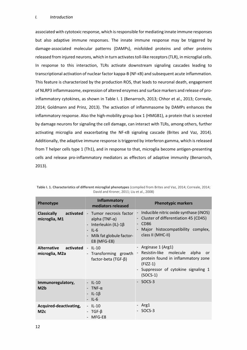
Table I. 1. Characteristics of different microglial phenotypes (compiled from Brites and Vaz, 2014; Correale, 2014; David and Kroner, 2011; Liu et al., 2008)
Phenotype Inflammatory
mediators released Phenotypic markers
Classically activated microglia, M1
- Tumor necrosis factor alpha (TNF-α)
- Interleukin (IL)-1β - IL-6 - Milk fat globule factor-
E8 (MFG-E8)
- Inducible nitric oxide synthase (iNOS) - Cluster of differentiation 45 (CD45) - CD86 - Major histocompatibility complex,
class II (MHC-II)
Alternative activated microglia, M2a
- IL-10 - Transforming growth
factor-beta (TGF-β)
- Arginase 1 (Arg1) - Resistin-like molecule alpha or
protein found in inflammatory zone (FIZZ-1)
- Suppressor of cytokine signaling 1 (SOCS-1)
Immunoregulatory, M2b
- IL-10 - TNF-α - IL-1β - IL-6
- SOCS-3
Acquired-deactivating, M2c
- IL-10 - TGF-β - MFG-E8
- Arg1 - SOCS-3

Modulation of microglia reactivity by S100B in Multiple Sclerosis
13
M2 microglia phenotype plays a crucial role in repair and healing of tissues, once
microglia secrete extracellular matrix proteins and growth factors, and promote phagocytosis of
cellular and myelin debris contributing for the remyelination process. Moreover, these microglia
are also known for their involvement in synapse repair and remodeling (Correale, 2014). M2
microglial phenotype is induced by signals from apoptotic cells, as heat shock protein 60
(Hsp60), that activates triggering receptor in myeloid cells-2 (TREM-2), or by anti-inflammatory
cytokines such as interleukin (IL)- 4 and IL-13, which are released from Th2 helper cells
(Benarroch, 2013; Olah et al., 2011b). Thus, this phenotype seems to be beneficial compared
with M1 however, a prolonged activation of this M2 phenotype may be harmful for preventing
axonal growth (Brites and Vaz, 2014). In addition, there are three different subclasses of M2
microglia, M2a, M2b and M2c, which are induced through polarizing signals and have different
functional properties. M2a is an alternative activation repair/remodeling phenotype, which is
recruited for phagocytosis and inflammation repair and induced by IL-4 and IL-13, and express
specific markers, as presented in Table I. 1 (Brites and Vaz, 2014; Chhor et al., 2013; Varnum and
Ikezu, 2012). Moreover, M2a microglia release anti-inflammatory mediators that trigger an anti-
inflammatory response and promote tissue repair (Correale, 2014; Goldmann and Prinz, 2013).
The immunoregulatory or M2b phenotype is stimulated through immune complexes, TLR
agonists and IL-1R ligands. Although M2b microglia release IL-10, an anti-inflammatory cytokine,
they also release some pro-inflammatory cytokines (Table I. 1), which suggests that M2b
microglial cells may act in and modulate different conditions of inflammation (Chhor et al., 2013;
David and Kroner, 2011). The specific markers of M2b phenotype are SOCS-3 and IL-1R
antagonist (IL-1Ra) (Chhor et al., 2013). The acquired-deactivating or M2c phenotype is induced
by IL-10, TGF-β, glucocorticoids and enhances anti-inflammatory marker expression (Table I. 1)
while decreasing pro-inflammatory cytokine levels (Chhor et al., 2013; Varnum and Ikezu, 2012).
Besides modulating the anti-inflammatory polarization through the downregulation of pro-
inflammatory markers, SOCS-1 also regulates M2 phenotype, since the expression of SOCS-1 is
increased in M2 phenotype (Davey et al., 2006; Guedes et al., 2013; Wilson, 2014).
As already mentioned, an important neuroprotective function of microglial cells is
phagocytosis. M1 and M2c microglia can produce MFG-E8 that recognizes the
phosphatidylserine (PS) exteriorized by apoptotic cells and triggers a signaling cascade that
stimulates the phagocytic process of dying cells (Brites and Vaz, 2014).
I. Introduction
14
1.4.1.3. Microglia in MS
The role of microglia in MS is controversial. Several evidences indicate that classically
activated microglia promotes neuroinflammation and demyelination in MS and in EAE,
contributing to CNS injury. To elucidate the function of microglial cells, it has been shown that
the inhibition of microglial activation results in a delayed EAE onset (Bogie et al., 2014; Giunti et
al., 2014; Heppner et al., 2005). In addition, microglial activation has been observed in post-
mortem brain tissue of MS patients as well as the presence of clusters of activated microglia
within the normal-appearing white matter (Giunti et al., 2014; van Horssen et al., 2012). Our
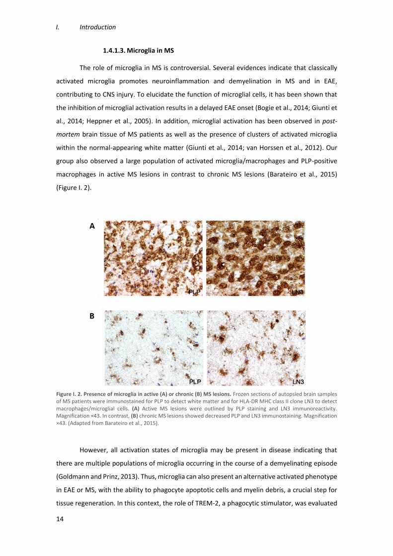
group also observed a large population of activated microglia/macrophages and PLP-positive
macrophages in active MS lesions in contrast to chronic MS lesions (Barateiro et al., 2015)
(Figure I. 2).
Figure I. 2. Presence of microglia in active (A) or chronic (B) MS lesions. Frozen sections of autopsied brain samples of MS patients were immunostained for PLP to detect white matter and for HLA-DR MHC class II clone LN3 to detect macrophages/microglial cells. (A) Active MS lesions were outlined by PLP staining and LN3 immunoreactivity. Magnification ×43. In contrast, (B) chronic MS lesions showed decreased PLP and LN3 immunostaining. Magnification ×43. (Adapted from Barateiro et al., 2015).
However, all activation states of microglia may be present in disease indicating that
there are multiple populations of microglia occurring in the course of a demyelinating episode
(Goldmann and Prinz, 2013). Thus, microglia can also present an alternative activated phenotype
in EAE or MS, with the ability to phagocyte apoptotic cells and myelin debris, a crucial step for
tissue regeneration. In this context, the role of TREM-2, a phagocytic stimulator, was evaluated
Modulation of microglia reactivity by S100B in Multiple Sclerosis
15
and it has been demonstrated that this receptor is upregulated in EAE, facilitating the debris
clearance. Moreover, its blockade results into EAE exacerbation with more infiltrates and
demyelination in the parenchyma (Piccio et al., 2007).
A study that evaluated the role of microglia/macrophages polarization in remyelination
showed that a switch from an M1- to an M2-phenotype is essential for the remyelination
process. Particularly, they observed that this change occurs at the time of oligodendrocytes
differentiation from oligodendrocyte progenitor cells, which have been recruited into the lesion.
Thus, they demonstrated that M2 microglia are necessary for oligodendrocyte differentiation
and maturation, which are very important for the efficient remyelination (Miron et al., 2013).
Given that microglial cells present distinct roles along disease progression, it is
important to understand how they exert a beneficial role in MS with the objective to modulate
this reactivity to a more neuroprotective phenotype during the course of the disease.
2. S100B
S100 proteins are part of a low-molecular-weight and acidic proteins family that are
known to contain two distinct EF-hand helix-loop-helix calcium-binding sites (Donato et al.,
2013). This family is subdivided into three subgroups according to their action: (i) only exert
intracellular regulatory effects, (ii) have intracellular and extracellular functions, or (iii) play
extracellular regulatory effects (Donato et al., 2013). S100B is a 10.5 kDa member of this family,
with both intracellular and extracellular functions, that is expressed in some cell types from
different tissues and that appears to be expressed at highest levels, in the CNS, namely by glial
cells (Adami et al., 2001; Michetti et al., 2012). Usually, this protein exists within cells as a
homodimer but it can also form heterocomplexes when associated with S100A1 monomer
(Donato et al., 2009).
Due to important functions that S100B presents in proliferation, migration,
differentiation and apoptosis, it plays a critical role during brain development acting as a
neurotrophic factor, by furthering neurite outgrowth and neuronal survival for low/physiological
levels (nM) (Donato et al., 2013; Koppal et al., 2001).
On the other hand, it was also demonstrated that S100B expression is increased in many
tumors as well as in the aging brain and in the brain of patients affected by different pathologies
as Alzheimer's disease, HIV infection, chronic epilepsy and other brain pathological conditions
as MS (Donato et al., 2009; Hein Née Maier et al., 2008). Once these high concentrations of

I. Introduction
16
S100B are present in several brain pathologies, this protein may be considered a biomarker of
brain damage (Donato et al., 2013).
2.1. Dual role of S100B in physiology and pathology
As mentioned above, S100B is a Ca2+ sensor, so, upon binding to Ca2+, S100B changes its
conformation leading to hydrophobic patch exposure to the solvent and its interaction with
target proteins (Sorci et al., 2013). Several of these target proteins have been already identified
and its interaction with S100B is involved in important intracellular processes (Table I. 2).
Accordingly, S100B acts as a stimulator of proliferation and migration as well as an inhibitor of
apoptosis and differentiation. Moreover, S100B is involved in the regulation of energy
metabolism, transcription, and protein phosphorylation, as well as in Ca2+ homeostasis (Donato
et al., 2009; Sorci et al., 2010).
It is known that astrocytes are the main cell type in CNS that express and secrete S100B,
however, other S100B-expressing cells can release the protein in case of damage or necrosis
(Sorci et al., 2010). Therefore, besides having a regulatory function within the cytoplasm where
it is expressed, S100B can act as a signal molecule in extracellular space given that it can be
released by damaged cells (Rustandi et al., 2000; Shashoua et al., 1984; Sorci et al., 2010). Once
released, S100B may have a beneficial or harmful action. There are evidences that this release
may be mostly dependent on the presence and activation of its receptor RAGE (Receptor for
Advanced Glycation Endproducts), which engagement may stimulate further S100B release
(Donato et al., 2009; Sorci et al., 2013).
RAGE is a member of the immunoglobulin-like cell surface receptor superfamily
composed by a cytosolic domain responsible for signal transduction, a transmembrane domain
which anchors it in the membrane, a variable binding-domain and two constant domains
(Ostendorp et al., 2007; Sparvero et al., 2009). This receptor is able to transduce inflammatory
stimuli and the effects of neurotrophic and neurotoxic factors and therefore, S100B emerged as
a damage-associated protein that regulate inflammation-related events and play a role in
pathophysiology of neurodegenerative disorders and inflammatory brain diseases (Bianchi et
al., 2011; Donato et al., 2013; Zhang et al., 2011b).

Modulation of microglia reactivity by S100B in Multiple Sclerosis
17
Table I. 2. S100B target proteins involved in intracellular processes (adapted from Donato et al., 2013)
Intracellular Function Target Protein
Ca2+ homeostasis - AHNAK - Annexin 6
Cell locomotion - Src/PI3-K/RhoA/ROCK - Src/PI3-K/Akt/GSK3β/Rac1
Cell proliferation and differentiation
- p53 - PI3-K/Akt/p21WAF1/cdk4/Rb/E2F - IKKβ/NF-KB
Channel Activity - EAG1 K+
Cytoskeleton
- Microtubules - Type III intermediate filaments - Caldesmon - Calponin - IQGAP1
Enzymes
- Membrane-bound GC - Fructose-1,6-biphosphate aldolase - Phosphoglucomutase - Twichin - Ndr - Src
Protein degradation - E3 ligase hdm2
Protein phosphorylation
- p53 - τ proteins - GAP43 - p80 - GFAP - Vimentin
Receptor function - D2 receptor/ERK1/2/adenyl cyclase
Transcription factors - p53
Besides having different functional roles, the extracellular form of S100B, which has
more effects in CNS, presents different effects on neurons, astrocytes and microglia, depending
on the concentration (Donato et al., 2013). At physiological levels the protein displays trophic
effects on neurons promoting neuron survival and growth as well as microglia quiescence,
although high concentration of S100B display pro-inflammatory effects and activate pro-
apoptotic pathways (Reali et al., 2005). In particular, high levels of S100B can stimulate the nitric
oxide synthesis by astrocytes and microglia leading to neuronal and astrocyte apoptosis.
Moreover, high S100B stimulates the release of cytokines contributing to brain inflammatory
response (Bianchi et al., 2011).

I. Introduction
18
2.1.1. Extracellular S100B effect on microglial cells
The high expression levels of S100B are normally associated to astrogliosis in the course
of neurodegenerative diseases. These levels might be the result of neuronal and glial cells
pathology given that the protein is released by damaged oligodendroglial or astroglial cells,
which have will then activate microglial cells responsible for the innate immune response (Sorci
et al., 2010). However, expression levels of S100B exerts two different roles in microglia
depending on concentration.
At physiological concentrations, S100B can prevent microglia activation via STAT3
pathway and may also act as a signaling trophic protein that promotes a more protective
phenotype of microglia (Zhang et al., 2011b). On the other hand, high S100B concentrations, in
the presence of bacterial endotoxin or interferon-γ (IFN-γ), play a main role on microglia
activation, exacerbating brain inflammatory response. This activation was shown to be mediated
by stimulation of iNOS leading to an increase of nitric oxide release (Adami et al., 2001; Bianchi
et al., 2007).
In addition, there is an evidence that S100B stimulates the microglia migration via RAGE-
dependent mechanism. It was demonstrated that once S100B accumulates in the extracellular
space after brain damage, the increased S100B levels might contribute to intensify the
inflammatory response by activating microglia and stimulating their migration (Bianchi et al.,
2011).
2.2. S100B in MS
Increased S100B levels were first detected in cerebrospinal fluid (CSF) of MS patients
(Michetti et al., 1979). Another study revealed that S100B levels in CSF were higher in patients
with RRMS than in patients with SPMS (Bartosik-Psujek et al., 2011). We confirmed that both in
CSF and in serum samples from RRMS patients there is a significant increase of S100B production
at the time of diagnosis (Barateiro et al., 2015). S100B increase may exacerbate the release of
inflammatory mediators and subsequently contribute to neuronal death (Hu et al., 1996). These
results indicate S100B as a potential biomarker for MS diagnosis and prognosis, possibly helping
to distinguish between relapsing remitting and progressive phases of MS.
One study reported the presence of S100B in acute lesions of post-mortem brain tissue
of patients with RRMS (Petzold et al., 2002). In similar samples, our recent work demonstrated
that S100B is overexpressed both in active and chronic lesions, mainly by astrocytes (Figure I. 3).
Modulation of microglia reactivity by S100B in Multiple Sclerosis
19
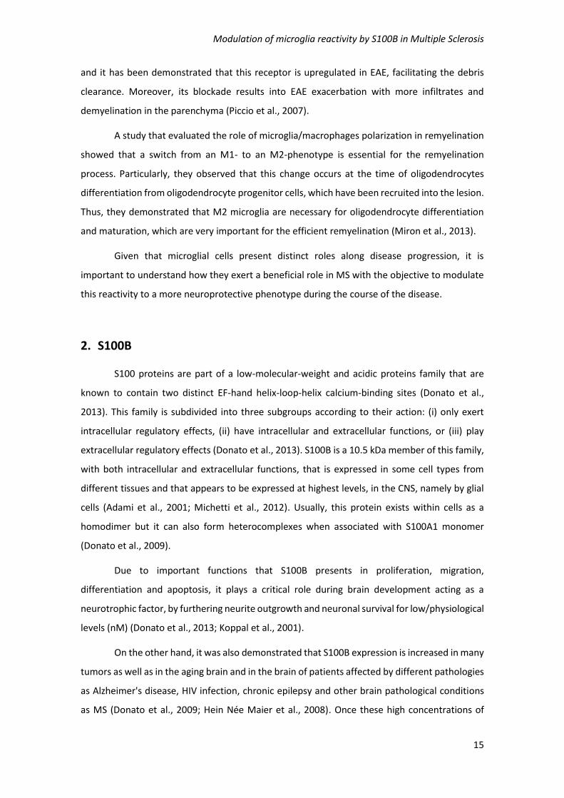
In addition, it showed that in chronic lesions S100B is still diffusely expressed into the
demyelinated area (Barateiro et al., 2015). Interestingly, those active MS lesions also showed an
increase of RAGE expression that co-localized with microglia/macrophages cells, while almost
no RAGE was found in chronic lesions that are almost depleted of microglia/macrophages
(Figure I. 3).
Figure I. 3. Expression of S100B and its receptor RAGE in active (A) and chronic (B) MS lesions. Frozen sections of autopsied brain samples of MS patients were immunostained for S100B and RAGE. (A) S100B and its receptor RAGE are markedly expressed in active MS lesions by astrocytes and activated macrophages/microglia, respectively. Magnification x40. Insets show the co-localization of (I) glial fibrillary acidic protein (GFAP, red), an astrocytic marker, with S100B (green) and the co-localization of (II) LN3 (red), a marker of activated macrophages/microglia with RAGE (green). Magnification x63. (B) S100B but not RAGE is continuously expressed in chronic MS lesions. Magnification x40. (Adapted from Barateiro et al., 2015).
Given the increased expression of S100B during MS episodes, the role of S100B in
different neurodegenerative diseases and its apparent involvement in microglia activation, it is
of utmost importance to assess whether by modulation of S100B function it is possible to change
the reactivity of microglia to a more neuroprotective phenotype during the course of MS.
3. Experimental models to study MS pathophysiology
Failure to understand the neurodegenerative mechanisms involved in MS and
therapeutic inefficacy of diverse treatments led to development of multiple experimental
I. Introduction
20
models that mimic the hallmarks of the disease. However, the complexity of MS still hinders the
development of the perfect model for this disease (Murta and Ferrari, 2013).
The EAE, a model characterized by inflammation, demyelination and
neurodegeneration, is one of the most widely used in vivo model to study MS that involves the
immunization of genetically susceptible animals with a myelin protein inducing brain
inflammation and destruction of myelin. However, EAE models fail to expect the clinical efficacy
in patients (Mathew et al., 2013; Ransohoff, 2012). Moreover, in vivo models entail an expensive
cost, as well as ethical problems and, therefore, alternative models should be used whenever
possible.
For decades, organotypic slice cultures were used in CNS research due to three-
dimensional architecture, maintenance of contact between different cells and the presence of
all the cells of CNS which play a key role in lesion recovery (Denic et al., 2011; Gähwiler, 1984).
Organotypic slice culture is a more complex model to study cell-cell interactions, being named
ex vivo, and can be prepared from different brain regions, such as cerebellum, hippocampus,
striatum, cortex and spinal cord, (Birgbauer et al., 2004; Gähwiler, 1984; Stoppini et al., 1991).
The preferential region used in MS research, compared with other CNS regions is cerebellum
due to the abundance of white matter and the well-known pattern of myelin tracts (Zhang et
al., 2011a).
This method was initially used for electrophysiological studies, in 1941, but the process
of myelination was only reported in 1956, in cerebellar slices (Hild, 1956; Levi and Meyer, 1941).
In 2004, lysophosphatidylcholine (LPC) was used to demyelinate rat cerebellar slices, which was
followed by the reappearance of myelin sheaths, suggesting remyelination (Birgbauer et al.,
2004). Thus, LPC can be used as a good molecule to induce demyelination in cerebellar
organotypic slice cultures (COSC), providing a model that allows the study of demyelination and
remyelination in an ex vivo model. This model offers advantages compared with other in vitro
models, once it can mimic the multicellular complexity as well as the structure and functionality
of in vivo conditions (Cho et al., 2007). Besides preserving glial cells contribution into
myelination-associated processes, organotypic slice model excludes the systemic immune
system interactions that would render the model more complex (Miron et al., 2010). Thus, this
ex vivo model is an attractive proposal for MS study and the assay of potential new therapeutic
strategies.

Modulation of microglia reactivity by S100B in Multiple Sclerosis
21
3.1. Novel findings on the role of S100B in the ex vivo demyelinated model
Recently, our group showed that S100B is highly released upon LPC-induced
demyelination in COSC (Figure I. 4), mostly by astrocytes, in parallel to a massive gliosis
(Barateiro et al., 2015).
Figure I. 4. S100B is markedly released upon LPC-induced demyelination of cerebellar organotypic slice cultures. Cerebellar organotypic slice cultures were exposed to LPC at 7 days in vitro during 18 h and allowed to recover for 30 h. Samples for detection of S100B secretion were collected before the incubation (0 h), at 18 h post-incubation with LPC and at 48 h, i.e. after 30 h of recovery. Results are mean ± SEM. ***p<0.001 vs. Control. (Adapted from Barateiro et al., 2015).
Curiously, when S100B was therapeutically neutralized using a specific antibody, we
could observe a reduced demyelination (Figure I. 5), as well as reduced astrogliosis.
Nevertheless, although the density of microglia in the slice showed no apparent differences
upon anti-S100B co-treatment, the release of cytokines was markedly reduced suggesting a
potential inhibition of microglia pro-inflammatory response (Barateiro et al., 2015).

I. Introduction
22
Figure I. 5. Blockade of S100B following demyelination partially prevents loss of myelinated fibers. Cerebellar organotypic slice cultures were exposed to LPC at 7 days in vitro during 18 h and allowed to recover for 30 h. Slices were double immunostained with neurofilament-200 (NF-200), MBP and DAPI antibodies. (A) Confocal images are shown. Scale bar represents 100 μm. (B) Quantification of axon integrity was taken by averaging the area occupied by NF-200 staining for each stack. (C) The percentage of myelinated fibers was calculated by the ratio between the area of co-localization of NF-200 and MBP and the total area occupied by NF-200. Results are mean ± SEM. **p<0.01 vs. Control; ##p<0.01 vs. LPC. (Adapted from Barateiro et al., 2015).
So, in preliminary studies we explored the distribution of microglia along the slice.
Usually in COSC microglia is distributed along the slice while myelin is found on the top of it. As
shown in Figure I. 6, LPC-induced demyelination markedly increase microglia density, namely at
the bottom of the slice. However, co-incubation of LPC and anti-S100B, although not preventing
the increase of Iba-1 staining, suggest a more evident microglia in the region with highest
amount of myelin debris (Figure I. 5). These images suggest that activated microglia moved to
myelin surrounds when S100B was blocked, possibly increasing the phagocytosis of myelin
debris which is crucial for further remyelination (Afonso, 2014). Since there are recent evidences
that microglia switch from a cytotoxic to a protector phenotype during remyelination initiation

Modulation of microglia reactivity by S100B in Multiple Sclerosis
23
(Miron et al., 2013), it would be interest to evaluate if we can accelerate this change of microglia
phenotype by neutralizing excessive S100B in the milieu and only preserving it physiological low
concentration.
Figure I. 6. Blockade of S100B following demyelination apparently induces microglia migration near to myelin debris. Cerebellar organotypic slice cultures were exposed to LPC at 7 days in vitro, in presence or absence of S100B antibody, during 18 h and allowed to recover for 30 h. Slices are incubated with Iba-1 and MBP antibodies and analyzed by immunohistochemistry. (Adapted from Afonso V MS thesis).

I. Introduction
24
4. Aims
The principal aim of this work is to evaluate the role of S100B in microglial response
during MS. More specifically, we want to understand whether we may promote a more
neuroprotective microglia phenotype, during the course of MS, through the modulation of
S100B levels.
Therefore, in order to display how S100B modulates microglial reactivity in an ex vivo
model of demyelination, we will assess (i) the effect of S100B in microglia phenotype-related
cell surface receptors; (ii) the role of S100B in microglia phenotype-related markers and (iii) the
importance of S100B in microglia phagocytic ability.

Modulation of microglia reactivity by S100B in Multiple Sclerosis
25
II. Material and Methods
1. Animals
CD1 mouse pups were acquired from Instituto Gulbenkian de Ciência (IGC, Lisboa,
Portugal). Animal care followed the recommendations of European Convention for the
Protection of Vertebrate Animals Used for Experimental and Other Scientific Purposes (Council
Directive 86/609/EEC) and National Law 1005/92 (rules for protection of experimental animals).
All animal procedures were approved by the Institutional Animal Care and Use Committee. All
efforts were made to minimize animal suffering and to reduce the number of animals used in
order to use alternatives procedures to in vivo.
2. Cerebellar Organotypic Slice Cultures (COSC) and its Treatment
Cerebellum parasagittal slices were obtained from postnatal day 10 (P10) CD1 mouse
pups. Briefly, after mice decapitation, the cerebellums were isolated from brains and 400 µm
sagittal slices obtained using a Mcllwain tissue chopper. Four slices of different animals were
transferred into membrane culture inserts (BD Falcon, #353493, Lincoln Park, NJ, USA) in 6-well
cell culture plates that contain 1 mL of medium in per well and kept at 37°C, in 5% CO2
conditioned atmosphere. Half media was replaced every day and slices were maintained for 7
days in vitro (DIV) before treatment, to allow the clearance of debris and full myelination
(Birgbauer et al., 2004). The culture medium consisted of 50% minimal essential media (MEM),
(Gibco, Life Technologies, Inc., Grand Islands, USA), 25% of both heat-inactivated horse serum
(Gibco) and Earle’s balanced salt solution (EBSS, Gibco), 6.5 mg/mL glucose, 25mM HEPES
(Biochrom AG, Berlin, Germany), 1% of L-glutamine (Sigma-Aldrich, St. Louis, MO, USA) and 1%
of antibiotic-antimycotic (Sigma-Aldrich). After 3 DIV, slice culture media was totally changed by
a serum-free media that consists of Neurobasal-A (Gibco) supplemented with 2% B-27 (Gibco),
2 mM L-glutamine, 36 mM glucose, 1% of antibiotic-antimycotic and 25 mM HEPES.

II. Material and Methods
26
After 7 DIV, culture media was removed and slices were incubated with 0.5 mg/mL LPC
in serum-free culture media, during 18h, at 37°C. To evaluate how S100B modulates the
microglial reactivity, some slices were also incubated with LPC in the presence of anti-S100B
antibody (1:500, AbCam, Cambridge, UK). Additionally, to assess the effect of the presence of
an antibody we also incubated the slices with a control IgG (1:500, Santa Cruz Biotechnology,
CA, USA). After incubation, the LPC-containing medium was removed and the slices were
maintained in fresh medium or in medium supplemented with anti-S100B antibody or IgG during
30h. Following these 30h, slices were: (i) stored in TRIzol® reagent, at -20°C, for RNA extraction;
(ii) incubated with ice-cold cell lysis buffer (radio-immunoprecipitation assay buffer – RIPA), for
protein extraction; (iii) dissociated and stained with specific antibodies for flow cytometry
analysis; (iv) incubated with 1 µm fluorescent latex beads (1:100, Sigma Chemical Co., St. Louis,
MO, USA) during 1h, at 37°C, for phagocytic studies; or (v) fixed in 4% paraformaldehyde (PFA)
for immunohistochemistry assays. All supernatants were collected both before and after
incubation with LPC.
3. Total RNA Extraction, Reverse Transcription, Semi-quantitative
RealTime Polymerase Chain Reaction
For the determination of expression levels of diverse genes of interest, total cytoplasmic
RNA was isolated from treated slices using the TRIzol® reagent method in accordance with the
manufacturer’s guidelines (Invitrogen, Carlsbad, CA, USA) and RNA concentration was quantified
using Nanodrop ND-100 Spectrophotometer (NanoDrop Technologies, Wilmington, DE, USA).
Aliquots of 500 ng of total RNA were reversely transcribed using the SensiFAST cDNA Synthesis
Kit (Bioline), under manufacturer’s instructions. Semi quantitative (q)RealTime-PCR was
performed on a RealTime PCR detection system (Applied Biosystems 7300 Fast RealTime PCR
System, Applied Biosystem, Madrid, Spain) using a SensiFAST SYBR® Hi-Rox Kit (Bioline), under
optimized conditions: 50°C for 2 min, 95°C for 2 min followed by 40 cycles at 95°C for 5 s and
62°C for 30 s. In order to verify the specificity of the amplification, a melt-curve analysis was
performed immediately after the amplification protocol (95°C for 15 s, followed by 60ºC for 30
s and 95°C for 15 s). The PCR was performed in 96-well plates with each sample performed in
duplicate, and a non-template control was included for each gene. The sequences used as
primers are listed in the Table II. 1.
Relative mRNA concentrations were calculated using the Pfaffl modification of the ΔΔCT
equation, where CT is the cycle number at which fluorescence passes the threshold level of

Modulation of microglia reactivity by S100B in Multiple Sclerosis
27
detection, taking into account the efficiencies of individual genes. The results were normalized
to the housekeeping gene β-actin in the same sample and the initial amount of the template of
each trial was determined as relative expression by the formula 2-ΔΔCT. ΔCT is the value obtained
for each sample by performing the difference between the mean CT value of each gene of
interest and the mean CT value of β-actin. ΔΔCT of one sample is the difference between its ΔCT
value and the ΔCT of the sample chosen as reference.
Table II. 1. Primers used for qRealTime PCR
Gene Forward Reverse
Arg1 cttggcttgcttcggaactc ggagaaggcgtttgcttagttc
β-actin gctccggcatgtgcaa aggatcttcatgaggtagt
CEBP-α agcttacaacaggccaggtttc cggctggcgacatacagtac
CX3CL1 ctcacgaatcccagtggctt tttctccttcgggtcagcac
CX3CR1 tcgtcttcacgttcggtctg ctcaaggccaggttcaggag
FIZZ-1 gccaggtcctggaacctttc ggagcagggagatgcagatgag
HMGB1 ctcagagaggtggaagaccatgt gggatgtaggttttcatttctctttc
IL-1β caggctccgagatgaacaac ggtggagagctttcagctcata
IL-6 ccggagaggagacttcacag ggaaattggggtaggaagga
IL-18 tggttccatgctttctggactcct ttcctgggccaagaggaagtg
iNOS acccacatctggcagaatgag agccatgacctttcgcattag
MBP ccatccaagaagaccccaca cccctgtcaccgctaaagaa
MHC-II tgggcaccatcttcatcattc ggtcacccagcacaccactt
NLRP3 tgctcttcactgctatcaagccct acaagcctttgctccagaccctat
SOCS-1 caccttcttggtgcgcg aagccatcttcacgctgagc
TGF-β cagagctgcgcttgcagag gtcagcagccggttaccaag
TLR2 tgctttcctgctgaagattt tgtaccgcaacagcttcagg
TLR4 acctggctggtttacacgtc gtgccagagacattgcagaa
TNF-α tactgaacttcggggtgattggtcc cagccttgtcccttgaagagaacc
4. Protein Extraction and Western Blot
For the determination of cellular protein content, proteins were obtained by lysing cells
in ice-cold cell lysis RIPA buffer, followed by sonication and centrifugation at 12,000 g for 10 min.
Total protein concentrations were always measured using the Bradford method with Bio-Rad’s
Protein Assay Reagent (BioRad Laboratories, Hercules, CA, USA).
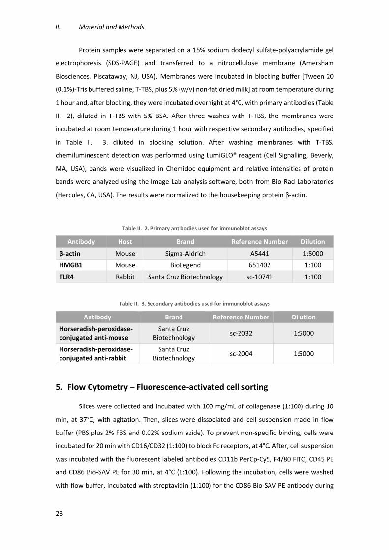
II. Material and Methods
28
Protein samples were separated on a 15% sodium dodecyl sulfate-polyacrylamide gel
electrophoresis (SDS-PAGE) and transferred to a nitrocellulose membrane (Amersham
Biosciences, Piscataway, NJ, USA). Membranes were incubated in blocking buffer [Tween 20
(0.1%)-Tris buffered saline, T-TBS, plus 5% (w/v) non-fat dried milk] at room temperature during
1 hour and, after blocking, they were incubated overnight at 4°C, with primary antibodies (Table
II. 2), diluted in T-TBS with 5% BSA. After three washes with T-TBS, the membranes were
incubated at room temperature during 1 hour with respective secondary antibodies, specified
in Table II. 3, diluted in blocking solution. After washing membranes with T-TBS,
chemiluminescent detection was performed using LumiGLO® reagent (Cell Signalling, Beverly,
MA, USA), bands were visualized in Chemidoc equipment and relative intensities of protein
bands were analyzed using the Image Lab analysis software, both from Bio-Rad Laboratories
(Hercules, CA, USA). The results were normalized to the housekeeping protein β-actin.
Table II. 2. Primary antibodies used for immunoblot assays
Antibody Host Brand Reference Number Dilution
β-actin Mouse Sigma-Aldrich A5441 1:5000
HMGB1 Mouse BioLegend 651402 1:100
TLR4 Rabbit Santa Cruz Biotechnology sc-10741 1:100
Table II. 3. Secondary antibodies used for immunoblot assays
Antibody Brand Reference Number Dilution
Horseradish-peroxidase-conjugated anti-mouse
Santa Cruz Biotechnology
sc-2032 1:5000
Horseradish-peroxidase-conjugated anti-rabbit
Santa Cruz Biotechnology
sc-2004 1:5000
5. Flow Cytometry – Fluorescence-activated cell sorting
Slices were collected and incubated with 100 mg/mL of collagenase (1:100) during 10
min, at 37°C, with agitation. Then, slices were dissociated and cell suspension made in flow
buffer (PBS plus 2% FBS and 0.02% sodium azide). To prevent non-specific binding, cells were
incubated for 20 min with CD16/CD32 (1:100) to block Fc receptors, at 4°C. After, cell suspension
was incubated with the fluorescent labeled antibodies CD11b PerCp-Cy5, F4/80 FITC, CD45 PE
and CD86 Bio-SAV PE for 30 min, at 4°C (1:100). Following the incubation, cells were washed
with flow buffer, incubated with streptavidin (1:100) for the CD86 Bio-SAV PE antibody during

Modulation of microglia reactivity by S100B in Multiple Sclerosis
29
30 min, and then resuspended in flow buffer. Expression of surface antigens was measured using
the BD FACSCalibur flow cytometer and data analyzed using the FlowJo software.
6. Immunostaining procedure for phagocytic analysis
For immunostaining procedure, insert membranes, which contain the fixed slices, were
cut, placed into a cover glass and blocked with 1nM HEPES, 2% heat-inactivated horse serum,
10% heat-inactivated goat serum (Biochrom), 1% bovine serum albumin (BSA, Sigma-Aldrich)
and 0.25% Triton X-100 (Roche Diagnostics, Indianapolis, USA) in Hank’s balanced salt solution
(HBSS, Gibco) for three hours, at room temperature. After blocking, slices were incubated with
primary antibody (Iba-1, 1:250, Wako) diluted in the blocking solution, for 24h, at 4°C. After
incubation, slices were washed three times for 15 min each with 0.01% Triton X-100 in PBS (T-
PBS) and incubated with secondary antibody (Alexa 594 anti-rabbit, 1:1000 Invitrogen) in
blocking solution, for 24 h, at 4°C. Slices were then washed three times for 15 min each with T-
PBS, incubated with DAPI (1:1000, 3 min), washed three times for 15 min each with T-PBS and
mounted using Fluoromount-G (Southern Biotech, Birmingham, AL) for confocal microscopy.
Fluorescent images were acquired using a Leica DM6000 inverted microscope and analyzed with
ImageJ soſtware. For each image were determined the total number of phagocyting cells and
the total number of internalized beads.
7. Statistical analysis
Results are presented as mean ± SEM. Differences between groups were determined by
one-way ANOVA using GraphPad PRISM 5.0 (GraphPad Software, San Diego, CA, USA), as
appropriate. The p-values lower than 0.05 were considered as being statistically significant.


Modulation of microglia reactivity by S100B in Multiple Sclerosis
31
III. Results
1. Myelin-related protein expression is recovered after S100B blockage
Knowing that myelin-related MBP is an important constituent of myelin sheaths and to
show that our ex vivo model is a good model to study demyelinated lesions, we decided to
evaluate the gene expression of this protein extracted from COSC at 48 h post-incubation with
LPC, by qRealTime PCR, using specific primers.
As illustrated in Figure III. 1, LPC insult promoted a significant decrease in MBP and PLP
expression (0.36-fold, p<0.001). Interestingly, co-incubation with anti-S100B antibody markedly
prevented this MBP reduction (~80%, p<0.001), while the co-treatment with IgG did not
significantly changed MBP mRNA expression.
Figure III. 1. S100B neutralization attenuates demyelination induced by LPC. Cerebellar organotypic slice cultures were exposed to LPC at 7 days in vitro, in presence or absence of S100B and IgG antibodies, during 18 h and allowed to recover for 30 h. Gene expression of MBP was evaluated at 48 h by qRealTime PCR. Results are mean ± SEM. ***p<0.001 vs. Control; ###p<0.001 vs. LPC

III. Results
32
2. Neutralization of S100B prevents a microglia pro-inflammatory
phenotype
Along with demyelination, MS lesions are also characterized by microgliosis (Dyer et al.,
2005). Knowing that the inhibition of S100B does not prevent the increase of microglia density
although it prevents the release of cytokines on our ex vivo demyelinating model (Barateiro et
al., 2015), we decided to characterize the populations of reactive microglia in our COSC at 48 h
post-LPC, by flow cytometry. Generally it is accepted that pro-inflammatory activated microglia
is characterized as CD11bhigh, CD86high, CD45dim hi and F4/80high staining (Devarajan et al., 2014).
Figure III. 2. S100B abrogation significantly decreases CD11b+/CD86+ microglia population induced by LPC-demyelination. Cerebellar organotypic slice cultures were treated with LPC, at 7 days in vitro, in presence or absence of S100B and IgG antibodies, during 18 h and allowed to recover for 30 h. Dissociated slices were stained with CD11b- and CD86-specific monoclonal antibodies and analyzed by flow cytometry. (A) Data are expressed as mean of percentage of CD11b+ and (B) CD11b+/CD86+ cells ± SEM. ***p<0.001 and *p<0.05 vs. Control; ###p<0.001 and #p<0.05 vs. LPC. (C) Representative images of cytograms are showed.
As shown in Figure III. 2 A, treatment with LPC induced an increase of the population of
more reactive microglia expressing CD11b (from 36.99% to 52.26%, p<0.05), which is prevented
by anti-S100B neutralization. Next, we analyzed which CD11b+ population also expressed the
pro-inflammatory cell surface receptors CD86, CD45 and F4/80. Figure III. 2 B clearly shows that
LPC treatment increases the number of CD11b+/CD86+ microglia (from 3.88% to 49.75%,
p<0.001), which is partially prevented by S100B neutralization by ~50% (p<0.001), while IgG had

Modulation of microglia reactivity by S100B in Multiple Sclerosis
33
no effect. These results show that, blockade of S100B may prevent a pro-inflammatory
phenotype, as demonstrated by a significant reduction on CD11b+/CD86+ microglia population.
On the other hand, we saw no differences between control and LPC, or even anti-S100B
treatments in the other markers (Table III. 1), suggesting that demyelination did not influence
their expression.
Table III. 1. Demyelination or antibodies treatment on cerebellar organotypic slice cultures do not change the percentage of CD11b-positive cells that express F4/80 and CD45 markers.
CD11b+ F4/80+ CD11b+ CD452+
Control 63.06 % ± 8.04 % 16.03 % ± 7.01 %
LPC 65.56 % ± 7.24 % 20.58 % ± 7.25 %
LPC + anti-S100B 60.08 % ± 7.07 % 24.78 % ± 5.97 %
LPC + IgG 61.86 % ± 6.94 % 22.25 % ± 7.05 %
Cerebellar organotypic slice cultures were treated with LPC, at 7 days in vitro, in presence or absence of S100B or IgG antibodies, during 18 h and allowed to recover for 30 h. Dissociated slices were stained with CD11b-, F4/80- and CD45-specific monoclonal antibodies and analyzed by flow cytometry. Data are expressed as mean of percentage of positive cells ± SEM.
3. Abrogation of S100B shifts microglia from a pro-inflammatory
phenotype to a more neuroprotective one
As mentioned above, MS lesions are characterized by microglia activation that lead to
the secretion of pro-inflammatory cytokines and inflammasome-related proteins production
that enhance the inflammatory response, contributing to disease progression (Benarroch, 2013;
Vainchtein et al., 2014). Moreover, NLRP3 inflammasome, a complex involved in the maturation
and secretion of pro-inflammatory molecules, have recently been associated with MS pathology.
Once we have shown that S100B inhibition seems to prevent a pro-inflammatory response from
microglia, we decided to explore the expression of these several inflammatory mediators upon
demyelination and in the presence of anti-S100B antibody.
As shown in Figure III. 3, LPC-induced demyelination increased the expression of
cytokines TNF-α and IL-1β but decreased the IL-6 expression (1.46-, 6.24- and 0.35-fold, p<0.01
and p<0.001, respectively). Anti-S100B treatment partially prevented LPC-induced alterations in
cytokine expression (~56.52%, 63.17% and 76.92%, p<0.01, respectively), while no significant
changes was observed upon IgG treatment. These results confirm the stimulation of an

III. Results
34
inflammatory response upon demyelination and suggesting the involvement of S100B in
cytokine release by microglial cells.
Figure III. 3. S100B neutralization prevents the increase of TNF-α and IL-1β expression and the inhibition of IL-6 expression induced by LPC-demyelination. Cerebellar organotypic slice cultures were exposed to LPC at 7 days in vitro, in presence or absence of S100B and IgG antibodies, during 18 h and allowed to recover for 30 h. Gene expression of TNF-α, IL-1β and IL-6 were evaluated at 48 h by qRealTime PCR. Results are mean ± SEM. ***p<0.001 and **p<0.01 vs. Control; ###p<0.001 and ##p<0.01 vs. LPC.
Inflammasomes are cytosolic sensors that detect alterations and are involved in
maturation and release of pro-inflammatory cytokines, such as HMGB1, IL-1β and IL-18 (Inoue
and Shinohara, 2013; Lu et al., 2013). The NLRP3 inflammasome consists of three proteins that
has already been associated with MS development (Inoue and Shinohara, 2013). As we have
previously observed, a marked increase of IL-1β expression upon demyelination and its
protection after S100B inhibition, we next evaluated the gene expression of the other
inflammasome-associated molecules.
Figure III. 4. S100B neutralization prevents NLRP3 activation and inflammasome-related molecules expression induced by LPC-demyelination. Cerebellar organotypic slice cultures were exposed to LPC at 7 days in vitro, in presence or absence of S100B and IgG antibodies, during 18 h and allowed to recover for 30 h. (A) Gene expression of HMGB1, IL-18 and NLRP3 were evaluated at 48 h by qRealTime PCR. (B) Protein expression of HMGB1 was evaluated at 48 h by Western Blot. Results are mean ± SEM. ***p<0.001 and **p<0.01 vs. Control and ###p<0.001, ##p<0.01 and #p<0.05 vs. LPC.
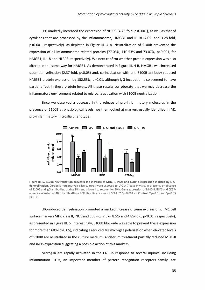
Modulation of microglia reactivity by S100B in Multiple Sclerosis
35
LPC markedly increased the expression of NLRP3 (4.75-fold, p<0.001), as well as that of
cytokines that are processed by the inflammasome, HMGB1 and IL-18 (4.05- and 3.28-fold,
p<0.001, respectively), as depicted in Figure III. 4 A. Neutralization of S100B prevented the
expression of all inflammasome-related proteins (77.05%, 110.53% and 73.07%, p<0.001, for
HMGB1, IL-18 and NLRP3, respectively). We next confirm whether protein expression was also
altered in the same way for HMGB1. As demonstrated in Figure III. 4 B, HMGB1 was increased
upon demyelination (2.37-fold, p<0.05) and, co-incubation with anti-S100B antibody reduced
HMGB1 protein expression by 152.55%, p<0.01, although IgG incubation also seemed to have
partial effect in these protein levels. All these results corroborate that we may decrease the
inflammatory environment related to microglia activation with S100B neutralization.
Since we observed a decrease in the release of pro-inflammatory molecules in the
presence of S100B at physiological levels, we then looked at markers usually identified in M1
pro-inflammatory microglia phenotype.
Figure III. 5. S100B neutralization prevents the increase of MHC-II, iNOS and CEBP-α expression induced by LPC-demyelination. Cerebellar organotypic slice cultures were exposed to LPC at 7 days in vitro, in presence or absence of S100B and IgG antibodies, during 18 h and allowed to recover for 30 h. Gene expression of MHC-II, iNOS and CEBP-α were evaluated at 48 h by qRealTime PCR. Results are mean ± SEM. ***p<0.001 vs. Control; ##p<0.01 and #p<0.05 vs. LPC.
LPC-induced demyelination promoted a marked increase of gene expression of M1 cell
surface markers MHC class II, iNOS and CEBP-α (7.87-, 8.51- and 4.85-fold, p<0.01, respectively),
as presented in Figure III. 5. Interestingly, S100B blockade was able to prevent these expression
for more than 60% (p<0.05), indicating a reduced M1 microglia polarization when elevated levels
of S100B are neutralized in the culture medium. Antiserum treatment partially reduced MHC-II
and iNOS expression suggesting a possible action at this markers.
Microglia are rapidly activated in the CNS in response to several injuries, including
inflammation. TLRs, an important member of pattern recognition receptors family, are

III. Results
36
considered to be involved in MS pathology. Since there are studies showing the elevated
expression of both TLR2 and TLR4 in MS patients (Miranda-Hernandez and Baxter, 2013), we
decided to analyze the gene expression of these receptors.
Figure III. 6. S100B inhibition prevents the alterations of TLR2/4 expression induced by LPC-demyelination. Cerebellar organotypic slice cultures were exposed to LPC at 7 days in vitro, in presence or absence of S100B and IgG antibodies, during 18 h and allowed to recover for 30 h. (A) Gene expression of TLR2 and TLR4 were evaluated at 48 h by qRealTime PCR. (B) Protein expression of TLR4 was evaluated at 48 h by Western Blot. Results are mean ± SEM. ***p<0.001 and **p<0.01 vs. Control; ###p<0.001 and ##p<0.01 vs. LPC.
Accordingly with previous studies, both mRNA and protein TLR4 levels were increased
upon demyelination (1.59- and 2.69-fold, p<0.01, respectively) possibly indicating an attempt of
microglia to respond to an inflammatory stimulus (Figure III. 6 A, B). S100B inhibition prevented
both LPC-induced TLR4 gene and protein expression (179.66% and 72.78%, p<0.001,
respectively). However, also IgG co-incubation decreased TLR4 expression suggesting a potential
effect of the antiserum. Concerning TLR2, its gene expression was decreased by LPC-induced
demyelination (0.53-fold, p<0.001), what was abrogated by anti-S100B co-incubation to control
levels (p<0.01). These results indicating that the inflammatory response may not occur through
TLR2 pathway (Figure III. 6 A).
Having confirmed the reduction of a pro-inflammatory phenotype with S100B inhibition,
and knowing that microglia are the most responsible for inflammatory cytokine production, we
evaluated whether microglia could be shifting from a M1 to a M2 phenotype with S100B
blockade. In this regard, we analyzed COSC lysates 48 h after LPC treatment for different M2-
like gene expression.
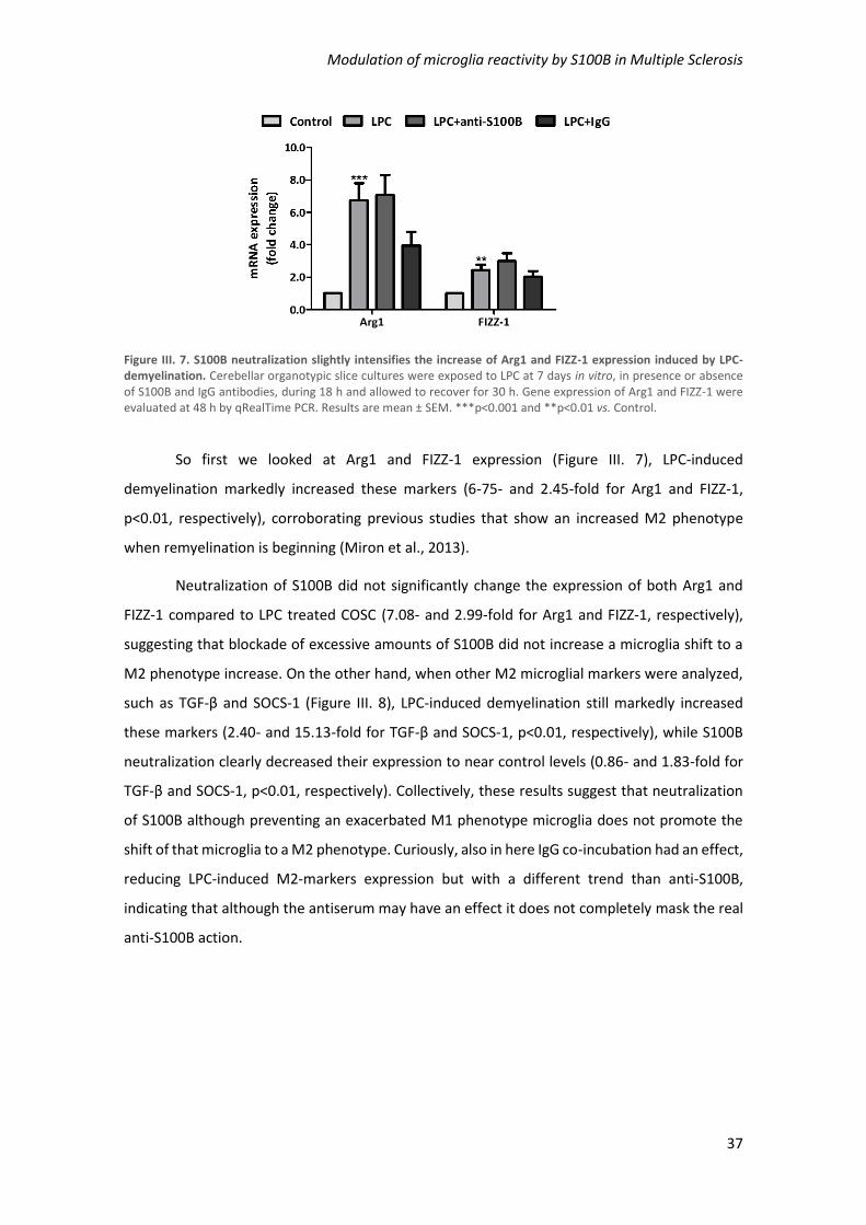
Modulation of microglia reactivity by S100B in Multiple Sclerosis
37
Figure III. 7. S100B neutralization slightly intensifies the increase of Arg1 and FIZZ-1 expression induced by LPC-demyelination. Cerebellar organotypic slice cultures were exposed to LPC at 7 days in vitro, in presence or absence of S100B and IgG antibodies, during 18 h and allowed to recover for 30 h. Gene expression of Arg1 and FIZZ-1 were evaluated at 48 h by qRealTime PCR. Results are mean ± SEM. ***p<0.001 and **p<0.01 vs. Control.
So first we looked at Arg1 and FIZZ-1 expression (Figure III. 7), LPC-induced
demyelination markedly increased these markers (6-75- and 2.45-fold for Arg1 and FIZZ-1,
p<0.01, respectively), corroborating previous studies that show an increased M2 phenotype
when remyelination is beginning (Miron et al., 2013).
Neutralization of S100B did not significantly change the expression of both Arg1 and
FIZZ-1 compared to LPC treated COSC (7.08- and 2.99-fold for Arg1 and FIZZ-1, respectively),
suggesting that blockade of excessive amounts of S100B did not increase a microglia shift to a
M2 phenotype increase. On the other hand, when other M2 microglial markers were analyzed,
such as TGF-β and SOCS-1 (Figure III. 8), LPC-induced demyelination still markedly increased
these markers (2.40- and 15.13-fold for TGF-β and SOCS-1, p<0.01, respectively), while S100B
neutralization clearly decreased their expression to near control levels (0.86- and 1.83-fold for
TGF-β and SOCS-1, p<0.01, respectively). Collectively, these results suggest that neutralization
of S100B although preventing an exacerbated M1 phenotype microglia does not promote the
shift of that microglia to a M2 phenotype. Curiously, also in here IgG co-incubation had an effect,
reducing LPC-induced M2-markers expression but with a different trend than anti-S100B,
indicating that although the antiserum may have an effect it does not completely mask the real
anti-S100B action.
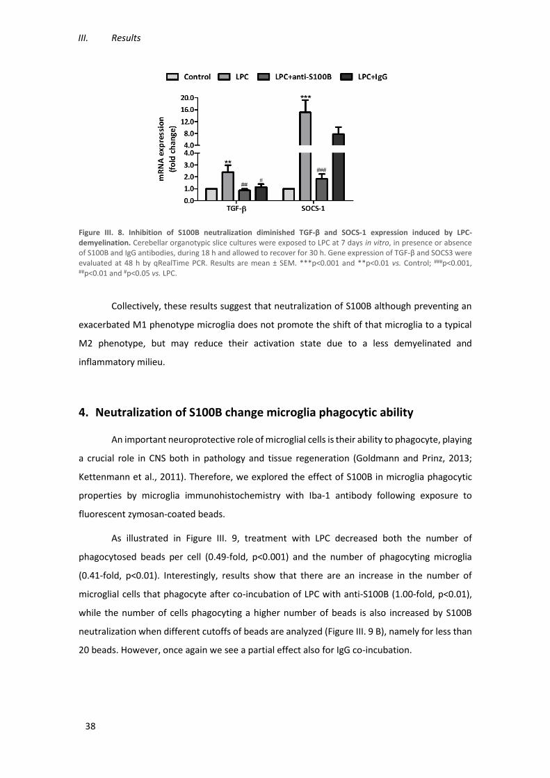
III. Results
38
Figure III. 8. Inhibition of S100B neutralization diminished TGF-β and SOCS-1 expression induced by LPC-demyelination. Cerebellar organotypic slice cultures were exposed to LPC at 7 days in vitro, in presence or absence of S100B and IgG antibodies, during 18 h and allowed to recover for 30 h. Gene expression of TGF-β and SOCS3 were evaluated at 48 h by qRealTime PCR. Results are mean ± SEM. ***p<0.001 and **p<0.01 vs. Control; ###p<0.001, ##p<0.01 and #p<0.05 vs. LPC.
Collectively, these results suggest that neutralization of S100B although preventing an
exacerbated M1 phenotype microglia does not promote the shift of that microglia to a typical
M2 phenotype, but may reduce their activation state due to a less demyelinated and
inflammatory milieu.
4. Neutralization of S100B change microglia phagocytic ability
An important neuroprotective role of microglial cells is their ability to phagocyte, playing
a crucial role in CNS both in pathology and tissue regeneration (Goldmann and Prinz, 2013;
Kettenmann et al., 2011). Therefore, we explored the effect of S100B in microglia phagocytic
properties by microglia immunohistochemistry with Iba-1 antibody following exposure to
fluorescent zymosan-coated beads.
As illustrated in Figure III. 9, treatment with LPC decreased both the number of
phagocytosed beads per cell (0.49-fold, p<0.001) and the number of phagocyting microglia
(0.41-fold, p<0.01). Interestingly, results show that there are an increase in the number of
microglial cells that phagocyte after co-incubation of LPC with anti-S100B (1.00-fold, p<0.01),
while the number of cells phagocyting a higher number of beads is also increased by S100B
neutralization when different cutoffs of beads are analyzed (Figure III. 9 B), namely for less than
20 beads. However, once again we see a partial effect also for IgG co-incubation.
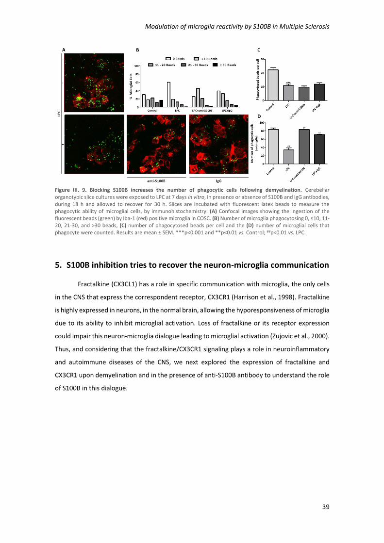
Modulation of microglia reactivity by S100B in Multiple Sclerosis
39
Figure III. 9. Blocking S100B increases the number of phagocytic cells following demyelination. Cerebellar organotypic slice cultures were exposed to LPC at 7 days in vitro, in presence or absence of S100B and IgG antibodies, during 18 h and allowed to recover for 30 h. Slices are incubated with fluorescent latex beads to measure the phagocytic ability of microglial cells, by immunohistochemistry. (A) Confocal images showing the ingestion of the fluorescent beads (green) by Iba-1 (red) positive microglia in COSC. (B) Number of microglia phagocytosing 0, ≤10, 11-20, 21-30, and >30 beads, (C) number of phagocytosed beads per cell and the (D) number of microglial cells that phagocyte were counted. Results are mean ± SEM. ***p<0.001 and **p<0.01 vs. Control; ##p<0.01 vs. LPC.
5. S100B inhibition tries to recover the neuron-microglia communication
Fractalkine (CX3CL1) has a role in specific communication with microglia, the only cells
in the CNS that express the correspondent receptor, CX3CR1 (Harrison et al., 1998). Fractalkine
is highly expressed in neurons, in the normal brain, allowing the hyporesponsiveness of microglia
due to its ability to inhibit microglial activation. Loss of fractalkine or its receptor expression
could impair this neuron-microglia dialogue leading to microglial activation (Zujovic et al., 2000).
Thus, and considering that the fractalkine/CX3CR1 signaling plays a role in neuroinflammatory
and autoimmune diseases of the CNS, we next explored the expression of fractalkine and
CX3CR1 upon demyelination and in the presence of anti-S100B antibody to understand the role
of S100B in this dialogue.
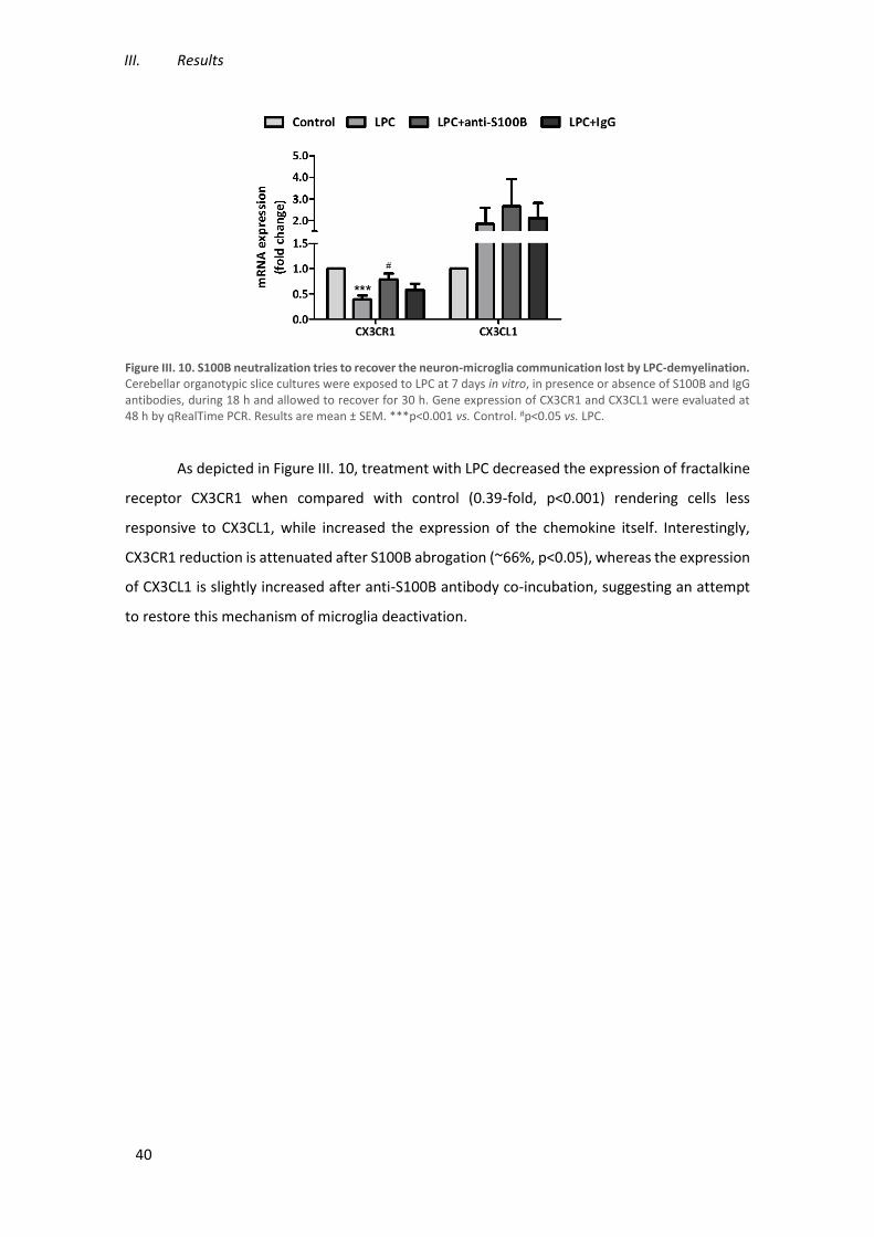
III. Results
40
Figure III. 10. S100B neutralization tries to recover the neuron-microglia communication lost by LPC-demyelination. Cerebellar organotypic slice cultures were exposed to LPC at 7 days in vitro, in presence or absence of S100B and IgG antibodies, during 18 h and allowed to recover for 30 h. Gene expression of CX3CR1 and CX3CL1 were evaluated at 48 h by qRealTime PCR. Results are mean ± SEM. ***p<0.001 vs. Control. #p<0.05 vs. LPC.
As depicted in Figure III. 10, treatment with LPC decreased the expression of fractalkine
receptor CX3CR1 when compared with control (0.39-fold, p<0.001) rendering cells less
responsive to CX3CL1, while increased the expression of the chemokine itself. Interestingly,
CX3CR1 reduction is attenuated after S100B abrogation (~66%, p<0.05), whereas the expression
of CX3CL1 is slightly increased after anti-S100B antibody co-incubation, suggesting an attempt
to restore this mechanism of microglia deactivation.

Modulation of microglia reactivity by S100B in Multiple Sclerosis
41
IV. Discussion
S100B is associated with different neurodegenerative pathologies, including MS. Indeed,
increased levels of S100B have been detected in both CSF and post-mortem demyelinated
plaques of MS patients. The development of these demyelinated lesions, one hallmark of MS,
are a result of several mechanisms including gliosis, inflammation, demyelination, axonal loss
and neuronal degeneration (Milo and Miller, 2014). Thus, with this thesis we proposed to study
the role of S100B in MS associated microglia reactivity, using an ex vivo model of demyelination
(Birgbauer et al., 2004). Firstly, we evaluated whether the expression of a protein constituent of
myelin sheaths could be recovered, in the course of the demyelinating insult with LPC by
neutralization of excessive S100B. Our results revealed that the expression of MBP mRNA was
decreased after demyelinating insult, according with Birgbauer and collaborators, which
reported a significant decrease in myelin marker expression (Birgbauer et al., 2004).
Interestingly, treatment with anti-S100B antibody prevented this demyelination, as previously
reported by us (Barateiro et al., 2015), increasing the MBP mRNA expression. These data suggest
that the neutralization of S100B prevented an exacerbated demyelination by keeping S100B
concentration close to physiological levels and, therefore, having a protective role on
demyelinating event.
In healthy CNS, microglial cells, which are constantly patrolling the parenchyma, are
characterized by a ramified morphology and low levels of CD11b and F4/80,
macrophage/microglia activation marker, and CD45 and CD86, M1 markers (Devarajan et al.,
2014; Ponomarev et al., 2013). As we known, microglia become active during CNS inflammation,
acquiring phenotypic and morphologic changes in response to inflammation. These activated
microglia are generally characterized as CD11bhigh/CD86high/F4/80high and CD45dim high (Devarajan
et al., 2014; Ponomarev et al., 2005, 2013). It is difficult to differentiate CNS resident microglia
from peripheral macrophages because they present a similar pattern, however, our
demyelinating ex vivo model does not involve these peripheral type of cells. Accordingly with
IV. Discussion
42
previous studies, we observed an increase of activated microglia population upon
demyelination, observed through CD11b augment, which suggests their increased ability to
respond to an injury. Once having confirmed the activation of microglia after a demyelinated
insult, we evaluated their phenotype analyzing the expression of M1 markers and we only
observed an upregulation of CD86 in response to LPC stimulation. Notably, the effect of S100B
neutralization diminished CD86 cell surface expression suggesting a reduction of this pro-
inflammatory phenotype. Ponomarev and collaborators, reported a decrease in the expression
of reactive marker CD11b when microglia acquire the M2 phenotype (Ponomarev et al., 2013),
our results showed that neutralization of S100B promoted a reduction in CD11b levels
expression suggesting a shift of microglia phenotype into a more anti-inflammatory one. In turn,
we also analyzed other M1 markers, such as F4/80 and CD45, which did not statistically differ
between conditions, indicating that in our demyelinating ex vivo model those are not the best
markers to detect changes in microglia phenotype. Moreover, the population of microglia
expressing CD45 was very low, corroborating the concept that the expression of this marker may
distinguish between CD45dim hi microglia from CD45high macrophages.
Although they are often referred to have a “resting” phenotype under healthy
conditions, microglia cells are constantly patrolling their milieu and, therefore, their activation
due to any damage, may contribute to an inflammatory environment by the secretion of
inflammatory factors. According to Donato and collaborators which reported an increase of pro-
inflammatory cytokines release with high S100B levels (Donato et al., 2013), our results showed
an increase in the expression of first-line cytokines TNF-α and IL-1β upon a demyelinated insult,
in parallel to an increased S100B expression as previously reported by us (Barateiro et al., 2015).
Moreover, our data also showed that S100B blockade prevents this cytokine increase. Once
these cytokines are mainly released by activated microglia, these results suggest a microglial
shift from a pro-inflammatory phenotype to less inflammatory one. In addition, our results
showed a demyelination-induced decrease in pro-inflammatory IL-6 mRNA expression that
returned to control levels following S100B neutralization. In spite of the large number of studies
that reported the role of IL-6 in MS pathology, there is still a great controversy. As already
mentioned, several studies reported an increase in IL-6 mRNA expression in chronic lesions
(Maimone et al., 1997; Woodroofe and Cuzner, 1993), CSF (Navikas et al., 1996) and in
peripheral blood monocytes (Frei et al., 1991) from MS patients, showing that this cytokine is
involved in MS progression. However, there are studies that particularly shown low IL-6 levels
in chronic inactive lesions and in plaques with oligodendrocyte loss (Schönrock et al., 2000),
Modulation of microglia reactivity by S100B in Multiple Sclerosis
43
suggesting that the reduced IL-6 expression upon demyelination, observed in our results, may
be associated with oligodendrocyte damage/death (Bø et al., 2013).
During MS pathophysiology microglia are activated in response to demyelination and
release different inflammatory cytokines. Recently, some studies suggest the involvement of
NLRP3 inflammasome on MS and EAE progression (Inoue and Shinohara, 2013). As mentioned
above, inflammasomes are involved in maturation and release of cytokines. Indeed, IL-1β,
HMGB1 and IL-18 are cytokines matured by NLRP3 inflammasome. HMGB1, a cytokine involved
in the start of CNS inflammatory response, is increased in MS and EAE lesions (Andersson et al.,
2008). IL-18, a pro-inflammatory cytokine has also been revealed to be increased in MS patients
(Sedimbi et al., 2013). Corroborating these studies, our results showed an increased expression
of NLRP3, HMGB1 and IL-18 upon LPC-induced demyelination. Interestingly, there are a manifest
reduction of inflammasome-related molecules by neutralization of S100B, indicating that
excessive S100B expression following demyelination is not only exacerbating cytokine
expression but also their maturation into active forms.
In the activated state, microglia exhibit an upregulation of several markers according to
the acquired phenotype. Particularly, in response to demyelination, pro-inflammatory microglia
may increase the expression of several inflammatory markers, including MHC-II, iNOS and CEBP-
α. It has been demonstrated that classically activated microglia increased the expression of
CEBP-α after brain injury, indicating that this transcription factor may regulate microglial
function during brain damage (Walton et al., 1998). Also, Ponomarev and collaborators showed,
in EAE model, an augmented expression of CEBP-α associated with the upregulation of other M1
markers, such as MHC-II and iNOS (Ponomarev et al., 2011). Our ex vivo model also showed an
increase of these pro-inflammatory markers in response to LPC thus corroborating these
findings. Interestingly, the inhibition of excessive S100B in the milieu appears to be effective in
the prevention of the expression of these cytokines, corroborating once again that S100B
blockade reduces M1 pro-inflammatory microglia phenotype.
MS is a chronic disorder where activated microglia play an important immune response.
Microglia sensor danger/stranger signals activating the TLRs (Glass et al., 2010). Once TLRs are
engaged, it initiates a cascade of events culminating into the activation of the transcription
factor NF-κB and contribute to inflammation (Kettenmann et al., 2011). In this context, TLRs
have been shown to be implicated in several CNS diseases including MS (Miranda-Hernandez
and Baxter, 2013). Our results showed an augment of TLR4 but not of TLR2 gene expression
upon demyelinated conditions, indicating that in our model TLR4 is possibly more responsible
for microglia activation than TLR2. Moreover, the treatment with anti-S100B prevented the
IV. Discussion
44
increase of TLR4 and maintained the TLR2 expression at control levels, indicating that excessive
S100B may also be exacerbating inflammation through TLR4.
In turn, anti-inflammatory microglia, usually found in initial remyelination, are
characterized by upregulated Arg1, FIZZ-1, TGF-β and SOCS-1, amongst many other markers
(Chhor et al., 2013; Correale, 2014; David and Kroner, 2011; Hu et al., 2014; Ponomarev et al.,
2013; Wilson, 2014). In accordance with these previous studies, 48 h after a demyelinating insult
there are a marked upregulation of Arg1 and FIZZ-1 gene expression. Curiously, S100B
neutralization did not significantly changes these levels suggesting a maintenance of M2
phenotype in the presence of low levels of S100B protein. However, TGF-β and SOCS-1
expression was almost completed abrogated by the inhibition of S100B. Besides the important
immunosuppressive role of TGF-β described in relapsing EAE and MS, where it prevents T cells
from entering into CNS, there are evidences that this cytokine can increase Th17 production in
inflammatory tissues further promoting pro-inflammatory gene expression in CNS tissue
(Mirshafiey and Mohsenzadegan, 2009). So, the reduction of TGF-β levels observed by S100B
neutralization may suggest an important involvement of S100B in Th17 response and
recruitment to the CNS, which should be further elucidated in in vivo systems. In addition,
despite of microglia phenotypes have been considered separable stages, different populations
with distinct phenotypes are simultaneously present upon inflammatory conditions. As already
mentioned, SOCS-1 inhibits the expression of pro-inflammatory markers at high levels (Davey et
al., 2006). In addition, as we already demonstrated, there is an increase of several pro-
inflammatory markers expression upon demyelination. However, in accordance with Kakhki and
collaborators that showed an increase of SOCS-1 expression in MS patients, our results revealed
that SOCS-1 mRNA was overexpressed in a demyelinated condition (Kakhki et al., 2015). Thus,
the fact that there is an increased expression of SOCS-1 after the incubation with LPC may
indicate a response from microglia to fight the high inflammatory burden.
Phagocytosis is a crucial mechanism for clearance of pathogens, dying cells and debris
in order to maintain tissue health. Microglia are able to phagocyte either debris, including myelin
debris, and apoptotic cells that are internalized through phosphatidylserine receptors,
stimulating an anti-inflammatory response; or infectious pathogens through TLRs, inducing the
release of pro-inflammatory cytokines (Napoli and Neumann, 2010). While M1 microglia exert
cytotoxic effects on neurons, M2 microglia exhibit phagocytic capacity promoting neurite
outgrowth (Prinz and Priller, 2014). Here, we investigated the ability of microglia to phagocyte
zymosan-coated latex beads, which would mimic the phagocytosis of a pathogen. LPC treatment
reduced both the number of phagocytosed beads per cell as well as the number of phagocyting
Modulation of microglia reactivity by S100B in Multiple Sclerosis
45
microglia. These results may suggest that either microglia ability to phagocyte is reduced by LPC-
induced demyelination or that in the presence of demyelination, microglia has to phagocyte
myelin debris and are no longer able to further phagocyte the beads to which they are exposed
at the end of the 48 h post-LPC. Curiously, although S100B inhibition did not change the number
of beads that microglia can phagocyte, we verified an increase of microglial cells that
phagocytosed less beads. This data may suggest that microglia may show an enhanced
phagocytic ability upon neutralization of excessive S100B or that there is less myelin debris to
phagocyte and therefore microglia are more prone to zymosan-coated beads. To that, it would
be interesting to investigate the co-localization of myelin and microglia in our model of
demyelination to analyze the amount of myelin debris that have been already phagocytosed by
microglia during culture period to better clarify this issue.
An extensively described axis of communication between neurons and microglia is
fractalkine-CX3CL1/CX3CR1. This signaling pathway is very important in the maintenance of
microglia under a vigilant phenotype. Our results showed diminished mRNA expression of
CX3CR1 but increased gene expression of its ligand after demyelination, suggesting a reduced
response of microglia to CX3CL1. Cardona and collaborators showed that CX3CR1 deficient
microglia overexpressed IL-1β and displayed neurotoxic activity indicating that this axis might
be compromised during a demyelinated insult and that microglia can acquire an activated
phenotype (Cardona et al., 2006). Furthermore, CX3CR1 knockout mice showed increased
toxicity in response to LPS treatment and augmented neurodegeneration (Cardona et al., 2006).
Although it has already been reported an augmented expression of CX3CR1 in inflammatory
lesions of MS patients (Wollberg et al., 2014), our results suggest that in our ex vivo
demyelination model the lack of fractalkine receptor may be involved in the activation of
microglia into a pro-inflammatory phenotype releasing several inflammatory mediators and
promoting neurodegeneration. We also verified that with S100B neutralization, the CX3CR1
expression levels returned to control values, possibly as an attempt to restore this pathway,
justifying why S100B protein, at physiological levels, was reported to prevent the activation of
microglial cells (Zhang et al., 2011b). Regarding to fractalkine-CX3CL1, our results revealed an
overexpression of this gene expression levels after LPC treatment and a slight increase upon co-
incubation with anti-S100B antibody. Many neurodegenerative and inflammatory disorders that
are associated with increased microglial activation, including MS, show disruption of the
fractalkine/CX3CR1 crosstalk. Transgenic mice that develop EAE spontaneously and mice
actively immunized with proteolytic peptide display an augmented fractalkine in brain microglia
(Sheridan and Murphy, 2013). Therefore, a CNS injury, mimicked into our model by LPC

IV. Discussion
46
treatment, may be a trigger to increase CX3CL1 expression which may either contribute to
disease progression or be a mechanism by which other cells attempt to stop microglia high
reactivity shifting them to a quiescent/vigilant phenotype.
IgG co-incubation appears to have a similar effect of S100B neutralization in some cases,
which might indicate that an introduction of an IgG to the incubation medium may have an effect
by itself which may mask the intended neutralization of S100B. Therefore, it would be
interesting to investigate the role of additional S100B modulators that have been already
demonstrated to play a role in preventing S100B-related damage in cancer and experimental
traumatic brain injury, in our ex vivo model of demyelination (Capoccia et al., 2015; Kabadi et
al., 2015; Zimmer et al., 2013).
Concluding Remarks
With this thesis we demonstrated, using an ex vivo COSC model, that LPC effectively
induced demyelination through reduction of myelin protein expression and confirmed that the
anti-S100B treatment partially prevent this demyelination. Moreover, we showed that the
demyelinated insult induced an inflammatory environment resulting from the release of
inflammatory molecules by activated microglia. On the other hand, the blockade of S100B seems
to prevent the expression of pro-inflammatory mediators and promote the shift microglia to a
damage repair phenotype with the increase of phagocytic ability (Figure IV. 1), although M2-like
markers were not induced. Overall, our results strongly suggest that inhibition of excessive
S100B upon demyelination prevents both demyelination and the exacerbation of the
inflammatory milieu, possibly also affecting microglia activation and the recruitment of other
immune cell to CNS parenchyma. Further studies on in vivo MS models will better clarify whether
targeting of S100B can be a good therapeutic strategy to reduce damage and increase recovery
in MS.

Modulation of microglia reactivity by S100B in Multiple Sclerosis
47
Figure IV. 1. Schematic representation of main findings. (A) In demyelinated conditions there is an upregulation of pro-inflammatory cytokines. (B) Blocking S100B showed a reduction of pro-inflammatory environment and an increased phagocytosis.

Modulation of microglia reactivity by S100B in Multiple Sclerosis
49
V. References
Adami, C., Sorci, G., Blasi, E., Agneletti, A.L., Bistoni, F., and Donato, R. (2001). S100B Expression
in and Effects on Microglia. Glia 33, 131–142.
Afonso, V. (2014). Role of S100B on Central Nervous System. Universidade de Lisboa, Faculdade
de Farmácia.
Andersson, A., Covacu, R., Sunnemark, D., Danilov, A.I., Dal Bianco, A., Khademi, M., Wallström,
E., Lobell, A., Brundin, L., Lassmann, H., et al. (2008). HMGB1 expression in active lesions of
human and experimental multiple sclerosis. J. Leukoc. Biol. 84, 1248–1255.
Barateiro, A., Afonso, V., Santos, G., Cerqueira, J.J., Brites, D., van Horssen, J., and Fernandes, A.
(2015). S100B as a Potential Biomarker and Therapeutic Target in Multiple Sclerosis. Mol.
Neurobiol.
Bartosik-Psujek, H., Psujek, M., Jaworski, J., and Stelmasiak, Z. (2011). Total tau and S100b
proteins in different types of multiple sclerosis and during immunosuppressive treatment with
mitoxantrone. Acta Neurol. Scand. 123, 252–256.
Benarroch, E.E. (2013). Microglia: Multiple roles in surveillance, circuit shaping, and response to
injury. Neurology 81, 1079–1088.
Berger, T., and Reindl, M. (2007). Multiple sclerosis: disease biomarkers as indicated by
pathophysiology. J. Neurol. Sci. 259, 21–26.
Bianchi, R., Adami, C., Giambanco, I., and Donato, R. (2007). S100B binding to RAGE in microglia
stimulates COX-2 expression. J. Leukoc. Biol. 81, 108–118.
Bianchi, R., Giambanco, I., and Donato, R. (2010). S100B/RAGE-dependent activation of
microglia via NF-kB and AP-1 Co-regulation of COX-2 expression by S100B, IL-1β and TNF-α.
Neurobiol. Aging 31, 665–677.

V. References
50
Bianchi, R., Kastrisianaki, E., Giambanco, I., and Donato, R. (2011). S100B protein stimulates
microglia migration via RAGE-dependent up-regulation of chemokine expression and release. J.
Biol. Chem. 286, 7214–7226.
Birgbauer, E., Rao, T.S., and Webb, M. (2004). Lysolecithin induces demyelination in vitro in a
cerebellar slice culture system. J. Neurosci. Res. 78, 157–166.
Bø, L., Esiri, M., Evangelou, N., and Kuhlmann, T. (2013). Myelin Repair and Neuroprotection in
Multiple Sclerosis. In Myelin Repair and Neuroprotection in Multiple Sclerosis, I.D. Duncan, and
R.J.M. Franklin, eds. (Boston, MA: Springer US), pp. 23–46.
Bogie, J.F.J., Stinissen, P., and Hendriks, J.J. a (2014). Macrophage subsets and microglia in
multiple sclerosis. Acta Neuropathol. 128, 191–213.
Brites, D., and Vaz, A.R. (2014). Microglia centered pathogenesis in ALS: insights in cell
interconnectivity. Front. Cell. Neurosci. 8.
Capoccia, E., Cirillo, C., Marchetto, A., Tiberi, S., Sawikr, Y., Pesce, M., D’Alessandro, A., Scuderi,
C., Sarnelli, G., Cuomo, R., et al. (2015). S100B-p53 disengagement by pentamidine promotes
apoptosis and inhibits cellular migration via aquaporin-4 and metalloproteinase-2 inhibition in
C6 glioma cells. Oncol. Lett. 9, 2864–2870.
Cardona, A.E., Pioro, E.P., Sasse, M.E., Kostenko, V., Cardona, S.M., Dijkstra, I.M., Huang, D.,
Kidd, G., Dombrowski, S., Dutta, R., et al. (2006). Control of microglial neurotoxicity by the
fractalkine receptor. Nat. Neurosci. 9, 917–924.
Charil, A., and Filippi, M. (2007). Inflammatory demyelination and neurodegeneration in early
multiple sclerosis. J. Neurol. Sci. 259, 7–15.
Chhor, V., Le Charpentier, T., Lebon, S., Oré, M.-V., Celador, I.L., Josserand, J., Degos, V., Jacotot,
E., Hagberg, H., Sävman, K., et al. (2013). Characterization of phenotype markers and
neuronotoxic potential of polarised primary microglia in vitro. Brain. Behav. Immun. 32, 70–85.
Cho, S., Wood, A., and Bowlby, M. (2007). Brain Slices as Models for Neurodegenerative Disease
and Screening Platforms to Identify Novel Therapeutics. Curr. Neuropharmacol. 5, 19–33.
Ciccarelli, O., Barkhof, F., Bodini, B., Stefano, N. De, Golay, X., Nicolay, K., Pelletier, D., Pouwels,
P.J.W., Smith, S. a, Wheeler-Kingshott, C. a M., et al. (2014). Pathogenesis of multiple sclerosis:
insights from molecular and metabolic imaging. Lancet Neurol. 13, 807–822.
Compston, A., and Coles, A. (2008). Multiple sclerosis. Lancet 372, 1502–1517.

Modulation of microglia reactivity by S100B in Multiple Sclerosis
51
Correale, J. (2014). The role of microglial activation in disease progression. Mult. Scler. J. 20,
1288–1295.
Davey, G.M., Heath, W.R., and Starr, R. (2006). SOCS1: a potent and multifaceted regulator of
cytokines and cell-mediated inflammation. Tissue Antigens 67, 1–9.
David, S., and Kroner, A. (2011). Repertoire of microglial and macrophage responses after spinal
cord injury. Nat. Rev. Neurosci. 12, 388–399.
Dendrou, C.A., Fugger, L., and A, F.M. (2015). Immunopathology of multiple sclerosis. Nat. Rev.
Immunol. 15, 545–558.
Denic, A., Johnson, A.J., Bieber, A.J., Warrington, A.E., Rodriguez, M., and Pirko, I. (2011). The
relevance of animal models in multiple sclerosis research. Pathophysiology 18, 21–29.
Devarajan, G., Chen, M., Muckersie, E., and Xu, H. (2014). Culture and characterization of
microglia from the adult murine retina. ScientificWorldJournal. 2014, 894368.
Domercq, M., Sánchez-Gómez, M.V., Sherwin, C., Etxebarria, E., Fern, R., and Matute, C. (2007).
System xc- and glutamate transporter inhibition mediates microglial toxicity to
oligodendrocytes. J. Immunol. 178, 6549–6556.
Donato, R., Sorci, G., Riuzzi, F., Arcuri, C., Bianchi, R., Brozzi, F., Tubaro, C., and Giambanco, I.
(2009). S100B’s double life: intracellular regulator and extracellular signal. Biochim. Biophys.
Acta 1793, 1008–1022.
Donato, R., Cannon, B.R., Sorci, G., Riuzzi, F., Hsu, K., Weber, D., and Geczy, C.L. (2013). Functions
of S100 Proteins. Curr. Mol. Med. 13, 24–57.
Dyer, J.K., Bourque, J. a, and Steeves, J.D. (2005). The role of complement in immunological
demyelination of the mammalian spinal cord. Spinal Cord 43, 417–425.
Ellwardt, E., and Zipp, F. (2014). Molecular mechanisms linking neuroinflammation and
neurodegeneration in MS. Exp. Neurol. 262, 8–17.
Fischer, M.T., Sharma, R., Lim, J.L., Haider, L., Frischer, J.M., Drexhage, J., Mahad, D., Bradl, M.,
van Horssen, J., and Lassmann, H. (2012). NADPH oxidase expression in active multiple sclerosis
lesions in relation to oxidative tissue damage and mitochondrial injury. Brain a J. Neurol. a J.
135, 886–899.
Frei, K., Fredrikson, S., Fontana, A., and Link, H. (1991). Interleukin-6 is elevated in plasma in
multiple sclerosis. J. Neuroimmunol. 31, 147–153.

V. References
52
Friese, M. a, Schattling, B., and Fugger, L. (2014). Mechanisms of neurodegeneration and axonal
dysfunction in multiple sclerosis. Nat. Rev. Neurol. 10, 225–238.
Gähwiler, B.H. (1984). Slice cultures of cerebellar, hippocampal and hypothalamic tissue.
Experientia 40, 235–243.
Giunti, D., Parodi, B., Cordano, C., Uccelli, A., and Kerlero de Rosbo, N. (2014). Can we switch
microglia’s phenotype to foster neuroprotection? Focus on multiple sclerosis. Immunology 141,
328–339.
Glass, C.K., Saijo, K., Winner, B., Marchetto, M.C., and Gage, F.H. (2010). Mechanisms Underlying
Inflammation in Neurodegeneration. Cell 140, 918–934.
Goldmann, T., and Prinz, M. (2013). Role of Microglia in CNS Autoimmunity. Clin. Dev. Immunol.
2013.
Gonsette, R.E. (2008). Neurodegeneration in multiple sclerosis: the role of oxidative stress and
excitotoxicity. J. Neurol. Sci. 274, 48–53.
Guedes, J., Cardoso, a. L.C., and Pedroso De Lima, M.C. (2013). Involvement of MicroRNA in
microglia-mediated immune response. Clin. Dev. Immunol. 2013.
Haider, L., Fischer, M.T., Frischer, J.M., Bauer, J., Höftberger, R., Botond, G., Esterbauer, H.,
Binder, C.J., Witztum, J.L., and Lassmann, H. (2011). Oxidative damage in multiple sclerosis
lesions. Brain 134, 1914–1924.
Hametner, S., Wimmer, I., Haider, L., Pfeifenbring, S., Brück, W., and Lassmann, H. (2013). Iron
and neurodegeneration in the multiple sclerosis brain. Ann. Neurol. 74, 848–861.
Harrison, J.K., Jiang, Y., Chen, S., Xia, Y., Maciejewski, D., McNamara, R.K., Streit, W.J., Salafranca,
M.N., Adhikari, S., Thompson, D. a, et al. (1998). Role for neuronally derived fractalkine in
mediating interactions between neurons and CX3CR1-expressing microglia. Proc. Natl. Acad. Sci.
U. S. A. 95, 10896–10901.
Hein Née Maier, K., Köhler, A., Diem, R., Sättler, M.B., Demmer, I., Lange, P., Bähr, M., and Otto,
M. (2008). Biological markers for axonal degeneration in CSF and blood of patients with the first
event indicative for multiple sclerosis. Neurosci. Lett. 436, 72–76.
Heppner, F.L., Greter, M., Marino, D., Falsig, J., Raivich, G., Hövelmeyer, N., Waisman, A., Rülicke,
T., Prinz, M., Priller, J., et al. (2005). Experimental autoimmune encephalomyelitis repressed by
microglial paralysis. Nat. Med. 11, 146–152.
Modulation of microglia reactivity by S100B in Multiple Sclerosis
53
Herz, J., Zipp, F., and Siffrin, V. (2010). Neurodegeneration in autoimmune CNS inflammation.
Exp. Neurol. 225, 9–17.
Hild, W. (1956). Myelin formation in central nervous system tissue cultures. Verh. Anat. Ges. 53,
315–317.
Hoek, R.M. (2000). Down-Regulation of the Macrophage Lineage Through Interaction with OX2
(CD200). Science (80-. ). 290, 1768–1771.
Van Horssen, J., Schreibelt, G., Drexhage, J., Hazes, T., Dijkstra, C.D., van der Valk, P., and de
Vries, H.E. (2008). Severe oxidative damage in multiple sclerosis lesions coincides with enhanced
antioxidant enzyme expression. Free Radic. Biol. Med. 45, 1729–1737.
Van Horssen, J., Singh, S., van der Pol, S., Kipp, M., Lim, J.L., Peferoen, L., Gerritsen, W., Kooi, E.-
J., Witte, M.E., Geurts, J.J., et al. (2012). Clusters of activated microglia in normal-appearing
white matter show signs of innate immune activation. J. Neuroinflammation 9.
Hu, J., Castets, F., Guevara, J.L., and Van Eldik, L.J. (1996). S100β stimulates inducible nitric oxide
synthase activity and mRNA levels in rat cortical astrocytes. J. Biol. Chem. 271, 2543–2547.
Hu, X., Leak, R.K., Shi, Y., Suenaga, J., Gao, Y., Zheng, P., and Chen, J. (2014). Microglial and
macrophage polarization-new prospects for brain repair. Nat. Rev. Neurol. 11, 56–64.
Inoue, M., and Shinohara, M.L. (2013). NLRP3 Inflammasome and MS/EAE. Autoimmune Dis.
2013.
Jack, C., Ruffini, F., Bar-Or, A., and Antel, J.P. (2005). Microglia and multiple sclerosis. J. Neurosci.
Res. 81, 363–373.
Jones, R.S., and Lynch, M. a (2014). How dependent is synaptic plasticity on microglial
phenotype? Neuropharmacology 96, 3–10.
Kabadi, S. V, Stoica, B. a, Zimmer, D.B., Afanador, L., Duffy, K.B., Loane, D.J., and Faden, A.I.
(2015). S100B inhibition reduces behavioral and pathologic changes in experimental traumatic
brain injury. J. Cereb. Blood Flow Metab. 1–11.
Kakhki, M.P., Rakhshi, N., Heidary, M., Behmanesh, M., and Nikravesh, A. (2015). Expression of
suppressor of cytokine signaling 1 (SOCS1) gene dramatically increases in relapsing-remitting
multiple sclerosis. J. Neurol. Sci. 350, 40–45.
Kettenmann, H., Hanisch, U.-K., Noda, M., and Verkhratsky, A. (2011). Physiology of microglia.
Physiol. Rev. 91, 461–553.

V. References
54
Könnecke, H., and Bechmann, I. (2013). The role of microglia and matrix metalloproteinases
involvement in neuroinflammation and gliomas. Clin. Dev. Immunol. 2013.
Koppal, T., Lam, A.G.M., Guo, L., and Eldik, L.J. Van (2001). S100B proteins that lack one or both
cysteine residues can induce inflammatory responses in astrocytes and microglia. Neurochem.
Int. 39, 401–407.
Lassmann, H. (2010). Axonal and neuronal pathology in multiple sclerosis: what have we learnt
from animal models. Exp. Neurol. 225, 2–8.
Lassmann, H. (2011). Pathophysiology of inflammation and tissue injury in multiple sclerosis:
what are the targets for therapy. J. Neurol. Sci. 306, 167–169.
Lassmann, H. (2013). Multiple sclerosis: Lessons from molecular neuropathology. Exp. Neurol.
262, 2–7.
Lassmann, H., and van Horssen, J. (2011). The molecular basis of neurodegeneration in multiple
sclerosis. FEBS Lett. 585, 3715–3723.
Lassmann, H., van Horssen, J., and Mahad, D. (2012). Progressive multiple sclerosis: pathology
and pathogenesis. Nat. Rev. Neurol. 8, 647–656.
Levi, G., and Meyer, H. (1941). Nouvelles recherches sur le tissu nerveux cultivé in vitro.
Morphologie, croissance et relations réciproques des neurones. Arch. Biol. 52, 133–278.
Lindberg, R.L.P., and Kappos, L. (2006). Transcriptional profiling of multiple sclerosis: towards
improved diagnosis and treatment. Expert Rev. Mol. Diagn. 6, 843–855.
Liu, Y., Stewart, K.N., Bishop, E., Marek, C.J., Kluth, D.C., Rees, A.J., and Wilson, H.M. (2008).
Unique expression of suppressor of cytokine signaling 3 is essential for classical macrophage
activation in rodents in vitro and in vivo. J. Immunol. 180, 6270–6278.
Love, S. (2006). Demyelinating diseases. J. Clin. Pathol. 59, 1151–1159.
Lu, B., Wang, H., Andersson, U., and Tracey, K.J. (2013). Regulation of HMGB1 release by
inflammasomes. Protein Cell 4, 163–167.
Lu, F., Selak, M., O’Connor, J., Croul, S., Lorenzana, C., Butunoi, C., and Kalman, B. (2000).
Oxidative damage to mitochondrial DNA and activity of mitochondrial enzymes in chronic active
lesions of multiple sclerosis. J. Neurol. Sci. 177, 95–103.
Maimone, D., Guazzi, G.C., and Annunziata, P. (1997). IL-6 detection in multiple sclerosis brain.
J. Neurol. Sci. 146, 59–65.
Modulation of microglia reactivity by S100B in Multiple Sclerosis
55
Mao, P., and Reddy, P.H. (2010). Is multiple sclerosis a mitochondrial disease? Biochim. Biophys.
Acta 1802, 66–79.
Mathew, A., Pakan, J.M.P., Collin, E.C., Wang, W., McDermott, K.W., Fitzgerald, U., Reynolds, R.,
and Pandit, A.S. (2013). An ex-vivo multiple sclerosis model of inflammatory demyelination using
hyperbranched polymer. Biomaterials 34, 5872–5882.
Michetti, F., Massaro, A., and Murazio, M. (1979). The nervous system-specific S-100 antigen in
cerebrospinal fluid of multiple sclerosis patients. Neurosci. Lett. 11, 171–175.
Michetti, F., Corvino, V., Geloso, M.C., Lattanzi, W., Bernardini, C., Serpero, L., and Gazzolo, D.
(2012). The S100B protein in biological fluids: more than a lifelong biomarker of brain distress.
J. Neurochem. 120, 644–659.
Milo, R., and Miller, A. (2014). Revised diagnostic criteria of multiple sclerosis. Autoimmun. Rev.
13, 518–524.
Miranda-Hernandez, S., and Baxter, A.G. (2013). Role of toll-like receptors in multiple sclerosis.
Am. J. Clin. Exp. Immunol. 2, 75–93.
Miron, V.E., Ludwin, S.K., Darlington, P.J., Jarjour, A. a, Soliven, B., Kennedy, T.E., and Antel, J.P.
(2010). Fingolimod (FTY720) enhances remyelination following demyelination of organotypic
cerebellar slices. Am. J. Pathol. 176, 2682–2694.
Miron, V.E., Boyd, A., Zhao, J.-W., Yuen, T.J., Ruckh, J.M., Shadrach, J.L., van Wijngaarden, P.,
Wagers, A.J., Williams, A., Franklin, R.J.M., et al. (2013). M2 microglia and macrophages drive
oligodendrocyte differentiation during CNS remyelination. Nat. Neurosci. 16, 1211–1218.
Mirshafiey, A., and Mohsenzadegan, M. (2009). TGF-β as a promising option in the treatment of
multiple sclerosis. Neuropharmacology 56, 929–936.
Murta, V., and Ferrari, C.C. (2013). Influence of Peripheral inflammation on the progression of
multiple sclerosis: evidence from the clinic and experimental animal models. Mol. Cell. Neurosci.
53, 6–13.
Napoli, I., and Neumann, H. (2010). Protective effects of microglia in multiple sclerosis. Exp.
Neurol. 225, 24–28.
Navikas, V., Matusevicius, D., Söderström, M., Fredrikson, S., Kivisäkk, P., Ljungdahl, A.,
Höjeberg, B., and Link, H. (1996). Increased interleukin-6 mRNA expression in blood and
cerebrospinal fluid mononuclear cells in multiple sclerosis. J Neuroimmunol 64, 63–69.

V. References
56
Olah, M., Biber, K., Vinet, J., and Boddeke, H.W.G.M. (2011a). Microglia phenotype diversity.
CNS Neurol. Disord. Drug Targets 10, 108–118.
Olah, M., Biber, K., Vinet, J., and Boddeke, H.W.G.M. (2011b). Microglia phenotype diversity.
CNS Neurol. Disord. Drug Targets 10, 108–118.
Ostendorp, T., Leclerc, E., Galichet, A., Koch, M., Demling, N., Weigle, B., Heizmann, C.W.,
Kroneck, P.M.H., and Fritz, G. (2007). Structural and functional insights into RAGE activation by
multimeric S100B. EMBO J. 26, 3868–3878.
Perry, V.H., and Teeling, J. (2013). Microglia and macrophages of the central nervous system:
the contribution of microglia priming and systemic inflammation to chronic neurodegeneration.
Semin. Immunopathol. 35, 601–612.
Perry, V.H., Nicoll, J. a R., and Holmes, C. (2010). Microglia in neurodegenerative disease. Nat.
Rev. Neurol. 6, 193–201.
Petzold, A., Eikelenboom, M.J., Gveric, D., Keir, G., Chapman, M., Lazeron, R.H.C., Cuzner, M.L.,
Polman, C.H., Uitdehaag, B.M.J., Thompson, E.J., et al. (2002). Markers for different glial cell
responses in multiple sclerosis: clinical and pathological correlations. Brain 125, 1462–1473.
Piccio, L., Buonsanti, C., Mariani, M., Cella, M., Gilfillan, S., Cross, A.H., Colonna, M., and Panina-
Bordignon, P. (2007). Blockade of TREM-2 exacerbates experimental autoimmune
encephalomyelitis. Eur. J. Immunol. 37, 1290–1301.
Pitt, D., Werner, P., and Raine, C.S. (2000). Glutamate excitotoxicity in a model of multiple
sclerosis. Nat. Med. 6, 67–70.
Ponomarev, E.D., Shriver, L.P., Maresz, K., and Dittel, B.N. (2005). Microglial cell activation and
proliferation precedes the onset of CNS autoimmunity. J. Neurosci. Res. 81, 374–389.
Ponomarev, E.D., Veremeyko, T., Barteneva, N., Krichevsky, A.M., and Weiner, H.L. (2011).
MicroRNA-124 promotes microglia quiescence and suppresses EAE by deactivating
macrophages via the C/EBP-α-PU.1 pathway. Nat. Med. 17, 64–70.
Ponomarev, E.D., Veremeyko, T., and Weiner, H.L. (2013). MicroRNAs are universal regulators
of differentiation, activation, and polarization of microglia and macrophages in normal and
diseased CNS. Glia 61, 91–103.
Prinz, M., and Priller, J. (2014). Microglia and brain macrophages in the molecular age: from
origin to neuropsychiatric disease. Nat. Rev. Neurosci. 15, 300–312.

Modulation of microglia reactivity by S100B in Multiple Sclerosis
57
Ransohoff, R.M. (2012). Animal models of multiple sclerosis: the good, the bad and the bottom
line. Nat. Neurosci. 15, 1074–1077.
Reali, C., Scintu, F., Pillai, R., Donato, R., Michetti, F., and Sogos, V. (2005). S100B Counteracts
Effects of the Neurotoxicant Trimethyltin on Astrocytes and Microglia. 686, 677–686.
Rosenberg, G. a (1995). Matrix Metalloproteinases in Brain Injury. J. Neurotrauma 12, 833–842.
Rustandi, R.R., Baldisseri, D.M., and Weber, D.J. (2000). letters Structure of the negative
regulatory domain of p53 bound to S100B ( ββ ). Nat. Struct. Biol. 7.
Schönrock, L.M., Gawlowski, G., and Brück, W. (2000). Interleukin-6 expression in human
multiple sclerosis lesions. Neurosci. Lett. 294, 45–48.
Sedimbi, S.K., Hägglöf, T., and Karlsson, M.C.I. (2013). IL-18 in inflammatory and autoimmune
disease. Cell. Mol. Life Sci. 70, 4795–4808.
Shashoua, V.E., Hesse, G.W., and Moore, B.W. (1984). Proteins of the brain extracellular fluid:
evidence for release of S-100 protein. J. Neurochem. 42, 1536–1541.
Sheridan, G.K., and Murphy, K.J. (2013). Neuron-glia crosstalk in health and disease: fractalkine
and CX3CR1 take centre stage. Open Biol. 3, 130181.
Sorci, G., Bianchi, R., Riuzzi, F., Tubaro, C., Arcuri, C., Giambanco, I., and Donato, R. (2010). S100B
Protein, A Damage-Associated Molecular Pattern Protein in the Brain and Heart, and Beyond.
Cardiovasc. Psychiatry Neurol. 2010.
Sorci, G., Riuzzi, F., Arcuri, C., Tubaro, C., Bianchi, R., Giambanco, I., and Do-, R. (2013). S100B
protein in tissue development, repair and regeneration. World J. Biol. Chem. 4, 1–12.
Sparvero, L.J., Asafu-Adjei, D., Kang, R., Tang, D., Amin, N., Im, J., Rutledge, R., Lin, B., Amoscato,
A. a, Zeh, H.J., et al. (2009). RAGE (Receptor for Advanced Glycation Endproducts), RAGE ligands,
and their role in cancer and inflammation. J. Transl. Med. 7.
Srinivasan, R., Sailasuta, N., Hurd, R., Nelson, S., and Pelletier, D. (2005). Evidence of elevated
glutamate in multiple sclerosis using magnetic resonance spectroscopy at 3 T. Brain 128, 1016–
1025.
Stadelmann, C., Wegner, C., and Brück, W. (2011). Inflammation, demyelination, and
degeneration - recent insights from MS pathology. Biochim. Biophys. Acta 1812, 275–282.
Stoppini, L., Buchs, P. a, and Muller, D. (1991). A simple method for organotypic cultures of
nervous tissue. J. Neurosci. Methods 37, 173–182.

V. References
58
Takaki, J., Fujimori, K., Miura, M., Suzuki, T., Sekino, Y., and Sato, K. (2012). L-glutamate released
from activated microglia downregulates astrocytic L-glutamate transporter expression in
neuroinflammation: the “collusion” hypothesis for increased extracellular L-glutamate
concentration in neuroinflammation. J. Neuroinflammation 9, 275.
Vainchtein, I.D., Vinet, J., Brouwer, N., Brendecke, S., Biagini, G., Biber, K., Boddeke, H.W.G.M.,
and Eggen, B.J.L. (2014). In acute experimental autoimmune encephalomyelitis, infiltrating
macrophages are immune activated, whereas microglia remain immune suppressed. Glia 62,
1724–1735.
Varnum, M.M., and Ikezu, T. (2012). The classification of microglial activation phenotypes on
neurodegeneration and regeneration in Alzheimer’s disease brain. Arch. Immunol. Ther. Exp.
(Warsz). 60, 251–266.
Vercellino, M., Merola, A., Piacentino, C., Votta, B., Capello, E., Mancardi, G.L., Mutani, R.,
Giordana, M.T., and Cavalla, P. (2007). Altered glutamate reuptake in relapsing-remitting and
secondary progressive multiple sclerosis cortex: correlation with microglia infiltration,
demyelination, and neuronal and synaptic damage. J. Neuropathol. Exp. Neurol. 66, 732–739.
Villarreal, A., Seoane, R., González Torres, A., Rosciszewski, G., Angelo, M.F., Rossi, A., Barker, P.
a, and Ramos, A.J. (2014). S100B protein activates a RAGE-dependent autocrine loop in
astrocytes: implications for its role in the propagation of reactive gliosis. J. Neurochem. 190–
205.
Vogel, D.Y.S., Vereyken, E.J.F., Glim, J.E., Heijnen, P.D. a M., Moeton, M., van der Valk, P., Amor,
S., Teunissen, C.E., van Horssen, J., and Dijkstra, C.D. (2013). Macrophages in inflammatory
multiple sclerosis lesions have an intermediate activation status. J. Neuroinflammation 10, 35.
Walton, M., Saura, J., Young, D., Macgibbon, G., Hansen, W., Lawlor, P., Sirimanne, E., Gluckman,
P., and Dragunow, M. (1998). CCAAT-enhancer binding protein a is expressed in activated
microglial cells after brain injury. Brain Res. Mol. Brain Res. 61, 11–22.
Waxman, S.G. (2008). Axonal Dysfunction in Chronic Multiple Sclerosis: Meltdown in the
Membrane. Ann. Neurol. 63, 411–413.
Wilson, H.M. (2014). SOCS proteins in macrophage polarization and function. Front. Immunol.
5.
Witte, M.E., Geurts, J.J.G., de Vries, H.E., van der Valk, P., and van Horssen, J. (2010).
Mitochondrial dysfunction: a potential link between neuroinflammation and
neurodegeneration? Mitochondrion 10, 411–418.
Modulation of microglia reactivity by S100B in Multiple Sclerosis
59
Witte, M.E., Nijland, P.G., Drexhage, J. a R., Gerritsen, W., Geerts, D., Van Het Hof, B., Reijerkerk,
A., De Vries, H.E., Van Der Valk, P., and Van Horssen, J. (2013). Reduced expression of PGC-1α
partly underlies mitochondrial changes and correlates with neuronal loss in multiple sclerosis
cortex. Acta Neuropathol. 125, 231–243.
Witte, M.E., Mahad, D.J., Lassmann, H., and van Horssen, J. (2014). Mitochondrial dysfunction
contributes to neurodegeneration in multiple sclerosis. Trends Mol. Med. 20, 179–187.
Wollberg, A.R., Ericsson-Dahlstrand, A., Juréus, A., Ekerot, P., Simon, S., Nilsson, M., Wiklund, S.-
J., Berg, A.-L., Ferm, M., Sunnemark, D., et al. (2014). Pharmacological inhibition of the
chemokine receptor CX3CR1 attenuates disease in a chronic-relapsing rat model for multiple
sclerosis. Proc. Natl. Acad. Sci. U. S. A. 111, 5409–5414.
Woodroofe, M.N., and Cuzner, M.L. (1993). Cytokine mRNA expression in inflammatory multiple
sclerosis lesions: detection by non-radioactive in situ hybridization. Cytokine 5, 583–588.
Young, E. a, Fowler, C.D., Kidd, G.J., Chang, A., Rudick, R., Fisher, E., and Trapp, B.D. (2008).
Imaging correlates of decreased axonal Na+/K+ ATPase in chronic multiple sclerosis lesions. Ann.
Neurol. 63, 428–435.
Zhang, H., Jarjour, A. a, Boyd, A., and Williams, A. (2011a). Central nervous system remyelination
in culture - a tool for multiple sclerosis research. Exp. Neurol. 230, 138–148.
Zhang, L., Liu, W., Alizadeh, D., Zhao, D., Farrukh, O., Badie, S.A., and Badie, B. (2011b). S100B
Attenuates Microglia Activation in Gliomas: Possible Role of STAT3 Pathway. Glia 59, 486–498.
Zimmer, D.B., Lapidus, R.G., and Weber, D.J. (2013). In Vivo Screening of S100B Inhibitors for
Melanoma Therapy. In Calcium-Binding Proteins and RAGE: From Structural Basics to Clinical
Applications, C.W. Heizmann, ed. pp. 201–213.
Zujovic, V., Benavides, J., Vigé, X., Carter, C., and Taupin, V. (2000). Fractalkine modulates TNF-
α secretion and neurotoxicity induced by microglial activation. Glia 29, 305–315.