Dissertação Final em PDF
56
FUNDAÇÃO UNIVERSIDADE FEDERAL DO PAMPA PROGRAMA DE PÓS-GRADUAÇÃO EM CIÊNCIAS FARMACÊUTICAS CAMILA MARTINS GÜEZ “AVALIAÇÃO DOS EFEITOS DO EXTRATO HIDROALCOÓLICO DO MANJERICÃO (Ocimum basilicum L.) SOBRE PARÂMETROS OXIDATIVOS, INFLAMATÓRIOS E GENOTOXICOLÓGICOS EM CULTURA DE LEUCÓCITOS HUMANOS” DISSERTAÇÃO DE MESTRADO Uruguaiana, RS, Brasil. 2014
Transcript of Dissertação Final em PDF

FUNDAÇÃO UNIVERSIDADE FEDERAL DO PAMPA
PROGRAMA DE PÓS-GRADUAÇÃO EM CIÊNCIAS FARMACÊUTICAS
CAMILA MARTINS GÜEZ
“AVALIAÇÃO DOS EFEITOS DO EXTRATO HIDROALCOÓLICO
DO MANJERICÃO (Ocimum basilicum L.) SOBRE PARÂMETROS
OXIDATIVOS, INFLAMATÓRIOS E GENOTOXICOLÓGICOS EM
CULTURA DE LEUCÓCITOS HUMANOS”
DISSERTAÇÃO DE MESTRADO
Uruguaiana, RS, Brasil.
2014

II
CAMILA MARTINS GÜEZ
“AVALIAÇÃO DOS EFEITOS DO EXTRATO HIDROALCOÓLICO
DO MANJERICÃO (Ocimum basilicum L.) SOBRE PARÂMETROS
OXIDATIVOS, INFLAMATÓRIOS E GENOTOXICOLÓGICOS EM
CULTURA DE LEUCÓCITOS HUMANOS”
Dissertação apresentada ao programa
de Pós graduação Stricto Sensu em
Ciências Farmacêuticas da
Universidade Federal do Pampa,
como requisito parcial para obtenção
do Título de Mestre em Ciências
Farmacêuticas.
.
Orientador: Prof. Dr. Michel Mansur Machado
Co-Orientador: Prof. Dr. Luís Flávio Souza de Oliveira
Uruguaiana
2014

III
IV
AGRADECIMENTOS
Primeiramente aos meus pais, por sempre depositarem confiança e terem
proporcionado os melhores alicerces na construção do meu futuro, minha mãe e meu
eterno pai com seus ensinamentos que jamais serão levados ou esquecidos.
Aos queridos orientadores, Michel Mansur Machado e Luís Flávio Souza de
Oliveira, pelo profissionalismo, ensinamentos constantes e disponibilidade para resolver
qualquer problema relacionado ao trabalho, além da amizade que construímos ao passar
desse tempo. Serei sempre grata pelo apoio fornecido e saibam que tenho vocês como
grandes exemplos.
Aos colegas do laboratório NUBIOTOXIM, que se tornaram amigos e parceiros.
Obrigada por surgirem no meu caminho com palavras de apoio, ideias, muita ajuda e
colaboração no desenvolvimento do trabalho. Com certeza a parcela de vocês para a
realização deste, foi fundamental e de grande importância.
À Universidade Federal de Santa Maria, e ao Laboratório de Química
Farmacêutica, Profª. Drª. Margareth Linde Athayde e sua doutoranda Aline Augusti
Boligon, pela disponibilidade e ajuda na realização de parte dos testes.
Aos professores da banca, pela disponibilidade em fazer a leitura do trabalho de
dissertação do mestrado.
A todos que de alguma maneira ou outra ajudaram na elaboração e execução
deste trabalho.

V
RESUMO
AVALIAÇÃO DOS EFEITOS DO EXTRATO HIDROALCOÓLICO
DO MANJERICÃO (Ocimum basilicum L.) SOBRE PARÂMETROS
OXIDATIVOS, INFLAMATÓRIOS E GENOTOXICOLÓGICOS EM
CULTURA DE LEUCÓCITOS HUMANOS
A utilização de produtos naturais na medicina popular como recurso
terapêutico é uma tendência generalizada pela população brasileira. Esta tendência tem
contribuído significativamente para o consumo não só de produtos naturais, como
também de medicamentos fitoterápicos. Neste contexto, o Manjericão (Ocimum
basilicum L.) surge como um destes recursos terapêuticos, sendo amplamente utilizado
como planta ornamental e como condimento. Originário da Ásia tropical, hoje é
cultivado no mundo todo. Entre os usos populares do O. basilicum L., está o emprego
como antioxidante, anti-inflamatório, anti-cancerígeno, antimicrobiano, agente
cardiovascular, fatores associados ao envelhecimento, dentre outros. Diversos
metabólitos secundários com atividade antioxidante e anti-inflamatória já foram
identificados nesta espécie e trabalhos relatam que o Ácido Rosmarínico é o composto
biologicamente mais ativo. Sabendo da importância econômica e da enorme difusão
mundial de seus usos, tanto na culinária, quanto na medicina popular, torna-se
importante a comprovação dos efeitos do Manjericão, visando garantir sua eficácia e
sua segurança, já que estudos de toxicidade para esta espécie são raros e usualmente não
abordam aspectos genéticos. Este trabalho, portanto, teve como objetivo a avaliação da
atividade antioxidante pelo teste da peroxidação lipídica, conteúdo de proteína
carbonilada, vitamina C; atividade das enzimas Superóxido dismutase (SOD) e Catalase
(CAT); análise dos parâmetros inflamatórios pela da avaliação de citocinas anti-
inflamatórias e pró-inflamatórias, e a toxicidade genética do extrato hidroalcoólico
frente a leucócitos humanos em cultura celular, através da viabilidade e proliferação
celular, ensaio cometa, teste de micronúcleo e instabilidade cromossômica. Observou-se
que o extrato etanólico de O. basilicum agiu como agente antioxidante
reduzindo/revertendo os efeitos causados pelo peróxido de hidrogênio, ações essas que
podem ser explicadas pela composição rica em polifenois e flavonoides do extrato, além
de seu conteúdo de ácido rosmarínico, um importante antioxidante com atividade
comprovada e descrita na literatura. Em relação aos parâmetros genotoxicológicos, para
todos os testes, os resultados foram dose-dependentes. Enquanto que para os parâmetros
inflamatórios, o extrato apresentou capacidade anti-inflamatória, onde o possível
mecanismo envolvido ocorre pela interação de inibição das citocinas mediadoras pró-
inflamatórias e o estímulo de citocinas mediadoras anti-inflamatórias.
Palavras chave: Radicais Livres, manjericão, anti-inflamatória, estresse oxidativo,
genotoxicologia.
VI
ABSTRACT
EVALUATION OF THE EFFECTS OF HYDROALCOHOLIC EXTRACT OF
BASIL (Ocimum basilicum L.) ON OXIDATIVE, INFLAMMATORY AND
GENOTOXIC PARAMETERS IN HUMAN CULTURE LEUKOCYTE
The use of natural products in folk medicine as a treatment method is a genereal ternd
for the Brazilian population. This trend has contributed significantly to the consumption
not only of natural products, as well as herbal medicines. In this context, Basil (Ocimum
basilicum L.) arises as one of these therapeutical resources. Basil is widely used as an
ornamental plant and as a condiment. Original in tropical Asia, is now grown
worldwide. Among the popular uses of Ocimum basilicum L., its employment as an
antioxidant, anti-inflammatory, anti-carcinogenic, anti-microbial, cardiovascular agent,
factors associated with aging, between others. Several secondary metabolites with
antioxidant and anti-inflammatory activity have been identified in this species and
several studies have reported that rosmarinic acid is the most biologically active
compound. Knowing the economic importance and worldwide dissemination of their
uses, both in cooking, as in folk medicine, it is important to prove the effects of Basil, to
ensure its efficacy and safety, as toxicicty studies for this species are rare and usually do
not approach genetic aspects. This study evaluated the antioxidant activity by testing
lipid peroxidation, protein carbonyl content, vitamin C; activity of superoxide dismutase
(SOD) and catalase (CAT); analysis of inflammatory parameters for the evaluation of
anti-inflammatory and pro-inflammatory cytokines, and genetic toxicity of the
hydroalcoholic extract in human white blood cells (WBC) using cell culture, through
the analysis of cell viability and cellular proliferation, chromosome instability, comet
assay and micronucleus test. With our results it is possible to verify that O. basilicum
extract acts as an antioxidant and effectively reverts or subjugates the effects of a high
oxidizing agent as hydrogen peroxide and, these actions are explained because its
composition, which is rich in polyphenols and flavonoids besides several compound as
Rosmarinic Acid, who have a well-known antioxidant activity. In toxicological tests, all
results were dose-dependent. In the anti-inflammatory aspect, we show that our extract
actually presents these properties and the mechanism involved in these particular
actions are a composed interaction between the inhibition of pro-inflammatory mediator
and the stimulation of anti-inflammatory cytokines.
Keywords: Free radicals, basil, anti-inflammatory, oxidative stress, genotoxicology.

VII
LISTA DE ILUSTRAÇÕES
Página
PARTE I 12
REVISÃO BIBLIOGRÁFICA 13
Figura 01 - Folha de Manjericão (Ocimum basilicum, variedade Genovese) 14
Figura 02 - Compostos que definem os quimiotipos do Manjericão 15
PARTE II - MANUSCRITO 19
RESULTS AND DISCUSSION 28
Figure 01 - Dose-effect curve using the crude extract of Ocimum basilicum
in different doses (0.0001 mg/mL to 100 mg/mL) to determine the lethal
dose 50 (LD50) in human leukocytes.
29
Figure 02 - Effects of Ocimum basilicum extract in oxidative parameters of
human leukocytes in culture subject to hydrogen peroxide. 30
Figure 03 - Effects of Ocimum basilicum extract in anti-genotoxic
parameters of human leukocytes in culture exposed to hydrogen peroxide. 33
Figure 04 - Effects of Ocimum basilicum extract in inflammatory
parameters of human leukocytes in culture exposed to dextran solution. 35

VIII
LISTA DE TABELAS
Página
Table 01 - Concentrations of some biologically important groups and
compounds presents in Ocimum basilicum L extract. 29

IX
LISTA DE ABREVIATURAS
µM - Micromolar
ANOVA - Análise de variância
AR – Ácido Rosmarínico
BHT - Butil-hidroxitolueno
CAT – Catalase
CLAE - Cromatografia líquida de alta eficiência
CN – Controle negativo
COX - Ciclooxigenase
CP – Controle positivo
DAD - Detector de arranjo de fotodiodos
DI – Índice de dano
DL – Dose letal
DNA - Ácido desoxirribonucleico
EROs - Espécies reativas de oxigênio
HPLC – High Performance Liquid Chromatography
IL – Interleucina
LD – Dose letal (Lethal dose)
mA - Miliamperes
MDA – Malondialdeído
mM – Milimolar
MN – Micronúcleo
NC – Controle negativo
nMol - Nanomolar
NO - Óxido nítrico
ObE – Extrato de Ocimum basilicum
OMS - Organização Mundial da Saúde
PC – Controle positivo
RL’s – Radicais Livres
ROS – Reactive Species of Oxygen
SOD - Superóxido dismutase
TBARS - Espécies reativas ao ácido tiobarbitúrico
TNF – α - Fator de necrose tumoral alfa

X
SUMÁRIO
Página
PARTE I 12
1 INTRODUÇÃO 12
2 REVISÃO BIBLIOGRÁFICA 13
2.1 Manjericão 13
2.2 Compostos isolados do Manjericão 15
2.3 Atividades biológicas do Manjericão 16
2.4 Toxicidade do Manjericão 17
3 OBJETIVOS 18
3.1 Geral 18
3.1 Específicos 18
PARTE II 19
4 MANUSCRITO 19
PARTE III 44
5 DISCUSSÃO GERAL 44
6 CONCLUSÕES 50
7 REFERÊNCIAS BIBLIOGRÁFICAS 51

11
APRESENTAÇÃO
A dissertação divide-se em três partes principais.
Parte I: Encontram-se INTRODUÇÃO, REVISÃO BIBILIOGRÁFICA e
OBJETIVOS.
Parte II: Encontram-se os resultados que estão apresentados sob a forma de manuscrito,
descritos no item MANUSCRITO. As seções materiais e métodos, resultados, discussão
dos resultados e referências bibliográficas, encontram-se no próprio manuscrito e
representam a íntegra deste estudo.
Parte III: Encontram-se os itens DISCUSSÃO e CONCLUSÃO. E as REFERÊNCIAS
referem-se somente às citações que aparecem nos itens introdução, revisão
bibliográfica, discussão e conclusão desta dissertação.

12
PARTE I
1 INTRODUÇÃO
A utilização de produtos naturais na medicina popular como recurso
terapêutico é amplamente difundido na população brasileira, e esta inclinação tem
contribuído significativamente para o consumo não só de produtos naturais, como
também de medicamentos fitoterápicos. Um recente trabalho da Organização Mundial
da Saúde (OMS) que avaliou o uso da medicinal tradicional em diferentes regiões,
mostra que o uso de plantas e seus derivados podem alcançar mais de 90% de
determinadas regiões do mundo, como China e Malásia (WHO, 2012).
A medicina natural é um recurso terapêutico muito utilizado na automedicação
entretanto, devido à facilidade do acesso, pode agravar seus riscos potenciais de
toxicidade. Além disso, há uma grande lacuna, relacionada ao entendimento popular,
que deve ser preenchida através de ações que busquem melhorar a difusão do
conhecimento sobre o uso seguro e racional de plantas medicinais e medicamentos
fitoterápicos, entre elas, a disponibilização de informações com comprovação científica,
as quais devem ser transmitidas aos médicos, farmacêuticos e aos usuários destes
produtos (FARNSWORTH et al.,1985).
Os radicais livres (RLs) são agentes oxidantes caracterizados como espécies
atômicas ou moleculares que possuem um ou mais elétrons desemparelhados na sua
órbita externa, altamente reativas que agem como eletrófilos (GILLHAN et al., 1997).
As reações dos radicais com o organismo resultam em danos celulares e teciduais que
contribuem no surgimento de patologias. Os radicais livres agem nos componentes
celulares oxidando lipídeos, proteínas, ácidos nucléicos e glicídios (CHOI et al., 2002).
Neste contexto, o Manjericão (Ocimum basilicum L.) surge como alternativa de
tratamento, além de ser amplamente utilizado como planta ornamental e como
condimento alimentar. Esta espécie pertence à família Lamiaceae e é originário da Ásia
tropical, porém hoje é cultivado no mundo todo (SHIGA, et al., 2009). Entre os usos
populares o Ocimum basilicum é utilizado como agente antioxidante, anti-inflamatório,
anti-cancerígeno, antimicrobiano, agente cardiovascular (SAKR & AL-AMOUDI,
2012), no desconforto abdominal causado pela cólica, além de seu uso em problemas

13
respiratórios, como tosse e asma (HUSSIEN, TESHALE & MOHAMMED, 2011) e
como antiagregante plaquetário (BRAVO, et al., 2008).
Diversos metabólitos secundários com atividade antioxidante e anti-inflamatória
já foram identificados nesta espécie, sendo os principais: os polifenois, flavonoides e
terpenos (LEE & SCAGEL, 2009). Diversos trabalhos relatam que entre os
componentes desta planta, o Ácido Rosmarínico (RA) é o composto biologicamente
mais ativo (SHIGA, et al., 2009; LEE & SCAGEL, 2009; JAVANMARDI, et al., 2002).
Sabendo da importância econômica e da enorme difusão mundial de seus usos,
tanto na culinária, quanto na medicina popular, torna-se importante comprovar os
efeitos farmacológicos e toxicológicos do Manjericão, visando sua eficácia e aumentar a
sua segurança, já que estudos de toxicidade para esta espécie são raros e usualmente não
abordam aspectos genéticos.

14
2 REVISÃO BIBLIOGRÁFICA
2.1 Manjericão
O Manjericão é uma planta herbácea, aromática e medicinal, conhecida desde a
antiguidade pelos indianos, gregos, egípcios e romanos. Ele é envolto de cultura
espiritual e simbolismos, sendo, inclusive, considerado sagrado entre alguns povos
hindus, por representar Tulasi, esposa do deus Vishnu. Está relacionado com
sentimentos de ódio, amor e luto, mas com certeza é mais amplamente conhecido pelos
seus poderes culinários (AKHTAR & MUNIR, 1989).
O manjericão apresenta caule ereto e ramificado, e atinge cerca de um metro de
altura. Suas folhas são delicadas, ovaladas, pubescentes e de cor verde-brilhante
(Figura 01). As inflorescências são do tipo espiga e compostas por flores brancas,
lilases ou avermelhadas. Sua polinização é cruzada e os frutos são do tipo aquênio, de
coloração preto-azulada. Ocorre mais de 60 variedades diferentes de manjericão, com
variações na cor, tamanho e forma das folhas, porte da planta e concentração de aroma.
As folhas do manjericão apresentam sabor e aroma doce e picante característico. Elas
são utilizadas secas ou frescas na preparação de diversos pratos quentes ou frios, e estão
intimamente relacionadas à gastronomia italiana (KÉITA, et al., 2001).
Figura 01: Folha de Manjericão (Ocimum basilicum, variedade Genovese).
(Imagem disponível em http://www.veggieconquest.com/?s=basil, acesso em 20/11/2012).
15
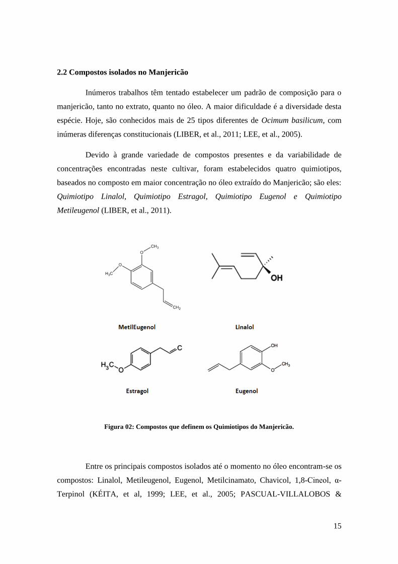
2.2 Compostos isolados no Manjericão
Inúmeros trabalhos têm tentado estabelecer um padrão de composição para o
manjericão, tanto no extrato, quanto no óleo. A maior dificuldade é a diversidade desta
espécie. Hoje, são conhecidos mais de 25 tipos diferentes de Ocimum basilicum, com
inúmeras diferenças constitucionais (LIBER, et al., 2011; LEE, et al., 2005).
Devido à grande variedade de compostos presentes e da variabilidade de
concentrações encontradas neste cultivar, foram estabelecidos quatro quimiotipos,
baseados no composto em maior concentração no óleo extraído do Manjericão; são eles:
Quimiotipo Linalol, Quimiotipo Estragol, Quimiotipo Eugenol e Quimiotipo
Metileugenol (LIBER, et al., 2011).
Figura 02: Compostos que definem os Quimiotipos do Manjericão.
Entre os principais compostos isolados até o momento no óleo encontram-se os
compostos: Linalol, Metileugenol, Eugenol, Metilcinamato, Chavicol, 1,8-Cineol, α-
Terpinol (KÉITA, et al, 1999; LEE, et al., 2005; PASCUAL-VILLALOBOS &

16
BALLESTA-ACOSTA; 2003), Canfora, Limoneno, Geraniol e Farmaseno (LABRA, et
al., 2004).
Já no extrato, os compostos identificados até o momento, foram: Novadensina,
Salvigenina, Circiseol, Eupatorin, Gardenina B (GRAYER, et al., 1996), Ácido
Chicórico, Ácido Caftárico (LEE & SCAGEL, 2009; KWEE & NIEMEYER, 2011),
Ácido-p-Cumárico, Peonidina-3,5-Diglicosídeo (PHIPPEN & SIMON, 1998) e o Ácido
Rosmarínico (GRAYER, et al., 1996; LEE & SCAGEL, 2009; SHIGA, et al., 2009;
JAVANMARDI, et al., 2002; NGUYEN & NIEMEYER, 2008).
2.3 Atividade biológica do Manjericão
O Manjericão tem sido escolha para uma vasta gama de testes biológicos,
usualmente em animais e modelos experimentais in vitro. Entre os que apresentaram
atividades mais relacionadas ao proposto neste trabalho, destacam-se alguns:
Sakr & Al-Amoudi (2012) relatam que após administração de 20 mL/kg de
extrato aquoso de manjericão em ratos albinos durante seis semanas aumentaram as
atividades da Superóxido dismutase (SOD) e da catalase (CAT), além de reduzirem a
peroxidação lipídica causada pela administração de deltrametrina. Ainda sobre a
atividade antioxidante, Thirugnanasampandan & Jayakumar (2011) avaliaram os efeitos
do extrato etanólico de Ocimum basilicum, nas concentrações de 50 a 350 µg/mL em
cultura de células hepáticas humanas e observaram uma redução na produção de óxido
nítrico, com efeitos dose-dependente. Berić e colaboradores, em 2008, demostraram que
o óleo essencial de Manjericão, Quimiotipo Linalol, mostrou-se eficaz para proteger
cepas de Salmonella dos efeitos do peróxido de hidrogênio (H2O2).
Duraisamy e colaboradores (2011) avaliaram os efeitos do extrato etanólico in
vitro em parâmetros inflamatórios. Os resultados mostraram que o extrato foi capaz de
reduzir a atividade das lipooxigenases in vitro. Mueller, Hobiger & Jungbauer (2010)
avaliaram os efeitos do extrato aquoso de O. basilicum a 0,5 mg/mL em cultura de
macrófagos. Os resultados mostraram uma redução de 57% na concentração de
Interleucina 6, 24% de redução no Fator de Necrose Tumoral α (TNF- α), 19% de
redução na atividade da ciclooxigenase 2 (COX-2) e um aumento de 54% na
concentração de Interleucina 10 (IL-10). Já um trabalho publicado em 2009 por Rakha e

17
colaboradores demonstrou que o extrato das sementes de manjericão reduziu o edema
de pata induzida por carragenina em 10%, mostrando, assim, atividade anti-
inflamatória.
2.4 Toxicidade do Manjericão
Apesar da ampla utilização desta espécie na culinária e dos diversos usos
populares, estudos envolvendo toxicidade são raros para este espécie.
Entre os estudos publicados nos últimos anos, envolvendo animais, temos o
trabalho de Shiga e colaboradores (2009) que utilizaram ratos machos e avaliaram doses
entre 4000 e 6000 mg/kg. Neste trabalho nenhuma morte ou sinal de toxicidade foi
registrado. Já Fandohan e colaboradores em 2008 determinaram que o óleo de O.
basilicum apresentava toxicidade em ratos após administração por 14 dias de doses de
1500mg/kg/dia. Com isso foi determinado uma dose letal (DL50) (dose responsável pela
morte de 50% dos animais teste) de 3.250 mg/kg.
Já com culturas de célula, a literatura é mais restrita. Bravo e colaboradores
(2008) realizaram um trabalho em cultura de células com macrófagos, avaliando apenas
a viabilidade celular. Os resultados mostraram que até 60µg/mL (valor máximo testado)
não ocorreram alterações neste parâmetro. Koko e colaboradores (2008) avaliando a
viabilidade de linfócitos in vitro observaram que concentrações de 25 a 100 µg/mL
reduziram em torno de 70% a viabilidade celular. Quanto a proliferação, Gomez-Flores
e sua equipe (2008) determinaram que o extrato aquoso apresentava atividade
linfoproliferativa a partir de 31,25µg/mL e o mesmo acontecia com o extrato metanólico
em concentrações a partir de 125µg/mL.

18
3 OBJETIVOS
3.1 Geral:
Determinar os efeitos in vitro do extrato de Ocimum basilicum L. (Manjericão) sobre
parâmetros genotoxicológicos, oxidativos e inflamatórios, em cultura de leucócitos
humanos.
3.2 Específicos:
Determinar o perfil fitoquímico do extrato de Ocimum basilicum L., quantificando
os níveis de polifenois, flavonoides, níveis de vitamina C, quantificação dos
principais princípios ativos conhecidos;
Determinar os efeitos do extrato de Ocimum basilicum L. sobre parâmetros
protetores genotoxicológicos em culturas de leucócitos humanos;
Avaliar os efeitos antioxidantes desta espécie no estresse oxidativo causados pelo
peróxido de hidrogênio em cultura de leucócitos humanos;
Avaliar os efeitos do extrato de Ocimum basilicum L. sobre parâmetros
inflamatórios em cultura de leucócitos humanos.

19
PARTE II
4 MANUSCRITO
Evaluation of Basil Extract (Ocimum basilicum L.) on Oxidative, Anti-Genotoxic
and Anti-Inflammatory Effects in Human Leukocytes Cell Cultures Exposed to
Challenging Agents.
Camila Martins Güez, Raul Oliveira de Souza, Paula Fischer, Maria Fernanda de Moura
Leão, Aline Augusti Boligon, Margareth Linde Athayde, Luísa Zuravski, Luís Flávio
Souza de Oliveira, Michel Mansur Machado.
Submetido a “Basic & Clinic Pharmacology & Toxicology”

20

21
Evaluation of Basil Extract (Ocimum basilicum L.) on Oxidative, Anti-Genotoxic
and Anti-Inflammatory Effects in Human Leukocytes Cell Cultures Exposed to
Challenging Agents.
Camila Martins Güez¹, Raul Oliveira de Souza², Paula Fischer², Maria Fernanda de
Moura Leão2, Aline Augusti Boligon3, Margareth Linde Athayde³, Luísa Zuravski4,
Luís Flávio Souza de Oliveira4, Michel Mansur Machado4*.
1 Programa de Pós Graduação em Ciências Farmacêuticas, Universidade Federal do
Pampa, Uruguaiana, Rio Grande do Sul, Brasil.
2 Curso de Farmácia, Universidade Federal do Pampa, Uruguaiana, Rio Grande do
Sul, Brasil.
3 Programa de Pós Graduação em Ciências Farmacêuticas, Universidade Federal de
Santa Maria, Santa Maria, Rio Grande do Sul, Brasil.
4 Departamento de Farmácia, Universidade Federal do Pampa, Uruguaiana, Rio
Grande do Sul, Brasil.
*Correspondences must be addressed to Prof. Dr. Michel Mansur Machado,
Universidade Federal do Pampa – Campus Uruguaiana. BR 472, Km 585, Caixa postal
118, Uruguaiana, RS, Brazil, CEP: 97500-970. Tel.: 5555 3413-4321.
E-mail address: [email protected]

22
Abstract
Ocimum is one of the most important genera of the Lamiaceae family. Several studies
about Basil and its popular use reveal many characteristics from the herb, being used as
antioxidant, anti-aging, anti-inflammatory, anti-carcinogenic, anti-microbial,
cardiovascular agent, among others. In this paper we evaluated genotoxic, oxidative and
anti-inflammatory parameters from the extract of Ocimum basilicum in different
concentrations, using human leukocytes cultures exposed to challenging agents. With
our results it is possible to verify that O. basilicum extract acts as an antioxidant and
effectively reverts or subjugates the effects of a high oxidizing agent as hydrogen
peroxide and, these actions are explained because its composition, which is rich in
polyphenols and flavonoids besides several compound as Rosmarinic Acid, who have a
well-known antioxidant activity. In the anti-inflammatory aspect, we show that our
extract actually presents these properties and the mechanism involved in these particular
actions are a composed interaction between the inhibition of pro-inflammatory mediator
and the stimulation of anti-inflammatory cytokines. Although pharmacodynamics
studies are necessary to evaluate the activities in vivo, our results demonstrated that
Basil could act as antioxidant and anti-inflammatory becoming into a possible
alternative for medicinal treatment.
Key words: Free radicals, basil, anti-inflammatory, oxidative stress, genotoxicology.

23
1. Introduction
The free radicals are oxidizing agents, having one or more unpaired electrons
in their outer orbital making them highly reactive species that act as electrophiles [1].
The reactions of free radicals with the organism result in cell and tissue damage that
contributes to the development of pathologies. The free radicals act on cellular
components by oxidizing lipids, proteins, nucleic acids and carbohydrates [2].
The genus Ocimum L. includes approximately 150 species, possessing a great
variation in plant morphology and biology, essential oil content, and chemical
composition [3]. Ocimum basilicum, popular known as Basil or Sweet Basil, is a
common herb that belongs to Lamiaceae family. Studies have shown many
pharmacological effects in several diseases, acting like a potent antioxidant, anti-aging,
anticancer, antiviral and antimicrobial properties [4].
Traditionally, Basil has been used as a medicinal and aromatic herb, to add
aroma and flavor to food [5] and several secondary metabolites like polyphenols,
flavonoids and terpenes, with recognized potential biologic effects that have been
identified in this specie [6]. Studies have reported that in the components of the plant,
Rosmarinic Acid (RA) is the most biologically active compound present in Basil [6-8].
Researchers also tried to establish a standard of composition for Basil, both in the
extract as in oil, but the biggest difficulty is the fact of existing more than 25 different
types of O. basilicum, with many constitutional differences [9,10].
Knowing about the economic importance and global dissemination of their
uses in cooking and folk medicine it is important to investigate the pharmacological and
toxicological effects of Basil, in order to ensure its efficacy and safety, since toxicity
studies for these species are rare and do not focus on genetic aspects. The main purpose
of the present study was to evaluate the oxidative, genotoxic and anti-inflammatory
parameters that may be present in the extract from Basil leaves using human leukocytes
cells cultures.

24
2. Material and Methods
2.1 Chemical, apparatus and general procedures: All chemical were of analytical
grade. Methanol, acetic acid, gallic acid, chlorogenic acid and caffeic acid purchased
from Merck (Darmstadt, Germany). Quercetin, rutin, rosmarinic acid, and kaempferol
were acquired from Sigma Chemical Co. (St. Louis, MO, USA). High performance
liquid chromatography (HPLC-DAD) was performed with a Shimadzu Prominence
Auto Sampler (SIL-20A) HPLC system (Shimadzu, Kyoto, Japan), equipped with
Shimadzu LC-20AT reciprocating pumps connected to a DGU 20A5 degasser with a
CBM 20A integrator, SPD-M20A diode array detector and LC solution 1.22 SP1
software.
2.2 Plant material: The dry leaves of Ocimum basilicum L., variety Genovese, were
purchased from a local market in Uruguaiana – RS – Brazil (Latitude 29°45'17'';
Longitude 57°05'18''). The leaves were triturated and macerated at room temperature in
hydroalcoholic solution (30H2O: 70 Ethanol v/v) at concentration of 20g per 100mL of
solvent for a week under daily shaking. The maceration process was repeated for two
more weeks to exhaustion of the vegetable material. In the end of three weeks, the
filtrates were pooled and evaporated under reduced pressure in a rotary evaporator in
order to remove ethanol and water. The dry extract of Ocimum basilicum (ObE) was
used in the following tests.
2.3 Phytochemical analysis of ObE: The ObE was analyzed according to Boligon [11],
Laghari [12] and Machado [13] using high performance liquid chromatography (HPLC)
for the determination of the compounds concentrations: rosmarinic acid, gallic acid,
chlorogenic acid, caffeic acid, rutin, quercetin, and kaempferol. The analysis of the
polyphenols and flavonoids from the ObE were performed through specific colorimetric
reactions using Folin Ciocalteau’s reagent [14] and aluminum chloride [15],
respectively.
2.4 Cytotoxicity curve in leukocytes: Initially, the dose-effect cytotoxicity curve was
determined in leukocytes using the ObE dissolved in PBS Buffer pH 7.2 at doses
ranging from 0.0001 mg/mL to 100 mg/mL, to determine the lethal dose 50% (LD50).
25
Human leukocytes cultures were prepared using 0.5 mL of venous blood collected by
venipuncture from a male volunteer (survey approved by the Ethics Committee of the
Federal University of Santa Maria, approval letter Number: 23,081) and immediately
transferred to RPMI 1640 medium supplemented with 10% fetal bovine serum, 1%
streptomycin / penicillin and phytohemaglutinin according to a previous study described
by Santos Montagner [16]. Cells were kept at 37ºC for 72 hours. The analyzed
parameter was cell proliferation according to Burow [17].
2.5 Leukocytes culture sample preparations: To assess the anti-oxidative status and
anti-genotoxic profiles, the leukocyte cultures were divided in six groups. The groups
were: a negative control (phosphate buffer pH 7.2); a positive control (hydrogen
peroxide 100 μM); a group with rosmarinic acid at the concentration previously found
in the plant by the phytochemical analysis; and three groups with different extract
concentrations obtained from toxicity studies, i.e., the LD50, LD50/10 and LD50/100.
Groups with rosmarinic acid and extracts have also received 100 mM H2O2 to induce
oxidation. To assess the inflammatory parameters the same groups division described
above were used, with exception of positive control that has incorporated Ibuprofen 100
μM. Additionally, all groups received dextran 1% to induce inflammatory process,
except the negative control. The concentrations were selected to establish effective
doses and exhibit low toxicity to leukocytes. All tests were performed in triplicate.
2.6 Evaluation of antioxidants parameters of ObE in human leukocytes cultures: To
analyze the oxidative parameters, we used classical techniques as Lipid Peroxidation,
Protein Carbonylation, Ascorbic Acid Content, and Superoxide dismutase and Catalase
activities. The assays were carried out in triplicate.
2.6.1 Lipid peroxidation: The extent of lipid peroxidation was estimated as the
concentration of thiobarbituric acid reactive products (malondialdehyde) according to
Ohkawa [18]. The method measures spectrophotometrically the color produced by the
reaction of TBA with malondialdehyde (MDA) at 532 nm.
2.6.2 Protein carbonylation: The protocol was performed according to Morabito [19].
In this technique, Carbonyl (CO) groups (aldehydes and ketones) are produced on

26
protein side chains when they are oxidized, reacting with 2,4-dinitrophenylhydrazine,
forming a color complex.
2.6.3 Ascorbic acid content: According to the method of Jacques-Silva [20] the curve of
ascorbic acid was taken as a reference and the reference samples were mixed with
trichloroacetic acid 13,3% and 2,4-dinitrophenylhydrazine. After incubation period, the
samples were measured at 520 nm in spectrophotometer. The extracts were measured
using the same procedure.
2.6.4 Catalase activity: Catalase activity was determined from the rate of decomposition
of H2O2 [21]. One unit of catalase activity was defined as the required activity to
degrade one mol of hydrogen peroxide in 60s.
2.6.5 Superoxide dismutase activity: Superoxide dismutase (SOD) (E.C.1.15.1.1)
activity was measured spectrophotometrically according to Boveris [22]. The technique
is based on the inhibition of the reaction of superoxide anion with epinephrine. The
oxidation reaction of epinephrine produces adenocromo that can be detected
spectrophotometrically (480 nm). The enzyme activity was determined by measuring
the rate of formation of adenocromo. The reaction medium contains glycine-NaOH and
epinephrine. An unit of enzyme activity is defined as the amount of enzymes required to
inhibit the rate of epinephrine autoxidation by 50%.
2.7 Evaluation of anti-genotoxic parameters of ObE in human leukocytes cultures: the
techniques used to evaluate the anti-genotoxic parameters were: cell proliferation and
inviability, DNA damage, Chromosomal Instability and Micronuclei frequency.
2.7.1 Cellular proliferation and viability: Viability is assessed by the loss of membrane
integrity, using the trypan blue [17]. In this technique, the same samples and respective
concentrations are combined with Turk's solution (Acid acetic acid 3% plus gentian
violet 1% in water), and after three minutes, the sample is placed in a Neubauer
chamber. The differentiation of living and dead cells is observed by the blue coloration
of dead cells. A total of 300 cells are counted and the amount of total leukocytes
(proliferation) is achieved through counting in a Neubauer chamber.

27
2.7.2 Alkaline comet DNA assay: This test was assayed following procedures of Singh
[23]. After incubation, the samples (leukocytes) were mixed with low-melting point
agarose and placed on a microscope slide pre-coated with normal melting point agarose.
The slides were immersed in a lysis solution, and an electrophoresis was performed (20
min at 300 mA and 25 V). In the end, the slides were neutralized and left to dry
overnight at room temperature. The dry slides were re-hydrated and then fixed for 10
min, and left to dry again. The last stage was the coloring and the use of stop solution.
The slides were analyzed under blind conditions. DNA damage was given as DNA
damage index (DI). The DNA damage was calculated from cells in different damage
classes (completely undamaged: 100 cells × 0 to maximum damaged − 100 cells × 4).
2.7.3 Chromosomal instability: Colcemid was added in each leukocyte culture and
incubated at 37°C during 60 min. After this, the cells were centrifuged at 1,800 rpm for
10 min. The cell pellets were re-suspended in hypotonic solution and incubated at 37ºC
for 16 min. After a new centrifugation, the cell pellets were re-suspended in acetic acid:
methanol (3:1) and poured into a flask containing a fixative solution followed by
centrifugation. The slides were prepared by dropping 3 or 4 drops of cell suspension
into a cold, wet slide, which then was air dried. The cells were analyzed with 10X
magnification to verify the density and distribution of metaphase chromosomes [24].
2.7.4 Micronuclei frequency: The cells were placed in a conic tube with saline and
centrifuged in 1,000 rpm for 5 min (this procedure was repeated). One milliliter with the
cell pellet was kept in the tubes mixed with the pipette and spread over the slide (two
per sample) and left to dry in room temperature. Slides were stained by panoptic method
and then analyzed under optical microscopy in immersion lens. For each slide, 1,000
cells were counted [25].
2.8 Evaluation of Anti-inflammatory Parameters of ObE in Human Leukocytes
Cultures: For the determination of Necrosis Tumoral Factor–α (TNF-α); Interleukine-10
(IL-10); Interleukine-6 (IL-6), Inhibition of COX-2 activities and Nitric Oxide
production, the measurements were made using ELISA kits according to the specific
instructions of the manufacturer. All tests were performed in triplicate. The results of

28
these tests were expressed in percentage of production in relation to the negative
control.
2.9. Statistical analysis: Data were expressed as mean ± standard deviation (SD).
Comparisons between groups were performed using one-way analysis of variance
(ANOVA), followed by post hoc of Bonferroni for multiple comparison tests. Nonlinear
regression analysis was used to determine LD50. Results were considered statistically
significant when p<0.05.
3. Results and Discussion
Medicinal plants are rich sources of natural antioxidants and represent a
promising perspective in discovery of new drugs toward therapeutic area. Most
members of the Lamiaceae family have shown interesting biological effects due to their
antioxidant compounds [26].
Phenolic compounds are broadly distributed in the plant kingdom and are the
most abundant secondary metabolites found in plants [27]. The key role of phenolic
compounds as antibacterial is emphasized in several reports [26, 28]. Flavonoids
compounds occur naturally in plant foods and are a common component of our diet.
Table 1 shows the data from analysis by HPLC-DAD method [29] to determine the
concentration of totals polyphenol and flavonoids and some compounds present in
Ocimum basilicum L, all described in literature as biologically active drugs.

29
Table 1 - Concentrations of some biologically important groups and compounds
presents in Ocimum basilicum L extract.
Group / Compound Concentration in µg/g Dry Weight
Polyphenol compounds 23,780.00 ± 145.30
Total flavonoids 15,982.00 ± 341.61
Quercetin 558.37 ± 2.41
Rutin 398.49 ± 0.97
Gallic acid 2,330.52 ± 81.19
Caffeic acid 4,780.00 ± 14.52
Chlorogenic acid 2,875.00 ± 103.56
Rosmarinic acid 3,530.00 ± 2.87
Kaempferol 342.00 ± 18.79 Data expressed as mean ± S.D. Results were confirmed by the analysis triple repetition and sampling in
triplicate.
In Figure 01, we show the dose-effect curve. As it is showed below, when the dose of
the extract is increased, the total number of leukocytes decreases, showing a dose-
dependent effect. It is important to note that in LD50 concentration (35.44 µg/mL), the
cellular inviability and DNA Damage showed no significant changes (data not shown),
corroborating with the study by Gomez-Flores [30]. This result allowed us to establish
the doses that were used in our protocols. Aiming to search for a dose with high
affectivities and low toxicity, we determine the work doses for this protocol: 35.44
µg/mL (LD50), 3.544 µg/mL (LD50/10) and 0.3544 µg/mL (LD50/100).
Figure 01 - Dose-effect curve using the crude extract of Ocimum basilicum in different doses (0.0001
mg/mL to 100 mg/mL) to determine the lethal dose 50% (LD50) in human leukocytes. The inset
shows the total amount of leukocytes versus the tests concentrations of O. basilicum extract.
30
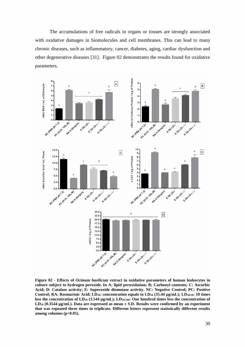
The accumulations of free radicals in organs or tissues are strongly associated
with oxidative damages in biomolecules and cell membranes. This can lead to many
chronic diseases, such as inflammatory, cancer, diabetes, aging, cardiac dysfunction and
other degenerative diseases [31]. Figure 02 demonstrates the results found for oxidative
parameters.
Figure 02 - Effects of Ocimum basilicum extract in oxidative parameters of human leukocytes in
culture subject to hydrogen peroxide. In A: lipid peroxidation; B; Carbonyl contents; C: Ascorbic
Acid; D: Catalase activity; E: Superoxide dismutase activity. NC: Negative Control; PC: Positive
Control; RA: Rosmarinic Acid; LD50: concentration equals to LD50 (35.44 µg/mL); LD50/10: 10 times
less the concentration of LD50 (3.544 µg/mL); LD50/100: One hundred times less the concentration of
LD50 (0.3544 µg/mL). Data are expressed as mean ± S.D. Results were confirmed by an experiment
that was repeated three times in triplicate. Different letters represent statistically different results
among columns (p<0.05).

31
The involvement of reactive oxygen species in interactions with
polyunsaturated fatty acids in cell membranes generates what is called lipid
peroxidation. The result is the formation of hydro or lipoperoxides highly reactive that
may trigger the oxidative cascade resulting in damage to membrane integrity [32]. The
results found in lipid peroxidation show that LD50 and LD50/10 obtained values are close
to the control of rosmarinic acid, a well know antioxidant agent. Moreover, the values
of peroxidation in LD50 3,576 nMol of MDA/mL in erythrocytes and LD50/10 4,226
nMol of MDA/mL in erythrocytes, were 41,81% and 31,29% lower when compared to
the positive control, respectively. The content of protein carbonyl is the most general
indicator and the most commonly used marker of protein oxidation, and its
accumulation has been observed in several human diseases [33]. In protein carbonyl the
values found show that LD50 was 28% and LD50/10 18,8% lower when compared to the
positive control, and approximate to negative control and rosmarinic acid control. The
vitamin C or acid ascorbic has been implicated in different biological processes and
plays an important role in oxidant defense. Acid ascorbic have function in several
enzymatic steps acting like a cofactor in the synthesis of collagen, monoamines, amino
acids, peptide hormones, and carnitine. Samples with higher concentrations of extract
showed higher concentrations of ascorbic acid. This fact is directly related to the
concentrations of polyphenols and antioxidant action. The most concentrated samples,
LD50 and DL50/10, have higher levels of polyphenols. Polyphenols are a first line of
defense against oxidative action of hydrogen peroxide. When polyphenols decrease
their concentrations, other non-enzymatic antioxidants come into play, thereby reducing
the concentration of vitamin C [35, 36].
Mammalian cells have elaborate antioxidant defense mechanisms to control
damage effects of reactive oxygen species (ROS) and the catalase enzyme is one of
these, protecting cells against the toxic effects of hydrogen peroxide [37]. The results of
Catalase analysis show the ability of O. basilicum extract in neutralizing the effects of
hydrogen peroxide. The results were dose-related and show a better effect at LD50
concentration. Superoxide has been implicated in reactions associated with aging, in
pathophysiological processes, due to its transformation into more reactive species, like
hydroxyl radical that initiates lipid peroxidation.

32
According to our results, the activity of SOD enzyme was not affected in the
three concentrations of O. basilicum extract. All these results are related to the presence
of antioxidant compounds present in the extract (Table 01), as the groups of
polyphenols and flavonoids. This information confirms the data from the literature that
phenolic compounds, especially flavonoids act in the oxidative metabolism, not by
changing the enzymatic defenses, but by directing neutralizing of reactive species in
order to stabilize them [38].
We also evaluated the effects of hydrogen peroxide and the counter effects of
O. basilicum extract on human leukocytes cells. The results of these markers are show
in Figure 03.

33
Figure 03 - Effects of Ocimum basilicum extract in anti-genotoxic parameters of human leukocytes
in culture exposed to hydrogen peroxide. In A: cell proliferation; B; cell inviability; C: percentage
of abnormal chromossomics; D: DNA damage index; E: frequency of micronuclei. NC: Negative
Control; PC: Positive Control; RA: Rosmarinic Acid; LD50: concentration equals to LD50 (35.44
µg/mL); LD50/10: 10 times less the concentration of LD50 (3.544 µg/mL); LD50/100: One hundred times
less the concentration of LD50 (0.3544 µg/mL). Data are expressed as mean ± S.D. Results were
confirmed by an experiment that was repeated three times in triplicate. Different letters represent
statistically different results among columns (p<0.05).
In cellular proliferation (Figure 3A), the concentrations of LD50 and LD50/100
the cellular increase was similar to the rosmarinic acid control and different from the
positive control, whereas the lowest concentration LD50/100 presented a decrease in
cellular proliferation witch may be compared to the positive control. Figure 3B shows
the percentage of unviable cells for both controls groups and for the three
concentrations groups tested. The negative control presented only 0.667% whereas

34
positive control was 14% of unviable cells. Rosmarinic acid was 1.33% and the other
three concentrations of O. basilicum: LD50: 1.667%; LD50/10: 3.67%; LD50/100: 10.67%
of unviable cells.
According to Collins [39], the high viability of cells is required as a previous
condition for the performance of the comet assay. Chromosomal instability results in
numerical and structural chromosomal complexity and several studies associated these
instabilities with poor prognosis in solid tumors [40].
The results demonstrate that all concentrations of extract of Basil have
presented percentages of chromosomal abnormalities (Figure 3C) similar to the negative
control and even the lowest concentration presented 33.334% less mitotic index when
compared to positive control.
The comet assay is one of the most promising genotoxicity tests developed to
measure and analyze DNA damage in single cells [41]. The test was used as a parameter
for assessing the DNA damage index (Figure 3D). As we can see at the lowest
concentration - LD50/100, the cells presented the highest damage index compared with
positive control, whereas the other two concentrations of extract, LD50 and LD50/10,
showed damage index of 44.45% and 19.18% lower when compared with the positive
control, respectively.
The micronuclei assay provides a convenient and reliable index of both
chromosome breakage and chromosome loss. The micronuclei are expressed in dividing
cells that either contain chromosome breaks lacking centromeres (acentric fragments)
and/or whole chromosomes that are unable to migrate to the poles during mitosis [42].
The results show the micronucleus frequency (MN) found at the control groups and
three concentrations of plant extract under analysis (Figure 3E). The frequency of MN
was dose-dependent. The lowest concentration of the extract has shown the highest
frequency of cells with this alteration.
The Basil was frequently related as a common anti-inflammatory [43]. Aiming
to confirm this activity and evaluate the mechanism involved, we perform a series of
tests in human leukocytes cell exposed to a pro-inflammatory agent. The results are
presented in Figure 04.

35
Figure 04 - Effects of Ocimum basilicum extract in inflammatory parameters of human leukocytes
in culture exposed to dextran solution. All the results are presented as percentage in relation to the
negative control. In A: tumoral necrose factor-α (TNF-α); B; interleukine-10 (IL-10); C:
interleukine-6 (IL-6); D: Ciclooxigenase type 2 activity (COX2 activity); E: nitric oxide (NO)
production. NC: Negative Control; PC: Positive Control; RA: Rosmarinic Acid; LD50:
concentration equals to LD50 (35.44 µg/mL); LD50/10: 10 times less the concentration of LD50 (3.544
µg/mL); LD50/100: One hundred times less the concentration of LD50 (0.3544 µg/mL). Data are
expressed as mean ± S.D. Results were confirmed by an experiment that was repeated three times
in triplicate. Different letters represent statistically different results among columns (p<0.05).

36
Cytokines are subdivided in proinflammatory (initiate defense against
pathogens) and anti-inflammatory (regulate the inflammatory process helping to balance
the inflammatory response) acting with an important role in inflammatory response. The
proinflammatory cytokines includes IL-1, IL-2, IL-6, IL-8, TNF-α, and the anti-
inflammatory cytokines includes IL-1 antagonist receptor, IL-4, IL-10, and IL-13 [44].
The results show that the production of the proinflammatory cytokines such as TNF- α
and IL – 6, were not affected in the three different doses of O. basilicum extract.
However, the percentage of production of IL-10, the anti-inflammatory cytokine, shows
an increase in the percentage of more than 60% at the highest dose of the extract when
compared with the positive control (Ibuprofen). The percentage of production of IL-10
also demonstrated to be dose-dependent, but in all concentrations this production was
higher than the negative and positive controls. The cyclooxygenases (COX) have been
identified in three different isoforms. COX-2 acts like the inducible isoform, which is
regulated by growth factors and different cytokines (IL1β, IL6, or TNFα) [45]. The
ability of the extract from Ocimum basilicum to inhibit COX-2 was determined and the
results show that the extract was not capable to reduce the activity of the
cyclooxygenase like the positive control (Ibuprofen) but his activity decrease when
compared to the negative control (100%) and was dose-dependent. The inhibition of
COX-2 in LD50 and LD50/10 approximates to the inhibition caused by rosmarinic acid
and several studies report many properties of rosmarinic acid including cyclooxygenase
inhibition [35, 36].
The percentage of nitric oxide generated was measured and cells pretreated
with the three plant extracts showed a dose-dependent inhibition. The negative control
was considered 100%. The maximal NO inhibitory effect was exhibited by O. basilicum
at dose of LD50. Comparing to the negative control, the percentage of reduction was
higher than 30% (34.67%). In a similar study using three plants of Lamiaceae family, O.
basilicum extract demonstrated the higher content of phenols and also maximal levels of
DNA protection and free radical scavenging against toxicity induced by cadmium
chloride [46]. The results suggested that the anti-inflammatory activities of these
extracts could be explained, at least in part, by their antioxidant properties [34].
Rosmarinic acid was one of the most abundant caffeic acid esters present in O.
basilicum [34]. It has been related that this compound has antioxidant, anti-HIV, and
anti-inflammatory or cyclooxygenase and lipoxygenases inhibitory activities [35, 36].

37
4. Conclusions
It is possible to verify that Ocimum basilicum extract acts as an antioxidant and
effectively reverts or subjugates the effects of a high oxidizing agent as hydrogen
peroxide. These actions are explained by its composition, which is rich in polyphenols
and flavonoids besides several compound as Rosmarinic Acid, who have a well know
antioxidant activity.
In the anti-inflammatory aspect, we show that our extract actually presents this
properties and the mechanism involved in this particular actions are a composed
interaction between the inhibition of pro-inflammatory mediator and the stimulation of
anti-inflammatory cytokines.
Although pharmacodynamics studies are necessary to evaluate the activities in
vivo, our results demonstrated that Basil could act as antioxidant and anti-inflammatory,
becoming a possible alternative for medicinal treatment.
Conflict of Interests
The authors declare no conflict of interests.
Acknowledgments
The authors are thankful to the financial support of Fundação de Amparo a Pesquisa do
Rio Grande do Sul (FAPERGS) and to Conselho Nacional de Desenvolvimento
Científico e Tecnológico (CNPq).
References
[1] B. Gillham, D. K. Papachristodoulou, J. H. W. Thomas, “Biochemical basis of
medicine,” Oxford: Reed Educational And Professional Publishing Ltda, Ed. 3, pp.
196-202, 1997.

38
[2] C. W. Choi, et al., “Antioxidant activity and free radical scavenging capacity
between korean medicinal plants and flavonoids by assay-guided comparison,” Plant
Science, vol. 163, no.6, pp. 1161-1168, 2002.
[3] F. Danesi, S. Elementi, R. Neri, M. D’antuono. Maranesi, E. Bordoni, “Effect of
cultivar on the protection of cardiomyocytes from oxidative stress by essential oils and
aqueous extracts of Basil,” Journal of Agricultural and Food Chemistry, vol. 56, no. 21,
pp. 9911–9917, 2008.
[4] S. A. Sakr, W. M. Al-Amoudi, “Effect of leave extract of Ocimum basilicum on
deltamethrin induced nephrotoxicity and oxidative stress in albino rats,” Journal Of
Applied Pharmaceutical Science, vol. 02, no. 05, pp. 22-27, 2012.
[5] F. V. Roberto, J. E. Simon, “Chemical characterization of basil (Ocimum spp.)
found in the markets and used in traditional medicine in Brazil,” Economic Botany, vol.
54, no. 2, pp.207–216, 2000.
[6] J. Lee, C. F. Scagel, “Chicoric acid found in basil (Ocimum Basilicum L.) leaves,”
Food Chemistry, vol. 115, no. 2, pp. 650–656, 2009.
[7] T. Shiga, K. Shoji, H. Shimada, S. N. Hashida, F. Goto, T. Yoshihara, “Effect of
light quality on rosmarinic acid content and antioxidant activity of sweet basil, Ocimum
Basilicum L.,” Plant Biotechnology Journal, vol. 26, no. 2, pp. 255–259, 2009.
[8] J. Javanmardi, A. Khalighi, A. Kashi, H. P. Bais, J. M. Vivanco, “Chemical
characterization of basil (Ocimum Basilicum L.) found in local accessions and used in
traditional medicines in Iran,” Journal Of Agricultural And Food Chemistry, vol. 50, no.
21, pp. 5878–5883, 2002.
[9] Z. Liber, K. J. Stanko, O. Politeoc, F. Strikic, I. Kolakb, M. Milosc, Z. Satovicb, Z.
“Chemical Characterization and Genetic Relationships among Ocimum basilicum L.
Cultivars,” Chemistry & biodiversity, vol. 8, no. 11, pp. 1978-1989, 2011.

39
[10] S. Lee, K. Umano, T. Shibamoto, K. G. LEE, “Identification of volatile
components in basil (Ocimum basilicum L.) and thyme leaves (Thymus vulgaris L.) and
their antioxidant properties” Food Chemistry, vol. 91, no. 1, pp. 131–137, 2005.
[11] A. A. Boligon, A. C. Feltrin, M. M. Machado, V. Janonik, M. L. Athayde “HPLC
analysis and phytoconstituents isolated from ethyl acetate fraction of Scutia Buxifolia
Reiss. Leaves,” Latin American Journal Of Pharmacy, vol. 28, no. 1, pp. 121-124,
2009.
[12] A. H. Laghari, S. Memon, A. Nelofar, K. M. Khan, A. Yasmin, “Determination of
free phenolic acids and antioxidant activity of methanolic extracts obtained from fruits
and leaves of Chenopodium album,” Food Chemistry. vol. 126, no 4, pp. 1850–1855,
2011.
[13] M. M. Machado, F. F. F. dos S. Montagner, A. Boligon, M. L. Athayde, M. I. U.
Rocha, J. P. Lera, C. Belló, I. B. M. da Cruz, “Determination of polyphenol contents
and antioxidant capacity of no-alcoholic red grape products (Vitis labrusca) from
conventional and organic crops,” Química Nova. vol.34, no.5, pp. 798-803, 2011.
[14] S. Chandra, E. G. Mejia, “Polyphenolic compounds, antioxidant capacity, and
quinone reductase activity of an aqueous extract of Ardisia compressa in comparison to
mate (Ilex paraguariensis) and green (Camellia sinensis) teas,” Journal of Agricultural
and Food Chemistry, vol. 52, pp. 3583-3589, 2004.
[15] J. Zhishen, T. Mengcheng, W. Jianming “The determination of flavonoid contents
in mulberry and their scavenging effects on superoxide radicals,” Food Chemistry, vol.
64, no. 4, pp. 555-559, 1999.
[16] G. F. F. Santos Montagner, M. Sagrillo, M. M. Machado, R.C. Almeida, C. P.
Mostardeiro, M. F. F. Duarte, I. B. M. Cruz, “Toxicological effects of ultraviolet
radiation on lymphocyte cells with different manganese superoxide dismutase Ala16val
polymorphism genotypes,” Toxicology In Vitro, vol. 24, pp. 1410-1416, 2010.

40
[17] M. E. Burow, C. B. Weldon, Y. Tang, G. L. Navar, S. Krajewsky, J. C. Reed, T.
G. Hammond, S. Clejan, B. S. Beckman, “Differences in susceptibility to tumor
necrosis factor Α- induced apoptosis among mcf-7 breast cancer cell variants,” Cancer
Research, vol. 58, no. 21, pp. 4940-4946, 1998.
[18] H. Ohkawa, N. Ohishi, K. Yagi, “Assay for lipid peroxides in animal tissues by
thiobarbituric acid reaction,” Analytical Biochemistry, vol. 95, no. 2, pp. 351-358, 1979.
[19] F. Morabito, M. Cristiani, A. Saija, C. Stelitano, V. Callea, A. Tomaino, P. L.
Minciullo, S. Gangemi, “Lipid peroxidation and protein oxidation in patients affected
by Hodgkin’s lymphoma,” Mediators Of Inflammation. vol.13, no. 5-6, pp. 381–383,
2004.
[20] M. C. Jacques-Silva, C. W. Nogueira, L. C. Broch, E. M Flores, J. B. T. Rocha.
“Diphenyl diselenide and ascorbic acid changes deposition of selenium and ascorbic
acid in liver and brain of mice,” Pharmacology & Toxicology, vol. 88, no. 3, pp.119–
125, 2001.
[21] H. Aebi, S. R. Wyss, B. Scherze, F. Skvaril, “Heterogenecity of erythrocyte
catalase isolation and characterization of normal and variant erythrocyte catalase and
their subunit,” Enzyme, vol. 17, no. 5, pp. 307- 318, 1974.
[22] A. Boveris, E. Cadenas, “Cellular source and steady-state levels of reactive oxygen
species. In: Clerch L, Massaro D. “Oxygen, gene expression and cellular function,”
Marcel Decker: New York, vol. 105, pp. 1-25, 1997.
[23] N. Singh, M. McCoy, R. Tice, E. A. Schneider “A simple technique for
quantification of low levels of DNA damage in individuals cells,” Experimental Cell
Research. vol. 175, pp. 184–191, 1995.
[24] J. J. Yunis, “High resolution of human chromosomes,” Science, vol. 191, no. 4233,
pp. 1268–1270, 1976.

41
[25] W. Schmid, “The Micronucleus Test,” Mutation Research, vol. 31, pp. 09-15,
1975.
[26] P. Schofield, D. M. Mbugua, A. N. Pell, “Analysis of condensed tannins: A
review,” Animal Feed Science and technology, vol. 91, no.1-2, pp. 21-40, 2001.
[27] A. S. Komali, Z. Zheng, K. Shetty, “A mathematical model for the growth kinetics
and synthesis of phenolics in oregano (Origanum vulgare) shoot cultures inoculated
with Pseudomonas species,” Process Biochemistry, vol. 35, no. 3, pp. 227-235, 1999.
[28] J. K. S. Moller, H. L. Madsen, T. Altonen, L. H. Skibsted, “Dittany (Origanum
dictammus) as a source of water-extractable antioxidants,” Food Chemistry, vol. 64, no.
2, pp. 215-219, 1999.
[29] C. A. Rice-Evans, N. J. Miller, G. Paganga, “Structure-antioxidant activity
relationships of flavonoids and phenolic acids,” Free Radical Biology and Medicine,
vol. 20, no. 7, pp. 933-956, 1996.
[30] R. Gomez-Flores, L. Verástegui-Rodríguez, R. Quintanilla-Licea, P. Tamez-
Guerra, R. Tamez-Guerra, C. Rodríguez-Padilla, “In vitro rat lymphocyte proliferation
induced by Ocimum basilicum, Persea americana, Lantago virginica, and Rosa spp.
extracts,” Journal of Medicinal Plants Research, vol. 2, no. 1, pp. 005-010, 2008.
[31] S. Wang. E. A. Konorev, S. Kotamraju, J. Joseph, S. Kalivendi, B. Kalyanaraman,
“Doxorubicin induces apoptosis in normal and tumor cells via distinctly different
mechanisms, intermediacy of H2o2 and P53- dependent pathways,” Journal of
Biological Chemistry, vol. 279, no. 24, pp. 25535-25543, 2004.
[32] A. H. K. Tsang, & K. K. K. Chung, “Oxidative and nitrosative stress in Parkinson's
disease. Biochimica Et Biophysica Acta, vol. 1792, no. 7, pp. 643-650, 2009.
[33] I. Dalle-Donne, R. Rossi, D. Giustarini, A. Milzani, A. Colombo, “Protein carbonyl
groups as biomarkers of oxidative stress,” Clinica Chimica Acta, vol. 329, no. 1-2, pp.
23–38, 2003.

42
[34] C. Jayasinghe, N. Gotoh, ,T. Aoki, S. Wada, “Phenolics composition and
antioxidant activity of sweet basil (Ocimum Basilicum L.),” Journal of Agricultural and
Food Chemistry. vol. 51, no. 15, pp. 4442-4449, 2003.
[35] M. A. Kelm, M. G. Nair, G. M. Strasburg, D. L. Dewitt, “Antioxidant and
cyclooxygenase inhibitory phenolic compounds from Ocimum sanctum Linn.,”
Phytomedicine, vol. 7, no. 1, pp. 7-13, 2000.
[36] M. Petersen, M. S. J. Simmonds,”Molecules of interest: Rosmarinic acid,”
Phytochemistry, vol. 62, pp. 1212-125, 2003.
[37] M. M. Goyal, A. Basak, “Human catalase: Looking for complete identity,” Protein
& Cell, vol. 1, no. 10, pp. 888-897, 2010.
[38] X. Liu, C. Cui, M. Zhao, J. Wang, W. Luo, B. Yang, Y. Jiang, “Identification of
phenolics in the fruit of emblica (Phyllanthus emblica L.) and their antioxidant
activities,” Food Chemistry, vol. 109, no. 4, pp. 909-915, 2008.
[39] A. R. Collins, A. A. Oscoz, G. Brunborg, I. Gaivão, L. Giovannelli, M.
Kruszewski, C. C. Smith, R. Stetina, “The comet assay: Topical issues,” Mutagenesis,
vol.23, no.3, pp.143-151, 2008.
[40] N. J. Birkbak, A. C. Eklund, Q. Li, S. E. Mcclelland, D. Endesfelder, P. Tan, I. B.
Tan, A. L. Richardson, Z. Szallasi, C. Swanton, “Paradoxical relationship between
chromosomal instability and survival outcome in cancer,” Cancer Research, vol. 71, no.
10, p.p. 3447-3452, 2011.
[41] I. Mukhopadhyay, D. K. Chowdhuri, M. Bajpayee, A. Dhawan, “Evaluation of in
vivo genotoxicity of cypermethrin in drosophila melanogaster using the alkaline comet
assay,” Mutagenesis, vol. 19, no. 2, pp. 85-90, 2004.
[42] M. Fenech, “The in vitro micronucleus technique,” Mutation Research, vol. 455,
no. 1-2 pp. 81–95, 2000.

43
[43] A. Durasaisamy, N. Narayanaswamy, A. Sebastian, K. P. Balakrishnan “Sun
protection and anti-inflammatory activities of some medicinal plants,” International.
Journal of Research in Cosmetic Science, vol. 1, no. 1, pp. 13-16, 2011.
[44] S. L. Goldstein, J. C. Leung, D. M. Silverstain, “Pro- and anti-inflammatory
cytokines in chronic pediatric dialysis patients: Effect of aspirin,” Clinical Journal of
the American Society of Nephrology, vol 1, 979 –986, 2006.
[45] C. Sobolewski, C. Cerella, M. Dicato, L. Ghibelli, M. Diederich, “The role of
cyclooxygenase-2 in cell proliferation and cell death in human malignancies,”
International Journal Of Cell Biology , vol. 2010, Article ID 215158, 21 pages, 2010.
[46] R. Thirugnanasampandan, R. Jayakumar, “Protection of cadmium chloride induced
DNA damage by Lamiaceae plants,” Asian Pacific Journal Of Tropical Biomedicine,
vol. 1, no. 5, pp. 391-394, 2011.

44
PARTE III
5 DISCUSSÃO GERAL
As plantas são fontes ricas na obtenção de antioxidantes naturais, podendo
representar uma perspectiva promissora na descoberta de novas drogas, no que diz
respeito à área terapêutica. Grande parte dos membros pertencentes à família
Lamiaceae, demonstram ter efeitos biológicos interessantes devido à presença destes
compostos antioxidantes (SCHOFIELD et. al., 2001).
O papel-chave dos compostos fenólicos como antibacterianos é enfatizada em
muitos estudos (SCHOFIELD et. al., 2001; MOLLER et. al., 1999). Os flavonoides
ocorrem naturalmente em alimentos de origem vegetal o que o torna um componente
comum presente em nossa dieta. A Tabela 01 do manuscrito mostra os dados da
análise realizada através do método de HPLC-DAD (RICE-EVANS et. al., 1996) para
determinar a concentração de polifenóis totais e flavonoides e alguns compostos
presentes no Ocimum basilicum L, todos descritos como biologicamente ativos. Os
resultados mostraram que o material analisado apresenta concentrações elevadas de
compostos antioxidantes, em especial dos ácidos Rosmarínico, Caféico, Clorogênico e
Gálico, todos na ordem de mg/g de planta. Cabe salientar que todos estes compostos
apresentam níveis amplamente variantes no gênero Ocimum (SHIGA et. al., 2009).
Na Figura 01 do manuscrito, é demonstrada a curva dose-efeito do extrato de
O. basilicum L. onde foi possível observar que com o aumento da concentração do
extrato o número total de leucócitos diminuía, em uma relação de efeito dose-
dependente. Na concentração determinada como DL50 (35,44 µg/mL) é importante
salientar que os testes de inviabilidade celular e de dano ao DNA não apresentaram
diferenças significativas (dados não mostrados), resultado esse que corrobora o estudo
realizado por Gomez-Flores e colaboradores (2008). A partir desse resultado foi
possível estabelecer as doses que foram utilizadas nos protocolos da pesquisa. Tendo
como objetivo procurar uma dose com alta efetividade e baixa toxicidade, as doses de
trabalho utilizadas, foram de 35,44 µg/mL (DL50), 3,544 µg/mL (DL50/10) e 0,3544
µg/mL (DL50/100).
O acúmulo de radicais livres em órgãos ou tecidos está fortemente associado
com os danos oxidativos causados nas biomoléculas e nas membranas celulares. Danos
esses, que podem induzir a diversas doenças crônicas, como processos inflamatórios,

45
câncer, diabetes, envelhecimento, disfunção cardíaca e outras doenças degenerativas
(WANG et. al., 2004). A Figura 02 do manuscrito mostra o resultado encontrado em
relação aos parâmetros oxidativos, peroxidação lipídica, proteína carbonilada, ácido
ascórbico, atividade da enzima catalase, atividade da enzima superóxido dismutase do
extrato de O. basilicum.
A participação de espécies reativas de oxigênio (ERO) em interações com
ácidos graxos poliinsaturados em membranas celulares gera o que é usualmente
chamado de peroxidação lipídica. O resultado dessa reação é a formação de hidro ou
lipoperóxidos altamente reativos que podem desencadear a cascata oxidativa, resultando
em danos à integridade celular (TSANG & CHUNG, 2009).
Os resultados encontrados no ensaio da peroxidação lipídica demonstraram que
os valores de DL50 e DL50/10 obtidos aproximam-se ao controle de ácido rosmarínico,
um agente antioxidante bastante conhecido. Além disso, os valores da peroxidação
obtidos para DL50 (3,576 nMol de MDA/mL em eritrócitos) e DL50/10 (4,226 nMol de
MDA/mL em eritrócitos) foram 41,81% e 31,29%, mais baixos quando comparados ao
controle positivo, respectivamente. O conteúdo de proteína carbonilada é o marcador
mais geral e comumente utilizado na determinação da oxidação protéica e seu acúmulo
tem sido observado em diversas doenças (DALLE-DONNE et. al.,2003). Os valores de
proteína carbonilada encontrados mostraram que para DL50 e DL50/10, os valores foram,
respectivamente, de 28% e de 18,8% menores quando comparados com o controle
positivo. Valores esses que se aproximam ao controle negativo e ao controle de ácido
rosmarínico. A vitamina C (ácido ascórbico), tem sido implicada em vários processos
biológicos e tem um papel importante na defesa antioxidante. O ácido ascórbico tem
função em diversos passos enzimáticos agindo como cofator na síntese de colágeno,
monoaminas, aminoácidos, hormônios peptídicos e carnitina. As amostras com maior
concentração de extrato mostraram maiores concentrações de ácido ascórbico. Esse fato
está diretamente relacionado com as concentrações de polifenóis e com a ação
antioxidante. As amostras mais concentradas, DL50 e DL50/10, apresentam maiores teores
de polifenóis. Os polifenóis constituem uma primeira linha de defesa contra a ação
oxidante do peróxido de hidrogênio. Quando os polifenóis diminuem suas
concentrações, outros antioxidantes não enzimáticos, entram em ação, reduzindo assim
as concentrações de vitamina C.

46
As células do nosso organismo desenvolveram mecanismos de defesa
antioxidante para o controle dos danos causados pelas espécies reativas de oxigênio
(ERO) e a enzima catalase é um desses exemplos, por possuir um papel importante na
proteção das células contra os efeitos tóxicos causados pelo peróxido de hidrogênio
(GOYAL & BASAK, 2010). Os resultados encontrados para a enzima Catalase
demonstraram a capacidade do extrato de O. basilicum em neutralizar os efeitos do
peróxido de hidrogênio, os quais foram dose-dependente, com melhores resultados na
concentração utilizada para DL50. O superóxido tem sido implicado em reações
associadas ao envelhecimento, em processos patofisiológicos, devido a sua
transformação em espécies mais reativas, como por exemplo, o radical hidroxila o qual
é responsável pelo início da peroxidação lipídica.
De acordo com o resultado encontrado, a atividade da enzima SOD não foi
afetada em nenhuma das concentrações do extrato de O. basilicum. Podendo ser
possível correlacionar com a presença dos compostos antioxidantes presentes no extrato
(Tabela 01 do manuscrito), como polifenóis e flavonoides. Esses dados confirmam aos
encontrados na literatura que relatam que os compostos fenólicos, especialmente os
flavonoides, agem no metabolismo oxidativo, não por alteração das defesas enzimáticas,
mas sim na neutralização direta das espécies reativas a fim de estabilizá-las (LIU et. al.,
2008), além disso, cabe salientar que o peróxido de hidrogênio utilizado como indutor
da oxidação no protocolo não é um substrato direto da SOD.
Também foram avaliados os efeitos do peróxido de hidrogênio no extrato de
Ocimum basilicum em cultura de leucócitos humanos, avaliando parâmetros
antigenotóxicos como, proliferação celular, inviabilidade celular, porcentagem de
cromossomos anormais, índice de dano ao DNA e frequência de micronúcleos. Os
resultados desses marcadores estão demonstrados na Figura 03 do manuscrito. Na
proliferação celular, Figura 3A do manuscrito, para as concentrações de DL50 e
DL50/10 o aumento celular foi similar ao controle de ácido rosmarínico e diferente em
relação ao controle positivo (peróxido de hidrogênio), enquanto que a concentração
mais baixa (DL50/100) demonstrou um decréscimo na proliferação celular, o qual pode
ser comparado ao controle positivo. A Figura 3B do manuscrito mostra a porcentagem
de células inviáveis para ambos os grupos controle e para as três concentrações de
extrato testadas. O controle negativo apresentou apenas 0,667% de células inviáveis
enquanto que o controle positivo foi de 14%. O ácido rosmarínico apresentou 1,33% de

47
células inviáveis e para as outras concentrações de extrato foi DL50: 1.667%; DL50/10:
3.67%; DL50/100: 10.67% de células inviáveis.
De acordo com Collins e colaboradores (2008), a alta viabilidade de células é
requerida como uma condição prévia para que se tenha um bom desempenho no ensaio
cometa. A instabilidade cromossômica resulta em complexos numéricos e estruturais
cromossômicos e diversos estudos associam essas instabilidades com um mau
prognóstico em tumores sólidos (BIRKBAK et. al., 2011). Os resultados também
mostram que todas as concentrações do extrato do Manjericão apresentaram
porcentagens de anormalidade cromossômica (Figura 3C do manuscrito) similares aos
do controle negativo e mesmo na concentração mais baixa do extrato, o índice mitótico
foi 33,334% menor, quando comparado ao controle positivo. O ensaio cometa é um dos
ensaios genotóxicos mais promissores e foi desenvolvido para determinar e analisar o
dano causado ao DNA, utilizando células isoladas (MUKHOPADHYAY et. al., 2004).
O teste foi utilizado como um parâmetro para avaliar o índice de dano ao DNA (Figura
3D do manuscrito). Na menor concentração DL50/100, as células apresentaram o maior
índice de dano ao ser comparado com o controle positivo, enquanto que para as outras
duas concentrações de extrato DL50 e D50/10, mostraram um índice de dano de 44,45% e
19,18%, menor quando comparadas com o controle positivo, respectivamente. O teste
do micronúcleo fornece um índice confiável que representa perdas ou quebras
cromossômicas. O micronúcleo é expresso em células no processo de divisão, as quais
possam conter quebras cromossômicas com falta de centrômeros (fragmentos
acêntricos) e/ou cromossomos inteiros, porém, que não são capazes de migrar para os
pólos durante a mitose (FENECH, 2000). Na Figura 3E do manuscrito, a frequência
de micronúcleos foi dose-dependente, ou seja, quanto menor a concentração do extrato
do manjericão, maior a frequência de micronúcleos.
O manjericão sempre foi relatado como um potente anti-inflamatório
(DURASAISAMY, 2011). Visando a confirmação dessa atividade e buscando avaliar o
mecanismo envolvido, foram realizados testes em leucócitos humanos expostos à
agentes pró-inflamatórios. Os resultados estão demonstrados na Figura 04 do
manuscrito.
As citocinas são subdivididas em pró-inflamatórias, as quais se relacionam ao
início da defesa contra agentes patogênicos e as citocinas anti-inflamatórias, que

48
regulam o processo e ajudam no equilíbrio da resposta inflamatória. As citocinas pró-
inflamatórias incluem as interleucinas (IL) 1, IL-2, IL-6, IL-8, fator de necrose tumoral
– alfa (TNF-α) enquanto que as citocinas anti-inflamatórias incluem o receptor
antagonista da IL-1, IL-4, IL-10, e IL-13 (GOLDSTEIN, 2006). Os resultados
demonstraram que a produção das citocinas pró-inflamatórias (TNF- α e IL – 6), não
foram afetadas nas três diferentes doses do extrato de O. basilicum. No entanto, a
porcentagem de produção da IL-10, a citocina anti-inflamatória, demonstrou um
aumento de mais de 60% na dose mais alta do extrato, quando comparada com o
controle de Ibuprofeno. A porcentagem de produção da IL-10 também demonstrou
dose-dependência, porém em todas as concentrações essa produção foi maior em
relação aos controles positivo e negativo. As ciclooxigenases (COX) são identificadas
em três isoformas principais, a COX-2 é a isoforma que age de forma indutível, a qual é
regulada por fatores de crescimento e outras citocinas (IL1β, IL6, ou TNF-α)
(SOBOLEWSKI et al., 2010). A habilidade do extrato de O. basilicum em inibir a
COX-2 foi determinada e os resultados mostraram que o extrato não reduziu a atividade
da ciclooxigenase, diferentemente do que ocorre com o controle positivo (Ibuprofeno),
mas essa atividade aumentou quando comparada ao controle negativo (100%) e também
apresentou o efeito dose-dependente. A inibição da COX-2 na DL50 e na DL50/10 se
aproxima à inibição causada pelo ácido rosmarínico, corroborando com estudos que
relatam as propriedades do ácido rosmarínico, incluindo a inibição das ciclooxigenases
(KELM et al., 2000; PETERSEN & SIMMONDS, 2003) .
A porcentagem de óxido nítrico (NO) gerado foi determinada e as células pré-
tratadas com as três concentrações do extrato mostraram uma inibição dose-dependente.
O controle negativo foi considerado 100%. O efeito inibitório máximo de óxido nítrico
no extrato foi na DL50, e quando comparado ao controle negativo, a porcentagem de
redução foi de 34,67%. Em um estudo similar, utilizando três plantas pertencentes à
família Lamiaceae, o extrato de O. basilicum demonstrou possuir o maior conteúdo de
fenois, assim como elevados níveis de proteção ao DNA e captura de radicais livres em
resposta à toxicidade induzida pelo cloreto de cádmio (THIRUGNANASAMPANDAN
& JAYAKUMAR, 2011). Os resultados sugerem que a atividade antiinflamatória
desses extratos pode ser explicada, pelo menos em parte, pelas suas propriedades
antioxidantes (JAYASINGHE et al., 2003). O ácido rosmarínico foi o éster de ácido
cafeico em maior abundância encontrado no O. basilicum (JAYASINGHE et al., 2003).
Esse composto tem sido relatado por apresentar atividade antioxidante e por apresentar

49
atividade anti-inflamatória através da inibição das ciclooxigenases e∕ou lipooxigenases
(KELM et. al., 2000; PETERSEN & SIMMONDS, 2003).

50
6 CONCLUSÕES
Através desse estudo, foi possível verificar que o extrato de Ocimum basilicum
age como um antioxidante e efetivamente reverte/reduz os efeitos de um agente
antioxidante forte, como é o caso do peróxido de hidrogênio. Essas ações podem ser
explicadas devido à composição do extrato, pela abundância de polifenóis e flavonóides
ou a presença de outros compostos como o ácido rosmarínico, o qual possui potencial
antioxidante bem estabelecido e conhecido na literatura.
Na avaliação dos parâmetros genotoxicológicos, as concentrações do extrato
apresentaram-se dose-dependentes em todos os testes aplicados, porém torna-se
necessário destacar que a concentração correspondente ao valor de DL50 apresentou
resultados similares ao controle de ácido rosmarínico, tido como um controle
antioxidante.
Nos parâmetros anti-inflamatórios, foi demonstrado que o extrato possui
compostos com essa propriedade e o mecanismo possivelmente envolvido corresponde
a uma interação entre a inibição dos mediadores inflamatórios e o estímulo de citocinas
mediadoras anti-inflamatórias.
Nossos resultados sugerem que o Manjericão pode agir como antioxidante e
anti-inflamatório surgindo como uma possível alternativa no tratamento médico, no
entanto, são necessários estudos de farmacodinâmica para avaliar sua atividade in vivo.
51
7 REFERÊNCIAS BIBLIOGRÁFICAS
AKHTAR, M. S.; MUNIR, M. Evaluation of the gastric antiulcerogenic effects of
solanum nigrum, brassica oleracea and ocimum basilicum in rats. Journal of
Ethnopharmacology, vol. 27, no. 1-2, pp. 163 -176, 1989.
ARCT, J. E PYTKOWSKA, K. Flavonoids As Components Of Biologically Active
Cosmeceuticals. Clinics in Dermatology, vol.26, no.4, pp.347-357, 2008.
BERIč, T.; et al. Protective effect of basil (Ocimum basilicum L.) against oxidative
DNA damage and mutagenesis. Food and Chemical Toxicology, vol. 46, no. 2, pp.
724–732, 2008.
BIRKBAK, N. J., EKLUND, A. C., LI, Q., MCCLELLAND, S. E., ENDESFELDER,
D., TAN, P., TAN, I. B., RICHARDSON, A. L., SZALLASI, Z., SWANTON, C.,
“Paradoxical relationship between chromosomal instability and survival outcome in
cancer,” Cancer Research, vol. 71, no. 10, pp. 3447-3452, 2011.
BIESALSKI, H.K. Free Radical Theory Of Aging. Current Opinion In Clinical
Nutrition And Metabolic Care, vol. 5, no. 1, pp. 5-10, 2002.
BRAVO, E., AMRANI, S., AZIZ, M., HARNAFI, H., NAPOLITANO, M., Ocimum
basilicum ethanolic extract decreases cholesterol synthesis and lipid accumulation in
human macrophages. Fitoterapia, vol. 79, no. 7-8 pp. 515–523, 2008.
BURNS, J., GARDNER, P. T., O´NEIL, J., CRAWFORD, S., MORECROFT, I.,
MEPHAIL, D. B., LISTER, C., MATTHEWS, D., MACLEAN, M. R., LEAN, M. E. J.,
DUTHIE, G. E CROZIER, A. Relationship Among Antioxidant Activity Vasodilation
Capacity, And Phenolic Content Of Red Wines. Journal of Agricultural and Food
Chemistry, vol.48, no.2, pp.220-230, 2000.
COLLINS, A. R., OSCOZ, A. A., BRUNBORG, G., GAIVÃO, I., GIOVANNELLI, L.,
KRUSZEWSKI, M., SMITH, C. C., STETINA, R., “The comet assay: Topical issues,”
Mutagenesis, vol.23, no.3, pp.143-151, 2008.
CHOI, C.W., Et Al. Antioxidant Activity And Free Radical Scavenging Capacity
Between Korean Medicinal Plants And Flavonoids By Assay-Guided Comparison.Plant
Science, vol. 163, no. 6, pp.1161-1168, 2002.

52
DALLE-DONNE, I., ROSSI, R., GIUSTARINI, D., MILZANI, A., COLOMBO, A.,
“Protein carbonyl groups as biomarkers of oxidative stress,” Clinica Chimica Acta,
vol. 329, no. 1-2, pp. 23–38, 2003.
DURAISAMY, A., NARAYANASWAMY, N., SEBASTIAN, A., BALAKRISHNAN,
K. P. Sun protection and anti-inflammatory activities of some medicinal plants
International. Journal of Research in Cosmetic Science, vol. 1, no. 1, pp. 13-16, 2011.
FANDOHAN, P., GNONLONFIN, B., LALEYE, A., GBENOU, J. D. DARBOUX, R.,
MOUDACHIROU, M. Toxicity and gastric tolerance of essential oils from
Cymbopogon citratus, Ocimum gratissimum and Ocimum basilicum in Wistar rats.
Food and Chemical Toxicology, vol. 46, no. 7, pp. 2493–2497, 2008.
FARNSWORTH, N. R. Et Al. Medicinal Plants In Therapy. Bulletin Of The World
Health Organization. vol. 63, no. 6, pp. 965-981, 1985.
FENECH, M., “The in vitro micronucleus technique,” Mutation Research, vol. 455,
no. 1-2, pp. 81–95, 2000.
GILLHAM, B.; PAPACHRISTODOULOU, D. K.; THOMAS, J. H. WILLS´:
Biochemical Basis Of Medicine. 3. Ed. Oxford: Reed Educational And Professional
Publishing Ltda, pp. 196-202, 1997.
GOLDSTEIN, S. L., LEUNG, J. C., SILVERSTAIN, D. M. “Pro- and anti-
inflammatory cytokines in chronic pediatric dialysis patients: Effect of aspirin,” Clinical
Journal of the American Society of Nephrology, vol 1, no. 5, pp. 979 –986, 2006.
GOMEZ-FLORES, R., VERÁSTEGUI-RODRÍGUEZ, L., QUINTANILLA-LICEA,
R., TAMEZ-GUERRA, P., TAMEZ-GUERRA, R., RODRÍGUEZ-PADILLA, C., “In
vitro rat lymphocyte proliferation induced by Ocimum basilicum, Persea americana,
Lantago virginica, and Rosa spp. extracts,” Journal of Medicinal Plants Research,
vol. 2, no. 1, pp. 005-010, 2008.
GOYAL, M. M., BASAK, A., “Human catalase: Looking for complete identity,”
Protein & Cell, vol. 1, no. 10, pp. 888-897, 2010.
GRAYER, R. J., KITE, G. C., GOLDSTONE, F. J., BRYAN, S. E., PATON, A.,
PUTIEVSKY, E. Infraspecific taxonomy and essential oil chemotypes in sweet basil,
Ocimum basilicum. Phytochemistry, vol. 43, no. 5, pp. 1033–1039, 1996.
53
HADI, S. M., BHAT, S.H., AZMI, A.S., HANIF, S., SHAMIM, U. E ULLAH, M.F.
Oxidative Breakage Of Cellular Dna By Plant Polyphenols: A Putative Mechanism
Form Anticancer Properties. Seminars in Cancer Biology, vol 17, no. 5, pp.370-376,
2007.
HANASAKI, Y., OGAWA, S. E FUKUI, S. The Correlation Between Active Oxygens
Scavenging And Antioxidative Effects Of Flavonoids. Free Radical Biology and
Medicine, vol.16, no.6, pp.845-850, 1994.
HUSSEIN, J.; TESHALE, C.; MOHAMMED, J. Assessment of the antimicrobial
effects of some Ethiopian aromatic spices and herbs hydrosols. International Journal
of Pharmacology, vol. 7, no. 5, pp. 635-640, 2011.
JAVANMARDI, J., KHALIGHI, A., KASHI, A., BAIS, H. P., VIVANCO, J. M.
Chemical characterization of basil (Ocimum basilicum L.) found in local accessions and
used in traditional medicines in Iran. Journal of Agricultural and Food Chemistry,
vol. 50, no. 21, pp. 5878–5883, 2002.
JAYASINGHE, C., GOTOH, N., AOKI, T., WADA, S., “Phenolics composition and
antioxidant activity of sweet basil (Ocimum Basilicum L.),” Journal of Agricultural
and Food Chemistry. vol. 51, no. 15, pp. 4442-4449, 2003.
KÉITA, S. M., VINCENT, C., SCHMIT J. P., ARNASON, J. T., BÉLANGER, A.
Efficacy of essential oil of Ocimum basilicum L. and O. gratissimum L. applied as an
insecticidal fumigant and powder to control Callosobruchus maculatus (Fab.). Journal
of Stored Products Research. vol. 37, no. 4, pp. 339–349, 2001.
KELM, M. A., NAIR, M. G., STRASBURG, G. M., DEWITT, D. L., “Antioxidant and
cyclooxygenase inhibitory phenolic compounds from Ocimum sanctum Linn.,”
Phytomedicine, vol. 7, no. 1, pp. 7-13, 2000.
KHAN, N. E MUKHTAR, H. Tea Polyphenols For Health Promotion. Life Sciences,
vol.81, no.7, pp.519-533, 2007.
KWEE, E. M., NIEMEYER, E. D., Variations in phenolic composition and antioxidant
properties among 15 basil (Ocimum basilicum L.) cultivars. Food Chemistry, vol. 128,
no. 4, pp. 1044–1050, 2011.
KOKO, W. S., MESAIK, M. A., YOUSAF, S., GALAL, M., CHOUDHARY, I. In
vitro immunomodulating properties of selected Sudanese medicinal plants. Journal of
Ethnopharmacology. vol. 118, no. 1, pp. 26–34 , 2008.

54
LABRA, M., MIELE, M., LEDDA, B., GRASSI, F., MAZZEI, M., SALA, F.
Morphological characterization, essential oil composition and DNA genotyping of
Ocimum basilicum L. cultivars. Plant Science, vol. 167, no.4, pp. 725–731, 2004.
LEE, J., SCAGEL, C. F. Chicoric acid found in basil (Ocimum basilicum L.) leaves.
Food Chemistry, vol. 115, no. 2, pp. 650–656, 2009.
LEE S. J., UMANO, K., SHIBAMOTO, T., LEE, K. G. Identification of volatile
components in basil (Ocimum basilicum L.) and thyme leaves (Thymus vulgaris L.) and
their antioxidant properties Food Chemistry, vol. 91, no. 1, pp. 131–137, 2005.
LIBER, Z., STANKO, K. J., POLITEOC, O., STRIKIC, F., KOLAKB, I., MILOSC,
M., SATOVICB, Z. Chemical Characterization and Genetic Relationships among
Ocimum basilicum L. Cultivars. Chemistry & biodiversity, vol. 8, no. 11, pp. 1978-
1989, 2011.
LIU, X., CUI, C., ZHAO, M., WANG, J., LUO, W., YANG, B., JIANG, Y.,
“Identification of phenolics in the fruit of emblica (Phyllanthus emblica L.) and their
antioxidant activities,” Food Chemistry, vol. 109, no. 4, pp. 909-915, 2008.
MOLLER, J. K. S., MADSEN, H. L., ALTONEN, T., SKIBSTED, L. H., “Dittany
(Origanum dictammus) as a source of water-extractable antioxidants,” Food
Chemistry, vol. 64, no. 2, pp. 215-219, 1999.
MUELLER, M., HOBIGER, S., JUNGBAUER, A. Anti-inflammatory activity of
extracts from fruits, herbs and spices. Food Chemistry, vol. 122, no. 4, pp. 987–996,
2010.
MUKHOPADHYAY, I., CHOWDHURI, D. K., BAJPAYEE, M., DHAWAN, A.,
“Evaluation of in vivo genotoxicity of cypermethrin in drosophila melanogaster using
the alkaline comet assay,” Mutagenesis, vol. 19, no. 2, pp. 85-90, 2004.
NGUYEN, P. M., NIEMEYER, E. D. Effects of nitrogen fertilization on the phenolic
composition and antioxidant properties of basil (Ocimum basilicum L.). Journal of
Agricultural and Food Chemistry, vol, 56, no. 18, pp. 8685–8691, 2008.
PETERSEN, M., SIMMONDS, M. S. J.,”Molecules of interest: Rosmarinic acid,”
Phytochemistry, vol. 62, no. 2, pp. 121-125, 2003.

55
PHIPPEN, W. B., SIMON, J. E. Anthocyanins in Basil (Ocimum basilicum L). Journal
of Agricultural and Food Chemistry, vol. 46, no. 5, pp. 1734-1738, 1998.
RAKHA, P.; SHARMA, S.; PARLE, M. Anti-inflamatory potencial of the seeds of
Ocimum basilicum Linn. in rat. Asian Journal of Bio Science, vol. 5. no. 1, pp. 16-18,
2009.
RICE-EVANS, C. A., MILLER, N. J., PAGANGA, G., “Structure-antioxidant activity
relationships of flavonoids and phenolic acids,” Free Radical Biology and Medicine,
vol. 20, no. 7, pp. 933-956, 1996.
SAKR, S. A., Al-AMOUDI, W. M. Effect of leave extract of Ocimum basilicum on
deltamethrin induced nephrotoxicity and oxidative stress in albino rats. Journal of
Applied Pharmaceutical Science, vol. 2, no. 5, pp. 22-27, 2012.
SCHOFIELD, P., MBUGUA, D. M., PELL, A. N., “Analysis of condensed tannins: A
review,” Animal Feed Science and technology, vol. 91, no.1-2, pp. 21-40, 2001.
SHIGA, T., SHOJI, K., SHIMADA, H., HASHIDA, S. N., GOTO, F., YOSHIHARA,
T. Effect of light quality on rosmarinic acid content and antioxidant activity of sweet
basil, Ocimum basilicum L. Plant Biotechnology Journal, 26, pp. 255–259, 2009.
SOBOLEWSKI, C., CERELLA, C., DICATO, M., GHIBELLI, L.;DIEDERICH, M.
“The role of cyclooxygenase-2 in cell proliferation and cell death in human
malignancies,” International Journal Of Cell Biology , vol. 2010, Article ID 215158,
21 pages, 2010.
THIRUGNANASAMPANDAN, R., JAYAKUMAR, R. Protection of cadmium
chloride induced DNA damage by Lamiaceae Plants. Asian Pacific Journal of
Tropical Biomedicine, vol 1, no. 5, pp. 391-394, 2011.
TSANG, A. H. K., & CHUNG, K. K. K., “Oxidative and nitrosative stress in
Parkinson's disease. Biochimica Et Biophysica Acta, vol. 1792, no. 7, pp. 643-650,
2009.
VILLALOBOS, M. J. P., ACOSTA, M. C. B. Chemical variation in an Ocimum
basilicum germplasm collection and activity of the essential oils on Callosobruchus
maculatus. Biochemical Systematics and Ecology, vol. 31, no. 07, pp. 673-679, 2003.

56
WANG, S., KONOREV, E. A., KOTAMRAJU, S., JOSEPH, J., KALIVENDI, S.,
KALYANARAMAN, B., “Doxorubicin induces apoptosis in normal and tumor cells via
distinctly different mechanisms, intermediacy of H2o2 and P53- dependent pathways,”
Journal of Biological Chemistry, vol. 279, no. 24, pp. 25535-25543, 2004.
WHO/WPRO, “The regional strategy for traditional medicine in the western pacific
(2011–2020),” Tech. Rep., World Health Organization Regional Office for the Western
Pacific Region, Manila, Philippines, 2012.
YAMAMOTO, Y. Role Of Active Oxygen Species And Antioxidants In Photoaging.
Journal of Dermatological Science, vol.27, no. 1, pp. 1-4, 2001.