EFEITOS DO DIABETES MELLITUS E DA SUPLEMENTAÇÃO COM ... Luciana.… · por tornarem os meus dias...
108
UNIVERSIDADE FEDERAL DE UBERLÂNDIA INSTITUTO DE GENÉTICA E BIOQUÍMICA PÓS-GRADUAÇÃO EM GENÉTICA E BIOQUÍMICA EFEITOS DO DIABETES MELLITUS E DA SUPLEMENTAÇÃO COM ANTIOXIDANTES NO ESTRESSE OXIDATIVO E NA EXPRESSÃO DE MIOSINAS NO CÉREBRO DE RATOS Aluno: Luciana Karen Calábria Orientador: Prof. Dr. Foued Salmen Espindola UBERLÂNDIA - MG 2010
Transcript of EFEITOS DO DIABETES MELLITUS E DA SUPLEMENTAÇÃO COM ... Luciana.… · por tornarem os meus dias...
UNIVERSIDADE FEDERAL DE UBERLÂNDIA INSTITUTO DE GENÉTICA E BIOQUÍMICA
PÓS-GRADUAÇÃO EM GENÉTICA E BIOQUÍMICA
EFEITOS DO DIABETES MELLITUS E DA SUPLEMENTAÇÃO
COM ANTIOXIDANTES NO ESTRESSE OXIDATIVO E NA
EXPRESSÃO DE MIOSINAS NO CÉREBRO DE RATOS
Aluno: Luciana Karen Calábria Orientador: Prof. Dr. Foued Salmen Espindola
UBERLÂNDIA - MG 2010

ii
UNIVERSIDADE FEDERAL DE UBERLÂNDIA INSTITUTO DE GENÉTICA E BIOQUÍMICA
PÓS-GRADUAÇÃO EM GENÉTICA E BIOQUÍMICA
EFEITOS DO DIABETES MELLITUS E DA SUPLEMENTAÇÃO
COM ANTIOXIDANTES NO ESTRESSE OXIDATIVO E NA
EXPRESSÃO DE MIOSINAS NO CÉREBRO DE RATOS
Aluno: Luciana Karen Calábria Orientador: Prof. Dr. Foued Salmen Espindola
Tese apresentada à Universidade Federal de Uberlândia como parte dos requisitos para obtenção do Título Doutor em Genética e Bioquímica (Área Bioquímica).
UBERLÂNDIA - MG 2010

iii

iv
UNIVERSIDADE FEDERAL DE UBERLÂNDIA INSTITUTO DE GENÉTICA E BIOQUÍMICA
PÓS-GRADUAÇÃO EM GENÉTICA E BIOQUÍMICA
EFEITOS DO DIABETES MELLITUS E DA SUPLEMENTAÇÃO COM
ANTIOXIDANTES NO ESTRESSE OXIDATIVO E NA EXPRESSÃO DE
MIOSINAS NO CÉREBRO DE RATOS
ALUNO: Luciana Karen Calábria
COMISSÃO EXAMINADORA
Presidente: Prof. Dr. Foued Salmen Espindola Examinadores: Prof. Dr. Ernesto Akio Taketomi (UFU)
Prof. Dr. Marcelo Lazzaron Lamers (UFRGS)
Prof. Dr. Pablo Marco Veras Peixoto (NYU)
Prof. Dr. Paulo Tannus Jorge (UFU)
Profa. Dra. Françoise Vasconcelos Botelho (UFU)
Profa. Dra. Lusânia Maria Greggi Antunes (USP) Data da Defesa: 06 / 08 / 2010 As sugestões da Comissão Examinadora e as Normas PGGB para o formato da Dissertação/Tese foram contempladas ___________________________________ Prof. Dr. Foued Salmen Espindola

v
“Se você quer avançar para o infinito
explore o finito em todas as direções.”
Goethe
vi
AGRADECIMENTOS
A Deus e à minha Santinha que estiveram comigo, me protegendo, em
todos os momentos.
À minha mãezona Querles P. A. Calábria e ao meu pai-herói Olívio
Calábria, minhas fortalezas que eu admiro e que me ensinam todos os dias que
vale a pena ter fé e acreditar.
À minha irmãzinha Luanda Calábria, minha amiga e cúmplice em todos os
momentos.
Ao meu noivo Alexandre A. A. de Rezende, por tudo o que você é e por
tudo o que eu sou quando estamos juntos. Muito obrigada por todo o
companheirismo, o apoio e o incentivo que foram essenciais para eu concluir
essa etapa.
À minha vozinha Ortisa F. Calábria, que me incentivou a sempre olhar
para frente.
À madrinha Helena Calábria e à minha irmã Kênia C. Calábria por todo o
apoio imprescindível.
Às minhas amigas Vanessa N. Oliveira, Renata Alves e Lidiane K. Alves,
por tornarem os meus dias ainda mais leves.
A todos vocês, muito obrigada por compreenderem a minha ausência,
tendo a certeza de que eu conseguiria finalizar com êxito mais este projeto.
Amo MUITO cada um de vocês, de uma forma única e especial!
Ao Prof. Pablo M. V. Peixoto, que me ajudou a traçar os objetivos da
minha tese.
À Vanessa N. Oliveira, que me auxiliou em cada pedacinho da minha tese
e não mediu esforços para que ela fosse finalizada com perfeição.
À pequena Alice V. Costa, que sempre tão fiel me auxiliou nos trabalhos
experimentais e me provou que “ensinar” é “aprender”.
Ao amigo Decivaldo S. Dias, por todas as noites de experimentos e
discussões que me fizeram amadurecer como cientista.
Aos amigos Renato J. S. Oliveira, Simone R. Deconte e Neire M. Gouveia,
por todo o apoio no laboratório, pelos grupos de discussão, pelos experimentos
em conjunto, confirmando que juntos podemos ser “mais”.

vii
Às colegas do Grupo de Plantas Medicinais, Fernanda V. Alves, Fabiana
B. Furtado e Vilma Moura, pela concretização do Projeto FAPEMIG-PPSUS.
À Profa. Luciana A. Rezende, ao colega Carlos A. Arcaro-Filho e às
alunas Aline C. Bizaro, Fernanda A. Anjos e Renata Dessordi (UNAERP), pelo
apoio na coleta dos tecidos em Ribeirão Preto/SP.
Aos colegas de bancada, Gabriel C. N. Cruz, Rafael Nascimento e
Washington J. Carvalho. Sem o auxílio de vocês, com certeza, eu não teria
conseguido concretizar alguns dos objetivos propostos na minha tese.
Ao Prof. Roy E. Larson e à Hellen C. Ishikawa-Ankerhold, pela produção
do anticorpo anti-miosina-IIB e por toda a revisão nos artigos. Muito obrigada
pelo carinho.
À Andréa A. Vilela, pela produção do anticorpo anti-miosina-Va. Você
plantou no laboratório as suas sementes que hoje estão dando bons frutos.
Ao técnico Felipe Gonçalves e ao Laboratório de Análises Clínicas da
Faculdade de Medicina Veterinária (UFU), pelo auxílio nas análises
bioquímicas.
À técnica e amiga Deborah C. R. Fagundes, por todo o apoio e auxílio nos
cortes dos blocos.
A todos os colegas de pós-graduação, em especial à Renata S.
Rodrigues e ao Alexandre A. A. de Rezende da Genética e Bioquímica, à
Cecília C. Simeão e ao Willian D. Guilherme da Educação e aos outros colegas
da Associação dos Pós-graduandos da Universidade Federal de Uberlândia,
que como eu, ainda acreditam que podemos construir uma Universidade e
Programas de Pós-graduação com qualidade e participação efetiva dos alunos.
À CAPES, pela concessão da bolsa de doutorado.
Aos animais que doaram a vida à pesquisa.
À Universidade Federal de Uberlândia e aos seus professores Adriano
Loyola, Ana Bonetti, Antônio Mundim, João Batista Destro, Kelly Yoneyama,
Luiz Ricardo Goulart, Marcelo Beletti, Mário A. Spanó, Veridiana Rodrigues e
tantos outros... Obrigada pelos ensinamentos e apoio na minha formação
acadêmico-científica ao longo de todos esses anos.
viii
Aos professores Ernesto A. Taketomi, Francoise V. Botelho, Lusânia M.
G. Antunes, Marcelo L. Lamers, Pablo Peixoto e Paulo T. Jorge, por fazerem
parte da banca e por trazerem valiosas sugestões.
Em especial, ao professor, orientador e amigo Foued S. Espindola, que
me deu a oportunidade, abriu as portas do seu laboratório para que eu pudesse
mergulhar no mundo científico, me deu asas e a liberdade para voar aonde eu
quisesse. À você, que não mediu esforços para que eu crescesse como pessoa
e como profissional, saiba que serei eternamente grata por ter confiado na
minha capacidade e investido, desde maio de 2001, cada minuto da sua vida
acadêmica à minha orientação. Muito obrigada!

ix
SUMÁRIO
Apresentação 1
Capítulo 1 2
1. Fundamentação Teórica 3
1.1 Diabetes mellitus 3
1.2 Diabetes e o estresse oxidativo 4
1.3 Cérebro e o estresse oxidativo 6
1.4 Antioxidantes 7
1.4.1 Antioxidantes enzimáticos 9
1.4.2 Antioxidantes não enzimáticos 10
1.5 Cálcio/calmodulina 12
1.6 Proteínas ligantes de calmodulina 15
1.7 Miosinas 15
1.7.1 Miosina-IIB 18
1.7.2 Miosina-Va 20
2. Referências 23
Capítulo 2 43
1. Artigo Científico: “Overexpression of myosin-IIB in brains of a streptozotocin-induced diabetes rat model”
44
2. Conclusões 67
Capítulo 3 68 1. Artigo Científico: “Myosins are differentially expressed under oxidative stress in streptozotocin-induced diabetes rat brains”
69
2. Conclusões 99

APRESENTAÇÃO
O formato desta tese obedece às normas do Programa de Pós-graduação
em Genética e Bioquímica. Ela é composta de três capítulos, sendo o capítulo 1
referente à fundamentação teórica, que embasa os outros dois capítulos.
Capítulo 1 - Fundamentação Teórica.
Capítulo 2 - Superexpressão de miosina-IIB no cérebro de ratos diabéticos
induzidos por estreptozotocina.
Capítulo 3 – Efeito da suplementação de cálcio, zinco e vitamina E no
estresse oxidativo e na expressão de miosinas no cérebro de ratos
diabéticos induzidos por estreptozotocina.
Os capítulos 2 e 3 foram escritos no formatado de um artigo científico, em
inglês, revisado por nativos que compõem a Comissão do American Journal
Experts (http://www.journalexperts.com). Cada artigo representa o estudo na
íntegra, está formatado dentro das normas das revistas citadas nos seus
referentes capítulos, e após as considerações dos membros da banca e a defesa
da tese foram submetidos para publicação.

Capítulo 1

3
1. FUNDAMENTAÇÃO TEÓRICA
1.1 Diabetes mellitus
Segundo a Sociedade Brasileira de Diabetes, o Diabetes mellitus é uma
doença metabólica caracterizada pelo aumento significativo de glicose circulante
no sangue, resultante de anormalidades na secreção e/ou ação da insulina, tendo
como consequência os distúrbios no metabolismo de carboidratos, gorduras e
proteínas. Em países em desenvolvidos, esta doença é a maior causa de
mortalidade (Tunali e Yanardag, 2006) e vem aumentando consideravelmente o
seu acometimento na população mundial, sendo um significante fator de risco
para problemas vasculares (Watkins, 2003) e complicações que afetam os olhos,
rins, coração, nervos e o cérebro (Mccall, 1992; Biessels, Kappelle et al., 1994;
Gispen e Biessels, 2000).
A insulina é um hormônio secretado pelas células beta das ilhotas de
Langerhans do pâncreas, responsável pela captação e direcionamento da glicose
livre na corrente sanguínea para os tecidos. Além de controlar a glicemia, a
insulina é importante na estimulação da síntese protéica e no controle do peso
corporal, atuando como fator de crescimento e diferenciação celular, incluindo os
neurônios do sistema nervoso central, como também participando dos processos
de formação de memória, aprendizado e plasticidade sináptica (Zhao, Chen et al.,
1999; Mauvais-Jarvis e Kahn, 2000; Haber, Curi et al., 2001; Park, 2001).
A glicose é a principal fonte de energia do organismo, essencial para o corpo
na realização de suas funções, como crescimento, reparo, atividade física e
manutenção da temperatura corporal. Porém, quando em excesso, pode trazer
várias complicações à saúde. No sistema nervoso central, os distúrbios vão desde
alterações na neurotransmissão e nos níveis de neurotransmissores, mudanças
estruturais, redução na atividade motora, comportamento depressivo, morte
celular, até anormalidades eletrofisiológicas e na aprendizagem e memória
(Lackovic, Salkovic et al., 1985; Bitar, Koulu et al., 1987; Mooradian, 1988; Mccall,
1992; Biessels, Kappelle et al., 1994; Di Mario, Morano et al., 1995; Helkala,
Niskanen et al., 1995; Ramakrishnan, Suthanthirarajan et al., 1996;
Ramakrishnan, Nazer et al., 2003; Ramakrishnan, Sheeladevi et al., 2004;

Ramakrishnan, Prabhakaran
2009).
1.2 Diabetes e o estresse oxidativo
No diabetes mellitus,
sobre a célula e seus consti
do processo de glicação não
1999). Estes processos estão relacionados com o aumento na produção de
espécies reativas do oxigênio (Figura 01) e com a formação de
glicação avançada, os quais contribuem para a modificação irreversível de
proteínas, DNA e lipídios; e com o aumento da presença de produtos de
peroxidação lipídica (Jennings, Jones
Genet, Kale et al., 2002; Siddiqui, Taha
em que a glicose é oxidada na presença de íons metálicos livres leva a liberação
de radicais superóxido e hidroxila, que pode afetar a oxidação de proteínas
e Dean, 1987; Wolff, Jiang et al.
Figura 01: Espécies reativas do oxigênio.
radical peróxido de hidrogênio.
Vários estudos têm voltado a atenção no estado antioxidante e no aumento
do estresse oxidativo no diabetes
Oranje, Rondas-Colbers et
este aumento é o fator primário nas complicações desta doença, ou se este é
meramente consequência dos danos teciduais, refletindo a presença das
Ramakrishnan, Prabhakaran et al., 2005; Hernandez-Fonseca, Rincon
1.2 Diabetes e o estresse oxidativo
mellitus, a hiperglicemia é responsavel por inúmeros efeitos
sobre a célula e seus constituintes, provocando alterações celulares decorrentes
do processo de glicação não-enzimática e da glico-oxidação (Baynes e Thorpe,
. Estes processos estão relacionados com o aumento na produção de
espécies reativas do oxigênio (Figura 01) e com a formação de produtos
, os quais contribuem para a modificação irreversível de
proteínas, DNA e lipídios; e com o aumento da presença de produtos de
(Jennings, Jones et al., 1987; Rosen, Nawroth et al.
, 2002; Siddiqui, Taha et al., 2005). Além disso, esse processo
em que a glicose é oxidada na presença de íons metálicos livres leva a liberação
de radicais superóxido e hidroxila, que pode afetar a oxidação de proteínas
et al., 1991).
Figura 01: Espécies reativas do oxigênio. Radical ânion superóxido, radical hidroxila
Vários estudos têm voltado a atenção no estado antioxidante e no aumento
do estresse oxidativo no diabetes (Uzel, Sivas et al., 1987; Jennings, 1994;
et al., 1999). Entretanto, ainda não há um consenso se
este aumento é o fator primário nas complicações desta doença, ou se este é
meramente consequência dos danos teciduais, refletindo a presença das
4
Fonseca, Rincon et al.,
a hiperglicemia é responsavel por inúmeros efeitos
tuintes, provocando alterações celulares decorrentes
(Baynes e Thorpe,
. Estes processos estão relacionados com o aumento na produção de
produtos finais da
, os quais contribuem para a modificação irreversível de
proteínas, DNA e lipídios; e com o aumento da presença de produtos de
et al., 2001;
. Além disso, esse processo
em que a glicose é oxidada na presença de íons metálicos livres leva a liberação
de radicais superóxido e hidroxila, que pode afetar a oxidação de proteínas (Wolff
Radical ânion superóxido, radical hidroxila e não-
Vários estudos têm voltado a atenção no estado antioxidante e no aumento
, 1987; Jennings, 1994;
. Entretanto, ainda não há um consenso se
este aumento é o fator primário nas complicações desta doença, ou se este é
meramente consequência dos danos teciduais, refletindo a presença das
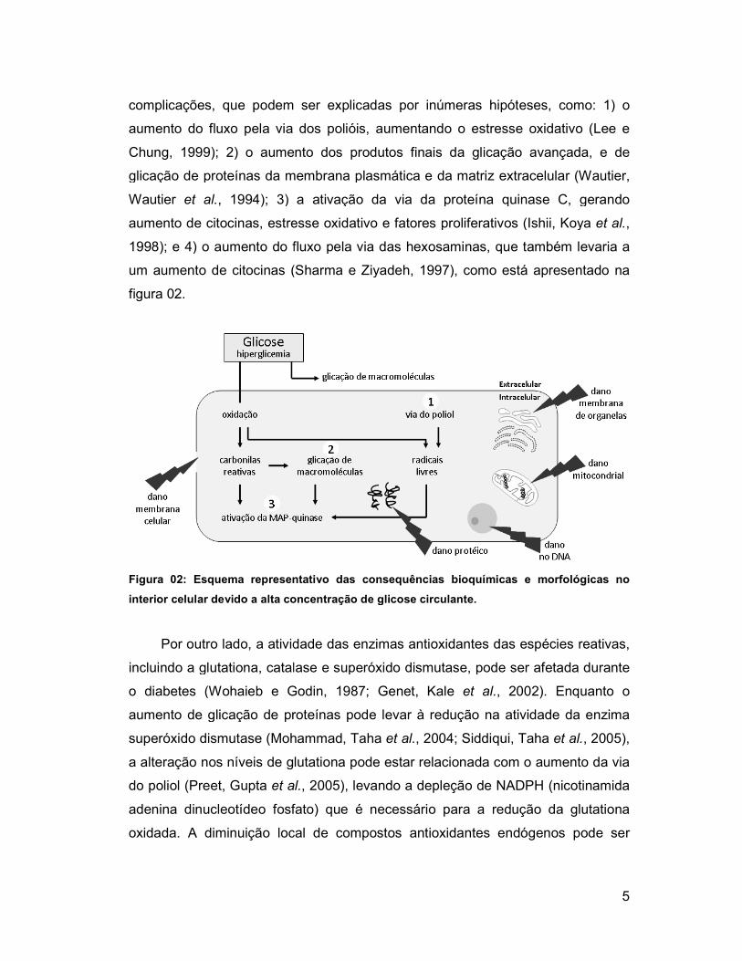
complicações, que podem ser explicadas por inúmeras hipót
aumento do fluxo pela via dos polióis, aumentando o estresse oxidativo
Chung, 1999); 2) o aumento dos produtos finais da glicação avançada, e de
glicação de proteínas da membrana plasmática e da matriz extracelular
Wautier et al., 1994); 3) a ativação da via da proteína quinase C, gerando
aumento de citocinas, estresse oxidativo e fatores proliferativos
1998); e 4) o aumento do fluxo pela via das hexosaminas, que também levaria a
um aumento de citocinas (Sharma e Ziyadeh, 1997)
figura 02.
Figura 02: Esquema representativo d
interior celular devido a alta concentração de glicose circulante.
Por outro lado, a atividade das enzimas antioxidantes das espécies reativas,
incluindo a glutationa, catalase e superóxido dismutase, pode se
o diabetes (Wohaieb e Godin, 1987; Genet, Kale
aumento de glicação de proteínas pode levar à redução na atividade da enzima
superóxido dismutase (Mohammad, Taha
a alteração nos níveis de glutationa pode estar relacionada com o aumento da via
do poliol (Preet, Gupta et al.
adenina dinucleotídeo fosfato) que é necessário para a redução da glutationa
oxidada. A diminuição local de compostos antioxidantes endógenos pode ser
complicações, que podem ser explicadas por inúmeras hipóteses, como: 1) o
aumento do fluxo pela via dos polióis, aumentando o estresse oxidativo
; 2) o aumento dos produtos finais da glicação avançada, e de
glicação de proteínas da membrana plasmática e da matriz extracelular
; 3) a ativação da via da proteína quinase C, gerando
aumento de citocinas, estresse oxidativo e fatores proliferativos (Ishii, Koya
; e 4) o aumento do fluxo pela via das hexosaminas, que também levaria a
(Sharma e Ziyadeh, 1997), como está apresentado na
Figura 02: Esquema representativo das consequências bioquímicas e morfológicas no
interior celular devido a alta concentração de glicose circulante.
Por outro lado, a atividade das enzimas antioxidantes das espécies reativas,
incluindo a glutationa, catalase e superóxido dismutase, pode ser afetada durante
(Wohaieb e Godin, 1987; Genet, Kale et al., 2002). Enquanto o
aumento de glicação de proteínas pode levar à redução na atividade da enzima
(Mohammad, Taha et al., 2004; Siddiqui, Taha et al.
a alteração nos níveis de glutationa pode estar relacionada com o aumento da via
et al., 2005), levando a depleção de NADPH (nicotinamida
adenina dinucleotídeo fosfato) que é necessário para a redução da glutationa
oxidada. A diminuição local de compostos antioxidantes endógenos pode ser
5
eses, como: 1) o
aumento do fluxo pela via dos polióis, aumentando o estresse oxidativo (Lee e
; 2) o aumento dos produtos finais da glicação avançada, e de
glicação de proteínas da membrana plasmática e da matriz extracelular (Wautier,
; 3) a ativação da via da proteína quinase C, gerando
(Ishii, Koya et al.,
; e 4) o aumento do fluxo pela via das hexosaminas, que também levaria a
, como está apresentado na
as consequências bioquímicas e morfológicas no
Por outro lado, a atividade das enzimas antioxidantes das espécies reativas,
r afetada durante
. Enquanto o
aumento de glicação de proteínas pode levar à redução na atividade da enzima
et al., 2005),
a alteração nos níveis de glutationa pode estar relacionada com o aumento da via
nicotinamida
adenina dinucleotídeo fosfato) que é necessário para a redução da glutationa
oxidada. A diminuição local de compostos antioxidantes endógenos pode ser

6
devido ao aumento do consumo pelas espécies reativas, elevando a quantidade
de peróxido de hidrogênio (Ikebuchi, Kashiwagi et al., 1993).
O aumento da peroxidação lipídica já foi demonstrado no cérebro de ratos
diabéticos e ratos envelhecidos (Mooradian e Smith, 1992; Kumar e Menon, 1993;
Leutner, Eckert et al., 2001; Genet, Kale et al., 2002; Siddiqui, Taha et al., 2005;
Sinha, Baquer et al., 2005; Kumar, Taha et al., 2008). Os produtos oriundos da
oxidação de ácidos graxos insaturados e do colesterol podem ser mensurados
para avaliar a peroxidação lipídica que ocorre nos tecidos. Dentre estes produtos,
o aldeído é um dos mais estudados (Esterbauer, Gebicki et al., 1992). Diversos
aldeídos reativos, como o malondialdeído, podem se ligar aos resíduos de
aminoácidos positivamente carregados das apolipoproteínas, principalmente da
lisina, produzindo alterações de cargas na superfície das lipoproteínas (Holvoet,
Perez et al., 1995; Kesavulu, Rao et al., 2001).
1.3 Cérebro e o estresse oxidativo
O cérebro é especialmente susceptível ao dano oxidativo devido: 1) ao
consumo elevado de oxigênio, ATP e glicose; 2) ao seu abundante conteúdo
lipídico; 3) ao líquido cefalorraquidiano conter complexos de ferro e cobre, que
catalisam a formação de radicais hidroxila altamente reativos; 4) à liberação de
espécies reativas do oxigênio durante a oxidação da dopamina e
neurotransmissão glutamatérgica; 5) à interação do óxido nítrico com o radical
superóxido, levando a degeneração neuronal; 6) à baixa quantidade relativa de
enzimas antioxidantes comparado com outros tecidos; 7) aos neurônios serem
células não-replicáveis e qualquer dano ao tecido cerebral pelas espécies reativas
do oxigênio ser acumulativo ao longo do tempo (Sacks, 1965; Olanow, 1993;
Reiter, 1995; Halliwell, 2001; Cui, Luo et al., 2004; Poon, Vaishnav et al., 2006;
Yanardag e Tunali, 2006; Baquer, Taha et al., 2009; Nazaroglu, Sepici-Dincel et
al., 2009). Além disso, vários estudos demonstram que o diabetes está associado
com um aumento nos danos cerebrais provocados pelo estresse oxidativo no
cérebro (Sanders, Rauscher et al., 2001; Ozkaya, Agar et al., 2002; Yanardag e
Tunali, 2006; Celik e Erdogan, 2008; Nazaroglu, Sepici-Dincel et al., 2009). Em
diabéticos, os níveis de glicose são responsávéis pelo aumento de até quatro

7
vezes nos níveis de glicose neuronal. Se isso é persistente ou se torna um evento
regular, o metabolismo da glicose intracelular pode levar a um desbalanço entre a
defesa antioxidante e os danos teciduais no cérebro, assim como nos neurônios
(Pari e Latha, 2004; Tomlinson e Gardiner, 2008). Além disso, a hiperglicemia
pode causar o aumento na produção de radicais livres via auto-oxidação da
glicose e glicação enzimática de proteínas, levando ao dano oxidativo nas
membranas (Wolff, 1993), ativando a apoptose celular e alterando a transmissão
sináptica (Arroba, Frago et al., 2005; Artola, 2008; Tomlinson e Gardiner, 2008).
Estudos sugerem que os radicais livres derivados (radicais superóxido,
peróxido e hidroxila) desempenham papel crucial no diabetes, promovendo a
glicação não-específica de proteínas, peroxidação de lipídios em membranas,
interação de proteínas, deficiência na função de organelas e morte celular. No
entanto, o sistema biológico possui mecanismos de defesa contra essas espécies
reativas do oxigênio, sendo que em condições fisiológicas normais, existe um
balanço entre a produção de espécies reativas do oxigênio e os sistemas
antioxidantes (Taniyama e Griendling, 2003; Robertson, Harmon et al., 2004).
1.4 Antioxidantes
O organismo possui sistemas de defesa antioxidante enzimático e não-
enzimático (Figura 03) que incluem as moléculas que estabilizam as espécies
reativas do oxigênio, ácido úrico, ácido ascórbico, alfa-tocoferol; moléculas que
contêm sulfidrila e enzimas antioxidantes, como o superóxido dismutase, a
catalase e a glutationa peroxidase (Frei, Stocker et al., 1988; Stinefelt, Leonard et
al., 2005). Em condições patológicas, em que a produção excessiva de espécies
reativas do oxigênio supera a defesa antioxidante, o estresse oxidativo pode
modificar irreversivelmente macromoléculas biológicas, como o DNA, as
proteínas, os carboidratos e os lipídeos (Du, Edelstein et al., 2000).
Considerando que o estresse oxidativo desempenha um importante papel
nas complicações do diabetes, a terapia antioxidante tem atraído a atenção de
pesquisadores. Além dos estudos com antioxidantes tradicionais, como vitamina
C, vitamina E e superóxidos miméticos, outras moléculas com ação antioxidante
têm sido investigadas. Vários estudos demonstram o papel de macronutrientes na

8
prevenção do diabetes (Marshall, Hoag et al., 1994; Meyer, Kushi et al., 2000;
Van Dam, Willett et al., 2002; Liu, Serdula et al., 2004; Schulze, Liu et al., 2004;
Tinker, Bonds et al., 2008), mas, por outro lado, existem poucas evidências do
papel das vitaminas e minerais na prevenção primária e secundária desta doença.
Sendo assim, a justificativa para a utilização destes antioxidantes baseia-se em
grande parte, nas experiências com animais e em estudos epidemiológicos
(Kadowaki e Norman, 1984; Beaulieu, Kestekian et al., 1993; Feskens, Virtanen et
al., 1995; Will, Ford et al., 1999; Maestro, Campion et al., 2000; Ford e Mokdad,
2001; Pittas, Lau et al., 2007).
Figura 03: Origem dos antioxidantes celulares. Modificado de Machlin e Bendich, 1987.
Como o diabetes está associado com o aumento do estresse oxidativo
(Wen, Skidmore et al., 2002; Ceriello e Motz, 2004), este fato reforça o interesse
no uso de suplementos antioxidantes como uma tentativa de prevenir as
complicações a longo prazo. No que diz respeito à prevenção do diabetes e a
modificação na dieta, os relatos atuais ainda não permitem que qualquer
recomendação segura e específica seja feita em relação ao uso de suplementos.
Dado que o diabetes é uma condição de estresse oxidativo aumentado, a terapia
antioxidante poderia representar um potencial coadjuvante no tratamento
farmacológico antidiabético.
Apesar do uso de suplementos oferecer benefícios aparentes, ainda são
necessários dados mais consistentes sobre os efeitos benéficos em relação ao

diabetes. Quanto às altas doses de suplementos antioxidantes, já existem dados
que indicam não só a falta do benefício em termos do controle glicêmico, mas
também a progressão das complicações do diabetes e de danos potenciais
(Halliwell, 1995; Hasanain e Mooradian, 2002; Ward, Wu
1.4.1 Antioxidantes enzimáticos
As enzimas antioxidantes têm como principal função eliminar as espécies
reativas do oxigênio e corrigir pequenos
destas moléculas. As alterações na atividade destas enzimas podem ser
consideradas como biomarcadores da
contexto, temos três enzimas: su
peroxidase (Figura 04).
Figura 04: Antioxidantes enzimáticos.
Os processos de formação do superóxido e do peróxido estão
correlacionados, pois o superóxido é convertido em peróxido e oxigênio por uma
reação catalisada pela superóxido dismutase. Esta enzima possui várias
isoformas, diferindo-se quanto à natureza do centro metálico ativo, por sua
constituição em aminoácidos, pelo número de subunidades, pelos seus cofatores
e outras características. Os efeitos citot
limitados por sua degradação pela catalase. Esta enzima, por sua vez, exerce a
função de decompor o peróxido de hidrogênio em água e oxigênio, existindo sob
duas isoformas: selênio-independente e selênio
Gomez et al., 1999), que diferem
natureza ligante do selênio no centro ativo e quanto aos seus mecanismos
catalíticos. O substrato para a reação catalítica da glutationa peroxidase é o
es. Quanto às altas doses de suplementos antioxidantes, já existem dados
a falta do benefício em termos do controle glicêmico, mas
também a progressão das complicações do diabetes e de danos potenciais
(Halliwell, 1995; Hasanain e Mooradian, 2002; Ward, Wu et al., 2007).
1.4.1 Antioxidantes enzimáticos
As enzimas antioxidantes têm como principal função eliminar as espécies
reativas do oxigênio e corrigir pequenos desvios nas concentrações fisiol
destas moléculas. As alterações na atividade destas enzimas podem ser
consideradas como biomarcadores da resposta antioxidante (Sies, 1993)
contexto, temos três enzimas: superóxido dismutase, catalase e glutationa
Figura 04: Antioxidantes enzimáticos.
Os processos de formação do superóxido e do peróxido estão
correlacionados, pois o superóxido é convertido em peróxido e oxigênio por uma
lisada pela superóxido dismutase. Esta enzima possui várias
se quanto à natureza do centro metálico ativo, por sua
constituição em aminoácidos, pelo número de subunidades, pelos seus cofatores
e outras características. Os efeitos citotóxicos do peróxido de hidrogênio são
limitados por sua degradação pela catalase. Esta enzima, por sua vez, exerce a
função de decompor o peróxido de hidrogênio em água e oxigênio, existindo sob
independente e selênio-dependente (Mates, Perez
, que diferem-se quanto ao número de subunidades, a
natureza ligante do selênio no centro ativo e quanto aos seus mecanismos
catalíticos. O substrato para a reação catalítica da glutationa peroxidase é o
9
es. Quanto às altas doses de suplementos antioxidantes, já existem dados
a falta do benefício em termos do controle glicêmico, mas
também a progressão das complicações do diabetes e de danos potenciais
As enzimas antioxidantes têm como principal função eliminar as espécies
desvios nas concentrações fisiológicas
destas moléculas. As alterações na atividade destas enzimas podem ser
(Sies, 1993). Neste
peróxido dismutase, catalase e glutationa
Os processos de formação do superóxido e do peróxido estão
correlacionados, pois o superóxido é convertido em peróxido e oxigênio por uma
lisada pela superóxido dismutase. Esta enzima possui várias
se quanto à natureza do centro metálico ativo, por sua
constituição em aminoácidos, pelo número de subunidades, pelos seus cofatores
óxicos do peróxido de hidrogênio são
limitados por sua degradação pela catalase. Esta enzima, por sua vez, exerce a
função de decompor o peróxido de hidrogênio em água e oxigênio, existindo sob
(Mates, Perez-
se quanto ao número de subunidades, a
natureza ligante do selênio no centro ativo e quanto aos seus mecanismos
catalíticos. O substrato para a reação catalítica da glutationa peroxidase é o
10
peróxido de hidrogênio ou o peróxido orgânico, que são decompostos em água ou
álcool. A glutationa peroxidase compete com a catalase pelo peróxido de
hidrogênio como substrato e é a principal fonte de proteção contra as espécies
reativas do oxigênio (Valko, Rhodes et al., 2006).
Em diabéticos, as atividades da superóxido dismutase e da catalase,
parecem estar diminuídas no cérebro de ratos (Kumar e Menon, 1993; Makar,
Rimpel-Lamhaouar et al., 1995); enquanto que no cérebro de camundongos foi
observado o aumento na atividade desta enzima (Huang, Juang et al., 1999). Isso
demonstra o quanto os resultados referentes ao estresse oxidativo são variáveis
entre as espécies.
1.4.2 Antioxidantes não-enzimáticos
Vários compostos não-enzimáticos, como a glutationa reduzida, incluindo
os carotenóides e as vitaminas A, C e E, têm sido relatados por possuírem
propriedade antioxidante no plasma e em tecidos (Frei, Stocker et al., 1988), além
de alguns minerais, como zinco, magnésio e selênio (Martini, Catania et al.,
2010).
Os tocoferóis são chamados antioxidantes primários porque interrompem
diretamente a oxidação, convertendo os radicais livres em espécies mais
estáveis. A vitamina E (tocoferol-OH) é um clássico exemplo de antioxidante que
limita os efeitos deletérios das reações oxidantes, interrompendo as reações em
cadeia iniciadas pelos radicais livres, doando um átomo de hidrogênio para um
radical peroxil para formar peróxido lipídico, impedindo o dano oxidativo (Burton e
Traber, 1990; Martini, Catania et al., 2010). Contudo, quando a vitamina E age,
são gerados radicais tocoferoxila e a regeneração é requerida, a fim de evitar
indesejáveis processos oxidativos mediados pela tocoferoxila. Assim, os
resultados contraditórios de estudos com vitamina E, podem ser devido à falta de
avaliação deste sistema de regeneração, composta de ácido ascórbico, glutationa
reduzida e co-enzima Q10 (Nwose, Jelinek et al., 2008).
Estudos recentes de diabetes em animais mostram que o uso da vitamina
E reduz o risco da doença e suas complicações (Sena, Nunes et al., 2008;
Shirpoor, Salami et al., 2009), inclusive tendo efeito protetor no cérebro (Kabay,

11
Ozden et al., 2009). No entanto, uma importante limitação destes estudos
observacionais é que não se consegue distinguir claramente se o menor risco da
doença, associado com os altos níveis de vitamina E, é devido à suplementação
da vitamina ou a outros fatores ligados ao estilo de vida, tais como o aumento da
prática de exercício físico e uma dieta mais saudável. Sendo assim, esses
ensaios clínicos não confirmam os benefícios da vitamina E, de forma isolada, na
prevenção e/ou tratamento do diabetes.
O zinco não é considerado um antioxidante como a vitamina E.
Entretanto, este mineral pode limitar os danos induzidos pelo estresse oxidativo
(Bunk, Dnistrian et al., 1989; Noh e Koo, 2001), estabilizando a estrutura da
membrana plasmática (Bray e Bettger, 1990), restringindo a produção de radicais
livres endógenos (Bray e Bettger, 1990; Bell, Sakanashi et al., 1998), contribuindo
na estrutura da enzima superóxido dismutase (Marklund, 1982; Davis, Klevay et
al., 1998), e mantendo a concentração de metalotioneína tecidual, também
considerado um protetor contra os danos oxidativos (Cousins, 1985; Ebadi,
Leuschen et al., 1996; Rojas, Cerutis et al., 1996; El Refaey, Ebadi et al., 1997).
Além disso, em vários sistemas, o zinco pode antagonizar a propriedade catalítica
dos metais de transição, ferro e cobre, no que diz respeito à sua capacidade de
promover a formação de hidroxilas pelo peróxido de hidrogênio e superóxido
(Powell, 2000).
No cérebro de diabéticos, estudos mostraram que o tratamento com zinco
reduz significativamente a astrocitose (Wei, Liu et al., 2009) e previne a apoptose
induzida (Thomas e Caffrey, 1991; Matsushita et al., 1996) sugerindo ser um
tratamento potencial na prevenção dos efeitos deletérios do diabetes e na
redução do estresse oxidativo (Santon, Formigari et al., 2006). Por outro lado,
outros estudos comprovaram que a suplementação de zinco induz à apoptose
(Weissgarten, Berman et al., 2002; Chang, Torzillo et al., 2006). Wiseman, Wells
et al., (2007) revelaram que o aumento de zinco intracelular é um evento chave
associado à interrupção da função mitocondrial, além de induzir vias apoptóticas
mediadas pelo peróxido de hidrogênio. Além do mais, existem evidências de que
a ingestão excessiva de zinco pode induzir a uma condição patológica associada
ao estresse oxidativo (Yanagisawa, Sato et al., 2004).

12
Estudos clínicos sugerem que a suplementação com o cálcio pode afetar
indiretamente o metabolismo da glicose (Zemel, Thompson et al., 2004; Zemel,
Donnelly et al., 2008) prevenindo o Diabetes mellitus do tipo II. Desta forma, o
potencial papel da suplementação de cálcio na prevenção primária do diabetes
tem sido investigado (Pittas, Lau et al., 2007; De Boer, Tinker et al., 2008).
1.5 Cálcio/Calmodulina
Ambos os diabetes, tipo I e tipo II, estão associados com os distúrbios na
regulação do cálcio intracelular. A hiperglicemia causa uma mudança nos níveis
de cálcio citosólico devido a um aumento no influxo deste íon e a mobilização do
mesmo no interior de algumas células, promovendo a estocagem. Além disso, a
hiperglicemia tem sido associada a um decréscimo do efluxo de íons cálcio da
célula (Massry e Smogorzewski, 1997). A combinação do aumento do influxo e o
decréscimo do efluxo de cálcio leva a um aumento nos níveis basais citosólicos
que pode, consequentemente, afetar a função celular (Figura 05). Deste modo, o
aumento de cálcio citosólico durante o diabetes está ligado à falha de vários
componentes de transdução de sinal podendo levar a um prejuízo neuronal
associado com o estado patológico, aumentando a morte celular em áreas
específicas do cérebro (Klein, Hains et al., 2004), e podendo alterar os níveis e a
função de proteínas ligantes de cálcio/calmodulina.
O aumento da entrada de cálcio pode acontecer devido à ativação de canais
de cálcio mediado pela proteína-G, levando a estimulação de várias vias
celulares, incluindo a via da proteína quinase dependente de AMPc e canais de
cálcio, o sistema da proteína quinase e o da fosfolipase C, estabelecendo um
novo processo patológico sujeito à disfunção celular durante as condições
hiperglicêmicas (Demerdash, Seyrek et al., 1996; Massry e Smogorzewski, 1997).
Por outro lado, Bhardwaj e Kaur (1999) demostraram em diabetes, um aumento
da atividade do AMPc e da proteína kinase A, e uma diminuição da atividade da
fosfolipase A2 em terminações nervosas, que são mediadas pela ativação da
proteína quinase II dependente de cálcio/calmodulina (CaMKII) (Piomelli e
Greengard, 1991).
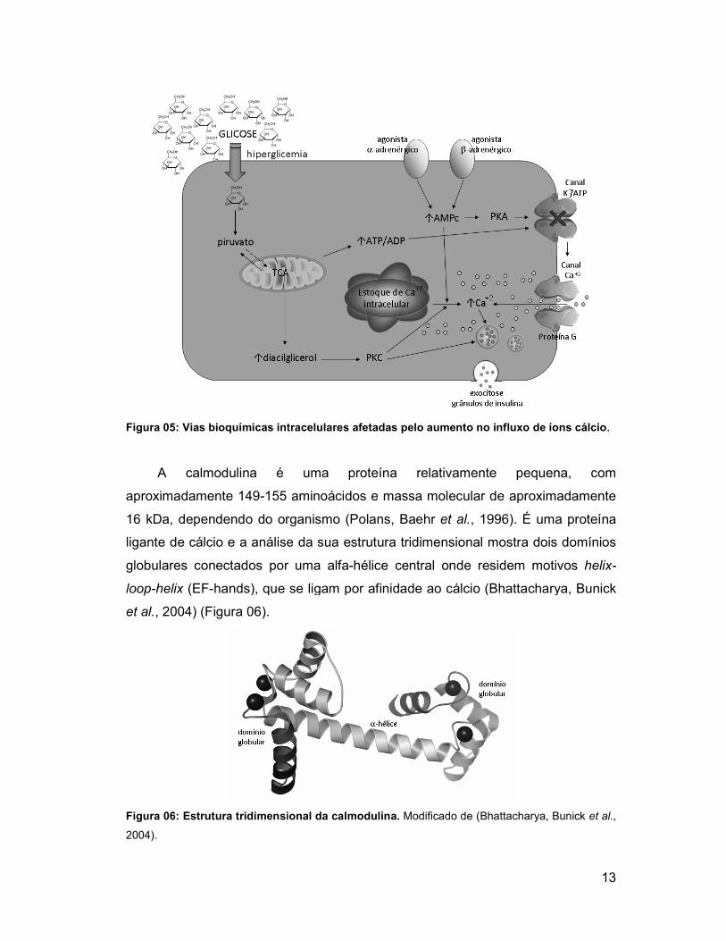
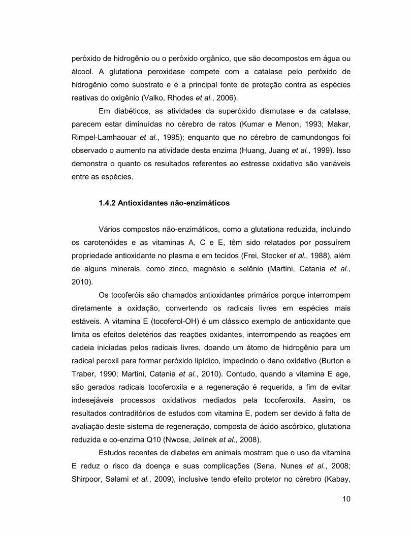
Figura 05: Vias bioquímicas intracelulares afetadas pelo aumento no influxo de íons cálcio
A calmodulina é uma proteína relativamente pequena, com
aproximadamente 149-155 aminoácidos
16 kDa, dependendo do organismo
ligante de cálcio e a análise
globulares conectados por uma alfa
loop-helix (EF-hands), que se ligam por afinidade ao cálcio
et al., 2004) (Figura 06).
Figura 06: Estrutura tridimensional da calmodulina.
2004).
ias bioquímicas intracelulares afetadas pelo aumento no influxo de íons cálcio
A calmodulina é uma proteína relativamente pequena, com
155 aminoácidos e massa molecular de aproximadamente
16 kDa, dependendo do organismo (Polans, Baehr et al., 1996). É uma proteína
análise da sua estrutura tridimensional mostra dois domínios
globulares conectados por uma alfa-hélice central onde residem motivos
hands), que se ligam por afinidade ao cálcio (Bhattacharya, Bunick
Figura 06: Estrutura tridimensional da calmodulina. Modificado de (Bhattacharya, Bunick
13
ias bioquímicas intracelulares afetadas pelo aumento no influxo de íons cálcio.
A calmodulina é uma proteína relativamente pequena, com
e massa molecular de aproximadamente
. É uma proteína
da sua estrutura tridimensional mostra dois domínios
onde residem motivos helix-
(Bhattacharya, Bunick
(Bhattacharya, Bunick et al.,
A comparação da estrutura e função da calmodulina em diferentes
organismos indica que essa molécula é altamente conservada
et al., 1975; Dedman, Welsh
durante a evolução, embora algumas espécies possuam mais de uma isoforma.
Uma prova disto é a variação em um pequeno número de aminoácidos
funcionalmente idênticos (Klee, Draetta
e a de microorganismos eucarióticos.
A calmodulina está diretamente relacionada com a secreção de insulina no
pâncreas (Norling, Colca et al.
Epstein et al., 1995), afetando os alvos de
coração, tecido adiposo, rim e músculo esquelético
Hoskins e Scott, 1983; Solomon, Palazzolo
Solomon, Palazzolo et al., 1994)
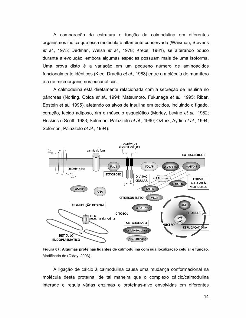
Figura 07: Algumas proteínas ligantes de calmodulina com sua localização celular e função.
Modificado de (O'day, 2003).
A ligação de cálcio à calmodulina causa uma mudança c
molécula desta proteína, de tal maneira que o complexo
interage e regula várias enzimas e proteínas
A comparação da estrutura e função da calmodulina em diferentes
organismos indica que essa molécula é altamente conservada (Waisman, Stevens
, 1975; Dedman, Welsh et al., 1978; Krebs, 1981), se alterando pouco
durante a evolução, embora algumas espécies possuam mais de uma isoforma.
Uma prova disto é a variação em um pequeno número de aminoácidos
(Klee, Draetta et al., 1988) entre a molécula de mamífero
s eucarióticos.
A calmodulina está diretamente relacionada com a secreção de insulina no
et al., 1994; Matsumoto, Fukunaga et al., 1995; Ribar,
, afetando os alvos de insulina em tecidos, incluindo o fígado,
coração, tecido adiposo, rim e músculo esquelético (Morley, Levine
Hoskins e Scott, 1983; Solomon, Palazzolo et al., 1990; Ozturk, Aydin
, 1994).
Figura 07: Algumas proteínas ligantes de calmodulina com sua localização celular e função.
à calmodulina causa uma mudança conformacional na
molécula desta proteína, de tal maneira que o complexo cálcio/calmodulina
interage e regula várias enzimas e proteínas-alvo envolvidas em diferentes
14
A comparação da estrutura e função da calmodulina em diferentes
(Waisman, Stevens
, se alterando pouco
durante a evolução, embora algumas espécies possuam mais de uma isoforma.
Uma prova disto é a variação em um pequeno número de aminoácidos
entre a molécula de mamífero
A calmodulina está diretamente relacionada com a secreção de insulina no
, 1995; Ribar,
insulina em tecidos, incluindo o fígado,
et al., 1982;
et al., 1994;
Figura 07: Algumas proteínas ligantes de calmodulina com sua localização celular e função.
onformacional na
/calmodulina
alvo envolvidas em diferentes

15
aspectos da atividade celular (Figura 07), como síntese e degradação de
nucleotídeos, transcrição de genes, regulação de diferentes sistemas de
transporte, controle do metabolismo celular, organização do citoesqueleto,
citocinese, contração muscular, regulação do volume osmótico, endocitose e
exocitose, fertilização do zigoto, comunicação intercelular, proliferação celular,
diferenciação e apoptose (Cheung, 1980; Klee e Vanaman, 1982; Means, Tash et
al., 1982; Carafoli, 1987; Babu, Bugg et al., 1988; Espindola, Espreafico et al.,
1992; Chamberlain, Roth et al., 1995; Carafoli, Nicotera et al., 1997; Colombo,
Beron et al., 1997; Chin e Means, 2000; Carafoli, Santella et al., 2001).
1.6 Proteínas ligantes de cálcio/calmodulina
As proteínas ligantes de calmodulina compreendem um grupo diversificado.
Essa interação é regulada usualmente pelo nível citoplasmático de íons cálcio e
baseado nisso é possível que as proteínas ligantes se classifiquem em três
categorias: Ca+2-dependente, Ca+2-independente e Ca+2-inibido (O'day, 2003).
Algumas proteínas Ca+2-dependentes têm um ou mais domínios ligantes de
calmodulina com aproximadamente 20 resíduos de aminoácido, e têm sido
agrupadas em dois motivos relacionados, baseados na posição dos resíduos
hidrofóbicos conservados (Crivici e Ikura, 1995; Rhoads e Friedberg, 1997), como
1-8-14 (Dasgupta, Honeycutt et al., 1989) e 1-5-10 (Picciotto, Czernik et al., 1993).
Por outro lado, a calmodulina também pode se ligar às proteínas-alvo de maneira
Ca+2-independente através de uma sequência repetida de isoleucina e glutamina
(IQxxxRGxxxR), também chamada de motivo IQ. Em algumas regiões do cérebro
de rato existem poucas proteínas ligantes de calmodulina Ca+2-independentes,
mas um grande número de proteínas Ca+2-dependentes (O'day, Lydan et al.,
2001; O'day, Payne et al., 2001).
Dentre as proteínas ligantes de calmodulina, tem-se a CaMKII que é o
principal mediador neuronal de sinalização via cálcio, integrando múltiplas
funções relacionadas. Esta molécula, que parece ser relativamente vulnerável em
estágios patológicos, está associada com o influxo de cálcio dentro da célula e
está implicada numa variedade de eventos em neurônios, como na liberação e

16
síntese de neurotransmissores e canais iônicos, e na expressão gênica (Bading,
Ginty et al., 1993; Kitamura, Miyazaki et al., 1993; Blanquet e Lamour, 1997).
A literatura revela a isquemia e a hipoglicemia associadas a uma modulação
permanente da atividade da CaMKII (Hu, Kurihara et al., 1995; Hu e Wieloch,
1995; Kolb, Hudmon et al., 1995), mostrando que a elevação dos níveis de cálcio
intracelular em cultura de neurônios resulta na autofosforilação da CaMKII e
produz a forma da enzima independente de cálcio (Fukunaga, Rich et al., 1989;
Fukunaga, Soderling et al., 1992). Em um estado hiperglicêmico, os níveis da
CaMKII e a sua atividade estão aumentados em diferentes regiões do cérebro
(Bhardwaj e Kaur, 1999). As alterações drásticas da atividade desta enzima
podem ser atribuídas aos fatores de modificação covalente ou interações
endógenas do inibidor/ativador. No entanto, é também sugerido que o mecanismo
que envolve a CaMKII em funções neuronais esteja relacionado com a regulação
da expressão gênica.
Berggard, Arrigoni et al., (2006) identificaram 18 proteínas ligantes de
calmodulina em cérebro de camundongo envolvidas em função do citoesqueleto,
como actina, dineína, mielina, espectrina e tubulina. Além destas proteínas do
citoesqueleto e motores moleculares, algumas miosinas também se ligam a
calmodulina pelo motivo IQ (Hoyt, Hyman et al., 1997).
1.7 Miosinas
A superfamília miosina, baseado em análise do domínio motor, possui pelo
menos 20 classes (Berg, Powell et al., 2001; Krendel e Mooseker, 2005), apesar
de dados filogenéticos revelarem pelo menos 35 classes (Odronitz e Kollmar,
2007), e por análises do sequenciamento genômico cerca de 40 classes de
miosinas (Richards e Cavalier-Smith, 2005).
As miosinas são proteínas motoras conservadas, encontradas em todos os
eucariotos de levedura a mamíferos, possuindo atividade ATPase que converte a
energia de hidrólise da adenosina trifosfato (ATP) em movimento quando ligada à
actina. Muitas das cadeias pesadas de miosinas consistem de três domínios: 1) a
cabeça globular N-terminal ou domínio motor catalítico, que possui sítios ligantes
de ATP e actina; 2) região do pescoço, onde se ligam as cadeias leves de miosina

e/ou calmodulina, consistindo de um ou mais motivos IQ; 3) e uma cauda C
terminal, capaz de se ligar às cargas que serão transportadas ou ainda interagir
com o domínio cauda de outras miosinas. Vários estudos têm indicado este
domínio como o mais divergente entre as classes de miosinas, o que confere
diferentes funções celulare
1997; Buss, Spudich et al., 2004; Krendel e Mooseker, 2005)
Embora as miosinas desempenhem diversos papéis, incluindo o movimento
de organelas, endocitose, exocit
em diferentes tipos celulares
somente as das classes I, II, V, VI, IX e XVIII participam de funções específicas
nos neurônios de vertebrados
também envolvidas em patologias, como a miosina
Seidman, 2001), perda de aud
(Heath, Campos-Barros et al.
(Pastural, Barrat et al., 1997; Westbroek, Lambert
(De Souza Martins, Romao
Figura 08: Funções das miosinas no citoplasma da célula.
(1998).
e/ou calmodulina, consistindo de um ou mais motivos IQ; 3) e uma cauda C
terminal, capaz de se ligar às cargas que serão transportadas ou ainda interagir
com o domínio cauda de outras miosinas. Vários estudos têm indicado este
domínio como o mais divergente entre as classes de miosinas, o que confere
diferentes funções celulares a estes motores moleculares (Hoyt, Hyman
, 2004; Krendel e Mooseker, 2005).
Embora as miosinas desempenhem diversos papéis, incluindo o movimento
de organelas, endocitose, exocitose, transporte de RNAm e transdução de sinal
em diferentes tipos celulares (Titus, 1997; Mermall, Post et al., 1998)
das classes I, II, V, VI, IX e XVIII participam de funções específicas
os neurônios de vertebrados (Bridgman e Elkin, 2000; Bridgman, 2004)
patologias, como a miosina-II em miopatias
, perda de audição (Avraham, 2002) e macrotrombocitopenia
et al., 2001), e a miosina-V na Síndrome de Griscelli
, 1997; Westbroek, Lambert et al., 2001) e hipotiroidismo
et al., 2009).
Figura 08: Funções das miosinas no citoplasma da célula. Modificado de Mermall, Post
17
e/ou calmodulina, consistindo de um ou mais motivos IQ; 3) e uma cauda C-
terminal, capaz de se ligar às cargas que serão transportadas ou ainda interagir
com o domínio cauda de outras miosinas. Vários estudos têm indicado este
domínio como o mais divergente entre as classes de miosinas, o que confere
(Hoyt, Hyman et al.,
Embora as miosinas desempenhem diversos papéis, incluindo o movimento
ose, transporte de RNAm e transdução de sinal
(Figura 08),
das classes I, II, V, VI, IX e XVIII participam de funções específicas
(Bridgman e Elkin, 2000; Bridgman, 2004), estando
II em miopatias (Seidman e
e macrotrombocitopenia
V na Síndrome de Griscelli
e hipotiroidismo
Mermall, Post et al.
18
As miosinas também estão envolvidas no tráfego de transportadores de
glicose (GLUT) (Huang e Czech, 2007). Neste sentido, a miosina-Ic (Bose,
Guilherme et al., 2002; Bose, Robida et al., 2004; Huang, Lifshitz et al., 2004;
Huang, Imamura et al., 2005) e a miosina-Va (Yoshizaki, Imamura et al., 2007)
foram encontradas mediando o transporte de vesículas de GLUT4, e a miosina-VI
com o GLUT1 (Reed, Cefalu et al., 2005). Além disso, Yoshizaki, Imamura et al.,
(2007) demonstraram que a insulina estimula a fosforilação da miosina-Va
aumentando a afinidade da proteína aos filamentos de actina, considerando que a
inibição da sua função possa levar a um bloqueio do transporte de glicose
estimulado pela insulina.
Recentemente, uma nova miosina foi descoberta em mitocôndria, sendo
designada Myo19 (Quintero, Divito et al., 2009), que possui aproximadamente
35% de identidade com a sequência do domínio motor das miosinas das classes
V e VI de humano, estando expressa em células, tecidos e tumores de
vertebrados. Além disso, foi revelado que este motor molecular participa da
dinâmica mitocondrial normal, possui três motivos IQ e não é regulado pela
fosforilação da sua cadeia pesada.
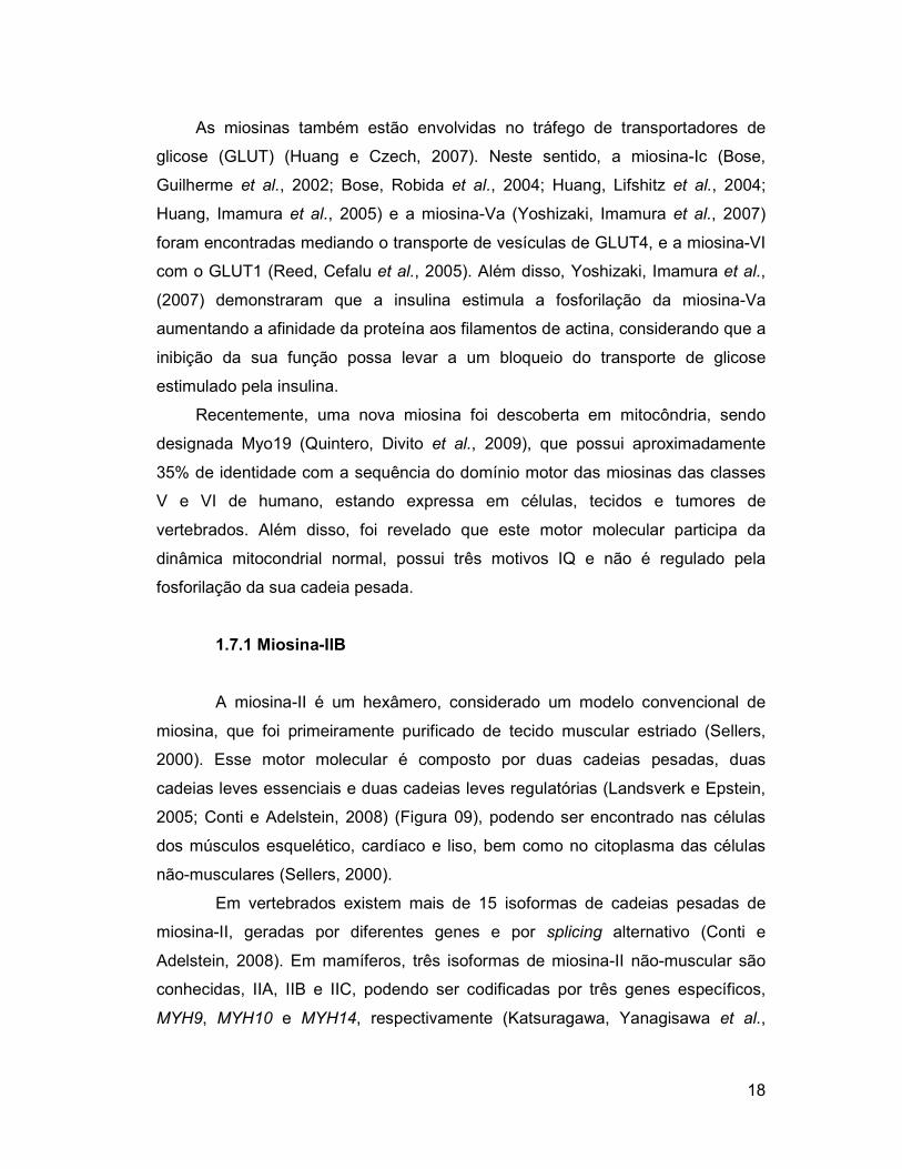
1.7.1 Miosina-IIB
A miosina-II é um hexâmero, considerado um modelo convencional de
miosina, que foi primeiramente purificado de tecido muscular estriado (Sellers,
2000). Esse motor molecular é composto por duas cadeias pesadas, duas
cadeias leves essenciais e duas cadeias leves regulatórias (Landsverk e Epstein,
2005; Conti e Adelstein, 2008) (Figura 09), podendo ser encontrado nas células
dos músculos esquelético, cardíaco e liso, bem como no citoplasma das células
não-musculares (Sellers, 2000).
Em vertebrados existem mais de 15 isoformas de cadeias pesadas de
miosina-II, geradas por diferentes genes e por splicing alternativo (Conti e
Adelstein, 2008). Em mamíferos, três isoformas de miosina-II não-muscular são
conhecidas, IIA, IIB e IIC, podendo ser codificadas por três genes específicos,
MYH9, MYH10 e MYH14, respectivamente (Katsuragawa, Yanagisawa et al.,

1989; Kawamoto e Adelstein, 1991; Simons, Wang
Berg, Powell et al., 2001; Golomb, Ma
Figura 09: Esquema da molécula de miosina
essencial (ELC) e regulatória (RLC).
As isoformas IIA e IIB se localizam diferencialmente dentro d
essa distribuição sugere que as duas proteínas tenham importâncias funcionais
distintas (Maupin, Phillips et al.
al., 1996), estando amplamente exp
cordão espinhal (Kawamoto e Adelstein, 1991; Miller, Bower
Adelstein, 1995).
Em células não-musculares, a miosina
variam desde a citocinese, migração neuronal e prolongamento de neuritos, até
tráfego de membrana dentro da célula, exocitose e transporte de organelas em
axônios (De Lozanne e Spudich, 1987; Knecht e Loomis, 1987; Mochida,
Kobayashi et al., 1994; Mochida, 1995; Wylie, Wu
al., 2001; Wylie e Chantler, 2001; Degiorgis, Reese
As atividades funcionais das cadeias leves e pesadas da miosina
reguladas por fosforilação (Bresnick, 1999)
que podem afetar outros aspectos da dinâmica do citoesqueleto, incluindo as
alterações no movimento, divisão celular ou secreção
as formas de miosina, as da classe II têm sido estudadas mais extensivamente e
parecem ter papel na organização e comportamento do citoesqueleto de cones de
crescimento (Vallee, Seale
total ou parcial desta miosina pode levar a danos no sistema nervoso em
resultado de um defeito na migração celular
Dentre as isoformas, a IIB é a mai
Mehta et al., 1991) e possui localização cortical no corpo celular e axônio,
funcionando como um mediador
1989; Kawamoto e Adelstein, 1991; Simons, Wang et al., 1991; Bresnick, 1999;
2001; Golomb, Ma et al., 2004).
Figura 09: Esquema da molécula de miosina-II com a sua cadeia pesada e cadeias leves
essencial (ELC) e regulatória (RLC). Modificado de Lowey e Trybus, 2010.
As isoformas IIA e IIB se localizam diferencialmente dentro d
essa distribuição sugere que as duas proteínas tenham importâncias funcionais
et al., 1994; Rochlin, Itoh et al., 1995; Kelley, Sellers
, estando amplamente expressas em neurônios do córtex, cerebelo e
(Kawamoto e Adelstein, 1991; Miller, Bower et al., 1992; Itoh e
musculares, a miosina-II tem diversas funções, que
esde a citocinese, migração neuronal e prolongamento de neuritos, até
tráfego de membrana dentro da célula, exocitose e transporte de organelas em
(De Lozanne e Spudich, 1987; Knecht e Loomis, 1987; Mochida,
, 1994; Mochida, 1995; Wylie, Wu et al., 1998; Tullio, Bridgman
, 2001; Wylie e Chantler, 2001; Degiorgis, Reese et al., 2002).
As atividades funcionais das cadeias leves e pesadas da miosina
(Bresnick, 1999), envolvendo diferentes vias e enzimas
que podem afetar outros aspectos da dinâmica do citoesqueleto, incluindo as
alterações no movimento, divisão celular ou secreção (Spudich, 1994)
as formas de miosina, as da classe II têm sido estudadas mais extensivamente e
parecem ter papel na organização e comportamento do citoesqueleto de cones de
et al., 2009). Além disso, já foi descrito que a remoção
total ou parcial desta miosina pode levar a danos no sistema nervoso em
a migração celular (Brown e Bridgman, 2004)
Dentre as isoformas, a IIB é a mais enriquecida em cérebro
e possui localização cortical no corpo celular e axônio,
funcionando como um mediador da motilidade em cones de crescimento
19
, 1991; Bresnick, 1999;
II com a sua cadeia pesada e cadeias leves
As isoformas IIA e IIB se localizam diferencialmente dentro das células e
essa distribuição sugere que as duas proteínas tenham importâncias funcionais
, 1995; Kelley, Sellers et
ressas em neurônios do córtex, cerebelo e
, 1992; Itoh e
II tem diversas funções, que
esde a citocinese, migração neuronal e prolongamento de neuritos, até
tráfego de membrana dentro da célula, exocitose e transporte de organelas em
(De Lozanne e Spudich, 1987; Knecht e Loomis, 1987; Mochida,
, 1998; Tullio, Bridgman et
As atividades funcionais das cadeias leves e pesadas da miosina-IIB são
, envolvendo diferentes vias e enzimas
que podem afetar outros aspectos da dinâmica do citoesqueleto, incluindo as
(Spudich, 1994). De todas
as formas de miosina, as da classe II têm sido estudadas mais extensivamente e
parecem ter papel na organização e comportamento do citoesqueleto de cones de
. Além disso, já foi descrito que a remoção
total ou parcial desta miosina pode levar a danos no sistema nervoso em
(Brown e Bridgman, 2004).
s enriquecida em cérebro (Murakami,
e possui localização cortical no corpo celular e axônio,
da motilidade em cones de crescimento (Cheng,

20
Murakami et al., 1992; Rochlin, Itoh et al., 1995). Além disso, a cadeia leve desta
miosina pode interagir com as subunidades do receptor NMDA (Husi, Ward et al.,
2000; Amparan, Avram et al., 2005), funcionando como um importante regulador
da morfologia dos dendritos neuronais (Ryu, Liu et al., 2006).
1.7.2 Miosina-Va
A miosina-V foi inicialmente caracterizada como uma proteína ligante de
calmodulina no cérebro, com várias propriedades bioquímicas semelhantes às
miosinas (Larson, Pitta et al., 1988; Larson, Espindola et al., 1990; Espindola,
Espreafico et al., 1992; Cheney, O'shea et al., 1993; Coelho e Larson, 1993;
Nascimento, Cheney et al., 1996)
A cadeia pesada de miosina-V consiste de três domínios (Figura 09): 1)
domínio motor com duas cadeias pesadas com, aproximadamente, 212 kDa
(Espreafico, Cheney et al., 1992; Cheney, O'shea et al., 1993) e com alta
afinidade pela actina na presença de ATP (Espreafico, Cheney et al., 1992;
Cheney, O'shea et al., 1993); 2) domínio pescoço, contendo seis sítios ligantes de
cadeias leves, com 4 a 5 moléculas de calmodulina para cada cadeia pesada
(Espreafico, Cheney et al., 1992; Cheney, O'shea et al., 1993), e duas cadeias
leves essenciais de 17 kDa e 23 kDa; 3) domínio cauda dividido em duas regiões:
um domínio globular C-terminal (Espreafico, Cheney et al., 1992; Cheney, O'shea
et al., 1993) e uma região alfa-helicoidal coiled-coil que está envolvida na
dimerização e possui uma sequência PEST (Rogers, Wells et al., 1986;
Espreafico, Cheney et al., 1992) rica em aminoácidos prolina, ácido glutâmico,
serina e treonina, considerado um importante sítio para proteólise mediada pela
calpaína (Rechsteiner e Rogers, 1996). A esta região também se liga uma cadeia
leve de dineína (8-10 kDa) (Benashski, Harrison et al., 1997; Espindola, Suter et
al., 2000; Hodi, Nemeth et al., 2006) que possivelmente estabiliza a interação
entre as cadeias pesadas, e auxilia na ligação da carga à miosina-V (Reck-
Peterson, Provance et al., 2000).
Na presença de íons cálcio, a atividade ATPase da miosina-V é
aumentada, permanecendo-se numa conformação mais compactada que permite
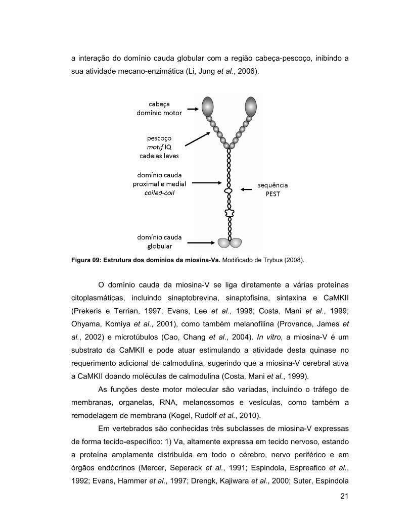
a interação do domínio cauda globular com a região cabeça
sua atividade mecano-enzimática
Figura 09: Estrutura dos domínios da miosina
O domínio cauda da miosina
citoplasmáticas, incluindo sinaptobrevina, sinaptofisina, sintaxina e CaMKII
(Prekeris e Terrian, 1997; Evans, Lee
Ohyama, Komiya et al., 2001)
al., 2002) e microtúbulos (Cao, Chang
substrato da CaMKII e pode atuar estimulando a atividade desta quinase no
requerimento adicional de calmodulina, sugerindo que a miosina
a CaMKII doando moléculas de calmodulina
As funções deste motor molecular são variadas, incluindo o tráfego de
membranas, organelas, RNA, melanossomos e vesículas, como também a
remodelagem de membrana
Em vertebrados são conhecidas três subclasses de miosina
de forma tecido-específico: 1) Va, altamente expressa em tecido nervoso, estando
a proteína amplamente distribuída em todo o cérebro, nervo periférico e em
órgãos endócrinos (Mercer, Seperack
1992; Evans, Hammer et al.
a interação do domínio cauda globular com a região cabeça-pescoço, inibindo a
enzimática (Li, Jung et al., 2006).
: Estrutura dos domínios da miosina-Va. Modificado de Trybus (2008).
O domínio cauda da miosina-V se liga diretamente a várias proteínas
citoplasmáticas, incluindo sinaptobrevina, sinaptofisina, sintaxina e CaMKII
(Prekeris e Terrian, 1997; Evans, Lee et al., 1998; Costa, Mani et al.
, 2001), como também melanofilina (Provance, James
(Cao, Chang et al., 2004). In vitro, a miosina
substrato da CaMKII e pode atuar estimulando a atividade desta quinase no
ento adicional de calmodulina, sugerindo que a miosina-V cerebral ativa
a CaMKII doando moléculas de calmodulina (Costa, Mani et al., 1999).
As funções deste motor molecular são variadas, incluindo o tráfego de
branas, organelas, RNA, melanossomos e vesículas, como também a
remodelagem de membrana (Kogel, Rudolf et al., 2010).
Em vertebrados são conhecidas três subclasses de miosina-V expressas
específico: 1) Va, altamente expressa em tecido nervoso, estando
a proteína amplamente distribuída em todo o cérebro, nervo periférico e em
(Mercer, Seperack et al., 1991; Espindola, Espreafico
et al., 1997; Drengk, Kajiwara et al., 2000; Suter, Espindola
21
pescoço, inibindo a
V se liga diretamente a várias proteínas
citoplasmáticas, incluindo sinaptobrevina, sinaptofisina, sintaxina e CaMKII
et al., 1999;
(Provance, James et
, a miosina-V é um
substrato da CaMKII e pode atuar estimulando a atividade desta quinase no
V cerebral ativa
.
As funções deste motor molecular são variadas, incluindo o tráfego de
branas, organelas, RNA, melanossomos e vesículas, como também a
V expressas
específico: 1) Va, altamente expressa em tecido nervoso, estando
a proteína amplamente distribuída em todo o cérebro, nervo periférico e em
, 1991; Espindola, Espreafico et al.,
, 2000; Suter, Espindola

22
et al., 2000; Rose, Lejen et al., 2002; Rudolf, Kogel et al., 2003; Varadi, Tsuboi et
al., 2005; Watanabe, Nomura et al., 2005; Espindola, Banzi et al., 2008) 2) Vb,
presente em diferentes tecidos (Zhao, Koslovsky et al., 1996), incluindo o epitelial,
mas com distribuição limitada no cérebro, especialmente no hipocampo, giro
denteado, amígdala e córtex (Zhao, Koslovsky et al., 1996; Lapierre, Kumar et al.,
2001; Swiatecka-Urban, Talebian et al., 2007); 3) Vc, presente em tecidos
exócrinos, como o pâncreas, a próstata e a glândula mamária, além do cerebelo
(Bridgman e Elkin, 2000; Rodriguez e Cheney, 2002; Marchelletta, Jacobs et al.,
2008; Jacobs, Weigert et al., 2009).
O cérebro possui grande quantidade de miosina-Va, o que sugere um
envolvimento na transmissão sináptica. Dados de imunodetecção em cérebro e
cerebelo de ratos mostraram intensa marcação nas extensões dendríticas das
células de Purkinje e na região perinuclear (Espindola, Espreafico et al., 1992;
Tilelli, Martins et al., 2003), sendo as vesículas e as organelas as principais
cargas transportadas por esta miosina nos neurônios e em outros tipos celulares
(Langford e Molyneaux, 1998; Depina e Langford, 1999,).
23
2. REFERÊNCIAS1
Amparan, D., D. Avram, et al. Direct interaction of myosin regulatory light chain with the NMDA receptor. J Neurochem, v.92, n.2, Jan, p.349-61. 2005. Arroba, A. I., L. M. Frago, et al. Activation of caspase 8 in the pituitaries of streptozotocin-induced diabetic rats: implication in increased apoptosis of lactotrophs. Endocrinology, v.146, n.10, Oct, p.4417-24. 2005. Artola, A. Diabetes-, stress- and ageing-related changes in synaptic plasticity in hippocampus and neocortex--the same metaplastic process? Eur J Pharmacol, v.585, n.1, May 6, p.153-62. 2008. Avraham, K. B. The genetics of deafness: a model for genomic and biological complexity. Ernst Schering Res Found Workshop, n.36, p.71-93. 2002. Babu, Y. S., C. E. Bugg, et al. Structure of calmodulin refined at 2.2 A resolution. J Mol Biol, v.204, n.1, Nov 5, p.191-204. 1988. Bading, H., D. D. Ginty, et al. Regulation of gene expression in hippocampal neurons by distinct calcium signaling pathways. Science, v.260, n.5105, Apr 9, p.181-6. 1993. Baquer, N. Z., A. Taha, et al. A metabolic and functional overview of brain aging linked to neurological disorders. Biogerontology, v.10, n.4, Aug, p.377-413. 2009. Baynes, J. W. e S. R. Thorpe. Role of oxidative stress in diabetic complications: a new perspective on an old paradigm. Diabetes, v.48, n.1, Jan, p.1-9. 1999. Beaulieu, C., R. Kestekian, et al. Calcium is essential in normalizing intolerance to glucose that accompanies vitamin D depletion in vivo. Diabetes, v.42, n.1, Jan, p.35-43. 1993. Bell, R. C., T. M. Sakanashi, et al. High fructose intake significantly reduces kidney copper concentrations in diabetic, islet transplanted rats. Biol Trace Elem Res, v.61, n.2, Feb, p.137-49. 1998. Benashski, S. E., A. Harrison, et al. Dimerization of the highly conserved light chain shared by dynein and myosin V. J Biol Chem, v.272, n.33, Aug 15, p.20929-35. 1997. Berg, J. S., B. C. Powell, et al. A millennial myosin census. Mol Biol Cell, v.12, n.4, Apr, p.780-94. 2001.
1 As referências deste capítulo foram formatadas conforme as normas da ABNT.

24
Berggard, T., G. Arrigoni, et al. 140 mouse brain proteins identified by Ca2+-calmodulin affinity chromatography and tandem mass spectrometry. J Proteome Res, v.5, n.3, Mar, p.669-87. 2006. Bhardwaj, S. K. e G. Kaur. Effect of diabetes on calcium/calmodulin dependent protein kinase-II from rat brain. Neurochem Int, v.35, n.4, Oct, p.329-35. 1999. Bhattacharya, S., C. G. Bunick, et al. Target selectivity in EF-hand calcium binding proteins. Biochim Biophys Acta, v.1742, n.1-3, Dec 6, p.69-79. 2004. Biessels, G. J., A. C. Kappelle, et al. Cerebral function in diabetes mellitus. Diabetologia, v.37, n.7, Jul, p.643-50. 1994. Bitar, M. S., M. Koulu, et al. Diabetes-induced changes in monoamine concentrations of rat hypothalamic nuclei. Brain Res, v.409, n.2, Apr 21, p.236-42. 1987. Blanquet, P. R. e Y. Lamour. Brain-derived neurotrophic factor increases Ca2+/calmodulin-dependent protein kinase 2 activity in hippocampus. J Biol Chem, v.272, n.39, Sep 26, p.24133-6. 1997. Bose, A., A. Guilherme, et al. Glucose transporter recycling in response to insulin is facilitated by myosin Myo1c. Nature, v.420, n.6917, Dec 19-26, p.821-4. 2002. Bose, A., S. Robida, et al. Unconventional myosin Myo1c promotes membrane fusion in a regulated exocytic pathway. Mol Cell Biol, v.24, n.12, Jun, p.5447-58. 2004. Bray, T. M. e W. J. Bettger. The physiological role of zinc as an antioxidant. Free Radic Biol Med, v.8, n.3, p.281-91. 1990. Bresnick, A. R. Molecular mechanisms of nonmuscle myosin-II regulation. Curr Opin Cell Biol, v.11, n.1, Feb, p.26-33. 1999. Bridgman, P. C. Myosin-dependent transport in neurons. J Neurobiol, v.58, n.2, Feb 5, p.164-74. 2004. Bridgman, P. C. e L. L. Elkin. Axonal myosins. J Neurocytol, v.29, n.11-12, Nov-Dec, p.831-41. 2000. Brown, M. E. e P. C. Bridgman. Myosin function in nervous and sensory systems. J Neurobiol, v.58, n.1, Jan, p.118-30. 2004. Bunk, M. J., A. M. Dnistrian, et al. Dietary zinc deficiency decreases plasma concentrations of vitamin E. Proc Soc Exp Biol Med, v.190, n.4, Apr, p.379-84. 1989. Burton, G. W. e M. G. Traber. Vitamin E: antioxidant activity, biokinetics, and bioavailability. Annu Rev Nutr, v.10, p.357-82. 1990.

25
Buss, F., G. Spudich, et al. Myosin VI: cellular functions and motor properties. Annu Rev Cell Dev Biol, v.20, p.649-76. 2004. Cao, T. T., W. Chang, et al. Myosin-Va binds to and mechanochemically couples microtubules to actin filaments. Mol Biol Cell, v.15, n.1, Jan, p.151-61. 2004. Carafoli, E. Intracellular calcium homeostasis. Annu Rev Biochem, v.56, p.395-433. 1987. Carafoli, E., P. Nicotera, et al. Calcium signalling in the cell nucleus. Cell Calcium, v.22, n.5, Nov, p.313-9. 1997. Carafoli, E., L. Santella, et al. Generation, control, and processing of cellular calcium signals. Crit Rev Biochem Mol Biol, v.36, n.2, Apr, p.107-260. 2001. Celik, S. e S. Erdogan. Caffeic acid phenethyl ester (CAPE) protects brain against oxidative stress and inflammation induced by diabetes in rats. Mol Cell Biochem, v.312, n.1-2, May, p.39-46. 2008. Ceriello, A. e E. Motz. Is oxidative stress the pathogenic mechanism underlying insulin resistance, diabetes, and cardiovascular disease? The common soil hypothesis revisited. Arterioscler Thromb Vasc Biol, v.24, n.5, May, p.816-23. 2004. Chamberlain, L. H., D. Roth, et al. Distinct effects of alpha-SNAP, 14-3-3 proteins, and calmodulin on priming and triggering of regulated exocytosis. J Cell Biol, v.130, n.5, Sep, p.1063-70. 1995. Chang, A. B., P. J. Torzillo, et al. Zinc and vitamin A supplementation in Indigenous Australian children hospitalised with lower respiratory tract infection: a randomised controlled trial. Med J Aust, v.184, n.3, Feb 6, p.107-12. 2006. Cheney, R. E., M. K. O'shea, et al. Brain myosin-V is a two-headed unconventional myosin with motor activity. Cell, v.75, n.1, Oct 8, p.13-23. 1993. Cheng, T. P., N. Murakami, et al. Localization of myosin IIB at the leading edge of growth cones from rat dorsal root ganglionic cells. FEBS Lett, v.311, n.2, Oct 19, p.91-4. 1992. Cheung, W. Y. Calmodulin plays a pivotal role in cellular regulation. Science, v.207, n.4426, Jan 4, p.19-27. 1980. Chin, D. e A. R. Means. Calmodulin: a prototypical calcium sensor. Trends Cell Biol, v.10, n.8, Aug, p.322-8. 2000. Coelho, M. V. e R. E. Larson. Ca(2+)-dependent phosphorylation of the tail domain of myosin-V, a calmodulin-binding myosin in vertebrate brain. Braz J Med Biol Res, v.26, n.5, May, p.465-72. 1993.

26
Colombo, M. I., W. Beron, et al. Calmodulin regulates endosome fusion. J Biol Chem, v.272, n.12, Mar 21, p.7707-12. 1997. Conti, M. A. e R. S. Adelstein. Nonmuscle myosin II moves in new directions. J Cell Sci, v.121, n.Pt 1, Jan 1, p.11-8. 2008. Costa, M. C., F. Mani, et al. Brain myosin-V, a calmodulin-carrying myosin, binds to calmodulin-dependent protein kinase II and activates its kinase activity. J Biol Chem, v.274, n.22, May 28, p.15811-9. 1999. Cousins, R. J. Absorption, transport, and hepatic metabolism of copper and zinc: special reference to metallothionein and ceruloplasmin. Physiol Rev, v.65, n.2, Apr, p.238-309. 1985. Crivici, A. e M. Ikura. Molecular and structural basis of target recognition by calmodulin. Annu Rev Biophys Biomol Struct, v.24, p.85-116. 1995. Cui, K., X. Luo, et al. Role of oxidative stress in neurodegeneration: recent developments in assay methods for oxidative stress and nutraceutical antioxidants. Prog Neuropsychopharmacol Biol Psychiatry, v.28, n.5, Aug, p.771-99. 2004. Dasgupta, M., T. Honeycutt, et al. The gamma-subunit of skeletal muscle phosphorylase kinase contains two noncontiguous domains that act in concert to bind calmodulin. J Biol Chem, v.264, n.29, Oct 15, p.17156-63. 1989. Davis, C. D., L. M. Klevay, et al. Extracellular superoxide dismutase activity: a promising indicator of zinc status in humans. FASEB J, v.12, p.A346(abs.). 1998. De Boer, I. H., L. F. Tinker, et al. Calcium plus vitamin D supplementation and the risk of incident diabetes in the Women's Health Initiative. Diabetes Care, v.31, n.4, Apr, p.701-7. 2008. De Lozanne, A. e J. A. Spudich. Disruption of the Dictyostelium myosin heavy chain gene by homologous recombination. Science, v.236, n.4805, May 29, p.1086-91. 1987. De Souza Martins, S. C., L. F. Romao, et al. Effect of thyroid hormone T3 on myosin-Va expression in the central nervous system. Brain Res, v.1275, Jun 12, p.1-9. 2009. Dedman, J. R., M. J. Welsh, et al. Ca2+-dependent regulator. Production and characterization of a monospecific antibody. J Biol Chem, v.253, n.20, Oct 25, p.7515-21. 1978. Degiorgis, J. A., T. S. Reese, et al. Association of a nonmuscle myosin II with axoplasmic organelles. Mol Biol Cell, v.13, n.3, Mar, p.1046-57. 2002.

27
Demerdash, T. M., N. Seyrek, et al. Pathways through which glucose induces a rise in [Ca2+]i of polymorphonuclear leukocytes of rats. Kidney Int, v.50, n.6, Dec, p.2032-40. 1996. Depina, A. S. e G. M. Langford. Vesicle transport: the role of actin filaments and myosin motors. Microscopy research and technique, v.47, n.2, p.93-106. 1999,. Di Mario, U., S. Morano, et al. Electrophysiological alterations of the central nervous system in diabetes mellitus. Diabetes Metab Rev, v.11, n.3, Oct, p.259-77. 1995. Drengk, A. C., J. K. Kajiwara, et al. Immunolocalisation of myosin-V in the enteric nervous system of the rat. J Auton Nerv Syst, v.78, n.2-3, Jan 14, p.109-12. 2000. Du, X. L., D. Edelstein, et al. Hyperglycemia-induced mitochondrial superoxide overproduction activates the hexosamine pathway and induces plasminogen activator inhibitor-1 expression by increasing Sp1 glycosylation. Proc Natl Acad Sci U S A, v.97, n.22, Oct 24, p.12222-6. 2000. Ebadi, M., M. P. Leuschen, et al. The antioxidant properties of zinc and metallothionein. Neurochem Int, v.29, n.2, Aug, p.159-66. 1996. El Refaey, H., M. Ebadi, et al. Identification of metallothionein receptors in human astrocytes. Neurosci Lett, v.231, n.3, Aug 15, p.131-4. 1997. Espindola, F. S., S. R. Banzi, et al. Localization of myosin-Va in subpopulations of cells in rat endocrine organs. Cell Tissue Res, v.333, n.2, Aug, p.263-79. 2008. Espindola, F. S., E. M. Espreafico, et al. Biochemical and immunological characterization of p190-calmodulin complex from vertebrate brain: a novel calmodulin-binding myosin. J Cell Biol, v.118, n.2, Jul, p.359-68. 1992. Espindola, F. S., D. M. Suter, et al. The light chain composition of chicken brain myosin-Va: calmodulin, myosin-II essential light chains, and 8-kDa dynein light chain/PIN. Cell Motil Cytoskeleton, v.47, n.4, Dec, p.269-81. 2000. Espreafico, E. M., R. E. Cheney, et al. Primary structure and cellular localization of chicken brain myosin-V (p190), an unconventional myosin with calmodulin light chains. J Cell Biol, v.119, n.6, Dec, p.1541-57. 1992. Esterbauer, H., J. Gebicki, et al. The role of lipid peroxidation and antioxidants in oxidative modification of LDL. Free Radic Biol Med, v.13, n.4, Oct, p.341-90. 1992. Evans, L. L., J. Hammer, et al. Subcellular localization of myosin V in nerve growth cones and outgrowth from dilute-lethal neurons. J Cell Sci, v.110 ( Pt 4), Feb, p.439-49. 1997.
28
Evans, L. L., A. J. Lee, et al. Vesicle-associated brain myosin-V can be activated to catalyze actin-based transport. J Cell Sci, v.111 ( Pt 14), Jul 30, p.2055-66. 1998. Feskens, E. J., S. M. Virtanen, et al. Dietary factors determining diabetes and impaired glucose tolerance. A 20-year follow-up of the Finnish and Dutch cohorts of the Seven Countries Study. Diabetes Care, v.18, n.8, Aug, p.1104-12. 1995. Ford, E. S. e A. H. Mokdad. Fruit and vegetable consumption and diabetes mellitus incidence among U.S. adults. Prev Med, v.32, n.1, Jan, p.33-9. 2001. Frei, B., R. Stocker, et al. Antioxidant defenses and lipid peroxidation in human blood plasma. Proc Natl Acad Sci U S A, v.85, n.24, Dec, p.9748-52. 1988. Fukunaga, K., D. P. Rich, et al. Generation of the Ca2(+)-independent form of Ca2+/calmodulin-dependent protein kinase II in cerebellar granule cells. J Biol Chem, v.264, n.36, Dec 25, p.21830-6. 1989. Fukunaga, K., T. R. Soderling, et al. Activation of Ca2+/calmodulin-dependent protein kinase II and protein kinase C by glutamate in cultured rat hippocampal neurons. J Biol Chem, v.267, n.31, Nov 5, p.22527-33. 1992. Genet, S., R. K. Kale, et al. Alterations in antioxidant enzymes and oxidative damage in experimental diabetic rat tissues: effect of vanadate and fenugreek (Trigonellafoenum graecum). Mol Cell Biochem, v.236, n.1-2, Jul, p.7-12. 2002. Gispen, W. H. e G. J. Biessels. Cognition and synaptic plasticity in diabetes mellitus. Trends Neurosci, v.23, n.11, Nov, p.542-9. 2000. Golomb, E., X. Ma, et al. Identification and characterization of nonmuscle myosin II-C, a new member of the myosin II family. J Biol Chem, v.279, n.4, Jan 23, p.2800-8. 2004. Haber, E. P., R. Curi, et al. Secreção da insulina: efeito autócrino da insulina e modulação por ácidos graxos. Arquivos Brasileiros de Endocrinologia & Metabolismo, v.45, n.3, p.219-27. 2001. Halliwell, B. How to characterize an antioxidant: an update. In: C. Rice-Evans, B. Halliwell, et al (Ed.). Free Radicals and Oxidative Stress Environment, Drugs and Food Additives. London: Portland Press, 1995. How to characterize an antioxidant: an update, p. 73-101 ______. Role of free radicals in the neurodegenerative diseases: therapeutic implications for antioxidant treatment. Drugs Aging, v.18, n.9, p.685-716. 2001. Hasanain, B. e A. D. Mooradian. Antioxidant vitamins and their influence in diabetes mellitus. Curr Diab Rep, v.2, n.5, Oct, p.448-56. 2002.

29
Heath, K. E., A. Campos-Barros, et al. Nonmuscle myosin heavy chain IIA mutations define a spectrum of autosomal dominant macrothrombocytopenias: May-Hegglin anomaly and Fechtner, Sebastian, Epstein, and Alport-like syndromes. Am J Hum Genet, v.69, n.5, Nov, p.1033-45. 2001. Helkala, E. L., L. Niskanen, et al. Short-term and long-term memory in elderly patients with NIDDM. Diabetes Care, v.18, n.5, May, p.681-5. 1995. Hernandez-Fonseca, J. P., J. Rincon, et al. Structural and ultrastructural analysis of cerebral cortex, cerebellum, and hypothalamus from diabetic rats. Exp Diabetes Res, v.2009, p.329632. 2009. Hodi, Z., A. L. Nemeth, et al. Alternatively spliced exon B of myosin Va is essential for binding the tail-associated light chain shared by dynein. Biochemistry, v.45, n.41, Oct 17, p.12582-95. 2006. Holvoet, P., G. Perez, et al. Malondialdehyde-modified low density lipoproteins in patients with atherosclerotic disease. J Clin Invest, v.95, n.6, Jun, p.2611-9. 1995. Hoskins, B. e J. M. Scott. Calmodulin levels and divalent cation pump activities in kidneys of streptozotocin-diabetic rats. Res Commun Chem Pathol Pharmacol, v.39, n.2, Feb, p.189-99. 1983. Hoyt, M. A., A. A. Hyman, et al. Motor proteins of the eukaryotic cytoskeleton. Proc Natl Acad Sci U S A, v.94, n.24, Nov 25, p.12747-8. 1997. Hu, B. R., J. Kurihara, et al. Persistent translocation and inhibition of Ca2+/calmodulin-dependent protein kinase II in the crude synaptosomal fraction of the vulnerable hippocampus following hypoglycemia. J Neurochem, v.64, n.3, Mar, p.1361-9. 1995. Hu, B. R. e T. Wieloch. Persistent translocation of Ca2+/calmodulin-dependent protein kinase II to synaptic junctions in the vulnerable hippocampal CA1 region following transient ischemia. J Neurochem, v.64, n.1, Jan, p.277-84. 1995. Huang, J., T. Imamura, et al. Disruption of microtubules ablates the specificity of insulin signaling to GLUT4 translocation in 3T3-L1 adipocytes. J Biol Chem, v.280, n.51, Dec 23, p.42300-6. 2005. Huang, S. e M. P. Czech. The GLUT4 glucose transporter. Cell Metab, v.5, n.4, Apr, p.237-52. 2007. Huang, S., L. Lifshitz, et al. Phosphatidylinositol-4,5-bisphosphate-rich plasma membrane patches organize active zones of endocytosis and ruffling in cultured adipocytes. Mol Cell Biol, v.24, n.20, Oct, p.9102-23. 2004. Huang, W. C., S. W. Juang, et al. Changes of superoxide dismutase gene expression and activity in the brain of streptozotocin-induced diabetic rats. Neurosci Lett, v.275, n.1, Nov 5, p.25-8. 1999.

30
Husi, H., M. A. Ward, et al. Proteomic analysis of NMDA receptor-adhesion protein signaling complexes. Nat Neurosci, v.3, n.7, Jul, p.661-9. 2000. Ikebuchi, M., A. Kashiwagi, et al. Effect of medium pH on glutathione redox cycle in cultured human umbilical vein endothelial cells. Metabolism, v.42, n.9, Sep, p.1121-6. 1993. Ishii, H., D. Koya, et al. Protein kinase C activation and its role in the development of vascular complications in diabetes mellitus. J Mol Med, v.76, n.1, Jan, p.21-31. 1998. Itoh, K. e R. S. Adelstein. Neuronal cell expression of inserted isoforms of vertebrate nonmuscle myosin heavy chain II-B. J Biol Chem, v.270, n.24, Jun 16, p.14533-40. 1995. Jacobs, D. T., R. Weigert, et al. Myosin Vc is a molecular motor that functions in secretory granule trafficking. Mol Biol Cell, v.20, n.21, Nov, p.4471-88. 2009. Jennings, P. E. The potential of gliclazide, a sulphonylurea to influence the oxidative processes within the pathogenesis of diabetic vascular disease. Adv Exp Med Biol, v.366, p.313-24. 1994. Jennings, P. E., A. F. Jones, et al. Increased diene conjugates in diabetic subjects with microangiopathy. Diabet Med, v.4, n.5, Sep-Oct, p.452-6. 1987. Kabay, S. C., H. Ozden, et al. Protective effects of vitamin E on central nervous system in streptozotocin-induced diabetic rats. Clin Invest Med, v.32, n.5, p.E314-21. 2009. Kadowaki, S. e A. W. Norman. Dietary vitamin D is essential for normal insulin secretion from the perfused rat pancreas. J Clin Invest, v.73, n.3, Mar, p.759-66. 1984. Katsuragawa, Y., M. Yanagisawa, et al. Two distinct nonmuscle myosin-heavy-chain mRNAs are differentially expressed in various chicken tissues. Identification of a novel gene family of vertebrate non-sarcomeric myosin heavy chains. Eur J Biochem, v.184, n.3, Oct 1, p.611-6. 1989. Kawamoto, S. e R. S. Adelstein. Chicken nonmuscle myosin heavy chains: differential expression of two mRNAs and evidence for two different polypeptides. J Cell Biol, v.112, n.5, Mar, p.915-24. 1991. Kelley, C. A., J. R. Sellers, et al. Xenopus nonmuscle myosin heavy chain isoforms have different subcellular localizations and enzymatic activities. J Cell Biol, v.134, n.3, Aug, p.675-87. 1996.

31
Kesavulu, M. M., B. K. Rao, et al. Lipid peroxidation and antioxidant enzyme status in Type 2 diabetics with coronary heart disease. Diabetes Res Clin Pract, v.53, n.1, Jul, p.33-9. 2001. Kitamura, Y., A. Miyazaki, et al. Stimulatory effects of protein kinase C and calmodulin kinase II on N-methyl-D-aspartate receptor/channels in the postsynaptic density of rat brain. J Neurochem, v.61, n.1, Jul, p.100-9. 1993. Klee, C. B., G. F. Draetta, et al. Calcineurin. Adv Enzymol Relat Areas Mol Biol, v.61, p.149-200. 1988. Klee, C. B. e T. C. Vanaman. Calmodulin. Adv Protein Chem, v.35, p.213-321. 1982. Klein, J. P., B. C. Hains, et al. Apoptosis of vasopressinergic hypothalamic neurons in chronic diabetes mellitus. Neurobiol Dis, v.15, n.2, Mar, p.221-8. 2004. Knecht, D. A. e W. F. Loomis. Antisense RNA inactivation of myosin heavy chain gene expression in Dictyostelium discoideum. Science, v.236, n.4805, May 29, p.1081-6. 1987. Kogel, T., R. Rudolf, et al. Distinct roles of myosin Va in membrane remodeling and exocytosis of secretory granules. Traffic, v.11, n.5, May, p.637-50. 2010. Kolb, S. J., A. Hudmon, et al. Ca2+/calmodulin kinase II translocates in a hippocampal slice model of ischemia. J Neurochem, v.64, n.5, May, p.2147-56. 1995. Krebs, J. A survey of structural studies on calmodulin. Cell Calcium, v.2, n.4, Aug, p.295-311. 1981. Krendel, M. e M. S. Mooseker. Myosins: tails (and heads) of functional diversity. Physiology (Bethesda), v.20, Aug, p.239-51. 2005. Kumar, J. S. e V. P. Menon. Effect of diabetes on levels of lipid peroxides and glycolipids in rat brain. Metabolism, v.42, n.11, Nov, p.1435-9. 1993. Kumar, P., A. Taha, et al. Effect of dehydroepiandrosterone (DHEA) on monoamine oxidase activity, lipid peroxidation and lipofuscin accumulation in aging rat brain regions. Biogerontology, v.9, n.4, Aug, p.235-46. 2008. Lackovic, Z., M. Salkovic, et al. Dopamine, norepinephrine and serotonin in the brain of diabetic rats and man. Journal of Neurochemistry v.44, p.94-98. 1985. Landsverk, M. L. e H. F. Epstein. Genetic analysis of myosin II assembly and organization in model organisms. Cell Mol Life Sci, v.62, n.19-20, Oct, p.2270-82. 2005.

32
Langford, G. M. e B. J. Molyneaux. Myosin V in the brain: mutations lead to neurological defects. Brain Res Brain Res Rev, v.28, n.1-2, Nov, p.1-8. 1998. Lapierre, L. A., R. Kumar, et al. Myosin vb is associated with plasma membrane recycling systems. Mol Biol Cell, v.12, n.6, Jun, p.1843-57. 2001. Larson, R. E., F. S. Espindola, et al. Calmodulin-binding proteins and calcium/calmodulin-regulated enzyme activities associated with brain actomyosin. J Neurochem, v.54, n.4, Apr, p.1288-94. 1990. Larson, R. E., D. E. Pitta, et al. A novel 190 kDa calmodulin-binding protein associated with brain actomyosin. Braz J Med Biol Res, v.21, n.2, p.213-7. 1988. Lee, A. Y. e S. S. Chung. Contributions of polyol pathway to oxidative stress in diabetic cataract. FASEB J, v.13, n.1, Jan, p.23-30. 1999. Leutner, S., A. Eckert, et al. ROS generation, lipid peroxidation and antioxidant enzyme activities in the aging brain. J Neural Transm, v.108, n.8-9, p.955-67. 2001. Li, X. D., H. S. Jung, et al. The globular tail domain of myosin Va functions as an inhibitor of the myosin Va motor. J Biol Chem, v.281, n.31, Aug 4, p.21789-98. 2006. Liu, S., M. Serdula, et al. A prospective study of fruit and vegetable intake and the risk of type 2 diabetes in women. Diabetes Care, v.27, n.12, Dec, p.2993-6. 2004. Maestro, B., J. Campion, et al. Stimulation by 1,25-dihydroxyvitamin D3 of insulin receptor expression and insulin responsiveness for glucose transport in U-937 human promonocytic cells. Endocr J, v.47, n.4, Aug, p.383-91. 2000. Makar, T. K., K. Rimpel-Lamhaouar, et al. Antioxidant defense systems in the brains of type II diabetic mice. J Neurochem, v.65, n.1, Jul, p.287-91. 1995. Marchelletta, R. R., D. T. Jacobs, et al. The class V myosin motor, myosin 5c, localizes to mature secretory vesicles and facilitates exocytosis in lacrimal acini. Am J Physiol Cell Physiol, v.295, n.1, Jul, p.C13-28. 2008. Marklund, S. L. Human copper-containing superoxide dismutase of high molecular weight. Proc Natl Acad Sci U S A, v.79, n.24, Dec, p.7634-8. 1982. Marshall, J. A., S. Hoag, et al. Dietary fat predicts conversion from impaired glucose tolerance to NIDDM. The San Luis Valley Diabetes Study. Diabetes Care, v.17, n.1, Jan, p.50-6. 1994. Martini, L. A., A. S. Catania, et al. Role of vitamins and minerals in prevention and management of type 2 diabetes mellitus. Nutr Rev, v.68, n.6, Jun, p.341-54. 2010.

33
Massry, S. G. e M. Smogorzewski. Role of elevated cytosolic calcium in the pathogenesis of complications in diabetes mellitus. Miner Electrolyte Metab, v.23, n.3-6, p.253-60. 1997. Mates, J. M., C. Perez-Gomez, et al. Antioxidant enzymes and human diseases. Clin. Biochem, v.32, p.595–603. 1999. Matsumoto, K., K. Fukunaga, et al. Ca2+/calmodulin-dependent protein kinase II and synapsin I-like protein in mouse insulinoma MIN6 cells. Endocrinology, v.136, n.9, Sep, p.3784-93. 1995. Maupin, P., C. L. Phillips, et al. Differential localization of myosin-II isozymes in human cultured cells and blood cells. J Cell Sci, v.107 ( Pt 11), Nov, p.3077-90. 1994. Mauvais-Jarvis, F. e C. R. Kahn. Understanding the pathogenesis and treatment of insulin resistance and type 2 diabetes mellitus: what can we learn from transgenic and knockout mice? Diabetes Metab, v.26, n.6, Dec, p.433-48. 2000. Mccall, A. L. The impact of diabetes on the CNS. Diabetes, v.41, n.5, May, p.557-70. 1992. Means, A. R., J. S. Tash, et al. Regulation of the cytoskeleton by Ca2+-calmodulin and cAMP. Ann N Y Acad Sci, v.383, p.69-84. 1982. Mercer, J. A., P. K. Seperack, et al. Novel myosin heavy chain encoded by murine dilute coat colour locus. Nature, v.349, n.6311, Feb 21, p.709-13. 1991. Mermall, V., P. L. Post, et al. Unconventional myosins in cell movement, membrane traffic, and signal transduction. Science, v.279, n.5350, Jan 23, p.527-33. 1998. Meyer, K. A., L. H. Kushi, et al. Carbohydrates, dietary fiber, and incident type 2 diabetes in older women. Am J Clin Nutr, v.71, n.4, Apr, p.921-30. 2000. Miller, M., E. Bower, et al. Myosin II distribution in neurons is consistent with a role in growth cone motility but not synaptic vesicle mobilization. Neuron, v.8, n.1, Jan, p.25-44. 1992. Mochida, S. Role of myosin in neurotransmitter release: functional studies at synapses formed in culture. J Physiol Paris, v.89, n.2, p.83-94. 1995. Mochida, S., H. Kobayashi, et al. Myosin II is involved in transmitter release at synapses formed between rat sympathetic neurons in culture. Neuron, v.13, n.5, Nov, p.1131-42. 1994. Mohammad, S., A. Taha, et al. Lower doses of vanadate in combination with trigonella restore altered carbohydrate metabolism and antioxidant status in alloxan-diabetic rats. Clin Chim Acta, v.342, n.1-2, Apr, p.105-14. 2004.

34
Mooradian, A. D. Diabetic complications of the central nervous system. Endocr Rev, v.9, n.3, Aug, p.346-56. 1988. Mooradian, A. D. e T. L. Smith. The effect of experimentally induced diabetes mellitus on the lipid order and composition of rat cerebral microvessels. Neurosci Lett, v.145, n.2, Oct 12, p.145-8. 1992. Morley, J. E., A. S. Levine, et al. Calmodulin levels in diabetic mice. Biochem Biophys Res Commun, v.108, n.4, Oct 29, p.1418-23. 1982. Murakami, N., P. Mehta, et al. Studies on the distribution of cellular myosin with antibodies to isoform-specific synthetic peptides. FEBS Lett, v.278, n.1, Jan 14, p.23-5. 1991. Nascimento, A. A., R. E. Cheney, et al. Enzymatic characterization and functional domain mapping of brain myosin-V. J Biol Chem, v.271, n.29, Jul 19, p.17561-9. 1996. Nazaroglu, N. K., A. Sepici-Dincel, et al. The effects of sulfonylurea glyburide on superoxide dismutase, catalase, and glutathione peroxidase activities in the brain tissue of streptozotocin-induced diabetic rat. J Diabetes Complications, v.23, n.3, May-Jun, p.209-13. 2009. Noh, S. K. e S. I. Koo. Enteral infusion of phosphatidylcholine increases the lymphatic absorption of fat, but lowers alpha-tocopherol absorption in rats fed a low zinc diet*. J Nutr Biochem, v.12, n.6, Jun, p.330-337. 2001. Norling, L. L., J. R. Colca, et al. Activation of calcium and calmodulin dependent protein kinase II during stimulation of insulin secretion. Cell Calcium, v.16, n.2, Aug, p.137-50. 1994. Nwose, E. U., H. F. Jelinek, et al. The 'vitamin E regeneration system' (VERS) and an algorithm to justify antioxidant supplementation in diabetes--a hypothesis. Med Hypotheses, v.70, n.5, p.1002-8. 2008. O'day, D. H. CaMBOT: profiling and characterizing calmodulin-binding proteins. Cell Signal, v.15, n.4, Apr, p.347-54. 2003. O'day, D. H., M. Lydan, et al. Decreases in calmodulin binding proteins and calmodulin dependent protein phosphorylation in the medial preoptic area at the onset of maternal behavior in the rat. J Neurosci Res, v.64, n.6, Jun 15, p.599-605. 2001. O'day, D. H., L. A. Payne, et al. Loss of calcineurin from the medial preoptic area of primiparous rats. Biochem Biophys Res Commun, v.281, n.4, Mar 9, p.1037-40. 2001.

35
Odronitz, F. e M. Kollmar. Drawing the tree of eukaryotic life based on the analysis of 2,269 manually annotated myosins from 328 species. Genome Biol, v.8, n.9, p.R196. 2007. Ohyama, A., Y. Komiya, et al. Globular tail of myosin-V is bound to vamp/synaptobrevin. Biochem Biophys Res Commun, v.280, n.4, Feb 2, p.988-91. 2001. Olanow, C. W. A radical hypothesis for neurodegeneration. Trends Neurosci, v.16, n.11, Nov, p.439-44. 1993. Oranje, W. A., G. J. Rondas-Colbers, et al. Lack of effect on LDL oxidation and antioxidant status after improvement of metabolic control in type 2 diabetes. Diabetes Care, v.22, n.12, Dec, p.2083-4. 1999. Ozkaya, Y. G., A. Agar, et al. The effect of exercise on brain antioxidant status of diabetic rats. Diabetes Metab, v.28, n.5, Nov, p.377-84. 2002. Ozturk, Y., S. Aydin, et al. Effect of short and long term streptozotocin diabetes on smooth muscle calmodulin levels in the rat. Cell Calcium, v.16, n.2, Aug, p.81-6. 1994. Pari, L. e M. Latha. Protective role of Scoparia dulcis plant extract on brain antioxidant status and lipidperoxidation in STZ diabetic male Wistar rats. BMC Complement Altern Med, v.4, Nov 2, p.16. 2004. Park, C. R. Cognitive effects of insulin in the central nervous system. Neurosci Biobehav Rev, v.25, n.4, Jun, p.311-23. 2001. Pastural, E., F. J. Barrat, et al. Griscelli disease maps to chromosome 15q21 and is associated with mutations in the myosin-Va gene. Nat Genet, v.16, n.3, Jul, p.289-92. 1997. Picciotto, M. R., A. J. Czernik, et al. Calcium/calmodulin-dependent protein kinase I. cDNA cloning and identification of autophosphorylation site. J Biol Chem, v.268, n.35, Dec 15, p.26512-21. 1993. Piomelli, D. e P. Greengard. Bidirectional control of phospholipase A2 activity by Ca2+/calmodulin-dependent protein kinase II, cAMP-dependent protein kinase, and casein kinase II. Proc Natl Acad Sci U S A, v.88, n.15, Aug 1, p.6770-4. 1991. Pittas, A. G., J. Lau, et al. The role of vitamin D and calcium in type 2 diabetes. A systematic review and meta-analysis. J Clin Endocrinol Metab, v.92, n.6, Jun, p.2017-29. 2007. Polans, A., W. Baehr, et al. Turned on by Ca2+! The physiology and pathology of Ca(2+)-binding proteins in the retina. Trends Neurosci, v.19, n.12, Dec, p.547-54. 1996.

36
Poon, H. F., R. A. Vaishnav, et al. Quantitative proteomics analysis of differential protein expression and oxidative modification of specific proteins in the brains of old mice. Neurobiol Aging, v.27, n.7, Jul, p.1010-9. 2006. Powell, S. R. The antioxidant properties of zinc. J Nutr, v.130, n.5S Suppl, May, p.1447S-54S. 2000. Preet, A., B. L. Gupta, et al. Restoration of ultrastructural and biochemical changes in alloxan-induced diabetic rat sciatic nerve on treatment with Na3VO4 and Trigonella--a promising antidiabetic agent. Mol Cell Biochem, v.278, n.1-2, Oct, p.21-31. 2005. Prekeris, R. e D. M. Terrian. Brain myosin V is a synaptic vesicle-associated motor protein: evidence for a Ca2+-dependent interaction with the synaptobrevin-synaptophysin complex. J Cell Biol, v.137, n.7, Jun 30, p.1589-601. 1997. Provance, D. W., T. L. James, et al. Melanophilin, the product of the leaden locus, is required for targeting of myosin-Va to melanosomes. Traffic, v.3, n.2, Feb, p.124-32. 2002. Quintero, O. A., M. M. Divito, et al. Human Myo19 is a novel myosin that associates with mitochondria. Curr Biol, v.19, n.23, Dec 15, p.2008-13. 2009. Ramakrishnan, R., M. Y. Nazer, et al. An experimental analysis of the catecholamines in hyperglycemia and acidosis induced rat brain. Int J Immunopathol Pharmacol, v.16, n.3, Sep-Dec, p.233-9. 2003. Ramakrishnan, R., K. Prabhakaran, et al. Involvement of Ca2+/calmodulin-dependent protein kinase II in the modulation of indolamines in diabetic and hyperglycemic rats. J Neurosci Res, v.80, n.4, May 15, p.518-28. 2005. Ramakrishnan, R., R. Sheeladevi, et al. PKC-alpha mediated alterations of indoleamine contents in diabetic rat brain. Brain Res Bull, v.64, n.2, Aug 30, p.189-94. 2004. Ramakrishnan, R., N. Suthanthirarajan, et al. Brain dopamine in experimental diabetes. Indian J Physiol Pharmacol, v.40, n.2, Apr, p.193-5. 1996. Rechsteiner, M. e S. W. Rogers. PEST sequences and regulation by proteolysis. Trends Biochem Sci, v.21, n.7, Jul, p.267-71. 1996. Reck-Peterson, S. L., D. W. Provance, Jr., et al. Class V myosins. Biochim Biophys Acta, v.1496, n.1, Mar 17, p.36-51. 2000. Reed, B. C., C. Cefalu, et al. GLUT1CBP(TIP2/GIPC1) interactions with GLUT1 and myosin VI: evidence supporting an adapter function for GLUT1CBP. Mol Biol Cell, v.16, n.9, Sep, p.4183-201. 2005.

37
Reiter, R. J. Oxidative processes and antioxidative defense mechanisms in the aging brain. FASEB J, v.9, n.7, Apr, p.526-33. 1995. Rhoads, A. R. e F. Friedberg. Sequence motifs for calmodulin recognition. FASEB J, v.11, n.5, Apr, p.331-40. 1997. Ribar, T. J., P. N. Epstein, et al. Targeted overexpression of an inactive calmodulin that binds Ca2+ to the mouse pancreatic beta-cell results in impaired secretion and chronic hyperglycemia. Endocrinology, v.136, n.1, Jan, p.106-15. 1995. Richards, T. A. e T. Cavalier-Smith. Myosin domain evolution and the primary divergence of eukaryotes. Nature, v.436, n.7054, Aug 25, p.1113-8. 2005. Robertson, R. P., J. Harmon, et al. Beta-cell glucose toxicity, lipotoxicity, and chronic oxidative stress in type 2 diabetes. Diabetes, v.53 Suppl 1, Feb, p.S119-24. 2004. Rochlin, M. W., K. Itoh, et al. Localization of myosin II A and B isoforms in cultured neurons. J Cell Sci, v.108 ( Pt 12), Dec, p.3661-70. 1995. Rodriguez, O. C. e R. E. Cheney. Human myosin-Vc is a novel class V myosin expressed in epithelial cells. J Cell Sci, v.115, n.Pt 5, Mar 1, p.991-1004. 2002. Rogers, S., R. Wells, et al. Amino acid sequences common to rapidly degraded proteins: the PEST hypothesis. Science, v.234, n.4774, Oct 17, p.364-8. 1986. Rojas, P., D. R. Cerutis, et al. 6-Hydroxydopamine-mediated induction of rat brain metallothionein I mRNA. Neurotoxicology, v.17, n.2, Summer, p.323-34. 1996. Rose, S. D., T. Lejen, et al. Molecular motors involved in chromaffin cell secretion. Ann N Y Acad Sci, v.971, Oct, p.222-31. 2002. Rosen, P., P. P. Nawroth, et al. The role of oxidative stress in the onset and progression of diabetes and its complications: a summary of a Congress Series sponsored by UNESCO-MCBN, the American Diabetes Association and the German Diabetes Society. Diabetes Metab Res Rev, v.17, n.3, May-Jun, p.189-212. 2001. Rudolf, R., T. Kogel, et al. Myosin Va facilitates the distribution of secretory granules in the F-actin rich cortex of PC12 cells. J Cell Sci, v.116, n.Pt 7, Apr 1, p.1339-48. 2003. Ryu, J., L. Liu, et al. A critical role for myosin IIb in dendritic spine morphology and synaptic function. Neuron, v.49, n.2, Jan 19, p.175-82. 2006. Sacks, W. Cerebral Metabolism of Doubly Labeled Glucose in Humans in Vivo. J Appl Physiol, v.20, Jan, p.117-30. 1965.

38
Sanders, R. A., F. M. Rauscher, et al. Effects of quercetin on antioxidant defense in streptozotocin-induced diabetic rats. J Biochem Mol Toxicol, v.15, n.3, p.143-9. 2001. Santon, A., A. Formigari, et al. Effect of Zn treatment on wild type and MT-null cell lines in relation to apoptotic and/or necrotic processes and on MT isoform gene expression. Biochim Biophys Acta, v.1763, n.3, Mar, p.305-12. 2006. Schulze, M. B., S. Liu, et al. Glycemic index, glycemic load, and dietary fiber intake and incidence of type 2 diabetes in younger and middle-aged women. Am J Clin Nutr, v.80, n.2, Aug, p.348-56. 2004. Seidman, J. G. e C. Seidman. The genetic basis for cardiomyopathy: from mutation identification to mechanistic paradigms. Cell, v.104, n.4, Feb 23, p.557-67. 2001. Sellers, J. R. Myosins: a diverse superfamily. Biochim Biophys Acta, v.1496, n.1, Mar 17, p.3-22. 2000. Sena, C. M., E. Nunes, et al. Supplementation of coenzyme Q10 and alpha-tocopherol lowers glycated hemoglobin level and lipid peroxidation in pancreas of diabetic rats. Nutr Res, v.28, n.2, Feb, p.113-21. 2008. Sharma, K. e F. N. Ziyadeh. Biochemical events and cytokine interactions linking glucose metabolism to the development of diabetic nephropathy. Semin Nephrol, v.17, n.2, Mar, p.80-92. 1997. Shirpoor, A., S. Salami, et al. Cardioprotective effect of vitamin E: rescues of diabetes-induced cardiac malfunction, oxidative stress, and apoptosis in rat. J Diabetes Complications, v.23, p.310–316. 2009. Siddiqui, M. R., A. Taha, et al. Amelioration of altered antioxidant status and membrane linked functions by vanadium and Trigonella in alloxan diabetic rat brains. J Biosci, v.30, n.4, Sep, p.483-90. 2005. Sies, H. Strategies of antioxidant defense. Eur J Biochem, v.215, n.2, Jul 15, p.213-9. 1993. Simons, M., M. Wang, et al. Human nonmuscle myosin heavy chains are encoded by two genes located on different chromosomes. Circ Res, v.69, n.2, Aug, p.530-9. 1991. Sinha, N., N. Z. Baquer, et al. Anti-lipidperoxidative role of exogenous dehydroepiendrosterone (DHEA) administration in normal ageing rat brain. Indian J Exp Biol, v.43, n.5, May, p.420-4. 2005. Solomon, S. S., M. R. Palazzolo, et al. Regulation of calmodulin gene expression by insulin is both transcriptional and post-transcriptional. J Lab Clin Med, v.124, n.3, Sep, p.348-58. 1994.
39
______. Expression of calmodulin gene is down-regulated in diabetic BB rats. Biochem Biophys Res Commun, v.168, n.3, May 16, p.1007-12. 1990. Spudich, J. A. How molecular motors work. Nature, v.372, n.6506, Dec 8, p.515-8. 1994. Stinefelt, B., S. S. Leonard, et al. Free radical scavenging, DNA protection, and inhibition of lipid peroxidation mediated by uric acid. Ann Clin Lab Sci, v.35, n.1, Winter, p.37-45. 2005. Suter, D. M., F. S. Espindola, et al. Localization of unconventional myosins V and VI in neuronal growth cones. J Neurobiol, v.42, n.3, Feb 15, p.370-82. 2000. Swiatecka-Urban, A., L. Talebian, et al. Myosin Vb is required for trafficking of the cystic fibrosis transmembrane conductance regulator in Rab11a-specific apical recycling endosomes in polarized human airway epithelial cells. J Biol Chem, v.282, n.32, Aug 10, p.23725-36. 2007. Taniyama, Y. e K. K. Griendling. Reactive oxygen species in the vasculature: molecular and cellular mechanisms. Hypertension, v.42, n.6, Dec, p.1075-81. 2003. Thomas, D. J. e T. C. Caffrey. Lipopolysaccharide induces double-stranded DNA fragmentation in mouse thymus: protective effect of zinc pretreatment. Toxicology, v.68, n.3, p.327-37. 1991. Tilelli, C. Q., A. R. Martins, et al. Immunohistochemical localization of myosin Va in the adult rat brain. Neuroscience, v.121, n.3, p.573-86. 2003. Tinker, L. F., D. E. Bonds, et al. Low-fat dietary pattern and risk of treated diabetes mellitus in postmenopausal women: the Women's Health Initiative randomized controlled dietary modification trial. Arch Intern Med, v.168, n.14, Jul 28, p.1500-11. 2008. Titus, M. A. Motor proteins: myosin V--the multi-purpose transport motor. Curr Biol, v.7, n.5, May 1, p.R301-4. 1997. Tomlinson, D. R. e N. J. Gardiner. Glucose neurotoxicity. Nat Rev Neurosci, v.9, n.1, Jan, p.36-45. 2008. Tullio, A. N., P. C. Bridgman, et al. Structural abnormalities develop in the brain after ablation of the gene encoding nonmuscle myosin II-B heavy chain. J Comp Neurol, v.433, n.1, Apr 23, p.62-74. 2001. Tunali, S. e R. Yanardag. Effect of vanadyl sulfate on the status of lipid parameters and on stomach and spleen tissues of streptozotocin-induced diabetic rats. Pharmacol Res, v.53, n.3, Mar, p.271-7. 2006.

40
Uzel, N., A. Sivas, et al. Erythrocyte lipid peroxidation and glutathione peroxidase activities in patients with diabetes mellitus. Horm Metab Res, v.19, n.2, Feb, p.89-90. 1987. Valko, M., C. J. Rhodes, et al. Free radicals, metals and antioxidants in oxidative stress-induced cancer. Chem Biol Interact, v.160, n.1, Mar 10, p.1-40. 2006. Vallee, R. B., G. E. Seale, et al. Emerging roles for myosin II and cytoplasmic dynein in migrating neurons and growth cones. Trends Cell Biol, v.19, n.7, Jul, p.347-55. 2009. Van Dam, R. M., W. C. Willett, et al. Dietary fat and meat intake in relation to risk of type 2 diabetes in men. Diabetes Care, v.25, n.3, Mar, p.417-24. 2002. Varadi, A., T. Tsuboi, et al. Myosin Va transports dense core secretory vesicles in pancreatic MIN6 beta-cells. Mol Biol Cell, v.16, n.6, Jun, p.2670-80. 2005. Waisman, D., F. C. Stevens, et al. The distribution of the Ca++-dependent protein activator of cyclic nucleotide phosphodiesterase in invertebrates. Biochem Biophys Res Commun, v.65, n.3, Aug 4, p.975-82. 1975. Ward, N. C., J. H. Wu, et al. The effect of vitamin E on blood pressure in individuals with type 2 diabetes: a randomized, double-blind, placebo-controlled trial. J Hypertens, v.25, n.1, Jan, p.227-34. 2007. Watanabe, M., K. Nomura, et al. Myosin-Va regulates exocytosis through the submicromolar Ca2+-dependent binding of syntaxin-1A. Mol Biol Cell, v.16, n.10, Oct, p.4519-30. 2005. Watkins, P. J. Cardiovascular disease, hypertension, and lipids. BMJ, v.326, n.7394, Apr 19, p.874-6. 2003. Wautier, J. L., M. P. Wautier, et al. Advanced glycation end products (AGEs) on the surface of diabetic erythrocytes bind to the vessel wall via a specific receptor inducing oxidant stress in the vasculature: a link between surface-associated AGEs and diabetic complications. Proc Natl Acad Sci U S A, v.91, n.16, Aug 2, p.7742-6. 1994. Wei, W., Q. Liu, et al. Oxidative stress, diabetes, and diabetic complications. Hemoglobin, v.33, n.5, p.370-7. 2009. Weissgarten, J., S. Berman, et al. Zn metabolism affects apoptosis rate and proliferative responsiveness of PBMC from patients on chronic hemodialysis. Metabolism, v.51, n.11, Nov, p.1392-6. 2002. Wen, Y., J. C. Skidmore, et al. Relationship of glycation, antioxidant status and oxidative stress to vascular endothelial damage in diabetes. Diabetes Obes Metab, v.4, n.5, Sep, p.305-8. 2002.

41
Westbroek, W., J. Lambert, et al. The dilute locus and Griscelli syndrome: gateways towards a better understanding of melanosome transport. Pigment Cell Res, v.14, n.5, Oct, p.320-7. 2001. Will, J. C., E. S. Ford, et al. Serum vitamin C concentrations and diabetes: findings from the Third National Health and Nutrition Examination Survey, 1988-1994. Am J Clin Nutr, v.70, n.1, Jul, p.49-52. 1999. Wiseman, D. A., S. M. Wells, et al. Alterations in zinc homeostasis underlie endothelial cell death induced by oxidative stress from acute exposure to hydrogen peroxide. Am J Physiol Lung Cell Mol Physiol, v.292, n.1, Jan, p.L165-77. 2007. Wohaieb, S. A. e D. V. Godin. Alterations in free radical tissue-defense mechanisms in streptozocin-induced diabetes in rat. Effects of insulin treatment. Diabetes, v.36, n.9, Sep, p.1014-8. 1987. Wolff, S. P. Diabetes mellitus and free radicals. Free radicals, transition metals and oxidative stress in the aetiology of diabetes mellitus and complications. Br Med Bull, v.49, n.3, Jul, p.642-52. 1993. Wolff, S. P. e R. T. Dean. Glucose autoxidation and protein modification. The potential role of 'autoxidative glycosylation' in diabetes. Biochem J, v.245, n.1, Jul 1, p.243-50. 1987. Wolff, S. P., Z. Y. Jiang, et al. Protein glycation and oxidative stress in diabetes mellitus and ageing. Free Radic Biol Med, v.10, n.5, p.339-52. 1991. Wylie, S. R. e P. D. Chantler. Separate but linked functions of conventional myosins modulate adhesion and neurite outgrowth. Nat Cell Biol, v.3, n.1, Jan, p.88-92. 2001. Wylie, S. R., P. J. Wu, et al. A conventional myosin motor drives neurite outgrowth. Proc Natl Acad Sci U S A, v.95, n.22, Oct 27, p.12967-72. 1998. Yanagisawa, H., M. Sato, et al. Excessive zinc intake elevates systemic blood pressure levels in normotensive rats--potential role of superoxide-induced oxidative stress. J Hypertens, v.22, n.3, Mar, p.543-50. 2004. Yanardag, R. e S. Tunali. Vanadyl sulfate administration protects the streptozotocin-induced oxidative damage to brain tissue in rats. Mol Cell Biochem, v.286, n.1-2, Jun, p.153-9. 2006. Yoshizaki, T., T. Imamura, et al. Myosin 5a is an insulin-stimulated Akt2 (protein kinase Bbeta) substrate modulating GLUT4 vesicle translocation. Mol Cell Biol, v.27, n.14, Jul, p.5172-83. 2007. Zemel, M. B., J. E. Donnelly, et al. Effects of dairy intake on weight maintenance. Nutr Metab (Lond), v.5, p.28. 2008.

42
Zemel, M. B., W. Thompson, et al. Calcium and dairy acceleration of weight and fat loss during energy restriction in obese adults. Obes Res, v.12, n.4, Apr, p.582-90. 2004. Zhao, L. P., J. S. Koslovsky, et al. Cloning and characterization of myr 6, an unconventional myosin of the dilute/myosin-V family. Proc Natl Acad Sci U S A, v.93, n.20, Oct 1, p.10826-31. 1996. Zhao, W., H. Chen, et al. Brain insulin receptors and spatial memory. Correlated changes in gene expression, tyrosine phosphorylation, and signaling molecules in the hippocampus of water maze trained rats. J Biol Chem, v.274, n.49, Dec 3, p.34893-902. 1999.

Capítulo 2
Superexpressão de miosina-IIB no cérebro de ratos
diabéticos induzidos por estreptozotocina

44
1. ARTIGO CIENTÍFICO
Biochimica et Biophysica Acta (fator de impacto: 4,139)
OVEREXPRESSION OF MYOSIN-IIB IN BRAINS OF A STREPTOZOTOCIN-
INDUCED DIABETES RAT MODEL
Luciana Karen Calábria1, Gabriel Costa Nunes da Cruz2, Rafael Nascimento1,
Washington João Carvalho1, Neire Moura de Gouveia1, Fernanda Vieira Alves1,
Fabiana Barcelos Furtado1, Marcelo Valle de Sousa2, Luiz Ricardo Goulart1,
Foued Salmen Espindola1.
1Institute of Genetics and Biochemistry, Federal University of Uberlândia, Campus
Umuarama, 38400-902, Uberlândia-MG, Brazil. 2Brazilian Center for Protein Research, Department of Cell Biology, University of
Brasília, Campus Darcy Ribeiro, 70910-900, Brasília-DF, Brazil.

45
Abstract
Ca+2/calmodulin complex interacts with and regulates various enzymes and target
proteins, known as calmodulin-binding proteins (CaMBPs). This group of proteins
includes molecular motors such as myosins. Non-muscle myosin-IIB was shown in
our study to be overexpressed in diabetic rat brains. In this study, we compared
CaMBPs, purified by immobilized-calmodulin, affinity chromatography, isolated
from non-diabetic and streptozotocin-induced diabetic rat brains. Proteins eluted
with EGTA and urea were separated on a SDS-PAGE gel, digested and submitted
to peptide mass fingerprinting analysis. There were fifteen and sixteen intense
bands for non-diabetic and diabetic brains, respectively. Two proteins were
exclusively in non-diabetic brains, four were found exclusively in diabetic brains
and thirteen were found in both. Protein sequences were then analyzed for the
presence of the calmodulin-binding sites. A large fraction of the eluted proteins
(83%) had putative IQ motifs or calmodulin-binding sites. The myosin-IIB affinity
chromatography elution in association with western blot, immunohistochemistry
and RT-PCR analyses strongly suggest that myosin-IIB protein and mRNA are
highly expressed levels in diabetic rat brains. This is the first study that identified
and showed calmodulin-binding proteins differential expression in non-diabetic and
diabetic rat brains through a comparative proteomic analysis, and it opens up a
new field of study relating myosin-IIB expression in the brain, diabetes mellitus and
intracellular calcium regulation.
Key-words: Diabetes mellitus; brain; calcium; calmodulin; myosin-IIB.

46
1. Introduction
Diabetes mellitus is a common metabolic disorder characterized by
hyperglycemia that affects the central nervous system, causing alterations in
neurotransmission, electrophysiological abnormalities, structural changes and
moderate disturbances in learning and memory [1-5]. Hyperglycemia causes an
acute rise in cytosolic calcium concentrations due to increased calcium influx into
cells, and in certain cells, hyperglycemia causes the release of intracellular
calcium stores as well. Additionally, hyperglycemia has been associated with
decreased calcium efflux [6]. The combination of increased calcium influx and
decreased calcium efflux leads to sustained elevation of the basal levels of
cytosolic calcium, which may adversely affect cell functions. Poorly controlled
diabetes mellitus has been shown to increase basal cell death in specific areas of
the brain [7]; thus, this could alter the levels and functions of Ca+2/calmodulin-
binding proteins.
Based on calcium ion dependence for calmodulin binding, it is possible to
classify calmodulin-binding proteins into three categories: Ca+2-dependent, Ca+2-
independent and Ca+2-inhibited [8]. Myosins constitute a large family of actin-
based motor proteins [9], for instance myosin-II. Most non-muscle cells express
myosin-IIA, myosin-IIB, and myosin-IIC motor protein isoforms. Each myosin-II
motor protein exists as a complex consisting of two copies each of heavy chain,
essential and regulatory light chains, whose functional activities are regulated by
phosphorylation [10]. The myosin II isoforms showed differences in their biological
properties, tissue distribution and intracellular localization, indicating that each
isoform might perform different cellular functions [10, 11]. Previous report has
shown that total or partial ablation of myosin-IIB can lead to damage to the
nervous system as a result of a defect in migration [12].
In the present investigation, we identified calmodulin-binding proteins in non-
diabetic and diabetic rat brains and showed that non-muscle myosin heavy chain
IIB is overexpressed in diabetic rat brains.
47
2. Material and methods
2.1. Animals
Male Wistar rats (weight between 180–220g) were housed under standard
conditions (22±1oC, humidity 60±5%, 12 h light/12 h dark cycle) with food and
water ad libitum. All procedures for the handling, use and euthanasia of animals
followed the resolutions proposed by the Brazilian Society of Science in
Laboratory Animals and by the Ethics Committee in Animal Research of the
Federal University of Uberlândia, Brazil.
2.2. Induction of Diabetes mellitus
Rats were starved for 24 h, anesthetized by intraperitoneal injection of
xylazine/ketamine (1:1 v/v), and then injected with streptozotocin (40 mg/kg body
weight, 0.01 M citrate buffer, pH 4.5; Sigma-Aldrich) into the penile vein (2 mL/kg).
Fasting continued 90 min after injection. Ten days after injection of streptozotocin,
rats with fasting blood glucose levels above 200 mg/dL were scored diabetic. The
glycemia was monitored for the subsequent 10 days with Biocheck Glucose Test
Strips (Bioeasy). Animals (n = 32) were sacrificed by decapitation 20 days after the
induction of diabetes and brains from diabetic and non-diabetic rats were
surgically removed.
2.3. Affinity chromatography
Calmodulin-coupled Sepharose-4B resin affinity chromatography (CaM-
sepharose-4B, Amersham Pharmacia Biotech) was carried out as previously
described [13]. The resin was loaded into a glass column (10 x 0.5 cm) and
equilibrated with 10 volumes of equilibration buffer (50 mM Tris-HCl pH 7.5, 2 mM
CaCl2, 1 mM β-mercaptoethanol) containing 100 mM NaCl. Four diabetic and four
non-diabetic brains were homogenized on ice in three volumes of homogenization
buffer (50 mM Tris-HCl, pH 7.5, 1 mM EDTA, 1 mM β-mercaptoethanol, 1 mM
benzamidine, 0.2 mM phenylmethane sulfonyl-fluoride, 0.1 mM aprotinin, 20
48
ug/mL leupeptin, 0.1 mM pefabloc). The homogenate was centrifuged at 15,000 x
g for 30 min at 4oC. Supernatants were prepared for CaM-Sepharose-4B affinity
chromatography by adding 2 mM CaCl2 and were applied to the column. The
excluded volume was discarded, and the resin was washed with 10 volumes of
equilibration buffer containing 200 mM NaCl and followed by 10 volumes of the
same buffer containing 500 mM NaCl. CaMBPs were eluted with buffer containing
50 mM Tris-HCl, pH 7.5, 2 mM EGTA, 1 mM β-mercaptoethanol, 100 mM NaCl
and 0.2 mM PMSF, followed by elution with buffer containing 50 mM Tris-HCl (pH
7.5), 6 M urea, 1 mM β-mercaptoethanol and 0.2 mM PMSF. Affinity
chromatography was performed without specific protection of
phosphate/phosphatases and purification was carried out in triplicate. Protein
content in the loaded supernatants and eluted fractions was estimated following a
modification of Bradford assay [14] and samples with protein concentrations below
2 µg/µL were treated with 10% trichloracetic acid for 15 min on ice, and
centrifugated at 12,000 x g for 10 min at 4oC. The protein precipitate was then
solubilized in a small volume of electrophoresis sample buffer containing an
additional 100 mM Tris-HCl (pH 8.0) and 25% glycerol, analyzed on a 5-22%
gradient SDS-PAGE [15], and stained with Coomassie Brilliant Blue R-250.
2.4. Protein digestion and mass spectrometry
Calmodulin-binding proteins present the eluted fractions were destained in
the polyacrylamide gel. Tryptic digests were prepared on an AnchorChipTM plate
(Bruker Daltonics) according to the modified method of Zhang et al. [16]. Mass
spectra were obtained using an Autoflex II MALDI-TOF/TOF mass spectrometer
(Bruker Daltonics) in delayed extraction and reflector modes. The spectra were
processed using the FlexAnalysis 2.4 and BioTools 3.0 software tools (Bruker
Daltonics). Peptide masses (MH+) were recorded in the range of 700-3500 Da.
Internal calibration was performed using known trypsin autolysis ion peaks (842.50
and 2211.09).

49
2.5. Database search
Monoisotopic masses of tryptic peptides were used to identify the proteins by
peptide mass fingerprinting. Proteins searches in the NCBI (National Center for
Biotechnology Information) database were carried out using Mascot software [17],
assuming a p-value < 0.05. Error tolerance for peptide mass was lower than 100
ppm, and no restrictions were imposed on protein molecular mass or phylogenetic
lineage. Searches that provided no significant scores were then restricted to
Rodentia. Further search parameters were the presence of one missed cleavage
and the presence of sites for modifications, including methionine oxidation,
cysteine carbamidomethylation and N-terminal acetylation (protein). Identified
proteins were categorized via UniProt/TrEMBL. Those proteins that could not be
categorized using this method were classified according to cellular localization
using the Psort II database (http://psort.nibb.ac.jp/form2.html). Considering the
primary sequence of the identified proteins, a web-based database
(http://calcium.uhnes.utoronto.ca/ctdb) was used for identification of putative CaM-
binding motifs, which were classified as “potential IQ motif,” “potential motif 1-5-10
subclass,” “potential CaM-binding site,” “nonspecific CaM-binding site” and “absent
CaM-binding motif” [18, 19].
2.6. Western Blotting
Myosin-IIB expression was evaluated in diabetic and non-diabetic brain
homogenates (n = 3, respectively) in homogenization buffer (50 mM Tris-HCl, pH
7.5, 10 mM EDTA, 2 mM EGTA, 5 mM ATP, 2 mM dithiothreitol, 1 mM
benzamidine, 0.5 mM phenylmethane sulfonyl-fluoride, 0.1 M aprotinin, 20 ug/mL
leupeptin, 0.1 mM pefabloc). The homogenates were cleared by centrifugation at
15,000 x g for 30 min at 4ºC. Protein content in the supernatants was estimated
using the Bradford assay and solubilized in a small volume of electrophoresis
sample buffer containing an additional 100 mM Tris-HCl, pH 8.0, and 25%
glycerol. All supernatant samples containing 30 µg of protein were analyzed on a
5-22% SDS-PAGE and electroblotted on nitrocellulose membranes in Tris-glycine
buffer [20]. Membranes were then incubated with 5% dried milk in TBS-T (50 mM

50
Tris-HCl, pH 8.0, 150 mM NaCl, 0.05% Tween 20) and then probed with anti-
myosin-IIB primary antibody diluted to 0.2 µg/mL. Following washes with TBS-T,
blots were incubated with a peroxidase-conjugated anti-rabbit IgG (GE Healthcare;
diluted 1:2000) and washed several times with TBS-T. Antibodies bound to the
membranes were visualized by chemiluminescence after treatment with ECLTM
(GE Healthcare) followed by exposure to HyperfilmTM (GE Healthcare) following
manufacturer’s instructions. The intensity of the protein bands was analyzed and
compared using Scion Image software, version Alpha 4.0.3.2 (Scion Corporation)
and results were expressed as percentage of total content.
2.7. RNA isolation and cDNA synthesis
Total RNA was isolated from four diabetic and four non-diabetic brains using
TRIzol reagent (Invitrogen) following manufacturer’s instructions and then
resuspended in DEPC-treated water. Its quality and quantity was established by
reading the optical density of each sample at 260 and 280 nm using NanoDrop®
ND-1000 Spectrophotometer (Nanodrop Technologies) and agarose gel
electrophoresis. One microgram of total RNA was reverse transcribed at 37°C for
1 h in a 20 µL reaction mixture containing the first strand buffer, 40 U Murine
Monoley Leukemia Virus Reverse Transcriptase (Invitrogen), 0.25 mM of each
dNTP, 10 U RNAsin (Invitrogen), 0.5 mM DTT, and 126 pmol hexamer random
primers (Invitrogen).
2.8. Preparation of quantification standard
Standards were prepared by cloning PCR products of MYH10 and the
housekeeping beta-2-microglobulin (B2M) fragments using TOPO TA Cloning
Dual Promoter Kit (Invitrogen). The recombinant plasmid DNA was isolated and
sequenced using MegaBACE 1000 automatic sequencer (Molecular Dynamics).
The sequencing reaction was carried out using the DyEnamic ET Dye Terminator
Cycle Sequencing Kit (GE Healthcare) following manufacturer’s instructions.
Plasmid concentration was measured and the copy numbers calculated according
to Yin et al [21]. Serial dilutions of each standard were made in the range 10 to 107
51
copies per µL for MYH10 and B2M. The efficiency reaction was calculated
according to a formula, E = (10-1/slope - 1) x 100, where the log of the each dilution
was plotted with ∆CT of housekeeping and target genes.
2.9. Real-time PCR conditions
The qPCR assay was carried out in 7300 Real-time PCR System (PE
Applied Biosystems) using SYBR Green qPCR Master Mix reagent (Fermentas).
The thermal cycling profile used was the Universal Program (PE Applied
Biosystems). Primers were designed between exons junctions to avoid
amplification of contaminating genomic DNA using Primers Express software (PE
Applied Biosystems). For B2M fragment amplification, the set of primers were: 5’-
CGT CGT GCT TGC CAT TCA-3’ and 5’-TCC TCA ACT GCT ACG TGT CTC AG-
3’. The MYH10 forward and reverse primers were respectively: 5’-CCA TGC CGG
AGA ACA CAG T-3’ and 5’-AAG CCC AGA CCA AAG AGC AG-3’. The relative
expression of each specific product was calculated by 2-∆∆CT (CT = fluorescence
threshold value; ∆CT = CT of the target gene - CT of the reference gene (B2M);
∆∆CT = ∆CT of the target sample - ∆CT of the calibrator sample). All samples were
run in duplicates.
2.10. Immunohistochemistry
Brains were dissected and fixed with 10% formaldehyde solution in
phosphate-buffered saline 0.1 M (pH 7.4) for 24 h, dehydrated in ethanol, cleared
in xylene and embedded in paraffin. Five micrometer sections were pretreated with
4 mM citrate buffer (pH 6.0), containing 0.025% Tween 20, in a microwave for 5
min. Thereafter, sections were incubated with anti-myosin-IIB for 16 h followed by
incubation with the Post Primary Block, NovoLinkTM Max Polymer Detection
System (Novocastra Laboratories Ltd). After three washes with TBS-T, the
sections were incubated with the NovoLink polymer for 30 min at 37oC.
Chromogen development was performed with 3,3’-diaminobenzidine, and the
material was counterstained with Harris hematoxylin, dehydrated, mounted with

52
Permount and analyzed using a light microscope (Zeiss Axiolab). For negative
controls, the primary antibody was omitted in the reaction.
2.11. Statistical analysis
Statistical analysis was carried out by the Student t-test. The data were
analyzed using the SigmaStat 3.5 software (Systat Software Inc.). Means and
standard deviations were calculated. A p-value < 0.05 was considered significant.
3. Results and Discussion
In this study, we identified proteins of non-diabetic and diabetic rat brains that
interact with the Ca+2/calmodulin complex. SDS-PAGE analysis of the EGTA-
eluted fractions from a CaM-Sepharose-4B column revealed a similar protein
profile for both types of brains, with fifteen intense bands for non-diabetic and
sixteen for diabetic brains (Figure 1). The fraction eluted with 6 M urea revealed
two highly expressed proteins in diabetic brains: myosin-IIB and cytoplasmic actin.
Two calmodulin-binding proteins were found only in non-diabetic brains, four were
exclusively identified in diabetic and thirteen were found in both types of rat brains,
as indicated in Table 1. The sequences of all identified proteins were verified in the
database with respect to the presence of a calmodulin-binding motif. The proteins
were classified as “potential IQ motif” (two), “potential motif 1-5-10 subclass” (two),
“potential calmodulin-binding site” (eleven), “nonspecific calmodulin-binding motif”
(two) and “absent calmodulin-binding motif” (one), as shown in Table 1.
Two metabolic enzymes were identified, phosphofructokinase and acyl CoA
synthetase, that are well-known calmodulin-binding proteins. Although,
phosphofructokinase have been identified in both brains, there is report that the
levels of this enzyme are decreased in diabetic brains [22]. Cytoplasmic proteins
were also identified in the non-diabetic and diabetic brains, including glycolipid
transfer protein, a protein that accelerates the intermembrane transfer of various
glycolipids [23]; ubiquitin protein ligase E3C, which is thought to be the component
of the ubiquitin conjugation system most directly responsible for substrate
recognition [24, 25] and modulating the function of the synapse [26]; and heat
53
shock protein 8 (HSP70). Studies have revealed that calmodulin binds to HSP70
in a calcium-dependent manner [27], and activates calcineurin via a calmodulin-
dependent and independent pathways [28]. It is known that members of the
HSP70 family can act as chaperones, regulating the folding and stability of several
proteins, including receptors and signal transduction regulators, or as biomarkers
of cellular death [29].
Calmodulin is present in the nucleus of different cell types, suggesting that it
is involved in the regulation of nuclear functions [8]. Three proteins predicted by
Psort II to be localized to the nucleus were also detected, such as zinc finger
protein 260, similar centromere protein F and microtubule-actin crosslinking factor
1. The identification of nuclear proteins was not unexpected, as subcellular
fractionation of biological samples is seldom complete [30]. Furthermore, all of
these proteins interact with calmodulin and contain calmodulin-binding motifs.
Both the alpha and beta isoforms of Ca+2/calmodulin-dependent protein
kinase II (CaMKII) were identified. These abundant neural proteins mediate
diverse physiological responses to increases in intracellular Ca+2 concentrations
by Ca+2/calmodulin complex activation in neurons [31]. The alpha and beta
isoforms are neuron specifics, and highly abundant in rat brains [32]. CaMKII
seems to be relatively vulnerable to pathological states, which are associated with
massive Ca+2 influxes into cells [33]. Activity and enzyme content of CaMKII are
increased in diabetic rat brains, and this change may be a molecular cause of
previously reported deficits in learning and hippocampal plasticity [33, 34]. This
enzyme was identified in four distinct bands in non-diabetic and diabetic brains
that differed slightly in mass, suggesting that the CaMKII proteins may be post-
translationally modified, including phosphorylation, which is a key factor in the
trafficking and functioning of this protein [35].
Many of the identified proteins, including actin, dynein, myosin, spectrin and
tubulin, are implicated in cytoskeleton function. Modification of actin expression
pattern in diabetic brains may be related to structural changes driven by the
reorganization of actin-microfilaments in the cell during the evolution of diabetes.
Changes in neuronal activity can bring about rapid alteration of the size and
morphology of these structures [36, 37] which in turn may change the synaptic
efficacy, in particular the dynamics of actin [38]. In another proteomic analysis of
54
postsynaptic density, many actin-regulatory proteins were identified, as well as an
insulin receptor substrate protein that, when associated, increases the dynamics of
actin [39].
For the first time, we report here myosin-IIB overexpression in diabetic
brains. Although, myosin-IIB lacks a calmodulin binding site, this molecular motor
has a regulatory light chain that interacts and it is regulated by a myosin light chain
kinase that has calmodulin-binding site. Thus, myosin-IIB may have been co-
purified with others calmodulin-binding molecules in our experiment.
Analysis revealed 61 peptides with m/z ratios ranging from 704.3800 to
2532.1070, in accordance with the predicted mass of in silico-digested non-muscle
myosin heavy chain IIB (Figure 2A). The myosin-IIB identified in this search has
the accession number 13928704, an estimated molecular mass of 229.79 kDa and
an isoelectric point of 5.49 (Figure 2B). Searches considering these peptides
masses using Mascot in the NCBI protein database covered 34% of the Rattus
norvegicus myosin-IIB sequence (Figure 2C).
To evaluate expressed protein concentration of myosin-IIB, western blot
analysis was done using supernatant from rat brains and exhibited an increase of
about 59% in brain of diabetic rats compared with non-diabetic rats (p < 0.05)
(Figure 3). The differential expression of myosin-IIB protein in non-diabetic and
diabetic brains, as found by affinity chromatography, SDS-PAGE and western
blotting, was validated considering the MYH10 mRNA expression by real-time
PCR, and distribution of myosin-IIB protein by immunohistochemistry.
Real-time PCR (Figure 4) revealed that MYH10 mRNA was significantly 1.4-
fold higher in diabetic brain (p < 0.001) than non-diabetic, in agreement with
western blot. Amplification efficiency was tested by standard curves for Rattus
novergicus B2M (R2 = 0.9997) and MYH10 (R2 = 0.996) generated by plotting the
value of CT cycle vs. the log of plasmid concentration (from 104 to 106 copies).
Linear regression analysis was used to determine the slope which corresponds to
the amplification efficiency. Slope value of about -3.2 and R2 ≥ 0.99 were admitted
to reaction efficiency of 100% (Figure 4B).
As shown in Figure 5, myosin-IIB protein distribution was analyzed by
immunohistochemistry. In the cerebral cortex, myosin-IIB immunoreactivity was
observed mainly in pyramidal neurons that are known to be projecting neurons.

55
Neuronal and glial cells of the frontal and temporal cortex exhibited increase
immunostaining for myosin-IIB in diabetic rat brains. Nevertheless, myosin-IIB
distribution on others regions of the brain is the same for non-diabetic and diabetic
rats. In normal rat brain, the distribution of nonmuscle myosin isoforms (myosin-IIA
and myosin-IIB) was previously reported and our results are in according with it
[40].
Disturbance in insulin signaling appears to be the main common impairment
that affects cell growth and differentiation, cellular repairs mechanisms, energy
metabolism, and glucose levels [41]. There are data reported that IGF-I increase
type IIb myosin heavy chain expression mRNA levels in skeletal muscle [42].
Extensive evidences confirmed that myosin-IIB plays a role in the secretory
processes of a variety of cells, including mast cells [43], natural killer cells [44],
hippocampal cells [45], sensory neurons [46], chromaffin cells [47], beta cells [48],
exocrine cells [49-51] and oocytes [52]. Moreover, this myosin is abundant in
neuronal cell bodies and neurites, and it is important for growth cone motility and
axon outgrowth [53, 54].
On the other hand, myosin-IIA was recently localized in mitochondria [55],
which could probably have relevant role during hyperglycemia. Also, it is possible
that myosin-IIB expression alters due to decreased insulin levels in consequence
of streptozotocin-induced diabetes, and if myosin-IIB overexpression is specific to
the model, the inhibition should at least partly restore myosin levels in diabetes
[56].
4. Conclusions
The identification of calmodulin-binding proteins in the brain that are
expressed at different levels under distinct conditions can elucidate many
important biochemical pathways and aid to discovering novel calmodulin targets.
Our study is the first that describes calmodulin-binding proteins in the non-diabetic
and diabetic rat brains through a comparative proteomic study, as well as the
overexpression of non-muscle myosin heavy chain IIB in diabetic rats. This report
opens up new areas of study related to the link between myosin-IIB levels in the

56
brain and Diabetes mellitus, a metabolic disorder that is associated with a wide
variety of long term complications.
Acknowledgements
This work was supported by grants from FAPEMIG to FSE, from CNPq to
MVS and LRG, by CAPES fellowship to LKC and RN, by CNPq fellowship to
GCNC and NMG, and by FAPEMIG fellowship to FVA and FBF. We thank Prof.
Dr. Roy Edward Larson for the gift of the myosin-IIB antibody.

57
Figures and Legends
Figure 1 – Profile of calmodulin-binding proteins from brains of diabetic and non-diabetic
rats. (S) brain supernatant; (E1) proteins eluted from the CaM-Sepharose-4B column with 2 mM
EGTA; (E2) proteins eluted from the CaM-Sepharose-4B column with 6 M urea; (1-16) CaMBP
bands from the brains of diabetic and non-diabetic rats excised for PMF analysis; (Rectangles)
CaMBP band identified as non-muscle myosin heavy chain IIB (2) and cytoplasmic actin (11).

58
Figure 2 – MALDI-TOF MS spectrum and properties of myosin-IIB. (A) Spectrum of the peptide
masses generated from the digested band showing the m/z (mass to charge ratio) (x-axis) and the
intensity of the molecular ions (y-axis). From the results of the MS analysis, all peaks represent
peptides used for identification of the protein as myosin-IIB by PMF. (B) Table of the main
characteristics of the myosin-IIB identification in the database. (C) Myosin-IIB sequence (accession
number 13928704) indicating the regions covered by the 61 peptides from the spectrum
(underlined/bold).

59
Figure 3 – Myosin-IIB expression in non-diabetic and diabetic rat brains. Western blots and
quantification of myosin-IIB expression from supernatant samples of non-diabetic and diabetic rat
brains. The amount of myosin-IIB protein presented on the immunoblot was determined
densitometrically and expressed as a related percentage of the non-diabetic and diabetic groups.
(*) p < 0.05, n = 3 rats/group.

60
Figure 4 – MYH10 expression in the non-diabetic and diabetic rat brains. (A) The box-plot
diagram shows significantly upregulated MYH10 mRNA expression in the diabetic compared non-
diabetic brain samples. (*) p < 0.001, n = 6 rats/group. (B) Standard curves for Rattus novergicus
B2M (R2 = 0.9997, slope = -3.73) and MYH10 (R2 = 0.996, slope = -3.27) generated by plotting the
value of CT cycle vs. the log of plasmid concentration (from 104 to 106 copies). Linear regression
graphic was used to determine the amplification efficiency.

Figure 5 – Comparison of myosin-IIB distribution in non-diabetic and diabetic rat brain.
In n
euro
nal (arrow
) a
nd g
lial (arrowhead
) ce
lls o
f th
e fr
onta
l
and
tem
por
al c
ort
ex,
dia
bet
ic (B, E
) e
xhib
its h
igh
er
exp
ress
ion
for
myo
sin-
IIB
tha
n no
n-d
iab
etic
(A, D
), it
pre
sent
ed im
mun
osta
inin
g in
bro
wn.
Con
tro
l (C,
F).
Bar
: 2
5 µ
m.
61
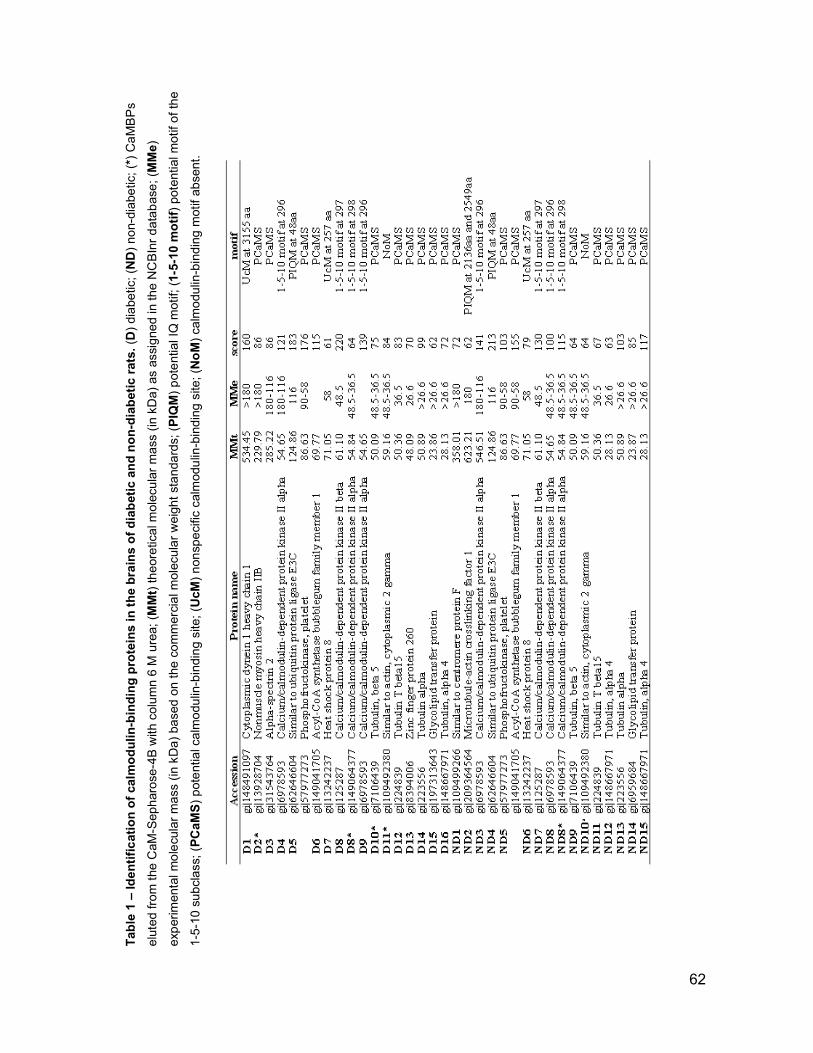
Table 1 – Identification of calmodulin-binding proteins in the brains of diabetic and non-diabetic rats. (D
) di
abe
tic; (ND
) no
n-di
abe
tic;
(*)
Ca
MB
Ps
elut
ed fr
om
the
CaM
-Sep
har
ose
-4B
with
col
um
n 6
M u
rea;
(MMt)
the
ore
tica
l mol
ecu
lar
ma
ss (
in k
Da)
as
assi
gne
d in
the
NC
BIn
r d
ata
bas
e; (MMe)
exp
erim
enta
l mo
lecu
lar
ma
ss (
in k
Da)
ba
sed
on
the
co
mm
erci
al m
olec
ula
r w
eigh
t st
and
ard
s; (PIQM
) p
ote
ntia
l IQ
mot
if; (1-5-10 motif)
pot
entia
l mot
if o
f th
e
1-5-
10 s
ub
cla
ss;
(PCaMS
) p
ote
ntia
l ca
lmo
dulin
-bin
din
g si
te;
(UcM
) n
onsp
eci
fic c
alm
odu
lin-b
ind
ing
site
; (NoM
) ca
lmo
dulin
-bin
din
g m
otif
abse
nt.
62

63
References
[1] G.J. Biessels, A.C. Kappelle, B. Bravenboer, D.W. Erkelens, W.H. Gispen, Cerebral function in diabetes mellitus, Diabetologia 37 (1994) 643-650.
[2] U. Di Mario, S. Morano, E. Valle, G. Pozzessere, Electrophysiological alterations of the central nervous system in diabetes mellitus, Diabetes Metab Rev 11 (1995) 259-277.
[3] E.L. Helkala, L. Niskanen, H. Viinamaki, J. Partanen, M. Uusitupa, Short-term and long-term memory in elderly patients with NIDDM, Diabetes Care 18 (1995) 681-685.
[4] A.L. McCall, The impact of diabetes on the CNS, Diabetes 41 (1992) 557-570.
[5] A.D. Mooradian, Diabetic complications of the central nervous system, Endocr Rev 9 (1988) 346-356.
[6] S.G. Massry, M. Smogorzewski, Role of elevated cytosolic calcium in the pathogenesis of complications in diabetes mellitus, Miner Electrolyte Metab 23 (1997) 253-260.
[7] J.P. Klein, B.C. Hains, M.J. Craner, J.A. Black, S.G. Waxman, Apoptosis of vasopressinergic hypothalamic neurons in chronic diabetes mellitus, Neurobiol Dis 15 (2004) 221-228.
[8] D.H. O'Day, CaMBOT: profiling and characterizing calmodulin-binding proteins, Cell Signal 15 (2003) 347-354.
[9] M.S. Mooseker, R.E. Cheney, Unconventional myosins, Annu Rev Cell Dev Biol 11 (1995) 633-675.
[10] A.R. Bresnick, Molecular mechanisms of nonmuscle myosin-II regulation, Curr Opin Cell Biol 11 (1999) 26-33.
[11] P. Maupin, C.L. Phillips, R.S. Adelstein, T.D. Pollard, Differential localization of myosin-II isozymes in human cultured cells and blood cells, J Cell Sci 107 ( Pt 11) (1994) 3077-3090.
[12] M.E. Brown, P.C. Bridgman, Retrograde flow rate is increased in growth cones from myosin IIB knockout mice, J Cell Sci 116 (2003) 1087-1094.
[13] L.K. Calabria, L. Garcia Hernandez, R.R. Teixeira, M. Valle de Sousa, F.S. Espindola, Identification of calmodulin-binding proteins in brain of worker honeybees, Comp Biochem Physiol B Biochem Mol Biol 151 (2008) 41-45.
[14] M.M. Bradford, A rapid and sensitive method for the quantitation of microgram quantities of protein utilizing the principle of protein-dye binding, Anal Biochem 72 (1976) 248-254.
[15] U.K. Laemmli, N. Teaff, J. D'Ambrosia, Maturation of the head of bacteriophage T4. III. DNA packaging into preformed heads, J Mol Biol 88 (1974) 749-765.
[16] X. Zhang, L. Shi, S. Shu, Y. Wang, K. Zhao, N. Xu, S. Liu, P. Roepstorff, An improved method of sample preparation on AnchorChip targets for MALDI-MS and MS/MS and its application in the liver proteome project, Proteomics 7 (2007) 2340-2349.
[17] D.N. Perkins, D.J. Pappin, D.M. Creasy, J.S. Cottrell, Probability-based protein identification by searching sequence databases using mass spectrometry data, Electrophoresis 20 (1999) 3551-3567.

64
[18] K.L. Yap, J. Kim, K. Truong, M. Sherman, T. Yuan, M. Ikura, Calmodulin target database, J Struct Funct Genomics 1 (2000) 8-14.
[19] T. Berggard, G. Arrigoni, O. Olsson, M. Fex, S. Linse, P. James, 140 mouse brain proteins identified by Ca2+-calmodulin affinity chromatography and tandem mass spectrometry, J Proteome Res 5 (2006) 669-687.
[20] H. Towbin, T. Staehelin, J. Gordon, Electrophoretic transfer of proteins from polyacrylamide gels to nitrocellulose sheets: procedure and some applications, Proc Natl Acad Sci U S A 76 (1979) 4350-4354.
[21] J.L. Yin, N.A. Shackel, A. Zekry, P.H. McGuinness, C. Richards, K.V. Putten, G.W. McCaughan, J.M. Eris, G.A. Bishop, Real-time reverse transcriptase-polymerase chain reaction (RT-PCR) for measurement of cytokine and growth factor mRNA expression with fluorogenic probes or SYBR Green I, Immunol Cell Biol 79 (2001) 213-221.
[22] L.K. Srivastava, N.Z. Baquer, Changes in phosphofructokinase and pyruvate kinase in rat brain regions during alloxan-induced diabetes, Enzyme 32 (1984) 84-88.
[23] T. Sasaki, Glycolipid transfer protein and intracellular traffic of glucosylceramide, Experientia 46 (1990) 611-616.
[24] A. Hershko, H. Heller, S. Elias, A. Ciechanover, Components of ubiquitin-protein ligase system. Resolution, affinity purification, and role in protein breakdown, J Biol Chem 258 (1983) 8206-8214.
[25] M. Scheffner, U. Nuber, J.M. Huibregtse, Protein ubiquitination involving an E1-E2-E3 enzyme ubiquitin thioester cascade, Nature 373 (1995) 81-83.
[26] R.K. Murphey, T.A. Godenschwege, New roles for ubiquitin in the assembly and function of neuronal circuits, Neuron 36 (2002) 5-8.
[27] M.A. Stevenson, S.K. Calderwood, Members of the 70-kilodalton heat shock protein family contain a highly conserved calmodulin-binding domain, Mol Cell Biol 10 (1990) 1234-1238.
[28] J.S. Someren, L.E. Faber, J.D. Klein, J.A. Tumlin, Heat shock proteins 70 and 90 increase calcineurin activity in vitro through calmodulin-dependent and independent mechanisms, Biochem Biophys Res Commun 260 (1999) 619-625.
[29] M.E. Feder, G.E. Hofmann, Heat-shock proteins, molecular chaperones, and the stress response: evolutionary and ecological physiology, Annu Rev Physiol 61 (1999) 243-282.
[30] A.K. Leung, J.S. Andersen, M. Mann, A.I. Lamond, Bioinformatic analysis of the nucleolus, Biochem J 376 (2003) 553-569.
[31] R.J. Colbran, Targeting of calcium/calmodulin-dependent protein kinase II, Biochem J 378 (2004) 1-16.
[32] L. Brocke, M. Srinivasan, H. Schulman, Developmental and regional expression of multifunctional Ca2+/calmodulin-dependent protein kinase isoforms in rat brain, J Neurosci 15 (1995) 6797-6808.
[33] S.K. Bhardwaj, G. Kaur, Effect of diabetes on calcium/calmodulin dependent protein kinase-II from rat brain, Neurochem Int 35 (1999) 329-335.
[34] M. Di Luca, L. Ruts, F. Gardoni, F. Cattabeni, G.J. Biessels, W.H. Gispen, NMDA receptor subunits are modified transcriptionally and post-translationally in the brain of streptozotocin-diabetic rats, Diabetologia 42 (1999) 693-701.

65
[35] J.E. Lisman, A.M. Zhabotinsky, A model of synaptic memory: a CaMKII/PP1 switch that potentiates transmission by organizing an AMPA receptor anchoring assembly, Neuron 31 (2001) 191-201.
[36] S. Kaech, H. Parmar, M. Roelandse, C. Bornmann, A. Matus, Cytoskeletal microdifferentiation: a mechanism for organizing morphological plasticity in dendrites, Proc Natl Acad Sci U S A 98 (2001) 7086-7092.
[37] A. Matus, Actin-based plasticity in dendritic spines, Science 290 (2000) 754-758.
[38] E.N. Star, D.J. Kwiatkowski, V.N. Murthy, Rapid turnover of actin in dendritic spines and its regulation by activity, Nat Neurosci 5 (2002) 239-246.
[39] K.W. Li, M.P. Hornshaw, R.C. Van Der Schors, R. Watson, S. Tate, B. Casetta, C.R. Jimenez, Y. Gouwenberg, E.D. Gundelfinger, K.H. Smalla, A.B. Smit, Proteomics analysis of rat brain postsynaptic density. Implications of the diverse protein functional groups for the integration of synaptic physiology, J Biol Chem 279 (2004) 987-1002.
[40] N. Murakami, M. Elzinga, Immunohistochemical studies on the distribution of cellular myosin II isoforms in brain and aorta, Cell Motil Cytoskeleton 22 (1992) 281-295.
[41] N.Z. Baquer, A. Taha, P. Kumar, P. McLean, S.M. Cowsik, R.K. Kale, R. Singh, D. Sharma, A metabolic and functional overview of brain aging linked to neurological disorders, Biogerontology 10 (2009) 377-413.
[42] R.A. Shanely, K.A. Zwetsloot, T.E. Childs, S.J. Lees, R.W. Tsika, F.W. Booth, IGF-I activates the mouse type IIb myosin heavy chain gene, Am J Physiol Cell Physiol 297 (2009) C1019-1027.
[43] R.I. Ludowyke, Z. Elgundi, T. Kranenburg, J.R. Stehn, C. Schmitz-Peiffer, W.E. Hughes, T.J. Biden, Phosphorylation of nonmuscle myosin heavy chain IIA on Ser1917 is mediated by protein kinase C beta II and coincides with the onset of stimulated degranulation of RBL-2H3 mast cells, J Immunol 177 (2006) 1492-1499.
[44] M.M. Andzelm, X. Chen, K. Krzewski, J.S. Orange, J.L. Strominger, Myosin IIA is required for cytolytic granule exocytosis in human NK cells, J Exp Med 204 (2007) 2285-2291.
[45] T.A. Ryan, Inhibitors of myosin light chain kinase block synaptic vesicle pool mobilization during action potential firing, J Neurosci 19 (1999) 1317-1323.
[46] S. Mochida, H. Kobayashi, Y. Matsuda, Y. Yuda, K. Muramoto, Y. Nonomura, Myosin II is involved in transmitter release at synapses formed between rat sympathetic neurons in culture, Neuron 13 (1994) 1131-1142.
[47] P. Neco, D. Giner, S. Viniegra, R. Borges, A. Villarroel, L.M. Gutierrez, New roles of myosin II during vesicle transport and fusion in chromaffin cells, J Biol Chem 279 (2004) 27450-27457.
[48] J.R. Wilson, R.I. Ludowyke, T.J. Biden, A redistribution of actin and myosin IIA accompanies Ca(2+)-dependent insulin secretion, FEBS Lett 492 (2001) 101-106.
[49] G.V. Jerdeva, K. Wu, F.A. Yarber, C.J. Rhodes, D. Kalman, J.E. Schechter, S.F. Hamm-Alvarez, Actin and non-muscle myosin II facilitate apical exocytosis of tear proteins in rabbit lacrimal acinar epithelial cells, J Cell Sci 118 (2005) 4797-4812.

66
[50] A. Segawa, S. Yamashina, Roles of microfilaments in exocytosis: a new hypothesis, Cell Struct Funct 14 (1989) 531-544.
[51] R.R. Torgerson, M.A. McNiven, Agonist-induced changes in cell shape during regulated secretion in rat pancreatic acini, J Cell Physiol 182 (2000) 438-447.
[52] K.A. Becker, N.H. Hart, Reorganization of filamentous actin and myosin-II in zebrafish eggs correlates temporally and spatially with cortical granule exocytosis, J Cell Sci 112 ( Pt 1) (1999) 97-110.
[53] J. Brown, P.C. Bridgman, Role of myosin II in axon outgrowth, J Histochem Cytochem 51 (2003) 421-428.
[54] R.B. Vallee, G.E. Seale, J.W. Tsai, Emerging roles for myosin II and cytoplasmic dynein in migrating neurons and growth cones, Trends Cell Biol 19 (2009) 347-355.
[55] A.K. Lalwani, G. Atkin, Y. Li, J.Y. Lee, D.E. Hillman, A.N. Mhatre, Localization in stereocilia, plasma membrane, and mitochondria suggests diverse roles for NMHC-IIa within cochlear hair cells, Brain Res 1197 (2008) 13-22.
[56] S.A. Qureshi, V. Ding, Z. Li, D. Szalkowski, D.E. Biazzo-Ashnault, D. Xie, R. Saperstein, E. Brady, S. Huskey, X. Shen, K. Liu, L. Xu, G.M. Salituro, J.V. Heck, D.E. Moller, A.B. Jones, B.B. Zhang, Activation of insulin signal transduction pathway and anti-diabetic activity of small molecule insulin receptor activators, J Biol Chem 275 (2000) 36590-36595.
67
2. CONCLUSÕES
- Os cérebros dos ratos foram avaliados quanto ao perfil de proteínas ligantes de
calmodulina e revelaram a presença de miosina-IIB superexpressa no cérebro de
diabéticos.
- A diferença na expressão da proteína e do RNAm da miosina-IIB foi comprovada
pelas técnicas de western blot e PCR em tempo real.
- A análise da distribuição da miosina-IIB revelou que o córtex do cérebro de
diabéticos apresenta-se fortemente marcado.

Capítulo 3
Efeito da suplementação de cálcio, zinco e vitamina E no
estresse oxidativo e na expressão de miosinas no cérebro
de ratos diabéticos induzidos por estreptozotocina

69
1. ARTIGO CIENTÍFICO
PLoS Biology (fator de impacto: 12,68)
MYOSINS ARE DIFFERENTIALLY EXPRESSED UNDER OXIDATIVE
STRESS IN STREPTOZOTOCIN-INDUCED DIABETES RAT BRAINS
Luciana Karen Calábria1, Alice Vieira da Costa1, Renato José da Silva
Oliveira1, Simone Ramos Deconte1, Rafael Nascimento1, Washington João
Carvalho1, Vanessa Neves de Oliveira1, Carlos Alberto Arcaro Filho2,
Luciana Augusto de Rezende2, Luiz Ricardo Goulart1, Foued Salmen
Espindola1
1Institute of Genetics and Biochemistry, Federal University of Uberlândia,
Campus Umuarama, 38400-902, Uberlândia-MG, Brazil. 2Department of Chemistry, Physic and Mathematic, University of Ribeirão Preto,
14096-900, Ribeirão Preto-SP, Brazil.

70
Abstract
Diabetes mellitus is a chronic disease characterized by persistent
hyperglycemia, which may lead to brain tissue damage due to oxidative stress.
The enhancement of free radicals during hyperglycemia contributes to
increased neuronal death and changes in synaptic transmission. This study
evaluated the effect of oxidative stress and the use of antioxidants (calcium,
zinc and vitamin E) supplementation on myosins expression levels in brain
tissues of streptozotocin-induced diabetes experimental rat model. Lipid
peroxidation, antioxidant enzymes activities and myosins IIB and Va
expressions at transcriptional and protein levels were examined after 5 and 90
days post streptozotocin induction. The chronic effect of the streptozotocin-
induced diabetes led to upregulation of glutathione peroxidase (GPx) and
malondialdehyde (MDA) levels, and downregulation of superoxide dismutase
(SOD) and catalase (CAT) activities. These alterations were accompanied by
increased myosin-IIB and decreased myosin-Va expressions in diabetic rat
brains. The antioxidant supplementation protected against hyperglycemia
damage by reducing CAT activity and increasing GPx and SOD activities,
although it had no influence on MDA levels. Interestingly, only diabetic rats
restored myosin-IIB expression at both transcriptional and protein levels in brain
tissues after supplementation, and no differences were observed in myosin-Va
expression in comparison to non-diabetic rat brains. The functional alterations
of antioxidant enzymes and myosins IIB and Va in rat brains of experimentally-
induced diabetes were strongly associated with the disease and aging during
acute and chronic stages, but only myosin-IIB expression could be reversed by
antioxidants. These data highlight the supplementation (calcium, zinc and
vitamin E) antioxidants effects on the oxidative stress and the involvement of
myosins IIB and Va in the diabetes stages; however, their mechanisms of action
on enzymes activities, lipid peroxidation and myosins expression remain to be
elucidated.

71
Introduction
Diabetes mellitus is a multifactorial disease characterized by chronic
hyperglycemia resulting from abnormalities in insulin action and/or insulin
secretion [1]. Research evidences support that both acute and chronic
hyperglycemia produce negative impacts on central nervous system leading to
tissues damage [2,3]. One mechanism behind this neuronal injury is oxidative
stress, due to excessive free radical generation from the oxidation of elevated
intracellular glucose levels [4].
Brain contains large amounts of enzymes to protect against oxidative
damage [5]. Endogenous antioxidant system, including enzymatic (glutathione
peroxidase, superoxide dismutase, and catalase) and non-enzymatic (vitamin
E, vitamin C, glutathione and uric acid) antioxidants, offers protection to cells
and tissues against glucose-induced oxidative injury in diabetics [6,7,8,9,10].
The enhancement on oxygen free radical in brain during hyperglycemia
[11] contributes to increased neuronal death trough protein oxidation, DNA
damage, and peroxidation of membrane lipids [12] as well as changes in
synaptic transmission. These alterations could lead to abnormal synaptic
plasticity and cognitive impairments observed in experimental diabetes
[13,14,15,16,17,18].
Myosins are a family of molecular motors that contains many classes and
isoforms, which differ in their cellular distribution and function [19,20,21]. Among
the myosin classes identified so far, the family classes II and V have been best
characterized in neurons and are implicated in a wide variety of cellular
functions in nervous system, including neuronal migration, growth cone motility,
neuronal morphogenesis, axonal transport, and synaptic and sensory functions
[22]. There are three isoforms of the non-muscle myosin II: IIA, IIB and IIC
[20,23,24,25] and the myosin-V has been classified into subtypes Va, Vb and
Vc [20,26]. The expression of myosin isoforms and its function in presynaptic
terminals differs with type of neurons and properties of neurotransmitter release
[27].
The rationale for using certain vitamins and minerals, or natural
antioxidants, in the prevention and management of diabetes, is largely based on
animal experiments and epidemiologic studies [28,29,30,31]. Diabetes is
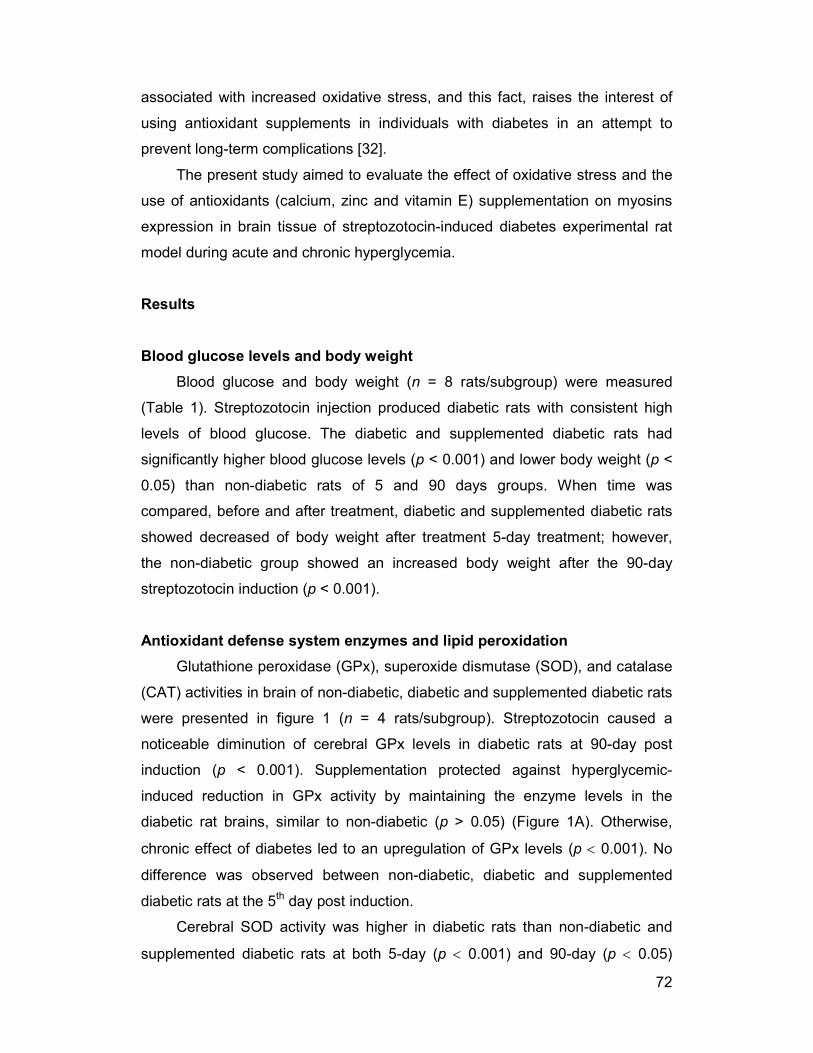
72
associated with increased oxidative stress, and this fact, raises the interest of
using antioxidant supplements in individuals with diabetes in an attempt to
prevent long-term complications [32].
The present study aimed to evaluate the effect of oxidative stress and the
use of antioxidants (calcium, zinc and vitamin E) supplementation on myosins
expression in brain tissue of streptozotocin-induced diabetes experimental rat
model during acute and chronic hyperglycemia.
Results
Blood glucose levels and body weight
Blood glucose and body weight (n = 8 rats/subgroup) were measured
(Table 1). Streptozotocin injection produced diabetic rats with consistent high
levels of blood glucose. The diabetic and supplemented diabetic rats had
significantly higher blood glucose levels (p < 0.001) and lower body weight (p <
0.05) than non-diabetic rats of 5 and 90 days groups. When time was
compared, before and after treatment, diabetic and supplemented diabetic rats
showed decreased of body weight after treatment 5-day treatment; however,
the non-diabetic group showed an increased body weight after the 90-day
streptozotocin induction (p < 0.001).
Antioxidant defense system enzymes and lipid peroxidation
Glutathione peroxidase (GPx), superoxide dismutase (SOD), and catalase
(CAT) activities in brain of non-diabetic, diabetic and supplemented diabetic rats
were presented in figure 1 (n = 4 rats/subgroup). Streptozotocin caused a
noticeable diminution of cerebral GPx levels in diabetic rats at 90-day post
induction (p < 0.001). Supplementation protected against hyperglycemic-
induced reduction in GPx activity by maintaining the enzyme levels in the
diabetic rat brains, similar to non-diabetic (p > 0.05) (Figure 1A). Otherwise,
chronic effect of diabetes led to an upregulation of GPx levels (p < 0.001). No
difference was observed between non-diabetic, diabetic and supplemented
diabetic rats at the 5th day post induction.
Cerebral SOD activity was higher in diabetic rats than non-diabetic and
supplemented diabetic rats at both 5-day (p < 0.001) and 90-day (p < 0.05)

73
periods. On the other hand, chronic effects of diabetes, decreased SOD activity
(p < 0.05), and no change was observed on the non-diabetic animals (Figure
1B).
Figure 1C shows that cerebral CAT activity was significantly elevated in
supplemented diabetic rats 5 days post induction when compared to the non-
diabetic and diabetic rats in the same period (p < 0.05). Although, CAT activity
was lower in rats at the 90-day, than rats at the 5-day period (p < 0.05), CAT
levels were kept elevated in diabetic rats when compared to non-diabetic and
supplemented rats. It is noteworthy that CAT levels were lower in supplemented
diabetic than diabetic rats of 90 days post induction (p < 0.05), although this has
not been observed in rats at the 5-day period. Besides, the increase in CAT
activity in diabetic rats was accompanied by a significant decrease in the GPx
activity in brain.
As presented in Figure 1D, the levels of malondialdehyde (MDA) were
markedly elevated in diabetic rats at the 5-day period (p < 0.005), but
supplementation of diabetic rats, had no influence on MDA levels. Moreover,
aged non-diabetic rats had higher MDA levels than young non-diabetics (p <
0.05). No difference was observed between rats at the 90-day period and
supplementation seems to be inefficient and does not affect the antioxidant
status of both groups.
Determination of changes in myosins protein expression levels
In order to check if the myosin proteins levels could be altered after 5 days
and 90 days of diabetes or supplementation duration, the amount of myosins IIB
(210 kDa) and Va (190 kDa) were estimated by Western blotting (n = 3
rats/subgroup). Figure 2 displays the combined results of immunoblots and
densitometrically quantitated myosins immunoreactivity represented as
percentage of non-diabetic from diabetic and supplemented diabetic rats.
In a general manner, diabetes increased myosin-IIB protein levels in brain
(p < 0.05). In diabetic rats of the 5-day group, myosin-IIB levels were 16.5%
higher than non-diabetic values, and 36.8% in diabetic rats at the 90-day period.
Myosin-Va protein levels have decreased 18% and 16% in diabetic rat brains as
compared to non-diabetic rats in both 5- and 90-day periods, respectively (p <

74
0.05). No significant differences were observed for myosins protein levels in
supplemented diabetic in both periods, except for myosin-IIB after the 5-day
period, in which antioxidant supplementation has restored the protein content in
the brain tissues to non-diabetic levels.
Determination of changes in myosins mRNA expression
Real time PCR (n = 3 rats/subgroup) was performed to measure changes
in myosins gene expression for both MYH10 (myosin-IIB) and MYO5A (myosin-
Va) at 5- and 90-day periods (Figure 4). Changes in gene expression were
calculated based on the 2-∆∆CT method with beta-2-microglobulin (B2M) as an
endogenous control. Diabetic showed increased MYH10 mRNA expression as
compared to non-diabetic at 5- and 90-day periods (~2-fold). MYO5A mRNA
was decreased in diabetic as compared to non-diabetic in both 5-day (0.35-fold)
and 90-days (0.7-fold) periods. Supplementation reduced MYH10 mRNA
expression in brains in both periods, conferring ~0.35-fold decrease compared
to diabetic group without antioxidants. However, no significant differences in
MYO5A mRNA expression was observed between diabetic and supplemented
diabetic in both periods. Amplification efficiency was tested by standard curves
for Rattus novergicus B2M (R2 = 0.9997), MYH10 (R2 = 0.996) and MYO5A (R2
= 0.9999) generated by plotting the value of CT cycle vs. the log of plasmid
concentration (from 103 to 106 copies).
Discussion
Streptozotocin-induced diabetes is a well-documented model of
experimental diabetes in rats. It provides a relevant example of endogenous
chronic oxidative stress as a result of hyperglycemia [33]. In the present study,
streptozotocin treatment produced significant increase in blood glucose levels
along with reduction in body weight. In addition, the acute effect of diabetes led
to decreased body weight while the chronic stage did not affect this
anthropometric parameter in diabetic rats. These results are in accordance with
other studies, which showed that Diabetes mellitus increase plasma glucose
levels and decrease body weight of diabetic rats [34,35,36,37,38]. In fact,
supplementation did not reduce blood glucose levels and this may have

75
contributed in part to the non-suppressive effect of antioxidants on oxidative
stress, though the body weight had been reduced in diabetic rats.
We have also analyzed hyperglycemia-induced oxidative stress in rat
brain. The increase in free radical generation along with depletion of
antioxidants is the mechanism involved in diabetes-induced oxidative stress.
There is evidence of alterations in free radical metabolism [39] and in the
antioxidant parameters status [40,41] during diabetes in various tissues.
Moreover, there are contradictory results in the literature regarding the effect of
hyperglycemia-induced diabetes on antioxidant enzymes activities [42,43,44].
Thus, the current study show the effects of the concomitant use of vitamin E,
calcium and zinc as antioxidants on the activities of defense enzymes, such as
glutathione peroxidase (GPx), superoxide dismutase (SOD) and catalase (CAT)
in rat brains after 5 and 90 days of supplementation.
GPx and SOD are the first line of defense against free radical attacks.
Their function is to catalyse the conversion of superoxide radicals to hydrogen
peroxide [45]. Cerebral levels of GPx, a potent endogenous antioxidant, were
reduced in diabetics at 90-day post induction. However, the activity of GPx has
been shown increased in brain [36,46,47]. This increase was also observed
after supplementation. GPx is responsible for decomposition of hydrogen
peroxide and other lipid peroxides, and it is possible that supplementation have
avoided the GPx activity reduction in induced hyperglycemia, maintaining the
enzyme levels in brain of diabetics, similar to non-diabetic rats.
The increase in cerebral SOD activity was observed in diabetic rats at 5-
and 90-day periods, which can lead to an important elimination of superoxide
ions that inhibit the formation of hydroxyl radical in tissues. The increase SOD
activity in type 2 diabetic mice brain has been reported [48,49] to be a putative
protection mechanism of oxidative stress. In the meantime, the SOD activity
also appears to be decreased in diabetic rats [36,50,51,52].
GPx activity in the supplemented diabetic rats remained at same levels as
in non-diabetic rats, whereas the SOD activity increased significantly compared
to non-diabetic. The alteration of antioxidant enzymes GPx and SOD levels in
the diabetic rats could be attributed to peroxidative damage of the tissues
caused by streptozotocin-induced hyperglycemia [53], while supplementation
with antioxidants contributed to maintaining the optimum condition of enzyme

76
activity in the cellular organelles, by protecting them from peroxidation in acute
diabetes.
Besides, the SOD and CAT are also the major antioxidant enzymes
against oxidative stress, and appear to be decreased in diabetic rats [50,51].
CAT is responsible for the catalytic decomposition of hydrogen peroxide formed
in cellular metabolism in oxygen and water molecules. Its increased activity at
the early stage of the disease (5 days) during antioxidant supplementation
followed by a decreased activity at the chronic stage (90 days), might indicate a
fine modulation of the CAT activity in order to protect the brain against free
radicals at acute stage of the diabetes and may also advocate for the
importance of antioxidant supplementation at this stage for a better tissue
response and protection. Simultaneous reduction in the activity of both SOD
and CAT, likely makes the brain more vulnerable to hyperglycemia-induced
oxidative stress. Although there are discrepancies in the levels of antioxidant
enzymes reported in diabetic rats [54,55] our results have shown an important
balance between GPx and CAT levels.
These antioxidant enzymes have a complementary catalytic activity
leading to reduced MDA concentration, which represents lipid peroxidation
products in tissue and blood. In the present study, we observed that MDA levels
were significantly higher in diabetic rats at a 5-day period, corroborating with
previous results [56]. Nevertheless, supplementation was not sufficient
counteract those elevated levels of lipid peroxidation and did not protected the
brain against the cytotoxic action and oxidative stress due to diabetes. It is well
known that hyperglycemia increases lipid peroxidation, which may contribute to
long-term tissue damage [57]. Besides, brain seems to be more sensitive than
the other tissues and hence the increase in GPx activity was not sufficient to
reduce MDA concentrations and thus to protect this tissue from lipid
peroxidation.
Minerals (calcium and zinc) and vitamin E were added in diet of the
diabetic rats to act as an antioxidant supplement in animals. Thus, our aim was
to evaluate the effects of this supplementation on brain. Evidence from clinical
studies has suggested that calcium could indirectly affect glucose metabolism,
which would be desirable for diabetes prevention [58,59]. Zinc acts as an
antioxidant in order to reduce oxidative stress, is essential for the function of
77
SOD [60], and is also involved in insulin synthesis [61] which are alterated in
diabetes [62,63]. Treatment with zinc significantly reduces astrocytosis [64],
elevates SOD activity [65], and may be able to prevent diabetes effects in brain
or against various damaging effects, including oxidative injuries [66] and
apoptosis [67,68,69]. Additionally, vitamin E possesses antioxidant activity
[1,70,71], neuroprotective action [72], and plays a role in hyperglycemia
prevention [73,74]. Moreover, treatment with vitamin E reduces GPx levels and
lipid peroxidation in brain [11]. In contrast, there are evidences that excessive
dietary zinc and vitamin E intake can induce pathological conditions associated
with oxidative stress [75,76,77,78]. In regard to diabetes prevention through
supplementation with micronutrients, the current evidences do not allow any
particular recommendation for mineral or vitamin supplementation on a large
scale. Given that diabetes is a condition of increased oxidative stress,
antioxidant therapy may represent a potential coadjuvant to antidiabetic
pharmacological treatment by improving the glucose metabolism [32].
On the other hand, our results have evidenced that chronic effect of
diabetes in brain led to an upregulation of GPx levels, and downregulation of
SOD and CAT activities, as well as a higher lipid peroxidation levels in old non-
diabetic than in young non-diabetic rats. Our findings are consistent with
previous reports [79,80,81,82], although another study has revealed that GPx
and CAT activities are relatively unaffected by age [80].
Recent studies have shown that peroxidative damage to lipid and protein
occurs with the aging process and the products of these reactions accumulate
in the brain during aging [49,81,82,83,84]. Moreover, the brain is susceptible to
oxidative stress, which is associated with age related brain dysfunction, due to
its high content of key compounds for oxidative damage and antioxidant
defense systems [85]. Thus, changes in the activities of oxidative enzymes
seem to be more severely affected in the brain during the aging process.
Evidence in literature suggests that there is a functional association between
oxidative damage and brain dysfunction [86].
Both, aging and diabetes affect cognition, synaptic plasticity and
neurotransmission in rats, hence the effects of diabetes and aging interact [87].
The impact of this interaction on the central nervous system is well recognized
such as neurophysiological and structural changes associated mainly with

78
cognitive deficits and Alzheimer disease [14]. Degenerative changes of neurons
and glia have been reported, and suggested that alteration in synaptic
transmission could contribute to cognitive impairments observed in diabetics
[17,18,37]. Besides, oxidative stress induced by hydrogen peroxide induces
cytoskeletal reorganization and significantly enhances association of myosin to
actin filaments [88]. Myosin has also been reported to be a particularly sensitive
target of oxidative damage [89,90,91], although a direct link between oxidative
stress and myosin dysfunction has yet to be established.
Several lines of evidence suggest that myosins may be involved in the
regulation of synaptic vesicles. Myosin-IIB modulates neurotransmitter release
from synapses [92] while myosin-Va mediates synaptic vesicle trafficking [26].
Here we show an increased protein expression of myosin-IIB, but a decreased
of myosin-Va expression in diabetic rat brains. However, only diabetic rats
restored protein and mRNA of myosin-IIB content to non-diabetic levels in brain
after supplementation, and no differences were noted for myosin-Va levels in
this condition. The increase in MYH10 mRNA and decrease MYO5A mRNA in
diabetic indicates that the functional alterations of myosins in brain may be
associated with aging, in which this myosins are expressed at higher levels in
adult stage than in early embryos and the expression increases with
development [24]. Moreover, the alterations in myosin protein and mRNA levels
may be linked with diabetes, even in transcriptional and translational levels,
during acute and chronic diseases.
Myosins have a domain motor that binds to actin and ATP [93], and are
modulated by ATPase activity [94]. The administration of streptozotocin reduces
ATP concentration in rats [95], and this modifies the activity of various ATP-
dependent proteins [96] like myosins. Therefore, the diabetes induction in rat
brains by the administration of streptozotocin may influence on the myosins
associated-ATP activity.
Diabetes mellitus studies have shown changes in expression or levels of
some myosins, as MYO9B in intestinal permeability [97], and myosin-V
immunoreactive myenteric neuronal density [98] in diabetic rats. However, no
evidences have been reported before about different expression profiles of
myosins IIB and Va in rat brains supplemented with antioxidants in acute and
chronic diabetes. We cannot exclude the possibility that other translational

79
alterations of myosin related to the redox state of the cell contribute to functional
impairment of myosin.
In agreement with the notion that unregulated Ca+2 release from the
endoplasmatic reticulum to the cytoplasm due to hyperglycemia might be a
mediator of beta cell dysfunction and apoptosis in diabetes, it is well recognized
that cytoplasmic Ca+2 overload is an ubiquitous cause of cell death in neurons
[99,100]. This Ca+2 influx leads to the activation of proteases like calpain [101]
and the overactivation of Ca+2-calpain pathways also contributes to apoptosis in
diabetes [102]. In vitro studies have shown that purified myosin-Va is
proteolyzed by calpain, producing two peptides of 130 kDa and 80 kDa [103].
Cleavage is at the PEST site located in the tail domain [104]. It is possible this
mechanism of Ca+2 influx, following the activation of calpain and clivage of
myosin had led to irreversible decrease of myosin-Va levels in diabetic rat
brains. Otherwise, myosin-IIB lacks this PEST sequence and levels may be
increased in diabetic as a compensatory mechanism.
In conclusion, antioxidant supplementation, calcium and zinc minerals,
and vitamin E, strengthened the protection mechanisms of rat brains in
experimentally-induced diabetes with increased of GPx activity and decreased
CAT levels after 90 days post induction, but it did not protect against the
oxidative damage at the chronic stage. Moreover, supplementation restored
myosin-IIB protein and mRNA levels after 5 days, but had no effect on the
myosin-Va levels in the brain in both acute and chronic stages.
The precise mechanism of the observed calcium, zinc and vitamin E
mediated regulation of enzymatic activities, lipid peroxidation and expression of
myosins cannot be ascertained from this study and remains to be explored in
the future. Considering the brain is a heterogenous tissue and it is composed of
different cell types and diverse functions, it is important to further detail the
modulation of antioxidant enzymes and myosins expression in the different
brain regions during acute and chronic diabetes. Therefore, this study
contributes to giving some insights between the association and alterations in
expression levels of myosins and antioxidant enzymes in the different regions of
the diabetic nervous system.

80
Material and Methods
Animals
All experimental procedures were conducted in accordance with the ethical
principles of the Brazilian Academy of Animal Experimentation and approved by
the Committee of Ethics in Animal Experimentation from the University of
Ribeirão Preto, UNAERP (066/09). Forty-eight male Wistar rats Rattus
norvegicus (weight: 200–290g) were housed under standard conditions
(22±1oC, humidity 60±5%, 12 h light/12 h dark cycle) with food and water ad
libitum on the Central Biotery of UNAERP.
Induction of diabetes mellitus
After one week of acclimatization, the rats were subjected to a 24-h starvation.
The animals were then anesthetized by intraperitoneal injection of
xylazine/ketamine (1:1 v/v), and then streptozotocin (40 mg/kg body weight;
Sigma-Aldrich), freshly dissolved in 0.01 M citrate buffer, pH 4.5, was injected
into the penile vein (2 mL/kg). Food was denied for 90 min post injection. At 10
days after the streptozotocin or buffer injection, blood glucose was determined
and animals with blood glucose above 200 mg/dL were scored diabetic. Animal
(n = 48) weight was monitored daily until decapitation and surgical removal of
brains 5 or 90 days after diabetes induction or supplementation.
Group distribution and rats supplementation
The rats were distributed randomly in three groups (n = 8, each): non-diabetic
(ND), diabetic (D), and diabetic supplemented (SD). All animals were fed diets
based on a modified AIN93G rodent diet, except supplemented diabetic group
that was fed with additional calcium (2.5-fold), zinc (500 mg), and vitamin E (20-
fold), following principles of American Institute of Nutrition [105].
Sample collection and tissue preparation
The brains of all animals were quickly removed, washed with chilled normal
saline and immersed in liquid nitrogen. Simultaneously, the blood was also
collected from the portal vein to confirm the glucose levels. For oxidative stress
markers and western blotting analyses, each brains was individually

81
homogenized on ice in homogenization buffer (50 mM Tris-HCl, pH 7.5, 10 mM
EDTA, 2 mM EGTA, 5 mM ATP, 2 mM dithiothreitol, 1 mM benzamidine, 0.5
mM phenylmethane sulfonyl-fluoride, 0.1 M aprotinin, 20 µg/mL leupeptin, 0.1
mM pefabloc). The homogenates were centrifuged at 15,000 x g for 30 min at
4ºC and total protein concentration in the supernatant samples was measured
following the Bradford assay [106].
Oxidative stress markers analysis
GPx and SOD activities: Glutathione peroxidase (GPx) and superoxide
dismutase (SOD) activities were measured using a commercial kit (Ransel and
Ransod, Randox Laboratories Ltd., Crumlin, UK), following manufacturer’s
instructions. CAT activity: Catalase (CAT) activity was assessed
spectrophotometrically monitoring hydrogen peroxide decomposition at 240 nm
[107] and the substrate concentration was 20 mM for brain measurements.
Determination of lipid peroxidation product: Lipid peroxidation in tissue was
determined by measuring the presence of malondialdehyde (MDA) using the
thiobarbituric acid test (TBARS), a commercial kit (Cayman Chemical Inc., MI,
USA), following manufacturer’s instructions.
Western blotting
Aliquots of supernatant samples were solubilized in a small volume of
electrophoresis sample buffer containing an additional 100 mM Tris-HCl, pH
8.0, and 25% glycerol. Supernatant samples containing 30 µg of protein were
analyzed by SDS-PAGE with a 5-22% acrylamide gradient and gels were
electroblotted on nitrocellulose membranes in Tris-glycine buffer [108]. Blots
were incubated with 5% dried milk in TBS-T (50 mM Tris-HCl, pH 8.0, 150 mM
NaCl, 0.05% Tween 20), then probed with anti-myosin-IIB and anti-myosin-Va
primary antibodies diluted to 0.2 µg/mL. The polyclonal antibodies were
generated in rabbits against non-muscle myosin II purified and myosin-V head
domain from chicken brain, and purified by affinity to antigen immobilized on
nitrocellulose strips as described previously [109,110,111]. Following washes
with TBS-T, blots were incubated with a peroxidase-conjugated anti-rabbit IgG
(GE Healthcare; diluted 1:2000) and washed several times with TBS-T.
Antibodies bound to the membranes were visualized by chemiluminescence

82
after treatment with ECLTM (GE Healthcare) followed by exposure to
HyperfilmTM (GE Healthcare) following manufacturer’s instructions. The intensity
of the protein bands was analyzed and compared using Scion Image software,
version Alpha 4.0.3.2 (Scion Corporation) and results were expressed as
percentage of total content.
mRNA expression levels using qRT-PCR
Total RNA was isolated from non-diabetic, diabetic and supplemented diabetic
brains separately using TRIzolTM reagent (Invitrogen) following manufacturer’s
instructions and then resuspended in DEPC-treated water. Its quality and
quantity was established by reading the optical density of each sample at 260
and 280 nm using NanoDrop® ND-1000 Spectrophotometer (Nanodrop
Technologies) and agarose gel electrophoresis. One microgram of total RNA
was reverse transcribed at 37°C for 1 h in a 20 µL reaction mixture containing
the first strand buffer, 40 U Murine Monoley Leukemia Virus Reverse
Transcriptase (Invitrogen), 0.25 mM of each dNTP, 10 U RNAsin (Invitrogen),
0.5 mM DTT, and 126 pmol hexamer random primers (Invitrogen). Standards
curves were prepared by cloning PCR products of MYH10, MYO5A and the
housekeeping beta-2-microglobulin (B2M) fragments using TOPO TA Cloning
Dual Promoter Kit (Invitrogen). The recombinant plasmid DNA was isolated and
sequenced using MegaBACE 1000 automatic sequencer (Molecular Dynamics).
The sequencing reaction was carried out using the DyEnamic ET Dye
Terminator Cycle Sequencing Kit (GE Healthcare) following manufacturer’s
instructions. The concentration of plasmid was measured and the copy numbers
calculated [112]. Serial dilutions of each standard curves were made in the
range 10 to 107 copies per µL for MYH10, MYO5A and B2M. The reaction
efficiency was calculated according to the formula E = (10-1/slope - 1) x 100,
where the log of the each dilution was plotted with ∆CT of housekeeping and
target genes, R2 ≥ 0.99 and slope value about -3.32 were admitted to reaction
efficiency of 100%. The qPCR assay was performed by using the 7300 Real-
time PCR System (PE Applied Biosystems) using SYBR Green qPCR Master
Mix reagent (Fermentas). The thermal cycling profile used was the Universal
Program (PE Applied Biosystems). Primers were designed between exons
junctions to avoid amplification of contaminating genomic DNA using Primers

83
Express software (PE Applied Biosystems). For B2M fragment amplification, the
set of primers were: 5’-CGT CGT GCT TGC CAT TCA-3’ and 5’-TCC TCA ACT
GCT ACG TGT CTC AG-3’. The MYH10 forward and reverse primers were
respectively: 5’-CCA TGC CGG AGA ACA CAG T-3’ and 5’-AAG CCC AGA
CCA AAG AGC AG-3’. The MYO5A forward and reverse primers were
respectively: 5’-ATT GAG GCT CGC TCT GTG GA-3’ and 5’-ACG CAA AGT
GGA TGA GCA GA-3’. The relative expression of each specific product was
calculated by 2-∆∆CT (CT = fluorescence threshold value; ∆CT = CT of the target
gene - CT of the reference gene (B2M); ∆∆CT = ∆CT of the target sample - ∆CT
of the calibrator sample). All samples were run in duplicates. For expression
analysis of the MYO5A gene, the cDNA was 4-fold diluted.
Statistical analysis
All values obtained are expressed as mean ± SEM). Data were initially
analyzed by one-way analysis of variance (ANOVA) using the SigmaStat 3.5
software (Systat Software Inc.). When differences were detected by ANOVA,
these sets of non-diabetic, diabetic and supplemented diabetic rats were
compared using Student’s t-test or Tukey’s test to determine the statistical
significance, which was assumed to be different when the comparison showed
a significance level of p < 0.05.
Acknowledgements
The authors are indebted to Fernanda Aceti dos Anjos, Renata Dessordi and
Aline Cavalli Bizaro from University of Ribeirão Preto for the technical support,
and Laboratory of Clinical Analysis of School of Veterinary Medicine, Federal
University of Uberlândia, especially Felipe Cesar Gonçalves for his help in
processing the biochemical analyses. We also thank Prof. Dr. Roy Edward
Larson for the gift of the myosin-IIB antibody, and Profa. Dra. Hellen Cristina
Ishikawa Ankerhold for critically reading the manuscript. This work was
supported by grants from FAPEMIG to FSE, CNPq to LRG, UNAERP to LAR
and, FAPESP to REL; by CAPES fellowship to LKC, RJOS and RN.
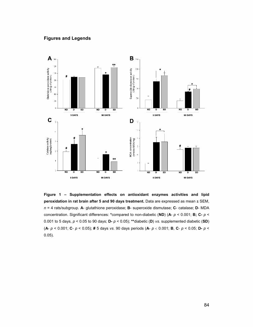
Figures and Legends
Figure 1 – Supplementation effects on antioxidant
peroxidation in rat brain after 5 and 90 days treatment.
n = 4 rats/subgroup. A- glutathione peroxidase;
concentration. Significant differences:
0.001 to 5 days, p < 0.05 to 90 day
(A- p < 0.001; C- p < 0.05); # 5 day
0.05).
Supplementation effects on antioxidant enzymes activities and lipid
peroxidation in rat brain after 5 and 90 days treatment. Data are expressed as mean ± SEM,
glutathione peroxidase; B- superoxide dismutase; C- catalase;
concentration. Significant differences: *compared to non-diabetic (ND) (A- p < 0.001;
days; D- p < 0.05); **diabetic (D) vs. supplemented diabetic (
days vs. 90 days periods (A- p < 0.001; B, C- p < 0.05;
84
enzymes activities and lipid
mean ± SEM,
catalase; D- MDA
< 0.001; B; C- p <
. supplemented diabetic (SD)
< 0.05; D- p <

Figure 2 – Supplementation and diabetes effects on m
yosin-IIB and m
yosin-Va protein levels in rat brain after 5 and 90 days treatment. W
este
rn
blot
s an
d qu
ant
ifica
tion
of
myo
sin
s e
xpre
ssio
ns
from
sup
ern
atan
t b
rain
sa
mp
les
of n
on-
dia
betic
(ND
), d
iab
etic
(D
) an
d su
pple
me
nte
d d
iabe
tic (SD
) ra
ts a
fter
5
and
90
days
. T
he a
mo
unt
of m
yosi
n pr
ote
ins
show
ed o
n t
he i
mm
unob
lot
was
det
erm
ine
d de
nsi
tom
etri
cally
an
d e
xpre
ssed
as
a re
late
d pe
rce
nta
ge
of t
he
grou
ps
(n
=
3 ra
ts/s
ubg
roup
).
Va
lues
re
pre
sent
m
ean
±
SE
M.
(*)
den
ote
a
stat
istic
ally
si
gni
fica
nt
(p
<
0.05
) di
ffe
ren
ce
as
com
par
ed
to ND
.
85

Figure 3 – Diabetes and supplementation effects on
expression in rat brain after 5 and 90 days.
and MYO5A mRNAs in brain samples of diabetic (
to non-diabetic (ND) rats after 5 and 90 days. Values represent mean ± SEM. (
statistically significant (p < 0.05 to
ND; (n = 3 rats/subgroup).
Diabetes and supplementation effects on MYH10 and MYO5A
expression in rat brain after 5 and 90 days. Real time PCR and relative expression of
mRNAs in brain samples of diabetic (D) and supplemented diabetic (SD
) rats after 5 and 90 days. Values represent mean ± SEM. (*
to MYH10 and p < 0.01 to MYO5A) difference as compared to
86
MYO5A mRNA
time PCR and relative expression of MYH10
SD) compared
*) denote a
) difference as compared to

Table 1.
Eff
ect
of in
ject
ion
stre
ptozo
toci
n and
supple
me
nta
tion o
n b
loo
d gl
uco
se a
nd
bo
dy
wei
ght
aft
er
5 a
nd
90 d
ays
(n =
8
rats
/su
bgro
up)
.
Param
eters
5 DAYS
90 DAYS
ND
D
SD
ND
D
SD
Blo
od g
lucose (
mg/d
L)
Initia
l 92
.25±1.6
9
447.1
3±1
5.6
0a
43
0.38±10.5
5a
89.
25±
2.28
398.8
8±1
9.09a
552.3
8±2
.38a
Fin
al
91.7
5±1.5
3
489.5
0±2
7.0
5a
46
1.00±29.4
5a
98.
38±
1.86
362.2
5±3
8.45a
467.1
3±3
9.3
9a
Body w
eig
ht
(g)
Initia
l 25
2.5
±7.6
1
236.7
5±8
.04
24
5.63±6.4
1
257.5
0±3
.32
249.8
8±4.3
8
221.7
5±4
.48
Fin
al
243.1
3±9.2
2
194.8
8±8
.96b,c
19
9.75±8.1
5b,c
362.3
8±1
8.5
5c
222.6
3±2
5.69b
253.7
5±3
1.1
5b
Da
ta a
re e
xpre
ssed
as
me
ans
± S
.E.M
. S
tatis
tical
ana
lysi
s w
as d
on
e w
ithin
da
ys-g
roup
s.
ND
, no
n-di
abe
tic; D
, di
abe
tic; SD
, su
ppl
eme
nte
d d
iab
etic
; 5 days
an
d 90 days
dia
betic
or
supp
lem
ent
atio
n p
erio
ds.
a p
< 0
.001
, si
gnifi
cant
vs.
ND
. b p
< 0
.05,
sig
nific
ant v
s. N
D.
c p
< 0
.001
, initi
al v
s. f
ina
l
87

88
References
1. Lamb RE, Goldstein BJ (2008) Modulating an oxidative-inflammatory
cascade: potential new treatment strategy for improving glucose
metabolism, insulin resistance, and vascular function. Int J Clin Pract 62:
1087-1095.
2. Nishikawa T, Edelstein D, Du XL, Yamagishi S, Matsumura T, et al. (2000)
Normalizing mitochondrial superoxide production blocks three pathways
of hyperglycaemic damage. Nature 404: 787-790.
3. Brands AM, Kessels RP, de Haan EH, Kappelle LJ, Biessels GJ (2004)
Cerebral dysfunction in type 1 diabetes: effects of insulin, vascular risk
factors and blood-glucose levels. Eur J Pharmacol 490: 159-168.
4. Russell JW, Golovoy D, Vincent AM, Mahendru P, Olzmann JA, et al. (2002)
High glucose-induced oxidative stress and mitochondrial dysfunction in
neurons. FASEB J 16: 1738-1748.
5. Tayarani I, Chaudiere J, Lefauconnier JM, Bourre JM (1987) Enzymatic
protection against peroxidative damage in isolated brain capillaries. J
Neurochem 48: 1399-1402.
6. Hammond B, Kontos HA, Hess ML (1985) Oxygen radicals in the adult
respiratory distress syndrome, in myocardial ischemia and reperfusion
injury, and in cerebral vascular damage. Can J Physiol Pharmacol 63:
173-187.
7. Del Boccio G, Lapenna D, Porreca E, Pennelli A, Savini F, et al. (1990) Aortic
antioxidant defence mechanisms: time-related changes in cholesterol-fed
rabbits. Atherosclerosis 81: 127-135.
8. Bonnefont-Rousselot D (2002) Glucose and reactive oxygen species. Curr
Opin Clin Nutr Metab Care 5: 561-568.
9. Bonnefont-Rousselot D, Bastard JP, Jaudon MC, Delattre J (2000)
Consequences of the diabetic status on the oxidant/antioxidant balance.
Diabetes Metab 26: 163-176.
10. Rahimi R, Nikfar S, Larijani B, Abdollahi M (2005) A review on the role of
antioxidants in the management of diabetes and its complications.
Biomed Pharmacother 59: 365-373.

89
11. Baydas G, Canatan H, Turkoglu A (2002) Comparative analysis of the
protective effects of melatonin and vitamin E on streptozocin-induced
diabetes mellitus. J Pineal Res 32: 225-230.
12. Hawkins CL, Davies MJ (2001) Generation and propagation of radical
reactions on proteins. Biochim Biophys Acta 1504: 196-219.
13. Biessels GJ, Kamal A, Urban IJ, Spruijt BM, Erkelens DW, et al. (1998)
Water maze learning and hippocampal synaptic plasticity in
streptozotocin-diabetic rats: effects of insulin treatment. Brain Res 800:
125-135.
14. Gispen WH, Biessels GJ (2000) Cognition and synaptic plasticity in diabetes
mellitus. Trends Neurosci 23: 542-549.
15. Popovic M, Biessels GJ, Isaacson RL, Gispen WH (2001) Learning and
memory in streptozotocin-induced diabetic rats in a novel spatial/object
discrimination task. Behav Brain Res 122: 201-207.
16. Fukui K, Onodera K, Shinkai T, Suzuki S, Urano S (2001) Impairment of
learning and memory in rats caused by oxidative stress and aging, and
changes in antioxidative defense systems. Ann N Y Acad Sci 928: 168-
175.
17. Grillo CA, Piroli GG, Wood GE, Reznikov LR, McEwen BS, et al. (2005)
Immunocytochemical analysis of synaptic proteins provides new insights
into diabetes-mediated plasticity in the rat hippocampus. Neuroscience
136: 477-486.
18. Artola A (2008) Diabetes-, stress- and ageing-related changes in synaptic
plasticity in hippocampus and neocortex--the same metaplastic process?
Eur J Pharmacol 585: 153-162.
19. Sellers JR (2000) Myosins: a diverse superfamily. Biochim Biophys Acta
1496: 3-22.
20. Berg JS, Powell BC, Cheney RE (2001) A millennial myosin census. Mol
Biol Cell 12: 780-794.
21. Foth BJ, Goedecke MC, Soldati D (2006) New insights into myosin evolution
and classification. Proc Natl Acad Sci U S A 103: 3681-3686.
22. Brown ME, Bridgman PC (2004) Myosin function in nervous and sensory
systems. J Neurobiol 58: 118-130.
90
23. Kawamoto S, Adelstein RS (1991) Chicken nonmuscle myosin heavy
chains: differential expression of two mRNAs and evidence for two
different polypeptides. J Cell Biol 112: 915-924.
24. Bridgman PC, Elkin LL (2000) Axonal myosins. J Neurocytol 29: 831-841.
25. Golomb E, Ma X, Jana SS, Preston YA, Kawamoto S, et al. (2004)
Identification and characterization of nonmuscle myosin II-C, a new
member of the myosin II family. J Biol Chem 279: 2800-2808.
26. Reck-Peterson SL, Provance DW, Jr., Mooseker MS, Mercer JA (2000)
Class V myosins. Biochim Biophys Acta 1496: 36-51.
27. Takagishi Y, Futaki S, Itoh K, Espreafico EM, Murakami N, et al. (2005)
Localization of myosin II and V isoforms in cultured rat sympathetic
neurones and their potential involvement in presynaptic function. J
Physiol 569: 195-208.
28. Beaulieu C, Kestekian R, Havrankova J, Gascon-Barre M (1993) Calcium is
essential in normalizing intolerance to glucose that accompanies vitamin
D depletion in vivo. Diabetes 42: 35-43.
29. Feskens EJ, Virtanen SM, Rasanen L, Tuomilehto J, Stengard J, et al.
(1995) Dietary factors determining diabetes and impaired glucose
tolerance. A 20-year follow-up of the Finnish and Dutch cohorts of the
Seven Countries Study. Diabetes Care 18: 1104-1112.
30. Ford ES, Mokdad AH (2001) Fruit and vegetable consumption and diabetes
mellitus incidence among U.S. adults. Prev Med 32: 33-39.
31. Pittas AG, Lau J, Hu FB, Dawson-Hughes B (2007) The role of vitamin D
and calcium in type 2 diabetes. A systematic review and meta-analysis. J
Clin Endocrinol Metab 92: 2017-2029.
32. Martini LA, Catania AS, Ferreira SR (2010) Role of vitamins and minerals in
prevention and management of type 2 diabetes mellitus. Nutr Rev 68:
341-354.
33. Low PA, Nickander KK, Tritschler HJ (1997) The roles of oxidative stress
and antioxidant treatment in experimental diabetic neuropathy. Diabetes
46 Suppl 2: S38-42.
34. Gardoni F, Kamal A, Bellone C, Biessels GJ, Ramakers GM, et al. (2002)
Effects of streptozotocin-diabetes on the hippocampal NMDA receptor
complex in rats. J Neurochem 80: 438-447.

91
35. Coleman E, Judd R, Hoe L, Dennis J, Posner P (2004) Effects of diabetes
mellitus on astrocyte GFAP and glutamate transporters in the CNS. Glia
48: 166-178.
36. Kamboj SS, Chopra K, Sandhir R (2008) Neuroprotective effect of N-
acetylcysteine in the development of diabetic encephalopathy in
streptozotocin-induced diabetes. Metab Brain Dis 23: 427-443.
37. Hernandez-Fonseca JP, Rincon J, Pedreanez A, Viera N, Arcaya JL, et al.
(2009) Structural and ultrastructural analysis of cerebral cortex,
cerebellum, and hypothalamus from diabetic rats. Exp Diabetes Res
2009: 329632.
38. Gomes RJ, de Oliveira CA, Ribeiro C, Mota CS, Moura LP, et al. (2009)
Effects of exercise training on hippocampus concentrations of insulin and
IGF-1 in diabetic rats. Hippocampus 19: 981-987.
39. Asplund K, Grankvist K, Marklund S, Taljedal IB (1984) Partial protection
against streptozotocin-induced hyperglycaemia by superoxide dismutase
linked to polyethylene glycol. Acta Endocrinol (Copenh) 107: 390-394.
40. Kakkar R, Kalra J, Mantha SV, Prasad K (1995) Lipid peroxidation and
activity of antioxidant enzymes in diabetic rats. Mol Cell Biochem 151:
113-119.
41. Kakkar R, Mantha SV, Radhi J, Prasad K, Kalra J (1997) Antioxidant
defense system in diabetic kidney: a time course study. Life Sci 60: 667-
679.
42. Genet S, Kale RK, Baquer NZ (2002) Alterations in antioxidant enzymes
and oxidative damage in experimental diabetic rat tissues: effect of
vanadate and fenugreek (Trigonellafoenum graecum). Mol Cell Biochem
236: 7-12.
43. Limaye PV, Raghuram N, Sivakami S (2003) Oxidative stress and gene
expression of antioxidant enzymes in the renal cortex of streptozotocin-
induced diabetic rats. Mol Cell Biochem 243: 147-152.
44. Panneerselvam SR, Govindasamy S (2004) Effect of sodium molybdate on
the status of lipids, lipid peroxidation and antioxidant systems in alloxan-
induced diabetic rats. Clin Chim Acta 345: 93-98.
45. Halliwell B (1992) Reactive oxygen species and the central nervous system.
J Neurochem 59: 1609-1623.
92
46. Ulusu NN, Sahilli M, Avci A, Canbolat O, Ozansoy G, et al. (2003) Pentose
phosphate pathway, glutathione-dependent enzymes and antioxidant
defense during oxidative stress in diabetic rodent brain and peripheral
organs: effects of stobadine and vitamin E. Neurochem Res 28: 815-823.
47. Siddiqui MR, Taha A, Moorthy K, Hussain ME, Basir SF, et al. (2005)
Amelioration of altered antioxidant status and membrane linked functions
by vanadium and Trigonella in alloxan diabetic rat brains. J Biosci 30:
483-490.
48. Huang WC, Juang SW, Liu IM, Chi TC, Cheng JT (1999) Changes of
superoxide dismutase gene expression and activity in the brain of
streptozotocin-induced diabetic rats. Neurosci Lett 275: 25-28.
49. Sinha N, Baquer NZ, Sharma D (2005) Anti-lipidperoxidative role of
exogenous dehydroepiendrosterone (DHEA) administration in normal
ageing rat brain. Indian J Exp Biol 43: 420-424.
50. Kumar JS, Menon VP (1993) Effect of diabetes on levels of lipid peroxides
and glycolipids in rat brain. Metabolism 42: 1435-1439.
51. Makar TK, Rimpel-Lamhaouar K, Abraham DG, Gokhale VS, Cooper AJ
(1995) Antioxidant defense systems in the brains of type II diabetic mice.
J Neurochem 65: 287-291.
52. Kuhad A, Chopra K (2007) Curcumin attenuates diabetic encephalopathy in
rats: behavioral and biochemical evidences. Eur J Pharmacol 576: 34-42.
53. Kwag OG, Kim SO, Choi JH, Rhee IK, Choi MS, et al. (2001) Vitamin E
improves microsomal phospholipase A2 activity and the arachidonic acid
cascade in kidney of diabetic rats. J Nutr 131: 1297-1301.
54. Wohaieb SA, Godin DV (1987) Alterations in free radical tissue-defense
mechanisms in streptozocin-induced diabetes in rat. Effects of insulin
treatment. Diabetes 36: 1014-1018.
55. Ozkaya YG, Agar A, Yargicoglu P, Hacioglu G, Bilmen-Sarikcioglu S, et al.
(2002) The effect of exercise on brain antioxidant status of diabetic rats.
Diabetes Metab 28: 377-384.
56. Miyake Y, Yamamoto K, Tsujihara N, Osawa T (1998) Protective effects of
lemon flavonoids on oxidative stress in diabetic rats. Lipids 33: 689-695.

93
57. Bhor VM, Raghuram N, Sivakami S (2004) Oxidative damage and altered
antioxidant enzyme activities in the small intestine of streptozotocin-
induced diabetic rats. Int J Biochem Cell Biol 36: 89-97.
58. Zemel MB, Thompson W, Milstead A, Morris K, Campbell P (2004) Calcium
and dairy acceleration of weight and fat loss during energy restriction in
obese adults. Obes Res 12: 582-590.
59. de Boer IH, Tinker LF, Connelly S, Curb JD, Howard BV, et al. (2008)
Calcium plus vitamin D supplementation and the risk of incident diabetes
in the Women's Health Initiative. Diabetes Care 31: 701-707.
60. Fridovich I (1995) Superoxide radical and superoxide dismutases. Annu Rev
Biochem 64: 97-112.
61. Valko M, Morris H, Cronin MT (2005) Metals, toxicity and oxidative stress.
Curr Med Chem 12: 1161-1208.
62. Taylor CG (2005) Zinc, the pancreas, and diabetes: insights from rodent
studies and future directions. Biometals 18: 305-312.
63. Scheede-Bergdahl C, Penkowa M, Hidalgo J, Olsen DB, Schjerling P, et al.
(2005) Metallothionein-mediated antioxidant defense system and its
response to exercise training are impaired in human type 2 diabetes.
Diabetes 54: 3089-3094.
64. Beltramini M, Zambenedetti P, Raso M, IbnlKayat MI, Zatta P (2006) The
effect of Zn(II) and streptozotocin administration in the mouse brain.
Brain Res 1109: 207-218.
65. Taylor CG, Bettger WJ, Bray TM (1988) Effect of dietary zinc or copper
deficiency on the primary free radical defense system in rats. J Nutr 118:
613-621.
66. Oteiza PI, Kleinman CG, Demasi M, Bechara EJ (1995) 5-Aminolevulinic
acid induces iron release from ferritin. Arch Biochem Biophys 316: 607-
611.
67. Thomas DJ, Caffrey TC (1991) Lipopolysaccharide induces double-stranded
DNA fragmentation in mouse thymus: protective effect of zinc
pretreatment. Toxicology 68: 327-337.
68. Matsushita K, Kitagawa K, Matsuyama T, Ohtsuki T, Taguchi A, et al.
(1996) Effect of systemic zinc administration on delayed neuronal death
in the gerbil hippocampus. Brain Res 743: 362-365.

94
69. Santon A, Formigari A, Albergoni V, Irato P (2006) Effect of Zn treatment on
wild type and MT-null cell lines in relation to apoptotic and/or necrotic
processes and on MT isoform gene expression. Biochim Biophys Acta
1763: 305-312.
70. Yoshida H, Sasaki K, Hirowatari Y, Kurosawa H, Sato N, et al. (2004)
Increased serum iron may contribute to enhanced oxidation of low-
density lipoprotein in smokers in part through changes in lipoxygenase
and catalase. Clin Chim Acta 345: 161-170.
71. Kowluru RA, Engerman RL, Kern TS (2000) Diabetes-induced metabolic
abnormalities in myocardium: effect of antioxidant therapy. Free Radic
Res 32: 67-74.
72. Valko M, Leibfritz D, Moncol J, Cronin MT, Mazur M, et al. (2007) Free
radicals and antioxidants in normal physiological functions and human
disease. Int J Biochem Cell Biol 39: 44-84.
73. Paolisso G, D'Amore A, Giugliano D, Ceriello A, Varricchio M, et al. (1993)
Pharmacologic doses of vitamin E improve insulin action in healthy
subjects and non-insulin-dependent diabetic patients. Am J Clin Nutr 57:
650-656.
74. Paolisso G, Di Maro G, Galzerano D, Cacciapuoti F, Varricchio G, et al.
(1994) Pharmacological doses of vitamin E and insulin action in elderly
subjects. Am J Clin Nutr 59: 1291-1296.
75. Yanagisawa H, Sato M, Nodera M, Wada O (2004) Excessive zinc intake
elevates systemic blood pressure levels in normotensive rats--potential
role of superoxide-induced oxidative stress. J Hypertens 22: 543-550.
76. Shelton RJ, Velavan P, Nikitin NP, Coletta AP, Clark AL, et al. (2005)
Clinical trials update from the American Heart Association meeting:
ACORN-CSD, primary care trial of chronic disease management,
PEACE, CREATE, SHIELD, A-HeFT, GEMINI, vitamin E meta-analysis,
ESCAPE, CARP, and SCD-HeFT cost-effectiveness study. Eur J Heart
Fail 7: 127-135.
77. Ward NC, Wu JH, Clarke MW, Puddey IB, Burke V, et al. (2007) The effect
of vitamin E on blood pressure in individuals with type 2 diabetes: a
randomized, double-blind, placebo-controlled trial. J Hypertens 25: 227-
234.

95
78. Cerielo A, Motz E, Cavarape A, Lizzio S, Russo A, et al. (1997)
Hyperglycemia counterbalances the antihypertensive effect of
glutathione in diabetic patients: evidence linking hypertension and
glycemia through the oxidative stress in diabetes mellitus. J Diabetes
Complications 11: 250-255.
79. Baquer NZ, Hothersall JS, McLean P, Greenbaum AL (1990) Effect of aging
on soluble and membrane bound enzymes in rat brain. Neurochem Int
16: 369-375.
80. Carrillo MC, Kanai S, Sato Y, Kitani K (1992) Age-related changes in
antioxidant enzyme activities are region and organ, as well as sex,
selective in the rat. Mech Ageing Dev 65: 187-198.
81. Moorthy K, Sharma D, Basir SF, Baquer NZ (2005) Administration of
estradiol and progesterone modulate the activities of antioxidant enzyme
and aminotransferases in naturally menopausal rats. Exp Gerontol 40:
295-302.
82. Moorthy K, Yadav UC, Siddiqui MR, Mantha AK, Basir SF, et al. (2005)
Effect of hormone replacement therapy in normalizing age related
neuronal markers in different age groups of naturally menopausal rats.
Biogerontology 6: 345-356.
83. Moorthy K, Yadav UC, Mantha AK, Cowsik SM, Sharma D, et al. (2004)
Estradiol and progesterone treatments change the lipid profile in naturally
menopausal rats from different age groups. Biogerontology 5: 411-419.
84. Bala K, Tripathy BC, Sharma D (2006) Neuroprotective and anti-ageing
effects of curcumin in aged rat brain regions. Biogerontology 7: 81-89.
85. Poon HF, Vaishnav RA, Getchell TV, Getchell ML, Butterfield DA (2006)
Quantitative proteomics analysis of differential protein expression and
oxidative modification of specific proteins in the brains of old mice.
Neurobiol Aging 27: 1010-1019.
86. Liu R, Liu IY, Bi X, Thompson RF, Doctrow SR, et al. (2003) Reversal of
age-related learning deficits and brain oxidative stress in mice with
superoxide dismutase/catalase mimetics. Proc Natl Acad Sci U S A 100:
8526-8531.
87. Kamal A, Biessels GJ, Urban IJ, Gispen WH (1999) Hippocampal synaptic
plasticity in streptozotocin-diabetic rats: impairment of long-term
96
potentiation and facilitation of long-term depression. Neuroscience 90:
737-745.
88. Zhu D, Tan KS, Zhang X, Sun AY, Sun GY, et al. (2005) Hydrogen peroxide
alters membrane and cytoskeleton properties and increases intercellular
connections in astrocytes. J Cell Sci 118: 3695-3703.
89. Aragno M, Mastrocola R, Catalano MG, Brignardello E, Danni O, et al.
(2004) Oxidative stress impairs skeletal muscle repair in diabetic rats.
Diabetes 53: 1082-1088.
90. Coirault C, Guellich A, Barbry T, Samuel JL, Riou B, et al. (2007) Oxidative
stress of myosin contributes to skeletal muscle dysfunction in rats with
chronic heart failure. Am J Physiol Heart Circ Physiol 292: H1009-1017.
91. Mihm MJ, Yu F, Reiser PJ, Bauer JA (2003) Effects of peroxynitrite on
isolated cardiac trabeculae: selective impact on myofibrillar energetic
controllers. Biochimie 85: 587-596.
92. Mochida S, Kobayashi H, Matsuda Y, Yuda Y, Muramoto K, et al. (1994)
Myosin II is involved in transmitter release at synapses formed between
rat sympathetic neurons in culture. Neuron 13: 1131-1142.
93. Geeves MA, Fedorov R, Manstein DJ (2005) Molecular mechanism of
actomyosin-based motility. Cell Mol Life Sci 62: 1462-1477.
94. Ishmael JE, Safic M, Amparan D, Vogel WK, Pham T, et al. (2007)
Nonmuscle myosins II-B and Va are components of detergent-resistant
membrane skeletons derived from mouse forebrain. Brain Res 1143: 46-
59.
95. Nitsch R, Hoyer S (1991) Local action of the diabetogenic drug,
streptozotocin, on glucose and energy metabolism in rat brain cortex.
Neurosci Lett 128: 199-202.
96. McLean WG, Pekiner C, Cullum NA, Casson IF (1992) Posttranslational
modifications of nerve cytoskeletal proteins in experimental diabetes. Mol
Neurobiol 6: 225-237.
97. Sapone A, de Magistris L, Pietzak M, Clemente MG, Tripathi A, et al. (2006)
Zonulin upregulation is associated with increased gut permeability in
subjects with type 1 diabetes and their relatives. Diabetes 55: 1443-
1449.
97
98. Pereira RV, de Miranda-Neto MH, da Silva Souza ID, Zanoni JN (2008)
Vitamin E supplementation in rats with experimental diabetes mellitus:
analysis of myosin-V and nNOS immunoreactive myenteric neurons from
terminal ileum. J Mol Histol 39: 595-603.
99. Bevers MB, Neumar RW (2008) Mechanistic role of calpains in
postischemic neurodegeneration. J Cereb Blood Flow Metab 28: 655-
673.
100. Saito K, Elce JS, Hamos JE, Nixon RA (1993) Widespread activation of
calcium-activated neutral proteinase (calpain) in the brain in Alzheimer
disease: a potential molecular basis for neuronal degeneration. Proc Natl
Acad Sci U S A 90: 2628-2632.
101. Huang Y, Wang KK (2001) The calpain family and human disease. Trends
Mol Med 7: 355-362.
102. Huang CJ, Gurlo T, Haataja L, Costes S, Daval M, et al. Calcium-activated
calpain-2 is a mediator of beta cell dysfunction and apoptosis in type 2
diabetes. J Biol Chem 285: 339-348.
103. Nascimento AA, Cheney RE, Tauhata SB, Larson RE, Mooseker MS
(1996) Enzymatic characterization and functional domain mapping of
brain myosin-V. J Biol Chem 271: 17561-17569.
104. Espindola FS, Espreafico EM, Coelho MV, Martins AR, Costa FR, et al.
(1992) Biochemical and immunological characterization of p190-
calmodulin complex from vertebrate brain: a novel calmodulin-binding
myosin. J Cell Biol 118: 359-368.
105. Reeves PG, Nielsen FH, Fahey GC, Jr. (1993) AIN-93 purified diets for
laboratory rodents: final report of the American Institute of Nutrition ad
hoc writing committee on the reformulation of the AIN-76A rodent diet. J
Nutr 123: 1939-1951.
106. Bradford MM (1976) A rapid and sensitive method for the quantitation of
microgram quantities of protein utilizing the principle of protein-dye
binding. Anal Biochem 72: 248-254.
107. Aebi H, Suter H, Feinstein RN (1968) Activity and stability of catalase in
blood and tissues of normal and acatalasemic mice. Biochem Genet 2:
245-251.

98
108. Towbin H, Staehelin T, Gordon J (1979) Electrophoretic transfer of
proteins from polyacrylamide gels to nitrocellulose sheets: procedure and
some applications. Proc Natl Acad Sci U S A 76: 4350-4354.
109. Larson RE, Ferro JA, Queiroz EA (1986) Isolation and purification of
actomyosin ATPase from mammalian brain. J Neurosci Methods 16: 47-
58.
110. Suter DM, Espindola FS, Lin CH, Forscher P, Mooseker MS (2000)
Localization of unconventional myosins V and VI in neuronal growth
cones. J Neurobiol 42: 370-382.
111. Espreafico EM, Cheney RE, Matteoli M, Nascimento AA, De Camilli PV, et
al. (1992) Primary structure and cellular localization of chicken brain
myosin-V (p190), an unconventional myosin with calmodulin light chains.
J Cell Biol 119: 1541-1557.
112. Yin JL, Shackel NA, Zekry A, McGuinness PH, Richards C, et al. (2001)
Real-time reverse transcriptase-polymerase chain reaction (RT-PCR) for
measurement of cytokine and growth factor mRNA expression with
fluorogenic probes or SYBR Green I. Immunol Cell Biol 79: 213-221.
99
2. CONCLUSÕES
- O efeito crônico do diabetes levou ao aumento dos níveis da atividade da
glutationa peroxidase e de malondialdeído, como também à diminuição da
atividade das enzimas superóxido dismutase e catalase no cérebro de ratos
diabéticos.
- As alterações nos marcadores enzimáticos e de peroxidação lipídica do
estresse oxidativo estão acompanhadas por um aumento na expressão de
miosina-IIB e diminuição de miosina-Va.
- A suplementação dos ratos diabéticos com uma ração enriquecida com
antioxidantes minerais (cálcio e zinco) e vitamina E protegeu o cérebro de
possíveis danos oxidativos consequentes da hiperglicemia, reduzindo a
atividade da catalase e aumentando os níveis de glutationa peroxidase e
superóxido dismutase, embora nenhuma influência tenha sido observada nos
níveis de malondialdeído.
- Quanto à expressão de miosinas, somente a da classe II reestabeleceu os
seus níveis no cérebro de ratos diabéticos após a suplementação.
- De forma geral, as alterações das enzimas antioxidantes e mosinas IIB e Va
no cérebro de ratos diabéticos estão associadas com os estágios agudo e
crônico do diabetes.