Systematization, description and territory of the caudal ... · central. Penetrou na fissura...
6
817 Pesq. Vet. Bras. 31(9):817-822, setembro 2011 RESUMO.- [Sistematização, descrição e território da arté- ria cerebral caudal no cérebro do jacaré do papo-amare- lo (Caiman latirostris).] Foram utilizadas trinta cabeças com o segmento de pescoço de Caiman latirostris. Os animais eram provenientes do Criatório Mister Caiman sob a autorização do Instituto Brasileiro de Meio Ambiente e dos Recursos Naturais Renováveis (Ibama). Os animais foram sacrificados de acordo com as normas de rotina de abate do frigorífico e as cabeças foram seccionadas na altura da terceira vértebra cervical. O sistema arterial foi lavado com solução salina 0.9% resfriada, com drenagem pelas veias jugulares. Em seguida, o sistema foi preenchido com injeção de látex, corado em vermelho As pe- ças foram então fixadas em formaldeído a 20.0% por sete dias. O cérebro foi removido, com um segmento de medula espinhal, a dura-máter retirada e as artérias dissecadas. Na altura da hipófise, a artéria carótida interna emitiu um ramo rostral e, um curto ramo caudal, continuando-se naturalmente, como artéria cerebral caudal. Esta se projetou látero-dorsalmente e ao sobrepassar o trato óptico, emitiu seu I (primeiro) ramo central. Penetrou na fissura transversa do cérebro, lançando a artéria diencefálica e a seguir seu II (segundo) ramo central. Systematization, description and territory of the caudal cerebral artery of the brain in broad-snouted Caiman ( Caiman latirostris) 1 Lygia Almeida 2* and Rui Campos 2,3 ABSTRACT.- Almeida L. & Campos R. 2011. Systematization, description and territory of the caudal cerebral artery of the brain in broad-snouted Caiman (Caiman latirostris). Pesquisa Veterinária Brasileira 31(9):817-822. Setor de Anatomia Animal, Departamento de Ciências Morfológicas, Faculdade de Veterinária, Universidade Federal do Rio Grande do Sul, Av. Bento Gonçalves 9090, Porto Alegre, RS 90540-000, Brazil. E-mail: l .al meid a@t err a.com.br Thirty heads with the neck segment of Caiman latirostris were used. The animals were provided from a creation center called Mister Caiman, under the authorization of the Brazilian Institute of Environment and Renewable Natural Resources (Ibama). Animals were sacrificed according to the slaughtering routine of the abattoir, and the heads were sectioned at the level of the third cervical vertebra. The arterial system was washed with cold saline solution, with drainage through jugular veins. Subsequently, the system was filled with red colored latex injection. Pieces were than fixed in 20% formaldehyde, for seven days. The brains were removed, with a spinal cord segment, the duramater removed and the arteries dissected. At the level of the hypophysis, the internal carotid artery gave off a rostral branch, and a short caudal branch, continuing, naturally, as the caudal cerebral artery. This artery projected laterodorsalwards and, as it overpassed the optic tract, gave off its I (the first) central branch. Penetrated in the cerebral transverse fissure, emitting the diencephalic artery and next its II (second) central branch. Still inside the fissure, originated occipital hemispheric branches and a pineal branch. Emerged from the cerebral transverse fissure, over the occipital pole of the cerebral hemisphere. Projected rostralwards, sagital to the cerebral longitudinal fissure, as interhemispheric artery. This artery gave off medial and convex hemispheric branches to the respective surfaces of the cerebral hemispheres, anastomosed with its contralateral homologous, forming the common ethmoidal artery. This artery entered the fissure between the olfactory peduncles, emerging ventrally and dividing into ethmoidal arteries, right and left, which progressed towards the nasal cavities, vascularizing them. The territory of the caudal cerebral artery included the most caudal area of the base of the cerebral hemisphere, its convex surface, the olfactory peduncles and bulbs, the choroid plexuses and the diencephalus with its parietal organs. INDEX TERMS: Alligator, cerebral arteries, brain vascularization, reptiles 1 Received on November 3, 2010. Accepted for publication on April 13, 2011. 2 Programa de Pós-Graduação em Medicina Veterinária, Faculdade de Veterinária, Universidade Federal do Rio Grande do Sul (UFRGS), Av. Bento Gonçalves 9090, Porto Alegre, RS, 91540-000, Brazil. * Corresponding author: l .al meid a@t err a.com.br 3 Departamento de Ciências Morfológicas, Centro de Ciências Básicas, UFRGS, Av. Bento Gonçalves 9090, Porto Alegre, RS 91540-000. E-mail: [email protected]
Transcript of Systematization, description and territory of the caudal ... · central. Penetrou na fissura...

817
Pesq. Vet. Bras. 31(9):817-822, setembro 2011
RESUMO.- [Sistematização, descrição e território da arté-ria cerebral caudal no cérebro do jacaré do papo-amare-lo (Caiman latirostris).] Foram utilizadas trinta cabeças como segmento de pescoço de Caiman latirostris. Os animais eramprovenientes do Criatório Mister Caiman sob a autorização doInstituto Brasileiro de Meio Ambiente e dos Recursos Naturais
Renováveis (Ibama). Os animais foram sacrificados de acordocom as normas de rotina de abate do frigorífico e as cabeçasforam seccionadas na altura da terceira vértebra cervical. Osistema arterial foi lavado com solução salina 0.9% resfriada,com drenagem pelas veias jugulares. Em seguida, o sistema foipreenchido com injeção de látex, corado em vermelho As pe-ças foram então fixadas em formaldeído a 20.0% por sete dias.O cérebro foi removido, com um segmento de medula espinhal,a dura-máter retirada e as artérias dissecadas. Na altura dahipófise, a artéria carótida interna emitiu um ramo rostral e,um curto ramo caudal, continuando-se naturalmente, comoartéria cerebral caudal. Esta se projetou látero-dorsalmente eao sobrepassar o trato óptico, emitiu seu I (primeiro) ramocentral. Penetrou na fissura transversa do cérebro, lançando aartéria diencefálica e a seguir seu II (segundo) ramo central.
Systematization, description and territory of the caudal cerebralartery of the brain in broad-snouted Caiman (Caiman latirostris)1
Lygia Almeida2* and Rui Campos2,3
ABSTRACT.- Almeida L. & Campos R. 2011. Systematization, description and territory ofthe caudal cerebral artery of the brain in broad-snouted Caiman (Caiman latirostris).Pesquisa Veterinária Brasileira 31(9):817-822. Setor de Anatomia Animal, Departamento deCiências Morfológicas, Faculdade de Veterinária, Universidade Federal do Rio Grande do Sul,Av. Bento Gonçalves 9090, Porto Alegre, RS 90540-000, Brazil. E-mail: [email protected]
Thirty heads with the neck segment of Caiman latirostris were used. The animals wereprovided from a creation center called Mister Caiman, under the authorization of the BrazilianInstitute of Environment and Renewable Natural Resources (Ibama). Animals were sacrificedaccording to the slaughtering routine of the abattoir, and the heads were sectioned at the levelof the third cervical vertebra. The arterial system was washed with cold saline solution, withdrainage through jugular veins. Subsequently, the system was filled with red colored latexinjection. Pieces were than fixed in 20% formaldehyde, for seven days. The brains were removed,with a spinal cord segment, the duramater removed and the arteries dissected. At the level ofthe hypophysis, the internal carotid artery gave off a rostral branch, and a short caudal branch,continuing, naturally, as the caudal cerebral artery. This artery projected laterodorsalwards and,as it overpassed the optic tract, gave off its I (the first) central branch. Penetrated in the cerebraltransverse fissure, emitting the diencephalic artery and next its II (second) central branch. Stillinside the fissure, originated occipital hemispheric branches and a pineal branch. Emergedfrom the cerebral transverse fissure, over the occipital pole of the cerebral hemisphere. Projectedrostralwards, sagital to the cerebral longitudinal fissure, as interhemispheric artery. This arterygave off medial and convex hemispheric branches to the respective surfaces of the cerebralhemispheres, anastomosed with its contralateral homologous, forming the common ethmoidalartery. This artery entered the fissure between the olfactory peduncles, emerging ventrally anddividing into ethmoidal arteries, right and left, which progressed towards the nasal cavities,vascularizing them. The territory of the caudal cerebral artery included the most caudal area ofthe base of the cerebral hemisphere, its convex surface, the olfactory peduncles and bulbs, thechoroid plexuses and the diencephalus with its parietal organs.
INDEX TERMS: Alligator, cerebral arteries, brain vascularization, reptiles
1Received on November 3, 2010.
Accepted for publication on April 13, 2011.2 Programa de Pós-Graduação em Medicina Veterinária, Faculdade de
Veterinária, Universidade Federal do Rio Grande do Sul (UFRGS), Av. Bento
Gonçalves 9090, Porto Alegre, RS, 91540-000, Brazil. *Corresponding
author: [email protected] Departamento de Ciências Morfológicas, Centro de Ciências Básicas,
UFRGS, Av. Bento Gonçalves 9090, Porto Alegre, RS 91540-000. E-mail:

Pesq. Vet. Bras. 31(9):817-822, setembro 2011
Lygia Almeida and Rui Campos818
Ainda dentro da fissura originou ramos hemisféricos occipitais,e um ramo pineal. Emergiu da fissura transversa do cérebro,no pólo occipital do hemisfério cerebral. Projetou-serostralmente, sagital a fissura longitudinal do cérebro, comoartéria interhemisférica. Esta artéria lançou ramoshemisféricos convexos e mediais para as respectivas faces doshemisférios cerebrais, anastomosou-se com sua homólogacontralateral, formando uma artéria etmoidal comum. Estamergulhou na fissura entre os pedúnculos olfatórios emergin-do ventralmente e, dividindo-se em artérias etmoidais, direitae esquerda, as quais progrediram para as cavidades nasais,vascularizando-as. O território da artéria cerebral caudal com-preendeu a área mais caudal da base do hemisfério cerebral,sua face convexa, os pendúculos e bulbos olfatórios, plexocorióides e o diencéfalo com seus órgãos parietais.
TERMOS DE INDEXAÇÃO: Artérias cerebrais, vascularização cere-bral, répteis, jacaré.
INTRODUCTION
The farming production of the broad-snouted Caiman, a Bra-zilian fauna species, and the authorization for the commer-cialization of their high quality skin and meat by the BrazilianInstitute of Environment and Renewable Natural Resources(Ibama), increased the interest in its production by thenational and international markets.
The distribution of the cerebral blood supply, as in anyother anatomical structure, is closely associated to the develo-pment stage and the consequent morphology presented by thecentral nervous system of each species. The classical textbooklittle or no concern in relation to irrigation brain on this ani-mal, although in the specialized literature, Burda (1969)studied the development of the intracranial arterial pattern inalligator embryos and the cerebrovascular changes that occurafter birth. De Vriese (1905) provided important contributionsto the phylogeny and ontogeny of the cerebral arteries,classifying several animal groups, according to the cerebralarterial circle formation into three distinct types. Kappers(1933), Schepers (1939) and Burda (1965) reported the arte-rial supply to the brain in turtles. Frizzo et al. (1994)investigated the vascularization of the subfornical organ ofturtles. Gillilan (1967) affirmed that the cerebral arterialpatterns of reptiles are similar and represent a stage oftransition between primitive and superior vertebrates. Dendy(1909) and Burda (1966) studied the intracranial arterialsystem of Sphenodon punctatus (iguana) and Crotaphytuscollaris (lizard), respectively. In addition, there were somedescriptions about the cerebral vascularization in Caimanlatirostris (Almeida & Campos 2010, Almeida & Campos 2011).
This study aims to describe and systematize the caudalcerebral artery of the broad-snouted Caiman, determining astandard model and territorial area of irrigation, and the mainvariations, in this species. These informations contribute forthe studies in this area of the comparative anatomy aboutthe phylogenetic development of the blood supply to the cen-tral nervous system.
MATERIALS AND METHODS
Thirty heads with the neck segment of Caiman latirostris were used.The animals were around three years old, weighting 8-10 kg, ma-
les, provided from a creation center called Mister Caiman, underthe authorization of the Brazilian Institute of Environment andRenewable Natural Resources (Ibama). Animals were sacrificed ac-cording to the slaughtering routine of the abattoir, and the thirtyheads were sectioned at the level of the third cervical vertebra.The azygous carotid artery was cannulated just before its divisioninto internal carotid arteries, and the arterial system was washedwith cold saline solution, containing 2500 IU heparin (Heparin;Cristalia Produtos Quimicos Farmaceuticos Ltda, Itapira, SP, Brazil)per animal, with drainage through jugular veins. Subsequently, thesystem was filled with latex injection (Cola 603, Bertoncini Ltda,São Paulo, SP, Brazil) colored with red pigment (Suvinil CoranteBASF S.A., São Bernardo do Campo, SP, Brazil). Pieces remained forat least one hour in running water for latex polymerization. Next, abone window was opened in the skullcap. Pieces were than fixedin 20% formaldehyde, for seven days. The brains were removed,with a spinal cord segment, the duramater removed and the arteriesdissected. Schematic drawings of the ventral and dorsal views ofthe brain arteries and of the interior of the cerebral fissure of allspecimens were prepared with the aid of a magnifying glass (LTS,5x increase and Stemi SV8 Zeiss; Goettingen, Germany). Since it isa reptile which the morphological aspects became closer to birdsthan mammals, the vessels were named according to the NominaAnatomica Avium (1993) and Nomina Anatomica Veterinaria(2005), with some designations based on the author’s interpreta-tion of previous reports. Some specimens were photographed toillustrate the vessel arrangements. The occurrence and distributi-on of the vessels was expressed as a percentage of the thirtyspecimens examined.
RESULTS
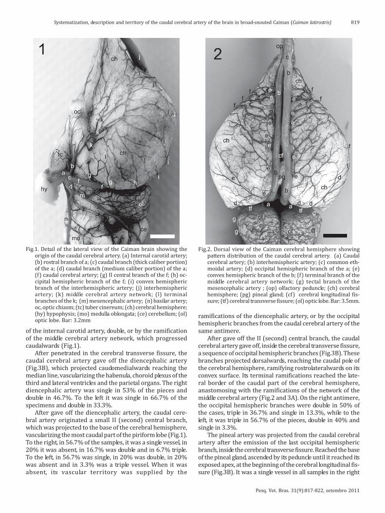
As it became close to the nape of neck, the azygous carotidartery divided into two internal carotid arteries, in anapproximate divergent angle of 70o, which penetrated in thecarotid foramina, laterally to the single occipital condylus andthe foramen magnun. The internal carotid artery ran alongthe carotid canal until it reached the sella turcica, where itemerged caudolateralwards the hypophysis. It curveddorsally, juxtaposed to the tuber cinerium, and on its base,emitted a rostral branch, of medium caliber, continuing ascaudal branch, of thick caliber (Fig.1).
The caudal cerebral artery, in Caiman, was a naturalcontinuation, of the thick caliber portion of the caudal branchof the internal carotid artery. In all samples, it was presentand constant as a single vessel of thick caliber. The thickcaliber of the caudal cerebral artery was due to the bloodsupply to the nasal cavity by the ethmoidal artery, its termi-nal branch (Fig.2 and 3A). The caudal cerebral arteryprojected laterodorsalwards and as it passed over the optictract, emitted its I (the first) central branch, which ranrostralwards, medial to the piriform lobe. The I (the first)central branch anastomosed with the first branches of themiddle cerebral artery network, and its caudal ramificationsreached the most rostral part of the piriform lobe, reachingthe lateral part of the base of the brain. The I (the first) cen-tral branch of the right caudal cerebral artery was a singlevessel in 73.3% of the cases, double in 16.7% and absent in10%. To the left, the I (the first) central branch of the caudalcerebral artery was single in 66.7%, absent in 26.7% anddouble in 10% of the samples. When it was absent, its vascularterritory was supplied by the presence of the rostral branch
Pesq. Vet. Bras. 31(9):817-822, setembro 2011
Systematization, description and territory of the caudal cerebral artery of the brain in broad-snouted Caiman (Caiman latirostris) 819
of the internal carotid artery, double, or by the ramificationof the middle cerebral artery network, which progressedcaudalwards (Fig.1).
After penetrated in the cerebral transverse fissure, thecaudal cerebral artery gave off the diencephalic artery(Fig.3B), which projected caudomedialwards reaching themedian line, vascularizing the habenula, choroid plexus of thethird and lateral ventricles and the parietal organs. The rightdiencephalic artery was single in 53% of the pieces anddouble in 46.7%. To the left it was single in 66.7% of thespecimens and double in 33.3%.
After gave off the diencephalic artery, the caudal cere-bral artery originated a small II (second) central branch,which was projected to the base of the cerebral hemisphere,vascularizing the most caudal part of the piriform lobe (Fig.1).To the right, in 56.7% of the samples, it was a single vessel, in20% it was absent, in 16.7% was double and in 6.7% triple.To the left, in 56.7% was single, in 20% was double, in 20%was absent and in 3.3% was a triple vessel. When it wasabsent, its vascular territory was supplied by the
ramifications of the diencephalic artery, or by the occipitalhemispheric branches from the caudal cerebral artery of thesame antimere.
After gave off the II (second) central branch, the caudalcerebral artery gave off, inside the cerebral transverse fissure,a sequence of occipital hemispheric branches (Fig.3B). Thesebranches projected dorsalwards, reaching the caudal pole ofthe cerebral hemisphere, ramifying rostrolateralwards on itsconvex surface. Its terminal ramifications reached the late-ral border of the caudal part of the cerebral hemisphere,anastomosing with the ramifications of the network of themiddle cerebral artery (Fig.2 and 3A). On the right antimere,the occipital hemispheric branches were double in 50% ofthe cases, triple in 36.7% and single in 13.3%, while to theleft, it was triple in 56.7% of the pieces, double in 40% andsingle in 3.3%.
The pineal artery was projected from the caudal cerebralartery after the emission of the last occipital hemisphericbranch, inside the cerebral transverse fissure. Reached the baseof the pineal gland, ascended by its peduncle until it reached itsexposed apex, at the beginning of the cerebral longitudinal fis-sure (Fig.3B). It was a single vessel in all samples in the right
Fig.1. Detail of the lateral view of the Caiman brain showing theorigin of the caudal cerebral artery. (a) Internal carotid artery;(b) rostral branch of a; (c) caudal branch (thick caliber portion)of the a; (d) caudal branch (medium caliber portion) of the a;(f) caudal cerebral artery; (g) II central branch of the f; (h) oc-cipital hemispheric branch of the f; (i) convex hemisphericbranch of the interhemispheric artery; (j) interhemisphericartery; (k) middle cerebral artery network; (l) terminalbranches of the k; (m) mesencephalic artery; (n) basilar artery;oc, optic chiasm; (tc) tuber cinereum; (ch) cerebral hemisphere;(hy) hypophysis; (mo) medulla oblongata; (ce) cerebellum; (ol)optic lobe. Bar: 3.2mm
Fig.2. Dorsal view of the Caiman cerebral hemisphere showingpattern distribution of the caudal cerebral artery. (a) Caudalcerebral artery; (b) interhemispheric artery; (c) common eth-moidal artery; (d) occipital hemispheric branch of the a; (e)convex hemispheric branch of the b; (f) terminal branch of themiddle cerebral artery network; (g) tectal branch of themesencephalic artery ; (op) olfactory peduncle; (ch) cerebralhemisphere; (pg) pineal gland; (cf) cerebral longitudinal fis-sure; (tf) cerebral transverse fissure; (ol) optic lobe. Bar: 3.5mm.
Pesq. Vet. Bras. 31(9):817-822, setembro 2011
Lygia Almeida and Rui Campos820
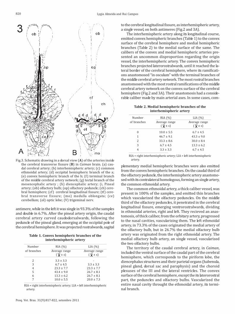
antimere, while in the left it was single in 93.3% of the samplesand double in 6.7%. After the pineal artery origin, the caudalcerebral artery curved caudodorsalwards, following thepeduncle of the pineal gland, emerging at the occipital pole ofthe cerebral hemisphere. It was projected rostralwards, sagital
to the cerebral longitudinal fissure, as interhemispheric artery,a single vessel, on both antimeres (Fig.2 and 3A).
The interhemispheric artery along its longitudinal course,emitted convex hemispheric branches (Table 1) to the convexsurface of the cerebral hemisphere and medial hemisphericbranches (Table 2) to the medial surface of the same. Thecalibers of the convex and medial hemispheric arteries pre-sented an uncommon disproportion regarding the originvessel, the interhemispheric artery. The convex hemisphericbranches projected laterorostralwards, until it reached the la-teral border of the cerebral hemisphere, where its ramificati-ons anastomosed “in osculum” with the terminal branches ofthe middle cerebral artery network. The most rostral branchesanastomosed with the most rostral ramifications of the middlecerebral artery network on the convex surface of the cerebralhemisphere (Fig.2 and 3A). Their anastomosis had a conside-rable caliber made by main arterial axes. In some cases, com-
Fig.3. Schematic drawing in a dorsal view (A) of the arteries insidethe cerebral transverse fissure (B) in Caiman brain. (a) cau-dal cerebral artery; (b) interhemispheric artery; (c) commonethmoidal artery; (d) occipital hemispheric branch of the a;(e) convex hemispheric branch of the b; (f) terminal branchof the middle cerebral artery network; (g) tectal branch of themesencephalic artery ; (h) diencephalic artery; (i, Pinealartery; (ob) olfactory bulb; (op) olfactory peduncle; (ch) cere-bral hemisphere; (cf) cerebral longitudinal fissure; (tf) cere-bral transverse fissure; (mo) medulla oblongata; (ce)cerebellum; (ol) optic lobe; (V) trigeminal nerv.
Table 2. Medial hemispheric branches of the
interhemispheric artery
Number RIA (%) LIA (%)
of branches Average range Average range
( x ± s) ( x ± s)
0 10.0 ± 5.5 6.7 ± 4.5
1 46.7 ± 9.1 43.3 ± 9.0
2 33.3 ± 8.6 30.0 ± 8.4
3 6.7 ± 4.5 13.3 ± 6.2
4 3.3 ± 3.3 6.7 ± 4.5
RIA = right interhemispheric artery; LIA = left interhemispheric
artery.
Table 1. Convex hemispheric branches of the
interhemispheric artery
Number RIA (%) LIA (%)
of branches Average range Average range
( x ± s) ( x ± s)
2 3.3 ± 3.3 -
3 6.7 ± 4.5 3.3 ± 3.3
4 23.3 ± 7.7 23.3 ± 7.7
5 43.4 ± 9.0 26.7 ± 8.1
6 13.3 ± 6.2 26.7 ± 8.1
7 10.0 ± 5.5 20.0 ± 7.3
RIA = right interhemispheric artery; LIA = left interhemispheric
artery.
plementary medial hemispheric branches were also emittedfrom the convex hemispheric branches. On the caudal third ofthe olfactory peduncle, the interhemispheric artery anastomo-sed with its contralateral homologous, forming an single artery,the common ethmoidal artery.
The common ethmoidal artery, a thick caliber vessel, waspresent in 100% of the samples, and emitted thin brancheswhich vascularized the olfactory peduncles. On the middlethird of the olfactory peduncles, it penetrated in the cerebrallongitudinal fissure, emerging ventrorostralwards, dividingin ethmoidal arteries, right and left. They recieved an anas-tomosis, of thick caliber, from the orbitary artery, progressedto the nasal cavities, vascularizing them. The left ethmoidalartery, in 73.3% of the cases originated the medial artery ofthe olfactory bulb, but in 26.7% the medial olfactory bulbartery was originated from the right ethmoidal artery. Themedial olfactory bulb artery, an single vessel, vascularizedthe two olfactory bulbs.
The territory of the caudal cerebral artery, in Caiman,included the ventral surface of the caudal part of the cerebralhemisphere, which corresponds to the piriform lobe, thediencephalus structures and their parietal organs (habenula,pineal gland, dorsal sac and paraphysis) and the choroidplexuses of the III and the lateral ventricles. The convexsurface of the cerebral hemisphere, except the its laterorostralpart, the peduncles and olfactory bulbs. Vascularized theentire nasal cavity throught the ethmoidal artery, its termi-nal branch.

Pesq. Vet. Bras. 31(9):817-822, setembro 2011
Systematization, description and territory of the caudal cerebral artery of the brain in broad-snouted Caiman (Caiman latirostris) 821
DISCUSSION
Regarding the blood supply sources to the brain of Caiman,in all reviewed literature, there is no reference to the subjectand the authors have only described a carotid artery at thebase of the skull. In two Caiman specimens examined, a singlesource was found, provided by the left carotid arch, whichfollowed the deep musculature of the neck, at the median line,until reached the caudal base of the skull and wasdenominated azygous carotid artery (Campos, personalcommunication). It divided into two internal carotid arteries,which vascularized the entire head, except the mandibula.
Regarding the blood vessels, in three adult alligators,
Burda (1969) reported that the internal carotid arterytravelled across the carotid canal on the basisphenoid bonefloor, at the caudal limit of the pituitary fossa (sella turcica),curved dorsalwards and penetrated in the cranial cavity (Dendy 1909, Burda 1965, Burda 1966, Gillilan 1967, Almeida& Campos 2010, Almeida & Campos 2011). These resultscoincide with the ones observed in broad-snouted Caiman(Caiman latirostris).
Burda (1965), studying the cerebral vessels in six turtlesof the genus Pseudemys, affirmed that the internal carotidartery, after the orbital artery’s origin, ran rostralwards ascerebral carotid artery, which divided in rostral encephalic(rostral branch) and caudal encephalic (caudal branch)arteries (Burda 1966, Burda 1969). Whereas Gillilan (1967),in the comparative study of the cerebral blood supply of thelizard, turtle and alligator, reported that the internal carotidartery divided in a rostral and a caudal branch (De Vriese1905, Dendy 1909, Kappers 1933, Scheppers 1939, Almeida& Campos 2010, Almeida & Campos 2011), the same wasobserved, in the majority of the pieces, in Caiman latirostris.Frizzo et al. (1994), researching the irrigation of thesubfornical organ in 32 turtles Chysemys dobigni, describedthat the internal carotid artery formed three divisions: ter-minal, rostral and caudal.
According to De Vriese (1905), in turtles, the rostral andthe caudal branches of the internal carotid artery had equalcalibers, whereas in saurians and crocodiles the rostralbranch had bigger caliber than the caudal branch. For Gillilan(1967) in reptiles, the caudal branch of the internal carotidartery was slightly bigger than the rostral branch. Inagreement with Gillilan (1967) the caudal branch was moredeveloped than the rostral branch, in Caiman, and presentedtwo portions, one thick and another of medium caliber(Almeida & Campos 2010, Almeida & Campos 2011).
According to Dendy (1909) in sphenodon (iguana), therostral portion (rostral branch) of the internal carotid arteryemitted the caudal, middle and rostral cerebral arteries(Kappers 1933, Schepers 1939, Burda 1966, Gillilan 1967).Whereas Gillilan (1967), the caudal tectal and cerebellararteries were projected from the caudal branch of the internalcarotid artery, and ran dorsalwards to the optic and cerebellarlobes, respectively.
Burda (1965) reported in turtles, that the caudal branchwent caudalwards and emitted, as collateral branches, the di-encephalic, mesencephalic and cerebellar arteries, while inCaiman, the diencephalic artery was a branch from the cau-dal cerebral artery (Almeida & Campos 2011). For Schepers
(1939), in turtles, the caudal branch of the internal carotidartery gave off, besides these branches, the choroid plexusartery of the fourth ventricle. Still for Schepers (1939), therostral branch of the internal carotid artery emitted, at thelevel of the optic tract, the caudal cerebral artery, whichproceeded laterodorsally towards the caudal pole of the cere-bral hemisphere (Dendy 1909, Kappers 1933, Burda 1966).According to Burda (1965), in turtles, the path was similar towhat reported by Schepers (1939), however the caudal ce-rebral artery was originated from the caudal branch, as inCaiman. Burda (1969), studying several embryonic stages,in alligator, observed discrepancies in the origin of the cau-dal cerebral artery, difficult to explain. In the embryo, thecaudal cerebral artery branched from the caudal branch ofthe cerebral carotid artery. But in adults, it was originated atthe angle between the rostral and caudal branches, or wasemitted from the rostral branch. Additionally reported, thatthe caudal cerebral artery was originated at the divergentangle between the rostral and caudal branches, proceededdorsalwards, penetrating in the fissure between the cerebralhemispheres and the optic lobes (Almeida & Campos 2011).The caudal cerebral artery emitted several branches to thecaudolateral and caudomedial regions of the cerebral hemis-phere and to the rostral surface of the optic lobe. Also emitteda choroid branch to the plexus of the third ventricle. Curvedcaudodorsalwards, emerging at the occipital pole of the cere-bral hemisphere, projecting rostralwards along the dorsalsurface of the cerebral hemisphere. The right and left caudalcerebral arteries, followed a parallel dorsal path. Next to therostral limit of the hemispheres, these two vessels anasto-mosed, to form the common ethmoidal artery, which readedventralwards through the cerebral longitudinal fissure,towards the rostral region of the cranial cavity (Gillilan 1967).This same artery gave off a medial olfactory artery betweenthe olfactory bulbs and bifurcated in right and left ethmoidalarteries. In Caiman, the caudal cerebral artery presented thesame behavior described by Burda (1969), except that wasdenominated interhemispheric artery, at the trajectorybetween the cerebral transverse fissure and its anastomosis,which originated the common ethmoidal artery (Almeida &Campos 2010, Almeida & Campos 2011).
Regarding the caudal cerebral artery, Dendy (1909) re-ported that the rostral branch originated a caudal cerebralartery, which ran dorsalwards between the optic lobe and thecerebral hemisphere. At the rostrodorsal border of the opticlobe divided into two vessels, the sacular artery, that suppliesthe choroid plexus of the dorsal sac and the dorsal cerebralartery to the choroid plexus of the third and lateral ventricles(Kappers 1933). Also to Burda (1966), in Crotaphytus collaris(lizard), the caudal cerebral artery was originated from therostral branch, after the emission of the mesencephalicartery. At the level of the rostrolateral border of the optic lobe,the caudal cerebral artery turned abruptly dorsalwards,disappearing into the fissure between the optic lobe and ce-rebral hemisphere. Instead, in Caiman, the caudal cerebraland mesencephalic arteries were always a dependency of thecaudal branch of the internal carotid artery. Still for Buda(1966), the caudal cerebral artery, ran dorsalwards, and intothe cerebral transverse fissure emitted a series of small
Pesq. Vet. Bras. 31(9):817-822, setembro 2011
Lygia Almeida and Rui Campos822
branches to the parietal organs, branches to the choroidplexus of the third and lateral ventricles. In Caiman, the cau-dal cerebral artery emitted, before entering the cerebraltransverse fissure, a branch denominated I (the first) centralbranch and inside the fissure gave off the diencephalic artery,the II (second) central branch, the occipital hemisphericbranches and the pineal artery (Almeida & Campos 2011).
According to Schepers (1939), in turtles, the caudal cere-bral artery originated from the rostral branch, projectedlaterodorsalwards over the optic tract and penetrated into thefissure between the optic lobe and the cerebral hemisphere.Whereas for Burda (1965) in turtles, the caudal branch ofthe cerebral carotid artery ran caudalwards, and gave off thecaudal cerebral artery, which projected ventrally to the cau-dal pole of the cerebral hemisphere, continuing along its me-diodorsal surface. Emitted numerous branches to this hemis-pheric region and gave off a branch to the pineal gland and abranch to the choroid plexus of the third ventricle. The cau-dal cerebral artery on both sides, ran rostralwards closelyassociated to each other, and, eventually, anastomosedforming a single median vessel. This vessel anastomosed withthe caudal ramifications of the arch that surrounded the baseof the olfactory bulb, formed by the rostral cerebral arteriesand the middle cerebral artery. The turtle has a large sessileolfactory bulb, while Caiman has a large pedunculatedolfactory bulb; this anatomical difference is responsible forthe relevant cerebral vascular changes between these species.Another important aspect is that the caudal cerebral artery,in Caiman, originates all the arterial vascularization of thenasal cavity, while in turtles, it is restricted to the cerebralhemispheres.
Regarding the diencephalic artery, Schepers (1939) re-ported that it was originated from the caudal branch,proceeded laterodorsalwards, disappearing caudal to the cau-dal pole of the cerebral hemisphere. Divided at the lateralsurface of the thalamus into a thalamic branch and a branchto the dorsal sac and choroid plexus of the third ventricle. InCaiman, the diencephalic artery was the second branchemitted from the caudal cerebral artery, inside the cerebraltransverse fissure, and ran towards to the roof of the thirdventricle and parietal organs.
According to Gillilan (1967) and Almeida & Campos(2011), branches that supplied the diencephalus and pinealgland were emitted from the caudal cerebral artery, as inCaiman. But to Dendy (1909), were the saccular arteries,originated from the caudal cerebral artery, that gave off therostral and caudal pineal vessels, which vascularized thehomonymous gland.
The caudal cerebral artery, when abandoned the cere-bral transverse fissure, was denominated interhemisphericartery, responsible for the irrigation of the convex surfaceof the cerebral hemispheres, with its convex hemisphericbranches, which was not mentioned in the consultedreferences. When reaching the olfactory peduncle, the twointerhemispheric arteries anastomosed to form a commonethmoidal artery (Burda 1969, Almeida & Campos 2011).These authors also cited that, the common ethmoidal arteryran between the two olfactory tract, towards the rostralregion of the cranial cavity. Originated a small medial
olfactory artery between the olfactory bulbs and bifurcatedin right and left ethmoidal arteries. In Caiman, the commonethmoidal artery was originated from the anastomosis ofthe interhemispheric arteries, however only the ethmoidalartery, from one antimere, gave off the medial artery of theolfactory bulb, which vascularized the two olfactory bulbs.According to Dendy (1909), in sphenodon, the olfactoryartery was the terminal branch of the middle cerebral arteryand ran rostrodorsally towards the olfactory tract (Burda1966). But for Schepers (1939), in turtles, was the rostralcerebral artery that gave off the terminal branches, lateraland medial, which supplied the peduncles and the olfactorybulbs (Gillilan 1967). According to Burda (1965), the rostralcommunicating artery originated the ethmoidal arteries,right and left. Each one of them immediately gave off,rostrally, a medial olfactory branch, which proceededrostralwards along the ventromedial surface of the olfactorylobe and nerve.
The caudal cerebral artery of Caiman is the most develo-ped and largest vessel of the brain of this animal, being alsoresponsible for the blood supply of the nasal cavities, a factthat so far, was not mentioned in the consulted referencesin any other reptiles.
Acknowledgments.- To the Programa Institucional de Capacitação de Do-
centes e Técnicos (PICDT), Universidade Federal do Paraná, and Coordena-
ção de Aperfeiçoamento de Pessoal de Ensino Superior (CAPES), for the
financial support.
REFERENCES
Almeida L. & Campos R. 2010. Systematization, description and territory
of the middle and rostral cerebral arteries in Broad-snouted Caimans
(Caiman latirostris). Acta Sci. Vet. 38 (3):262-268.
Almeida L. & Campos R. 2011. A Systematic study of the brain base arteries
in Broad-snouted Caiman (Caiman latirostris). J. Morphol. Sci. 28 (1): 62-
68.
Baumel J.J. 1993. Handbook of Avian Anatomy: Nomina Anatomica Avium.
2nd ed. Nuttal Ornithological Club, Cambridge. 779p.
Burda D. 1965. Development of intracranial arterial patterns in turtles. J.
Morphol. 116:171-188.
Burda D. 1966. Embryonic modifications of lacertilian intracranial arteries.
Am. J. Anat. 118:743-754.
Burda D. 1969. Developmental aspect of intracranial arterial supply in the
alligator brain. J. Comp. Neurol. 135:369-380.
Campos R. 2009. Comunicação pessoal (Universidade Federal do Rio Gran-
de do Sul, Porto Alegre, RS).
Dendy A. 1909. The intracranial vascular system of Sphenodon. Phil. Trans.
R. Soc. Lond. B 200:403-426.
De Vriese B. 1905. Sur la signification morphologique des artères cérébrales.
Archs Biol. 21:357-457.
Frizzo M.E.S., Campos R., Severino A.G. & Achaval M.E. 1994. The vasculature
of the subfornical organ of the turtle Chrysemys dorbigni. Ital. J. Anat.
Embryol. 99:109-121.
Gillilan L.A. 1967. A comparative study of the extrinsic and intrinsic arteri-
al blood supply to brains of the submammalian vertebrates. J. Comp.
Neurol. 130:175-196.
Kappers C.U.A. 1933. The forebrain arteries of plagiostomes, reptiles, birds,
and monotremes. Proc. Roy. Acad. 36:52-62.
Nomina Anatomica Veterinaria 2005. International Commitee on Veteri-
nary Gross Anatomical Nomenclature (ICVGAN). 5th ed. Available at http:/
/www.wavvaamav.org/Downloads/nav_2005.pdf (13 Oct. 2009).
Schepers G.W.H. 1939. The blood vascular system of the brain of Testudo
geometrica. J. Anat. 73:451-495.