UNIVERSIDADE FEDERAL DO RIO GRANDE DO NORTE CENTRO … · Resultados: Três ECRs envolvendo 255...
93
UNIVERSIDADE FEDERAL DO RIO GRANDE DO NORTE CENTRO DE CIÊNCIAS DA SAÚDE PROGRAMA DE PÓS-GRADUAÇÃO EM FISIOTERAPIA FISIOTERAPIA RESPIRATÓRIA EM CRIANÇAS COM PNEUMONIA: REVISÃO SISTEMÁTICA GABRIELA SUÉLLEN DA SILVA CHAVES Natal 2013
Transcript of UNIVERSIDADE FEDERAL DO RIO GRANDE DO NORTE CENTRO … · Resultados: Três ECRs envolvendo 255...

UNIVERSIDADE FEDERAL DO RIO GRANDE DO NORTE
CENTRO DE CIÊNCIAS DA SAÚDE
PROGRAMA DE PÓS-GRADUAÇÃO EM FISIOTERAPIA
FISIOTERAPIA RESPIRATÓRIA EM CRIANÇAS COM PNEUMONIA:
REVISÃO SISTEMÁTICA
GABRIELA SUÉLLEN DA SILVA CHAVES
Natal
2013
UNIVERSIDADE FEDERAL DO RIO GRANDE DO NORTE
CENTRO DE CIÊNCIAS DA SAÚDE
PROGRAMA DE PÓS-GRADUAÇÃO EM FISIOTERAPIA
FISIOTERAPIA RESPIRATÓRIA EM CRIANÇAS COM PNEUMONIA:
REVISÃO SISTEMÁTICA
GABRIELA SUÉLLEN DA SILVA CHAVES
Natal
2013
Dissertação apresentada à Universidade
Federal do Rio Grande do Norte -
Programa de Pós-Graduação em
Fisioterapia, para a obtenção do título de
Mestre em Fisioterapia.
Orientadora: Profa. Dra. Karla Morganna
Pereira Pinto de Mendonça

UNIVERSIDADE FEDERAL DO RIO GRANDE DO NORTE
CENTRO DE CIÊNCIAS DA SAÚDE
PROGRAMA DE PÓS-GRADUAÇÃO EM FISIOTERAPIA
FISIOTERAPIA RESPIRATÓRIA EM CRIANÇAS COM PNEUMONIA:
REVISÃO SISTEMÁTICA
BANCA EXAMINADORA
Profa. Dra. Karla Morganna Pereira Pinto de Mendonça - Presidente - UFRN
Profa. Dra. Gardênia Maria Holanda Ferreira - Membro interno - UFRN
Profa.Dra. Brenda Nazaré Gomes Andriolo - Membro externo à instituição-
UEPA
Aprovada em 18/11/2013
UNIVERSIDADE FEDERAL DO RIO GRANDE DO NORTE
CENTRO DE CIÊNCIAS DA SAÚDE
PROGRAMA DE PÓS-GRADUAÇÃO EM FISIOTERAPIA
Coordenador do Programa de Pós-Graduação em Fisioterapia:
Prof. Dr. Jamilson Simões Brasileiro

v
Dedicatória
A Deus, pela minha vida
e por sempre me mostrar os
caminhos que devo percorrer.
A minha Mãe por está
sempre ao meu lado me
apoiando e fortalecendo.

vi
Agradecimentos
Primeiramente eu agradeço a Deus, pois sem ele nada em nossa vida
seria possível. Agradeço pela minha vida, pela minha saúde, e por tudo e todos
que ele colocou em meu caminho para que eu pudesse alcançar todos os
objetivos.
À minha Mãe por todo amor e carinho dedicado a mim, pela força
quando tive vontade de jogar tudo para o alto, por sempre me mandar dormir
quando eu já não conseguia mais ficar na frente do computador, por acordar
bem cedo pra poder adiantar meu café nos dias que saia atrasada porque tinha
ido dormir tarde.
Ao meu pai Alencar, que mesmo não morando na mesma casa e não
saber dessas coisas de mestrado, sempre torceu e vibrou pelas minhas
conquistas.
As minhas irmãs Karla, por sempre me apoiar e me incentivar a fazer
tudo que podia fazer, por me ensinar muitas coisas do meio acadêmico e
acreditar em mim, e Daniela por sempre me incentivar e acreditar em mim. E
ao meu irmão Alessandro que mesmo mais distante sei que torce por mim.
Aos meus pequenos, meus sobrinhos, Luan, Karen, Kauan, Luiza e
Luma, por serem meu momento de relaxar, era brincando com eles que eu
esquecia tudo que tinha pra fazer.
À minha orientadora Karla Morganna, por ser mais que orientadora, ser
amiga! Pelos ensinamentos, por confiar em mim, por me fazer acreditar que eu
posso, por me fazer rir sempre com suas histórias, pela convivência além da
universidade.
Às Morgannetes, antigas e atuais, por fazer do nosso grupo um
excelente grupo de trabalho e estudo, pois sabemos que uma depende da
outra e que tudo no final dá certo porque estamos sempre juntas. Obrigada
especial para Thalita, Raquel e Raíssa por terem me ajudado desde o dia que

vii
falei que queria fazer o mestrado; e à Diana, pois esse trabalho não teria saído
se não fosse a ajuda dela em todas as etapas.
À minha turma do mestrado por fazerem todos os momentos desses
dois anos serem especiais. Agradeço a Deus por ter tido a oportunidade de
está com vocês, pois cada momento, mesmo sendo difícil, acabava sendo mais
divertido quando estávamos juntos.
Ao longo desses dois anos pude conhecer pessoas maravilhosas as
quais aprendi muito. E também pude continuar ao lado de pessoas muito
especiais que vem junto comigo desde a graduação. Agradeço a Lia que por
muitas vezes ouviu minhas fraquezas e me deu conselhos, por querer me
arrancar de casa na sexta, sábado e domingo, dizendo que era pra eu sair e
não ficar pregada em casa estudando sempre, porque eu ia endoidar. A
Aninha, pelas horas de conversas pelo whatsapp, pela amizade, por ter me
dado a chance de conviver com uma pessoa que admiro tanto. A Mayle, Juja,
Renatinha, Jú e Nicole pelas conversas, conselhos e incentivos sempre. Aos
meninos Diego, Rafael, Clécio e Nando, que sei que tá aqui com a gente agora,
por todas as conversas, resenhas, churrascos. É galera, vocês são muito
especiais, adoro vocês!
À minha amiga Ariane que mora um pouquinho longe, mas que mesmo
assim, sempre esteve perto, por todos os meios de comunicação me dando
conselhos em todas as minhas dúvidas, e olhe que não foram poucas, Às
meninas do grupo mulheres de quinta, por manter nosso grupo vivo desde a
graduação, me deixando feliz a cada novidade falada no nosso grupo.
Às amigas de infância, pois mesmo com muita dificuldade pra nos
reunirmos, sempre que isso acontece é uma festa.
Aos funcionários do Departamento de Fisioterapia da UFRN: Edriene
Marinho, Eudione Medeiros, Jeisiene Lira, Lucineide Ferreira, Marcos
Alexandre, e Patrícia Campos, por toda a assistência quando precisei.
Às professoras Brenda Andriolo e Gardênia Holanda por aceitarem o
convite para participar da banca de defesa e pelas considerações feitas.

viii
Às professoras Gardênia Holanda, Selma Bruno, Ana Cristina Maciel,
Harina Alves e Renata Côrte, pelo acompanhamento nos meus estágios em
docência, por todo apoio e ensinamentos.
À professora Andrea Lemos (UFPE) e Brenda Andriolo (UEPA) por
compartilhar conosco seus conhecimentos sobre revisão sistemática e
metanálise.
À professora Raquel Britto (UFMG) por nos receber tão bem durante o
PROCAD e por toda a assistência dada.
À Cibele Ribeiro pela disponibilidade em tirar nossas dúvidas da revisão.
Aos professores que colaboraram nas revisões e nos outros trabalhos
desenvolvidos: Gardenia Holanda, Guilherme Fregonezi, Fernando Dias,
Ricardo Guerra e Verônica Parreira (UFMG)
Aos alunos do curso de Fisioterapia da UFRN, os quais eu tive o prazer
de acompanhar durante os estágios em docência. Aprendi muito com vocês.
Obrigada a todos que direta ou indiretamente ajudaram nessa conquista.
ix
Dedicatória.................................................................................................... v
Agradecimentos............................................................................................ vi
Lista de abreviaturas.................................................................................... xi
Lista de figuras.............................................................................................. xii
Resumo......................................................................................................... xiii
Abstract......................................................................................................... xv
1 INTRODUÇÃO........................................................................................... 1
1.1 Justificativa............................................................................................. 5
1.2 Objetivos do estudo................................................................................ 6
2 MATERIAIS E MÉTODOS......................................................................... 7
2.1 Desenho do estudo................................................................................. 8
2.2 Etapas de uma revisão pela Colaboração Cochrane.......................... 8
2.3 Local de realização................................................................................. 10
2.4 Critérios para considerar estudos para a revisão................................. 10
2.4.1 Tipos de estudo................................................................................... 10
2.4.2 Tipos de participantes......................................................................... 10
2.4.3 Tipos de intervenção........................................................................... 10
2.4.4 Tipos de desfecho............................................................................... 10
2.4.4.1 Desfecho primário............................................................................ 11
2.4.4.2 Desfechos secundários.................................................................... 11
2.5 Métodos de busca para identificação dos estudos.............................. 11
2.5.1 Busca eletrônica.................................................................................. 11
2.5.2 Outras fontes de pesquisa................................................................... 12
2.6 Coleta de dados e análise...................................................................... 12
2.6.1 Seleção dos estudos........................................................................... 12
2.6.2 Extração dos dados............................................................................. 12
2.6.3 Avaliação do risco de viés dos estudos incluídos............................. 13
2.6.4 Dados incompletos ou ausentes......................................................... 14
2.6.5 Análise de subgrupo............................................................................ 14
2.6.6 Análise de sensibilidade...................................................................... 15
3 RESULTADOS E DISCUSSÃO................................................................. 16
Artigo: Chest Physiotherapy for pneumonia in Children............................... 18
Sumário
x
4 CONSIDERAÇÕES FINAIS....................................................................... 67
5 REFERÊNCIAS......................................................................................... 70
6 ANEXOS.................................................................................................... 76
Anexo 1: Artigo publicado.............................................................................

xi
Lista de abreviaturas
TEF Técnica de expiração forçada
CAR Ciclo ativo da respiração
DA Drenagem autógena
ELPr Expiração lenta prolongada
AFE Aumento do fluxo expiratório
ELTGOL Expiração lenta infralateral com a glote aberta
EDIC Exercícios com controle de fluxo inspiratório
PEP Pressão positiva expiratória
Chi2 Teste qui-quadrado
I2 Índice de heterogeneidade
RevMan Review Manager

xii
Lista de figuras
Figura 1 Fluxograma da estratégia de elaboração de atualização de uma
revisão sistemática pela Colaboração Cochrane.........................9

xiii
Resumo
Introdução: A pneumonia é uma doença pulmonar inflamatória e apresenta-se
como uma das maiores causas de morte em crianças menores de cinco anos
de idade em todo o mundo. Um recurso que é amplamente utilizado no
tratamento da pneumonia é a fisioterapia respiratória, uma vez que a aplicação
de suas técnicas pode ajudar a eliminar as secreções traqueobrônquicas a fim
de reduzir a resistência das vias aéreas, aumentar a troca gasosa e, assim,
diminuir o trabalho respiratório. Portanto, a fisioterapia respiratória pode
contribuir para a recuperação do paciente como um tratamento adjuvante ao
tratamento clínico padrão. Objetivos: avaliar a efetividade da fisioterapia
respiratória em relação melhora clínica em crianças de ambos os sexos,
apresentando qualquer tipo de pneumonia. Métodos: nessa revisão
sistemática foram pesquisadas as seguintes bases de dados: CENTRAL 2013,
Issue 4 , MEDLINE (1946 a maio semana 4, 2013) , EMBASE (1974 a maio de
2013) , CINAHL (1981 a maio de 2013) , LILACS (1982 a maio de 2013); Web
of Science (1950 a maio de 2013), Pedro (1950 a Maio de 2013); e o
ClinicalTrials.gov e a OMS ICTRP para identificar os ensaios clínicos previstos,
em andamento e inéditos . Para a busca manual foram consultadas as listas de
referências de artigos relevantes encontrados pelas buscas eletrônicas. Foram
incluídos ensaios clínicos randomizados (ECR) que, compararam técnicas de
fisioterapia respiratória combinadas ao tratamento clínico padrão versus o
tratamento padrão isolado. Dois revisores independentes selecionaram os
estudos a serem incluídos na revisão e avaliaram a qualidade dos estudos e
extraíram os dados. Resultados: Três ECRs envolvendo 255 crianças com
pneumonia foram incluídos na revisão, as quais realizaram fisioterapia
convencional, pressão expiratória positiva e pressão positiva contínua nas vias
aéreas. Os principais desfechos avaliados foram: tempo de internação
hospitalar, melhora clínica (observando-se os seguintes parâmetros: febre,
sinais de desconforto respiratório, taquipneia, dispneia e os níveis de saturação
periférica de oxigênio), redução dos ruídos adventícios, melhora na radiografia
de tórax e duração da tosse em dias. Dois dos estudos incluídos encontraram
uma melhora significativa na frequência respiratória e saturação de oxigênio.

xiv
Enquanto no terceiro estudo incluído, a fisioterapia respiratória convencional
não se mostrou superior em relação ao tratamento clínico padrão isolado para
a melhora clínica e tempo de internação hospitalar. Nenhum efeito adverso
relacionado às intervenções foi descrito. Devido às características diferentes
dos ensaios, tais como a duração do tratamento, os níveis de gravidade dos
tipos de pneumonia e as técnicas utilizadas em crianças com pneumonia, bem
como a diferenças na apresentação de análise estatística, não fomos capazes
de combinar os dados em metanálise. Dois estudos incluídos tiveram um baixo
risco de viés na maioria dos seus itens avaliados, enquanto que o terceiro
estudo obteve um risco de viés incerto. Conclusão: Essa revisão não fornece
evidências conclusivas que justifiquem o uso ou não de fisioterapia respiratória
em crianças com pneumonia, devido à falta de dados consistentes dos estudos
incluídos e baixo poder amostral.
Palavras-chave: Pneumonia; Criança; Fisioterapia; Ensaio clínico; Revisão.
xv
Abstract
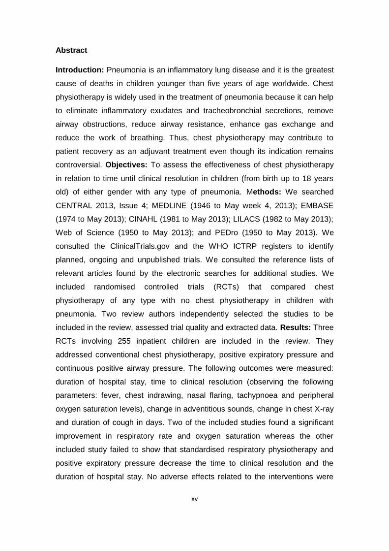
Introduction: Pneumonia is an inflammatory lung disease and it is the greatest
cause of deaths in children younger than five years of age worldwide. Chest
physiotherapy is widely used in the treatment of pneumonia because it can help
to eliminate inflammatory exudates and tracheobronchial secretions, remove
airway obstructions, reduce airway resistance, enhance gas exchange and
reduce the work of breathing. Thus, chest physiotherapy may contribute to
patient recovery as an adjuvant treatment even though its indication remains
controversial. Objectives: To assess the effectiveness of chest physiotherapy
in relation to time until clinical resolution in children (from birth up to 18 years
old) of either gender with any type of pneumonia. Methods: We searched
CENTRAL 2013, Issue 4; MEDLINE (1946 to May week 4, 2013); EMBASE
(1974 to May 2013); CINAHL (1981 to May 2013); LILACS (1982 to May 2013);
Web of Science (1950 to May 2013); and PEDro (1950 to May 2013). We
consulted the ClinicalTrials.gov and the WHO ICTRP registers to identify
planned, ongoing and unpublished trials. We consulted the reference lists of
relevant articles found by the electronic searches for additional studies. We
included randomised controlled trials (RCTs) that compared chest
physiotherapy of any type with no chest physiotherapy in children with
pneumonia. Two review authors independently selected the studies to be
included in the review, assessed trial quality and extracted data. Results: Three
RCTs involving 255 inpatient children are included in the review. They
addressed conventional chest physiotherapy, positive expiratory pressure and
continuous positive airway pressure. The following outcomes were measured:
duration of hospital stay, time to clinical resolution (observing the following
parameters: fever, chest indrawing, nasal flaring, tachypnoea and peripheral
oxygen saturation levels), change in adventitious sounds, change in chest X-ray
and duration of cough in days. Two of the included studies found a significant
improvement in respiratory rate and oxygen saturation whereas the other
included study failed to show that standardised respiratory physiotherapy and
positive expiratory pressure decrease the time to clinical resolution and the
duration of hospital stay. No adverse effects related to the interventions were

xvi
described. Due to the different characteristics of the trials, such as the duration
of treatment, levels of severity, types of pneumonia and the techniques used in
children with pneumonia, as well as differences in their statistical presentation,
we were not able to pool data. Two included studies had an overall low risk of
bias whereas one included study had an overall unclear risk of bias.
Conclusion: Our review does not provide conclusive evidence to justify the
use of chest physiotherapy in children with pneumonia due to a lack of data.
The number of included studies is small and they differed in their statistical
presentation.
Key-words: Pneumonia; Child; Physiotherapy; Clinical Trial; Review

1
1 INTRODUÇÃO
2
As doenças respiratórias em crianças menores de cinco anos de idade
tem sido motivo de preocupação para os profissionais de saúde devido à sua
alta taxa de morbidade e mortalidade observada em todo o mundo1. A
pneumonia é uma grande causa de morte entre crianças2,3 e de acordo com a
organização mundial de saúde é a maior causa de morte em crianças menores
de cinco anos de idade em todo o mundo1. A pneumonia adquirida na
comunidade é a mais comum entre crianças de todo o mundo, porém sua
incidência e taxa de mortalidade são significativamente maiores em países em
desenvolvimento do que em países industrializados4. A pneumonia hospitalar e
a associada à ventilação mecânica são responsáveis pelas principais causas
de infecções adquiridas nos hospital5.
A pneumonia é uma inflamação pulmonar caracterizada pela presença
de fluidos nos alvéolos gerando um acúmulo de secreções nas vias aéreas que
leva a um aumento na resistência destas em cada movimento respiratório,
contribuindo para a piora dos sintomas clínicos2,3,6 como: febre, taquipneia,
dispneia, tosse, sinais de desconforto respiratório (batimento de asa do nariz,
tiragens) e saturação de oxigênio reduzida7,8,9. De acordo com as diretrizes
clínicas o padrão-ouro para o diagnóstico da pneumonia é a presença de
infiltrados pulmonares indicados pelo raio-x de tórax10. Os principais agentes
etiológicos são Streptococcus pneumoniae e Haemophilus influenzae11,12.
O tratamento para as crianças com pneumonia é feito com uso de
antibióticos e em alguns casos estas são hospitalizadas e o uso de oxigênio
suplementar é necessário, depende da gravidade da doença7.

3
A fisioterapia respiratória é um importante adjuvante no tratamento de
muitas doenças respiratórias13 e é frequentemente utilizada em crianças com
doença respiratória crônica ou doença neuromuscular14. O objetivo principal da
fisioterapia respiratória pediátrica é ajudar na desobstrução traqueobrônquica,
além de diminuir a resistência das vias aéreas, melhorar a troca gasosa e
tornar a respiração mais fácil14 através das suas técnicas que combinam
percussão manual da caixa torácica com o posicionamento do paciente, para
drenagem do muco, técnicas respiratórias e tosse13. No entanto, é necessário
levar em consideração as peculiaridades do sistema respiratório das crianças.
Mesmo que os princípios mecânicos das técnicas aplicadas em pacientes
pediátricos sejam similares às aplicadas em adultos, a contínua mudança na
estrutura e função respiratória que ocorrem do nascimento a idade adulta
requer uma continua adaptação na aplicação das técnicas de fisioterapia
respiratória em cada grupo de idade15.
Os procedimentos de fisioterapia podem ser classificados como técnicas
convencionais, modernas ou instrumentais16,17. Drenagem postural, vibração,
percussão, huffing e tosse são técnicas tradicionais que objetivam facilitar a
desobstrução17,18. As técnicas modernas são aquelas que utilizam a variação
de fluxo através do controle respiratório a fim de mobilizar secreções, elas são
a técnica de expiração forçada (TEF), ciclo ativo da respiração (CAR) e
drenagem autógena (DA)17,19,20. Algumas técnicas europeias também são
classificadas como modernas, tais como: expiração lenta prolongada (ELPr) e
aumento do fluxo expiratório (AFE) que são usadas em pacientes pediátricos21,
bem como a expiração lenta infralateral com a glote aberta (ELTGOL) realizada

4
em crianças acima de 12 anos e exercícios com controle de fluxo inspiratório
(EDIC) utilizada em crianças acima de 4 anos22,23. Finalmente, as técnicas
instrumentais são: máscara de pressão positiva expiratória (PEP) e Flutter ®
que são utilizadas na intenção de manter a limpeza das vias aéreas, bem como
melhorar a ventilação mantendo as mesmas abertas durante a expiração17.
Outro instrumento que também pode ser utilizado para aumentar a expansão
pulmonar e melhorar a troca gasosa é o inspirômetro de incentivo24.
A fisioterapia respiratória pode ser vista como a aplicação terapêutica de
intervenções baseadas na fisiologia respiratória15. Algumas usam a posição do
corpo para melhorar a clearance, reexpansão e ventilação pulmonar25. Entre as
posições, a lateral é a que fornece as maiores mudanças dos volumes
estáticos, ventilação local, perfusão e difusão da capacidade funcional25-28. Isso
é consistente com as bases de fisiologia pulmonar, que mostram que as
diferenças na ventilação local são os resultados da variação vertical da pressão
pleural e que essas diferenças são influenciadas pela gravidade25. Esse
posicionamento promove frequentemente clearance mucociliar mesmo sem
aplicação de qualquer outra técnica25.
Outras técnicas usam a variação do fluxo através do controle
respiratório17,20 ou usam alguns dispositivos a fim de manter a desobstrução
das vias aéreas, bem como melhorar a ventilação por mantê-las abertas
durante toda a expiração17. Promovendo, portanto, benefícios que incluem a
eliminação de exudatos expiratórios e secreções traqueobrônquicas, remoção
das obstruções das vias aéreas, redução da resistência das mesmas, e assim,
promover a melhora da troca gasosa e redução do trabalho respiratório14,17,19,29.

5
1.1 Justificativa
A maioria das mortes na infância causadas por pneumonia poderia ser
evitada se intervenções efetivas fossem aplicadas entre as populações mais
vulneráveis1. A fisioterapia respiratória é amplamente utilizada porque pode
ajudar a eliminar os exudatos inflamatórios e secreções traqueobrônquicas,
removendo as obstruções das vias aéreas, diminuindo sua resistência,
melhorando, dessa forma, a troca gasosa e reduzindo o trabalho respiratório14.
Logo, a fisioterapia respiratória pode contribuir como adjuvante a recuperação
do paciente com diagnóstico de pneumonia13. Diante disso, essa revisão irá
considerar as evidências científicas para avaliar a efetividade da fisioterapia
respiratória em crianças com pneumonia.

6
1.2 Objetivos
1.2.1 Objetivo Geral
Avaliar a efetividade da fisioterapia respiratória em relação à melhora
clínica em crianças e adolescentes de ambos os sexos, com qualquer tipo de
pneumonia.
1.2.2 Objetivos específicos
a) identificar quais as técnicas de fisioterapia respiratória são mais
efetivas em pacientes que apresentam diagnóstico de pneumonia.
b) verificar a efetividade da realização da fisioterapia respiratória para os
seguintes desfechos: na ausculta pulmonar; na radiografia torácica e na
saturação periférica de oxigênio; duração, em dias, do tratamento com
antibiótico, tosse, produção de secreção e leucocitose.

7
2 MATERIAIS E MÉTODOS
8
2.1. Desenho do estudo
O presente estudo caracteriza-se como uma revisão sistemática
desenvolvida em parceria com a Colaboração Cochrane (The Cochrane
Collaboration).
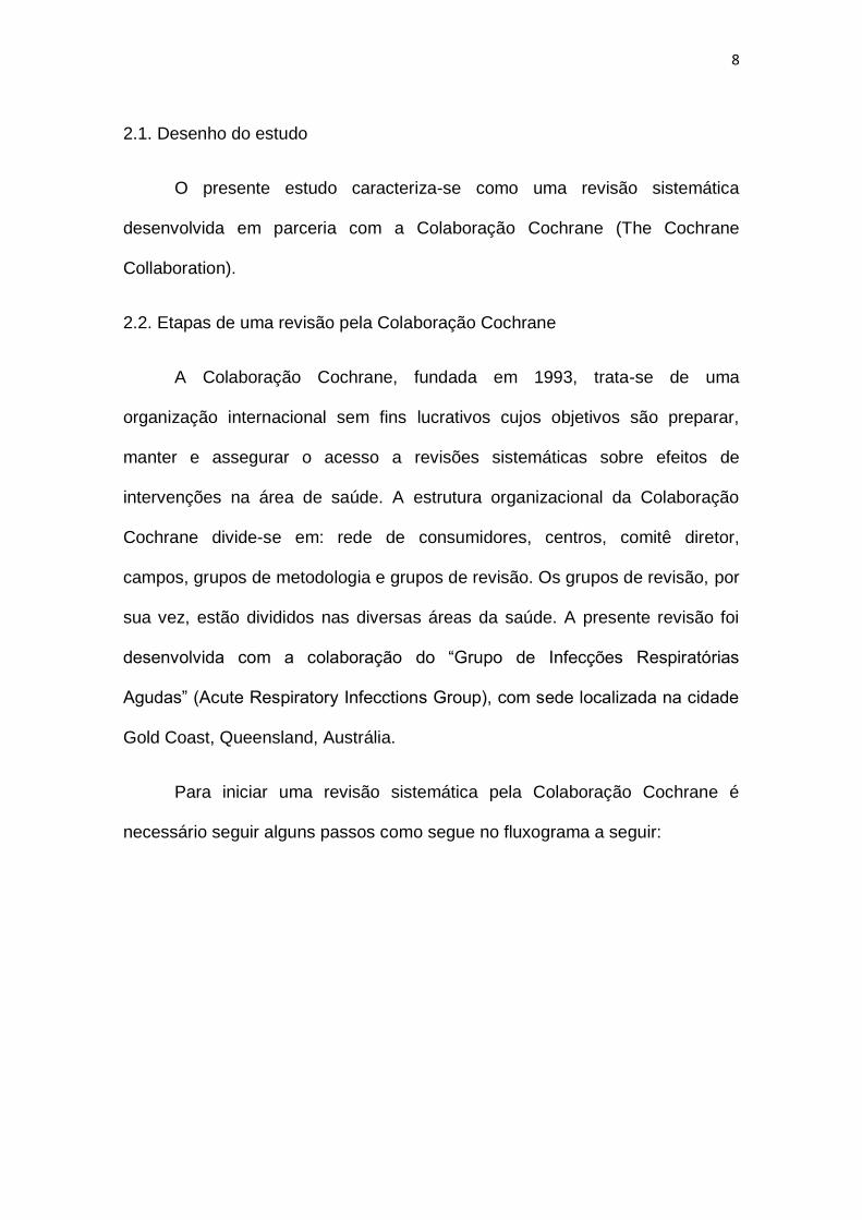
2.2. Etapas de uma revisão pela Colaboração Cochrane
A Colaboração Cochrane, fundada em 1993, trata-se de uma
organização internacional sem fins lucrativos cujos objetivos são preparar,
manter e assegurar o acesso a revisões sistemáticas sobre efeitos de
intervenções na área de saúde. A estrutura organizacional da Colaboração
Cochrane divide-se em: rede de consumidores, centros, comitê diretor,
campos, grupos de metodologia e grupos de revisão. Os grupos de revisão, por
sua vez, estão divididos nas diversas áreas da saúde. A presente revisão foi
desenvolvida com a colaboração do “Grupo de Infecções Respiratórias
Agudas” (Acute Respiratory Infecctions Group), com sede localizada na cidade
Gold Coast, Queensland, Austrália.
Para iniciar uma revisão sistemática pela Colaboração Cochrane é
necessário seguir alguns passos como segue no fluxograma a seguir:
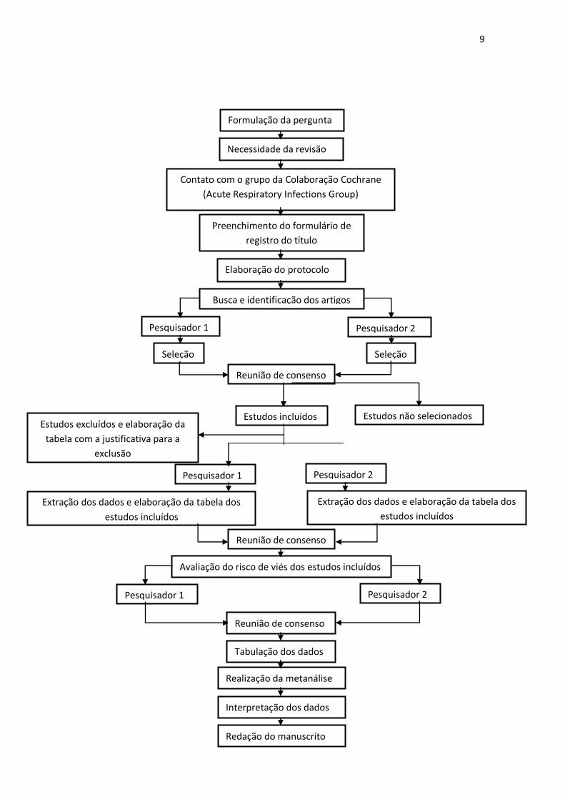
9
Necessidade da revisão
Contato com o grupo da Colaboração Cochrane
(Acute Respiratory Infections Group)
Preenchimento do formulário de
registro do título
Elaboração do protocolo
Busca e identificação dos artigos
Pesquisador 1 Pesquisador 2
Seleção Seleção
Reunião de consenso
Estudos incluídos Estudos não selecionados Estudos excluídos e elaboração da
tabela com a justificativa para a
exclusão
Pesquisador 1 Pesquisador 2
Extração dos dados e elaboração da tabela dos
estudos incluídos
Extração dos dados e elaboração da tabela dos
estudos incluídos
Reunião de consenso
Avaliação do risco de viés dos estudos incluídos
Pesquisador 1 Pesquisador 2
Reunião de consenso
Tabulação dos dados
Realização da metanálise
Interpretação dos dados
Redação do manuscrito
Formulação da pergunta
10
2.3. Local de realização
Departamento de Fisioterapia, Universidade Federal do Rio Grande do
Norte.
2.4. Critérios para considerar estudos para a revisão
2.4.1. Tipos de estudos
Estudos do tipo controlados randomizados nos quais tenham sido
aplicadas técnicas de fisioterapia respiratória em crianças com diagnóstico de
pneumonia.
2.4.2. Tipo de participantes
Foi planejado incluir estudos que apresentassem participação de
pacientes do nascimento até a idade de 18 (dezoito) anos. Foram incluídos os
estudos que abordaram qualquer tipo de técnica de fisioterapia respiratória
nesses pacientes independente do estágio da doença e em qualquer local de
tratamento (ambulatorial ou hospitalar).
2.4.3. Tipos de intervenção
Intervenção: Pacientes com diagnóstico de pneumonia que tenham
recebido qualquer tipo de fisioterapia respiratória combinado ao tratamento
padrão da pneumonia.
Comparação: Pacientes com diagnóstico de pneumonia que tenham
recebido apenas o tratamento padrão da pneumonia.
2.4.4. Tipos de desfecho

11
2.4.4.1. Desfecho primário
- Mortalidade
- Duração de permanência hospitalar (dias)
- Tempo de resolução clínica (dias) avaliando os parâmetros
clínicos: febre, aumento do trabalho respiratório e os níveis de
saturação periférica de oxigênio.
2.4.4.2. Desfechos secundários
- Melhora nos ruídos adventícios
- Melhora no raio-x de tórax
- Duração em dias do antibiótico, tosse e produção de secreção
- Duração em dias de leucocitose
- Clearance das vias aéreas
- Principais eventos adversos
2.5. Métodos de busca para identificação dos estudos
2.5.1. Busca Eletrônica
Foram utilizadas as seguintes fontes de estudos: CENTRAL 2013, Issue
4 , MEDLINE (1946 a maio de 2013), EMBASE (1974 a maio de 2013),
CINAHL (1981 a maio de 2013), LILACS (1982 a maio de 2013); Web of
Science (1950 a maio de 2013), Pedro (1950 a Maio de 2013). Não houve
restrições de idiomas. A escolha dos descritores e a decisão dos estudos a
serem incluídos são atribuições dos autores da revisão. Porém, a estratégia de

12
busca de artigos é realizada pela própria equipe do grupo de revisão da
Colaboração Cochrane.
2.5.2. Outras fontes de pesquisa
As listas de referências dos artigos incluídos na revisão foram
consultadas a fim de incluir estudos adicionais. Dois registros internacionais de
ensaios clínicos (ClinicalTrials.gov e International Clinical Trials Registry
Platform) também foram consultados, a fim de identificar os estudos em
andamento ou aqueles finalizados e cujos dados não foram publicados.
2.6. Coleta dos dados e análise
2.6.1. Seleção dos estudos
Dois revisores (GC e DF) avaliaram de forma independente os títulos e
os resumos de todos os estudos obtidos na busca eletrônica. A partir deste
ponto os textos completos desses artigos foram avaliados a fim de determinar
sua elegibilidade. Um terceiro (KM) autor foi consultado caso não houvesse um
consenso sobre a inclusão ou não de um determinado estudo.
2.6.2. Extração dos dados
Os dados foram extraídos e inseridos de maneira independente por dois
autores (GC e DF) no software Review Manager30, disponível para download
no site da Colaboração Cochrane. Os seguintes dados foram coletados, de
acordo com os métodos descritos no capítulo 7 do Cochrane Handbook for
Systematic Reviews of Interventions31.
13
Detalhes metodológicos (incluindo desenho do estudo, método de
randomização e sigilo de alocação, ocorrência ou não de
cegamento dos participantes e dos avaliadores, número de
desistências e exclusões).
Descrição dos participantes (amostra total, idade, gênero, tipo de
pneumonia, severidade da pneumonia, país, ambiente da
intervenção, critérios de inclusão e exclusão utilizados pelos
ensaios clínicos).
Descrição da intervenção (detalhes da fisioterapia respiratória,
incluindo frequência, intensidade e tempo).
Tipos de desfechos avaliados pelo estudo.
2.6.3. Avaliação do risco de viés dos estudos incluídos
Na intenção de evitar a possibilidade de viés, aumentando assim a
qualidade dos resultados, foi utilizada a ferramenta para avaliação do risco de
viés (The Cochrane Collaboration’s tool for assessing risk of bias) fornecida
pela Colaboração Cochrane, a qual inclui os seguintes itens: sequência de
randomização (random sequence generation), sigilo da alocação (allocation
concealment), cegamento dos participantes (blinding of participants),
cegamento dos avaliadores (blinding of outcome assessment), dados
incompletos (incomplete outcome data), descrição seletiva do desfecho
(selective reporting), e outros vieses (other bias). Cada item recebeu uma das
seguintes classificações: “alto risco de viés”, “baixo risco de viés” ou “risco de

14
viés incerto” de acordo com o Handbook for Systematic Reviews of
Interventions da Colaboração Cochrane32. A avaliação do risco de viés foi
realizada de maneira independente por dois revisores (GC e DF). Um terceiro
autor (KM) foi consultado na ausência de consenso em relação à avaliação do
risco de viés dos estudos incluídos.
2.6.4. Dados ausentes ou incompletos
Os autores dos estudos incluídos foram contatados para obtenção de
dados ausentes ou incompletos.
2.6.5. Análise de subgrupo e avaliação
A análise de subgrupo foi planejada caso tivesse sido possível combinar
os dados em metanálise. Teriam sido realizadas as seguintes análises de
subgrupo:
Idade (bebês, criança, adolescente)
Tipos de pneumonia (adquirida na comunidade, nosocomial)
Tipo de diagnóstico (padrão ouro e não padrão ouro)
Ambiente de tratamento (ambulatorial, hospitalar)
Técnicas (moderna, convencional, instrumental).

15
2.6.6. Análise de sensibilidade
A análise de sensibilidade foi planejada caso tivesse sido possível
combinar os dados em metanálise. Teria sido realizada a fim de explorar a
influência sobre os resultados dos seguintes fatores:
:
Qualidade dos estudos (estudos controlados randomizados com
pobre metodologia);
Tamanho do estudo (estratificação pelo tamanho da amostra);
Sigilo de alocação (alto risco de viés contra baixo risco de viés);
Mascaramento dos participantes (alto risco de viés contra baixo
risco de viés);
Mascaramento dos avaliadores (alta risco de viés contra baixo
risco de viés).

16
3 RESULTADOS E DISCUSSÃO

17
Os resultados e a discussão desta dissertação serão apresentados em
língua inglesa com o formato e a sequência preconizados para sua publicação
na Cochrane Library.

18
Chest physiotherapy for pneumonia in children
Review information
Review number: A185
Authors
Gabriela SS Chaves1, Guilherme AF Fregonezi2, Fernando AL Dias3, Cibele TD Ribeiro4, Ricardo O Guerra2, Diana A Freitas1, Veronica F Parreira5, Karla MPP Mendonça2
1Department of Physical Therapy, Federal University of Rio Grande do Norte, Natal, Brazil 2PhD Program in Physical Therapy, Federal University of Rio Grande do Norte, Natal, Brazil 3Department of Physiology, Federal University of Paraná, Curitiba, Brazil 4Graduate Program in Physiotherapy, Federal University of Rio Grande do Norte, Natal, Brazil 5Department of Physiotherapy, Universidade Federal de Minas Gerais (UFMG), Belo Horizonte, Brazil
Citation example: Chaves GSS, Fregonezi GAF, Dias FAL, Ribeiro CTD, Guerra RO, Freitas DA, Parreira VF, Mendonça KMPP. Chest physiotherapy for pneumonia in children. Cochrane Database of Systematic Reviews 2013, Issue 9. Art. No.: CD010277. DOI: 10.1002/14651858.CD010277.pub2.
Contact person
Karla MPP Mendonça
PhD Program in Physical Therapy Federal University of Rio Grande do Norte Avenida Senador Salgado Filho, 300 Bairro Lagoa Nova 59078-970 Natal Rio Grande do Norte Brazil E-mail: [email protected]
Dates
Assessed as Up-to-date:
Date of Search:
Next Stage Expected:
19
Protocol First Published: Issue 12, 2012
Review First Published: Issue 9, 2013
Last Citation Issue: Issue 9, 2013
Abstract
Background
Pneumonia is an inflammatory lung disease and it is the greatest cause
of deaths in children younger than five years of age worldwide. Chest
physiotherapy is widely used in the treatment of pneumonia because it can help
to eliminate inflammatory exudates and tracheobronchial secretions, remove
airway obstructions, reduce airway resistance, enhance gas exchange and
reduce the work of breathing. Thus, chest physiotherapy may contribute to
patient recovery as an adjuvant treatment even though its indication remains
controversial.
Objectives
To assess the effectiveness of chest physiotherapy in relation to time
until clinical resolution in children (from birth up to 18 years old) of either gender
with any type of pneumonia.
Search methods
We searched CENTRAL 2013, Issue 4; MEDLINE (1946 to May week 4,
2013); EMBASE (1974 to May 2013); CINAHL (1981 to May 2013); LILACS
(1982 to May 2013); Web of Science (1950 to May 2013); and PEDro (1950 to
May 2013).
We consulted the ClinicalTrials.gov and the WHO ICTRP registers to
identify planned, ongoing and unpublished trials. We consulted the reference
lists of relevant articles found by the electronic searches for additional studies.
Selection criteria

20
We included randomised controlled trials (RCTs) that compared chest
physiotherapy of any type with no chest physiotherapy in children with
pneumonia.
Data collection and analysis
Two review authors independently selected the studies to be included in
the review, assessed trial quality and extracted data.
Results
Three RCTs involving 255 inpatient children are included in the review.
They addressed conventional chest physiotherapy, positive expiratory pressure
and continuous positive airway pressure. The following outcomes were
measured: duration of hospital stay, time to clinical resolution (observing the
following parameters: fever, chest indrawing, nasal flaring, tachypnoea and
peripheral oxygen saturation levels), change in adventitious sounds, change in
chest X-ray and duration of cough in days. Two of the included studies found a
significant improvement in respiratory rate and oxygen saturation whereas the
other included study failed to show that standardised respiratory physiotherapy
and positive expiratory pressure decrease the time to clinical resolution and the
duration of hospital stay. No adverse effects related to the interventions were
described. Due to the different characteristics of the trials, such as the duration
of treatment, levels of severity, types of pneumonia and the techniques used in
children with pneumonia, as well as differences in their statistical presentation,
we were not able to pool data. Two included studies had an overall low risk of
bias whereas one included study had an overall unclear risk of bias.
Authors' conclusions
Our review does not provide conclusive evidence to justify the use of
chest physiotherapy in children with pneumonia due to a lack of data. The
number of included studies is small and they differed in their statistical
presentation.
21
Plain language summary
Chest physiotherapy for pneumonia in children
Pneumonia is an inflammatory lung disease and it is the greatest cause
of deaths in children younger than five years of age worldwide. Accumulation of
secretions in the airways due to respiratory infections contributes to the
worsening of clinical symptoms making it very difficult for the child to breathe.
Chest physiotherapy may contribute to patient recovery as a complementary
treatment because it can help to eliminate inflammatory secretions, remove
airway obstructions, reduce airway resistance and the work of breathing. Chest
physiotherapy techniques combine manual percussion of the chest wall and
strategic positioning of the patient for mucus drainage, with cough and
breathing techniques.
We looked for evidence for the effectiveness of chest physiotherapy in
children with pneumonia. We found three studies involving 255 children with
pneumonia aged 29 days to 12 years. In all included studies there was a group
that received some type of physiotherapy and another group that did not receive
physiotherapy, called a control group. Children in both groups underwent the
standard medical treatment for pneumonia. Two of the included studies found a
significant improvement in respiratory rate (decrease in the number of breaths
per minute) and oxygen saturation (measure of how much oxygen the blood is
carrying as a percentage of the maximum it could carry), whereas one included
study failed to show that standardised respiratory physiotherapy and positive
expiratory pressure (maintenance of a pressure in the lungs above atmospheric
pressure at the end of expiration) decreased the time to clinical resolution and
the duration of hospital stay. No adverse effects related to the interventions
were described. This systematic review was limited by the lack of studies and
the quality of the existing data. Two of the included studies had an overall low
risk of bias whereas one included study had an overall unclear risk of bias. The
studies differed in some of their characteristics, such as the duration of
treatment, levels of severity, types of pneumonia and the techniques used in
22
children with pneumonia. Moreover, the included studies reported different
outcomes and also had differences in their statistical presentation of data. As a
result, we were not able to compare the results from these trials by meta-
analysing (combining) them. There is no conclusive evidence in this review to
support or refute the use of physiotherapy in children with pneumonia. The
results are up to date as of May 2013.
Background
Description of the condition
Respiratory diseases in children under five years of age have been a
cause of concern for health professionals because of the high morbidity and
mortality observed worldwide (Chiesa 2008). Community-acquired pneumonia
(CAP) is common among children all over the world but the incidence and
mortality rate are significantly higher in low-income countries than in high-
income countries (Principi 2011). Hospital-acquired pneumonia (HAP) and
ventilator-associated pneumonia (VAP) together are the second most common
hospital-acquired infection (Rotstein 2008). According to the World Health
Organization (WHO), pneumonia is the single greatest cause of death in
children younger than five years of age worldwide (WHO 2011).
Pneumonia is an inflammation of the lung and fluid collection in the
alveoli (Oliveira 2011; Zhang 2012). The two leading causes of pneumonia in
low-income countries are Streptococcus pneumoniae (S. pneumoniae) and
Haemophilus influenzae (H. influenzae) (Dagan 2011; Gilani 2012). Children
with pneumonia are treated with antibiotics and in some cases hospitalisation
and oxygen supplementation are required, depending on the severity of the
disease (Scott 2012).
Accumulation of secretions in the airways due to respiratory infection
contributes to the worsening of clinical symptoms and leads to an increase in
airway resistance with each breath (Durbin 2008). Signs and symptoms that are
useful in diagnosing pneumonia are fever, tachypnoea, nasal flaring, cough,

23
breathlessness, lower chest wall indrawing and reduced oxygen saturation
(Bradley 2011; Ebell 2010; Scott 2012). However, according to clinical
guidelines, the gold standard for diagnosing pneumonia is the presence of lung
infiltrates indicated by chest radiography (Evertsen 2010).
Description of the intervention
Chest physiotherapy is an important adjuvant in the treatment of most
respiratory illnesses (Balachandran 2005) and is usually used in children with
chronic respiratory or neuromuscular disease (Gajdos 2010). The central aim of
paediatric chest physiotherapy is to assist the clearance of tracheobronchial
secretions, thereby to decrease airway resistance, improve gas exchange and
make breathing easier (Gajdos 2010). The techniques combine manual
percussion of the chest wall and strategic positioning of the patient for mucus
drainage with cough and breathing techniques (Balachandran 2005). However,
it is necessary to take into consideration the peculiarities of the respiratory
system of children. Even though the mechanical principles of the techniques
applied to paediatric patients are similar to those used in adults, the continuous
changes in respiratory structure and function that occur from birth to adulthood
require continuous adaptation in the application of chest physiotherapy
techniques in each age group (Oberwaldner 2000). The differences in the
respiratory structure and function of children limit or contraindicate some of the
techniques available for treatment in this age group (Oberwaldner 2000).
Despite improving the patient's respiratory status and expediting recovery, in
certain situations it may not be a useful intervention or may even be harmful, by
increasing bronchospasm, inducing pulmonary hypertension, repositioning a
foreign body or destabilising a sick infant (Wallis 1999). However, some chest
physiotherapy techniques were developed in order to be used exclusively in
children (Postiaux 1997).
Physiotherapy procedures can be classified as conventional, modern and
instrumental techniques (Morrison 2011; Yang 2010). Postural drainage,
vibration, percussion, huffing and coughing are traditional techniques the aim of

24
which is to facilitate mucociliary clearance (Main 2009; Yang 2010). Modern
techniques use the variation of flow through breath control in order to mobilise
secretions: these are the forced expiration technique, active cycle of breathing
and autogenic drainage (Robinson 2010; Roqué i Figuls 2012; Yang 2010).
Some European techniques are also described as modern: slow and prolonged
expiration and increased expiratory flow are used in paediatric patients
(Mucciollo 2008); total slow expiration with the glottis open in a lateral posture is
performed in children over 12 years; and exercises with inspiratory controlled
flow are used in children over four years (Postiaux 1997; Postiaux 2000).
Finally, instrumental techniques such as positive expiratory pressure mask and
flutter are used to maintain airway clearance, as well as to improve ventilation
by keeping the airways open during expiration (Yang 2010). Another tool that
can be used to increase lung expansion and improve gas exchange is incentive
spirometry (Restrepo 2011). (See Appendix 1 for further description of the
physiotherapy procedures).
How the intervention might work
Chest physiotherapy may be seen as the therapeutic application of
mechanical interventions based on respiratory physiology (Oberwaldner 2000).
Some techniques use body position to improve mucociliary clearance, re-
expansion and pulmonary ventilation (Alcoforado 2011). Among these positions,
the lateral position provides the biggest changes in static volumes, regional
ventilation, perfusion and diffusion lung capacity (Alcoforado 2011; Gillies 2012;
Krieg 2007; Manning 1999). This is consistent with the basis of pulmonary
physiology, which shows that differences in regional ventilation are the result of
the vertical variation of pleural pressure and that these differences are
influenced by gravity (Alcoforado 2011). This positioning often promotes
mucociliary clearance even without the application of any other technique
(Alcoforado 2011).
Other techniques use the variation of flow through breath control
(Robinson 2010; Yang 2010) or use devices to maintain airway clearance and
25
improve ventilation by keeping the airways open during expiration (Yang 2010).
The benefits include evacuating inflammatory exudates and tracheobronchial
secretions, removing airway obstructions, reducing airway resistance,
enhancing gas exchange and reducing the work of breathing (Roqué i Figuls
2012; Wallis 1999; Yang 2010).
Why it is important to do this review
The majority of childhood deaths caused by pneumonia could be avoided
if effective interventions were implemented on a broad scale and reached the
most vulnerable populations (WHO 2011). Chest physiotherapy is still widely
used because it can help to eliminate inflammatory exudates and
tracheobronchial secretions, remove airway obstructions, reduce airway
resistance, enhance gas exchange and reduce the work of breathing (Gajdos
2010). There is a systematic review involving adult patients with pneumonia
(Yang 2010). This review showed that, even though physiotherapy should not
be recommended as a conventional treatment for pneumonia in adults, it is still
a broadly used intervention. Thus, chest physiotherapy may contribute to
patient recovery as an adjuvant treatment even though its indication remains
controversial (Balachandran 2005; Wallis 1999). This review considers the
scientific evidence and evaluates the effects of chest physiotherapy for
pneumonia in children.
Objectives
To assess the effectiveness of chest physiotherapy in relation to time
until clinical resolution in children (from birth up to 18 years old) of either gender
with any type of pneumonia.
Methods
Criteria for considering studies for this review
Types of studies

26
Randomised controlled trials (RCTs), cluster-RCTs, cross-over or quasi-RCTs.
Types of participants
Children (from birth up to 18 years old) of either gender with any type of
pneumonia.
Types of interventions
Chest physiotherapy of any type compared with no chest physiotherapy.
Types of outcome measures
Primary outcomes
1. Mortality.
2. Duration of hospital stay (days).
3. Time to clinical resolution (days) of any of the following clinical
parameters: fever, increase of respiratory work (chest indrawing, nasal
flaring, tachypnoea) and peripheral oxygen saturation levels.
Secondary outcomes
1. Change in adventitious sounds.
2. Change in chest X-ray.
3. Duration in days of antibiotic therapy, cough and sputum production.
4. Duration in days of leukocytosis.
5. Airway clearance (measured by sputum weight or volume).
6. Number of adverse events (any undesired outcome due to the
intervention).
Search methods for identification of studies
Electronic searches
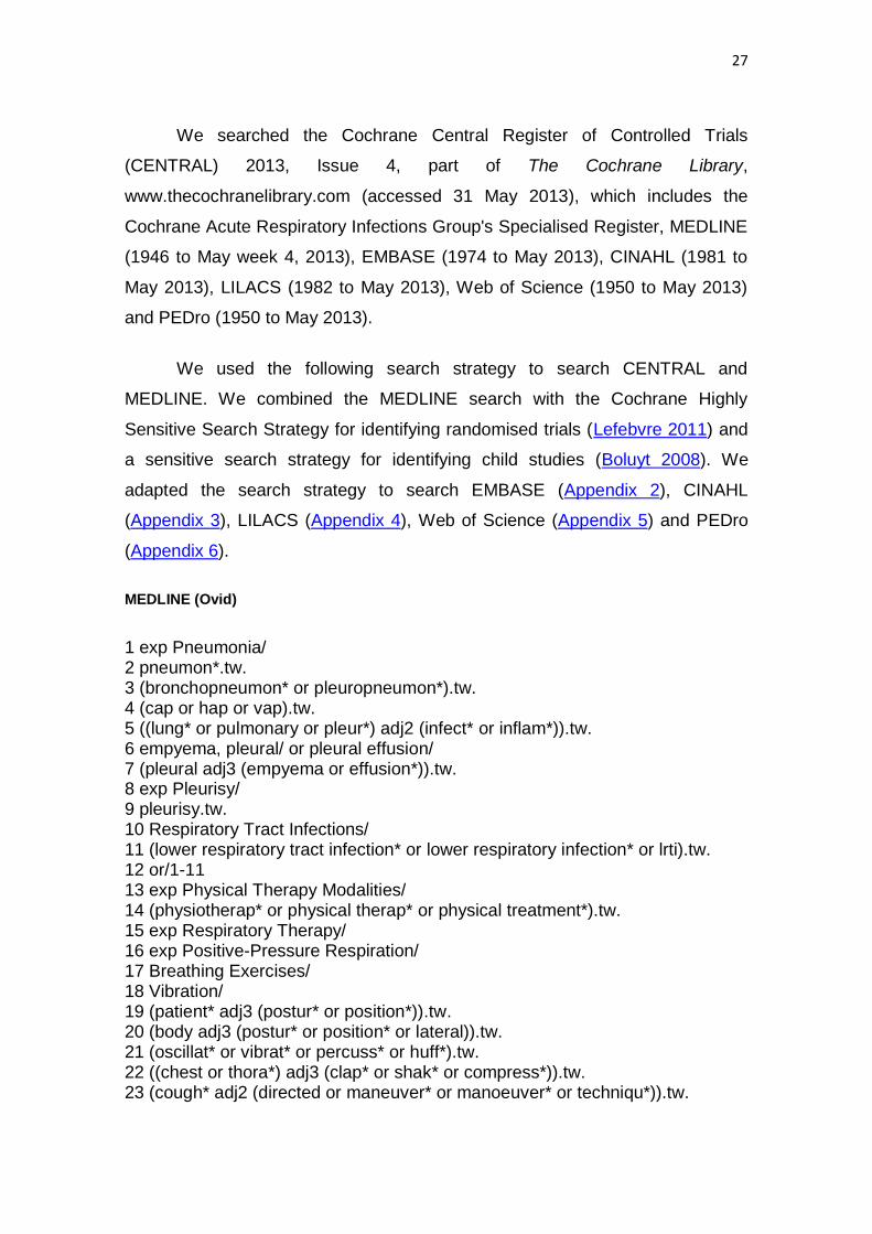
27
We searched the Cochrane Central Register of Controlled Trials
(CENTRAL) 2013, Issue 4, part of The Cochrane Library,
www.thecochranelibrary.com (accessed 31 May 2013), which includes the
Cochrane Acute Respiratory Infections Group's Specialised Register, MEDLINE
(1946 to May week 4, 2013), EMBASE (1974 to May 2013), CINAHL (1981 to
May 2013), LILACS (1982 to May 2013), Web of Science (1950 to May 2013)
and PEDro (1950 to May 2013).
We used the following search strategy to search CENTRAL and
MEDLINE. We combined the MEDLINE search with the Cochrane Highly
Sensitive Search Strategy for identifying randomised trials (Lefebvre 2011) and
a sensitive search strategy for identifying child studies (Boluyt 2008). We
adapted the search strategy to search EMBASE (Appendix 2), CINAHL
(Appendix 3), LILACS (Appendix 4), Web of Science (Appendix 5) and PEDro
(Appendix 6).
MEDLINE (Ovid)
1 exp Pneumonia/ 2 pneumon*.tw. 3 (bronchopneumon* or pleuropneumon*).tw. 4 (cap or hap or vap).tw. 5 ((lung* or pulmonary or pleur*) adj2 (infect* or inflam*)).tw. 6 empyema, pleural/ or pleural effusion/ 7 (pleural adj3 (empyema or effusion*)).tw. 8 exp Pleurisy/ 9 pleurisy.tw. 10 Respiratory Tract Infections/ 11 (lower respiratory tract infection* or lower respiratory infection* or lrti).tw. 12 or/1-11 13 exp Physical Therapy Modalities/ 14 (physiotherap* or physical therap* or physical treatment*).tw. 15 exp Respiratory Therapy/ 16 exp Positive-Pressure Respiration/ 17 Breathing Exercises/ 18 Vibration/ 19 (patient* adj3 (postur* or position*)).tw. 20 (body adj3 (postur* or position* or lateral)).tw. 21 (oscillat* or vibrat* or percuss* or huff*).tw. 22 ((chest or thora*) adj3 (clap* or shak* or compress*)).tw. 23 (cough* adj2 (directed or maneuver* or manoeuver* or techniqu*)).tw.

28
24 positive pressure ventilation*.tw. 25 positive expiratory pressure*.tw. 26 electrostimulat*.tw. 27 massag*.tw. 28 ((respirat* or ventilat*) adj2 muscle train*).tw. 29 ((postur* or autogenic) adj2 drain*).tw. 30 (breath* adj2 (control* or techni* or train* or exercis* or "active cycle")).tw. 31 ((forced or slow or prolonged or increas* or control*) adj2 (exhal* or expir*)).tw. 32 flutter.tw. 33 (incentive adj2 (inspiromet* or spiromet*)).tw. 34 eltgol.tw. 35 or/13-34 36 12 and 35
Searching other resources
We searched the trials registers ClinicalTrials.gov and the WHO ICTRP
(May 2013) in order to identify planned, ongoing and unpublished trials. We
consulted the reference lists of relevant articles found by the above searches for
additional studies.
Data collection and analysis
Selection of studies
Two review authors (DF, GC) independently read the titles and abstracts
identified from the initial search to select studies that met our inclusion criteria.
We retrieved full-text articles and reviewed the results to determine eligibility. A
third review author (KM) resolved differences when necessary.
Data extraction and management
Two review authors (DF, GC) independently extracted data into RevMan
5.2 (RevMan 2012) using a standard data collection form and resolved any
disagreements by discussion and consensus. According to the methods
described in the Cochrane Handbook for Systematic Reviews of Interventions
(Higgins 2011a), we collected the following information:
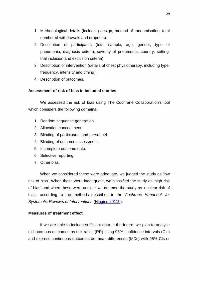
29
1. Methodological details (including design, method of randomisation, total
number of withdrawals and dropouts).
2. Description of participants (total sample, age, gender, type of
pneumonia, diagnosis criteria, severity of pneumonia, country, setting,
trial inclusion and exclusion criteria).
3. Description of intervention (details of chest physiotherapy, including type,
frequency, intensity and timing).
4. Description of outcomes.
Assessment of risk of bias in included studies
We assessed the risk of bias using The Cochrane Collaboration's tool
which considers the following domains:
1. Random sequence generation.
2. Allocation concealment.
3. Blinding of participants and personnel.
4. Blinding of outcome assessment.
5. Incomplete outcome data.
6. Selective reporting.
7. Other bias.
When we considered these were adequate, we judged the study as 'low
risk of bias'. When these were inadequate, we classified the study as 'high risk
of bias' and when these were unclear we deemed the study as 'unclear risk of
bias', according to the methods described in the Cochrane Handbook for
Systematic Reviews of Interventions (Higgins 2011b).
Measures of treatment effect
If we are able to include sufficient data in the future, we plan to analyse
dichotomous outcomes as risk ratios (RR) using 95% confidence intervals (CIs)
and express continuous outcomes as mean differences (MDs) with 95% CIs or

30
as standardised mean differences (SMDs) if different methods of measurement
are used in the studies.
Unit of analysis issues
Cluster-RCTs
We had planned to include cluster-RCTs in the analysis. We would have
adjusted the results when the unit of analysis in the trial is presented as the total
number of individual participants instead of the number of clusters. We would
have adjusted the results using the mean cluster size and intra cluster
correlation coefficient (Higgins 2011c). For meta-analysis, we would have
combined individually randomised trials using the generic inverse-variance
method as described in Chapter 16.3 of the Cochrane Handbook for Systematic
Reviews of Interventions (Higgins 2011c).
Cross-over trials
In randomised, cross-over studies, individuals receive each intervention
sequentially in a random order. A major concern in cross-over trials is the carry-
over effect. It occurs if an effect (e.g. pharmacological, physiological or
psychological) of the treatment in the first phase is carried over to the second
phase. As a consequence, on entry to the second phase the participants can
differ systematically from their initial state despite a wash-out phase. For the
same reason, cross-over trials are not appropriate if the condition of interest is
unstable (Elbourne 2002). However, cross-over studies usually have a wash-out
period, which is a stage after the first treatment but before the second
treatment, where time is given for the active effects of the first treatment to wear
off before the new treatment begins (i.e. to reduce the carry-over effect).
Inadequate wash-outs are seen when the carry-over effect exceeds the
washout period. When including both parallel and cross-over studies with an
adequate wash-out period, we will use the inverse-variance method, as
recommended by Elbourne (Elbourne 2002).

31
Dealing with missing data
We contacted trial authors in order to request additional papers and
obtain missing data.
Assessment of heterogeneity
If we are able to include sufficient data in the future, we plan to evaluate
heterogeneity of study results by looking at the forest plots in order to detect
non-overlapping CIs, with the application of the Chi2 test (with a P value of 0.10
to indicate statistical significance) and by applying the I2
statistic. According to
the Cochrane Handbook for Systematic Reviews of Interventions (Higgins
2011b) values up to 40% indicate that the heterogeneity may not be important,
while values between 30% and 60% indicate moderate heterogeneity, between
50% and 90% substantial heterogeneity and between 75% and 100%
considerable heterogeneity. We also plan to use the I2 statistic with a value of
50% as a moderate level of heterogeneity (Higgins 2011b).
Assessment of reporting biases
If we are able to meta-analyse sufficient data in the future, we plan to use
funnel plots as described in the Cochrane Handbook for Systematic Reviews of
Interventions (Higgins 2011d) to assess reporting bias among the studies. If
asymmetry is present, we also plan to explore possible causes including
publication bias, poor methodological quality and true heterogeneity.
Data synthesis
If we are able to meta-analyse sufficient data in the future, we plan to use
RevMan 5.2 (RevMan 2012) to combine the results when possible. If we
determine the heterogeneity to be moderate, substantial or significant, as
indicated by a value of the I2 statistic greater than 30%, we will use the random-
effects model to summarise results. Otherwise, we will use the fixed-effect

32
model. As meta-analyses could not be undertaken, we have provided a
narrative synthesis of the available data.
Subgroup analysis and investigation of heterogeneity
We plan to conduct the following subgroup analyses if we are able to include
sufficient data in the future and identify significant heterogeneity (a value of the
I2 statistic over 50%).
1. Age (infant, children and adolescents).
2. Type of pneumonia (community-acquired, nosocomial, etc).
3. Type of diagnosis (gold standard and non-gold standard).
4. Treatment setting (inpatient or outpatient).
5. Techniques (conventional, modern or instrumental).
Sensitivity analysis
If we are able to include sufficient data in the future, we will perform a
sensitivity analysis to explore the influence on the results of the following
factors.
1. Study quality (RCTs with poor methodology).
2. Study size (stratified by sample size).
3. Allocation concealment (high risk of bias versus low risk of bias).
4. Participant blinding (high risk of bias versus low risk of bias).
5. Assessor blinding (high risk of bias versus low risk of bias).
Results
Description of studies
See the Characteristics of included studies table.
Results of the search

33
In November 2012 we identified 623 trials with duplicates. This total was
composed of 239 hits from MEDLINE, 213 from EMBASE and CENTRAL, 71
from CINAHL, 16 from LILACS, 76 from Web of Science and eight from PEDro.
After duplicates were removed 446 trials remained. We also conducted an
additional search and found three more references in LILACS. We did not find
any ongoing studies suitable for the review in clinicaltrials.gov and the WHO
ICTRP. After screening the titles and abstracts, we identified seven trials as
potentially relevant. We obtained the full text for those trials with ambiguous
titles and abstracts so that we could determine whether to exclude them from
the review. Three trials (Lukrafka 2012; Paludo 2008; Zhao 2010) met the
inclusion criteria. See Figure 1 for full details on the results of the search. In
May 2013 we re-ran the literature searches. This search identified 22 trials after
duplicates were removed. No further studies were included in this review.
Included studies
Two included trials were conducted in Brazil (Lukrafka 2012; Paludo
2008) and one in China (Zhao 2010). Two trials were published in English
(Lukrafka 2012; Paludo 2008) and one in Chinese (Zhao 2010).
Study design
All included studies were RCTs.
Participants
In total, 255 children (aged 29 days to 12 years) were included in the
three trials, with 129 in the treatment group and 126 in the control group. One
study (Lukrafka 2012) stated that only previously healthy children were enrolled
in their study whereas the other two studies (Paludo 2008; Zhao 2010) do not
report this information. One trial (Lukrafka 2012) included only community-
acquired pneumonia and two trials (Paludo 2008; Zhao 2010) did not describe
the type of pneumonia. The severity of pneumonia was moderate in one trial
34
(Lukrafka 2012), severe in one trial (Zhao 2010) and not stated in the other trial
(Paludo 2008). All studies were conducted in a hospital setting.
Interventions
One trial (Lukrafka 2012) compared chest physiotherapy with a non-
mandatory request to maintain lateral positioning to improve air exchange, to
cough in order to clear secretions and to perform diaphragmatic and deep
breathing, for five minutes, once a day, during the whole hospital stay.
However, this recommendation has not been evaluated. One trial (Paludo 2008)
compared chest physiotherapy plus standard treatment for pneumonia with
standard treatment for pneumonia alone. One trial (Zhao 2010) compared
continuous positive airway pressure plus standard treatment for pneumonia with
standard treatment for pneumonia alone. These trials used different types of
chest physiotherapy, including conventional chest physiotherapy, breathing
exercises and positive expiratory pressure. In the three trials all patients
received antibiotic treatment and oxygen support if clinically indicated (Lukrafka
2012; Paludo 2008; Zhao 2010).
Outcome measures
The included studies did not address one of the primary outcomes of this
review (mortality). The other two primary outcomes (duration of hospital stay
and time to clinical resolution) were assessed in two trials (Lukrafka 2012;
Paludo 2008). However, in both trials, the outcome duration of hospital stay was
a secondary outcome, whereas the outcome time to clinical resolution was the
primary outcome. One trial (Zhao 2010) assessed only one outcome proposed
by the review (peripheral oxygen saturation levels).
Excluded studies
We excluded four trials from the review. See Characteristics of excluded studies
table.

35
Risk of bias in included studies
The detailed 'Risk of bias' judgements and quality of each study can be found in
the Characteristics of included studies table and are summarised in Figure 2.
Allocation (selection bias)
Two studies (Lukrafka 2012; Paludo 2008) described adequate sequence
generation and we judged them to be of low risk of bias. We judged one trial
(Zhao 2010) as unclear risk of bias due to insufficient information to permit a
judgement of low risk or high risk. Only one study clearly reported the method of
allocation concealment (Lukrafka 2012) and we judged it to be of low risk of
bias. We classified the other two trials (Paludo 2008; Zhao 2010) as unclear risk
of bias because there was insufficient information to permit a judgement of low
risk or high risk.
Blinding (performance bias and detection bias)
Two trials stated that the blinding of participants and personnel was not
possible and we judged them to have a high risk of bias as the outcomes may
be influenced by the lack of blinding (Lukrafka 2012; Paludo 2008). We judged
one trial (Zhao 2010) as unclear risk of bias because there was insufficient
information to permit a judgement of low risk or high risk.
Two studies described blinding of outcome assessors (Lukrafka 2012;
Paludo 2008) and we classified them as low risk of bias. We judged one trial
(Zhao 2010) as unclear risk of bias because there was insufficient information to
permit a judgement of low risk or high risk.
Incomplete outcome data (attrition bias)
Two trials described the occurrence of withdrawals and dropouts and we
judged them to be low risk of bias because the missing outcome data were
balanced numerically across the intervention groups (Lukrafka 2012; Paludo
2008). We judged one trial (Zhao 2010) to be of low risk of bias because there

36
were no withdrawals or dropouts. In Lukrafka 2012, from the 79 patients who
were randomised, four underwent chest drainage (three in the intervention
group) and three patients had atelectasis detected by chest X-ray (all in the
control group). Therefore, 72 patients (n = 35 in the intervention and n = 37 in
the control) remained in the study and follow-up (Lukrafka 2012). In Paludo
2008, from 98 patients who were randomised, four patients withdrew (in the
intervention group) because two were discharged/transferred before the second
assessment and two met an exclusion criterion; and five patients withdrew (in
the control group) because two were discharged before the second assessment
and three met an exclusion criterion. Therefore, 89 patients (n = 47 in the
intervention and n = 42 in the control) remained in the study and follow-up
(Paludo 2008). In Zhao 2010, of 94 patients who were randomised (n = 47 in
the intervention and n = 47 in the control) all of them completed the treatment.
Selective reporting (reporting bias)
One study (Lukrafka 2012) was registered in clinicaltrials.gov but there is
no information regarding the outcomes. Thus, we judged this trial to be of high
risk of bias. Two studies (Paludo 2008; Zhao 2010) are not available in trials
registers. However, Zhao 2010 adequately reported all outcome data and we
judged this study to be of low risk of bias. We judged the study of Paludo 2008
to be of high risk of bias because one or more outcomes of interest in the
review were reported incompletely.
Other potential sources of bias
We judged all three included studies to be at unclear risk of other
sources of bias as they did not provide sufficient information to assess whether
an important risk of bias exists (Lukrafka 2012; Paludo 2008; Zhao 2010).
Effects of interventions
Primary outcomes
37
Mortality
This outcome was not reported in the included studies. However, Lukrafka 2012
reported that there were no deaths.
Duration of hospital stay
Two studies reported this outcome (Lukrafka 2012; Paludo 2008) but in
both this was considered as a secondary outcome. There was no significant
difference in duration of hospitalisation between the control and intervention
groups (P = 0.11 and P = 0.79) in Lukrafka 2012 and Paludo 2008, respectively.
Time to clinical resolution
This outcome was considered in three trials (Lukrafka 2012; Paludo
2008; Zhao 2010). In Lukrafka 2012 this outcome was classified as a severity
score including tachypnoea, recession, fever, oxygen saturation and X-ray.
There were differences between baseline versus discharge within each group in
severity score and respiratory rate (P < 0.001) favouring the intervention group.
In Paludo 2008 there were no significant differences between the two groups in
these parameters of clinical evolution. The study Zhao 2010 considered only
peripheral oxygen saturation levels. This study reported that the intervention
group had improved peripheral oxygen saturation levels after application of
continuous positive airway pressure (CPAP) compared with the control group (P
< 0.001).
Secondary outcomes
Change in adventitious sounds
Only one trial described this outcome (Paludo 2008). This study reported
that the intervention group had a longer median duration of rhonchi on lung
auscultation (P = 0.03) than the control group.
Change in chest X-ray
38
Only one trial described this outcome (Lukrafka 2012). This outcome was
included in severity scores and there were no differences between the
intervention and control group.
Duration in days of antibiotic therapy, cough and sputum production
Only one trial described this outcome (Paludo 2008). This study reported
that the intervention group had a longer median duration of coughing (P = 0.04)
than the control group.
Duration in days of leukocytosis
This outcome was not reported in the included studies.
Airway clearance (measured by sputum weight or volume)
This outcome was not reported in the included studies.
Number of adverse events (any undesired outcome due to the intervention)
This outcome was not reported in the included studies.
Discussion
Summary of main results
This systematic review assessed the effectiveness of chest
physiotherapy in relation to time until clinical resolution in children with
pneumonia. Three randomised controlled trials (RCTs) involving 255
participants were included in this review, which appraised three types of chest
physiotherapy (standardised respiratory physiotherapy, positive expiratory
pressure and continuous positive airway pressure). None of the included
studies assessed the outcome mortality. Standardised respiratory
physiotherapy and positive expiratory pressure as an adjunct therapy were not
shown to decrease the time to clinical resolution and the duration of hospital
39
stay in children with pneumonia. However, the application of these techniques
improved some clinical parameters used to determine the time to clinical
resolution, such as respiratory rate. Continuous positive airway pressure
appears to improve oxygen saturation.
Overall completeness and applicability of evidence
The three included studies did not address all of our selected objectives.
None assessed one of the primary outcomes of the review (mortality), however
two studies (Lukrafka 2012; Paludo 2008) addressed the other two primary
outcomes and some of the secondary outcomes. The study by Zhao 2010
addressed only one of our primary outcomes (oxygen saturation).
Lukrafka 2012 evaluated some parameters such as fever, tachypnoea
and peripheral oxygen saturation levels but they were reported as a severity
score. In Paludo 2008 the trial authors expressed the baseline values as mean
deviations (MDs) and standard deviations (SDs) and the post-intervention
values as number of days. In Zhao 2010 the baseline and post-intervention
values were reported as MDs and SDs. It was not possible to pool data by
meta-analysis because of differences in the statistical presentation of data.
The ages of the participants differed between these studies and did not
include our proposed age ranges. The different age ranges in the studies may
have affected the results.
The chest physiotherapy techniques used in the included studies did not
cover all of the existing techniques. Moreover, the differences between
physiotherapy techniques and methods used in the three included trials were
also factors that prevented data pooling and analysis. In Lukrafka 2012, the
intervention group received a standardised respiratory physiotherapy
(positioning, thoracic vibration, thoracic compression, positive expiratory
pressure, breathing exercises and forced exhalation with the glottis open or
'huffing'). However, this trial considered positive expiratory pressure as a
conventional physiotherapy and this is an instrumental technique (Yang 2010).

40
In Paludo 2008 the intervention group only received conventional physiotherapy
and aspiration of secretions if necessary. In Zhao 2010 the intervention group
only received continuous positive airway pressure (CPAP). Thus, it was not
possible to pool and analyse data from these studies because of the different
chest physiotherapy techniques.
Moreover, the different levels of severity, types of pneumonia and
medications used may have affected the practice of physiotherapy and also the
duration of hospital stay. While the application of therapy led to improvement of
some clinical aspects it also led to a worsening of other factors such as cough
and rhonchi on lung auscultation (Paludo 2008). This can be explained because
some of the techniques applied in children in these trials are used in adults and
may not be appropriate for children, considering the anatomical and
physiological differences between these age groups (Oberwaldner 2000).
Quality of the evidence
This systematic review was limited by the lack of studies and the quality
of the existing data. Some points must be taken into consideration when
analysing the review results: the small number of included studies and
differences in the duration of treatment, levels of severity, types of pneumonia
and techniques used in children with pneumonia. Moreover, poor reporting of
methodological aspects of most of the included studies led to risk of bias.
Two studies (Lukrafka 2012; Paludo 2008) explain how randomisation
was conducted and we classified them as low risk of bias. Only one described
allocation concealment and we judged this trial as low risk of bias (Lukrafka
2012). According to Moher 2001, inadequately reported randomisation has
been associated with bias in estimating the effectiveness of interventions.
Savović 2012 showed that inadequate reporting of trial methods can severely
impede the assessment of trial quality and the risk of bias in trial results and this
is a particular problem for the assessment of sequence generation and
allocation concealment, which are often not described in trial publications. Two

41
studies reported adequate blinding of outcome assessment and we judged
them as low risk of bias (Lukrafka 2012; Paludo 2008). Two included trials
described chest physiotherapy as being performed by a physiotherapist, so it
might be difficult to blind the practitioners (Lukrafka 2012; Paludo 2008). In a
RCT, at least three distinct groups (trial participants, trial personnel and
outcome assessors) can potentially be blinded (Savović 2012). The description
of these methodological items is recommended by the CONSORT 2010
statement. Moreover, there are challenges in obtaining high-quality evidence for
physiotherapy interventions because of the difficulties in blinding the
intervention, standardising the method of chest physiotherapy and defining
clinically meaningful outcomes (Yang 2010). Only one protocol for an included
study was found in trials registers (Lukrafka 2012) but there was no information
regarding the outcomes. This aspect is covered in the CONSORT 2010
checklist of information to include when reporting a randomised trial. A study's
protocol registration provides information such as the main objective of the
study, inclusion and exclusion criteria, primary and secondary outcomes and
other methodological aspects. Clinical trial registration minimises or avoids the
consequences of non-publication of entire trials and selective reporting of
outcomes within trials (CONSORT 2010).
Potential biases in the review process
The included studies reported different data, therefore they could not be
pooled in meta-analysis and this may be considered a potential source of bias
in this review. Besides the bias found by using the 'Risk of bias' tool provided by
The Cochrane Collaboration, other factors may be considered as potential
biases. Differences in statistical presentation of data was one of the main
factors that prevented meta-analysis of data in this review. Another factor to
consider was the inability to conduct a subgroup analysis by age.
In trying to resolve these problems we contacted trial authors when
possible to obtain additional information about unpublished data. However, we
were not able to obtain further data from all three of the included trials. The time
42
of application of the techniques, the different techniques applied and follow-up
can also be considered a potential source of bias. All of these factors varied
between studies or were not reported.
Agreements and disagreements with other studies or reviews
Chest physiotherapy has been widely used for pneumonia but there is
weak evidence regarding its benefits (Guessous 2008). Furthermore, few RCTs
have been conducted because developing research on this topic is difficult due
to costs, the need for equipment and the requirement of experienced respiratory
therapists, physiotherapists or clinicians to perform the techniques (Guessous
2008).
There is one previously published systematic review on this topic (Yang
2010) but it focused on adults with pneumonia. However, the results of the
review support our findings because it did not show evidence of the
effectiveness of the application of physiotherapy in patients with pneumonia. In
the Yang 2010 review, all included studies were of poor to moderate
methodological quality. To our knowledge, this is the first systematic review to
assess the effectiveness of chest physiotherapy in children with pneumonia.
Authors' conclusions
Implications for practice
Although some outcomes evaluated in the included trials led to
improvement in the group of children with pneumonia who underwent chest
physiotherapy, it was not possible to perform a meta-analysis. Therefore, due to
a lack of information, this systematic review provides insufficient evidence to
justify the application of chest physiotherapy in children with pneumonia.
Implications for research

43
It is clear that there is a need for more randomised controlled trials of
high methodological quality addressing the use of chest physiotherapy in
children with pneumonia. Future studies should report methodological aspects
such as adequate random sequence generation and allocation concealment,
and blinding of outcome assessors, and consider key points such as
appropriate sample size with the power to detect expected differences,
standardisation of chest physiotherapy, appropriate outcomes and adverse
effects. Moreover, randomised trials should be reported following the
CONSORT statement (CONSORT 2010).
Acknowledgements
The authors would like to thank Clare Dooley, Assistant Managing Editor
of the ARI Group, for her assistance at the beginning of the review, Liz Dooley,
Managing Editor of the ARI Group, for her support and assistance since the
beginning of this review, and Sarah Thorning, ARI Group Trials Search Co-
ordinator, for her useful comments on the search strategy. We also thank the
Contact Editor John Holden and the referees Francis J Gilchrist, Rob Ware,
Manal Kassab, Susanna Esposito, Jordi Vilaró, Anne Lyddiatt, Viviana
Rodriguez and Mohamed Alaa.
Contributions of authors
Gabriela Chaves (GC): selected the studies, extracted data and drafted the final
review.
Guilherme Fregonezi (GF): contributed with clinical expertise and drafted the
final review.
Fernando Dias (FD): drafted the final review.
Cibele Ribeiro (CR): drafted the final review.
Ricardo Guerra (RG): contributed with methodological expertise and drafted the
final review.
Diana Freitas (DF): selected the studies, extracted data and drafted the final
review.
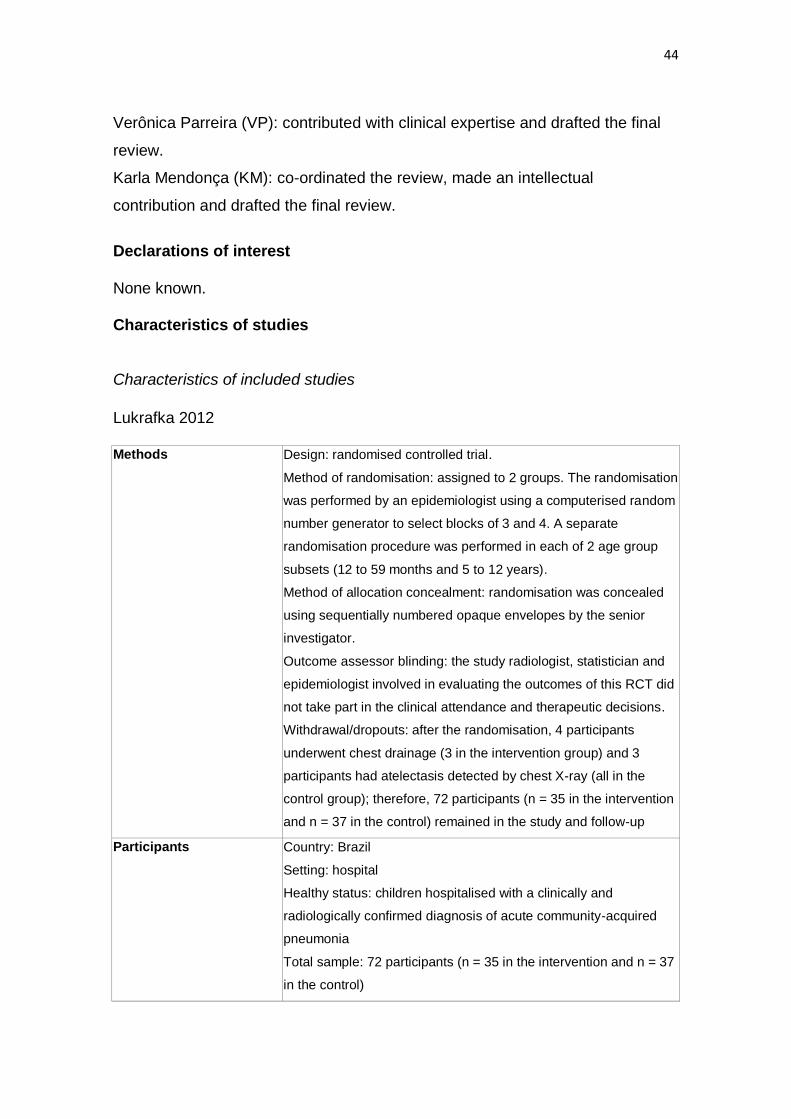
44
Verônica Parreira (VP): contributed with clinical expertise and drafted the final
review.
Karla Mendonça (KM): co-ordinated the review, made an intellectual
contribution and drafted the final review.
Declarations of interest
None known.
Characteristics of studies
Characteristics of included studies
Lukrafka 2012
Methods Design: randomised controlled trial.
Method of randomisation: assigned to 2 groups. The randomisation
was performed by an epidemiologist using a computerised random
number generator to select blocks of 3 and 4. A separate
randomisation procedure was performed in each of 2 age group
subsets (12 to 59 months and 5 to 12 years).
Method of allocation concealment: randomisation was concealed
using sequentially numbered opaque envelopes by the senior
investigator.
Outcome assessor blinding: the study radiologist, statistician and
epidemiologist involved in evaluating the outcomes of this RCT did
not take part in the clinical attendance and therapeutic decisions.
Withdrawal/dropouts: after the randomisation, 4 participants
underwent chest drainage (3 in the intervention group) and 3
participants had atelectasis detected by chest X-ray (all in the
control group); therefore, 72 participants (n = 35 in the intervention
and n = 37 in the control) remained in the study and follow-up
Participants Country: Brazil
Setting: hospital
Healthy status: children hospitalised with a clinically and
radiologically confirmed diagnosis of acute community-acquired
pneumonia
Total sample: 72 participants (n = 35 in the intervention and n = 37
in the control)
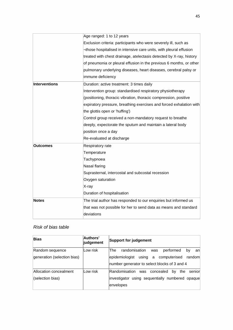
45
Age ranged: 1 to 12 years
Exclusion criteria: participants who were severely ill, such as
¬those hospitalised in intensive care units, with pleural effusion
treated with chest drainage, atelectasis detected by X-ray, history
of pneumonia or pleural effusion in the previous 6 months, or other
pulmonary underlying diseases, heart diseases, cerebral palsy or
immune deficiency
Interventions Duration: active treatment: 3 times daily
Intervention group: standardised respiratory physiotherapy
(positioning, thoracic vibration, thoracic compression, positive
expiratory pressure, breathing exercises and forced exhalation with
the glottis open or 'huffing')
Control group received a non-mandatory request to breathe
deeply, expectorate the sputum and maintain a lateral body
position once a day
Re-evaluated at discharge
Outcomes Respiratory rate
Temperature
Tachypnoea
Nasal flaring
Suprasternal, intercostal and subcostal recession
Oxygen saturation
X-ray
Duration of hospitalisation
Notes The trial author has responded to our enquiries but informed us
that was not possible for her to send data as means and standard
deviations
Risk of bias table
Bias Authors' judgement
Support for judgement
Random sequence
generation (selection bias)
Low risk The randomisation was performed by an
epidemiologist using a computerised random
number generator to select blocks of 3 and 4
Allocation concealment
(selection bias)
Low risk Randomisation was concealed by the senior
investigator using sequentially numbered opaque
envelopes

46
Blinding of participants and
personnel (performance
bias)
High risk No blinding and the outcome is likely to be
influenced by lack of blinding
Blinding of outcome
assessment (detection bias)
Low risk Blinding of outcome assessment ensured
Incomplete outcome data
(attrition bias)
Low risk Missing outcome data balanced in numbers across
intervention groups, with similar reasons for missing
data across groups
Selective reporting
(reporting bias)
High risk The study protocol is available, but there is no
information regarding the outcomes
Other bias Unclear risk Insufficient information to assess whether an
important risk of bias exists
Paludo 2008
Methods Design: randomised controlled trial
Method of randomisation: simple randomisation was performed
from a table of random numbers
Method of allocation concealment: not described
Outcome assessor blinding: all attending paediatricians were
blinded to group assignment and study protocol
Withdrawal/dropouts: 9 participants withdrew from the study
Participants Country: Brazil
Setting: hospital
Healthy status: participants hospitalised with a diagnosis of acute
pneumonia (did not specify the acquisition form)
Total sample: 89 participants
Age range: children aged 29 days to 12 years
Exclusion criteria: participants who needed a chest drain, had
haemodynamic instability, bone fragility or rib fractures and any
other contraindication to chest physical therapy were excluded
Interventions Duration: active treatment: twice daily
Intervention group: each session of chest physical therapy took
about 30 min and consisted of postural drainage, thoracic
squeezing, chest percussion, vibration, cough stimulation and
aspiration of secretions (if necessary)

47
Control group received standard treatment for pneumonia alone
Re-evaluated at discharge
Outcomes Time to clinical resolution: afebrile, absence of severe signs (chest
indrawing, nasal flaring), normal respiratory rate and arterial
oxygen saturation > 95%
Length hospital stay and persistence of respiratory symptoms and
signs (fever, cough, wheezing, tachypnoea, chest indrawing,
adventitious sounds on lung auscultation and arterial oxygen
saturation)
Notes The author has responded to our enquiries with further details
Risk of bias table
Bias Authors'
judgement Support for judgement
Random sequence
generation (selection bias)
Low risk Simple randomisation was performed from a table of
random numbers
Allocation concealment
(selection bias)
Unclear risk Insufficient information to permit judgement of 'low
risk' or 'high risk'
Blinding of participants and
personnel (performance
bias)
High risk No blinding and the outcome is likely to be
influenced by lack of blinding
Blinding of outcome
assessment (detection bias)
Low risk Blinding of outcome assessment ensured
Incomplete outcome data
(attrition bias)
Low risk Missing outcome data balanced in numbers across
intervention groups, with similar reasons for missing
data across groups
Selective reporting
(reporting bias)
High risk The study protocol is not available and one or more
outcomes of interest in the review are reported
incompletely so that they cannot be entered in a
meta-analysis
Other bias Unclear risk Insufficient information to assess whether an
important risk of bias exists
Zhao 2010

48
Methods Design: randomised controlled trial
Method of randomisation: not described
Method of allocation concealment: not described
Outcome assessor blinding: not described
Withdrawal/dropouts: not described
Participants Country: China
Setting: hospital
Healthy status: severe pneumonia
Total sample: 94 children (47 in each group)
Mean age: 10.79 ± 4.75 months
Age range: children aged 2 months to 2 years
Exclusion criteria: not described
Interventions Duration: active treatment: until the stabilisation of the patient
based on oxygen saturation
Intervention group: continuous positive airway pressure
Control group received standard treatment for pneumonia with
oxygen support
Re-evaluated 4 hours and 12 hours after treatment
Outcomes Arterial oxygen saturation; arterial oxygen pressure; arterial carbon
dioxide pressure
Notes This paper was translated from Chinese
Risk of bias table
Bias Authors'
judgement Support for judgement
Random sequence
generation (selection bias)
Unclear risk Insufficient information about the sequence
generation process
Allocation concealment
(selection bias)
Unclear risk Insufficient information about the allocation
concealment
Blinding of participants and
personnel (performance
bias)
Unclear risk Insufficient information to permit judgement of 'low
risk' or 'high risk'
Blinding of outcome
assessment (detection bias)
Unclear risk Insufficient information to permit judgement of 'low
risk' or 'high risk'
Incomplete outcome data
(attrition bias)
Low risk No missing outcome data
Selective reporting Low risk The study protocol is not available but the published

49
(reporting bias) reports include all expected outcomes, including
those that were pre-specified
Other bias Unclear risk Insufficient information to assess whether an
important risk of bias exists
Characteristics of excluded studies
Brunetto 2002
Reason for exclusion There is no control group
Campos 2007
Reason for exclusion There is no control group
Lanza 2009
Reason for exclusion The control group received some intervention
Santos 2009
Reason for exclusion There is no control group
References to studies
Included studies
Lukrafka 2012
Lukrafka JL, Fuchs SC, Fischer GB, Flores JA, Fachel JM, Castro-Rodriguez JA. Chest physiotherapy in paediatric patients hospitalised with community-acquired pneumonia: a randomised clinical trial. Archives of Disease in Childhood 2012;97:967-71.
Paludo 2008

50
Paludo C, Zhang L, Lincho CS, Lemos DV, Real GG, Bergamin JA. Chest physical therapy for children hospitalised with acute pneumonia: a randomised controlled trial. Thorax 2008;63:791-4.
Zhao 2010
Zhao S. Nasal continuous positive airway pressure for the treatment of severe pneumonia in children from the plateau area: clinical analysis of 47 cases. Chinese Journal of Contemporary Pediatrics 2010;12:226-7.
Excluded studies
Brunetto 2002
Brunetto AF, Paulin E. Importância da fisioterapia pulmonar no tratamento de pneumonias em crianças. Fisioterapia em Movimento 2002;15:39-45.
Campos 2007
Campos RS, Couto MDC, Albuquerque CLL, Siqueira AAF, Abreu LC. Effects of active-assisted expiratory flow increase in pneumopathic children. Arquivos Médicos do ABC 2007;32(Suppl 2):38-41.
Lanza 2009
Lanza FC, Gazzotti MR, Augusto LA, Mendes LMS, Paula C, Solé D. Flutter can improve lung obstruction in children with pneumonia? Revista Brasileira de Alergia e Imunopatologia 2009;32:59-62.
Santos 2009
Santos CIS, Ribeiro MAGO, Ribeiro JD, Morcillo AM. Respiratory physiotherapy in children with community-acquired pneumonia. Canadian Journal of Respiratory Therapy 2009;45:23-8.
Other references
Additional references
Alcoforado 2011
Alcoforado L, Pessôa Filho LC, Brandão DC, Galvão AM, Reinaux CMA, Andrade AD. Influence of change in lateral decubitus on pulmonary aerosol deposition. Revista Brasileira de Fisioterapia 2011;15(4):278-83.
Alexander 2011

51
Alexander S, Alshafi K, Balfour-Lynn I, Buchdahl R, Beresford F, Bossley C, et al. Clinical guidelines for the care of children with cystic fibrosis 2011. Royal Brompton and Harefield NHS Foundation Trust (available from: http://www.rbht.nhs.uk/childrencf (accessed 16 October 2012)) 2011.
Balachandran 2005
Balachandran A, Shivbalan S, Thangavelu S. Chest physiotherapy in pediatric practice. Indian Pediatrics 2005;42:559-68.
Boluyt 2008
Boluyt N, Tjosvold L, Lefebvre C, Klassen TP, Offringa M. Usefulness of systematic review search strategies in finding child health systematic reviews in MEDLINE. Archives of Pediatrics & Adolescent Medicine 2008;162(2):111-6.
Bradley 2011
Bradley JS, Byington CL, Shah SS, Alverson B, Carter ER, Harrison C, et al. The management of community-acquired pneumonia in infants and children older than 3 months of age: Clinical Practice Guidelines by the Pediatric Infectious Diseases Society and the Infectious Diseases Society of America. Clinical Infectious Diseases 2011;53(7):1-52.
Britto 2009
Britto RR, Brant TC, Parreira VF. Recursos Manuais e Instrumentais em Fisioterapia Respiratória. Manole, 2009.
Chiesa 2008
Chiesa AM, Westphal MF, Akerman M. Acute respiratory diseases: a study on health inequalities. Cadernos de Saúde Pública 2008;24:55-69.
CONSORT 2010
Schulz KF, Altman DG, Moher D, for the CONSORT Group. CONSORT 2010 Statement: updated guidelines for reporting parallel group randomised trials. BMC Medicine 2010;8:18.
Dagan 2011
Dagan R, Bhutta ZA, Quadros CA, Garau J, Klugman KP, Khuri-Bulos N, et al. The remaining challenge of pneumonia: the leading killer of children. Pediatric Infectious Disease Journal 2011;30:1-2.
Durbin 2008
52
Durbin WJ, Stille C. Pneumonia. Pediatrics in Review 2008;29:147-60.
Ebell 2010
Ebell MH. Clinical diagnosis of pneumonia in children. American Family Physician 2010;82:192-3.
Elbourne 2002
Elbourne DR, Altman DG, Higgins JPT, Curtin F, Worthington HV, Vaillancourt JM. Meta-analyses involving cross-over trials: methodological issues. International Journal of Epidemiology 2002;31:140-9.
Evertsen 2010
Evertsen J, Baumgardner DJ, Regnery A, Banerjee I. Diagnosis and management of pneumonia and bronchitis in outpatient primary care practices. Primary Care Respiratory Journal 2010;19:237-41.
Gajdos 2010
Gajdos V, Katsahian S, Beydon N, Abadie V, Pontual L, Larrar S, et al. Effectiveness of chest physiotherapy in infants hospitalized with acute bronchiolitis: a multicenter, randomized, controlled trial. PLoS Medicine 2010;7:1-12.
Gilani 2012
Gilani Z, Kwong YD, Levine OS, Deloria-Knoll M, Scott JAG, O'Brien KL, et al. A literature review and survey of childhood pneumonia etiology studies: 2000–2010. Clinical Infectious Diseases 2012;54(Suppl 2):102-8.
Gillies 2012
Gillies D, Wells D, Bhandari AP. Positioning for acute respiratory distress in hospitalised infants and children. Cochrane Database of Systematic Reviews 2012, Issue 7. Art. No.: CD003645. DOI: 10.1002/14651858.CD003645.pub2.
Guessous 2008
Guessous I, Cornuz J, Stoianov R, Burnand B, Fitting JW, Yersin B, et al. Efficacy of clinical guideline implementation to improve the appropriateness of chest physiotherapy prescription among inpatients with community-acquired pneumonia. Respiratory Medicine 2008;102:1257-63.
Higgins 2011a
53
Higgins JPT, Altman DG, Sterne JAC. Chapter 8: Assessing risk of bias in included studies. In: Higgins JPT, Green S, editor(s). Cochrane Handbook for Systematic Reviews of Interventions Version 5.1.0 [updated March 2011]. Available from www.cochrane-handbook.org. Chichester, UK: Wiley-Blackwell, 2011.
Higgins 2011b
Deeks JJ, Higgins JPT, Altman DG. Chapter 9: Analysing data and undertaking meta-analyses. In: Higgins JPT, Green S, editor(s). Cochrane Handbook for Systematic Reviews of Interventions Version 5.1.0 [updated March 2011]. Available from www.cochrane-handbook.org. Chichester, UK: Wiley-Blackwell, 2011.
Higgins 2011c
Higgins JPT, Deeks JJ, Altman DG. Chapter 16: Special topics in statistics. In: Higgins JPT, Green S, editor(s). Cochrane Handbook for Systematic Reviews of Interventions Version 5.1.0 [updated March 2011]. Available from www.cochrane-handbook.org. Chichester, UK: Wiley-Blackwell, 2011.
Higgins 2011d
Sterne JAC, Egger M, Moher D. Chapter 10: Addressing reporting biases. In: Higgins JPT, Green S, editor(s). Cochrane Handbook for Systematic Reviews of Interventions Version 5.1.0 [updated March 2011]. Available from www.cochrane-handbook.org. Chichester, UK: Wiley-Blackwell, 2011.
Krieg 2007
Krieg S, Alison JA, McCarren B, Cowell S. Position affects distribution of ventilation in the lungs of older people: an experimental study. Australian Journal of Physiotherapy 2007;53:179-84.
Lefebvre 2011
Lefebvre C, Manheimer E, Glanville J. Chapter 6: Searching for studies. In: Higgins JPT, Green S, editor(s). Cochrane Handbook for Systematic Reviews of Interventions. Version 5.1.0 [updated March 2011]. The Cochrane Collaboration. Available from www.cochrane-handbook.org. Chichester: Wiley-Blackwell, 2011.
Main 2009
Main E, Prasad A, van der Schans CP. Conventional chest physiotherapy compared to other airway clearance techniques for cystic fibrosis. Cochrane Database of Systematic Reviews 2009, Issue 2. Art. No.: CD002011. DOI: 10.1002/14651858.CD002011.pub2.

54
Manning 1999
Manning F, Dean E, Ross J, Abboud RT. Effects of side lying on lung function in older individuals. Physical Therapy 1999;79:456-66.
Moher 2001
Moher D, Schulz KF, Altman DG. The CONSORT statement: revised recommendations for improving the quality of reports of parallel-group randomised trials. Lancet 2001;357(9263):1191-4.
Morrison 2011
Morrison L, Agnew J. Oscillating devices for airway clearance in people with cystic fibrosis. Cochrane Database of Systematic Reviews 2011, Issue 1. Art. No.: CD006842. DOI: 10.1002/14651858.CD006842.pub2.
Mucciollo 2008
Mucciollo MH, Simionato NAF, Paula SCS, Feola AI, Monteiro VC, Ceccon MEJ. Respiratory physiotherapy in children with acute viral bronchiolitis: critical view. Pediatria [São Paulo] 2008;30(4):257-64.
Oberwaldner 2000
Oberwaldner B. Physiotherapy for airway clearance in paediatrics. European Respiratory Journal 2000;15:196-204.
Oliveira 2011
Oliveira TG, Moraes JSB, Moreira FT, Arrelaro RC, Ricardi VA, Bertagnon JRD, et al. Evaluation of hospitalisation of children aged 0 to 5 years admitted for respiratory infections at a large hospital. Einstein 2011;9:514-7.
Postiaux 1992
Postiaux G, Lens E. De ladite « Accélération du Flux Expiratoire (AFE) » :où Forced is... Fast (Expiration technique-FET). Annales de Kinésithérapie 1992;19(8):411-27.
Postiaux 1997
Postiaux G. Des techniques expiratoires lentes pour l'épuration des voies aériennes distales. Annales de Kinésithérapie 1997;24(4):166-77.
Postiaux 2000

55
Postiaux G. Kinésithérapie et pathologie du poumon profond: les techniques inspiratoires lentes pour l'épuration des voies aériennes périphériques. Revue des Maladies Respiratoires 2000;17:315-8.
Principi 2011
Principi N, Esposito S. Management of severe community-acquired pneumonia of children in developing and developed countries. Thorax 2011;66:815-22.
Restrepo 2011
Restrepo RD, Wettstein R, Wittnebel L, Tracy M. Incentive spirometry: 2011. Respiratory Care 2011;56(10):1600-4.
RevMan 2012
Review Manager (RevMan) [Computer program]. Version 5.2. Copenhagen: The Nordic Cochrane Centre, The Cochrane Collaboration, 2011.
Robinson 2010
Robinson KA, Mckoy N, Saldanha I, Odelola OA. Active cycle of breathing technique for cystic fibrosis. Cochrane Database of Systematic Reviews 2010, Issue 11. Art. No.: CD007862. DOI: 10.1002/14651858.CD007862.
Roqué i Figuls 2012
Roqué i Figuls M, Giné-Garriga M, Granados Rugeles C, Perrotta C. Chest physiotherapy for acute bronchiolitis in paediatric patients between 0 and 24 months old. Cochrane Database of Systematic Reviews 2012, Issue 2. Art. No.: CD004873. DOI: 10.1002/14651858.CD004873.pub4.
Rotstein 2008
Rotstein C, Evans G, Born A, Grossman R, Light RB, Magder S, et al. Clinical practice guidelines for hospital-acquired pneumonia and ventilator-associated pneumonia in adults. Canadian Journal of Infectious Diseases & Medical Microbiology 2008;19:19-53.
Savović 2012
Savović J, Jones H, Altman D, Harris R, Jüni P, Pildal J, et al. Influence of reported study design characteristics on intervention effect estimates from randomised controlled trials: combined analysis of meta-epidemiological studies. Health Technology Assessment 2012;16(35):1-82.
Scott 2012

56
Scott JAG, Wonodi C, Mosi JC, Deloria-Knoll M, DeLuca AN, Karron RA, et al. The definition of pneumonia, the assessment of severity, and clinical standardization in the pneumonia etiology research for child health study. Clinical Infectious Diseases 2012;54(Suppl 2):109-16.
Wallis 1999
Wallis C, Prasad A. Who needs chest physiotherapy? Moving from anecdote to evidence. Archives of Disease in Childhood 1999;80:393-7.
WHO 2011
World Health Organization. World Health Statistics 2011. WHO Library Cataloguing 2011.
Yang 2010
Yang M, Yan Y, Yin X, Wang BY, Wu T, Liu GJ, et al. Chest physiotherapy for pneumonia in adults. Cochrane Database of Systematic Reviews 2010, Issue 2. Art. No.: CD006338. DOI: 10.1002/14651858.CD006338.pub2.
Zhang 2012
Zhang Y, Fang C, Dong BR, Wu T, Deng JL. Oxygen therapy for pneumonia in adults. Cochrane Database of Systematic Reviews 2012, Issue 3. Art. No.: CD006607. DOI: 10.1002/14651858.CD006607.pub4.

57
Figure 1
Study flow diagram

58
Figure 2
'Risk of bias' summary: review authors' judgements about each risk of bias item for each included study.

59
Appendices
1 Description of the techniques used for chest physiotherapy
Conventional physiotherapy
Postural drainage - postural drainage is the positioning of the child with the assistance of gravity to mobilise the secretions towards the main bronchus (Britto 2009).
Vibration - in this technique a rapid vibratory impulse is transmitted through the chest wall from the flattened hands of the therapist by isometric alternate contraction of forearm flexor and extensor muscles, to loosen and dislodge the airway secretions (Britto 2009).
Percussion – the therapist can use single or both cupped hands or three fingers with the middle finger tented, or a facemask with the port either covered or occluded by a finger, and strike repeatedly at a rate of three per second over
the part of the bronchopulmonary segment which needs to be drained (Britto 2009).
Huffing – fast expiration at high volume performed by the patient (Britto 2009).
Coughing - child can be requested to cough. In unco-operative or small children tracheal stimulation or tickling can be done by placing index finger or thumb on the anterior side of the neck against trachea just above sternal notch with gentle but firm inward pressure in a circular pattern as the child begins to exhale (Britto 2009).
Modern techniques
Forced expiration technique - patient should undertake a diaphragmatic inspiration to medium volume, with relaxation of the scapulohumeral region and
with the mouth and glottis open (Britto 2009).
Active cycle of breathing - the patient can be positioned supine, prone, lateral or sitting and can be helped by the physiotherapist or perform this independently. This technique consists of the following phases (Alexander 2011; Britto 2009).
o Control of breathing: the patient must perform inhalations and exhalations at current volume level, relaxing the upper thoracic region and breathing quietly using the lower chest.
o Exercise chest expansion: this consists of deep breathing exercises performed as follows: slow nasal breathing at inspiratory reserve volume level, followed by a two to three-second post-inspiratory pause and ending with oral expiration at functional residual capacity level.
o Forced expiration technique: patient should undertake a diaphragmatic inspiration to medium volume, with relaxation of the scapulohumeral region and with the mouth and glottis open.
Autogenic drainage - a three-phase breathing regimen utilising high expiratory flow rates and variable lung volumes to unstick, collect and evacuate secretions. The patient is placed sitting, back straight and head slightly
hyperextended, hands resting on the upper left and right chest (Alexander 2011;
Britto 2009). The three phases are described as follows:

60
o Displacement: starts with a slow and forced oral expiration, recruiting a percentage of expiratory reserve volume, and then carrying inspiration to low volume, recruiting percentages of tidal volume followed by a two to three-second post-inspiratory pause. Finally, there is a slow oral exhalation recruiting a percentage of expiratory reserve volume.
o Collection: nasal inspiration to medium volume, recruiting a larger percentage of tidal volume, followed by a two to three-second post-inspiratory pause. Finally, there is a slow oral exhalation recruiting percentage of expiratory reserve volume.
o Elimination: nasal inspiration to high volume recruiting the tidal volume and a percentage of inspiratory reserve volume, followed by a two to three-second post-inspiratory pause. Then, there is oral expiration at the level of tidal volume. Finally, the forced expiration technique is performed to high volumes.
Slow and prolonged expiration - this is an entirely passive technique given the age and the inability of a small patient to co-operate. The child is positioned supine. The therapist places a hand on the patient's chest and the other one on the abdomen. At the end of a spontaneous expiration, pressure is applied to the chest caudally and on the abdomen cephalically. The pressure is maintained for two to three respiratory cycles. No pressure is exerted during the first part of expiration (Postiaux 1997).
Increased expiratory flow - this technique should be performed during the expiratory time using pressure exerted by the physiotherapist's hands on the child's chest, lying supine. The other hand of the professional remains static over the abdomen to prevent the dissipation of pressure to the abdominal compartment. The physical therapist will perform the movement on the chest with the goal of deflation, the speed of which should be more than a spontaneous expiration (Postiaux 1992).
Total slow expiration with the glottis open in a lateral posture - the patient is placed in lateral decumbency. The patient can be helped by the physiotherapist or can also perform this independently, without the help of the therapist. The patient starts by performing a nasal inspiration at tidal volume level. Then, the patient performs an oral slow expiration with the open glottis at residual volume level (Postiaux 1997).
Exercises of controlled inspiratory flow - this technique can be performed in two positions: the posterolateral and anterolateral. In the first position, patient is positioned in lateral decumbency with the trunk and pelvis tilted slightly above perpendicular to the plane of support. In the second position, the patient is positioned in lateral decumbency with the limb flexed and the upper hand on the occipital region to promote the elongation of the pectoral musculature. In both placements, the patient must perform a slow, deep inspiration recruiting the inspiratory reserve volume, then a two to three-second post-inspiratory pause, and then an oral expiration at functional residual capacity level (Postiaux 2000).
Instrumental techniques
Positive expiratory pressure mask - provides resistance to expiration through a mouthpiece or facemask, followed by forced expirations. This treatment must be carried out in a sitting position: the patient inhales and exhales through the mask 15 times (approximately two minutes). The inhalation is at tidal volume and the expiration is slightly active against the mask. The patient then removes

61
the mask and performs two or three forced expirations follow by a cough to clear secretions that are mobilised to the central airways. This procedure is followed by a one to two-minute period of relaxed, controlled breathing (Alexander 2011; Britto 2009).
Flutter - pipe-shaped device that creates oscillation and positive pressure on expiration used in conjunction with forced expirations. They perform a nasal inhalation, followed by an inspiratory pause lasting two to three seconds. Oral exhalation must be fast enough to move the ball. The sequence should be repeated for 10 to 15 breaths (Alexander 2011; Britto 2009).
Incentive spirometer - referred to as sustained maximal inspiration. It is accomplished by using a device that provides feedback when patient inhales at a predetermined flow or volume and sustains the inflation for at least five seconds (Restrepo 2011).

62
2 EMBASE (Elsevier) search strategy
#36 #11 AND #35 15056 #35 #12 OR #13 OR #15 OR #16 OR #17 OR #18 OR #19 OR #20 OR #21 OR #22 OR #23 OR #24 OR #25 OR #26 OR #27 OR #28 OR #29 OR #30 OR #31 OR #32 OR #33 OR #34 250196 #34 eltgol:ab,ti 4 #33 (incentive NEAR/2 (inspiromet* OR spiromet*)):ab,ti 209 #32 flutter:ab,ti 6494 #31 ((forced OR slow OR prolonged OR increas* OR control*) NEAR/2 (exhal* OR inhal*)):ab,ti 2516 #30 (breath* NEAR/2 (control* OR techni* OR train* OR exercis* OR 'active cycle')):ab,ti 4591 #29 ((postur* OR autogenic) NEAR/2 drain*):ab,ti 309 #28 ((respirat* OR ventilat*) NEAR/2 ('muscle train' OR 'muscle training')):ab,ti 222 #27 massag*:ab,ti 5850 #26 electrostimulat*:ab,ti 2042 #25 'positive pressure ventilation':ab,ti OR 'postive expiratory pressure':ab,ti 4009 #24 (cough* NEAR/2 (directed OR maneuver* OR manoeuver* OR techniqu*)):ab,ti169 #23 ((chest OR thora*) NEAR/3 (clap* OR shak* OR compress*)):ab,ti 3291 #22 oscillat*:ab,ti OR vibrat*:ab,ti OR percuss*:ab,ti OR huff*:ab,ti 69253 #21 (body NEAR/3 (postur* OR positon* OR lateral)):ab,ti 2919 #20 (patient* NEAR/3 (postur* OR position*)):ab,ti 11130 #19 'vibration'/de 11265 #18 'breathing exercise'/de 2748 #17 'artificial ventilation'/exp 85889 #16 'oxygen therapy'/de 13072 #15 'extracorporeal oxygenation'/de 6897 #14 'postural drainage'/de 474 #13 physiotherap*:ab,ti OR 'physical therapy':ab,ti OR 'physical therapies':ab,ti OR 'physical treatment':ab,ti OR 'physical treatments':ab,ti 26544 #12 'physiotherapy'/exp 34808 #11 #1 OR #2 OR #3 OR #4 OR #5 OR #6 OR #7 OR #8 OR #9 OR #10 261137 #10 'lower respiratory tract infection':ab,ti OR 'lower respiratory tract infections':ab,ti OR 'lower respiratory infection':ab,ti OR 'lower respiratory infections':ab,ti OR lrti:ab,ti 5195 #9 'lower respiratory tract infection'/de 5969 #8 pleurisy:ab,ti 2255 #7 'pleurisy'/de OR 'exudative pleurisy'/de 4261 #6 'pleura effusion'/de OR 'pleura empyema'/de 25867 #5 ((lung* OR pulmonary OR pleur*) NEAR/2 (infect* OR inflam*)):ab,ti 25127 #4 cap:ab,ti OR hap:ab,ti OR vap:ab,ti 30610 #3 bronchopneumon*:ab,ti OR pleuropneumon*:ab,ti 3654 #2 pneumon*:ab,ti 119018 #1 'pneumonia'/exp 138579
3 CINAHL (Ebsco) search strategy
S58 S35 AND S48 AND S57 71 S57 S49 OR S50 OR S51 OR S52 OR S53 OR S54 OR S55 OR S56 178,200 S56 (MH "Quantitative Studies") 8,230 S55 (MH "Placebos") 6,531 S54 TI placebo* OR AB placebo* 19,643 S53 TI random* OR AB random* 97,503 S52 TI ( (singl* or doubl* or trebl* or tripl*) W1 (blind* or mask*)) OR AB ((singl* or doubl* or trebl* or tripl*) W1 (blind* or mask*)) 14,307
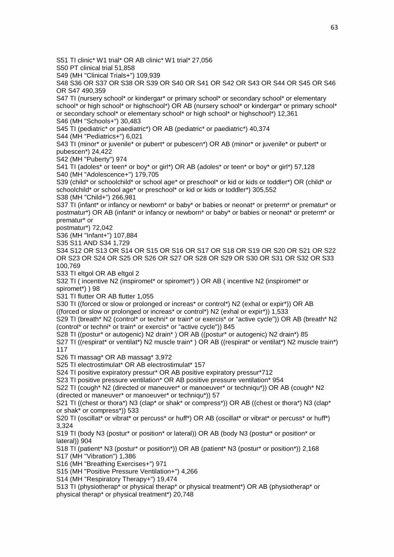
63
S51 TI clinic* W1 trial* OR AB clinic* W1 trial* 27,056 S50 PT clinical trial 51,858 S49 (MH "Clinical Trials+") 109,939 S48 S36 OR S37 OR S38 OR S39 OR S40 OR S41 OR S42 OR S43 OR S44 OR S45 OR S46 OR S47 490,359 S47 TI (nursery school* or kindergar* or primary school* or secondary school* or elementary school* or high school* or highschool*) OR AB (nursery school* or kindergar* or primary school* or secondary school* or elementary school* or high school* or highschool*) 12,361 S46 (MH "Schools+") 30,483 S45 TI (pediatric* or paediatric*) OR AB (pediatric* or paediatric*) 40,374 S44 (MH "Pediatrics+") 6,021 S43 TI (minor* or juvenile* or pubert* or pubescen*) OR AB (minor* or juvenile* or pubert* or pubescen*) 24,422 S42 (MH "Puberty") 974 S41 TI (adoles* or teen* or boy* or girl*) OR AB (adoles* or teen* or boy* or girl*) 57,128 S40 (MH "Adolescence+") 179,705 S39 (child* or schoolchild* or school age* or preschool* or kid or kids or toddler*) OR (child* or schoolchild* or school age* or preschool* or kid or kids or toddler*) 305,552 S38 (MH "Child+") 266,981 S37 TI (infant* or infancy or newborn* or baby* or babies or neonat* or preterm* or prematur* or postmatur*) OR AB (infant* or infancy or newborn* or baby* or babies or neonat* or preterm* or prematur* or postmatur*) 72,042 S36 (MH "Infant+") 107,884 S35 S11 AND S34 1,729 S34 S12 OR S13 OR S14 OR S15 OR S16 OR S17 OR S18 OR S19 OR S20 OR S21 OR S22 OR S23 OR S24 OR S25 OR S26 OR S27 OR S28 OR S29 OR S30 OR S31 OR S32 OR S33 100,769 S33 TI eltgol OR AB eltgol 2 S32 TI ( incentive N2 (inspiromet* or spiromet*) ) OR AB ( incentive N2 (inspiromet* or spiromet*) ) 98 S31 TI flutter OR AB flutter 1,055 S30 TI ((forced or slow or prolonged or increas* or control*) N2 (exhal or expir*)) OR AB ((forced or slow or prolonged or increas* or control*) N2 (exhal or expir*)) 1,533 S29 TI (breath* N2 (control* or techni* or train* or exercis* or "active cycle")) OR AB (breath* N2 (control* or techni* or train* or exercis* or "active cycle")) 845 S28 TI ((postur* or autogenic) N2 drain* ) OR AB ((postur* or autogenic) N2 drain*) 85 S27 TI ((respirat* or ventilat*) N2 muscle train* ) OR AB ((respirat* or ventilat*) N2 muscle train*) 117 S26 TI massag* OR AB massag* 3,972 S25 TI electrostimulat* OR AB electrostimulat* 157 S24 TI positive expiratory pressur* OR AB positive expiratory pressur*712 S23 TI positive pressure ventilation* OR AB positive pressure ventilation* 954 S22 TI (cough* N2 (directed or maneuver* or manoeuver* or techniqu*)) OR AB (cough* N2 (directed or maneuver* or manoeuver* or techniqu*)) 57 S21 TI ((chest or thora*) N3 (clap* or shak* or compress*)) OR AB ((chest or thora*) N3 (clap* or shak* or compress*)) 533 S20 TI (oscillat* or vibrat* or percuss* or huff*) OR AB (oscillat* or vibrat* or percuss* or huff*) 3,324 S19 TI (body N3 (postur* or position* or lateral)) OR AB (body N3 (postur* or position* or lateral)) 904 S18 TI (patient* N3 (postur* or position*)) OR AB (patient* N3 (postur* or position*)) 2,168 S17 (MH "Vibration") 1,386 S16 (MH "Breathing Exercises+") 971 S15 (MH "Positive Pressure Ventilation+") 4,266 S14 (MH "Respiratory Therapy+") 19,474 S13 TI (physiotherap* or physical therap* or physical treatment*) OR AB (physiotherap* or physical therap* or physical treatment*) 20,748

64
S12 (MH "Physical Therapy+") 61,365 S11 S1 OR S2 OR S3 OR S4 OR S5 OR S6 OR S7 OR S8 OR S9 OR S10 19,864 S10 TI (lower respiratory tract infection* or lower respiratory infection* or lrti ) OR AB (lower respiratory tract infection* or lower respiratory infection* or lrti) 597 S9 (MH "Respiratory Tract Infections") 3,194 S8 TI (pleural N3 (empyema or effusion*)) OR AB (pleural N3 (empyema or effusion*)) 929 S7 TI pleurisy OR AB pleurisy 65 S6 (MH "Pleurisy") 81 S5 (MH "Empyema") OR (MH "Pleural Effusion") 1,091 S4 TI ((lung* or pulmonary or pleur*) N2 (infect* or inflam*)) OR AB ((lung* or pulmonary or pleur*) N2 (infect* or inflam*)) 1,577 S3 TI (cap or hap or vap) OR AB (cap or hap or vap) 2,251 S2 TI (pneumon* or bronchopneumon* or pleuropneumon*) OR AB (pneumon* or bronchopneumon* or pleuropneumon*) 9,935
S1 (MH "Pneumonia+") 8,6664 LILACS (BIREME) search strategy
(MH:pneumonia OR pneumon$ OR Neumonía OR MH:C08.381.677$ OR MH:C08.730.610$ OR "Inflamación Experimental del Pulmón" OR "Inflamación del Pulmón" OR "Neumonía Lobar" OR Neumonitis OR "Inflamación Pulmonar" OR Pneumonía OR Pulmonía OR "Inflamação Experimental dos Pulmões" OR "Inflamação do Pulmão" OR "Pneumonia Lobar" OR Pneumonite OR "Inflamação Pulmonar" OR Pulmonia OR Bronchopneumonia OR Bronconeumonía OR Pleuropneumonia OR Pleuroneumonía OR MH:"Empyema, Pleural" OR "Empiema Pleural" OR "Pleural Effusion" OR "Derrame Pleural" OR MH:Pleurisy OR Pleuresia OR Pleurisia OR pleurisy OR "pleural effusion" OR MH:"Respiratory Tract Infections" OR "Infecciones del Sistema Respiratorio" OR "Infecções Respiratórias" OR " lower respiratory tract infection" OR "lower respiratory tract infections" OR "lower respiratory infection" OR "lower respiratory infections" or lrti OR "Infecciones de las Vías Respiratorias" OR "Infecciones del Aparato Respiratorio" OR "Infecciones del Tracto Respiratorio" OR "Infecciones Respiratorias" OR "Infecções das Vias Respiratórias" OR "Infecções do Aparelho Respiratório" OR "Infecções do Sistema Respiratório" OR "Infecções do Trato Respiratório") AND (MH:"Physical Therapy Modalities" OR MH:E02.779$ OR "Modalidades de Fisioterapia" OR "Modalidades de Fisioterapia" OR physiotherap$ OR "physical therapy" OR "physical therapies" OR "physical treatment" OR "physical treatments" OR "Modalidades de Terapia Física" OR Fisioterapia OR "Técnicas Fisioterápicas" OR MH:"Respiratory Therapy" OR MH:E02.880$ OR "Terapia Respiratoria" OR "inhalation therapy" OR "Terapia de Inhalación" OR "Terapia por Inalação" OR MH:"Positive-Pressure Respiration" OR MH:E02.041.625.790$ OR MH:E02.880.820.790$ OR "Respiración con Presión Positiva" OR "Respiração com Pressão Positiva" OR MH:"Breathing Exercises" OR "Ejercicios Respiratorios" OR "Exercícios Respiratórios" OR "Respiratory Muscle Training" OR "Entrenamiento del Musculo Respiratorio" OR "Exercícios para os Músculos Respiratórios" OR MH:Vibration OR Vibración OR Vibração OR oscillat$ OR vibrat$ OR percuss$ OR huff$ OR coughing Or "directed cough" OR "cough technique" OR "patient posture" OR "body posture" OR "patient position" OR "patient positioning" OR "body position" OR "lateral position" OR "lateral posture" OR ELTGOL OR "forced expiration technique" OR "active cycle of breathing" OR "slow expiration" OR "prolonged expiration" OR ELPr OR "increased expiratory flow" OR AFE OR "inspiratory controlled flow" OR EDIC OR "positive expiratory pressure" OR PEP OR flutter OR electrostimulat$ OR massag* OR "postural drainage")

65
5 Web of Science (Thomson Reuters) search strategy
# 7
76 #6 AND #5
Databases=SCI-EXPANDED, CPCI-S Timespan=All Years
Lemmatization=On
# 6
1,306,314 Topic=(random* or placebo* or crossover* or "cross over" or allocat* or ((singl* or doubl*) NEAR/1 (blind* or mask*))) OR Title=(trial)
Databases=SCI-EXPANDED, CPCI-S Timespan=All Years
Lemmatization=On
# 5
347 #4 AND #3
Databases=SCI-EXPANDED, CPCI-S Timespan=All Years
Lemmatization=On
# 4
1,748,376 Topic=(infant* or infancy or newborn* or baby* or babies or neonat* or preterm* or prematur* or postmatur* or child* or schoolchild* or "school age*" or preschool* or kid or kids or toddler* or adoles* or teen* or boy* or girl* or minor* or juvenile* or pubert* or pubescen* or pediatric* or paediatric* or kindergar* or highschool* or (school* NEAR/1 (nursery or primary or secondary or elementary or high)))
Databases=SCI-EXPANDED, CPCI-S Timespan=All Years
Lemmatization=On
# 3
1,590 #2 AND #1
Databases=SCI-EXPANDED, CPCI-S Timespan=All Years
Lemmatization=On
# 2
422,588 Topic=(physiotherap* or (physical NEAR/1 (treatment* or therap*)) or "respiratory therapy" or "positive pressure respiration" or "positive pressure ventilation" or (patient* NEAR/3 (posture or position*)) or (body NEAR/3 (postur* or position* or lateral)) or oscillat* or vibrar* or percuss* or huff* or (cough* NEAR/2 (directed or maneuver* or manoeuver* or techni*)) or "positive expiratory pressure" or electrostimulat* or massag* or ((respirat* or ventilat*) NEAR/2 "muscle training") or ((postur* or autogenic) NEAR/2 drain*) or (breath* NEAR/2 (control* or techni* or train* or exercis* or "active cycle")) or ((forced or slow or prolonged or increas* or control*) NEAR/2 (exhal* or expir)) or flutter or (incentive NEAR/2 (inspiromet* or spiromet*)) or eltgol)
Databases=SCI-EXPANDED, CPCI-S Timespan=All Years
Lemmatization=On
# 1
163,229 Topic=(pneumon* or bronchopneumon* or pleuropneumon* or pleurisy or ((lung* or pulmonary or pleur*) NEAR/2 (infect* or inflam*)) or (pleural NEAR/2 (empyema or effusion*)) or "lower respiratory tract infection" or "lower respiratory tract infections" or "lower respiratory infection" or "lower respiratory infections" or lrti)

66
Databases=SCI-EXPANDED, CPCI-S Timespan=All Years
Lemmatization=On
6 PEDro (Physiotherapy Evidence Database) search strategy
Pneumonia in title abstract field Paediatrics in subdivision field Clinical trials in methods field

67
4 CONSIDERAÇÕES FINAIS

68
A presente revisão sistemática, realizada em parceria com a
Colaboração Cochrane, incluiu três estudos envolvendo 255 crianças com
idade de 29 dias a 12 anos, apresentando diagnóstico de pneumonia,
assistidos em ambiente hospitalar. Embora alguns desfechos avaliados nos
estudos tenham demonstrado melhora através da realização da fisioterapia
respiratória, a evidência existente em relação aos benefícios da mesma, no
tratamento de crianças com pneumonia, ainda não é suficiente para comprovar
ou refutar os efeitos benéficos deste tipo de tratamento como terapia adjunta
de pacientes com pneumonia.
De uma maneira geral, os estudos revisados possuem baixa qualidade
metodológica apresentando ausência ou descrição superficial dos pontos
metodológicos de grande importância, como sigilo de alocação e cegamento
dos avaliadores. Porém, o cegamento é difícil de ser realizado devido à
natureza do estudo.
Dos nove desfechos propostos pelo protocolo da revisão, quatro não
foram avaliados pelos estudos incluídos (mortalidade, duração em dias da
leucocitose, a clearance das vias aéreas e o número de efeitos adversos).
O baixo número de estudos encontrados e a baixa qualidade
metodológica, bem como a ausência dos dados finais completos para que seja
realizada a metanálise, deixa clara a necessidade da realização de ensaios
clínicos que descrevam a metodologia seguindo as normas do CONSORT.
Futuros estudos são necessários com o objetivo de avaliar os efeitos da
fisioterapia respiratória em crianças com pneumonia. Um cálculo amostral
antes do início do estudo deve ser realizado, para que se possa obter um

69
número adequado de pacientes, realizar e descrever o método de
randomização, de sigilo da alocação e cegamento.
Apesar de não apresentar evidência suficiente para comprovar os efeitos
benéficos da fisioterapia no tratamento de crianças com pneumonia, a
fisioterapia respiratória parece ser bem tolerada pelos pacientes, oferecendo
baixos riscos e baixos custos para aqueles que a realizam.

70
5 REFERÊNCIAS
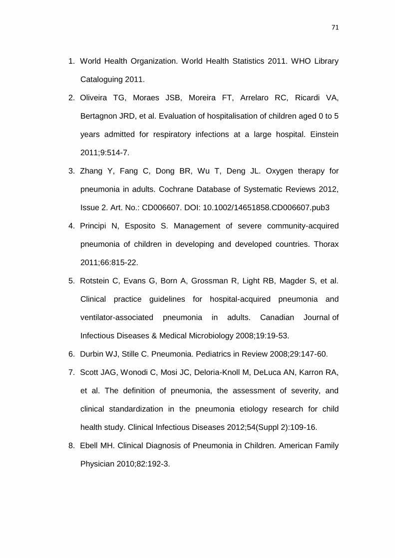
71
1. World Health Organization. World Health Statistics 2011. WHO Library
Cataloguing 2011.
2. Oliveira TG, Moraes JSB, Moreira FT, Arrelaro RC, Ricardi VA,
Bertagnon JRD, et al. Evaluation of hospitalisation of children aged 0 to 5
years admitted for respiratory infections at a large hospital. Einstein
2011;9:514-7.
3. Zhang Y, Fang C, Dong BR, Wu T, Deng JL. Oxygen therapy for
pneumonia in adults. Cochrane Database of Systematic Reviews 2012,
Issue 2. Art. No.: CD006607. DOI: 10.1002/14651858.CD006607.pub3
4. Principi N, Esposito S. Management of severe community-acquired
pneumonia of children in developing and developed countries. Thorax
2011;66:815-22.
5. Rotstein C, Evans G, Born A, Grossman R, Light RB, Magder S, et al.
Clinical practice guidelines for hospital-acquired pneumonia and
ventilator-associated pneumonia in adults. Canadian Journal of
Infectious Diseases & Medical Microbiology 2008;19:19-53.
6. Durbin WJ, Stille C. Pneumonia. Pediatrics in Review 2008;29:147-60.
7. Scott JAG, Wonodi C, Mosi JC, Deloria-Knoll M, DeLuca AN, Karron RA,
et al. The definition of pneumonia, the assessment of severity, and
clinical standardization in the pneumonia etiology research for child
health study. Clinical Infectious Diseases 2012;54(Suppl 2):109-16.
8. Ebell MH. Clinical Diagnosis of Pneumonia in Children. American Family
Physician 2010;82:192-3.

72
9. Bradley JS, Byington CL, Shah SS, Alverson B, Carter ER, Harrison C, et
al. The management of community-acquired pneumonia in infants and
children older than 3 months of age: Clinical Practice Guidelines by the
Pediatric Infectious Diseases Society and the Infectious Diseases
Society of America. Clinical Infectious Diseases 2011;53(7):1-52.
10. Evertsen J, Baumgardner DJ, Regnery A, Banerjee I. Diagnosis and
management of pneumonia and bronchitis in outpatient primary care
practices. Primary Care Respiratory Journal 2010;19:237-41.
11. Dagan R, Bhutta ZA, Quadros CA, Garau J, Klugman KP, Khuri-Bulos N,
et al. The remaining challenge of pneumonia: the leading killer of
children. Pediatric Infectious Disease Journal 2011;30:1-2.
12. Gilani Z, Kwong YD, Levine OS, Deloria-Knoll M, Scott JAG, O'Brien KL,
Feikin DR. A literature review and survey of childhood pneumonia
atiology studies: 2000–2010. Clinical Infectious Diseases 2012;54(Suppl
2):102-8.
13. Balachandran A, Shivbalan S, Thangavelu S. Chest Physiotherapy in
Pediatric Practice. Indian Pediatrics 2005;42:559-68.
14. Gajdos V, Katsahian S, Beydon N, Abadie V, Pontual L, Larrar S, et al.
Effectiveness of chest physiotherapy in infants hospitalized with acute
bronchiolitis: a multicenter, randomized, controlled trial. Plos Medicine
2010;7:1-12.
15. Oberwaldner B. Physiotherapy for airway clearance in paediatrics.
European Respiratory Journal 2000;15:196-204.
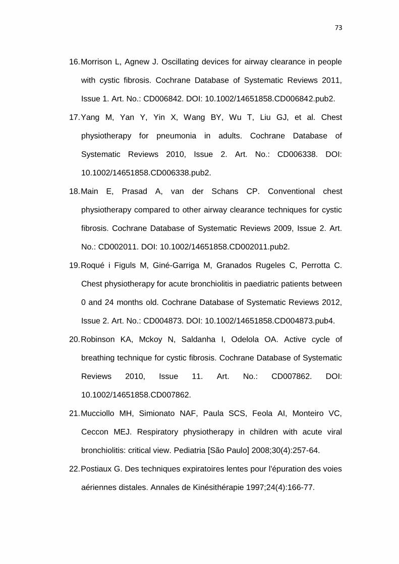
73
16. Morrison L, Agnew J. Oscillating devices for airway clearance in people
with cystic fibrosis. Cochrane Database of Systematic Reviews 2011,
Issue 1. Art. No.: CD006842. DOI: 10.1002/14651858.CD006842.pub2.
17. Yang M, Yan Y, Yin X, Wang BY, Wu T, Liu GJ, et al. Chest
physiotherapy for pneumonia in adults. Cochrane Database of
Systematic Reviews 2010, Issue 2. Art. No.: CD006338. DOI:
10.1002/14651858.CD006338.pub2.
18. Main E, Prasad A, van der Schans CP. Conventional chest
physiotherapy compared to other airway clearance techniques for cystic
fibrosis. Cochrane Database of Systematic Reviews 2009, Issue 2. Art.
No.: CD002011. DOI: 10.1002/14651858.CD002011.pub2.
19. Roqué i Figuls M, Giné-Garriga M, Granados Rugeles C, Perrotta C.
Chest physiotherapy for acute bronchiolitis in paediatric patients between
0 and 24 months old. Cochrane Database of Systematic Reviews 2012,
Issue 2. Art. No.: CD004873. DOI: 10.1002/14651858.CD004873.pub4.
20. Robinson KA, Mckoy N, Saldanha I, Odelola OA. Active cycle of
breathing technique for cystic fibrosis. Cochrane Database of Systematic
Reviews 2010, Issue 11. Art. No.: CD007862. DOI:
10.1002/14651858.CD007862.
21. Mucciollo MH, Simionato NAF, Paula SCS, Feola AI, Monteiro VC,
Ceccon MEJ. Respiratory physiotherapy in children with acute viral
bronchiolitis: critical view. Pediatria [São Paulo] 2008;30(4):257-64.
22. Postiaux G. Des techniques expiratoires lentes pour l'épuration des voies
aériennes distales. Annales de Kinésithérapie 1997;24(4):166-77.
74
23. Postiaux G. Kinésithérapie et pathologie du poumon profond: les
techniques inspiratoires lentes pour l'épuration des voies aériennes
périphériques. Revue des Maladies Respiratoires 2000;17:315-8.
24. Retrepo RD, Wettstein R, Wittnebel L, Tracy M. Incentive Spirometry:
2011. Respiratory Care 2011;56(10):1600-4.
25. Alcoforado L, Pessôa Filho LC, Brandão DC, Galvão AM, Reinaux CMA,
Andrade AD. Influence of change in lateral decubitus on pulmonary
aerosol deposition. Revista Brasileira de Fisioterapia 2011;15(4):278-83.
26. Gillies D, Wells D, Bhandari AP. Positioning for acute respiratory distress
in hospitalised infants and children. Cochrane Database of Systematic
Reviews 2012, Issue 7. Art. No.: CD003645. DOI:
10.1002/14651858.CD003645.pub2.
27. Krieg S, Alison JA, McCarren B, Cowell S. Position affects distribution of
ventilation in the lungs of older people: an experimental study. Australian
Journal of Physiotherapy 2007;53:179-84.
28. Manning F, Dean E, Ross J, Abboud RT. Effects of side lying on lung
function in older individuals. Physical Therapy 1999;79:456-66.
29. Wallis C, Prasad A. Who needs chest physiotherapy? Moving from
anecdote to evidence. Archives of Disease in Childhood 1999;80:393-7.
30. Review Manager (RevMan) [Computer program]. Version 5.1.
Copenhagen: The Nordic Cochrane Centre, The Cochrane Collaboration,
2011.
31. Higgins JPT, Altman DG, Sterne JAC. Chapter 8: Assessing risk of bias
in included studies. In: Higgins JPT, Green S, editor(s). Cochrane
75
Handbook for Systematic Reviews of Interventions Version 5.1.0
[updated March 2011]. Available from www.cochrane-handbook.org.
Chichester, UK: Wiley-Blackwell, 2011.
32. Deeks JJ, Higgins JPT, Altman DG. Chapter 9: Analysing data and
undertaking meta-analyses. In: Higgins JPT, Green S, editor(s).
Cochrane Handbook for Systematic Reviews of Interventions Version
5.1.0 [updated March 2011]. Available from www.cochrane-
handbook.org. Chichester, UK: Wiley-Blackwell, 2011.

76
6 ANEXOS