Caracterização Histopatológica da Osteonecrose Induzida...
90
Andréia Maria Rocha Moreira Caracterização Histopatológica da Osteonecrose Induzida por Bisfosfonatos em Modelo Animal Brasília 2015
Transcript of Caracterização Histopatológica da Osteonecrose Induzida...

Andréia Maria Rocha Moreira
Caracterização Histopatológica da Osteonecrose Induzida por
Bisfosfonatos em Modelo Animal
Brasília
2015


Andréia Maria Rocha Moreira
Caracterização Histopatológica da Osteonecrose Induzida por
Bisfosfonatos em Modelo Animal
Trabalho de Conclusão de Curso apresentado ao
Departamento de Odontologia da Faculdade de
Ciências da Saúde da Universidade de Brasília,
como requisito parcial para a conclusão do curso
de Graduação em Odontologia.
Orientador: Prof. Dr. André Ferreira Leite
Co-orientador: Prof. Dr. Paulo Tadeu de Souza
Figueiredo
Brasília
2015

Dedico este trabalho à minha querida mãe, Maria do Carmo. Ao
meu pai e amigo, Fernando. Aos meus irmãos Fernandinho e
Letícia. Às minhas avós Lourdinha e Lourdes e meus avôs Alípio
(in memoriam) e Bernardo (in memoriam).

AGRADECIMENTOS
A Deus, por tudo. Sem Ele nada seria possível. Agradeço
pela minha vida e pela minha família. Por me conceder força nos
momentos difíceis, por guiar meus caminhos e pela graça do
estudo.
À minha querida mãe, Maria do Carmo, que sempre me
incentivou a estudar, desde a minha infância. Obrigada por tanta
dedicação a mim e meus irmãos. Pela entrega e doação
incondicional. Pela educação alicerçada em valores tão nobres
para que sejamos pessoas de bem. Por ser minha melhor amiga,
por me ajudar. Por nos dar sempre um bom exemplo. Espero um
dia poder retribuir e corresponder a tudo isso.
Ao meu querido pai, Fernando, que acredita tanto em
mim. Obrigada por todo o auxílio ao longo deste curso de
graduação e em minha vida. Agradeço por me ajudar a ser uma
pessoa melhor, por ser meu grande amigo, por sempre me
aconselhar a seguir o bom caminho, pelo carinho, pelas palavras
sábias e pelas de conforto nos momentos difíceis. Obrigada por
tudo.
Ao meu irmão Fernandinho, a quem admiro tanto, meu
amigo, grande exemplo e incentivador e à minha irmã Letícia,
minha companheirinha, que tantas vezes foi tão generosa
comigo, mesmo quando ainda era criança, agradeço por tudo o
que fazem por mim e por tornarem a minha vida muito mais
alegre. Agradeço a toda a minha família, meus avós, tios e
primos, que são tão importantes para mim e sempre me
forneceram o apoio necessário para que tudo isso se tornasse
possível.
À equipe de professores que me ajudou neste trabalho de
conclusão de curso e em minha graduação. Agradeço a
paciência que tiveram comigo, a confiança, o incentivo, por
abrirem as portas e por me concederem tantas oportunidades.
Agradeço:
Ao Professor André Leite, meu orientador e amigo, um
exemplo de profissional e de simplicidade, que acompanhou
minha trajetória na universidade e contribuiu não só para a minha
formação acadêmica, mas também para meu crescimento
pessoal. Sou grata por tudo o que me ensinou e por toda a ajuda,
incentivo, consideração e pela oportunidade que me deu ao me
inserir neste projeto.
Ao Professor Paulo Tadeu, sempre tão presente ao longo
da minha graduação, a quem admiro a dedicação aos pacientes
e por fazer um trabalho tão nobre e diferenciado. Obrigada por
tudo o que fez por mim e pela contribuição e influência tão
significativas para a minha formação. Agradeço os ensinamentos
tão especiais e importantes que o senhor me passou durante
todos esses anos de convivência e aprendizado.
À Professora Ana Carolina, por me apresentar os
caminhos da pesquisa, por confiar em mim e por estimular o meu
desenvolvimento nas atividades do laboratório. Agradeço por
acrescentar tanto ao compartilhar seu conhecimento e
experiência, pela compreensão, pelo incentivo intenso e
constante, e por não ter medido esforços ao dedicar tanto tempo
a me ensinar e me ajudar na execução deste trabalho.
À Professora Nilce, agradeço a contribuição tão relevante
para a construção deste trabalho. Obrigada por me auxiliar na
interpretação dos resultados desta pesquisa. Por me ensinar, às
vezes de forma tão diferente, ao me inspirar a buscar respostas
mais concretas por meio da dúvida e do questionamento. Pela
presença ao longo do meu curso e pelas vezes em que me
instigou o desejo de buscar ser melhor e crescer.
À Professora Eliete, por me incentivar a estudar, inclusive
através do seu exemplo, agradeço pelas vezes em que
esclareceu nossas dúvidas e por colocar-se disponível a nos
ajudar.
À Professora Bruna Amorim, pelas vezes que me
auxiliou e por compartilhar suas experiências comigo, me

estimulando a ter coragem para enfrentar os desafios inerentes à
profissão.
Aos senhores, toda a minha gratidão e admiração!
À Glorinha, por ter se disponibilizado a me ensinar as
técnicas histológicas necessárias para a execução deste
trabalho. Obrigada pela paciência, gentileza e por compartilhar
seus conhecimentos comigo. Ao Gabriel Guillen, cuja
contribuição foi fundamental para o desenvolvimento desta
pesquisa. Obrigada pelo auxílio tão importante durante o trabalho
laboratorial. Agradeço à Pollyana e a todos os meus colegas e
amigos do laboratório, por me incentivarem, e por todas as vezes
que me ajudaram e colaboraram comigo.
A todos os professores, agradeço por todo o
conhecimento transmitido, por contribuírem para a minha
formação e por me ensinarem uma profissão. Não há palavras
que descrevam a minha gratidão a esses profissionais. Aos
funcionários agradeço a disponibilidade e por fornecerem
condições para que o espaço de ensino seja concreto. Aos
pacientes, agradeço a paciência, incentivo e por colaborarem
para o nosso processo de aprendizado. A todos os meus colegas
de curso, pelo companheirismo e cumplicidade.
À minha amiga e dupla Raiza, que esteve comigo durante
toda a minha graduação e com quem aprendi tantas coisas boas,
agradeço por todo o apoio e amizade. A sua contribuição somou
muito ao meu processo de formação. À minha amiga Helora,
minha primeira dupla, agradeço o seu companheirismo e por
participar de uma forma tão positiva desta importante etapa da
minha vida.
Por fim, agradeço a todas as pessoas boas que Deus foi
colocando em minha vida e que apesar de não contribuírem
diretamente com este trabalho, estenderam as mãos e me
ajudaram a chegar até aqui. Dentre tantas, deixo o meu carinho
especial à minha madrinha tia Denise e à minha avó Lourdinha.

EPÍGRAFE
“Alocução a meninos
(...) Pensem que todas as maravilhas, objetos de seus estudos,
são a obra de muitas gerações, uma obra coletiva que exige de
todos um esforço entusiasta e um labor difícil e impreterível.
Tudo isto, nas mãos de vocês, se torna uma herança. Vocês a
recebem, respeitam-na, aumentam-na e, mais tarde, irão
transmiti-la fielmente à sua descendência. Deste modo somos
mortais imortais porque criamos juntos obras que nos
sobrevivem.
Se refletirem seriamente sobre isto, encontrarão um sentido para
a vida e para seu progresso. E o julgamento que fizerem sobre
os outros homens e as outras épocas será mais verdadeiro”.
Albert Einstein

RESUMO
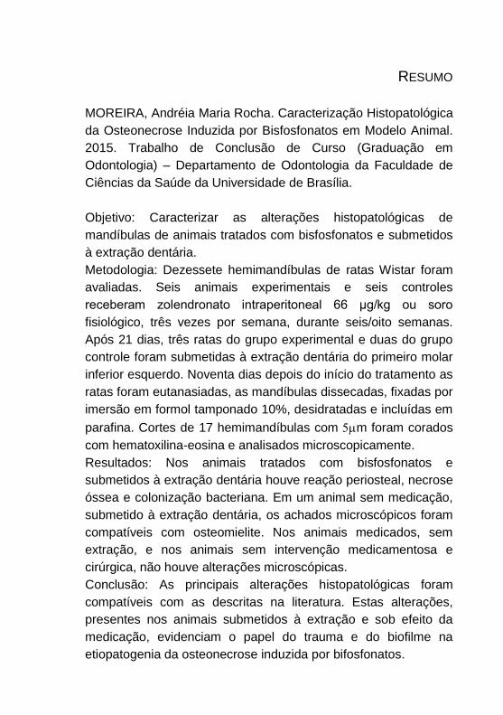
MOREIRA, Andréia Maria Rocha. Caracterização Histopatológica
da Osteonecrose Induzida por Bisfosfonatos em Modelo Animal.
2015. Trabalho de Conclusão de Curso (Graduação em
Odontologia) – Departamento de Odontologia da Faculdade de
Ciências da Saúde da Universidade de Brasília.
Objetivo: Caracterizar as alterações histopatológicas de
mandíbulas de animais tratados com bisfosfonatos e submetidos
à extração dentária.
Metodologia: Dezessete hemimandíbulas de ratas Wistar foram
avaliadas. Seis animais experimentais e seis controles
receberam zolendronato intraperitoneal 66 μg/kg ou soro
fisiológico, três vezes por semana, durante seis/oito semanas.
Após 21 dias, três ratas do grupo experimental e duas do grupo
controle foram submetidas à extração dentária do primeiro molar
inferior esquerdo. Noventa dias depois do início do tratamento as
ratas foram eutanasiadas, as mandíbulas dissecadas, fixadas por
imersão em formol tamponado 10%, desidratadas e incluídas em
parafina. Cortes de 17 hemimandíbulas com m foram corados
com hematoxilina-eosina e analisados microscopicamente.
Resultados: Nos animais tratados com bisfosfonatos e
submetidos à extração dentária houve reação periosteal, necrose
óssea e colonização bacteriana. Em um animal sem medicação,
submetido à extração dentária, os achados microscópicos foram
compatíveis com osteomielite. Nos animais medicados, sem
extração, e nos animais sem intervenção medicamentosa e
cirúrgica, não houve alterações microscópicas.
Conclusão: As principais alterações histopatológicas foram
compatíveis com as descritas na literatura. Estas alterações,
presentes nos animais submetidos à extração e sob efeito da
medicação, evidenciam o papel do trauma e do biofilme na
etiopatogenia da osteonecrose induzida por bifosfonatos.

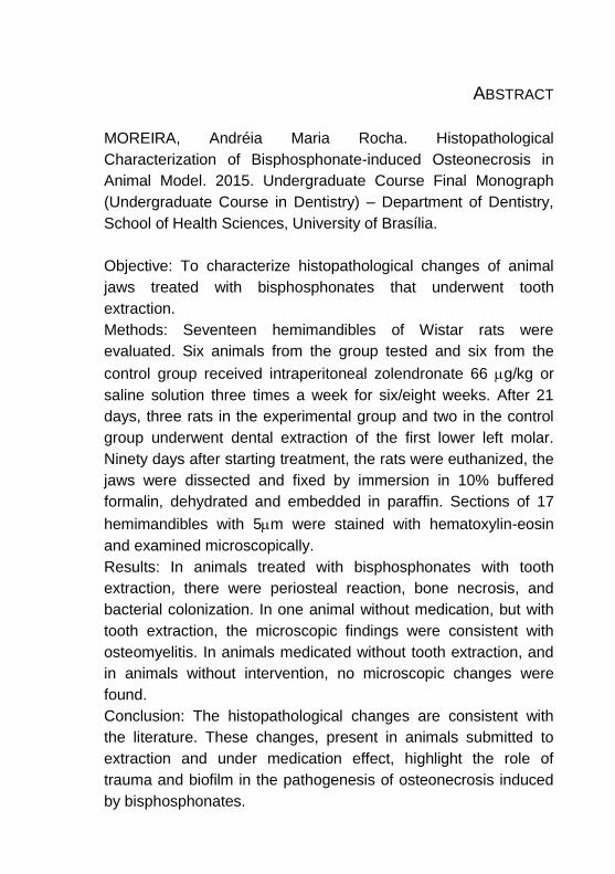
ABSTRACT
MOREIRA, Andréia Maria Rocha. Histopathological
Characterization of Bisphosphonate-induced Osteonecrosis in
Animal Model. 2015. Undergraduate Course Final Monograph
(Undergraduate Course in Dentistry) – Department of Dentistry,
School of Health Sciences, University of Brasília.
Objective: To characterize histopathological changes of animal
jaws treated with bisphosphonates that underwent tooth
extraction.
Methods: Seventeen hemimandibles of Wistar rats were
evaluated. Six animals from the group tested and six from the
control group received intraperitoneal zolendronate 66 g/kg or
saline solution three times a week for six/eight weeks. After 21
days, three rats in the experimental group and two in the control
group underwent dental extraction of the first lower left molar.
Ninety days after starting treatment, the rats were euthanized, the
jaws were dissected and fixed by immersion in 10% buffered
formalin, dehydrated and embedded in paraffin. Sections of 17
hemimandibles with 5m were stained with hematoxylin-eosin
and examined microscopically.
Results: In animals treated with bisphosphonates with tooth
extraction, there were periosteal reaction, bone necrosis, and
bacterial colonization. In one animal without medication, but with
tooth extraction, the microscopic findings were consistent with
osteomyelitis. In animals medicated without tooth extraction, and
in animals without intervention, no microscopic changes were
found.
Conclusion: The histopathological changes are consistent with
the literature. These changes, present in animals submitted to
extraction and under medication effect, highlight the role of
trauma and biofilm in the pathogenesis of osteonecrosis induced
by bisphosphonates.


SUMÁRIO
Artigo Científico ........................................................................... 19 Folha de Título ........................................................................ 21 Resumo ................................................................................... 22 Abstract ................................................................................... 24 Introdução................................................................................ 26 Metodologia ............................................................................. 29 Resultados............................................................................... 34
Discussão ................................................................................ 46 Conclusão ................................................................................ 50 Referências ............................................................................. 51
Anexos ......................................................................................... 55
Normas da Revista .................................................................. 55


19
ARTIGO CIENTÍFICO
Este trabalho de Conclusão de Curso é baseado no artigo
científico:
MOREIRA, Andréia Maria Rocha; GUILLEN, Gabriel
Albuquerque; FIGUEIREDO, Paulo Tadeu de Souza; POPPE,
Ana Carolina Acevedo; MELO, Nilce Santos de, LEITE, André
Ferreira. Caracterização Histopatológica da Osteonecrose
Induzida por Bisfosfonatos em Modelo Animal.
Apresentado sob as normas de publicação da Revista ORAL
SURGERY ORAL MEDICINE ORAL PATHOLOGY ORAL
RADIOLOGY AND ENDODONTOLOGY.

20

21
FOLHA DE TÍTULO
Caracterização Histopatológica da Osteonecrose Induzida por
Bisfosfonatos em Modelo Animal
Histopathological Characterization of Bisphosphonate-induced
Osteonecrosis in Animal Model
Andréia Maria Rocha Moreira1
Gabriel Albuquerque Guillen1
Paulo Tadeu de Souza Figueiredo2
Ana Carolina Acevedo Poppe2
Nilce Santos de Melo2
André Ferreira Leite2
1 Aluno de Graduação em Odontologia da Universidade de
Brasília. 2 Departamento de Odontologia, Faculdade de Ciências da
Saúde, Universidade de Brasília (UnB).
Correspondência: Prof. Dr. André Ferreira Leite
Campus Universitário Darcy Ribeiro - UnB - Faculdade de
Ciências da Saúde - Departamento de Odontologia - 70910-900 -
Asa Norte - Brasília - DF
E-mail: [email protected] / Telefone: (61) 31071849
22
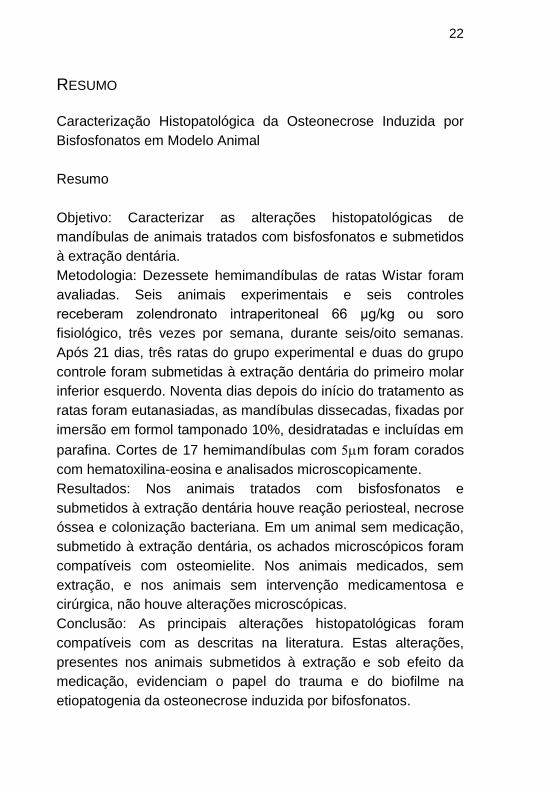
RESUMO
Caracterização Histopatológica da Osteonecrose Induzida por
Bisfosfonatos em Modelo Animal
Resumo
Objetivo: Caracterizar as alterações histopatológicas de
mandíbulas de animais tratados com bisfosfonatos e submetidos
à extração dentária.
Metodologia: Dezessete hemimandíbulas de ratas Wistar foram
avaliadas. Seis animais experimentais e seis controles
receberam zolendronato intraperitoneal 66 μg/kg ou soro
fisiológico, três vezes por semana, durante seis/oito semanas.
Após 21 dias, três ratas do grupo experimental e duas do grupo
controle foram submetidas à extração dentária do primeiro molar
inferior esquerdo. Noventa dias depois do início do tratamento as
ratas foram eutanasiadas, as mandíbulas dissecadas, fixadas por
imersão em formol tamponado 10%, desidratadas e incluídas em
parafina. Cortes de 17 hemimandíbulas com m foram corados
com hematoxilina-eosina e analisados microscopicamente.
Resultados: Nos animais tratados com bisfosfonatos e
submetidos à extração dentária houve reação periosteal, necrose
óssea e colonização bacteriana. Em um animal sem medicação,
submetido à extração dentária, os achados microscópicos foram
compatíveis com osteomielite. Nos animais medicados, sem
extração, e nos animais sem intervenção medicamentosa e
cirúrgica, não houve alterações microscópicas.
Conclusão: As principais alterações histopatológicas foram
compatíveis com as descritas na literatura. Estas alterações,
presentes nos animais submetidos à extração e sob efeito da
medicação, evidenciam o papel do trauma e do biofilme na
etiopatogenia da osteonecrose induzida por bifosfonatos.
23
Palavras-chave
Modelo animal; Histologia; Osteonecrose relacionada à
medicação.
Relevância Clínica
A osteonecrose dos maxilares relacionada à medicação
(MRONJ) é considerada um efeito adverso da administração de
bisfosfonatos e outras drogas. A MRONJ é uma condição de alta
morbidade com grande impacto clínico e de difícil tratamento. O
crescente uso destas medicações pode vir a elevar o número de
pacientes afetados por esta condição, cuja fisiopatologia ainda
não está completamente esclarecida. Modelos animais de
indução de MRONJ com consequente análise histológica podem
auxiliar na compreensão da etiopatogenia da doença.

24
ABSTRACT
Histopathological Characterization of Bisphosphonate-induced
Osteonecrosis in Animal Model.
Abstract
Objective: To characterize histopathological changes of animal
jaws treated with bisphosphonates that underwent tooth
extraction.
Methods: Seventeen hemimandibles of Wistar rats were
evaluated. Six animals from the group tested and six from the
control group received intraperitoneal zolendronate 66 g/kg or
saline solution three times a week for six/eight weeks. After 21
days, three rats in the experimental group and two in the control
group underwent dental extraction of the first lower left molar.
Ninety days after starting treatment, the rats were euthanized, the
jaws were dissected and fixed by immersion in 10% buffered
formalin, dehydrated and embedded in paraffin. Sections of 17
hemimandibles with 5m were stained with hematoxylin-eosin
and examined microscopically.
Results: In animals treated with bisphosphonates with tooth
extraction, there were periosteal reaction, bone necrosis, and
bacterial colonization. In one animal without medication, but with
tooth extraction, the microscopic findings were consistent with
osteomyelitis. In animals medicated without tooth extraction, and
in animals without intervention, no microscopic changes were
found.
Conclusion: The histopathological changes are consistent with
the literature. These changes, present in animals submitted to
extraction and under medication effect, highlight the role of
trauma and biofilm in the pathogenesis of osteonecrosis induced
by bisphosphonates.

25
Keywords
Animal Model; Histology; Medication-Related Osteonecrosis.
26
INTRODUÇÃO
Os bisfosfonatos são uma classe de medicamentos que
atuam sobre o processo de remodelação óssea, promovendo
aumento da densidade óssea mineral. São indicados para o
tratamento de doenças que causam reabsorção óssea, como
osteoporose e metástases ósseas líticas associadas ao câncer
de mama e outros tumores sólidos e também lesões osteolíticas
vinculadas ao mieloma múltiplo. Também são utilizados no
tratamento da hipercalcemia associada à malignidade,
osteogênese imperfeita e doença de Paget.1
Estes medicamentos têm estrutura química análoga a do
pirofosfato, caracterizado pela ligação P-O-P. Entretanto, nos
bisfosfonatos, a ligação P-O-P foi substituída por uma ligação P-
C-P, na qual o átomo de carbono é o átomo geminal na cadeia, o
que fornece maior resistência à hidrólise. Duas cadeias
covalentes secundárias (R1 e R2) estão ligadas ao átomo de
carbono. Por meio da cadeia R1, o bisfosfonato liga-se ao cálcio
presente nas apatitas do sistema esquelético, permitindo assim a
ligação do bisfosfonato ao tecido ósseo. A estrutura da cadeia R2
afeta a inibição da reabsorção óssea e de acordo com a estrutura
molecular desta cadeia, os bisfosfonatos podem ser nitrogenados
ou não-nitrogenados. A inserção de nitrogênio à cadeia R2
aumenta a afinidade do bisfosfonato por tecido ósseo e a sua
capacidade antirreabsorção.2 O zolendronato e o pamidronato,
por exemplo, são bisfosfonatos contendo nitrogênio.3
O mecanismo de inibição da reabsorção óssea dos
bisfosfonatos ocorre principalmente através da redução da
atividade osteoclástica. Os bisfosfonatos ligam-se à
hidroxiapatita do tecido ósseo. Ao reabsorver o tecido
impregnado de bisfosfonatos, os osteoclastos apresentam
prejuízo na capacidade de aderência óssea, formação da borda
em escova e produção de prótons. Além disso, os bisfosfonatos

27
também atuam através da indução de osteoclastos à apoptose e
reduzem o recrutamento de novos osteoclastos.4
Um dos efeitos adversos que pode ocorrer devido ao uso
destas medicações é a osteonecrose, atualmente denominada
osteonecrose dos maxilares relacionada à medicação. Ao ser
descrita pela primeira vez, esta osteonecrose recebeu o nome de
osteonecrose por bisfosfonatos.3 Atualmente, como se verificou
que outros medicamentos antiangiogênicos e antirreabsortivos
também podem causar osteonecrose, a American Association of
Oral and Maxillofacial Surgeons, recomenda o nome de
osteonecrose dos maxilares relacionada à medicação (MRONJ).
Em coerência com a literatura, neste trabalho será empregado
termo MRONJ, embora tenha sido usado o bisfosfonato como
medicamento indutor de osteonecrose nos animais.5
Para o diagnóstico desta condição, três critérios são
necessários: tratamento atual ou anterior com agentes
antirreabsortivos ou antiangiogênicos; osso exposto ou que pode
ser sondado através de fistula intraoral ou extraoral na região
maxilofacial que não cura em até oito semanas; e ausência de
histórico de radioterapia na região de cabeça e pescoço.5 A
maioria dos casos de MRONJ ocorre devido à extração dentária
anterior, porém há relatos de desenvolvimento espontâneo. A
mandíbula tem sido mais afetada que a maxila. Há mais casos
de MRONJ devido à administração de bisfosfonatos
intravenosos, principalmente o Zolendronato, quando comparado
aos bisfosfonatos administrados via oral.6
Apesar das várias hipóteses descritas na literatura que
buscam elucidar a etiopatogenia da MRONJ, a patogênese da
doença ainda não está completamente esclarecida.5,7-12
A
hipótese que visa explicar o desenvolvimento da MRONJ baseia-
se na supressão da remodelação óssea, através da atividade
antiosteoclástica promovida pelos bisfosfonatos. Os
bisfosfonatos alteram o equilíbrio fisiológico da interação entre
osteoclastos e osteoblastos,2 causando prejuízo na função

28
osteoclástica e alterando o processo de remodelação óssea
fisiológica. Assim, o organismo pode não ser capaz de reparar as
microfraturas que ocorrem devido à exposição a cargas
mecânicas normais ou a danos maiores, como a extração
dentária13
. Além disso, o Zolendronato parece afetar o tecido da
mucosa oral por meio da indução de apoptose das células
epiteliais ou fibroblastos. Desta forma, lesões aos tecidos moles
podem levar ao desenvolvimento da MRONJ ou potencializá-la14
.
O efeito antiangiogênico promovido pelo zolendronato, que
atrasa a cicatrização de feridas pós-exodontias, e o aumento a
predisposição à infecção bacteriana proporcionada por este
bisfosfonato também estão descritos na literatura.11
A avaliação histopatológica da MRONJ é essencial visto
que a MRONJ apresenta características clínicas e radiográficas
semelhantes a outras doenças, como osteomielite e
osteorradionecrose.15,16,17
Além disso, este tipo de análise em
modelo animal pode auxiliar na compreensão da etiopatogênese
da MRONJ, ainda incerta.18,19
Em animais, as características histopatológicas da
MRONJ são descritas como a presença de áreas de necrose
óssea com lacunas osteocíticas (osteoplastos) vazias, ou seja,
com ausência ou escassa quantidade de osteócitos; biofilme
microbiano, muitas vezes composto por colônias de bactérias
compatíveis com Actinomyces spp; infiltrado inflamatório próximo
às áreas de sequestro ósseo ou infecção; redução da
vascularização óssea; entre outras.7,9,10,19,20
Na análise histopatológica de espécimes de pacientes
portadores de MRONJ foram descritos, entre outros achados,
áreas de reação periosteal e bordas ósseas com padrão irregular
sugerindo interrupção das lacunas de Howship.8,16
A reação
periosteal também é visualizada nos exames de imagem. No
entanto, parece paradoxal, que diante da alteração da
remodelação óssea, haja a formação periosteal.
29
Desta forma, o principal objetivo deste estudo é
caracterizar as alterações histopatológicas de mandíbulas, em
animais submetidos à extração dentária e em uso de
bisfosfonatos intravenosos.
METODOLOGIA
O projeto foi aprovado no Comitê de Ética de Uso Animal
do Instituto de Ciências Biológicas da Universidade de Brasília
(CEUA) em 12 de janeiro de 2012 sob o protocolo UnBDOC
número 41597/2010.
O material biológico usado para a presente análise
histopatológica foi coletado e processado previamente durante o
projeto de mestrado da aluna Fernanda Ogata Luz denominado
“Análise por microtomografia da maxila e mandíbula de ratos em
tratamento com bisfosfonatos”. Na dissertação de mestrado, o
objetivo foi analisar tomograficamente os maxilares de ratas
submetidas a tratamento medicamentoso com Zolendronato
associado ou não à extração dentária. A metodologia da
pesquisa envolveu a adaptação dos animais no biotério,
tratamento medicamentoso e cirúrgico, realização dos exames
de microtomografia no início e no fim do tratamento e eutanásia
dos animais.21
A divisão dos grupos experimentais e controles
assim como os tratamentos realizados serão descritos a seguir.
Trinta e seis ratas Wistar, com peso aproximado de 300g (90
dias de idade) foram divididas em dois grupos (Figura 1):
Grupo 1: Grupo Zolendronato, composto por 24 ratas que
receberam Zolendronato 66 μg/kg, via intraperitoneal, três vezes
por semana, durante seis ou oito semanas.
Grupo 2: Grupo Controle, composto por 12 ratas que
receberam soro fisiológico via intraperitoneal, três vezes por
semana, durante seis ou oito semanas.
30
Após 21 dias, de forma aleatória, procedeu-se à
exodontia do primeiro molar inferior esquerdo em,
aproximadamente, um terço dos animais de cada grupo; do
primeiro molar superior esquerdo em, aproximadamente, um
terço dos animais de cada grupo e o restante não sofreu
extração dentária. Noventa dias depois do início do tratamento,
as ratas foram eutanasiadas com gás carbônico e decapitadas.
Posteriormente à extração dentária do primeiro molar inferior
esquerdo, oito animais (dois controles e seis experimentais)
morreram 21
(Figura 1).
ANÁLISE HISTOPATOLÓGICA
1. GRUPOS DE ESTUDO:
Para esse trabalho foram realizadas preparações
histológicas a partir de 12 animais, distribuídos em dois grupos,
experimental e controle, conforme detalhado na figura 1. Dos 12
animais foram preparadas 17 hemimandíbulas, também
distribuídas em dois grupos, de acordo com o desenho
experimental.

31

32
Figura 1: Em azul, distribuição dos animais na primeira etapa do projeto,
desenvolvida durante o mestrado da aluna Fernanda Ogata Luz
(n=número de ratas). Em vermelho, distribuição dos animais que
morreram após a exodontia (n=número de ratas). Em verde, distribuição
dos grupos de animais selecionados para análise histológica (n=número
de ratas). Em rosa, distribuição das peças anatômicas
(hemimandíbulas) submetidas à análise histopatológica(n=número de
hemimandíbulas) .
2. PREPARAÇÃO HISTOLÓGICA:
Após eutanásia, as cabeças dos animais foram fixadas
por imersão em formol tamponado 10% durante 24 horas e
armazenadas em tampão fosfato (0,1M, 7,0-7,2 pH). Em seguida,
foram descalcificadas em solução de EDTA 4.13% durante um
período de três meses, com trocas a cada 72 horas. A maxila e a
mandíbula de cada animal foram removidas, seccionadas
sagitalmente e o excesso de tecidos moles dissecado. As
hemimaxilas e hemimandíbulas foram desidratadas em
concentrações crescentes de álcool etílico (50%, 70%, 80%,
95%, 100%), as amostras impregnadas no tolueno e incluídas
em parafina Paraplast®.
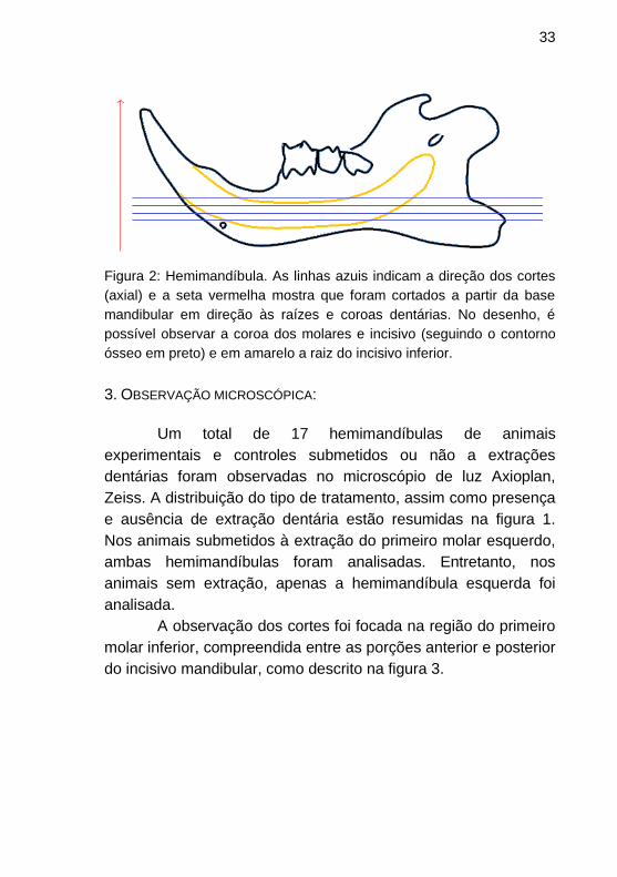
Foram realizados cortes axiais seriados de 5 μm em
micrótomo Leica RM 2125RT, dos quais a cada dez cortes
desprezados, um corte foi aproveitado. Os cortes foram
realizados da região da base da mandíbula em direção às raízes
e coroas dentárias de acordo com a Figura 2. Foram
confeccionadas aproximadamente 665 lâminas e destas 220
foram selecionadas e coradas por meio da coloração
hematoxilina-eosina. Após coloração, as lâminas foram
montadas com Entellan®
33
Figura 2: Hemimandíbula. As linhas azuis indicam a direção dos cortes
(axial) e a seta vermelha mostra que foram cortados a partir da base
mandibular em direção às raízes e coroas dentárias. No desenho, é
possível observar a coroa dos molares e incisivo (seguindo o contorno
ósseo em preto) e em amarelo a raiz do incisivo inferior.
3. OBSERVAÇÃO MICROSCÓPICA:
Um total de 17 hemimandíbulas de animais
experimentais e controles submetidos ou não a extrações
dentárias foram observadas no microscópio de luz Axioplan,
Zeiss. A distribuição do tipo de tratamento, assim como presença
e ausência de extração dentária estão resumidas na figura 1.
Nos animais submetidos à extração do primeiro molar esquerdo,
ambas hemimandíbulas foram analisadas. Entretanto, nos
animais sem extração, apenas a hemimandíbula esquerda foi
analisada.
A observação dos cortes foi focada na região do primeiro
molar inferior, compreendida entre as porções anterior e posterior
do incisivo mandibular, como descrito na figura 3.

34
Figura 3: A. Esquema representativo do corte histológico: 1: Região
anterior do incisivo. 2: Região posterior do incisivo. 3. Osso cortical. 4.
Osso esponjoso. 5. Músculo. B. A área delimitada pelo quadrado
representa a principal região da hemimandíbula analisada neste estudo.
As lâminas foram analisadas quanto à presença de
necrose óssea. Ademais, a presença de inflamação, necrose
celular, biofilme e reação periosteal também foram parâmetros
avaliados.
RESULTADOS
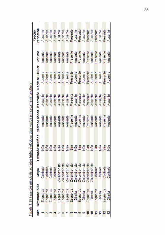
Os achados histopatológicos evidenciaram diferenças
entre as hemimandíbulas dos grupos controle e experimental. Os
principais resultados estão sintetizados na Tabela 1.
GRUPO EXPERIMENTAL - ZOLENDRONATO E EXTRAÇÃO DENTÁRIA:
As hemimandíbulas dos animais do grupo experimental
submetidos à extração dentária apresentaram intensa necrose
óssea, caracterizada por grandes áreas com ausência de
osteócitos nos osteoplastos nas áreas de osso esponjoso e de
osso cortical e ausência de vascularização (Figura 4). Essas
35

36
Figura 4. Grupo experimental com extração dentária. HE. A: Osso
necrótico (ON) e inflamação (seta). B: Osteoplastos vazios (seta),
biofilme (asterisco) e inflamação (ponta da seta).
*
A
B
ON
ON
37
características foram observadas desde a base mandibular,
antes mesmo de serem observadas as raízes dentárias.
Principalmente próximo às zonas de necrose óssea e biofilmes,
havia focos de infiltrado inflamatório (Figura 4). Foram
observadas várias áreas de biofilmes, principalmente sobre as
corticais ósseas e osso esponjoso, muitas vezes compatíveis
com colônias de Actinomyces spp, que se apresentam como uma
massa basofílica com estruturas semelhantes a raios, em
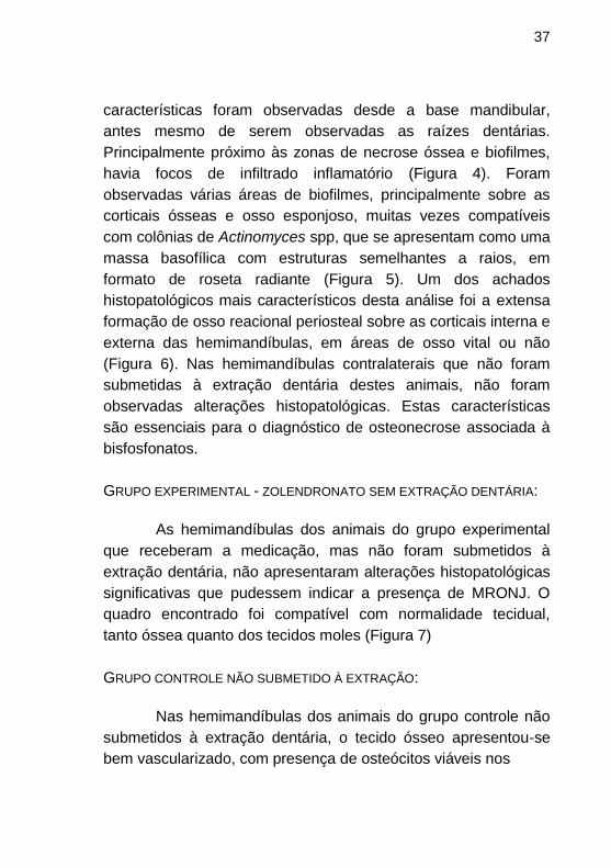
formato de roseta radiante (Figura 5). Um dos achados
histopatológicos mais característicos desta análise foi a extensa
formação de osso reacional periosteal sobre as corticais interna e
externa das hemimandíbulas, em áreas de osso vital ou não
(Figura 6). Nas hemimandíbulas contralaterais que não foram
submetidas à extração dentária destes animais, não foram
observadas alterações histopatológicas. Estas características
são essenciais para o diagnóstico de osteonecrose associada à
bisfosfonatos.
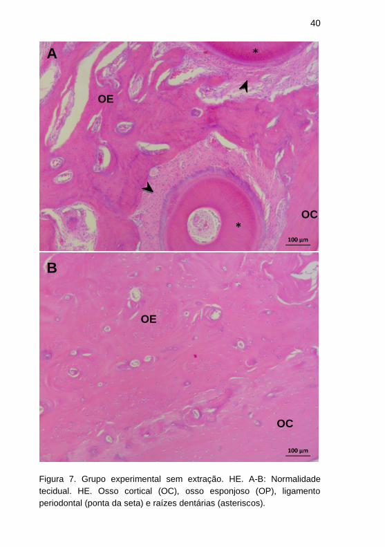
GRUPO EXPERIMENTAL - ZOLENDRONATO SEM EXTRAÇÃO DENTÁRIA:
As hemimandíbulas dos animais do grupo experimental
que receberam a medicação, mas não foram submetidos à
extração dentária, não apresentaram alterações histopatológicas
significativas que pudessem indicar a presença de MRONJ. O
quadro encontrado foi compatível com normalidade tecidual,
tanto óssea quanto dos tecidos moles (Figura 7)
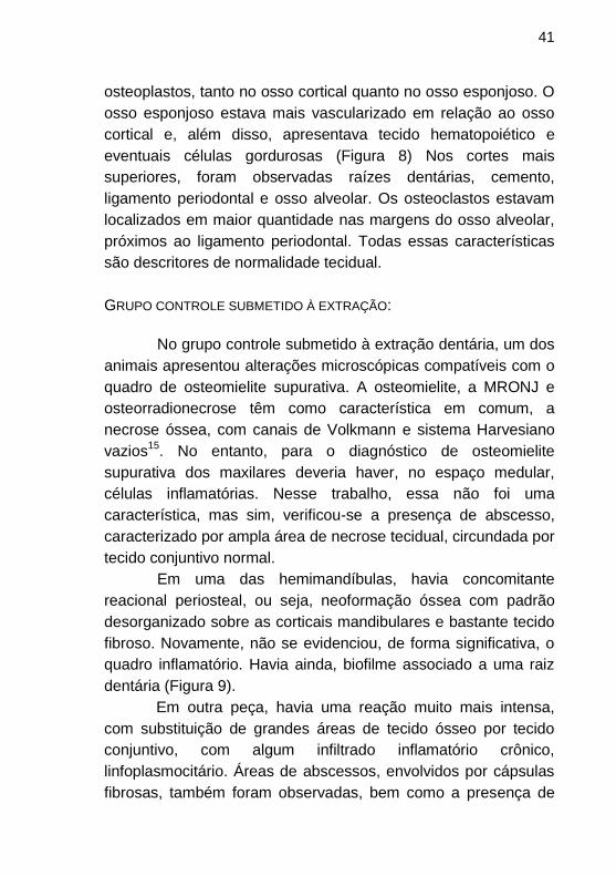
GRUPO CONTROLE NÃO SUBMETIDO À EXTRAÇÃO:
Nas hemimandíbulas dos animais do grupo controle não
submetidos à extração dentária, o tecido ósseo apresentou-se
bem vascularizado, com presença de osteócitos viáveis nos

38
Figura 5. Grupo experimental com extração dentária. HE. A:
Osteonecrose (ON) do osso alveolar e biofilme (seta). B: osso alveolar
necrótico (ON), com biofilme aderido às paredes (ponta da seta). No
interior do alvéolo há colônias sugestivas de Actinomyces israeli (seta).
A
ON
ON
B
39
Figura 6. Grupo experimental com extração dentária. HE. A-B: Reação
periosteal (RP) sobre o osso cortical (OC). A seta indica o limite entre
ambos. C: Osteócito viáveis (seta) e tecido conjuntivo (asterisco) na
reação periosteal.
OC
RP
A
B C
OC
RP
*
40
Figura 7. Grupo experimental sem extração. HE. A-B: Normalidade
tecidual. HE. Osso cortical (OC), osso esponjoso (OP), ligamento
periodontal (ponta da seta) e raízes dentárias (asteriscos).
OC
OC
*
*
OE
OE
A
B
41
osteoplastos, tanto no osso cortical quanto no osso esponjoso. O
osso esponjoso estava mais vascularizado em relação ao osso
cortical e, além disso, apresentava tecido hematopoiético e
eventuais células gordurosas (Figura 8) Nos cortes mais
superiores, foram observadas raízes dentárias, cemento,
ligamento periodontal e osso alveolar. Os osteoclastos estavam
localizados em maior quantidade nas margens do osso alveolar,
próximos ao ligamento periodontal. Todas essas características
são descritores de normalidade tecidual.
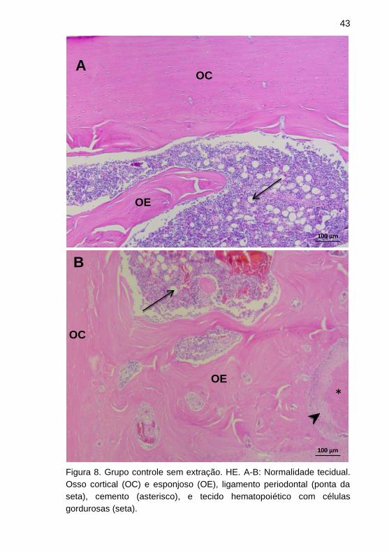
GRUPO CONTROLE SUBMETIDO À EXTRAÇÃO:
No grupo controle submetido à extração dentária, um dos
animais apresentou alterações microscópicas compatíveis com o
quadro de osteomielite supurativa. A osteomielite, a MRONJ e
osteorradionecrose têm como característica em comum, a
necrose óssea, com canais de Volkmann e sistema Harvesiano
vazios15
. No entanto, para o diagnóstico de osteomielite
supurativa dos maxilares deveria haver, no espaço medular,
células inflamatórias. Nesse trabalho, essa não foi uma
característica, mas sim, verificou-se a presença de abscesso,
caracterizado por ampla área de necrose tecidual, circundada por
tecido conjuntivo normal.
Em uma das hemimandíbulas, havia concomitante
reacional periosteal, ou seja, neoformação óssea com padrão
desorganizado sobre as corticais mandibulares e bastante tecido
fibroso. Novamente, não se evidenciou, de forma significativa, o
quadro inflamatório. Havia ainda, biofilme associado a uma raiz
dentária (Figura 9).
Em outra peça, havia uma reação muito mais intensa,
com substituição de grandes áreas de tecido ósseo por tecido
conjuntivo, com algum infiltrado inflamatório crônico,
linfoplasmocitário. Áreas de abscessos, envolvidos por cápsulas
fibrosas, também foram observadas, bem como a presença de

42
extensas zonas de necrose óssea. Também estavam presentes,
biofilmes compatíveis com colônias de Actinomyces spp, e
significativa reação periosteal. As hemimandíbulas do lado
contralateral a estas não apresentaram alterações
histopatológicas (Figura 10).
43
Figura 8. Grupo controle sem extração. HE. A-B: Normalidade tecidual.
Osso cortical (OC) e esponjoso (OE), ligamento periodontal (ponta da
seta), cemento (asterisco), e tecido hematopoiético com células
gordurosas (seta).
OC
OC
*
OE
OE
A
B
44
Figura 9. Grupo controle com extração dentária. HE. A: Sobre o osso
cortical (OC), presença de reação periosteal (RP). A seta indica o limite
ente ambos. B: Biofilme (seta) sobre raiz dentária (asterisco).
A
B
*
OC
RP

45
Figura 10. Grupo controle com extração dentária. Osteomielite
supurativa. HE. A: Biofilme (asterisco) sobre osso cortical (OC)
necrótico. Reabsorção óssea: osso esponjoso sendo reabsorvido (ponta
da seta) e substituição por tecido conjuntivo (seta). B: Abscesso
envilvido por cápsula fibrosa.
A
B
*
OC
*
46
DISCUSSÃO
A MRONJ é uma doença recentemente descrita. O
primeiro relato de um paciente com MRONJ foi publicado em
2003.3 Desde então, mais de duas mil publicações sobre
osteonecrose por bisfosfonatos estão disponíveis na literatura. A
MRONJ em pacientes com osteoporose submetidos ao
tratamento com bisfosfonatos intravenosos tem prevalência em
torno de 0,005%, podendo variar entre 0% a 0,348%. A
incidência de osteonecrose nesses pacientes é de até
90/100.000 pacientes, por ano. Já a prevalência e a incidência da
MRONJ em pacientes oncológicos submetidos ao tratamento
com bisfosfonatos intravenosos tendem a ser maiores, com
valores variando entre 0% a 0,186% e até 12,222 casos por
100.000 pacientes por ano, respectivamente.22
Embora, a patofisiologia da MRONJ ainda não esteja
completamente elucidada, modelos animais podem auxiliar a
compreendê-la, uma vez que nestes a análise histopatológica
pode ser feita de forma muito mais completa e não restrita
apenas a áreas de sequestro ósseo como geralmente ocorre
com amostras de pacientes9. No entanto, persiste a necessidade
de modelos animais melhor caracterizados que possibilitem uma
aproximação da condição de osteonecrose encontrada em
pacientes22
. Grande parte dos modelos animais descritos na
literatura apresentam metodologias diferentes e na maioria das
vezes associam a administração de bisfosfonatos a outros
fatores de risco, terapias e/ou comorbidades. Isso dificulta a
comparação entre estudos. Além disso, reduz a confiabilidade do
modelo e a reprodução da condição clínica mais comum, que é a
MRONJ associada à extração dentária.7,10
Neste trabalho, a administração do zolendronato foi feita
em animais associando-se apenas a exodontia como fator de
risco, a fim de avaliar os efeitos desta medicação na fisiologia
óssea e para verificar o papel do trauma no desenvolvimento da

47
osteonecrose. Não houve casos de osteonecrose espontânea.
No entanto, no grupo dos animais submetidos à exodontia, todos
apresentaram quadros morfológicos compatíveis com o descrito
para MRONJ. Em todas as lâminas examinadas havia necrose
óssea, caracterizada pela presença de lacunas osteocíticas
vazias (os chamados osteoplastos), em grande extensão,
presença de biofilme, a maioria com formato compatível com
colônias de Actinomyces. De forma significativa, encontrou-se
reação periosteal intensa, em quadro semelhante com o visto em
pacientes. A reação periosteal e o tecido fibroso entremeado nas
trabéculas ósseas, a presença de osteoclastos, tal como
encontrada em nosso trabalho, foram descritos também em
outros estudos.8,16
Esse fato corrobora os relatos da literatura,
que apontam o trauma como um fator de risco importante para o
desenvolvimento da MRONJ.7,12,23
Diante disso, reforça-se a
importância da extração dentária como fator desencadeador de
MRONJ em modelos animais.
Em nosso estudo, no grupo experimental submetido à
exodontia, verificou-se a presença de inflamação em pontos
focais, principalmente em áreas próximas à necrose óssea e
biofilme bacteriano. Desta forma, a inflamação não parece ter
sido evento significativamente importante na patogênese da
doença. Esses dados são diversos dos relatados, mas podem
ser creditados ao tamanho da amostra. A presença do biofilme
foi, ao contrário, significativa e relevante. Em todas as lâminas
examinadas, havia nítidas coleções bacterianas, com morfologia
compatível com Actinomyces spp. Os biofilmes estavam
dispostos na interface óssea, sobre a cortical, próximo às raízes,
nos espaços medulares e nos álveolos pós-extração. Esses
dados reforçam o papel do biofilme na etiopatogênese da
MRONJ, assim como a extração dentária. Em termos gerais, os
achados desse estudo mostraram semelhança com os descritos
na literatura.7,9,10,19,20
48
Na hemimandíbula de um dos animais do grupo
experimental que recebeu a medicação e foi submetido à
extração dentária, foi observada intensa necrose tecidual,
inclusive da polpa dentária do incisivo e dos molares e do tecido
conjuntivo presente no tecido ósseo. Este rato foi o único do
grupo experimental que recebeu medicação durante oito
semanas e foi submetido à extração dentária. São necessárias
mais investigações para concluir se esta necrose ocorreu devido
a alguma eventual falha no processo de preparo da amostra,
danificando-a, ou se o tempo de administração do bisfosfonato é
um fator importante para o desenvolvimento desta doença, bem
como a sua intensidade.
Os animais do grupo experimental que receberam a
medicação, sem a etapa da extração dentária, não apresentaram
alterações histopatológicas, caracterizando um quadro de
normalidade tecidual. Em um estudo utilizando um modelo
animal de indução de MRONJ em ratos que receberam
Zolendronato e Pamidronato mas não foram submetidos à
extração dentária, os autores encontraram discreto infiltrado
inflamatório, necrose de tecidos moles além de alterações ósses
relacionadas à medicação. Os autores encontraram lacunas
osteocíticas vazias e infiltrado inflamatório misto invadindo o
epitélio em 40% da amostra. Dois animais, nesse estudo,
preencheram os critérios de osteonecrose.24
Esses resultados
são divergentes dos encontrados em nosso estudo,
possivelmente devido a diferenças na dosagem utilizada ou ao
reduzido tamanho da nossa amostra. Esse tópico alerta para a
necessidade de padronização das metodologias, para facilitar a
comparação.
Em um animal do grupo controle, sem medicação e com
exodontia, alguns abscessos foram visualizados, variando em
extensão. Dois destes abscessos estavam localizados próximo a
glândula salivar, com perda de tecido acinar e persistência
ductal. As alterações verificadas nas hemimandíbulas das ratas
49
deste grupo submetidas à extração dentária podem ter ocorrido
devido à fratura de raízes durante o procedimento cirúrgico e/ou
infecção secundária. A incerteza da origem do processo
inflamatório é um fator limitante, inclusive descrita em outro
estudo.24
A presença de abscesso em grupos controles
semelhantes foi descrita em outro estudo.7 De forma significativa,
encontrou-se reação periosteal, focos de necrose óssea e áreas
de remodelação óssea nos ratos do grupo controle, embora
escassa. Além disso, em um dos ratos do grupo controle, a
formação de tecido fibroso foi intensa. Esse quadro sugere o
diagnóstico de osteomielite, conforme descrito na literatura.15
A
presença de reação periosteal nesse grupo não encontra
justificativa a não ser a inferência que o trauma atuou como
indutor do desenvolvimento de respostas teciduais frente a
estímulos de baixa intensidade. As características morfológicas
dos animais do grupo controle sem exodontia foram compatíveis
com os padrões de normalidade.
A reação periosteal é uma reação do osso cortical frente
a determinados estímulos. A apresentação bilateral desta
condição normalmente está associada a algum estímulo de
natureza sistêmica. Já uma manifestação unilateral, geralmente
está associada a estímulos locais, como trauma e infecção.25
A
presença da reação periosteal nas hemimandíbulas de ratas dos
grupos experimental e controle submetidas à exodontia pode ser
justificada por fatores como trauma e infecção que estiveram
presentes em ambos os grupos e as hemimandíbulas
contralaterais não apresentaram este tipo de reação. Isso
demonstra que neste presente estudo, a reação periosteal
aparentemente está mais associada a estímulos locais do que
com a administração da medicação. Entretanto, não podemos
ainda afirmar que o uso do bisfosfonato não foi importante para o
desenvolvimento da reação periosteal.25
A principal região analisada microscopicamente,
conforme demonstrado na figura 4, foi escolhida por englobar a

50
área dos molares. Além disso, nesta área há osso esponjoso,
cortical e medula óssea ou raízes dentárias, osso alveolar e
ligamento, a depender da posição do corte. Essa escolha se deu
pela riqueza anatômica e por permitirem a analogia com as
imagens microtomográficas. As hemimandíbulas analisadas
neste trabalho foram submetidas ao escaneamento
microtomográfico. Na análise microtomográfica, as
hemimandíbulas dos animais controles não apresentaram
alterações, à semelhança dos achados microscópicos. Nos
animais do grupo experimental submetidos à extração dentária,
havia lesão osteolítica, com ou sem rompimento da cortical
óssea e reação periosteal, o que foi confirmado em nossos
achados histopatológicos.21
Esse estudo apresentou limitações. Inicialmente tentou-
se preparar todas as amostras disponíveis, porém, por questões
de disponibilidade técnica, dificuldades laboratoriais, nem toda a
amostra foi processada. Assim, as maxilas não fizeram parte
dessa análise. Esse fato, embora seja limitante, representa a
maioria das situações descritas em pacientes, nos quais a
MRONJ é mais comum na mandíbula. A perda de alguns animais
contribuiu para esse quadro, mas a literatura evidencia as
dificuldades técnicas envolvidas no desenrolar de trabalhos com
animais e apresenta amostras com quantitativo semelhante.
CONCLUSÃO
As alterações histopatológicas observadas nesse estudo
foram compatíveis com alterações características da MRONJ
descritas em outros trabalhos presentes na literatura. Os
principais achados histopatológicos foram observados nas
hemimandíbulas em que houve a extração do primeiro molar
inferior e a administração de medicação zolendronato. Estes
resultados demonstram o impacto que a extração dentária possui
51
no desenvolvimento da MRON. Além disso, o biofilme também foi
um fator importante na etiopatogenia da doença. Neste estudo
não foram observadas alterações histopatológicas significativas
nas hemimandíbulas sem extração dentária.
Uma vez que a criação de modelos animais de indução
da MRONJ e a consequente análise histopatológica podem
auxiliar na compreensão da patogênese da doença, sugere-se
que mais estudos sejam desenvolvidos.
REFERÊNCIAS
1. Russel RG. Bisphosphonate: the first 40 years. Bone 2011; 49:2-19.
2. Badel T, Pavicin IS, Carek AJ, Rosin-Grget K, Grbesa D.
Pathophysiology of osteonecrosis of the jaw in patients treated
with bisphosphonate. Coll Antropol. 2013;37:645-51
3. Marx RE. Pamidronate (Aredia) and zoledronate (Zometa) induced
avascular necrosis of the jaws: a growing epidemic. J Oral
Maxillofac Surg. 2003;61:1115-7.
4. Rosen HN. Pharmacology of Bisphosphonates. UptoDate. 2015.
Disponível em: <
http://www.uptodate.com/contents/pharmacology-of-
bisphosphonates> Acesso em: 16 jul. 2015
5. Ruggiero SL, Dodson TB, Fantasia J, Goodday R, Aghaloo
T, Mehrotra B, O'Ryan F. American Association of Oral and
Maxillofacial Surgeons position paper on medication-
related osteonecrosis of the jaw--2014 update. J Oral Maxillofac
Surg. 2014;72:1938-56.
6. Fliefel R, Tröltzsch M, Kühnisch J, Ehrenfeld M, Otto S.
Treatment strategies and outcomes of bisphosphonate-
http://www.ncbi.nlm.nih.gov/pubmed/?term=Ruggiero%20SL%5BAuthor%5D&cauthor=true&cauthor_uid=25234529

52
related osteonecrosis of the jaw (BRONJ) with characterization
of patients: a systematic review. Int J Oral Maxillofac
Surg. 2015; 44:568-85.
7. Barba-Recreo P, Del Castillo Pardo de Vera JL, García-Arranz
M, Yébenes L, Burgueño M. Zoledronic acid - related
osteonecrosis of the jaws. Experimental model with dental
extractions in rats. J Craniomaxillofac Surg. 2014;42:744-50.
8. Bedogni A, Blandamura S, Lokmic Z, Palumbo C, Ragazzo M, Ferrari
F, Tregnaghi A, Pietrogrande F, Procopio O, Saia G, Ferretti
M, Bedogni G, Chiarini L, Ferronato G, Ninfo V, Lo Russo L, Lo
Muzio L, Nocini PF. Bisphosphonate-
associated jawbone osteonecrosis:a correlation between imagin
g techniques andhistopathology. Oral Surg Oral Med Oral
Pathol Oral Radiol Endod. 2008;105:358-64.
9. Biasotto M, Chiandussi S, Zacchigna S, Moimas S, Dore F, Pozzato
G, Cavalli F, Zanconati F, Contardo L, Giacca M, Di Lenarda R.
A novel animal model to study non-spontaneous
bisphosphonates osteonecrosis of jaw. J Oral Pathol
Med. 2010;39:390-6.
10. Conte Neto N, Spolidorio LC, Andrade CR, S Bastos A, Guimarães
M, Marcantonio E Jr.
Experimental development of bisphosphonate-
related osteonecrosis of the jaws in rodents. Int J Exp
Pathol. 2013;94:65-73.
11. Kobayashi Y, Hiraga T, Ueda A, Wang L, Matsumoto-Nakano
M, Hata K, Yatani H, Yoneda T. Zoledronic acid delays wound
healing of the tooth extraction socket, inhibits oral epithelial cell
migration, and promotes proliferation and adhesion to
hydroxyapatite of oral bacteria, without causing osteonecrosis of
the jaw, in mice. J Bone Miner Metab. 2010;28:165-75.
12. Agaçayak KS, Yuksel H, Atilgan S, Koparal M, Uçan MC, Ozgöz
M, Yaman F, Atalay Y, Acikan I. Experimental investigation of
http://www.ncbi.nlm.nih.gov/pubmed/?term=Tregnaghi%20A%5BAuthor%5D&cauthor=true&cauthor_uid=18280968
http://www.ncbi.nlm.nih.gov/pubmed/?term=Ferronato%20G%5BAuthor%5D&cauthor=true&cauthor_uid=18280968
http://www.ncbi.nlm.nih.gov/pubmed/?term=Zacchigna%20S%5BAuthor%5D&cauthor=true&cauthor_uid=20202091
http://www.ncbi.nlm.nih.gov/pubmed/?term=Zanconati%20F%5BAuthor%5D&cauthor=true&cauthor_uid=20202091
53
relationship between trauma and bisphosphonate-
related osteonecrosis. Niger J Clin Pract. 2014;17:559-64
13. Ruggiero SL. Guidelines for the diagnosis of bisphosphonate-related
osteonecrosis of the jaw (BRONJ). Clin Cases Miner Bone
Metab. 2007;4:37-42.
14. Scheper MA, Badros A, Chaisuparat R, Cullen KJ, Meiller TF.
Effect of zoledronic acid on oral fibroblasts and epithelial cells: a
potential mechanism of bisphosphonate-associated
osteonecrosis. Br J Haematol. 2009;144:667-76.
15. Marx RE, Tursun R. Suppurative osteomyelitis, bisphosphonate
induced osteonecrosis, osteoradionecrosis: a blinded
histopathologic comparison and its implications for
the mechanism of each disease. Int J Oral Maxillofac
Surg. 2012;41:283-9
16. Franco-Pretto E, Pacheco M, Moreno A, Messa O, Gnecco J.
Bisphosphonate-induced osteonecrosis of the
jaws: clinical, imaging, and histopathology findings. Oral Surg
Oral Med Oral Pathol Oral Radiol. 2014;118:408-17.
17. Hansen T, Kunkel M, Weber A, James Kirkpatrick C.
Osteonecrosis of the jaws in patients treated with
bisphosphonates histomorphologic analysis in comparisonwith i
nfected osteoradionecrosis. J Oral Pathol Med. 2006;35:155-60.
18. Biasotto M, Chiandussi S, Zacchigna S, Moimas S, Dore F, Pozzato
G, Cavalli F, Zanconati F, Contardo L, Giacca M, Di Lenarda R.
A novel animal model to study non-spontaneous
bisphosphonates osteonecrosis of jaw. J Oral Pathol
Med. 2010;39:390-6.
19. Marino KL, Zakhary I, Abdelsayed RA, Carter JA, O'Neill
JC, Khashaba RM, Elsalanty M, Stevens MR, Borke JL.
Development of a rat model of bisphosphonate-related
http://www.ncbi.nlm.nih.gov/pubmed/?term=Zacchigna%20S%5BAuthor%5D&cauthor=true&cauthor_uid=20202091
http://www.ncbi.nlm.nih.gov/pubmed/?term=Zanconati%20F%5BAuthor%5D&cauthor=true&cauthor_uid=20202091
http://www.ncbi.nlm.nih.gov/pubmed/?term=Khashaba%20RM%5BAuthor%5D&cauthor=true&cauthor_uid=21905888

54
osteonecrosis of the jaw (BRONJ). J Oral Implantol. 2012;38
:511-8.
20. Maahs MP, Azambuja AA, Campos MM, Salum FG, Cherubini K.
Association between bisphosphonates and jaw osteonecrosis:
a study in Wistar rats. Head Neck. 2011;33:199-207
21. Luz, FO. Modelo Animal de Indução de Osteonecrose por Uso de
Bisfosfonatos em Ratos com Análise Microtomográfica.
Dissertação (Mestrado em Ciências da Saúde). Universidade
de Brasília. Brasília 2013
22. Khan AA, Morrison A, Hanley DA, Felsenberg D, McCauley
LK, O'Ryan F et al. Diagnosis and management of
osteonecrosis of the jaw: a systematic review and international
consensus. J Bone Miner Res. 2015 ;30:3-23. (Até 6)
23. Yang H, Pan H, Yu F, Chen K, Shang G, Xu Y A novel model
of bisphosphonate-related osteonecrosis of the jaw in rats. Int J
Clin Exp Pathol. 2015;8:5161-7.
24. Senel FC, Kadioglu Duman M, Muci E, Cankaya M, Pampu
AA, Ersoz S, Gunhan O. Jaw bone changes in rats after
treatment with zoledronate and pamidronate. Oral Surg Oral
Med Oral Pathol Oral Radiol Endod. 2010;109:385-91.
25. Rana RS, Wu JS, Eisenberg RL. Periosteal reaction. AJR Am J
Roentgenol. 2009;193:W259-72.
http://www.ncbi.nlm.nih.gov/pubmed/?term=Azambuja%20AA%5BAuthor%5D&cauthor=true&cauthor_uid=20848442
http://www.ncbi.nlm.nih.gov/pubmed/?term=Cherubini%20K%5BAuthor%5D&cauthor=true&cauthor_uid=20848442
http://www.ncbi.nlm.nih.gov/pubmed/?term=McCauley%20LK%5BAuthor%5D&cauthor=true&cauthor_uid=25414052

55
ANEXOS
NORMAS DA REVISTA
Section Scope Statements
The Oral and Maxillofacial Surgery Section aims to publish an
extensive range of original articles that advances patient care
through enhanced understanding of diagnosis, surgical and
adjunctive treatment of diseases, and injuries and defects
involving both the functional and esthetic aspects of the hard and
soft tissues of the oral and maxillofacial regions. The section also
seeks research regarding both the basic science of and
management of persons with oral and maxillofacial conditions.
Articles presenting ethical, original, well-documented, and
reproducible research are given preference.
The Oral Medicine Section aims to publish a broad range of
original articles that help clinicians understand more thoroughly
the pathobiology, etiology, diagnosis, prevention, and
management of oral conditions related to underlying medical
conditions, including diseases of the head, neck, and oral
mucosal structures, orofacial pain conditions, salivary gland
disorders, and taste disorders. The section also seeks research
regarding the dental management of persons with medical
problems and/or complicated medical conditions. The published
findings must contribute substantively to the body of oral
medicine literature and should lead to improved clinical decision-
making and enhanced care of medically-related disorders or
conditions affecting the oral and maxillofacial region. Articles
presenting original, well-documented, and reproducible research
are preferred.
The Oral and Maxillofacial Pathology Section encourages the
56
submission of original articles of high scientific quality that
investigate the pathogenesis, diagnosis, and management of
diseases affecting the oral and maxillofacial region. Submitted
manuscripts may summarize findings from clinical, translational,
or basic research in the broad field of oral and maxillofacial
pathology but must contribute substantively to the body of
knowledge in this field and should be of obvious clinical and/or
diagnostic significance to the practicing oral and maxillofacial
pathologist. Areas of focus may include the investigation of
disease pathogenesis, the diagnosis of disease using
microscopic, clinical, radiographic, biochemical, molecular, or
other methods as well as the natural history and management of
patients with various conditions of the head, neck, and oral
mucosal structures. Diagnostic accuracy studies should conform
to the principles of the STARD document http://www.stard-
statement.org. Articles presenting novel and reproducible
research that introduce new knowledge and observations are
especially encouraged. This section also welcomes the
submission of topical review papers on relevant subjects.
The Oral and Maxillofacial Radiology Section publishes original
peer-reviewed contributions to the advancement of diagnostic
clinical oral and maxillofacial radiology and related imaging
sciences. The section considers original clinical and experimental
research papers, technological developments, extensive
systematic reviews of the literature, comprehensive pictorial
reviews, special reports, and invited papers on subjects that will
appeal to clinicians involved in the diagnostic imaging of hard and
soft tissue maxillofacial pathology, selection criteria, computer-
assisted diagnosis, craniofacial analysis, image-guided surgical
navigation, image processing, dosimetry, radiation physics,
biology, and safety.
The section also seeks extensive case series representing
various expressions of particular conditions, descriptions of
57
innovative imaging technique applications to these series, and
description of novel imaging features to assist imaging specialists
develop clinical protocols and interpretive knowledge based on
multiple observations. Only papers contributing substantively to
the body of knowledge in oral and maxillofacial imaging and
performed with scientific rigor will be considered. These papers
should assist clinicians in developing evidence-based practice
and provide improved clinical decision-making regarding the
performance of specific techniques and interpretation of resulting
images affecting the oral and maxillofacial region. Diagnostic
accuracy studies should conform to the principles of the STARD
document http://www.stard-statement.org).
Types of Papers
1. Original Research Article. Reports of original research
(preclinical, clinical, or translational) that are well-documented,
novel, and significant. Original research manuscripts will be
organized into six parts: (1) Abstract; (2) Introduction; (3)
Materials and Methods; (4) Results; (5) Discussion; (6)
References.
2. Review article. Manuscripts that review the current status of a
given topic, diagnosis, or treatment. These manuscripts should
not be an exhaustive review of the literature but rather should be
a review of contemporary thought with respect to the topic.
Systematic reviews and meta-analyses manuscripts should follow
PRISMA ( http://www.prisma-statement.org) and the Institute of
Medicines' guidelines (
http://www.iom.edu/Reports/2011/Finding-What-Works-in-Health-
Care-Standards-for-Systematic-Reviews/Standards.aspx).
3. Clinicopathologic Conference (CPC). Manuscripts that
document interesting, challenging, or unusual cases that present
58
unexpected or interesting diagnostic challenges. The presentation
should simulate clinical work-up, including the formulation of a
detailed and well thought out differential diagnosis. The complete
diagnostic evaluation, management, and follow-up must be
included. CPC articles must be organized into six parts: (1) Title:
Provide a descriptive clinical title that does not reveal the final
diagnosis. (2) Clinical presentation: Describe the clinical and
imaging characteristics of the lesion. Use clinical photographs
and radiographs as appropriate. (3) Differential diagnosis: List
and discuss lesions to be considered as reasonable diagnostic
possibilities. The authors are reminded that the most important
part of the CPC manuscript is the clinical differential diagnosis,
where the authors guide the readership through their own
diagnostic thought process. This will require the formulation of a
list of the most probable diagnostic possibilities (ideally at least 5-
6 entities) based on the clinical presentation, medical history,
and/or radiographic studies. (4) Diagnosis: Histopathologic
findings illustrated with appropriate photomicrographs. (5)
Management: Describe the treatment of the patient and response
to treatment. (6) Discussion: Concentrate on the most interesting
aspect(s) of the case. No abstract is needed for CPC
manuscripts.
4. Medical Management and Pharmacology Update
(MMPU). This section is intended to provide concise, current
reviews of medical problems and how they relate to dentistry.
Manuscripts should include a good review of the clinical aspects
of the disease, stressing the impact of the disease on the dental
management and dental treatment of the patient. Emphasis
should be placed on new developments, new research, or new
approaches to therapy or management. Manuscripts should not
be an exhaustive review of the literature but rather a review of
contemporary thought with respect to the topic. Likewise, the
bibliography need not be all inclusive but rather should include

59
only seminal, contemporary references deemed by the author to
be most pertinent. The desired format for manuscripts submitted
for the MMPU section includes: (1) abstract; (2) topic
introduction/overview; (3) epidemiology/demographics; (4)
etiology and pathogenesis; (5) clinical presentation/physical
findings; (6) diagnosis (laboratory tests, diagnostic imaging, etc.);
(7) medical management and treatment; (8) complications; (9)
prognosis; oral manifestations/dental implications and
significance; and (10) dental management (of patients with the
disease). Manuscripts should not exceed 12 pages in 12-point,
double-spaced Times New Roman (tables and figures count
toward the 12-page limit).
5. Pharmacology Update is a component of the MMPU section
that offers the reader the opportunity to obtain concise
information regarding drugs used in the practice of medicine,
clinical dentistry, and dental specialties. Manuscripts should
present clearly and concisely the background information
regarding the disease or condition that is managed, the
indications, rationale for and approved uses of the specific drugs
or class of drugs, the advantages and benefits of the drug or drug
class over previous drugs, mechanism of action, criteria for
selection, usual dosage, pharmacokinetics, adverse effects, drug
interactions, and oral health and dental management
considerations. Emphasis should be placed on new
developments, effectiveness in clinical trials, therapeutic
outcomes, and safety. Manuscripts should reflect contemporary
thought with respect to the topic. Use of figures to illustrate the
mechanism of action and tables to present therapeutic outcomes,
drug interactions, and adverse effects are encouraged.
Manuscripts should utilize the MMPU categories for formatting
the paper. Text should not exceed 3,000 words. Font should be
12-point, double-spaced Times New Roman. A maximum of 50
references is recommended.
60
6. Case Reports. These types of publications often add little to
the scientific knowledge base. However, excellent case reports
may be published as online only papers if they meet certain
criteria, such as: (1) rare or unusual lesions/conditions that need
documentation, (2) well-documented cases showing unusual or
"atypical" clinical or microscopic features or behavior, or (3) cases
showing good long-term follow-up information, particularly in
areas in which good statistics on results of treatment are needed.
A case report should either present unique features of the
condition or lesion, novel treatment regimens, or provide the
basis for a new plausible medical theory about the pathogenesis
of a particular disease or condition so clinicians can provide
better care regarding patients with chronic and painful conditions
relevant to medical disorders and/or medical therapy.
General inquiries and communications regarding editorial
management should be addressed to Alice M. Landwehr,
Managing Editor: [email protected].
General correspondence to the Editor-in-Chief, Mark W. Lingen,
DDS, PhD: [email protected]
Publisher-specific inquiries should be addressed to: Jane Ryley,
Elsevier Inc., 3251 Riverport Lane, Maryland Heights, MO 63043;
e-mail: [email protected].
Issue Manager, Jill Shepherd. Telephone: (352) 483-8113; fax:
(352) 483-3417; e-mail: [email protected].
BEFORE YOU BEGIN
Ethics in publishing
For information on Ethics in publishing and Ethical guidelines for
journal publication see
http://www.elsevier.com/publishingethics and

61
http://www.elsevier.com/journal-authors/ethics.
Conflict of interest
All authors must disclose any financial and personal relationships
with other people or organizations that could inappropriately
influence (bias) their work. Examples of potential conflicts of
interest include employment, consultancies, stock ownership,
honoraria, paid expert testimony, patent
applications/registrations, and grants or other funding. If there are
no conflicts of interest then please state this: 'Conflicts of interest:
none'. See also http://www.elsevier.com/conflictsofinterest.
Further information and an example of a Conflict of Interest form
can be found at:
http://service.elsevier.com/app/answers/detail/a_id/286/supporthu
b/publishing.
Submission declaration
Submission of an article implies that the work described has not
been published previously (except in the form of an abstract or as
part of a published lecture or academic thesis or as an electronic
preprint, see http://www.elsevier.com/sharingpolicy), that it is
not under consideration for publication elsewhere, that its
publication is approved by all authors and tacitly or explicitly by
the responsible authorities where the work was carried out, and
that, if accepted, it will not be published elsewhere including
electronically in the same form, in English or in any other
language, without the written consent of the copyright-holder.
If there is any overlap between the submission and any other
material, published or submitted, detail the nature of and reason
for the overlap for the editors' assessment. Although poster
presentations and abstracts are not considered duplicate
62
publication, they should be stated on the title page. Further
information about Elsevier's standards for publication ethics is
available at
http://www.elsevier.com/wps/find/intro.cws_home/ethical_guidelin
es.
Authorship
All authors should have made substantial contributions to all of
the following: (1) the conception and design of the study, or
acquisition of data, or analysis and interpretation of data, (2)
drafting the article or revising it critically for important intellectual
content, (3) final approval of the version to be submitted.
All authors must have seen and approved the submission of the
manuscript and be willing to take responsibility for the entire
manuscript. All persons listed as authors must meet the criteria
for authorship according to the "Uniform Requirements for
Manuscripts Submitted to Biomedical Journals: Writing and
Editing for Biomedical Publication" available at www.icmje.org.
All persons who are identified as authors must have made
substantial contribution to the manuscript through significantly
contributing to the conception, design, analysis or interpretation of
data; drafting or significantly revising the manuscript; and
providing final approval of the manuscript throughout all its
iterations. All three of these conditions must be met by each
author. No additional authors can be added after submission
unless editors receive agreement from all authors and detailed
information is supplied as to why the author list should be
amended. Persons who contribute to the effort in supporting roles
should not be included as authors; they should be acknowledged
at the end of the paper (see Acknowledgments below).
Changes to authorship
63
Authors are expected to consider carefully the list and order of
authors before submitting their manuscript and provide the
definitive list of authors at the time of the original submission. Any
addition, deletion or rearrangement of author names in the
authorship list should be made only before the manuscript has
been accepted and only if approved by the journal Editor. To
request such a change, the Editor must receive the following from
the corresponding author: (a) the reason for the change in author
list and (b) written confirmation (e-mail, letter) from all authors
that they agree with the addition, removal or rearrangement. In
the case of addition or removal of authors, this includes
confirmation from the author being added or removed.
Only in exceptional circumstances will the Editor consider the
addition, deletion or rearrangement of authors after the
manuscript been accepted. While the Editor considers the
request, publication of the manuscript will be suspended. If the
manuscript has already been published in an online issue, any
requests approved by the Editor will result in a corrigendum.
Registration of clinical trials
Registration in a public trials registry is a condition for publication
of clinical trials in this journal in accordance with International
Committee of Medical Journal Editors (ICMJE,
http://www.icmje.org) recommendations. Trials must register at
or before the onset of patient enrolment. The clinical trial
registration number should be included at the end of the abstract
of the article. A clinical trial is defined as any research study that
prospectively assigns human participants or groups of humans to
one or more health-related interventions to evaluate the effects of
health outcomes. Health-related interventions include any
intervention used to modify a biomedical or health-related
outcome (for example drugs, surgical procedures, devices,
64
behavioural treatments, dietary interventions, and process-of-care
changes). Health outcomes include any biomedical or health-
related measures obtained in patients or participants, including
pharmacokinetic measures and adverse events. Purely
observational studies (those in which the assignment of the
medical intervention is not at the discretion of the investigator) will
not require registration.
Clinical trial results
In line with the position of the International Committee of Medical
Journal Editors, the journal will not consider results posted in the
same clinical trials registry in which primary registration resides to
be prior publication if the results posted are presented in the form
of a brief structured (less than 500 words) abstract or table.
However, divulging results in other circumstances (e.g., investors'
meetings) is discouraged and may jeopardise consideration of the
manuscript. Authors should fully disclose all posting in registries
of results of the same or closely related work.
Article transfer service
This journal is part of our Article Transfer Service. This means
that if the Editor feels your article is more suitable in one of our
other participating journals, then you may be asked to consider
transferring the article to one of those. If you agree, your article
will be transferred automatically on your behalf with no need to
reformat. Please note that your article will be reviewed again by
the new journal. More information about this can be found
here: http://www.elsevier.com/authors/article-transfer-service.
Copyright
Upon acceptance of an article, authors will be asked to complete
65
a 'Journal Publishing Agreement' (for more information on this
and copyright, see http://www.elsevier.com/copyright). An e-
mail will be sent to the corresponding author confirming receipt of
the manuscript together with a 'Journal Publishing Agreement'
form or a link to the online version of this agreement.
Subscribers may reproduce tables of contents or prepare lists of
articles including abstracts for internal circulation within their
institutions. Permission of the Publisher is required for resale or
distribution outside the institution and for all other derivative
works, including compilations and translations (please consult
http://www.elsevier.com/permissions). If excerpts from other
copyrighted works are included, the author(s) must obtain written
permission from the copyright owners and credit the source(s) in
the article. Elsevier has preprinted forms for use by authors in
these cases: please consult
http://www.elsevier.com/permissions.
For open access articles: Upon acceptance of an article, authors
will be asked to complete an 'Exclusive License Agreement' (for
more information see
http://www.elsevier.com/OAauthoragreement). Permitted third
party reuse of open access articles is determined by the author's
choice of user license (see
http://www.elsevier.com/openaccesslicenses).
Author rights
As an author you (or your employer or institution) have certain
rights to reuse your work. For more information see
http://www.elsevier.com/copyright.
Role of the funding source
You are requested to identify who provided financial support for
66
the conduct of the research and/or preparation of the article and
to briefly describe the role of the sponsor(s), if any, in study
design; in the collection, analysis and interpretation of data; in the
writing of the report; and in the decision to submit the article for
publication. If the funding source(s) had no such involvement then
this should be stated.
Funding body agreements and policies
Elsevier has established a number of agreements with funding
bodies which allow authors to comply with their funder's open
access policies. Some authors may also be reimbursed for
associated publication fees. To learn more about existing
agreements please visit
http://www.elsevier.com/fundingbodies.
After acceptance, open access papers will be published under a
noncommercial license. For authors requiring a commercial CC
BY license, you can apply after your manuscript is accepted for
publication.
Creative Commons Attribution-NonCommercial-NoDerivs (CC
BY-NC-ND)
For non-commercial purposes, lets others distribute and copy the
article, and to include in a collective work (such as an anthology),
as long as they credit the author(s) and provided they do not alter
or modify the article.
Creative Commons Attribution-NonCommercial-ShareAlike (CC
BY-NC-SA)
For non-commercial purposes, lets others distribute and copy the
article, create extracts, abstracts and other revised versions,
adaptations or derivative works of or from an article (such as a
67
translation), include in a collective work (such as an anthology),
text and data mine the article, as long as they credit the author(s),
do not represent the author as endorsing their adaptation of the
article, do not modify the article in such a way as to damage the
author's honor or reputation, and license their new adaptations or
creations under identical terms (CC BY-NC-SA).
The open access publication fee for this journal is USD 1850,
excluding taxes. Learn more about Elsevier's pricing
policy:http://www.elsevier.com/openaccesspricing.
The open access publication fee for this journal is $3,000,
excluding taxes. Learn more about Elsevier's pricing policy:
http://www.elsevier.com/openaccesspricing.
Green open access
Authors can share their research in a variety of different ways and
Elsevier has a number of green open access options available.
We recommend authors see our green open access page for
further information ( http://elsevier.com/greenopenaccess).
Authors can also self-archive their manuscripts immediately and
enable public access from their institution's repository after an
embargo period. This is the version that has been accepted for
publication and which typically includes author-incorporated
changes suggested during submission, peer review and in editor-
author communications. Embargo period: For subscription
articles, an appropriate amount of time is needed for journals to
deliver value to subscribing customers before an article becomes
freely available to the public. This is the embargo period and
begins from the publication date of the issue your article appears
in.
This journal has an embargo period of 12 months.

68
Language (usage and editing services)
Please write your text in standard, grammatical English
(American or British usage is accepted, but not a mixture of
these). Authors who feel their English language manuscript may
require editing to eliminate possible grammatical or spelling errors
and to conform to correct scientific English may wish to use the
English Language Editing service available from Elsevier's
WebShop ( http://webshop.elsevier.com/languageediting/) or
visit our customer support site ( http://support.elsevier.com) for
more information. Such assistance does not guarantee
acceptance but may enhance the review, improve the chance of
acceptance, and reduce the time until publication if the article is
accepted.
Informed consent and patient details
Studies on patients or volunteers require ethics committee
approval and informed consent, which should be documented in
the paper. Appropriate consents, permissions and releases must
be obtained where an author wishes to include case details or
other personal information or images of patients and any other
individuals in an Elsevier publication. Written consents must be
retained by the author and copies of the consents or evidence
that such consents have been obtained must be provided to
Elsevier on request. For more information, please review
the Elsevier Policy on the Use of Images or Personal Information
of Patients or other Individuals,
http://www.elsevier.com/patient-consent-policy. Unless you have
written permission from the patient (or, where applicable, the next
of kin), the personal details of any patient included in any part of
the article and in any supplementary materials (including all
illustrations and videos) must be removed before submission.
69
Submission
Our online submission system guides you stepwise through the
process of entering your article details and uploading your files.
The system converts your article files to a single PDF file used in
the peer-review process. Editable files (e.g., Word, LaTeX) are
required to typeset your article for final publication. All
correspondence, including notification of the Editor's decision and
requests for revision, is sent by e-mail.
Submit your article
Please submit your article via http://ees.elsevier.com/tripleo.
PREPARATION
Use of word processing software
It is important that the file be saved in the native format of the
word processor used. The text should be in single-column format.
Keep the layout of the text as simple as possible. Most formatting
codes will be removed and replaced on processing the article. In
particular, do not use the word processor's options to justify text
or to hyphenate words. However, do use bold face, italics,
subscripts, superscripts etc. When preparing tables, if you are
using a table grid, use only one grid for each individual table and
not a grid for each row. If no grid is used, use tabs, not spaces, to
align columns. The electronic text should be prepared in a way
very similar to that of conventional manuscripts (see also the
Guide to Publishing with Elsevier:
http://www.elsevier.com/guidepublication). Note that source files
of figures, tables and text graphics will be required whether or not
you embed your figures in the text. See also the section on
70
Electronic artwork.
To avoid unnecessary errors you are strongly advised to use the
'spell-check' and 'grammar-check' functions of your word
processor.
LaTeX
You are recommended to use the Elsevier article
class elsarticle.cls ( http://www.ctan.org/tex-
archive/macros/latex/contrib/elsarticle) to prepare your
manuscript and BibTeX ( http://www.bibtex.org) to generate
your bibliography.
For detailed submission instructions, templates and other
information on LaTeX, see http://www.elsevier.com/latex.
Article structure
Essential Title Page Information
The title page of the manuscript should include the title of the
article, the full name of the author(s), academic degrees,
positions, and institutional affiliations. The corresponding author's
address, business and home telephone numbers, fax number,
and e-mail address should be given. Disclosures must appear on
the title page (see Disclosures).
• Title. Concise and informative. Titles are often used in
information-retrieval systems. Avoid abbreviations and formulae
where possible.
• Author names, academic degrees, positions, and institutional
affiliations. Where the family name may be ambiguous (e.g., a
double name), please indicate this clearly. Present the authors'
affiliation addresses (where the actual work was done) below the
names. Indicate all affiliations with a lower-case superscript letter
71
immediately after the author's name and in front of the
appropriate address. Provide the full postal address of each
affiliation, including the country name and, if available, the e-mail
address of each author.
• Corresponding author. Clearly indicate who will handle
correspondence at all stages of refereeing and publication, also
post-publication. Ensure that phone numbers (with country and
area code) are provided in addition to the e-mail address and the
complete postal address. Contact details must be kept up to date
by the corresponding author.
• Present/permanent address. If an author has moved since the
work described in the article was done, or was visiting at the time,
a 'Present address' (or 'Permanent address') may be indicated as
a footnote to that author's name. The address at which the author
actually did the work must be retained as the main, affiliation
address. Superscript Arabic numerals are used for such
footnotes.
• Disclosures must appear on the title page (see “Conflict of
Interest” above).
Include on the title page a word count for the abstract (if relevant
to article type), a complete manuscript word count (to include
body text and figure legends), number of references, number of
figures/tables, and number of supplementary elements, if any.
Statement of Clinical Relevance
For Original research, Review, and MMPU manuscripts, please
provide a brief statement of no more than 40 words that
succinctly summarizes the clinical relevance of the findings
described in your manuscript.
For example:

72
"The risk of postoperative bleeding complications in patients in
whom anticoagulation is continued for dental surgery is
exceedingly small and is outweighed by the small risk of serious
and sometimes fatal embolic events when anticoagulation is
interrupted for dental surgery." (Wahl et al. 119(2)
doi:10.1016/j.oooo.2014.10.011)
Abstract
A structured abstract, limited to 200 words, must be used for
data-based research articles. The structured abstract is to contain
the following major headings: Objective(s); Study Design;
Results; and Conclusion(s). The Objective(s) reflects the purpose
of the study, that is, the hypothesis that is being tested. The
Study Design should include the setting for the study, the
subjects (number and type), the treatment or intervention, and the
type of statistical analysis. The Results include the outcome of
the study and statistical significance if appropriate. The
Conclusion(s) states the significance of the results. For nondata-
based submissions, the abstract should be an unstructured
summary of less than 150 words. No abstract is needed for
submissions to the CPC section.
Subdivision - unnumbered sections
Divide your article into the following clearly defined sections.
Each subsection is given a brief heading. Each heading should
appear on its own separate line. Subsections should be used as
much as possible when cross-referencing text: refer to the
subsection by heading as opposed to simply 'the text'.
Introduction
State the problem being investigated, summarize the existing

73
knowledge to place the problem in context, and describe the
hypothesis and general experimental design. Avoid a detailed
literature survey or a summary of the results.
Materials and Methods
As relevant, the Materials and Methods section should describe in
adequate detail the experimental subjects, their important
characteristics, and the methods, apparatus, and procedures
used so that other researchers can reproduce the experiment.
When the manuscript submitted reports on research in which
humans are involved as experimental subjects directly or
indirectly, the Materials and Methods section must indicate that
the protocol was reviewed by the appropriate institutional review
board (IRB), is in compliance with the Helsinki Declaration, and
that each subject in the project signed a detailed informed
consent form. Authors should verify compliance with the Health
Insurance Portability and Accountability Act of 1996 (HIPAA)
before submission. Provide sufficient detail to allow the work to
be reproduced. Methods already published should be indicated
by a reference; only relevant modifications should be described.
Animals. Please indicate that protocols were reviewed by the
appropriate institutional committee with respect to the humane
care and treatment of animals used in the study.
Results
Results should be clear and concise and presented in a logical
sequence. Tables and illustrations may be helpful in clarifying the
findings and can reduce the length of the manuscript.
Discussion
74
The Discussion states the significance of the results and
limitations of the study. Authors should discuss their findings in
the framework of previously published research. They should
explain why their results support or contradict existing knowledge.
If appropriate, the authors may suggest further research to follow
up on their findings.
Units
Follow internationally accepted rules and conventions: use the
international system of units (SI). If other units are mentioned,
please give their equivalent in SI.
Dental Nomenclature. Because of competing dental
nomenclature systems, confusion can be eliminated by identifying
teeth by their name, rather than a number or letter. Be consistent
throughout the manuscript.
In tables, use the Universal Numbering System to identify the
teeth. For example, the maxillary right permanent lateral incisor is
designated tooth 7. The mandibular right deciduous second molar
is designated tooth T. Identify the numbers/letters in the footnote
to the table like any other abbreviations.
Math formulae
Present simple formulae in the line of normal text where possible
and use the solidus (/) instead of a horizontal line for small
fractional terms, e.g., X/Y. In principle, variables are to be
presented in italics. Powers of e are often more conveniently
denoted by exp. Number consecutively any equations that have
to be displayed separately from the text (if referred to explicitly in
the text).
75
Footnotes
Footnotes should be used sparingly. Number them consecutively
throughout the article. Many word processors can build footnotes
into the text, and this feature may be used. Otherwise, please
indicate the position of footnotes in the text and list the footnotes
themselves separately at the end of the article. Do not include
footnotes in the Reference list.
Acknowledgments
The names of persons who have contributed substantially to a
manuscript but who do not fulfill the criteria for authorship, along
with their conflicts of interest, funding sources, and industry
relations, if relevant, are to be listed in the Acknowledgment
section. This section should include individuals who provided any
writing, editorial, statistical assistance, etc. Collate
acknowledgments in a separate section at the end of the article
before the references and do not, therefore, include them on the
title page, as a footnote to the title or otherwise. Do not include
statements of the authors' funding, conflicts, or other disclosures
in the Acknowledgments; these must appear on the title page.
References
Citation in text
References should be complete and reflect the current state of
knowledge on the topic. Make sure all references have been
verified and are cited consecutively in the text (not including
tables) by superscript numbers. The reference list should be
typed double-spaced on a separate page of the manuscript file
76
and numbered in the same order as the reference citations
appear in the text.
Please ensure that every reference cited in the text is also
present in the reference list (and vice versa). Any references cited
in the abstract must be given in full. Unpublished results and
personal communications are not to be cited in the reference list
but are to be cited in parentheses at the appropriate place in the
text. Citation of a reference as 'in press' implies that the item has
been accepted for publication, and publication information must
be updated if the manuscript is accepted.
Reference links
Increased discoverability of research and high quality peer review
are ensured by online links to the sources cited. In order to allow
us to create links to abstracting and indexing services, such as
Scopus, CrossRef and PubMed, please ensure that data provided
in the references are correct. Please note that incorrect
surnames, journal/book titles, publication year and pagination
may prevent link creation. When copying references, please be
careful as they may already contain errors. Use of the DOI is
encouraged.
Web references
As a minimum, the full URL should be given and the date when
the reference was last accessed. Any further information, if
known (DOI, author names, dates, reference to a source
publication, etc.), should also be given. Web references can be
listed separately (e.g., after the reference list) under a different
heading if desired, or can be included in the reference list.
Reference style
77
If accepted, the reference style used by the journal will be applied
to the accepted article by Elsevier at the proof stage. Make sure
the information in each reference is complete and correct. To see
the format used by the journal, refer to a recent issue.
Journal abbreviation source
Journal names should be abbreviated according to the List of
Title Word Abbreviations: http://www.issn.org/services/online-
services/access-to-the-ltwa/.
Mendeley
If you manage your research with Mendeley Desktop, you can
easily install the reference style for this journal by clicking the link
below:
http://open.mendeley.com/use-citation-style/oral-surgery-oral-
medicine-oral-pathology-and-oral-radiology
When preparing your manuscript, you will then be able to select
this style using the Mendeley plug-ins for Microsoft Word or
LibreOffice. For more information about the Citation Style
Language, visit http://citationstyles.org.
Artwork
Electronic artwork
Illustrations should be numbered with Arabic numerals in the
order of appearance in the text and accompanied by suitable
legends (see Figure Captions).
78
A reasonable number of halftone illustrations or line drawings will
be reproduced at no cost to the author. At the editors' discretion,
color illustrations may be published in grayscale with the color
image available in the online edition of the Journal; elaborate
tables and extra illustrations, if accepted, may also appear as
supplementary material in the online edition only. Typewritten or
freehand lettering on illustrations is not acceptable. All lettering
must be done professionally, and letters should be in proportion
to the drawings or photographs on which they appear.
Figures must be submitted in electronic figure file format. For best
reproduction, images should be submitted in .tif format. Figures in
.jpg format may be acceptable if they meet minimum resolution
guidelines. Images embedded in programs such as PowerPoint
or Word will not be accepted. Photographic images must be
submitted at 300 ppi (pixels per inch) with the following
dimensions: Full page 5" wide (1,500 pixels wide) or half page 3"
wide (900 pixels wide). Screen capture resolutions (typically 72
ppi) will not provide adequate reproduction quality. Line-art
images (charts, graphs) must be submitted at 1200 ppi with the
following dimensions: Full page 5" wide (6000 pixels wide) or half
page 3" wide (3600 pixels wide).
Avoid background gridlines and other formatting that do not
convey information (e.g., superfluous use of 3-dimensional
formatting, background shadings). All images should be cropped
to show only the area of interest and the anatomy necessary to
establish a regional frame of reference. Although multipart figures
are not preferred, if they are used, label multipart figures with
capital letters (e.g., A, B, C, etc); do not exceed nine parts to one
figure. If images are to be combined in one figure, they should be
the same height and magnification to facilitate reproduction.
For advice on image enhancement and annotation refer to Corl
79
FM, et al. A five-step approach to digital image manipulation for
the radiologist. RadioGraphics 2002;22:981-992.
For further information, please see www.elsevier.com/artwork.
See also Permissions.
Color artwork
If, together with your accepted article, you submit usable color
figures then Elsevier will ensure, at no additional charge, that
these figures will appear in color on the Web (e.g., ScienceDirect
and other sites) in addition to color reproduction in print. For
further information on the preparation of electronic artwork,
please see http://www.elsevier.com/artworkinstructions.
Please note: Because of technical complications that can arise by
converting color figures to 'gray scale' (for the printed version
should you not opt for color in print), please submit in addition
usable black and white versions of all the color illustrations.
Illustration services
Elsevier's WebShop (
http://webshop.elsevier.com/illustrationservices) offers Illustration
Services to authors preparing to submit a manuscript but
concerned about the quality of the images accompanying their
article. Elsevier's expert illustrators can produce scientific,
technical and medical-style images, as well as a full range of
charts, tables and graphs. Image 'polishing' is also available,
where our illustrators take your image(s) and improve them to a
professional standard. Please visit the website to find out more.
Figure captions
Each illustration must be accompanied by a legend. These should
80
be typed double-spaced on a separate page. Keep text in the
illustrations themselves to a minimum but explain all symbols and
abbreviations used. If an illustration has been taken from
published or copyrighted material, the legend must give full credit
to the original source and accompanied by signed, written
permission from the copyright holder (see Permissions below).
Artwork: General points
• Make sure you use uniform lettering and sizing of your original
artwork.
• Embed the used fonts if the application provides that option.
• Aim to use the following fonts in your illustrations: Arial, Courier,
Times New Roman, Symbol, or fonts that look similar.
• Number the illustrations according to their sequence in the text.
• Use a logical naming convention for your artwork files.
• Provide captions to illustrations to appear as a separate page in
the manuscript file.
• Size the illustrations close to the desired dimensions of the
printed version.
• Submit each illustration as a separate file.
A detailed guide on electronic artwork is available on our
website: http://www.elsevier.com/artworkinstructions
You are urged to visit this site; some excerpts from the detailed
information are given here.
Formats
Please 'Save as' or convert the images to one of the following
formats (note the resolution requirements for line drawings,
halftones, and line/halftone combinations given below):
EPS (or PDF): Vector drawings, embed all used fonts.
TIFF (or JPEG): Color or grayscale photographs (halftones), keep
to a minimum of 300 ppi.
TIFF (or JPEG): Bitmapped (pure black & white pixels) line
drawings, keep to a minimum of 1200 ppi.

81
Please do not:
• Supply files that are optimized for screen use (e.g., GIF, BMP,
PICT, WPG); these typically have a low number of pixels and
limited set of colors;
• Supply files that are too low in resolution;
• Submit graphics that are disproportionately large for the content.
Tables
Number tables consecutively using Roman numerals in
accordance with their appearance in the text.
Each table should be submitted as a separate file. Tables should
be self-explanatory and should supplement, not duplicate, the
text. All table reference citations should be repeats of numbers
assigned within the text, not initial citations. A concise title should
be supplied for each table. All columns should carry concise
headings describing the data therein. Type all footnotes
immediately below the table and define abbreviations (see also
Dental Nomenclature above). If a table or any data therein have
been previously published, a footnote to the table must give full
credit to the original source and accompanied by signed, written
permission from the copyright holder (see Permissions below).
Supplementary Data
To save print pages and/or shorten an article to a readable length
while allowing for detailed information to be available to interested
readers, authors are encouraged to provide information that is
essential for the discussion of the results of the submission in the
submission itself and utilize supporting information to describe
experimental details and nonessential but useful information as
Supplementary Material. If the manuscript is accepted for print
publication, a reference to the online material will appear in the
print version.
82
Supplementary files offer the author additional possibilities to
publish supporting applications, high-resolution images,
background datasets, sound clips and more. Supplementary files
supplied will be published online alongside the electronic version
of your article in Elsevier Web products, including
ScienceDirect: http://www.sciencedirect.com. In order to
ensure that your submitted material is directly usable, please
provide the data in one of our recommended file formats. Authors
should submit the material in electronic format together with the
article and supply a concise and descriptive caption for each file.
For more detailed instructions please visit our artwork instruction
pages at http://www.elsevier.com/artworkinstructions.
Upload material, figures, and tables for online publication under
the submission item "Supplementary Material" through the EES
system. Be sure to change the description of the Supplementary
Material to reflect the content; for example, Supplementary
Detailed Methodology, Supplementary Figure Sx, Supplementary
Table Sx.
Please order material such as Figures and Supplemental Figures
separately in order of the callouts/first mentions in the text. For
example: Figure 1, Figure 2; Supplemental Figure S1,
Supplemental Figure S2, etc.
In the text be sure that you add behind the reference to the
supplemental material "(Supplemental Table Sx; available at
[URL/link*])." *To be provided by the production department.
AudioSlides
The journal encourages authors to create an AudioSlides
presentation with their published article. AudioSlides are brief,
83
webinar-style presentations that are shown next to the online
article on ScienceDirect. This gives authors the opportunity to
summarize their research in their own words and to help readers
understand what the paper is about. More information and
examples are available at http://www.elsevier.com/audioslides.
Authors of this journal will automatically receive an invitation e-
mail to create an AudioSlides presentation after acceptance of
their paper.
NEW! Virtual Microscope Feature
After submission of your paper, if it contains conventional slide
images, you may receive an invitation to complement the slide
images with high resolution versions for use with the Virtual
Microscope. After accepting the invitation, you will have the
opportunity to upload the high resolution slide images using the
Virtual Microscope authoring system and will receive instructions
on how to include references to the high resolution slides in your
manuscript. Readers of your final, published paper will be able to
see the slides at their highest resolution using the Virtual
Microscope and can link from the article straight to the slides. For
more information about the Virtual Microscope:
http://www.elsevier.com/about/content-innovation/virtual-
microscope.
For uploading of the slide images or for support, please contact
Imaging Data DICOM Viewer
If your paper contains images generated from DICOM data, you
may receive an invitation from the Section editor(s) after
submission inviting you to complement your online article by
providing volumetric radiological data of a case, a specific
84
example, or multiple datasets in DICOM format. Readers will be
able to interact, adjust, display, and view the DICOM data using
an interactive viewer embedded within your article. Specifically,
the viewer will enable users to explore the DICOM data as 2D
orthogonal MPR series, 3D volume rendering and 3D MIP.
Specific enhancements include zoom, rotate and pan 3D
reconstructions, section through the volume, and change opacity
and threshold level. Each DICOM dataset will have to be zipped
in a folder and uploaded to the online submission system via the
"DICOM dataset" submission category. The recommended size of
a single uncompressed dataset is 200 MB or less. Please provide
a short informative description for each dataset by filling in the
'Description' field when uploading each ZIP file. Note: All datasets
will be available for download from the online article on
ScienceDirect, so please ensure that all DICOM files
are anonymized before submission. For more information see:
http://www.elsevier.com/about/content-innovation/radiological-
data
Video data
Elsevier accepts video material and animation sequences to
support and enhance your scientific research. Authors who have
video or animation files that they wish to submit with their article
are strongly encouraged to include links to these within the body
of the article. This can be done in the same way as a figure or
table by referring to the video or animation content and noting in
the body text where it should be placed. All submitted files should
be properly labeled so that they directly relate to the video file's
content. In order to ensure that your video or animation material
is directly usable, please provide the files in one of our
recommended file formats with a preferred maximum size of 150
MB. Video and animation files supplied will be published online in
the electronic version of your article in Elsevier Web products,
85
including ScienceDirect: http://www.sciencedirect.com. Please
supply 'stills' with your files: you can choose any frame from the
video or animation or make a separate image. These will be used
instead of standard icons and will personalize the link to your
video data. For more detailed instructions please visit our video
instruction pages at
http://www.elsevier.com/artworkinstructions. Note: since video
and animation cannot be embedded in the print version of the
journal, please provide text for both the electronic and the print
version for the portions of the article that refer to this content.
Permissions
Upload written permissions from the copyright holder to republish
previously published material. Authors are responsible for
obtaining and uploading any needed permissions and for clearly
and completely identifying any overlapping material and/or quoted
or paraphrased passages with proper attribution in the text to
avoid plagiarism (including self-plagiarism). The Permissions
FAQ for Authors is available at
http://www.elsevier.com/authors/permission-seeking-guidelines-
for-elsevier-authors. For assistance, please contact Elsevier's
Permissions Helpdesk: +1-800-523-4069 x 3808; +1-215-239-
3805; [email protected]
Written, signed permission(s) from the patient or legal guardian
is/are required for publication of recognizable photographs.
Clearly state in your cover letter that patient consent has been
obtained and is immediately available upon request. If it is
impossible to obtain a consent form, the image(s) must be
removed or sufficiently cropped to the area of interest only or
otherwise changed so the patient cannot be recognized.
However, blurring or placing bars over the eyes is no longer
acceptable to eliminate the need for a signed consent form. The
restrictions for photos have become very strict.

86
Letters to the Editor
Letters to the Editor should be a succinct comment pertaining to a
paper(s) published in the Journal within the past year or to related
topics. Provide a unique title for the Letter on the title page with
complete contact information for the author(s). Double-space the
text of the Letter. References, including reference to the pertinent
article(s) in the Journal, should conform to style for manuscripts
(see References). If accepted, the author(s) of the pertinent
article(s) may be contacted to prepare a response to the
comment.
Announcements
Announcements must be received by the Editorial Office at least
10 weeks before the desired month of publication. Items
published at no charge include those received from a sponsoring
society of the Journal; courses and conferences sponsored by
state, regional, or national dental organizations; and programs for
the dental profession sponsored by government agencies. All
other announcements selected for publication by the Editor carry
a charge of $60 US, and the fee must accompany the request to
publish.
Virtual Microscope
The journal encourages authors to supplement in-article
microscopic images with corresponding high resolution versions
for use with the Virtual Microscope viewer. The Virtual
Microscope is a web based viewer that enables users to view
microscopic images at the highest level of detail and provides
features such as zoom and pan. This feature for the first time
gives authors the opportunity to share true high resolution

87
microscopic images with their readers. More information and
examples are available at
http://www.elsevier.com/about/content-innovation/virtual-
microscope. Authors of this journal will receive an invitation e-mail
to create microscope images for use with the Virtual Microscope
when their manuscript is first reviewed. If you opt to use the
feature, please contact [email protected] for
instructions on how to prepare and upload the required high
resolution images.
Submission Checklist
The following list will be useful during the final checking of an
article prior to sending it to the journal for review. Please consult
this Guide for Authors for further details of any item.
Ensure that the following items are present:
__ Letter of submission, to include disclosure of any previous
publications or submissions with any overlapping information
__ Statement of clinical relevance (uploaded separately)
__ Title page
__ Title of article
__ Full names(s), academic degree(s), affiliation(s) and titles of
author(s)
__ Author to whom correspondence, proof, and reprint requests
are to be sent, including address and business and home
telephone numbers, fax number, and e-mail address
__ Any conflict of interest statement(s), disclosure(s), and/or
financial support information, including donations
__ Word count for the abstract (if relevant to article type), a
complete manuscript word count (to include body text and figure
legends), number of references, and number of figures/tables
__ Structured abstract (double-spaced as part of manuscript file),
as relevant to article type
__ Article proper (double-spaced)

88
__ Statement of IRB review and compliance with Helsinki
Declaration (stated in Methods section of manuscript, as relevant)
__ References (double-spaced on a separate page of the
manuscript file)
__ Figure legends (double-spaced, on a separate page of the
manuscript file)
__ Tables (double-spaced, uploaded separately as word
processing [eg, .doc] files)
__ Illustrations, properly formatted (uploaded as separate files)
__ Video/computer graphics, properly formatted (uploaded as
separate files)
__ Signed permission to reproduce any previously published
material, in all forms and media (scanned in as a file and
uploaded as Permission)
__ Signed permission to publish photographs of identifiable
persons from the individual or legal guardian specifying
permission in all forms and media (scanned in as a file and
uploaded as Permission)
For any further information please visit our customer support site
at http://support.elsevier.com.
AFTER ACCEPTANCE
Use of the Digital Object Identifier
The Digital Object Identifier (DOI) may be used to cite and link to
electronic documents. The DOI consists of a unique alpha-
numeric character string which is assigned to a document by the
publisher upon the initial electronic publication. The assigned DOI
never changes. Therefore, it is an ideal medium for citing a
document, particularly 'Articles in press' because they have not
yet received their full bibliographic information. Example of a
correctly given DOI (in URL format; here an article in the
journalPhysics Letters B):
89
http://dx.doi.org/10.1016/j.physletb.2010.09.059
When you use a DOI to create links to documents on the web, the
DOIs are guaranteed never to change.
Proofs
One set of page proofs (as PDF files) will be sent by e-mail to the
corresponding author or a link will be provided in the e-mail so
that authors can download the files themselves. Elsevier now
provides authors with PDF proofs which can be annotated; for
this you will need to download Adobe Reader version 9 (or
higher) available free from http://get.adobe.com/reader.
Instructions on how to annotate PDF files will accompany the
proofs (also given online). The exact system requirements are
given at the Adobe site:
http://www.adobe.com/products/reader/tech-specs.html.
If you do not wish to use the PDF annotations function, you may
list the corrections (including replies to the Query Form) and
return them to Elsevier in an e-mail. Please list your corrections
quoting line number. If, for any reason, this is not possible, then
mark the corrections and any other comments (including replies
to the Query Form) on a printout of your proof and return by fax,
or scan the pages and e-mail, or by post. Please use this proof
only for checking the typesetting, editing, completeness and
correctness of the text, tables and figures. Significant changes to
the article as accepted for publication will only be considered at
this stage with permission from the Editor. We will do everything
possible to get your article published quickly and accurately. It is
important to ensure that all corrections are sent back to us in one
communication: please check carefully before replying, as
inclusion of any subsequent corrections cannot be guaranteed.
Proofreading is solely your responsibility.

90
AUTHOR INQUIRIES
You can track your submitted article at
http://www.elsevier.com/track-submission. You can track your
accepted article at http://www.elsevier.com/trackarticle. You are
also welcome to contact Customer Support via
http://support.elsevier.com.