UNIVERSIDADE FEDERAL DE PERNAMBUCO - … · andresa pereira de oliveira-mendes aplicaÇÃo da...
149
UNIVERSIDADE FEDERAL DE PERNAMBUCO Programa de Pós-Graduação em Inovação Terapêutica ANDRESA PEREIRA DE OLIVEIRA-MENDES APLICAÇÃO DA CITOMETRIA DE FLUXO NO DIAGNÓSTICO E CRITÉRIO DE CURA DA LEISHMANIOSE TEGUMENTAR AMERICANA Recife 2015
Transcript of UNIVERSIDADE FEDERAL DE PERNAMBUCO - … · andresa pereira de oliveira-mendes aplicaÇÃo da...

UNIVERSIDADE FEDERAL DE PERNAMBUCO
Programa de Pós-Graduação em Inovação Terapêutica
ANDRESA PEREIRA DE OLIVEIRA-MENDES
APLICAÇÃO DA CITOMETRIA DE FLUXO NO DIAGNÓSTICO E
CRITÉRIO DE CURA DA LEISHMANIOSE TEGUMENTAR
AMERICANA
Recife
2015

ANDRESA PEREIRA DE OLIVEIRA-MENDES
APLICAÇÃO DA CITOMETRIA DE FLUXO NO
DIAGNÓSTICO E CRITÉRIO DE CURA DA LEISHMANIOSE
TEGUMENTAR AMERICANA
Tese de Doutorado apresentada ao Programa de Pós-
Graduação em Inovação Terapêutica da
Universidade Federal de Pernambuco, para a
obtenção do Título de Doutor em Inovação
Terapêutica.
Orientadora: Profa. Drª. Valéria Rêgo Alves Pereira
Recife
2015


UNIVERSIDADE FEDERAL DE PERNAMBUCO CENTRO DE CIÊNCIAS BIOLÓGICAS
PROGRAMA DE PÓS-GRADUAÇÃO EM INOVAÇÃO TERAPÊUTICA
Recife, 05 de março de 2015.
Tese de Doutorado defendida e APROVADA, por decisão unânime, em 05 de março de 2015, cuja Banca Examnadora foi constituída pelos seguintes professores: PRESIDENTE E PRIMEIRA EXAMINADORA INTERNA: Profa. Dra. Valéria Rêgo Alves Pereira (Departamento de Imunologia- Centro de Pesquisas Aggeu Magalhães)
Assinatura:__________________________________________
SEGUNDA EXAMINADORA INTERNA: Profa. Dra. Maira Galdino da Rocha Pitta (Departamento de Bioquímica- Universidade Federal de Pernambuco)
Assinatura:__________________________________________
TERCEIRA EXAMINADORA INTERNA: Profa. Dra. Ana Durce Oliveira da Paixão (Departamento de Fisiologia e Farmacologia- Universidade Federal de Pernambuco)
Assinatura:__________________________________________ PRIMEIRA EXAMINADORA EXTERNA: Profa. Dra. Márcia Bezerra da Silva (Departamento de Biofísica- Universidade Federal de Pernambuco)
Assinatura:__________________________________________ SEGUNDA EXAMINADORA EXTERNA: Profa. Dra. Silvia Maria Lucena Montenegro (Departamento de Imunologia- Centro de Pesquisas Aggeu Magalhães)
Assinatura:__________________________________________

Dedico este trabalho ao meu marido, Adim Mendes, que me ensina diariamente,
com muito amor, a ter perseverança e maturidade.
E aos meus pais, pelo exemplo de determinação, de como obter virtudes e da
capacidade de mudança com o passar do tempo, sem endurecer. Amo vocês!

AGRADECIMENTOS
A Deus, por me dar forças e motivar em todos os momentos da minha vida.
A minha avó, Maria José, por toda credibilidade e confiança depositada em mim.
Aos meus pais, irmãos e sobrinhos pelo amor, confiança e incentivo.
Ao meu marido, Adim Mendes, com muito amor, pelo companheirismo e compreensão
constantes.
Ao meu filho, Adam Mendes, com todo amor, por alegrar os meus dias e tornar minha vida
mais doce.
A minha orientadora, Drª Valéria Pereira, com carinho, pela amizade, confiança, oportunidade
e excelente orientação.
Ao nosso colaborador, Dr. Olindo Martins-Filho, pela ajuda na concretização do trabalho.
A toda equipe do laboratório de Imunogenética, pelo convívio e ajuda na realização deste
trabalho.
A Amanda, Beatriz, Carol, Marina e Fabiana por todas as situações que passamos juntas, pela
acolhida nas horas de desespero e pelos bons momentos juntos. Vocês foram anjos enviados
para auxiliar nos desafios da minha vida.
A Aline, Thiago e Lucas pelos ensinamentos e amizade conquistada e, principalmente pela
sinceridade, pelas palavras de apoio e incentivo.
A todos do departamento de Imunologia, em especial Éricka e Ana Waléria, pelo apoio desde
sempre, vocês são amigas muito queridas.
A Maria Edileuza, pela disponibilidade, ensinamentos e principalmente, por sua amizade.
Aos colegas de doutorado, pelo convívio e companhia durante esses quatro anos.
Aos estimados amigos de graduação, Clarissa, Luanna, Ronaldo, Amanda e Helena com
enornme carinho. É muito bom ter vocês por perto!
A Facepe pela bolsa de estudo.
A coordenação da Pós-Graduação em Inovação Terapêutica.
A todos que direta ou indiretamente contribuíram para realização deste trabalho. Sou
imensamente grata. Obrigada!

"Dê o primeiro passo na fé.
Você não precisa ver a escada inteira.
Apenas dê o primeiro passo."
Martin Luther King, Jr
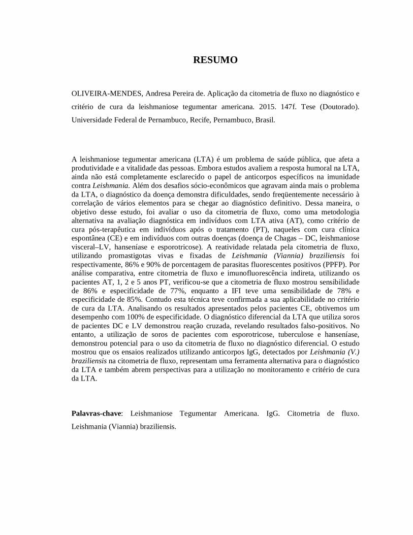
RESUMO
OLIVEIRA-MENDES, Andresa Pereira de. Aplicação da citometria de fluxo no diagnóstico e
critério de cura da leishmaniose tegumentar americana. 2015. 147f. Tese (Doutorado).
Universidade Federal de Pernambuco, Recife, Pernambuco, Brasil.
A leishmaniose tegumentar americana (LTA) é um problema de saúde pública, que afeta a produtividade e a vitalidade das pessoas. Embora estudos avaliem a resposta humoral na LTA, ainda não está completamente esclarecido o papel de anticorpos específicos na imunidade contra Leishmania. Além dos desafios sócio-econômicos que agravam ainda mais o problema da LTA, o diagnóstico da doença demonstra dificuldades, sendo freqüentemente necessário à correlação de vários elementos para se chegar ao diagnóstico definitivo. Dessa maneira, o objetivo desse estudo, foi avaliar o uso da citometria de fluxo, como uma metodologia alternativa na avaliação diagnóstica em indivíduos com LTA ativa (AT), como critério de cura pós-terapêutica em indivíduos após o tratamento (PT), naqueles com cura clínica espontânea (CE) e em indivíduos com outras doenças (doença de Chagas – DC, leishmaniose visceral–LV, hanseníase e esporotricose). A reatividade relatada pela citometria de fluxo, utilizando promastigotas vivas e fixadas de Leishmania (Viannia) braziliensis foi respectivamente, 86% e 90% de porcentagem de parasitas fluorescentes positivos (PPFP). Por análise comparativa, entre citometria de fluxo e imunofluorescência indireta, utilizando os pacientes AT, 1, 2 e 5 anos PT, verificou-se que a citometria de fluxo mostrou sensibilidade de 86% e especificidade de 77%, enquanto a IFI teve uma sensibilidade de 78% e especificidade de 85%. Contudo esta técnica teve confirmada a sua aplicabilidade no critério de cura da LTA. Analisando os resultados apresentados pelos pacientes CE, obtivemos um desempenho com 100% de especificidade. O diagnóstico diferencial da LTA que utiliza soros de pacientes DC e LV demonstrou reação cruzada, revelando resultados falso-positivos. No entanto, a utilização de soros de pacientes com esporotricose, tuberculose e hanseníase, demonstrou potencial para o uso da citometria de fluxo no diagnóstico diferencial. O estudo mostrou que os ensaios realizados utilizando anticorpos IgG, detectados por Leishmania (V.) braziliensis na citometria de fluxo, representam uma ferramenta alternativa para o diagnóstico da LTA e também abrem perspectivas para a utilização no monitoramento e critério de cura da LTA.
Palavras-chave: Leishmaniose Tegumentar Americana. IgG. Citometria de fluxo.
Leishmania (Viannia) braziliensis.

ABSTRACT
OLIVEIRA-MENDES, Andresa Pereira de. Aplication of flow cytometry on the diagnosis
and cure criterion of Americano f American Tegumentary Leishmaniasis. 2015. 147f. Thesis
(Doctorate). Universidade Federal de Pernambuco, Recife, Pernambuco, Brazil.
American tegumentar leishmaniasis (ATL) is a public health problem, affecting the productivity and vitality. Although there are studies to assess the humoral response in the ATL, is not yet fully understood the role of specific antibodies in immunity against Leishmania. In addition to the socioeconomic challenges that further aggravate the problem of ATL, the diagnosis shows difficulties and is often necessary to the correlation of various elements to reach a definitive diagnosis. Thus, the aim of this study was to evaluate the use of flow cytometry as an alternative methodology in the diagnostic evaluation in patients with active ATL before treatment (BT), as post-therapy cure criteria in individuals after treatment (AT), those with spontaneous clinical cure (EC) and in individuals with other diseases (Chagas disease - CD, visceral leishmaniasis-LV, leprosy and sporotrichosis). The reactivity reported by flow cytometry, using live and fixed promastigotes of Leishmania (V.) braziliensis were respectively 86% and 90% percentage of positive fluorescent parasites (PPFP). A comparative analysis of flow cytometry and indirect immunofluorescence using patient AT, 1, PT 2 and 5 years, it has been found that the flow cytometry showed a sensitivity of 86% and specificity of 77% and had a sensitivity IIF of 78% and specificity of 85%. Though this technique has confirmed its applicability in the healing criterion of ATL, analyzing the results presented by the EC patients achieved a performance with 100% specificity. The differential diagnosis of ATL using sera from patients of CD and LV demonstrated cross-reactivity, revealing false-positive results. Moreover, the use of sera from patients with sporotrichosis, tuberculosis and leprosy showed potential for the use of flow cytometry in the differential diagnosis. The study showed that testing performed using IgG antibodies, detected by Leishmania (V.) braziliensis in flow cytometry, are an alternative tool for the diagnosis of ATL and also open up prospects for use in monitoring and in its cure criterion.
Keywords: American Tegumentary Leishmaniasis. IgG. Flow Cytometry. Leishmania (Viannia) braziliensis.

LISTA DE ILUSTRAÇÕES Figura 1 Distribuição mundial da leishmaniose tegumentar.........................................
20
Figura 2 Formas da Leishmania : A- promastigotas, B- amastigotas dentro de monócito.........................................................................................................
21
Figura 3 Ciclo de vida da Leishmania sp. LC - leishmaniose cutânea.........................
22
Figura 4 Taxonomia da Leishmania.............................................................................
25
Figura 5 Formas clínicas da leishmaniose tegumentar americana. A - leishmaniose cutânea localizada; B - leishmaniose cutânea disseminada; C - Leishmaniose cutânea difusa; D - leishmaniose mucocutânea...................................................................................................
26
Figura 6 Cicatrizes deixadas pelas lesões da leishmaniose tegumentar americana. A- lesão em processo de cicatrização, B e C- lesões completamente reepitelizadas. ................................................................................................
30
Figura 7 Organograma com diagnóstico da LTA.........................................................
37
Figura 8 Fluxograma de coleta de amostras..................................................................
42
Figura 9 Seleção da população de formas promastigotas de Leishmania (Viannia) braziliensis, utilizando-se os parâmetros de tamanho e granulosidade (A). Histogramas individuais representando o percentual de parasitos fluoresecentes positivos (PPFP) obtidos com controle interno da reação (B), após a incubação com um soro de um indivíduo não infectado (C) e um soro de um paciente portador de LTA (D)...................................................................................................................
47
Figura 10 Anti-live FC-ALPA-IgG (A) and anti-fixed FC-AFPA-IgG (B) L. (V.) braziliensis IgG reactivity in serum samples from patients with Localized Cutaneous Leishmaniasis (LCL= ) and Non-Infected individuals (NI= ). (Artigo 1)………………………………………
54
Figura 11 Anti-live FC-ALPA-IgG (A) and anti-fixed FC-AFPA-IgG (B) L. (V.) braziliensis IgG reactivity in serum samples from patients with Localized Cutaneous Leishmaniasis (LCL= ) and Non-Infected individuals (NI= ). (Artigo 1)……………………………………….………………………
55
Figura 12 Comparative reactivity of anti-live FC-ALPA-IgG (A) and anti-fixed FC-AFPA-IgG (B) L. (V.) braziliensis IgG reactivity in serum samples from patients with Localized Cutaneous Leishmaniasis before treatment (BT= ) and after treatment (AT= ). (Artigo 1)……………………….…
55
Figura 13 Differential Anti-live FC-ALPA-IgG (A) and anti-fixed FC-AFPA-IgG (B) L. (V.) braziliensis IgG reactivity in serum samples from patients with Localized Cutaneous Leishmaniasis before treatment (BT= ) and after treatment (AT= ). (Artigo 1)……………………………………...……….
56
Figura 14 Differential anti-L. (V.) braziliensis IgG reactivity of paired samples detected by FC-ALPA-IgG (A) and FC-AFPA-IgG (B). (Artigo 1)………..
57
Figura 15 Differential anti-L. (V.) braziliensis IgG reactivities of paired samples detected by FC-ALPA-IgG (A) and FC-AFPA-IgG (B) defined as differential percentage of fluorescent positive parasites (ΔPPFP) for pairs of samples from LCL patients evaluated before and after treatment. (Artigo

1)………………………………………..
58
Figura 16 IgG reactivity in sera of ATL patients before and 1, 2 and 5 years after treatment, subjected to indirect immunofluorescence method. (Artigo 2)………………………………………………...…………………………..
76
Figura 17 Titration curves of antibodies IgG anti-fixed promastigotes of L. (V.) braziliensis present in sera of ATL patients classified as the presence of injury (BT) and the absence of injury (1, 2 and 5 years AT). (Artigo 2)…………………………………………………………………………..
76
Figura 18 Evaluation of the flow cytometry assay applicability on identifying patients with active ATL from the sera of patients from the region title with 256,512, 1024 and 2048 reactivity between BT and 1, 2 and 5 years AT. (Artigo 2)…………………………..…………………………………...
78
Figura 19 Comparison between ROC curves of IFA (--) and flow cytometry in serum dilutions of 1:256, (--), BT with 1, 2 and 5 years AT, built from the performance, sensitivity and 100 - specificity of the evaluated tests. (Artigo 2)……………………………………………………………………
80
Figura 20 Titration curve average of IgG antibodies anti-fixed Leishmania braziliensis promastigotes from American tegumentary leishmaniasis patients (ATL ● n=15), non-infected individuals (NI ○n=8) and spontaneously cured patients (CUR ● n=8) serum samples (A). The IgG antibodies anti-fixed Leishmania braziliensis of individual serum samples from ATL, NI and CUR groups at 1/1,024 dilution (B). (Artigo 3).............
94
Figura 21 The IgG antibodies anti-fixed Leishmania braziliensis of individual serum samples from ATL, NI + CUR groups at 1/1,024 dilution (A). ROC curve analyses confirmed the previously elected cut-off, and demonstrated outstanding performance indexes (Sensitivity-Se; Specificity-Sp; Area under the curve-AUC (B,C); and Positive/Negative Likelihood Ratio-LR+/LR- for ATL (D). (Artigo 3).................................................................
95
Figura 22 Titration curve average of IgG antibodies anti-fixed Leishmania braziliensis promastigotes from American tegumentary leishmaniasis patients (ATL ● n=15) and Non-ATL individuals (NI + CUR + VL + CH + HN + SPOR + TB ● n=37) serum samples (A). The IgG antibodies anti-fixed Leishmania braziliensis of individual serum samples from ATL and Non-ATL groups at 1/1,024 dilution (B). ROC curve analyses confirmed the previously elected cut-off, and demonstrated performance indexes (Sensitivity-Se; Specificity-Sp; Area under the curve-AUC (C). (Artigo 3).
96 Figura 23 Titration curve average of IgG antibodies anti-fixed Leishmania braziliensis
promastigotes from Visceral Leishmaniasis patients (VL ■ n=04); Chagas disease patients (CH ♦ n=10); Hanseniasis patients (HN ● n=05), Sporotrichosis patient (SPOR ▲n=01) and Tuberculosis patient (TB ▼n=01) serum samples (A). The IgG antibodies anti-fixed Leishmania braziliensis of individual serum samples from ATL, NI, CUR, VL, CH, HN, SPOR and TB groups at 1/1,024 dilution (B). (Artigo 3)……
98

LISTA DE TABELAS
Tabela 1 Casos confirmados notificados no sistema de informação de agravos de notificação – Sinan Net, no período de 2007 a 2013, no Brasil.........
21
Tabela 2 Vantagens e desvantagens dos métodos utilizados para o diagnóstico
laboratorial da LTA.
40
Tabela 3 Categories for the diagnosis test results cjassification of the patients. (Artigo 2)................................................................................................
74
Tabela 4 Characteristics of patients and laboratory results (Artigo 2)..................
74
LISTA DE ABREVIAÇÕES E SIGLAS
BCG Bacillus Calmette-Guérin
CE Cura Espontânea
CF Citometria de Fluco
DC Doença de Chagas
ELISA Ensaio imunoenzimático
CF- AAPF -IgG Anticorpos IgG de anti- promastigotas fixadas
CF- AAPV -IgG Anticorpos IgG de anti- promastigotas vivas
FITC Isotiocianato de fluoresceína (Fluorescein isothiocyanate)
FSC Detector de Dispersão Frontal (Forward Scatter)
HIV Vírus da Imunodeficiência Humana
IDRM Intradermorreação de Montenegro
IFI Imunofluorescência Indireta
IFN-γ Interferon-gama
Ig Imunoglobulina
IL Interleucina
LC Leishmaniose Cutânea
LD Leishmaniose Difusa
LM Leishmaniose Mucocutânea
LTA Leishmaniose Tegumentar Americana
LVA Leishmaniose Visceral Americana
L. (V.) Leishmania (Viannia)
NNN Meio de cultura Novy MCNeal Nicole
PBS Solução tampão fosfato (Phosphate buffer solution)
PCR Reação em cadeia da polimerase(Polymerase chain reaction)
PPFP Porcentagem de parasitos fluorescentes positivos
ROC Característica Operativa do Receptor (Receiver Operating
Characteristic)
SFB Soro fetal bovino
SSC Detector de Dispersão Lateral (Side Scatter)
TGF-β Fator de crescimento tumoral (Tumor growth factor)
TNF-α fator de necrose tumoral - alfa
SUMÁRIO RESUMO
ABSTRACT
LISTA DE ILUSTRAÇÕES
LISTA DE TABELAS
LISTA DE ABREVIAÇÕES E SIGLAS
1 - INTRODUÇÃO................................................................................................. 15
2 - OBJETIVOS...................................................................................................... 18
2.1- Geral................................................................................................................. 18
2.2- Específicos........................................................................................................ 18
3- REVISÃO DA LITERATURA ........................................................................ 19
3.1 -Aspectos Gerais da Leishmaniose Tegumentar Americana........................ 19
3.2 -Manifestações Clínicas da Leishmaniose Tegumentar Americana............ 23
3.3- Tratamento e Controle da Leishmaniose Tegumentar Americana............ 26
3.4- Critério de Cura da Leishmaniose Tegumentar Americana...................... 30
3.5 -Resposta Imune na Leishmaniose Tegumentar Americana........................ 32
3.6 -Diagnóstico Clínico da Leishmaniose Tegumentar Americana.................. 35
3.7 – Diagnóstico Laboratorial da Leishmaniose Tegumentar Americana...... 36
3.7.1- Citometria de fluxo no diagnóstico da Leishmaniose Tegumentar
Americana................................................................................................................
39
4- MATERIAL E MÉTODOS............................................................................... 42
4.1- Tipo de Estudo................................................................................................. 42
4.2- Critérios de Inclusão....................................................................................... 42
4.3- Critérios de Exclusão...................................................................................... 43
4.4- População de estudo........................................................................................ 43
4.5- Grupo Controle............................................................................................... 44
4.6- Exames Laboratoriais de Avaliação dos Pacientes...................................... 44
4.7- Obtenção de Soro ........................................................................................... 44
4.8- Reação de Imunofluorescência Indireta....................................................... 44
4.9-Obtenção das Formas Promastigotas Vivas e Fixadas de L. (V.)
braziliensis...............................................................................................................
45
4.10- Citometria de Fluxo...................................................................................... 46
4.11- Análise Estatística......................................................................................... 47

5- RESULTADOS E DISCUSSÃO....................................................................... 48
5.1- Artigo 1: publicado na revista Diagnostic Microbiology and Infectious
Disease, V.74 (3): p 292-298..................................................................................
48
5.2- Artigo 2: publicado na revista Journal of Immunological Methods, V.387 (1-2): p 245-253……………………………………………………………
67
5.3- Artigo 3: Manuscrito submetido ao periódico: Diagnostic microbiology
and infectious diseases ...........................................................................................
88
6- CONCLUSÕES.................................................................................................. 106
7- PERSPECTIVAS............................................................................................... 107
REFERÊNCIAS BIBLIOGRÁFICAS
APÊNDICE A
TERMO DE CONSENTIMENTO LIVRE E ESCLARECIDO – Grupo
Paciente
APÊNDICE B
TERMO DE CONSENTIMENTO LIVRE E ESCLARECIDO – Grupo
Paciente menor de 18 anos
APÊNDICE C
TERMO DE CONSENTIMENTO LIVRE E ESCLARECIDO – Grupo
Controle
ANEXO 1
PARECER DO COMITÊ DE ÉTICA EM PESQUISA
ANEXO 2
CAPITULO DE LIVRO- Immunity against Leishmaniasis

Oliveira-Mendes, A.P. Aplicação da citometria de fluxo no diagnóstico e critério de cura... 15
1 INTRODUÇÃO___________________________________________________________
A leishmaniose tegumentar americana (LTA) é um problema de saúde pública, que
afeta a produtividade e a vitalidade dos indivíduos. Essa doença infecciosa apresenta
distribuição mundial e acomete, principalmente, aqueles com menor status socioeconômico de
países menos desenvolvidos. Nas Américas, a LTA está distribuída do sul dos Estados Unidos
ao Norte da Argentina, com exceção do Chile e Uruguai. Devido a sua ampla distribuição,
registrando casos em todas as regiões brasileiras, é uma das afecções dermatológicas de maior
magnitude no Brasil. Em Pernambuco, é uma doença endêmica e 60% dos casos concentram-
se na Zona da Mata. Suas perspectivas de controle são ainda muito dependentes do progresso
nas pesquisas científicas, visando obter estratégias mais efetivas para o monitoramento de
casos e do vetor (BRANDÃO-FILHO et al., 2003; DESJEUX, 2004; BRITO et al., 2009;
2012; MS, 2010; OMS, 2010).
Os protozoários causadores da LTA são organismos dimórficos, caracterizados por
apresentarem durante seu ciclo de vida duas formas evolutivas. A forma promastigota ou
flagelada infectante, encontrada na luz do tubo digestivo dos insetos vetores, os flebotomíneos
fêmeas, que transmitem a infecção aos hospedeiros vertebrados. Já a forma amastigota, é
aflagelada, arredondada e imóvel, se multiplica obrigatoriamente dentro de células do sistema
monocítico fagocitário desses hospedeiros vertebrados, estando envolvidas na evolução ou
cura da doença (CROFT; COOMBS, 2003; MS 2010; TIUMAN et al., 2011).
As manifestações clínicas da LTA variam desde as lesões cutâneas localizadas, a
forma mais comumente encontrada, até graves lesões mucocutâneas desfigurantes, devido a
destruição das regiões mucosas. Até então, quatorze espécies de Leishmania foram descritas e
estão divididas entre os subgêneros Leishmania e Viannia, causando uma variedade de lesões
no homem. A Leishmania (Viannia) braziliensis (L. (V.) braziliensis), principal espécie
envolvida na transmissão da LTA até o momento em Pernambuco, é considerada uma das
espécies mais importantes para a saúde pública, não só por ser responsável pela maioria dos
casos de LTA no país, como também pela sua capacidade de causar as infecções
dermatológicas descritas acima (BRANDÃO-FILHO et al., 2003; SILVEIRA; LAINSON;
CORBETT, 2004; BRITO et al., 2009; 2012; GOTO; LINDOSO, 2010).
O tratamento da LTA apresenta limitações, as drogas disponíveis são tóxicas e
muitas vezes causam reações adversas graves e, além disso, não existe um critério de cura
efetivo. O critério de cura é baseado na cura clínica pela reepitelização completa das lesões

Oliveira-Mendes, A.P. Aplicação da citometria de fluxo no diagnóstico e critério de cura... 16
ulceradas ou não, regressão total do infiltrado e eritema (GONTIJO; MAYRINK et al., 2006;
MS, 2010; OMS, 2010). Desta forma, torna-se relevante a busca de um critério de cura pós-
tratamento específico para LTA, (SCHUBACH et al., 1998; MENDONÇA et al., 2004).
Embora estudos mostrem que as células T e a imunidade mediada por células
contribuem para a patogênese das diferentes manifestações clínicas da LTA, ainda não está
completamente esclarecido o papel de anticorpos específicos na imunidade contra Leishmania
(TRUJILLO et al., 1999; SOUZA et al., 2005).
Segundo Rodriguez et al. (1996), há correlação das subclasses de IgG com as
manifestações clínicas da LTA. Assim, níveis elevados de anticorpos dos isotipos IgG1, IgG2
e IgG3 e baixos níveis ou ausência de anticorpos do isotipo IgG4 podem ser detectados no
soro de pacientes com LC (leishmaniose cutânea). No soro de pacientes com LM
(leishmaniose mucocutânea) tem-se altos níveis de IgG1, enquanto os níveis de IgG2, IgG3 e
IgG4 são comparados aos encontrados no soro de pacientes com LC. Já nos pacientes com
LD, os níveis de IgG4 são bastante elevados, enquanto os níveis de IgG1 e IgG2 são
semelhantes aos encontrados no soro de pacientes com LC e LM (RODRIGUEZ et al., 1996;
REIS et al., 2006). Em 2001, Brito e colaboradores observaram níveis aumentados de IgG em
pacientes curados espontaneamente, sugerindo a importância do achado para a dinâmica da
resposta de anticorpos e evolução da LTA.
Além dos desafios sócio-econômicos que agravam o problema da LTA, o
diagnóstico da doença demonstra dificuldades, pois o mesmo é realizado por associações
clínicas, epidemiológicas e laboratoriais (KAR, 1995; BRITO et al., 2000; 2008, TAVARES
et al., 2003; MS, 2010; ELMAHALLAWY et al., 2014). Os testes sorológicos convencionais
mais utilizados no diagnóstico laboratorial da LTA são a reação de imunofluorescência
indireta (IFI) e o ensaio imunoenzimático (enzyme linked immunosorbent assay - ELISA).
Entretanto, esses métodos apresentam limitações, pois não correlacionam os níveis de
anticorpos circulantes com o estágio da doença e podem apresentar reações cruzadas com
outros tripanosomatídeos (KAR, 1995; VEXENAT et al., 1996; BRITO et al., 2001; OMS,
2010).
Um dos grandes desafios enfrentados pelos pesquisadores tem sido a escolha da
preparação antigênica ideal para análise sorológica, tanto no que se refere ao estudo dos
mecanismos moduladores/indutores de doença, bem como para o diagnóstico, prognóstico e
critérios de monitoração da infecção (BRITO et al., 2000; 2001; GONÇALVES et al., 2002;
ROCHA et al.,2002; CELESTE et al., 2004; ROCHA et al., 2006). Pelas limitações dessas
técnicas, abordagens imunológicas alternativas vêm sendo empregadas. Uma delas é a
Oliveira-Mendes, A.P. Aplicação da citometria de fluxo no diagnóstico e critério de cura... 17
citometria de fluxo (CF), tecnologia usada na análise quantitativa de anticorpos e vem sendo
utilizada no diagnóstico sorológico da LTA (ROCHA et al., 2002; 2006). Em paralelo, alguns
estudos avaliam diminuição dos níveis de anticorpos em pacientes após o tratamento com
antimoniais e a possibilidade de utilizar testes sorológicos, como a citometria de fluxo, não
apenas para o diagnóstico, mas como critério de cura da LTA (AMATO et al., 1998; BRITO
et al., 2001).
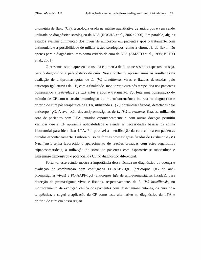
O presente estudo apresenta o uso da citometria de fluxo nesses dois aspectos, ou seja,
para o diagnóstico e para critério de cura. Nesse contexto, apresentamos os resultados da
avaliação de antipromastigotas de L. (V.) braziliensis vivas e fixadas detectadas pelo
anticorpo IgG através da CF, com a finalidade monitorar a cura pós terapêutica nos pacientes
comparando a reatividade de IgG antes a após o tratamento. Foi feita uma comparação do
método de CF com o ensaio imunológico de imunofluorencência indireta no diagnóstico e
critério de cura pós terapêutica da LTA, utilizando L. (V.) braziliensis fixadas, detectadas pelo
anticorpo IgG. A avaliação das antipromastigotas de L. (V.) braziliensis fixadas, utilizando
soro de pacientes com LTA, curados espontaneamente e com outras doenças permitiu
verificar que a CF apresenta aplicabilidade e atende as necessidades básicas da rotina
laboratorial para identificar LTA. Foi possível a identificação da cura clínica em pacientes
curados espontaneamente. Embora o uso de formas promastigotas fixadas de Leishmania (V.)
braziliensis tenha favorecido o aparecimento de reações cruzadas com estes organismos
tripanosomatídeos, a utilização de soros de pacientes com esporotricose tuberculose e
hanseníase demonstrou o potencial da CF no diagnóstico diferencial.
Portanto, esse estudo mostra a importância dessa técnica no diagnóstico da doença e
avaliação da combinação com conjugados FC-AAPV-IgG (anticorpos IgG de anti-
promastigotas vivas) e FC-AAPF-IgG (anticorpos IgG de anti-promastigotas fixadas), para
detecção de promastigotas vivos e fixados, respectivamente, de L. (V.) braziliensis, no
monitoramento da evolução clínica dos pacientes com leishmaniose cutânea, da cura pós-
terapêutica, e sugeri a aplicação da CF como teste alternativo no diagnóstico da LTA e
critério de cura em nossa região.

Oliveira-Mendes, A.P. Aplicação da citometria de fluxo no diagnóstico e critério de cura... 18
2 OBJETIVOS_________________________________________________________
2.1- Geral
Avaliar o uso da citometria de fluxo como uma técnica alternativa na avaliação
diagnóstica e como critério de cura de portadores de leishmaniose tegumentar americana em
Pernambuco.
2.2- Específicos
2.2.1- Comparar o uso de promastigotas Leishmania (Viannia) braziliensis vivas e
fixadas na detecção do anticorpo IgG através da citometria de fluxo
2.2.2- Avaliar a reatividade da Leishmania (Viannia) braziliensis viva e fixada no
monitoramento da cura clínica dos pacientes antes e 1, 2 e 5 anos após o tratamento
quimioterápico
2.2.3- Comparar as técnicas de citometria de fluxo, na reação de imunofluorescência
indireta e na identificação dos pacientes antes e 1, 2 e 5 anos após o tratamento
quimioterápico.
2.2.4- Avaliar a aplicabilidade da citometria de fluxo como critério de cura pós-
terapêutica.
2.2.5- Analisar o desempenho da citometria de fluxo na identificação de pacientes
com cura espontânea.
2.2.6- Verificar a sensibilidade e especificidade da citometria de fluxo, comparando-
se o soro dos pacientes com LTA com o soro de pacientes portadores de outras doenças.

Oliveira-Mendes, A.P. Aplicação da citometria de fluxo no diagnóstico e critério de cura... 19
3 REVISÃO DA LITERATURA______________________________________________
3.1- Aspectos Gerais da Leishmaniose Tegumentar Americana
A LTA é uma doença infecciosa negligenciada e que acomete principalmente aqueles
com menor status socioeconômico e com limitada capacidade de assumir os custos da doença
(diagnóstico, hospitalização, tratamento) e suas consequências, que envolvem desde lesões
simples até deformidades marcantes (OMS, 2010; MS, 2010; CASTRO, 2013). A maior parte
das pessoas afetadas vive em aldeias pobres em áreas rurais, e estão permanentemente
expostas aos fatores de risco da doença, normalmente por razões ocupacionais (OMS, 2010;
MS 2010; BRITO et al 2012). Dessa maneira, a LTA é uma doença que merece maior atenção
devido à sua magnitude, alta morbidade e possibilidade de assumir formas que podem
determinar lesões destrutivas, desfigurantes e também incapacitantes, com grande repercussão
no campo psicossocial do indivíduo (GONTIJO; CARVALHO, 2003; ANDRADE et al.,
2005; NEITZKE-ABREU et al., 2014).
Atualmente, a LTA ocorre de forma endêmica em 82 países e estima-se a incidência
de 1,5 milhão de novos casos notificados por ano (figura 1). Cerca de 90% desses casos
ocorrem no Marrocos, na Etiópia, Tunísia, Afeganistão, Paquistão, Irã, Iraque, Arábia
Saudita, Síria, Brasil, Bolívia, Colômbia, Equador, Peru e Venezuela. Nas Américas, a LTA
ocorre desde o Sul dos Estados Unidos até o norte da Argentina, com exceção do Uruguai e
do Chile (GONTIJO; CARVALHO, 2003; GOTO; LINDOSO, 2010; OMS, 2010). Em
decorrência do seu caráter epidêmico e alto coeficiente de detecção, a LTA é considerada uma
das dez endemias mundiais prioritárias. Ocupando o segundo lugar entre as seis doenças
infecto-parasitárias mais frequentes do mundo, caracteriza-se ela como problema de saúde
pública, tanto pela sua expansão geográfica quanto pela tendência de urbanização (GAZOZAI
et al., 2010; DOENÇAS NEGLIGENCIADAS, 2010).
No Brasil, a LTA apresenta-se distribuída por todos os Estados, avança na Região
Centro-Oeste e já se encontra na periferia das grandes cidades do Nordeste e do Centro-Oeste.
No ano de 2013, foram notificados 19. 652 novos casos da doença, no Sistema de Informação
de Agravos de Notificações (SINAN Net), em todo o país (tabela 1). Com expressivo número
de casos, a LTA também é caracterizada por apresentar uma importante difusão espacial.
Nesse contexto, a LTA apresenta importante heterogeneidade, relacionada às diferentes
espécies de Leishmania envolvidas em sua etiologia, às formas clínicas associadas e ao

Oliveira-Mendes, A.P. Aplicação da citometria de fluxo no diagnóstico e critério de cura... 20
padrão de transmissão envolvido (LAINSON; SHAW, 1998; BRANDÃO-FILHO, 1999; MS,
2010; DATASUS, 2014; MENEZES-SOUZA et al., 2014).
Figura 1: Distribuição mundial da leishmaniose tegumentar. Fonte: OMS, 2014.
A LTA apresenta sua incidência acentuada em Pernambuco, por todas as regiões do
Estado, principalmente na Zona da Mata, onde estão concentrados cerca de 60% dos casos
registrados (BRANDÃO-FILHO et al., 1999; BRITO et al., 2008). É importante também
registrar o aumento de sua ocorrência no Sertão do Estado (ANDRADE et al., 2009). No
biênio 2008-2009 foram confirmados 889 casos novos de LTA em Pernambuco, distribuídos
em aproximadamente 34% dos municípios. Para demonstrar o aumento e a força da infecção
nesta região, foram realizados inquéritos epidemiológicos, caracterizando a distribuição da
infecção e da doença na população. Os estudos mostraram que, em Pernambuco, a LTA
ocorre em ambos os sexos e em todas as faixas etárias. O aumento da incidência e a
persistência da transmissão neste Estado revela um importante problema de saúde pública
devido à expansão espacial (BRANDÃO-FILHO et al., 1999; 2003; BRITO et al., 2008;
ANDRADE et al., 2012).

Oliveira-Mendes, A.P. Aplicação da citometria de fluxo no diagnóstico e critério de cura... 21
Tabela 1: Casos confirmados notificados no sistema de informação de agravos de notificação – Sinan Net, no período de 2007 a 2013, no Brasil.
Ano de diagnóstico Casos confirmados
2013 19.652*
2012 25.647
2011 23.054
2010 23.493
2009 23.318
2008 21.581
2007 22.556
Fonte: DATASUS, 2015. * Dados atualizados até 24/09/2014.
O agente etiológico da LTA é um protozoário intracelular, que apresenta DNA
extranuclear, cinetoplasto e uma organela mitocondrial, e que pertence à ordem
Kinetoplastida, família Trypanosomatidae, gênero Leishmania, e divide-se em dois
subgêneros, Leishmania e Viannia. Durante o seu ciclo de vida, esse parasito apresenta-se
sob duas formas evolutivas (figura 2), a primeira forma é a promastigota, forma alongada e
com o flagelo livre, encontrada no trato digestivo dos hospedeiros invertebrados, os
flebotomíneos. A outra forma, a amastigota, forma arredondada ou ovalada sem flagelo, é
encontrada no interior de monócitos, histiócitos e macrófagos, que são células do sistema
fagocítico mononuclear do hospedeiro mamífero (VEGA-LOPEZ, 2003; BASANO;
CAMARGO, 2004; MS, 2010).
Figura 2: Formas da Leishmania : A- promastigotas, B- amastigotas dentro de monócito. Fonte: CITADIN, 2008.
A B

Oliveira-Mendes, A.P. Aplicação da citometria de fluxo no diagnóstico e critério de cura... 22
O ciclo de vida da Leishmania (figura 3) tem início, em figura 3A, quando a fêmea de
um inseto vetor, o flebotomíneo, se alimenta de um hospedeiro vertebrado contaminado, pois
as formas amastigotas são ingeridas durante o repasto sanguíneo. No intestino do
flebotomíneo-fêmea (figura 3B), as formas amastigotas sofrem divisões binárias e se
transformam em formas flageladas, as promastigotas. Essas formas migram para a faringe e o
esôfago do inseto, onde maturam para formas promastigotas metacíclicas infectantes. Quando
o flebotomíneo-fêmea realiza um novo repasto sanguíneo em um hospedeiro vertebrado
(figura 3C), libera as formas promastigotas. Em seguida, as formas promastigotas
metacíclicas são fagocitadas pelos macrófagos e transformam-se em amastigotas (figura 3D).
As formas amastigotas se multiplicam por divisão binária, até o rompimento da membrana
celular do macrófago, que as libera para serem fagocitadas por outros macrófagos (figura 3E).
O ciclo se reinicia (figura 3A) quando uma fêmea do vetor, ao realizar um repasto sanguíneo,
ingere o sangue e, com ele, macrófagos parasitados (DESJEUX, 2004; REITHINGER et al.,
2007; NEVES et al., 2009; OMS, 2014; PACE, 2014).
Figura 3: Ciclo de vida da Leishmania sp. LC - leishmaniose cutânea. Fonte: Adaptado de OMS, 2014.
A epidemiologia da LTA é extremamente diversificada, os ciclos de transmissão
implicam um grande número de parasitos, reservatórios e vetores envolvidos. Nas Américas,
são atualmente reconhecidas 11 espécies dermotrópicas de Leishmania causadoras da doença

Oliveira-Mendes, A.P. Aplicação da citometria de fluxo no diagnóstico e critério de cura... 23
humana, e oito espécies descritas somente em animais (figura 4). No entanto, no Brasil, já
foram identificadas sete espécies envolvidas na etiologia da LTA, sendo seis do subgênero
Viannia e uma do subgênero Leishmania. As três principais espécies são: L. (V.) braziliensis,
L. (V.) guyanensis e L. (L.) amazonensis e, mais recentemente, as espécies L. (V.) lainsoni, L.
(V.) naiffi, L. (V.) lindenberg e L. (V.) shawi que foram identificadas em estados das Regiões
Norte e Nordeste (DESJEUX, 2004; BRITO et al., 2009; OMS, 2010). A distribuição da
infecção e da doença na população foi bem caracterizada na Zona da Mata de Pernambuco,
em inquéritos epidemiológicos realizados entre 1991 e 1996. E, desde então, amostras
isoladas de pacientes dessa região apresentaram perfis isoenzimáticos de variantes para a
principal espécie circulante, que é a L. (V.) braziliensis (BRITO et al., 1993; BRANDÃO-
FILHO et al., 1999; MARTINS et al., 2010; MENEZES-SOUZA et al., 2014).
3.2 - Manifestações Clínicas da Leishmaniose Tegumentar Americana
A LTA apresenta manifestações clínicas diversas, que variam de acordo com a espécie
envolvida na transmissão, com a susceptibilidade da população e com seu nível de exposição.
Fatores relacionados aos indivíduos também influenciam na apresentação da doença, e nesses
podemos incluir o status imunológico do hospedeiro, relacionado às interações do seu sistema
imune inato e adaptativo, assim como ao estado nutricional e aos fatores genéticos do mesmo
(REITHINGER et al., 2007; CASTELLANO et al., 2009; BRITO et al., 2012; BRELAZ et
al., 2012). Embora a apresentação clínica da LTA esteja frequentemente relacionada a uma
determinada espécie ou subgênero de Leishmania, nenhuma se restringe à mesma. Ademais,
uma significativa, porém variável, proporção de infecções é assintomática, ao passo que,
sendo sintomáticas, as lesões cutâneas apresentam-se nas formas localizada, disseminada,
difusa, e mucocutânea, a mais agressiva (figura 5) (DESJEUX, 2004; AMEEN, 2010; GOTO;
LINDOSO, 2010).
Podendo evoluir para a cura espontânea, a forma cutânea localizada (LC) é a
manifestação mais recorrente da doença, em tal caso, não é menos perigosa. As lesões são
únicas ou múltiplas e assumem a forma típica com bordas elevadas, sendo a úlcera indolor,
arredondada ou oval (REITHINGER et al., 2007; GOTO; LINDOSO, 2010). A densidade de
parasitos nas bordas da lesão é grande nas fases iniciais da doença, no entanto, diminuem nas
lesões crônicas. Todas as espécies dermotrópicas de Leishmania são capazes de desencadear a
LC (figura 5A), todavia, no Brasil, o principal agente etiológico é a L. (V.) braziliensis, que

Oliveira-Mendes, A.P. Aplicação da citometria de fluxo no diagnóstico e critério de cura... 24
tem a disseminação por via hematogênica como a principal complicação. Além disso, ela
pode evoluir para a leishmaniose mucocutânea (MS, 2010; GOTO; LINDOSO, 2010;
MANSUETO et al., 2014).
Com numerosas lesões nodulares ou ulceradas, a forma disseminada (figura 5B) tem
sido descrita em associação com L. (V.) braziliensis, L. (V.) panamensis, L. (V.) guyanensis e
infecções L. (L.) amazonensis. A apresentação dessa forma varia de 10 a 300 lesões, em partes
do corpo contínuas ou não. Além disso, podem ocorrer com ou sem envolvimento das
mucosas. Essa forma difere da forma anérgica cutânea difusa (figura 5C), onde lesões
múltiplas, nodulares, não ulceradas são encontradas por todo o corpo. A leishmaniose cutânea
difusa, no Brasil, tem sido associada com a L. (L.) amazonensis. O desenvolvimento dessas
lesões está relacionado à capacidade particular que a L. amazonensis tem de interferir
negativamente em vários mecanismos imunológicos, necessários à geração de uma resposta
imune efetiva, dessa maneira, levando à proliferação dos parasitos e à ocorrência de
metástases do parasito de um sítio para o outro, através dos vasos linfáticos ou da migração de
macrófagos parasitados, tornando a leishmaniose difusa uma doença rara (REITHINGER et
al., 2007; GOTO; LINDOSO, 2010; OMS, 2010; AMEEN, 2010; MANSUETO et al., 2014).
Comumente conhecida como “úlcera de bauru” ou “espúndia”, a forma grave
mucocutânea (figura 5D) da leishmaniose ocorre como consequência do desenvolvimento
secundário da forma cutânea localizada, e é caracterizada pela destruição da cavidade oral-
nasal, faringe e laringe, em virtude de uma reação imunológica exagerada. O agente
etiológico causador da leishmaniose mucocutânea (LM), em nosso país, é a L (V.)
braziliensis; entretanto, já foram citados casos na literatura atribuídos à L (L.) amazonensis e à
L (V.) guyanensis. A LM, normalmente, surge da evolução crônica da LC, de casos com cura
espontânea sem tratamento ou de casos em que o tratamento foi inadequado, desenvolvendo
lesões secundárias destrutivas em mucosas e cartilagens. O processo infeccioso começa
indolor e com infiltrado discreto no septo nasal, que evolui para a formação de crostas,
epistaxe, disfagia, odinofagia, rouquidão e tosse, dificuldades respiratórias, além de
dificuldade na fala e na alimentação do doente. A LM pode também atingir as conjuntivas
oculares, as mucosas dos órgãos genitais e do ânus. Na fase final, ocorre mutilação grave,
com obstrução e destruição do nariz, faringe e laringe. A LM praticamente nunca cicatriza
espontaneamente, e as infecções secundárias são frequentes, sendo a pneumonia intercorrente
a causa mais comum de morte (NEVES et al., 2009; AMEEN, 2010; GOTO; LINDOSO,
2010; OMS, 2010; NUNES et al., 2011; MANSUETO et al., 2014; MIGNOGNA et al.,
2014).

Oliveira-Mendes, A.P. Aplicação da citometria de fluxo no diagnóstico e critério de cura... 25

Oliveira-Mendes, A.P. Aplicação da citometria de fluxo no diagnóstico e critério de cura... 26
Figura 5: Formas clínicas da leishmaniose tegumentar americana. A - leishmaniose cutânea localizada; B - leishmaniose cutânea disseminada; C - Leishmaniose cutânea difusa; D - leishmaniose mucocutânea. Fonte: A e B, Ministério da Saúde, 2007; C e D, Almeida, 2013.
3.3 - Tratamento e Controle da Leishmaniose Tegumentar Americana
O tratamento tem como propósito acelerar a cura, reduzir o risco de formação de
cicatrizes, prevenir a progressão da doença, e a sua escolha, advém do tamanho e localização
da lesão, do número e do potencial de disseminação das lesões. Em todas as formas clínicas
da LTA, as drogas de primeira escolha são os antimoniais pentavalentes (Sb+5), cujos
esquemas terapêuticos são padronizados pela Organização Mundial de Saúde. As doses são
calculadas em mg/Sb+5/kg/dia e há dois tipos de antimoniais pentavalentes utilizados: o
antimoniato de N-metilglucamina (Glucantime®) e o estibogluconato de sódio (Pentostam®).
O primeiro é distribuído pelo programa de controle de LTA do Ministério da Saúde do Brasil
e é indicado como primeira escolha para o tratamento de todas as formas de LTA, exceto para
pacientes coinfectados com HIV e para gestantes. Já o segundo, estibogluconato de sódio, não
é comercializado no Brasil (GONTIJO; CARVALHO et al., 2003; MS, 2010; AMEEN,
2010; OLIVEIRA et al., 2011).
Oliveira-Mendes, A.P. Aplicação da citometria de fluxo no diagnóstico e critério de cura... 27
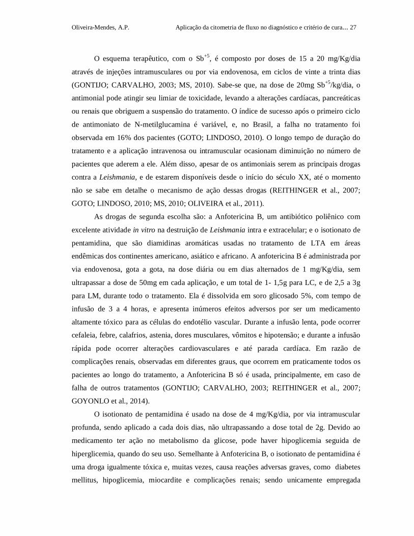
O esquema terapêutico, com o Sb+5, é composto por doses de 15 a 20 mg/Kg/dia
através de injeções intramusculares ou por via endovenosa, em ciclos de vinte a trinta dias
(GONTIJO; CARVALHO, 2003; MS, 2010). Sabe-se que, na dose de 20mg Sb+5/kg/dia, o
antimonial pode atingir seu limiar de toxicidade, levando a alterações cardíacas, pancreáticas
ou renais que obriguem a suspensão do tratamento. O índice de sucesso após o primeiro ciclo
de antimoniato de N-metilglucamina é variável, e, no Brasil, a falha no tratamento foi
observada em 16% dos pacientes (GOTO; LINDOSO, 2010). O longo tempo de duração do
tratamento e a aplicação intravenosa ou intramuscular ocasionam diminuição no número de
pacientes que aderem a ele. Além disso, apesar de os antimoniais serem as principais drogas
contra a Leishmania, e de estarem disponíveis desde o início do século XX, até o momento
não se sabe em detalhe o mecanismo de ação dessas drogas (REITHINGER et al., 2007;
GOTO; LINDOSO, 2010; MS, 2010; OLIVEIRA et al., 2011).
As drogas de segunda escolha são: a Anfotericina B, um antibiótico poliênico com
excelente atividade in vitro na destruição de Leishmania intra e extracelular; e o isotionato de
pentamidina, que são diamidinas aromáticas usadas no tratamento de LTA em áreas
endêmicas dos continentes americano, asiático e africano. A anfotericina B é administrada por
via endovenosa, gota a gota, na dose diária ou em dias alternados de 1 mg/Kg/dia, sem
ultrapassar a dose de 50mg em cada aplicação, e um total de 1- 1,5g para LC, e de 2,5 a 3g
para LM, durante todo o tratamento. Ela é dissolvida em soro glicosado 5%, com tempo de
infusão de 3 a 4 horas, e apresenta inúmeros efeitos adversos por ser um medicamento
altamente tóxico para as células do endotélio vascular. Durante a infusão lenta, pode ocorrer
cefaleia, febre, calafrios, astenia, dores musculares, vômitos e hipotensão; e durante a infusão
rápida pode ocorrer alterações cardiovasculares e até parada cardíaca. Em razão de
complicações renais, observadas em diferentes graus, que ocorrem em praticamente todos os
pacientes ao longo do tratamento, a Anfotericina B só é usada, principalmente, em caso de
falha de outros tratamentos (GONTIJO; CARVALHO, 2003; REITHINGER et al., 2007;
GOYONLO et al., 2014).
O isotionato de pentamidina é usado na dose de 4 mg/Kg/dia, por via intramuscular
profunda, sendo aplicado a cada dois dias, não ultrapassando a dose total de 2g. Devido ao
medicamento ter ação no metabolismo da glicose, pode haver hipoglicemia seguida de
hiperglicemia, quando do seu uso. Semelhante à Anfotericina B, o isotionato de pentamidina é
uma droga igualmente tóxica e, muitas vezes, causa reações adversas graves, como diabetes
mellitus, hipoglicemia, miocardite e complicações renais; sendo unicamente empregada
Oliveira-Mendes, A.P. Aplicação da citometria de fluxo no diagnóstico e critério de cura... 28
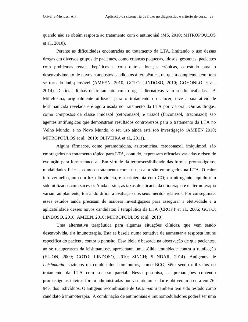
quando não se obtém resposta ao tratamento com o antimonial (MS, 2010; MITROPOULOS
et al., 2010).
Perante as dificuldades encontradas no tratamento da LTA, limitando o uso dessas
drogas em diversos grupos de pacientes, como crianças pequenas, idosos, gestantes, pacientes
com problemas renais, hepáticos e com outras doenças crônicas, o estudo para o
desenvolvimento de novos compostos candidatos à terapêutica, ou que a complementem, tem
se tornado indispensável (AMEEN, 2010; GOTO; LINDOSO, 2010; GOYONLO et al.,
2014). Distintas linhas de tratamento com drogas alternativas vêm sendo avaliadas. A
Miltefosina, originalmente utilizada para o tratamento do câncer, teve a sua atividade
leishmanicida revelada e é agora usada no tratamento da LTA por via oral. Outras drogas,
como compostos da classe imidazol (cetoconazol) e triazol (fluconazol, itraconazol) são
agentes antifúngicos que demonstram resultados controversos para o tratamento da LTA no
Velho Mundo; e no Novo Mundo, o seu uso ainda está sob investigação (AMEEN 2010;
MITROPOULOS et al., 2010; OLIVEIRA et al., 2011).
Alguns fármacos, como paramomicina, azitromicina, cetoconazol, imiquimod, são
empregados no tratamento tópico para LTA, contudo, expressam eficácias variadas e risco de
evolução para forma mucosa. Em virtude da termosensibilidade das formas promastigotas,
modalidades físicas, como o tratamento com frio e calor são empregados na LTA. O calor
infravermelho, ou com luz ultravioleta, e a crioterapia com CO2 ou nitrogênio líquido têm
sido utilizados com sucesso. Ainda assim, as taxas de eficácia da crioterapia e da termoterapia
variam amplamente, tornando difícil a avaliação dos seus méritos relativos. Por conseguinte,
esses estudos ainda precisam de maiores investigações para assegurar a efetividade e a
aplicabilidade desses novos candidatos à terapêutica da LTA (CROFT et al., 2006; GOTO;
LINDOSO, 2010; AMEEN, 2010; MITROPOULOS et al., 2010).
Uma alternativa terapêutica para algumas situações clínicas, que vem sendo
desenvolvida, é a imunoterapia. Esta se baseia numa tentativa de aumentar a resposta imune
específica do paciente contra o parasito. Essa ideia é baseada na observação de que pacientes,
ao se recuperarem da leishmaniose, apresentam uma sólida imunidade contra a reinfecção
(EL-ON, 2009; GOTO; LINDOSO, 2010; SINGH; SUNDAR, 2014). Antígenos de
Leishmania, sozinhos ou combinados com outros, como BCG, vêm sendo utilizados no
tratamento da LTA com sucesso parcial. Nessa pesquisa, as preparações contendo
promastigotas inteiras foram administradas por via intramuscular e obtiveram a cura em 76-
94% dos indivíduos. O antígeno recombinante de Leishmania também tem sido testado como
candidato à imunoterapia. A combinação de antimoniais e imunomoduladores poderá ser uma
Oliveira-Mendes, A.P. Aplicação da citometria de fluxo no diagnóstico e critério de cura... 29
alternativa de tratamento para pacientes refratários ao tratamento com o antimonial. Embora
essa modalidade de tratamento seja crescente, ensaios clínicos são ainda necessários para
demonstrar o seu benefício na rotina clínica (CONVIT et al., 2003; 2004; EL-ON, 2009;
GOTO; LINDOSO, 2010; AMEEN, 2010).
Em virtude da diversidade de agentes etiológicos, de reservatórios, de vetores e da
situação epidemiológica da LTA, várias abordagens são utilizadas para o controle dessa
endemia. Dentre essas, ressalta-se a vigilância epidemiológica com identificação precoce dos
casos e tratamento; medidas de atuação na cadeia de transmissão, impedindo a infecção de
vetores, hospedeiros e reservatórios; medidas educativas, incluindo a utilização de
mosquiteiros, telas finas nas janelas e portas, repelentes e roupas que protejam áreas expostas.
Outras abordagens incluem medidas administrativas, como saneamento (evitando o acúmulo
de lixo e de detritos que possam atrair roedores e pequenos mamíferos, que podem servir
como reservatório). Melhorias das condições de habitação e capacitação de profissionais de
saúde também são essenciais. O desenvolvimento de uma vacina eficiente e operacional seria
uma medida definitiva e eficaz em termos de custos e de prevenção (BASANO; CAMARGO,
2004; DESJEUX, 2004; OMS, 2010).
A LTA é uma doença complexa e heterogênea, que está se tornando cada vez mais
prevalente, tanto nos países onde ocorre de forma endêmica, quanto nos que ocorre de forma
não endêmica. Além disso, um número crescente de casos é visto em viajantes. Por
conseguinte, a corrente situação exige novas opções de tratamento e controle, incluindo a
imunoterapia e o progresso contínuo no desenvolvimento de vacinas. (NOAZIN et al., 2008;
OKWOR; UZONNA, 2009; AMEEN, 2010; MANSUETO et al., 2014). A tentativa de
vacinação contra a leishmaniose cutânea remonta a centenas de anos, e é alvo de experiências
na América Latina desde o início do século vinte. As duas principais tentativas de vacinas
avaliadas no Brasil foram a pentavalente de Mayrink e colaboradores, conhecida como
Leishvacin®, e a vacina monovalente simplificada de L. amazonensis. Os estudos foram
inconclusivos pela ausência de um grupo placebo e a eficácia da vacina não pôde ser avaliada
(MAYRINK et al., 2002; NOAZIN et al., 2008; GOTO; LINDOSO, 2010).
A presença de uma resposta imune induzida no indivíduo pela infecção confirma a
viabilidade da prevenção da LTA através da vacinação profilática (MUTISO et al., 2013).
Embora não haja nenhuma vacina licenciada contra qualquer forma de leishmaniose humana
para uso geral, tem havido numerosas tentativas para desenvolver uma vacina eficaz contra a
leishmaniose e há várias categorias de vacinas candidatas. Dessa maneira, as que estão em
desenvolvimento estão divididas em: vacina Leishmania viva, a Leishmanization; vacinas de
Oliveira-Mendes, A.P. Aplicação da citometria de fluxo no diagnóstico e critério de cura... 30
primeira geração que consistem de Leishmania inteira morta ou frações do parasito; as
vacinas de segunda geração, que incluem vacinas de proteínas recombinantes, vacinas de
DNA e as suas combinações; e as vacinas de Leishmania vivas atenuadas (HANDMAN,
2001; NOAZIN et al., 2008; OKWOR; UZONNA, 2009; COSTA et al., 2011; MUTISO et
al., 2013). Os mecanismos imunológicos da LTA humana ainda não são totalmente
compreendidos e as respostas observadas na proteção por vacinação em modelos
experimentais de infecção podem não refletir as respostas necessárias para a sua eficácia em
áreas endêmicas (HANDMAN, 2001; NOAZIN et al., 2008; MUTISO et al., 2013). Existem
ainda muitos desafios para que vacinas para prevenção da LTA se tornem uma realidade.
3.4 - Critério de Cura da Leishmaniose Tegumentar Americana
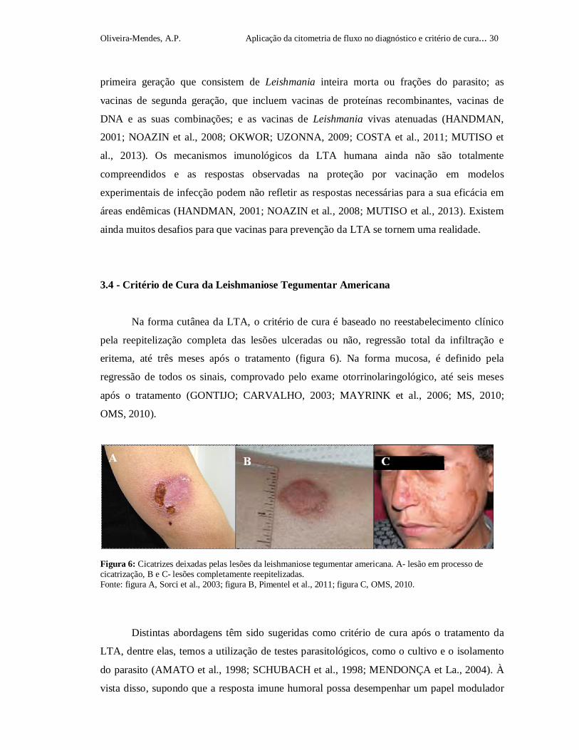
Na forma cutânea da LTA, o critério de cura é baseado no reestabelecimento clínico
pela reepitelização completa das lesões ulceradas ou não, regressão total da infiltração e
eritema, até três meses após o tratamento (figura 6). Na forma mucosa, é definido pela
regressão de todos os sinais, comprovado pelo exame otorrinolaringológico, até seis meses
após o tratamento (GONTIJO; CARVALHO, 2003; MAYRINK et al., 2006; MS, 2010;
OMS, 2010).
Figura 6: Cicatrizes deixadas pelas lesões da leishmaniose tegumentar americana. A- lesão em processo de cicatrização, B e C- lesões completamente reepitelizadas. Fonte: figura A, Sorci et al., 2003; figura B, Pimentel et al., 2011; figura C, OMS, 2010.
Distintas abordagens têm sido sugeridas como critério de cura após o tratamento da
LTA, dentre elas, temos a utilização de testes parasitológicos, como o cultivo e o isolamento
do parasito (AMATO et al., 1998; SCHUBACH et al., 1998; MENDONÇA et La., 2004). À
vista disso, supondo que a resposta imune humoral possa desempenhar um papel modulador

Oliveira-Mendes, A.P. Aplicação da citometria de fluxo no diagnóstico e critério de cura... 31
da resposta celular durante a infecção, Bitterncourt et al (1968) sugeriram a possibilidade da
pesquisa de anticorpos no monitoramento de cura da LTA. Os autores estudaram a reatividade
de anticorpos anti-Leishmania através da sorologia convencional pela IFI e sugeriram como
critério de cura pós-terapêutica na LTA.
Ao avaliar a possibilidade de utilizar a IFI como critério de cura da LTA, estudos
apontaram a diminuição dos níveis de anticorpos em pacientes após o tratamento com
antimoniais, propondo a existência de correlação entre a cura clínica das lesões e a
negativação dos títulos da IFI (AMATO et al., 1998; SCHUBACH et al., 1998). Em estudos
análogos, a atenção tem se voltado para a perspectiva de utilização do teste de ELISA, na
avaliação do valor preditivo da persistência de anticorpos IgG, durante o desenvolvimento da
LTA (MENDONÇA et al., 2004; ROMERO et al., 2005). A demonstração da persistência
parasitária após a cura clínica da LTA desperta para o desenvolvimento de pesquisas voltadas
para a evolução clínica, epidemiológica e para o controle da LTA. Contudo, a utilização
desses testes para avaliação do critério de cura da LTA trata-se ainda de um assunto
controverso, uma vez que ocorre a demonstração da persistência de positividade nos ensaios
após cura, espontânea ou pós-terapêutica (MARTINS-FILHO et al., 1995; ROCHA et al.,
2002; 2006; OLIVEIRA et al., 2013).
Até então, a dinâmica da produção de anticorpos após o tratamento com antimoniais
não é conhecida o bastante, e valores preditivos de baixos ou altos níveis de anticorpos
dirigidos contra antígenos específicos continuam indefinidos (OLIVEIRA et al., 2013;
PEREIRA et al., 2012; ROMERO et al., 2005). A dificuldade pode ser atribuída à falta de
uma preparação antigênica padrão, para ser usada na detecção de anticorpos específicos. Com
isso em mente, Rocha et al. (2002), utilizando a citometria de fluxo, perceberam suas
aplicabilidades para o diagnóstico. A técnica surge como uma nova abordagem, para ser
empregada na pesquisa de anticorpos, dado que possui um sistema isento de variabilidades
metodológicas inerentes ao analista, e tem eficácia superior aos diferentes protocolos de
detecção convencionais. Ademais, é cabível ao diagnóstico e critério de cura pós-terapêutica
de diferentes infecções, dentre elas a LTA.

Oliveira-Mendes, A.P. Aplicação da citometria de fluxo no diagnóstico e critério de cura... 32
3.5- Resposta Imune na Leishmaniose Tegumentar Americana
A resposta imunológica desempenha um importante papel na cura clínica da doença ou
na sua progressão. Embora altos títulos de anticorpos possam ser encontrados em todas as
manifestações clínicas da LTA, essa patogênese, parece depender da resposta imune mediada
por células no hospedeiro, influenciando o desenvolvimento da doença, seja pelo tipo de
linfócitos T efetores envolvidos, ou pelo perfil de citocinas secretadas (MOUGNEAU; BIHL;
GLAICHENHAUS, 2011; DA-CRUZ et al., 2005).
Enquanto os primeiros estudos demonstraram um papel crítico das células T CD4+ na
imunidade dirigida à Leishmania, trabalhos recentes têm proporcionado uma nova visão sobre
o papel das células da imunidade inata, tais como neutrófilos, monócitos, NK e células
dendríticas (MOUGNEAU; BIHL; GLAICHENHAUS, 2011). Os neutrófilos são uma das
primeiras células a chegar ao local da infecção, sendo infectados pelas leishmanias. Eles
também podem ser considerados como “cavalos de troia", já que facilitam a infecção
silenciosa dos macrófagos ao fagocitarem neutrófilos contaminados (NYLEEN; GAUTAM,
2010; NYLÉN; EIDSMO, 2012; SANTOS; BRODSKYN, 2014). Junto com fagócitos, as
células NK, células exterminadoras naturais, representam a primeira linha de defesa contra os
patógenos, através da produção de citocinas pró-inflamatórias TNF e IFN- γ. As células
dendríticas são um importante componente de ligação da resposta imune inata e adaptativa,
através do reconhecimento da infecção parasitária e da apresentação celular dos parasitos e/ou
de seus antígenos (NYLEEN; GAUTAM, 2010; SOUZA et al., 2013; SANTOS;
BRODSKYN, 2014). Além desses mecanismos de resposta imune inata, tem-se também o
sistema complemento, que desempenha papel pró-inflamatório, sendo ativado pela via
clássica e alternativa na leishmaniose (MOUGNEAU; BIHL; GLAICHENHAUS, 2011).
A maior parte dos conhecimentos de imunologia na LTA, sobretudo em relação à
interação parasito-hospedeiro, são provenientes dos estudos de infecções em modelos
animais, e o camundongo é o modelo extensivamente recorrido, na tentativa de se identificar e
dissecar a contribuição de genes modificadores da doença para a patogênese (MOUGNEAU;
BIHL; GLAICHENHAUS, 2011; LORÍA-CERVERA; ANDRADE-NARVÁEZ 2014).
Embora, o camundongo apresente a doença semelhantemente aos humanos, existem
limitações, como a variação genética generalizada presente na população humana que não se
limita à variação genética nos camundongos puros. Além de que, não existe um modelo para
LM. Ademais, a via de inoculação é diferente em condições experimentais, que é por via
subcutânea ou intravenosa e que não pode ser comparada à picada do flebotomíneo vetor, na
Oliveira-Mendes, A.P. Aplicação da citometria de fluxo no diagnóstico e critério de cura... 33
qual ocorre a inoculação de grande número de parasitos e existe a presença da saliva ativando
o sistema imune (SAKTHIANANDESWAREN; FOOTE; HANDMAN 2009; MOUGNEAU;
BIHL; GLAICHENHAUS, 2011). Em estudos realizados em camundongos, a resposta imune
mediada por células T na infecção por L (V.) braziliensis é pouco caracterizada. Isso acontece
em razão da resistência natural de muitas linhagens de camundongos frente à infecção, sendo
necessária uma grande carga parasitária para desencadear a mesma. Consequentemente,
devido à dificuldade em estabelecer modelos animais adequados para se estudar a L. (V.)
braziliensis, surgiram os estudos com pacientes (LIMA et al, 1999; MOURA et al, 2005;
REIS et al, 2006).
Na leishmaniose humana e experimental, e em todas as formas clínicas da doença, a
imunidade é predominantemente mediada por linfócitos T, de maneira que, a resposta
linfocitária na LTA é caracterizada principalmente pelo aumento de células T CD4+. Há mais
de 20 anos, se aceita que a diferença entre a resistência e a susceptibilidade à infecção está
associada ao nível de expansão de células T CD4+, com direcionamento para um perfil de
citocinas Th1 e Th2, respectivamente (MOUGNEAU; BIHL; GLAICHENHAUS, 2011;
SOUZA et al., 2013). A interleucina-12 (IL-12), produzida por macrófagos e células
dendríticas, o interferon-gama (IFN-γ) produzido pelas células NK, e as células T
previamente ativadas promovem o desenvolvimento de células Th1, enquanto que a IL-4
induz o desenvolvimento de células Th2. Linfócitos T CD4+ que apresentam o perfil Th1
produzem IFN-γ e fator de necrose tumoral - alfa (TNF-α), assim como também expressam o
fator de transcrição T-bet. Por outro lado, as células que apresentam o perfil Th2 produzem
IL-4, IL-5, IL-10, IL-13, e expressam o fator de transcrição GATA-3 (SAKAGUCHI et al.,
2008; REIS et al., 2006; SOUZA et al., 2013).
Outros subtipos de células T CD4+, como células Treg e células Th17, também
parecem apresentar papel importante na susceptibilidade e resistência à LTA. As células T
regulatórias (Treg), que se acumulam no local da lesão, estão relacionadas à regulação das
células efetoras locais, e podem ser naturais ou induzidas. Citocinas, como IL- 10 e TGF-β, e
substâncias como o ácido retinoico facilitam a diferenciação de células T naïve em Treg
induzidas que expressam o fator de transcrição Foxp3 (NYLÉN; EIDSMO, 2012; SANTOS;
BRODSKYN, 2014). Estudos recentes têm evidenciado também o subtipo celular, conhecido
como Th17, relacionado com a produção de IL-17. A princípio, a IL-17 está envolvida com a
patogênese de doenças inflamatórias crônicas ou autoimunes (BACELLAR et al., 2009;
NYLEEN; GAUTAM, 2010; NYLÉN; EIDSMO, 2012; SANTOS; BRODSKYN, 2014).

Oliveira-Mendes, A.P. Aplicação da citometria de fluxo no diagnóstico e critério de cura... 34
Embora estudos avaliem a resposta humoral na LTA, ainda não está completamente
esclarecido o papel de anticorpos específicos na imunidade contra Leishmania (TRUJILLO et
al., 1999; SOUZA et al., 2005). Na última década, estudos apresentados, principalmente em
modelos experimentais, apontam que as células B, e seus anticorpos contribuem para a
susceptibilidade e doença na LTA. Entretanto, os anticorpos, em geral, não têm sido
considerados como fator importante na resistência à doença (SACKS et al., 1984; SACKS;
NOBEN-TRAUTH, 2002; KEDZIERSKI; EVANS, 2014). Sacks et al., (1984), constataram
que a depleção de células B, observando anticorpos anti-IgM, resultou em melhora na
resistência para Leishmania em camundongos BALB/c. Além disso, o estudo também implica
que, durante a infecção por Leishmania, em camundongos com depleção de células B, células
T supressoras não são produzidas. A importância das células B para o desenvolvimento e
susceptibilidade de respostas de células Th2 foi observada a partir da capacidade dessas
células em apresentar antígenos a células T. Além disso, tem sido demonstrado que a IL-10
produzida por células B pode desempenhar um papel na susceptibilidade à infecção cutânea
por inibição da produção de IL-12 por células dendríticas in vitro (RONET et al., 2008; 2010;
KEDZIERSKI; EVANS, 2014).
Alguns tipos celulares, como macrófagos, possuem receptores de superfície para a
porção Fc dos anticorpos IgG, os receptores Fc. A associação dos receptores Fc-IgG com
macrófagos induz a produção de IL-10 e impulsiona o desenvolvimento alternativo de
macrófagos ativados com fraca capacidade leishmanicida (NYLÉN; EIDSMO, 2012). Diante
disso, é de conhecimento que a classe IgG não só oferece proteção contra o parasito, como
também contribui para a progressão da infecção (SOUZA et al., 2013).
Há um consenso geral de que as células T e a imunidade mediada por células
contribuem para a patogênese das diferentes formas de LTA. Isso acontece devido à
influência do padrão de citocinas na ativação, proliferação e diferenciação de linfócitos B em
células produtoras de imunoglobulinas (TRUJILLO et al., 1999; SOUZA et al., 2005;
SOUZA et al., 2013). Estudos realizados em humanos, sugerem que citocinas do perfil Th1,
produtoras de IFN-γ, induzem a produção da imunoglobulina G1 (IgG1) e IgG3 em humanos
(OZBILGE et al., 2006). Quando associado a IL-6, o IFN- γ induz a produção de IgG2 e
antagoniza a produção de IgG1 (KAWANO et al., 1994). É notório, que a produção de IgG3,
IgG4 e IgE está associada à IL-4, e sua inibição está associada ao IFN-γ, enquanto o aumento
dos níveis de IgG1 e IgG3 está associado ao IL-10 (BUXBAUM, 2008; SKEIKY et al.,1997).
No entanto, os níveis de IgA dependem de TGF-β (STAVNEZER; KANG, 2009).

Oliveira-Mendes, A.P. Aplicação da citometria de fluxo no diagnóstico e critério de cura... 35
Nas leishmanioses cutânea (LC) e mucocutânea (LM), a imunidade celular e a
predominância de isotipos IgG1, IgG2 e IgG3 têm sido associadas à resposta do tipo Th1; já o
perfil Th2 tem sido relacionado à leishmaniose cutânea difusa (LD), com presença de IgG4
(SOUZA et al., 2005), direcionando a atenção para a correlação das subclasses de IgG com as
manifestações clínicas da LTA. Estudos demonstram que todos os isotipos específicos anti-
Leishmania, exceto IgD, são detectados no soro de pacientes com LTA. A frequência de
detecção das subclasses de IgG na LTA, de acordo com Pissinate et al., (2008), é IgG1 >
IgG3 > IgG2 = IgG4. Os pacientes com um maior tempo de evolução da doença apresentam
altos níveis de IgE, e nos pacientes com a forma mucocutânea, os níveis de IgA se mostram
aumentados (O’NEIL et al., 1993). A intensidade da resposta humoral parece estar
relacionada com a carga parasitária e a cronicidade da infecção, e podem ser observados altos
títulos de anticorpos em todas as manifestações clínicas da LTA (TRUJILLO et al., 1999;
OZBILGE et al., 2006).
A análise da resposta imune humoral na LTA tem abordado o papel das
imunoglobulinas em mecanismos imunopatológicos envolvidos na resistência e/ou patogênese
da infecção; além disso, também avaliam o uso em investigações sorológicas no diagnóstico e
no monitoramento da eficácia pós-tratamento (REIS et al., 2006; PEREIRA et al., 2012;
OLIVEIRA et al., 2013). A associação entre o perfil da resposta imune humoral e a
patogênese de um dado processo infeccioso tem sido feita em várias doenças, tais como
hanseníase, AIDS, filariose e malária (KURNIAWAN et al. 1993, HUSSAIN et al. 1995,
OUAAZ et al. 1996, RODRIGUEZ, 1996; PERLMANN et al. 1997). No entanto,
recentemente, tem-se observado que os métodos imunossorológicos disponíveis para pesquisa
de anticorpos na LTA apresentam resultados controversos, devido a sua baixa sensibilidade e
especificidade (RIBEIRO et al., 2007; BOURDOISEAU et al., 2009; MUKBEL et al., 2006).
Diante disso, estudos têm mostrado a vantagem do uso de anticorpos específicos no
diagnóstico da leishmaniose visceral (SOUZA et al., 2013; PASSOS et al., 2005; OZBILGE
et al., 2006; CHATTERJEE et al., 1998).
3.6 - Diagnóstico Clínico da Leishmaniose Tegumentar Americana
A infecção humana por LTA pode ser assintomática ou apresentar um espectro de
formas clínicas que podem variar, aparecendo desde lesões cutâneas localizadas,
disseminadas, difusas ou nodulares, até as graves lesões mucocutâneas (Figura 4)

Oliveira-Mendes, A.P. Aplicação da citometria de fluxo no diagnóstico e critério de cura... 36
(GRIMALDI; TESH, 1993). As lesões cutâneas podem ainda apresentar-se morfologicamente
como: impetigoide, liquenoide, tuberculosa ou lupoide, nodular, vegetante e ectimatoide, o
que dificulta ainda mais o diagnóstico clínico. Nas lesões mucocutâneas, podem ser
observadas úlceras infiltrantes ou úlceras vegetantes (ANDRADE et. al, 2005). As lesões
podem ser confundidas tanto com doenças infecciosas, comoparacoccidioidomicose,
histoplasmose e sífilis; quanto com doenças neoplásicas, como carcinoma e linfoma (ÁVILA
et. al, 2004).
O diagnóstico clínico da LTA pode ser feito com base nas características da lesão
associadas à anamnese, onde os dados epidemiológicos são de grande importância. São
frequentes as ulcerações com bordas elevadas em moldura, fundo com tecido granuloso e
indolores. A apresentação clínica exibe polimorfismo e o espectro de gravidade dos sinais e
sintomas é variável, o que torna o diagnóstico clínico difícil (GONTIJO; CARVALHO, 2003;
MS, 2007). Devido à dificuldade encontrada para o diagnóstico clínico seguro, os métodos
laboratoriais fazem parte do arsenal de investigação.
3.7 – Diagnóstico Laboratorial da Leishmaniose Tegumentar Americana
As diferentes espécies de Leishmania envolvidas, as formas clínicas e o tempo de
evolução das lesões levam à variabilidade das manifestações da infecção e direcionam o
diagnóstico para os métodos laboratoriais. A associação das evidências clínicas,
epidemiológicas e laboratoriais é necessária para se chegar ao diagnóstico final da LTA, pois
ainda não se dispõe de um método que possa ser usado como padrão-ouro para detecção e
diagnóstico de infecção por Leishmania (BRITO et al., 2000; REIS, 2007). Os exames
laboratoriais podem ser divididos em técnicas de detecção do parasito, técnica molecular e
técnicas de imunodiagnósticos (Figura 7).

Oliveira-Mendes, A.P. Aplicação da citometria de fluxo no diagnóstico e critério de cura... 37
Figura 7: Organograma com diagnóstico da LTA. Fonte: Da autora.
O diagnóstico de certeza de um processo infeccioso é feito pelo encontro do parasito,
através da pesquisa direta, isolamento em cultura, inoculação em animais experimentais
(MACHADO, 2004; MS, 2007). A pesquisa direta do parasito pode ser realizada em material
obtido da lesão por escarificação, aspiração, ou biópsia da borda da lesão. A visualização em
microscópio óptico da forma amastigota de Leishmania sp é possível após coloração pelo
método de Giemsa ou Leishman (GENARO, 2000). Essa técnica tem sensibilidade de 50 a
70% e depende do número de parasitos presentes na lâmina (GOTO; LINDOSO, 2010). A
positividade do teste é inversamente proporcional ao tempo de evolução da lesão cutânea,
sendo rara após um ano. Embora seja uma técnica simples, rápida e de baixo custo, não é
capaz de detectar parasitos em todos os pacientes (BENSOUSSAN et al., 2006; VEGA-
LÓPEZ, 2003).
O isolamento do parasito pode ser feito através da cultura em meios apropriados, como
o meio bifásico Novy e McNeal modificado por Nicolle-NNN (Novy & MacNeal 1903,
Nicolle 1908), Liver Infusion Tryptose- LIT (Camargo 1964) e/ou Schneiders, suplementado
com soro fetal bovino inativado, a partir de material obtido por punção aspirativa ou biópsia
das lesões dos pacientes. A sensibilidade do isolamento em cultura é geralmente baixa, em
torno de 20 a 40%. A baixa sensibilidade da cultura está relacionada, em muitos casos, à
escassez do parasito nas lesões, principalmente L. (V.) braziliensis (BRANDÃO-FILHO et al.,
1999; BENSOUSSAN et al., 2006; RODRÍGUEZ-GONZÁLEZ et al., 2006; REITHINGER

Oliveira-Mendes, A.P. Aplicação da citometria de fluxo no diagnóstico e critério de cura... 38
et al., 2007). A inoculação em animais susceptíveis para cultivo in vivo tem o hamster
(Mesocricetus auratus) como animal mais usado. Porém, o longo período de
acompanhamento até a formação da úlcera no animal e os elevados custos para manutenção
dos animais limitam o método às instituições de pesquisa científica; tornando-o de pouco
valor prático para a rotina laboratorial (RODRIGUES, 2000; BENSOUSSAN et al., 2006;
MS, 2007).
A Reação em Cadeia da Polimerase PCR convencional é uma técnica que permite a
amplificação de segmentos específicos de DNA a partir de oligonucleotídeos iniciadores
(primers) pareados especificamente nas margens da região alvo, permitindo o seu uso como
instrumento específico para o diagnóstico de diversas doenças infecciosas (GOMES et al.,
1999; RODRIGUES, 2000; 2002; SINGH, 1997). Embora várias abordagens moleculares
tenham sido desenvolvidas para o diagnóstico da leishmaniose e a PCR seja uma técnica
altamente sensível e específica, trata-se de um teste sofisticado para uso na rotina laboratorial,
com exigências técnicas e custo elevado, limitando-se a laboratórios de referência e/ou a
clínicas médicas (REITHINGER et al., 2007; SOUSA et al., 2013).
O diagnóstico imunológico baseia-se na avaliação da resposta imune celular utilizando
a técnica de Intradermorreação de Montenegro (IDRM) e/ou na avaliação da resposta imune
humoral utilizando as técnicas de imunofluorescência indireta (IFI), ensaio imunoenzomático
(ELISA), western blot e citometria de fluxo. A detecção da imunidade celular através da
IDRM tem sido empregada como o importante recurso no diagnóstico imunológico da LTA,
dado a sua grande sensibilidade e especificidade. O teste consiste na injeção de uma
suspensão de antígeno preparado com promastigotas para a indução de resposta de
hipersensibilidade tardia, onde a reação é lida após 48 a 72 horas (MACHADO, 2004; GOTO;
LINDOSO, 2010). Embora apresente resultado positivo na maioria dos casos de LTA (90%),
o resultado é negativo em lesões recentes, na forma cutânea difusa e em pacientes
imunodeprimidos. Em áreas endêmicas, é comum o teste ser positivo devido à ocorrência de
infecções subclínicas (VEGA-LÓPEZ, 2003; REIS, 2007). Além disso, o teste não diferencia
infecção de doença e nem doença ativa de pregressa (WEIGLE et al., 2002; REITHINGER et
al., 2007; GOTO; LINDOSO; 2010).
A avaliação da resposta imune humoral na LTA tem sido geralmente abordada através
de testes sorológicos, e os mais utilizados atualmente são a IFI, o ELISA e o Western blot.
Esses testes apresentam controvérsias no diagnóstico LTA, principalmente considerando a
baixa sensibilidade, especificidade e pouca reprodutibilidade. Além disso, podem apresentar
reações cruzadas com outros tripanosomatídeos. Sabe-se ainda que baixos níveis de
Oliveira-Mendes, A.P. Aplicação da citometria de fluxo no diagnóstico e critério de cura... 39
anticorpos são observados por essas técnicas, e que ocorre ausência de correlação entre níveis
de anticorpos circulantes com a presença de infecção ativa (KAR, 1995; PASSOS et al., 2000;
BRITO et al., 2000; 2001; SCHALLIG; OSKAM, 2002; SAVANI et al., 2003; ALVES;
BEVILACQUA, 2004).
A IFI é uma técnica amplamente utilizada no diagnóstico da LTA, na qual são
observadas, frequentemente, reações cruzadas com outros tripanosomatídeos, ocorrendo falso-
positivos com soros de pacientes portadores de doença de Chagas e leishmaniose visceral, por
exemplo (VEXENAT et al. 1996, BRITO 2008), bem como com soros de pacientes
portadores de hanseníase e de tuberculose (KAR, 1995). Além disso, ocorre grande
variabilidade de resultados em técnicas sorológicas que dependem da natureza do antígeno,
que pode estar associada aos diferentes antígenos utilizados e, principalmente, pela forma
como foram produzidos e purificados (TAVARES et al. 2003). O ELISA consiste em uma
metodologia de realização simples e rápida, bastante utilizada nas investigações científicas.
Embora apresente maior sensibilidade em relação à IFI, tem sido menos utilizado na rotina
laboratorial por apresentar custo mais elevado e exigências laboratoriais superiores à IFI
(MACHADO, 2004; ROMERO et al., 2005). O Western blot tem sido mais utilizado no
diagnóstico da leishmaniose visceral (KAR, 1995). Em estudo comparativo, Brito (1999)
demonstrou que a técnica de Western blot apresentou maior sensibilidade e especificidade em
relação às técnicas de IFI e ELISA.
3.7.1 – Citometria de Fluxo no diagnóstico da Leishmaniose Tegumentar Americana
Um aspecto limitante em relação ao diagnóstico da LTA no homem é a ausência de
um teste capaz de identificar com eficiência os casos com formas leves da doença (tabela 2).
Testes diagnósticos eficientes, capazes de proporcionar um diagnóstico precoce e preciso, são
essenciais para determinar a escolha do tratamento na vigilância epidemiológica de doenças
infecciosas; e as abordagens sorológicas para detectar anticorpos específicos contra um agente
infeccioso constituem-se em uma alternativa valiosa para um diagnóstico precoce. Diante
disso, vários esforços têm sido feitos objetivando o desenvolvimento de uma abordagem
sorológica confiável para o diagnóstico da LTA usando preparações de antígenos para
detectar anticorpos anti-Leishmania (SOUSA et al., 2013; KOTRESHA; NOORDIN, 2010;
PASSOS et al., 2005; GONÇALVES et al., 2002; RYAN et al., 2002; SILVEIRA et al.,
2001). Um método confiável para o diagnóstico baseado na citometria de fluxo para detecção
de anticorpos antiformas de L. (V.) braziliensis foi descrito por Rocha et al. (2002; 2006).

Oliveira-Mendes, A.P. Aplicação da citometria de fluxo no diagnóstico e critério de cura... 40
Tabela 2: Vantagens e desvantagens dos métodos utilizados para o diagnóstico laboratorial da LTA.
Oliveira-Mendes, A.P. Aplicação da citometria de fluxo no diagnóstico e critério de cura... 41
A citometria de fluxo tornou-se uma ferramenta importante no diagnóstico e
prognóstico de diferentes doenças, pois é um recurso que permite uma análise rápida, objetiva
e quantitativa de células em suspensão (FALDYNA et al., 2001;STETLER-STEVENSON,
2003; NAKAGE et al., 2005). As células da amostra em suspensão são marcadas com
anticorpos monoclonais específicos ligados a fluorocromos, que permitem a identificação e a
quantificação de células pelo tamanho, granulosidade e intensidade de fluorescência. Essa
técnica apresenta várias vantagens para os imunoensaios, tais como capacidade de produção
elevada, possibilidade de quantificação do analito, volume de amostra reduzida, elevada
reprodutibilidade e sensibilidade, uma ampla faixa dinâmica e, sobretudo, potencial para
multiplexagem (SOUSA et al., 2013; JANI et al., 2002).
Considerando as aplicabilidades da citometria de fluxo descritas por Martins- Filho et
al., (1995), o presente trabalho busca uma metodologia alternativa através da pesquisa de
anticorpos IgG de L. (V.) braziliensis. Dessa forma, acreditamos que a aplicação da citometria
de fluxo, na detecção de LTA em pacientes antes e após-tratamento, e também curados
espontaneamente, possa contribuir para a obtenção de um método de maior sensibilidade e
especificidade, com aplicação diagnóstica e como critério de cura na LTA.
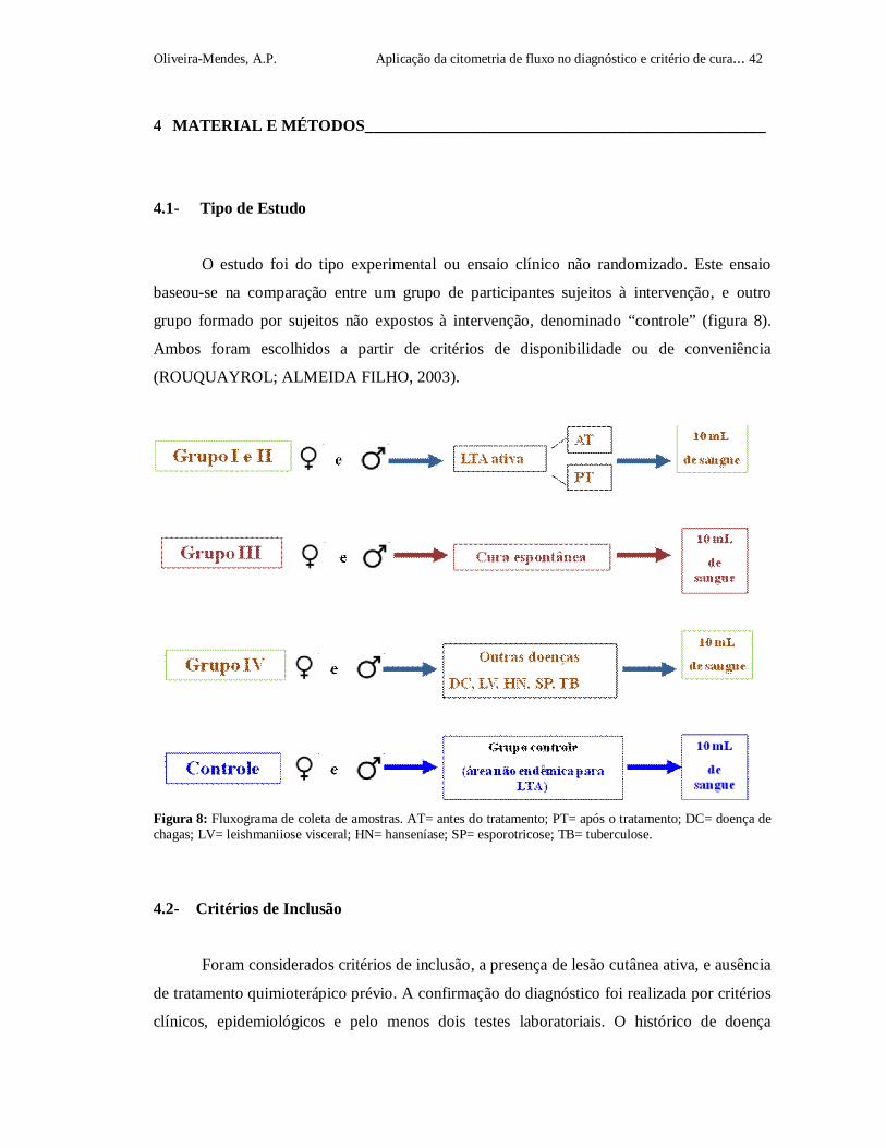
Oliveira-Mendes, A.P. Aplicação da citometria de fluxo no diagnóstico e critério de cura... 42
4 MATERIAL E MÉTODOS_________________________________________________
4.1- Tipo de Estudo
O estudo foi do tipo experimental ou ensaio clínico não randomizado. Este ensaio
baseou-se na comparação entre um grupo de participantes sujeitos à intervenção, e outro
grupo formado por sujeitos não expostos à intervenção, denominado “controle” (figura 8).
Ambos foram escolhidos a partir de critérios de disponibilidade ou de conveniência
(ROUQUAYROL; ALMEIDA FILHO, 2003).
Figura 8: Fluxograma de coleta de amostras. AT= antes do tratamento; PT= após o tratamento; DC= doença de chagas; LV= leishmaniiose visceral; HN= hanseníase; SP= esporotricose; TB= tuberculose.
4.2- Critérios de Inclusão
Foram considerados critérios de inclusão, a presença de lesão cutânea ativa, e ausência
de tratamento quimioterápico prévio. A confirmação do diagnóstico foi realizada por critérios
clínicos, epidemiológicos e pelo menos dois testes laboratoriais. O histórico de doença

Oliveira-Mendes, A.P. Aplicação da citometria de fluxo no diagnóstico e critério de cura... 43
pregressa e apresentação de cicatriz característica da lesão (aspecto de pergaminho), e
ausência de quimioterapia foram os critérios para inclusão no grupo de cura espontânea.
4.3- Critérios de Exclusão
Como critérios de exclusão foram considerados a ausência de cicatriz, tratamento
quimioterápico prévio para LTA, e presença de lesão de outras doenças dermatológicas.
4.4- População de estudo
Os pacientes do presente estudo foram procedentes de Moreno, e municípios vizinhos,
como Camaragibe (Região Metropolitana de Recife- RMR), Vitória de Santo Antão e
Vicência, áreas endêmicas de LTA em Pernambuco. Foram selecionados os pacientes
suspeitos que apresentavam lesão e história clínica compatível com a doença, os quais foram
encaminhados ao ambulatório de Dermatologia do Hospital Universitário de Pernambuco. Os
pacientes foram esclarecidos do objetivo do estudo e foram realizados os procedimentos de
biópsia, punção aspirativa, escarificação na borda da lesão. Foram feitas as coletas de 10 ml
de sangue antes e após tratamento quimioterápico com Glucantime® (antimoniato de N-
metilglucamina), e em pacientes que tiveram a cura espontânea e que não receberam
tratamento. O critério de cura clínica para os pacientes tratados ou curados espontaneamente
foi estabelecido considerando-se a completa cicatrização da lesão. O tratamento
quimioterápico foi realizado nos postos de saúde dos municípios deste estudo, utilizando-se o
Glucantime® (antimoniato de N-metilglucamina), administrado por via intramuscular. O
tratamento foi feito em ciclos de 20 dias em doses diárias de 15mg/Kg e acompanhado
durante três meses até a completa cicatrização. Foram formados quatro grupos de pacientes: i)
portadores de lesões ativas com faixa etária superior a 13 anos; ii) pacientes com cura clínica
após (um, dois e cinco anos) de tratamento; iii) pacientes com cura clínica espontânea, ou
seja, sem tratamento prévio com quimioterápico e iv) pacientes com outras doenças não
leishmanióticas (Doença de Chagas, leishmaniose visceral, esporotricose, tuberculose e
hanseníase). O material coletado foi processado no Laboratório de Imunogenética do
Departamento de Imunologia do Centro de Pesquisas Aggeu Magalhães-FIOCRUZ, na cidade
de Recife-PE. Os protocolos experimentais foram submetidos e aprovados pelo Comitê de

Oliveira-Mendes, A.P. Aplicação da citometria de fluxo no diagnóstico e critério de cura... 44
Ética do CPqAM/FIOCRUZ, parecer nº 011/2013, registro no CAEE 11083812.7.0000.5190
(anexo 1).
4.5- Grupo Controle
O grupo controle foi constituído por indivíduos considerados saudáveis, residentes em
área não endêmica, não gestante, sem tratamento crônico, não fumante, não receptores de
transfusão sangüínea e sem história prévia da doença.
4.6- Exames Laboratoriais de Avaliação dos Pacientes
Além da avaliação clínica e epidemiológica, os pacientes foram submetidos a alguns
procedimentos laboratoriais para confirmação da doença. Os exames foram realizados em
colaboração com o Serviço de Referência em Leishmanioses CPqAM/FIOCRUZ/PE e
incluíram: pesquisa direta; punção aspirativa; biópsia; teste de intradermorreação de
Montenegro (IDRM); reação em cadeia de polimerase (PCR) que utilizou primers específicos
para o subgênero Viannia: B1 (5’- GGGGTTGGTGTAATATAGTGG-3’) e B2 (5’-
CTAATTGTGCACGGGGAGG-3’) (DE BRUJIN; BARKER, 1992; SAMBROOK;
FRITSCH; MANIATIS, 1989) e a e imunofluorescência indireta (IFI).
4.7- Obtenção de Soro
A partir do sangue coletado, o soro foi obtido após centrifugação a 400 X g por 5
minutos na temperatura ambiente. As amostras de soros foram inativadas a 56C por 30
minutos e centrifugadas a 4C, 1000 X g por 5 minutos. Após a centrifugação, o sobrenadante
foi aliquotado e estocado a -20C até sua utilização.
4.8- Reação de Imunofluorescência Indireta
As lâminas contendo a suspensão antigênica de promastigotas de Leishmania fixadas
(L. major -like promastigotas / MHOM/BR/71/49) foram revestidas com 20 µl de amostras de
soro diluídas na proporção de 1:20 a 1:320 em 0,01 M PBS pH 7,2. Dois soros padrão foram

Oliveira-Mendes, A.P. Aplicação da citometria de fluxo no diagnóstico e critério de cura... 45
adicionados (positivo e negativo) e incubados numa câmara úmida durante 30 minutos a 37
°C. Após a incubação, o excesso de soro foi removido das lâminas, lavando-as por imersão
em PBS pH 7,2, três vezes com intervalos de 10 minutos. As lâminas foram secas com uma
toalha de papel e incubadas numa diluição de 1:100 do anti-IgG humano (específicos da
cadeia pesada) conjugado com isotiocianato de fluoresceína - FITC (Bio -Mérieux), preparado
com o azul de Evans (40 mg), previamente diluído 1:10 em solução tampão de PBS. Após a
reação, as lâminas foram lavadas três vezes em PBS durante 10 minutos e secadas com
toalhas de papel. A montagem foi feita com glicerina tamponada pH 8,5 e, em seguida, a
lâmina foi observada no microscópio de fluorescência, com objetiva de 250 X. Uma parte das
reações de IFI foi realizada em colaboração com o laboratório de Soroepidemiologia e
Imunobiologia do Instituto de Medicina Tropical da Universidade de São Paulo.
4.9- Obtenção das Formas Promastigotas Vivas e Fixadas de L. (V.) braziliensis
Inicialmente, formas promastigotas da cepa de referência (MHOM/BR/75/M2903)
mantidas in vitro foram expandidas em meio Schneider´s até atingir a fase exponencial. Em
seguida, os parasitos foram transferidos para tubos de polipropileno de 50mL, e foram
submetidos a baixa centrifugação (7 X g) , 25C por 10 minutos. Em seguida, os parasitos
foram recuperados do sobrenadante, após 10 minutos em repouso na temperatura ambiente, e
o sobrenadante foi transferido para outro tubo de 50mL e o sedimento desprezado. Os
parasitos foram lavados em Salina tamponada contendo 10% de SFB, por três vezes, a 4C,
871 X g durante 10 minutos, e o sedimento formado foi homogeneizado cuidadosamente. A
alíquota de parasitos da suspensão foi contada e a concentração ajustada para os ensaios de
imunofluorescência indireta e citometria de fluxo.
As formas promastigotas de L. (V.) braziliensis fixadas foram obtidas a partir das
formas promastigotas vivas. Para o processo de fixação, o sedimento formado foi
ressuspendido em solução fixadora MFF (10g de paraformaldeído, 10,2g de cacodilato de
sódio, 6,65g de cloreto de sódio/L) e estocado 24h a 4C. Após incubação, os parasitos foram
lavados em Salina tamponada contendo 10% de SFB, por duas vezes. Em seguida, a
concentração foi ajustada para o ensaio de citometria de fluxo.

Oliveira-Mendes, A.P. Aplicação da citometria de fluxo no diagnóstico e critério de cura... 46
4.10- Citometria de Fluxo
A análise da reatividade de anticorpos antiformas promastigotas de Leishmania (V.)
braziliensis foi feita inicialmente pela seleção desses parasitos no gráfico FSC (canal de
dispersão frontal) versus SSC (canal de dispersão lateral) em escala logarítmica, avaliando-se
os parâmetros de tamanho e granulosidade ou complexidade interna, respectivamente (Figura
8). As formas promastigotas apresentaram uma distribuição característica e homogênea em
gráficos de tamanho x granulosidade, permitindo o posicionamento do marcador sobre a
região correspondente à população de interesse (R1). Utilizando histogramas de intensidade
de fluorescência em função do número de parasitos, foi analisada a intensidade de
fluorescência relativa apresentada pela população selecionada. Para cada amostra individual
foram adquiridas informações relativas aos parâmetros de tamanho, granulosidade e
intensidade relativa de fluorescência de 20.000 parasitos. Neste estudo, foi empregado o
anticorpo marcado com FITC, correspondente à fluorescência do tipo 1 (FL1-fluorescência
verde). Os resultados das análises de fluorescência apresentados pelos parasitos após
incubação com soros obtidos de pacientes portadores leishmaniose tegumentar e soros
controle foram expressos sob a forma de percentual de parasitos fluorescentes positivos
(PPFP), observados para cada teste individual em relação ao controle do conjugado. O PPFP
foi determinado para cada amostra através do estabelecimento de um limiar de negatividade
em função da curva de fluorescência, obtida para o controle da ligação inespecífica do
conjugado. Para cada experimento foi estabelecido um limiar de reatividade de, no máximo,
2% de PPFP para o controle interno da reação (controle do conjugado). Em seguida,
empregando-se o mesmo marcador, foram obtidos os valores de PPFP para amostras
individuais.

Oliveira-Mendes, A.P. Aplicação da citometria de fluxo no diagnóstico e critério de cura... 47
Figura 9: Seleção da população de formas promastigotas de Leishmania (Viannia) braziliensis, utilizando-se os parâmetros de tamanho e granulosidade (A). Histogramas individuais representando o percentual de parasitos fluoresecentes positivos (PPFP) obtidos com controle interno da reação (B), após a incubação com um soro de um indivíduo não infectado (C) e um soro de um paciente portador de LTA (D).
4.11- Análise Estatística
A análise estatística foi realizada para a avaliação do desempenho dos testes
diagnósticos em estudo, para a definição do ponto de corte através da construção da receiver
operating characteristic curve – curva ROC, avaliação dos índices de desempenho e intervalo
de confiança. Os testes foram realizados utilizando o programa estatístico MedCalc
Statistical. Todas as conclusões foram tomadas no nível de significância de 5%.
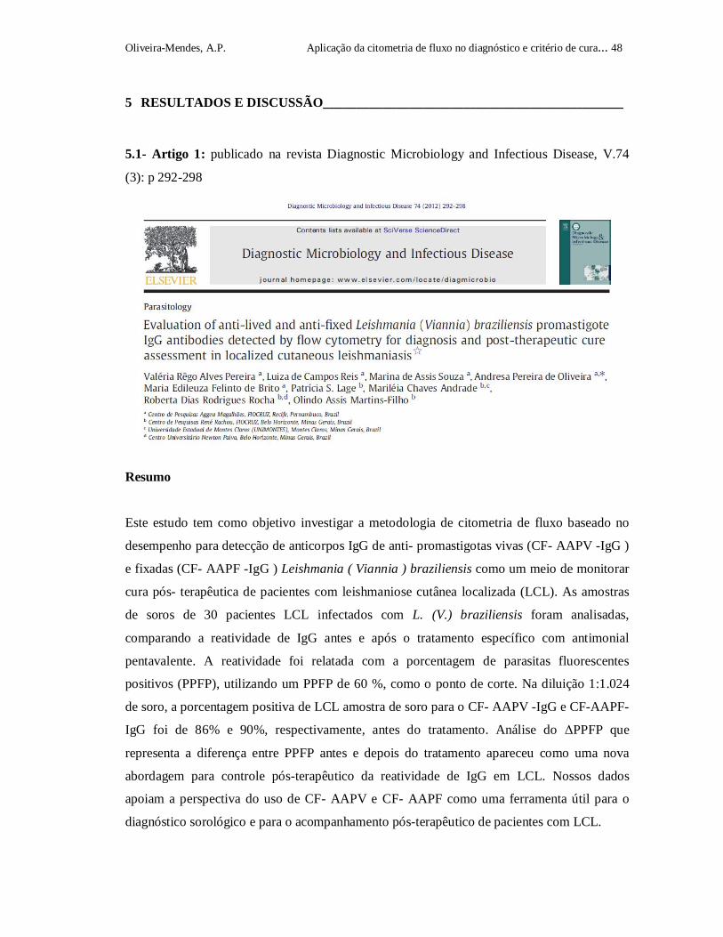
Oliveira-Mendes, A.P. Aplicação da citometria de fluxo no diagnóstico e critério de cura... 48
5 RESULTADOS E DISCUSSÃO_____________________________________________
5.1- Artigo 1: publicado na revista Diagnostic Microbiology and Infectious Disease, V.74
(3): p 292-298
Resumo
Este estudo tem como objetivo investigar a metodologia de citometria de fluxo baseado no
desempenho para detecção de anticorpos IgG de anti- promastigotas vivas (CF- AAPV -IgG )
e fixadas (CF- AAPF -IgG ) Leishmania ( Viannia ) braziliensis como um meio de monitorar
cura pós- terapêutica de pacientes com leishmaniose cutânea localizada (LCL). As amostras
de soros de 30 pacientes LCL infectados com L. (V.) braziliensis foram analisadas,
comparando a reatividade de IgG antes e após o tratamento específico com antimonial
pentavalente. A reatividade foi relatada com a porcentagem de parasitas fluorescentes
positivos (PPFP), utilizando um PPFP de 60 %, como o ponto de corte. Na diluição 1:1.024
de soro, a porcentagem positiva de LCL amostra de soro para o CF- AAPV -IgG e CF-AAPF-
IgG foi de 86% e 90%, respectivamente, antes do tratamento. Análise do ΔPPFP que
representa a diferença entre PPFP antes e depois do tratamento apareceu como uma nova
abordagem para controle pós-terapêutico da reatividade de IgG em LCL. Nossos dados
apoiam a perspectiva do uso de CF- AAPV e CF- AAPF como uma ferramenta útil para o
diagnóstico sorológico e para o acompanhamento pós-terapêutico de pacientes com LCL.

Oliveira-Mendes, A.P. Aplicação da citometria de fluxo no diagnóstico e critério de cura... 49
Abstract
This study aims to investigate a flow cytometry performance-based methodology to detect
anti-live (FC-ALPA-IgG) and anti-fixed (FC-AFPA-IgG) Leishmania (Viannia) braziliensis
promastigote IgG as a means to monitor post-therapeutic cure of patients with Localized
Cutaneous Leishmaniasis (LCL). Serum samples from 30 LCL patients infected with L. (V.)
braziliensis were assayed, comparing the IgG reactivity before and after specific treatment
with pentavalent antimonial. Reactivities were reported as the percentage of positive
fluorescent parasites (PPFP), using a PPFP of 60% as a cut-off. In the serum dilution 1:1,024,
the positive percentage of LCL serum sample for FC-ALPA-IgG and FC-AFPA-IgG was 86%
and 90%, respectively, before treatment. Analysis of ∆PPFP that represents the difference
between PPFP after and before treatment appeared as a new approach to monitor post-
therapeutic IgG reactivity in LCL. Our data support the perspective of using FC-ALPA and
FC-AFPA as a useful serological tool for diagnosis and for post-therapeutic follow-up of LCL
patients.
Keywords: Flow Cytometry, FC-ALPA, FC-AFPA, IgG, L. (V.) braziliensis, LCL.
1 Introduction
Leishmaniasis encompasses multiple clinical syndromes, most notably, visceral,
cutaneous, and mucosal forms. Localized Cutaneous Leishmaniasis (LCL) is a disease
associated with infections caused by several species of the genus Leishmania (Grimaldi; Tesh,
1993). Clinical manifestations depend on the parasite factors, the epidemiological
characteristic of the vector and the host genetic and immunological constitution (Rogers et al.,
2002). Cutaneous Leishmaniasis is a serious public health problem and is endemic to Brazil,
particularly in the State of Pernambuco, where Leishmania (Viannia) braziliensis is known as
the major circulating species (Brito et al., 2009).
At present, there is not a gold-standard test for Cutaneous Leishmaniasis and,
frequently, a combination of different diagnostic techniques is needed to obtain more precise
results. Thus, diagnosis is performed by the association of clinical, epidemiological and
laboratorial aspects. These techniques include amastigote identification by tissue
immunocytochemical techniques, “imprints” (printing by biopsy apposition), in aspirated

Oliveira-Mendes, A.P. Aplicação da citometria de fluxo no diagnóstico e critério de cura... 50
lesion and in histopathological evaluation, besides promastigote identification in vitro. The
indirect immunofluorescence (IIF), ELISA (Enzyme-linked immunosorbent assay) and
Western-blot, based on the presence of specific antibodies against parasite antigens, are the
serological methods used. Montenegro skin test is a late hypersensitivity test based in the
immunity mediated by cells. At the same time, there has been notable improvement in
techniques such as polymerase chain reaction (PCR), Real-time PCR and flow cytometry with
the objective of increasing sensitivity and specificity (Vega-Lopez, 2003).
The traditional laboratory methods have several limitations and present difficulties.
Furthermore, immunoassays can show low antibody titers, due to a cross reactivity with
Trypanosoma cruzi that depends on the antigen, as well as the lack of well-standardized
procedures used to detect the specific antibodies. Moreover, antibody production after
treatment is not yet clear and the predictive value of lower or higher levels against specific
antigenic fractions during follow-up is not well defined (Brito et al., 2001).
Chemotherapy treatment is based, primarily, on the administration of pentavalent
antimonials. These antimonials are highly toxic and administered via intramuscular injections
for a prolonged period. Besides toxicity, one of the limitations in the treatment is the absence
of an objective cure criterion. The cure criterion adopted by many authors is the complete
healing of the lesion. Nevertheless, this is an unsatisfactory criterion, as the reactivation of
the lesions may occur even after the treatment (Gontijo & Carvalho, 2003; Mendonça et al.,
2004; Schubach et al., 1998). Therefore, these facts point to the need of new tools that can
help determine prognostics and efficient diagnosis of Cutaneous Leishmaniasis.
More recently, a flow-cytometry analysis has been described for diagnosis purposes.
The anti-live promastigote antibodies (FC-ALPA-IgG) detected through this method showed
high sensitivity and specificity applicable in the diagnosis of LCL (Rocha et al., 2002; 2006).
This study opens new perspectives for the use of FC-ALPA-IgG in the response evaluation of
novel therapeutic protocols.
Based on that, in the present study we report the use of flow cytometry-based methods
for the assessment of humoral response against live and fixed Leishmania (Viannia)
braziliensis promastigote forms. Herein, we propose to investigate the performance of FC-
ALPA-IgG and FC-AFPA-IgG (anti-fixed promastigote antibodies) to monitor post-
therapeutic cure of LCL patients, comparing the IgG reactivity before and after specific
treatment.

Oliveira-Mendes, A.P. Aplicação da citometria de fluxo no diagnóstico e critério de cura... 51
2 Population, Materials and Methods
2.1 Study Population
A total of 30 patients with Localized Cutaneous Leishmaniasis -LCL (ages ranging
from 14 to 83 years old, mean=26±16.5 years, including 20 males and 10 females) from
endemic areas of the State of Pernambuco (Amaraji and Moreno municipalities) were
evaluated on clinical, epidemiological features and laboratorial criteria. All patients showed
positive diagnoses for LCL, including direct parasite detection, Montenegro skin test, indirect
immunofluorescence and PCR. Serum samples were collected from all LCL patients before
the treatment onset.
All patients received the standard treatment for LCL, carried out with N-methyl
glucamine antimoniate (Glucantime®) and the therapeutic scheme was made of doses of 20
mg/Kg/day through subcutaneous injections during thirty days. The occurrence of adverse
reactions was monitored throughout the treatment period. After treatment, sera sample were
collected from all patients, in a cross-sectional investigation with intervals ranging from 1 to
24 months, further grouped in 1-3 months (n = 4), 4-7 months (n= 14) and 12-24 months
(n=12). All patients underwent blood collection twice: prior to and after the end of
chemotherapy treatment with Glucantime®.
Eight serum samples from healthy individuals from non-endemic areas and without
previous LCL infection were used as a control for non-infected individuals (NI). All of them
signed the “Term of Free and Informed Consent” and the CPqAM / Fiocruz Research Ethics
Committee (Protocol no 27/04) approved the experimental protocols.
2.2 Parasite Preparation
Leishmania (Viannia) braziliensis promastigote forms (MHOM/BR/75/2903) were
cultivated in liver infusion tryptose medium (LIT) at 24 ± 1°C temperature. After serial
passage in vitro, the parasites, in stationary-phase, were collected and homogenized in a low-
rotation vortex to dissolve clumps. All procedures were performed aseptically in a biohazard
hood. The homogenate was centrifuged at 100 X g, for 10 min to remove cell debris. Prior to
the recovery of the parasite suspension, the supernatant was left to rest for 10 min at room
temperature. Then, the supernatant was transferred to another tube and centrifuged at 1000 X
g for 10 min, at 4°C. The pellet of parasites was suspended in buffer pH 7.2, containing 10%
Oliveira-Mendes, A.P. Aplicação da citometria de fluxo no diagnóstico e critério de cura... 52
heat-inactivated fetal bovine serum (PBS-10% FBS) (Gibco, Grand Island, NY) and
centrifuged at 1000 X g for 10 min. Two distinct parasite preparations were used to evaluate
the anti-L. (V.) braziliensis IgG reactivity referred to as “live” and “fixed” promastigote
suspensions. For the live promastigote suspension, parasites were immediately re-suspended
in PBS 10% FBS and used for the immunofluorescence by flow cytometry. For the fixed
promastigote suspension, the parasites were re-suspended in equal volume of PBS and fixing
solution (per liter, 10g of paraformaldehyde, 10.2 g of sodium cacodylate and 6.65 g of
sodium chloride, pH 7.2; Sigma Chemical Corp., St Louis, Mo) and stored overnight at 4ºC.
The fixed promastigotes were washed in PBS and stored at 4C until use. Both parasite
suspensions were counted in a Neubauer hemacytometer chamber and the concentration
adjusted to 5 X 10⁶/ml. The live or fixed promastigotes were used separately in the
immunofluorescence assays by flow cytometry.
2.3 Immunofluorescence by Flow Cytometry
The immunofluorescence assays to detect anti-live (FC-ALPA-IgG) and anti-fixed
(FC-AFPA-IgG) Leishmania (V.) braziliensis IgG antibodies were performed as described by
Rocha et al. (2002). Suspension of live or fixed parasites (2.5 x 10⁵/well) were incubated in
96-wells round bottom polystyrene plates, at 37°C for 30 min in the presence of different
dilutions (1:128 to 1:16,384 for FC-ALPA-IgG and 1:256 to 1:32,768 for FC-AFPA-IgG) of
serum samples. After incubation with the test serum samples, the parasites were washed twice
with 150 l of PBS-10%FBS (at 1,000 X g for 10 min at 4°C). Afterwards, the parasites were
re-incubated at 37°C for 30 min in the dark, in the presence of fluorescein isothiocyanate
(FITC)-conjugated anti-human IgG antibody (Sigma Chemical Corp., St. Louis, MO) diluted
at 1:1,200 in PBS-10% FBS for live parasites and at 1:16,000 for fixed parasites. After a
second wash procedure, the FITC-labeled parasites were fixed for 30 min with a FACS fixing
solution (per liter, 10 g of paraformaldehyde, 10.2 g of sodium cacodylate and 6.65 g of
sodium chloride, pH 7.2; Sigma Chemical Co.) before being analyzed in the cytometer. Flow
cytometry measurements were performed in a maximum period of 24 hours after parasite
fixation. Each assay included an internal control of nonspecific binding in which parasites, not
exposed to human serum, were incubated with FITC-conjugated anti-human IgG. In all
experiments, positive and negative LCL control samples were included. The positive control
chosen was a patient's serum with parasitological, immunological and molecular diagnostics
all positive for LCL. The optimum working concentration of sera and anti-human antibody
Oliveira-Mendes, A.P. Aplicação da citometria de fluxo no diagnóstico e critério de cura... 53
conjugates were determined with the checkerboard titration method as previously proposed by
Rocha et al. (2002). All FC-ALPA and FC-AFPA tests were repeated and confirmed, assuring
the repeatability of the test.
2.4 Flow Cytometry Data Acquisition and Analysis
Flow cytometry acquisition was performed using a Becton Dickinson FACSCaliburTM
equipment. The Cell-QuestTM software package was used in both data storage and analysis.
Stained parasites were run in the cytometer, and 10,000 events per sample were acquired.
Promastigotes were identified based on their specific forward (FSC) and side (SSC) light
scattering properties. Following FSC and SSC gain adjustments, parasites were found by
assuming a characteristic FSC X SSC dot plot distribution. The relative FITC fluorescence
intensity of each event was analyzed with a single histogram representation. A marker was set
up on the histogram representation of the FITC-conjugated internal control and used in all
data analysis reported here to determine, for each sample, the percentage of positive
fluorescent parasites (PPFP) as previously described by Rocha et al. (2002). The receiver
operating characteristic curve - ROC curve (Greiner et al. 2000) was used to select the cut-off
value to discriminate between positive and negative results of FC-ALPA-IgG and FC-AFPA-
IgG. The analysis of paired ∆PPFP, defined as the difference between PPFP after treatment
and PPFP before treatment was used as a new approach to monitor post-therapeutic IgG
reactivity in LCL as proposed by Lemos et al. (2007) to monitor patients with Visceral
Leishmaniasis.
3 Results
3.1 Initial Serum Screening
In the first step of this study, we analyzed the anti-Leishmania braziliensis IgG
reactivity profile for live (FC-ALPA) and fixed (FC-AFPA) antigen preparations, aiming to
characterize the titration curve and the serum dilution to differentiate Localized Cutaneous
Leishmaniasis patients and non-infected individuals. Data analysis showed that both antigen
preparation FC-ALPA-IgG and FC-AFPA-IgG have the same titration curve and patterns of
reactivity (PPFP values). This analysis identified the specific serum dilution (1:1.024 for FC-
ALPA-IgG and FC-AFPA-IgG) that best segregates the PPFP values of LCL patients and

Oliveira-Mendes, A.P. Aplicação da citometria de fluxo no diagnóstico e critério de cura... 54
A B
A B
0
20
40
60
80
100
128
512
1,024
4,096
2,048
8,192
16,38425
6
16,384
0
20
40
60
80
100
32,76851
2
1,024
4,096
2,048
8,192
256
Inverse of Serum Dilution
PPFP
FC-ALPA-IgG FC-AFPA-IgG
non-infected individuals. As observed in Figure 1, the results were classified as positive with
PPFP 60% and negative, PPFP 60%. This percentage was done according to Martins-Filho
et al.(1995), from the analysis of antibody titration curves of individual sera, expressed by the
mean of PPFP values. The initial screening showed that sera were considered negative
(PPFP 60%) when reacting in dilution 1:128 to 1:16,384 for FC-ALPA-IgG and 1:512 to
1:32,768 for FC-AFPA (Figure 1). In view of these results, FC-ALPA-IgG and FC-AFPA-
IgG were able to discriminate the IgG reactivities of patients compared to the control group.
Figure 1: Anti-live FC-ALPA-IgG (A) and anti-fixed FC-AFPA-IgG (B) L. (V.) braziliensis IgG reactivity in
serum samples from patients with Localized Cutaneous Leishmaniasis (LCL= ) and Non-Infected individuals
(NI= ). The results are expressed as mean percentage of positive fluorescent parasites (PPFP) at sera dilutions
1:128 to 1:16,384 for FC-ALPA-IgG and sera dilutions 1:256 to 1:32,768 for FC-AFPA-IgG. The rectangles
represent the selected serum dilution of the higher segregation range between patients and negative control
(1:1,024 for FC-ALPA-IgG and FC-AFPA-IgG).
3.2 Performance of Anti-live and fixed L. (V.) braziliensis IgG Reactivity to identify
active LCL
In this step, we evaluated the test performance in the identification of patients with
active LCL. Thus, sera were separated and reactivity was determined in the dilution 1:1,024
and the cut-off of PPFP-60% (Figure 1). Using this approach, the sensitivity of FC-ALPA-
IgG and FC-AFPA-IgG was 86% and 90%, respectively. The specificity was 78%, showing
cross-reactivity of 22% for both parasite preparations (live and fixed) (Figure 2). The results
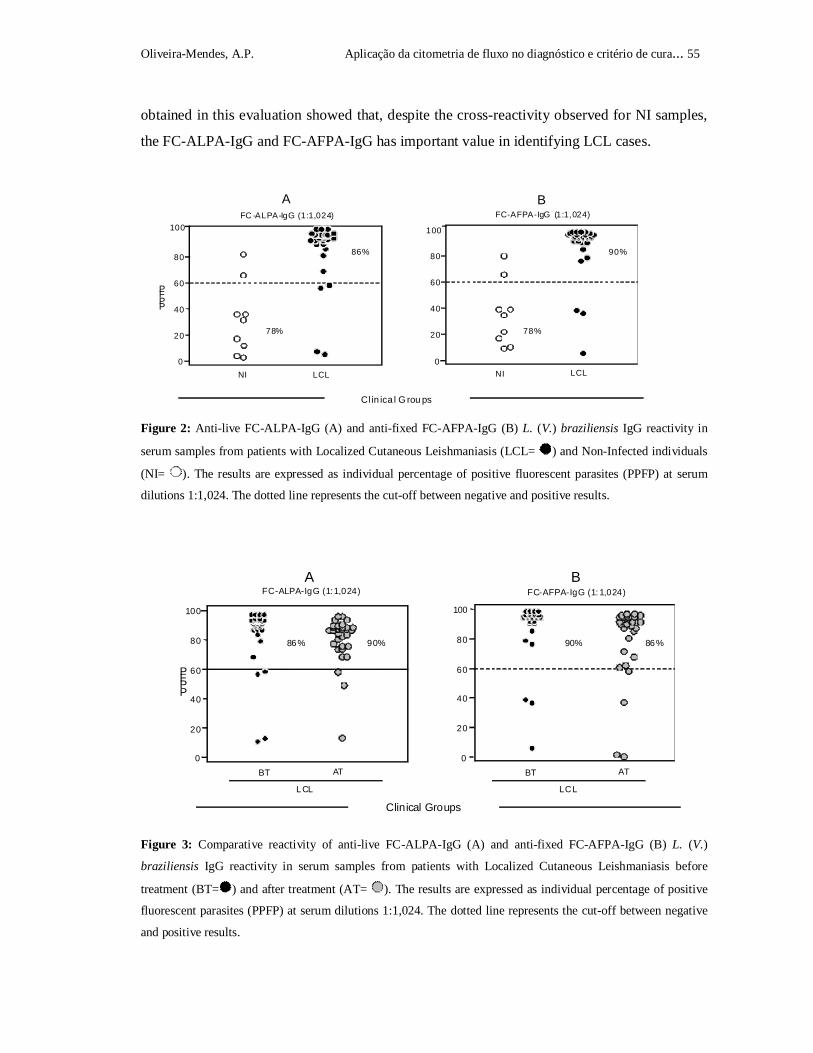
Oliveira-Mendes, A.P. Aplicação da citometria de fluxo no diagnóstico e critério de cura... 55
A B
PPFP
100
80
60
40
20
0NI LCL
100
80
60
40
20
0NI LCL
FC -ALPA-IgG (1:1,024) FC-AFPA-IgG (1:1,024)
Cl in ica l G rou ps
86% 90%
78% 78%
A B
PPFP
100
80
60
40
20
0
100
80
60
40
20
0ATBT
LCL
Clinical Groups
ATBT
LCL
FC-ALPA-IgG (1:1,024) FC-AFPA-IgG (1: 1,024)
86% 90% 90% 86%
obtained in this evaluation showed that, despite the cross-reactivity observed for NI samples,
the FC-ALPA-IgG and FC-AFPA-IgG has important value in identifying LCL cases.
Figure 2: Anti-live FC-ALPA-IgG (A) and anti-fixed FC-AFPA-IgG (B) L. (V.) braziliensis IgG reactivity in
serum samples from patients with Localized Cutaneous Leishmaniasis (LCL= ) and Non-Infected individuals
(NI= ). The results are expressed as individual percentage of positive fluorescent parasites (PPFP) at serum
dilutions 1:1,024. The dotted line represents the cut-off between negative and positive results.
Figure 3: Comparative reactivity of anti-live FC-ALPA-IgG (A) and anti-fixed FC-AFPA-IgG (B) L. (V.)
braziliensis IgG reactivity in serum samples from patients with Localized Cutaneous Leishmaniasis before
treatment (BT= ) and after treatment (AT= ). The results are expressed as individual percentage of positive
fluorescent parasites (PPFP) at serum dilutions 1:1,024. The dotted line represents the cut-off between negative
and positive results.

Oliveira-Mendes, A.P. Aplicação da citometria de fluxo no diagnóstico e critério de cura... 56
A B
PPFP
2,048 8,192 16,384
0
20
40
60
80
100
4,096 32,768
0
20
40
60
80
100
2,048 4,096 8,192 16,384
* * * *
Inverse of Serum Dilution
FC-ALPA-IgG FC-AFPA-IgG
BT
AT
BT
AT
BT
AT
BT
AT
BT
AT
BT
AT
BT
AT
BT
AT
BT
AT
Additionally, we analyzed the applicability of FC-ALPA-IgG and FC-AFPA-IgG to
monitor the post-therapeutic cure of LCL. The analysis of PPFP values performed after
treatment did not demonstrate applicability of this parameter for post-therapeutic cure
assessment at serum dilution 1:1,024. No significant changes in the PPFP values at serum
dilution 1:1,024 were observed after treatment showed reactivity less than 60% to FC-ALPA-
IgG and FC-AFPA-IgG (Figure 3).
Further comparative analysis used the PPFP values of paired samples evaluated before
and after treatment along the titration curves (1:2,048 to 1:16,384 for FC-ALPA-IgG and
1:2,048 to 1:32,678 for FC-AFPA-IgG). Our data demonstrated that only FC-ALPA-IgG led
to differential reactivity when comparing the mean PPFP values observed before and after
treatment (Figure 4 -asterisks).
Figure 4: Differential Anti-live FC-ALPA-IgG (A) and anti-fixed FC-AFPA-IgG (B) L. (V.) braziliensis IgG
reactivity in serum samples from patients with Localized Cutaneous Leishmaniasis before treatment (BT= )
and after treatment (AT= ). The results are expressed as individual percentage of positive fluorescent parasites
(PPFP) for each pair of samples at serum dilutions 1:2,048 to 1:16,384 for FC-ALPA-IgG and 1:2,048 to
1:32,768 for FC-AFPA-IgG. Significant differences between the mean PPFP values before (BT) and after
treatment (AT), at p<0.05 are highlighted by *.
3.3 Introducing PPFP as a New Strategy Applied to Post-therapeutic Cure Assessment
in LCL
As the analysis of PPFP values performed after treatment did not demonstrate any
applicability for post-therapeutic cure assessment at a serum dilution 1:1,024, we searched for
a new tool to comparatively analyze the IgG reactivity before and after treatment.

Oliveira-Mendes, A.P. Aplicação da citometria de fluxo no diagnóstico e critério de cura... 57
A B
-10 08 1%
19 %
-60
-20
2 0
6 0
100
4,096
8,192
16,384
2,04877 %
2 3 %
7 7 %
2 3%
8 1%
1 9%
5 5%-100
-60
-20
20
60
100
4,096
8,192
16,384
2,048
32,768
6 1%
3 9 %
6 1%
3 9%
42 %
5 8%
5 8 %
4 2 % 4 5%
FC -ALPA-IgG F C-A FPA -IgG
?PPFP
Inverse of Serum D ilut ion
The proposed strategy was to analyze the differential PPFP reactivity detected by
paired samples (delta reactivity []). In our case, we have used anti-Leishmania IgG reactivity
PPFP that represents the difference between PPFP after treatment and PPFP before
treatment (PPFP= PPFPAT – PPFPBT).
Initially, we evaluated the PPFP values throughout FC-ALPA-IgG and FC-AFPA-IgG
titration curves, aiming to identify the serum dilution range 1:2,048 to 1:16,384 for FC-
ALPA-IgG and 1:2,048 to 1:32,678 for FC-AFPA-IgG that represent the highest differential
reactivities (Figure 5). As shown in Figure 5, the FC-ALPA-IgG at serum dilution 1:8,192
showed that 81% of the treated patients displayed negative PPFP values, demonstrating a
decrease in IgG reactivity after treatment. Moreover, the FC-AFPA-IgG at serum dilution
1:4,096 was able to identify 61% of patients with negative PPFP values. Consequently, these
dilutions were the best choices to analyze the PPFP values for FC-ALPA-IgG and FC-
AFPA-IgG, respectively.
Figure 5: Differential anti-L. (V.) braziliensis IgG reactivity of paired samples detected by FC-ALPA-IgG (A)
and FC-AFPA-IgG (B). The results are expressed as differential percentage of fluorescent positive parasites
(ΔPPFP) for each pair of samples from LCL patients ( ) evaluated before and after treatment. The ΔPPFP was
defined as the difference in the PPFP values observed after treatment, taking the PPFP before treatment as the
reference (ΔPPFP= PPFPAT – PPFPBT). The gray rectangle identifies the serum dilution that yielded the higher
frequency of negative ΔPPFP values. The dashed line represents the cut-off edge of ΔPPFP=0 referring to
unaltered serological profile.
After setting the specific serum dilutions to monitor the PPFP values, we
investigated whether the time after treatment would interfere in the performance of this new
strategy of post-therapeutic follow-up. For this purpose, the treated patients were categorized
into three subgroups based on the time after treatment when they were evaluated, including 1-

Oliveira-Mendes, A.P. Aplicação da citometria de fluxo no diagnóstico e critério de cura... 58
A B
?PPFP
-100
-25
0
25
100
25% 29% 33%
0%0% 21 %
-100
-25
0
25
100
43%
0%0% 7 %
58%75%
FC-ALPA-IgG (1:8,192) FC-AFPA-IgG (1:4,096)
Time After Treatment (months)
1-3mAT 4-7mAT 12-24mAT 1-3mAT 4-7mAT 12-24mAT
3 mAT; 4-7 mAT; and 12-24 mAT. Data analysis was performed after the establishment of a
gray zone corresponding to the first quartile of the PPFP range (cut-off edge of 25%,
considering PPFP values from 0 to 100%) according to Lemos et al, 2007. We believe that the
use of this gray zone would give further strength to data interpretation, since it would avoid
interference regarding the possible intrinsic flow cytometry measurement variability. Our data
demonstrated that 75% of the patients evaluated at 1-3mAT showed negative PPFP values
detected by FC-ALPA-IgG whereas 25% of them showed negative PPFP values detected by
FC-AFPA-IgG. Patients evaluated at 4-7mAT displayed 43% and 29% of negative PPFP
values detected by FC-ALPA-IgG and FC-AFPA-IgG, respectively. When PPFP reactivity
was evaluated 12-24mAT, 58% and 33% of the patients presented negative results in the FC-
ALPA-IgG and FC-AFPA-IgG, respectively (Figure 6).
Figure 6: Differential anti-L. (V.) braziliensis IgG reactivities of paired samples detected by FC-ALPA-IgG (A)
and FC-AFPA-IgG (B) defined as differential percentage of fluorescent positive parasites (ΔPPFP) for pairs of
samples from LCL patients evaluated before and after treatment. Patients were categorized into three groups
based on the time (months) after treatment (mAT) and referred as 1-3 mAT ( ), 4-7 mAT ( ) and 12-24 mAT
( ). The results are expressed as ΔPPFP for each pair of samples. Data analysis was performed after the
establishment of a gray zone (the gray rectangle) corresponding to the first quartile of the ΔPPFP range (cut-off
of 25%).
4 Discussion
One of the major limitations for the use of serological approaches in the diagnosis and
cure assessment of Cutaneous Leishmaniasis is the scarcity of sensitive methods to
discriminate the IgG reactivity during active infection and the residual serological reactivity
Oliveira-Mendes, A.P. Aplicação da citometria de fluxo no diagnóstico e critério de cura... 59
after effective treatment. In general, the diagnosis and the cure criterion are based on clinical
and epidemiological findings and complete therapeutic effectiveness is considered when
complete lesion healing is observed. Nevertheless, this is an unsatisfactory criterion, as lesion
reactivation may occur even after treatment and complete healing of the initial lesion.
Although cutaneous lesions caused by Leishmania (Viannia) braziliensis are usually
susceptible to antimonial treatment and healing occurs at the end of therapy, the occurrence of
more than 90% of relapses up to one year in Cutaneous Leishmaniasis strengthens the
suggestion of clinical control for at least one year post treatment (Coutinho et al., 1996).
Given this context, it has been suggested that the decrease in anti-Leishmania
antibodies titer in serum of patients after treatment could be an alternative method to
monitoring specific post-therapeutic cure (Chiari et al., 1973, Mendonça et al., 1988).
Moreover, the presence of anti-Leishmania antibodies after treatment could indicate the
persistence of the parasite and be a predictive factor in Cutaneous Leishmaniasis recurrence
(Saravia et al., 1989). Associated with these aspects, the existence of various parasite species
and vectors, as well as parasite-host relation represent important elements related to
Cutaneous Leishmaniasis complexity. This fact points to the need for new tools that help to
determine prognostics and an efficient diagnosis.
Serological tests to diagnose Cutaneous Leishmaniasis may also present limitations,
such as low sensitivity, low specificity, low reproducibility, low antibody titers, the absence
of correlation between the circulating antibody levels with the disease stage and they can
show crossing reactions with other species of the Trypanosomatidae family (Brito et al.,
2000;2001; Kar, 1995).
In this study, we have evaluated the applicability of an indirect immunofluorescence
assay based on flow cytometry methodology to detect anti-live and anti-fixed Leishmania
(Viannia) braziliensis promastigote IgG for the serological diagnosis of LCL. In addition, we
have verified the applicability of serological reactivity to assess post-therapeutic cure in cured
patients after different periods following treatment with pentavalent antimonial
chemotherapy. The rationale underlying our experimental design was the fact that the live
promastigote preparation would represent a useful tool to work with a selective set of antigens
(outer membrane epitopes) and contribute to a more refined analysis. However, the labile
nature of this antigenic preparation is the major concern about its use in clinical laboratory
practice. On the other hand, although the use of fixed promastigotes represents a feasible way
to produce and store a bulk amount of pre-fixed antigen that contributes to large scale
production, it would contribute to the development of a larger amount of intracytoplasmatic

Oliveira-Mendes, A.P. Aplicação da citometria de fluxo no diagnóstico e critério de cura... 60
epitopes and complicate the observation of minor changes in the serological reactivity
following therapeutic intervention (Pissinate et al., 2008).
Another important aspect of the serological methods applied to Cutaneous
Leishmaniasis diagnosis is the choice of antigen sources, which still represents a relevant
obstacle. When total promastigotes are used as antigen, it is common to find false positive
reactions due to cross-reactions with other diseases. It is important to investigate alternative
preparations to detect Leishmania antibodies (Celeste et al., 2004, Gonçalves et al., 2002). In
the present investigation we have chosen the L. (V.) braziliensis promastigotes as the
antigenic source to access IgG reactivity in LCL patients. Although this species is very
difficult to grow in vitro, requiring the use of axenic cultures, complex medium composition
and fine pH and temperature controls (Lemesre et al., 1988), L. (V.) braziliensis is the most
important LCL causative agent in Brazil and especially in Pernambuco.
A diagnostic method based on flow-cytometry to detect anti-live L. (V.) braziliensis
antibodies has been described by Rocha et al. (2002, 2006). They demonstrated 96%
sensitivity for FC-ALPA-IgG L. braziliensis in active patients. Using fixed L. amazonensis
promastigotes Pissinate et al. (2008), showed a good performance of FC-AFPA-IgG in the
serological diagnosis of LCL. However they found cross-reactivity with other co-endemic
diseases, like tripanosomatid infections. Our data demonstrated that both methods display low
specificity and still require methodological adjustments in order to improve their performance
as confirmatory diagnostic tools. In fact, Rocha et al. (2002) and Pissinate et al. (2008) have
already demonstrated that the occurrence of false positive results in the FC-ALPA-IgG and
FC-AFPA-IgG in endemic areas are mostly related to the cross-reactivity of serum samples
from patients with Chagas disease and Visceral Leishmaniasis, co-endemic diseases generally
observed in areas of prevalent Cutaneous Leishmaniasis.
In our study, we have found that FC-ALPA-IgG using L. (V.) braziliensis displayed
86% sensitivity whereas FC-AFPA-IgG showed 90% sensitivity for the diagnosis of LCL.
This difference obtained could be explained by the heterogeneity of L. (V.) spp. in Brazil.
Brito et al (2009) demonstrated the presence of 10 circulating zymodemes in the well-defined
“Zona da Mata” of Pernambuco. The heterogeneity observed among L. (V.) braziliensis
parasites from this region is noteworthy, particularly in contrast to the homogeneity of
parasites isolated from other regions of Brazil (Brandão-Filho et al., 2003, Brito et al., 1993,
Cupolillo et al., 2003).
The results obtained showed that both techniques (FC-ALPA-IgG and FC-AFPA-IgG)
are useful for the sero-diagnosis of LCL as compared to the conventional

Oliveira-Mendes, A.P. Aplicação da citometria de fluxo no diagnóstico e critério de cura... 61
immunofluorescence assay. Although FC-AFPA-IgG display a slight higher sensitivity in the
diagnosis of LCL, the FC-ALPA-IgG seems to be more reliable for cure monitoring, being
able to identify more differences between IgG reactivity before and after treatment when
assessed by PPFP. We found 81% of the treated patients with negative PPFP results for
FC-ALPA-IgG compared to 61% negative PPFP for FC-AFPA-IgG. These results show that
FC-ALPA-IgG represents a better performance than FC-AFPA-IgG for post-therapeutic
monitoring of LCL patients. We have a general belief that the FC-ALPA-IgG represents
better performance than the FC-AFPA-IgG, as previous studies of our group have
demonstrated a real advantage of using live, instead of fixed, parasites in serological
approaches applied to the diagnosis and cure assessment of human protozooses (Martins-
Filho et al., 1995, 2002, Vitelli-Avelar et al., 2007, Pissinate et al., 2008). In fact, the use of
live promastigotes seems to represent a better tool to achieve better performance of
serological approaches since in this antigenic preparation only the outer membrane epitopes
are available for IgG binding in contrast with the fixed antigenic preparation on which the
cytoplasmic antigens are also available for IgG recognition. The use of a selected set of outer
membrane antigens is a good strategy to work with a more restricted IgG repertoire that
would potentially find slight differences resulting from the loss of B-cell clones early after
effective etiological treatment.
We also investigated whether performance would be influenced if the test was
performed at different times after treatment. For this purpose serum samples collected after
treatment were segregated into three groups referred to as 1-3mAT, 4-7mAT and 12-24mAT.
The FC-ALPA-IgG and FC-AFPA-IgG were assayed and the PPFP values generated to
monitor sero-reactivity at different times following treatment. Our findings demonstrated an
overall low performance of PPFP to demonstrate differential reactivity according to the time
after treatment. However, the FC-ALPA-IgG still demonstrated better performance as
compared to the FC-AFPA-IgG, leading to higher frequency of cases with negative PPFP. It
is important to mention that the low performance of PPFP to detect differential reactivity in
this cross-sectional investigation should not be considered the end point of using this
parameter for cure assessment in LCL since this approach would be better evaluated in a
longitudinal investigation in order to generate more accurate data for cure assessment in LCL.
Although most flow cytometry-based methods still represent higher cost compared to
conventional methods, such as immunosorbent and immunofluorescence assay, the possibility
of working with a microplate serological approach has reduced the final cost of a given test.

Oliveira-Mendes, A.P. Aplicação da citometria de fluxo no diagnóstico e critério de cura... 62
Moreover, at the present time, several clinical laboratories in developing countries are
considering the acquisition of flow cytometers. Therefore, in the near future, the
implementation of new flow cytometry-based tests will became routine as will the interchange
between research centers and clinical laboratories. In our experience, flow cytometry-based
serological approaches present good reproducibility and outstanding concordance among
independent analysts (Garcia et al., 2009).
In conclusion, our data suggested that the new flow cytometry-based methodology has
promising potential to identify active LCL clinical cases in patients. Further longitudinal
studies are currently under investigation in order to better characterize the approach for
monitoring post-therapeutic cure, as well as to obtain the clinical values of this new approach
and to validate its use in medical studies.
Acknowledgements
We are grateful to L. F. da Rocha for technical assistance. This study was supported
by the State of Pernambuco Research Foundation (FACEPE), by the Brazilian National
Research Council (CNPq) and by the Oswaldo Cruz Foundation (FIOCRUZ). MCA is
thankful to FAPEMIG for the BIP fellowship program. OAMF is grateful to the CNPq
research fellowship program (PQ).
5 References
Brandão-Filho SP, Brito MEF, Carvalho FG, Ishikawa EA, Cupolillo E, Shaw JJ. 2003. Wild
and siynantropic host of Leishmania (Viannia) braziliensis in the endemic cutaneous
leishmaniasis locality of Amaraji, Pernambuco, State, Brazil. Trans R Soc Trop Med Hyg 97:
291–296.
Brito MEF, Brandão-Filho SP, Sales NRS, Cupolillo E, Grimaldi G Jr, Momen H. 1993.
Human cutaneous leishmaniasis due to a new enzymatic variant of Leishmania (Viannia)
braziliensis occurs in Pernambuco, Brazil. Mem Inst Osvaldo Cruz 88: 33– 634.

Oliveira-Mendes, A.P. Aplicação da citometria de fluxo no diagnóstico e critério de cura... 63
Brito MEF, Mendonça MG, Gomes YM, Jardim ML, Abath FG. 2000. Identification of
potentially diagnostic Leishmania braziliensis antigens in human cutaneous leishmaniasis by
immmunoblot analysis. Clin Diagn Lab Immunol 7: 318–321.
Brito ME, Mendonça MG, Gomes YM, Jardim ML, Abath FG. 2001. Dynamics of the
antibody response in patients with therapeutic or spontaneous cure of American cutaneous
leishmaniasis. Trans R Soc Trop Med Hyg 95: 203-206.
Brito ME, Andrade MS, Mendonça MG, Silva CJ, Almeida EL, Lima BS, Felix SM, Abath
FG, Graça GC, Porrozzi R, Ishikawa EA, Shaw JJ, Cupolillo E, Brandão-Filho SP. 2009.
Species diversity of Leishmania (Viannia) parasites circulating in an endemic area for
cutaneous leishmaniasis located in the Atlantic rainforest region of northeastern Brazil. Trop
Med Int Health 14(10): 1278-1286.
Celeste BJ, Angel SO, Castro LG, Gidlund M, Goto H. 2004. Leishmania infantum heat shock
protein 83 for the serodiagnosis of tegumentary leishmaniasis. Braz J Med Biol Res 37: 1591–
1593.
Chiari CA, Mayrink W, Magalhães PA. 1973. Reação de imunofluorescência indireta no
controle de tratamento de leishmaniose tegumentar americana. Rev Inst Med Trop São Paulo
15: 298-303.
Coutinho SG, Oliveira MP, Da-Cruz AM, De-Luca PM, Mendonça SC, Bertho AL, Soong D,
McMahon-Pratt D. 1996. T-cell responsiveness of American cutaneous leishmaniasis patients
to purified Leishmania pifanoi amastigote antigens and Leishmania braziliensis promastigote
antigens: immunologic patterns associated with cure. Exp Parasitol 84: 144-155.
Cupolillo E, Brahim LR, Toaldo CB. 2003. Genetic polymorphism and molecular
epidemiology of Leishmania (Viannia) braziliensis from different hosts and geographic areas
in Brazil. J Clin Microbiol 41: 3126–3132.
Garcia LM, Coelho-Dos-Reis JG, Peruhype-Magalhães V, Teixeira-Carvalho A, Rocha RD,
Araújo MS, Gomes IT, Carvalho SF, Dietze R, Lemos EM, Andrade MC, Martins-Filho OA.
2009. Anti-fixed Leishmania chagasi promastigotes IgG antibodies detected by flow

Oliveira-Mendes, A.P. Aplicação da citometria de fluxo no diagnóstico e critério de cura... 64
cytometry (FC-AFPA-IgG) as a tool for serodiagnosis and for post-therapeutic cure
assessment in American visceral leishmaniasis. J Immunol Methods. 350:36-45.
Gonçalves CCM, Reiche EMV, Abreu-filho BA, Silveira TG, Felizardo TC, Maia KR,
Costacurta R, Padovesi EJ, Dias Filho BP, Jankevicius SI, Jankevicius JV. 2002. Evaluation
of antigens from various Leishmania species in a western blot for diagnosis of American
tegumentary leishmaniasis. Am J Trop Med Hyg 66: 91–102.
Gontijo B, Carvalho MLR. 2003. American cutaneous leishmaniasis. Rev Soc Bras Med Trop
36: 71-80.
Greiner, M., D. Pfeiffer, and R. D. Smith. Principles and practical application of the receiver-
operating characteristic analysis for diagnostic tests. Prev Vet Med 2000; 45: 23–41
Grimaldi G, Tesh RB. 1993. Leishmaniasis of the New World: current concepts and
implications for future research. Clin Microbiol Rev 6: 230–250.
Kar K. 1995. Serodiagnosis of leishmaniasis. Crit Rev Microbiol 21: 123–152.
Lemesre JL, Darcy F, Capron A, Santoro F. 1988. Requirements of defined cultivation
conditions for standard growth of Leishmania promastigotes in vitro. Acta Trop (Basel) 45:
99-108.
Lemos EM, Gomes IT, Carvalho SFG, Rocha RDR, Pissinate JF, Martins-Filho AO, Dietze
R. 2007. Detection of Anti-Leishmania (Leishmania) chagasi immunoglobulin G by flow
cytometry for cure assessment following chemotherapeutic treatment of American Visceral
leishmaniasis. Clin Vaccine Immunol, 14(05): 569-576.
Martins-Filho, O.A., Pereira, M.E.S., Carvalho, J.F., Cançado, J.R., Brener, Z., 1995. Flow
cytometry, a new approach to detect anti-live trypomastigote antibodies and monitor the
efficacy of specific treatment in human Chagas´ disease. Clin. Diag. Lab. Immunol. 2: 569-
573.
Oliveira-Mendes, A.P. Aplicação da citometria de fluxo no diagnóstico e critério de cura... 65
Martins-Filho, O.A., Elói-Santos, S.M., Carvalho, A.T., Corrêa-Oliveira, R., Rassi, A.,
Luquetti, A.O., Rassi, G.G., Brener Z., 2002. Double-blind study to evaluate flow cytometry
analysis of anti-live trypomastigote antibodies for monitoring treatment efficacy in cases of
human Chagas´ disease. Clin. Diag. Lab. Immunol. 9:1107-1113.
Mendonça SCF, Souza WJS, Nunes MP, Marzochi MCA, Coutinho SG. 1988. Indirect
immunofluorescence test in New World leishmaniasis: serological and clinical relationship.
Mem Inst Oswaldo Cruz 83: 347–355.
Mendonça MG, Brito MEF, Rodrigues EH, Bandeira V, Jardim ML, Abath FG. 2004.
Persistence of Leishmania parasites in scars after clinical cure of American cutaneous
leishmaniasis: is there a sterile cure? J Infect Dis 189: 1018-1023.
Pissinate JF, Gomes IT, Peruhype-Magalhães V, Dietze R, Martins-Filho OA, Lemos EM.
2008. Upgrading the flow-cytometry analysis of anti-Leishmania immunoglobulins for the
diagnosis of American tegumentary leishmaniasis. J Immunol Methods, 336: 193-202.
Rocha RD, Gontijo CM, Eloi-Santos SM, Teixeira-Carvalho A, Correa-Oliveira R, Ferrari
TC, Marques MJ, Mayrink W, Martins-Filho AO. 2006. Clinical value of anti-live
Leishmania (Viannia) braziliensis immunoglobulin G subclasses, detected by flow cytometry,
for diagnosing active localized cutaneous leishmaniasis. Trop Med Int Health 11: 156-166.
Rocha RD, Gontijo CM, Eloi-Santos SM, Teixeira Carvalho A, Correa-Oliveira R, Marques
MJ, Genaro O, Mayrink W, Martins-Filho OA. 2002. Anti-live Leishmania (Viannia)
braziliensis promastigote antibodies detected by flow cytometry to identify active infection in
American cutaneous leishmaniasis. Rev Soc Bras Med Trop 35: 551-562.
Rogers KA, DeKrey GK, Mbow ML, Gillespie RD, Brodskin CI, Titus RG. 2002. Type 1 and
type 2 responses to Leishmania major. FEMS Microbiol Lett 209(1): 1-7.
Saravia NG, Valderrama L, Labrada M, Holguín AF, Navas C, Palma G, Weigle KA. 1989.
The Relationship of Leishmania braziliensis Subspecies and Immune Response to Disease
Expression in New World Leishmaniasis. J Infect Dis 159(4): 725-735.

Oliveira-Mendes, A.P. Aplicação da citometria de fluxo no diagnóstico e critério de cura... 66
Schubach A, Haddad F, Oliveira-Neto MP, Degrave W, Pirmez C, Grimaldi GJr, Fernandes
O. 1998. Detection of Leishmania DNA by polymerase chain reaction in scars of treated
human patients. J Infect Dis 178(3): 911-914.
Vega-López F. 2003. Diagnosis of cutaneous leishmaniasis. Curr Opin Infect Dis 16: 97-101.
Vitelli-Avelar, D.M., Sathler-Avelar, R., Wendling, A.P.B., Rocha, R.D.R., Teixeira-
Carvalho, A., Martins, N.E., Dias, J.C.P., Rassi, A., Luquetti, A.O., Elói-Santos, S.M.,
Martins-Filho, O.A., 2007. Non-conventional flow cytometry approaches to detect anti-
Trypanosoma cruzi immunoglobulin G in the clinical laboratory. J. Immunol. Methods
318:102-112.

Oliveira-Mendes, A.P. Aplicação da citometria de fluxo no diagnóstico e critério de cura... 67
5.2- Artigo 2: publicado na revista Journal of Immunological Methods, V.387 (1-2): p 245-253
Resumo
O objetivo deste estudo foi comparar as técnicas de imunofluorescência indireta (IFI) e
citometria de fluxo para avaliação clínica e laboratorial dos pacientes antes e depois da cura
clínica e avaliar a aplicabilidade da citometria de fluxo no acompanhamento pós- terapêutico
de pacientes com leishmaniose tegumentar americana (LTA). Soros de 14 pacientes antes do
tratamento (AT), 13 pacientes um ano após o tratamento (PT), 10 pacientes 2 e 5 anos foram
avaliadas. Os resultados de citometria de fluxo foram expressos em níveis de reatividade de
IgG, com base na percentagem de parasitos fluorescentes positivos (PPFP). A diluição 1:256
permitiu a diferenciação entre os indivíduos AT e PT. A análise comparativa dos IFI e
citometria de fluxo pela curva ROC (Receiver Operating Characteristic Curve) apresentaram,
respectivamente, a AUC (área sob a curva) = 0,8 (IC 95% = 0,64-0,89 ) e AUC = 0,90 (IC
95% = 0,75 -0,95 ), demonstrando que a citometria de fluxo teve acurácia equivalente. Nossos
dados demonstraram que 20% de PPFP foi o melhor ponto de corte identificado pela curva
ROC para o ensaio de citometria de fluxo. Este teste mostrou sensibilidade de 86% e
especificidade de 77%, enquanto a IFI teve uma sensibilidade de 78% e especificidade de
85%. A triagem após o tratamento, através da análise comparativa dos índices de desempenho
das técnicas, 1, 2 e 5 anos PT, mostraram um desempenho igual da citometria de fluxo em
Oliveira-Mendes, A.P. Aplicação da citometria de fluxo no diagnóstico e critério de cura... 68
comparação com a IFI. No entanto, a citometria de fluxo mostrou ser uma melhor alternativa
diagnóstica quando aplicada ao estudo do critério de cura na LTA. As informações obtidas
neste trabalho abrem perspectivas para o monitoramento de cura pós terapêutica na LTA.
Abstract
The aim of this study was to compare the techniques of indirect immunofluorescence assay
(IFA) and flow cytometry to clinical and laboratorial evaluation of patients before and after
clinical cure and to evaluate the applicability of flow cytometry in post-therapeutic
monitoring of patients with american tegumentary leishmaniasis (ATL). Sera from 14 patients
before treatment (BT), 13 patients 1 year after treatment (AT), 10 patients 2 and 5 years AT
were evaluated. The results from flow cytometry were expressed as levels of IgG reactivity,
based on the percentage of positive fluorescent parasites (PPFP). The 1:256 sample dilution
allowed us to differentiate individuals BT and AT. Comparative analysis of IFA and flow
cytometry by ROC (Receiver Operating Characteristic Curve) showed, respectively, AUC
(area under curve) = 0.8 (95% CI = 0.64-0.89) and AUC = 0,90 (95% CI = 0,75-0.95),
demonstrating that the flow cytometry had equivalent accuracy. Our data demonstrated that
20% was the best cut-off point identified by the ROC curve for the flow cytometry assay.
This test showed a sensitivity of 86% and specificity of 77% while the IFA had a sensitivity
of 78% and specificity of 85%. The after-treatment screening, through comparative analysis
of the techniques performance indexes, 1, 2 and 5 years AT, showed an equal performance of
the flow cytometry comparing with the IFA. However, flow cytometry shows to be a better
diagnostic alternative when applied to the study of ATL in the cure criterion. The information
obtained in this work opens perspectives to monitor cure after treatment of ATL.
Keywords: Flow Cytometry, Indirect Immunofluorescence Assay, IgG, L. (V.) braziliensis,
American Tegumentary Leishmaniasis.
1 INTRODUCTION
The american tegumentary leishmaniasis (ATL) is a disease that can be caused by several
species of unicellular protozoa of the genus Leishmania (Basano; Camargo, 2004). ATL has
an increased incidence in Pernambuco, a State in the Brazilian northeast, where we can

Oliveira-Mendes, A.P. Aplicação da citometria de fluxo no diagnóstico e critério de cura... 69
highlight the Zona da Mata region, with more than 60% of the total reported cases (Brandão-
Filho et al. 1999; Brito et al., 2008). The increasing incidence and transmission persistence in
Brazil revealed that ATL is a preoccupying public health problem, where Leishmania
(Viannia) braziliensis is the main responsible specie circulating in Pernambuco (Brito et al.,
2008, Andrade et al., 2009).
So far the cure criterion is based on the clinical cure, seen by the complete reepithelialization
of the skin wound, ulcerated or not, and by the total regression of the infiltration and
erythema, observed usually three months after treatment in the cutaneous form (Gontijo;
Carvalho, 2003; Mayrink et al. 2006). Several approaches have been suggested as cure criteria
after treatment of ATL. However, assuming that the humoral immune response may have a
modulator role on the cellular response during the infection, (Bitterncourt et al 1968),
suggested the possibility of researching antibodies as a way to monitor the ATL cure. The
dynamics of the antibody production after treatment is still not well known, and the levels of
antibodies against specific antigens remain undefined (Romero et al., 2005). The problem can
be attributed to the lack of a standard antigenic preparation to detect specific antibodies.
Although high titers of antibodies can be found in all clinical manifestations of ATL, it is
generally accepted that the protective immune response in Leishmania infection is primarily
cellular, demoting the antibodies participation to background (Liew, O'Donnell, 1993).
Studies related to the analysis of the humoral immune response in ATL have approached the
role of immunoglobulins in immunopathologic mechanisms involved on the resistance and /
or pathogenesis of the infection. In addition, they have also evaluated the use of serological
investigations in diagnosis and monitoring the post-treatment efficacy (O'Neil et al. 1993;
Molesleh et al. 1995; Chiari et al., 1973). Studies have shown that all anti-Leishmania specific
antibodies isotypes, except IgD, are detected in the serum of patients with ATL. The
frequency detection of IgG subclasses in ATL, according to Pissinate et al. (2008), is IgG1>
IgG3> IgG2 = IgG4. Patients with a longer time of disease evolution show high levels of IgE,
while IgA levels are increased in patients with the mucocutaneous form (O'Neil et al., 1993).
The intensity of the humoral response seems to be related to the parasite load and chronicity
of the infection. High antibody titers can be observed in all clinical manifestations of ATL
(Trujillo et al., 1999). Available studies with immunoserologic methods for antibodies
research in ATL are controversial, due to their low sensitivity and specificity (Bourdoiseau et
al. 2009; Mukbel et al., 2006).
Since there is no diagnostic test considered as the gold standard for ATL, the diagnosis is
done by the association of clinical, epidemiological and laboratorial aspects (Brito et al.,

Oliveira-Mendes, A.P. Aplicação da citometria de fluxo no diagnóstico e critério de cura... 70
2008, Kar, 1995). The clinical diagnosis is not always easy or immediate, because the ATL
injuries can be mistakenly identified as lesions of other diseases, such as chromomycosis,
sporotrichosis, some types of cancer, paracoccidioidomycosis, syphilis, cutaneous
tuberculosis and lepromatous leprosy (Gontijo; Carvalho, 2003; Marzochi 1992). Moreover,
there are limitations in conventional diagnostic methods. The indirect immunofluorescence
assay (IFA) and enzyme immunoassay - ELISA, and conventional serological tests that are
more widely used, do not correlate the levels of circulating antibodies with the stage of the
disease, and may have crossed reactions with other trypanosomatids (Kar, 1995; Vexenat et
al., 1996). Due to the limitations of these techniques, alternative immunological approaches
have been employed.
One of them is the flow cytometry, a technology that simultaneously measures and analyzes
various physical characteristics of individual particles, as the fluid flow passes through a light
beam, thereby allowing the detection of anti-Leishmania antibodies (Rocha et al., 2002,
2006). Considering the applicability of the flow cytometry, as a system free from
methodological variability inherent in the analyst and with superior applicability to different
protocols of conventional detection and revelation; Rocha et al. (2002) brought an alternative
by testing IgG antibodies of anti-live Leishmania (V.) braziliensis, applicable to the diagnosis
and post-therapeutic criteria of cure for ATL.
Regarding the applicability of flow cytometry, as described by Martins-Filho et al. (1995), as
a methodology with superior sensitivity to the different protocols proposed for conventional
detection, this study sought a better understanding of the humoral immune response linked to
the clinical progression of ATL patients. It aimed the flow cytometry technique as an effective
diagnostic method and to evaluate its applicability in monitoring post-therapeutic cure of
ATL. Thus, the indirect immunofluorescence assay (IFA) and flow cytometry were compared
in clinical and laboratorial evaluation of patients before and after clinical cure, and the
applicability of flow cytometry was evaluated for the post-therapeutic monitoring of these
patients.

Oliveira-Mendes, A.P. Aplicação da citometria de fluxo no diagnóstico e critério de cura... 71
2 MATERIALS AND METHODS
2.1 PATIENTS
A total of 14 patients with active localized cutaneous leishmaniasis, from endemic areas of the
State of Pernambuco - Brazil were evaluated on clinical, epidemiological features and
laboratorial criteria. All patients showed positive diagnosis for ATL, such as: direct research,
Montenegro skin test, indirect immunofluorescense and PCR. The treatment was given with
N-methyl glucamine antimoniate (Glucantime®) and the therapeutic scheme was made of
doses of 20 mg/Kg/day through subcutaneous injections during twenty or thirty days. The
cure criterion was clinical, with the total healing of lesions and re-epithelialization of the skin.
Sera sample were collected before chemotherapy (n=14) and in different years after treatment:
1 year (n = 13), 2 years (n= 10) and 5 years (n=10). Eight control sera were taken from
healthy blood of volunteer donors. All of them signed the “Term of Free and Informed
Consent” and the CPqAM / Fiocruz Research Ethics Committee (Protocol no 27/04) has
approved the experimental protocols.
2.2 PARASITE PREPARATION
Leishmania (Viannia) braziliensis promastigotes (MHOM/BR/75/M2903) maintained in vitro
in the Immunogenetics laboratory / CPqAM / FIOCRUZ / PE, were expanded in Schneider's
medium until the exponential phase. Then the parasites were subjected to low-rotation (7 X
g), 25 C for 10 minutes. Then the parasites were recovered from the supernatant after 10
minutes rest at room temperature. The supernatant was transferred to another tube and the
pellet discarded. The parasites were washed in buffer pH 7.2, containing 10% heat-inactivated
fetal bovine serum (PBS-10% FBS) three times at 4 C, 871 X g for 10 minutes. The
sediment formed was thoroughly homogenized. The parasites were then resuspended for
fixing, in an equal volume of 1% paraformaldehyde, and incubated overnight. They were
washed with buffered saline containing 10% FBS. The aliquot of parasite suspension was
counted and the concentration adjusted for the flow cytometry test.
Oliveira-Mendes, A.P. Aplicação da citometria de fluxo no diagnóstico e critério de cura... 72
2.3 INDIRECT IMMUNOFLUORESCENCE REACTION
The IFA test was performed in collaboration with the Seroepidemiology and Immunobiology
laboratory of the Institute of Tropical Medicine, University of São Paulo. The slides,
containing the antigenic suspension of fixed promastigotes of Leishmania (L. major-like
promastigotes / MHOM/BR/71/49) were coated with 20 µl of serum samples diluted in the
ratio of 1:20 to 1:320 in 0.01 M PBS, pH 7.2. Two standard sera were added (positive and
negative), and incubated in a moist chamber for 30 minutes at 37 °C. After incubation, the
excess sera were removed from the slides, washing them by immersion in PBS, pH 7.2 three
times at intervals of 10 minutes. The slides were dried with paper towel in a dilution ratio of
1:100 of anti-human IgG (heavy chain specific) conjugated to fluorescein isothiocyanate -
FITC (Biolab Mérieux) prepared in Evans blue (40 mg) in buffer solution PBS (diluted at
1:10 ratio in the same buffer) was placed on slides, incubating with same conditions. After the
reaction, the slides were washed three times in PBS for 10 min and dried with paper towels.
The assembly was made with buffered glycerine, pH 8.5 and then the slide was observed in
the fluorescence microscope, with the objective of 250 X.
2.4 FLOW CYTOMETRY AND DATA ANALYSIS
The flow cytometry assay for the detection of Leishmania (V.) braziliensis anti-promastigotes
was performed according to Rocha et al. (2002). The parasite suspension (2.5 x 10⁵/well) was
incubated in 96-well U-bottom plates at 37°C for 30 minutes with different dilutions of
inactivated serum samples (1:64 to 1:8192). The parasites were washed twice with 150 µl of
PBS-10% FBS (1000 X g for 10 min at 4 °C). Then they were incubated at 37° C for 30
minutes protected from the light with anti-human IgG conjugated to fluorescein
isothiocyanate - FITC (Sigma Chemical Corp., St. Louis, MO) diluted 1:400 in PBS -10%
FBS. After the second washing, the FITC-labeled parasites were fixed for 30 minutes before
being analyzed in the flow cytometer (FACScalibur, Becton Dickinson) and identified using
the software “Cell Quest Pro” linked to the cytometer. The results of the samples in the flow
cytometer were acquired within a maximum period of 24 hours after fixation of the parasites.
Stained parasites were run in the cytometer, and 10.000 events per sample were acquired. The
promastigotes were identified based on their specific forward (FSC) and side (SSC) light
scattering properties. Following FSC and SSC gain adjustments, the parasites were found by
assuming a characteristic FSC X SSC dot plot distribution. The relative FITC fluorescence

Oliveira-Mendes, A.P. Aplicação da citometria de fluxo no diagnóstico e critério de cura... 73
intensity of each event was analyzed with a single histogram representation. A marker was set
up on the histogram representation of the FITC-conjugated internal control and used in all
data analysis reported here to determine, for each sample, the percentage of positive
fluorescent parasites (PPFP) as previously described by Rocha et al. (2002).
2.5 CUT OFF and Receiver Operating Characteristic Curve (ROC Curve)
For the IFA test, results were considered positive, when values were equal to or greater than
the titer of 1:20. On the flow cytometry, we used the cut-off suggested by Martins-Filho
(1995), in order to analyze the test reactivity, where PPFP> 20% were considered positive for
the test. The ROC curve was used to select the cut-off value to discriminate positive and
negative results of IFA and flow cytometry.
2.6 STATISTICAL ANALYSIS
The tests performance was investigated determining the results, which were classified into
four categories, according to the lesion group (before treatment - BT) or absence of lesion
(after-treatment group - AT). These categories are shown in Table 1 and are defined as
follows: True-Positive (TP) = lesion and positive test; False Positive (FP) = no lesion and a
positive test; False Negative (FN) = lesion and test negative; and true-negative (TN) = no
lesion and negative test. Based on these elements, the sensitivity and specificity indexes were
calculated. The sensitivity was calculated by the ratio a/(a+c) translating thereby the
proportion of patients with ATL (lesion), where IFA tests or flow cytometry were positive.
The specificity is related to the proportion of individuals without the clinical manifestations of
ATL (no lesion), those who are negative can be determined by the ratio d/(b+d). The receiver
operating characteristic curve (ROC curve) was built applying the sensitivity values in the
ordinate axis, and the complement of specificity in the abscissa axis. The curve was used to
select the cut-off value to discriminate negative from low positive and high positive PPFP
results. The test's global accuracy was also evaluated, taking the area under the ROC curve
(AUC) according to Swets (1988). All analysis were performed by MedCalc software.

Oliveira-Mendes, A.P. Aplicação da citometria de fluxo no diagnóstico e critério de cura... 74
3 RESULTS
3.1 STUDY POPULATION SCREENING
Most patients had ulcerated lesions with raised borders and granulomatous background
distributed in uncovered areas of the body. Only one patient presented the disseminated
cutaneous form, with more than 20 papular lesions, distributed throughout the body surface.
And another patient had the mucocutaneous form. The laboratory diagnosis of ATL was
confirmed in collaboration with the CPqAM Leishmaniasis Reference Service and with
Seroepidemiology and Immunobiology Laboratory (IMT / USP). Patients were included only
after a positive diagnosis in at least one of the tests (Table 2).
Oliveira-Mendes, A.P. Aplicação da citometria de fluxo no diagnóstico e critério de cura... 75
3.2 APPLICABILITY OF INDIRECT IMMUNOFLUORESCENCE AND FLOW
CYTOMETRY TESTS IN THE IDENTIFICATION OF PRESENCE OR ABSENCE
OF ATL IN PATIENTS
Patients who showed titers of 1:20 and above, were considered positive by IFA. We evaluated
the applicability of the IFA method to identify clinical activity in ATL by total IgG reactivity
of sera from individuals (Figure 1) in terms of sensitivity and specificity. These patients were
classified according to the presence of ATL (BT- before treatment) and the absence of ATL
(1, 2 and 5 years after treatment – AT). The analyzed patients showed a sensitivity of 92.86%.
The specificity in patients 1 year AT was 38.46%; 30% in patients 2 years AT and 50% in
patients 5 years AT for the IFA test. On the flow cytometry, we used a cut-off of 20% PPFP,
as proposed by Martins-Filho (1995). The reactivity was expressed as the mean of the
percentage of positive fluorescent parasites (PPFP), (Figure 2), obtained after incubating the
sera at dilutions of 1:64 to 1:8192, with fixed promastigotes under previously standardized
conditions. The analysis of the titration curves of IgG antibodies anti-live promastigotes of L.
(V.) braziliensis present in patients' sera, suggested that 1:256 to 1:2048 dilutions are the
regions (R) of reactivity that correspond to the best titers of the differential reaction between
BT individuals and 1, 2 and 5 years AT. These dilutions of individual serum samples BT and
AT were analyzed to evaluate its applicability to identify the patients’ clinical activity for
ATL (Figure 3). In serum dilutions of 1:256, the patients showed a sensitivity of 86% and
specificity of 77% in patients 1 year AT; specificity of 80% in patients 2 years AT and
specificity of 70% in patients 5 years AT on the flow cytometry test.

Oliveira-Mendes, A.P. Aplicação da citometria de fluxo no diagnóstico e critério de cura... 76
Figura 1: IgG reactivity in sera of ATL patients before and 1, 2 and 5 years after treatment, subjected to indirect immunofluorescence method. The dotted line corresponds to the cutoff of 1:20.
Figura 2: Titration curves of antibodies IgG anti-fixed promastigotes of L. (V.) braziliensis present in sera of ATL patients classified as the presence of injury (BT) and the absence of injury (1, 2 and 5 years AT). The dotted line corresponds to a cutoff of 20% PPFP.
Oliveira-Mendes, A.P. Aplicação da citometria de fluxo no diagnóstico e critério de cura... 77
3.3 GLOBAL ACCURACY OF INDIRECT IMMUNOFLUORESCENCE AND FLOW
CYTOMETRY TESTS
IFA titers and flow cytometry values in serum dilutions of 1:256, were used to build the ROC
curve for the tests (Figure 4). With the ROC analysis, we found that the Area Under Curve
IFA (AUC = 0.8; 95% CI =0.78 to 0.99) differed from the observed in the flow cytometry
(AUC = 0.9; 95% CI= 0.75 to 0.99). Thus, we observed that the accuracy for the IFA test was
equivalent to the flow cytometry’s. Therefore, the ROC curve showed that a cut-off of 20% of
PPFP with sensitivity of 86% and specificity of 77% was to demonstrate the performance of
the test using flow cytometry. This result is in agreement with the cut-off of 20% previously
described by Martins-Filho et al. (1995). About the cut-off point used for the IFA, the ROC
curve showed that the 1:20 titer resulted in a sensitivity of 78% and a specificity of 85%.
4 DISCUSSION
Leishmania (V.) braziliensis is the main species responsible for the disease in Brazil,
classified as a species of great importance to the public health. It is responsible for a wide
spectrum of lesions, which makes the clinical diagnosis neither simple nor immediate.
Moreover, their lesions are often confused with other skin infections, reinforcing the need for
a differential diagnosis (Marzochi 1992; Pirmez et al. 1999; Avila et al., 2004). Thus, the
socio-economic challenges associated with methodological difficulties in establishing a safe
diagnosis of ATL, justifies the development of new diagnostic approaches for the disease. The
performance evaluation of individual samples by flow cytometry, at the cut-off point of 20%,
demonstrated the segregation of BT patients and individuals in different regions of AT, which
had not been possible through the analysis of the titers of IFA. We decided to do a
comparative analysis of test performance with the proposed technique IFA, the most
conventional serological technique used for detection of antibodies indicated in the diagnosis
of ATL by Leishmania (V.) braziliensis. Although, in some situations, the IFA serological
results present difficulties on finding parasites (Sampaio et al. 2009; Molesleh et al., 1995).

Oliveira-Mendes, A.P. Aplicação da citometria de fluxo no diagnóstico e critério de cura... 78

Oliveira-Mendes, A.P. Aplicação da citometria de fluxo no diagnóstico e critério de cura... 79

Oliveira-Mendes, A.P. Aplicação da citometria de fluxo no diagnóstico e critério de cura... 80
Figura 3: Evaluation of the flow cytometry assay applicability on identifying patients with active ATL from the
sera of patients from the region title with 256, 512, 1024 and 2048 reactivity between BT and 1, 2 and 5 years
AT. ROC curve with AUC in different times. The dotted line corresponds to a cutoff of 20% PPFP.
Figure 4: Comparison between ROC curves of IFA (--) and flow cytometry in serum dilutions of 1:256, (--), BT with 1, 2 and 5 years AT, built from the performance, sensitivity and 100 - specificity of the evaluated tests. AUC = area under the curve of flow cytometry and IFA.
In a comparative analysis of the performance indexes, the sensitivity values found for the IFA
and flow cytometry demonstrated that flow cytometry has applicability to screening ATL in
epidemiological studies or as part of initial research in a patient, since it was frequently
positive (12/14) in the presence of the disease. The sensitivity found for IFA agrees with other
studies (Reis et al., 2008, Romero et al. 2005; Szargiki et al., 2009) and, despite the
equivalent sensitivity to flow cytometry, studies report that the sensitivity of IFA for the
cutaneous forms of L. (V.) braziliensis may present variation from 50 to 80% (Marzochi,
1992; Tavares et al., 2003). Studies show that the sensitivity of a diagnostic method depends
on the multifactorial aspects inherent of the antigen nature used as well as the detection
system intrinsic to a particular technology (Kar 1995, Romero et al., 2005). Thus, the best
performance and reliability of the flow cytometry methodology has been guaranteed by using
intact parasites, studying selective membrane antigens, available on the parasites of L. (V.)
braziliensis.
Regarding the specificity, the data analysis showed that the flow cytometry is better when
compared to the IFA, with an average performance and below desired levels. Gontijo et al.
(2002) reported a reactivity of 21.5% for IFA in individuals without clinical signs of ATL,

Oliveira-Mendes, A.P. Aplicação da citometria de fluxo no diagnóstico e critério de cura... 81
residents of endemic areas for the disease. Therefore, we attributed the false-positive results in
flow cytometry to the possibility of re-infection, since those individuals are from endemic
areas and immunologically sensitized (Rocha et al., 2006). Although, we could not exclude
the possibility of occurring cross-reactivity with other infectious agents. In fact, Pereira et al
2012, demonstrated that methods which have displayed specificity still have required
methodological adjustments in order to improve their performance as a tool for a confirmatory
diagnostic.
In the ROC curve, the comparative analysis of the overall accuracy of the IFA and flow
cytometry demonstrated an approximated accuracy of the flow cytometry when compared to
IFA in groups 1, 2 and 5 years after treatment. The ROC curve allowed the identification and
selection of a best cut-off point for the flow cytometry with these patients. Thus, through the
ROC curve, the value of PPFP = 20%, giving a better counterbalance of sensitivity and
specificity. This did not happen with Rocha et al 2002, while studying anti-live L. braziliensis
with cut-off of 50% and with others studies, that studying IgG subclasses the cut-off was 60%
(Rocha et al, 2006; Pereira et al, 2012). The difference between our study compared to these,
is that we have used fixed parasites. The use of fixed promastigotes represents a feasible way
to store a bulk amount of antigenic support that contributes to a large scale production.
Moreover, there risk of infection during manipulation of living parasites, further support the
use of fixed preparations as antigen source for serological tests (Pissinate et al, 2008).
The cure criterion found so far is only clinically, and it is based on the re-epithelialization of
the lesions. The conventional serological tests are still considered limited, because they do not
distinguish active infection from past infection. The dynamics of the reactivity of antibodies
directed against the membrane antigens after treatment of ATL remains unclear (Brito et al.
2001). Considering the applicability of the flow cytometry as a post-treatment cure criterion,
from the antibody titration curve, we conducted the follow-up of BT patients, one, two and
five years AT. The flow cytometry showed a better performance compared to IFA. We
observed a greater decrease in the percentage of positives and above the median values of
PPFP, suggesting that there is applicability in the post-therapeutic cure control in ATL. Thus,
the test identified cure in most patients, noting that to validate these results a larger number of
patients should be evaluated.
Although most flow cytometry-based methods still represent higher cost compared to
conventional methods, such as immunosorbent and immunofluorescence assay (Pereira et al
2012), many clinical laboratories, at the present time, have invested in the acquisition of a
flow cytometer due to its frequent use for many different sub-areas of biology sciences and
Oliveira-Mendes, A.P. Aplicação da citometria de fluxo no diagnóstico e critério de cura... 82
health (Vieira et al. 2003; Nakage et al. 2005; Machado Jr et al., 2006; Thomas, Buxbaum et
al., 2008; Santiago et al., 2008). The possibility of working with a microplate serological
approach has reduced the final cost of a given test (Pereira et al 2012).Thus, in this context,
the expansion of the use of flow cytometry will, in the near future, aid the implementation of
new methodologies in conventional laboratories, providing greater exchange between the
research centers of technological development and the clinical laboratory routine. This new
serologic methodology, which uses flow cytometry to detect anti-L.(V.) braziliensis, in our
experience, presented good reproducibility and outstanding concordance among independent
analysts (Garcia et al., 2009). In addition, it has proved to be an alternative diagnostic applied
to the study of ATL and that the information obtained in this study opens the possibility to
monitor the after treatment cure of ATL. Furthermore, the preparation of other studies in this
line, with a larger sample, would contribute to the understanding of the humoral immune
response in patients with ATL.
Acknowledgements
The authors are grateful to L. F. da Rocha for technical assistance. This work was supported
by the State of Pernambuco Research Foundation (FACEPE), by the Brazilian National
Research Council (CNPq) and by the Oswaldo Cruz Foundation (FIOCRUZ).
5 REFERENCES
1. Andrade, M.S., et al. 2009. New outbreak of American tegumentary leishmaniasis in a
military training center in the Zona da Mata region, in the north of the State of Pernambuco.
Rev Soc Bras Med Trop. 42, 594-596.
2. Ávila, S.C.G.P.D., et al. 2004. Retrospective study of American Cutaneous Leishmaniasis
cases diagnosed in the Pathology Laboratory of Hospital de Base-FAMERP: a clinical and
anatomopathological focus. Arq Ciênc Saúde.11, 1-5.
3. Basano, S.A., Camargo, L.M.A. 2004. Leishmaniose tegumentar americana: histórico,
epidemiologia e perspectivas de controle. Rev Bras Epidemiol. 7, 328-337.

Oliveira-Mendes, A.P. Aplicação da citometria de fluxo no diagnóstico e critério de cura... 83
4. Bittencourt, A.L., et al 1968. Pesquisa de anticorpos circulantes pelo método de
imunofluorescência na leishmaniose tegumentar. Rev Inst Med Trop São Paulo. 10, 247-252.
5. Brandão- Filho, S.P., et al 1999. Epidemiological surveys confirm an increasing burden of
cutaneous leishmaniasis in north-east Brazil. Trans R Soc Trop Med Hyg. 93, 488-494.
6. Brito, M.E.F.., et al 2001. Dynamics of the antibody response in patients with therapeutic
or spontaneous cure of American cutaneous leishmaniasis. Trans R Soc Trop Med Hyg., 95,
203-206.
7. Brito, M.E.F., et al 2008. Clinical epidemiological profile of American tegumentary
leishmaniasis at the Pinto Sugar Mill in Moreno Municipality, Greater Metropolitan Recife,
Pernambuco State, Brazil. Cad de Saúde Pública. 24, 2445-2448.
8. Boudoiseau, G, et al 2009. Effective humoral and cellular immunoprotective responses in
Li ESAp-MDP vaccinated protected dogs. Vet Immunol Immunop. 128, 71-78.
9. Carvalho-Neta, A.V., et al 2006. Citometria de fluxo no diagnóstico da leishmaniose
visceral canina. Arq Bras Med Vet zootec, 58, 480-488.
10. Chiari, C.A., et al 1973. Pesquisa de anticorpos, por imunofluorescência, em soros de
pacientes com Leishmaniose tegumentar americana apresentando lesões cutâneas recentes.
Rev Inst Med Trop São Paulo. 15,304-309.
11. Garcia L.M., et al 2009. Anti-fixed Leishmania chagasi promastigotes IgG antibodies
detected by flow cytometry (FC-AFPA-IgG) as a tool for serodiagnosis and for post-
therapeutic cure assessment in American visceral leishmaniasis. J Immunol Methods. 350:36-
45.
12. Gontijo, C.M.F., et al, 2002. Epidemiological studies of an outbreak of cutaneous
leishmaniasis in the Rio Jequitinhonha Valley, Minas Gerais, Brazil. Acta Tropica, 81,143-
150.

Oliveira-Mendes, A.P. Aplicação da citometria de fluxo no diagnóstico e critério de cura... 84
13. Gontijo, B., Carvalho, M.L.R. 2003. Leishmaniose tegumentar americana. Rev Soc Bras
Med Trop. 36, 71-80.
14. Kar, K., 1995. Serodiagnosis of leishmaniasis. Crit Rev Microbiol. 21, 123-152.
15. Liew, F., O’Donnell, C.A. 1993. Immunology of leishmaniasis. Adv Parasitol 32, 161-
259.
16. Martins- Filho, O.A., et al 1995. Flow cytometry, a new approach to detect anti-live
trypomastigote antibodies and monitor the efficacy of specific treatment in Human Chagas'
disease. Clin Diagn Lab Immunol 2, 569-573.
17. Marzochi, M.C.A. 1992. Leishmanioses no Brasil. As leishmanioses tegumentares. J Bras
Med. 63, 82-104.
18. Machado-Jr, J.C., et al 2006. A citometria de fluxo como instrumento de avaliação da
atividade imunomodulatória de extratos e substâncias isoladas de plantas medicinais. Braz J
Pharmacognosy, 16, 645-655.
19. Mayrink, W., et al 2006. Immunotherapy, immunochemotherapy and chemotherapy for
American cutaneous leishmaniasis treatment. Rev Soc Bras Med Trop. 39, 14-21.
20. Mendonça, S.C.F., et al 1988. Indirect immunofluorescence test in new world
leishmaniasis: serological and clinical relationship. Mem Inst Oswaldo Cruz, 83, 347-55.
21. Mosleh, I.M., et al 1995. Serodiagnosis of cutaneous leishmaniasis in Jordan using
indirect antibody test and the enzyme-linked immunosorbent assay. Acta Tropica 59, 163-
172.
22. Mukbel, R., et al 2006. Soluble factors from Leishmania major-specific CD4þT cells and
B cells limit L. amazonensis amastigote survival within infected macrophages. Microb Infect
8, 2547-2555.
23. Nakage A.P.M., et al 2005. Flow cytometry methodology and application in the veterinary
hematology. Ciênc Rural, 35, 966-973.

Oliveira-Mendes, A.P. Aplicação da citometria de fluxo no diagnóstico e critério de cura... 85
24. Netto, E.M., et al 1990. Long-term follow-up of patients with Leishmania (Viannia)
braziliensis infection and treated with Glucantime. Trans R Trop Med Hyg 84, 367-70.
25. O`Neil C.E., et al 1993. Leishmania (Viannia) panamensis – specific IgE and IgA
antibodies relation to expression of human tegumentary leishmaniasis. Am J Trop Med Hyg.
49, 181-188.
26. Pereira V.R.A., et al 2012. Evaluation of anti-lived and anti-
fixed Leishmania (Viannia)braziliensis promastigote IgG antibodies detected by flow
cytometry for diagnosis and post-therapeutic cure assessment in localized cutaneous
leishmaniasis. DOI: 10.106/j.diagmicrobio.2012.06.025.
27. Pirmez, C., et al 1999. Use of PCR in diagnosis of human american tegumentary
leishmaniasis in Rio de Janeiro, Brazil. J Clin Microbiol 37, 1819-1823.
28. Pissinati, J.F., et al 2008. Upgrading the flow-cytometric analysis of anti-Leishmania
immunoglobulins for the diagnosis of American tegumentary leishmaniasis, J Immunol
Methods. 336, 193-202.
29. Reis, L.C., et al 2008. Clinical, epidemiological and laboratory aspects of patients with
American cutaneous leishmaniasis in the State of Pernambuco. Rev Soc Bras Med Trop 41,
439-443.
30. Rocha, R.D.R., et al 2002. Anticorpos antipromastigotas vivas de Leishmania (Viannia)
braziliensis, detectados pela citometria de fluxo, para identificação da infecção ativa de
leishmaniose tegumentar americana. Rev Soc Bras Med Trop. 35, 551-562.
31. Rocha, R.D.R., et al 2006. Clinical value of anti-live Leishmania (Viannia) braziliensis
immunoglobulin G subclasses, detected by flow cytometry, for diagnosing active localized
cutaneous leishmaniasis. Trop Med and Intl Health 11, 156-166.

Oliveira-Mendes, A.P. Aplicação da citometria de fluxo no diagnóstico e critério de cura... 86
32. Romero, G.A.S., et al 2005. Antibody response in patients with cutaneous leishmaniasis
infected by Leishmania (Viannia) braziliensis or Leishmania (Viannia) guyanensis in Brazil.
Acta Tropica. 93, 49-56.
33. Sampaio R.N.R., et al 2009. Study on the transmission of American cutaneous
leishmaniasis in the Federal District. Rev Soc Bras Med Trop 42, 686-690.
34. Santiago, M.A., et al 2008. Differentiation between canine cutaneous and visceral
leishmaniasis by the detection of immunoglobulin G specific for Leishmania (Viannia)
braziliensis and Leishmania (Leishmania) chagasi antigens using flow cytometry. Vet
Parasitol 154, 341–346.
35. Schubach A., et al 1998. Cutaneous scars in american tegumentary leishmaniasis patients:
a site of Leishmania (Viannia) braziliensis persistence and viability eleven years after
antimonial therapy and clinical cure.. Am J Trop Med Hyg 58, 824-827.
36. Souza W.J.S., et al 1982. Utilização da reação de imunofluorescência indireta no
acompanhamento da terapêutica da Leishmaniose Tegumentar Americana. Mem Inst Oswaldo
Cruz 77, 247-253.
37. Szargiki R., et al 2009. Comparison of Serological and Parasitological Methods for
Cutaneous Leishmaniasis Diagnosis in the State of Paraná, Brazil. Braz J Infect Dis 13, 47-52.
38. Swets, J.A., 1988. Measuring the accuracy of diagnostic systems. Science 240,1285.
39. Tavares C.A., et al 2003. Molecular diagnosis of leishmaniasis. Exp Rev Mol Diagn 3,
657-667.
40. Thomas B.N., Buxbaum L.U. 2008. Fc_RIII Mediates Immunoglobulin G-Induced
Interleukin-10 and Is Required for Chronic Leishmania mexicana Lesions. Infect Immun 76,
623-631.

Oliveira-Mendes, A.P. Aplicação da citometria de fluxo no diagnóstico e critério de cura... 87
41. Trujillo, C., et al. 1999. The humoral immune response to the kinetoplastid membrane
protein-11 in patients with American leishmaniasis and chagas disease: prevalence of IgG
subclasses and mapping of epitopes. Immunol Lett 70, 203-209.
42. Vexenat, A.C., et al 1996. Cross-reactivity of antibodies in human infections by the
kinetoplastid protozoa Trypanosoma cruzi, Leishmania chagasi and Leishmania (Viannia)
braziliensis. Rev Inst Med Trop. 38, 177-185.
43. Vieira, L.M., et al 2003. Flow cytometric assay optimization for peripheral blood
monocyte tissue factor analysis. Rev Bras Hematol Hemoter 25, 141-147.

Oliveira-Mendes, A.P. Aplicação da citometria de fluxo no diagnóstico e critério de cura... 88
5.3- Artigo 3: Manuscrito submetido ao periódico: Diagnostic Microbiology and Infectious
Disease
Spontaneous cure assessment in American Tegumentary Leishmaniasis by flow
cytometry-based serology
Andresa Pereira de Oliveira a⁎, Beatriz Coutinho de Oliveira a, Maria Carolina Accioly Brelaz de Castro a, Marina de Assis Souza a, Luiza Campos Reis b, Maria Edileuza Felinto de Brito a, Fernanda Fortes de Araújo c, Andréa Teixeira-Carvalhoc, Olindo Assis Martins-Filho c, Valéria Rêgo Alves Pereiraa. a Centro de Pesquisas Aggeu Magalhães, FIOCRUZ, Recife, Pernambuco, Brasil. Pós-graduação em Inovação, Terapêutica, Recife, Pernambuco, Brasil. b Faculdade de Medicina e Instituto de Medicina Tropical de São Paulo, Universidade de São Paulo, Brasil c Centro de Pesquisas René Rachou, FIOCRUZ, Minas Gerais, Brasil
Resumo
Vários esforços foram feitos para desenvolver uma abordagem sorológica confiável utilizando
a citometria de fluxo no diagnóstico e monitoramento cura da leishmaniose tegumentar
americana (LTA). O presente estudo teve como objetivo demonstrar a aplicação da citometria
de fluxo baseada na sorologia para identificar cura espontânea em LTA. Para este propósito,
selecionamos pacientes não tratados com LTA e indivíduos com completa cura e
reepitelização da pele sem intervenção terapêutica ativa. A análise da média da porcentagem
de parasitos fluorescentes positivos (PPFP) ao longo das curvas de titulação de anti-
promastigotas fixados de L. (V.) braziliensis utilizando anticorpos IgG confirmou a
aplicabilidade deste método para monitorar cura espontânea da LTA com excelente índices de
desempenho, co-positividade (100% ) e co-negatividade (100%). A análise do índice de razão
de verosimilhança corrobora o excelente desempenho deste método de avaliação da cura
espontânea em LTA. Em uma abordagem complementar este estudo teve como objetivo
avaliar o desempenho do teste para o diagnóstico LTA em uma população, incluindo outras
doenças infecciosas/parasitárias. Os dados demonstraram um desempenho moderado da prova
para efeitos de diagnóstico diferencial com 43% de reatividade cruzada especialmente com
leishmaniose visceral, doença de Chagas e hanseníase. Juntos, estes resultados demonstraram
a aplicabilidade da citometria de fluxo baseada na sorologia para acompanhar o

Oliveira-Mendes, A.P. Aplicação da citometria de fluxo no diagnóstico e critério de cura... 89
monitoramento de cura em pacientes com LTA, mas aconselhamos que ajustes na
metodologia ainda sejam necessários, a fim de melhorar o seu desempenho para o diagnóstico
diferencial de LTA em áreas co-endémicas para outras doenças infecciosas/parasitárias.
Palavras-chave: LTA, Citometria de fluxo, cura espontânea e outras doenças.
ABSTRACT
Several efforts have been made to develop reliable flow cytometry serological approaches to
American Tegumentary Leishmaniasis (ATL) diagnosis and cure monitoring. The present
study aimed to demonstrate the application of flow cytometry-based serology to identify
spontaneous cure in ATL. For this purpose, we have enrolled untreated patients with active
ATL and subjects with complete healing and reepithelialization of the skin without
therapeutic intervention. The analysis of the mean percentage of positive fluorescent parasites
(PPFP) along the titration curves of anti-fixed promastigote L. (V.) braziliensis IgG confirmed
the applicability of this method for monitoring spontaneous cure of ATL with outstanding co-
positivity (100%) and co-negativity (100%) performance indexes. The analysis of likelihood
ratio index corroborate the excellent performance this method for spontaneous cure
assessment in ATL. In a complementary approach this study intended to evaluate the
performance of the test for ATL diagnosis in a population including other infectious/parasitic
diseases. Data demonstrated a moderate performance of the test for differential diagnosis
purposes with 43% of cross reactivity especially with visceral leishmaniasis, Chagas disease
and hanseniasis. Together, these findings demonstrated the applicability of the flow
cytometry-based serology for follow-up cure monitoring of ATL patients but advise that
adjustments in the methodology are still required in order to improve its performance for the
differential diagnosis of ATL in co-endemic areas for other infectious/parasitic disease.
Keywords: ATL, flow cytometry, spontaneous cure and other diseases.

Oliveira-Mendes, A.P. Aplicação da citometria de fluxo no diagnóstico e critério de cura... 90
1. INTRODUCTION
American Tegumentary Leishmaniasis (ATL) is an infectious, chronic, non-contagious
disease that affects millions of people worldwide and is still a serious public health problem
(Couto, 2014). The most common species in Brazil are L. (Viannia) braziliensis, Leishmania
(Viannia) guyanensis, and L. (Leishmania) amazonensis. ATL is currently distributed in all of
the states, particularly in the state of Pernambuco.
ATL may present diverse clinical forms, from self-limiting cutaneous lesions to
mucocutaneous disfiguring lesion forms, depending on the immune status of the patient and
species of Leishmania (Murback 2011), with the Leishmania (Viannia) braziliensis the main
species involved in the transmission of ATL so far in Pernambuco (Brito et al, 2009; de
Oliveira, 2012).
Since there is no diagnostic test considered as the gold standard for ATL, diagnosis of
leishmaniasis is based on criteria that consider epidemiological data, clinical features and
laboratory test results (Brito 2008; Kar 1995). The association of some of these elements is
frequently necessary to achieve the final diagnosis (Guedes 2008). Diagnosis is reached
through direct research, culture in specific medium or histopathological test. Immunological
tests, such as Montenegro’s intradermal reaction and indirect immunofluorescence, are
indirect methods used (Murback, 2011). Timely and adequate treatment is of great importance
to prevent the evolution of the disease to destructive and severe forms. Drugs currently
employed present high toxicity and frequent adverse effects, and none of them is sufficiently
effective (Ourives-Neves, 2011; Lima 2007), besides there is no effective cure criterion which
is based on clinical spontaneous cure assessment (Gontijo and Camargo, 2003).
Several efforts have been made to develop reliable flow cytometry serological
approaches to ATL diagnosis and cure monitoring by using distinct antigen preparations to
detect anti-Leishmania antibodies (Gonçalves et al., 2002; Celeste et al., 2004; Ryan et al.,
2002; Rocha et al., 2006, Pereira et al. 2012; Oliveira et al, 2013). Together, these approaches
have demonstrated that flow-cytometry-based methods are reliable tools applicable to the
diagnosis and post-therapeutic cure assessment in ATL. Bittencourt et al., 1968 already
suggested the possibility of antibodies search as a way to monitor the ATL cure. In 2001,
Brito and colleagues, observed increased levels of IgG in spontaneously cured patients,
suggesting a particular interest in evaluating whether the dynamics of the antibody response
could be useful to monitor clinical cure of ATL. Pereira and colleagues have demonstrated
that although flow cytometry-based serology was not able to detect changes in the anti-

Oliveira-Mendes, A.P. Aplicação da citometria de fluxo no diagnóstico e critério de cura... 91
Leishmania IgG reactivity. An early (1month-24 month) and after the end of etiological
treatment assessments, employing a novel methodological approach by delta-reactivity pattern
would represent a plausible method for post-therapeutic monitoring of ATL. Lately, Oliveira
and colleagues, 2013 have demonstrated that flow cytometry can, in fact, be applicable for
cure monitoring of ATL when performed later on after the end of etiological treatment. These
authors demonstrated that flow cytometric serology was able to distinguish the IgG reactivity
observed in patient with active ATL from those observed 1-5 years after the end of ATL
treatment.
In the present work, we intended to validate the flow cytometry serology using anti-
fixed Leishmania (Viannia) braziliensis promastigote IgG antibodies to diagnoses ATL
infection and further demonstrate its applicability to identify spontaneous cure by
demonstrating differential reactivity as compared to patients with active infection.
2. POPULATION, MATERIALS AND METHODS
2.1 Study Population
A total of 52 individuals were evaluated in this study. The patients with positive
diagnosis for American Tegumentary Leishmaniasis (ATL=15) were selected before
treatment and had their positivity confirmed in at least two diagnostic tests, including:
Montenegro intradermoreaction, indirect immunofluorescence and PCR. Subjects
spontaneously cured ATL (CUR=08) showed complete healing and reepithelialization of the
skin without therapeutic intervention. Sera samples from 8 healthy individuals with no
documented infection or exposure to Leishmania parasites were included as non-infected
controls (NI). In addition, 21 sera samples were used for the differential diagnosis assessment
including Visceral Leishmaniasis (VL=04), Chagas disease (CH=10), Hanseniasis (HN=05),
Sporotrichosis (SPOR=01) and Tuberculosis (TB=01). All collected samples were from
endemic areas in Pernambuco State, with the exception of CH group samples that are from
Minas Gerais State. All samples were evaluated for clinical, epidemiological and laboratorial
criteria and all individuals signed the “Term of Free and Informed Consent”. CPqAM/Fiocruz
(Protocol no. 011/2013) and CPqRR/Fiocruz (CAAE: 0012.0.245.175-07) Research Ethics
Committees has approved the experimental protocols.
Oliveira-Mendes, A.P. Aplicação da citometria de fluxo no diagnóstico e critério de cura... 92
2.2 Parasite Preparation
L. (V.) braziliensis promastigote forms (MHOM/BR/75/M2903) were cultivated in
Schneider's medium until exponential phase. The parasites were centrifuged at low-speed
(100×g), 25°C for 10min and recovered from the supernatant after 10min rest at room
temperature. The supernatant was transferred to another tube and washed in buffer pH 7.2,
containing 10% heat-inactivated fetal bovine serum (phosphate buffered saline [PBS]-10%
FBS) three times at 4°C, 871×g for 10min. The pellet of parasites were thoroughly
homogenized, re-suspended for fixing in an equal volume of 1% paraformaldehyde, and
incubated overnight. The parasites were washed with PBS-10% FBS. The parasite suspension
was counted, the concentration was adjusted for 5.0x10⁶/ml and used for flow cytometric
immunofluorescence assay.
2.3 Flow Cytometry and Analysis
Flow cytometry acquisition and analysis for IgG detection of anti-fixed L. (V.)
braziliensis promastigote was performed according to Oliveira et al (2013). Briefly, the
parasite suspension (2.5×10⁵/well) was incubated in 96-well U-bottom plates at 37°C for
30min in the presence of serial dilutions of inactivated serum samples (from 1:64 to 1:8,192).
After incubation with sera, the parasites were washed twice with 150μl of PBS-10% FBS
(1,000 ×g for 10min at 4°C) and re-incubated at 37°C for 30 min in the dark with anti-human
IgG antibody conjugated with fluorescein isothiocyanate – FITC (Sigma Chemical Corp., St.
Louis,MO) previously diluted 1:4,000 in PBS – 10% FBS. The FITC-labeled parasites were
washed twice and fixed with FACS fix solution for 30min and stored at 4oC up to 24h before
flow cytometry analyses.
Flow cytometric acquisition (10,000 events per sample) was performed on a
FACScalibur, Becton Dickinson and the “CellQuest Pro” software was used for both data
storage and analysis. The parasites were identified based on their specific forward (FSC) and
side (SSC) light-scattering properties. Parasites were selected by gating on the FSC×SSC dot-
plot distribution. The relative FITC fluorescence intensity for each parasite was analyzed by a
single histogram representation. A marker was set on the internal control to confine the
nonspecific binding of FITC-conjugated antibody up to 2% and used to determine for each
sample the percentage of positive fluorescent parasites (PPFP) as previously described by
Oliveira et al (2012).

Oliveira-Mendes, A.P. Aplicação da citometria de fluxo no diagnóstico e critério de cura... 93
2.4 Statistical Analysis
Performance indexes were calculated to determine the applicability of flow cytometry
serology to the diagnosis of ATL as well as spontaneous cure assessment, including “co-
positivity” or sensitivity = ([true positive / real positive] x 100) and “co-negativity” or
specificity = ([true negative / real negative] x 100). In addition, data were submitted to the
ROC curve analysis to identify the area under the curve (AUC) as the global test accuracy.
The data set was also analyzed by the two-graph receiver operating characteristic (TG-ROC)
and Likelihood ratios - LR (LR+ = Se / (1–Sp) and LR- = (1–Se) / Sp).
Statistical analyses were performed using the MedCalc® statistical software and the
Computer Method for Diagnostic Tests – CMDT, version 1.0β (Berlin: Freie Universität
Berlin; 1997. Copyright © 1997-1999 Jens Briesofsky).
3. RESULTS
3.1 Applicability of flow cytometry-based serology for spontaneous cure assessment in
ATL
Serum samples from 15 patients positive for ATL, 8 spontaneously cured (CUR) and 8
healthy individuals (NI) were tested by flow cytometry. The analysis of the mean PPFP
values along the titration curves of anti-fixed promastigote Leishmania IgG (from 1:64 to
1:8,192) were used to select the 1/1,024 serum dilution as previously reported by Pereira et
al., 2012 and Oliveira et al. 2013. At this serum dilution data also confirmed the higher
segregation range among ATL, CUR and NI group (Figure 1A).
The Figure 1B showed the co-positivity and co-negativity performance indexes
confirming the selection of 1/1,024 sera dilution as the most promising condition to
discriminate the PPFP values of ATL from those provided by CUR and NI sera samples using
the PPFP=20% as the cutoff edge. These results suggest the applicability of the method for
monitoring spontaneous cure of the disease.

Oliveira-Mendes, A.P. Aplicação da citometria de fluxo no diagnóstico e critério de cura... 94
Figure 1: Titration curve average of IgG antibodies anti-fixed Leishmania braziliensis promastigotes from American tegumentary leishmaniasis patients (ATL ● n=15), non-infected individuals (NI ○n=8) and spontaneously cured patients (CUR ● n=8) serum samples (A). The IgG antibodies anti-fixed Leishmania braziliensis of individual serum samples from ATL, NI and CUR groups at 1/1,024 dilution (B). The results were expressed as percentage of positive fluorescent parasites (PPFP). The dotted line represents the cut-off between negative and positive results (cut-off = 20%).
3.2 Performance indexes of IgG anti-fixed L. (V.) braziliensis promastigotes antibodies
for spontaneous cure monitoring in ATL
ROC curves were prepared to confirm the selection of 1/1,024 sera dilution as the
most capable condition to discriminate the PPFP values among the groups (Figure 2A) as well
as to assess the performance of IgG anti-fixed L. (V.) braziliensis promastigotes antibodies for
spontaneous cure assessment in ATL. The analysis of the area under the ROC curve
demonstrated an outstanding performance of the test (AUC=1.0; IC95%:1.0-1.0). The
performance analysis of IgG anti-fixed L. (V.) braziliensis promastigotes antibodies for
spontaneous cure assessment in ATL demonstrated the outstanding performance indexes with
100% of sensitivity (IC95% =79-100) and 100% of specificity (95%IC=78-100) using the
PPFP=20% as the cutoff edge (Figure 2B,C). Likelihood ratio (LR) corroborate the
performance of IgG anti-fixed L. (V.) braziliensis promastigotes antibodies for spontaneous
cure assessment in ATL, demonstrating that positive results (PPFP>20%) has infinite times
more chance to come from an ATL patient then NI+CUR individuals. On the other hand,

Oliveira-Mendes, A.P. Aplicação da citometria de fluxo no diagnóstico e critério de cura... 95
negative results (PPFP<20%) is unlikely to belong to an ATL individual, strongly suggesting
the spontaneous cure (Figure 2D).
Figure 2: The IgG antibodies anti-fixed Leishmania braziliensis of individual serum samples from ATL, NI + CUR groups at 1/1,024 dilution (A). ROC curve analyses confirmed the previously elected cut-off, and demonstrated outstanding performance indexes (Sensitivity-Se; Specificity-Sp; Area under the curve-AUC (B,C); and Positive/Negative Likelihood Ratio-LR+/LR- for ATL (D). The results were expressed as percentage of positive fluorescent parasites (PPFP). The dotted line and gray bars represents the cut-off between negative and positive results (cut-off = 20%).
3.3 Performance indexes of IgG anti-fixed L. (V.) braziliensis promastigotes antibodies
for ATL diagnosis in a population including other infectious/parasitic diseases
Serum samples from 15 patients positive for ATL, 8 spontaneously cured (CUR) and 8
healthy individuals (NI) together with 21 sera samples for the differential diagnosis
assessment including Visceral Leishmaniasis (VL=4), Chagas disease (CH=10), Hanseniasis
(HN=05), Sporotrichosis (SPOR=01) and Tuberculosis (TB=01) were tested by flow
cytometry. The analysis of the mean PPFP values along the titration curves of anti-fixed
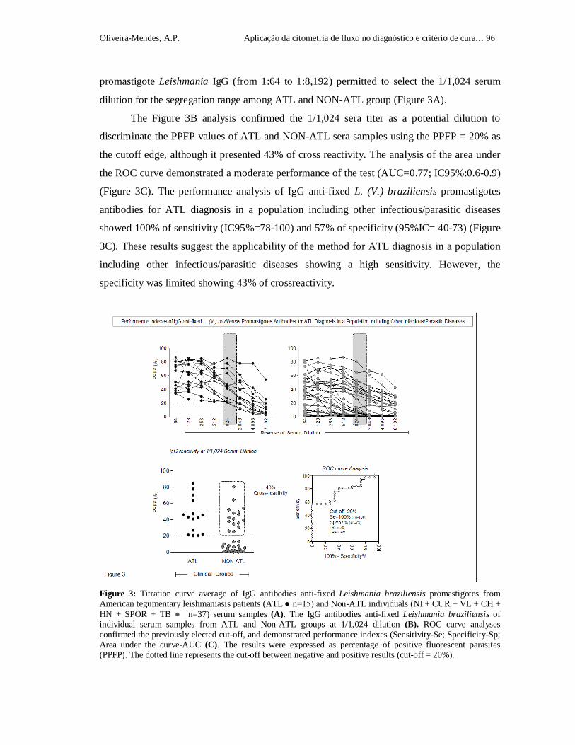
Oliveira-Mendes, A.P. Aplicação da citometria de fluxo no diagnóstico e critério de cura... 96
promastigote Leishmania IgG (from 1:64 to 1:8,192) permitted to select the 1/1,024 serum
dilution for the segregation range among ATL and NON-ATL group (Figure 3A).
The Figure 3B analysis confirmed the 1/1,024 sera titer as a potential dilution to
discriminate the PPFP values of ATL and NON-ATL sera samples using the PPFP = 20% as
the cutoff edge, although it presented 43% of cross reactivity. The analysis of the area under
the ROC curve demonstrated a moderate performance of the test (AUC=0.77; IC95%:0.6-0.9)
(Figure 3C). The performance analysis of IgG anti-fixed L. (V.) braziliensis promastigotes
antibodies for ATL diagnosis in a population including other infectious/parasitic diseases
showed 100% of sensitivity (IC95%=78-100) and 57% of specificity (95%IC= 40-73) (Figure
3C). These results suggest the applicability of the method for ATL diagnosis in a population
including other infectious/parasitic diseases showing a high sensitivity. However, the
specificity was limited showing 43% of crossreactivity.
Figure 3: Titration curve average of IgG antibodies anti-fixed Leishmania braziliensis promastigotes from American tegumentary leishmaniasis patients (ATL ● n=15) and Non-ATL individuals (NI + CUR + VL + CH + HN + SPOR + TB ● n=37) serum samples (A). The IgG antibodies anti-fixed Leishmania braziliensis of individual serum samples from ATL and Non-ATL groups at 1/1,024 dilution (B). ROC curve analyses confirmed the previously elected cut-off, and demonstrated performance indexes (Sensitivity-Se; Specificity-Sp; Area under the curve-AUC (C). The results were expressed as percentage of positive fluorescent parasites (PPFP). The dotted line represents the cut-off between negative and positive results (cut-off = 20%).

Oliveira-Mendes, A.P. Aplicação da citometria de fluxo no diagnóstico e critério de cura... 97
3.4 Major infectious/parasitic diseases with cross reactivating in IgG anti-fixed L. (V.)
braziliensis promastigotes antibody assay
Serum samples from 15 patients positive for ATL, 8 spontaneously cured (CUR) and
8 healthy individuals (NI) together with 21 sera samples for the differential diagnosis
assessment including Visceral Leishmaniasis (VL=04), Chagas disease (CH=10), Hanseniasis
(HN=05), Sporotrichosis (SPOR=01) and Tuberculosis (TB=01) were tested aiming the
evaluation of the clinical value of the differential diagnosis in a population including other
infectious/parasitic diseases (Figure 4A). The analysis of the 1/1,024 sera dilution
(PPFP≥20% as cutoff) showed high cross reactivity from ATL samples when compared with
VL (100%) and CH (100%) and HN (40%) with a great amount of false-positives (Figure 4).
On the other hand, sera samples from SPOR and TB group did not show any cross reactivity
with ATL group (Figure 4B).
4. DISCUSSION
ATL has been diagnosed microscopically by the identification of Leishmania parasites
or amastigotes within macrophages of sample tissue. However, the presence of parasites
depends on the duration of lesions, being fewer in number in chronic lesions (Andrade-
Narvaez, 2005; Ameen, 2010). Although these tests are 100% specific, their sensitivity varies
with the technician expertise. To maximize sensitivity and specificity, the following four
investigations should ideally be performed: direct microscopy, histopathology, culture and
PCR (Ameen, 2010). Therefore, serological tests are very important alternative for monitoring
clinical cure, especially because the spontaneously cured patients, which presents decrease of
the parasite load and of IgG antibody reactivity (Brito et al.,2001), can be identified.
The present work evaluated the detection of IgG antibodies anti-fixed L. (V.)
braziliensis promastigote by flow cytometry to identify active infection, spontaneous cure and
to proceed differential diagnosis of ATL. Several serological methods have been performed
for diagnosis of ATL and for monitoring clinical cure through the decrease of parasite load
after the healing of lesions (Romero et al., 2005; Mendonça et al., 2004). Previously, Romero
et al (2005) demonstrated a decrease on the reactivity levels of IgG after specific treatment,
showing that ELISA technique has a better performance compared to the IFA test. However,

Oliveira-Mendes, A.P. Aplicação da citometria de fluxo no diagnóstico e critério de cura... 98
Figure 4. Titration curve average of IgG antibodies anti-fixed Leishmania braziliensis promastigotes from Visceral Leishmaniasis patients (VL ■ n=04); Chagas disease patients (CH ♦ n=10); Hanseniasis patients (HN ● n=05), Sporotrichosis patient (SPOR ▲n=01) and Tuberculosis patient (TB ▼n=01) serum samples (A). The IgG antibodies anti-fixed Leishmania braziliensis of individual serum samples from ATL, NI, CUR, VL, CH, HN, SPOR and TB groups at 1/1,024 dilution (B). The results were expressed as percentage of positive fluorescent parasites (PPFP). The dotted line represents the cut-off between negative and positive results (cut-off = 20%).

Oliveira-Mendes, A.P. Aplicação da citometria de fluxo no diagnóstico e critério de cura... 99
the cure criterions for ATL remain controversial, since the persistence of positivity in
conventional serological tests can be seen after spontaneous or therapeutic cure.
Several efforts have been made aiming to build up a more reliable serological
approach to ATL diagnosis by using distinct antigen preparations to detect anti-Leishmania
antibodies (Gonçalves et al., 2002; Celeste et al., 2004; Ryan et al., 2002; Rocha et al., 2006).
The spontaneously cured patients (CUR) are the target of immunological research, possibly
because they present a differential and more efficient protective response when compared
with the ATL active infection cases (Snoek et al., 2009; Brito et al., 2001). Our results from
CUR group showed a high performance of the method, presenting low reactivity of PPFP
(PPFP<20%) similar with NI individuals. The infection by L. (V.) braziliensis tends to be
difficult to heal and sometimes it can cause metastasis, involving mucous lesions on the
nasopharynx (WHO, 2010). However, the spontaneous cure of the lesions suggests the
development of an immune response capable to control the infection by Leishmania (Brito et
al., 2001). Previous studies showed that spontaneously cured patients present low levels of
IgG antibodies, which correlate with the decrease of parasite load. Thus, the low reactivity of
antibodies showed by CUR patients in the present work suggests that our method can be
useful to monitoring clinical cure. These findings are in agreement with other authors that also
have shown that low levels of antibodies after therapy for ATL can indicate a successful cure
(Lemos et al., 2007; Pissinate et al., 2008; Garcia et al., 2009).
It is important to point out that a differential diagnosis for ATL is essential and
desirable, since some diseases such as hanseniasis, skin cancer, tuberculosis and cutaneous
mycosis present similar clinical aspects with ATL, generating false-positive diagnosis.
Besides that, Chagas disease (CH) and visceral leishmaniasis (VL), which present co-
endemicity with ATL, are targets of cross reactivity (Goto and Lindoso, 2010; Reithinger et
al 2007). Here, we report the applicability of flow cytometry for ATL diagnosis in a
population including other infectious/parasitic diseases (VL and CH). Our data suggested
that the new flow cytometry-based methodology has a good performance to identify active
infection and spontaneous cure, however has high cross reactivity with VL, CH and HN,
presenting great percentage of false-positive results.
These findings were already expected since some of the leishmanial antigens have
common cross-reactive epitopes shared with other microorganisms particularly
Trypanosoma, Mycobacteria, Plasmodium (Kar, 1995). Indeed, the use of whole
promastigote as the source of antigens in the direct agglutination test (DAT) and
immunofluorescent test (IFAT) gave cross reactions with the sera of leprosy, tuberculosis,

Oliveira-Mendes, A.P. Aplicação da citometria de fluxo no diagnóstico e critério de cura... 100
and African trypanosomiasis patients, representing for long a challenge to researchers
(Celeste et al., 2004; Junqueira Pedras et al., 2003). Additionally, high frequency of cross
reactivity was observed for sera from Chagas disease patients and with malaria patients, with
both antigens, as previously reported for IgG and IgG3 (Luquetti, 1990, Marzochi, 1992,
Teixeira and Vexanat, 1996).
The differential diagnosis, in order to reduce the disadvantage related to the
occurrence of cross reactivity is highly relevant, mainly in regions where these parasites are
co-endemic (Rocha et al., 2006). Several researchers have aimed to overcome the limitations
of ATL serological diagnosis with alternative antigenic preparations to detect Leishmania
antibodies (Pappas et al. 1983; Gonçalves et al. 2002; Celeste et al. 2004). The use of live
promastigotes would represent a useful tool to increase specificity, since only external
membrane epitopes are accessible for IgG binding, avoiding cross reactivity with intracellular
components commonly distributed among trypanosomatidae (Andrade et al., 1988;
Chiaramonte et al., 1996). On the other hand, the use of fixed promastigotes represents a
practical way to store a massiveness amount of antigenic support that contributes to large
scale production. Moreover, the risk of infection during manipulation of live parasites, further
support the use of fixed preparations as antigen source for serological tests.
In summary, our data suggest the potential and applicability of the cytometry-based
methodology for an alternative diagnosis for ATL active infection and follow-up cure
monitoring of ATL patients but advise that adjustments in the methodology are still required
in order to improve its performance for the differential diagnosis of ATL in co-endemic areas
for other infectious/parasitic disease.
ACKNOWLEDGMENTS
This work was supported by the State of Pernambuco Research Foundation (FACEPE), by the
Brazilian National Research Council (CNPq) and by the Oswaldo Cruz Foundation
(FIOCRUZ). The authors are grateful to LF da Rocha for technical assistance, to the
Leishmaniasis Reference Service in the CPqAM / FIOCRUZ for laboratory support and Dr.
Patricia Moura from University of Pernambuco (UPE) for attachment FFA thanks Science
without Borders Program for post doctorate scholarship(MEC/MCTI/CAPES/CNPq/FAPs).
ATC and OAMF thank CNPq for fellowship (PQ).

Oliveira-Mendes, A.P. Aplicação da citometria de fluxo no diagnóstico e critério de cura... 101
REFERENCES
1- Adams, E.R., Schoone, G.J., Ageed, A.F., Safi, S.E., Schallig, H.D.F.H. 2010. Development of a Reverse Transcriptase Loop-Mediated Isothermal Amplification (LAMP) Assay for the Sensitive Detection of Leishmania Parasites in Clinical Samples. Am J Trop Med Hyg, 82: 591–596. 2- Alves, W.A., and Bevilacqua, P.D. 2004. Reflexões sobre a qualidade do diagnóstico da leishmaniose visceral canina em inquéritos epidemiológicos: o caso da epidemia de Belo Horizonte, Minas Gerais, Brasil, 1993-1997. Cad Saúde Pública, 20: 259-265. 3- Ameen, M. 2010. Cutaneous leishmaniasis: advances in disease pathogenesis, diagnostics and therapeutics. Clin Exp Dermatol, 35: 699-705. 4- Anthony, R.L., Christensen, H.A., Johnson, C.M. 1980. Immunobsorbent assay (ELISA) for the serodiagnosis of New World Leishmaniasis. Am J Trop Med Hyg, 29: 190-194. 5-Brandão-Filho, S.P., Brito, M.E., Carvalho, F.G., Ishikawa, E.A., Cupolillo, E., Floeter-Winter, L., Shaw, J.J. 2003. Wild and synanthropic hosts of Leishmania (Viannia)braziliensis in the endemic cutaneous leishmaniasis locality of Amaraji, Pernambuco State, Brazil. , 97(3): 291-306. 6- Brito, M.E.F. 1999. Desenvolvimento de um método diagnóstico para a leishmaniose tegumentar americana com base em Western blot de frações antigênicas de Leishmania braziliensis. Rev Soc Bras Med Trop, 32(3): 313-314. 7- Brito, M.E., Mendonça, M.G., Gomes, Y.M., Jardim, M.L., Abath, F.G. 2000. Identification of potentially diagnostic Leishmania braziliensis antigens in human cutaneous leishmaniasis by immunoblot analysis, 7(2): 318-321. 8- Brito, M.E., Mendonça, M.G., Gomes, Y.M., Jardim M.L., Abath, F.G. 2001. Dynamics of the antibody response in patients with therapeutic or spontaneous cure of American cutaneous leishmaniasis. Trans R Soc Trop Med Hyg, 95(2): 203-206. 9-Brito, M.E., Andrade, M.S., Mendonça, M.G., Silva, C.J., Almeida, E.L., Lima, B.S., Félix, S.M., Abath, F.G., da Graça, G.C., Porrozzi, R., Ishikawa, E.A., Shaw, J.J., Cupolillo, E., Brandão-Filho, S.P. 2009. Species diversity of Leishmania (Viannia) parasites circulating in an endemic area for cutaneous leishmaniasis located in the Atlantic rainforest region of northeastern Brazil, 14(10): 1278-1286.

Oliveira-Mendes, A.P. Aplicação da citometria de fluxo no diagnóstico e critério de cura... 102
10- de Brito, M.E., Andrade, M.S., Dantas-Torres, F., Rodrigues, E.H., Cavalcanti, M. de P., de Almeida, A.M., Brandão-Filho, S.P. 2012. Cutaneous leishmaniasis in northeastern Brazil: a critical appraisal of studies conducted in State of Pernambuco. Rev Soc Bras Med Trop, 45(4): 425-429. 11- Carvalho, E.M., Correia Filho, D., Bacellar, O., Almeida, R.P., Lessa, H., Rocha, H. 1995. Characterization of the immune response in subjects with self healing cutaneous leishmaniasis. Am J Trop Med Hyg. 53(3): 273-277. 12- Celeste, B.J., Angel, S.O., Castro, L.G., Gidlund, M., Goto, H. 2004. Leishmania infantum heat shock protein 83 for the serodiagnosis of tegumentary leishmaniasis. Braz J Med Biol Res, 37(11):1591-1593. 13- Chiaramonte, M.G., Frank, F.M., Furer, G.M., Taranto, N.J., Margni, R.A., Malchiodi, E.L. 1999. Polymerase chain reaction reveals Trypanosoma cruzi infection suspected by serology in cutaneous and mucocutaneous leishmaniasis patients. Acta Trop, 72(3):295-308. 14- Desfeux, P. 2004. Leishmaniasis: current situation and new perspectives. Comp Immunol Microbiol Infect Dis 27 (5): 305-318. 15- Garcia, L.M., Coelho-Dos-Reis, J.G., Peruhype-Magalhães, V., Teixeira-Carvalho, A., Rocha, R.D., Araújo, M.S., Gomes, I.T., Carvalho, S.F., Dietze, R., Lemos, E.M., Andrade, M.C., Martins-Filho, O.A. 2009. Anti- fixed Leishmania chagasi promastigotes IgG antibodies detected by flow cytometry (FC-AFPA-IgG) as a tool for serodiagnosis and for post-therapeutic cure assessment in American visceral leishmaniasis. J Immunol Methods, 350(1-2):36-45. 16-Gonçalves, C.C.M., Reiche, E.M., Abreu Filho, B.A., Silveira, T.G., Felizardo, T.C., Maia, K.R., Costacurta, R., Padovesi, E.J., Dias Filho, B.P., Jankevicius, S.I., Jankevicius, J.V. 2002. Evaluation of antigens from various Leishmania species in a Western blot for diagnosis of American tegumentary leishmaniasis.Am J Trop Med Hyg, 66(1):91-102. 17- Gontijo, B., and Carvalho, M.L.R. 2003. Leishmaniose tegumentar americana. Rev Soc Bras Med Trop, 36:71-80. 18- Goto, H., and Lindoso, J.A.L. Current diagnosis and treatment of cutaneous and mucocutaneous leishmaniasis. Expert Rev Anti-infect Ther, 8(4): 419-433. 19- Junqueira Pedras, M., Orsini, M., Castro, M., Passos, V.M., Rabello, A. 2003. Antibody subclass profile against Leishmania braziliensis and Leishmania amazonensis in the diagnosis and follow-up of mucosal leishmaniasis. Diagn Microbiol Infect Dis, 47: 477-485.

Oliveira-Mendes, A.P. Aplicação da citometria de fluxo no diagnóstico e critério de cura... 103
20- Kar K. 1995. Serodiagnosis of Leishmaniasis. Critical Rev Microb 21(2): 123-152. 21- Lemos, E.M., Gomes, I.T., Carvalho, S.F., Rocha, R.D., Pissinate, J.F., Martins-Filho, O.A., Dietze, R. 2007. Detection of anti-leishmania (Leishmania) chagasi immunoglobulin G flow cytometry for cure assess mentfollowing chemotherapeutic treatment of American visceral leishmaniasis. Clin Vaccine Immunol, 14(5):569-576. 22- Malchiodi, E.L., Chiaramonte, M.G., Taranto, N.J., Zwirner, N.W., Margni, R.A.1994. Cross-reactivity studies and differential serodiagnosis of human infections caused by Trypanosoma cruzi and Leishmania spp; use of immunoblotting and ELISA with a purified antigen (Ag163B6). Clin Exp Immunol. 97(3):417-23. 23- Mendonça, M.G., de Brito, M.E., Rodrigues, E.H., Bandeira, V., Jardim, M.L., Abath, F.G. 2004. Persistence of Leishmania parasites in scars after clinical cure of American cutaneous leishmaniasis: is there a sterile cure? J Infect Dis, 189: 1018-1023. 24- Ministério da Saúde. 2010. Manual de Vigilância da Leishmaniose Tegumentar Americana. 2 ed. Brasília, Brazil. 25- de Oliveira, A.P., de Castro, M.C., de Almeida, A.F., Souza, M.A., de Oliveira, B.C., Reis, L.C., Goto, H., de Brito, M.E., Celeste, B.J., Martins-Filho, O.A., Pereira, V.R. 2013. Comparison of flow cytometry and indirect immunofluorescence assay in the diagnosis and cure criterion after therapy of American tegumentary leishmaniasis by anti-live Leishmania (Viannia) braziliensis immunoglobulin G. J Immunol Methods, 387:245-253. 26- Pereira, V.R., Reis, L.C., Souza, M.A., de Oliveira, A.P., de Brito, M.E., Lage, P.S., Andrade, M.C., Rocha, R.D., Martins-Filho, O.A. 2012. Evaluation of anti-lived and anti-fixed Leishmania (Viannia)braziliensis promastigote IgG antibodies detected by flow cytometry for diagnosis and post-therapeutic cure assessment in localized cutaneous leishmaniasis. Diagn Microbiol Infect Dis, 74: 292-298. 27- Pissinate, J.F., Gomes, I.T., Peruhype-Magalhães, V., Dietze, R., Martins-Filho, O.A., Lemos, E.M. 2008. Upgrading the flow-cytometric analysis of anti-Leishmania immunoglobulins for the diagnosis of American tegumentary leishmaniasis, J Immunol Methods, 336:193-202. 28- Reis, L.C., de Brito, M.E., Souza, M.A., Pereira, V.R. 2006. Mecanismos imunológicos na resposta celular e humoral na leishmaniose tegumentar americana. Rev Patol Trop, 35: 103-115.

Oliveira-Mendes, A.P. Aplicação da citometria de fluxo no diagnóstico e critério de cura... 104
29- Reithinger, R., Dujardin, J.C., Louzir, H., Pirmez, C., Alexander, B., Brooker, S. 2007. Cutaneous Leishmaniasis. Lancet Infect Dis, 7:581-596. 30- Rocha, R.D.R., Gontijo, C.M.F., Carvalho, A.T., Corrêa- Oliveira, R., Marques, M.J., Genaro, O., Mayrink, W., Martins-Filho, O.A. 2002, Anticorpos antipromastigotas vivas de Leishmania (Viannia) braziliensis, detectados pela citometria de fluxo, para identificação da infecção ativa de leishmaniose tegumentar americana. Rev. Soc. Bras. Med. Trop, 35:551-562. 31- Rocha, R.D., Gontijo, C.M., Elói-Santos, S.M., Teixeira-Carvalho, A., Corrêa-Oliveira, R., Ferrari, T.C., Marques, M.J., Mayrink, W., Martins-Filho, O.A. 2006. Clinical value of anti-live Leishmania (Viannia) braziliensis immunoglobulin G subclasses, detected by flow cytometry, for diagnosing active localized cutaneous leishmaniasis. Trop Med Int Health, 11: 156-166. 32- Rodríguez, V., Centeno, M., Ulrich, M. 1996. The IgG isotypes of specific antibodies in patients with American cutaneous leishmaniasis; relationship to the cell-mediated immune response. Parasit Immunol, 18:341-345. 33- Romero, G.A., de la Glória Orge Orge, M., de Farias Guerra, M.V., Paes, M.G., de Oliveira Macêdo, V., de Carvalho, E.M. 2005. Antibody response in patients with cutaneous leishmaniasis infected by Leishmania (Viannia) braziliensis or Leishmania (Viannia) guyanensis in Brazil. Acta Trop, 93: 49-56. 34- Silveira, F. T.; Lainson, R.; Corbett, C. E. 2004. Clinical and immunopathological spectrum of American cutaneous leishmaniasis with special reference to the disease in Amazonian Brazil: a review. Mem Inst Oswaldo Cruz, 99: 239-251. 35- van der Snoek EM, Lammers AM, Kortbeek LM, Roelfsema JH, Bart A, Jaspers CA. 2009. Spontaneous cure of American cutaneous leishmaniasis due to Leishmania naiffi in two Dutch infantry soldiers. Clin Exp Dermatol. 34(8):e889-891. 36- de Souza, M.A., da Silva, A.G., Afonso-Cardoso, S.R., Favoreto, S.J., Ferreira, M.S. 2005. Perfil de isotipos de imunoglobulinas e subclasses de IgG na leishmaniose tegumentar americana. Rev Soc Bras Med Trop, 38:137-141. 37- Trujillo, C., Ramírez, R., Vélez, I.D., Berberich, C. 1999. The humoral immune response to the kinetoplastid membrane protein-11 in patients with American leishmaniasis and chagas disease: prevalence of IgG subclasses and mapping of epitopes. Immunol Lett, 70: 203-209.

Oliveira-Mendes, A.P. Aplicação da citometria de fluxo no diagnóstico e critério de cura... 105
38- Antunes Uchôa, C.M., Barrientos Serra, C.M., Duarte, R., Magalhães, C.M., Silva, R.M., Theophilo, F., Figliuolo, L.P., Horta, F.T., Madeira, M.F. 2001. Serological and epidemiological features of canine American tegumentary Leishmaniasis from Maricá, Rio de Janeiro, Brazil. Rev Soc Bras Med Trop. 34(6): 563-568. 39 – World Health Organization. 2010. Control of the leishmaniasis: report of a meeting of the WHO Expert Committee on the Control of Leishmaniases.WHO Press, Geneva.
Oliveira-Mendes, A.P. Aplicação da citometria de fluxo no diagnóstico e critério de cura... 106
6. CONCLUSÕES
I- A utilização de formas promastigotas vivas ou fixadas de Leishmania Viannia
braziliensis, apresentou sensibilidade semelhante no estudo da LTA por citometria de
fluxo, portanto aplicável como metodologia alternativa no diagnóstico da LTA.
II- A reatividade da Leishmania (Viannia) braziliensis no monitoramento da cura clínica
dos pacientes após o tratamento por citometria de fluxo demonstrou ser mais específica
utilizando parasitos vivos.
III- A análise comparativa dos valores de sensibilidade entre citometria de fluxo e
imunofluorescência indireta, utilizando os pacientes AT, demonstrou que a citometria de
fluxo foi positiva na presença da doença.
IV- A aplicabilidade da citometria de fluxo como critério de cura da LTA foi confirmada a
partir do acompanhamento dos pacientes 1, 2 e 5 anos após o tratamento da LTA, onde se
observou melhor ganho em especificidade da citometria de fluxo em relação a
imunofluorescência indireta.
V- A porcentagem de parasitos fluorescentes positivos observadas nos pacientes curados
espontaneamente localizou-se numa região de baixa reatividade em 100% dos pacientes,
cofirmando a reprodutibilidade da citometria de fluxo na identificação de pacientes
curados.
VI- O diagnóstico diferencial da LTA utilizando soros de pacientes com DC e LV
demonstrou reação cruzada por citometria de fluxo. Porém, apresentou aplicabilidade
com soros de pacientes com outras doenças dermatológicas, clinicamente semelhantes a
LTA.
VII- Portanto, a presente tese demonstrou que os ensaios realizados utilizando anticorpo IgG
detectados por Leishmania (V.) braziliensis através citometria de fluxo representam além
de uma ferramenta alternativa para o diagnóstico da LTA, devido a reprodutibilidade e a
possibilidade de identificar pacientes com a infecção e após cura clínica ou espontânea;
mais também abrem perspectivas para a utilização no monitoramento e critério de cura da
LTA.

Oliveira-Mendes, A.P. Aplicação da citometria de fluxo no diagnóstico e critério de cura... 107
7. PERSPECTIVAS
I- Avaliar a aplicabilidade da citometria de fluxo na identificação dos pacientes com doença
ativa e após cura clínica por tratamento quimioeterápico ou espontânea com ênfase na
pesquisa de isotipos IgG (IgG1, IgG2, IgG3, IgG4) anti-promastigotas vivas e fixadas de
Leishmania (Viannia) braziliensis;
II- Analisar a curva de titulação dos anticorpos IgG por citometria de fluxo na identificação
de pacientes 10 anos após o tratamento e a partir da curva ROC determinar os melhores
valores de PPFP para o ensaio.
III- Avaliar a aplicabilidade da citometria de fluxo, utilizando-se diferentes proteínas
recombinantes, como metodologia alternativa no diagnóstico e monitoramento pós-
terapêutico de pacientes infectados com LTA.

Oliveira-Mendes, A.P. Aplicação da citometria de fluxo no diagnóstico e critério de cura... 108
REFERÊNCIAS BIBLIOGRÁFICAS ALVES, W. A.; BEVILACQUA, P. D. Reflexões sobre a qualidade do diagnóstico da leishmaniose visceral canina em inquéritos epidemiológicos: o caso da epidemia de Belo Horizonte, Minas Gerais, Brasil, 1993-1997. Caderno de Saúde Pública, Rio de Janeiro, v. 20, n. 1, p. 259-265, Jan/Fev, 2004. ALMEIDA, A.F. Avaliação da produção de citocinas Th 17, Th1 e Th2 por linfócitos T em pacientes com leishmaniose tegumentar americana. Fundação Oswaldo Cruz, Pernambuco, Brasil, 2013. AMATO,V. S. et al. An avaluation of clinical, serologic, anatomopathologic and imunohistochemical findings for fifteen patients with mucosal leishmaniasis before and after treatment. Revista do Instituto de Medicina Tropical, São Paulo, v. 40, n. 1, p. 23-30, jan/Feb, 1998. AMEEN, M. Cutaneous leishmaniasis: advances in disease pathogenesis, diagnostics and therapeutics. Clinical and Experimental Dermatology, v. 35, p. 699-705, Jan, 2010. ANDRADE, B.B. et. al. Métodos Diagnósticos da Leishmaniose Tegumentar: Fatos, Falácias e Perspectivas Diagnostic Methods of Tegumentary Leishmaniasis: Facts, Fallacies and Perspectives. Gazeta Médica da Bahia, v. 75, n. 1, p. 75-82, Jan/Jun, 2005. ANDRADE, M.S. et al. New outbreak of American tegumentary leishmaniasis in a military training center in the Zona da Mata region, in the north of the State of Pernambuco. Revista da Sociedade Brasileira de Medicina Tropical, v. 42, n. 5, p. 594-596, Set-Out, 2009. ANDRADE, T. A. S. et al. Perfil epidemiológico dos casos notificados de leishmaniose tegumentar americana no município de Igarassu (PE) no período de 2008 a 2010. Scire Salutis, Aquidabã, v.2, n.2, p.5‐15, 2012. ÁVILA, S. C.G.P. D. et. al. Estudo retrospectivo dos casos de Leishmaniose Tegumentar Americana diagnosticados no laboratório de patologia do Hospital de Base da FAMERP nos anos de 1995-2000, com enfoque clínico e anatomopatológico. Arquivos de Ciências da Saúde,v.11, n.2, Apr/Jun, 2004. BACELLAR, O. et al. Interleukin 17 production among patients with american cutaneous leishmaniasis. The Journal of Infectious Diseases, Boston, v. 200, p. 75-78, May, 2009.

Oliveira-Mendes, A.P. Aplicação da citometria de fluxo no diagnóstico e critério de cura... 109
BASANO, S.A.; CAMARGO L.M.A. Leishmaniose tegumentar americana: histórico, epidemiologia e perspectivas de controle. Revista Brasileira de Epidemiologia, v. 7, n.3, p. 328-337, Sep, 2004. BENSOUSSAN, E., et al. Comparison of PCR assays for diagnosis of cutaneous leishmaniasis. Journal of Clinical Microbiology, Washington, v. 44, n. 4, p. 1435– 1439, Apr. 2006. BITTENCOURT, A.L., et al. Pesquisa de anticorpos circulantes pelo método de imunofluorescência na leishmaniose tegumentar. Revista do Instituto de Medicina Tropical de São Paulo, v. 10, n. 4, p. 247-252, Jul/Ago, 1968. BOURDOISEAU, G., et al. Effective humoral and cellular immunoprotective responses in Li ESAp-MDP vaccinated protected dogs. Veterinary Immunology and Immunopathology, v.128, p. 71-78, 2009. BRANDÃO-FILHO, S. P., et al. Epidemiological surveys confirm an increasing burden of cutaneous leishmaniasis in north-east Brazil. Transactions of The Royal Medicine Hygiene, London, v. 93, p. 488-494, Sep/Oct. 1999. BRANDÃO-FILHO, S.P. et al. Wild and synanthropic hosts of Leishmania (Viannia)braziliensis in the endemic cutaneous leishmaniasis locality of Amaraji, Pernambuco State, Brazil. Transactions of the Royal Society of Tropical Medicine and Hygiene, London, v. 97, p.291-296, May/June, 2003. BRELAZ, M. C. et al. Leishmania (Viannia) braziliensis antigenic fractions: the immune response characterization of patients at the initial phase of disease. Parasite Immunology, Oxford, v.34., n. 4, p. 236-239, 2012. BRITO, M. E. F., et al. Human cutaneous leishmaniasis due to a new enzymatic variant of Leishmania (Viannia) braziliensis occurring in Pernambuco, Brazil. Memórias do Instituto Oswaldo Cruz, Rio de Janeiro, v. 88, p. 633-634, Oct. 1993. BRITO, M.E.F. Desenvolvimento de um método diagnóstico para a leishmaniose tegumentar americana com base em Western blot de frações antigênicas de Leishmania braziliensis. Revista da Sociedade Brasileira de Medicina Tropical, v. 32, n. 3, p. 313-314, Mai/Jun, 1999. BRITO, M. E. F., et al. Identification of potentially diagnostic Leishmania braziliensis antigens in human cutaneous leishmaniasis by immunoblot analysis. Clinical and diagnostic Laboratory Immunology, Washington, v. 7, n. 2, p. 318-321, Mar. 2000.

Oliveira-Mendes, A.P. Aplicação da citometria de fluxo no diagnóstico e critério de cura... 110
BRITO, M. E. F., et al. Dynamics of the antibody response in patients with therapeutic or spontaneous cure of American cutaneous leishmaniasis. Transactions of the Royal Society of Tropical Medicine and Hygiene, London, v. 95, p. 203-206, Mar/Apr.2001. BRITO, M. E. F., et al. Clinical epidemiological profile of American tegumentary leishmaniasis at the Pinto Sugar Mill in Moreno Municipality, Greater Metropolitan Recife, Pernambuco State, Brazil. Cadernos de Saúde Pública, v. 24, n.10, p. 2445- 2448, Oct, 2008. BRITO, M. E. F. et al. Species diversity of Leishmania (Viannia) parasites circulating in an endemic area for cutaneous leishmaniasis located in the Atlantic rainforest region of northeastern Brazil. Tropical Medicine and International Health, Malden, v. 14, n. 10, p. 1-9, 2009. BRITO, M. E. F. et al. Cutaneous leishmaniasis in northeastern Brazil: a critical appraisal of studies conducted in State of Pernambuco. Revista da Sociedade Brasileira de Medicina Tropical, Rio de Janeiro, v.45, n.4, p.425-429, 2012. CAMARGO EP. Growth and differentiation of Trypanosoma cruzi. I. Origin of metacyclic trypanosomes in liquid media. Revista do Instituto de Medicina Tropical de Sao Paulo, v. 6, p. 93-100, 1964. CARVALHO, L.P. et al. Protective and pathologic immune responses in human tegumentary leishmaniasis. Frontiers in immunology, Lausanne, v.2, p.301, 2012. CARVALHO, E. M. et al. Characterization of the immune response in subjects with self healing cutaneous leishmaniasis. American Journal of Tropical Medicine and Hygiene, Northbrook, v. 53, n. 3, p. 273-277, 1995. CASTELLANO, L. R. et al. Th1/Th2 immune responses are associated with active cutaneous leishmaniasis and clinical cure is associated with strong interferon-gamma production. Human Immunology, New York, v. 70, n. 6, p. 383-390, 2009. CASTRO, M.C.A.B. Estudo do papel de linfócitos T CD4+ e CD8+ e suas citocinas na Leishmaniose Tegumentar Americana. Tese doutorado. Universidade Federal de Pernambuco, Pernambuco, Brasil, 2013. CELESTE, B. J. et al. Leishmania infantum heat shock protein 83 for the serodiagnosis of tegumentary leishmaniasis. Brazilian Journal of Medical and Biological Research, v.37, p. 1591–1593, 2004.
Oliveira-Mendes, A.P. Aplicação da citometria de fluxo no diagnóstico e critério de cura... 111
CHATTERJEE, M., et al. Distribution of IgG subclasses in antimonial unresponsive Indian kala-azar patients. Clinical and Experimental Immunology, v.114, n.3, p. 408- 413, 1998. CITADIN, R. F. Leishmaniose visceral: relato de três casos e revisão da literatura. Universidade Federal de Santa Catarina, Florianópolis, Brasil, 2008. CONVIT, J. et al. Immunotherapy of American cutaneous leishmaniasis in Venezuela during the period 1990-1999. Transactions of the Royal Society of Tropical Medicine and Hygiene, v. 97, p. 469-72, 2003. CONVIT , J. et al. Therapy of Venezuelan patients with severe or early lesions of diffuse cutaneous leishmaniasis with a vaccine containing pasteurized Leishmania promastigotes and bacillus Calmette- Guerin – preliminary report. Memórias do Instituto Oswaldo Cruz, v. 99, p. 57-62, 2004. COSTA, N. et al. Vaccines for the leishmaniasis: Proposals for a research agenda. PLOS Neglected Tropical Diseases, v. 5, n.3, p.1-9, 2011. CROFT, L.S; COOMBS, H.G. Leishmaniasis – current chemotherapy and recent advances in the search for novel drugs. Trends in parasitology, v. 19, p. 502- 508, 2003. CROFT, S. L.; SUNDAR, S.; FAIRLAMB, A. Drug Resistance in Leishmaniasis. Clinical Microbiology Reviews, Washington, v. 19, n. 1, p. 111-126, 2006. DA-CRUZ, A. M. et al. Flow cytometric analysis of cellular infiltrate from American tegumentary leishmaniasis lesions. The British journal of dermatologys, Derby, v. 153, n. 3, p. 537-543, 2005. DATASUS, Leishmaniose tegumentar americana- Casos confirmados notificados no sistema de informação de agravos de notificação- SINAN Net. Disponível em: http://dtr2004.saude.gov.br/sinanweb/tabnet/dh?sinannet/lta/bases/ltabrnet.def. Acesso em: janeiro de 2015. DE BRUIJIN, M.; BARKER, D. C. Diagnosis of New World leishmaniasis: specific detection of species of the Leishmania braziliensis complex by amplification of kinetoplast DNA. Acta Tropica, Cambridge, v. 52, p. 45-58, Set., 1992. DESJEUX, P. Leishmaniasis: current situation and new perspectives. Comparative Immunology, Microbiology & Infectious Diseases, Exeter, v. 27, p. 305-318, Sep. 2004.

Oliveira-Mendes, A.P. Aplicação da citometria de fluxo no diagnóstico e critério de cura... 112
DOENÇAS NEGLIGENCIADAS. Ciência e tecnologia para o desenvolvimento nacional- Estudos estratégicos. Academia Brasileira de Letras, Rio de Janeiro, Brasil, 2010. ELMAHALLAWY, E. K. et al., Diagnosis of leishmaniasis. The Journal of Infection in Developing Countries, v. 8, n. 8, p. 961-972. 2014. EL-ON, J. Current Status and Perspectives of the Immunotherapy of Leishmaniasis. The Israel Medical Association Journal, V. 11, p. 623- 628, 2009. FALDYNA, M. et al. Lymphocyte subsets in peripheral blood of dogs – a flow cytometric study. Veterinary Immunology and Immunopathology, Amsterdam, v.82, n.1-2, p.23-37, 2001. FNS/FUNDAÇÃO NACIONAL DE SAÚDE. Manual de controle da Leishmaniose Tegumentar Americana. 5ª ed., Ed. Brasília/DF, 2000. GASCAN, H.,et al. Human B cell clones can be induced to proliferate and to switch to IgE and IgG4 synthesis by interleukin 4 and a signal provided by activated CD4+ T cell clones. Journal of Experimental Medicine, v. 173, n.3, p. 747-750, Mar, 1991. GAZOZAI, S.U. et al. Comparison of diagnostic methods in cutaneous leishmaniasis (histopathology compared to skin smears). Pakistan Journal of Pharmaceutical Sciences, v. 23, n. 4, p. 363-366, Oct, 2010. GENARO, O. Leishmaniose Tegumentar Americina. In: NEVES, D. P.; MELO, L. A., GENARO, O.; LINARDI, P. M. Parasitologia Humana, 10ª ed. , São Paulo, Atheneu, cap.8, p.36-53, 2000. GRIMALDI, G.JR., TESH, R. B. – Leishmaniasis of the New World: current concepts and implications for future research. Clinical Microbiology Reviews, Washington, v.6, n. 3, p. 230-250, Jul. 1993. GOMES, M. L. et al. Chagas disease diagnosis: comparative analysis of parasitologic, molecular and serologic methods. American Journal of Tropical Medicine Hygiene, Baltimore, v. 60, p. 205-210, 1999. GONÇALVES, C.C.M., et al. Evaluation of antigens from various Leishmania species in a western blot for diagnosis of American tegumentary leishmaniasis. The American Society of Tropical Medicine and Hygiene, v.66, n. 1, p. 91-102, 2002.

Oliveira-Mendes, A.P. Aplicação da citometria de fluxo no diagnóstico e critério de cura... 113
GONTIJO, B. CARVALHO M. L. R. Leishmaniose tegumentar americana. Revista da Sociedade Brasileira de Medicina Tropical, Uberaba, v. 36, p.71-80, Jan./ Fev., 2003. GOTO, H; LINDOSO, J.A.L. Current diagnosis and treatment of cutaneous and mucocutaneous leishmaniasis. Expert Reviews of Anti-infective Therapy, v. 8, n. 4, p. 419-433, 2010. GOYONLO, V. M. et al. Efficacy of intralesional amphotericin B for the treatment of cutaneous leishmaniasis.Indian Journal of Dermatology, v. 59, n. 6, p. 631, 2014. HANDMAN, E. Leishmaniasis: Current status of vaccine development. Clinical microbiology reviews, v. 14, n. 2, p. 229-243, 2001. HUSSAIN, R, et al. Immunoglobulin G1 (IgG1) and IgG3 antibodies are markers of progressive disease in leprosy. Infection and Immunity, v. 63, n. 2, p. 410-415, Feb, 1995. JANI, I. V. et al. Multiplexed immunoassays by flow cytometry for diagnosis and surveillance of infectious diseases in resourcepoor settings. Lancet Infect Diseases, v. 2, p. 243–250, 2002. KAR, K. Serodiagnosis of leishmaniasis. Critical Reviews in Microbiology, Boca Raton, v. 21, n. 2, p. 123-152, 1995. KEDZIERSKI, L.; EVANS, K. Immune responses during cutaneous and visceral leishmaniasis. Parasitology, v. 141, p. 1544-1562, 2014. KOTRESHA, D.; NOORDIN, R. Recombinant proteins in the diagnosis of toxoplasmosis. Acta Pathologica Microbiologica et Immunologica Scandinavica, v.118, 529–542, 2010. KURNIAWAN, A, et al. Differential expression of IgE and IgG4 specific antibody responses in asymptomatic and chronic human filariasis. Journal of Immunology, v. 150, n. 9, p. 3941-3950, Mai, 1993. LAINSON, R.; SHAW J.J. New World leishmaniasis. The neotropical Leishmania species. In Collier L, Balows A & Sussman M (Eds). Topley & Wilson’s Microbiology and Microbial Infectious Diseases, 9nd Ed., Arnold, London, v. 5, p. 241-266, 1998.

Oliveira-Mendes, A.P. Aplicação da citometria de fluxo no diagnóstico e critério de cura... 114
LIMA, H. C., et al. Resolution of an infection with Leishmania braziliensis confers complete protection to a subsequent challenge with Leishmania major in BALB/c mice. Memórias do Instituto Oswaldo Cruz, Rio de Janeiro, v. 94 p. 71-76, 1999. LORÍA-CERVERA, E.; N.; ANDRADE-NARVÁEZ, F. J. Review animal models for the study of leishmaniasis immunology. Revista do Instituto de Medicina Tropical de São Paulo, v. 56, n. 1, p. 1-11, 2014 MACHADO, P. E.Comparação dos métodos parasitológico, imunológico e molecular na detecção de Leishmania spp. Em amostras de pacientes com leishmaniose tegumentar americana no Estado de Santa Catarina. Dissertação mestrado. Universidade Federal de Santa Catarina, Florianópolis, Brasil, 2004. MANSUETO, P. et al. Leishmaniasis in travelrs: A literature review. Travel Medicine and Infectious Disease, v. 12, p. 563- 581, 2014. MARTINS-FILHO, O.A., et al. Flow cytometry, a new approach to detect anti-live trypomastigote antibodies and monitor the efficacy of specific treatment in Human Chagas' disease. Clinical and diagnostic Laboratory Immunology, v. 2, n. 5, p. 569- 573, 1995. MARTINS, L.; ALEXANDRINO, A.; GUIMARÃES, G. The detection of Leishmania braziliensis DNA in American tegumentary leishmaniasis patients. Revista de Saúde Pública, v.44, n. 3, p. 1-4, 2010. MAYRINK, W., et al. Imunoterapia, imunoquioterapia e Quimioterapia no Tratamento da Leishmaniose Tegumentar Americana. VII Reunião Anual de Pesquisa Aplicada em Leishmanioses. Uberaba, MG. Revista da Sociedade Brasileira de Medicina Tropical, v.35, p.122-123, 2002. MAYRINK, W., et al. Immunotherapy, immunochemotherapy and chemotherapy for American cutaneous leishmaniasis treatment. Revista da Sociedade Brasileira de Medicina Tropical, Rio de Janeiro, v. 39, n. 1, p. 14-21, Jan/Feb. 2006. MENDONÇA, M. G., et al. Persistence of Leishmania parasites in scars after clinical cure of American cutaneous leishmaniasis: is there a sterile cure? Journal of Infectious Diseases, Chicago, v. 189, n. 6, p. 1018-1023, Mar. 2004. MENEZES-SOUZA, D., et al., Epitope mapping of the HSP83.1 protein of Leishmania braziliensis discloses novel targets for immunodiagnosis of tegumentary and visceral clinical forms of leishmaniasis. Clinical and Vaccine Immunology, v. 21, n. 7, p. 949-959, 2014.

Oliveira-Mendes, A.P. Aplicação da citometria de fluxo no diagnóstico e critério de cura... 115
MIGNOGNA, M. D., et al. Mucosal leishmaniasis with primary oral involvement: a case series and a review of the literature. Oral Diseases, 2014. MITROPOULOS, P. et al. New World cutaneous leishmaniasis: updated review of current and future diagnosis and treatment. The American Academy of Dermatology, 2010. MOURA, T. R., et al. Toward a novel experimental model or infection to study American cutaneous leishmaniasis caused by Leishmania braziliensis. Infection and Immunity, Washington, v. 73, n. 9, p. 5827-5834, 2005. MOUGNEAU, E.; BIHL, F.; GLAICHENHAUS, N. Cell biology and immunology of Leishmania. Immunological Reviews, v. 240, p. 286-296, 2011. MS/MINISTÉRIO DA SAÚDE. Manual de Vigilância da Leishmaniose tegumentar americana. 2. ed. Brasília, 2010. MS/MINISTÉRIO DA SAÚDE. Manual de Vigilância da Leishmaniose Tegumentar Americana. Brasília, 180p., 2007. MUKBEL, R., et al. Soluble factors from Leishmania major-specific CD4þT cells and B cells limit L. amazonensis amastigote survival within infected macrophages. Microbes and infeccion, v. 8, p. 2547-2555, 2006. MUTISO, J. M.; et l. Development of Leishmania vaccines: predicting the future from past and present experience. The Journal of Biomedical Research, v. 27, n. 2, p. 85-102, 2013. NAKAGE, A. P. M., et al. Flow cytometry methodology and application in the veterinary hematology. Ciência Rural, v. 35, n. 4, p. 966-973, jul/ago, 2005. NEVES, J. et al. Parasitologia Dinâmica, 3 ed., São Paulo: Atheneu, 2009. NEITZKE-ABREU, H. C., et al. Cutaneous leishmaniasis with atypical clinical manifestations: Case report. ID Cases 1, p. 60-62, 2014. NOAZIN, S., et al. First generation leishmaniasis vaccines: A review of Field efficacy trials. Vaccine, v. 26, p. 6759- 6767, 2008. NICOLLE, C. Nouvelles acquisition sur le Kala-azar culture; innovation auchien; etiologie – C.R. Academy Sciences, Paris, v.146, p.498-499, 1908.
Oliveira-Mendes, A.P. Aplicação da citometria de fluxo no diagnóstico e critério de cura... 116
NOVY, F. G.; MCNEAL, W. J. The cultivation of Trypanosoma brucei: a preliminary note. Journal of the American Medical Association, v.41, p.1266-1268, 1903. NYLEEN, S.; GAUTAM, S. Immunolgical perspectives of leishmaniasis. Symposium Leishmaniasis, v. 2, n. 2, p. 135-146, 2010. NYLÉN, S.; EIDSMO, L. Tissue and immunity in cutaneous leishmaniasis. Parasite Immunology, v. 34, p. 551-561, 2012. NUNES, C.S., et al. Leishmaniose mucosa: considerações epidemiológicas e de tratamento. Revista Brasileira de Medicina Familiar e Comunidade, v. 6, p. 52-56, 2011. OKWOR, I.; UZONNA, J. Vaccines and vaccination strategies against human cutaneous leishmaniasis. Human Vaccines, v. 5,n. 5 p. 291–301, May. 2009. OLIVEIRA, A.P. et al. Comparison of flow cytometry and indirect immunofluorescence assay in the diagnosis and cure criterion after therapy of American tegumentary leishmaniasis by anti-live Leishmania (Viannia) braziliensis immunoglobulin G. Journal of immunological methods, Amsterdam, v.387, n.1-2, p.245-253, 2013. OMS/ ORGANIZAÇÃO MUNDIAL DE SAÚDE. Control of the leishmaniases: report of a meeting of the WHO Expert Committee on the Control of Leishmaniases. Geneva, 2010. OMS/ ORGANIZAÇÃO MUNDIAL DE SAÚDE. Neglected Tropical Diseases- A statistical update- latest data available. Geneva, 2014. O`NEIL CE, et al. Leishmania (Viannia) panamensis – specific IgE and IgA antibodies relation to expression of human tegumentary leishmaniasis. The American Journal of Tropical Medicine and Hygiene, 49: 181-188, 1993. OUAAZ, F.,et al. Role of IgE immune complexes in the regulation of HIV-1 replication and increased cell death of infected U1 monocytes: involvement of CD23/Fc épsilon RII-mediated nitric oxide and cyclic AMP pathways. Molecular Medicine, v. 2, n.1, p. 38-49, Jan, 1996. OZBILGE, H., et al. IgG and IgG subclass antibodies in patients with active cutaneous leishmaniasis. Journal of Medical Microbiology, v. 55, p. 1329-1331, 2006. PACE, D. Leishmaniasis.The British Infection Association, p. 10-18, 2014.

Oliveira-Mendes, A.P. Aplicação da citometria de fluxo no diagnóstico e critério de cura... 117
PASSOS, V. M. A., et al. American cutaneous leishmaniasis: use of a skin test as a redictor of relapse after treatment. Bulletin of the World Health Organization, v.78, n. 8, p. 968-974, 2000. PASSOS, S., et al. Recombinant Leishmania antigens for serodiagnosis of visceral leishmaniasis. Clin Diagn Lab Immunol, v. 12, p. 1164–1167, 2005. PEREIRA, V R A, et al. Evaluation of anti-lived and anti-fixed Leishmania (Viannia)braziliensis promastigote IgG antibodies detected by flow cytometry for diagnosis and post-therapeutic cure assessment in localized cutaneous leishmaniasis. Diagnostic Microbiology and Infectious Disease, v.74, p. 292-298, 2012. PERLMANN, P., et al. Immunoglobulin E, a pathogenic factor in Plasmodium falciparum malaria. Infection and Immunity, v. 65, n.1, p. 116-121, Jan, 1997. PIMENTEL ET, M. I. F., et al. American cutaneous leishmaniasis caused by Leishmania (Viannia) braziliensis resistant to meglumine antimoniate, but with good response to pentamidine: a case report. Revista da Sociedade Brasileira de Medicina Tropical, v. 44, n. 2, p. 254-256,2011. PISSINATI, J. F., et al. Upgrading the flow-cytometric analysis of anti-Leishmania immunoglobulins for the diagnosis of American tegumentary leishmaniasis, Journal of Immunological Methods, v.336, p. 193-202, 2008. REIS, L. C., et al. Mecanismos imunológicos na resposta celular e humoral na leishmaniose tegumentar americana. Revista de Patologia Tropical, São Paulo, v. 35, n. 2, p. 103-115, Maio/Ago. 2006. REIS, L.C. Caracterização da resposta imune celular em portadores de leishmaniose tegumentar americana antes e após tratamento quimioterápico. Dissertação mestrado. Fundação Oswaldo Cruz, Pernambuco, Brasil, 2007. REITHINGER, R. et al. Cutaneous Leishmaniasis. The Lancet Infectious Diseases, New York, v. 7, p. 581-596, 2007. RIBEIRO, C. F., et al. Use of ELISA employing Leishmania (Viannia) braziliensis and Leishmania (Leishmania) chagasi antigens for the detection of IgG and IgG1 and IgG2 subclasses in the diagnosis of American tegumentary leishmaniasis in dogs. Veterinary Parasitology, v.148, p. 200-206, 2007.
Oliveira-Mendes, A.P. Aplicação da citometria de fluxo no diagnóstico e critério de cura... 118
ROCHA, R. D. R., et al. Anticorpos antipromastigotas vivas de Leishmania (Viannia) braziliensis, detectados pela citometria de fluxo, para identificação da infecção ativa de leishmaniose tegumentar americana. Revista da Sociedade Brasileira de Medicina Tropical, Rio de Janeiro, v. 35, n. 6, p. 551-562, Nov/Dec. 2002. ROCHA, R. D. R., et al. Clinical value of anti-live Leishmania (Viannia) braziliensis immunoglobulin G subclasses, detected by flow cytometry, for diagnosing active localized cutaneous leishmaniasis. Tropical Medicine and International Health, Oxford, v.11, n. 2, p. 156-166, Feb. 2006. RODRIGUES, E.H.G. Validação de abordagens moleculares para o diagnóstico da leishmaniose tegumentar americana em Pernambuco. Dissertação mestrado. Fundação Oswaldo Cruz. Pernambuco, Brasil, 2000. RODRIGUES, E. H. G. et al. Evalution of PCR for diagnosis of American Cutaneous Leishmaniasis in an Area of endemicity in Northeastern Brazil. Journal of Clinical Microbiology, Washington, v. 40, p. 3572-3576, Oct., 2002 RODRIGUEZ, V.,et al. The IgG isotypes of specific antibodies in patients with American cutaneous leishmaniasis; relationship to the cell-mediated immune response. Parasity Immunology, v. 18, n.7, p. 341-345, 1996. RODRÍGUEZ-GONZÁLEZ, I., et al. Identification and biochemical characterization of Leishmania strains isolated in Peru, Mexico, and Spain. Experimental Parasitology, Orlando, v. 112, n. 1, p. 44-51, Jan. 2006. ROMERO, G.A.S., et al. Antibody response in patients with cutaneous leishmaniasis infected by Leishmania (Viannia) braziliensis or Leishmania (Viannia) guyanensis in Brazil. Acta Tropica, v. 93, n.1, p. 49-56, 2005. RONET, C., et al. Leishmania major-specific B cells are necessary for Th2 cell development and susceptibility to L. major LV39 in BALB/c mice.Journal of Immunology, v. 180, p. 4825–4835, 2008. RONET, C., et al. Regulatory B cells shape the development of Th2 immune responses in BALB/c mice infected with Leishmania major through IL-10 production. Journal of Immunology, v. 184, p. 886–894, 2010. ROUQUAYROL, M. Z.; ALMEIDA FILHO, N. Elementos de epidemiologia epidemiológica. In:______. Epidemiologia & Saúde. 6a ed. Rio de Janeiro: MEDSI, p.149-177, 2003.

Oliveira-Mendes, A.P. Aplicação da citometria de fluxo no diagnóstico e critério de cura... 119
RYAN, J.R., et al. Enzyme-linked immunosorbent assay based on soluble promastigote antigen detects immunoglobulin M (IgM) and IgG antibodies in sera from cases of visceral and cutaneous leishmaniasis. Journal of Clinical Microbiology, v. 40, n. 3, p. 1037-1043, Mar, 2002. SACKS, D., et al. Cutaneous leishmaniasis in anti-IgM- treated mice: enhaced resistance due to funtional depletion of a B cell-dependent T cell involved in the supressor pathway. The Journal of Immunology, v. 132, n. 4, p. 2072- 2077, 1984. SACKS, D.; NOBEN-TRAUTH, N. The immunology of susceptibility and resistance to Leishmania major in mice. Nature Publishing Group, v.2, p. 845- 858, 2002. SAKAGUCHI S. et al. Regulatory T Cells and Immune Tolerance. Cell, Cambridge, v. 133, p. 775-87, 2008. SKEIKY, Y. A., et al. Association of Leishmania heat shock protein 83 antigen and immunoglobulin G4 antibody titers in Brazilian patients with diffuse cutaneous leishmaniasis. Infection and Immunity, v. 65, n. 12, p. 5368–5370, 1997. SAKTHIANANDESWAREN, A.; FOOTE, S.; HANDMAN, E. The role of host genetics in leishmaniasis. Trends in Parasitology, v. 25, n. 8, p. 383-391, 2009. SAMBROOK, J.; FRITSCH, E. F.; MANIATIS, T. Molecular cloning: a laboratory manual. Cold Harbor Laboratory Press, V. 3, 1989. SANTOS, S. C.; BRODSKYN, C. I. The role of CD4 and CD8 T cells in human cutaneous leishmaniasis. Frontiers in Public Health, v. 2, p. 1-6, 2014. SAVANI, E. S. M. M., et al. Vigilância de leishmaniose visceral americana em cães de área não endêmica, São Paulo. Revista de Saúde Pública, v.37, n.2, p.260-262, 2003. SCHALLING, H.D.F.H.; OSKAM, L. Review: Molecular biological applications in the diagnosis and control of leishmaniasis and parasite identification. Tropical Medicine and International Health, v., n. 8, p. 641–65, 2002. SCHUBACH, A., et al. Detection of Leishmania DNA by polymerase chain reaction in scars of treated human patients. Journal of Infectious Diseases, v. 178, n. 3, p. 911- 914, Sep. 1998.

Oliveira-Mendes, A.P. Aplicação da citometria de fluxo no diagnóstico e critério de cura... 120
SILVEIRA, J.F. et al. Chagas disease: recombinant Trypanosoma cruzi antigens for serological diagnosis. Trends in Parasitology, v. 17, p. 286–291, 2001. SILVEIRA, F. T.; LAINSON, R.; CORBETT, C. E. Clinical and immunopathological spectrum of American cutaneous leishmaniasis with special reference to the disease in Amazonian Brazil: a review. Memórias do Instituto Oswaldo Cruz, Rio de Janeiro, v. 99, n. 3, p. 239-251, 2004. SINGH, B. Molecular methods for diagnosis and epidemiological studies of parasitic infections. International Journal for Parasitology, v.27, p.1135-1145, 1997. SINGH, O. P.; SUNDAR, S. Immunotherapy and target therapies in treatment of visceral leishmaniasis: current status and future prospects. Frontiers in Immunology, v. 5, p. 1-9, 2014. SORCI, J. et al. Back from the peace corps – with skin lesions from South America. The Journal of Family Practice, v. 52, n.7, p. 531-537, 2003. SOUZA, M. A. et al. Perfil de isotipos de imunoglobulinas e subclasses de IgG na leishmaniose tegumentar americana. Revista da Sociedade Brasileira de Medicina Tropical, Rio de Janeiro, v. 38, n. 2, p. 137-141, Mar/Apr. 2005. SOUZA, M.A. et al. Immunity against Leishmaniasis. Leishmaniasis: Symptoms, Treatment and Potential Complications. 1ed. NOVA PUBLISHER, At press. ISBN: 978-1-62417-700-2, 2013. STAVNEZER, J.; KANG, J. The Surprising Discovery That TGFβ Specifically Induces the IgA Class Switch. Journal of Immunology, v.182, p.5-7, Jan., 2009. STETLER STEVENSON, M. Flow cytometry in lymphoma diagnosis and prognosis: useful? Best Practice and Research: Clinical Haematology, v. 16, n. 4, p. 583-597, 2003. TAVARES, C.A., et al. Molecular diagnosis of leishmaniasis. Expert Review of Molecular Diagnostics, v.3, n. 5, p. 657-667, 2003. TIUMAN T.S. et al. Recent advances in leishmaniasis treatment. International journal of infectious diseases, Hamilton, v.15, n. 8, e525-32, 2011.

Oliveira-Mendes, A.P. Aplicação da citometria de fluxo no diagnóstico e critério de cura... 121
TRUJILLO, C., et al. The humoral immune response to the kinetoplastid membrane protein-11 in patients with American leishmaniasis and chagas disease: prevalence of IgG subclasses and mapping of epitopes. Immunology Letters, Amsterdam, v. 70, n. 3, p. 203-209, Dec. 1999. VEGA-LÓPEZ, F. Diagnosis of cutaneous leishmaniasis. Current Opinion in Infectious Diseases, Hagerstown, v. 16, n. 2, p. 97-101, Apr. 2003. VEXENAT, A.C., et al. Cross-reactivity of antibodies in human infections by the kinetoplastid protozoa Trypanosoma cruzi, Leishmania chagasi and Leishmania (Viannia) braziliensis. Revista Instituto Medicina Tropical, v. 38, n.3, p177-185, Mai/Jun, 1996. WEIGLE, K.A., et al. PCR-based diagnosis of acute and chronic cutaneous leishmaniasis caused by Leishmania (Viannia). Journal of Clinical Microbiology, v.40, n.2, p. 601-606, 2002.
Oliveira-Mendes, A.P. Aplicação da citometria de fluxo no diagnóstico e critério de cura... 122
APÊNDICE A
TERMO DE CONSENTIMENTO LIVRE E ESCLARECIDO – Grupo Paciente Projeto: “Caracterização da resposta imune humoral em portadores de Leishmaniose Tegumentar Americana ativa e após cura clínica”.
Eu, ............................................................................................., concordo em participar voluntariamente neste projeto que será desenvolvido no Centro de Pesquisas Aggeu Magalhães da Fundação Oswaldo Cruz (CPqAM/FIOCRUZ). Fui informado que o objetivo principal do referido projeto é a investigação da defesa do organismo dos pacientes com leishmaniose tegumentar ativa, e após a cura clínica espontânea ou após tratamento quimioterápico.
Como faço parte do grupo de pacientes, serei submetido a coleta de 10 ml de sangue venoso antes e 10 ml de sangue venoso após a cura clínica espontânea ou após tratamento quimioterápico e a exames que incluirão a intradermoreação de Montenegro, pesquisa direta; punção aspirativa, biópsia da borda da lesão ativa, imunofluorescência indireta e PCR. Todas as informações e os detalhes dos exames que serão realizados serão previamente esclarecidos para mim. Além disso, também fui informado que receberei os resultados desses exames. Todo procedimento será realizado com material estéril descartável e por profissionais de saúde de reconhecida capacidade para executar os procedimentos. Fui informado que a coleta de sangue é um método invasivo, por isso pode causar desconforto no local, na hora da picada e que não devo friccionar (movimento com força) o local para não haver formação de hematoma, ou seja, pequena mancha roxa. O esquema terapêutico com Glucantime® (remédio utilizado para o tratamento) será composto por doses de 20 mg/Kg/dia através de injeções intramusculares em ciclos de vinte a trinta dias, sendo realizado no posto de saúde do município de Moreno por funcionários qualificados (médicos, enfermeiros ou auxiliares de enfermagem). Fui informado que esse remédio (Glucantime®) é o mais utilizado, promove cura da doença e pode ter efeitos colaterais como náuseas e indisposição (moleza). Fui informado que se ocorrer qualquer alteração em meu organismo, deverei procurar o médico do posto de saúde.Esse trabalho trará grande benefício, pois indicará se componentes do sistema de defesa do organismo poderão ser usadas como marcadores da resposta terapêutica e se outras células do sistema de defesa do organismo participam na evolução clínica de pacientes com leishmaniose tegumentar americana em Pernambuco. Antes de minha participação no referido projeto, fui incentivado a pedir esclarecimento adicional que julgasse necessário, esclarecida por um participante do projeto. Estou ciente que poderei recusar ou retirar meu consentimento, em qualquer momento da investigação, sem qualquer punição ou prejuízo. Autorizo a Fundação Oswaldo Cruz (CPqAM/FIOCRUZ) a utilização das informações obtidas através dos resultados dos procedimentos em reuniões, congressos, patentes e publicações científicas preservando, neste caso, a minha identidade. Autorizo, também que o CPqAM/FIOCRUZ poderá estocar amostra biológica para posteriores estudos. Fui informado que para usar a amostra biológica estocada, um pesquisador do projeto entrará em contato comigo.
Estou ciente que este documento é feito em duas vias, ficando uma em minha posse e a outra com a equipe do projeto e que todas e quaisquer dúvidas que eu venha a ter sobre o significado dos termos empregados no presente texto me serão completamente esclarecidos por um dos membros do projeto antes que eu assine este impresso. _____________________________________________________________________ ____________ Assinatura do paciente data Endereço do sujeito da pesquisa: _________________________________________________________________________ ____________________________________________________________________ ______________ Assinatura do médico responsável – CPqAM/FIOCRUZ data Pesquisador responsável: Dra. Valéria Rêgo Alves Pereira. Telefone para contato: 81 21012500 ______________________________________________________________________
Departamento de Imunologia, Centro de Pesquisas Aggeu Magalhães, FIOCRUZ, Av. Moraes Rego, s/n , Cidade Universitária, Cx. Postal 7472, CEP: 50670-420, Recife – PE, Brasil. Tel.: (081) 3301 2500; Fax: (081) 3453 2449; http://www.cpqam.fiocruz.
FIOCRUZ Centro de Pesquisas AGGEU MAGALHÃES
Fundação Oswaldo Cruz, Ministério da Saúde

Oliveira-Mendes, A.P. Aplicação da citometria de fluxo no diagnóstico e critério de cura... 123
APÊNDICE B
TERMO DE CONSENTIMENTO LIVRE E ESCLARECIDO – Grupo Paciente menor (15 a 18 anos) Projeto: “Caracterização da resposta imune humoral em portadores de Leishmaniose Tegumentar Americana ativa e após cura clínica”.
Eu, ............................................................................................., concordo que meu filho ......................................................................................... participe voluntariamente neste projeto que será desenvolvido no Centro de Pesquisas Aggeu Magalhães da Fundação Oswaldo Cruz (CPqAM/FIOCRUZ). Fui informado que o objetivo principal do referido projeto é a investigação da defesa do organismo dos pacientes com leishmaniose tegumentar ativa, e após a cura clínica espontânea ou após tratamento quimioterápico. Como meu filho (a) faz parte do grupo de pacientes, será submetido a coleta de 10 ml de sangue venoso antes e 10 ml de sangue venoso após a cura clínica espontânea ou após tratamento quimioterápico e a exames que incluirão a intradermoreação de Montenegro, pesquisa direta; punção aspirativa, biópsia da borda da lesão ativa, imunofluorescência indireta e PCR. Todas as informações e os detalhes dos exames que serão realizados serão previamente esclarecidos para mim que sou responsável pelo menor. Além disso, também fui informado que como responsável pelo menor receberei os resultados desses exames. Todo procedimento será realizado com material estéril descartável e por profissionais de saúde de reconhecida capacidade para executar os procedimentos. Fui informado como responsável pelo menor que a coleta de sangue é um método invasivo, por isso pode causar desconforto no local, na hora da picada e que ele não deve friccionar o local para não haver formação de hematoma, ou seja, pequena mancha roxa. O esquema terapêutico com Glucantime® (remédio utilizado para o tratamento) será composto por doses de 20 mg/Kg/dia através de injeções intramusculares em ciclos de vinte a trinta dias, sendo realizado no posto de saúde do município de Moreno por funcionários qualificados (médicos, enfermeiros ou auxiliares de enfermagem). Fui informado que esse remédio (Glucantime®) é o mais utilizado, promove cura da doença e pode ter efeitos colaterais como náuseas e indisposição (moleza). Fui informado como responsável pelo menor que se ocorrer qualquer alteração no organismo do menor que sou responsável, deverei procurar o médico do posto de saúde. Esse trabalho trará grande benefício, pois indicará se componentes do sistema de defesa do organismo poderão ser usadas como marcadores da resposta terapêutica e se outras células do sistema de defesa do organismo participam na evolução clínica de pacientes com leishmaniose tegumentar americana em Pernambuco. Antes da participação do menor que sou responsável no referido projeto, fui incentivado a pedir esclarecimento adicional que julgasse necessário, esclarecida por um participante do projeto. Estou ciente que o menor que sou responsável poderá recusar ou retirar o consentimento, em qualquer momento da investigação, sem qualquer punição ou prejuízo. Como responsável pelo menor, autorizo a Fundação Oswaldo Cruz (CPqAM/FIOCRUZ) a utilização das informações obtidas através dos resultados dos procedimentos em reuniões, congressos, patentes e publicações científicas preservando, neste caso, a identidade do menor que sou responsável. Como responsável pelo menor, também autorizo que o CPqAM/FIOCRUZ poderá estocar amostra biológica para posteriores estudos. Como responsável pelo menor, fui informado que para usar a amostra biológica estocada, um pesquisador do projeto entrará em contato comigo. Como responsável pelo menor, estou ciente que este documento é feito em duas vias, ficando uma em minha posse e a outra com a equipe do projeto e que todas e quaisquer dúvidas que eu venha a ter como responsável pelo menor sobre o significado dos termos empregados no presente texto me serão completamente esclarecidos por um dos membros do projeto antes que eu assine este impresso.
________________________________________________ ____________ Assinatura do responsável pelo menor data Endereço do sujeito da esquisa:___________________________________________________________________________ _______________________________________________________ ______________ Assinatura do médico responsável – CPqAM/FIOCRUZ data Pesquisador responsável: Dra. Valéria Rêgo Alves Pereira. Telefone para contato: 81 21012500
Departamento de Imunologia, Centro de Pesquisas Aggeu Magalhães, FIOCRUZ, Av. Moraes Rego, s/n , Cidade Universitária, Cx. Postal 7472, CEP: 50670-420, Recife – PE, Brasil. Tel.: (081) 3301 2500; Fax: (081) 3453 2449; http://www.cpqam.fiocruz.
FIOCRUZ Centro de Pesquisas AGGEU MAGALHÃES
Fundação Oswaldo Cruz, Ministério da Saúde

Oliveira-Mendes, A.P. Aplicação da citometria de fluxo no diagnóstico e critério de cura... 124
APÊNDICE C
TERMO DE CONSENTIMENTO LIVRE E ESCLARECIDO – Grupo Controle Projeto: “Caracterização da resposta imune humoral em portadores de Leishmaniose Tegumentar Americana ativa e após cura clínica”.
Eu, ............................................................................................... concordo em participar voluntariamente neste projeto que será desenvolvido no Centro de Pesquisas Aggeu Magalhães da Fundação Oswaldo Cruz (CPqAM/FIOCRUZ). O objetivo principal do referido projeto é a investigação da defesa do organismo dos pacientes com leishmaniose tegumentar ativa, e após a cura clínica espontânea ou após tratamento quimioterápico. Eu farei parte do grupo controle, ou seja, grupo de indivíduos que não apresentam a doença e que servirão de comparação com os indivíduos doentes. Serei submetido a uma única coleta de 10 ml sangue venoso.
Todo procedimento será realizado com material estéril descartável e por profissionais de saúde de reconhecida capacidade para executar os procedimentos. Fui informado que a coleta de sangue é um método invasivo, por isso pode causar desconforto no local, na hora da picada e que não devo friccionar (movimento com força) o local para não haver formação de hematoma, ou seja, pequena mancha roxa. Esse trabalho trará grande benefício, pois indicará se componentes do sistema de defesa do organismo poderão ser usadas como marcadores da resposta terapêutica e se outras células do sistema de defesa do organismo participam na evolução clínica de pacientes com leishmaniose tegumentar americana em Pernambuco. Como faço parte do grupo controle não serei submetido a nenhum tratamento com Glucantime® (remédio utilizado no tratamento da leishmaniose tegumentar americana). Fui informado que esse remédio (Glucantime®) é o mais utilizado, promove cura da doença e pode ter efeitos colaterais como náuseas e indisposição (moleza).
Antes de minha participação no referido projeto, fui incentivado a pedir esclarecimento adicional que julgasse necessário, esclarecido por um participante do projeto, sobretudo em relação a importância do grupo controle. Estou ciente que poderei recusar ou retirar meu consentimento, em qualquer momento da investigação, sem qualquer punição ou prejuízo. Autorizo a Fundação Oswaldo Cruz (CPqAM/FIOCRUZ) a utilização das informações obtidas através dos resultados dos procedimentos em reuniões, congressos, patentes e publicações científicas preservando, neste caso, a minha identidade. Autorizo, também que o CPqAM/FIOCRUZ poderá estocar amostra biológica para posteriores estudos. Fui informado que para usar a amostra biológica estocada, um pesquisador do projeto entrará em contato comigo.
Estou ciente que este documento é feito em duas vias, ficando uma em minha posse e a outra com a equipe do projeto e que todas e quaisquer dúvidas que eu venha a ter sobre o significado dos termos empregados no presente texto me serão completamente esclarecidos por um dos membros do projeto antes que eu assine este impresso.
______________________________________________ _____________ Assinatura do voluntário data Endereço do sujeito da pesquisa:_________________________________________________________________________ ____________________________________________________________________________________________________ _______________________________________________ ______________ Assinatura do médico responsável – CPqAM/FIOCRUZ data Pesquisador responsável: Dra. Valéria Rêgo Alves Pereira. Telefone para contato: 81 21012500 ______________________________________________________________________________________________
Departamento de Imunologia, Centro de Pesquisas Aggeu Magalhães, FIOCRUZ, Av. Moraes Rego, s/n , Cidade Universitária, Cx. Postal 7472, CEP: 50670-420, Recife – PE, Brasil.
Tel.: (081) 3301 2500; Fax: (081) 3453 2449; http://www.cpqam.fiocruz.
FIOCRUZ Centro de Pesquisas AGGEU MAGALHÃES
Fundação Oswaldo Cruz, Ministério da Saúde
Oliveira-Mendes, A.P. Aplicação da citometria de fluxo no diagnóstico e critério de cura... 125
ANEXO 1
PARECER DO COMITÊ DE ÉTICA EM PESQUISA

Oliveira-Mendes, A.P. Aplicação da citometria de fluxo no diagnóstico e critério de cura... 126
ANEXO 2
CAPÍTULO DE LIVRO – Immunity against Leishmaniasis
Oliveira-Mendes, A.P. Aplicação da citometria de fluxo no diagnóstico e critério de cura... 127

Oliveira-Mendes, A.P. Aplicação da citometria de fluxo no diagnóstico e critério de cura... 128

Oliveira-Mendes, A.P. Aplicação da citometria de fluxo no diagnóstico e critério de cura... 129

Oliveira-Mendes, A.P. Aplicação da citometria de fluxo no diagnóstico e critério de cura... 130

Oliveira-Mendes, A.P. Aplicação da citometria de fluxo no diagnóstico e critério de cura... 131

Oliveira-Mendes, A.P. Aplicação da citometria de fluxo no diagnóstico e critério de cura... 132

Oliveira-Mendes, A.P. Aplicação da citometria de fluxo no diagnóstico e critério de cura... 133

Oliveira-Mendes, A.P. Aplicação da citometria de fluxo no diagnóstico e critério de cura... 134

Oliveira-Mendes, A.P. Aplicação da citometria de fluxo no diagnóstico e critério de cura... 135

Oliveira-Mendes, A.P. Aplicação da citometria de fluxo no diagnóstico e critério de cura... 136

Oliveira-Mendes, A.P. Aplicação da citometria de fluxo no diagnóstico e critério de cura... 137

Oliveira-Mendes, A.P. Aplicação da citometria de fluxo no diagnóstico e critério de cura... 138

Oliveira-Mendes, A.P. Aplicação da citometria de fluxo no diagnóstico e critério de cura... 139

Oliveira-Mendes, A.P. Aplicação da citometria de fluxo no diagnóstico e critério de cura... 140

Oliveira-Mendes, A.P. Aplicação da citometria de fluxo no diagnóstico e critério de cura... 141
Oliveira-Mendes, A.P. Aplicação da citometria de fluxo no diagnóstico e critério de cura... 142
Oliveira-Mendes, A.P. Aplicação da citometria de fluxo no diagnóstico e critério de cura... 143

Oliveira-Mendes, A.P. Aplicação da citometria de fluxo no diagnóstico e critério de cura... 144

Oliveira-Mendes, A.P. Aplicação da citometria de fluxo no diagnóstico e critério de cura... 145

Oliveira-Mendes, A.P. Aplicação da citometria de fluxo no diagnóstico e critério de cura... 146
Oliveira-Mendes, A.P. Aplicação da citometria de fluxo no diagnóstico e critério de cura... 147