EFEITOS DA GASTRECTOMIA VERTICAL SOBRE O TRATO...
58
UNIVERSIDADE ESTADUAL DO OESTE DO PARANÁ – CAMPUS DE CASCAVEL CENTRO DE CIÊNCIAS BIOLÓGICAS E DA SAÚDE PROGRAMA DE PÓS-GRADUAÇÃO STRICTO SENSU EM BIOCIÊNCIAS E SAÚDE – NÍVEL MESTRADO ANGÉLICA NOVI CAPELASSI EFEITOS DA GASTRECTOMIA VERTICAL SOBRE O TRATO GASTROINTESTINAL EM RATOS OBESOS PELA DIETA DE CAFETERIA CASCAVEL-PR 06/2016
Transcript of EFEITOS DA GASTRECTOMIA VERTICAL SOBRE O TRATO...

UNIVERSIDADE ESTADUAL DO OESTE DO PARANÁ – CAMPUS DE CASCAVEL
CENTRO DE CIÊNCIAS BIOLÓGICAS E DA SAÚDE
PROGRAMA DE PÓS-GRADUAÇÃO STRICTO SENSU EM BIOCIÊNCIAS E
SAÚDE – NÍVEL MESTRADO
ANGÉLICA NOVI CAPELASSI
EFEITOS DA GASTRECTOMIA VERTICAL SOBRE O TRATO GASTROINTESTINAL EM RATOS OBESOS PELA DIETA DE
CAFETERIA
CASCAVEL-PR
06/2016
ANGÉLICA NOVI CAPELASSI
EFEITOS DA GASTRECTOMIA VERTICAL SOBRE O TRATO GASTROINTESTINAL EM RATOS OBESOS PELA DIETA DE
CAFETERIA
Dissertação apresentada ao Programa de Pós-graduação Stricto Sensu em Biociências e Saúde – Nível Mestrado, do Centro de Ciências Biológicas e da Saúde, da Universidade Estadual do Oeste do Paraná, como requisito parcial para obtenção do título de Mestre em Biociências e Saúde. Área de concentração: Biologia, processo saúde-doença e políticas de saúde
ORIENTADORA: Profª. Drª. Maria Lúcia Bonfleur COORIENTADOR: Prof. Dr. Allan Cezar Faria Araújo
CASCAVEL-PR
06/2016

Dedico este trabalho à minha família:
Angela, Aparecido e Daniel.
São meus pilares, minha essência, amo vocês!
AGRADECIMENTOS
Aos meus pais Angela e Aparecido, por todo amor, apoio em meus
estudos, e por nunca pouparem esforços para que eu pudesse realizar meus
sonhos.
Ao meu irmão Daniel e minha cunhada Cristiane, e a toda minha família,
pelo apoio e carinho.
À minha orientadora Profª. Drª. Maria Lúcia Bonfleur, por toda sua ajuda e
paciência durante o mestrado. Seu profissionalismo e amor à pesquisa são
evidentes em suas atitudes.
Aos coorientadores Profª. Drª. Sandra Balbo eProf. Dr. Allan Cezar Faria
Araújo, pela assistênciae parceria no decorrer do mestrado.
À doutoranda Regina Kunz, por nunca ter negado ajuda quando eu precisei
(e foram muitas vezes), e por sua amizade e carinho. Você tem minha admiração.
À pós-doutoranda Ana Claudia, que me auxiliou e ensinou muito durante
esse período.
Às amigas do laboratório: Mariana, Gabriela Soares, Kathia, Iala, Carla,
Milara, Gabriela Bronczek, Júlia, Thais, e em especial Fernanda, pela
cooperação, companhia e risadas durante esses dois anos, vocês são demais!
À todas as meninas da minha turma do mestrado, em especial à Carolyne
Doneda, Bruna Bratti e Lana pela amizade.
Às amigas da vida: Nicole, Thaisse, Geleys, Denise Fanelli, Denise Prado,
Fabiana, Etielly, Ariane, Thamara e Gizele, que mesmo com a distânciasão
importantes para mim.
À todas as pessoas que, não se encontram citadas aqui, mas que de
alguma forma tenham feito parte da minha vida e colaborado para o meu
desenvolvimento pessoal e profissional.
À CAPES pelo apoio financeiro.
À Deus por todas as vezes que clamei por ânimo, paciência, capacidade, e
tive minhas forças renovadas. Por todas as alegrias, e também as dificuldades,
que me ajudaram a refletir, aprender e crescer. “Até aqui o Senhor nos ajudou”.
(1Samuel 7:12).

“Onde existir um ser humano realizado, Deus está plenamente
revelado”.
(Andrés Torres Queiruga)

RESUMO
A obesidade é um fator de risco para o desenvolvimento de várias doenças, tais
como diabetes e hipertensão. Além disso, está associada com o desenvolvimento
de doenças no trato gastrointestinal (TGI), como refluxo gastroesofágico e
gastrite. A cirurgia bariátrica tem provado ser a única modalidade de tratamento
eficaz com efeitos sustentados por longo prazo para o tratamento da obesidade
mórbida e suas comorbidades. Estudos demonstram que os procedimentos
bariátricos alteram a morfologia e o funcionamento do TGI. Poucos estudos
investigam os efeitos da gastrectomia vertical (GV), uma técnica restritiva que
reduz o tamanho do estômago, sobre o TGI. Dessa forma, nosso objetivo foi
analisar o efeito da GV sobre a histopatologia gástrica e a morfologia do intestino
delgado (duodeno, jejuno e íleo) em ratos obesos pela dieta de cafeteria. Para
este trabalho, ratos Wistar com 8 semanas de vida foram divididos em dois
grupos: controle (CTL), que recebeu dieta padrão, e cafeteria (CAF), que recebeu
dieta de cafeteria para indução da obesidade. Após dois meses, o grupo CAF foi
submetido à pseudo-cirurgia (CAF PC) ou à gastrectomia vertical (CAF GV). Três
meses após o procedimento cirúrgico foi realizada a avaliação da obesidade e o
estômago, duodeno, jejuno e íleo foram coletados e analisados. O peso corporal e
o peso das gorduras retroperitonial e perigonadal foi maior nos animais CAF PC
em comparação com os CTL. A GV não influenciou estes parâmetros. Em relação
a morfologia do estômago, observamos que os animais CTL apresentaram
morfologia normal desse órgão, enquanto os animais CAF PC apresentaram
alterações na mucosa gástrica com presença de hiperemia, infiltrado inflamatório
leve e difuso na mucosa e submucosa, bem como.leve erosão da mucosa
gástrica e atrofia. A GV exacerbou as alterações no estômago. Os animais CAF
GV apresentaram erosão intensa da mucosa gástrica, edema, infiltrado
inflamatório moderado e difuso na mucosa e submucosa, bem como atrofia da
camada muscular e da mucosa do órgão. Em relação a morfometria do intestino
delgado, nenhuma alteração foi encontrada no duodeno, jejuno e íleo entre os
animais CAF PC e CTL. Entretanto, os animais CAF GV apresentaram aumento
da espessura total do duodeno, bem como, aumento na espessura da camada
mucosa e submucosa e na altura das vilosidades. O jejuno e o íleo não
apresentaram modificações. Assim, concluímos que, a dieta de cafeteria promove
alterações na mucosa gástrica sem modificar o intestino delgado. A GV, três
meses após o procedimento, exacerba as alterações gástricas e promove
modificações na morfometria do duodeno.
Palavras-chaves: Obesidade; Gastrectomia vertical; Cirurgia bariátrica; Trato
gastrointestinal.
ABSTRACT
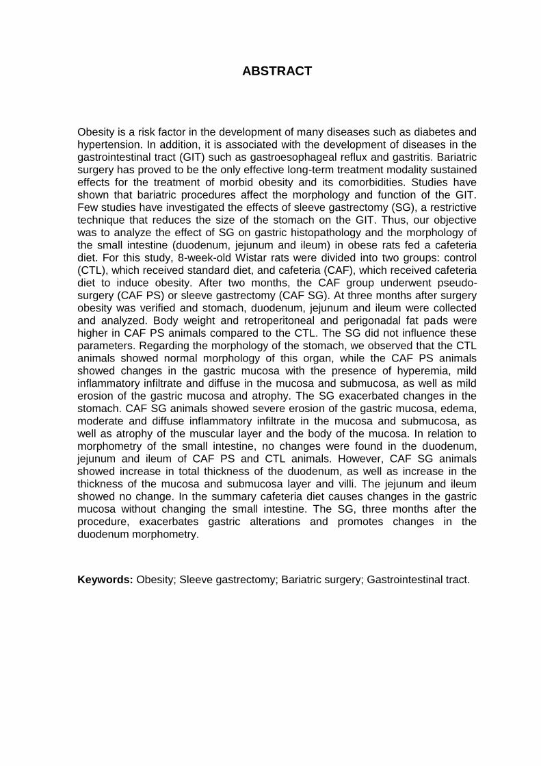
Obesity is a risk factor in the development of many diseases such as diabetes and hypertension. In addition, it is associated with the development of diseases in the gastrointestinal tract (GIT) such as gastroesophageal reflux and gastritis. Bariatric surgery has proved to be the only effective long-term treatment modality sustained effects for the treatment of morbid obesity and its comorbidities. Studies have shown that bariatric procedures affect the morphology and function of the GIT. Few studies have investigated the effects of sleeve gastrectomy (SG), a restrictive technique that reduces the size of the stomach on the GIT. Thus, our objective was to analyze the effect of SG on gastric histopathology and the morphology of the small intestine (duodenum, jejunum and ileum) in obese rats fed a cafeteria diet. For this study, 8-week-old Wistar rats were divided into two groups: control (CTL), which received standard diet, and cafeteria (CAF), which received cafeteria diet to induce obesity. After two months, the CAF group underwent pseudo-surgery (CAF PS) or sleeve gastrectomy (CAF SG). At three months after surgery obesity was verified and stomach, duodenum, jejunum and ileum were collected and analyzed. Body weight and retroperitoneal and perigonadal fat pads were higher in CAF PS animals compared to the CTL. The SG did not influence these parameters. Regarding the morphology of the stomach, we observed that the CTL animals showed normal morphology of this organ, while the CAF PS animals showed changes in the gastric mucosa with the presence of hyperemia, mild inflammatory infiltrate and diffuse in the mucosa and submucosa, as well as mild erosion of the gastric mucosa and atrophy. The SG exacerbated changes in the stomach. CAF SG animals showed severe erosion of the gastric mucosa, edema, moderate and diffuse inflammatory infiltrate in the mucosa and submucosa, as well as atrophy of the muscular layer and the body of the mucosa. In relation to morphometry of the small intestine, no changes were found in the duodenum, jejunum and ileum of CAF PS and CTL animals. However, CAF SG animals showed increase in total thickness of the duodenum, as well as increase in the thickness of the mucosa and submucosa layer and villi. The jejunum and ileum showed no change. In the summary cafeteria diet causes changes in the gastric mucosa without changing the small intestine. The SG, three months after the procedure, exacerbates gastric alterations and promotes changes in the duodenum morphometry.
Keywords: Obesity; Sleeve gastrectomy; Bariatric surgery; Gastrointestinal tract.
SUMÁRIO
LISTA DE FIGURAS............................................................................................10
LISTA DE ABREVIATURAS.................................................................................11
1. INTRODUÇÃO..................................................................................................12
2. REVISÃO DE LITERATURA............................................................................15
2.1 Obesidade.......................................................................................................15
2.2 Adaptações do trato gastrointestinal na obesidade........................................16
2.3 Cirurgia bariátrica e adaptações do trato gastrointestinal após
cirurgia..................................................................................................................19
2.4 Modelos experimentais de obesidade..............................................................23
3. REFERÊNCIAS.................................................................................................26
4. ARTIGO CIENTÍFICO.......................................................................................33
5. ANEXOS ..........................................................................................................51
5.1 Anexo A – Parecer de protocolo do Comitê de Ética no Uso de Animais da UNIOESTE.............................................................................................................51 5.2 Anexo B – Normas da Revista Científica.........................................................52

11
LISTA DE FIGURAS
Figura 1 – Técnica cirúrgica de gastrectomia verticalsegundo Huang et al.
(2014)............................................................................................................Pág.21

12
LISTA DE ABREVIATURAS
BGAL - banda gástrica ajustável laparoscópica BI - balão intragástrico CAF – cafeteria CEUA - Comitê de Ética no Uso de Animais DBP - derivação biliopancreática DDJ - derivação duodeno jejunal DGYR - derivação gástrica em Y de Roux DHGNA - doença hepática gordurosa não alcoólica DJI - derivação jejunoileal DM2 - diabetes mellitus tipo 2 DS - Duodenal Switch
GLP-1 - peptídeo semelhante ao glucagon tipo 1 GLUT1 - proteína transportadora de glicose tipo 1 GV - gastrectomia vertical IMC - Índice de Massa Corporal PNAE - Programa Nacional de Alimentação Escolar PNAN - Política Nacional de Alimentação e Nutrição SISVAN - Sistema de Vigilância Alimentar e Nutricional UNIOESTE – Universidade Estadual do Oeste do Paraná TGI – Trato gastrointestinal VIGITEL - Vigilância de Fatores de Risco e Proteção para Doenças Crônicas por Inquérito Telefônico
13
1. INTRODUÇÃO
A obesidade é considerada uma epidemia mundial que afeta países
desenvolvidos e em desenvolvimento, atingindo indivíduos de todas as idades
(CABALLERO, 2007). Na obesidade ocorrem alterações no equilíbrio energético
do organismo, onde a obtenção de energia excede seu gasto, sendo então o
excesso armazenado na forma de gordura no tecido adiposo (HILL; WYATT,
PETERS, 2012). Além disso, a facilidade de transportes e o avanço da tecnologia
provocaram uma redução na atividade física que, quando associados a um
comportamento alimentar inadequado, auxiliam no desenvolvimento da obesidade
(TAUBES, 1998). Antigamente, a obesidade era sinônimo de saúde e de riqueza.
Atualmente, ela é causa de problemas sociais e psicológicos, visto que, pessoas
obesas tem tendência ao isolamento social, relações interpessoais conturbadas,
dificuldades de locomoção, depressão, ansiedade entre outros. Além disso, ela
pode levar a prejuízos orgânicos, como hipertensão arterial, cardiopatias,
problemas ortopédicos, aterosclerose,doença hepática gordurosa não alcóolica
(DHGNA), diabetes mellitus tipo 2 (DM2) entre outras (GURA, 1997; WALLEY;
BLAKEMORE; FROGUEL, 2006).
O fato da obesidade ter aumentado em todas as idades e classes sociais,
bem como, o alto valor gasto pela união para o tratamento de doenças
associadas a ela, demonstra a importância da adoção de medidas de prevenção,
controle e tratamento de doenças crônicas não transmissíveis. Assim, no Brasil,
foram criadas políticas de saúde que visam o combate à obesidade como o
Programa Nacional de Alimentação Escolar (PNAE) que visa à formação de
hábitos alimentaressaudáveis, por meio da oferta da alimentação escolar e de
ações de educação alimentar e nutricional (PORTAL BRASIL, 2014a); já o
Sistema de Vigilância Alimentar e Nutricional (SISVAN), corresponde a um
sistema de informações que tem como objetivo principal promover informação
contínua sobre as condições nutricionais da população e os fatores que as
influenciam (PORTAL BRASIL, 2014b); e a Política Nacional de Alimentação e
Nutrição (PNAN) apresenta como propósito a melhoria das condições de
alimentação, nutrição e saúde da população brasileira, mediante a promoção de
14
práticas alimentares adequadas e saudáveis, a vigilância alimentar e nutricional, a
prevenção e o cuidado integral dos agravos relacionados à alimentação e nutrição
(PORTAL BRASIL, 2014c).
Uma vez instalada a obesidade, a busca pela perda de peso é
frequentemente realizada através do controle alimentar, exercícios físicos
regulares e o uso de medicamentos (VILLARINI et al., 2015). Quando essas
alternativas, isoladas ou em conjunto, falham, a cirurgia bariátrica é considerada
como forma de tratamento (BUCHWALD; OIEN, 2009; SJÖSTRÖM, 2004).
Ressalta-se que a cirurgia bariátrica é indicada para pacientes com Índice de
Massa Corporal (IMC)≥ 40,0 kg/m², ou aqueles com IMC ≥ 35,0 kg/m² que
apresentam comorbidades,tais como distúrbios metabólicos, doenças
cardiorrespiratórias, doença articular grave e problemas psicológicos relacionados
com a obesidade (FRIED et al., 2014). Entre os diferentes procedimentos
cirúrgicos, a gastrectomia vertical (GV), é uma das técnicas mais utilizadas.
Nesse procedimento ocorre a remoção do fundo gástrico e a maior parte do antro
do estômago, criando um tubo gástrico que restringe a ingestão oral. Essa técnica
tornou-se cada vez mais popular devido à sua relativa simplicidade, preservação
do piloro, e por evitar a má absorção pós-operatória (D'HONDT et al., 2011).
Estudos demonstram que a obesidade e as cirurgias bariátricassão
condições que podem levar a um processo de adaptação do trato gastrointestinal
(TGI). Indivíduos obesos costumam apresentar refluxo gastroesofágico e gastrite
(BORG et al., 2009; KIM et al., 2007; YAMAMOTO; WATABE; TAKEHARA,
2012).Dailey (2014) mostrou que existem diferenças na anatomia do epitélio
intestinal entre indivíduos obesos e magros, decorrentes da hiperfagia ou do tipo
de dieta associada com a obesidade. Além disso, a exposição prolongada a
dietas altamente calóricas está relacionada com alterações morfológicas e
funcionais no TGI em animais experimentais, como aumento na altura das
vilosidades e na densidade das criptas, e redução no número de células de
Paneth (BECERRIL et al., 2005; MAH et al., 2014). As cirurgias bariátricas
também podem provocar mudanças na estrutura e funcionalidade do estômago
(ARAPIS et al. 2015; MARTIN et al. 2012;MILLER; REID; BROWN, 2016) e
intestino delgado (CAVIN et al., 2016;MUMPHREY et al., 2015; SAEIDI et al.,
2013; LI et al., 2013)em humanos e em animais experimentais. Porém, poucos
15
estudos investigam as adaptações morfológicas intestinais após a GV e, além
disso, esses estudos são controversos (CAVIN et al., 2016; MUMPHREY et al.,
2015 ).
Levando em consideração o exposto acima, propomos o presente estudo
para responder o seguinte questionamento: Quais os efeitos da GV, três meses
após o procedimento cirúrgico, sobre a histomorfologia do estômago e sobre a
morfometria intestinal (duodeno, jejuno e íleo) em ratos obesos pela dieta de
cafeteria?

16
2. REVISÃO DE LITERATURA
2.1 Obesidade
A obesidade afeta todas as faixas etárias e grupos socioeconômicos, tanto
em países desenvolvidos quanto em países em desenvolvimento(CABALLERO,
2007). Para diagnóstico de sobrepeso e obesidade em adultos, a medida mais
usada é o IMC, definido como o peso em quilogramas dividido pelo quadrado da
estatura em metros (kg/m²). É um índice simples onde indivíduos com IMC entre
25 a 30 kg/m² são classificados com sobrepeso e indivíduos com IMC acima de
30 kg/m² são classificados como obesos (FERNANDES; CLEMENTE; MANCINI,
2013). Utilizando este índice de diagnóstico, dados mostram que a incidência da
obesidade está aumentando em todo o mundo,sendo considerada uma pandemia.
A prevalência global da obesidade em 2008 era de 10% para o sexo masculino e
14% para o feminino. As crianças também são alvos da obesidade, em 2012 mais
de 40 milhões de crianças menores de cinco anos estavam com sobrepeso ou
obesas (WHO, 2014). No Brasil 52,5% da população se encontrava com excesso
de peso e 17,9% apresentava obesidade em 2014 (BRASIL, 2014). De acordo
com a Organização Mundial da Saúde, 65% da população mundial vive em países
onde o sobrepeso e a obesidade mata mais pessoas do que o baixo peso, e cerca
de 3,4 milhões de adultos morrem a cada ano em decorrência da obesidade
(WHO, 2014).
A obesidade é definida como o acúmulo de gordura localizada ou
generalizada(LUZ; ENCARNAÇÃO, 2008), decorrente de balanço energético
positivo, ou seja, quando a energia consumida (proveniente dos alimentos)
excede o gasto (que envolve as calorias gastas para manutenção da taxa
metabólica basal, digestão de alimentos e realização de atividade física)(HILL;
WYATT; PETERS, 2012).Sua origem é multifatorial, incluindo fatores genéticos,
metabólicos, endócrinos, neurais, psicológicos, ambientais entre outros
(MOLINATTI; LIMONE, 1992). Há uma mudança evidente no comportamento
alimentar da população nas últimas décadas, marcada pelo aumento no consumo
de produtos industrializados ricos em gorduras, açúcar e sal, com baixo valor
nutritivo, caracterizando uma má nutrição. Esse fato, associado à mudança no
17
estilo de vida, com a diminuição de atividade física, têm desempenhado um
importante papel na pandemia da obesidade(FERNANDES; CLEMENTE;
MANCINI, 2013).
O excesso de peso eleva drasticamente o risco de uma pessoa
desenvolver várias doenças não transmissíveis, como diabetes, hipertensão,
acidente vascular cerebral, dislipidemia, apneia do sono, câncer, esteato-hepatite
não alcoólica, entre outras comorbidades graves (FRIED et al., 2014). No TGI a
obesidade está relacionada com vários diagnósticos, incluindo a doença
diverticular, refluxo gastroesofágico, gastrite, pólipos e câncer de cólon (BORG et
al., 2009). As consequências funcionais da obesidade têm sido extensivamente
estudadas no fígado, músculo esquelético, tecido adiposo e pâncreas, mas pouco
se conhece sobre seus efeitos no epitélio intestinal, o local inicial da absorção de
nutrientes (MAH et al., 2014).
2.2 Adaptações do trato gastrointestinal na obesidade
O estômago é um segmento dilatado do TGI. Suas principais funções
sãoarmazenar grandes quantidades de alimento até que ele possa ser
processado; misturar o alimento com as secreções gástricas até formar uma
mistura semilíquida denominada de quimo; e esvaziar lentamente o quimo para o
intestino delgado, a uma vazão compatível com digestão e absorção adequadas
(GUYTON; HALL, 2011). Anatomicamente, o estômago possui uma pequena
curvatura côncava e uma grande curvatura convexa. Possui quatro regiões: a
cárdia é uma região estreita na junção gastroesofágica, com 2 a 3 cm de
extensão; o fundo é uma região em forma cúpula, à esquerda do esôfago,
frequentemente preenchida com gás; o corpo é a maior região, responsável pela
formação do quimo; e o piloro é uma região em forma de funil, provida de um
esfíncter pilórico, que controla a liberação do quimo para o duodeno (GARTNER;
HIATT, 2003).
Todas as regiões gástricas apresentam pregas longitudinais da mucosa e
submucosa, que desaparecem no estômago distendido. O epitélio que recobre a
superfície do estômago é colunar simples, e todas as células secretam muco
alcalino que protege as células da acidez. A mucosa gástrica sofre invaginações
formando as fossetas (fovéolas) gástricas, onde é lançada a secreção de
glândulas. As glândulas gástricas produzem aproximadamente 2 a 3 litros de
18
suco gástrico por dia. As células parietais produzem ácido clorídrico e fator
intrínseco; as células principais produzem enzimas (pepsinogênio, renina e lipase
gástrica); as células superficiais de revestimento produzem muco; as
enteroendócrinas produzem hormônios; há também nas glândulas células de
reserva e células mucosas do colo (GARTNER; HIATT, 2003; JUNQUEIRA;
CARNEIRO, 2013).
O intestino delgado é o sítio terminal de digestão dos alimentos, absorção
de nutrientes e secreção endócrina. Esse órgão é relativamente longo
(aproximadamente 5 metros), e é dividido em três segmentos: duodeno, jejuno e
íleo (JUNQUEIRA; CARNEIRO, 2013). A absorção dos macronutrientes
(carboidratos, proteínas e lipídeos), vitaminas, água e eletrólitos ocorre
principalmente no duodeno e porção proximal do jejuno. O íleo absorve alguns
substratos, como os sais biliares e a vitamina B12. O cólon absorve um volume
menor de água, todos os eletrólitos que o alcançam e alguns produtos da
fermentação bacteriana (AIRES, 2008).
Como todos os componentes do TGI, o intestino delgado trata-se de um
tubo oco de diâmetro variável, formado por quatro camadas distintas: mucosa,
submucosa, muscular e serosa. Na camada mucosa os vilos são projeções
alongadas formadas pelo epitélio e lâmina própria. O epitélio de revestimento dos
vilos é formado principalmente por células absortivas (enterócitos) e células
caliciformes produtoras de muco, e continua com o epitélio das criptas. As criptas
são depressões em formato tubular e representam o compartimento proliferativo
do intestino. Seu epitélio é composto por algumas células absortivas, células
caliciformes, células enteroendócrinas, células de Paneth e células tronco.
Pregas, vilos e microvilosidades aumentam cerca de 600 vezes a superfície de
revestimento intestinal (JUNQUEIRA; CARNEIRO, 2013).
Os enterócitos são células colunares altas com microvilosidades em seus
ápices formando uma borda em escova, e são unidos por junções para formar
uma membrana relativamente impermeável. Os enterócitos contêm enzimas que
digerem substâncias alimentares específicas, e a função dessas células é
internalizar as moléculas de nutrientes produzidos durante a digestão. O ciclo de
vida de um enterócito é cerca de 3-5 dias. Para essa renovação, as células
epiteliais mais profundas nas criptas sofrem mitose contínua e novas células
migram da base das criptas em direção às pontas das vilosidades, reconstituindo
19
o epitélio do vilo e formando novas enzimas digestivas. Conforme as células do
vilo envelhecem, acabam se desprendendo nas secreções intestinais. O
crescimento e desenvolvimento da mucosa são regulados por sinais hormonais,
nervosos, imunológicos, dietéticos e mecânicos (GARTNER; HIATT, 2003;
GUYTON; HALL, 2011; PÁCHA, 2000).
O epitélio intestinal é uma barreira entre o mundo "exterior" e
"interior". Além das funções de absorver nutrientes, íons e água, esse órgão tem a
função de proteger o TGI de toxinas potencialmente prejudiciais, bactérias e
outros patógenos que também existem no lúmen intestinal (RAYBOULD, 2012).
A saúde de um indivíduo depende da digestão e absorção eficiente de
todos os nutrientes necessários a partir da dieta. Isto requer a detecção de
componentes dos alimentos por células enteroendócrinas, ativação de vias
neurais e hormonais para regular funções motoras, secretoras e de absorção no
TGI, além de regular a ingestão de alimentos e as concentrações plasmáticas de
glicose (RAYBOULD, 2012).
Estudos demonstram que a obesidade está relacionada com alterações no
TGI (BECERRIL et al., 2005; DAILEY, 2014; YAMAMOTO et al., 2012). Indivíduos
obesos apresentam refluxo gastroesofágico e gastrite (BORG et al., 2009; KIM et
al., 2007; YAMAMOTO; WATABE; TAKEHARA, 2012), além de apresentar
diferenças na anatomia do epitélio intestinal(DAILEY, 2014). E animais
experimentais obesos tem aumento na altura das vilosidades e na densidade das
criptas, e redução no número de células de Paneth (BECERRIL et al., 2005; MAH
et al., 2014).
O aumento do IMC foi associado com anormalidades endoscópicas
superiores, tais como: gastrite erosiva, úlcera gástrica, úlcera duodenal e
esofagite de refluxo (KIM et al., 2007). Uma revisão sobre a gastrite
endoscópica e histológica na obesidade mórbida revelou uma associação
consistente da obesidade com ambos os tipos de gastrite. Os resultados
indicam o aparecimento de uma nova categoria de gastrite, a gastrite
relacionada com a obesidade.Hábitos alimentares inadequados e fatores
hormonais como a adiponectina e a leptina, podem estar envolvidos no
mecanismo da patogênese da gastrite na obesidade(YAMAMOTO; WATABE;
TAKEHARA, 2012).
20
Existe um equilíbrio entre o fornecimento de alimentos e a capacidade de
digestão e absorção do intestino (RAYBOULD, 2012). A mucosa intestinal é
sensível a alterações neste ambiente eresponde com atrofia quando há falta de
alimentos,e hipertrofia quando existe sobrecarga de alimentos (PLUSKE;
HAMPSON; WILLIAMS, 1997). A atrofia da mucosa é caracterizada pela
diminuição da função intestinal, assim como as alterações morfológicas, incluindo
diminuição da altura das vilosidades, a profundidade das criptas, a área de
superfície, e o número de células epiteliais. Ocorre comumente na ausência de
alimentação (NIINIKOSKIet al., 2004). Já a proliferação é um mecanismo de
adaptação no qual ocorre aumentoda altura das vilosidades, a profundidade das
criptas, área superficial e peso úmido intestinal (DROZDOWSKI; CLANDININ;
THOMSON, 2009).
Em roedores, o jejum ou nutrição parenteral total leva a reduções rápidas
na massa epitelial do intestino delgado, associadas com a proliferação reduzida
nas criptas e aumento da apoptose em criptas e vilosidades. Esta é uma
adaptação fisiológica a uma necessidade reduzida de absorção de nutrientes
(NELSON et al., 2008). Por outro lado, em camundongos obesos por dieta
hiperlipídica, a altura das vilosidades e a densidade das criptas estão
significativamente aumentadas, indicando aumento do número de criptas para
suprir a ampliação das vilosidades(MAH et al., 2014).
Dailey (2014) mostrou que existem diferenças na anatomia do epitélio
intestinal entre indivíduos obesos e magros, decorrentes da hiperfagia ou do tipo
de dieta associada com a obesidade. Observou-se aumento no comprimento das
vilosidades, profundidade de criptas e números de células epiteliais em indivíduos
obesos em comparação com magros. Além disso, uma das consequências da
obesidade, o DM2, está associadoà hiperplasia e hipertrofia intestinal em
roedores (ADACHI et al., 2003). Em humanos obesos e diabéticos, a
hiperglicemia crônica está associada com o aumento da massa de enterócitos e
aumento da renovação dessas células no intestino delgado. Com isso, o intestino
delgado é cada vez mais reconhecido por desempenhar um papel na
patofisiologia do DM2 (VERDAMet al., 2011).
2.3Cirurgia bariátrica e adaptações do trato gastrointestinal após cirurgia

21
Nos últimos anos, a cirurgia bariátrica tem evoluído como uma opção de
conduta bem sucedida para controle de peso e glicêmico em pacientes obesos
com DM2mal controlado.Buchwaldet al. (2009) mostrou que 82% dos pacientes
tiveram resolução de manifestações clínicas e laboratoriais relacionadas ao DM2,
dois anos após a cirurgia. Também foi demonstrada a resolução e melhoria da
hipertensão, dislipidemia e apneia do sono (BUCHWALDet al., 2004).
Estritamente falando, o termo cirurgia bariátrica é aplicada a todos os
procedimentos cirúrgicos que visam reduzir o excesso de peso. Atualmente, os
candidatos para intervenções bariátricas incluem pacientes com obesidade
mórbida (IMC> 40,0 kg/m²), ou aqueles com IMC> 35,0 kg/m² que apresentam
importantes comorbidades como o DM2, hipertensão e apneia obstrutiva do sono
(FRIED et al., 2014).
Tradicionalmente, as cirurgias bariátricas são classificadas como
procedimentos restritivos, disabsortivos ou mistos. Técnicas restritivas diminuem
o tamanho do estômago, o que desencadeia saciedade com volumes menores de
alimentos do que antes da cirurgia. Procedimentos disabsortivos desviam
segmentos do intestino, causando assim certo grau de má absorção de
macronutrientes. Os procedimentos mistos combinam restrição e má absorção
(TACK; DELOOSE, 2014). Entre os tipos de técnicas restritivas, pode-se citar a
banda gástrica ajustável laparoscópica (BGAL), o balão intragástrico (BI) e a GV.
Entre os procedimentos disabsortivos puros, está à derivação jejunoileal (DJI), a
derivação duodeno jejunal (DDJ), bem como a derivação biliopancreática (DBP).
O duodenal switch(DS)e a derivação gástrica em Y de Roux (DGYR) são
exemplos de técnicas mistas. A DGYR é a cirurgia mais comumente realizada nos
Estados Unidos, seguida pela GV (BUCHWALD; OIEN, 2009).
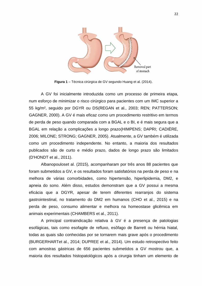
A GV é uma técnica segura e bem aceita que tornou-se cada vez mais
popular devido à sua relativa simplicidade, preservação do piloro, e por evitar a
má absorção pós-operatória. Na GV ocorre a remoção do fundo gástrico e a maior
parte do antro do estômago, criando assim um tubo gástrico que restringe a
ingestão oral (D'HONDTet al., 2011), como pode ser observado na Figura 1.
22
Figura 1 – Técnica cirúrgica de GV segundo Huang et al. (2014).
A GV foi inicialmente introduzida como um processo de primeira etapa,
num esforço de minimizar o risco cirúrgico para pacientes com um IMC superior a
55 kg/m², seguido por DGYR ou DS(REGAN et al., 2003; REN; PATTERSON;
GAGNER, 2000). A GV é mais eficaz como um procedimento restritivo em termos
de perda de peso quando comparada com a BGAL e o BI, e é mais segura que a
BGAL em relação a complicações a longo prazo(HIMPENS; DAPRI; CADIÈRE,
2006; MILONE; STRONG; GAGNER, 2005). Atualmente, a GV também é utilizada
como um procedimento independente. No entanto, a maioria dos resultados
publicados são de curto e médio prazo, dados de longo prazo são limitados
(D'HONDT et al., 2011).
Albanopouloset al. (2015), acompanharam por três anos 88 pacientes que
foram submetidos a GV, e os resultados foram satisfatórios na perda de peso e na
melhora de várias comorbidades, como hipertensão, hiperlipidemia, DM2, e
apneia do sono. Além disso, estudos demonstram que a GV possui a mesma
eficácia que a DGYR, apesar de terem diferentes rearranjos do sistema
gastrointestinal, no tratamento do DM2 em humanos (CHO et al., 2015) e na
perda de peso, consumo alimentar e melhora na homeostase glicêmica em
animais experimentais (CHAMBERS et al., 2011).
A principal contraindicação relativa à GV é a presença de patologias
esofágicas, tais como esofagite de refluxo, esôfago de Barrett ou hérnia hiatal,
todas as quais são conhecidas por se tornarem mais grave após o procedimento
(BURGERHARTet al., 2014; DUPREE et al., 2014). Um estudo retrospectivo feito
com amostras gástricas de 656 pacientes submetidos a GV mostrou que, a
maioria dos resultados histopatológicos após a cirurgia tinham um elemento de
23
gastrite crônica (74,4%), o que está de acordo com estudos anteriores mostrando
a sua alta prevalência na população obesa (ALMAZEEDI et al., 2013).
Uma vasta gama de alterações patológicas são vistas em amostras de
ressecção gástrica seguinte a GV em humanos, tais como gastrite crônica,
gastrite associada a Helicobacter, pólipos benignos da glândula fúndica, gastrite
linfocitária, gastrite atrófica autoimune, gastrite crônica com metaplasia intestinal e
pólipos hiperplásicos (MILLER; REID; BROWN, 2016).
Martin et al. (2012), analisaram a morfologia e histologia gástrica em ratos
obesos por dieta hiperlipídica, submetidos a GV, 4 e 16 semanas após a
cirurgia. O tamanho do estômago do grupo GV, após ambos os períodos
experimentais, foi semelhante à dos ratos pseudo-operados devido ao
alargamento do estômago residual. Traços de gastrite cística profunda,
caracterizada pelo alongamento foveolar com hiperplasia e dilatação cística das
glândulas, foram observados nos estômagos residuais dos ratos operados. Estes
resultados foram observados, principalmente, após 16 semanas, embora eles
também foram detectados ocasionalmente após quatro semanas de pós-
operatório.Arapiset al. (2015), analisaram a remodelação da mucosa gástrica
após GV em ratos obesos por dieta hiperlipídica. Os resultados mostraram
hiperplasia das células mucosas do colo, uma população de células de trânsito do
estômago que tem capacidade de se diferenciar em células zimogênicas e
pépticas. Houve também, no antro, redução do número de células de produtoras
de gastrina em conformidade com a redução do RNAm de gastrina. Estes dados
apoiam a ideia de que, depois da GV, a mucosa gástrica remanescente sofre
modificação na população e na função das células.
O TGI é o alvo direto dos procedimentos bariátricos e além do estômago,
alterações intestinais também são encontradas. A remodelação intestinal precoce
e a adaptação desencadeada por tais intervenções podem ser o ponto de partida
para a melhora metabólica observada pós-cirurgia. A maior parte dos estudos
demonstram os efeitos da DGYR sobre a morfologia do TGI (CAVIN et al., 2016;
MUMPHREY et al., 2015; SAEIDI et al., 2013). Porém, poucos estudos investigam
as adaptações morfológicas intestinais após a GV e, além disso, esses estudos
são controversos (CAVIN et al., 2016; MUMPHREY et al., 2015).
Saeidiet al. (2013) mostraram que ocorre reprogramação do metabolismo
intestinal da glicose após a DGYR em ratos obesos, provavelmente para

24
satisfazer as crescentes demandas bioenergéticas. Os autores concluíram que
essa reprogramação é desencadeada pela exposição desse segmento intestinal a
alimentos não digeridos. Ratos Zucker geneticamente obesos apresentaram
mudanças distintas nos segmentos intestinais anulados e reanastomosados, 14
dias após a DDJ. O duodeno e jejuno proximal apresentaram autólise e atrofia,
enquanto o jejuno distal exibiu aumento considerávelna proliferação de células da
mucosa. Como a DDJ desvia o fluxo de alimentos do estômago diretamente para
o jejuno distal, houve uma hiperplasia compensatória da mucosa, para aumentar
a capacidade de digestão e absorção nesse segmento. Morfologicamente, o
jejuno mostrou aumento da densidade das vilosidades, na espessura da parede, e
alargamento da circunferência. Por outro lado, a exclusão do fluxo de alimentos
no duodeno e jejuno proximal conduziu a aumento da apoptose, diminuição da
proliferação celular e atrofia da mucosa (LI et al., 2013).
Análises morfométricas do intestino de ratos obesos por dieta hiperlipídica
mostraram que em ratos submetidos à GV não houve qualquer resposta
hipertrófica no duodeno, jejuno e íleo. Em contraste, ratos submetidos à DGYR as
áreas transversais e espessura da mucosa para os locais intestinais
correspondentes (alça biliopancreática, alça de Roux e alça comum) foram
maiores. Da mesma forma, o número de células depeptídeo semelhante ao
glucagon tipo 1(GLP-1) e a expressão da proteína de hexoquinase II foram
aumentadas após DGYR, mas não houve modificações nesses parâmetros após
GV (MUMPHREY et al.,2015). Outro estudo em ratos e humanos submetidos à
DGYR, demonstrou que a alça de Roux tornou-se hiperplásica, com aumento do
número de células produtoras de incretinas. Além disso, a expressão de
transportadores de glicose e genes relacionados com a hipoxia aumentou, e o
transportador de glicose tipo 1(GLUT1) apareceu na membrana basolateral do
enterócito. Em contraste, não houve hiperplasia do intestino após a GV, mas a
absorção intestinal de glicose alimentar foi reduzida e a densidade de células
endócrinas secretoras de GLP-1 aumentou (CAVIN et al., 2016).
2.4 Modelos experimentais de obesidade
A experimentação animal é de grande importância nas pesquisas
científicas, contribuindo para o desenvolvimento da ciência e tecnologia. Sua
vasta contribuição nos diferentes campos científicos vem promovendo ao longo

25
dos anos a descoberta de medidas profiláticas e tratamentos de inúmeras
doenças que acometem os seres vivos. Animais de várias espécies têm sido
utilizados,tais como o rato, camundongo, coelho, vaca, porco, macaco, cachorro,
entre outros (CHORILLI; MICHELIN; SALGADO, 2007).
Na tentativa de compreender a fisiopatologia da obesidadebem como, os
efeitos e mecanismos envolvidos com a melhora metabólica após cirurgias
bariátricas, vários modelos experimentaissão utilizados. Dentre os diferentes
modelos encontram-se os oriundos de linhagens genéticas, provocadas por
mutações autossômicas recessivas (ratos Zuckerfa/fa, camundongos ob/ob) bem
como, camundongos transgênicos e knockouts para determinados genes
específicos; roedores submetidos a lesões eletrolítica no hipotálamo ventromedial
e lesões químicas em regiões hipotalâmicas por agentes específicos como o
glutamato monossódico (BRAY; YORK, 1979; OLNEY, 1969).
Dentre os modelos de dietas experimentais para roedores, podemos
destacar a dieta hiperlipídica, que contêm mais de 30% da energia total
proveniente de gordura e leva ao desenvolvimento da obesidade em animais
(HARIRI; THIBAULT, 2010); e a dieta de cafeteria (CAF), que possui alto valor
calórico e baixo valor nutritivo. Esse modelo é o que mais fielmente assemelha-se
a grande variedade de alimentos relacionados à pandemia da obesidade na
sociedade ocidental (CASTELL-AUVÍ et al., 2011).
A dieta de CAF consiste de alimentar os animais com elevada quantidade
de sal, açúcar e gordura; assim, imitando a dieta consumida pelas culturas
ocidentais. A dieta promove hiperfagia voluntária que resulta em rápido aumento
de peso, adipogênese e inflamação(CASTELL-AUVÍ et al., 2011; SAMPEY et al.,
2011). Além disso, ratos alimentados com a dieta de CAF apresentam esteatose
hepática não alcoólica,intolerância à glicose (SAMPEYet al., 2011) e resistência à
insulina (CASTELL-AUVÍ et al., 2011), ou seja um estado pré-diabético.
A dieta de CAF também é conhecida por causar mudanças na morfologia e
funcionamento do TGI em ratos, que podem estar relacionadas com a exposição
prolongada a alimentos de alto valor calórico (BACERRIL et al., 2005). Foram
relatadas alterações como o aumento no comprimento do intestino delgado e
esvaziamento mais lento do intestino grosso (DAMETO et al., 1991); aumento na
absorção intestinal de L-alanina (SANCHÍS; ALEMANY; REMESAR, 1994);
estimulação da proliferação celular e síntese proteica no intestino delgado

26
(ESTORNELL; CABO; BARBER, 1995) e redução no número de células de
Paneth (BACERRIL et al., 2005).
Scoariset al. (2010), investigaram os efeitos da dieta de CAF no intestino
delgado de ratos Wistar adultos em condições sedentárias e após o treinamento
físico. Os resultados mostraram que dieta de CAF, caracterizada
comohiperlipídica, levou a obesidade nos animais. A obesidade induzida pela
dieta causou aumento no comprimento do intestino delgado, na altura das
vilosidades, na profundidade das criptas e na espessura de toda a parede do
jejuno. Houve também aumento da atividade enzimática da fosfatase alcalina,
lipase, e sacarase, e redução do número de células caliciformes. O treinamento
físico atenuou esses parâmetros.
27
3. REFERÊNCIAS
ADACHI, T.; MORI, C.; SAKURAI, K.; SHIHARA, N.; TSUDA, K.; YASUDA, K. Morphological changes and increased sucrase and isomaltase activity in small intestines of insulin-deficient and type 2 diabetic rats. EndocrineJournal, v. 50, n. 3, p. 271–279, 2003. AIRES, M. M. Fisiologia. 3 ed. Rio de Janeiro: Guanabara Koogan, 2008. ALBANOPOULOS, K.; TSAMIS, D.; NATOUDI, M.; ALEVIZOS, L.; ZOGRAFOS, G.; LEANDROS, E.The impact of laparoscopic sleeve gastrectomy on weight loss and obesity-associated comorbidities: the results of 3 years of follow-up. Surgical Endoscopy, 2015. ALMAZEEDI, S.; AL-SABAH, S.; AL-MULLA, A.; AL-MURAD, A.; AL-MOSSAWI, A.; AL-ENEZI, K.; JUMAA, T.; BASTAKI, W. Gastric Histopathologies in Patients Undergoing Laparoscopic Sleeve Gastrectomies. Obesity Surgery, v. 23, n. 3, p. 314-319, 2013. ARAPIS, K.; CAVIN, J. B.; GILLARD, L.; CLUZEAUD, F.; LETTÉRON, P.; DUCROC, R.; LE BEYEC, J.; HOURSEAU, M.; COUVELARD, A.; MARMUSE, J. P.; LE GALL, M.; BADO, A. Remodeling of the Residual Gastric Mucosa after Roux-En-Y Gastric Bypass or Vertical Sleeve Gastrectomy in Diet-Induced Obese Rats. Plos One, v. 10, n. 3, p. 1-18, 2015. BECERRIL, A.; CASTILLO-ROBLES, G.; GONZÁLEZ-HERNÁNDEZ, M.; VILLANUEVA, I. Influence of high-calorie (cafeteria) diets on the population of Paneth cells in the small intestine of the rat.European Journal of Morphology, v. 42, n. 4/5, p. 201-207, 2005. BORG, B. B.; GUPTA, N. K.; ZUCKERMAN, G. R.; BANERJEE, B.; GYAWALI, C. P. Impact of Obesity on Bowel Preparation for Colonoscopy. ClinicalGastroenterologyandHepatology, v. 7, n. 6, p. 670-675, 2009. BRASIL. MINISTÉRIO DA SAÚDE. Vigitel Brasil 2014: vigilância de fatores de risco e proteção para doenças crônicas por inquérito telefônico. Brasília: Ministério da Saúde, 2014. Disponível em:http://portalsaude.saude.gov.br/images/pdf/2015/abril/15/PPT-Vigitel-2014-.pdf. Acesso em: 02 de setembro de 2015. BRAY, G. A.; YORK, D. A. Hypothalamic and genetic obesity in experimental animals: an autonomic and endocrine hypothesis. Physiology Review, v. 59, n. 3, p. 719- 809, 1979. BUCHWALD, H.; AVIDOR, Y.; BRAUNWALD, E.; JENSEN, M. D.; PORIES, W.; FAHRBACH, K.; SCHOELLES, K. Bariatric surgery: a systematic review and meta-analysis. JAMA, v. 292, n. 14, p. 1724–1737, 2004.
28
BUCHWALD, H.; ESTOK, R.; FAHRBACH, K.; FAHRBACH, K.; BANEL, D.; JENSEN, M. D.; PORIES, W. J.; BANTLE, J. P.; SLEDGE, I. Weight and type 2 diabetes after bariatric surgery: systematic review and meta-analysis. The American Journal of Medicine, v. 122, n. 3, p. 248–56, 2009. BUCHWALD, H.; OIEN, D. M. Metabolic/bariatric surgery Worldwide 2008.Obesity Surgery, v.19, n. 12, p. 1605-1611, 2009. BURGERHART, J. S.; SCHOTBORGH, C. A. I.; SCHOON, E. J.; SMULDERS, J. F.; MEEBERG, P. C.; SIERSEMA, P. D.; SMOUT, A. J. Effect of sleeve gastrectomy on gastroesophageal reflux.Obesity Surgery, v. 24, n. 9,p. 1436–1441, 2014. CABALLERO, B. The global epidemic of obesity: an overview. Epidemiologic Reviews, v. 29, p. 1-5, 2007. CASTELL-AUVÍ, A.; CEDÓ, L.; PALLARÈS, V.; BALY, M.; ARDÉVOL, A.; PINENT, M. The effects of a cafeteria diet on insulin production and clearance in rats. British Journal of Nutrition, v. 108, n. 7, p. 1155-62, 2011. CAVIN, J. B.; COUVELARD, A.; LEBTAHI, R.; DUCROC, R.; ARAPIS, K.; VOITELLIER, E.; CLUZEAUD, F.; GILLARD, L.; HOURSEAU, M.; MIKAIL, N.; RIBEIRO-PARENTI, L.; KAPEL, N.; MARMUSE, J. P.; BADO, A.; LE GALL, M.Differences in Alimentary Glucose Absorption and Intestinal Disposal of Blood Glucose After Roux-en-Y Gastric Bypass vs Sleeve Gastrectomy. Gastroenterology, v. 150, n. 2, p. 454-464, 2016. CHAMBERS, A. P.; STEFATER, M. A.; WILSON-PEREZ, H. E.; JESSEN, L.;SISLEY, S.; RYAN, K. K.; GAITONDE, S.; SORRELL, J. E.; TOURE, M.; BERGER, J.; D'ALESSIO, D. A.; SANDOVAL, D. A.; SEELEY, R. J.; WOODS, S. C. Similar effects of roux-en-Y gastric bypass and vertical sleeve gastrectomy on glucose regulation in rats. Physiology & Behavior, v. 105, p. 120-123, 2011. CHO, J. M.; KIM, H. J.; MENZO, E. L.; PARK, S.; SZOMSTEIN, S.; ROSENTHAL, R. J. Effect of sleeve gastrectomy on type 2 diabetes as an alternative treatment modality to Roux-en-Y gastric bypass: systemic review and meta-analysis. Surgery for Obesity and Related Diseases, v. 0, n. 0, p. 1-8, 2015. CHORILLI, M.; MICHELIN, D. C.; SALGADO, H. R. N. Animais de laboratório: o camundongo. Revista de Ciências Farmacêuticas Básica e Aplicada, v. 28, n. 1, p. 11-23, 2007.
DAILEY, M. J. Nutrient-induced intestinal adaption and its effect in
obesity.Physiology & Behavior, v. 136, p. 74-78, 2014.
DAMETO, M. C.; RAYÓ, J. M.; ESTEBAN, S.; PLANAS, B.; TUR, J. A. Effect of cafeteria diet on the gastrointestinal transit and emptying in the rat. Comparative Biochemistry and Physiology, v. 99, n. 4, p. 651-655, 1991.
29
D'HONDT, M.; VANNESTE, S.; POTTEL, H.; DEVRIENDT, D.; VAN ROOY, F.; VANSTEENKISTE, F. Laparoscopic sleeve gastrectomy as a single-stage procedure for the treatment of morbid obesity and the resulting quality of life, resolution of comorbidities, food tolerance, and 6-year weight loss. Surgical Endoscopy, v.25, n. 8, p. 2498–504, 2011. DROZDOWSKI, L. A.; CLANDININ, M. T.; THOMSON, A. B. Morphological, kinetic, membrane biochemical and genetic aspects of intestinal enteroplasticity. World Journal of Gastroenterology, v. 15, n. 7, p. 774-787, 2009. DUPREE, C. E.; BLAIR, K.; STEELE, S. R.; MARTIN, M. J.Laparoscopic sleeve gastrectomy in patients with preexisting gastroesophageal reflux disease: a national analysis. JAMA Surgery, v. 149, n. 4,p. 328–334, 2014. ESTORNELL, E.; CABO J.; BARBER, T. Protein Synthesis Is Stimulated in Nutritionally Obese Rats. The JournalofNutrition, v. 125, n. 5, p. 1309-1315, 1995. FERNANDES, A. T.; CLEMENTE, A. P. G.; MANCINI, M. C. Obesidade: estado de má nutrição. In: SAWAYA, A. L.; LEANDRO, C. G.; WAITZBERG, D. L. Fisiologia da nutrição na saúde e na doença: da biologia molecular ao tratamento. São Paulo: Atheneu, 2013. p. 415-424. FRANCISCHI, R. P. P.; PEREIRA, L. O.; FREITAS, C. S.; KLOPFER, M.;SANTOS, R. C.;VIEIRA, P.;LANCHA JÚNIOR, A. H. Obesity: UpdatedInformationAbout Its Etiology, MorbidityAndTreatment. Revista de Nutrição, v. 13, n. 1, p. 17-28, 2000. FRIED, M.; YUMUK, V.; OPPERT, J. M.; SCOPINARO, N.; TORRES, A.; WEINER, R.; YASHKOV, Y.; FRÜHBECK, G. Interdisciplinary European guidelines on metabolic and bariatric surgery. ObesitySurgery, v. 24, n. 1, p. 42–55, 2014. GARTNER, L. P.; HIATT, J. L. Tratado de Histologia em Cores. 2 ed. Rio de Janeiro: Guanabara Koogan, 2003. GURA, T. Obesity Sheds Its Secrets. Science, v. 275, p. 751-753, 1997. GUYTON, A. C.; HALL, J. E. Tratado de Fisiologia Médica. 11 ed. Rio de Janeiro: Elsevier, 2011. HARIRI, N.; THIBAULT, L. High-fat diet-induced obesity in animal models.Nutrition Research Reviews, v. 23, n. 2, p. 270-299, 2010. HILL, J. O.; WYATT, H. R.; PETERS, J. C. Energy balance and obesity.Circulation, v. 126, n. 1, p. 126–132, 2012. HIMPENS, J.; DAPRI, D.; CADIÈRE, G. B. A prospective randomized study between laparoscopic gastric banding and laparoscopic isolated sleeve
30
gastrectomy: results after 1 and 3 years. Obesity Surgery, v. 16, n. 11, p. 1450–1456, 2006. HUANG, X.; WENG, P.; ZHANG, H.; LU, Y. Remodeling intestinal flora with sleeve gastrectomy in diabetic rats.Journalof Diabetes Research, v. 2014, 2014. JUNQUEIRA, L.C.U.; CARNEIRO, J. Histologia Básica. 12 ed. Rio de Janeiro: Guanabara Koogan, 2013. KIM, H. J.; YOO, T. W.; PARK, D. I.; PARK, J. H.; CHO, Y. K.; SOHN, C.; JEON, W. K.; KIM, B. I. Influence of overweight and obesity on upper endoscopic findings. Gastroenterology, v. 22, n. 4, p. 477-481, 2007. KONTUREK, S. J.; KONTUREK, J. W.; PAWLIK, T.; BRZOZOWKI, T. Brain–gut axis and its role in the control of food intake.Journal of physiology and pharmacology, v. 55, p. 137–154, 2004. LI, B.; LU, Y.; SRIKANT, C. B.; GAO, Z. H.; LIU, J. L. Intestinal adaptation and Reg gene expression induced by antidiabetic duodenal-jejunal bypass surgery in Zucker fatty rats. American JournalofPhysiology, v. 304, n. 7, p. 635-645, 2013. LUZ, D. M. D; ENCARNAÇÃO, J. N. Vantagens e desvantagens da cirurgia bariátrica para o tratamento da obesidade mórbida. Revista Brasileira de Obesidade, Nutrição e Emagrecimento, v. 2, n. 10, p. 376-383, 2008. MAH, A. T.; LANDEGHEM, L. V.; GAVIN, H. E.; MAGNESS, S. T.; LUND, P. K. Impact of Diet-Induced Obesity on Intestinal Stem Cells: Hyperproliferation but Impaired Intrinsic Function That Requires Insulin/IGF1. Endocrinology, v. 155, n. 9, p. 3302–3314, 2014. MARTÍN, M.; BURRELL, M. A.; GÓMEZ-AMBROSI, J.; VALENTÍ, V.; BUENO, A.; RAMÍREZ, B.; BECERRIL, S.; LANCHA, A.; CALDERÓN, P. D. S.; MÉNDEZ-GIMÉNEZ, L.; CATALÁN, V.; RODRÍGUEZ, A.; FERNÁNDEZ, S.; MUÑOZ-NAVAS, M.; CIENFUEGOS, J. A.; FRÜHBECK, G. Short- and Long-Term Changes in Gastric Morphology and Histopathology Following Sleeve Gastrectomy in Diet-Induced Obese Rats. Obesity Surgery, v. 22, n. 4, p, 634-640, 2012. MILLER, G. C.; REID, A. S.; BROWN, I. S.The pathological findings seen in laparoscopic sleeve gastrectomies for weight loss.Pathology, v. 48, n. 3, p. 228–232, 2016. MILONE, L.; STRONG, V.; GAGNER, M. Laparoscopic sleeve gastrectomy is superior to intragastric balloon as a first-stage procedure for super-obese patients (BMI ≥ 50 kg/m²). Obesity Surgery, v. 15, n. 5, p. 612–617, 2005. MOLINATTI, G. M.; LIMONE, P. Obesity: a challenge for the clinician. Frontiers in diabetes, n. 11, p. 7-15, 1992.
31
MUMPHREY, M. B.; HAO, Z.; TOWNSEND, R. L.; PATTERSON, L. M.; BERTHOU, H. Sleeve Gastrectomy Does Not Cause Hypertrophy and Reprogramming of Intestinal Glucose Metabolism in Rats. Obesity Surgery, v. 15, n. 8, p. 1468-1473, 2015. NELSON, D. W.; MURALI, S. G.; LIU, X.; KOOPMANN, M. C.; HOLST, J. J.; NEY, D. M. Insulin-like growth factor I and glucagon-like peptide-2 responses to fasting followed by controlled or ad libitum refeeding in rats. American Journal of Physiology, v. 294, n. 4, p. 1175–1184, 2008. NIINIKOSKI, H.; STOLL, B.; GUAN, X.; KANSAGRA, K.; LAMBERT, B. D.; STEPHENS, J.; HARTMANN, B.; HOLST, J. J.; BURRIN, D. G. Onset of small intestinal atrophy is associated with reduced intestinal blood flow in TPN-fed neonatal piglets. Journal of Nutrition,v. 134, n. 6,p. 1467-1474, 2004. OLNEY, J. W. Brain Lesions, Obesity, and Other Disturbances in Mice Treated with Monosodium Glutamate. Science, v. 164, p. 719-721, 1969.
PÁCHA, J. Development of intestinal transport function in
mammals.Physiological Reviews, v. 80, n. 4, p. 1633-1667, 2000.
PLUSKE, J. R.; HAMPSON, D. J.; WILLIAMS, I. H. Factors influencing the
structure and function of the small intestine in the weaned pig: a review.
LivestockProduction Science, v. 51, p. 215–236, 1997.
PORTAL BRASIL. Fundo Nacional de Desenvolvimento e Educação: Alimentação Escolar (PNAE), 2012. Disponível em: http://www.fnde.gov.br/index.php/programas/alimentacao-escolar/alimentacao-escolar-apresentacao. Acesso em: 10 de outubro de 2014a. PORTAL BRASIL. Portal da saúde: PNAN - Política Nacional de Alimentação e Nutrição, 2012. Disponível em: http://dab.saude.gov.br/portaldab/pnan.php. Acesso em: 10 de outubro de 2014c. PORTAL BRASIL. Portal da saúde: Vigilância Alimentar e Nutricional –SISVAN, 2012. Disponível em: http://dab.saude.gov.br/portaldab/ape_vigilancia_alimentar.php?conteudo=sisvan. Acessoem: 10 de outubro de 2014b.
RAYBOULD, H. E. Gut microbiota, epithelial function and derangements in
obesity.The Journal of Physiology, v. 590, n. 3, p. 441-446, 2012. REGAN, J. P.; INABNET, W. B.; GAGNER, M.; POMP, A.Early experience with two-stage laparoscopic Roux-en-Y gastric bypass as an alternative in the super-super-obese patient.Obesity Surgery, v. 13, n. 6, p. 861–864, 2003. REN, C. J.; PATTERSON, E.; GAGNER, M. Early results of laparoscopic biliopancreatic diversion with duodenal switch: a case series of 40 consecutive patients. Obesity Surgery, v.10, n 6, p. 514–523, 2000.

32
SAEIDI, N.; MEOLI, L.; NESTORIDI, E.; GUPTA, N. K.; KVAS, S.; KUCHARCZYK, J.; BONAB, A. A.; FISCHMAN, A. J.; YARMUSH, M. L.; STYLOPOULOS, N. Reprogramming of Intestinal Glucose Metabolism and Glycemic Control in Rats After Gastric Bypass. Science, v. 341, n. 6144, p. 406–410, 2013. SAMPEY, B. P.; VANHOOSE, A. M.; WINFIELD, H. M.; FREEMERMAN, A. J.; MUEHLBAUER, M. J.; FUEGER, P.T.; NEWGARD, C. B.; MAKOWSKI, L. Cafeteria diet is a robust model of human metabolic syndrome with liver and adipose inflammation: comparison to high-fat diet. Obesity, v. 19, n. 6, p. 1109-1117, 2011. SANCHÍS, D.; ALEMANY, M.; REMESAR, X. L-Alanine transport in small intestine brush-border membrane vesicles of obese rats.Biochimica etBiophysicaActa, v. 1192, n. 2, p. 159-166, 1994. SCOARIS, C. R.; RIZO, G. V.; ROLDI, L. P.; MORAES, S. M. F.; PROENÇA, A. R. G.; PERALTA, R. M.; NATALI, M. R. M. Effects of cafeteria diet on the jejunum in sedentary and physically trained rats.Nutrition, v. 26, n. 3, p. 312-320, 2010.
SHAW, D.; GOHIL, K.; BASSON, M. D. Intestinal mucosal atrophy and
adaptation.World Journal of Gastroenterology, v. 18, n. 44, p. 6357-6375, 2012.
SJÖSTRÖM, L.; LINDROOS, A. K.; PELTONEN, M.; TORGERSON, J.; BOUCHARD, C.; CARLSSON, B.; DAHLGREN, S.; LARSSON, B.; NARBRO, K.; SJÖSTRÖM, C. D.; SULLIVAN, M.; WEDEL, H. Lifestyle, diabetes, and cardiovascular risk factors 10 years after bariatric surgery. The New England Journal of Medicine, v. 35, n. 26, p. 2683–93, 2004. TACK, J.; DELOOSE, E. Complications of bariatric surgery: dumping syndrome, reflux and vitamin deficiencies. Best Practice & Research Clinical Gastroenterology, v. 28, n. 4, p. 741–749, 2014. TAUBES, G. As obesity rates rise, experts struggle to explain why. Science, v. 280, p. 1367-1368, 1998. VERDAM, F. J.; GREVE, J. W. M.; ROOSTA, S.; EIJK, H. V.; BOUVY, N.; BUURMAN, W. A.; RENSEN, S. S. Small Intestinal Alterations in Severely Obese Hyperglycemic Subjects. The Journal of Clinical Endocrinology & Metabolism, v. 96, n. 2, p. E379 –E383, 2011. VILLARINI, M.; LANARI, C.; BARCHIESI, L.; CASCIARI, E.; TABASCIO, A.; CASTELLINI, M.; LEVORATO, S.; FORNACIARI, G.; MORETTI, M.;VILLARINI, A. Effects of the "PreveDi" lifestyle modification trial on metabolic syndrome. Annali di Igiene, v. 27, n. 3, p. 595-606, 2015. WALLEY, A. J.; BLAKEMORE, A. I. F.; FROGUEL, P. Genetics of obesity and the prediction of risk for health. Human molecular genetics, v. 15, n. 2, p. 124-130, 2006.

33
WHO.World Health Organization.2014. Disponível em: <http://www.who.int/mediacentre/factsheets/fs311/en/>. Acesso em: 26 de maio de 2014. YAMAMOTO, S.; WATABE, K.; TAKEHARA, T. Is Obesity a New Risk Factor for Gastritis? Digestion, v. 85, n. 2, p. 108-110, 2012.
34
VERTICAL SLEEVE GASTRECTOMY ALTERS THE GASTRIC MUCOSA AND
MORPHOMETRY OF THE DUODENUM IN OBESE RATS
Angélica Novi CapelassiI, Fernanda Soares da Silva-Morita
II, Regina Inês Kunz
III, Allan
Cezar Faria AraújoIV
, Helene Nara Henriques BlancV, Maria Lúcia Bonfleur
VI
I-MSc Program in Biosciences and Health, State University of Western Paraná, Cascavel-
PR.
II-MSc Program in Biosciences and Health, State University of Western Paraná, Cascavel-
PR.
III –PhD Program in Biological Sciences, State University of Maringá, Maringá-PR.
IV- Adjunct Professor of General Surgery, Centre for Medical and Pharmaceutical
Sciences, University of the WesternParaná, Cascavel, PR.
V- Adjunct Professor of Clinical Pathology, Federal University of Rio de Janeiro, Macaé -
RJ.
VI- Adjunct Professor of Human Physiology, Biological and Health Sciences Centre, State
University of Western Paraná, Cascavel-PR.
35
Abstract
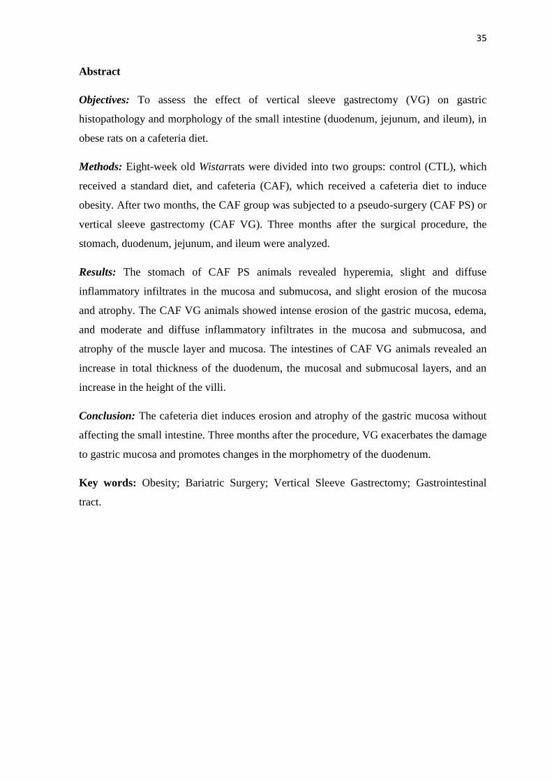
Objectives: To assess the effect of vertical sleeve gastrectomy (VG) on gastric
histopathology and morphology of the small intestine (duodenum, jejunum, and ileum), in
obese rats on a cafeteria diet.
Methods: Eight-week old Wistarrats were divided into two groups: control (CTL), which
received a standard diet, and cafeteria (CAF), which received a cafeteria diet to induce
obesity. After two months, the CAF group was subjected to a pseudo-surgery (CAF PS) or
vertical sleeve gastrectomy (CAF VG). Three months after the surgical procedure, the
stomach, duodenum, jejunum, and ileum were analyzed.
Results: The stomach of CAF PS animals revealed hyperemia, slight and diffuse
inflammatory infiltrates in the mucosa and submucosa, and slight erosion of the mucosa
and atrophy. The CAF VG animals showed intense erosion of the gastric mucosa, edema,
and moderate and diffuse inflammatory infiltrates in the mucosa and submucosa, and
atrophy of the muscle layer and mucosa. The intestines of CAF VG animals revealed an
increase in total thickness of the duodenum, the mucosal and submucosal layers, and an
increase in the height of the villi.
Conclusion: The cafeteria diet induces erosion and atrophy of the gastric mucosa without
affecting the small intestine. Three months after the procedure, VG exacerbates the damage
to gastric mucosa and promotes changes in the morphometry of the duodenum.
Key words: Obesity; Bariatric Surgery; Vertical Sleeve Gastrectomy; Gastrointestinal
tract.
36
Introduction
In recent decades, obesity has reached alarming proportions, especially in the
developed and developing countries, affecting individuals of all ages. Because of obesity,
changes take place in the energy balance of an organism, wherein, energy production
exceeds expenditure, and the excess energy is stored as fat in adipose tissue1. According to
the World Health Organization, 65% of the world's population lives in countries where
excess weight and obesity kill more people than deaths due to low weight, and about 3.4
million adults die each year, due to the comorbidities associated with obesity2.
Obesity is associated with development of various disorders, including type II
diabetes, cardiovascular disease, dyslipidemia, different types of cancers, and
musculoskeletal and psychosocial changes, among others3. Several studies indicate that
obesity is also associated with changes in the gastrointestinal tract and occurrence of
gastroesophageal reflux and gastritis, as well as morphological and functional changes in
the small intestine of obese humans 4,5
and experimental animals 6.
Although there are various modalities for the treatment of obesity, bariatric surgery
has proven to be the only long-term effective treatment, with sustained long-term effects
on morbid obesity and its comorbidities7,8
. Among the different surgical procedures
available, vertical sleeve gastrectomy (VG) is a widely used and secure technique. This
procedure involves removal of the gastric fundus and a greater part of antrum of the
stomach, creating a gastric tube, which restricts oral ingestion. This technique has become
increasingly popular due to its relative simplicity, preservation of the pylorus, and the
absence of postoperative malabsorption9. The main contraindication of VG is the presence
of esophageal pathologies, such as reflux esophagitis and Barrett's esophagus or hiatus
hernia, all of which are known to aggravate after the procedure10
. In addition, bariatric
surgeries are also associated with changes in the intestinal mucosa11-15
.
The Roux-en-Y gastric bypass (RYGB) leads to morphological and functional
adjustments in the intestine, in rats and humans11,12,14
. The duodenal jejunal bypass (DJB)
also causes morphological changes in the intestine in rats13
. However, few studies have
investigated the morphological adaptations of the intestine after VG; moreover, these
studies are controversial11,14
. Thus, our goal was to analyze the effect of VG on gastric
histopathology and morphology of the small intestine in rats with induced obesity from a
cafeteria diet. A cafeteria diet is an experimental model that best reflects the Western style
diet. It consists of highly palatable and calorie rich food, which leads to hyperphagia,

37
weight gain, excessive accumulation of fat, as well as development of a pre-diabetic
state16,17
.
Methods
Animals
Twenty-five 60 day-old Wistar rats were included in this experiment. The animals
were randomly divided into two groups: a control group (CTL), which received a standard
diet, and a cafeteria group (CAF), which received a cafeteria diet for induction of obesity.
After two months, the CAF group was further subdivided into two groups, one subjected to
a pseudo-surgery (CAF PS, n = 8) and the other to vertical sleeve gastrectomy (CAF VG, n
= 9). The groups were euthanized three months after the surgical procedure. All
experimental procedures were approved by the Ethics Committee on Animal Use (CEUA)
at the State University of Western Paraná (UniversidadeEstadual do Oeste do Paraná).
Diets
The animals in CTL group received a standard diet (Algomix, Ouro Verde do
Oeste, Brazil) and water ad libitum. The animals of CAF group received a cafeteria diet,
similar to that used in the study by Goularteet al.18
, with some modifications. It comprised
of: standard ration, Italian salami, milk bread, corn snack, marshmallow, mixed sausage,
chocolate cake, corn flour cookie, mortadella, bacon flavored salty snack, chocolate wafer
and 350 ml per day of degassed Coca-Cola™.
Pre- and perioperative care
Five days prior to the surgery, the animals in CAF PS and CAF VG groups
received a liquefied CAF diet with liquid-pasty consistency and Coca-Cola ad libitum. All
animals were fasted for 12 hours prior to the surgical procedure. The animals were
anesthetized using 1% isoflurane (BioChimico, Itatiaia, Brazil) under nasotracheal
intubation with urethral probe number four, oxygen at 1 L/min and spontaneous
ventilation. Five minutes before the start of surgery, a single-dose antibiotic prophylaxis
with intramuscular ceftriaxone (Teuto, Anápolis, Brazil), 50 mg/kg, and subcutaneous
analgesia with sodium dipyrone (Teuto, Anápolis, Brazil) at a dose of 50 mg/kg, were
administered. An aseptic technique was used for preparation of the operative field and the
abdominal wall, including antisepsis with polyvinylpyrrolidone. Shortly thereafter, a

38
subcutaneous injection of 20 ml warm physiological saline 0.9% was administered for
hydration. Body temperature was maintained with heaters and hot water bottles. Animals
of the CTL group received liquefied standard ration of liquid-pasty consistency for five
days, and went through a similar fasting period as the operated rats.
Surgical Procedures
For the pseudo-surgery, a 4 cm incision was made on the epigastric midline of the
abdominal wall of CAF PS animals. The abdominal cavity was exposed, the liver was
removed carefully, following which the stomach and intestine were handled. The
laparotomy was closed with continuous sutures in planes, including peritoneal and
aponeurotic planes, with polypropylene suture 4-O. The skin was closed with continuous
sutures, using 4-O polypropylene suture.
For the VG, a 4 cm incision was made at the epigastric midline on the abdomen. A
section of the stomach was held using a pair of scissors, from the angle of His to a point 3
mm proximal to the duodenum, with resection of approximately 80% of the stomach
including a complete resection of the gastric fundus. Three to four separate sutures were
secured in the sectioned stomach with 7-O polypropylene sutures, at the level of angle of
His, medial region of the body and distal portion of the antrum. Continuous sutures were
secured on two layers with 7-O polypropylene suture in the rest of the stomach, with
fixation on the previously secured sutures. This portion corresponded with 20% of the total
volume of the stomach. The laparotomy was closed with continuous sutures in layers,
including the peritoneal and aponeurotic layers with polypropylene suture 4-O. The skin
was closed with continuous sutures, using polypropylene suture 4-O.
Postoperative Care
A subcutaneous dose of 10 mL 0.9% saline solution was administered, divided into
two dorsal quadrants of the animal, 24 and 48 hours after surgery, to prevent dehydration.
The animals received only water ad libitum but not any diet until they completed 72 hours
of fasting. After this period, they were offered a liquefied diet of liquid-pasty consistency
for seven days. Subsequently, all animals again received the CAF diet. The CTL rats were
also subjected to 72 hours of fasting and then received a liquefied diet of liquid-pasty
consistency for the next seven days.

39
Evolution of body weight and assessment of obesity
The animals were weighed weekly, during the entire experimental period to
monitor the progression of body weight. After death, the perigonadal and retroperitoneal
fat pads were excised and weighed, to assess the accumulation of fat.
Histopathology and histomorphometry of the gastrointestinal tract
Cross-sectional samples of the stomach, duodenum, jejunum, and ileum were fixed
in paraformaldehyde 4% and embedded in paraffin. Semi-serial (6 μm thick) sections were
stained with hematoxylin and eosin (HE). The following lesions were checked during the
morphological assessment of the stomach: mucosal edema, erosion of the mucosa,
hemorrhage, hyperemia, atrophy, and fibrosis, presence of inflammatory infiltrate and
cellular atypia. The histopathological analyses were performed blind. To perform the
histomorphometric analyses, the stomach sections were photographed using an Olympus
microscope coupled to an imaging system, under a total amplification of 40×. Three fields
were photographed for each slide. These images were then analyzed randomly using the
free software, ImageJ. Three measurements from each image were selected to assess
thickness of the gastric mucosa, using the straight feature from the image analysis
program. The areas were chosen at random, from sites where it was possible to measure
the distance between the epithelial surface and the muscle of the mucosa (gastric mucosa).
Thickness of the mucosa was calculated as the average of three measurements on each
slide.
For morphometric analysis of the duodenum, jejunum, and ileum, the slides were
photomicrographed using an Olympus DP71 microscope. Two images were captured per
animal, with a 4× amplification for the analysis of total thickness of the intestinal wall,
thickness of the muscle layer, and thickness of the mucosa and submucosa. Four images
with a 10× amplification were taken to measure height of the villus and depth of the crypt.
The morphometric analyses were performed using the software Image Pro Plus ® 6.0
(Media Cybernetics, USA), previously calibrated. Total thickness of the intestinal wall,
thickness of the muscular layer, and thickness of the mucosa and submucosa were
measured at four random points of the section, one section per animal. For the height of the
villus and depth of crypt, 10 villi and 10 crypts were measured per animal.
Statistical analyses
The results were expressed as average ± standard error of the average. One-way
40
analysis of variance (ANOVA) was used for statistical evaluation followed by the Tukey
post-hoc test. The level of significance was set at P < 0.05. The software used for statistical
analyses was the GraphPad Prism version 5.00 for Windows (GraphPad Software, San
Diego, USA).
Results
The body weight of CAF PS and CAF VG animals, in the first week after surgery,
was higher, compared to CTL animals (P < 0.05, Figure 1A). In the fourth, fifth and sixth
week post-surgery, the animals of the CAF VG group exhibited increased body weight
compared to CTL animals (P < 0.05, P < 0.05 and P < 0.01). From the seventh to 13th
week post-surgery, the CAF PS and CAF VG groups developed a significant increase in
weight compared to the CTL group (Figure 1A). Reinforcing these results, we observed
that the area under the curve (AUC) of the progression of body weight was higher in CAF
PS and CAF VG groups compared to the CTL group (Figure 1B; P < 0.05 and P < 0.01).
The naso-anal length (NAL) was higher in CAF PS and CAF VG groups (26 ± 0.2 and 26
± 0.3 cm, p < 0.001) compared to the CTL animals (24 ± 0.2).
The animals of the CAF PS and CAF VG groups presented an increase of
approximately 57% and 53% in the weight of the retroperitoneal fat pads (Figure 1C, P <
0.001) and 44% and 35% in the weight of the perigonadal fat pads (Figure 1D, P < 0.01
and P < 0.05, respectively) compared to the CTL animals.

41
0 2 4 6 8 1 0 1 2 1 4
3 5 0
4 0 0
4 5 0
5 0 0
5 5 0
6 0 0C T L
C A F P S
C A F V G
W ee k s
*
#
#
#*
** *
**
*
Bo
dy
we
igh
t (g
)
C T L C A F P S C A F V G
0
2 0 0 0
4 0 0 0
6 0 0 0
8 0 0 0
a
b b
AU
V o
f b
od
y w
eig
ht
C T L C A F P S C A F V G
0
1
2
3
4
5
a
b
b
Re
tro
pe
rit
on
ea
l fa
t
(% o
f b
od
y w
eig
ht)
C T L C A F P S C A F V G
0
1
2
3
4
5
a
b
b
Pe
rig
on
ad
al
fa
t
(% o
f b
od
y w
eig
ht)
A B
C D
Figure 1 - Progression of body weight after the surgical procedure (A); area under the
curve (AUC) of progression of body weight (B) and weight of retroperitoneal (C) and
perigonadal fat pads (D) in, CTL, CAF PS and CAF VG animals. Data represent the
average ± SEM (n = 9). *CAF PS and CAF VG differ from CTL animals. #CAF VG differ
from CTL animals. Different letters on the bars refer to significant differences between the
groups. One-way ANOVA followed by the Tukey’s post-hoc test. P < 0.05.
The animals of CTL group presented a normal gastric morphology, with no changes
being observed in the mucosa, submucosa, muscle, and serosa of the organ (Figure 2A and
B). In group CAF PS, the presence of erythema, and slight and diffuse inflammatory
infiltrates were observed, both in the mucosa and submucosa, as well as slight erosion of
the gastric mucosa (loss of surface epithelium) and atrophy of the mucosa (Figure 2C and
D). In CAF VG group of animals, there was intense erosion of the gastric mucosa, edema,
and moderate and diffuse inflammatory infiltrates in both, the mucosa and submucosa
(Figure 2E and F). None of the groups had presence of hemorrhage, fibrosis, or cellular
42
atypia. On morphometric analyses of the stomach, the animals of CAF PS group showed a
reduction in the thickness of mucosa (560 ± 25) compared to the CTL animals (781 ± 53, P
< 0.05). The vertical sleeve gastrectomy did not modify this parameter.
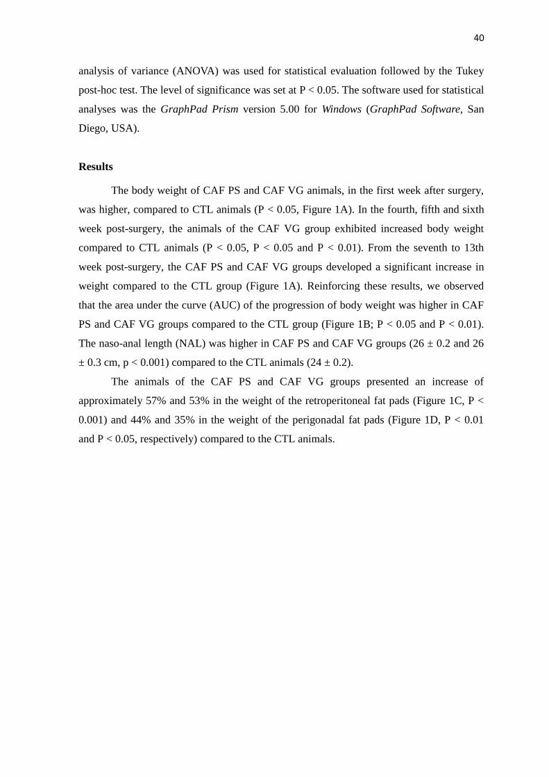
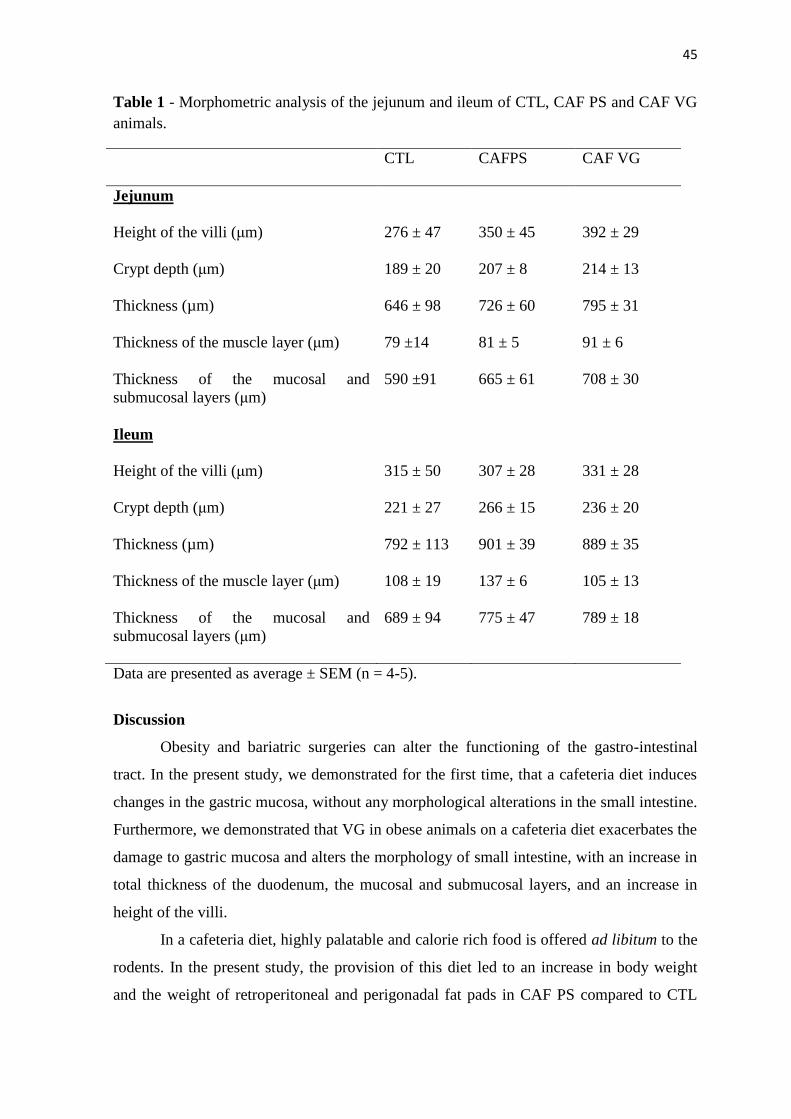
Figure 3 shows morphometric analyses of the duodenum. There were no changes in
any parameter evaluated among the CAF PS and CTL animals (Fig 3, A, B, C, D and E).
However, the animals of group CAF VG developed an increase in total thickness of the
duodenum and in the mucosal and submucosal layers, compared to CAF PS and CTL
animals (Figure 3A and C, respectively, P < 0.05). In addition, height of the villi was
approximately 27% and 32% higher in the CAF VG group compared to CAF PS and CTL
animals, respectively (Figure 3D; P < 0.05 and P < 0.01). There was no difference in the
thickness of muscle layer and depth of the crypts in the duodenum of animals studied
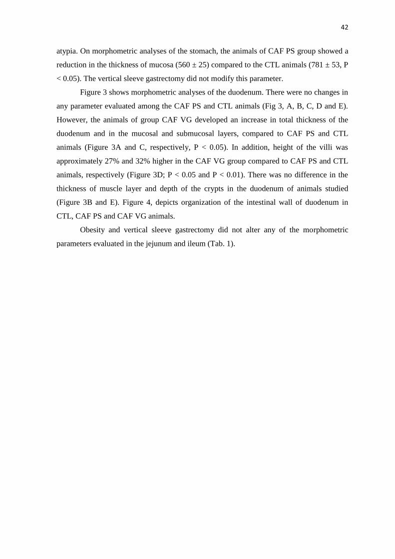
(Figure 3B and E). Figure 4, depicts organization of the intestinal wall of duodenum in
CTL, CAF PS and CAF VG animals.
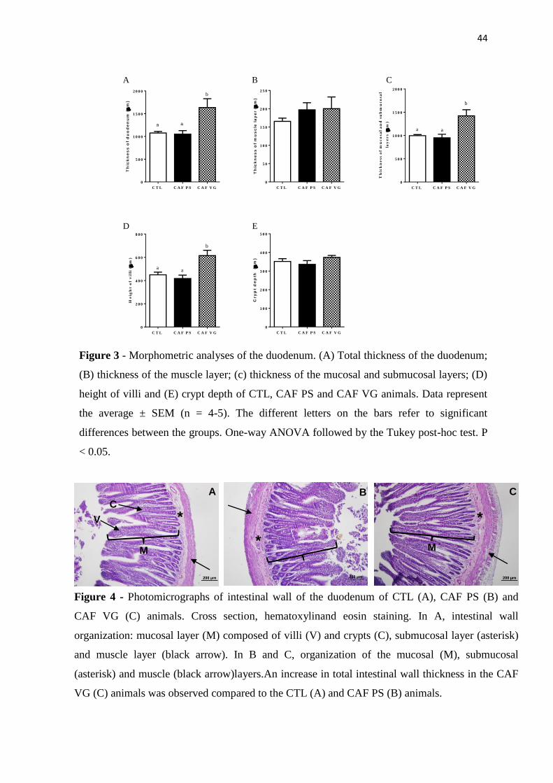
Obesity and vertical sleeve gastrectomy did not alter any of the morphometric
parameters evaluated in the jejunum and ileum (Tab. 1).

43
B A
C D
E F
Figure 2 - Gastric tissue photomicrographs of CTL (A and B), CAF PS (C and D) and CAF
GV (E and F) animals, cross section, hematoxylin and eosin staining. In A, gastric tissue
with normal aspect of the mucosa, submucosa and muscular layers and, in B, normal
appearance of the glands, lamina propria, muscular mucosa and submucosa.In C, mild
erosion of the mucosa (arrows) and, in D, hyperemia (arrows) in the lamina propria and
mild and diffuse inflammatory infiltrate in the mucosa. In E, intense mucosal erosion
(arrows) and, in F, moderate and diffuse inflammatory infiltrate in the mucosa and
submucosa, as well as mucosal edema.
44
C T L C A F P S C A F V G
0
5 0 0
1 0 0 0
1 5 0 0
2 0 0 0
a a
b
Th
ick
ne
ss
of
du
od
en
um
(
m)
C T L C A F P S C A F V G
0
5 0
1 0 0
1 5 0
2 0 0
2 5 0
Th
ick
ne
ss
of
mu
sc
le l
ay
er (
m)
C T L C A F P S C A F V G
0
5 0 0
1 0 0 0
1 5 0 0
2 0 0 0
a a
b
Th
ick
ne
ss
of m
uc
os
al
an
d s
ub
mu
co
sa
l
la
ye
rs
(
m)
C T L C A F P S C A F V G
0
2 0 0
4 0 0
6 0 0
8 0 0
aa
b
He
igh
t o
f v
illi
(
m)
C T L C A F P S C A F V G
0
1 0 0
2 0 0
3 0 0
4 0 0
5 0 0
Cry
pt
de
pth
(
m)
A B C
D E
Figure 3 - Morphometric analyses of the duodenum. (A) Total thickness of the duodenum;
(B) thickness of the muscle layer; (c) thickness of the mucosal and submucosal layers; (D)
height of villi and (E) crypt depth of CTL, CAF PS and CAF VG animals. Data represent
the average ± SEM (n = 4-5). The different letters on the bars refer to significant
differences between the groups. One-way ANOVA followed by the Tukey post-hoc test. P
< 0.05.
A
V
C
M
*
B C
M
*
Figure 4 - Photomicrographs of intestinal wall of the duodenum of CTL (A), CAF PS (B) and
CAF VG (C) animals. Cross section, hematoxylinand eosin staining. In A, intestinal wall
organization: mucosal layer (M) composed of villi (V) and crypts (C), submucosal layer (asterisk)
and muscle layer (black arrow). In B and C, organization of the mucosal (M), submucosal
(asterisk) and muscle (black arrow)layers.An increase in total intestinal wall thickness in the CAF
VG (C) animals was observed compared to the CTL (A) and CAF PS (B) animals.
*
45
Table 1 - Morphometric analysis of the jejunum and ileum of CTL, CAF PS and CAF VG
animals.
CTL CAFPS CAF VG
Jejunum
Height of the villi (μm)
Crypt depth (μm)
Thickness (µm)
276 ± 47
189 ± 20
646 ± 98
350 ± 45
207 ± 8
726 ± 60
392 ± 29
214 ± 13
795 ± 31
Thickness of the muscle layer (μm)
79 ±14 81 ± 5 91 ± 6
Thickness of the mucosal and
submucosal layers (μm)
Ileum
Height of the villi (μm)
Crypt depth (μm)
Thickness (µm)
Thickness of the muscle layer (μm)
Thickness of the mucosal and
submucosal layers (μm)
590 ±91
315 ± 50
221 ± 27
792 ± 113
108 ± 19
689 ± 94
665 ± 61
307 ± 28
266 ± 15
901 ± 39
137 ± 6
775 ± 47
708 ± 30
331 ± 28
236 ± 20
889 ± 35
105 ± 13
789 ± 18
Data are presented as average ± SEM (n = 4-5).
Discussion
Obesity and bariatric surgeries can alter the functioning of the gastro-intestinal
tract. In the present study, we demonstrated for the first time, that a cafeteria diet induces
changes in the gastric mucosa, without any morphological alterations in the small intestine.
Furthermore, we demonstrated that VG in obese animals on a cafeteria diet exacerbates the
damage to gastric mucosa and alters the morphology of small intestine, with an increase in
total thickness of the duodenum, the mucosal and submucosal layers, and an increase in
height of the villi.
In a cafeteria diet, highly palatable and calorie rich food is offered ad libitum to the
rodents. In the present study, the provision of this diet led to an increase in body weight
and the weight of retroperitoneal and perigonadal fat pads in CAF PS compared to CTL
46
animals, corroborating the previously published data16,17
. In order to reverse the condition
of obesity and the syndromes associated therewith, different strategies have been used,
such as diets with calorie restriction, administration of medications and metabolic and/or
bariatric surgeries. Among all the strategies, surgery has been the most effective, for
patients with morbid obesity7,8
. One of the surgical procedures, VG, where a gastric
resection of 80% with excision of the fundus and the body of the stomach19
is performed,
has stood out. It is a restrictive and simple technique, with short surgical time, few post-
surgical complications, and great benefit to patients with morbid obesity9. Studies in
humans and animals demonstrate that VG promotes rapid weight loss20,21
. However, in our
study, the CAF VG animals did not show a reduction in body weight compared to CAF PS
animals. In addition, the weight of retroperitoneal and perigonadal fat pads did not change.
This is in contrast to previously published data; perhaps, continuity of the cafeteria diet
might have influenced this parameter.
Other than diabetes and hypertension, obesity is also associated with
gastrointestinal diseases, such as gastroesophageal reflux, diseases of the gallbladder,
tumors of the colon, esophagus and pancreas, abdominal pain, vomiting22,23
and gastritis4.
Yamamoto et al.4 demonstrated an association between the reduction of hormone
adiponectin and presence of erosive gastritis in obese patients. The authors suggest that
adiponectin, due to its anti-inflammatory properties, protects the stomach from gastric
erosion. For the first time, we have demonstrated that the consumption of a CAF diet
promotes damage to the gastric mucosa. We observed the presence of hyperemia, slight
and diffuse inflammatory infiltrates, both, in the mucosa and submucosa, as well as slight
erosion and atrophy of the gastric mucosa. Thus, our results reveal an association between
high calorie food intake and changes in the mucosa of the stomach; however, the
mechanisms have not been elucidated.
Although VG is a simple, restrictive technique with few post-surgical
complications, it has some disadvantages such as hemorrhage, gastritis, and
dysplasia15,24
that are mainly due to direct intervention in the stomach. The main
contraindication of VG is the presence of esophageal pathologies, such as reflux
esophagitis and Barrett's esophagus, all of which are known to aggravate after the
procedure10
. Moreover, a wide range of pathological changes are seen in gastric resection
samples following VG in humans, such as chronic gastritis, Helicobacter-associated
gastritis, benign fundic gland polyps, lymphocytic gastritis, autoimmune atrophic gastritis,
chronic gastritis with intestinal metaplasia and hyperplastic polyps25
. In our study, we

47
demonstrated that three months after VG, the stomachs of CAF VG animals showed
intense erosion of the gastric mucosa, edema, and moderate and diffuse inflammatory
infiltrates, both in the mucosa and submucosa. Obese rats on high lipid diet, subjected to
VG, presented an increase in size of the residual stomach and signs of gastritis
cysticaprofunda, characterized by foveolar elongation with hyperplasia and cystic
dilatation of the glands19
. These changes were mainly observed 16 weeks after the surgical
procedure; however, they were also occasionally detected four weeks post-surgery. In
another study, also on obese rats fed with fat diet subjected to VG, there was hyperplasia of
the mucous neck cells and a reduction in the number of cells that produce gastrin in the
antrum of the stomach, which is in line with the reduction of the mRNA (messenger RNA)
levels of this hormone26
. These data support the possibility that, after VG, the remaining
gastric mucosa undergoes modification in the population and function of cells.
In addition to changes in the stomach, studies in rodents have shown that changes
in morphology and function of the small intestine may be related to prolonged exposure to
foods with high caloric value6, as well as to the quantity and/or quality of the food
available27
. An increase in the length of small intestine and lower intestinal emptying
time28
, stimulation of cell proliferation and protein synthesis in the small intestine29
and a
reduction in the number of Paneth cells6 were observed. Scoariset al.
30 observed an
increase in the height of villi, depth of crypts and thickness of the entire wall of jejunum in
obese rats on the cafeteria diet. Moreover, they confirmed an increase in the length of
small intestine, reduction in the number of goblet cells as well as increased enzymatic
activity of alkaline phosphatase, lipase, and saccharase. According to the authors, the high
content of lipids in the cafeteria diet fed to animals may be responsible for morphological
changes of the intestinal wall. In our study, there was no morphological change in the total
thickness, thickness of the mucosa and submucosa, height of the villi and crypt depth in the
duodenum, jejunum, and ileum of CAF PS compared to CTL animals. Probably the
difference in cafeteria diet composition used in both studies might be responsible for the
absence of intestinal wall changes in this study.
The CAF VG animals developed changes in morphology only of the duodenum,
with an increase in total thickness of the region, as well as an increase in thickness of the
mucosal and submucosal layers and the height of villi. Unlike our results, morphometric
analyses of the intestine of obese rats fed on hyperlipid diet showed that rats submitted to
VG did not present any hypertrophic response in the duodenum, jejunum, and ileum, and
showed no changes in the number of cells that produce Glucagon-like peptide-1 (GLP-1)
48
or protein expression of hexokinase II11
. However, another study on rats and human
subjects, did not reveal hyperplasia of the intestine after VG, but found a reduction in the
intestinal absorption of glucose and increase in the density of GLP-1 endocrine secretory
cells 14
. Since we used a cafeteria diet in our study, which is different in consistency from a
hyperlipid diet, we suggest that the remodeling in morphometric parameters found in the
duodenum may be due to the exposure of this intestinal segment to inadequately crushed
and initially digested food.
Conclusion
We conclude from this study that a cafeteria diet induces erosion and atrophy of the
gastric mucosa, without causing any morphological changes in the small intestine. The VG
performed in obese animals fed on cafeteria diet, exacerbates the damage to gastric
mucosa, three months after the surgical procedure. In addition, VG alters the morphology
of small intestine, with an increase in total thickness of the duodenum, as well as an
increase in thickness of the mucosal and submucosal layers and height of the villi.
Referências
1. Caballero, B. The global epidemic of obesity: an overview. Epidemiol Rev. 2007
Jun;29(1):1-5. doi: 10.1093/epirev/mxm012.
2. WHO. World Health Organization. 2014. Available in:
http://www.who.int/mediacentre/factsheets/fs311/en/. Acess: August 8, 2016.
3. Tadross JA, leRoux CW. The mechanisms of weight loss after bariatric surgery. Int
J Obes (Lond). 2009 Apr;33Suppl 1:S28-32. PMID: 19363504.
4. Yamamoto S, Watabe K, Takehara T. Is Obesity a New Risk Factor for Gastritis?
Digestion. 2012;85(2):108-10. PMID: 22269289.
5. Dailey MJ. Nutrient-induced intestinal adaption and its effect in obesity.
PhysiolBehav. 2014 Sep;136:74-8. PMID: 24704111.
6. Becerril A, Castillo-Robles G, González-Hernández M, Villanueva I. Influence of
high-calorie (cafeteria) diets on the population of Paneth cells in the small intestine
of the rat. Eur J Morphol. 2005 Oct-Dec;42(4-5):201-7. PMID: 16982477.
7. Sjöström L, Lindroos AK, Peltonen M, Torgerson J, Bouchard C, Carlsson B,
Dahlgren S, Larsson B, Narbro K, Sjöström CD, Sullivan M, Wedel H. Lifestyle,
diabetes, and cardiovascular risk factors 10 years after bariatric surgery. N Engl J
Med. 2004 Dec 23;351(26):2683-93. PMID: 15616203.
8. Zimmet P, Alberti KG, Rubino F, Dixon JB. IDF's view of bariatric surgery in type
2 diabetes. Lancet. 2011 Jul 9;378(9786):108-10. PMID: 21742162.
9. D'Hondt M, Vanneste S, Pottel H, Devriendt D, Van Rooy F, Vansteenkiste F.
Laparoscopic sleeve gastrectomy as a single-stage procedure for the treatment of
morbid obesity and the resulting quality of life, resolution of comorbidities, food
49
tolerance, and 6-year weight loss. SurgEndosc. 2011 Aug;25(8):2498-504. PMID:
21359900.
10. DuPree CE, Blair K, Steele SR, Martin MJ.Laparoscopic sleeve gastrectomy in
patients with preexisting gastroesophageal reflux disease: a national
analysis.JAMA Surg. 2014 Apr;149(4):328-34.PMID: 24500799.
11. Mumphrey MB, Hao Z, Townsend RL, Patterson LM, Berthoud HR. Sleeve
Gastrectomy Does Not Cause Hypertrophy and Reprogramming of Intestinal
Glucose Metabolism in Rats.Obes Surg. 2015 Aug;25(8):1468-73. PMID:
25566744.
12. Saeidi N, Meoli L, Nestoridi E, Gupta NK, Kvas S, Kucharczyk J, Bonab AA,
Fischman AJ, Yarmush ML, Stylopoulos N. Reprogramming of Intestinal Glucose
Metabolism and Glycemic Control in Rats After Gastric Bypass.Science. 2013 Jul
26;341(6144):406-10. PMID: 23888041.
13. Li B, Lu Y, Srikant CB, Gao ZH, Liu JL. Intestinal adaptation and Reg gene
expression induced by antidiabetic duodenal-jejunal bypass surgery in Zucker fatty
rats. Am J PhysiolGastrointest Liver Physiol. 2013 Apr 1;304(7):G635-45. PMID:
23370676.
14. Cavin JB, Couvelard A, Lebtahi R, Ducroc R, Arapis K, Voitellier E, Cluzeaud F,
Gillard L, Hourseau M, Mikail N, Ribeiro-Parenti L, Kapel N, Marmuse JP, Bado
A, Le Gall M. Differences in Alimentary Glucose Absorption and Intestinal
Disposal of Blood Glucose After Roux-en-Y Gastric Bypass vs Sleeve
Gastrectomy. Gastroenterology. 2016 Feb;150(2):454-64. PMID: 26481855.
15. Behrens C, Tang BQ, Amson BJ. Early results of a Canadian laparoscopic sleeve
gastrectomy experience. Can J Surg. 2011 Apr;54(2):138-43. PMID: 21251414.
16. Castell-Auví A, Cedó L, Pallarès V, Blay M, Ardévol A, Pinent M. The effects of a
cafeteria diet on insulin production and clearance in rats. Br J Nutr. 2012
Oct;108(7):1155-62.PMID: 22152054.
17. Sampey BP, Vanhoose AM, Winfield HM, Freemerman AJ, Muehlbauer MJ,
Fueger PT, Newgard CB, Makowski L. Cafeteria diet is a robust model of human
metabolic syndrome with liver and adipose inflammation: comparison to highfat
diet.Obesity (Silver Spring). 2011 Jun;19(6):1109-17. PMID: 21331068.
18. Goularte JF, Ferreira MB, Sanvitto GL. Effects of food pattern change and physical
exercise on cafeteria diet-induced obesity in female rats.Br J Nutr. 2012 Oct
28;108(8):1511-8. PMID: 22264412.
19. Martín M, Burrell MA, Gómez-Ambrosi J, Valentí V, Bueno Á, Ramírez B,
Becerril S, Lancha A, del Sol Calderón P, Méndez-Giménez L, Catalán V,
Rodríguez A, Fernández S, Muñoz-Navas M, Cienfuegos JA, Frühbeck G. Short-
and Long-Term Changes in Gastric Morphology and Histopathology Following
Sleeve Gastrectomy in Diet-Induced Obese Rats. Obes Surg. 2012 Apr;22(4):634-
40. PMID: 22297793.
20. Almogy G, Crookes PF, Anthone GJ. Longitudinal gastrectomy as a treatment for
the high-risk super-obese patient. Obes Surg. 2004 Apr;14(4):492-7. PMID:
15130224.
50
21. Chambers AP, Stefater MA, Wilson-Perez HE, Jessen L, Sisley S, Ryan KK,
Gaitonde S, Sorrell JE, Toure M, Berger J, D'Alessio DA, Sandoval DA, Seeley RJ,
Woods SC. Similar effects of roux-en-Y gastric bypass and vertical sleeve
gastrectomy on glucose regulation in rats. PhysiolBehav. 2011 Nov 30;105(1):120-
3. PMID: 21683726.
22. Renehan AG, Roberts DL, Dive C. Obesity and cancer: pathophysiological and
biological mechanisms. Arch PhysiolBiochem. 2008 Feb;114(1):71-83. PMID:
18465361.
23. Delgado-Aros S, Locke GR, Camilleri M, Talley NJ, Fett S, Zinsmeister AR,
Melton LJ. Obesity is associated with increased risk of gastrointestinal symptoms: a
population-based study.Am J Gastroenterol. 2004 Sep;99(9):1801-6.PMID:
15330922.
24. Abu-Jaish W, Rosenthal RJ. Sleeve gastrectomy: a new surgical approach for
morbid obesity. Expert Rev GastroenterolHepatol. 2010 Feb;4(1):101-19. PMID:
20136593.
25. Miller GC, Reid AS, Brown IS. The pathological findings seen in laparoscopic
sleeve gastrectomies for weight loss. Pathology. 2016 Apr;48(3):228-32. PMID:
27020497.
26. Arapis K, Cavin JB, Gillard L, Cluzeaud F, Lettéron P, Ducroc R, Le Beyec J,
Hourseau M, Couvelard A, Marmuse JP, Le Gall M, Bado A. Remodeling of the
Residual Gastric Mucosa after Roux-En-Y Gastric Bypass or Vertical Sleeve
Gastrectomy in Diet-Induced Obese Rats. PLoS One. 2015 Mar
30;10(3):e0121414. PMID: 25822172.
27. Little TJ, Horowitz M, Feinle-Bisset C. Modulation by high-fat diets of
gastrointestinal function and hormones associated with the regulation of energy
intake: implications for the pathophysiology of obesity. Am J ClinNutr. 2007
Sep;86(3):531-41. PMID: 17823414.
28. Dameto MC, Rayó JM, Esteban S, Planas B. Tur JA. Effect of cafeteria diet on the
gastrointestinal transit and emptying in the rat. Comp BiochemPhysiol A MolIntegr
Physiol. 1991 99(4):651-655. doi:10.1016/0300-9629(91)90145-3.
29. Estornell E, Cabo J, Barber T. Protein Synthesis Is Stimulated in Nutritionally
Obese Rats. J Nutr. 1995 May;125(5):1309-15. PMID: 7537806.
30. Scoaris CR, Rizo GV, Roldi LP, de Moraes SM, de Proença AR, Peralta RM,
Natali MR. Effects of cafeteria diet on the jejunum in sedentary and physically
trained rats. Nutrition. 2010 Mar;26(3):312-20. PMID: 19665869.
51
Acknowledgements
We would like to thank Suellen Camila Padilha, student of scientific initiation and BSc
student in Biological Sciences, for the aid in caring for experimental animals.
Correspondence
Maria Lucia Bonfleur
E-mail: [email protected]
Laboratory of Endocrine Physiology and Metabolism, UNIOESTE, Cascavel, Paraná.
Cascavel, PR, Brazil ZIP: 858119-110,
Phone: +55 45 3220 3257.
Declaration
We declare no conflict of interests.
The study was conducted in the Laboratory of Endocrine Physiology and Metabolism
(LAFEM), of the Biological and Health Sciences Center (CCBS) at the State University of
Western Paraná (UNIOESTE), campus Cascavel, PR-Brazil. The article is part of the
Masters Thesis of the graduate student Angélica Novi Capelassi, Bioscience and Health
Program. Supervisor: Maria Lucia Bonfleur.
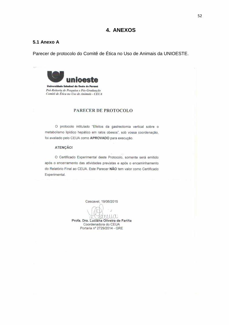
52
4. ANEXOS
5.1 Anexo A
Parecer de protocolo do Comitê de Ética no Uso de Animais da UNIOESTE.
53
5.2 Anexo B Normas da Revista Científica
A Revista Acta Cirúrgica Brasileira tem um padrão, estilo, regras e
normas que devem ser cumpridas. Seguir as Instruções aos Autores
em www.scielo.br/acb (português/inglês). Observar os modelos nos
artigos publicados. Revisão acurada do manuscrito antes de enviar,
pois se não cumprir as instruções, não será aceito.
A RevistaActaCirúrgicaBrasileiracumpre as normas do International
Committee of Medical Journal Editors: Ethical Considerations in the
Conduct and Reporting of Research, Authorship and Contributorship (www.icmje.org).
Autoria
A Revista Acta Cirúrgica Brasileira considera todos os participantes do
artigo como autores. Autores são considerados aqueles que têm
efetiva contribuição intelectual e científica na realização do trabalho. Não é papel do editor, arbitrar conflitos relacionados à autoria.
O autor principal é o responsável pela integridade do artigo, como
um todo, não necessariamente ser o primeiro autor, entretanto deve
ser o autor correspondente. A Revista não aceita alunos de
graduação e pós-graduação como autores principais. O autor principal
assume a responsabilidade pela participação efetiva de cada autor e
pela concordância com o conteúdo do artigo.
Participantes que tiveram atividade puramente técnica (ato operatório,
revisão bibliográfica, chefes de departamento, serviços ou financiados)
devem ser listados nos agradecimentos.
Contribuição dos autores
Cada autor deve assumir a responsabilidade de pelo menos um componente do trabalho:
1. Contribuição científica e intelectual efetiva para o estudo;
2. Concepção e delineamento;
3. Aquisição dos dados;
4. Interpretação dos dados;
5. Procedimentos técnicos;
6. Exames macroscópicos e histopatológicos;
7. Análise estatística;
8. Preparação do manuscrito;
9. Redação do manuscrito;
10. Revisão crítica;
11. Aprovação final.
Tipos de Publicação
Os Suplementos da revista podem ter objetivos úteis para
cooperação entre Instituições Acadêmicas. O Editor Chefe terá a
autoridade de enviar os suplementos para apreciação de pareceristas
que poderão rejeitar artigos. É fundamental distinguir nas referências
os artigos de suplementos e os da publicação regular.
54
A revista aceita artigos de pesquisa, preferencialmente de cirurgia
experimental, investigação clínica e de revisão solicitada pelo Corpo Editorial.
Revisões sistemáticas e metanálises enfocando temas cirúrgicos de
interesse atual serão consideradas para publicação. Todas as
metanálises de ensaios randomizados deverão atender os princípios do
QUORUM (Lancet. 1999;354:1896-1900) e o manuscrito submetido deverá conter o fluxograma detalhado da revisão sistemática.
A Revista não aceita relato de caso ou de casos e trabalhos retrospectivos.
Fluxograma do manuscrito
Os manuscritos submetidos pelos autores à Acta Cirúrgica Brasileira
serão inicialmente avaliados pelo Editor Chefe, quanto aos objetivos e
padrão da revista. Nesta fase, o manuscrito poderá ser devolvido ao
autor para adequações ou rejeitado. Caso o manuscrito esteja de
acordo com as normas, será encaminhado ao Editor Associado, que
designará os Pareceristas para avaliação do conteúdo científico. O
Editor Associado ao receber os pareceres, os encaminhará ao Editor
Chefe para a decisão final. O fluxo poderá ser acompanhado pelo autor
no sistema de gerenciamento.
Preparação dos manuscritos
O texto e o abstract devem ser revistos por especialistas da área
médica, habituados com a redação científica do idioma. O documento
principal (Maindocument), em inglês, deve conter a página inicial (Title
Page), as figuras com as legendas e as tabelas com os enunciados, além do manuscrito.
Página inicial
O título do artigo deve ser conciso, mas informativo. Inserir o número
um (algarismo arábico), sobrescrito, indicando após as referências o
local da realização da pesquisa (laboratório, centro de pesquisa, disciplina, departamento e instituição). Não incluir siglas.
Afiliação de cada autor: O nome completo de todos os autores com
os números subsequentes sobrescritos (algarismo romano) que
indicarão a categoria acadêmica mais elevada de cada um, com o
nome da disciplina/departamento e da instituição. Informar se tem bolsa de pesquisador do CNPq e respectivo nível.
Alunos em nível de Mestrado e Doutorado devem referir o nome do
Programa de Pós-Graduação, conforme a CAPES. Podem ser
considerados como primeiro autor e o Professor Orientador/Tutor como autor correspondente.
Alunos de graduação são bem-vindos. É desejável que tenham Bolsa
de Iniciação Científica (PIBIC-CNPq, IC-FAPESP, Institucional ou outras
55
agências).
Observar os títulos acadêmicos no idioma inglês. Consultar Acta Cir Bras. 2006;21(2):60 Mar-Abr. Academicdegreesexamples
ABSTRACT
Não deve exceder 200 palavras e deve ser apresentado de forma
estruturada:
PURPOSE (claro, preciso e conciso, sem comentários)
METHODS (amostra e procedimentos)
RESULTS (principais achados com dados especificados e significância
estatística)
CONCLUSION (clara, precisa e concisa, respondendo o objetivo, sem
comentários).
Key words devem constar no DeCS/MeSH.
Acessar http://decs.bvs.br. Termos que não fazem parte do DeCS impedem a localização do artigo.
Estrutura do manuscrito
Introdução
Deve apresentar o estado atual do tema e oferecer somente citações
pertinentes, sem fazer revisão extensa do assunto. Autores devem ser
apresentados em letras minúsculas ou simplesmente citados por números sobrescritos, sem o ano do artigo.
Métodos
Deve apresentar a amostragem (quantidade e qualidade) e os
procedimentos em pormenores suficientes que permitam a outros
pesquisadores reproduzirem os resultados. Identificar, precisamente,
todas as drogas, substâncias químicas e produtos utilizados, incluindo
os nomes genéricos, dosagens e formas de administração. Não referir
nomes de pacientes, iniciais ou número do protocolo hospitalar.
Descrever os métodos estatísticos com pormenor e fornecer
referências para os procedimentos consagrados. O autor principal é
responsável pela exatidão dos dados, incluindo todos os cálculos
estatísticos.
Ética
A investigação em seres humanos deve ser submetida ao Comitê de
Ética da Instituição (preencher o número do processo), cumprindo a
Declaração de Helsinki de 1975, revisada em 2008
(www.wma.net/e/policy/b3.htm) e a Resolução 466/12 do Conselho
Nacional de Saúde (http://conselho.saude.gov.br/resolucoes/2012/Reso466.pdf)
A investigação em animais deve ser submetida à Comissão de Ética no
Uso de Animais (CEUA). Preencher o número do processo de
aprovação da pesquisa, cumprindo a Lei Federal nº 11.794, de 8 de
outubro de 2008 (http://www.planalto.gov.br/ccivil_03/_Ato2007-
2010/2008/Lei/L11794.htm) e o Decreto nº 6.689, de 15 de julho de
2009 que regulamenta a Lei nº 11.794
(http://www.planalto.gov.br/ccivil_03/_Ato2007-
56
2010/2009/Decreto/D6899.htm).
Artigos de autores estrangeiros devem seguir o Council for
InternationalOrganizationof Medical Sciences (CIOMS)
http://www.cioms.ch/index.php/12-newsflash/227-cioms-and-iclas-
release-the-new-international-guiding-principles-for-biomedical-
research-involving-animals
O Corpo Editorial da Revista poderá recusar artigos que não cumpram rigorosamente os preceitos éticos da pesquisa.
Resultados
No texto enfatizar as observações importantes. Incluir os dados de significância estatística.
Tabelas: são abertas e numeradas consecutivamente (algarismos
arábicos) e apresentar um título breve, colocado acima. Utilizar
abreviaturas aprovadas e padronizadas. As abreviaturas não
padronizadas devem ser explicadas em notas de rodapé da tabela. Não deve ter linhas verticais.
Quadros: são fechados, com dados descritivos e apresentar um título
breve, colocado acima.
Figuras [Ilustrações, fotografias e gráficos]: devem ser de boa
qualidade. Não repetir no texto todos os dados constantes das tabelas
e figuras. As figuras também devem ser numeradas sequencialmente em algarismos arábicos. As legendas devem aparecer abaixo da figura.
Discussão
Enfatizar os aspectos novos e relevantes do estudo. Comparar os
métodos e resultados com os anteriormente publicados. Cotejo entre
as observações próprias com a de outros autores. Não repetir resultados.
Conclusão
Deve ser clara e concisa e responder aos objetivos do estudo. Evitar comentários e repetição de dados.
Referências
As referências devem ser atualizadas, selecionadas e utilizadas as mais
importantes pertinentes à pesquisa, publicadas em periódico indexado.
Serão aceitas, no máximo, 30 referências por artigo.
Devem ser cumpridas as normas de Vancouver (www.icmje.org). Além
disso, conforme determinação do SciELO, é necessária a inclusão
do doi ou PMID (PubMed). Os títulos dos periódicos devem ser
abreviados de acordo com o estilo usado no MEDLINE/PubMed.
As referências devem ser numeradas consecutivamente na ordem em
que foram mencionadas a primeira vez no texto. Devem ser incluídos
os nomes de todos os autores (et al. não é aceito). Não são aceitas
referências a resumos, comunicação pessoal ou qualquer outra fonte
não indexada (livros texto e teses).
57
O Editor leva em consideração a seriedade e o apuro nas referências,
sendo tão importantes quanto a própria pesquisa. Exemplos de referências
Fogarty BJ, Parks RW, Rowlands BJ, Diamond T. Renal dysfunction in
obstructive jaundice. Br J Surg. 1995 Jul;82(7):877-84. PMID:
7648096.
Fickert P, Krones E, Pollheimer MJ, Thueringer A, Moustafa T, Silbert
D, Halilbasic E, Yang M, Jaeschke H, Stokman G, Wells RG, Eller
K,Rosenkranz AR, Eggertsen G, Wagner CA, Langner C, Denk
H, Trauner M. Bile acids trigger cholemic nephropathy in common bile-
duct-ligated mice. Hepatology. 2013 Dec;58(6):2056-69. doi: 10.1002/hep.26599.
Agradecimentos
Reconhecer aqueles que contribuíram para o trabalho, mas sem possuírem as características de autores.
Correspondência Nome completo do autor principal, endereço, telefones e e-mail.
Declaração
Conflito de interesse e fonte de financiamento.
Declaração do conflito de interesse, a fim de cumprir a Resolução do
Conselho Federal de Medicina n.1595/2000, que veda artigos,
mensagens e materiais promocionais de produtos ou equipamentos de uso na área médica.
No final do manuscrito, mencionar o local da realização da pesquisa:
laboratório/centro de pesquisa, disciplina, departamento e instituição.
Se for aluno de Programa de Pós-graduação, informar se o artigo é
parte de tese (nível mestrado, doutorado ou pós-doutorado) e nome do Professor Orientador/Tutor/Supervisor.
Exemplo: 1Research performed at Laboratory of Experimental Surgery,
Department of Surgery and Liver Transplantation, Federal University of
Parana (UFPR), Curitiba-PR, Brazil. Part of Master degree thesis,
Postgraduate Program in Surgical Clinic. Tutor: Jorge Eduardo Fouto Matias
Direitos autorais
Os autores cujos trabalhos forem aceitos para publicação transferem
todos os direitos de reprodução (copyright) a Acta Cirúrgica Brasileira,
de acordo com o Ato de Direitos Autorais de 1976. Uma carta contendo
essa declaração deve ser encaminhada junto com o artigo, assinada
pelo autor principal/correspondente, responsável pelo artigo como um
todo, em nome de todos os autores. Exemplo:
"O autor abaixo assinado transfere todos os direitos autorais do artigo
intitulado "_____________________" à Acta Cirúrgica Brasileira.
Garante que o artigo é original, não infringe qualquer direito autoral ou
direito à propriedade de terceiros, não está em avaliação por outro
periódico e não foi previamente publicado. O trabalho foi lido e cada
http://www.ncbi.nlm.nih.gov/pubmed?term=Thueringer%20A%5BAuthor%5D&cauthor=true&cauthor_uid=23813550

58
um dos autores confirma sua contribuição".
Nome legível e assinatura
Taxa de publicação
Os autores devem compartilhar os custos da edição. Depositar o valor
de R$800,00 no Banco do Brasil - Ag.1898-8 Conta corrente:9161-8
em nome da SOBRADPEC - CNPJ 57.860.488/0001-44. Comunicar
por e-mail ([email protected]) confirmação do depósito identificando o remetente.
Submissão online
Os artigos devem ser enviados por meio do sistema eletrônico de
gerenciamento no
endereço: https://mc04.manuscriptcentral.com/acb-scielo.
Dúvidas/esclarecimentos entrar em contato com Prof. Saul
Goldenberg [email protected]